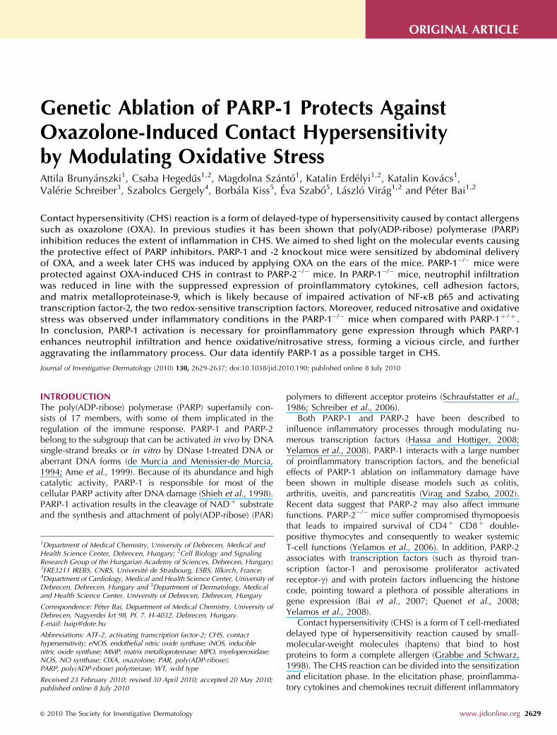
Genetic Ablation of PARP-1 Protects Against Oxazolone-Induced Contact Hypersensitivity by Modulating Oxidative Stress Attila Brunya ´nszki 1 , Csaba Hegedu + s 1,2 , Magdolna Sza ´nto ´ 1 , Katalin Erde ´lyi 1,2 , Katalin Kova ´cs 1 , Vale ´rie Schreiber 3 , Szabolcs Gergely 4 , Borba ´la Kiss 5 ,E ´ va Szabo ´ 5 , La ´szlo ´ Vira ´g 1,2 and Pe ´ter Bai 1,2 Contact hypersensitivity (CHS) reaction is a form of delayed-type of hypersensitivity caused by contact allergens such as oxazolone (OXA). In previous studies it has been shown that poly(ADP-ribose) polymerase (PARP) inhibition reduces the extent of inflammation in CHS. We aimed to shed light on the molecular events causing the protective effect of PARP inhibitors. PARP-1 and -2 knockout mice were sensitized by abdominal delivery of OXA, and a week later CHS was induced by applying OXA on the ears of the mice. PARP-1 / mice were protected against OXA-induced CHS in contrast to PARP-2 / mice. In PARP-1 / mice, neutrophil infiltration was reduced in line with the suppressed expression of proinflammatory cytokines, cell adhesion factors, and matrix metalloproteinase-9, which is likely because of impaired activation of NF-kB p65 and activating transcription factor-2, the two redox-sensitive transcription factors. Moreover, reduced nitrosative and oxidative stress was observed under inflammatory conditions in the PARP-1 / mice when compared with PARP-1 þ / þ . In conclusion, PARP-1 activation is necessary for proinflammatory gene expression through which PARP-1 enhances neutrophil infiltration and hence oxidative/nitrosative stress, forming a vicious circle, and further aggravating the inflammatory process. Our data identify PARP-1 as a possible target in CHS. Journal of Investigative Dermatology (2010) 130, 2629–2637; doi:10.1038/jid.2010.190; published online 8 July 2010 INTRODUCTION The poly(ADP-ribose) polymerase (PARP) superfamily con- sists of 17 members, with some of them implicated in the regulation of the immune response. PARP-1 and PARP-2 belong to the subgroup that can be activated in vivo by DNA single-strand breaks or in vitro by DNase I-treated DNA or aberrant DNA forms (de Murcia and Menissier-de Murcia, 1994; Ame et al., 1999). Because of its abundance and high catalytic activity, PARP-1 is responsible for most of the cellular PARP activity after DNA damage (Shieh et al., 1998). PARP-1 activation results in the cleavage of NAD þ substrate and the synthesis and attachment of poly(ADP-ribose) (PAR) polymers to different acceptor proteins (Schraufstatter et al., 1986; Schreiber et al., 2006). Both PARP-1 and PARP-2 have been described to influence inflammatory processes through modulating nu- merous transcription factors (Hassa and Hottiger, 2008; Yelamos et al., 2008). PARP-1 interacts with a large number of proinflammatory transcription factors, and the beneficial effects of PARP-1 ablation on inflammatory damage have been shown in multiple disease models such as colitis, arthritis, uveitis, and pancreatitis (Virag and Szabo, 2002). Recent data suggest that PARP-2 may also affect immune functions. PARP-2 / mice suffer compromised thymopoesis that leads to impaired survival of CD4 þ CD8 þ double- positive thymocytes and consequently to weaker systemic T-cell functions (Yelamos et al., 2006). In addition, PARP-2 associates with transcription factors (such as thyroid tran- scription factor-1 and peroxisome proliferator activated receptor-g) and with protein factors influencing the histone code, pointing toward a plethora of possible alterations in gene expression (Bai et al., 2007; Quenet et al., 2008; Yelamos et al., 2008). Contact hypersensitivity (CHS) is a form of T cell-mediated delayed type of hypersensitivity reaction caused by small- molecular-weight molecules (haptens) that bind to host proteins to form a complete allergen (Grabbe and Schwarz, 1998). The CHS reaction can be divided into the sensitization and elicitation phase. In the elicitation phase, proinflamma- tory cytokines and chemokines recruit different inflammatory & 2010 The Society for Investigative Dermatology www.jidonline.org 2629 ORIGINAL ARTICLE Received 23 February 2010; revised 30 April 2010; accepted 20 May 2010; published online 8 July 2010 1 Department of Medical Chemistry, University of Debrecen, Medical and Health Science Center, Debrecen, Hungary; 2 Cell Biology and Signaling Research Group of the Hungarian Academy of Sciences, Debrecen, Hungary; 3 FRE3211 IREBS, CNRS, Universite ´ de Strasbourg, ESBS, Illkirch, France; 4 Department of Cardiology, Medical and Health Science Center, University of Debrecen, Debrecen, Hungary and 5 Department of Dermatology, Medical and Health Science Center, University of Debrecen, Debrecen, Hungary Correspondence: Pe ´ter Bai, Department of Medical Chemistry, University of Debrecen, Nagyerdei krt 98, Pf. 7. H-4032, Debrecen, Hungary. E-mail: [email protected] Abbreviations: ATF-2, activating transcription factor-2; CHS, contact hypersensitivity; eNOS, endothelial nitric oxide synthase; iNOS, inducible nitric oxide synthase; MMP, matrix metalloproteinase; MPO, myeloperoxidase; NOS, NO synthase; OXA, oxazolone; PAR, poly(ADP-ribose); PARP, poly(ADP-ribose) polymerase; WT, wild type

Welcome message from author
This document is posted to help you gain knowledge. Please leave a comment to let me know what you think about it! Share it to your friends and learn new things together.
Transcript
Genetic Ablation of PARP-1 Protects AgainstOxazolone-Induced Contact Hypersensitivityby Modulating Oxidative StressAttila Brunyanszki1, Csaba Hegedu+s1,2, Magdolna Szanto1, Katalin Erdelyi1,2, Katalin Kovacs1,Valerie Schreiber3, Szabolcs Gergely4, Borbala Kiss5, Eva Szabo5, Laszlo Virag1,2 and Peter Bai1,2
Contact hypersensitivity (CHS) reaction is a form of delayed-type of hypersensitivity caused by contact allergenssuch as oxazolone (OXA). In previous studies it has been shown that poly(ADP-ribose) polymerase (PARP)inhibition reduces the extent of inflammation in CHS. We aimed to shed light on the molecular events causingthe protective effect of PARP inhibitors. PARP-1 and -2 knockout mice were sensitized by abdominal deliveryof OXA, and a week later CHS was induced by applying OXA on the ears of the mice. PARP-1�/� mice wereprotected against OXA-induced CHS in contrast to PARP-2�/� mice. In PARP-1�/� mice, neutrophil infiltrationwas reduced in line with the suppressed expression of proinflammatory cytokines, cell adhesion factors,and matrix metalloproteinase-9, which is likely because of impaired activation of NF-kB p65 and activatingtranscription factor-2, the two redox-sensitive transcription factors. Moreover, reduced nitrosative and oxidativestress was observed under inflammatory conditions in the PARP-1�/� mice when compared with PARP-1þ /þ .In conclusion, PARP-1 activation is necessary for proinflammatory gene expression through which PARP-1enhances neutrophil infiltration and hence oxidative/nitrosative stress, forming a vicious circle, and furtheraggravating the inflammatory process. Our data identify PARP-1 as a possible target in CHS.
Journal of Investigative Dermatology (2010) 130, 2629–2637; doi:10.1038/jid.2010.190; published online 8 July 2010
INTRODUCTIONThe poly(ADP-ribose) polymerase (PARP) superfamily con-sists of 17 members, with some of them implicated in theregulation of the immune response. PARP-1 and PARP-2belong to the subgroup that can be activated in vivo by DNAsingle-strand breaks or in vitro by DNase I-treated DNA oraberrant DNA forms (de Murcia and Menissier-de Murcia,1994; Ame et al., 1999). Because of its abundance and highcatalytic activity, PARP-1 is responsible for most of thecellular PARP activity after DNA damage (Shieh et al., 1998).PARP-1 activation results in the cleavage of NADþ substrateand the synthesis and attachment of poly(ADP-ribose) (PAR)
polymers to different acceptor proteins (Schraufstatter et al.,1986; Schreiber et al., 2006).
Both PARP-1 and PARP-2 have been described toinfluence inflammatory processes through modulating nu-merous transcription factors (Hassa and Hottiger, 2008;Yelamos et al., 2008). PARP-1 interacts with a large numberof proinflammatory transcription factors, and the beneficialeffects of PARP-1 ablation on inflammatory damage havebeen shown in multiple disease models such as colitis,arthritis, uveitis, and pancreatitis (Virag and Szabo, 2002).Recent data suggest that PARP-2 may also affect immunefunctions. PARP-2�/� mice suffer compromised thymopoesisthat leads to impaired survival of CD4þ CD8þ double-positive thymocytes and consequently to weaker systemicT-cell functions (Yelamos et al., 2006). In addition, PARP-2associates with transcription factors (such as thyroid tran-scription factor-1 and peroxisome proliferator activatedreceptor-g) and with protein factors influencing the histonecode, pointing toward a plethora of possible alterations ingene expression (Bai et al., 2007; Quenet et al., 2008;Yelamos et al., 2008).
Contact hypersensitivity (CHS) is a form of T cell-mediateddelayed type of hypersensitivity reaction caused by small-molecular-weight molecules (haptens) that bind to hostproteins to form a complete allergen (Grabbe and Schwarz,1998). The CHS reaction can be divided into the sensitizationand elicitation phase. In the elicitation phase, proinflamma-tory cytokines and chemokines recruit different inflammatory
& 2010 The Society for Investigative Dermatology www.jidonline.org 2629
ORIGINAL ARTICLE
Received 23 February 2010; revised 30 April 2010; accepted 20 May 2010;published online 8 July 2010
1Department of Medical Chemistry, University of Debrecen, Medical andHealth Science Center, Debrecen, Hungary; 2Cell Biology and SignalingResearch Group of the Hungarian Academy of Sciences, Debrecen, Hungary;3FRE3211 IREBS, CNRS, Universite de Strasbourg, ESBS, Illkirch, France;4Department of Cardiology, Medical and Health Science Center, University ofDebrecen, Debrecen, Hungary and 5Department of Dermatology, Medicaland Health Science Center, University of Debrecen, Debrecen, Hungary
Correspondence: Peter Bai, Department of Medical Chemistry, University ofDebrecen, Nagyerdei krt 98, Pf. 7. H-4032, Debrecen, Hungary.E-mail: [email protected]
Abbreviations: ATF-2, activating transcription factor-2; CHS, contacthypersensitivity; eNOS, endothelial nitric oxide synthase; iNOS, induciblenitric oxide synthase; MMP, matrix metalloproteinase; MPO, myeloperoxidase;NOS, NO synthase; OXA, oxazolone; PAR, poly(ADP-ribose);PARP, poly(ADP-ribose) polymerase; WT, wild type
cells (e.g., neutrophil granulocytes) (Olmos et al., 2007).Infiltration is accompanied by oxidative stress due to theformation of superoxide, hydrogen peroxide, nitric oxide, per-oxynitrite, and further reactive species (Morita et al., 1996;Rowe et al., 1997; Olmos et al., 2007; Korkina and Pastore,2009), leading to DNA breakage and PARP activation (Szaboet al., 2001). Recently, we showed that the inhibition of PARPactivation has beneficial effects during the CHS reaction (Baiet al., 2009). In this study we set out to identify the PARPisoform mediating CHS and to characterize the molecularevents during the elicitation phase of the CHS reaction.
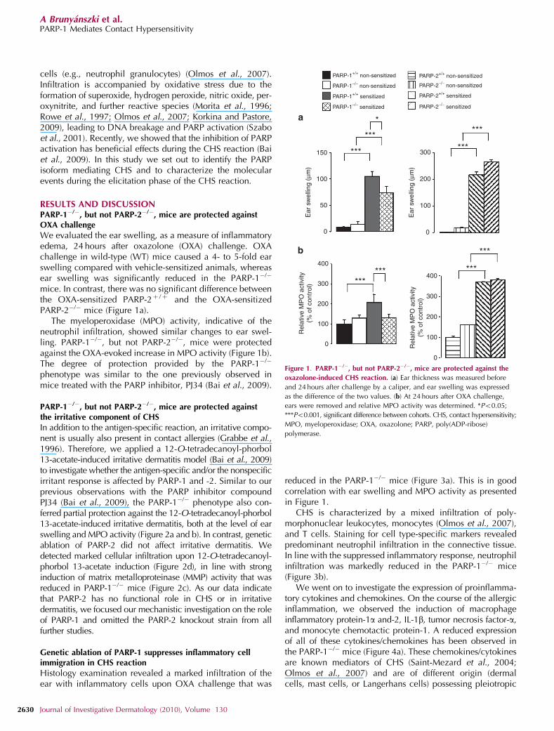
RESULTS AND DISCUSSIONPARP-1�/�, but not PARP-2�/�, mice are protected againstOXA challenge
We evaluated the ear swelling, as a measure of inflammatoryedema, 24 hours after oxazolone (OXA) challenge. OXAchallenge in wild-type (WT) mice caused a 4- to 5-fold earswelling compared with vehicle-sensitized animals, whereasear swelling was significantly reduced in the PARP-1�/�
mice. In contrast, there was no significant difference betweenthe OXA-sensitized PARP-2þ /þ and the OXA-sensitizedPARP-2�/� mice (Figure 1a).
The myeloperoxidase (MPO) activity, indicative of theneutrophil infiltration, showed similar changes to ear swel-ling. PARP-1�/�, but not PARP-2�/�, mice were protectedagainst the OXA-evoked increase in MPO activity (Figure 1b).The degree of protection provided by the PARP-1�/�
phenotype was similar to the one previously observed inmice treated with the PARP inhibitor, PJ34 (Bai et al., 2009).
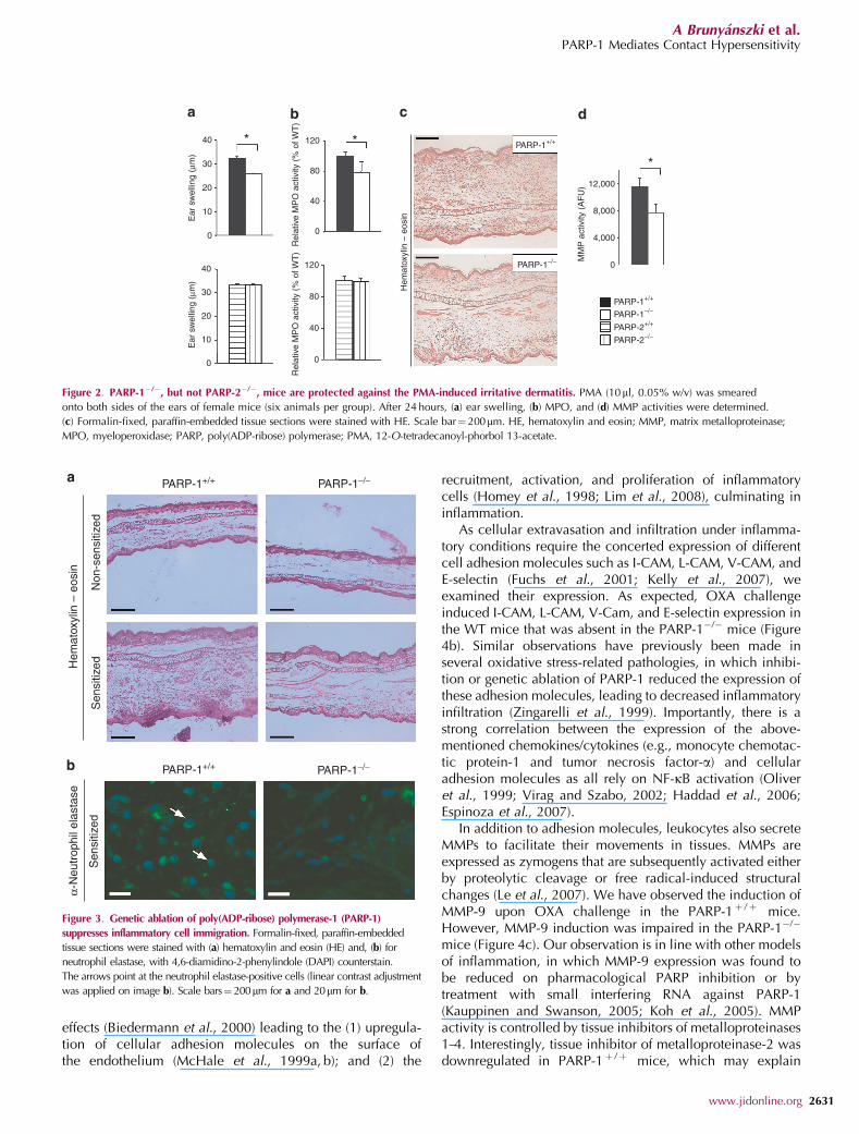
PARP-1�/�, but not PARP-2�/�, mice are protected againstthe irritative component of CHS
In addition to the antigen-specific reaction, an irritative compo-nent is usually also present in contact allergies (Grabbe et al.,1996). Therefore, we applied a 12-O-tetradecanoyl-phorbol13-acetate-induced irritative dermatitis model (Bai et al., 2009)to investigate whether the antigen-specific and/or the nonspecificirritant response is affected by PARP-1 and -2. Similar to ourprevious observations with the PARP inhibitor compoundPJ34 (Bai et al., 2009), the PARP-1�/� phenotype also con-ferred partial protection against the 12-O-tetradecanoyl-phorbol13-acetate-induced irritative dermatitis, both at the level of earswelling and MPO activity (Figure 2a and b). In contrast, geneticablation of PARP-2 did not affect irritative dermatitis. Wedetected marked cellular infiltration upon 12-O-tetradecanoyl-phorbol 13-acetate induction (Figure 2d), in line with stronginduction of matrix metalloproteinase (MMP) activity that wasreduced in PARP-1�/� mice (Figure 2c). As our data indicatethat PARP-2 has no functional role in CHS or in irritativedermatitis, we focused our mechanistic investigation on the roleof PARP-1 and omitted the PARP-2 knockout strain from allfurther studies.
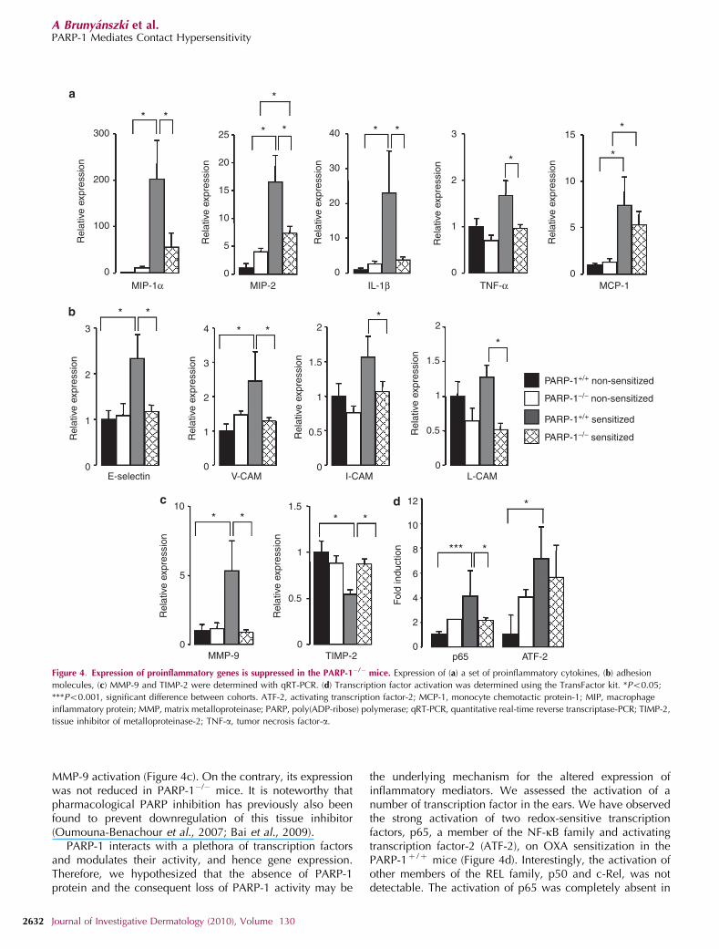
Genetic ablation of PARP-1 suppresses inflammatory cellimmigration in CHS reaction
Histology examination revealed a marked infiltration of theear with inflammatory cells upon OXA challenge that was
reduced in the PARP-1�/� mice (Figure 3a). This is in goodcorrelation with ear swelling and MPO activity as presentedin Figure 1.
CHS is characterized by a mixed infiltration of poly-morphonuclear leukocytes, monocytes (Olmos et al., 2007),and T cells. Staining for cell type-specific markers revealedpredominant neutrophil infiltration in the connective tissue.In line with the suppressed inflammatory response, neutrophilinfiltration was markedly reduced in the PARP-1�/� mice(Figure 3b).
We went on to investigate the expression of proinflamma-tory cytokines and chemokines. On the course of the allergicinflammation, we observed the induction of macrophageinflammatory protein-1a and-2, IL-1b, tumor necrosis factor-a,and monocyte chemotactic protein-1. A reduced expressionof all of these cytokines/chemokines has been observed inthe PARP-1�/� mice (Figure 4a). These chemokines/cytokinesare known mediators of CHS (Saint-Mezard et al., 2004;Olmos et al., 2007) and are of different origin (dermalcells, mast cells, or Langerhans cells) possessing pleiotropic
PARP-1+/+ non-sensitized
PARP-1−/− non-sensitized
PARP-1−/− sensitized
PARP-1+/+ sensitized
Ear
sw
ellin
g (μ
m)
150
100
50
0
Rel
ativ
e M
PO
act
ivity
(% o
f con
trol
)
400
300
200
100
0 Rel
ativ
e M
PO
act
ivity
(% o
f con
trol
)
400
300
200
100
0
Ear
sw
ellin
g (μ
m)
300
200
100
0
***
***
PARP-2+/+ non-sensitized
PARP-2−/− non-sensitized
PARP-2−/− sensitized
PARP-2+/+ sensitized
******
***
***
****
***
Figure 1. PARP-1�/�, but not PARP-2�/�, mice are protected against the
oxazolone-induced CHS reaction. (a) Ear thickness was measured before
and 24 hours after challenge by a caliper, and ear swelling was expressed
as the difference of the two values. (b) At 24 hours after OXA challenge,
ears were removed and relative MPO activity was determined. *Po0.05;
***Po0.001, significant difference between cohorts. CHS, contact hypersensitivity;
MPO, myeloperoxidase; OXA, oxazolone; PARP, poly(ADP-ribose)
polymerase.
2630 Journal of Investigative Dermatology (2010), Volume 130
A Brunyanszki et al.PARP-1 Mediates Contact Hypersensitivity
effects (Biedermann et al., 2000) leading to the (1) upregula-tion of cellular adhesion molecules on the surface ofthe endothelium (McHale et al., 1999a, b); and (2) the
recruitment, activation, and proliferation of inflammatorycells (Homey et al., 1998; Lim et al., 2008), culminating ininflammation.
As cellular extravasation and infiltration under inflamma-tory conditions require the concerted expression of differentcell adhesion molecules such as I-CAM, L-CAM, V-CAM, andE-selectin (Fuchs et al., 2001; Kelly et al., 2007), weexamined their expression. As expected, OXA challengeinduced I-CAM, L-CAM, V-Cam, and E-selectin expression inthe WT mice that was absent in the PARP-1�/� mice (Figure4b). Similar observations have previously been made inseveral oxidative stress-related pathologies, in which inhibi-tion or genetic ablation of PARP-1 reduced the expression ofthese adhesion molecules, leading to decreased inflammatoryinfiltration (Zingarelli et al., 1999). Importantly, there is astrong correlation between the expression of the above-mentioned chemokines/cytokines (e.g., monocyte chemotac-tic protein-1 and tumor necrosis factor-a) and cellularadhesion molecules as all rely on NF-kB activation (Oliveret al., 1999; Virag and Szabo, 2002; Haddad et al., 2006;Espinoza et al., 2007).
In addition to adhesion molecules, leukocytes also secreteMMPs to facilitate their movements in tissues. MMPs areexpressed as zymogens that are subsequently activated eitherby proteolytic cleavage or free radical-induced structuralchanges (Le et al., 2007). We have observed the induction ofMMP-9 upon OXA challenge in the PARP-1þ /þ mice.However, MMP-9 induction was impaired in the PARP-1�/�
mice (Figure 4c). Our observation is in line with other modelsof inflammation, in which MMP-9 expression was found tobe reduced on pharmacological PARP inhibition or bytreatment with small interfering RNA against PARP-1(Kauppinen and Swanson, 2005; Koh et al., 2005). MMPactivity is controlled by tissue inhibitors of metalloproteinases1–4. Interestingly, tissue inhibitor of metalloproteinase-2 wasdownregulated in PARP-1þ /þ mice, which may explain
* *
Ear
sw
ellin
g (μ
m)
40
30
20
10
0
Ear
sw
ellin
g (μ
m)
40
30
20
10
0
Rel
ativ
e M
PO
act
ivity
(%
of W
T)
Hem
atox
ylin
– e
osin
120
80
40
0
Rel
ativ
e M
PO
act
ivity
(%
of W
T)
120
80
40
0
PARP-1+/+
PARP-1+/+
PARP-1−/−
PARP-1−/−
PARP-2+/+
PARP-2−/−
MM
P a
ctiv
ity (
AF
U) 12,000
8,000
4,000
0
*
Figure 2. PARP-1�/�, but not PARP-2�/�, mice are protected against the PMA-induced irritative dermatitis. PMA (10ml, 0.05% w/v) was smeared
onto both sides of the ears of female mice (six animals per group). After 24 hours, (a) ear swelling, (b) MPO, and (d) MMP activities were determined.
(c) Formalin-fixed, paraffin-embedded tissue sections were stained with HE. Scale bar¼ 200mm. HE, hematoxylin and eosin; MMP, matrix metalloproteinase;
MPO, myeloperoxidase; PARP, poly(ADP-ribose) polymerase; PMA, 12-O-tetradecanoyl-phorbol 13-acetate.
PARP-1+/+
Non
-sen
sitiz
edS
ensi
tized
Sen
sitiz
ed
Hem
atox
ylin
– e
osin
α-N
eutr
ophi
l ela
stas
e
PARP-1−/−
PARP-1+/+ PARP-1−/−
Figure 3. Genetic ablation of poly(ADP-ribose) polymerase-1 (PARP-1)
suppresses inflammatory cell immigration. Formalin-fixed, paraffin-embedded
tissue sections were stained with (a) hematoxylin and eosin (HE) and, (b) for
neutrophil elastase, with 4,6-diamidino-2-phenylindole (DAPI) counterstain.
The arrows point at the neutrophil elastase-positive cells (linear contrast adjustment
was applied on image b). Scale bars¼ 200mm for a and 20mm for b.
www.jidonline.org 2631
A Brunyanszki et al.PARP-1 Mediates Contact Hypersensitivity
MMP-9 activation (Figure 4c). On the contrary, its expressionwas not reduced in PARP-1�/� mice. It is noteworthy thatpharmacological PARP inhibition has previously also beenfound to prevent downregulation of this tissue inhibitor(Oumouna-Benachour et al., 2007; Bai et al., 2009).
PARP-1 interacts with a plethora of transcription factorsand modulates their activity, and hence gene expression.Therefore, we hypothesized that the absence of PARP-1protein and the consequent loss of PARP-1 activity may be
the underlying mechanism for the altered expression ofinflammatory mediators. We assessed the activation of anumber of transcription factor in the ears. We have observedthe strong activation of two redox-sensitive transcriptionfactors, p65, a member of the NF-kB family and activatingtranscription factor-2 (ATF-2), on OXA sensitization in thePARP-1þ /þ mice (Figure 4d). Interestingly, the activation ofother members of the REL family, p50 and c-Rel, was notdetectable. The activation of p65 was completely absent in
Rel
ativ
e ex
pres
sion
Rel
ativ
e ex
pres
sion
300
200
0
100
MIP-1α MIP-2 IL-1β TNF-α MCP-1
25
20
15
10
5
0
Rel
ativ
e ex
pres
sion
40
20
30
10
0
Rel
ativ
e ex
pres
sion
3
2
1
0
E-selectin
MMP-9
Rel
ativ
e ex
pres
sion
Rel
ativ
e ex
pres
sion
3
2
1
0V-CAM
Rel
ativ
e ex
pres
sion
4
3
2
1
0I-CAM
Rel
ativ
e ex
pres
sion
2
1.5
1
0.5
0L-CAM
Rel
ativ
e ex
pres
sion
2
1.5
1
0.5
0
TIMP-2
Rel
ativ
e ex
pres
sion
1.5
1
0.5
0p65 ATF-2
Fol
d in
duct
ion
12
10
8
6
4
2
0
Rel
ativ
e ex
pres
sion
15
10
5
0
PARP-1+/+ non-sensitized
PARP-1−/− non-sensitized
PARP-1−/− sensitized
PARP-1+/+ sensitized
10
5
0
* *
*
* * * *
* *
*
* *
* **
*
* * * *
*** *
*
Figure 4. Expression of proinflammatory genes is suppressed in the PARP-1�/� mice. Expression of (a) a set of proinflammatory cytokines, (b) adhesion
molecules, (c) MMP-9 and TIMP-2 were determined with qRT-PCR. (d) Transcription factor activation was determined using the TransFactor kit. *Po0.05;
***Po0.001, significant difference between cohorts. ATF-2, activating transcription factor-2; MCP-1, monocyte chemotactic protein-1; MIP, macrophage
inflammatory protein; MMP, matrix metalloproteinase; PARP, poly(ADP-ribose) polymerase; qRT-PCR, quantitative real-time reverse transcriptase-PCR; TIMP-2,
tissue inhibitor of metalloproteinase-2; TNF-a, tumor necrosis factor-a.
2632 Journal of Investigative Dermatology (2010), Volume 130
A Brunyanszki et al.PARP-1 Mediates Contact Hypersensitivity
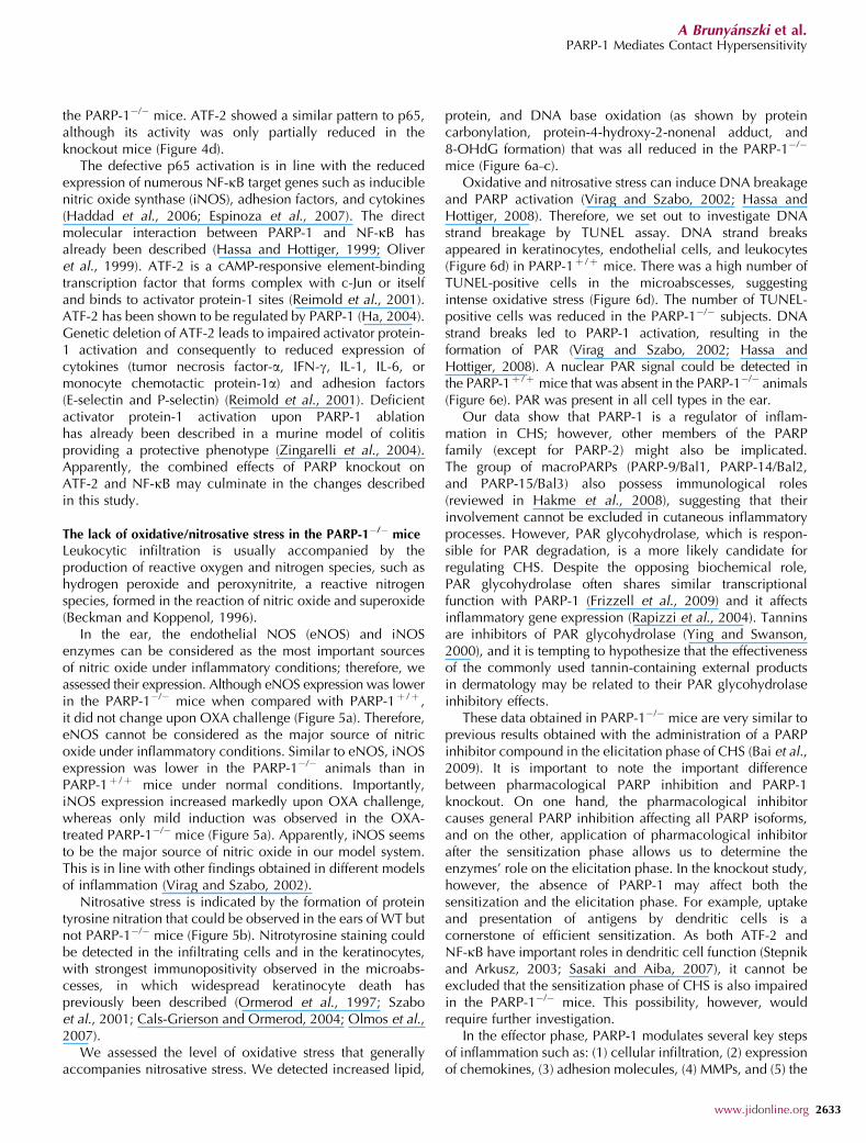
the PARP-1�/� mice. ATF-2 showed a similar pattern to p65,although its activity was only partially reduced in theknockout mice (Figure 4d).
The defective p65 activation is in line with the reducedexpression of numerous NF-kB target genes such as induciblenitric oxide synthase (iNOS), adhesion factors, and cytokines(Haddad et al., 2006; Espinoza et al., 2007). The directmolecular interaction between PARP-1 and NF-kB hasalready been described (Hassa and Hottiger, 1999; Oliveret al., 1999). ATF-2 is a cAMP-responsive element-bindingtranscription factor that forms complex with c-Jun or itselfand binds to activator protein-1 sites (Reimold et al., 2001).ATF-2 has been shown to be regulated by PARP-1 (Ha, 2004).Genetic deletion of ATF-2 leads to impaired activator protein-1 activation and consequently to reduced expression ofcytokines (tumor necrosis factor-a, IFN-g, IL-1, IL-6, ormonocyte chemotactic protein-1a) and adhesion factors(E-selectin and P-selectin) (Reimold et al., 2001). Deficientactivator protein-1 activation upon PARP-1 ablationhas already been described in a murine model of colitisproviding a protective phenotype (Zingarelli et al., 2004).Apparently, the combined effects of PARP knockout onATF-2 and NF-kB may culminate in the changes describedin this study.
The lack of oxidative/nitrosative stress in the PARP-1�/� mice
Leukocytic infiltration is usually accompanied by theproduction of reactive oxygen and nitrogen species, such ashydrogen peroxide and peroxynitrite, a reactive nitrogenspecies, formed in the reaction of nitric oxide and superoxide(Beckman and Koppenol, 1996).
In the ear, the endothelial NOS (eNOS) and iNOSenzymes can be considered as the most important sourcesof nitric oxide under inflammatory conditions; therefore, weassessed their expression. Although eNOS expression was lowerin the PARP-1�/� mice when compared with PARP-1þ /þ ,it did not change upon OXA challenge (Figure 5a). Therefore,eNOS cannot be considered as the major source of nitricoxide under inflammatory conditions. Similar to eNOS, iNOSexpression was lower in the PARP-1�/� animals than inPARP-1þ /þ mice under normal conditions. Importantly,iNOS expression increased markedly upon OXA challenge,whereas only mild induction was observed in the OXA-treated PARP-1�/� mice (Figure 5a). Apparently, iNOS seemsto be the major source of nitric oxide in our model system.This is in line with other findings obtained in different modelsof inflammation (Virag and Szabo, 2002).
Nitrosative stress is indicated by the formation of proteintyrosine nitration that could be observed in the ears of WT butnot PARP-1�/� mice (Figure 5b). Nitrotyrosine staining couldbe detected in the infiltrating cells and in the keratinocytes,with strongest immunopositivity observed in the microabs-cesses, in which widespread keratinocyte death haspreviously been described (Ormerod et al., 1997; Szaboet al., 2001; Cals-Grierson and Ormerod, 2004; Olmos et al.,2007).
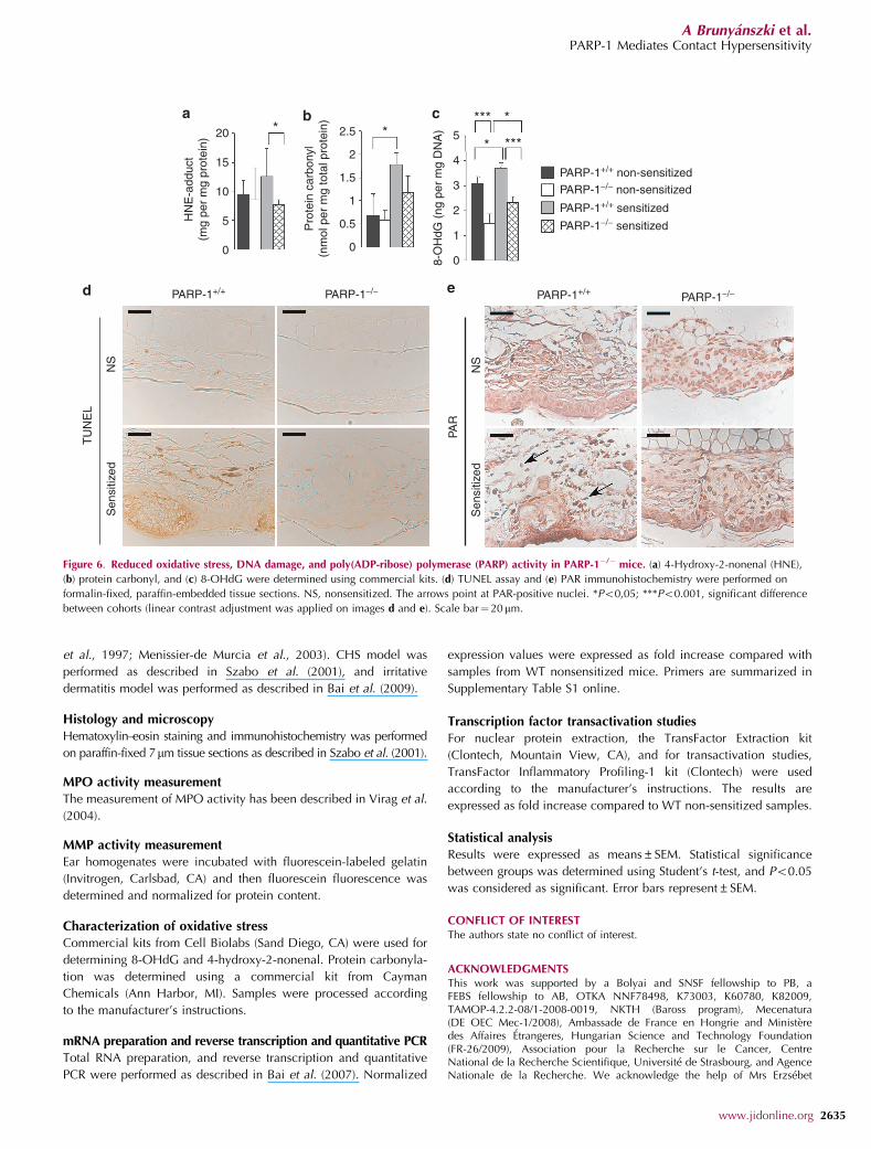
We assessed the level of oxidative stress that generallyaccompanies nitrosative stress. We detected increased lipid,
protein, and DNA base oxidation (as shown by proteincarbonylation, protein-4-hydroxy-2-nonenal adduct, and8-OHdG formation) that was all reduced in the PARP-1�/�
mice (Figure 6a–c).Oxidative and nitrosative stress can induce DNA breakage
and PARP activation (Virag and Szabo, 2002; Hassa andHottiger, 2008). Therefore, we set out to investigate DNAstrand breakage by TUNEL assay. DNA strand breaksappeared in keratinocytes, endothelial cells, and leukocytes(Figure 6d) in PARP-1þ /þ mice. There was a high number ofTUNEL-positive cells in the microabscesses, suggestingintense oxidative stress (Figure 6d). The number of TUNEL-positive cells was reduced in the PARP-1�/� subjects. DNAstrand breaks led to PARP-1 activation, resulting in theformation of PAR (Virag and Szabo, 2002; Hassa andHottiger, 2008). A nuclear PAR signal could be detected inthe PARP-1þ /þ mice that was absent in the PARP-1�/� animals(Figure 6e). PAR was present in all cell types in the ear.
Our data show that PARP-1 is a regulator of inflam-mation in CHS; however, other members of the PARPfamily (except for PARP-2) might also be implicated.The group of macroPARPs (PARP-9/Bal1, PARP-14/Bal2,and PARP-15/Bal3) also possess immunological roles(reviewed in Hakme et al., 2008), suggesting that theirinvolvement cannot be excluded in cutaneous inflammatoryprocesses. However, PAR glycohydrolase, which is respon-sible for PAR degradation, is a more likely candidate forregulating CHS. Despite the opposing biochemical role,PAR glycohydrolase often shares similar transcriptionalfunction with PARP-1 (Frizzell et al., 2009) and it affectsinflammatory gene expression (Rapizzi et al., 2004). Tanninsare inhibitors of PAR glycohydrolase (Ying and Swanson,2000), and it is tempting to hypothesize that the effectivenessof the commonly used tannin-containing external productsin dermatology may be related to their PAR glycohydrolaseinhibitory effects.
These data obtained in PARP-1�/� mice are very similar toprevious results obtained with the administration of a PARPinhibitor compound in the elicitation phase of CHS (Bai et al.,2009). It is important to note the important differencebetween pharmacological PARP inhibition and PARP-1knockout. On one hand, the pharmacological inhibitorcauses general PARP inhibition affecting all PARP isoforms,and on the other, application of pharmacological inhibitorafter the sensitization phase allows us to determine theenzymes’ role on the elicitation phase. In the knockout study,however, the absence of PARP-1 may affect both thesensitization and the elicitation phase. For example, uptakeand presentation of antigens by dendritic cells is acornerstone of efficient sensitization. As both ATF-2 andNF-kB have important roles in dendritic cell function (Stepnikand Arkusz, 2003; Sasaki and Aiba, 2007), it cannot beexcluded that the sensitization phase of CHS is also impairedin the PARP-1�/� mice. This possibility, however, wouldrequire further investigation.
In the effector phase, PARP-1 modulates several key stepsof inflammation such as: (1) cellular infiltration, (2) expressionof chemokines, (3) adhesion molecules, (4) MMPs, and (5) the
www.jidonline.org 2633
A Brunyanszki et al.PARP-1 Mediates Contact Hypersensitivity

redox-sensitive p65 and ATF-2 transcription factors. In apossible scenario, the increasing level of oxidative/nitrosative stress enhances PARP-1 activation that in turnboosts all the previously described processes, furtheraggravating oxidative/nitrosative stress and creating a viciouscircle. In fact, PARP-1 has a central role in the control ofoxidative stress in the elicitation phase of CHS, and itsinhibition or genetic deletion disrupts the self-intensifyingpropagation of inflammation. Altogether, these results pro-vide further support for the possible therapeutic applicabilityof PARP inhibitors in CHS.
MATERIALS AND METHODSMaterials
OXA and all chemicals were from Sigma-Aldrich (St Louis, MO)
unless stated otherwise.
Animal studies
All animal experiments were approved by the local ethical
committee (9/2008/DE MAB) and were performed according to the
European Union and national guidelines. Homozygous female
PARP-1 and PARP-2 knockout mice and their respective WT
littermates on C57/Bl6 background were used (Menissier-de Murcia
Rel
ativ
e iN
OS
exp
ress
ion
Rel
ativ
e eN
OS
exp
ress
ion
160
120
80
40
0
1.5
1
0.5
0
PARP-1+/+ non-sensitized
PARP-1−/− non-sensitized
PARP-1−/− sensitized
PARP-1+/+ sensitized
***
*** *
PARP-1+/+ PARP-1−/−
NS
Nitr
otyr
osin
e
Sen
sitiz
ed
Figure 5. Reduced nitrosative stress in poly(ADP-ribose) polymerase (PARP)-1�/� mice. (a) Inducible and endothelial nitric oxide synthase (iNOS and eNOS)
expression was determined by quantitative real-time reverse transcriptase PCR (qRT-PCR). (b) Nitrotyrosine immunohistochemistry was performed on
formalin-fixed, paraffin-embedded tissue sections. NS, nonsensitized. *Po0,05; ***Po0.001, significant difference between cohorts (linear contrast adjustment
was applied on image b). Scale bar¼ 20mm.
2634 Journal of Investigative Dermatology (2010), Volume 130
A Brunyanszki et al.PARP-1 Mediates Contact Hypersensitivity
et al., 1997; Menissier-de Murcia et al., 2003). CHS model was
performed as described in Szabo et al. (2001), and irritative
dermatitis model was performed as described in Bai et al. (2009).
Histology and microscopy
Hematoxylin–eosin staining and immunohistochemistry was performed
on paraffin-fixed 7mm tissue sections as described in Szabo et al. (2001).
MPO activity measurement
The measurement of MPO activity has been described in Virag et al.
(2004).
MMP activity measurement
Ear homogenates were incubated with fluorescein-labeled gelatin
(Invitrogen, Carlsbad, CA) and then fluorescein fluorescence was
determined and normalized for protein content.
Characterization of oxidative stress
Commercial kits from Cell Biolabs (Sand Diego, CA) were used for
determining 8-OHdG and 4-hydroxy-2-nonenal. Protein carbonyla-
tion was determined using a commercial kit from Cayman
Chemicals (Ann Harbor, MI). Samples were processed according
to the manufacturer’s instructions.
mRNA preparation and reverse transcription and quantitative PCR
Total RNA preparation, and reverse transcription and quantitative
PCR were performed as described in Bai et al. (2007). Normalized
expression values were expressed as fold increase compared with
samples from WT nonsensitized mice. Primers are summarized in
Supplementary Table S1 online.
Transcription factor transactivation studies
For nuclear protein extraction, the TransFactor Extraction kit
(Clontech, Mountain View, CA), and for transactivation studies,
TransFactor Inflammatory Profiling-1 kit (Clontech) were used
according to the manufacturer’s instructions. The results are
expressed as fold increase compared to WT non-sensitized samples.
Statistical analysis
Results were expressed as means±SEM. Statistical significance
between groups was determined using Student’s t-test, and Po0.05
was considered as significant. Error bars represent±SEM.
CONFLICT OF INTERESTThe authors state no conflict of interest.
ACKNOWLEDGMENTSThis work was supported by a Bolyai and SNSF fellowship to PB, aFEBS fellowship to AB, OTKA NNF78498, K73003, K60780, K82009,TAMOP-4.2.2-08/1-2008-0019, NKTH (Baross program), Mecenatura(DE OEC Mec-1/2008), Ambassade de France en Hongrie and Ministeredes Affaires Etrangeres, Hungarian Science and Technology Foundation(FR-26/2009), Association pour la Recherche sur le Cancer, CentreNational de la Recherche Scientifique, Universite de Strasbourg, and AgenceNationale de la Recherche. We acknowledge the help of Mrs Erzsebet
HN
E-a
dduc
t(m
g pe
r m
g pr
otei
n)
20
15
10
5
0
*
Pro
tein
car
bony
l(n
mol
per
mg
tota
l pro
tein
)
2.5
2
1.5
1
0.5
0
*
8-O
HdG
(ng
per
mg
DN
A) 5
4
3
2
1
0
* ***
*** *
PARP-1+/+ non-sensitizedPARP-1−/− non-sensitized
PARP-1−/− sensitized
PARP-1+/+ sensitized
PARP-1+/+
Sen
sitiz
ed
TU
NE
L
NS
PARP-1−/− PARP-1+/+ PARP-1−/−
Sen
sitiz
ed
PAR
NS
Figure 6. Reduced oxidative stress, DNA damage, and poly(ADP-ribose) polymerase (PARP) activity in PARP-1�/� mice. (a) 4-Hydroxy-2-nonenal (HNE),
(b) protein carbonyl, and (c) 8-OHdG were determined using commercial kits. (d) TUNEL assay and (e) PAR immunohistochemistry were performed on
formalin-fixed, paraffin-embedded tissue sections. NS, nonsensitized. The arrows point at PAR-positive nuclei. *Po0,05; ***Po0.001, significant difference
between cohorts (linear contrast adjustment was applied on images d and e). Scale bar¼ 20 mm.
www.jidonline.org 2635
A Brunyanszki et al.PARP-1 Mediates Contact Hypersensitivity
Herbaly in processing histology samples and Mrs Julia Hunyadi for technicalassistance.
SUPPLEMENTARY MATERIAL
Supplementary material is linked to the online version of the paper at http://www.nature.com/jid
REFERENCES
Ame JC, Rolli V, Schreiber V et al. (1999) PARP-2, a novel mammalian
DNA damage-dependent poly(ADP-ribose) polymerase. J Biol Chem
274:17860–8
Bai P, Hegedus C, Szabo E et al. (2009) Poly(ADP-ribose) polymerase
mediates inflammation in a mouse model of contact hypersensitivity.
J Invest Dermatol 129:234–8
Bai P, Houten SM, Huber A et al. (2007) Poly(ADP-ribose) polymerase-2
controls adipocyte differentiation and adipose tissue function through
the regulation of the activity of the retinoid X receptor/peroxisome
proliferator-activated receptor-gamma heterodimer. J Biol Chem
282:37738–46
Beckman JS, Koppenol WH (1996) Nitric oxide, superoxide, and peroxyni-
trite: the good, the bad, and ugly. Am J Physiol 271:C1424–37
Biedermann T, Kneilling M, Mailhammer R et al. (2000) Mast cells control
neutrophil recruitment during T cell-mediated delayed-type hypersensi-
tivity reactions through tumor necrosis factor and macrophage inflam-
matory protein 2. J Exp Med 192:1441–52
Cals-Grierson MM, Ormerod AD (2004) Nitric oxide function in the skin.
Nitric Oxide 10:179–93
de Murcia G, Menissier-de Murcia J (1994) Poly(ADP-ribose) polymerase: a
molecular nick-sensor. Trends Biochem Sci 19:172–6
Espinoza LA, Smulson ME, Chen Z (2007) Prolonged poly(ADP-ribose)
polymerase-1 activity regulates JP-8-induced sustained cytokine expres-
sion in alveolar macrophages. Free Radic Biol Med 42:1430–40
Frizzell KM, Gamble MJ, Berrocal JG et al. (2009) Global analysis of
transcriptional regulation by poly(ADP-ribose) polymerase-1 and
poly(ADP-ribose) glycohydrolase in MCF-7 human breast cancer cells.
J Biol Chem 284:33926–38
Fuchs J, Zollner TM, Kaufmann R et al. (2001) Redox-modulated pathways in
inflammatory skin diseases. Free Radic Biol Med 30:337–53
Grabbe S, Schwarz T (1998) Immunoregulatory mechanisms involved
in elicitation of allergic contact hypersensitivity. Immunol Today
19:37–44
Grabbe S, Steinert M, Mahnke K et al. (1996) Dissection of antigenic and
irritative effects of epicutaneously applied haptens in mice. Evidence that
not the antigenic component but nonspecific proinflammatory effects of
haptens determine the concentration-dependent elicitation of allergic
contact dermatitis. J Clin Invest 98:1158–64
Ha HC (2004) Defective transcription factor activation for proinflammatory
gene expression in poly(ADP-ribose) polymerase 1-deficient glia. Proc
Natl Acad Sci USA 101:5087–92
Haddad M, Rhinn H, Bloquel C et al. (2006) Anti-inflammatory effects of
PJ34, a poly(ADP-ribose) polymerase inhibitor, in transient focal cerebral
ischemia in mice. Br J Pharmacol 149:23–30
Hakme A, Huber A, Dolle P et al. (2008) The macroPARP genes Parp-9 and
Parp-14 are developmentally and differentially regulated in mouse
tissues. Dev Dyn 237:209–15
Hassa PO, Hottiger MO (1999) A role of poly (ADP-ribose) polymerase in
NF-kappaB transcriptional activation. Biol Chem 380:953–9
Hassa PO, Hottiger MO (2008) The diverse biological roles of mammalian
PARPS, a small but powerful family of poly-ADP-ribose polymerases.
Front Biosci 13:3046–82
Homey B, Assmann T, Vohr HW et al. (1998) Topical FK506 suppresses
cytokine and costimulatory molecule expression in epidermal and local
draining lymph node cells during primary skin immune responses.
J Immunol 160:5331–40
Kauppinen TM, Swanson RA (2005) Poly(ADP-ribose) polymerase-1 promotesmicroglial activation, proliferation, and matrix metalloproteinase-9-mediated neuron death. J Immunol 174:2288–96
Kelly M, Hwang JM, Kubes P (2007) Modulating leukocyte recruitment ininflammation. J Allergy Clin Immunol 120:3–10
Koh SH, Chang DI, Kim HT et al. (2005) Effect of 3-aminobenzamide, PARPinhibitor, on matrix metalloproteinase-9 level in plasma and brain ofischemic stroke model. Toxicology 214:131–9
Korkina L, Pastore S (2009) The role of redox regulation in the normalphysiology and inflammatory diseases of skin. Front Biosci (Elite Ed)1:123–41
Le NT, Xue M, Castelnoble LA et al. (2007) The dual personalities of matrixmetalloproteinases in inflammation. Front Biosci 12:1475–87
Lim YM, Moon SJ, An SS et al. (2008) Suitability of macrophage inflammatoryprotein-1beta production by THP-1 cells in differentiating skin sensitizersfrom irritant chemicals. Contact Dermatitis 58:193–8
McHale JF, Harari OA, Marshall D et al. (1999a) TNF-alpha and IL-1sequentially induce endothelial ICAM-1 and VCAM-1 expression inMRL/lpr lupus-prone mice. J Immunol 163:3993–4000
McHale JF, Harari OA, Marshall D et al. (1999b) Vascular endothelial cellexpression of ICAM-1 and VCAM-1 at the onset of eliciting contacthypersensitivity in mice: evidence for a dominant role of TNF-alpha.J Immunol 162:1648–55
Menissier-de Murcia J, Niedergang C, Trucco C et al. (1997) Requirement ofpoly(ADP-ribose) polymerase in recovery from DNA damage in miceand in cells. Proc Natl Acad Sci USA 94:7303–7
Menissier-de Murcia J, Ricoul M, Tartier L et al. (2003) Functional interactionbetween PARP-1 and PARP-2 in chromosome stability and embryonicdevelopment in mouse. EMBO J 22:2255–63
Morita H, Hori M, Kitano Y (1996) Modulation of picryl chloride-inducedcontact hypersensitivity reaction in mice by nitric oxide. J InvestDermatol 107:549–52
Oliver FJ, Menissier-de Murcia J, Nacci C et al. (1999) Resistance to endotoxicshock as a consequence of defective NF-kappaB activation in poly (ADP-ribose) polymerase-1 deficient mice. EMBO J 18:4446–54
Olmos A, Giner RM, Recio MC et al. (2007) Effects of plant alkylphenols oncytokine production, tyrosine nitration and inflammatory damage in theefferent phase of contact hypersensitivity. Br J Pharmacol 152:366–73
Ormerod AD, Dwyer CM, Reid A et al. (1997) Inducible nitric oxide synthasedemonstrated in allergic and irritant contact dermatitis. Acta DermVenereol 77:436–40
Oumouna-Benachour K, Hans CP, Suzuki Y et al. (2007) Poly(ADP-ribose)polymerase inhibition reduces atherosclerotic plaque size and promotesfactors of plaque stability in apolipoprotein E-deficient mice: effects onmacrophage recruitment, nuclear factor-kappaB nuclear translocation,and foam cell death. Circulation 115:2442–50
Quenet D, Gasser V, Fouillen L et al. (2008) The histone subcode: poly(ADP-ribose) polymerase-1 (Parp-1) and Parp-2 control cell differentiation byregulating the transcriptional intermediary factor TIF1beta and theheterochromatin protein HP1alpha. FASEB J 22:3853–65
Rapizzi E, Fossati S, Moroni F et al. (2004) Inhibition of poly(ADP-ribose)glycohydrolase by gallotannin selectively up-regulates expression ofproinflammatory genes. Mol Pharmacol 66:890–8
Reimold AM, Kim J, Finberg R et al. (2001) Decreased immediateinflammatory gene induction in activating transcription factor-2 mutantmice. Int Immunol 13:241–8
Rowe A, Farrell AM, Bunker CB (1997) Constitutive endothelial and induciblenitric oxide synthase in inflammatory dermatoses. Br J Dermatol 136:18–23
Saint-Mezard P, Rosieres A, Krasteva M et al. (2004) Allergic contactdermatitis. Eur J Dermatol 14:284–95
Sasaki Y, Aiba S (2007) Dendritic cells and contact dermatitis. Clin RevAllergy Immunol 33:27–34
Schraufstatter IU, Hinshaw DB, Hyslop PA et al. (1986) Oxidant injury ofcells. DNA strand-breaks activate polyadenosine diphosphate-ribosepolymerase and lead to depletion of nicotinamide adenine dinucleotide.J Clin Invest 77:1312–20
2636 Journal of Investigative Dermatology (2010), Volume 130
A Brunyanszki et al.PARP-1 Mediates Contact Hypersensitivity
Schreiber V, Dantzer F, Ame JC et al. (2006) Poly(ADP-ribose): novelfunctions for an old molecule. Nat Rev Mol Cell Biol 7:517–28
Shieh WM, Ame JC, Wilson MV et al. (1998) Poly(ADP-ribose) polymerasenull mouse cells synthesize ADP-ribose polymers. J Biol Chem273:30069–72
Stepnik M, Arkusz J (2003) Molecular events associated with dendritic cellsactivation by contact sensitizers. Int J Occup Med Environ Health16:191–9
Szabo E, Virag L, Bakondi E et al. (2001) Peroxynitrite production, DNAbreakage, and poly(ADP-ribose) polymerase activation in a mousemodel of oxazolone-induced contact hypersensitivity. J Invest Dermatol117:74–80
Virag L, Bai P, Bak I et al. (2004) Effects of poly(ADP-ribose) polymeraseinhibition on inflammatory cell migration in a murine model of asthma.Med Sci Monit 10:BR77–83
Virag L, Szabo C (2002) The therapeutic potential of poly(ADP-ribose)
polymerase inhibitors. Pharmacol Rev 54:375–429
Yelamos J, Monreal Y, Saenz L et al. (2006) PARP-2 deficiency affects the
survival of CD4+CD8+ double-positive thymocytes. EMBO J 25:4350–60
Yelamos J, Schreiber V, Dantzer F (2008) Toward specific functions of
poly(ADP-ribose) polymerase-2. Trends Mol Med 14:169–78
Ying W, Swanson RA (2000) The poly(ADP-ribose) glycohydrolase inhibitor
gallotannin blocks oxidative astrocyte death. NeuroReport 11:1385–8
Zingarelli B, Hake PW, Burroughs TJ et al. (2004) Activator protein-1
signalling pathway and apoptosis are modulated by poly(ADP-ribose)
polymerase-1 in experimental colitis. Immunology 113:509–17
Zingarelli B, Szabo C, Salzman AL (1999) Blockade of poly(ADP-ribose)
synthetase inhibits neutrophil recruitment, oxidant generation, and
mucosal injury in murine colitis. Gastroenterology 116:335–45
www.jidonline.org 2637
A Brunyanszki et al.PARP-1 Mediates Contact Hypersensitivity
Related Documents