General osteology and arthrology Dr. Magdolna Kovács Dept. of Anatomy February, 2007.

Welcome message from author
This document is posted to help you gain knowledge. Please leave a comment to let me know what you think about it! Share it to your friends and learn new things together.
Transcript

General osteology and arthrology
Dr. Magdolna Kovács Dept. of AnatomyFebruary, 2007.

Osteology(study of bones)
osteon - bone logos - science
Skeleton –mostly bones but cartilage also contributes
Parts: Head skullTrunc vertebral column, sternum, ribs (thorax)
shoulder girdle (scapula, clavicle)hip bone (pelvis)
Limbs upper, lower
Functions: Frame of the bodyProtect organsPassive organ of movementProduce blood cells (marrow)Storage and exchange of Ca and P ions
Composition:– organic and inorganic components35% 65%elastic rigid
Calcination - decalcification

Classification of Bones 1. Long (tubular)2. Cubic (short)3. Flat4. Pneumatic
(irregular)
long
cubic
flat
cubic
cubic
pneumatic

Structure of the Long Bone
EPIPHYSIS
EPIPHYSIS
DIAPHYSIS
Nutrient a.
Cartilago articularis
Substantia spongiosa
Substantia compacta
Cavitas medullaris
(red bone marrow)
(fatty bone marrow)
Periosteum (outer fibrousinner osteogenic layer)
Proximalis
Distalis
METHAPHYSIS
Linea epiphysialis
Endosteum

Stress trajectories
Structure: Spongy and Compact substance
radiograph
Femur
vertebra
PressureTensile force
Body weight:
Force transmission in spongy bone by trabeculae
Economic structure:
Resistent to mechanicalstresses
Lightness without loss of strenght !!
Irregular, anastomosing bars and lamellae
Regular, concentric tubules - osteons

Structure of Trabecular Bonein Calcaneus
Trajectorial system – Trabecular architecture
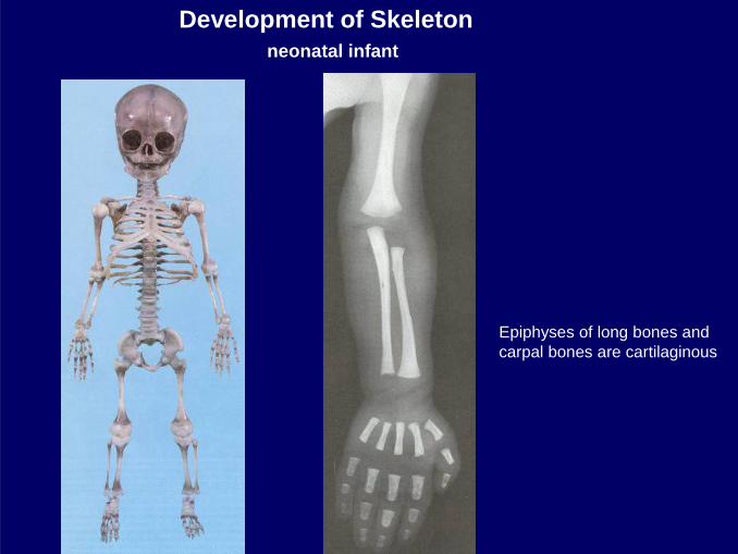
Development of Skeleton neonatal infant
Epiphyses of long bones and carpal bones are cartilaginous

Epiphyseal discbetween epiphysis and metaphysis
Replacement of ossified cartilage – longitudinal growth!!!! Becomes ossified about 20 y – no further growth!

1 y 2 y 12 y
26 y
Scelatal
Skeletal maturity of the handradiograph
Changes of catrilaginous and membranous skeleton to fully ossified bones

ArthrologyClassification of Joints
1. Interrupted Joints / Synovial Joints 2. Continuous Joints(Diarthroses) (Synarthroses)
Arthron- joint

Synostosis(Osseus joint)
sacrum
Syndesmosis(Fibrous joint)
A. Sutura
B. C.
s. ligamentosa gomphosis
1. 2. 3.
Synchondrosis(Cartilaginous joint)
symphysis pubica intervertebral discs
(plana, serratasquamosa)
Continuous Joints Synarthroses

Synovial joint – Articulatio – DiarthrosisGeneral characteristics
Capsula fibrosaCapsula synovialis
CAVUM ARTICULARE
Cartilagoarticularis
Vagina synovialis(Tendon sheath)
CAPUThead (male)
CAVITAS articularissocket (female)
CAPSULA ARTICULARIS

General components: Additional structures:1. Joint cavity2. Art. surfaces covered by hyalin cartilage3. Capsule – synovial and fibrous layer4. Ligaments
1. Discus, meniscus2. Articular lips (labrum - fibrocartilage)3. Bursae4. Synovial tendon sheaths (vagina synovialis)

Shape No. axis Function Movementsof movement
Cylindrical (trochlea) Uniaxial A./ Hinge (Ginglymus)B./ Pivot (Trochoid)
Ellipsoidal Biaxial A./ EllipsoidSaddle B./ Saddle
Spheroidal Triaxial Freely movable(multiaxial) (ball and socket)
Irregular Fix joint
Classification of Joints
1.Flexion-extension and2. Adduction-abduction
1.Flexion-extension and2. Adduction-abduction and3. Rotation
1. Flexion-extension
1. Rotation

1. Ginglymus 2. Trochoid
Uniaxial joints
Humero-ulnar jointtransverse axis
Radio-ulnar prox. jointlongitudinal axis

MOVEMENTS at UNIAXIAL JOINTS
HINGE joint- Ginglymus
Flexion- extension
Flexion- extension
MOVEMENTS around
LONGITUDINAL axis
PIVOT - Trochoid joint
Supination PronationPalmar flexion--dorsal flexion
Plantar flexion--dorsal flexion
MOVEMENTS around
TRANSVERSE axis

MOVEMENTS at BIAXIAL JOINTS
Ellipsoid and Saddle Joints
Flexion-extension
Flexion-extension
Palmar flexion--dorsal flexion
Plantar flexion--dorsal flexion
MOVEMENTS around
axisTRANSVERSE
MOVEMENTS around
axisSAGITTAL
Abduction Adduction

Spheroidal – Ball and socket joint
Circumduction
Components:flexion-extensionadduction-abductionrotation
MOVEMENTS around the 3 main axis
and COMBINATION of BASIC MOVEMENTS
Related Documents