General Microbiology

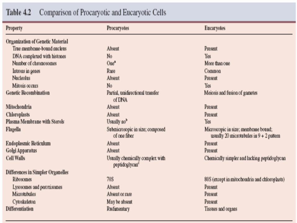
General Microbiology. Prokaryotic vs. Eukaryotic Cells w Prokaryotic cells No Nucleus No Organelles Cell Wall of peptidoglycan Binary Fission 1 circular.
Dec 14, 2015
Welcome message from author
This document is posted to help you gain knowledge. Please leave a comment to let me know what you think about it! Share it to your friends and learn new things together.
Transcript

General Microbiology
Prokaryotic vs. Eukaryotic Cells
Prokaryotic cells• No Nucleus
• No Organelles
• Cell Wall of peptidoglycan
• Binary Fission
• 1 circular chromosome
Eukaryotic Cells• Nucleus
• Organelles
• If cell wall, Cellulose or chitin
• Mitosis
• Linear chromosomes

Prokaryotic Cell Structure

Prokaryotic Cell Structure

Cytoplasmic Membrane
Membrane that encloses cytoplasm called a plasma membrane
In prokaryotes & eukaryotes, the membranes are quite similar Called a unit membrane

Ribosomes - present in cytoplasmic matrix and attached to plasma membrane, made up of protein and RNA, site of protein synthesis.
Prokaryotic Ribosome
70 S• 50 S
• 30 S
Eukaryotic Ribosomes
80 S• 60 S
• 40 S
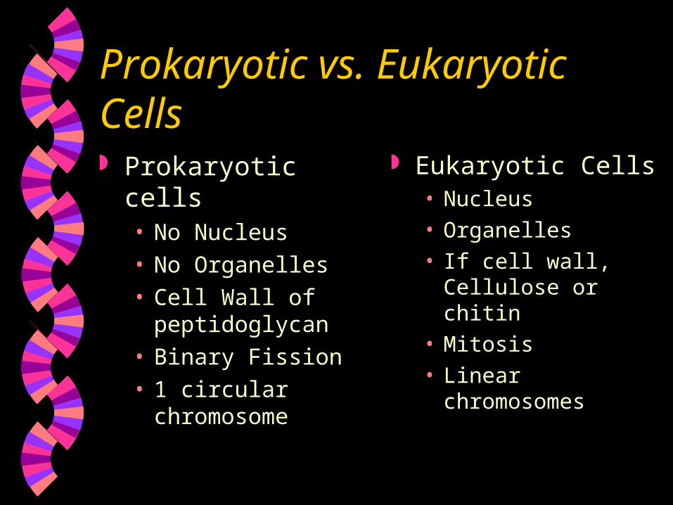
Nuclear area (nucleoid) The procaryotic chromosome
is located in an irregularly shaped region called the nucleoid.
Prokaryotes contain a single circle of double-stranded DNA, but some have a linear DNA chromosome.
Vibrio cholerae have more than one chromosome.
composed of about 60% DNA, 30% RNA, and 10% protein by weight.
A section of actively growing E. coli. A model of two nucleoids in growing E. coli cell.
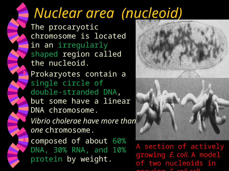
Plasmids Extrachromosomal material, < 30
genes. Double-stranded DNA molecules,
usually circular, or linear that can exist and replicate independently of the chromosome or may be integrated with it (episomes).
Plasmids are inherited during cell division and sometimes are lost, known as curing.

Bacterial Cell Walls Layer outside the plasma membrane. Functions:Determine shape of the cellProtect cell from osmosis lysis.Protect from toxic substance &
pathogens.

Bacterial Cell WallsMost have cell walls composed of
peptidoglycan. Few lack a cell wall. Peptidoglycan outside p.membrane
protein + polysaccharide, also called Murein
Peptidoglycan Polymer of long chains of alternating sugars, NAG and NAM & A.A.
NAG and NAM held together by protein chains.

Bacterial Cell Walls Chains of NAG and NAM are
attached to other chains by tetrapeptide cross bridges
Tetrapeptide cross bridges two amino acids are L-isomers and two are D-isomers
Glycan and peptide linked to form a mesh-like structure

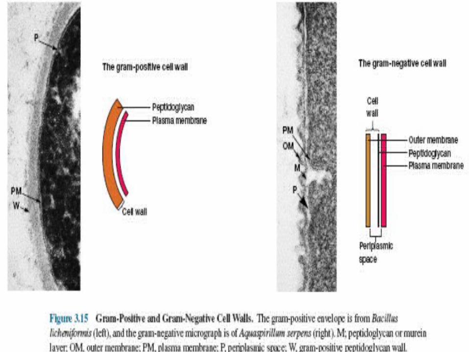
Bacterial Cell Walls
Two basic types:• Gram Positive
• Gram Negative
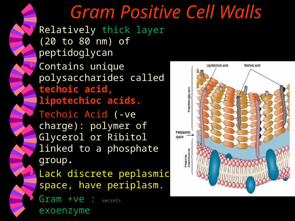
Gram Positive Cell Walls Relatively thick layer (20
to 80 nm) of peptidoglycan Contains unique
polysaccharides called techoic acid, lipotechioc acids.
Techoic Acid (-ve charge): polymer of Glycerol or Ribitol linked to a phosphate group.
Lack discrete peplasmic space, have periplasm.
Gram +ve : secrets exoenzyme

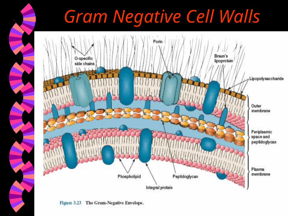
Gram Negative Cell Walls Have a only a thin layer of peptidoglycan
bounded to either side of periplasmic space. Peptidoglycan mesh is only one to two layer
thick (7 to 8 nm). Periplasmic space : greater in size. Outer membrane is linked to the cell by
lipoprotein and adhession sites joining plasma membrane.
Outer membrane contains Lipopolysaccharides consists of 3 parts
1. Lipid A 2. Core polysaccharide 3. O side chain

Gram Negative Cell WallsFunctions of LPS Bcz of the charged sugars and phosphate of
core polysaccharide it contributes to –Ve charge of surface.
Lipid A helps in stabilization of outer membrane structures.
Helps in creating permeability barrier. Protect bacteria from host defense. Lipid A is toxic : LPS act as endotoxins.
Porin proteins allow the passage of molecules smaller than 600 to 700 Daltons.
Gram Negative Cell Walls

Archaeal Cell Walls Do not have peptidoglycan Cell Walls consists of complex
heteropolysaccharides. E.g. Methanobacterium contains
pseudomurein, a peptidoglycan that has L-Amino acids instead of D-Amino acids, N- acetyltalosaminuroic acids instead of NAM and B (1-3) glycosidic bonds instead of B (1-4) glycosidic bonds.
Cytoplasmic membrane is also different
Components External to the Cell Wall Layer Of Material Outside Cell Wall 1. Capsule
• if the layer is well organized and firmly attached to cell wall
2. Slime Layer• if the layer is unorganized and loosely attached to
cell wall.
• 3. Glycocalyx
if the layer consist of network of plysaccharide. Bacillus anthracis has a capsule of poly-D-glutamic
acid.

Klebsiella pneumoniae
S-layer of Deinococcus radiodurans
Many gram-positive and gram-negative bacteria have a regularlystructured layer called an S-layer on their surface.pattern like floor tiles composed of protein or glycoprotein. In gram-negative bacteriathe S-layer adheres directly to the outer membrane.it is associated with peptidoglycan surface in gram-positive bacteria.
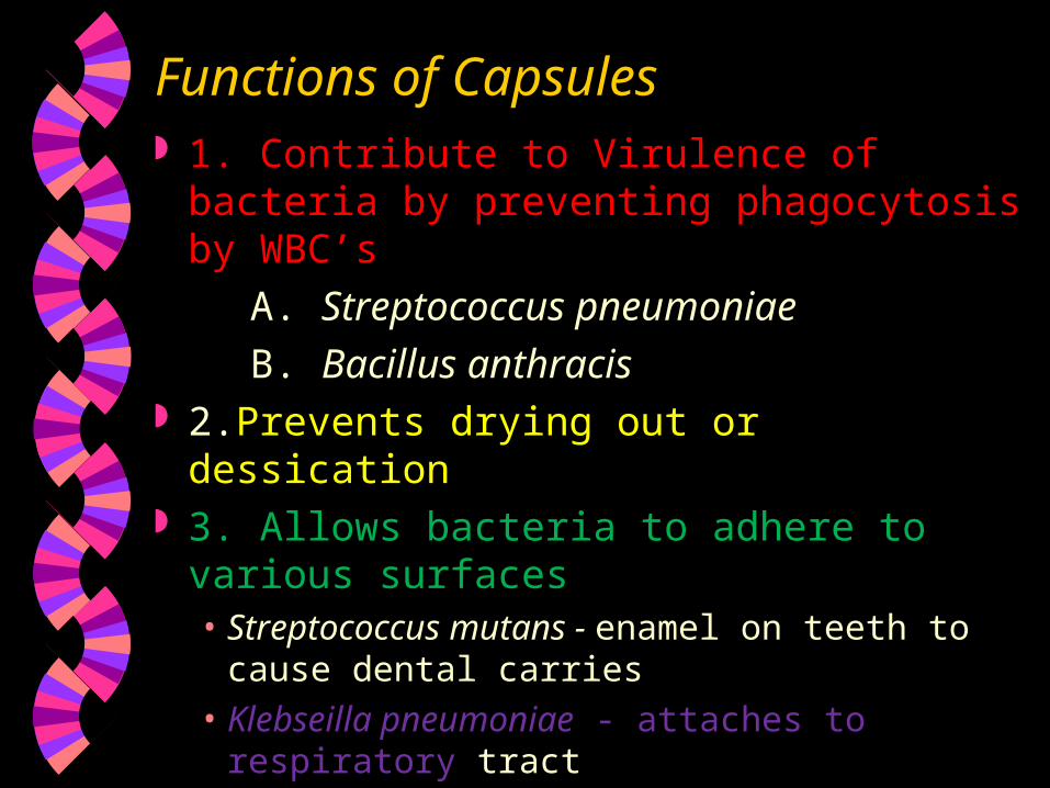
Functions of Capsules 1. Contribute to Virulence of bacteria by
preventing phagocytosis by WBC’s
A. Streptococcus pneumoniae
B. Bacillus anthracis 2.Prevents drying out or dessication 3. Allows bacteria to adhere to various
surfaces• Streptococcus mutans - enamel on teeth to
cause dental carries• Klebseilla pneumoniae - attaches to respiratory
tract
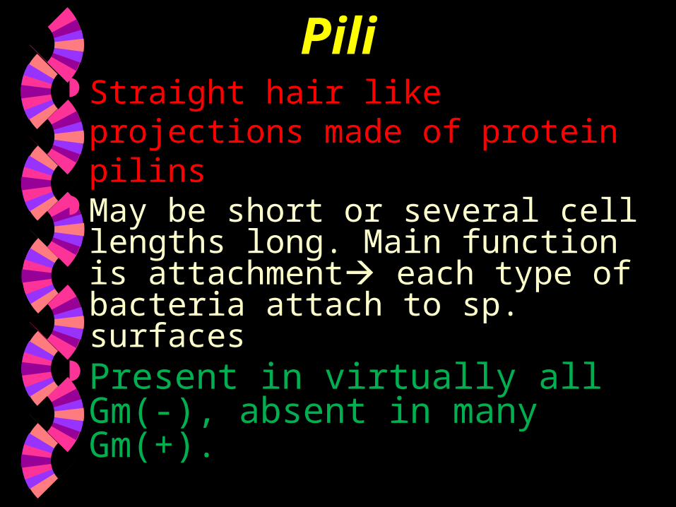
Pili Straight hair like projections made
of protein pilins May be short or several cell
lengths long. Main function is attachment each type of bacteria attach to sp. surfaces
Present in virtually all Gm(-), absent in many Gm(+).
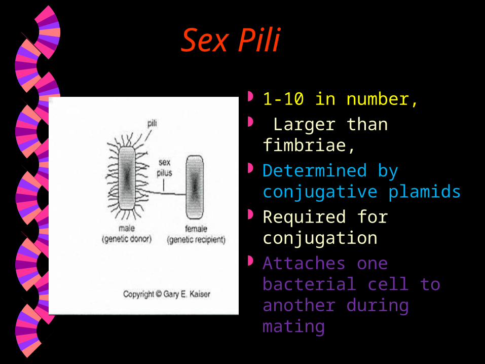
Sex Pili
1-10 in number, Larger than fimbriae, Determined by
conjugative plamids Required for
conjugation Attaches one bacterial
cell to another during mating

Fimbriae Filamentous appendages
that are shorter, finer, hairlike, thinner, straighter and more numerous than flagella
Found mostly in Gram (-) Bacteria
Used for attachment and also required for twitching motility of P.aeruginosa and E.coli, N.gonorrhoea.
E. Coli (pathogenic)
Neisseria gonorrhoeae

Flagella Thread like structures that
extend from the surface of the envelope
Function Locomotion, allow bacteria to seek favorable conditions.

Examples of various patterns offlagellation as seen in the light microscope. (a) Monotrichous polar (Pseudomonas).
(b) Lophotrichous (Spirillum).
(c) Peritrichous


Motility Spirochetes: Spinning motility : by Axial
filament. Spiroplasma : Swimming motility : by Kinks. Cynobacteria, Myxobacteria : gliding
motility. Almost all Spiral bacteria are motile
About 1/2 of Bacilli are motile
Almost all Cocci are non-motile

Chemotaxis Bacteria do not always swim aimlessly but are
attracted by such nutrients as sugars and amino acids, and are repelled by many harmful substances and bacterial waste products.
Bacteria also can respond to other environmental cues such as temperature, light, oxygen, osmotic pressure and gravity.
Chemotaxis bacteria sense certain chemicals and move toward nutrients or away from toxins

Endospores – a special resistant dormant structure, formed under periods of environmental stress Only found in Gram (+) Bacteria Bacillus
• Bacillus cereus• Bacillus anthracis
Clostridium• Clostridium tetani• Clostridium botulinum• Clostridium perfringens

Endospores Extremely resistant to heat, cold, chemicals,
lack of water, ultraviolet radiation, gamma radiation, chemical disinfectants.
In fact, some endospores have remained viable for around 1,00,000 years, and actinomycete spores have been recovered alive after burial in the mud for 7,500 years.
Most vegetative bacterial cells are killed at temps. above 70 C (160 F)• Endospores can survivein boiling water for
several hours (some for as long as 20 hours)
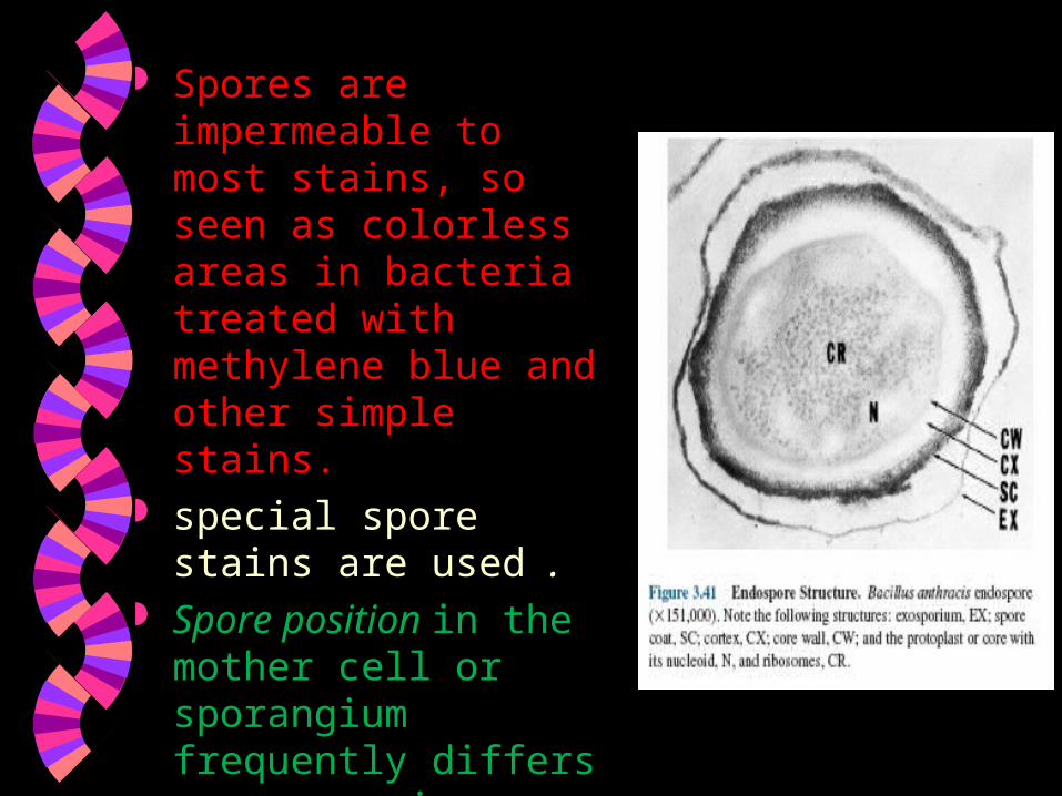
Spores are impermeable to most stains, so seen as colorless areas in bacteria treated with methylene blue and other simple stains.
special spore stains are used .
Spore position in the mother cell or sporangium frequently differs among species.

Spores may be centrally located, close to one end (subterminal), or definitely terminal.
surrounded by a thin, delicate covering called the exosporium.
Spore Coat lies beneath the exosporium, is composed of several protein layers, and may be fairly thick, impermeable & responsible for the spore’s resistance to chemicals.

Endospore heat resistance

Germination of Spore 1. Activation: prepare spore for germination
& results from treatment like heating. 2. Germination: breaking of the spore
dormant state, by swelling, rupture or absorption of spore coat, loss of resistance to heat & other stresses and increase in metabolic activity.
3. Out growth : spore protoplast makes new components, emerges from the spore coat and develops again in to an active bacterium.

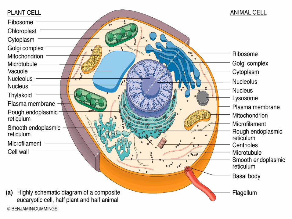
Eukaryotic Cell - Organelles Nucleus Nucleoli Endoplasmic Reticulum (E.R.)
• rE.R.• sE.R.
Ribosomes Golgi Body Lysosomes


Prokaryotes vs. Eukaryotes
Structural Difference Chemical Difference Size Difference
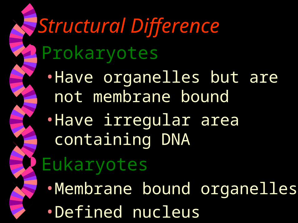
Structural Difference Prokaryotes
• Have organelles but are not membrane bound
• Have irregular area containing DNA Eukaryotes
• Membrane bound organelles
• Defined nucleus

Chemical Difference
Prokaryotes• Cell Wall composed of
peptidoglycan
Eukaryotes• Do not contain peptidoglycan in their
cell walls
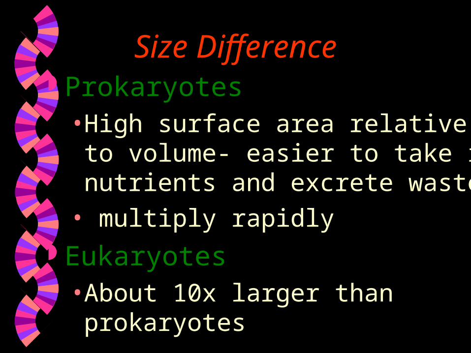
Size Difference Prokaryotes
• High surface area relative to volume- easier to take in nutrients and excrete wastes
• multiply rapidly Eukaryotes
• About 10x larger than prokaryotes

Thank you
Related Documents