1 General Management Of Facial Injuries The modern management of trauma is based on a firm understanding of the pathophysiology of trauma and an understanding of how patients actually die. This understanding has led to the development of several trauma systems, of which the Advanced Trauma Life Support (ATLS) is now generally recognized as the 'gold standard'. ATLS was originally introduced by the American College of Surgeons Committee of Trauma and is now taught in over 50 countries worldwide. It provides a systematic approach that should ensure that life-threatening and subsequent injuries are identified and managed in an appropriate and timely manner. Principles of ATLS management: ABCDE of assessment (Rapid Primary Survey) Primum non nocere (First, do no harm) Concept of the 'golden hour' (i.e. time is of the essence) Need for frequent reassessment of evolving injuries Importance of understanding the mechanism of injury Triaging of facial injuries: Commented [DS21] :

Welcome message from author
This document is posted to help you gain knowledge. Please leave a comment to let me know what you think about it! Share it to your friends and learn new things together.
Transcript

1
General Management
Of Facial Injuries
The modern management of trauma is based on a firm understanding of the
pathophysiology of trauma and an understanding of how patients actually
die. This understanding has led to the development of several trauma
systems, of which the Advanced Trauma Life Support (ATLS) is now
generally recognized as the 'gold standard'. ATLS was originally introduced
by the American College of Surgeons Committee of Trauma and is now
taught in over 50 countries worldwide. It provides a systematic approach that
should ensure that life-threatening and subsequent injuries are identified and
managed in an appropriate and timely manner.
Principles of ATLS management:
ABCDE of assessment (Rapid Primary Survey)
Primum non nocere (First, do no harm)
Concept of the 'golden hour' (i.e. time is of the essence)
Need for frequent reassessment of evolving injuries
Importance of understanding the mechanism of injury
Triaging of facial injuries:
Commented [DS21] :
2
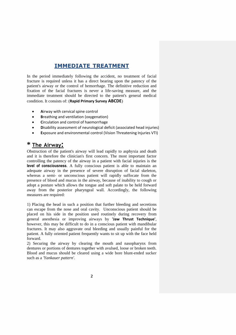
TREATMENT IMMEDIATE
In the period immediately following the accident, no treatment of facial
fracture is required unless it has a direct bearing upon the patency of the
patient's airway or the control of hemorrhage. The definitive reduction and
fixation of the facial fractures is never a life-saving measure, and the
immediate treatment should be directed to the patient's general medical
condition. It consists of: (Rapid Primary Survey ABCDE)
Airway with cervical spine control
Breathing and ventilation (oxygenation)
Circulation and control of haemorrhage
Disability assessment of neurological deficit (associated head injuries)
Exposure and environmental control (Vision Threatening Injuries VTI)
* The Airway: Obstruction of the patient's airway will lead rapidly to asphyxia and death
and it is therefore the clinician's first concern. The most important factor
controlling the patency of the airway in a patient with facial injuries is the
level of consciousness. A fully conscious patient is able to maintain an
adequate airway in the presence of severe disruption of facial skeleton,
whereas a semi- or unconscious patient will rapidly suffocate from the
presence of blood and mucus in the airway, because of inability to cough or
adopt a posture which allows the tongue and soft palate to be held forward
away from the posterior pharyngeal wall. Accordingly, the following
measures are required:
1) Placing the head in such a position that further bleeding and secretions
can escape from the nose and oral cavity. Unconscious patient should be
placed on his side in the position used routinely during recovery from
general anesthesia or improving airways by 'Jaw Thrust Technique', however, this may be difficult to do in a conscious patient with mandibular
fractures. It may also aggravate oral bleeding and usually painful for the
patient. A fully oriented patient frequently wants to sit up with the face held
forward.
2) Securing the airway by clearing the mouth and nasopharynx from
dentures or portions of dentures together with avulsed, loose or broken teeth.
Blood and mucus should be cleared using a wide bore blunt-ended sucker
such as a 'Yankauer pattern'.

3
3) In Le Fort fractures, the upper jaw may have been pushed downwards and
backwards so that the soft palate is resting upon the dorsum of the tongue
and occlude the oral airway. In such cases two fingers are inserted behind the
hard palate and the upper jaw is pulled gently upwards and forwards to
enable the patient to breathe through the mouth.
4) Nasopharyngeal airway (armoured soft latex nasopharyngeal tubes) can
facilitate the management of patients with Le Fort II and III fractures and the
nose is cleared with a suction apparatus.
5) Arresting nasal hemorrhage by anterior or posterior nasal packing for Le
Fort II and III fractures and with severe injuries to the nasal complex in
which the nares are blocked with blood clot or bleed profusely which cause
occlusion of the nasal airway.
6) If there is bilateral fractures in the mental region, the skeletal support of
the tongue tends to be displaced backwards by the pull action of the
geniohyoid and genioglossus muscles which are attached to the genial
tubercles, this will result in a backward displacement of the tongue and
obstruct the airway which results in respiratory embarrassment in such case,
the chin must lifted and a tongue stitch may be required and the thread of the
suture must be grasped outside the mouth by artery forceps, and the patient
must be transported lying on his side to dribble out saliva and blood from
the mouth.
7) Continuous supervision is necessary either by the operator or by an
experienced member of nursing staff. The lips should be coated with sterile
petroleum jelly to prevent them from adhering together.
8) Endotracheal Intubation may be required to ensure a patent airway in
most patients with fractures of the middle third. The problems of airway
maintenance are increased considerably in the unconscious. The rapid
passage of an endotracheal tube is by far the most effective way of clearing
and preserving the airway. Endotracheal intubation is usually required in
patients with multiple injuries particularly of the head, face and chest. Such
patients are often deeply unconscious on admission. All patients are at risk
of unexpected vomiting, but those with facial injuries are at greatest risk. A
full stomach, alcohol intoxication and brain injuries are factors that
predispose to vomiting. Swallowed blood also seems to be a potent stimulus.
These are all commonly associated with facial trauma. It is therefore
important to identify those patients who are at such a high risk of vomiting
and intubated to secure the airway before it happens.

4
9) Tracheostomy: emergency incision into the trachea (tracheostomy)
should never be necessary if effective medical skill is available, that is where
an endotracheal tube can be passed. The indications for tracheostomy in
maxillofacial injuries are:
i. When prolonged artificial ventilation is necessary, e.g. some severe
associated head and chest injuries.
ii. To facilitate anaesthesia for surgical repair in certain major
injuries.
iii. To ensure a safe postoperative recovery after extensive reparative
surgery.
iv. Following obstruction of the airway from laryngeal oedema or
occasionally direct injury to the base of the tongue and oropharynx.
v. Serious haemorrhage into the airway particularly when further
secondary haemorrhage is a possibility.
i.e. 1. Surgical cricothyroidotomy is advocated through incision of the
cricothyroid membrane. A slightly smaller size tracheostomy tube (i.e. cuffed
size 4 or 5) being maintained for 24 hours and replaced with tracheostomy.
The complication is glottic and sub-glottis stenosis.
2. Needle cricothyroidotomy by 12 G venflon with 10 ml syringe.
* Hemorrhage: The majority of fractures of the facial skeleton are closed injuries, and in
spite of the extensive nature of the skeletal damage, severe haemorrhage is
unusual when there are extensive soft-tissue lacerations, particularly after
missile injury, these require urgent attention as local blood loss can be
considerable.
Control of hemorrhage:
Significant bleeding from external wounds, such as the scalp, can
simply be controlled with pressure or any strong suture to hand. A
continuous suture is both quick and effective. In the scalp, full
thickness 'bites' are required to ensure the vessels are included in the
layer. Obvious bleeding vessels should be secured with artery forceps,
ligated if possible, and temporary pressure dressing applied.
Occasionally brisk and persistent hemorrhage originates from grossly
displaced fracture of the mandible or midface. This can only be
controlled by manual reduction of the fracture and temporary
immobilization either manually, or by means of a wire ligature passed
around teeth on each side of fracture line ('bridle wire'). With very
mobile displaced midface fractures, manual reduction may be possible
and not only control blood loss but improves the airway. A well
placed mouth prop can sometimes help support.

5
Early intubation should again be considered, not only to protect the
airway, but also to allow effective control of bleeding.
Epistaxis of some degree is an inevitable consequence of injury to the
central middle third of the face.
1) It is usually stops spontaneously or is easily controlled by lightly
packing the nose via the anterior nares 'anterior nasal pack'. 2) Profuse hemorrhage into the nasopharynx from terminal branches
of the maxillary artery occurs on very rare occasions in association
with Le Fort fractures. This may be life –threatening both from the
point of view of actual blood loss and also obstruction of the airway.
A 'postnasal pack' is needed in this situation as a matter of extreme
urgency.
3) A variety of specially designed nasal balloons can be utilized.
4) If these specific devices are not available 2 urinary catheters
(Foley catheter) can be used. Each is passed via both nostrils into the
pharynx, inflated with saline then gently withdrawn until the balloon
wedges in the post-nasal space. The nasal cavity can then be packed.
Nasal packs are not without risk and aggressive packing should be
avoided especially if anterior cranial fossa or orbital fractures are evident
or suspected. Toxic shock, sinusitis, meningitis, brain
abscess and even blindness are all rare but potential complications
that have been reported.
How long packs are left in situ will depend on the clinical status of the
patient, but around 24-48 hours is usual.
If hemorrhage persists despite these interventions it is important to
consider coagulation abnormalities that can occur during
prolonged resuscitation associated with major blood loss.
Ligation of external carotid artery and ethmoidal arteries via the
neck and orbit respectively (if bleeding continues despite all previous
measures, and there are no clotting abnormalities). These steps are
rarely required nowadays and are extremely difficult to undertake as
emergency procedure. Due to the extensive collateral circulation
of the face ligating a single vessel is unlikely to be successful. Add to
this the urgency of hemostasis and the fact that the cervical spine may
not have been 'cleared', thereby preventing turning of the head for
access, and it is little wonder that these techniques are now rarely
undertaken.

6
Endovascular radiological intervention (superselective embolization)
is considered now as the preferred approach. It has been extensively
reported as very successful, with clear advantages over surgery. It is
increasingly used in solid organs and extremity trauma, and in
bleeding secondary to pelvic fractures. It is now well documented as a
successful treatment modality in penetrating injuries, blunt injuries
and intractable epistaxis. Catheter-guided angiography is used to
first identify and then occlude the bleeding points. Embolization
involves the use of a number of materials designed to stimulate
clotting locally. Superselective embolization can be performed without
the need for a general anesthetic and in experienced hands is relatively
quick. Its value therefore is seen in the unstable patient. Multiple
bleeding points can be precisely identified and the technique is
repeatable. However, immediate access to specialized radiological
facilities and on-site expertise is required.
i.e. It is always important to reserve blood for cross-matching, blood
transfusion may be required to compensate blood loss and avoid
hypovolemic shock.
* Shock:
Acute circulatory collapse is not usually a prominent feature of a fracture of
the facial skeleton, and if such a patient is severely shocked the possibility of
the coexistence of some other more serious injury should be suspected.
Estimated Fluid & Blood Losses
Class I Class II Class III Class IV Blood loss (mL) Blood loss (% vol) Pulse rate Blood pressure Pulse pressure Respiratory rate Urine output (mL/h) Mental status Fluid replacement
Up to 750 Up to 15 < 100 Normal Normal or ↑ 14-20 > 30 Slightly anxious Crystalloid
750-1500 15-30 > 100 Normal ↓ 20-30 20-30 Mildly anxious Crystalloid & blood
1500-2000 30-40 > 120 ↓ ↓ 30-40 5-15 Anxious, confused Crystalloid & blood
> 2000 > 40 > 140 ↓ ↓ > 35 Negligible Confused, lethargic Crystalloid

7
Surgical Hemostasis
(choice of use depend on the site and nature of injury and hemorrhage)
A. Mechanical procedures
1. digital pressure 2. vascular hemostat (Halsted's mosquito artery forceps) 3. clamps 4. ligatures (ligation e.g. transfixation suture for large arteries) 5. tourniquets 6. pressure packs (e.g. dry or wet swabs) 7. bone wax
B. Thermal agents
1. Heating:
1) Cautery (1928) cause denaturation of proteins result in coagulation of large areas of tissue. Is either actual (conduct heat ) or electrocautery (alternative current)
2) Direct current (20-100 mA) 3) High power argon-laser for superficial erosions
2. Cooling:
1) Direct cooling (e.g. iced saline) 2) Extreme cooling (cryogenic surgery) -20 to -180 C°: cause
cryogenic necrosis of small arteriols and venules (e.g. CO2 liquid -50 C°, and Nitrogen liquid -150 to -180 C°)
3) Generalized hypothermia down to 35 C°, this reduce blood flow to visceral organs but cause shivering and ventricular fibrillation.
C. Chemical agents
1. Adrenaline & Noradrenaline 2. Turpentine or Tannic acid (applied on gauze packs) 3. Oxidized regenerated cellulose (Surgicel) 4. Gelfoam (gelatin foam) 5. Microcrystalline collagen 6. Thrombin & Russell viper venom

8
PRELIMINARY EXAMINATION
This is the secondary survey. After the operator has established a
satisfactory airway and controlled hemorrhage, a full examination of the
patient should be carried out (top-to-toe examination). The definitive
treatment of a facial bone fracture is hardly ever an urgent procedure and
purpose of this preliminary general examination is to establish the presence
or otherwise of other more important injuries.
Head Injury The cranium should be palpated and inspected for evidence of lacerations
and bony damage and the level of consciousness determined.
A simple scale of level of consciousness is:
i. Fully conscious.
ii. Drowsy with disorientation, but responds rationally to questions and
requests.
iii. Semiconscious responding irrationally to spoken questions and
requests.
iv. Unconscious but responding purposefully to painful stimuli.
v. Unconscious with decerebrate reflex response to pain.
The assessment of the patient's consciousness can be made by noting the
patient's response using the simple AVPU scale:
A → responds Appropriately (Awake)
stimuli Verbalresponds to → V
stimuli Painfulresponds to → P
U → doesn't respond (Unconscious)
This coupled with an assessment of the pupil reaction, allows rapid
assessment of the degree of head injury.
The Glasgow Coma Scale (GCS): Points are awarded using the criteria given in the scale to give a total score
between (3 = deeply unconscious and unresponsive) to (15 = fully conscious,
alert and oriented). Any patient with a GCS score of less than 8 should be
considered as severe head injury unable to protect their airway (i.e. Below Eight Intubate). Those with a GCS score 9-12 are considered to have a
moderate head injury and a GCS of 13-15 indicates a minor head injury.

9
Eye opening is graded 1-4 as follows 1 = no eye opening 2 = opening to pain 3 = opening to speech 4 = spontaneous opening The best motor response is graded on limb movements from 1-6 1 = no movement 2 = extensor response only 3 = abnormal flexion 4 = withdrawal from painful stimuli 5 = movement towards painful stimuli 6 = movement of limb on command Capability of verbal response is graded from 1-5 1 = no verbal response 2 = inarticulate sound 3 = recognizable words inappropriately uttered 4 = confused conservation 5 = fully oriented Eyes
The eyes should be examined at an early stage both as part of neurological
examination and to determine whether there has been any physical injury to
the globe. Vision, pupil size and reaction to light should be recorded.
Signs and symptoms of orbital Compartment Syndrome (Retrobulbar
oedema) or Retrobulbar haemorrhage: retrobulbar pressures cause optic
nerve ischemia should recognized and treated promptly (compartment
syndrome → medical with Mannitol 1 gm/kg + acetazolamide 250-500 mg
to reduce intra-ocular pressure + 3-4 mg/kg i.v. dexamethasone to reduce
oedema & vascular spasm), while Retrobulbar hemorrhage → require
evacuation through lateral canthotomy as emergency before surgery), so
'buy time' by doing both as an emergency while preparing for surgery.
Irreversible ischemia of the visual pathway can occur within 1 hour, and
permanent visual loss (blindness) within 1 1/2 – 2 hours.
1. Pain (increasing)
2. Decreasing visual acuity
3. Diplopia with developing ophthalmoplegia (paralysis of ocular muscles)
4. Proptosis
5. Tense globe
6. Subconjunctival oedema/chemosis
7. Dilated pupil and pale optic disc
8. Loss of direct light reflex (relative afferent pupillary defect)

10
The spine
It should be assumed that any significant maxillofacial injury may be
associated with a cervical spine injury. Care, therefore must be taken when
the head and neck are manipulated during maintenance of the airway,
examination and radiology. A lateral view of the cervical spine showing all
cervical vertebrae must be examined and if there is a high index of suspicion,
then cervical anterioposterior and open mouth odontoid views should also
be taken. Confirmation of a cervical spine injury may require simple
tomography or computed tomography (CT) scanning.
The limbs
Rapid palpation of the limbs for deformity or bony tenderness should
precede the recording of reflexes.
Abdomen and chest
Examination by inspection and palpation will determine whether there is a
possibility of visceral injury or fracture of the chest wall or pelvis. The first
urine specimen should be examined for the presence of blood. The operator
will by this time have enough information to call for any assistance he may
require from other specialties.
Soft Tissue Laceration Soft tissue facial injuries fall into three main groups:
1. Hematomas.
2. Simple lacerations.
3. Lacerations involving specialized structures or organs.
The most common priority for patients with facial fractures is repair of soft-
tissue lacerations. Ideally these should be sutured before too much oedema
has occurred; that is within 1-8 hours of injury. Simple lacerations can be
dealt with under local analgesia. Extensive soft-tissue damage to the face
requires a long general anaesthesia for accurate repair and it is important that
the operator does not get carried away by his desire to produce a perfect
cosmetic result to the detriment of an already very ill patient if there is any
doubt about the general condition of the patient. The facial laceration should
be cleaned and closed as rapidly as possible , bearing in mind that the
underlying fractures can be treated at a later date and scars eventually
revised if necessary.
11
Stepwise options for the primary management of traumatic tissue loss
1. Immediate replacement of avulsed tissue as a free graft.
2. Dress wound and allow to heal by secondary intention.
3. Direct closure under an acceptable degree of tension.
4. Partial or full thickness skin graft.
5. Immediate reconstruction with a free composite graft (e.g. some nasal
defects).
6. Local or regional flap.
7. Avulsion of scalp/ear/ nose: consider replantation using microsurgical
techniques.
***************
HISTORY AND LOCAL EXAMINATION History of the injury and description of the patient's symptoms:
1. If the patient is unconscious or confused, any relevant facts
concerning the accident and the subsequent management of the patient
must be obtained from eye-witnesses, ambulance men, or medical and
dental practitioners who may have attended the patient following the
injury.
2. If the patient is conscious and co-operative a history can be obtained,
but as patients with facial injury may experience some difficulty in
talking owing to the pain and mobility of the fractures the
interrogation should be brief at this stage.
3. It is prudent to ask if loss of consciousness has occurred and, in that
event, whether the patient can remember up to the moment of the
accident or whether there is a memory gap. Retrograde amnesia is
failure to remember up to the time of injury and anterograde amnesia
is loss of memory following the accident, both are indicative of
cerebral damage.
4. It is also important to inquire whether the patient has any difficulty in
breathing or swallowing and whether he has a headache or pain
elsewhere in the body.
5. Information as to whether the patient was being treated with insulin,
steroid, or anticoagulant prior to the accident is also most important.

12
A detailed history is obtained when the patient can talk more
comfortably.
Local Clinical Examination of the Facial injury The examination of a patient with a recent severe injury to the facial
skeleton will be greatly facilitated if the patients face is gently washed
with warm water and cotton–wool swabs to remove caked blood. The
congealed blood in the palate and buccal sulcus can be removed with
cotton–wool held in untoothed forceps. Sometimes cotton–wool swabs
dipped in hydrogen peroxide will facilitate the removal of any
particularly tenacious clots in the mouth and upon the teeth. Care must be
taken not to introduce hydrogen peroxide into a compound fracture
owing to the risk of causing surgical emphysema or of introducing
infection into the fracture line.
Inspection Externally. The operator should take carful note of oedema,
ecchymosis, and soft-tissue lacerations. Any obvious bony deformities
haemorrhage, or cerebrospinal fluid leak should be recorded.
Palpation. Gentle palpation should begin at the back of the head and the
cranium should be explored for wounds and bony injuries .Then the
fingers should be run lightly over the zygomatic bone and arch, and
around the rim of the orbits. Areas of tenderness, step deformities, and
unnatural mobility are noted. Next, the nasal complex is examined in the
same manner. The eyelids are gently separated and, if the patient is
conscious, the vision is tested in each eye. Then the patient is asked to
follow the clinician's finger with his eyes and asked to report if diplopia
occurs. A note is made of alteration in the size of the two pupils, and the
light reflex is tested. The extent of the subconjunctival ecchymosis is
confirmed. The operator testes the two cheeks for anaesthesia in the
distribution of the infra-orbital nerve, also testing the lower lip for
anaesthesia in the distribution of the mental nerve. Finally, the mandible
is gently palpated beginning from the condyle to the symphysis.
Inspection Intra-orally. Gagging of the occlusion, derangement of the
bite, lacerations, ecchymosis and damage to the teeth and/or alveolus are
noted.
Palpation. Areas of tenderness, bony irregularities, crepitus and mobility
of the teeth and the alveolus are noted.
Next, the tooth bearing segment is gently manipulated to elicit unnatural
mobility. A finger and thumb are then placed over the frontonasal suture
line and movement of the facial skeleton is demonstrated by pressure
from the finger in the palate. If the dento-alveolar segment moves
independently of the remainder of the facial skeleton, it will be noted that
an associated Le Fort I type of fracture is present. Next, the teeth are
tapped and the cracked cup sound is elicited if there is a fracture above
the teeth. The mandibular alveolus is palpated gently for the presence of
13
any step deformity or crepitus. Finally, if the patient has teeth, they are
examined with a mirror and probe to demonstrate possible fracture,
mobility and subluxation.
CONTROL OF PAIN
There is surprisingly little pain from maxillofacial injuries. It is extremely
important to avoid giving powerful analgesics which:
1) Depress the level of consciousness and respiration. The risk of
respiratory obstruction is increased when such drugs as morphine and
its derivatives are given to a patient with injuries of the maxillofacial
region.
2) Morphine also depresses the couch reflex and so encourage the
aspiration of blood into the trachea.
3) It causes constriction of the pupil (miosis), which may mask an early
sign of the rise in intracranial pressure (as in cerebral hemorrhage).
4) Masks pain which may be due to intra–abdominal or intra–thoracic
injuries.
It is, however, most important to minimize discomfort in the early stages
after injury, as a patient is readily exhausted by efforts both to keep his
airway clear and to obtain nourishment. Local toilet, support of mobile
fractures, posture, and availability of suction and administration of
intravenous fluids are all of great importance in the early care of the patient.
The most useful drug for sedation in such cases is Diazepam (Valium) given
intravenously. Only about 10mg are usually necessary and this drug may be
combined with 15-30mg of Pentazocine (Fortral) as an analgesic. The effect
of the Pentazocine can be reversed, if required, by the narcotic antagonist
naloxone (Narcan) in a dose 0.1-0.4 mg.
***************
CONTROL OF INFECTION To prevent the development of infection in the fracture haematoma and
lacerated soft tissue, the patient should be given IM 1,000,000 units
Penicillin per day for five days or give Azithromycin if the patient is allergic
to penicillin. Penicillin does not pass into the CSF in adequate therapeutic
concentration and if a Le Fort II or III fracture is present, even without overt
cerebrospinal fluid rhinorrhoea, the patient should be given a course of
Sulphonamide therapy as Sulphadiazine (2 g as initial dose followed by 1 g
6-hourly for at least 5 days) as a prophylactic measure to prevent meningitis.
Tetanus prophylaxis should be considered, especially in unclean wounds.
Related Documents


![Cronicon · fractures [2]. Being the most prominent mobile bone of the facial skeleton, Mandibular fractures are among the most common injuries to the facial skeleton, with a 6:2](https://static.cupdf.com/doc/110x72/5f2b985f1c26767db7383601/cronicon-fractures-2-being-the-most-prominent-mobile-bone-of-the-facial-skeleton.jpg)


