General Guide for Cryogenically Storing Animal Cell Cultures

Welcome message from author
This document is posted to help you gain knowledge. Please leave a comment to let me know what you think about it! Share it to your friends and learn new things together.
Transcript
General Guide for Cryogenically Storing Animal Cell Cultures
Table of Contents
Introduction ..................................................................................................................... 3
Advantages of Freezing Cell Cultures ....................................................................... 3
General Events During Cell Freezing ......................................................................... 3
Practical Aspects of Cell Freezing ............................................................................... 4
Acknowledgment ......................................................................................................... 10
References ...................................................................................................................... 11
Ordering Information .................................................................................................. 12
General Guide for Cryogenically Storing Animal Cell Cultures
3
Introduction
Maintaining healthy, growing cell cultures is a demanding task made more difficult by the everpresent risk of their loss through accidents or contamination. In addition, actively growing cell cultures are not static but, like all populations of microorganisms, subject to age-related or environmentally-induced changes which can result in their ongoing evolution and potential loss.
These problems can be reduced by using cryogenic preservation to stop biological time for cell cultures, effectively putting them into true suspended animation. This concept, long a favorite ploy of science fiction writers and movie producers, has been a reality since the important discovery by Polge, Smith and Parkes14 in 1949 that glycerol prevents injury to cells caused by freezing. Many cook book-style protocols are available for freezing cells and these procedures usually perform well4,7,17-20. Recently, specialized protocols for freezing cell pellets, tissues, hematopoietic stem cells, and mesenchymal stem cells have also been developed3,9,12,15. It is essential, however, when problems arise or protocol adaptations and improvements must be made, that the underlying concepts on which they are based are well understood. This guide examines both the basic theoretical concepts and practical aspects necessary for successfully freezing animal cells and managing a cell repository.
Advantages of Freezing Cell Cultures
Once successfully frozen and stored, cell cultures require little time and effort for their maintenance. The only real cost is the expense of maintaining an ultracold (-130°C or lower) mechanical freezer or liquid nitrogen supply. This limited expense compares very favorably with the time, effort, and substantial cost of the media and supplies necessary for maintaining actively growing cultures, or for the cost of obtaining a new culture from a repository. Frozen cultures provide an important backup supply for replenishing occasional losses due to contamination or accidents and provide the assurance of a homogeneous culture supply. Cellular changes or alterations occur in all actively growing populations. These changes often result in the loss of important characteristics during evolution of the cultures thereby introducing unwanted variables into long-term experiments. Cryogenically preserved cultures apparently do not undergo any detectable changes once they are stored below -130°C1,10. Therefore, the biological effects of in vitro cellular aging and evolution may be minimized by frequently returning to frozen stock cultures, allowing ongoing long-term culture experiments to be successfully completed without these unwanted variables. Frozen cultures also provide a valuable baseline against which future experimentally-induced changes may be compared or measured.
General Events During Cell Freezing
To understand why freezing protocols work, it is necessary to examine both the intracellular and extracellular events occurring in animal cell cultures during the freezing process2,5,10. Initial cooling from room temperature to 0°C slows cellular metabolism, rapidly disrupting active transport and ionic pumping. This disruption usually does not result in cellular damage if the culture medium is osmotically balanced. As cooling continues (0˚C to -20˚C) ice crystals begin to form in the extracellular environment which increases the solute concentration of the culture medium. As a result, water begins to move out of the cells and into the partially frozen extracellular medium, beginning the process of cellular dehydration and shrinkage.
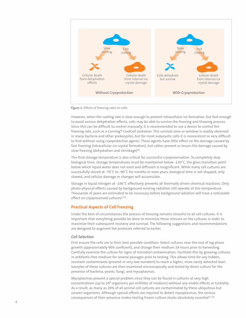
When the cooling process is rapid, intracellular ice crystals form before complete cellular dehydration has occurred. These ice crystals disrupt cellular organelles and membranes and lead to cell death during the recovery (thawing) process (Figure 1).
When the cooling process is slow, free intracellular water is osmotically pulled from the cells resulting in complete cellular dehydration and shrinkage. This can also cause cellular death, but there is little agreement on the mechanisms involved. The physical stresses of cellular shrinking may cause some damage resulting in irreparable membrane loss and cytoskeletal and organelle disruption. Damage may also be caused by the high concentrations of solutes in the remaining unfrozen extracellular medium (essentially a brine solution). These solutes attack cells both externally and internally, resulting in membrane damage, pH shifts, and general protein denaturation.
Advantages of Freezing Cultures
w Serves as a backup supply for emergencies.
w Reduces risk of microbial contamination and cross-contamination with other cell lines and genetic drift changes.
w Provides a more homogeneous population by minimizing culture aging and evolution.
4
However, when the cooling rate is slow enough to prevent intracellular ice formation, but fast enough to avoid serious dehydration effects, cells may be able to survive the freezing and thawing process. Since this can be difficult to control manually, it is recommended to use a device to control the freezing rate, such as a Corning® CoolCell container. This survival zone or window is readily observed in many bacteria and other prokaryotes, but for most eukaryotic cells it is nonexistent or very difficult to find without using cryoprotective agents. These agents have little effect on the damage caused by fast freezing (intracellular ice crystal formation), but rather prevent or lessen the damage caused by slow freezing (dehydration and shrinkage)10.
The final storage temperature is also critical for successful cryopreservation. To completely stop biological time, storage temperatures must be maintained below -130˚C, the glass transition point below which liquid water does not exist and diffusion is insignificant. While many cell cultures are successfully stored at -70˚C to -90˚C for months or even years, biological time is not stopped, only slowed, and cellular damage or changes will accumulate.
Storage in liquid nitrogen at -196˚C effectively prevents all thermally driven chemical reactions. Only photo-physical effects caused by background ionizing radiation still operate at this temperature. Thousands of years are estimated to be necessary before background radiation will have a noticeable effect on cryopreserved cultures2,10.
Practical Aspects of Cell Freezing
Under the best of circumstances the process of freezing remains stressful to all cell cultures. It is important that everything possible be done to minimize these stresses on the cultures in order to maximize their subsequent recovery and survival. The following suggestions and recommendations are designed to augment the protocols referred to earlier.
Cell SelectionFirst ensure the cells are in their best possible condition. Select cultures near the end of log phase growth (approximately 90% confluent), and change their medium 24 hours prior to harvesting. Carefully examine the culture for signs of microbial contamination. Facilitate this by growing cultures in antibiotic-free medium for several passages prior to testing. This allows time for any hidden, resistant contaminants (present in very low numbers) to reach a higher, more easily detected level. Samples of these cultures are then examined microscopically and tested by direct culture for the presence of bacteria, yeasts, fungi, and mycoplasmas.
Mycoplasmas present a special problem since they can be found in cultures at very high concentrations (up to 108 organisms per milliliter of medium) without any visible effects or turbidity. As a result, as many as 20% of all animal cell cultures are contaminated by these ubiquitous but unseen organisms. Although special efforts are required to detect mycoplasmas, the serious consequences of their presence makes testing frozen culture stocks absolutely essential11,16.
Figure 1. Effects of freezing rates on cells.
Slowcooling
Fastcooling
Cellular deathfrom internal icecrystal damage
Cellular deathfrom dehydration
effects
Slowcooling
Fastcooling
Cellular deathfrom internal icecrystal damage
Cells dehydratebut survive
Without Cryoprotection With Cryoprotection
5
Check for both the identity of the cultures and the presence of any expected special characteristics. Monitor cell identities by karyology and isoenzyme analysis, ensuring that they are, at the very least, the correct species13.
Cell HarvestingStart with the standard harvesting procedure generally recommended for the culture, and be as gentle as possible. Remove all dissociating agents by washing or inactivation (especially important when using serum-free medium). Centrifugation, when absolutely necessary, should only be hard enough to obtain a soft pellet; 100 x g for 5 to 6 minutes is usually sufficient. To ensure uniformity of the final frozen stock, pool the contents of all harvested culture vessels. This also makes it easier to perform essential quality control testing for microbial contamination and culture identity.
Count and then dilute or concentrate the harvested cell suspension to twice the desired final concentration, which is usually 4 to 10 million viable cells per milliliter. An equal volume of medium containing the cryoprotective agent at twice its final concentration will be added later to achieve the desired inoculum. Keep the cells chilled to slow their metabolism and prevent cell clumping. Avoid alkaline pH shifts by gassing with CO2 when necessary.
CryoprotectionAs mentioned earlier, cryoprotective agents are necessary to minimize or prevent the damage associated with slow freezing. The mechanisms providing this protection, although not completely understood, appear to work primarily by altering the physical conditions of both the ice and solutions immediately surrounding (external to) the cells. Permeation of the cells by cryoprotectants does not appear to be necessary for their proper functions5. Remember, protection against fast freezing damage (internal ice formation) is not provided by these agents, but rather by careful control of the freezing rate. A wide variety of chemicals provide adequate cryoprotection, including methyl acetamide, methyl alcohol, ethylene glycol and polyvinyl pyrrolidone8. However, dimethylsulfoxide (DMSO) and glycerol are the most convenient and widely used. Many of these agents, although providing excellent cryoprotection, have toxic side effects on cultures making their use difficult.
DMSO is most often used at a final concentration of 5% to 15% (v/v). Always use reagent or other high purity grades that have been tested for suitability. Sterilize by filtration through a 0.2 micron nylon membrane in a polypropylene or stainless steel housing, and store in small quantities (5 mL). CAUTION: Take special care to avoid contact with solutions containing DMSO. It is a very powerful polar solvent capable of rapidly penetrating intact skin and carrying in with it harmful contaminants such as carcinogens or toxins. Some cell lines are adversely affected by prolonged contact with DMSO. This can be reduced or eliminated by adding the DMSO to the cell suspension at 4°C and removing it immediately upon thawing. If this does not help, lower the concentration or try glycerol or another cryoprotectant.
Glycerol is generally used at a final concentration of between 5% and 20% (v/v). Sterilize by autoclaving for 15 minutes in small volumes (5 mL) and refrigerate in the dark. Although less toxic to cells than DMSO, glycerol frequently causes osmotic problems, especially after thawing. Always add it at room temperature or above and remove slowly by dilution.
High serum concentrations may also help cells survive freezing. Replacing standard media-cryoprotectant mixtures with 95% serum and 5% DMSO may be superior for some overly sensitive cell lines, especially hybridomas.
Add cryoprotective agents to culture medium (without cells) immediately prior to use to obtain twice the desired final concentration (2X). Mix this 2X solution with an equal volume of the harvested cell suspension (also 2X) to obtain the inoculum for freezing. This method is less stressful for cells, especially when using DMSO as the cryoprotectant.
Storage VesselsAfter the cryoprotective solution is mixed with the cell suspension, the resulting inoculum is added in small aliquots (usually 1 to 2 mL) to each storage vessel. Due to the extremely low temperatures encountered during cryogenic storage, not all vessel materials or designs are suitable or safe. Many materials become very brittle at these temperatures; vessels made from them may shatter or crack during storage or thawing. Carefully check the vessel manufacturers’ recommendations on proper selection and use.
Prevent Freezing Damage
w Use slow freezing to remove all intracellular water.
w Use cryoprotective agents to minimize dehydration effects.
w Store below -130˚C to completely stop biological time.
6
Also important is selecting the sealing system or cap design used to maintain the integrity of the vessel, especially for storage in liquid nitrogen. If these vessels leak during storage (as many do*), they will slowly fill with liquid nitrogen. When they are eventually returned to room temperature, the liquid nitrogen quickly vaporizes causing a rapid pressure buildup. The vessels may then violently blow off their caps or explode to vent the pressure and release their contents into the atmosphere. This is a very dangerous situation, especially if the vessels contained pathogenic organisms or potentially toxic or harmful substances.
WARNING: To avoid injury, DO NOT immerse plastic or glass cryogenic vials in liquefied gases such as liquid nitrogen. Vials immersed in liquefied gases can develop leaks. When such vials return to room temperature, rapid pressure can build up and shatter the vials, or cap seals. Harmful or biohazard materials contained in the vials can release during the venting of pressurized gas. Always store vials only in the vapor phase ABOVE any liquefied gas.
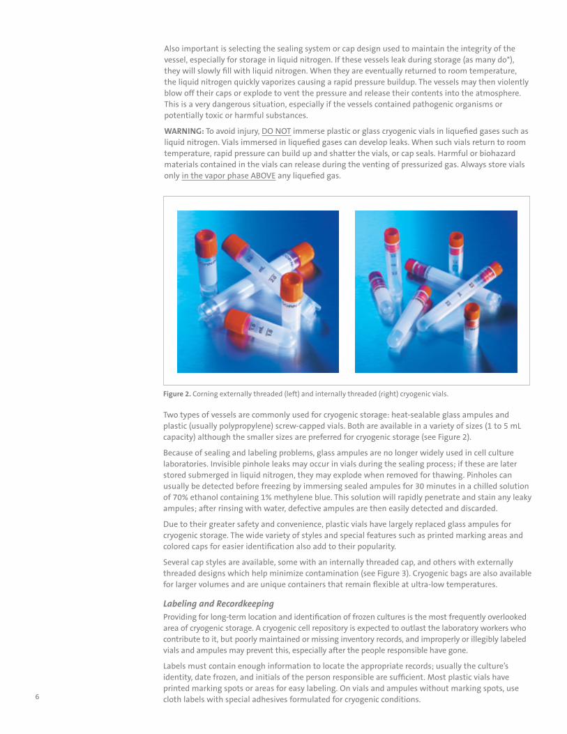
Two types of vessels are commonly used for cryogenic storage: heat-sealable glass ampules and plastic (usually polypropylene) screw-capped vials. Both are available in a variety of sizes (1 to 5 mL capacity) although the smaller sizes are preferred for cryogenic storage (see Figure 2).
Because of sealing and labeling problems, glass ampules are no longer widely used in cell culture laboratories. Invisible pinhole leaks may occur in vials during the sealing process; if these are later stored submerged in liquid nitrogen, they may explode when removed for thawing. Pinholes can usually be detected before freezing by immersing sealed ampules for 30 minutes in a chilled solution of 70% ethanol containing 1% methylene blue. This solution will rapidly penetrate and stain any leaky ampules; after rinsing with water, defective ampules are then easily detected and discarded.
Due to their greater safety and convenience, plastic vials have largely replaced glass ampules for cryogenic storage. The wide variety of styles and special features such as printed marking areas and colored caps for easier identification also add to their popularity.
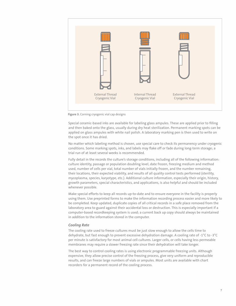
Several cap styles are available, some with an internally threaded cap, and others with externally threaded designs which help minimize contamination (see Figure 3). Cryogenic bags are also available for larger volumes and are unique containers that remain flexible at ultra-low temperatures.
Labeling and RecordkeepingProviding for long-term location and identification of frozen cultures is the most frequently overlooked area of cryogenic storage. A cryogenic cell repository is expected to outlast the laboratory workers who contribute to it, but poorly maintained or missing inventory records, and improperly or illegibly labeled vials and ampules may prevent this, especially after the people responsible have gone.
Labels must contain enough information to locate the appropriate records; usually the culture’s identity, date frozen, and initials of the person responsible are sufficient. Most plastic vials have printed marking spots or areas for easy labeling. On vials and ampules without marking spots, use cloth labels with special adhesives formulated for cryogenic conditions.
Figure 2. Corning externally threaded (left) and internally threaded (right) cryogenic vials.
7
Special ceramic-based inks are available for labeling glass ampules. These are applied prior to filling and then baked onto the glass, usually during dry heat sterilization. Permanent marking spots can be applied on glass ampules with white nail polish. A laboratory marking pen is then used to write on the spot once it has dried.
No matter which labeling method is chosen, use special care to check its permanency under cryogenic conditions. Some marking spots, inks, and labels may flake off or fade during long-term storage; a trial run of at least several weeks is recommended.
Fully detail in the records the culture’s storage conditions, including all of the following information: culture identity, passage or population doubling level, date frozen, freezing medium and method used, number of cells per vial, total number of vials initially frozen, and the number remaining, their locations, their expected viability, and results of all quality control tests performed (sterility, mycoplasma, species, karyotype, etc.). Additional culture information, especially their origin, history, growth parameters, special characteristics, and applications, is also helpful and should be included whenever possible.
Make special efforts to keep all records up-to-date and to ensure everyone in the facility is properly using them. Use preprinted forms to make the information recording process easier and more likely to be completed. Keep updated, duplicate copies of all critical records in a safe place removed from the laboratory area to guard against their accidental loss or destruction. This is especially important if a computer-based recordkeeping system is used; a current back up copy should always be maintained in addition to the information stored in the computer.
Cooling RateThe cooling rate used to freeze cultures must be just slow enough to allow the cells time to dehydrate, but fast enough to prevent excessive dehydration damage. A cooling rate of -1°C to -3°C per minute is satisfactory for most animal cell cultures. Larger cells, or cells having less permeable membranes may require a slower freezing rate since their dehydration will take longer.
The best way to control cooling rates is using electronic programmable freezing units. Although expensive, they allow precise control of the freezing process, give very uniform and reproducible results, and can freeze large numbers of vials or ampules. Most units are available with chart recorders for a permanent record of the cooling process.
Figure 3. Corning cryogenic vial cap designs
External ThreadCryogenic Vial
Internal ThreadCryogenic Vial
External ThreadCryogenic Vial
8
There are a variety of mechanical freezing units that provide adequate control of the cooling rate and are relatively inexpensive. Some units use racks designed to hold vials at predetermined depths in the neck of a liquid nitrogen freezer. The cooling rate is dependent on the total number of vials and the depth at which the rack is placed. Another design uses an alcohol-filled metal or plastic canister containing a rack with a capacity of up to 24 vials. The filled canister is placed in an ultracold mechanical freezer where the alcohol acts as a bath to achieve more uniform heat transfer and cooling. After freezing 4 to 5 hours, the vials are removed from the canister and transferred to their final storage locations.
A Corning® CoolCell® foam container, in combination with a -80°C freezer, will provide alcohol-free freezing at the rate of -1°C/minute that is ideal for cryopreservation of most cells and cell lines. Using a combination of uniform-density cross-linked polyethylene foam, a solid state core, and radial vial symmetry, freezing profiles are consistent and reproducible. The foam is non-absorbent and will impose negligible change in the freezer environment; thereby protecting nearby frozen samples. The removal of vials is accomplished by tipping the device upside down so the vials slide out. These devices are easy to use, are alcohol- and fluid-free, reproducible, and offer a simple consistent way to standardize controlled-rate freezing.
Insulated cardboard or polystyrene foam boxes are commonly used as freezing chambers in ultracold freezers. These homemade devices work well with many cell lines but do not always give controlled, reproducible, or uniform cooling. As a result, there may be serious differences in viability among the vials upon thawing. This homemade approach is not recommended for valuable or irreplaceable cultures.
No matter which cooling method is used, transfer from the cooling chamber or device to the final storage location must be done quickly to avoid warming of the vials. Use an insulated container filled with dry ice or liquid nitrogen as a transfer vessel to ensure that the cells remain below -70°C.
Cryogenic StorageOnly freezers capable of continually maintaining temperature below -130˚C should be considered for long-term cryogenic storage. Although most liquid nitrogen-cooled freezers and some specially designed mechanical freezers meet this requirement, most cell culture laboratories prefer liquid nitrogen freezers. The final choice is often based on the availability of a reliable supply of liquid nitrogen, the storage capacity required, and the size of the budget. Liquid nitrogen freezers permit storage either in the vapor phase above the liquid at a temperature between -140°C and -180°C, or submerged in the liquid at a temperature below -196°C. Using vapor phase storage greatly reduces the possibility of leaky vials or ampules exploding during removal. However, since the amount of liquid nitrogen in the freezer is reduced to provide space for vapor phase storage, the freezer’s holding time (the period it can maintain its storage temperature without adding more liquid nitrogen) is also reduced. This lowers the freezer’s margin of safety and requires more frequent monitoring and filling. Give careful consideration to these safety issues when deciding upon a storage method.
WARNING: To avoid injury, DO NOT immerse plastic or glass cryogenic vials in liquefied gases such as liquid nitrogen. Vials immersed in liquefied gases can develop leaks. When such vials return to room temperature, rapid pressure can build up and shatter the vials, or cap seals. Harmful or biohazard materials contained in the vials can release during the venting of pressurized gas. Always store vials only in the vapor phase ABOVE any liquefied gas.
Frequently check nitrogen levels in freezers; a schedule should be established and strictly adhered to. Nitrogen evaporation is dependent on both the degree of use and the static holding time of the freezer. Sudden, unexplained increases in the evaporation rate may signal damage to the insulation or other problems with the freezer and must be carefully investigated. Avoid frost or ice buildup around freezer openings; this increases the nitrogen evaporation rate and can cause elevated temperatures in the upper portion of vapor phase freezers. Audible alarm systems for detecting low liquid nitrogen levels are available to provide additional safeguards; however, they provide a false sense of security if not monitored 24 hours a day.
9
ThawingCAUTION: Always use appropriate safety equipment when removing vials and ampules from liquid or vapor phase nitrogen freezers. A full face shield, heavy gloves, and lab coat are strongly recommended for protection against exploding vials or ampules.
Remove the vial or ampule from its storage location and carefully check both the label and storage record to ensure that it is the correct culture. Place the vessel in warm water, agitating gently until completely thawed. Rapid thawing (60 to 90 seconds at 37°C) provides the best recovery for most cell cultures; it reduces or prevents the formation of damaging ice crystals within cells during rehydration.
RecoverySince some cryoprotective agents may damage cells upon prolonged exposure, remove the agents as quickly and gently as possible. Several approaches are used depending on both the cryoprotective agents and characteristics of the cells. Most cells recover normally if they have the cryoprotective agent removed by a medium change within 6 to 8 hours of thawing. Transfer the contents of the ampule or vial to a 75 cm² flask or other suitable vessel containing 0.2-0.3 milliliters per cm² of culture medium and incubate normally. As soon as a majority of the cells have attached, remove the medium containing the now diluted cryoprotective agent and replace with fresh medium.
For cells that are sensitive to cryoprotective agents, removing the old medium is easily accomplished by gentle centrifugation. Transfer the contents of the vial or ampule to a 15 mL centrifuge tube containing 10 mL of fresh medium and spin for 5 minutes at 100 x g. Discard the supernatant containing the cryoprotectant and resuspend the cell pellet in fresh medium. Then transfer the cell suspension to a suitable culture vessel and incubate normally.
When glycerol is used as the cryoprotectant, the sudden addition of a large volume of fresh medium to the thawed cell suspension can cause osmotic shock, damaging or destroying the cells. Use several stepwise dilutions with an equal volume of warm medium every 10 minutes before further processing to give the cells time to readjust their osmotic equilibrium.
Problem Solving SuggestionsViability problems associated with cryogenic storage are usually noticed soon after cultures are thawed and plated. There are four major areas where problems occur:
During harvesting and processing of the cellsProblems may be caused by excessive exposure of the cells to dissociating agents; using a cryoprotective agent that is toxic; or allowing high density cell suspensions to remain too long at room temperature or at a pH that is too basic.
During the cooling (freezing) processExcessive cell damage and reduced culture viability often result from using a cooling rate that is too fast or too slow, or when the cooling process is temporarily interrupted. Not using a suitable cryoprotective agent at an appropriate concentration will also result in viability problems.
During cyrogenic storage Culture viability is often reduced when vials are allowed to warm up during transfer to the freezer, or if the repository temperature is not consistently maintained at appropriate cryogenic temperatures.
During thawing and recovery Problems arise when the thawing process is too slow or the cryoprotectants are improperly removed (see above).
These viability problems can often be corrected by using the following technique to identify the stage in the freezing process where the problem originates.
Harvest enough cells to prepare at least four vials. Then remove a sample of cell suspension, equivalent in cell number to that which will be placed into the vials, and immediately place it into a culture vessel with an appropriate amount of medium and incubate. This culture will be used as a control to compare with the cultures set up in the remaining steps.
Freezing Checklist
w Gently harvest cultures.
w Check cultures for contamination, especially mycoplasma.
w Check culture identity by karyotyping or isoenzyme analysis.
w Use tested cryoprotective agents.
w Only use vials tested for cryogenic conditions.
w Ensure labels are permanent and complete.
w Control the cooling rate.
w Store cultures below -130˚C.
w Monitor liquid nitrogen levels frequently.
w Keep good records.
10
Next, add the cryoprotective agent to the remaining cells and divide among three vials. Place one vial at 4°C for one hour. Then remove the cells from the vial, process as though they had just been thawed from the freezer, and plate in medium as above. This culture will be compared with the control culture to determine if there are any problems associated with cryoprotective agent.
Meanwhile, process the remaining vials through the slow cooling process as usual. One vial is then immediately thawed and processed as above. This culture will be compared with the control culture to determine if there are any problems associated with the slow cooling process.
The remaining vial is then transferred to the cryogenic freezer and stored overnight before being thawed and processed as above. This culture will be compared with the control culture to determine if there are any problems associated with the cryogenic storage conditions. If additional vials of cells are available, several different recovery techniques should be used to determine if the recovery technique is the source of the problem.
By comparing all of the cultures to the original culture, it should then be possible to determine at which stage of the freezing process the problem occurred. Once this is known, the information presented in this guide and its references should be enough to eliminate the problem.
Managing a Cell RepositoryThe effort and expenses of managing a repository should be kept in line with the value of the cultures stored within it. This value is determined by answering two questions: How much time, money, and effort is already invested in these stored cell cultures? And, what are the consequences of losing them? Cultures that are easily replaced through other labs or commercial sources may not require special efforts, but unique cultures, such as hybridomas and other genetically engineered cells, are irreplaceable and require that special efforts be made to ensure their safety. The answers to these questions will help determine just how extensive and thorough your efforts should be.
Next, identify the potential problem areas that can cause the loss of these cultures. Some of these areas, such as vessel selection, recordkeeping, labeling, freezer monitoring, storage conditions, and quality issues (contamination and species identity), have already been discussed in this guide. Decide what steps are necessary to eliminate or minimize these problems. Split irreplaceable or extremely valuable cultures among several freezers, with at least one freezer in a separate location to protect against fire or other natural disasters. Colleagues in other labs or buildings may be able to provide good backup storage, especially if a reciprocal arrangement is made for them.
One final step remains; plan ahead for emergencies! One of the most serious and unexpected emergencies is the failure of a cryogenic freezer. Careful monitoring of the liquid nitrogen level or charting the temperature may give an early warning that failure is occurring, but middle of the night failures can and do happen. Have plans prepared in advance to deal with freezer failure and other problems. If these involve a colleague’s equipment, get permission and make all necessary arrangements in advance — late night phone calls are usually not appreciated.
This information has been compiled to provide a guide for better understanding of the cryogenic preservation process. For additional assistance in this area, visit www.corning.com/lifesciences, or contact Corning Scientific Support at 1.800.492.1110; outside the United States, call +1.978.442.2200; or email [email protected].
Acknowledgment
Corning wishes to acknowledge the contributions of John Ryan to this article.
11
References1. Aswood-Smith, M. J. and G. B. Friedmann, 1979. Lethal and Chromosomal Effects of Freezing,
Thawing, Storage Time and Xirradiation on Mammalian Cells Preserved at -196°C in Dimethylsulfoxide. Cryobiology 16:132-140.
2. Aswood-Smith, M. J., 1980. Low Temperature Preservation of Cells , Tissues and Organs, p. 19-44. In Low Temperature Preservation in Medicine and Biology. M. J. Aswood-Smith and J. Farrant, Eds. (Pitman Medical Limited, Kent, England).
3. Chatzistamatiou TK, Papassavas AC, Michalopoulos,E, Gamaloutsos C, Mallis P, Gontika I, Panagouli E, Koussoulakos SL, and Stavropoulos-Giokas C. (2014), Banking Protocols Validation for WJ-MSCs. Transfusion, 54: 3108-3120. doi:10.1111/trf.12743.
4. Coriell, L. L., 1979. Preservation, Storage and Shipment, p. 29-35. In Methods in Enzymology. Vol. 58, W. B. Jacoby and I. H. Pasten, Eds., (Academic Press, New York).
5. Farrant, J., 1989. General Observations on Cell Preservation, p. 1-18. In Low Temperature Preservation in Medicine and Biology, M. J. Aswood-Smith and J. Farrant, Eds. (Pitman Medical Limited, Kent, England).
6. Freshney, R. I., 1994. Culture of Animal Cells: A Manual of Basic Technique, p. 254-263. (3rd edition; Wiley-Liss, New York.
7. Hay, R. J., 1978. Preservation of Cell Culture Stocks in Liquid Nitrogen, p. 787-790. TCA Manual 4.
8. Klebe, R. J. and M. G. Mancuso, 1983. Identification of New Cryoprotective Agents for Cultured Mammalian Cells. In Vitro 19:167-170.
9. Litovchick L. Freezing Cell Pellets for Large-scale Immunoprecipitation. Cold Spring Harbor Protocols. 2019 Jul;2019(7) DOI: 10.1101/pdb.prot098541.
10. Mazur, P., 1984. Freezing of Living Cells: Mechanisms and Implications, p. C125-C142. Am. J. Physiol. 247 (Cell Physiol. 16).
11. McGarrity, G. J., J. Sarama, and V. Vanaman, 1985. Cell Culture Techniques. ASM News 51:170-183.
12. Pavel P, Laier S. (2019) A Freezing Protocol for Hematopoietic Stem Cells. In: Klein G, Wuchter P. (eds) Stem Cell Mobilization. Methods in Molecular Biology, vol 2017. Humana, New York, NY.
13. Peterson, W. D., W. F. Simpson and B. Hukku, 1973. Cell Culture Characterization: Monitoring for Cell Identification, p. 164-178. In Tissue Culture: Methods and Applications, P. F. Kruse and M. K. Patterson Jr. Eds. (Academic Press, New York).
14. Polge, C., A. U. Smith, and A. S. Parkes, 1949. Revival of Spermatozoa after Vitrification and Dehydration at Low Temperatures. Nature 164: 666.
15. Radaelli MRM, Almodin CG, Minguetti-Câmara VC, Cerialli PMA, Nassif AE, Gonçalves AJ. A Comparison Between a New Vitrification Protocol and the Slow Freezing Method in the Cryopreservation of Prepubertal Testicular Tissue. JBRA Assist Reprod. 2017;21(3):188-195. Published 2017 Sep 1. doi:10.5935/1518-0557.20170037.
16. Ryan, J., 1994. Understanding and Managing Cell Culture Contamination, TC-CI-559. Corning, Inc. Technical Monograph.
17. Schroy, C. B., and P. Todd, 1976. A Simple Method for Freezing and Thawing Cultured Cells, p. 309-310. TCA Manual 2, Procedure Number 76035.
18. Shannon, J. E. and M. L. Macy, 1973. Freezing, Storage, and Recovery of Cell Stocks, p. 712-718. In Tissue Culture: Methods and Applications. P. F. Kruse and M. K. Patterson Jr. Eds. (Academic Press, New York).
19. Smith, K. O., 1981. Low Temperature Storage of Surface Attached Living Cell Cultures. Cryobiology 18:251-257.
20. Waymouth, C. and D. S. Varnum, 1976. Simple Freezing Procedure for Storage in Serum-free Media of Cultured and Tumor Cells of Mouse, p. 311-313. TCA Manual 2, Procedure Number 76165.
12
Ordering Information
Corning® Cell Culture Flasks
Cat. No.Surface
Area (cm2) Flask StyleNeck Style
Cap Style Qty/Pk Qty/Cs
430639 25 Rectangular Canted Vent 20 200
430641U 75 U-shaped Canted Vent 5 100
430825 150 U-shaped Canted Vent 5 50
431080 175 U-shaped Canted Vent 5 50
431082 225 Rectangular Canted Vent 5 25
10020 1,720 Corning HYPERFlask®
- - 4 4
10030 1,720 Corning HYPERFlask®
- - 1 4
Costar® Multiple Well Plates, Tissue Culture-treatedCat. No. Description Plate Type Qty/Pk Qty/Cs
3516 6-well Clear 1 50
3513 12-well Clear 1 50
3526 24-well Clear 1 50
3548 48-well Clear 1 100
3596 96-well Clear 1 50
3595 96-well Clear* 1 50
3599 96-well Clear 1 100
3916 96-well Black 20 100
3917 96-well White 20 100
3603 96-well Black/Clear-bottom 1 48
3610 96-well White/Clear-bottom 1 48
*Low evaporation lid
Corning Dishes, Tissue Culture-treated
Cat. No.Dish Style*
(mm)Approx. Height
(mm)Growth Area
( cm2) Qty/Pk Qty/Cs
430165 35 10 8 20 500
430166 60 15 21 20 500
430167 100 20 55 20 500
430599 150 25 148 5 60
431110 245 25 500 4 16
*Dish Style (mm) 35 mm = 34.4 mm; 60 mm = 52.1 mm; 100 mm = 83.9 mm; 150 mm = 139.1 mm. Square dishes have interior bottom dimensions 224 x 224 mm.
Corning Cell Scrapers
Cat. No. DescriptionBlade Length
(cm)Handle Length
(cm) Qty/Pk Qty/Cs
3008 Cell lifter 1.9 18 1 100
3010 Scraper, small 1.8 25 1 100
3011 Scraper, large 3 39 1 100
Corning Cell StrainersCat. No. Description Qty/Pk Qty/Cs
431750 40 µm, blue 1 50
431751 70 µm, white 1 50
431752 100 µm, yellow 1 50
Corning Cryogenic VialsCat. No. Capacity (mL) Style Self-standing Qty/Pk Qty/Cs
430659 2 Round bottom, internal thread
Yes 50 500
430488 2 Round bottom, external thread
Yes 50 500
Falcon® Cell Culture Flasks, Tissue Culture-treated
Cat. No.Surface
Area (cm2) Flask Style Neck Style Cap Style Qty/Pk Qty/Cs
353018 25 Rectangular Canted Vent 20 100
353136 75 Rectangular Canted Vent 5 60
355001 150 Rectangular Canted Vent 5 40
353112 175 Rectangular Straight Vent 5 40
353138 225 Rectangular Canted Vent 5 30
353143 525 3-Layer - Vent 2 12
353144 875 5-Layer - Vent 1 8
Falcon Cell Culture Plates, Tissue Culture-treatedCat. No. Description Plate Type Qty/Pk Qty/Cs
353046 6-well Clear 1 50
353043 12-well Clear 1 50
353047 24-well Clear 1 50
353078 48-well Clear 1 50
353072 96-well Clear 1 50
Falcon Cell Culture Dishes
Cat. No.Dish Style
(mm) Approx. Height
(mm)Growth Area
(cm2) Qty/Pk Qty/Cs
353001 35 10 11.78 20 500
353002 60 15 21.29 20 500
353003 100 20 58.95 20 200
353025 150 25 156.36 10 100
Falcon Cell StrainersCat. No. Description Qty/Pk Qty/Cs
352340 40 µm, blue 1 50
352350 70 µm, white 1 50
352360 100 µm, yellow 1 50
Falcon Cell ScrapersCat. No. Description Qty/Pk Qty/Cs
353085 1.8 cm TPE blade with 18 cm polystyrene handle 1 100
353086 1.8 cm TPE blade with 25 cm polystyrene handle 1 100
353089 3.0 cm TPE blade with 25 cm polystyrene handle 1 100
353087 3.0 cm TPE blade with 40 cm polystyrene handle 1 100
13
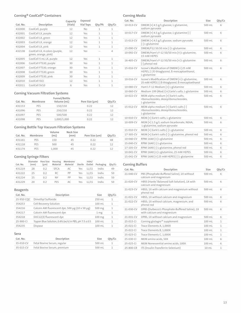
Corning® CoolCell® Containers
Cat. No. DescriptionCapacity
(Vials)Exposed Vial Tops Qty/Pk Qty/Cs
432000 CoolCell, purple 12 No 1 1
432001 CoolCell LX, purple 12 Yes 1 1
432002 CoolCell LX, green 12 Yes 1 1
432003 CoolCell LX, orange 12 Yes 1 1
432004 CoolCell LX, pink 12 Yes 1 1
432138 CoolCell LX, 4 colors (purple, green, orange, pink)
12 Yes -- 4
432005 CoolCell 5 mL LX, purple 12 Yes 1 1
432006 CoolCell FTS30, purple 30 Yes 1 1
432007 CoolCell FTS30, orange 30 Yes 1 1
432008 CoolCell FTS30, green 30 Yes 1 1
432009 CoolCell FTS30, pink 30 Yes 1 1
432010 CoolCell SV2 12 Yes 1 1
432011 CoolCell SV10 6 Yes 1 1
Corning Vacuum Filtration Systems
Cat. No. MembraneFunnel/Bottle Volume (mL) Pore Size (µm) Qty/Cs
431153 PES 150/150 0.22 12
431096 PES 250/250 0.22 12
431097 PES 500/500 0.22 12
431098 PES 1,000/1,000 0.22 12
Corning Bottle Top Vacuum Filtration Systems
Cat. No. Membrane Volume
(mL)Neck Size
(mm) Pore Size (um) Qty/Cs
431161 PES 150 45 0.22 48
431118 PES 500 45 0.22 12
431174 PES 1,000 45 0.22 12
Corning Syringe Filters
Cat. No.Diameter
(mm)Pore Size
(µm)Housing Material
Membrane Material Sterile
Inlet/Outlet Packaging Qty/Cs
431219 28 0.2 SFCA AC Yes LL/LS Indiv. 49
431222 25 0.2 RC PP Yes LL/LS Indiv. 50
431224 25 0.2 NY PP Yes LL/LS Indiv. 50
431229 20 0.2 PES AC Yes LL/LS Indiv. 50
ReagentsCat. No. Description Size Qty/Cs
25-950-CQC Dimethyl Sulfoxide 250 mL 1
354253 Cell Recovery Solution 100 mL 1
354216 Calcein AM fluorescent dye, 500 µg (10 x 50 µg) 500 mg 1
354217 Calcein AM fluorescent dye 1 mg 1
354218 DiIC12(3) fluorescent dye 100 mg 1
25-900-CI Trypan Blue Solution, 0.4% (w/v) in PBS, pH 7.5 ± 0.5 100 mL 1
354235 Dispase 100 mL 1
SeraCat. No. Description Size Qty/Cs
35-010-CV Fetal Bovine Serum, regular 500 mL 1
35-015-CV Fetal Bovine Serum, premium 500 mL 1
Corning MediaCat. No. Description Size Qty/Cs
10-013-CV DMEM [+] 4.5 g/L glucose, L-glutamine, sodium pyruvate
500 mL 6
10-017-CV DMEM [+] 4.5 g/L glucose, L-glutamine [-] sodium pyruvate
500 mL 6
15-013-CV DMEM [+] 4.5 g/L glucose, sodium pyruvate [-] L-glutamine
500 mL 6
15-090-CV DMEM/F12 50:50 mix [-] L-glutamine 500 mL 6
10-092-CV DMEM/Ham’s F-12 50/50 mix [+] L-glutamine, 15 mM HEPES
500 mL 6
16-405-CV DMEM/Ham’s F-12 50/50 mix [+] L-glutamine [-] phenol red
500 mL 6
15-016-CV Iscove's Modification of DMEM [+] 25 mM HEPES, [-] ß-thioglycerol, ß-mercaptoethanol, L-glutamine
500 mL 6
10-016-CV Iscove's Modification of DMEM [+] L-glutamine, 25 mM HEPES [-] ß-thioglycerol, ß-mercaptoethanol
500 mL 6
10-080-CV Ham’s F-12 Medium [+] L-glutamine 500 mL 6
10-060-CV Medium 199 (Mod.) [+] Earle’s salts, L-glutamine 500 mL 6
10-022-CV MEM alpha medium [+] Earle’s salts, ribonucleosides, deoxyribonucleosides, L-glutamine
500 mL 6
15-012-CV MEM alpha medium [+] Earle’s salts [-] ribonucleosides, deoxyribonucleosides, L-glutamine
500 mL 6
10-010-CV MEM [+] Earle’s salts, L-glutamine 500 mL 6
10-009-CV MEM [+] 1.5 g/L sodium bicarbonate, NEAA, L-glutamine, sodium pyruvate
500 mL 6
15-010-CV MEM [+] Earle’s salts [-] L-glutamine 500 mL 6
17-305-CV MEM [+] Earle’s salts [-] L-glutamine, phenol red 500 mL 6
10-040-CV RPMI 1640 [+] L-glutamine 500 mL 6
15-040-CV RPMI 1640 [-] L-glutamine 500 mL 6
17-105-CV RPMI 1640 [-] L-glutamine, phenol red 500 mL 6
10-041-CV RPMI 1640 [+] L-glutamine, 25 mM HEPES 500 mL 6
15-041-CV RPMI 1640 [+] 25 mM HEPES [-] L-glutamine 500 mL 6
Corning BuffersCat. No. Description Size Qty/Cs
21-040-CV PBS (Phosphate-Buffered Saline), 1X without calcium and magnesium
500 mL 6
21-020-CV HBSS (Hanks’ Balanced Salt Solution), 1X with calcium and magnesium
500 mL 6
21-023-CV HBSS, 1X with calcium and magnesium without phenol red
500 mL 6
21-021-CV HBSS, 1X without calcium and magnesium 500 mL 6
21-022-CV HBSS, 1X without calcium, magnesium, and phenol red
500 mL 6
21-030-CV DPBS (Dulbecco’s Phosphate-Buffered Saline), 1X with calcium and magnesium
500 mL 6
21-031-CV DPBS, 1X without calcium and magnesium 500 mL 6
25-015-CI Corning glutagro™ supplement 100 mL 1
25-021-CI Trace Elements A, 1,000X 100 mL 1
25-022-CI Trace Elements B, 1,000X 100 mL 1
25-023-CI Trace Elements C, 1,000X 100 mL 1
25-030-CI MEM amino acids, 50X 100 mL 6
25-025-CI MEM Nonessential amino acids, 100X 100 mL 6
25-800-CR ITS (Insulin-Transferrin-Selenium) 10 mL 1

© 2
004,
202
0 Co
rnin
g In
corp
orat
ed. A
ll ri
ghts
rese
rved
. 5/
20 C
LS-T
C-30
6 RE
V3
For a listing of trademarks, visit www.corning.com/clstrademarks. All other trademarks are the property of their respective owners.
For additional product or technical information, visit www.corning.com/lifesciences or call 800.492.1110. Outside the United States, call +1.978.442.2200 or contact your local Corning sales office.
Corning IncorporatedLife Sciences836 North St. Building 300, Suite 3401 Tewksbury, MA 01876t 800.492.1110 t 978.442.2200 f 978.442.2476www.corning.com/lifesciences
A S I A / P A C I F I C
Australia/New Zealandt 61 427286832Chinese Mainlandt 86 21 3338 4338f 86 21 3338 4300India t 91 124 4604000f 91 124 4604099
Japant 81 3-3586 1996 f 81 3-3586 1291Koreat 82 2-796-9500 f 82 2-796-9300Singaporet 65 6572-9740 f 65 6735-2913Taiwant 886 2-2716-0338 f 886 2-2516-7500
E U R O P [email protected] 0800 916 882f 0800 918 636Germanyt 0800 101 1153 f 0800 101 2427The Netherlands t 020 655 79 28f 020 659 76 73United Kingdomt 0800 376 8660f 0800 279 1117
All Other European Countriest +31 (0) 206 59 60 51 f +31 (0) 206 59 76 73
L A T I N A M E R I C [email protected]
Brazilt 55 (11) 3089-7400Mexicot (52-81) 8158-8400
For more specific information on claims, visit the Certificates page at www.corning.com/lifesciences.
Warranty/Disclaimer: Unless otherwise specified, all products are for research use only. Not intended for use in diagnostic or therapeutic procedures. Not for use in humans. Corning Life Sciences makes no claims regarding the performance of these products for clinical or diagnostic applications.
Related Documents