General Biology Part I - Mixed Majors Collection edited by: Shannon McDermott Content authors: OpenStax, Shannon McDermott, and Jeffrey Mahr Online: <https://legacy.cnx.org/content/col11749/1.6> This selection and arrangement of content as a collection is copyrighted by Shannon McDermott. Creative Commons Attribution License 4.0 http://creativecommons.org/licenses/by/4.0/ Collection structure revised: 2017/07/24 PDF Generated: 2018/05/22 09:12:19 For copyright and attribution information for the modules contained in this collection, see the "Attributions" section at the end of the collection. 1

Welcome message from author
This document is posted to help you gain knowledge. Please leave a comment to let me know what you think about it! Share it to your friends and learn new things together.
Transcript
General Biology Part I - Mixed MajorsCollection edited by: Shannon McDermottContent authors: OpenStax, Shannon McDermott, and Jeffrey MahrOnline: <https://legacy.cnx.org/content/col11749/1.6>This selection and arrangement of content as a collection is copyrighted by Shannon McDermott.Creative Commons Attribution License 4.0 http://creativecommons.org/licenses/by/4.0/Collection structure revised: 2017/07/24PDF Generated: 2018/05/22 09:12:19For copyright and attribution information for the modules contained in this collection, see the "Attributions"section at the end of the collection.
1

2
This OpenStax book is available for free at https://legacy.cnx.org/content/col11749/1.6
Table of ContentsPreface . . . . . . . . . . . . . . . . . . . . . . . . . . . . . . . . . . . . . . . . . . . . . . . . . . . 1Unit 1: The Chemistry of Life
Chapter 1: The Study of Life . . . . . . . . . . . . . . . . . . . . . . . . . . . . . . . . . . . . 51.1 Themes and Concepts of Biology . . . . . . . . . . . . . . . . . . . . . . . . . . . . . . 51.2 The Science of Biology . . . . . . . . . . . . . . . . . . . . . . . . . . . . . . . . . . 14
Chapter 2: The Chemical Foundation of Life . . . . . . . . . . . . . . . . . . . . . . . . . . . 272.1 Atoms, Isotopes, Ions, and Molecules: The Building Blocks . . . . . . . . . . . . . . . 282.2 Water . . . . . . . . . . . . . . . . . . . . . . . . . . . . . . . . . . . . . . . . . . . 39
Chapter 3: Biological Macromolecules . . . . . . . . . . . . . . . . . . . . . . . . . . . . . . 513.1 Carbon . . . . . . . . . . . . . . . . . . . . . . . . . . . . . . . . . . . . . . . . . . 523.2 Synthesis of Biological Macromolecules . . . . . . . . . . . . . . . . . . . . . . . . . 543.3 Carbohydrates . . . . . . . . . . . . . . . . . . . . . . . . . . . . . . . . . . . . . . . 553.4 Lipids . . . . . . . . . . . . . . . . . . . . . . . . . . . . . . . . . . . . . . . . . . . 623.5 Proteins . . . . . . . . . . . . . . . . . . . . . . . . . . . . . . . . . . . . . . . . . . 683.6 Nucleic Acids . . . . . . . . . . . . . . . . . . . . . . . . . . . . . . . . . . . . . . . 76
Unit 2: The CellChapter 4: Cell Structure . . . . . . . . . . . . . . . . . . . . . . . . . . . . . . . . . . . . . 85
4.1 Studying Cells . . . . . . . . . . . . . . . . . . . . . . . . . . . . . . . . . . . . . . . 854.2 Prokaryotic Cells . . . . . . . . . . . . . . . . . . . . . . . . . . . . . . . . . . . . . 884.3 Components and Structure of Cell Membranes . . . . . . . . . . . . . . . . . . . . . . 914.4 Eukaryotic Cells . . . . . . . . . . . . . . . . . . . . . . . . . . . . . . . . . . . . . . 994.5 Endomembrane System and Proteins . . . . . . . . . . . . . . . . . . . . . . . . . . 1034.6 The Cytoskeleton . . . . . . . . . . . . . . . . . . . . . . . . . . . . . . . . . . . . . 1084.7 Connections between Cells and Cellular Activities . . . . . . . . . . . . . . . . . . . . 112
Chapter 5: Membrane Transport . . . . . . . . . . . . . . . . . . . . . . . . . . . . . . . . . 1215.1 Passive Transport . . . . . . . . . . . . . . . . . . . . . . . . . . . . . . . . . . . . . 1215.2 Active Transport . . . . . . . . . . . . . . . . . . . . . . . . . . . . . . . . . . . . . . 1265.3 Bulk Transport . . . . . . . . . . . . . . . . . . . . . . . . . . . . . . . . . . . . . . . 127
Chapter 6: Metabolism . . . . . . . . . . . . . . . . . . . . . . . . . . . . . . . . . . . . . . . 1336.1 Potential, Kinetic, Free, and Activation Energy . . . . . . . . . . . . . . . . . . . . . . 1346.2 The Laws of Thermodynamics . . . . . . . . . . . . . . . . . . . . . . . . . . . . . . 1396.3 Adenosine Triphosphate . . . . . . . . . . . . . . . . . . . . . . . . . . . . . . . . . 1416.4 Enzymes . . . . . . . . . . . . . . . . . . . . . . . . . . . . . . . . . . . . . . . . . . 1446.5 Energy and Metabolism . . . . . . . . . . . . . . . . . . . . . . . . . . . . . . . . . . 1506.6 Energy in Living Systems . . . . . . . . . . . . . . . . . . . . . . . . . . . . . . . . . 154
Chapter 7: Photosynthesis . . . . . . . . . . . . . . . . . . . . . . . . . . . . . . . . . . . . 1597.1 Overview of Photosynthesis . . . . . . . . . . . . . . . . . . . . . . . . . . . . . . . . 1597.2 The Light-Dependent Reactions of Photosynthesis . . . . . . . . . . . . . . . . . . . . 1647.3 The Cyclic Pathway of Photosynthesis . . . . . . . . . . . . . . . . . . . . . . . . . . 1717.4 Using Light Energy to Make Organic Molecules . . . . . . . . . . . . . . . . . . . . . 172
Chapter 8: Cellular Respiration . . . . . . . . . . . . . . . . . . . . . . . . . . . . . . . . . . 1818.1 Glycolysis . . . . . . . . . . . . . . . . . . . . . . . . . . . . . . . . . . . . . . . . . 1818.2 Citric Acid Cycle and Oxidative Phosphorylation . . . . . . . . . . . . . . . . . . . . . 1838.3 Metabolism without Oxygen . . . . . . . . . . . . . . . . . . . . . . . . . . . . . . . . 187
Unit 3: GeneticsChapter 9: DNA Structure and Function . . . . . . . . . . . . . . . . . . . . . . . . . . . . . 193
9.1 Chromosome Structure . . . . . . . . . . . . . . . . . . . . . . . . . . . . . . . . . . 1949.2 DNA Structure and Sequencing . . . . . . . . . . . . . . . . . . . . . . . . . . . . . . 1979.3 DNA Replication in Prokaryotes . . . . . . . . . . . . . . . . . . . . . . . . . . . . . . 2009.4 Cloning . . . . . . . . . . . . . . . . . . . . . . . . . . . . . . . . . . . . . . . . . . 203
Chapter 10: Genes and Proteins . . . . . . . . . . . . . . . . . . . . . . . . . . . . . . . . . 20710.1 The Genetic Code . . . . . . . . . . . . . . . . . . . . . . . . . . . . . . . . . . . . 20710.2 Prokaryotic Transcription . . . . . . . . . . . . . . . . . . . . . . . . . . . . . . . . . 21210.3 RNA Processing in Eukaryotes . . . . . . . . . . . . . . . . . . . . . . . . . . . . . 21410.4 Ribosomes and Protein Synthesis . . . . . . . . . . . . . . . . . . . . . . . . . . . . 21710.5 Mutations . . . . . . . . . . . . . . . . . . . . . . . . . . . . . . . . . . . . . . . . . 221
Chapter 11: Gene Expression . . . . . . . . . . . . . . . . . . . . . . . . . . . . . . . . . . . 227
11.1 Regulation of Gene Expression . . . . . . . . . . . . . . . . . . . . . . . . . . . . . 22811.2 Prokaryotic Gene Regulation . . . . . . . . . . . . . . . . . . . . . . . . . . . . . . 23011.3 Eukaryotic Transcription Gene Regulation . . . . . . . . . . . . . . . . . . . . . . . . 23311.4 Eukaryotic Post-transcriptional Gene Regulation . . . . . . . . . . . . . . . . . . . . 23411.5 Eukaryotic Translational and Post-translational Gene Regulation . . . . . . . . . . . . 23711.6 Eukaryotic Epigenetic Gene Regulation . . . . . . . . . . . . . . . . . . . . . . . . . 23811.7 X Chromosome Inactivation . . . . . . . . . . . . . . . . . . . . . . . . . . . . . . . 241
Chapter 12: Mendel's Experiments and Heredity . . . . . . . . . . . . . . . . . . . . . . . . 24712.1 Mendel’s Experiments and the Laws of Probability . . . . . . . . . . . . . . . . . . . 24812.2 Characteristics and Traits . . . . . . . . . . . . . . . . . . . . . . . . . . . . . . . . 25112.3 Laws of Inheritance . . . . . . . . . . . . . . . . . . . . . . . . . . . . . . . . . . . 25412.4 Alternatives and Applications . . . . . . . . . . . . . . . . . . . . . . . . . . . . . . 256
Chapter 13: Modern Understandings of Inheritance . . . . . . . . . . . . . . . . . . . . . . 26913.1 Chromosomal Basis of Inherited Disorders . . . . . . . . . . . . . . . . . . . . . . . 270
Chapter 14: Cell Reproduction . . . . . . . . . . . . . . . . . . . . . . . . . . . . . . . . . . 27914.1 The Cell Cycle . . . . . . . . . . . . . . . . . . . . . . . . . . . . . . . . . . . . . . 27914.2 Control of the Cell Cycle . . . . . . . . . . . . . . . . . . . . . . . . . . . . . . . . . 28614.3 Cancer and the Cell Cycle . . . . . . . . . . . . . . . . . . . . . . . . . . . . . . . . 28814.4 Telomerase . . . . . . . . . . . . . . . . . . . . . . . . . . . . . . . . . . . . . . . . 291
Chapter 15: Meiosis and Sexual Reproduction . . . . . . . . . . . . . . . . . . . . . . . . . 29715.1 Sexual Reproduction . . . . . . . . . . . . . . . . . . . . . . . . . . . . . . . . . . . 29715.2 The Process of Meiosis . . . . . . . . . . . . . . . . . . . . . . . . . . . . . . . . . 298
Chapter 16: Biotechnology and Genomics . . . . . . . . . . . . . . . . . . . . . . . . . . . . 30916.1 Biotechnology . . . . . . . . . . . . . . . . . . . . . . . . . . . . . . . . . . . . . . 30916.2 Whole-Genome Sequencing . . . . . . . . . . . . . . . . . . . . . . . . . . . . . . . 315
Unit 4: Evolutionary ProcessesChapter 17: Evolution and the Origin of Species . . . . . . . . . . . . . . . . . . . . . . . . 319
17.1 Understanding Evolution . . . . . . . . . . . . . . . . . . . . . . . . . . . . . . . . . 32017.2 Population Evolution . . . . . . . . . . . . . . . . . . . . . . . . . . . . . . . . . . . 33317.3 Population Genetics . . . . . . . . . . . . . . . . . . . . . . . . . . . . . . . . . . . 33417.4 Formation of New Species . . . . . . . . . . . . . . . . . . . . . . . . . . . . . . . . 341
Index . . . . . . . . . . . . . . . . . . . . . . . . . . . . . . . . . . . . . . . . . . . . . . . . . . . 355
This OpenStax book is available for free at https://legacy.cnx.org/content/col11749/1.6
PREFACEWelcome to Biology, an OpenStax resource. This textbook was written to increase student access to high-qualitylearning materials, maintaining highest standards of academic rigor at little to no cost.
About OpenStaxOpenStax is a nonprofit based at Rice University, and it’s our mission to improve student access to education.Our first openly licensed college textbook was published in 2012, and our library has since scaled to over 20books for college and AP courses used by hundreds of thousands of students. Our adaptive learning technology,designed to improve learning outcomes through personalized educational paths, is being piloted in collegecourses throughout the country. Through our partnerships with philanthropic foundations and our alliance withother educational resource organizations, OpenStax is breaking down the most common barriers to learning andempowering students and instructors to succeed.
About OpenStax’s Resources
Customization
Biology is licensed under a Creative Commons Attribution 4.0 International (CC BY) license, which means thatyou can distribute, remix, and build upon the content, as long as you provide attribution to OpenStax and itscontent contributors.
Because our books are openly licensed, you are free to use the entire book or pick and choose the sections thatare most relevant to the needs of your course. Feel free to remix the content by assigning your students certainchapters and sections in your syllabus, in the order that you prefer. You can even provide a direct link in yoursyllabus to the sections in the web view of your book.
Instructors also have the option of creating a customized version of their OpenStax book. The custom versioncan be made available to students in low-cost print or digital form through their campus bookstore. Visit yourbook page on openstax.org for more information.
Errata
All OpenStax textbooks undergo a rigorous review process. However, like any professional-grade textbook,errors sometimes occur. Since our books are web based, we can make updates periodically when deemedpedagogically necessary. If you have a correction to suggest, submit it through the link on your book pageon openstax.org. Subject matter experts review all errata suggestions. OpenStax is committed to remainingtransparent about all updates, so you will also find a list of past errata changes on your book page onopenstax.org.
Format
You can access this textbook for free in web view or PDF through openstax.org, and in low-cost print and iBookseditions.
About BiologyBiology is designed to cover the scope and sequence requirements of a typical two-semester biology course forscience majors. The text provides comprehensive coverage of foundational research and core biology conceptsthrough an evolutionary lens. Biology includes rich features that engage students in scientific inquiry, highlightcareers in the biological sciences, and offer everyday applications. The book also includes clicker questions tohelp students understand—and apply—key concepts.
Coverage and ScopeIn developing Biology, we listened to hundreds of General Biology instructors who readily provided feedbackabout their courses, students, challenges, and hopes for innovation. The expense of textbooks and related itemsdid prove to be a barrier to learning. But more importantly, these teachers suggested improvements for the
Preface 1
textbook, which would ultimately lead to more meaningful and memorable learning experiences for students.
The result is a book that addresses a core organizational reality of the course and its materials—the sheerbreadth of the topical coverage. We provide a thorough treatment of biology’s foundational concepts whilecondensing selected topics in response to the market’s request for a textbook with a scope that is manageablefor instructors and students alike. We also strive to make biology, as a discipline, interesting and accessibleto students. In addition to a comprehensive coverage of core concepts and foundational research, we haveincorporated features that draw learners into the discipline in meaningful ways.
The pedagogical choices, chapter arrangements, and learning objective fulfillment were developed and vettedwith the feedback of another one hundred reviewers, who thoroughly read the material and offered detailedcritical commentary.
Unit 1: The Chemistry of Life. Our opening unit introduces students to the sciences, including the scientificmethod and the fundamental concepts of chemistry and physics that provide a framework within whichlearners comprehend biological processes.
Unit 2: The Cell. Students will gain solid understanding of the structures, functions, and processes of themost basic unit of life: the cell.
Unit 3: Genetics. Our comprehensive genetics unit takes learners from the earliest experiments thatrevealed the basis of genetics through the intricacies of DNA to current applications in the emerging studiesof biotechnology and genomics.
Unit 4: Evolutionary Processes. The core concepts of evolution are discussed in this unit with examplesillustrating evolutionary processes. Additionally, the evolutionary basis of biology reappears throughoutthe textbook in general discussion and is reinforced through special call-out features highlighting specificevolution-based topics.
Unit 5: Biological Diversity. The diversity of life is explored with detailed study of various organisms anddiscussion of emerging phylogenetic relationships. This unit moves from viruses to living organisms likebacteria, discusses the organisms formerly grouped as protists, and devotes multiple chapters to plant andanimal life.
Unit 6: Plant Structure and Function. Our plant unit thoroughly covers the fundamental knowledge of plantlife essential to an introductory biology course.
Unit 7: Animal Structure and Function. An introduction to the form and function of the animal body isfollowed by chapters on specific body systems and processes. This unit touches on the biology of allorganisms while maintaining an engaging focus on human anatomy and physiology that helps studentsconnect to the topics.
Unit 8: Ecology. Ecological concepts are broadly covered in this unit, with features highlighting localized,real-world issues of conservation and biodiversity.
Pedagogical Foundation and Features
Biology is grounded in a solid scientific base, with features that engage the students in scientific inquiry,including:
Evolution Connection features uphold the importance of evolution to all biological study throughdiscussions like “The Evolution of Metabolic Pathways” and “Algae and Evolutionary Paths toPhotosynthesis.”
Scientific Method Connection call-outs walk students through actual or thought experiments thatelucidate the steps of the scientific process as applied to the topic. Features include “Determining the TimeSpent in Cell Cycle Stages” and “Testing the Hypothesis of Independent Assortment.”
Career Connection features present information on a variety of careers in the biological sciences,introducing students to the educational requirements and day-to-day work life of a variety of professions,such as microbiologist, ecologist, neurologist, and forensic scientist.
Everyday Connection features tie biological concepts to emerging issues and discuss science in terms ofeveryday life. Topics include “Chesapeake Bay” and “Can Snail Venom Be Used as a Pharmacological PainKiller?”
2 Preface
This OpenStax book is available for free at https://legacy.cnx.org/content/col11749/1.6

Art and Animations That Engage
Our art program takes a straightforward approach designed to help students learn the concepts of biologythrough simple, effective illustrations, photos, and micrographs. Biology also incorporates links to relevantanimations and interactive exercises that help bring biology to life for students.
Art Connection features call out core figures in each chapter for student study. Questions about keyfigures, including clicker questions that can be used in the classroom, engage students’ critical thinking toensure genuine understanding.
Link to Learning features direct students to online interactive exercises and animations to add a fullercontext to core content.
Additional ResourcesStudent and Instructor Resources
We've compiled additional resources for both students and instructors, including Getting Started Guides, aninstructor solution manual, supplemental test items, and PowerPoint slides. Instructor resources require averified instructor account, which can be requested on your openstax.org log-in. Take advantage of theseresources to supplement your OpenStax book.
Partner Resources
OpenStax Partners are our allies in the mission to make high-quality learning materials affordable and accessibleto students and instructors everywhere. Their tools integrate seamlessly with our OpenStax titles at a low cost.To access the partner resources for your text, visit your book page on openstax.org.
About the Authors
Senior Contributing Authors
Yael Avissar (Cell Biology), Rhode Island CollegeJung Choi (Genetics), Georgia Institute of TechnologyJean DeSaix (Evolution), University of North Carolina at Chapel HillVladimir Jurukovski (Animal Physiology), Suffolk County Community CollegeRobert Wise (Plant Biology), University of Wisconsin, OshkoshConnie Rye (General Content Lead), East Mississippi Community College
Contributing Authors and Reviewers
Julie Adams, Aurora UniversitySummer Allen, Brown UniversityJames Bader, Case Western Reserve UniversityDavid Bailey, St. Norbert CollegeMark Belk, Brigham Young UniversityNancy Boury, Iowa State UniversityLisa Bonneau, Metropolitan Community College – Blue RiverGraciela Brelles-Marino, California State University PomonaMark Browning, Purdue UniversitySue Chaplin, University of St. ThomasGeorge Cline, Jacksonville State UniversityDeb Cook, Georgia Gwinnett CollegeDiane Day, Clayton State UniversityFrank Dirrigl, The University of Texas Pan AmericanWaneene Dorsey, Grambling State UniversityNick Downey, University of Wisconsin La CrosseRick Duhrkopf, Baylor UniversityKristy Duran, Adams State UniversityStan Eisen, Christian Brothers UniversityBrent Ewers, University of WyomingMyriam Feldman, Lake Washington Institute of Technology
Preface 3
Michael Fine, Virginia Commonwealth UniversityLinda Flora, Delaware County Community CollegeThomas Freeland, Walsh UniversityDavid Grisé, Texas A & M University – Corpus ChristiAndrea Hazard, SUNY CortlandMichael Hedrick, University of North TexasLinda Hensel, Mercer UniversityMark Kopeny, University of VirginiaNorman Johnson, University of Massachusetts AmherstGrace Lasker, Lake Washington Institute of Technology; Walden UniversitySandy Latourelle, SUNY PlattsburghTheo Light, Shippensburg UniversityClark Lindgren, Grinnell CollegeJames Malcolm, University of RedlandsMark Meade, Jacksonville State UniversityRichard Merritt, Houston Community CollegeJames Mickle, North Carolina State UniversityJasleen Mishra, Houston Community CollegeDudley Moon, Albany College of Pharmacy and Health SciencesShobhana Natarajan, Brookhaven CollegeJonas Okeagu, Fayetteville State UniversityDiana Oliveras, University of Colorado BoulderJohn Peters, College of CharlestonJoel Piperberg, Millersville UniversityJohanna Porter-Kelley, Winston-Salem State UniversityRobyn Puffenbarger, Bridgewater CollegeDennis Revie, California Lutheran UniversityAnn Rushing, Baylor UniversitySangha Saha, City College of ChicagoEdward Saiff, Ramapo College of New JerseyBrian Shmaefsky, Lone Star College SystemRobert Sizemore, Alcorn State UniversityMarc Smith, Sinclair Community CollegeFrederick Spiegel, University of ArkansasFrederick Sproull, La Roche CollegeBob Sullivan, Marist CollegeMark Sutherland, Hendrix CollegeToure Thompson, Alabama A&M UniversityScott Thomson, University of Wisconsin – ParksideAllison van de Meene, University of MelbourneMary White, Southeastern Louisiana UniversitySteven Wilt, Bellarmine UniversityJames Wise, Hampton UniversityRenna WolfeVirginia Young, Mercer UniversityLeslie Zeman, University of WashingtonDaniel Zurek, Pittsburg State UniversityShobhana Natarajan, Alcon Laboratories, Inc.
4 Preface
This OpenStax book is available for free at https://legacy.cnx.org/content/col11749/1.6
1 | THE STUDY OF LIFE
Figure 1.1 This NASA image is a composite of several satellite-based views of Earth. To make the whole-Earth image,NASA scientists combine observations of different parts of the planet. (credit: NASA/GSFC/NOAA/USGS)
Chapter Outline
1.1: Themes and Concepts of Biology
1.2: The Science of Biology
Introduction
Viewed from space, Earth offers no clues about the diversity of life forms that reside there. The first forms of lifeon Earth are thought to have been microorganisms that existed for billions of years in the ocean before plantsand animals appeared. The mammals, birds, and flowers so familiar to us are all relatively recent, originating130 to 200 million years ago. Humans have inhabited this planet for only the last 2.5 million years, and only inthe last 200,000 years have humans started looking like we do today.
1.1 | Themes and Concepts of Biology
By the end of this section, you will be able to:
• Identify and describe the properties of life
• Describe the levels of organization among living things
• Recognize and interpret a phylogenetic tree
• List examples of different sub disciplines in biology
Biology is the science that studies life, but what exactly is life? This may sound like a silly question with anobvious response, but it is not always easy to define life. For example, a branch of biology called virologystudies viruses, which exhibit some of the characteristics of living entities but lack others. It turns out thatalthough viruses can attack living organisms, cause diseases, and even reproduce, they do not meet the criteriathat biologists use to define life. Consequently, virologists are not biologists, strictly speaking. Similarly, somebiologists study the early molecular evolution that gave rise to life; since the events that preceded life are not
Chapter 1 | The Study of Life 5
biological events, these scientists are also excluded from biology in the strict sense of the term.
From its earliest beginnings, biology has wrestled with three questions: What are the shared properties thatmake something “alive”? And once we know something is alive, how do we find meaningful levels of organizationin its structure? And, finally, when faced with the remarkable diversity of life, how do we organize the differentkinds of organisms so that we can better understand them? As new organisms are discovered every day,biologists continue to seek answers to these and other questions.
1.1a Properties of Life
All living organisms share several key characteristics or functions: order, sensitivity or response to theenvironment, reproduction, adaptation, growth and development, regulation, homeostasis, energy processing,and evolution. When viewed together, these nine characteristics serve to define life.
Order
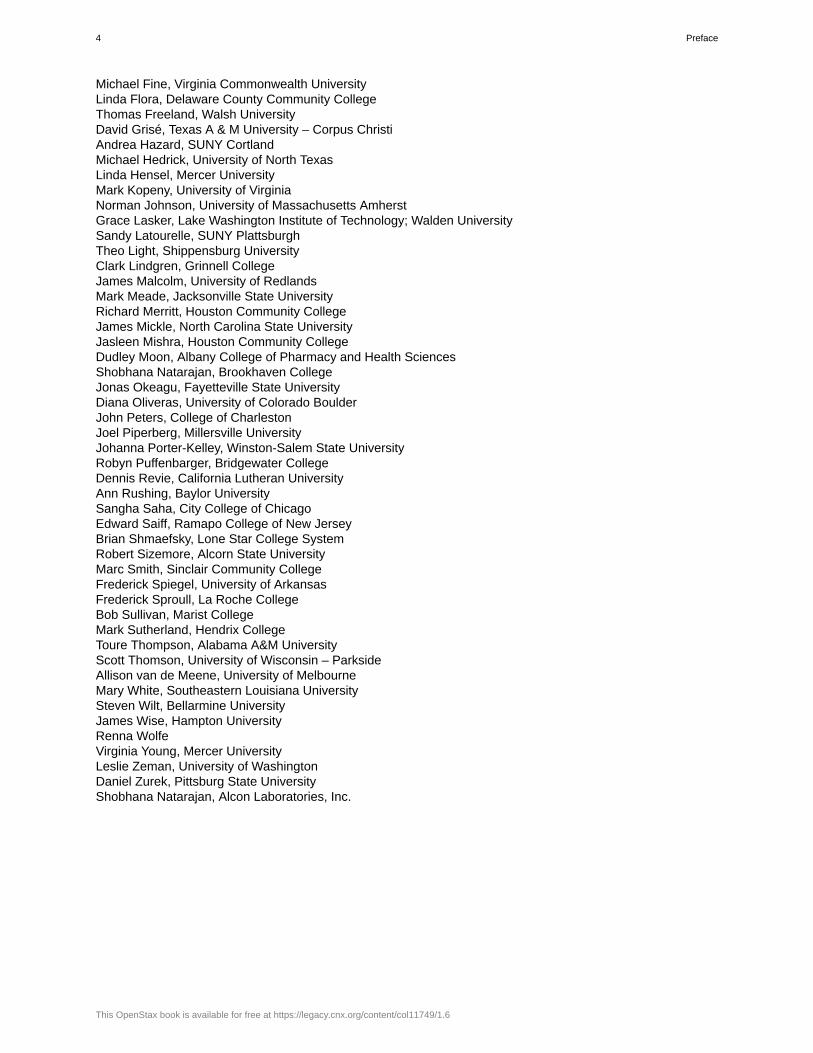
Figure 1.2 A toad represents a highly organized structure consisting of cells, tissues, organs, and organ systems.(credit: “Ivengo”/Wikimedia Commons)
Organisms are highly organized, coordinated structures that consist of one or more cells. Even very simple,single-celled organisms are remarkably complex: inside each cell, atoms make up molecules; these in turn makeup cell organelles and other cellular inclusions. In multicellular organisms ( Figure 1.2), similar cells form tissues.Tissues, in turn, collaborate to create organs (body structures with a distinct function). Organs work together toform organ systems.
Sensitivity or Response to Stimuli
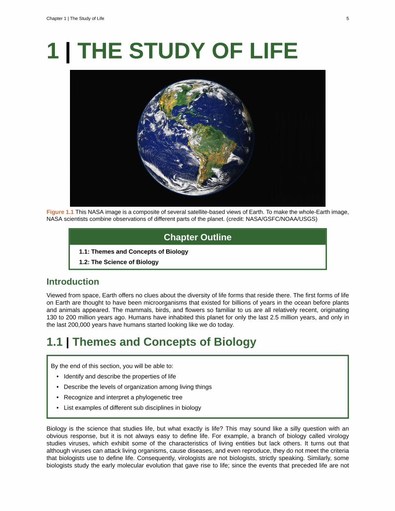
Figure 1.3 The leaves of this sensitive plant ( Mimosa pudica ) will instantly droop and fold when touched. After a fewminutes, the plant returns to normal. (credit: Alex Lomas)
Organisms respond to diverse stimuli. For example, plants can bend toward a source of light, climb on fencesand walls, or respond to touch ( Figure 1.3). Even tiny bacteria can move toward or away from chemicals
6 Chapter 1 | The Study of Life
This OpenStax book is available for free at https://legacy.cnx.org/content/col11749/1.6
(a process called chemotaxis ) or light ( phototaxis ). Movement toward a stimulus is considered a positiveresponse, while movement away from a stimulus is considered a negative response.
Watch this video (http://openstaxcollege.org/l/movement_plants) to see how plants respond to astimulus—from opening to light, to wrapping a tendril around a branch, to capturing prey.
Reproduction
Single-celled organisms reproduce by first duplicating their DNA, and then dividing it equally as the cell preparesto divide to form two new cells. Multicellular organisms often produce specialized reproductive germline cellsthat will form new individuals. When reproduction occurs, genes containing DNA are passed along to anorganism’s offspring. These genes ensure that the offspring will belong to the same species and will have similarcharacteristics, such as size and shape.
Growth and Development
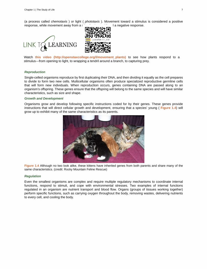
Organisms grow and develop following specific instructions coded for by their genes. These genes provideinstructions that will direct cellular growth and development, ensuring that a species’ young ( Figure 1.4) willgrow up to exhibit many of the same characteristics as its parents.
Figure 1.4 Although no two look alike, these kittens have inherited genes from both parents and share many of thesame characteristics. (credit: Rocky Mountain Feline Rescue)
Regulation
Even the smallest organisms are complex and require multiple regulatory mechanisms to coordinate internalfunctions, respond to stimuli, and cope with environmental stresses. Two examples of internal functionsregulated in an organism are nutrient transport and blood flow. Organs (groups of tissues working together)perform specific functions, such as carrying oxygen throughout the body, removing wastes, delivering nutrientsto every cell, and cooling the body.
Chapter 1 | The Study of Life 7
Homeostasis
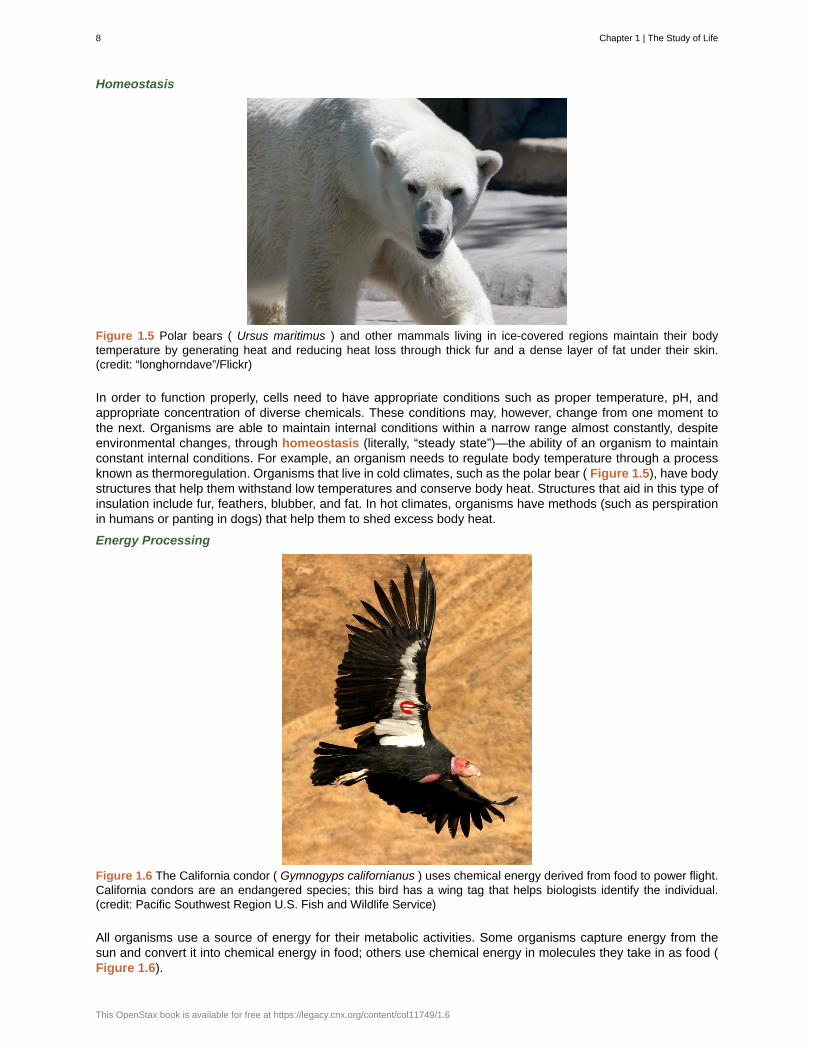
Figure 1.5 Polar bears ( Ursus maritimus ) and other mammals living in ice-covered regions maintain their bodytemperature by generating heat and reducing heat loss through thick fur and a dense layer of fat under their skin.(credit: “longhorndave”/Flickr)
In order to function properly, cells need to have appropriate conditions such as proper temperature, pH, andappropriate concentration of diverse chemicals. These conditions may, however, change from one moment tothe next. Organisms are able to maintain internal conditions within a narrow range almost constantly, despiteenvironmental changes, through homeostasis (literally, “steady state”)—the ability of an organism to maintainconstant internal conditions. For example, an organism needs to regulate body temperature through a processknown as thermoregulation. Organisms that live in cold climates, such as the polar bear ( Figure 1.5), have bodystructures that help them withstand low temperatures and conserve body heat. Structures that aid in this type ofinsulation include fur, feathers, blubber, and fat. In hot climates, organisms have methods (such as perspirationin humans or panting in dogs) that help them to shed excess body heat.
Energy Processing
Figure 1.6 The California condor ( Gymnogyps californianus ) uses chemical energy derived from food to power flight.California condors are an endangered species; this bird has a wing tag that helps biologists identify the individual.(credit: Pacific Southwest Region U.S. Fish and Wildlife Service)
All organisms use a source of energy for their metabolic activities. Some organisms capture energy from thesun and convert it into chemical energy in food; others use chemical energy in molecules they take in as food (Figure 1.6).
8 Chapter 1 | The Study of Life
This OpenStax book is available for free at https://legacy.cnx.org/content/col11749/1.6
1.1b Levels of Organization of Living Things
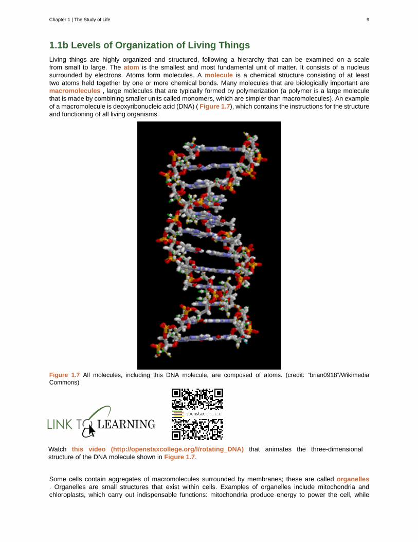
Living things are highly organized and structured, following a hierarchy that can be examined on a scalefrom small to large. The atom is the smallest and most fundamental unit of matter. It consists of a nucleussurrounded by electrons. Atoms form molecules. A molecule is a chemical structure consisting of at leasttwo atoms held together by one or more chemical bonds. Many molecules that are biologically important aremacromolecules , large molecules that are typically formed by polymerization (a polymer is a large moleculethat is made by combining smaller units called monomers, which are simpler than macromolecules). An exampleof a macromolecule is deoxyribonucleic acid (DNA) ( Figure 1.7), which contains the instructions for the structureand functioning of all living organisms.
Figure 1.7 All molecules, including this DNA molecule, are composed of atoms. (credit: “brian0918”/WikimediaCommons)
Watch this video (http://openstaxcollege.org/l/rotating_DNA) that animates the three-dimensionalstructure of the DNA molecule shown in Figure 1.7.
Some cells contain aggregates of macromolecules surrounded by membranes; these are called organelles. Organelles are small structures that exist within cells. Examples of organelles include mitochondria andchloroplasts, which carry out indispensable functions: mitochondria produce energy to power the cell, while
Chapter 1 | The Study of Life 9
chloroplasts enable green plants to utilize the energy in sunlight to make sugars. All living things are madeof cells; the cell itself is the smallest fundamental unit of structure and function in living organisms. (Thisrequirement is why viruses are not considered living: they are not made of cells. To make new viruses, they haveto invade and hijack the reproductive mechanism of a living cell; only then can they obtain the materials theyneed to reproduce.) Some organisms consist of a single cell and others are multicellular. Cells are classifiedas prokaryotic or eukaryotic. Prokaryotes are single-celled or colonial organisms that do not have membrane-bound nuclei; in contrast, the cells of eukaryotes do have membrane-bound organelles and a membrane-boundnucleus.
In larger organisms, cells combine to make tissues , which are groups of similar cells carrying out similar orrelated functions. Organs are collections of tissues grouped together performing a common function. Organsare present not only in animals but also in plants. An organ system is a higher level of organization thatconsists of functionally related organs. Mammals have many organ systems. For instance, the circulatory systemtransports blood through the body and to and from the lungs; it includes organs such as the heart and bloodvessels. Organisms are individual living entities. For example, each tree in a forest is an organism. Single-celled prokaryotes and single-celled eukaryotes are also considered organisms and are typically referred to asmicroorganisms.
All the individuals of a species living within a specific area are collectively called a population . For example,a forest may include many pine trees. All of these pine trees represent the population of pine trees in thisforest. Different populations may live in the same specific area. For example, the forest with the pine treesincludes populations of flowering plants and also insects and microbial populations. A community is the sumof populations inhabiting a particular area. For instance, all of the trees, flowers, insects, and other populationsin a forest form the forest’s community. The forest itself is an ecosystem. An ecosystem consists of all theliving things in a particular area together with the abiotic, non-living parts of that environment such as nitrogenin the soil or rain water. At the highest level of organization ( Figure 1.8), the biosphere is the collection of allecosystems, and it represents the zones of life on earth. It includes land, water, and even the atmosphere to acertain extent.
10 Chapter 1 | The Study of Life
This OpenStax book is available for free at https://legacy.cnx.org/content/col11749/1.6
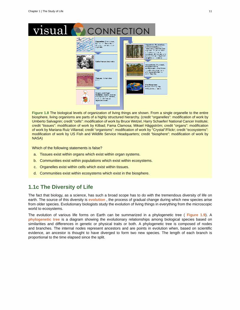
Figure 1.8 The biological levels of organization of living things are shown. From a single organelle to the entirebiosphere, living organisms are parts of a highly structured hierarchy. (credit “organelles”: modification of work byUmberto Salvagnin; credit “cells”: modification of work by Bruce Wetzel, Harry Schaefer/ National Cancer Institute;credit “tissues”: modification of work by Kilbad; Fama Clamosa; Mikael Häggström; credit “organs”: modificationof work by Mariana Ruiz Villareal; credit “organisms”: modification of work by "Crystal"/Flickr; credit “ecosystems”:modification of work by US Fish and Wildlife Service Headquarters; credit “biosphere”: modification of work byNASA)
Which of the following statements is false?
a. Tissues exist within organs which exist within organ systems.
b. Communities exist within populations which exist within ecosystems.
c. Organelles exist within cells which exist within tissues.
d. Communities exist within ecosystems which exist in the biosphere.
1.1c The Diversity of Life
The fact that biology, as a science, has such a broad scope has to do with the tremendous diversity of life onearth. The source of this diversity is evolution , the process of gradual change during which new species arisefrom older species. Evolutionary biologists study the evolution of living things in everything from the microscopicworld to ecosystems.
The evolution of various life forms on Earth can be summarized in a phylogenetic tree ( Figure 1.9). Aphylogenetic tree is a diagram showing the evolutionary relationships among biological species based onsimilarities and differences in genetic or physical traits or both. A phylogenetic tree is composed of nodesand branches. The internal nodes represent ancestors and are points in evolution when, based on scientificevidence, an ancestor is thought to have diverged to form two new species. The length of each branch isproportional to the time elapsed since the split.
Chapter 1 | The Study of Life 11
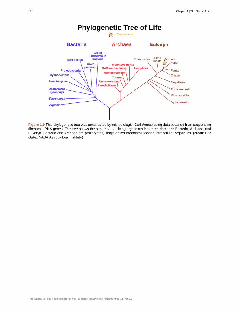
Figure 1.9 This phylogenetic tree was constructed by microbiologist Carl Woese using data obtained from sequencingribosomal RNA genes. The tree shows the separation of living organisms into three domains: Bacteria, Archaea, andEukarya. Bacteria and Archaea are prokaryotes, single-celled organisms lacking intracellular organelles. (credit: EricGaba; NASA Astrobiology Institute)
12 Chapter 1 | The Study of Life
This OpenStax book is available for free at https://legacy.cnx.org/content/col11749/1.6
Carl Woese and the Phylogenetic TreeIn the past, biologists grouped living organisms into five kingdoms: animals, plants, fungi, protists, andbacteria. The organizational scheme was based mainly on physical features, as opposed to physiology,biochemistry, or molecular biology, all of which are used by modern systematics. The pioneering work ofAmerican microbiologist Carl Woese in the early 1970s has shown, however, that life on Earth has evolvedalong three lineages, now called domains—Bacteria, Archaea, and Eukarya. The first two are prokaryoticcells with microbes that lack membrane-enclosed nuclei and organelles. The third domain contains theeukaryotes and includes unicellular microorganisms together with the four original kingdoms (excludingbacteria). Woese defined Archaea as a new domain, and this resulted in a new taxonomic tree ( Figure1.9). Many organisms belonging to the Archaea domain live under extreme conditions and are calledextremophiles. To construct his tree, Woese used genetic relationships rather than similarities based onmorphology (shape).
Woese’s tree was constructed from comparative sequencing of the genes that are universally distributed,present in every organism, and conserved (meaning that these genes have remained essentially unchangedthroughout evolution). Woese’s approach was revolutionary because comparisons of physical features areinsufficient to differentiate between the prokaryotes that appear fairly similar in spite of their tremendousbiochemical diversity and genetic variability ( Figure 1.10). The comparison of homologous DNA and RNAsequences provided Woese with a sensitive device that revealed the extensive variability of prokaryotes,and which justified the separation of the prokaryotes into two domains: bacteria and archaea.
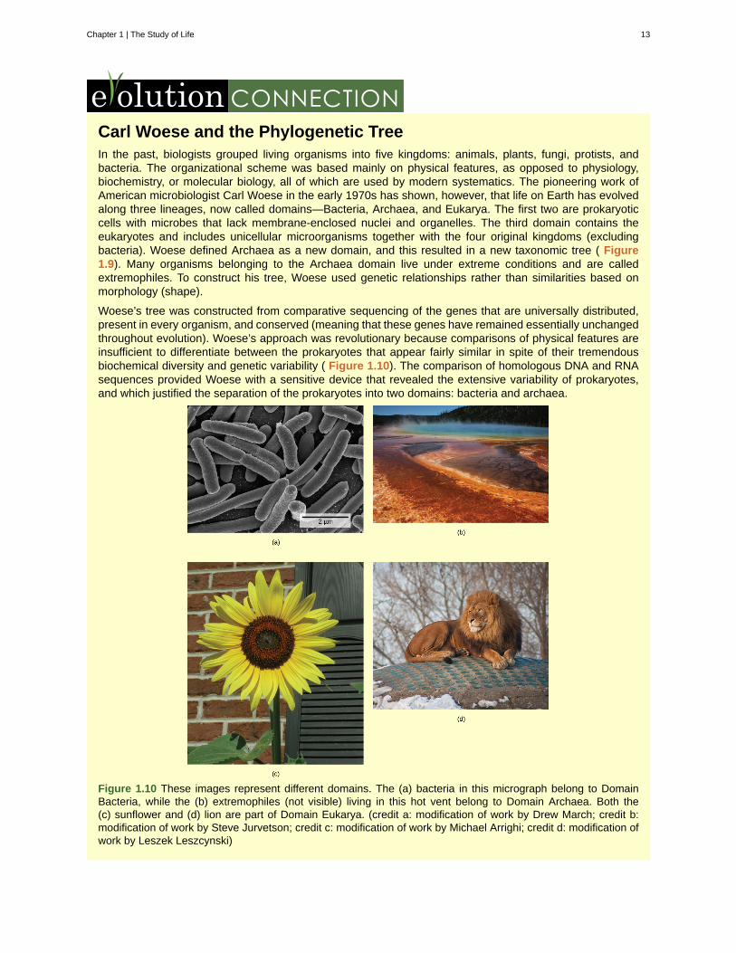
Figure 1.10 These images represent different domains. The (a) bacteria in this micrograph belong to DomainBacteria, while the (b) extremophiles (not visible) living in this hot vent belong to Domain Archaea. Both the(c) sunflower and (d) lion are part of Domain Eukarya. (credit a: modification of work by Drew March; credit b:modification of work by Steve Jurvetson; credit c: modification of work by Michael Arrighi; credit d: modification ofwork by Leszek Leszcynski)
Chapter 1 | The Study of Life 13
1.2 | The Science of Biology
By the end of this section, you will be able to:
• Identify the shared characteristics of the natural sciences
• Summarize the steps of the scientific method
• Compare inductive reasoning with deductive reasoning
• Describe the goals of basic science and applied science
(a) (b)
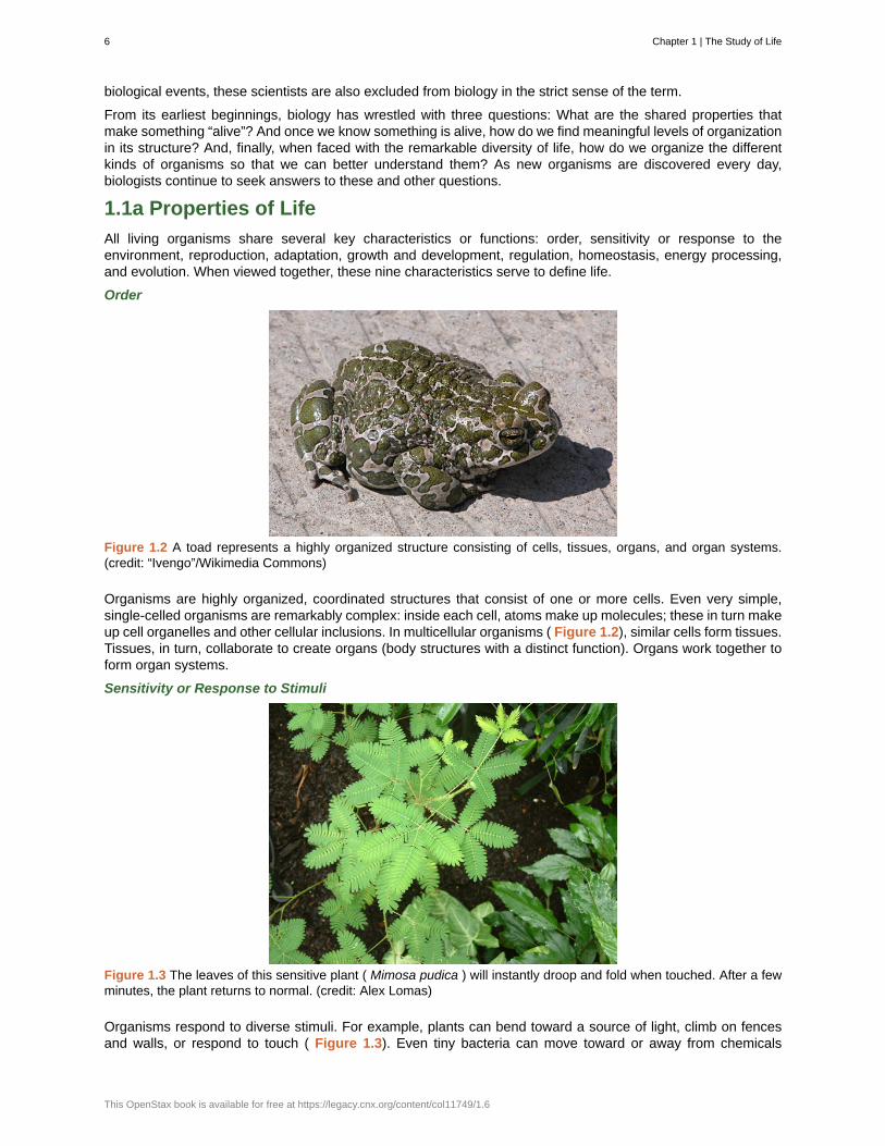
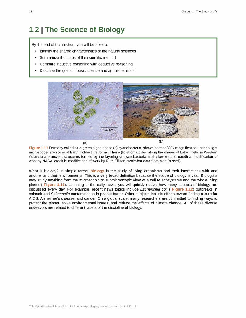
Figure 1.11 Formerly called blue-green algae, these (a) cyanobacteria, shown here at 300x magnification under a lightmicroscope, are some of Earth’s oldest life forms. These (b) stromatolites along the shores of Lake Thetis in WesternAustralia are ancient structures formed by the layering of cyanobacteria in shallow waters. (credit a: modification ofwork by NASA; credit b: modification of work by Ruth Ellison; scale-bar data from Matt Russell)
What is biology? In simple terms, biology is the study of living organisms and their interactions with oneanother and their environments. This is a very broad definition because the scope of biology is vast. Biologistsmay study anything from the microscopic or submicroscopic view of a cell to ecosystems and the whole livingplanet ( Figure 1.11). Listening to the daily news, you will quickly realize how many aspects of biology arediscussed every day. For example, recent news topics include Escherichia coli ( Figure 1.12) outbreaks inspinach and Salmonella contamination in peanut butter. Other subjects include efforts toward finding a cure forAIDS, Alzheimer’s disease, and cancer. On a global scale, many researchers are committed to finding ways toprotect the planet, solve environmental issues, and reduce the effects of climate change. All of these diverseendeavors are related to different facets of the discipline of biology.
14 Chapter 1 | The Study of Life
This OpenStax book is available for free at https://legacy.cnx.org/content/col11749/1.6
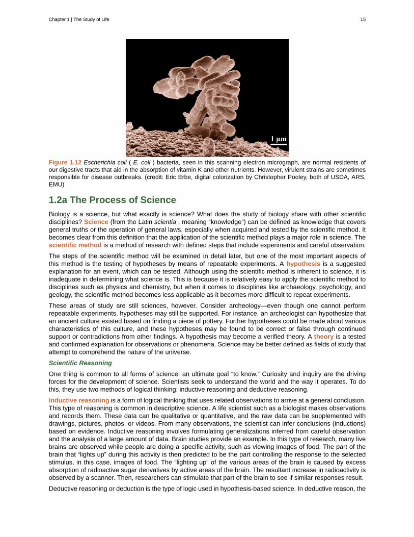
Figure 1.12 Escherichia coli ( E. coli ) bacteria, seen in this scanning electron micrograph, are normal residents ofour digestive tracts that aid in the absorption of vitamin K and other nutrients. However, virulent strains are sometimesresponsible for disease outbreaks. (credit: Eric Erbe, digital colorization by Christopher Pooley, both of USDA, ARS,EMU)
1.2a The Process of Science
Biology is a science, but what exactly is science? What does the study of biology share with other scientificdisciplines? Science (from the Latin scientia , meaning “knowledge”) can be defined as knowledge that coversgeneral truths or the operation of general laws, especially when acquired and tested by the scientific method. Itbecomes clear from this definition that the application of the scientific method plays a major role in science. Thescientific method is a method of research with defined steps that include experiments and careful observation.
The steps of the scientific method will be examined in detail later, but one of the most important aspects ofthis method is the testing of hypotheses by means of repeatable experiments. A hypothesis is a suggestedexplanation for an event, which can be tested. Although using the scientific method is inherent to science, it isinadequate in determining what science is. This is because it is relatively easy to apply the scientific method todisciplines such as physics and chemistry, but when it comes to disciplines like archaeology, psychology, andgeology, the scientific method becomes less applicable as it becomes more difficult to repeat experiments.
These areas of study are still sciences, however. Consider archeology—even though one cannot performrepeatable experiments, hypotheses may still be supported. For instance, an archeologist can hypothesize thatan ancient culture existed based on finding a piece of pottery. Further hypotheses could be made about variouscharacteristics of this culture, and these hypotheses may be found to be correct or false through continuedsupport or contradictions from other findings. A hypothesis may become a verified theory. A theory is a testedand confirmed explanation for observations or phenomena. Science may be better defined as fields of study thatattempt to comprehend the nature of the universe.
Scientific Reasoning
One thing is common to all forms of science: an ultimate goal “to know.” Curiosity and inquiry are the drivingforces for the development of science. Scientists seek to understand the world and the way it operates. To dothis, they use two methods of logical thinking: inductive reasoning and deductive reasoning.
Inductive reasoning is a form of logical thinking that uses related observations to arrive at a general conclusion.This type of reasoning is common in descriptive science. A life scientist such as a biologist makes observationsand records them. These data can be qualitative or quantitative, and the raw data can be supplemented withdrawings, pictures, photos, or videos. From many observations, the scientist can infer conclusions (inductions)based on evidence. Inductive reasoning involves formulating generalizations inferred from careful observationand the analysis of a large amount of data. Brain studies provide an example. In this type of research, many livebrains are observed while people are doing a specific activity, such as viewing images of food. The part of thebrain that “lights up” during this activity is then predicted to be the part controlling the response to the selectedstimulus, in this case, images of food. The “lighting up” of the various areas of the brain is caused by excessabsorption of radioactive sugar derivatives by active areas of the brain. The resultant increase in radioactivity isobserved by a scanner. Then, researchers can stimulate that part of the brain to see if similar responses result.
Deductive reasoning or deduction is the type of logic used in hypothesis-based science. In deductive reason, the
Chapter 1 | The Study of Life 15
pattern of thinking moves in the opposite direction as compared to inductive reasoning. Deductive reasoningis a form of logical thinking that uses a general principle or law to forecast specific results. From those generalprinciples, a scientist can extrapolate and predict the specific results that would be valid as long as the generalprinciples are valid. Studies in climate change can illustrate this type of reasoning. For example, scientists maypredict that if the climate becomes warmer in a particular region, then the distribution of plants and animalsshould change. These predictions have been made and tested, and many such changes have been found, suchas the modification of arable areas for agriculture, with change based on temperature averages.
Both types of logical thinking are related to the two main pathways of scientific study: descriptive science andhypothesis-based science. Descriptive (or discovery) science , which is usually inductive, aims to observe,explore, and discover, while hypothesis-based science , which is usually deductive, begins with a specificquestion or problem and a potential answer or solution that can be tested. The boundary between these twoforms of study is often blurred, and most scientific endeavors combine both approaches. The fuzzy boundarybecomes apparent when thinking about how easily observation can lead to specific questions. For example, agentleman in the 1940s observed that the burr seeds that stuck to his clothes and his dog’s fur had a tiny hookstructure. On closer inspection, he discovered that the burrs’ gripping device was more reliable than a zipper.He eventually developed a company and produced the hook-and-loop fastener popularly known today as Velcro.Descriptive science and hypothesis-based science are in continuous dialogue.
Art Connection
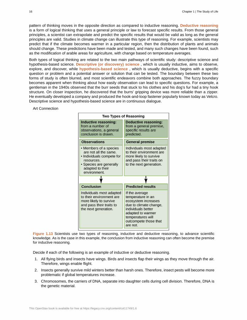
Figure 1.13 Scientists use two types of reasoning, inductive and deductive reasoning, to advance scientificknowledge. As is the case in this example, the conclusion from inductive reasoning can often become the premisefor inductive reasoning.
Decide if each of the following is an example of inductive or deductive reasoning.
1. All flying birds and insects have wings. Birds and insects flap their wings as they move through the air.Therefore, wings enable flight.
2. Insects generally survive mild winters better than harsh ones. Therefore, insect pests will become moreproblematic if global temperatures increase.
3. Chromosomes, the carriers of DNA, separate into daughter cells during cell division. Therefore, DNA isthe genetic material.
16 Chapter 1 | The Study of Life
This OpenStax book is available for free at https://legacy.cnx.org/content/col11749/1.6
4. Animals as diverse as humans, insects, and wolves all exhibit social behavior. Therefore, socialbehavior must have an evolutionary advantage.
1.2b The Scientific Method
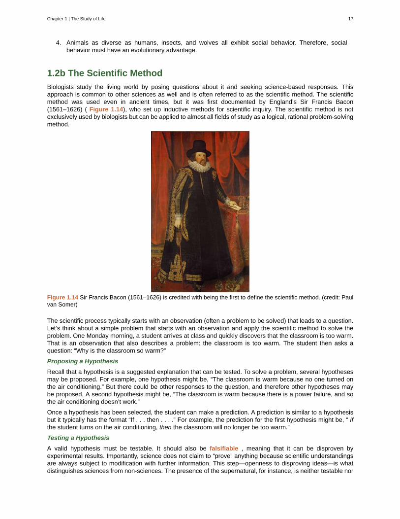
Biologists study the living world by posing questions about it and seeking science-based responses. Thisapproach is common to other sciences as well and is often referred to as the scientific method. The scientificmethod was used even in ancient times, but it was first documented by England’s Sir Francis Bacon(1561–1626) ( Figure 1.14), who set up inductive methods for scientific inquiry. The scientific method is notexclusively used by biologists but can be applied to almost all fields of study as a logical, rational problem-solvingmethod.
Figure 1.14 Sir Francis Bacon (1561–1626) is credited with being the first to define the scientific method. (credit: Paulvan Somer)
The scientific process typically starts with an observation (often a problem to be solved) that leads to a question.Let’s think about a simple problem that starts with an observation and apply the scientific method to solve theproblem. One Monday morning, a student arrives at class and quickly discovers that the classroom is too warm.That is an observation that also describes a problem: the classroom is too warm. The student then asks aquestion: “Why is the classroom so warm?”
Proposing a Hypothesis
Recall that a hypothesis is a suggested explanation that can be tested. To solve a problem, several hypothesesmay be proposed. For example, one hypothesis might be, “The classroom is warm because no one turned onthe air conditioning.” But there could be other responses to the question, and therefore other hypotheses maybe proposed. A second hypothesis might be, “The classroom is warm because there is a power failure, and sothe air conditioning doesn’t work.”
Once a hypothesis has been selected, the student can make a prediction. A prediction is similar to a hypothesisbut it typically has the format “If . . . then . . . .” For example, the prediction for the first hypothesis might be, “ Ifthe student turns on the air conditioning, then the classroom will no longer be too warm.”
Testing a Hypothesis
A valid hypothesis must be testable. It should also be falsifiable , meaning that it can be disproven byexperimental results. Importantly, science does not claim to “prove” anything because scientific understandingsare always subject to modification with further information. This step—openness to disproving ideas—is whatdistinguishes sciences from non-sciences. The presence of the supernatural, for instance, is neither testable nor
Chapter 1 | The Study of Life 17
falsifiable.
To test a hypothesis, a researcher will conduct one or more experiments designed to eliminate one or more ofthe hypotheses. Each experiment will have one or more variables and one or more controls. A variable is anypart of the experiment that can vary or change during the experiment.
To test a hypothesis, a researcher will conduct one or more experiments designed to eliminate one or more of thehypotheses. Experiments typically have a dependent variable , independent variable , and several controlledvariables . The dependent variable is some changing aspect of the experiment that you want to find out. Forexample, if you're testing how a particular drug dosage fights cancer, your dependent variable could be howmany cancer cells died. Your independent variable is what you changed to get that result. So in this example,your independent variable would be the different dosages of the drug. A controlled variable is any part of theexperimental setup that you kept the same. The more controlled variables you have, the more accurate your datais likely to be. For example, in the drug dosage experiment, maybe you only tested pancreatic cancer patients,who were 60-70 years old, and had an early-stage diagnosis. If you had NO controlled variables, you couldn't besure whether your results are due to the different drug dosage or something else.
The control group contains every feature of the experimental group except it is not given the manipulationthat is hypothesized about. Therefore, if the results of the experimental group differ from the control group, thedifference must be due to the hypothesized manipulation, rather than some outside factor. To go back to ourprevious example, if you had a collection of patients who all received a certain dosage of the drug, and anothergroup who did not receive the drug at all, the latter group is the control group. It's important to use control groupsto know whether your experiment results are real. For example, if you saw no difference in cancer cell deathbetween the group who received the drug versus those who did not, that is a clear result indicating the drug atthat particular dosage is ineffective. The group who received no treatment is called a negative control , andin drug studies, these groups often receive a placebo , which is a pill/liquid that looks like the drug, but doesnot actually contain the drug. It contains nothing, or maybe a sugar solution that has no effect on the body. Apositive control is a sample/individual or group of samples/individuals who you know will actually demonstratea difference from the negative control. For example, if you were comparing a new cancer drug to a known drugthat works, the individuals receiving the known drug would be the positive control group.
Again, the point of science is to test a hypothesis, and using that data, generate and test another one, untilyou have a particular answer. Sometimes this takes years and years to get a full understanding of how or whysomething works. To go back to the air conditioning hypothesis, the student would find out if the air conditioningis on. If the air conditioning is turned on but does not work, there should be another reason, and this hypothesisshould be rejected. To test the second hypothesis, the student could check if the lights in the classroom arefunctional. If so, there is no power failure and this hypothesis should be rejected. Each hypothesis should betested by carrying out appropriate experiments. Be aware that rejecting one hypothesis does not determinewhether or not the other hypotheses can be accepted; it simply eliminates one hypothesis that is not valid( Figure 1.15). Using the scientific method, the hypotheses that are inconsistent with experimental data arerejected.
While this “warm classroom” example is based on observational results, other hypotheses and experimentsmight have clearer controls. For instance, a student might attend class on Monday and realize she had difficultyconcentrating on the lecture. One observation to explain this occurrence might be, “When I eat breakfast beforeclass, I am better able to pay attention.” The student could then design an experiment with a control to test thishypothesis.
In hypothesis-based science, specific results are predicted from a general premise. This type of reasoning iscalled deductive reasoning: deduction proceeds from the general to the particular. But the reverse of the processis also possible: sometimes, scientists reach a general conclusion from a number of specific observations. Thistype of reasoning is called inductive reasoning, and it proceeds from the particular to the general. Inductive anddeductive reasoning are often used in tandem to advance scientific knowledge ( Figure 1.13).
18 Chapter 1 | The Study of Life
This OpenStax book is available for free at https://legacy.cnx.org/content/col11749/1.6
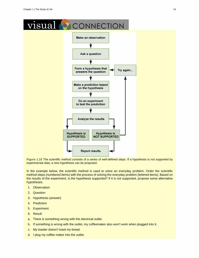
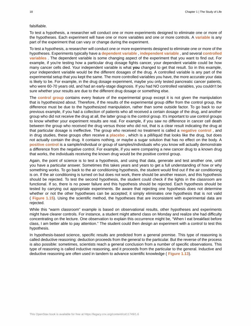
Figure 1.15 The scientific method consists of a series of well-defined steps. If a hypothesis is not supported byexperimental data, a new hypothesis can be proposed.
In the example below, the scientific method is used to solve an everyday problem. Order the scientificmethod steps (numbered items) with the process of solving the everyday problem (lettered items). Based onthe results of the experiment, is the hypothesis supported? If it is not supported, propose some alternativehypotheses.
1. Observation
2. Question
3. Hypothesis (answer)
4. Prediction
5. Experiment
6. Result
a. There is something wrong with the electrical outlet.
b. If something is wrong with the outlet, my coffeemaker also won’t work when plugged into it.
c. My toaster doesn’t toast my bread.
d. I plug my coffee maker into the outlet.
Chapter 1 | The Study of Life 19
e. My coffeemaker works.
f. Why doesn’t my toaster work?
The scientific method may seem too rigid and structured. It is important to keep in mind that, although scientistsoften follow this sequence, there is flexibility. Sometimes an experiment leads to conclusions that favor a changein approach; often, an experiment brings entirely new scientific questions to the puzzle. Many times, sciencedoes not operate in a linear fashion; instead, scientists continually draw inferences and make generalizations,finding patterns as their research proceeds. Scientific reasoning is more complex than the scientific methodalone suggests. Notice, too, that the scientific method can be applied to solving problems that aren’t necessarilyscientific in nature.
1.2c Two Types of Science: Basic Science and Applied Science
The scientific community has been debating for the last few decades about the value of different types ofscience. Is it valuable to pursue science for the sake of simply gaining knowledge, or does scientific knowledgeonly have worth if we can apply it to solving a specific problem or to bettering our lives? This question focuseson the differences between two types of science: basic science and applied science.
Basic science or “pure” science seeks to expand knowledge regardless of the short-term application of thatknowledge. It is not focused on developing a product or a service of immediate public or commercial value. Theimmediate goal of basic science is knowledge for knowledge’s sake, though this does not mean that, in the end,it may not result in a practical application.
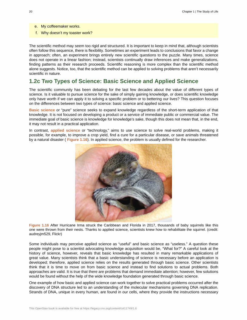
In contrast, applied science or “technology,” aims to use science to solve real-world problems, making itpossible, for example, to improve a crop yield, find a cure for a particular disease, or save animals threatenedby a natural disaster ( Figure 1.16). In applied science, the problem is usually defined for the researcher.
Figure 1.16 After Hurricane Irma struck the Caribbean and Florida in 2017, thousands of baby squirrels like thisone were thrown from their nests. Thanks to applied science, scientists knew how to rehabilitate the squirrel. (credit:audreyjm529, Flickr)
Some individuals may perceive applied science as “useful” and basic science as “useless.” A question thesepeople might pose to a scientist advocating knowledge acquisition would be, “What for?” A careful look at thehistory of science, however, reveals that basic knowledge has resulted in many remarkable applications ofgreat value. Many scientists think that a basic understanding of science is necessary before an application isdeveloped; therefore, applied science relies on the results generated through basic science. Other scientiststhink that it is time to move on from basic science and instead to find solutions to actual problems. Bothapproaches are valid. It is true that there are problems that demand immediate attention; however, few solutionswould be found without the help of the wide knowledge foundation generated through basic science.
One example of how basic and applied science can work together to solve practical problems occurred after thediscovery of DNA structure led to an understanding of the molecular mechanisms governing DNA replication.Strands of DNA, unique in every human, are found in our cells, where they provide the instructions necessary
20 Chapter 1 | The Study of Life
This OpenStax book is available for free at https://legacy.cnx.org/content/col11749/1.6
for life. During DNA replication, DNA makes new copies of itself, shortly before a cell divides. Understandingthe mechanisms of DNA replication enabled scientists to develop laboratory techniques that are now used toidentify genetic diseases, pinpoint individuals who were at a crime scene, and determine paternity. Without basicscience, it is unlikely that applied science would exist.
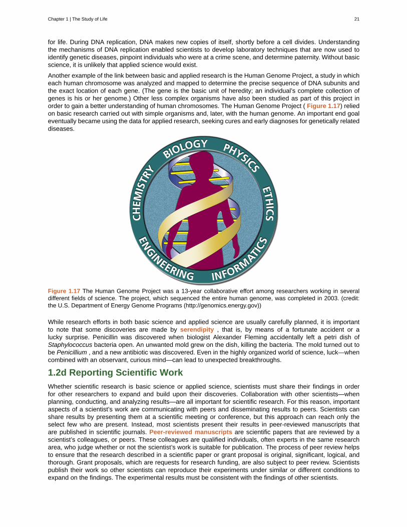
Another example of the link between basic and applied research is the Human Genome Project, a study in whicheach human chromosome was analyzed and mapped to determine the precise sequence of DNA subunits andthe exact location of each gene. (The gene is the basic unit of heredity; an individual’s complete collection ofgenes is his or her genome.) Other less complex organisms have also been studied as part of this project inorder to gain a better understanding of human chromosomes. The Human Genome Project ( Figure 1.17) reliedon basic research carried out with simple organisms and, later, with the human genome. An important end goaleventually became using the data for applied research, seeking cures and early diagnoses for genetically relateddiseases.
Figure 1.17 The Human Genome Project was a 13-year collaborative effort among researchers working in severaldifferent fields of science. The project, which sequenced the entire human genome, was completed in 2003. (credit:the U.S. Department of Energy Genome Programs (http://genomics.energy.gov))
While research efforts in both basic science and applied science are usually carefully planned, it is importantto note that some discoveries are made by serendipity , that is, by means of a fortunate accident or alucky surprise. Penicillin was discovered when biologist Alexander Fleming accidentally left a petri dish ofStaphylococcus bacteria open. An unwanted mold grew on the dish, killing the bacteria. The mold turned out tobe Penicillium , and a new antibiotic was discovered. Even in the highly organized world of science, luck—whencombined with an observant, curious mind—can lead to unexpected breakthroughs.
1.2d Reporting Scientific Work
Whether scientific research is basic science or applied science, scientists must share their findings in orderfor other researchers to expand and build upon their discoveries. Collaboration with other scientists—whenplanning, conducting, and analyzing results—are all important for scientific research. For this reason, importantaspects of a scientist’s work are communicating with peers and disseminating results to peers. Scientists canshare results by presenting them at a scientific meeting or conference, but this approach can reach only theselect few who are present. Instead, most scientists present their results in peer-reviewed manuscripts thatare published in scientific journals. Peer-reviewed manuscripts are scientific papers that are reviewed by ascientist’s colleagues, or peers. These colleagues are qualified individuals, often experts in the same researcharea, who judge whether or not the scientist’s work is suitable for publication. The process of peer review helpsto ensure that the research described in a scientific paper or grant proposal is original, significant, logical, andthorough. Grant proposals, which are requests for research funding, are also subject to peer review. Scientistspublish their work so other scientists can reproduce their experiments under similar or different conditions toexpand on the findings. The experimental results must be consistent with the findings of other scientists.
Chapter 1 | The Study of Life 21
A scientific paper is very different from creative writing. Although creativity is required to design experiments,there are fixed guidelines when it comes to presenting scientific results. First, scientific writing must be brief,concise, and accurate. A scientific paper needs to be succinct but detailed enough to allow peers to reproducethe experiments.
The scientific paper consists of several specific sections—introduction, materials and methods, results, anddiscussion. This structure is sometimes called the “IMRaD” format. There are usually acknowledgment andreference sections as well as an abstract (a concise summary) at the beginning of the paper. There might beadditional sections depending on the type of paper and the journal where it will be published; for example, somereview papers require an outline.
The introduction starts with brief, but broad, background information about what is known in the field. A goodintroduction also gives the rationale of the work; it justifies the work carried out and also briefly mentions the endof the paper, where the hypothesis or research question driving the research will be presented. The introductionrefers to the published scientific work of others and therefore requires citations following the style of the journal.Using the work or ideas of others without proper citation is considered plagiarism .
The materials and methods section includes a complete and accurate description of the substances used,and the method and techniques used by the researchers to gather data. The description should be thoroughenough to allow another researcher to repeat the experiment and obtain similar results, but it does not haveto be verbose. This section will also include information on how measurements were made and what typesof calculations and statistical analyses were used to examine raw data. Although the materials and methodssection gives an accurate description of the experiments, it does not discuss them.
Some journals require a results section followed by a discussion section, but it is more common to combineboth. If the journal does not allow the combination of both sections, the results section simply narrates thefindings without any further interpretation. The results are presented by means of tables or graphs, but noduplicate information should be presented. In the discussion section, the researcher will interpret the results,describe how variables may be related, and attempt to explain the observations. It is indispensable to conduct anextensive literature search to put the results in the context of previously published scientific research. Therefore,proper citations are included in this section as well.
Finally, the conclusion section summarizes the importance of the experimental findings. While the scientificpaper almost certainly answered one or more scientific questions that were stated, any good research shouldlead to more questions. Therefore, a well-done scientific paper leaves doors open for the researcher and othersto continue and expand on the findings.
Review articles do not follow the IMRAD format because they do not present original scientific findings, orprimary literature; instead, they summarize and comment on findings that were published as primary literatureand typically include extensive reference sections.
22 Chapter 1 | The Study of Life
This OpenStax book is available for free at https://legacy.cnx.org/content/col11749/1.6
abstract
applied science
atom
basic science
biology
biosphere
cell
community
conclusion
control group
controlled variables
deductive reasoning
dependent variable
Descriptive (or discovery) science
discussion
ecosystem
eukaryotes
evolution
falsifiable
homeostasis
hypothesis
hypothesis-based science
independent variable
inductive reasoning
introduction
macromolecules
materials and methods
molecule
KEY TERMS
opening section of a scientific paper that summarizes the research and conclusions
form of science that aims to solve real-world problems
smallest and most fundamental unit of matter
science that seeks to expand knowledge and understanding regardless of the short-termapplication of that knowledge
the study of living organisms and their interactions with one another and their environments
collection of all the ecosystems on Earth
smallest fundamental unit of structure and function in living things
set of populations inhabiting a particular area
section of a scientific paper that summarizes the importance of the experimental findings
one or more samples/individuals whose outcome is known; used to compare the other samples/individuals to
part of an experiment that is unchanged for all samples/individuals
form of logical thinking that uses a general inclusive statement to forecast specific results
the part of an experiment that you want to find out
form of science that aims to observe, explore, and investigate
section of a scientific paper in which the author interprets experimental results, describes howvariables may be related, and attempts to explain the phenomenon in question
all the living things in a particular area together with the abiotic, nonliving parts of that environment
a group of organisms that all share one major similarity: their DNA is enclosed in a nucleus
process of gradual genetic change in a population over time
able to be disproven by experimental results
ability of an organism to maintain constant internal conditions
suggested explanation for an observation, which can be tested
form of science that begins with a specific question and potential testable answers
the part of an experiment that you change to determine its effect on the dependentvariable
form of logical thinking that uses related observations to arrive at a general conclusion
opening section of a scientific paper, which provides background information about what wasknown in the field prior to the research reported in the paper
large molecules
section of a scientific paper that includes a complete description of the substances,methods, and techniques used by the researchers to gather data
chemical structure consisting of at least two atoms held together by one or more chemical bonds
Chapter 1 | The Study of Life 23
negative control
organ system
organelles
Organisms
Organs
Peer-reviewed manuscripts
phylogenetic tree
placebo
plagiarism
population
positive control
Prokaryotes
results
Review articles
science
scientific method
serendipity
theory
tissues
variable
one or more samples/individuals who are known in advance to demonstrate a negative result
level of organization that consists of functionally related interacting organs
structures within a cell that perform a specific function for the cell
living things
a group of tissues that work together to perform some bodily function
scientific paper that is reviewed by a scientist's colleagues who are experts in thefield of study
diagram showing the evolutionary relationships among various biological species based onsimilarities and differences in genetic or physical traits or both; in essence, a hypothesis concerningevolutionary connections
a pill that looks like a drug, but doesn't actually contain the drug; used as negative controls in blind-studies where the participants don't know whether they're getting a treatment or not
using other people’s work or ideas without proper citation, creating the false impression that thoseare the author’s original ideas
all of the individuals of a species living within a specific area
one or more samples/individuals who are known in advance to demonstrate a positive result
a group of organisms that all share one major similarity: their DNA is NOT enclosed in a nucleus
section of a scientific paper in which the author narrates the experimental findings and presentsrelevant figures, pictures, diagrams, graphs, and tables, without any further interpretation
paper that summarizes and comments on findings that were published as primary literature
knowledge that covers general truths or the operation of general laws, especially when acquired andtested by the scientific method
method of research with defined steps that include observation, formulation of a hypothesis,testing, and confirming or falsifying the hypothesis
fortunate accident or a lucky surprise
tested and confirmed explanation for observations or phenomena
a group of cells, proteins, and fluid that work together to perform some function
part of an experiment that the experimenter can vary or change
CHAPTER SUMMARY
1.1 Themes and Concepts of Biology
Biology is the science of life. All living organisms share several key properties such as order, sensitivity orresponse to stimuli, reproduction, growth and development, regulation, homeostasis, and energy processing.Living things are highly organized parts of a hierarchy that includes atoms, molecules, organelles, cells,tissues, organs, and organ systems. Organisms, in turn, are grouped as populations, communities,ecosystems, and the biosphere. The great diversity of life today evolved from less-diverse ancestral organismsover billions of years. A diagram called a phylogenetic tree can be used to show evolutionary relationshipsamong organisms.
1.2 The Science of Biology
Biology is the science that studies living organisms and their interactions with one another and their
24 Chapter 1 | The Study of Life
This OpenStax book is available for free at https://legacy.cnx.org/content/col11749/1.6
environments. Science attempts to describe and understand the nature of the universe in whole or in part byrational means. Science has many fields; those fields related to the physical world and its phenomena areconsidered natural sciences.
Science can be basic or applied. The main goal of basic science is to expand knowledge without anyexpectation of short-term practical application of that knowledge. The primary goal of applied research,however, is to solve practical problems.
Two types of logical reasoning are used in science. Inductive reasoning uses particular results to producegeneral scientific principles. Deductive reasoning is a form of logical thinking that predicts results by applyinggeneral principles. The common thread throughout scientific research is the use of the scientific method, astep-based process that consists of making observations, defining a problem, posing hypotheses, testing thesehypotheses, and drawing one or more conclusions. The testing uses proper controls. Scientists present theirresults in peer-reviewed scientific papers published in scientific journals. A scientific research paper consists ofseveral well-defined sections: introduction, materials and methods, results, and, finally, a concludingdiscussion. Review papers summarize the research done in a particular field over a period of time.
Chapter 1 | The Study of Life 25

26 Chapter 1 | The Study of Life
This OpenStax book is available for free at https://legacy.cnx.org/content/col11749/1.6
2 | THE CHEMICALFOUNDATION OF LIFE
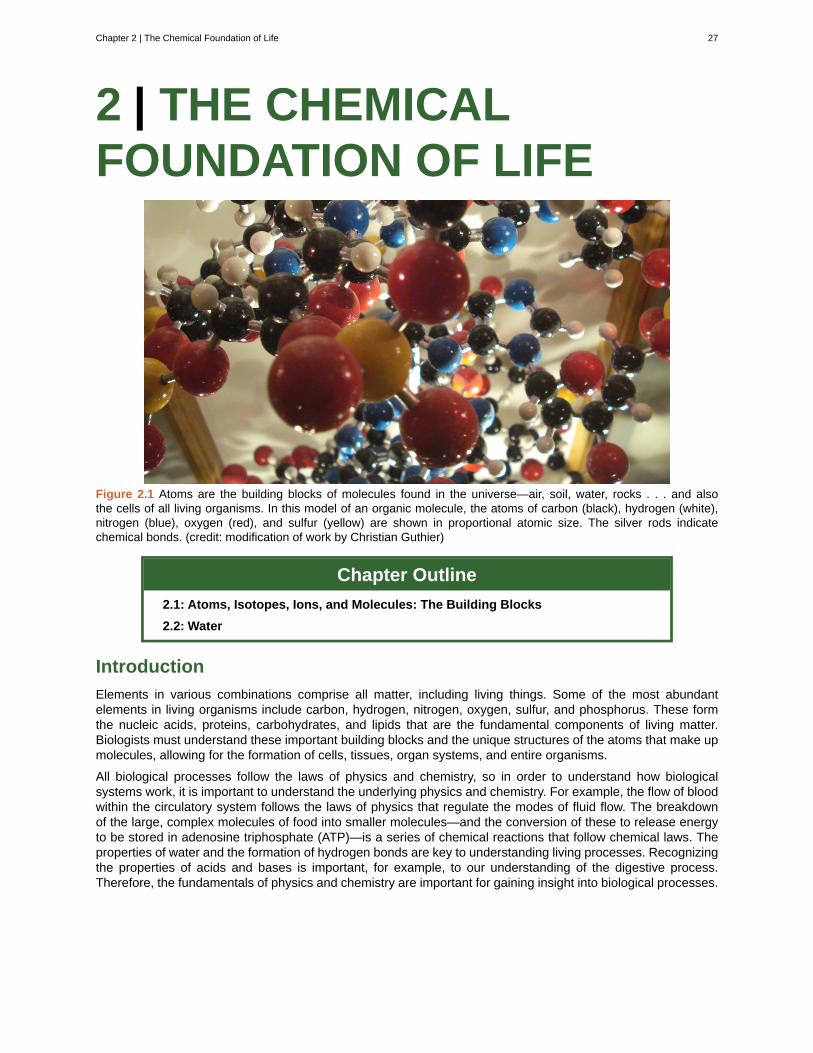
Figure 2.1 Atoms are the building blocks of molecules found in the universe—air, soil, water, rocks . . . and alsothe cells of all living organisms. In this model of an organic molecule, the atoms of carbon (black), hydrogen (white),nitrogen (blue), oxygen (red), and sulfur (yellow) are shown in proportional atomic size. The silver rods indicatechemical bonds. (credit: modification of work by Christian Guthier)
Chapter Outline
2.1: Atoms, Isotopes, Ions, and Molecules: The Building Blocks
2.2: Water
Introduction
Elements in various combinations comprise all matter, including living things. Some of the most abundantelements in living organisms include carbon, hydrogen, nitrogen, oxygen, sulfur, and phosphorus. These formthe nucleic acids, proteins, carbohydrates, and lipids that are the fundamental components of living matter.Biologists must understand these important building blocks and the unique structures of the atoms that make upmolecules, allowing for the formation of cells, tissues, organ systems, and entire organisms.
All biological processes follow the laws of physics and chemistry, so in order to understand how biologicalsystems work, it is important to understand the underlying physics and chemistry. For example, the flow of bloodwithin the circulatory system follows the laws of physics that regulate the modes of fluid flow. The breakdownof the large, complex molecules of food into smaller molecules—and the conversion of these to release energyto be stored in adenosine triphosphate (ATP)—is a series of chemical reactions that follow chemical laws. Theproperties of water and the formation of hydrogen bonds are key to understanding living processes. Recognizingthe properties of acids and bases is important, for example, to our understanding of the digestive process.Therefore, the fundamentals of physics and chemistry are important for gaining insight into biological processes.
Chapter 2 | The Chemical Foundation of Life 27
2.1 | Atoms, Isotopes, Ions, and Molecules: The
Building Blocks
By the end of this section, you will be able to:
• Define matter and elements
• Describe the interrelationship between protons, neutrons, and electrons
• Compare the ways in which electrons can be donated or shared between atoms
• Explain the ways in which naturally occurring elements combine to create molecules, cells, tissues,organ systems, and organisms
At its most fundamental level, life is made up of matter. Matter is any substance that occupies space andhas mass. Elements are unique forms of matter with specific chemical and physical properties that cannot bebroken down into smaller substances by ordinary chemical reactions. There are 118 elements, but only 92 occurnaturally. The remaining elements are synthesized in laboratories and are unstable.
Each element is designated by its chemical symbol, which is a single capital letter or, when the first letter isalready “taken” by another element, a combination of two letters. Some elements follow the English term for theelement, such as C for carbon and Ca for calcium. Other elements’ chemical symbols derive from their Latinnames; for example, the symbol for sodium is Na, referring to natrium , the Latin word for sodium.
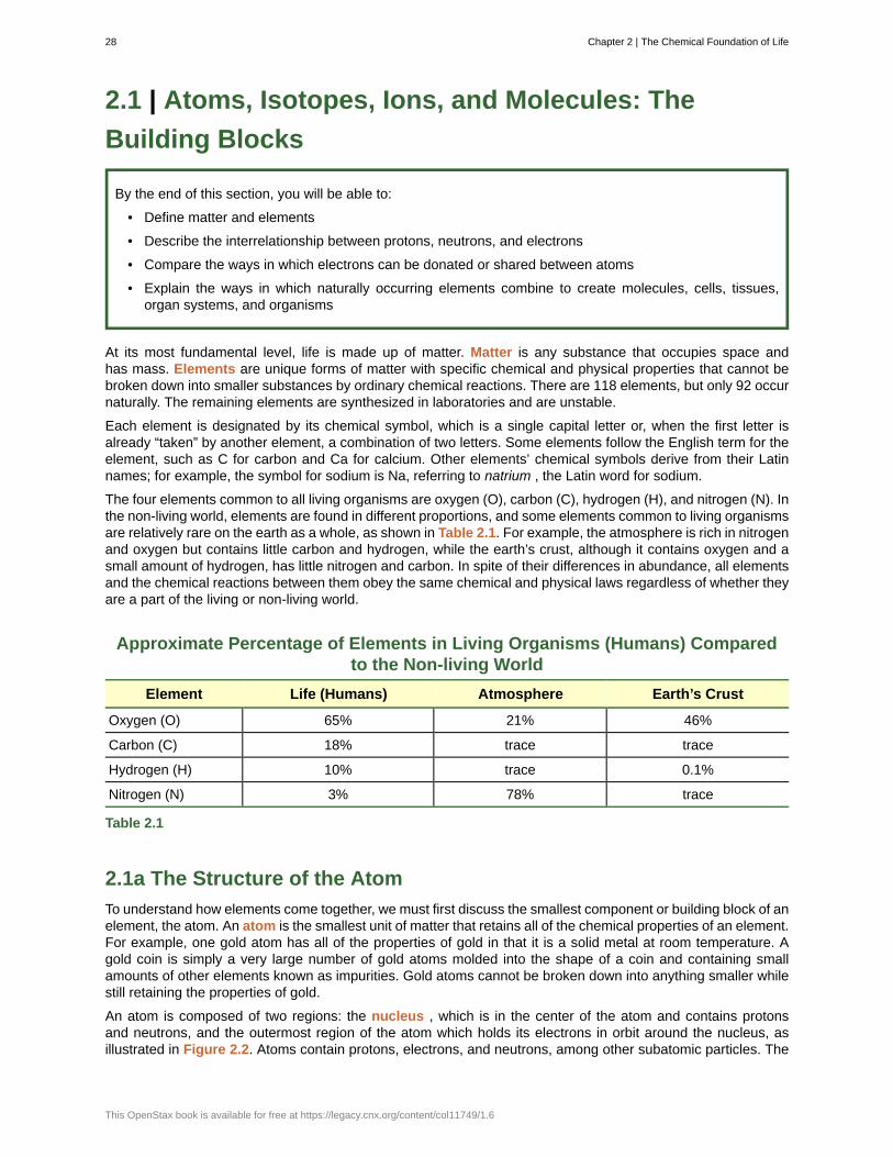
The four elements common to all living organisms are oxygen (O), carbon (C), hydrogen (H), and nitrogen (N). Inthe non-living world, elements are found in different proportions, and some elements common to living organismsare relatively rare on the earth as a whole, as shown in Table 2.1. For example, the atmosphere is rich in nitrogenand oxygen but contains little carbon and hydrogen, while the earth’s crust, although it contains oxygen and asmall amount of hydrogen, has little nitrogen and carbon. In spite of their differences in abundance, all elementsand the chemical reactions between them obey the same chemical and physical laws regardless of whether theyare a part of the living or non-living world.
Approximate Percentage of Elements in Living Organisms (Humans) Comparedto the Non-living World
Element Life (Humans) Atmosphere Earth’s Crust
Oxygen (O) 65% 21% 46%
Carbon (C) 18% trace trace
Hydrogen (H) 10% trace 0.1%
Nitrogen (N) 3% 78% trace
Table 2.1
2.1a The Structure of the Atom
To understand how elements come together, we must first discuss the smallest component or building block of anelement, the atom. An atom is the smallest unit of matter that retains all of the chemical properties of an element.For example, one gold atom has all of the properties of gold in that it is a solid metal at room temperature. Agold coin is simply a very large number of gold atoms molded into the shape of a coin and containing smallamounts of other elements known as impurities. Gold atoms cannot be broken down into anything smaller whilestill retaining the properties of gold.
An atom is composed of two regions: the nucleus , which is in the center of the atom and contains protonsand neutrons, and the outermost region of the atom which holds its electrons in orbit around the nucleus, asillustrated in Figure 2.2. Atoms contain protons, electrons, and neutrons, among other subatomic particles. The
28 Chapter 2 | The Chemical Foundation of Life
This OpenStax book is available for free at https://legacy.cnx.org/content/col11749/1.6
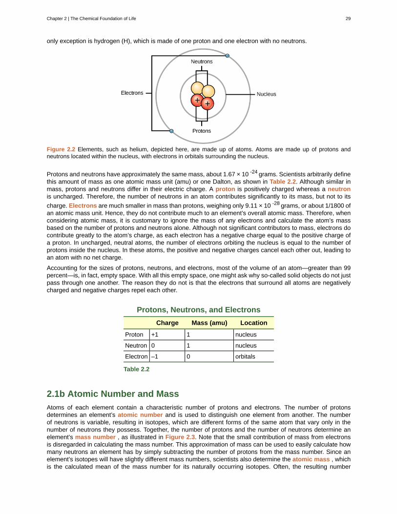
only exception is hydrogen (H), which is made of one proton and one electron with no neutrons.
Figure 2.2 Elements, such as helium, depicted here, are made up of atoms. Atoms are made up of protons andneutrons located within the nucleus, with electrons in orbitals surrounding the nucleus.
Protons and neutrons have approximately the same mass, about 1.67 × 10 -24 grams. Scientists arbitrarily definethis amount of mass as one atomic mass unit (amu) or one Dalton, as shown in Table 2.2. Although similar inmass, protons and neutrons differ in their electric charge. A proton is positively charged whereas a neutronis uncharged. Therefore, the number of neutrons in an atom contributes significantly to its mass, but not to its
charge. Electrons are much smaller in mass than protons, weighing only 9.11 × 10 -28 grams, or about 1/1800 ofan atomic mass unit. Hence, they do not contribute much to an element’s overall atomic mass. Therefore, whenconsidering atomic mass, it is customary to ignore the mass of any electrons and calculate the atom’s massbased on the number of protons and neutrons alone. Although not significant contributors to mass, electrons docontribute greatly to the atom’s charge, as each electron has a negative charge equal to the positive charge ofa proton. In uncharged, neutral atoms, the number of electrons orbiting the nucleus is equal to the number ofprotons inside the nucleus. In these atoms, the positive and negative charges cancel each other out, leading toan atom with no net charge.
Accounting for the sizes of protons, neutrons, and electrons, most of the volume of an atom—greater than 99percent—is, in fact, empty space. With all this empty space, one might ask why so-called solid objects do not justpass through one another. The reason they do not is that the electrons that surround all atoms are negativelycharged and negative charges repel each other.
Protons, Neutrons, and Electrons
Charge Mass (amu) Location
Proton +1 1 nucleus
Neutron 0 1 nucleus
Electron –1 0 orbitals
Table 2.2
2.1b Atomic Number and Mass
Atoms of each element contain a characteristic number of protons and electrons. The number of protonsdetermines an element’s atomic number and is used to distinguish one element from another. The numberof neutrons is variable, resulting in isotopes, which are different forms of the same atom that vary only in thenumber of neutrons they possess. Together, the number of protons and the number of neutrons determine anelement’s mass number , as illustrated in Figure 2.3. Note that the small contribution of mass from electronsis disregarded in calculating the mass number. This approximation of mass can be used to easily calculate howmany neutrons an element has by simply subtracting the number of protons from the mass number. Since anelement’s isotopes will have slightly different mass numbers, scientists also determine the atomic mass , whichis the calculated mean of the mass number for its naturally occurring isotopes. Often, the resulting number
Chapter 2 | The Chemical Foundation of Life 29
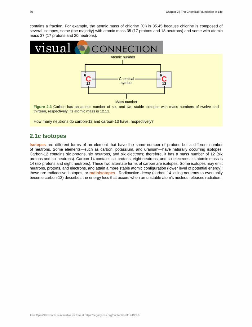
contains a fraction. For example, the atomic mass of chlorine (Cl) is 35.45 because chlorine is composed ofseveral isotopes, some (the majority) with atomic mass 35 (17 protons and 18 neutrons) and some with atomicmass 37 (17 protons and 20 neutrons).
Figure 2.3 Carbon has an atomic number of six, and two stable isotopes with mass numbers of twelve andthirteen, respectively. Its atomic mass is 12.11.
How many neutrons do carbon-12 and carbon-13 have, respectively?
2.1c Isotopes
Isotopes are different forms of an element that have the same number of protons but a different numberof neutrons. Some elements—such as carbon, potassium, and uranium—have naturally occurring isotopes.Carbon-12 contains six protons, six neutrons, and six electrons; therefore, it has a mass number of 12 (sixprotons and six neutrons). Carbon-14 contains six protons, eight neutrons, and six electrons; its atomic mass is14 (six protons and eight neutrons). These two alternate forms of carbon are isotopes. Some isotopes may emitneutrons, protons, and electrons, and attain a more stable atomic configuration (lower level of potential energy);these are radioactive isotopes, or radioisotopes . Radioactive decay (carbon-14 losing neutrons to eventuallybecome carbon-12) describes the energy loss that occurs when an unstable atom’s nucleus releases radiation.
30 Chapter 2 | The Chemical Foundation of Life
This OpenStax book is available for free at https://legacy.cnx.org/content/col11749/1.6
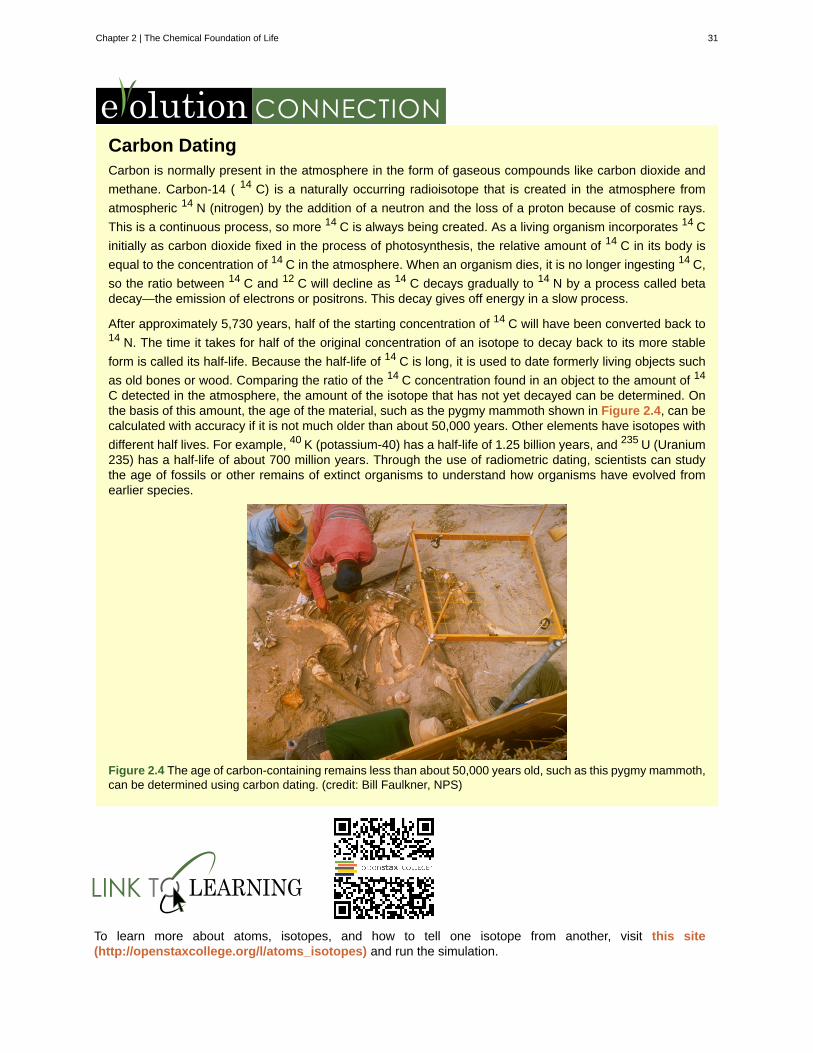
Carbon DatingCarbon is normally present in the atmosphere in the form of gaseous compounds like carbon dioxide and
methane. Carbon-14 ( 14 C) is a naturally occurring radioisotope that is created in the atmosphere from
atmospheric 14 N (nitrogen) by the addition of a neutron and the loss of a proton because of cosmic rays.
This is a continuous process, so more 14 C is always being created. As a living organism incorporates 14 C
initially as carbon dioxide fixed in the process of photosynthesis, the relative amount of 14 C in its body is
equal to the concentration of 14 C in the atmosphere. When an organism dies, it is no longer ingesting 14 C,
so the ratio between 14 C and 12 C will decline as 14 C decays gradually to 14 N by a process called betadecay—the emission of electrons or positrons. This decay gives off energy in a slow process.
After approximately 5,730 years, half of the starting concentration of 14 C will have been converted back to14 N. The time it takes for half of the original concentration of an isotope to decay back to its more stable
form is called its half-life. Because the half-life of 14 C is long, it is used to date formerly living objects such
as old bones or wood. Comparing the ratio of the 14 C concentration found in an object to the amount of 14
C detected in the atmosphere, the amount of the isotope that has not yet decayed can be determined. Onthe basis of this amount, the age of the material, such as the pygmy mammoth shown in Figure 2.4, can becalculated with accuracy if it is not much older than about 50,000 years. Other elements have isotopes with
different half lives. For example, 40 K (potassium-40) has a half-life of 1.25 billion years, and 235 U (Uranium235) has a half-life of about 700 million years. Through the use of radiometric dating, scientists can studythe age of fossils or other remains of extinct organisms to understand how organisms have evolved fromearlier species.
Figure 2.4 The age of carbon-containing remains less than about 50,000 years old, such as this pygmy mammoth,can be determined using carbon dating. (credit: Bill Faulkner, NPS)
To learn more about atoms, isotopes, and how to tell one isotope from another, visit this site(http://openstaxcollege.org/l/atoms_isotopes) and run the simulation.
Chapter 2 | The Chemical Foundation of Life 31
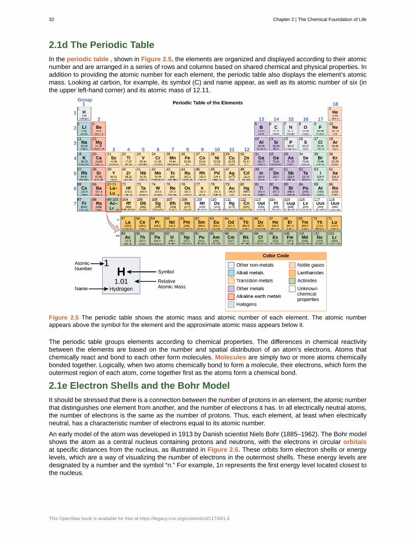
2.1d The Periodic Table
In the periodic table , shown in Figure 2.5, the elements are organized and displayed according to their atomicnumber and are arranged in a series of rows and columns based on shared chemical and physical properties. Inaddition to providing the atomic number for each element, the periodic table also displays the element’s atomicmass. Looking at carbon, for example, its symbol (C) and name appear, as well as its atomic number of six (inthe upper left-hand corner) and its atomic mass of 12.11.
Figure 2.5 The periodic table shows the atomic mass and atomic number of each element. The atomic numberappears above the symbol for the element and the approximate atomic mass appears below it.
The periodic table groups elements according to chemical properties. The differences in chemical reactivitybetween the elements are based on the number and spatial distribution of an atom’s electrons. Atoms thatchemically react and bond to each other form molecules. Molecules are simply two or more atoms chemicallybonded together. Logically, when two atoms chemically bond to form a molecule, their electrons, which form theoutermost region of each atom, come together first as the atoms form a chemical bond.
2.1e Electron Shells and the Bohr Model
It should be stressed that there is a connection between the number of protons in an element, the atomic numberthat distinguishes one element from another, and the number of electrons it has. In all electrically neutral atoms,the number of electrons is the same as the number of protons. Thus, each element, at least when electricallyneutral, has a characteristic number of electrons equal to its atomic number.
An early model of the atom was developed in 1913 by Danish scientist Niels Bohr (1885–1962). The Bohr modelshows the atom as a central nucleus containing protons and neutrons, with the electrons in circular orbitalsat specific distances from the nucleus, as illustrated in Figure 2.6. These orbits form electron shells or energylevels, which are a way of visualizing the number of electrons in the outermost shells. These energy levels aredesignated by a number and the symbol “n.” For example, 1n represents the first energy level located closest tothe nucleus.
32 Chapter 2 | The Chemical Foundation of Life
This OpenStax book is available for free at https://legacy.cnx.org/content/col11749/1.6
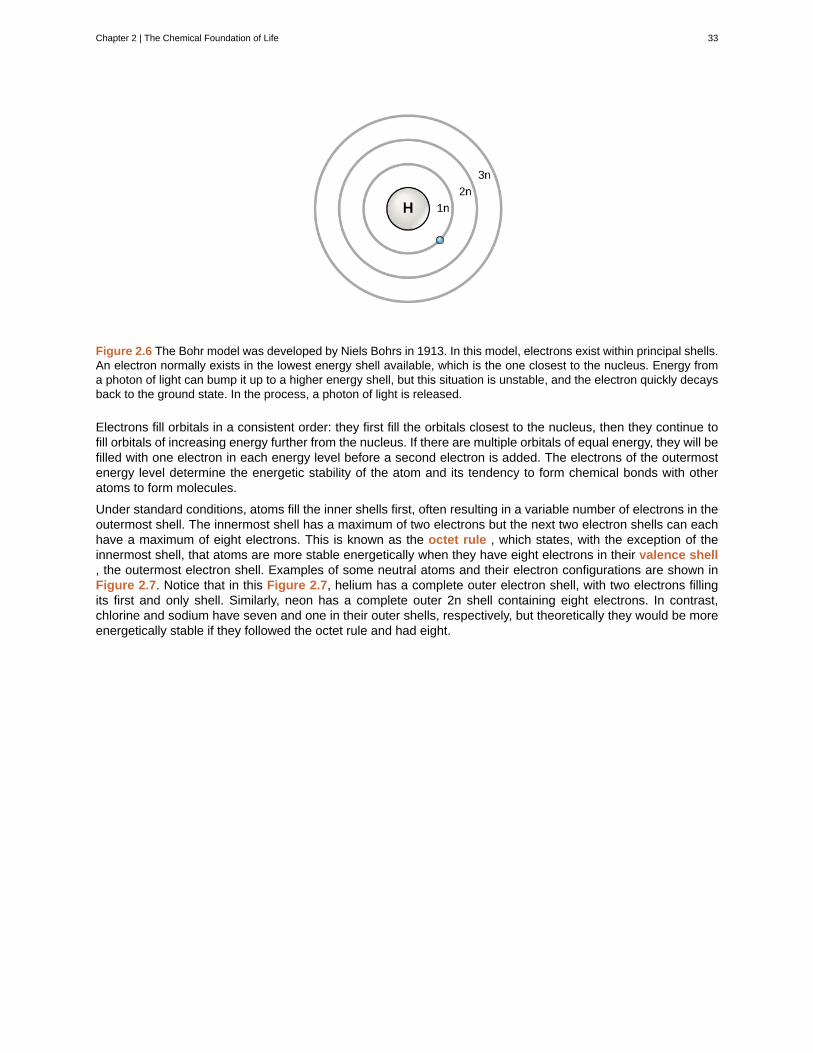
Figure 2.6 The Bohr model was developed by Niels Bohrs in 1913. In this model, electrons exist within principal shells.An electron normally exists in the lowest energy shell available, which is the one closest to the nucleus. Energy froma photon of light can bump it up to a higher energy shell, but this situation is unstable, and the electron quickly decaysback to the ground state. In the process, a photon of light is released.
Electrons fill orbitals in a consistent order: they first fill the orbitals closest to the nucleus, then they continue tofill orbitals of increasing energy further from the nucleus. If there are multiple orbitals of equal energy, they will befilled with one electron in each energy level before a second electron is added. The electrons of the outermostenergy level determine the energetic stability of the atom and its tendency to form chemical bonds with otheratoms to form molecules.
Under standard conditions, atoms fill the inner shells first, often resulting in a variable number of electrons in theoutermost shell. The innermost shell has a maximum of two electrons but the next two electron shells can eachhave a maximum of eight electrons. This is known as the octet rule , which states, with the exception of theinnermost shell, that atoms are more stable energetically when they have eight electrons in their valence shell, the outermost electron shell. Examples of some neutral atoms and their electron configurations are shown inFigure 2.7. Notice that in this Figure 2.7, helium has a complete outer electron shell, with two electrons fillingits first and only shell. Similarly, neon has a complete outer 2n shell containing eight electrons. In contrast,chlorine and sodium have seven and one in their outer shells, respectively, but theoretically they would be moreenergetically stable if they followed the octet rule and had eight.
Chapter 2 | The Chemical Foundation of Life 33
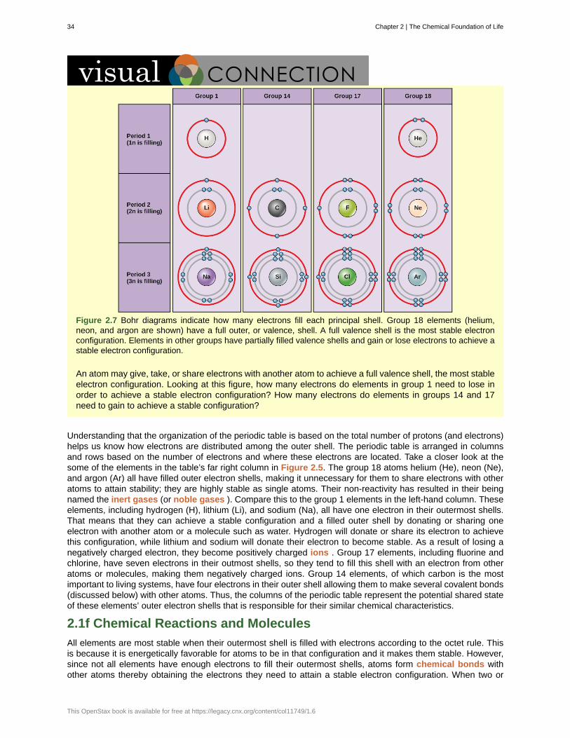
Figure 2.7 Bohr diagrams indicate how many electrons fill each principal shell. Group 18 elements (helium,neon, and argon are shown) have a full outer, or valence, shell. A full valence shell is the most stable electronconfiguration. Elements in other groups have partially filled valence shells and gain or lose electrons to achieve astable electron configuration.
An atom may give, take, or share electrons with another atom to achieve a full valence shell, the most stableelectron configuration. Looking at this figure, how many electrons do elements in group 1 need to lose inorder to achieve a stable electron configuration? How many electrons do elements in groups 14 and 17need to gain to achieve a stable configuration?
Understanding that the organization of the periodic table is based on the total number of protons (and electrons)helps us know how electrons are distributed among the outer shell. The periodic table is arranged in columnsand rows based on the number of electrons and where these electrons are located. Take a closer look at thesome of the elements in the table’s far right column in Figure 2.5. The group 18 atoms helium (He), neon (Ne),and argon (Ar) all have filled outer electron shells, making it unnecessary for them to share electrons with otheratoms to attain stability; they are highly stable as single atoms. Their non-reactivity has resulted in their beingnamed the inert gases (or noble gases ). Compare this to the group 1 elements in the left-hand column. Theseelements, including hydrogen (H), lithium (Li), and sodium (Na), all have one electron in their outermost shells.That means that they can achieve a stable configuration and a filled outer shell by donating or sharing oneelectron with another atom or a molecule such as water. Hydrogen will donate or share its electron to achievethis configuration, while lithium and sodium will donate their electron to become stable. As a result of losing anegatively charged electron, they become positively charged ions . Group 17 elements, including fluorine andchlorine, have seven electrons in their outmost shells, so they tend to fill this shell with an electron from otheratoms or molecules, making them negatively charged ions. Group 14 elements, of which carbon is the mostimportant to living systems, have four electrons in their outer shell allowing them to make several covalent bonds(discussed below) with other atoms. Thus, the columns of the periodic table represent the potential shared stateof these elements’ outer electron shells that is responsible for their similar chemical characteristics.
2.1f Chemical Reactions and Molecules
All elements are most stable when their outermost shell is filled with electrons according to the octet rule. Thisis because it is energetically favorable for atoms to be in that configuration and it makes them stable. However,since not all elements have enough electrons to fill their outermost shells, atoms form chemical bonds withother atoms thereby obtaining the electrons they need to attain a stable electron configuration. When two or
34 Chapter 2 | The Chemical Foundation of Life
This OpenStax book is available for free at https://legacy.cnx.org/content/col11749/1.6
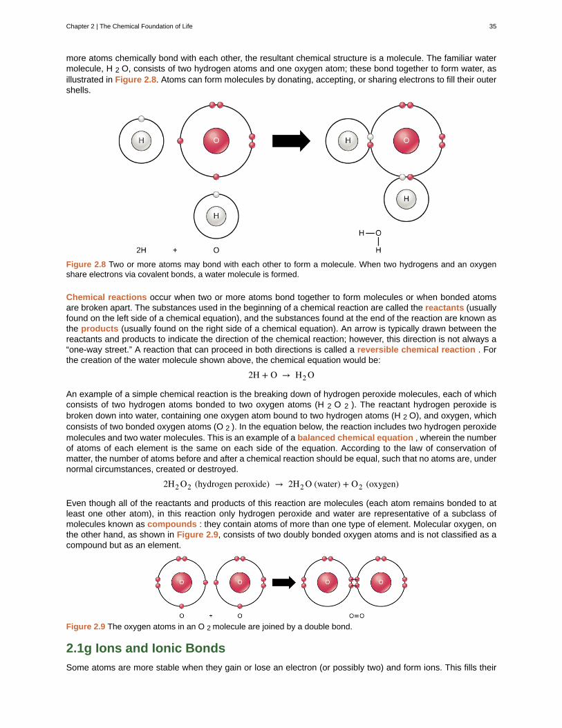
more atoms chemically bond with each other, the resultant chemical structure is a molecule. The familiar watermolecule, H 2 O, consists of two hydrogen atoms and one oxygen atom; these bond together to form water, asillustrated in Figure 2.8. Atoms can form molecules by donating, accepting, or sharing electrons to fill their outershells.
Figure 2.8 Two or more atoms may bond with each other to form a molecule. When two hydrogens and an oxygenshare electrons via covalent bonds, a water molecule is formed.
Chemical reactions occur when two or more atoms bond together to form molecules or when bonded atomsare broken apart. The substances used in the beginning of a chemical reaction are called the reactants (usuallyfound on the left side of a chemical equation), and the substances found at the end of the reaction are known asthe products (usually found on the right side of a chemical equation). An arrow is typically drawn between thereactants and products to indicate the direction of the chemical reaction; however, this direction is not always a“one-way street.” A reaction that can proceed in both directions is called a reversible chemical reaction . Forthe creation of the water molecule shown above, the chemical equation would be:
2H + O → H2 O
An example of a simple chemical reaction is the breaking down of hydrogen peroxide molecules, each of whichconsists of two hydrogen atoms bonded to two oxygen atoms (H 2 O 2 ). The reactant hydrogen peroxide isbroken down into water, containing one oxygen atom bound to two hydrogen atoms (H 2 O), and oxygen, whichconsists of two bonded oxygen atoms (O 2 ). In the equation below, the reaction includes two hydrogen peroxidemolecules and two water molecules. This is an example of a balanced chemical equation , wherein the numberof atoms of each element is the same on each side of the equation. According to the law of conservation ofmatter, the number of atoms before and after a chemical reaction should be equal, such that no atoms are, undernormal circumstances, created or destroyed.
2H2 O2 (hydrogen peroxide) → 2H2 O (water) + O2 (oxygen)
Even though all of the reactants and products of this reaction are molecules (each atom remains bonded to atleast one other atom), in this reaction only hydrogen peroxide and water are representative of a subclass ofmolecules known as compounds : they contain atoms of more than one type of element. Molecular oxygen, onthe other hand, as shown in Figure 2.9, consists of two doubly bonded oxygen atoms and is not classified as acompound but as an element.
Figure 2.9 The oxygen atoms in an O 2 molecule are joined by a double bond.
2.1g Ions and Ionic Bonds
Some atoms are more stable when they gain or lose an electron (or possibly two) and form ions. This fills their
Chapter 2 | The Chemical Foundation of Life 35
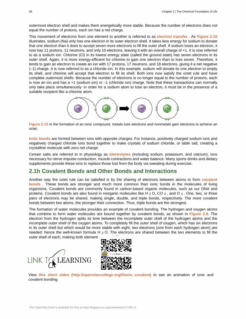
outermost electron shell and makes them energetically more stable. Because the number of electrons does notequal the number of protons, each ion has a net charge.
This movement of electrons from one element to another is referred to as electron transfer . As Figure 2.10illustrates, sodium (Na) only has one electron in its outer electron shell. It takes less energy for sodium to donatethat one electron than it does to accept seven more electrons to fill the outer shell. If sodium loses an electron, itnow has 11 protons, 11 neutrons, and only 10 electrons, leaving it with an overall charge of +1. It is now referredto as a sodium ion. Chlorine (Cl) in its lowest energy state (called the ground state) has seven electrons in itsouter shell. Again, it is more energy-efficient for chlorine to gain one electron than to lose seven. Therefore, ittends to gain an electron to create an ion with 17 protons, 17 neutrons, and 18 electrons, giving it a net negative(–1) charge. It is now referred to as a chloride ion. In this example, sodium will donate its one electron to emptyits shell, and chlorine will accept that electron to fill its shell. Both ions now satisfy the octet rule and havecomplete outermost shells. Because the number of electrons is no longer equal to the number of protons, eachis now an ion and has a +1 (sodium ion) or –1 (chloride ion) charge. Note that these transactions can normallyonly take place simultaneously: in order for a sodium atom to lose an electron, it must be in the presence of asuitable recipient like a chlorine atom.
Figure 2.10 In the formation of an ionic compound, metals lose electrons and nonmetals gain electrons to achieve anoctet.
Ionic bonds are formed between ions with opposite charges. For instance, positively charged sodium ions andnegatively charged chloride ions bond together to make crystals of sodium chloride, or table salt, creating acrystalline molecule with zero net charge.
Certain salts are referred to in physiology as electrolytes (including sodium, potassium, and calcium), ionsnecessary for nerve impulse conduction, muscle contractions and water balance. Many sports drinks and dietarysupplements provide these ions to replace those lost from the body via sweating during exercise.
2.1h Covalent Bonds and Other Bonds and Interactions
Another way the octet rule can be satisfied is by the sharing of electrons between atoms to form covalentbonds . These bonds are stronger and much more common than ionic bonds in the molecules of livingorganisms. Covalent bonds are commonly found in carbon-based organic molecules, such as our DNA andproteins. Covalent bonds are also found in inorganic molecules like H 2 O, CO 2 , and O 2 . One, two, or threepairs of electrons may be shared, making single, double, and triple bonds, respectively. The more covalentbonds between two atoms, the stronger their connection. Thus, triple bonds are the strongest.
The formation of water molecules provides an example of covalent bonding. The hydrogen and oxygen atomsthat combine to form water molecules are bound together by covalent bonds, as shown in Figure 2.8. Theelectron from the hydrogen splits its time between the incomplete outer shell of the hydrogen atoms and theincomplete outer shell of the oxygen atoms. To completely fill the outer shell of oxygen, which has six electronsin its outer shell but which would be more stable with eight, two electrons (one from each hydrogen atom) areneeded: hence the well-known formula H 2 O. The electrons are shared between the two elements to fill theouter shell of each, making both elements more stable.
View this short video (http://openstaxcollege.org/l/ionic_covalent) to see an animation of ionic andcovalent bonding.
36 Chapter 2 | The Chemical Foundation of Life
This OpenStax book is available for free at https://legacy.cnx.org/content/col11749/1.6
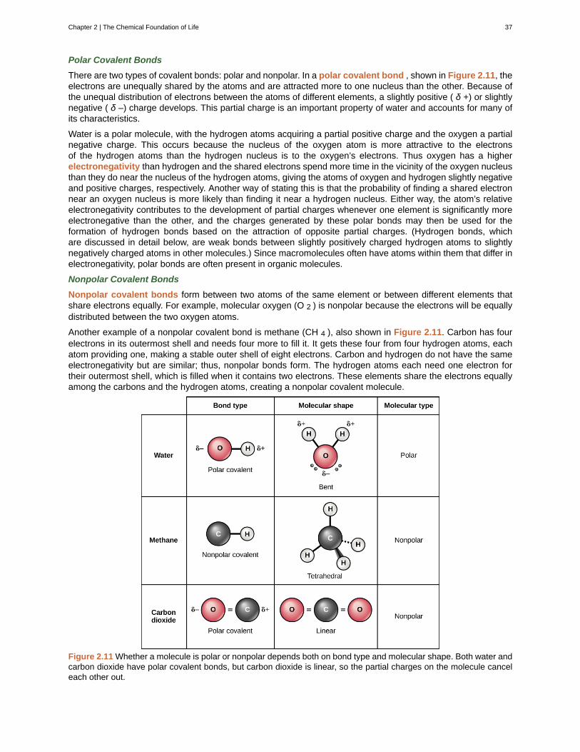
Polar Covalent Bonds
There are two types of covalent bonds: polar and nonpolar. In a polar covalent bond , shown in Figure 2.11, theelectrons are unequally shared by the atoms and are attracted more to one nucleus than the other. Because ofthe unequal distribution of electrons between the atoms of different elements, a slightly positive ( δ +) or slightlynegative ( δ –) charge develops. This partial charge is an important property of water and accounts for many ofits characteristics.
Water is a polar molecule, with the hydrogen atoms acquiring a partial positive charge and the oxygen a partialnegative charge. This occurs because the nucleus of the oxygen atom is more attractive to the electronsof the hydrogen atoms than the hydrogen nucleus is to the oxygen’s electrons. Thus oxygen has a higherelectronegativity than hydrogen and the shared electrons spend more time in the vicinity of the oxygen nucleusthan they do near the nucleus of the hydrogen atoms, giving the atoms of oxygen and hydrogen slightly negativeand positive charges, respectively. Another way of stating this is that the probability of finding a shared electronnear an oxygen nucleus is more likely than finding it near a hydrogen nucleus. Either way, the atom’s relativeelectronegativity contributes to the development of partial charges whenever one element is significantly moreelectronegative than the other, and the charges generated by these polar bonds may then be used for theformation of hydrogen bonds based on the attraction of opposite partial charges. (Hydrogen bonds, whichare discussed in detail below, are weak bonds between slightly positively charged hydrogen atoms to slightlynegatively charged atoms in other molecules.) Since macromolecules often have atoms within them that differ inelectronegativity, polar bonds are often present in organic molecules.
Nonpolar Covalent Bonds
Nonpolar covalent bonds form between two atoms of the same element or between different elements thatshare electrons equally. For example, molecular oxygen (O 2 ) is nonpolar because the electrons will be equallydistributed between the two oxygen atoms.
Another example of a nonpolar covalent bond is methane (CH 4 ), also shown in Figure 2.11. Carbon has fourelectrons in its outermost shell and needs four more to fill it. It gets these four from four hydrogen atoms, eachatom providing one, making a stable outer shell of eight electrons. Carbon and hydrogen do not have the sameelectronegativity but are similar; thus, nonpolar bonds form. The hydrogen atoms each need one electron fortheir outermost shell, which is filled when it contains two electrons. These elements share the electrons equallyamong the carbons and the hydrogen atoms, creating a nonpolar covalent molecule.
Figure 2.11 Whether a molecule is polar or nonpolar depends both on bond type and molecular shape. Both water andcarbon dioxide have polar covalent bonds, but carbon dioxide is linear, so the partial charges on the molecule canceleach other out.
Chapter 2 | The Chemical Foundation of Life 37
Hydrogen Bonds
Ionic and covalent bonds between elements require energy to break. Ioonic bonds are not as strong as covalent,which determines their behavior in biological systems. However, not all bonds are ionic or covalent bonds.Weaker bonds, like hydrogen bonds, can also form between molecules. Hydrogen bonds provide many of thecritical, life-sustaining properties of water and also stabilize the structures of proteins and DNA, the building blockof cells.
When polar covalent bonds containing hydrogen form, the hydrogen in that bond has a slightly positive chargebecause hydrogen’s electron is pulled more strongly toward the other element and away from the hydrogen.Because the hydrogen is slightly positive, it will be attracted to neighboring negative charges. When this
happens, a weak interaction occurs between the δ + of the hydrogen from one molecule and the δ – charge onthe more electronegative atoms of another molecule, usually oxygen or nitrogen, or within the same molecule.This interaction is called a hydrogen bond . This type of bond is common and occurs regularly between watermolecules. Individual hydrogen bonds are weak and easily broken; however, they occur in very large numbersin water and in organic polymers, creating a major force in combination. Hydrogen bonds are also responsiblefor zipping together the DNA double helix.
Pharmaceutical ChemistPharmaceutical chemists are responsible for the development of new drugs and trying to determine themode of action of both old and new drugs. They are involved in every step of the drug development process.Drugs can be found in the natural environment or can be synthesized in the laboratory. In many cases,potential drugs found in nature are changed chemically in the laboratory to make them safer and moreeffective, and sometimes synthetic versions of drugs substitute for the version found in nature.
After the initial discovery or synthesis of a drug, the chemist then develops the drug, perhaps chemicallyaltering it, testing it to see if the drug is toxic, and then designing methods for efficient large-scale production.Then, the process of getting the drug approved for human use begins. In the United States, drug approval ishandled by the Food and Drug Administration (FDA) and involves a series of large-scale experiments usinghuman subjects to make sure the drug is not harmful and effectively treats the condition it aims to treat. Thisprocess often takes several years and requires the participation of physicians and scientists, in addition tochemists, to complete testing and gain approval.
An example of a drug that was originally discovered in a living organism is Paclitaxel (Taxol), an anti-cancerdrug used to treat breast cancer. This drug was discovered in the bark of the pacific yew tree. Anotherexample is aspirin, originally isolated from willow tree bark. Finding drugs often means testing hundreds ofsamples of plants, fungi, and other forms of life to see if any biologically active compounds are found withinthem. Sometimes, traditional medicine can give modern medicine clues to where an active compound canbe found. For example, the use of willow bark to make medicine has been known for thousands of years,dating back to ancient Egypt. It was not until the late 1800s, however, that the aspirin molecule, known asacetylsalicylic acid, was purified and marketed for human use.
Occasionally, drugs developed for one use are found to have unforeseen effects that allow these drugs tobe used in other, unrelated ways. For example, the drug minoxidil (Rogaine) was originally developed totreat high blood pressure. When tested on humans, it was noticed that individuals taking the drug wouldgrow new hair. Eventually the drug was marketed to men and women with baldness to restore lost hair.
The career of the pharmaceutical chemist may involve detective work, experimentation, and drugdevelopment, all with the goal of making human beings healthier.
38 Chapter 2 | The Chemical Foundation of Life
This OpenStax book is available for free at https://legacy.cnx.org/content/col11749/1.6
2.2 | Water
By the end of this section, you will be able to:
• Describe the properties of water that are critical to maintaining life
• Explain why water is an excellent solvent
• Provide examples of water’s cohesive and adhesive properties
• Discuss the role of acids, bases, and buffers in homeostasis
Why do scientists spend time looking for water on other planets? Why is water so important? It is because wateris essential to life as we know it. Water is one of the more abundant molecules and the one most critical to life onEarth. Approximately 60–70 percent of the human body is made up of water. Without it, life as we know it simplywould not exist.
The polarity of the water molecule and its resulting hydrogen bonding make water a unique substance withspecial properties that are intimately tied to the processes of life. Life originally evolved in a watery environment,and most of an organism’s cellular chemistry and metabolism occur inside the watery contents of the cell’scytoplasm. Special properties of water are its high heat capacity and heat of vaporization, its ability to dissolvepolar molecules, its cohesive and adhesive properties, and its dissociation into ions that leads to the generationof pH. Understanding these characteristics of water helps to elucidate its importance in maintaining life.
2.2a Water’s Polarity
One of water’s important properties is that it is composed of polar molecules: the hydrogen and oxygen withinwater molecules (H 2 O) form polar covalent bonds. While there is no net charge to a water molecule, the polarityof water creates a slightly positive charge on hydrogen and a slightly negative charge on oxygen, contributingto water’s properties of attraction. Water’s charges are generated because oxygen is more electronegativethan hydrogen, making it more likely that a shared electron would be found near the oxygen nucleus than thehydrogen nucleus, thus generating the partial negative charge near the oxygen.
As a result of water’s polarity, each water molecule attracts other water molecules because of the oppositecharges between water molecules, forming hydrogen bonds. Water also attracts or is attracted to other polarmolecules and ions. A polar substance that interacts readily with or dissolves in water is referred to ashydrophilic (hydro- = “water”; -philic = “loving”). In contrast, non-polar molecules such as oils and fats do notinteract well with water, as shown in Figure 2.12 and separate from it rather than dissolve in it, as we see insalad dressings containing oil and vinegar (an acidic water solution). These nonpolar compounds are calledhydrophobic (hydro- = “water”; -phobic = “fearing”).
Chapter 2 | The Chemical Foundation of Life 39
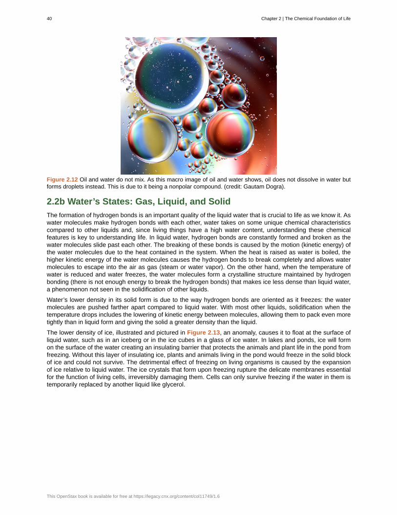
Figure 2.12 Oil and water do not mix. As this macro image of oil and water shows, oil does not dissolve in water butforms droplets instead. This is due to it being a nonpolar compound. (credit: Gautam Dogra).
2.2b Water’s States: Gas, Liquid, and Solid
The formation of hydrogen bonds is an important quality of the liquid water that is crucial to life as we know it. Aswater molecules make hydrogen bonds with each other, water takes on some unique chemical characteristicscompared to other liquids and, since living things have a high water content, understanding these chemicalfeatures is key to understanding life. In liquid water, hydrogen bonds are constantly formed and broken as thewater molecules slide past each other. The breaking of these bonds is caused by the motion (kinetic energy) ofthe water molecules due to the heat contained in the system. When the heat is raised as water is boiled, thehigher kinetic energy of the water molecules causes the hydrogen bonds to break completely and allows watermolecules to escape into the air as gas (steam or water vapor). On the other hand, when the temperature ofwater is reduced and water freezes, the water molecules form a crystalline structure maintained by hydrogenbonding (there is not enough energy to break the hydrogen bonds) that makes ice less dense than liquid water,a phenomenon not seen in the solidification of other liquids.
Water’s lower density in its solid form is due to the way hydrogen bonds are oriented as it freezes: the watermolecules are pushed farther apart compared to liquid water. With most other liquids, solidification when thetemperature drops includes the lowering of kinetic energy between molecules, allowing them to pack even moretightly than in liquid form and giving the solid a greater density than the liquid.
The lower density of ice, illustrated and pictured in Figure 2.13, an anomaly, causes it to float at the surface ofliquid water, such as in an iceberg or in the ice cubes in a glass of ice water. In lakes and ponds, ice will formon the surface of the water creating an insulating barrier that protects the animals and plant life in the pond fromfreezing. Without this layer of insulating ice, plants and animals living in the pond would freeze in the solid blockof ice and could not survive. The detrimental effect of freezing on living organisms is caused by the expansionof ice relative to liquid water. The ice crystals that form upon freezing rupture the delicate membranes essentialfor the function of living cells, irreversibly damaging them. Cells can only survive freezing if the water in them istemporarily replaced by another liquid like glycerol.
40 Chapter 2 | The Chemical Foundation of Life
This OpenStax book is available for free at https://legacy.cnx.org/content/col11749/1.6
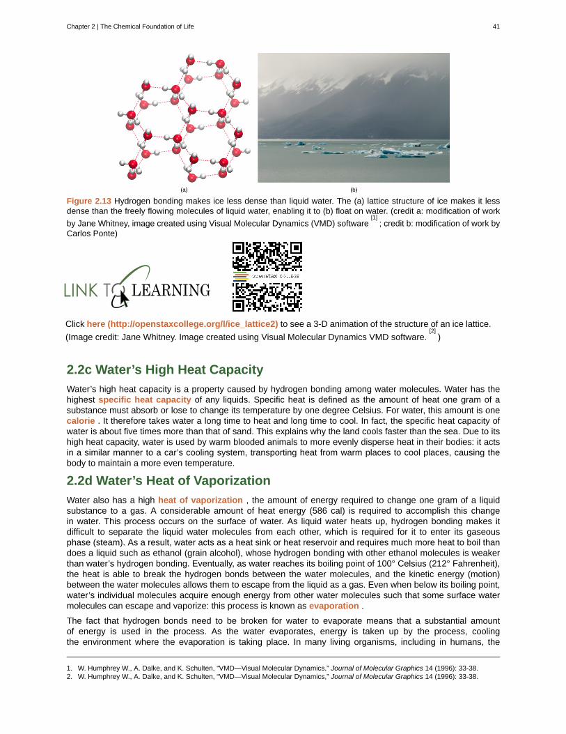
Figure 2.13 Hydrogen bonding makes ice less dense than liquid water. The (a) lattice structure of ice makes it lessdense than the freely flowing molecules of liquid water, enabling it to (b) float on water. (credit a: modification of work
by Jane Whitney, image created using Visual Molecular Dynamics (VMD) software[1]
; credit b: modification of work byCarlos Ponte)
Click here (http://openstaxcollege.org/l/ice_lattice2) to see a 3-D animation of the structure of an ice lattice.
(Image credit: Jane Whitney. Image created using Visual Molecular Dynamics VMD software.[2]
)
2.2c Water’s High Heat Capacity
Water’s high heat capacity is a property caused by hydrogen bonding among water molecules. Water has thehighest specific heat capacity of any liquids. Specific heat is defined as the amount of heat one gram of asubstance must absorb or lose to change its temperature by one degree Celsius. For water, this amount is onecalorie . It therefore takes water a long time to heat and long time to cool. In fact, the specific heat capacity ofwater is about five times more than that of sand. This explains why the land cools faster than the sea. Due to itshigh heat capacity, water is used by warm blooded animals to more evenly disperse heat in their bodies: it actsin a similar manner to a car’s cooling system, transporting heat from warm places to cool places, causing thebody to maintain a more even temperature.
2.2d Water’s Heat of Vaporization
Water also has a high heat of vaporization , the amount of energy required to change one gram of a liquidsubstance to a gas. A considerable amount of heat energy (586 cal) is required to accomplish this changein water. This process occurs on the surface of water. As liquid water heats up, hydrogen bonding makes itdifficult to separate the liquid water molecules from each other, which is required for it to enter its gaseousphase (steam). As a result, water acts as a heat sink or heat reservoir and requires much more heat to boil thandoes a liquid such as ethanol (grain alcohol), whose hydrogen bonding with other ethanol molecules is weakerthan water’s hydrogen bonding. Eventually, as water reaches its boiling point of 100° Celsius (212° Fahrenheit),the heat is able to break the hydrogen bonds between the water molecules, and the kinetic energy (motion)between the water molecules allows them to escape from the liquid as a gas. Even when below its boiling point,water’s individual molecules acquire enough energy from other water molecules such that some surface watermolecules can escape and vaporize: this process is known as evaporation .
The fact that hydrogen bonds need to be broken for water to evaporate means that a substantial amountof energy is used in the process. As the water evaporates, energy is taken up by the process, coolingthe environment where the evaporation is taking place. In many living organisms, including in humans, the
1. W. Humphrey W., A. Dalke, and K. Schulten, “VMD—Visual Molecular Dynamics,” Journal of Molecular Graphics 14 (1996): 33-38.2. W. Humphrey W., A. Dalke, and K. Schulten, “VMD—Visual Molecular Dynamics,” Journal of Molecular Graphics 14 (1996): 33-38.
Chapter 2 | The Chemical Foundation of Life 41
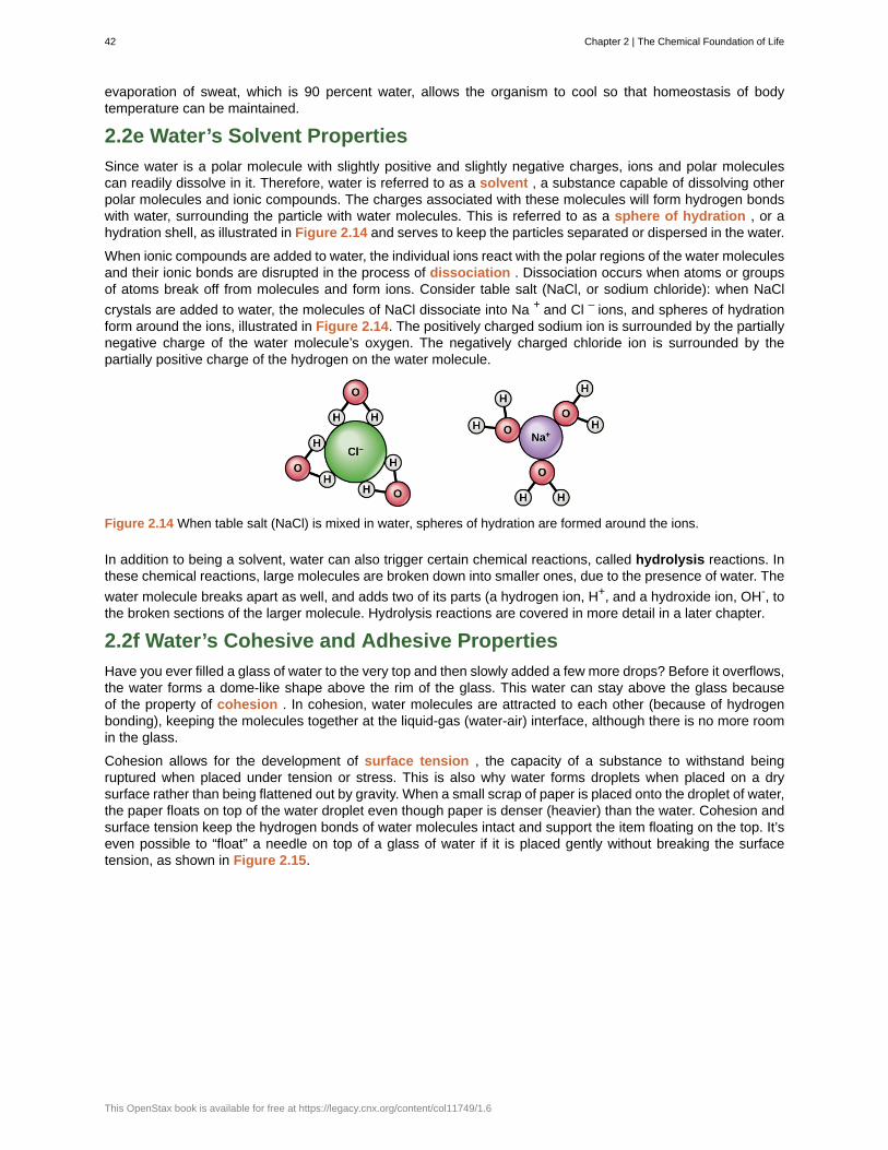
evaporation of sweat, which is 90 percent water, allows the organism to cool so that homeostasis of bodytemperature can be maintained.
2.2e Water’s Solvent Properties
Since water is a polar molecule with slightly positive and slightly negative charges, ions and polar moleculescan readily dissolve in it. Therefore, water is referred to as a solvent , a substance capable of dissolving otherpolar molecules and ionic compounds. The charges associated with these molecules will form hydrogen bondswith water, surrounding the particle with water molecules. This is referred to as a sphere of hydration , or ahydration shell, as illustrated in Figure 2.14 and serves to keep the particles separated or dispersed in the water.
When ionic compounds are added to water, the individual ions react with the polar regions of the water moleculesand their ionic bonds are disrupted in the process of dissociation . Dissociation occurs when atoms or groupsof atoms break off from molecules and form ions. Consider table salt (NaCl, or sodium chloride): when NaCl
crystals are added to water, the molecules of NaCl dissociate into Na + and Cl – ions, and spheres of hydrationform around the ions, illustrated in Figure 2.14. The positively charged sodium ion is surrounded by the partiallynegative charge of the water molecule’s oxygen. The negatively charged chloride ion is surrounded by thepartially positive charge of the hydrogen on the water molecule.
Figure 2.14 When table salt (NaCl) is mixed in water, spheres of hydration are formed around the ions.
In addition to being a solvent, water can also trigger certain chemical reactions, called hydrolysis reactions. Inthese chemical reactions, large molecules are broken down into smaller ones, due to the presence of water. The
water molecule breaks apart as well, and adds two of its parts (a hydrogen ion, H+, and a hydroxide ion, OH-, tothe broken sections of the larger molecule. Hydrolysis reactions are covered in more detail in a later chapter.
2.2f Water’s Cohesive and Adhesive Properties
Have you ever filled a glass of water to the very top and then slowly added a few more drops? Before it overflows,the water forms a dome-like shape above the rim of the glass. This water can stay above the glass becauseof the property of cohesion . In cohesion, water molecules are attracted to each other (because of hydrogenbonding), keeping the molecules together at the liquid-gas (water-air) interface, although there is no more roomin the glass.
Cohesion allows for the development of surface tension , the capacity of a substance to withstand beingruptured when placed under tension or stress. This is also why water forms droplets when placed on a drysurface rather than being flattened out by gravity. When a small scrap of paper is placed onto the droplet of water,the paper floats on top of the water droplet even though paper is denser (heavier) than the water. Cohesion andsurface tension keep the hydrogen bonds of water molecules intact and support the item floating on the top. It’seven possible to “float” a needle on top of a glass of water if it is placed gently without breaking the surfacetension, as shown in Figure 2.15.
42 Chapter 2 | The Chemical Foundation of Life
This OpenStax book is available for free at https://legacy.cnx.org/content/col11749/1.6

Figure 2.15 The weight of the needle is pulling the surface downward; at the same time, the surface tension is pullingit up, suspending it on the surface of the water and keeping it from sinking. Notice the indentation in the water aroundthe needle. (credit: Cory Zanker)
These cohesive forces are related to water’s property of adhesion , or the attraction between water moleculesand other molecules. This attraction is sometimes stronger than water’s cohesive forces, especially when thewater is exposed to charged surfaces such as those found on the inside of thin glass tubes known as capillarytubes. Adhesion is observed when water “climbs” up the tube placed in a glass of water: notice that the waterappears to be higher on the sides of the tube than in the middle. This is because the water molecules areattracted to the charged glass walls of the capillary more than they are to each other and therefore adhere to it.This type of adhesion is called capillary action , and is illustrated in Figure 2.16.
Figure 2.16 Capillary action in a glass tube is caused by the adhesive forces exerted by the internal surface ofthe glass exceeding the cohesive forces between the water molecules themselves. (credit: modification of work byPearson-Scott Foresman, donated to the Wikimedia Foundation)
Why are cohesive and adhesive forces important for life? Cohesive and adhesive forces are important for thetransport of water from the roots to the leaves in plants. These forces create a “pull” on the water column.This pull results from the tendency of water molecules being evaporated on the surface of the plant to stayconnected to water molecules below them, and so they are pulled along. Plants use this natural phenomenon tohelp transport water from their roots to their leaves. Without these properties of water, plants would be unable toreceive the water and the dissolved minerals they require. In another example, insects such as the water strider,shown in Figure 2.17, use the surface tension of water to stay afloat on the surface layer of water and even
Chapter 2 | The Chemical Foundation of Life 43
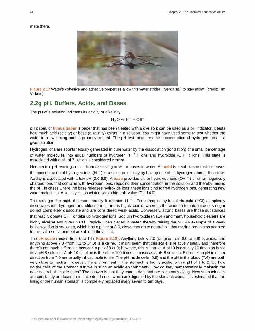
mate there.
Figure 2.17 Water’s cohesive and adhesive properties allow this water strider ( Gerris sp.) to stay afloat. (credit: TimVickers)
2.2g pH, Buffers, Acids, and Bases
The pH of a solution indicates its acidity or alkalinity.
H2 O ↔ H+ + OH-
pH paper, or litmus paper is paper that has been treated with a dye so it can be used as a pH indicator. It testshow much acid (acidity) or base (alkalinity) exists in a solution. You might have used some to test whether thewater in a swimming pool is properly treated. The pH test measures the concentration of hydrogen ions in agiven solution.
Hydrogen ions are spontaneously generated in pure water by the dissociation (ionization) of a small percentage
of water molecules into equal numbers of hydrogen (H + ) ions and hydroxide (OH - ) ions. This state isassociated with a pH of 7, which is considered neutral.
Non-neutral pH readings result from dissolving acids or bases in water. An acid is a substance that increases
the concentration of hydrogen ions (H + ) in a solution, usually by having one of its hydrogen atoms dissociate.
Acidity is associated with a low pH (0.0-6.9). A base provides either hydroxide ions (OH – ) or other negativelycharged ions that combine with hydrogen ions, reducing their concentration in the solution and thereby raisingthe pH. In cases where the base releases hydroxide ions, these ions bind to free hydrogen ions, generating newwater molecules. Alkalinity is associated with a high pH value (7.1-14.0).
The stronger the acid, the more readily it donates H + . For example, hydrochloric acid (HCl) completelydissociates into hydrogen and chloride ions and is highly acidic, whereas the acids in tomato juice or vinegardo not completely dissociate and are considered weak acids. Conversely, strong bases are those substances
that readily donate OH – or take up hydrogen ions. Sodium hydroxide (NaOH) and many household cleaners are
highly alkaline and give up OH – rapidly when placed in water, thereby raising the pH. An example of a weakbasic solution is seawater, which has a pH near 8.0, close enough to neutral pH that marine organisms adaptedto this saline environment are able to thrive in it.
The pH scale ranges from 0 to 14 ( Figure 2.18). Anything below 7.0 (ranging from 0.0 to 6.9) is acidic, andanything above 7.0 (from 7.1 to 14.0) is alkaline. It might seem that this scale is relatively small, and thereforethere's not much difference between a pH of 8 or 9; however, this is untrue. A pH 9 is actually 10 times as basicas a pH 8 solution. A pH 10 solution is therefore 100 times as basic as a pH 8 solution. Extremes in pH in eitherdirection from 7.0 are usually inhospitable to life. The pH inside cells (6.8) and the pH in the blood (7.4) are bothvery close to neutral. However, the environment in the stomach is highly acidic, with a pH of 1 to 2. So howdo the cells of the stomach survive in such an acidic environment? How do they homeostatically maintain thenear neutral pH inside them? The answer is that they cannot do it and are constantly dying. New stomach cellsare constantly produced to replace dead ones, which are digested by the stomach acids. It is estimated that thelining of the human stomach is completely replaced every seven to ten days.
44 Chapter 2 | The Chemical Foundation of Life
This OpenStax book is available for free at https://legacy.cnx.org/content/col11749/1.6
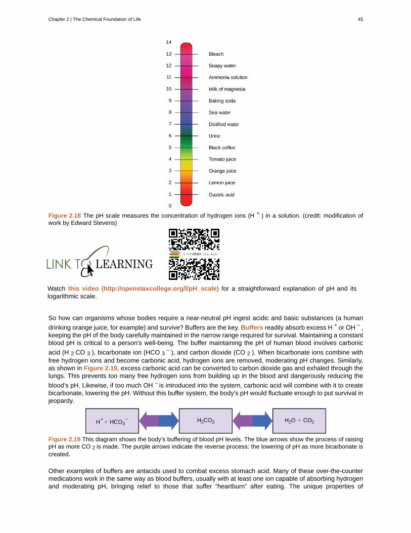
Figure 2.18 The pH scale measures the concentration of hydrogen ions (H + ) in a solution. (credit: modification ofwork by Edward Stevens)
Watch this video (http://openstaxcollege.org/l/pH_scale) for a straightforward explanation of pH and itslogarithmic scale.
So how can organisms whose bodies require a near-neutral pH ingest acidic and basic substances (a human
drinking orange juice, for example) and survive? Buffers are the key. Buffers readily absorb excess H + or OH – ,keeping the pH of the body carefully maintained in the narrow range required for survival. Maintaining a constantblood pH is critical to a person’s well-being. The buffer maintaining the pH of human blood involves carbonic
acid (H 2 CO 3 ), bicarbonate ion (HCO 3– ), and carbon dioxide (CO 2 ). When bicarbonate ions combine with
free hydrogen ions and become carbonic acid, hydrogen ions are removed, moderating pH changes. Similarly,as shown in Figure 2.19, excess carbonic acid can be converted to carbon dioxide gas and exhaled through thelungs. This prevents too many free hydrogen ions from building up in the blood and dangerously reducing the
blood’s pH. Likewise, if too much OH – is introduced into the system, carbonic acid will combine with it to createbicarbonate, lowering the pH. Without this buffer system, the body’s pH would fluctuate enough to put survival injeopardy.
Figure 2.19 This diagram shows the body’s buffering of blood pH levels. The blue arrows show the process of raisingpH as more CO 2 is made. The purple arrows indicate the reverse process: the lowering of pH as more bicarbonate iscreated.
Other examples of buffers are antacids used to combat excess stomach acid. Many of these over-the-countermedications work in the same way as blood buffers, usually with at least one ion capable of absorbing hydrogenand moderating pH, bringing relief to those that suffer “heartburn” after eating. The unique properties of
Chapter 2 | The Chemical Foundation of Life 45
water that contribute to this capacity to balance pH—as well as water’s other characteristics—are essential tosustaining life on Earth.
To learn more about water. Visit the U.S. Geological Survey Water Science for Schools(http://openstaxcollege.org/l/all_about_water) All About Water! website.
46 Chapter 2 | The Chemical Foundation of Life
This OpenStax book is available for free at https://legacy.cnx.org/content/col11749/1.6
acid
adhesion
atom
atomic mass
atomic number
balanced chemical equation
base
Buffers
calorie
capillary action
chemical bonds
Chemical reactions
cohesion
compounds
covalent bonds
dissociation
electrolytes
electron transfer
electronegativity
Electrons
Elements
evaporation
heat of vaporization
hydrogen bond
hydrolysis
KEY TERMS
molecule that donates hydrogen ions and increases the concentration of hydrogen ions in a solution
attraction between water molecules and other molecules
the smallest unit of matter that retains all of the chemical properties of an element
calculated mean of the mass number for an element’s isotopes
total number of protons in an atom
statement of a chemical reaction with the number of each type of atom equalizedfor both the products and reactants
molecule that donates hydroxide ions or otherwise binds excess hydrogen ions and decreases theconcentration of hydrogen ions in a solution
substance that prevents a change in pH by absorbing or releasing hydrogen or hydroxide ions
amount of heat required to change the temperature of one gram of water by one degree Celsius
occurs because water molecules are attracted to charges on the inner surfaces of narrowtubular structures such as glass tubes, drawing the water molecules to the sides of the tubes
interaction between two or more of the same or different atoms that results in the formation ofmolecules
process leading to the rearrangement of atoms
intermolecular forces between water molecules caused by the polar nature of water; responsible forsurface tension
substance composed of molecules consisting of atoms of at least two different elements
type of strong bond formed between two of the same or different elements; forms whenelectrons are shared between atoms
release of an ion from a molecule such that the original molecule now consists of an ion and the
charged remains of the original, such as when water dissociates into H + and OH -
ion necessary for nerve impulse conduction, muscle contractions, and water balance
movement of electrons from one element to another; important in creation of ionic bonds
ability of some elements to attract electrons (often of hydrogen atoms), acquiring partialnegative charges in molecules and creating partial positive charges on the hydrogen atoms
negatively charged subatomic particle that resides outside of the nucleus in the electron orbital;lacks functional mass and has a negative charge of -1 unit
one of 118 unique substances that cannot be broken down into smaller substances; each elementhas unique properties and a specified number of protons
separation of individual molecules from the surface of a body of water, leaves of a plant, or theskin of an organism
high amount of energy required for liquid to turn into gas
weak bond between slightly positively charged hydrogen atoms to slightly negatively chargedatoms in other molecules
reactions that use water to break apart large molecules
Chapter 2 | The Chemical Foundation of Life 47
hydrophilic
hydrophobic
inert gases
Ionic bonds
ions
Isotopes
litmus paper
mass number
matter
Molecules
neutral
neutron
noble gases
Nonpolar covalent bonds
nucleus
octet rule
orbitals
periodic table
pH scale
polar covalent bond
products
proton
radioisotopes
reactants
reversible chemical reaction
solvent
specific heat capacity
describes ions or polar molecules that interact well with other polar molecules such as water
describes uncharged non-polar molecules that do not interact well with polar molecules such aswater
(also called noble gases) element with filled outer electron shell that is unreactive with other atoms
chemical bond that forms between ions with opposite charges (cations and anions)
atom or chemical group that does not contain equal number of protons and electrons
one or more forms of an element that have different numbers of neutrons
(also, pH paper) filter paper that has been treated with a natural water-soluble dye that changesits color as the pH of the environment changes so it can be used as a pH indicator
total number of protons and neutrons in an atom
anything that has mass and occupies space
two or more atoms chemically bonded together
pH of 7, achieved when a solution has an equal concentration of H+ ions and OH- ions
uncharged particle that resides in the nucleus of an atom; has a mass of one amu
see inert gases
type of covalent bond that forms between atoms when electrons are shared equallybetween them
core of an atom; contains protons and neutrons
rule that atoms are most stable when they hold eight electrons in their outermost shells
region surrounding the nucleus; contains electrons
organizational chart of elements indicating the atomic number and atomic mass of eachelement; provides key information about the properties of the elements
scale ranging from zero to 14 that is inversely proportional to the concentration of hydrogen ions in asolution
type of covalent bond that forms as a result of unequal sharing of electrons, resulting inthe creation of slightly positive and slightly negative charged regions of the molecule
molecule found on the right side of chemical equation
positively charged particle that resides in the nucleus of an atom; has a mass of one amu and a chargeof +1
isotope that emits radiation composed of subatomic particles to form more stable elements
molecule found on the left side of a chemical equation
chemical reaction that functions bi-directionally, where products may turn intoreactants if their concentration is great enough
substance capable of dissolving another substance
the amount of heat one gram of a substance must absorb or lose to change itstemperature by one degree Celsius
48 Chapter 2 | The Chemical Foundation of Life
This OpenStax book is available for free at https://legacy.cnx.org/content/col11749/1.6
sphere of hydration
surface tension
valence shell
when a polar water molecule surrounds charged or polar molecules thus keeping themdissolved and in solution
tension at the surface of a body of liquid that prevents the molecules from separating; createdby the attractive cohesive forces between the molecules of the liquid
outermost shell of an atom
CHAPTER SUMMARY
2.1 Atoms, Isotopes, Ions, and Molecules: The Building Blocks
Matter is anything that occupies space and has mass. It is made up of elements. All of the 92 elements thatoccur naturally have unique qualities that allow them to combine in various ways to create molecules, which inturn combine to form cells, tissues, organ systems, and organisms. Atoms, which consist of protons, neutrons,and electrons, are the smallest units of an element that retain all of the properties of that element. Electronscan be transferred, shared, or cause charge disparities between atoms to create bonds, including ionic,covalent, and hydrogen bonds.
2.2 Water
Water has many properties that are critical to maintaining life. It is a polar molecule, allowing for the formationof hydrogen bonds. Hydrogen bonds allow ions and other polar molecules to dissolve in water. Therefore, wateris an excellent solvent. The hydrogen bonds between water molecules cause the water to have a high heatcapacity, meaning it takes a lot of added heat to raise its temperature. As the temperature rises, the hydrogenbonds between water continually break and form anew. This allows for the overall temperature to remainstable, although energy is added to the system. Water also exhibits a high heat of vaporization, which is key tohow organisms cool themselves by the evaporation of sweat. Water's cohesive forces allow for the property ofsurface tension, whereas its adhesive properties are seen as water rises inside capillary tubes. The pH value isa measure of hydrogen ion concentration in a solution and is one of many chemical characteristics that is highlyregulated in living organisms through homeostasis. Acids and bases can change pH values, but buffers tend tomoderate the changes they cause. These properties of water are intimately connected to the biochemical andphysical processes performed by living organisms, and life would be very different if these properties werealtered, if it could exist at all.
Chapter 2 | The Chemical Foundation of Life 49

50 Chapter 2 | The Chemical Foundation of Life
This OpenStax book is available for free at https://legacy.cnx.org/content/col11749/1.6
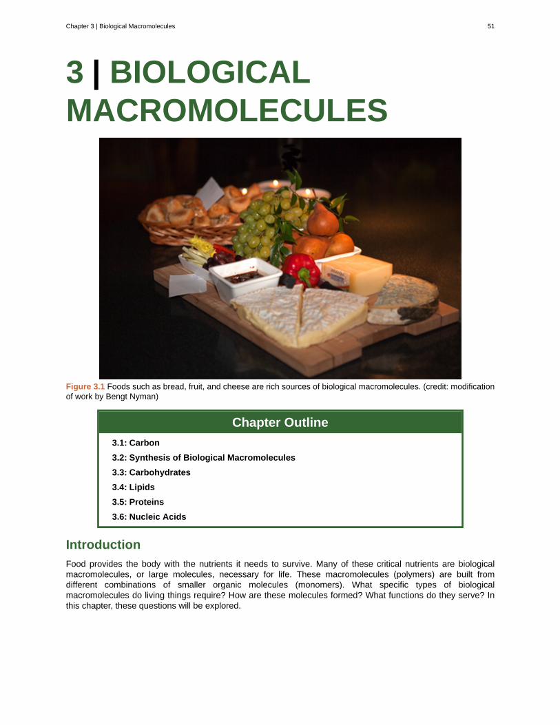
3 | BIOLOGICALMACROMOLECULES
Figure 3.1 Foods such as bread, fruit, and cheese are rich sources of biological macromolecules. (credit: modificationof work by Bengt Nyman)
Chapter Outline
3.1: Carbon
3.2: Synthesis of Biological Macromolecules
3.3: Carbohydrates
3.4: Lipids
3.5: Proteins
3.6: Nucleic Acids
Introduction
Food provides the body with the nutrients it needs to survive. Many of these critical nutrients are biologicalmacromolecules, or large molecules, necessary for life. These macromolecules (polymers) are built fromdifferent combinations of smaller organic molecules (monomers). What specific types of biologicalmacromolecules do living things require? How are these molecules formed? What functions do they serve? Inthis chapter, these questions will be explored.
Chapter 3 | Biological Macromolecules 51
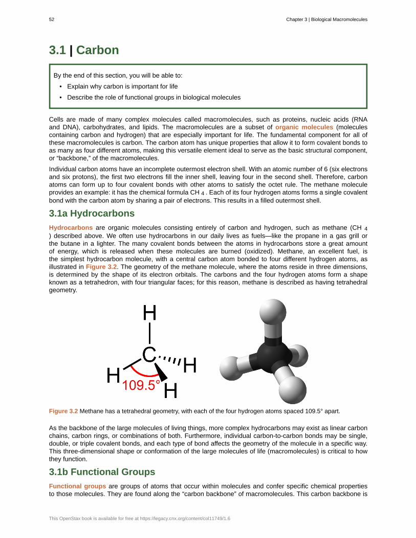
3.1 | Carbon
By the end of this section, you will be able to:
• Explain why carbon is important for life
• Describe the role of functional groups in biological molecules
Cells are made of many complex molecules called macromolecules, such as proteins, nucleic acids (RNAand DNA), carbohydrates, and lipids. The macromolecules are a subset of organic molecules (moleculescontaining carbon and hydrogen) that are especially important for life. The fundamental component for all ofthese macromolecules is carbon. The carbon atom has unique properties that allow it to form covalent bonds toas many as four different atoms, making this versatile element ideal to serve as the basic structural component,or “backbone,” of the macromolecules.
Individual carbon atoms have an incomplete outermost electron shell. With an atomic number of 6 (six electronsand six protons), the first two electrons fill the inner shell, leaving four in the second shell. Therefore, carbonatoms can form up to four covalent bonds with other atoms to satisfy the octet rule. The methane moleculeprovides an example: it has the chemical formula CH 4 . Each of its four hydrogen atoms forms a single covalentbond with the carbon atom by sharing a pair of electrons. This results in a filled outermost shell.
3.1a Hydrocarbons
Hydrocarbons are organic molecules consisting entirely of carbon and hydrogen, such as methane (CH 4) described above. We often use hydrocarbons in our daily lives as fuels—like the propane in a gas grill orthe butane in a lighter. The many covalent bonds between the atoms in hydrocarbons store a great amountof energy, which is released when these molecules are burned (oxidized). Methane, an excellent fuel, isthe simplest hydrocarbon molecule, with a central carbon atom bonded to four different hydrogen atoms, asillustrated in Figure 3.2. The geometry of the methane molecule, where the atoms reside in three dimensions,is determined by the shape of its electron orbitals. The carbons and the four hydrogen atoms form a shapeknown as a tetrahedron, with four triangular faces; for this reason, methane is described as having tetrahedralgeometry.
Figure 3.2 Methane has a tetrahedral geometry, with each of the four hydrogen atoms spaced 109.5° apart.
As the backbone of the large molecules of living things, more complex hydrocarbons may exist as linear carbonchains, carbon rings, or combinations of both. Furthermore, individual carbon-to-carbon bonds may be single,double, or triple covalent bonds, and each type of bond affects the geometry of the molecule in a specific way.This three-dimensional shape or conformation of the large molecules of life (macromolecules) is critical to howthey function.
3.1b Functional Groups
Functional groups are groups of atoms that occur within molecules and confer specific chemical propertiesto those molecules. They are found along the “carbon backbone” of macromolecules. This carbon backbone is
52 Chapter 3 | Biological Macromolecules
This OpenStax book is available for free at https://legacy.cnx.org/content/col11749/1.6
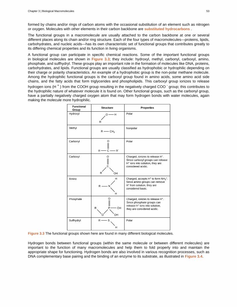
formed by chains and/or rings of carbon atoms with the occasional substitution of an element such as nitrogenor oxygen. Molecules with other elements in their carbon backbone are substituted hydrocarbons .
The functional groups in a macromolecule are usually attached to the carbon backbone at one or severaldifferent places along its chain and/or ring structure. Each of the four types of macromolecules—proteins, lipids,carbohydrates, and nucleic acids—has its own characteristic set of functional groups that contributes greatly toits differing chemical properties and its function in living organisms.
A functional group can participate in specific chemical reactions. Some of the important functional groupsin biological molecules are shown in Figure 3.3; they include: hydroxyl, methyl, carbonyl, carboxyl, amino,phosphate, and sulfhydryl. These groups play an important role in the formation of molecules like DNA, proteins,carbohydrates, and lipids. Functional groups are usually classified as hydrophobic or hydrophilic depending ontheir charge or polarity characteristics. An example of a hydrophobic group is the non-polar methane molecule.Among the hydrophilic functional groups is the carboxyl group found in amino acids, some amino acid sidechains, and the fatty acids that form triglycerides and phospholipids. This carboxyl group ionizes to release
hydrogen ions (H + ) from the COOH group resulting in the negatively charged COO - group; this contributes tothe hydrophilic nature of whatever molecule it is found on. Other functional groups, such as the carbonyl group,have a partially negatively charged oxygen atom that may form hydrogen bonds with water molecules, againmaking the molecule more hydrophilic.
Figure 3.3 The functional groups shown here are found in many different biological molecules.
Hydrogen bonds between functional groups (within the same molecule or between different molecules) areimportant to the function of many macromolecules and help them to fold properly into and maintain theappropriate shape for functioning. Hydrogen bonds are also involved in various recognition processes, such asDNA complementary base pairing and the binding of an enzyme to its substrate, as illustrated in Figure 3.4.
Chapter 3 | Biological Macromolecules 53
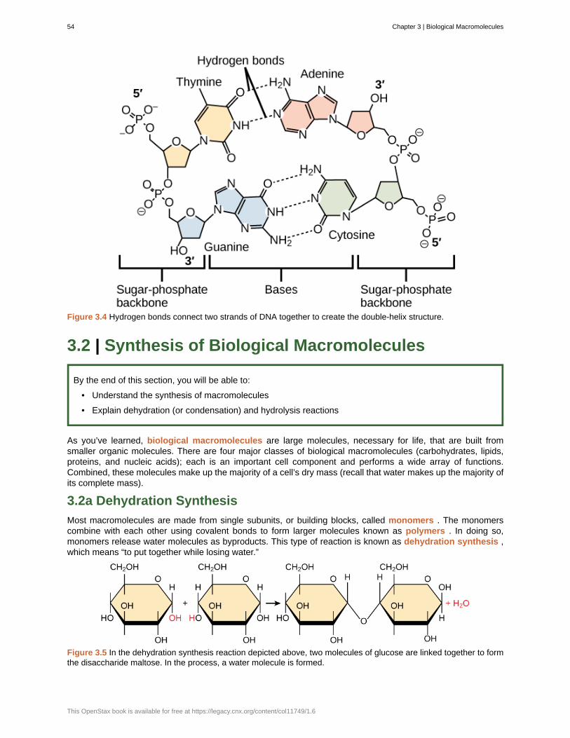
Figure 3.4 Hydrogen bonds connect two strands of DNA together to create the double-helix structure.
3.2 | Synthesis of Biological Macromolecules
By the end of this section, you will be able to:
• Understand the synthesis of macromolecules
• Explain dehydration (or condensation) and hydrolysis reactions
As you’ve learned, biological macromolecules are large molecules, necessary for life, that are built fromsmaller organic molecules. There are four major classes of biological macromolecules (carbohydrates, lipids,proteins, and nucleic acids); each is an important cell component and performs a wide array of functions.Combined, these molecules make up the majority of a cell’s dry mass (recall that water makes up the majority ofits complete mass).
3.2a Dehydration Synthesis
Most macromolecules are made from single subunits, or building blocks, called monomers . The monomerscombine with each other using covalent bonds to form larger molecules known as polymers . In doing so,monomers release water molecules as byproducts. This type of reaction is known as dehydration synthesis ,which means “to put together while losing water.”
Figure 3.5 In the dehydration synthesis reaction depicted above, two molecules of glucose are linked together to formthe disaccharide maltose. In the process, a water molecule is formed.
54 Chapter 3 | Biological Macromolecules
This OpenStax book is available for free at https://legacy.cnx.org/content/col11749/1.6
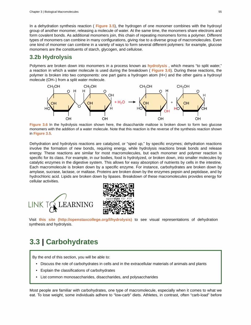
In a dehydration synthesis reaction ( Figure 3.5), the hydrogen of one monomer combines with the hydroxylgroup of another monomer, releasing a molecule of water. At the same time, the monomers share electrons andform covalent bonds. As additional monomers join, this chain of repeating monomers forms a polymer. Differenttypes of monomers can combine in many configurations, giving rise to a diverse group of macromolecules. Evenone kind of monomer can combine in a variety of ways to form several different polymers: for example, glucosemonomers are the constituents of starch, glycogen, and cellulose.
3.2b Hydrolysis
Polymers are broken down into monomers in a process known as hydrolysis , which means “to split water,”a reaction in which a water molecule is used during the breakdown ( Figure 3.6). During these reactions, thepolymer is broken into two components: one part gains a hydrogen atom (H+) and the other gains a hydroxylmolecule (OH–) from a split water molecule.
Figure 3.6 In the hydrolysis reaction shown here, the disaccharide maltose is broken down to form two glucosemonomers with the addition of a water molecule. Note that this reaction is the reverse of the synthesis reaction shownin Figure 3.5.
Dehydration and hydrolysis reactions are catalyzed, or “sped up,” by specific enzymes; dehydration reactionsinvolve the formation of new bonds, requiring energy, while hydrolysis reactions break bonds and releaseenergy. These reactions are similar for most macromolecules, but each monomer and polymer reaction isspecific for its class. For example, in our bodies, food is hydrolyzed, or broken down, into smaller molecules bycatalytic enzymes in the digestive system. This allows for easy absorption of nutrients by cells in the intestine.Each macromolecule is broken down by a specific enzyme. For instance, carbohydrates are broken down byamylase, sucrase, lactase, or maltase. Proteins are broken down by the enzymes pepsin and peptidase, and byhydrochloric acid. Lipids are broken down by lipases. Breakdown of these macromolecules provides energy forcellular activities.
Visit this site (http://openstaxcollege.org/l/hydrolysis) to see visual representations of dehydrationsynthesis and hydrolysis.
3.3 | Carbohydrates
By the end of this section, you will be able to:
• Discuss the role of carbohydrates in cells and in the extracellular materials of animals and plants
• Explain the classifications of carbohydrates
• List common monosaccharides, disaccharides, and polysaccharides
Most people are familiar with carbohydrates, one type of macromolecule, especially when it comes to what weeat. To lose weight, some individuals adhere to “low-carb” diets. Athletes, in contrast, often “carb-load” before
Chapter 3 | Biological Macromolecules 55
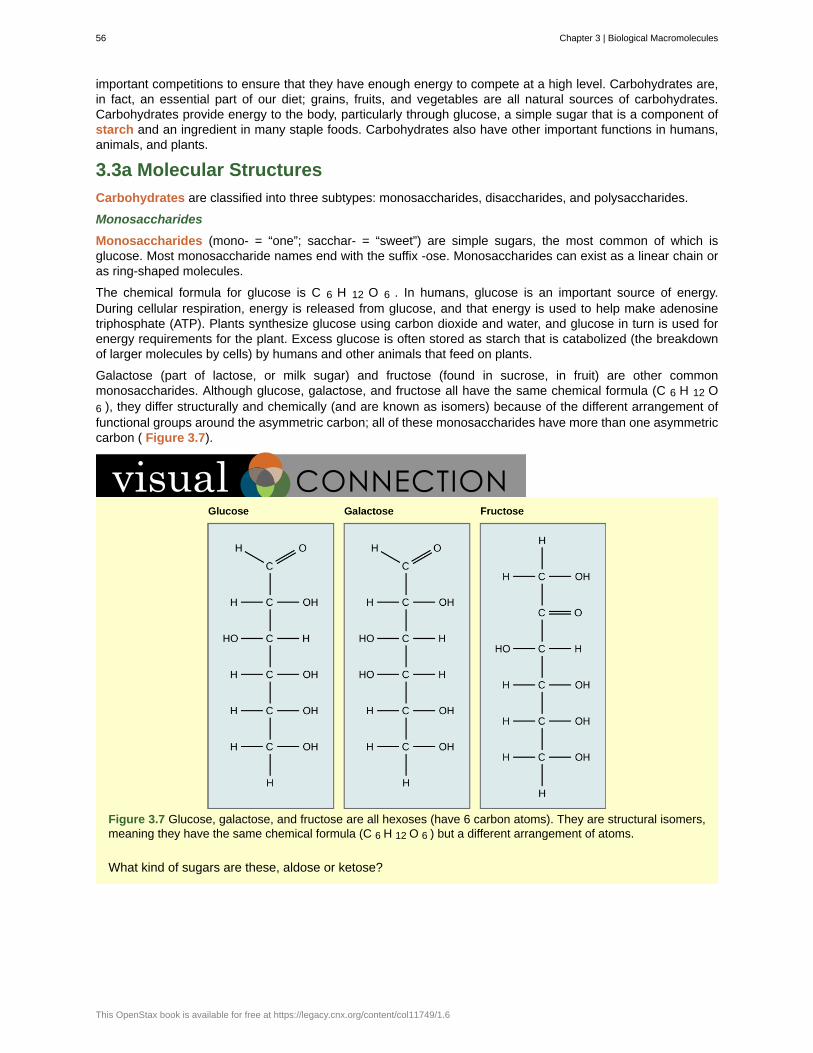
important competitions to ensure that they have enough energy to compete at a high level. Carbohydrates are,in fact, an essential part of our diet; grains, fruits, and vegetables are all natural sources of carbohydrates.Carbohydrates provide energy to the body, particularly through glucose, a simple sugar that is a component ofstarch and an ingredient in many staple foods. Carbohydrates also have other important functions in humans,animals, and plants.
3.3a Molecular Structures
Carbohydrates are classified into three subtypes: monosaccharides, disaccharides, and polysaccharides.
Monosaccharides
Monosaccharides (mono- = “one”; sacchar- = “sweet”) are simple sugars, the most common of which isglucose. Most monosaccharide names end with the suffix -ose. Monosaccharides can exist as a linear chain oras ring-shaped molecules.
The chemical formula for glucose is C 6 H 12 O 6 . In humans, glucose is an important source of energy.During cellular respiration, energy is released from glucose, and that energy is used to help make adenosinetriphosphate (ATP). Plants synthesize glucose using carbon dioxide and water, and glucose in turn is used forenergy requirements for the plant. Excess glucose is often stored as starch that is catabolized (the breakdownof larger molecules by cells) by humans and other animals that feed on plants.
Galactose (part of lactose, or milk sugar) and fructose (found in sucrose, in fruit) are other commonmonosaccharides. Although glucose, galactose, and fructose all have the same chemical formula (C 6 H 12 O6 ), they differ structurally and chemically (and are known as isomers) because of the different arrangement offunctional groups around the asymmetric carbon; all of these monosaccharides have more than one asymmetriccarbon ( Figure 3.7).
Figure 3.7 Glucose, galactose, and fructose are all hexoses (have 6 carbon atoms). They are structural isomers,meaning they have the same chemical formula (C 6 H 12 O 6 ) but a different arrangement of atoms.
What kind of sugars are these, aldose or ketose?
56 Chapter 3 | Biological Macromolecules
This OpenStax book is available for free at https://legacy.cnx.org/content/col11749/1.6
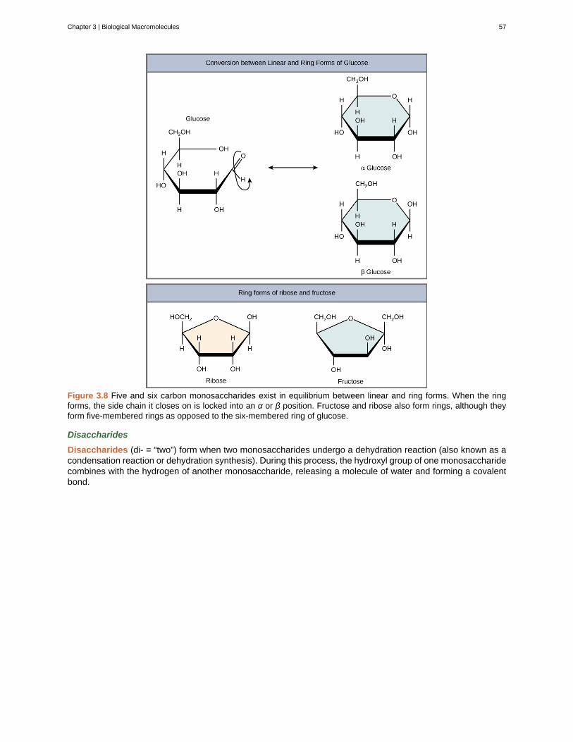
Figure 3.8 Five and six carbon monosaccharides exist in equilibrium between linear and ring forms. When the ringforms, the side chain it closes on is locked into an α or β position. Fructose and ribose also form rings, although theyform five-membered rings as opposed to the six-membered ring of glucose.
Disaccharides
Disaccharides (di- = “two”) form when two monosaccharides undergo a dehydration reaction (also known as acondensation reaction or dehydration synthesis). During this process, the hydroxyl group of one monosaccharidecombines with the hydrogen of another monosaccharide, releasing a molecule of water and forming a covalentbond.
Chapter 3 | Biological Macromolecules 57
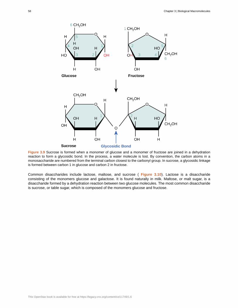
Figure 3.9 Sucrose is formed when a monomer of glucose and a monomer of fructose are joined in a dehydrationreaction to form a glycosidic bond. In the process, a water molecule is lost. By convention, the carbon atoms in amonosaccharide are numbered from the terminal carbon closest to the carbonyl group. In sucrose, a glycosidic linkageis formed between carbon 1 in glucose and carbon 2 in fructose.
Common disaccharides include lactose, maltose, and sucrose ( Figure 3.10). Lactose is a disaccharideconsisting of the monomers glucose and galactose. It is found naturally in milk. Maltose, or malt sugar, is adisaccharide formed by a dehydration reaction between two glucose molecules. The most common disaccharideis sucrose, or table sugar, which is composed of the monomers glucose and fructose.
58 Chapter 3 | Biological Macromolecules
This OpenStax book is available for free at https://legacy.cnx.org/content/col11749/1.6
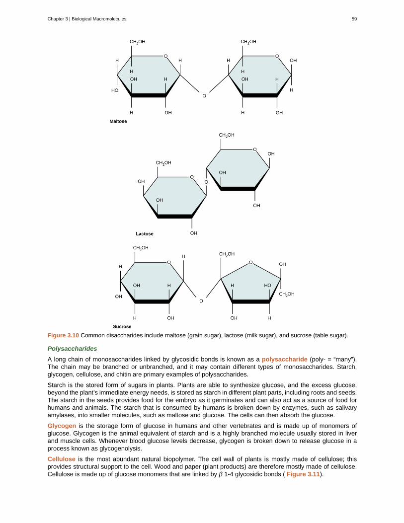
Figure 3.10 Common disaccharides include maltose (grain sugar), lactose (milk sugar), and sucrose (table sugar).
Polysaccharides
A long chain of monosaccharides linked by glycosidic bonds is known as a polysaccharide (poly- = “many”).The chain may be branched or unbranched, and it may contain different types of monosaccharides. Starch,glycogen, cellulose, and chitin are primary examples of polysaccharides.
Starch is the stored form of sugars in plants. Plants are able to synthesize glucose, and the excess glucose,beyond the plant’s immediate energy needs, is stored as starch in different plant parts, including roots and seeds.The starch in the seeds provides food for the embryo as it germinates and can also act as a source of food forhumans and animals. The starch that is consumed by humans is broken down by enzymes, such as salivaryamylases, into smaller molecules, such as maltose and glucose. The cells can then absorb the glucose.
Glycogen is the storage form of glucose in humans and other vertebrates and is made up of monomers ofglucose. Glycogen is the animal equivalent of starch and is a highly branched molecule usually stored in liverand muscle cells. Whenever blood glucose levels decrease, glycogen is broken down to release glucose in aprocess known as glycogenolysis.
Cellulose is the most abundant natural biopolymer. The cell wall of plants is mostly made of cellulose; thisprovides structural support to the cell. Wood and paper (plant products) are therefore mostly made of cellulose.Cellulose is made up of glucose monomers that are linked by β 1-4 glycosidic bonds ( Figure 3.11).
Chapter 3 | Biological Macromolecules 59
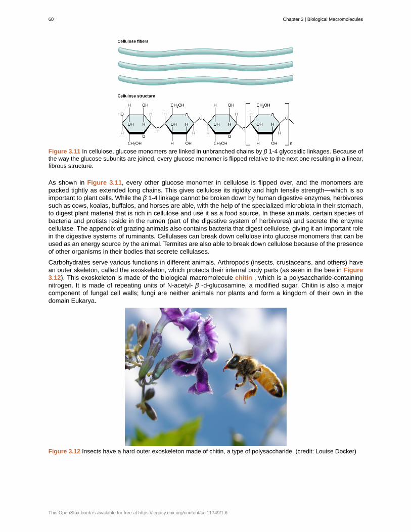
Figure 3.11 In cellulose, glucose monomers are linked in unbranched chains by β 1-4 glycosidic linkages. Because ofthe way the glucose subunits are joined, every glucose monomer is flipped relative to the next one resulting in a linear,fibrous structure.
As shown in Figure 3.11, every other glucose monomer in cellulose is flipped over, and the monomers arepacked tightly as extended long chains. This gives cellulose its rigidity and high tensile strength—which is soimportant to plant cells. While the β 1-4 linkage cannot be broken down by human digestive enzymes, herbivoressuch as cows, koalas, buffalos, and horses are able, with the help of the specialized microbiota in their stomach,to digest plant material that is rich in cellulose and use it as a food source. In these animals, certain species ofbacteria and protists reside in the rumen (part of the digestive system of herbivores) and secrete the enzymecellulase. The appendix of grazing animals also contains bacteria that digest cellulose, giving it an important rolein the digestive systems of ruminants. Cellulases can break down cellulose into glucose monomers that can beused as an energy source by the animal. Termites are also able to break down cellulose because of the presenceof other organisms in their bodies that secrete cellulases.
Carbohydrates serve various functions in different animals. Arthropods (insects, crustaceans, and others) havean outer skeleton, called the exoskeleton, which protects their internal body parts (as seen in the bee in Figure3.12). This exoskeleton is made of the biological macromolecule chitin , which is a polysaccharide-containingnitrogen. It is made of repeating units of N-acetyl- β -d-glucosamine, a modified sugar. Chitin is also a majorcomponent of fungal cell walls; fungi are neither animals nor plants and form a kingdom of their own in thedomain Eukarya.
Figure 3.12 Insects have a hard outer exoskeleton made of chitin, a type of polysaccharide. (credit: Louise Docker)
60 Chapter 3 | Biological Macromolecules
This OpenStax book is available for free at https://legacy.cnx.org/content/col11749/1.6
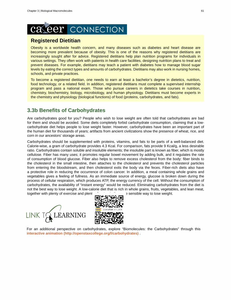
Registered DietitianObesity is a worldwide health concern, and many diseases such as diabetes and heart disease arebecoming more prevalent because of obesity. This is one of the reasons why registered dietitians areincreasingly sought after for advice. Registered dietitians help plan nutrition programs for individuals invarious settings. They often work with patients in health care facilities, designing nutrition plans to treat andprevent diseases. For example, dietitians may teach a patient with diabetes how to manage blood sugarlevels by eating the correct types and amounts of carbohydrates. Dietitians may also work in nursing homes,schools, and private practices.
To become a registered dietitian, one needs to earn at least a bachelor’s degree in dietetics, nutrition,food technology, or a related field. In addition, registered dietitians must complete a supervised internshipprogram and pass a national exam. Those who pursue careers in dietetics take courses in nutrition,chemistry, biochemistry, biology, microbiology, and human physiology. Dietitians must become experts inthe chemistry and physiology (biological functions) of food (proteins, carbohydrates, and fats).
3.3b Benefits of Carbohydrates
Are carbohydrates good for you? People who wish to lose weight are often told that carbohydrates are badfor them and should be avoided. Some diets completely forbid carbohydrate consumption, claiming that a low-carbohydrate diet helps people to lose weight faster. However, carbohydrates have been an important part ofthe human diet for thousands of years; artifacts from ancient civilizations show the presence of wheat, rice, andcorn in our ancestors’ storage areas.
Carbohydrates should be supplemented with proteins, vitamins, and fats to be parts of a well-balanced diet.Calorie-wise, a gram of carbohydrate provides 4.3 Kcal. For comparison, fats provide 9 Kcal/g, a less desirableratio. Carbohydrates contain soluble and insoluble elements; the insoluble part is known as fiber, which is mostlycellulose. Fiber has many uses; it promotes regular bowel movement by adding bulk, and it regulates the rateof consumption of blood glucose. Fiber also helps to remove excess cholesterol from the body: fiber binds tothe cholesterol in the small intestine, then attaches to the cholesterol and prevents the cholesterol particlesfrom entering the bloodstream, and then cholesterol exits the body via the feces. Fiber-rich diets also havea protective role in reducing the occurrence of colon cancer. In addition, a meal containing whole grains andvegetables gives a feeling of fullness. As an immediate source of energy, glucose is broken down during theprocess of cellular respiration, which produces ATP, the energy currency of the cell. Without the consumption ofcarbohydrates, the availability of “instant energy” would be reduced. Eliminating carbohydrates from the diet isnot the best way to lose weight. A low-calorie diet that is rich in whole grains, fruits, vegetables, and lean meat,together with plenty of exercise and plenty of water, is the more sensible way to lose weight.
For an additional perspective on carbohydrates, explore “Biomolecules: the Carbohydrates” through thisinteractive animation (http://openstaxcollege.org/l/carbohydrates) .
Chapter 3 | Biological Macromolecules 61
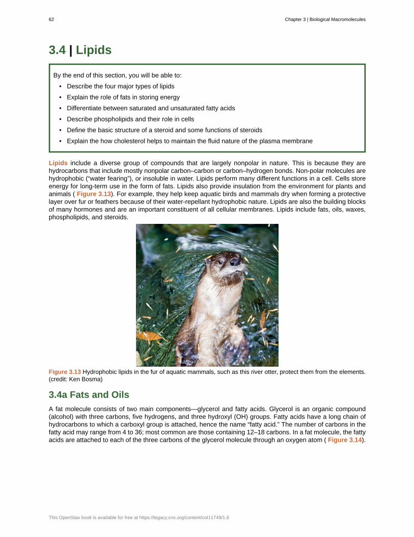
3.4 | Lipids
By the end of this section, you will be able to:
• Describe the four major types of lipids
• Explain the role of fats in storing energy
• Differentiate between saturated and unsaturated fatty acids
• Describe phospholipids and their role in cells
• Define the basic structure of a steroid and some functions of steroids
• Explain the how cholesterol helps to maintain the fluid nature of the plasma membrane
Lipids include a diverse group of compounds that are largely nonpolar in nature. This is because they arehydrocarbons that include mostly nonpolar carbon–carbon or carbon–hydrogen bonds. Non-polar molecules arehydrophobic (“water fearing”), or insoluble in water. Lipids perform many different functions in a cell. Cells storeenergy for long-term use in the form of fats. Lipids also provide insulation from the environment for plants andanimals ( Figure 3.13). For example, they help keep aquatic birds and mammals dry when forming a protectivelayer over fur or feathers because of their water-repellant hydrophobic nature. Lipids are also the building blocksof many hormones and are an important constituent of all cellular membranes. Lipids include fats, oils, waxes,phospholipids, and steroids.
Figure 3.13 Hydrophobic lipids in the fur of aquatic mammals, such as this river otter, protect them from the elements.(credit: Ken Bosma)
3.4a Fats and Oils
A fat molecule consists of two main components—glycerol and fatty acids. Glycerol is an organic compound(alcohol) with three carbons, five hydrogens, and three hydroxyl (OH) groups. Fatty acids have a long chain ofhydrocarbons to which a carboxyl group is attached, hence the name “fatty acid.” The number of carbons in thefatty acid may range from 4 to 36; most common are those containing 12–18 carbons. In a fat molecule, the fattyacids are attached to each of the three carbons of the glycerol molecule through an oxygen atom ( Figure 3.14).
62 Chapter 3 | Biological Macromolecules
This OpenStax book is available for free at https://legacy.cnx.org/content/col11749/1.6
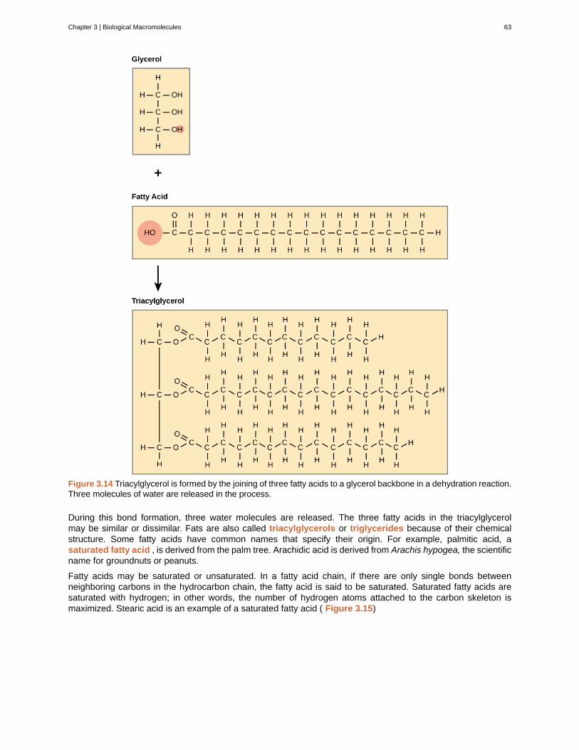
Figure 3.14 Triacylglycerol is formed by the joining of three fatty acids to a glycerol backbone in a dehydration reaction.Three molecules of water are released in the process.
During this bond formation, three water molecules are released. The three fatty acids in the triacylglycerolmay be similar or dissimilar. Fats are also called triacylglycerols or triglycerides because of their chemicalstructure. Some fatty acids have common names that specify their origin. For example, palmitic acid, asaturated fatty acid , is derived from the palm tree. Arachidic acid is derived from Arachis hypogea, the scientificname for groundnuts or peanuts.
Fatty acids may be saturated or unsaturated. In a fatty acid chain, if there are only single bonds betweenneighboring carbons in the hydrocarbon chain, the fatty acid is said to be saturated. Saturated fatty acids aresaturated with hydrogen; in other words, the number of hydrogen atoms attached to the carbon skeleton ismaximized. Stearic acid is an example of a saturated fatty acid ( Figure 3.15)
Chapter 3 | Biological Macromolecules 63
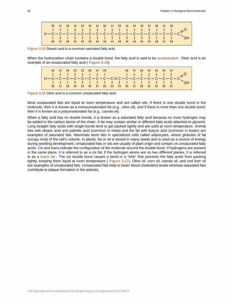
Figure 3.15 Stearic acid is a common saturated fatty acid.
When the hydrocarbon chain contains a double bond, the fatty acid is said to be unsaturated . Oleic acid is anexample of an unsaturated fatty acid ( Figure 3.16).
Figure 3.16 Oleic acid is a common unsaturated fatty acid.
Most unsaturated fats are liquid at room temperature and are called oils. If there is one double bond in themolecule, then it is known as a monounsaturated fat (e.g., olive oil), and if there is more than one double bond,then it is known as a polyunsaturated fat (e.g., canola oil).
When a fatty acid has no double bonds, it is known as a saturated fatty acid because no more hydrogen maybe added to the carbon atoms of the chain. A fat may contain similar or different fatty acids attached to glycerol.Long straight fatty acids with single bonds tend to get packed tightly and are solid at room temperature. Animalfats with stearic acid and palmitic acid (common in meat) and the fat with butyric acid (common in butter) areexamples of saturated fats. Mammals store fats in specialized cells called adipocytes, where globules of fatoccupy most of the cell’s volume. In plants, fat or oil is stored in many seeds and is used as a source of energyduring seedling development. Unsaturated fats or oils are usually of plant origin and contain cis unsaturated fattyacids. Cis and trans indicate the configuration of the molecule around the double bond. If hydrogens are presentin the same plane, it is referred to as a cis fat; if the hydrogen atoms are on two different planes, it is referredto as a trans fat . The cis double bond causes a bend or a “kink” that prevents the fatty acids from packingtightly, keeping them liquid at room temperature ( Figure 3.17). Olive oil, corn oil, canola oil, and cod liver oilare examples of unsaturated fats. Unsaturated fats help to lower blood cholesterol levels whereas saturated fatscontribute to plaque formation in the arteries.
64 Chapter 3 | Biological Macromolecules
This OpenStax book is available for free at https://legacy.cnx.org/content/col11749/1.6
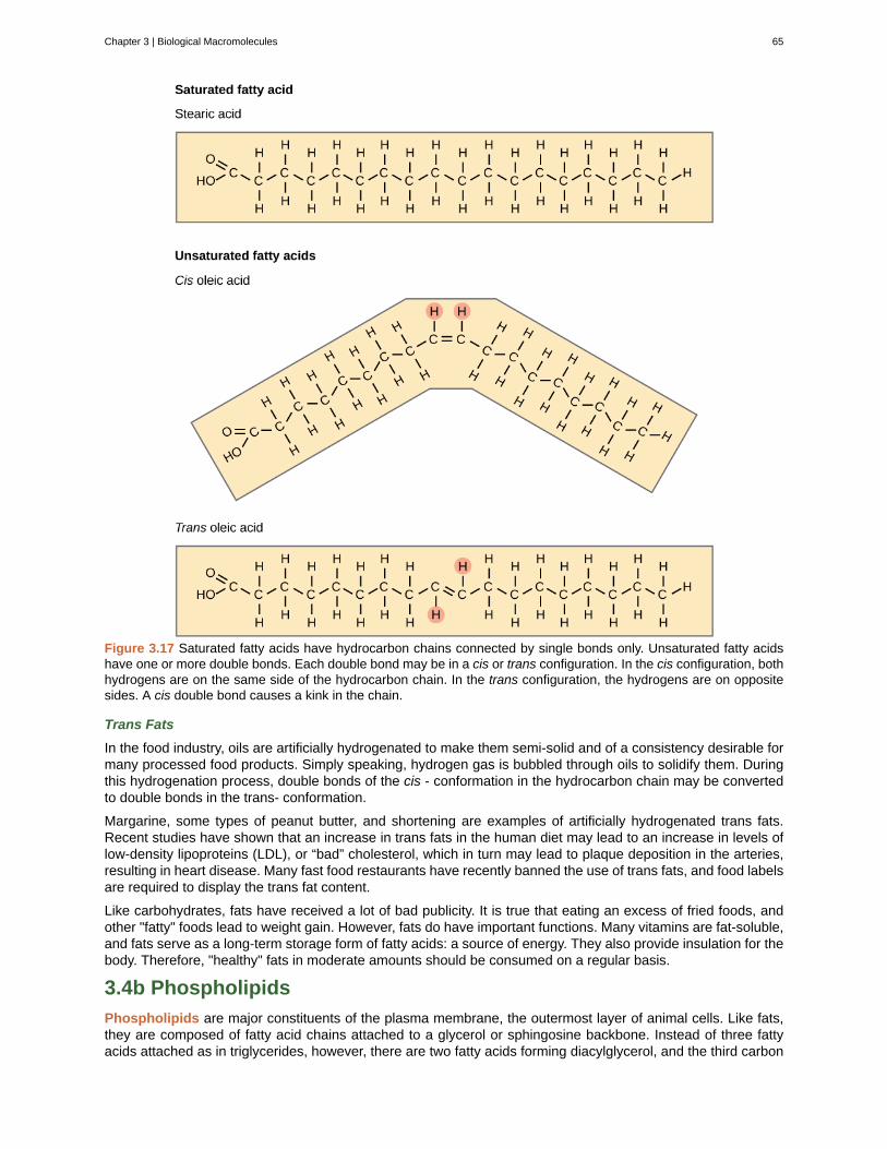
Figure 3.17 Saturated fatty acids have hydrocarbon chains connected by single bonds only. Unsaturated fatty acidshave one or more double bonds. Each double bond may be in a cis or trans configuration. In the cis configuration, bothhydrogens are on the same side of the hydrocarbon chain. In the trans configuration, the hydrogens are on oppositesides. A cis double bond causes a kink in the chain.
Trans Fats
In the food industry, oils are artificially hydrogenated to make them semi-solid and of a consistency desirable formany processed food products. Simply speaking, hydrogen gas is bubbled through oils to solidify them. Duringthis hydrogenation process, double bonds of the cis - conformation in the hydrocarbon chain may be convertedto double bonds in the trans- conformation.
Margarine, some types of peanut butter, and shortening are examples of artificially hydrogenated trans fats.Recent studies have shown that an increase in trans fats in the human diet may lead to an increase in levels oflow-density lipoproteins (LDL), or “bad” cholesterol, which in turn may lead to plaque deposition in the arteries,resulting in heart disease. Many fast food restaurants have recently banned the use of trans fats, and food labelsare required to display the trans fat content.
Like carbohydrates, fats have received a lot of bad publicity. It is true that eating an excess of fried foods, andother "fatty" foods lead to weight gain. However, fats do have important functions. Many vitamins are fat-soluble,and fats serve as a long-term storage form of fatty acids: a source of energy. They also provide insulation for thebody. Therefore, "healthy" fats in moderate amounts should be consumed on a regular basis.
3.4b Phospholipids
Phospholipids are major constituents of the plasma membrane, the outermost layer of animal cells. Like fats,they are composed of fatty acid chains attached to a glycerol or sphingosine backbone. Instead of three fattyacids attached as in triglycerides, however, there are two fatty acids forming diacylglycerol, and the third carbon
Chapter 3 | Biological Macromolecules 65
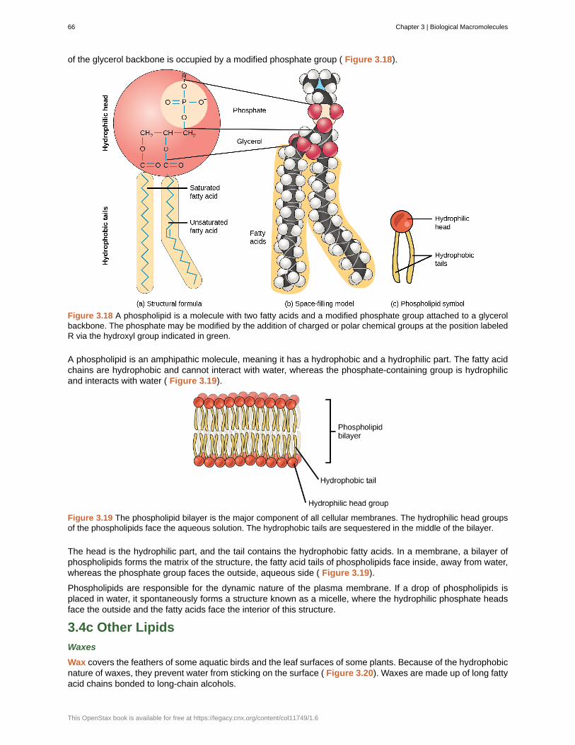
of the glycerol backbone is occupied by a modified phosphate group ( Figure 3.18).
Figure 3.18 A phospholipid is a molecule with two fatty acids and a modified phosphate group attached to a glycerolbackbone. The phosphate may be modified by the addition of charged or polar chemical groups at the position labeledR via the hydroxyl group indicated in green.
A phospholipid is an amphipathic molecule, meaning it has a hydrophobic and a hydrophilic part. The fatty acidchains are hydrophobic and cannot interact with water, whereas the phosphate-containing group is hydrophilicand interacts with water ( Figure 3.19).
Figure 3.19 The phospholipid bilayer is the major component of all cellular membranes. The hydrophilic head groupsof the phospholipids face the aqueous solution. The hydrophobic tails are sequestered in the middle of the bilayer.
The head is the hydrophilic part, and the tail contains the hydrophobic fatty acids. In a membrane, a bilayer ofphospholipids forms the matrix of the structure, the fatty acid tails of phospholipids face inside, away from water,whereas the phosphate group faces the outside, aqueous side ( Figure 3.19).
Phospholipids are responsible for the dynamic nature of the plasma membrane. If a drop of phospholipids isplaced in water, it spontaneously forms a structure known as a micelle, where the hydrophilic phosphate headsface the outside and the fatty acids face the interior of this structure.
3.4c Other Lipids
Waxes
Wax covers the feathers of some aquatic birds and the leaf surfaces of some plants. Because of the hydrophobicnature of waxes, they prevent water from sticking on the surface ( Figure 3.20). Waxes are made up of long fattyacid chains bonded to long-chain alcohols.
66 Chapter 3 | Biological Macromolecules
This OpenStax book is available for free at https://legacy.cnx.org/content/col11749/1.6
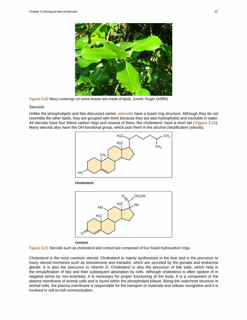
Figure 3.20 Waxy coverings on some leaves are made of lipids. (credit: Roger Griffith)
Steroids
Unlike the phospholipids and fats discussed earlier, steroids have a fused ring structure. Although they do notresemble the other lipids, they are grouped with them because they are also hydrophobic and insoluble in water.All steroids have four linked carbon rings and several of them, like cholesterol, have a short tail ( Figure 3.21).Many steroids also have the OH functional group, which puts them in the alcohol classification (sterols).
Figure 3.21 Steroids such as cholesterol and cortisol are composed of four fused hydrocarbon rings.
Cholesterol is the most common steroid. Cholesterol is mainly synthesized in the liver and is the precursor tomany steroid hormones such as testosterone and estradiol, which are secreted by the gonads and endocrineglands. It is also the precursor to Vitamin D. Cholesterol is also the precursor of bile salts, which help inthe emulsification of fats and their subsequent absorption by cells. Although cholesterol is often spoken of innegative terms by non-scientists, it is necessary for proper functioning of the body. It is a component of theplasma membrane of animal cells and is found within the phospholipid bilayer. Being the outermost structure inanimal cells, the plasma membrane is responsible for the transport of materials and cellular recognition and it isinvolved in cell-to-cell communication.
Chapter 3 | Biological Macromolecules 67
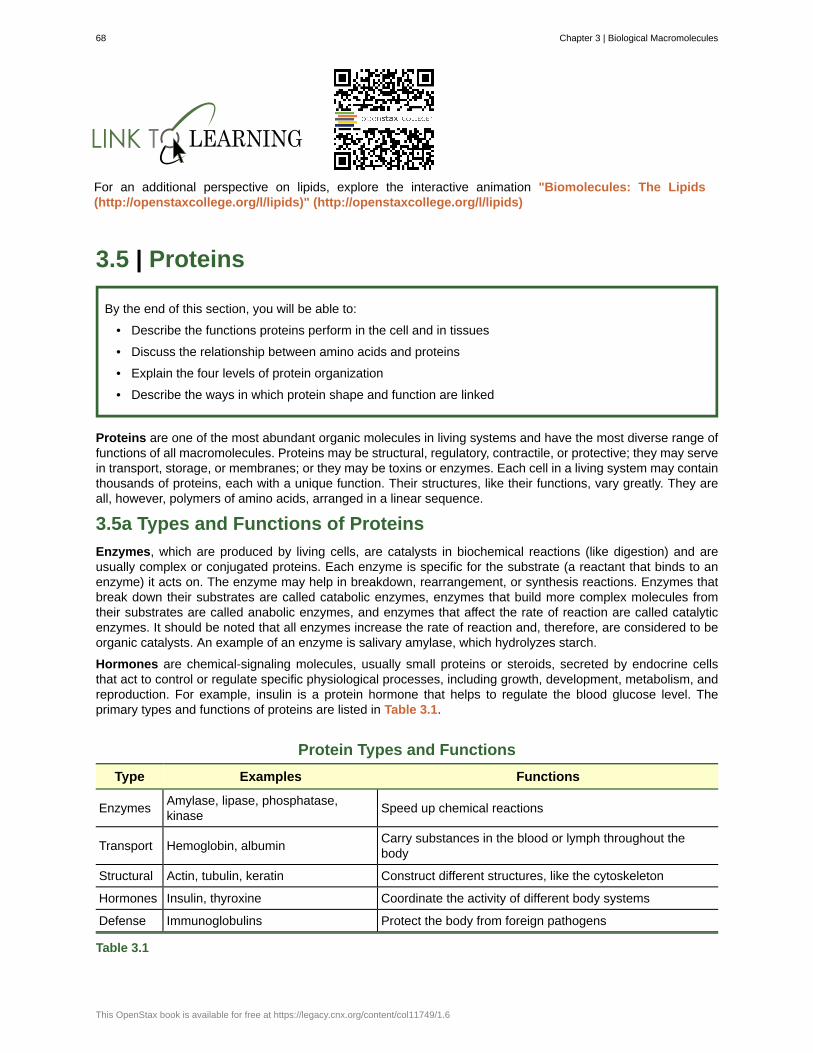
For an additional perspective on lipids, explore the interactive animation "Biomolecules: The Lipids(http://openstaxcollege.org/l/lipids)" (http://openstaxcollege.org/l/lipids)
3.5 | Proteins
By the end of this section, you will be able to:
• Describe the functions proteins perform in the cell and in tissues
• Discuss the relationship between amino acids and proteins
• Explain the four levels of protein organization
• Describe the ways in which protein shape and function are linked
Proteins are one of the most abundant organic molecules in living systems and have the most diverse range offunctions of all macromolecules. Proteins may be structural, regulatory, contractile, or protective; they may servein transport, storage, or membranes; or they may be toxins or enzymes. Each cell in a living system may containthousands of proteins, each with a unique function. Their structures, like their functions, vary greatly. They areall, however, polymers of amino acids, arranged in a linear sequence.
3.5a Types and Functions of Proteins
Enzymes, which are produced by living cells, are catalysts in biochemical reactions (like digestion) and areusually complex or conjugated proteins. Each enzyme is specific for the substrate (a reactant that binds to anenzyme) it acts on. The enzyme may help in breakdown, rearrangement, or synthesis reactions. Enzymes thatbreak down their substrates are called catabolic enzymes, enzymes that build more complex molecules fromtheir substrates are called anabolic enzymes, and enzymes that affect the rate of reaction are called catalyticenzymes. It should be noted that all enzymes increase the rate of reaction and, therefore, are considered to beorganic catalysts. An example of an enzyme is salivary amylase, which hydrolyzes starch.
Hormones are chemical-signaling molecules, usually small proteins or steroids, secreted by endocrine cellsthat act to control or regulate specific physiological processes, including growth, development, metabolism, andreproduction. For example, insulin is a protein hormone that helps to regulate the blood glucose level. Theprimary types and functions of proteins are listed in Table 3.1.
Protein Types and Functions
Type Examples Functions
EnzymesAmylase, lipase, phosphatase,kinase
Speed up chemical reactions
Transport Hemoglobin, albuminCarry substances in the blood or lymph throughout thebody
Structural Actin, tubulin, keratin Construct different structures, like the cytoskeleton
Hormones Insulin, thyroxine Coordinate the activity of different body systems
Defense Immunoglobulins Protect the body from foreign pathogens
Table 3.1
68 Chapter 3 | Biological Macromolecules
This OpenStax book is available for free at https://legacy.cnx.org/content/col11749/1.6
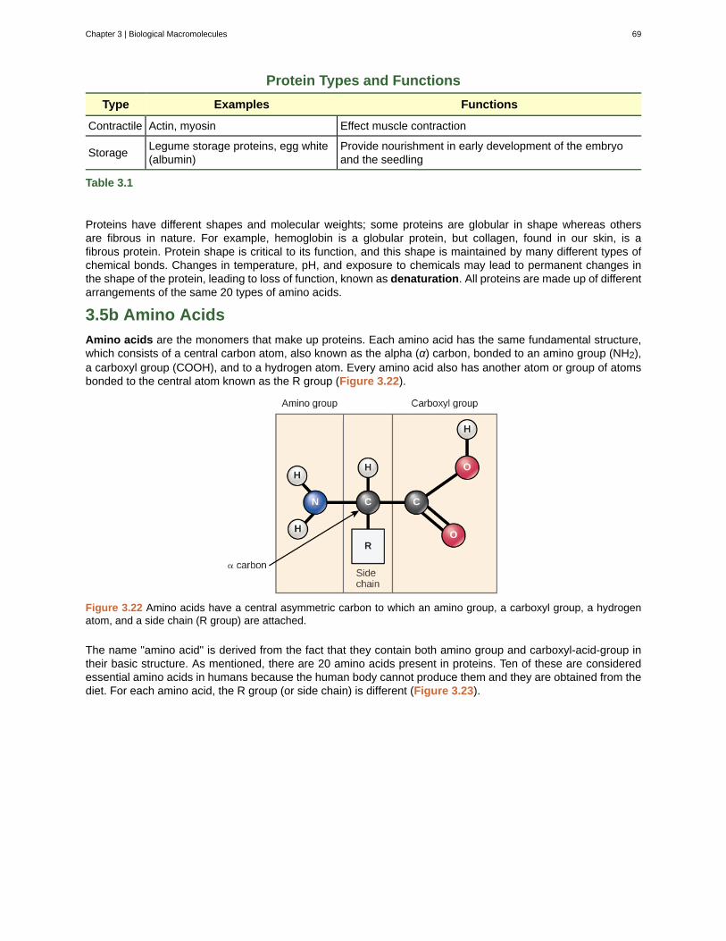
Protein Types and Functions
Type Examples Functions
Contractile Actin, myosin Effect muscle contraction
StorageLegume storage proteins, egg white(albumin)
Provide nourishment in early development of the embryoand the seedling
Table 3.1
Proteins have different shapes and molecular weights; some proteins are globular in shape whereas othersare fibrous in nature. For example, hemoglobin is a globular protein, but collagen, found in our skin, is afibrous protein. Protein shape is critical to its function, and this shape is maintained by many different types ofchemical bonds. Changes in temperature, pH, and exposure to chemicals may lead to permanent changes inthe shape of the protein, leading to loss of function, known as denaturation. All proteins are made up of differentarrangements of the same 20 types of amino acids.
3.5b Amino Acids
Amino acids are the monomers that make up proteins. Each amino acid has the same fundamental structure,which consists of a central carbon atom, also known as the alpha (α) carbon, bonded to an amino group (NH2),a carboxyl group (COOH), and to a hydrogen atom. Every amino acid also has another atom or group of atomsbonded to the central atom known as the R group (Figure 3.22).
Figure 3.22 Amino acids have a central asymmetric carbon to which an amino group, a carboxyl group, a hydrogenatom, and a side chain (R group) are attached.
The name "amino acid" is derived from the fact that they contain both amino group and carboxyl-acid-group intheir basic structure. As mentioned, there are 20 amino acids present in proteins. Ten of these are consideredessential amino acids in humans because the human body cannot produce them and they are obtained from thediet. For each amino acid, the R group (or side chain) is different (Figure 3.23).
Chapter 3 | Biological Macromolecules 69
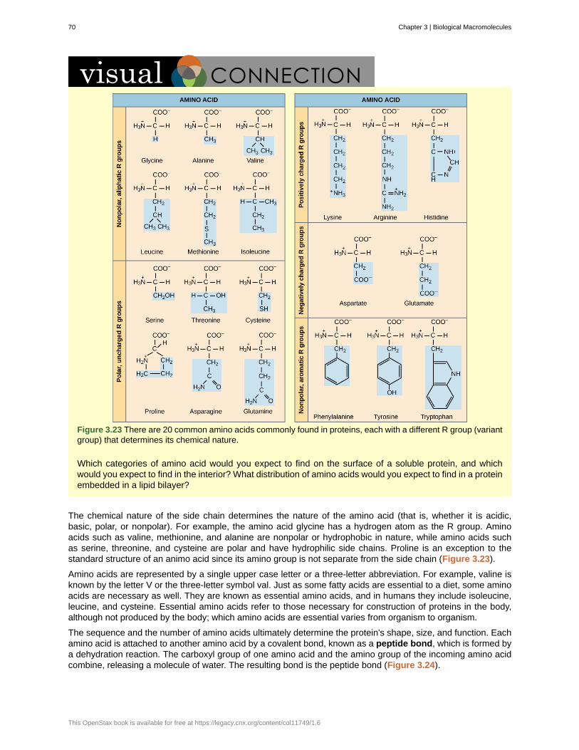
Figure 3.23 There are 20 common amino acids commonly found in proteins, each with a different R group (variantgroup) that determines its chemical nature.
Which categories of amino acid would you expect to find on the surface of a soluble protein, and whichwould you expect to find in the interior? What distribution of amino acids would you expect to find in a proteinembedded in a lipid bilayer?
The chemical nature of the side chain determines the nature of the amino acid (that is, whether it is acidic,basic, polar, or nonpolar). For example, the amino acid glycine has a hydrogen atom as the R group. Aminoacids such as valine, methionine, and alanine are nonpolar or hydrophobic in nature, while amino acids suchas serine, threonine, and cysteine are polar and have hydrophilic side chains. Proline is an exception to thestandard structure of an animo acid since its amino group is not separate from the side chain (Figure 3.23).
Amino acids are represented by a single upper case letter or a three-letter abbreviation. For example, valine isknown by the letter V or the three-letter symbol val. Just as some fatty acids are essential to a diet, some aminoacids are necessary as well. They are known as essential amino acids, and in humans they include isoleucine,leucine, and cysteine. Essential amino acids refer to those necessary for construction of proteins in the body,although not produced by the body; which amino acids are essential varies from organism to organism.
The sequence and the number of amino acids ultimately determine the protein's shape, size, and function. Eachamino acid is attached to another amino acid by a covalent bond, known as a peptide bond, which is formed bya dehydration reaction. The carboxyl group of one amino acid and the amino group of the incoming amino acidcombine, releasing a molecule of water. The resulting bond is the peptide bond (Figure 3.24).
70 Chapter 3 | Biological Macromolecules
This OpenStax book is available for free at https://legacy.cnx.org/content/col11749/1.6

Figure 3.24 Peptide bond formation is a dehydration synthesis reaction. The carboxyl group of one amino acid is linkedto the amino group of the incoming amino acid. In the process, a molecule of water is released.
The products formed by such linkages are called peptides. As more amino acids join to this growing chain,the resulting chain is known as a polypeptide. While the terms polypeptide and protein are sometimes usedinterchangeably, a polypeptide is technically a polymer of amino acids, whereas the term protein is used for apolypeptide or polypeptides that have combined together, often have bound non-peptide prosthetic groups, havea distinct shape, and have a unique function.
Click through the steps of protein synthesis in this interactive tutorial (http://openstaxcollege.org/l/protein_synth) .
The Evolutionary Significance of Cytochrome cCytochrome c is an important component of the electron transport chain, a part of cellular respiration, andit is normally found in the cellular organelle, the mitochondrion. This protein has a heme prosthetic group,and the central ion of the heme gets alternately reduced and oxidized during electron transfer. Because thisessential protein’s role in producing cellular energy is crucial, it has changed very little over millions of years.Protein sequencing has shown that there is a considerable amount of cytochrome c amino acid sequencehomology among different species; in other words, evolutionary kinship can be assessed by measuring thesimilarities or differences among various species’ DNA or protein sequences.
Scientists have determined that human cytochrome c contains 104 amino acids. For each cytochrome cmolecule from different organisms that has been sequenced to date, 37 of these amino acids appear in thesame position in all samples of cytochrome c. This indicates that there may have been a common ancestor.On comparing the human and chimpanzee protein sequences, no sequence difference was found. Whenhuman and rhesus monkey sequences were compared, the single difference found was in one amino acid.In another comparison, human to yeast sequencing shows a difference in the 44th position.
3.5c Protein Structure
As discussed earlier, the shape of a protein is critical to its function. For example, an enzyme can bind to a
Chapter 3 | Biological Macromolecules 71
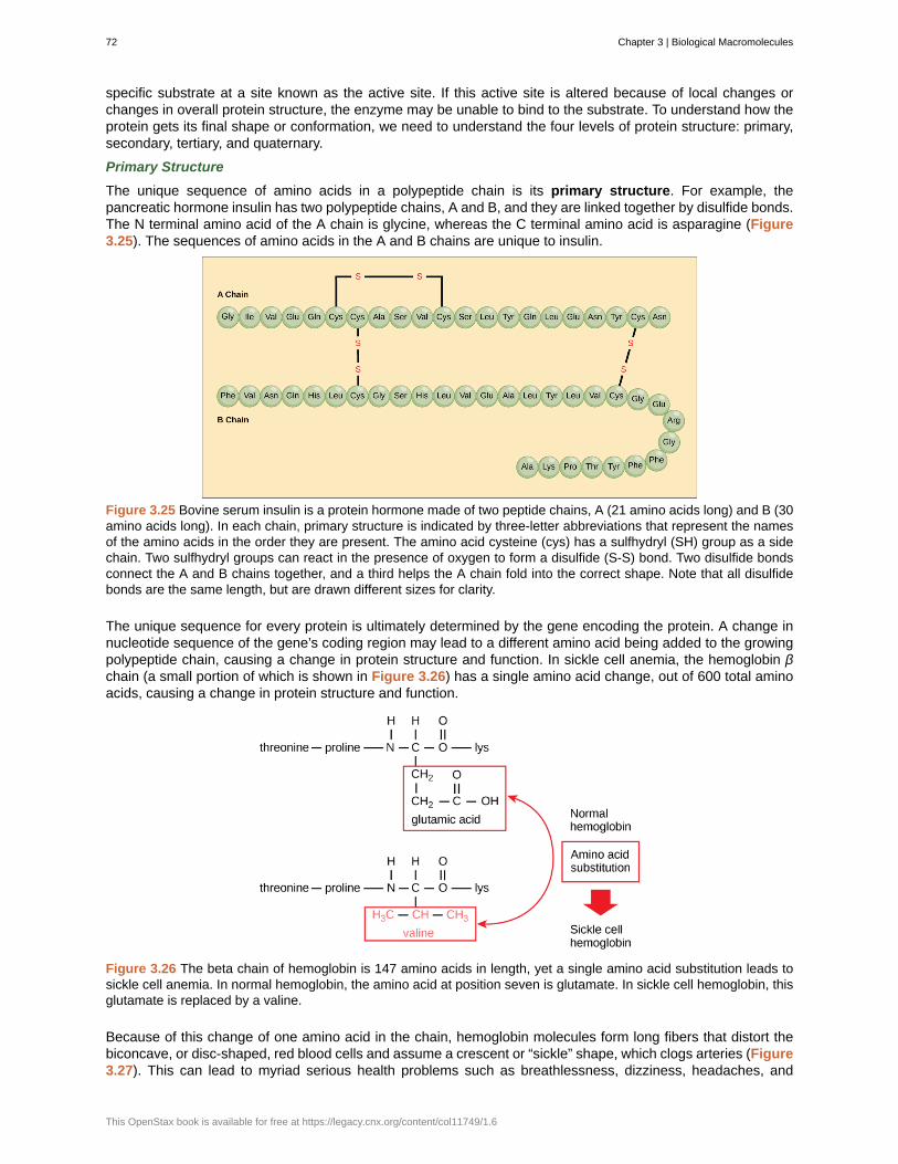
specific substrate at a site known as the active site. If this active site is altered because of local changes orchanges in overall protein structure, the enzyme may be unable to bind to the substrate. To understand how theprotein gets its final shape or conformation, we need to understand the four levels of protein structure: primary,secondary, tertiary, and quaternary.
Primary Structure
The unique sequence of amino acids in a polypeptide chain is its primary structure. For example, thepancreatic hormone insulin has two polypeptide chains, A and B, and they are linked together by disulfide bonds.The N terminal amino acid of the A chain is glycine, whereas the C terminal amino acid is asparagine (Figure3.25). The sequences of amino acids in the A and B chains are unique to insulin.
Figure 3.25 Bovine serum insulin is a protein hormone made of two peptide chains, A (21 amino acids long) and B (30amino acids long). In each chain, primary structure is indicated by three-letter abbreviations that represent the namesof the amino acids in the order they are present. The amino acid cysteine (cys) has a sulfhydryl (SH) group as a sidechain. Two sulfhydryl groups can react in the presence of oxygen to form a disulfide (S-S) bond. Two disulfide bondsconnect the A and B chains together, and a third helps the A chain fold into the correct shape. Note that all disulfidebonds are the same length, but are drawn different sizes for clarity.
The unique sequence for every protein is ultimately determined by the gene encoding the protein. A change innucleotide sequence of the gene’s coding region may lead to a different amino acid being added to the growingpolypeptide chain, causing a change in protein structure and function. In sickle cell anemia, the hemoglobin βchain (a small portion of which is shown in Figure 3.26) has a single amino acid change, out of 600 total aminoacids, causing a change in protein structure and function.
Figure 3.26 The beta chain of hemoglobin is 147 amino acids in length, yet a single amino acid substitution leads tosickle cell anemia. In normal hemoglobin, the amino acid at position seven is glutamate. In sickle cell hemoglobin, thisglutamate is replaced by a valine.
Because of this change of one amino acid in the chain, hemoglobin molecules form long fibers that distort thebiconcave, or disc-shaped, red blood cells and assume a crescent or “sickle” shape, which clogs arteries (Figure3.27). This can lead to myriad serious health problems such as breathlessness, dizziness, headaches, and
72 Chapter 3 | Biological Macromolecules
This OpenStax book is available for free at https://legacy.cnx.org/content/col11749/1.6
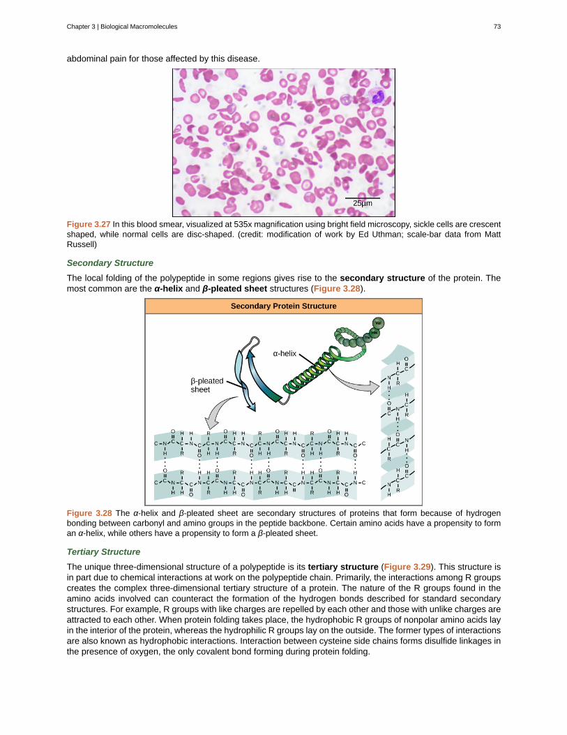
abdominal pain for those affected by this disease.
Figure 3.27 In this blood smear, visualized at 535x magnification using bright field microscopy, sickle cells are crescentshaped, while normal cells are disc-shaped. (credit: modification of work by Ed Uthman; scale-bar data from MattRussell)
Secondary Structure
The local folding of the polypeptide in some regions gives rise to the secondary structure of the protein. Themost common are the α-helix and β-pleated sheet structures (Figure 3.28).
Figure 3.28 The α-helix and β-pleated sheet are secondary structures of proteins that form because of hydrogenbonding between carbonyl and amino groups in the peptide backbone. Certain amino acids have a propensity to forman α-helix, while others have a propensity to form a β-pleated sheet.
Tertiary Structure
The unique three-dimensional structure of a polypeptide is its tertiary structure (Figure 3.29). This structure isin part due to chemical interactions at work on the polypeptide chain. Primarily, the interactions among R groupscreates the complex three-dimensional tertiary structure of a protein. The nature of the R groups found in theamino acids involved can counteract the formation of the hydrogen bonds described for standard secondarystructures. For example, R groups with like charges are repelled by each other and those with unlike charges areattracted to each other. When protein folding takes place, the hydrophobic R groups of nonpolar amino acids layin the interior of the protein, whereas the hydrophilic R groups lay on the outside. The former types of interactionsare also known as hydrophobic interactions. Interaction between cysteine side chains forms disulfide linkages inthe presence of oxygen, the only covalent bond forming during protein folding.
Chapter 3 | Biological Macromolecules 73
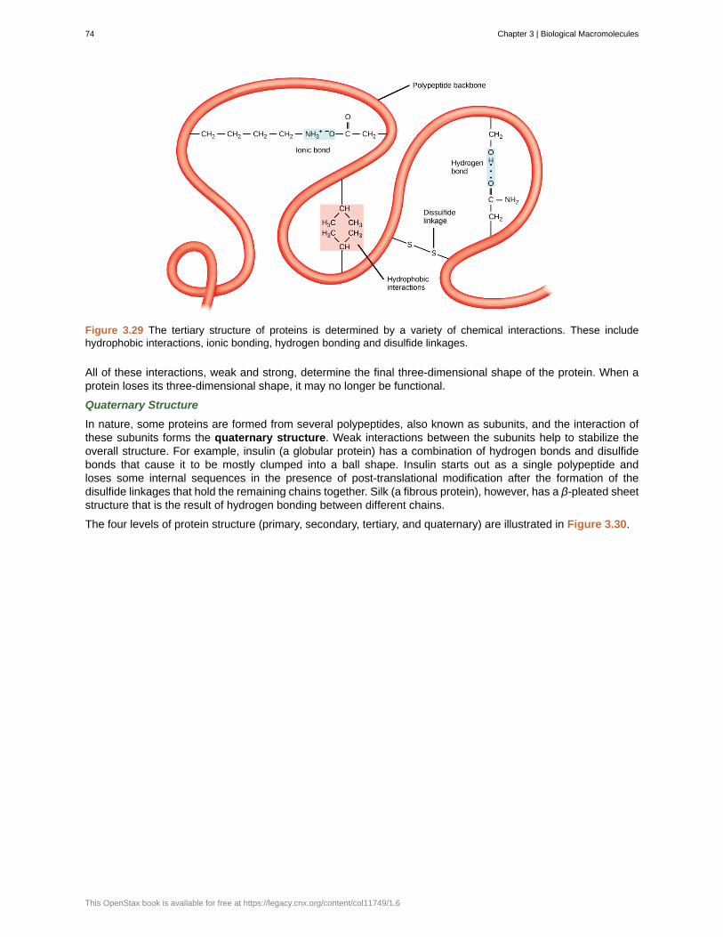
Figure 3.29 The tertiary structure of proteins is determined by a variety of chemical interactions. These includehydrophobic interactions, ionic bonding, hydrogen bonding and disulfide linkages.
All of these interactions, weak and strong, determine the final three-dimensional shape of the protein. When aprotein loses its three-dimensional shape, it may no longer be functional.
Quaternary Structure
In nature, some proteins are formed from several polypeptides, also known as subunits, and the interaction ofthese subunits forms the quaternary structure. Weak interactions between the subunits help to stabilize theoverall structure. For example, insulin (a globular protein) has a combination of hydrogen bonds and disulfidebonds that cause it to be mostly clumped into a ball shape. Insulin starts out as a single polypeptide andloses some internal sequences in the presence of post-translational modification after the formation of thedisulfide linkages that hold the remaining chains together. Silk (a fibrous protein), however, has a β-pleated sheetstructure that is the result of hydrogen bonding between different chains.
The four levels of protein structure (primary, secondary, tertiary, and quaternary) are illustrated in Figure 3.30.
74 Chapter 3 | Biological Macromolecules
This OpenStax book is available for free at https://legacy.cnx.org/content/col11749/1.6
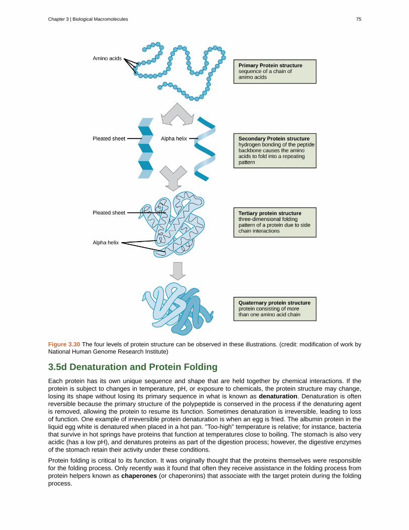
Figure 3.30 The four levels of protein structure can be observed in these illustrations. (credit: modification of work byNational Human Genome Research Institute)
3.5d Denaturation and Protein Folding
Each protein has its own unique sequence and shape that are held together by chemical interactions. If theprotein is subject to changes in temperature, pH, or exposure to chemicals, the protein structure may change,losing its shape without losing its primary sequence in what is known as denaturation. Denaturation is oftenreversible because the primary structure of the polypeptide is conserved in the process if the denaturing agentis removed, allowing the protein to resume its function. Sometimes denaturation is irreversible, leading to lossof function. One example of irreversible protein denaturation is when an egg is fried. The albumin protein in theliquid egg white is denatured when placed in a hot pan. "Too-high" temperature is relative; for instance, bacteriathat survive in hot springs have proteins that function at temperatures close to boiling. The stomach is also veryacidic (has a low pH), and denatures proteins as part of the digestion process; however, the digestive enzymesof the stomach retain their activity under these conditions.
Protein folding is critical to its function. It was originally thought that the proteins themselves were responsiblefor the folding process. Only recently was it found that often they receive assistance in the folding process fromprotein helpers known as chaperones (or chaperonins) that associate with the target protein during the foldingprocess.
Chapter 3 | Biological Macromolecules 75
For an additional perspective on proteins, view this animation (http://openstaxcollege.org/l/proteins) called“Biomolecules: The Proteins.”
3.6 | Nucleic Acids
By the end of this section, you will be able to:
• Describe the structure of nucleic acids and define the two types of nucleic acids
• Explain the structure and role of DNA
• Explain the structure and roles of RNA
Nucleic acids are the most important macromolecules for the continuity of life. They carry the genetic blueprintof a cell and carry instructions for the functioning of the cell.
3.6a DNA and RNA
The two main types of nucleic acids are deoxyribonucleic acid (DNA) and ribonucleic acid (RNA) . DNA isthe genetic material found in all living organisms, ranging from single-celled bacteria to multicellular mammals. Itis found in the nucleus of eukaryotes and in the organelles, chloroplasts, and mitochondria. In prokaryotes, theDNA is not enclosed in a membranous envelope.
The entire genetic content of a cell is known as its genome, and the study of genomes is genomics. In eukaryoticcells but not in prokaryotes, DNA forms a complex with histone proteins to form chromatin, the substance ofeukaryotic chromosomes. A chromosome may contain tens of thousands of genes. Many genes contain theinformation to make protein products; other genes code for RNA products.
The other type of nucleic acid, RNA, is mostly involved in protein synthesis. The DNA molecules never leavethe nucleus but instead use an intermediary to communicate with the rest of the cell. This intermediary is themessenger RNA (mRNA) . Other types of RNA are involved in protein synthesis and its regulation.
DNA and RNA are made up of monomers known as nucleotides . The nucleotides combine with each otherto form a nucleic acid, DNA or RNA. Each nucleotide is made up of three components: a nitrogenous base, apentose (five-carbon) sugar, and a phosphate group ( Figure 3.31). Each nitrogenous base in a nucleotide isattached to a sugar molecule, which is attached to one or more phosphate groups.
76 Chapter 3 | Biological Macromolecules
This OpenStax book is available for free at https://legacy.cnx.org/content/col11749/1.6
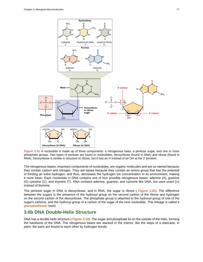
Figure 3.31 A nucleotide is made up of three components: a nitrogenous base, a pentose sugar, and one or morephosphate groups. Two types of pentose are found in nucleotides, deoxyribose (found in DNA) and ribose (found inRNA). Deoxyribose is similar in structure to ribose, but it has an H instead of an OH at the 2′ position.
The nitrogenous bases, important components of nucleotides, are organic molecules and are so named becausethey contain carbon and nitrogen. They are bases because they contain an amino group that has the potentialof binding an extra hydrogen, and thus, decreases the hydrogen ion concentration in its environment, makingit more basic. Each nucleotide in DNA contains one of four possible nitrogenous bases: adenine (A), guanine(G) cytosine (C), and thymine (T). RNA contains adenine, guanine, and cytosine like DNA, but uses uracil (U)instead of thymine.
The pentose sugar in DNA is deoxyribose, and in RNA, the sugar is ribose ( Figure 3.31). The differencebetween the sugars is the presence of the hydroxyl group on the second carbon of the ribose and hydrogenon the second carbon of the deoxyribose. The phosphate group is attached to the hydroxyl group of one of thesugar's carbons, and the hydroxyl group of a carbon of the sugar of the next nucleotide. This linkage is called aphosphodiester bond.
3.6b DNA Double-Helix Structure
DNA has a double-helix structure ( Figure 3.32). The sugar and phosphate lie on the outside of the helix, formingthe backbone of the DNA. The nitrogenous bases are stacked in the interior, like the steps of a staircase, inpairs; the pairs are bound to each other by hydrogen bonds.
Chapter 3 | Biological Macromolecules 77
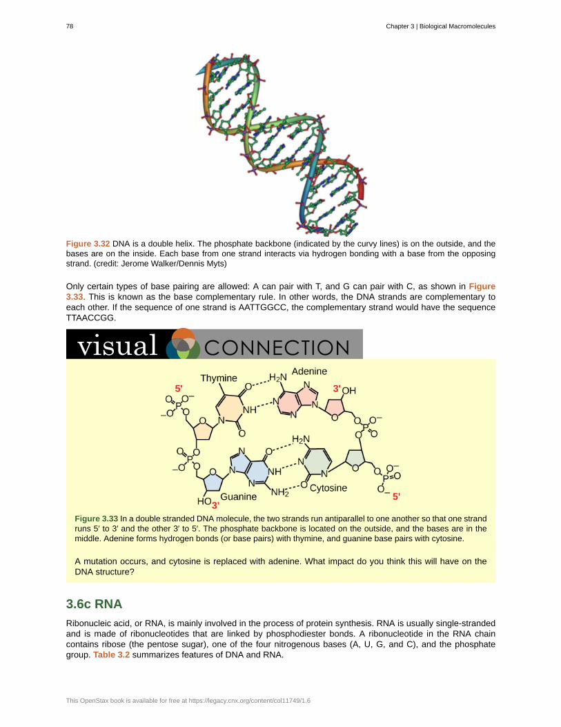
Figure 3.32 DNA is a double helix. The phosphate backbone (indicated by the curvy lines) is on the outside, and thebases are on the inside. Each base from one strand interacts via hydrogen bonding with a base from the opposingstrand. (credit: Jerome Walker/Dennis Myts)
Only certain types of base pairing are allowed: A can pair with T, and G can pair with C, as shown in Figure3.33. This is known as the base complementary rule. In other words, the DNA strands are complementary toeach other. If the sequence of one strand is AATTGGCC, the complementary strand would have the sequenceTTAACCGG.
Figure 3.33 In a double stranded DNA molecule, the two strands run antiparallel to one another so that one strandruns 5′ to 3′ and the other 3′ to 5′. The phosphate backbone is located on the outside, and the bases are in themiddle. Adenine forms hydrogen bonds (or base pairs) with thymine, and guanine base pairs with cytosine.
A mutation occurs, and cytosine is replaced with adenine. What impact do you think this will have on theDNA structure?
3.6c RNA
Ribonucleic acid, or RNA, is mainly involved in the process of protein synthesis. RNA is usually single-strandedand is made of ribonucleotides that are linked by phosphodiester bonds. A ribonucleotide in the RNA chaincontains ribose (the pentose sugar), one of the four nitrogenous bases (A, U, G, and C), and the phosphategroup. Table 3.2 summarizes features of DNA and RNA.
78 Chapter 3 | Biological Macromolecules
This OpenStax book is available for free at https://legacy.cnx.org/content/col11749/1.6
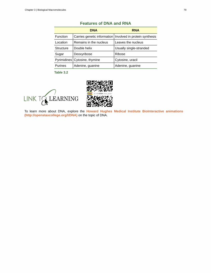
Features of DNA and RNA
DNA RNA
Function Carries genetic information Involved in protein synthesis
Location Remains in the nucleus Leaves the nucleus
Structure Double helix Usually single-stranded
Sugar Deoxyribose Ribose
Pyrimidines Cytosine, thymine Cytosine, uracil
Purines Adenine, guanine Adenine, guanine
Table 3.2
To learn more about DNA, explore the Howard Hughes Medical Institute BioInteractive animations(http://openstaxcollege.org/l/DNA) on the topic of DNA.
Chapter 3 | Biological Macromolecules 79
alpha-helix structure (α-helix)
amino acid
beta-pleated sheet (β-pleated)
biological macromolecules
Carbohydrates
cellulose
chaperone
chitin
dehydration synthesis
denaturation
deoxyribonucleic acid (DNA)
Disaccharides
enzyme
Functional groups
glycogen
hormone
Hydrocarbons
hydrolysis
Lipids
messenger RNA (mRNA)
monomers
Monosaccharides
Nucleic acids
nucleotides
organic molecules
peptide bond
KEY TERMS
type of secondary structure of proteins formed by folding of the polypeptide intoa helix shape with hydrogen bonds stabilizing the structure
monomer of a protein; has a central carbon or alpha carbon to which an amino group, a carboxylgroup, a hydrogen, and an R group or side chain is attached; the R group is different for all 20 amino acids
secondary structure found in proteins in which “pleats” are formed by hydrogenbonding between atoms on the backbone of the polypeptide chain
large molecule necessary for life that is built from smaller organic molecules
biological macromolecule in which the ratio of carbon to hydrogen to oxygen is 1:2:1;carbohydrates serve as energy sources and structural support in cells and form the cellular exoskeleton ofarthropods
polysaccharide that makes up the cell wall of plants; provides structural support to the cell
(also, chaperonin) protein that helps nascent protein in the folding process
type of carbohydrate that forms the outer skeleton of all arthropods that include crustaceans and insects;it also forms the cell walls of fungi
(also, condensation) reaction that links monomer molecules together, releasing amolecule of water for each bond formed
loss of shape in a protein as a result of changes in temperature, pH, or exposure to chemicals
double-helical molecule that carries the hereditary information of the cell
two sugar monomers linked together by a glycosidic bond
catalyst in a biochemical reaction that is usually a complex or conjugated protein
group of atoms that provides or imparts a specific function to a carbon skeleton
storage carbohydrate in animals
chemical signaling molecule, usually protein or steroid, secreted by endocrine cells that act to controlor regulate specific physiological processes
molecule that consists only of carbon and hydrogen
reaction causes breakdown of larger molecules into smaller molecules with the utilization of water
macromolecule that is nonpolar and insoluble in water
RNA that carries information from DNA to ribosomes during protein synthesis
smallest unit of larger molecules called polymers
single unit or monomer of carbohydrates
biological macromolecule that carries the genetic blueprint of a cell and carries instructions forthe functioning of the cell
monomer of nucleic acids; contains a pentose sugar, one or more phosphate groups, and anitrogenous base
any molecule containing carbon (except carbon dioxide)
bond formed between two amino acids by a dehydration reaction
80 Chapter 3 | Biological Macromolecules
This OpenStax book is available for free at https://legacy.cnx.org/content/col11749/1.6
phosphodiester
Phospholipids
polymers
polypeptide
polysaccharide
primary structure
protein
quaternary structure
ribonucleic acid (RNA)
saturated fatty acid
secondary structure
starch
steroids
substituted hydrocarbons
tertiary structure
trans fat
triacylglycerols
triglycerides
unsaturated
wax
linkage covalent chemical bond that holds together the polynucleotide chains with aphosphate group linking two pentose sugars of neighboring nucleotides
major component of membranes; composed of two fatty acids and a phosphate-containinggroup attached to a glycerol backbone
chain of monomer residues that is linked by covalent bonds; polymerization is the process of polymerformation from monomers by condensation
long chain of amino acids linked by peptide bonds
long chain of monosaccharides; may be branched or unbranched
linear sequence of amino acids in a protein
biological macromolecule composed of one or more chains of amino acids
association of discrete polypeptide subunits in a protein
single-stranded, often internally base paired, molecule that is involved in proteinsynthesis
long-chain of hydrocarbon with single covalent bonds in the carbon chain; the number ofhydrogen atoms attached to the carbon skeleton is maximized
regular structure formed by proteins by intramolecular hydrogen bonding between theoxygen atom of one amino acid residue and the hydrogen attached to the nitrogen atom of another aminoacid residue
storage carbohydrate in plants
type of lipid composed of four fused hydrocarbon rings forming a planar structure
hydrocarbon chain or ring containing an atom of another element in place of one ofthe backbone chains
three-dimensional conformation of a protein, including interactions between secondarystructural elements; formed from interactions between amino acid side chains
fat formed artificially by hydrogenating oils, leading to a different arrangement of double bond(s) thanthose found in naturally occurring lipids
fat molecule; consists of three fatty acids linked to a glycerol molecule
another name for triacylglycerol
molecule with one or more double bonds
lipid made of a long-chain fatty acid that is esterified to a long-chain alcohol; serves as a protective coatingon some feathers, aquatic mammal fur, and leaves
CHAPTER SUMMARY
3.1 Carbon
The unique properties of carbon make it a central part of biological molecules. Carbon binds to oxygen,hydrogen, and nitrogen covalently to form the many molecules important for cellular function. Carbon has fourelectrons in its outermost shell and can form four bonds. Carbon and hydrogen can form hydrocarbon chains orrings. Functional groups are groups of atoms that confer specific properties to hydrocarbon (or substitutedhydrocarbon) chains or rings that define their overall chemical characteristics and function.
Chapter 3 | Biological Macromolecules 81
3.2 Synthesis of Biological Macromolecules
Proteins, carbohydrates, nucleic acids, and lipids are the four major classes of biologicalmacromolecules—large molecules necessary for life that are built from smaller organic molecules.Macromolecules are made up of single units known as monomers that are joined by covalent bonds to formlarger polymers. The polymer is more than the sum of its parts: it acquires new characteristics, and leads to anosmotic pressure that is much lower than that formed by its ingredients; this is an important advantage in themaintenance of cellular osmotic conditions. A monomer joins with another monomer with the release of a watermolecule, leading to the formation of a covalent bond. These types of reactions are known as dehydration orcondensation reactions. When polymers are broken down into smaller units (monomers), a molecule of water isused for each bond broken by these reactions; such reactions are known as hydrolysis reactions. Dehydrationand hydrolysis reactions are similar for all macromolecules, but each monomer and polymer reaction is specificto its class. Dehydration reactions typically require an investment of energy for new bond formation, whilehydrolysis reactions typically release energy by breaking bonds.
3.3 Carbohydrates
Carbohydrates are a group of macromolecules that are a vital energy source for the cell and provide structuralsupport to plant cells, fungi, and all of the arthropods that include lobsters, crabs, shrimp, insects, and spiders.Carbohydrates are classified as monosaccharides, disaccharides, and polysaccharides depending on thenumber of monomers in the molecule. Monosaccharides are linked by glycosidic bonds that are formed as aresult of dehydration reactions, forming disaccharides and polysaccharides with the elimination of a watermolecule for each bond formed. Glucose, galactose, and fructose are common monosaccharides, whereascommon disaccharides include lactose, maltose, and sucrose. Starch and glycogen, examples ofpolysaccharides, are the storage forms of glucose in plants and animals, respectively. The long polysaccharidechains may be branched or unbranched. Cellulose is an example of an unbranched polysaccharide, whereasamylopectin, a constituent of starch, is a highly branched molecule. Storage of glucose, in the form of polymerslike starch of glycogen, makes it slightly less accessible for metabolism; however, this prevents it from leakingout of the cell or creating a high osmotic pressure that could cause excessive water uptake by the cell.
3.4 Lipids
Lipids are a class of macromolecules that are nonpolar and hydrophobic in nature. Major types include fats andoils, waxes, phospholipids, and steroids. Fats are a stored form of energy and are also known astriacylglycerols or triglycerides. Fats are made up of fatty acids and either glycerol or sphingosine. Fatty acidsmay be unsaturated or saturated, depending on the presence or absence of double bonds in the hydrocarbonchain. If only single bonds are present, they are known as saturated fatty acids. Unsaturated fatty acids mayhave one or more double bonds in the hydrocarbon chain. Phospholipids make up the matrix of membranes.They have a glycerol or sphingosine backbone to which two fatty acid chains and a phosphate-containinggroup are attached. Steroids are another class of lipids. Their basic structure has four fused carbon rings.Cholesterol is a type of steroid and is an important constituent of the plasma membrane, where it helps tomaintain the fluid nature of the membrane. It is also the precursor of steroid hormones such as testosterone.
3.5 Proteins
Proteins are a class of macromolecules that perform a diverse range of functions for the cell. They help inmetabolism by providing structural support and by acting as enzymes, carriers, or hormones. The buildingblocks of proteins (monomers) are amino acids. Each amino acid has a central carbon that is linked to anamino group, a carboxyl group, a hydrogen atom, and an R group or side chain. There are 20 commonlyoccurring amino acids, each of which differs in the R group. Each amino acid is linked to its neighbors by apeptide bond. A long chain of amino acids is known as a polypeptide.
Proteins are organized at four levels: primary, secondary, tertiary, and (optional) quaternary. The primarystructure is the unique sequence of amino acids. The local folding of the polypeptide to form structures such asthe α helix and β-pleated sheet constitutes the secondary structure. The overall three-dimensional structure isthe tertiary structure. When two or more polypeptides combine to form the complete protein structure, theconfiguration is known as the quaternary structure of a protein. Protein shape and function are intricately linked;any change in shape caused by changes in temperature or pH may lead to protein denaturation and a loss infunction.
82 Chapter 3 | Biological Macromolecules
This OpenStax book is available for free at https://legacy.cnx.org/content/col11749/1.6
3.6 Nucleic Acids
Nucleic acids are molecules made up of nucleotides that direct cellular activities such as cell division andprotein synthesis. Each nucleotide is made up of a pentose sugar, a nitrogenous base, and a phosphate group.There are two types of nucleic acids: DNA and RNA. DNA carries the genetic blueprint of the cell and is passedon from parents to offspring (in the form of chromosomes). It has a double-helical structure with the two strandsrunning in opposite directions, connected by hydrogen bonds, and complementary to each other. RNA is single-stranded and is made of a pentose sugar (ribose), a nitrogenous base, and a phosphate group. RNA isinvolved in protein synthesis and its regulation. Messenger RNA (mRNA) is copied from the DNA, is exportedfrom the nucleus to the cytoplasm, and contains information for the construction of proteins. Ribosomal RNA(rRNA) is a part of the ribosomes at the site of protein synthesis, whereas transfer RNA (tRNA) carries theamino acid to the site of protein synthesis. microRNA regulates the use of mRNA for protein synthesis.
Chapter 3 | Biological Macromolecules 83

84 Chapter 3 | Biological Macromolecules
This OpenStax book is available for free at https://legacy.cnx.org/content/col11749/1.6
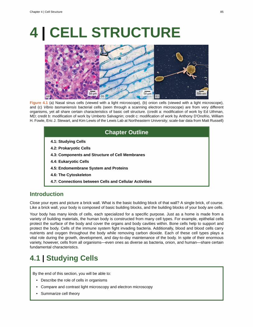
4 | CELL STRUCTURE
Figure 4.1 (a) Nasal sinus cells (viewed with a light microscope), (b) onion cells (viewed with a light microscope),and (c) Vibrio tasmaniensis bacterial cells (seen through a scanning electron microscope) are from very differentorganisms, yet all share certain characteristics of basic cell structure. (credit a: modification of work by Ed Uthman,MD; credit b: modification of work by Umberto Salvagnin; credit c: modification of work by Anthony D'Onofrio, WilliamH. Fowle, Eric J. Stewart, and Kim Lewis of the Lewis Lab at Northeastern University; scale-bar data from Matt Russell)
Chapter Outline
4.1: Studying Cells
4.2: Prokaryotic Cells
4.3: Components and Structure of Cell Membranes
4.4: Eukaryotic Cells
4.5: Endomembrane System and Proteins
4.6: The Cytoskeleton
4.7: Connections between Cells and Cellular Activities
Introduction
Close your eyes and picture a brick wall. What is the basic building block of that wall? A single brick, of course.Like a brick wall, your body is composed of basic building blocks, and the building blocks of your body are cells.
Your body has many kinds of cells, each specialized for a specific purpose. Just as a home is made from avariety of building materials, the human body is constructed from many cell types. For example, epithelial cellsprotect the surface of the body and cover the organs and body cavities within. Bone cells help to support andprotect the body. Cells of the immune system fight invading bacteria. Additionally, blood and blood cells carrynutrients and oxygen throughout the body while removing carbon dioxide. Each of these cell types plays avital role during the growth, development, and day-to-day maintenance of the body. In spite of their enormousvariety, however, cells from all organisms—even ones as diverse as bacteria, onion, and human—share certainfundamental characteristics.
4.1 | Studying Cells
By the end of this section, you will be able to:
• Describe the role of cells in organisms
• Compare and contrast light microscopy and electron microscopy
• Summarize cell theory
Chapter 4 | Cell Structure 85
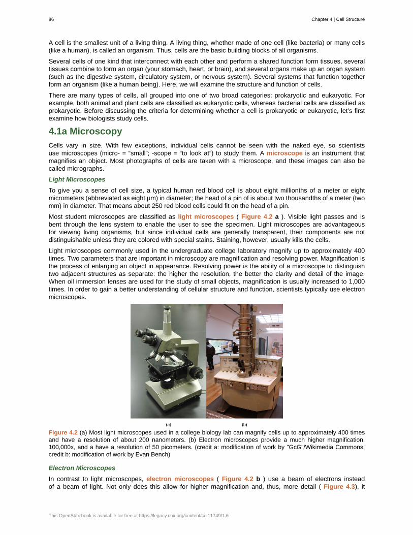
A cell is the smallest unit of a living thing. A living thing, whether made of one cell (like bacteria) or many cells(like a human), is called an organism. Thus, cells are the basic building blocks of all organisms.
Several cells of one kind that interconnect with each other and perform a shared function form tissues, severaltissues combine to form an organ (your stomach, heart, or brain), and several organs make up an organ system(such as the digestive system, circulatory system, or nervous system). Several systems that function togetherform an organism (like a human being). Here, we will examine the structure and function of cells.
There are many types of cells, all grouped into one of two broad categories: prokaryotic and eukaryotic. Forexample, both animal and plant cells are classified as eukaryotic cells, whereas bacterial cells are classified asprokaryotic. Before discussing the criteria for determining whether a cell is prokaryotic or eukaryotic, let’s firstexamine how biologists study cells.
4.1a Microscopy
Cells vary in size. With few exceptions, individual cells cannot be seen with the naked eye, so scientistsuse microscopes (micro- = “small”; -scope = “to look at”) to study them. A microscope is an instrument thatmagnifies an object. Most photographs of cells are taken with a microscope, and these images can also becalled micrographs.
Light Microscopes
To give you a sense of cell size, a typical human red blood cell is about eight millionths of a meter or eightmicrometers (abbreviated as eight μm) in diameter; the head of a pin of is about two thousandths of a meter (twomm) in diameter. That means about 250 red blood cells could fit on the head of a pin.
Most student microscopes are classified as light microscopes ( Figure 4.2 a ). Visible light passes and isbent through the lens system to enable the user to see the specimen. Light microscopes are advantageousfor viewing living organisms, but since individual cells are generally transparent, their components are notdistinguishable unless they are colored with special stains. Staining, however, usually kills the cells.
Light microscopes commonly used in the undergraduate college laboratory magnify up to approximately 400times. Two parameters that are important in microscopy are magnification and resolving power. Magnification isthe process of enlarging an object in appearance. Resolving power is the ability of a microscope to distinguishtwo adjacent structures as separate: the higher the resolution, the better the clarity and detail of the image.When oil immersion lenses are used for the study of small objects, magnification is usually increased to 1,000times. In order to gain a better understanding of cellular structure and function, scientists typically use electronmicroscopes.
Figure 4.2 (a) Most light microscopes used in a college biology lab can magnify cells up to approximately 400 timesand have a resolution of about 200 nanometers. (b) Electron microscopes provide a much higher magnification,100,000x, and a have a resolution of 50 picometers. (credit a: modification of work by "GcG"/Wikimedia Commons;credit b: modification of work by Evan Bench)
Electron Microscopes
In contrast to light microscopes, electron microscopes ( Figure 4.2 b ) use a beam of electrons insteadof a beam of light. Not only does this allow for higher magnification and, thus, more detail ( Figure 4.3), it
86 Chapter 4 | Cell Structure
This OpenStax book is available for free at https://legacy.cnx.org/content/col11749/1.6
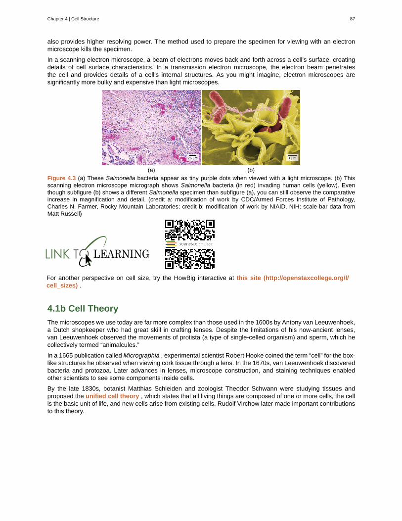
also provides higher resolving power. The method used to prepare the specimen for viewing with an electronmicroscope kills the specimen.
In a scanning electron microscope, a beam of electrons moves back and forth across a cell’s surface, creatingdetails of cell surface characteristics. In a transmission electron microscope, the electron beam penetratesthe cell and provides details of a cell’s internal structures. As you might imagine, electron microscopes aresignificantly more bulky and expensive than light microscopes.
(a) (b)Figure 4.3 (a) These Salmonella bacteria appear as tiny purple dots when viewed with a light microscope. (b) Thisscanning electron microscope micrograph shows Salmonella bacteria (in red) invading human cells (yellow). Eventhough subfigure (b) shows a different Salmonella specimen than subfigure (a), you can still observe the comparativeincrease in magnification and detail. (credit a: modification of work by CDC/Armed Forces Institute of Pathology,Charles N. Farmer, Rocky Mountain Laboratories; credit b: modification of work by NIAID, NIH; scale-bar data fromMatt Russell)
For another perspective on cell size, try the HowBig interactive at this site (http://openstaxcollege.org/l/cell_sizes) .
4.1b Cell Theory
The microscopes we use today are far more complex than those used in the 1600s by Antony van Leeuwenhoek,a Dutch shopkeeper who had great skill in crafting lenses. Despite the limitations of his now-ancient lenses,van Leeuwenhoek observed the movements of protista (a type of single-celled organism) and sperm, which hecollectively termed “animalcules.”
In a 1665 publication called Micrographia , experimental scientist Robert Hooke coined the term “cell” for the box-like structures he observed when viewing cork tissue through a lens. In the 1670s, van Leeuwenhoek discoveredbacteria and protozoa. Later advances in lenses, microscope construction, and staining techniques enabledother scientists to see some components inside cells.
By the late 1830s, botanist Matthias Schleiden and zoologist Theodor Schwann were studying tissues andproposed the unified cell theory , which states that all living things are composed of one or more cells, the cellis the basic unit of life, and new cells arise from existing cells. Rudolf Virchow later made important contributionsto this theory.
Chapter 4 | Cell Structure 87
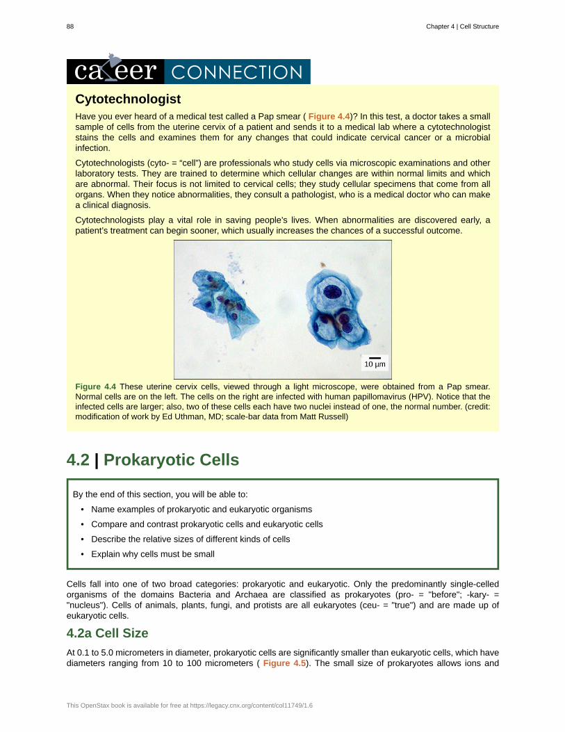
CytotechnologistHave you ever heard of a medical test called a Pap smear ( Figure 4.4)? In this test, a doctor takes a smallsample of cells from the uterine cervix of a patient and sends it to a medical lab where a cytotechnologiststains the cells and examines them for any changes that could indicate cervical cancer or a microbialinfection.
Cytotechnologists (cyto- = “cell”) are professionals who study cells via microscopic examinations and otherlaboratory tests. They are trained to determine which cellular changes are within normal limits and whichare abnormal. Their focus is not limited to cervical cells; they study cellular specimens that come from allorgans. When they notice abnormalities, they consult a pathologist, who is a medical doctor who can makea clinical diagnosis.
Cytotechnologists play a vital role in saving people’s lives. When abnormalities are discovered early, apatient’s treatment can begin sooner, which usually increases the chances of a successful outcome.
Figure 4.4 These uterine cervix cells, viewed through a light microscope, were obtained from a Pap smear.Normal cells are on the left. The cells on the right are infected with human papillomavirus (HPV). Notice that theinfected cells are larger; also, two of these cells each have two nuclei instead of one, the normal number. (credit:modification of work by Ed Uthman, MD; scale-bar data from Matt Russell)
4.2 | Prokaryotic Cells
By the end of this section, you will be able to:
• Name examples of prokaryotic and eukaryotic organisms
• Compare and contrast prokaryotic cells and eukaryotic cells
• Describe the relative sizes of different kinds of cells
• Explain why cells must be small
Cells fall into one of two broad categories: prokaryotic and eukaryotic. Only the predominantly single-celledorganisms of the domains Bacteria and Archaea are classified as prokaryotes (pro- = "before"; -kary- ="nucleus"). Cells of animals, plants, fungi, and protists are all eukaryotes (ceu- = "true") and are made up ofeukaryotic cells.
4.2a Cell Size
At 0.1 to 5.0 micrometers in diameter, prokaryotic cells are significantly smaller than eukaryotic cells, which havediameters ranging from 10 to 100 micrometers ( Figure 4.5). The small size of prokaryotes allows ions and
88 Chapter 4 | Cell Structure
This OpenStax book is available for free at https://legacy.cnx.org/content/col11749/1.6
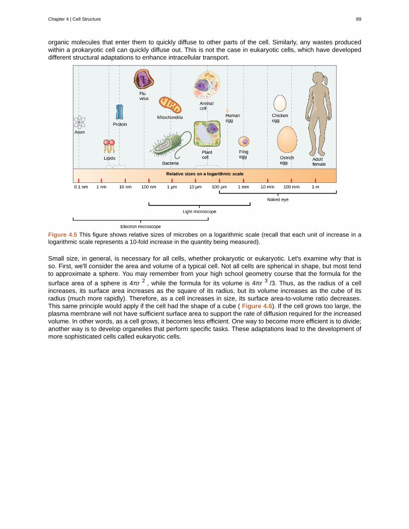
organic molecules that enter them to quickly diffuse to other parts of the cell. Similarly, any wastes producedwithin a prokaryotic cell can quickly diffuse out. This is not the case in eukaryotic cells, which have developeddifferent structural adaptations to enhance intracellular transport.
Figure 4.5 This figure shows relative sizes of microbes on a logarithmic scale (recall that each unit of increase in alogarithmic scale represents a 10-fold increase in the quantity being measured).
Small size, in general, is necessary for all cells, whether prokaryotic or eukaryotic. Let's examine why that isso. First, we'll consider the area and volume of a typical cell. Not all cells are spherical in shape, but most tendto approximate a sphere. You may remember from your high school geometry course that the formula for the
surface area of a sphere is 4πr 2 , while the formula for its volume is 4πr 3 /3. Thus, as the radius of a cellincreases, its surface area increases as the square of its radius, but its volume increases as the cube of itsradius (much more rapidly). Therefore, as a cell increases in size, its surface area-to-volume ratio decreases.This same principle would apply if the cell had the shape of a cube ( Figure 4.6). If the cell grows too large, theplasma membrane will not have sufficient surface area to support the rate of diffusion required for the increasedvolume. In other words, as a cell grows, it becomes less efficient. One way to become more efficient is to divide;another way is to develop organelles that perform specific tasks. These adaptations lead to the development ofmore sophisticated cells called eukaryotic cells.
Chapter 4 | Cell Structure 89
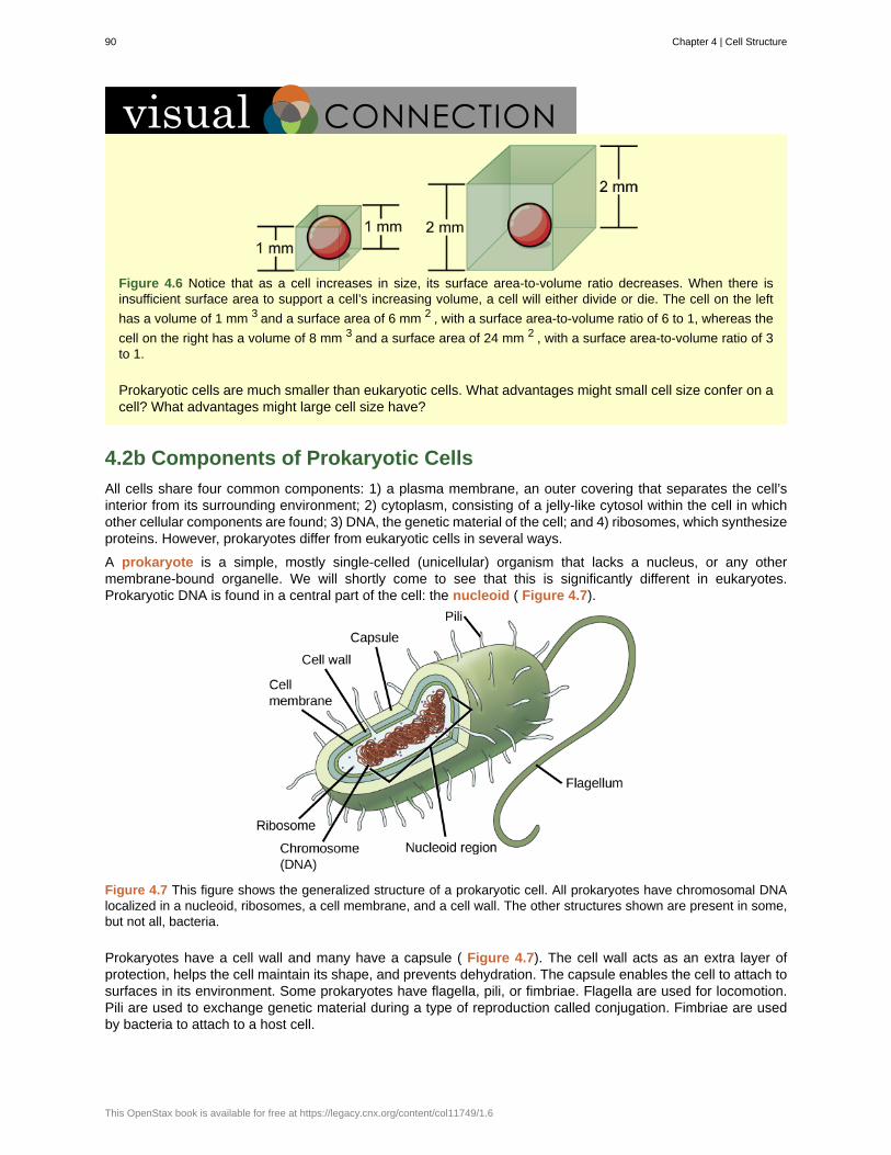
Figure 4.6 Notice that as a cell increases in size, its surface area-to-volume ratio decreases. When there isinsufficient surface area to support a cell’s increasing volume, a cell will either divide or die. The cell on the left
has a volume of 1 mm 3 and a surface area of 6 mm 2 , with a surface area-to-volume ratio of 6 to 1, whereas the
cell on the right has a volume of 8 mm 3 and a surface area of 24 mm 2 , with a surface area-to-volume ratio of 3to 1.
Prokaryotic cells are much smaller than eukaryotic cells. What advantages might small cell size confer on acell? What advantages might large cell size have?
4.2b Components of Prokaryotic Cells
All cells share four common components: 1) a plasma membrane, an outer covering that separates the cell’sinterior from its surrounding environment; 2) cytoplasm, consisting of a jelly-like cytosol within the cell in whichother cellular components are found; 3) DNA, the genetic material of the cell; and 4) ribosomes, which synthesizeproteins. However, prokaryotes differ from eukaryotic cells in several ways.
A prokaryote is a simple, mostly single-celled (unicellular) organism that lacks a nucleus, or any othermembrane-bound organelle. We will shortly come to see that this is significantly different in eukaryotes.Prokaryotic DNA is found in a central part of the cell: the nucleoid ( Figure 4.7).
Figure 4.7 This figure shows the generalized structure of a prokaryotic cell. All prokaryotes have chromosomal DNAlocalized in a nucleoid, ribosomes, a cell membrane, and a cell wall. The other structures shown are present in some,but not all, bacteria.
Prokaryotes have a cell wall and many have a capsule ( Figure 4.7). The cell wall acts as an extra layer ofprotection, helps the cell maintain its shape, and prevents dehydration. The capsule enables the cell to attach tosurfaces in its environment. Some prokaryotes have flagella, pili, or fimbriae. Flagella are used for locomotion.Pili are used to exchange genetic material during a type of reproduction called conjugation. Fimbriae are usedby bacteria to attach to a host cell.
90 Chapter 4 | Cell Structure
This OpenStax book is available for free at https://legacy.cnx.org/content/col11749/1.6
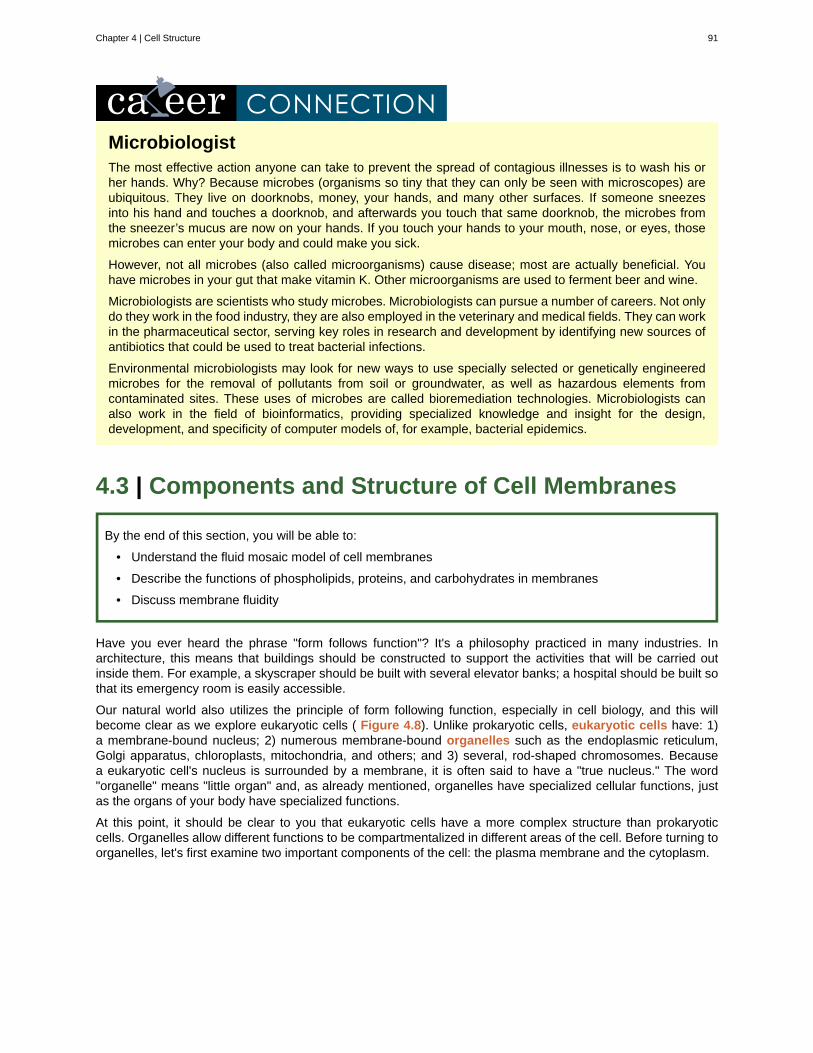
MicrobiologistThe most effective action anyone can take to prevent the spread of contagious illnesses is to wash his orher hands. Why? Because microbes (organisms so tiny that they can only be seen with microscopes) areubiquitous. They live on doorknobs, money, your hands, and many other surfaces. If someone sneezesinto his hand and touches a doorknob, and afterwards you touch that same doorknob, the microbes fromthe sneezer’s mucus are now on your hands. If you touch your hands to your mouth, nose, or eyes, thosemicrobes can enter your body and could make you sick.
However, not all microbes (also called microorganisms) cause disease; most are actually beneficial. Youhave microbes in your gut that make vitamin K. Other microorganisms are used to ferment beer and wine.
Microbiologists are scientists who study microbes. Microbiologists can pursue a number of careers. Not onlydo they work in the food industry, they are also employed in the veterinary and medical fields. They can workin the pharmaceutical sector, serving key roles in research and development by identifying new sources ofantibiotics that could be used to treat bacterial infections.
Environmental microbiologists may look for new ways to use specially selected or genetically engineeredmicrobes for the removal of pollutants from soil or groundwater, as well as hazardous elements fromcontaminated sites. These uses of microbes are called bioremediation technologies. Microbiologists canalso work in the field of bioinformatics, providing specialized knowledge and insight for the design,development, and specificity of computer models of, for example, bacterial epidemics.
4.3 | Components and Structure of Cell Membranes
By the end of this section, you will be able to:
• Understand the fluid mosaic model of cell membranes
• Describe the functions of phospholipids, proteins, and carbohydrates in membranes
• Discuss membrane fluidity
Have you ever heard the phrase "form follows function"? It's a philosophy practiced in many industries. Inarchitecture, this means that buildings should be constructed to support the activities that will be carried outinside them. For example, a skyscraper should be built with several elevator banks; a hospital should be built sothat its emergency room is easily accessible.
Our natural world also utilizes the principle of form following function, especially in cell biology, and this willbecome clear as we explore eukaryotic cells ( Figure 4.8). Unlike prokaryotic cells, eukaryotic cells have: 1)a membrane-bound nucleus; 2) numerous membrane-bound organelles such as the endoplasmic reticulum,Golgi apparatus, chloroplasts, mitochondria, and others; and 3) several, rod-shaped chromosomes. Becausea eukaryotic cell's nucleus is surrounded by a membrane, it is often said to have a "true nucleus." The word"organelle" means "little organ" and, as already mentioned, organelles have specialized cellular functions, justas the organs of your body have specialized functions.
At this point, it should be clear to you that eukaryotic cells have a more complex structure than prokaryoticcells. Organelles allow different functions to be compartmentalized in different areas of the cell. Before turning toorganelles, let's first examine two important components of the cell: the plasma membrane and the cytoplasm.
Chapter 4 | Cell Structure 91
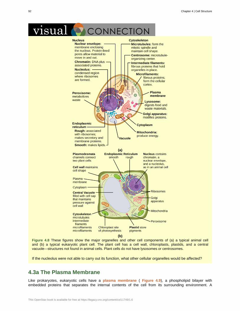
(a)
(b)Figure 4.8 These figures show the major organelles and other cell components of (a) a typical animal celland (b) a typical eukaryotic plant cell. The plant cell has a cell wall, chloroplasts, plastids, and a centralvacuole—structures not found in animal cells. Plant cells do not have lysosomes or centrosomes.
If the nucleolus were not able to carry out its function, what other cellular organelles would be affected?
4.3a The Plasma Membrane
Like prokaryotes, eukaryotic cells have a plasma membrane ( Figure 4.9), a phospholipid bilayer withembedded proteins that separates the internal contents of the cell from its surrounding environment. A
92 Chapter 4 | Cell Structure
This OpenStax book is available for free at https://legacy.cnx.org/content/col11749/1.6
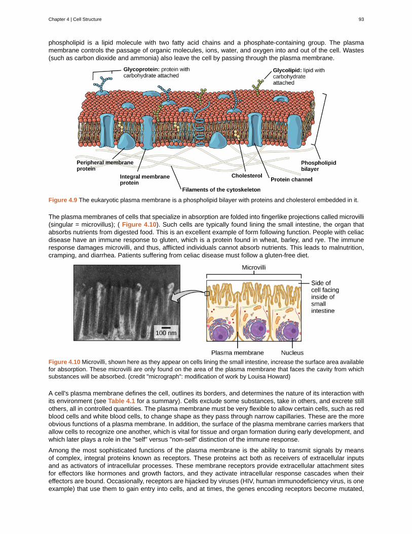
phospholipid is a lipid molecule with two fatty acid chains and a phosphate-containing group. The plasmamembrane controls the passage of organic molecules, ions, water, and oxygen into and out of the cell. Wastes(such as carbon dioxide and ammonia) also leave the cell by passing through the plasma membrane.
Figure 4.9 The eukaryotic plasma membrane is a phospholipid bilayer with proteins and cholesterol embedded in it.
The plasma membranes of cells that specialize in absorption are folded into fingerlike projections called microvilli(singular = microvillus); ( Figure 4.10). Such cells are typically found lining the small intestine, the organ thatabsorbs nutrients from digested food. This is an excellent example of form following function. People with celiacdisease have an immune response to gluten, which is a protein found in wheat, barley, and rye. The immuneresponse damages microvilli, and thus, afflicted individuals cannot absorb nutrients. This leads to malnutrition,cramping, and diarrhea. Patients suffering from celiac disease must follow a gluten-free diet.
Figure 4.10 Microvilli, shown here as they appear on cells lining the small intestine, increase the surface area availablefor absorption. These microvilli are only found on the area of the plasma membrane that faces the cavity from whichsubstances will be absorbed. (credit "micrograph": modification of work by Louisa Howard)
A cell's plasma membrane defines the cell, outlines its borders, and determines the nature of its interaction withits environment (see Table 4.1 for a summary). Cells exclude some substances, take in others, and excrete stillothers, all in controlled quantities. The plasma membrane must be very flexible to allow certain cells, such as redblood cells and white blood cells, to change shape as they pass through narrow capillaries. These are the moreobvious functions of a plasma membrane. In addition, the surface of the plasma membrane carries markers thatallow cells to recognize one another, which is vital for tissue and organ formation during early development, andwhich later plays a role in the "self" versus "non-self" distinction of the immune response.
Among the most sophisticated functions of the plasma membrane is the ability to transmit signals by meansof complex, integral proteins known as receptors. These proteins act both as receivers of extracellular inputsand as activators of intracellular processes. These membrane receptors provide extracellular attachment sitesfor effectors like hormones and growth factors, and they activate intracellular response cascades when theireffectors are bound. Occasionally, receptors are hijacked by viruses (HIV, human immunodeficiency virus, is oneexample) that use them to gain entry into cells, and at times, the genes encoding receptors become mutated,
Chapter 4 | Cell Structure 93
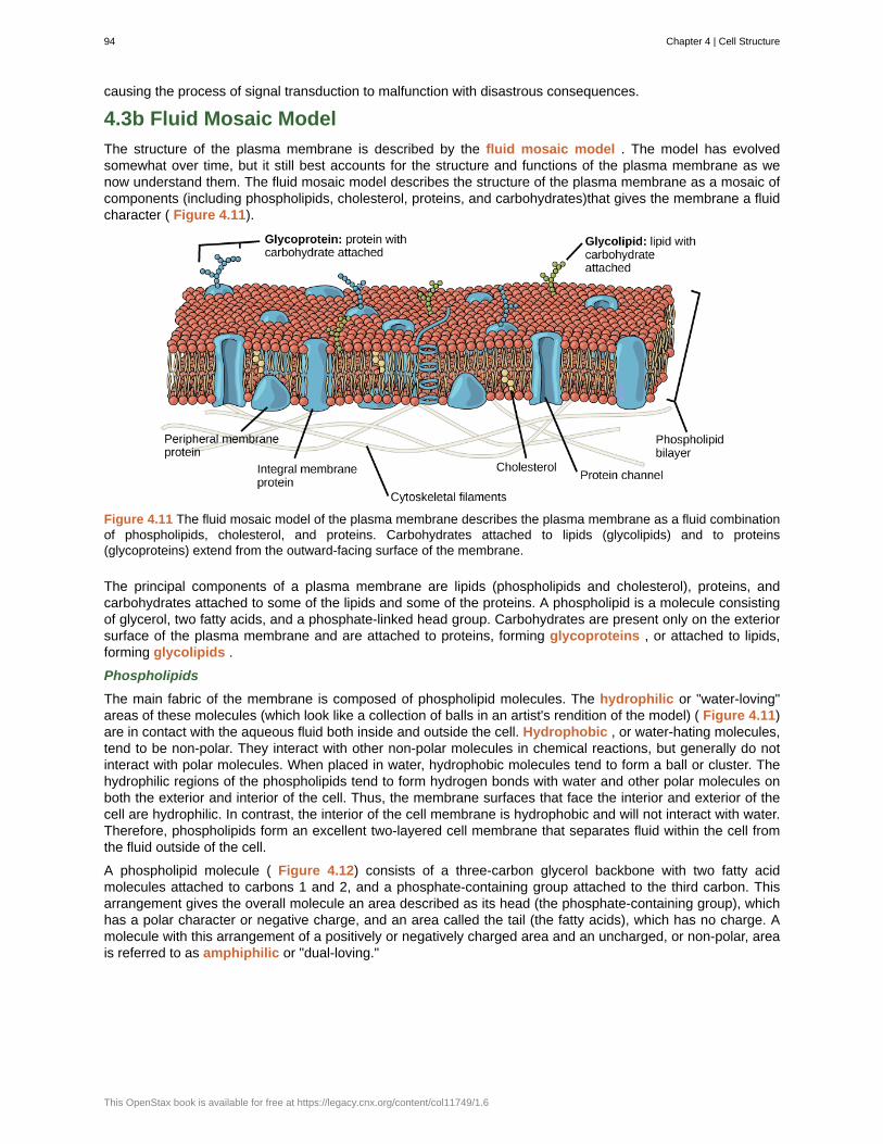
causing the process of signal transduction to malfunction with disastrous consequences.
4.3b Fluid Mosaic Model
The structure of the plasma membrane is described by the fluid mosaic model . The model has evolvedsomewhat over time, but it still best accounts for the structure and functions of the plasma membrane as wenow understand them. The fluid mosaic model describes the structure of the plasma membrane as a mosaic ofcomponents (including phospholipids, cholesterol, proteins, and carbohydrates)that gives the membrane a fluidcharacter ( Figure 4.11).
Figure 4.11 The fluid mosaic model of the plasma membrane describes the plasma membrane as a fluid combinationof phospholipids, cholesterol, and proteins. Carbohydrates attached to lipids (glycolipids) and to proteins(glycoproteins) extend from the outward-facing surface of the membrane.
The principal components of a plasma membrane are lipids (phospholipids and cholesterol), proteins, andcarbohydrates attached to some of the lipids and some of the proteins. A phospholipid is a molecule consistingof glycerol, two fatty acids, and a phosphate-linked head group. Carbohydrates are present only on the exteriorsurface of the plasma membrane and are attached to proteins, forming glycoproteins , or attached to lipids,forming glycolipids .
Phospholipids
The main fabric of the membrane is composed of phospholipid molecules. The hydrophilic or "water-loving"areas of these molecules (which look like a collection of balls in an artist's rendition of the model) ( Figure 4.11)are in contact with the aqueous fluid both inside and outside the cell. Hydrophobic , or water-hating molecules,tend to be non-polar. They interact with other non-polar molecules in chemical reactions, but generally do notinteract with polar molecules. When placed in water, hydrophobic molecules tend to form a ball or cluster. Thehydrophilic regions of the phospholipids tend to form hydrogen bonds with water and other polar molecules onboth the exterior and interior of the cell. Thus, the membrane surfaces that face the interior and exterior of thecell are hydrophilic. In contrast, the interior of the cell membrane is hydrophobic and will not interact with water.Therefore, phospholipids form an excellent two-layered cell membrane that separates fluid within the cell fromthe fluid outside of the cell.
A phospholipid molecule ( Figure 4.12) consists of a three-carbon glycerol backbone with two fatty acidmolecules attached to carbons 1 and 2, and a phosphate-containing group attached to the third carbon. Thisarrangement gives the overall molecule an area described as its head (the phosphate-containing group), whichhas a polar character or negative charge, and an area called the tail (the fatty acids), which has no charge. Amolecule with this arrangement of a positively or negatively charged area and an uncharged, or non-polar, areais referred to as amphiphilic or "dual-loving."
94 Chapter 4 | Cell Structure
This OpenStax book is available for free at https://legacy.cnx.org/content/col11749/1.6
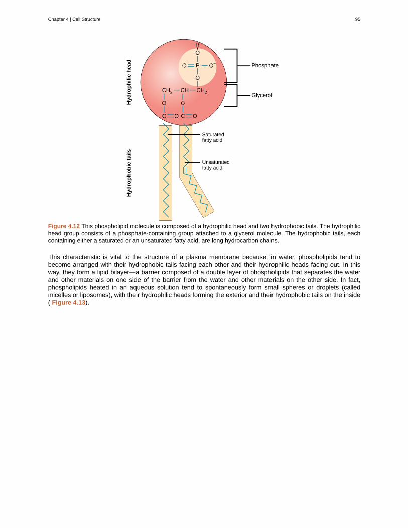
Figure 4.12 This phospholipid molecule is composed of a hydrophilic head and two hydrophobic tails. The hydrophilichead group consists of a phosphate-containing group attached to a glycerol molecule. The hydrophobic tails, eachcontaining either a saturated or an unsaturated fatty acid, are long hydrocarbon chains.
This characteristic is vital to the structure of a plasma membrane because, in water, phospholipids tend tobecome arranged with their hydrophobic tails facing each other and their hydrophilic heads facing out. In thisway, they form a lipid bilayer—a barrier composed of a double layer of phospholipids that separates the waterand other materials on one side of the barrier from the water and other materials on the other side. In fact,phospholipids heated in an aqueous solution tend to spontaneously form small spheres or droplets (calledmicelles or liposomes), with their hydrophilic heads forming the exterior and their hydrophobic tails on the inside( Figure 4.13).
Chapter 4 | Cell Structure 95
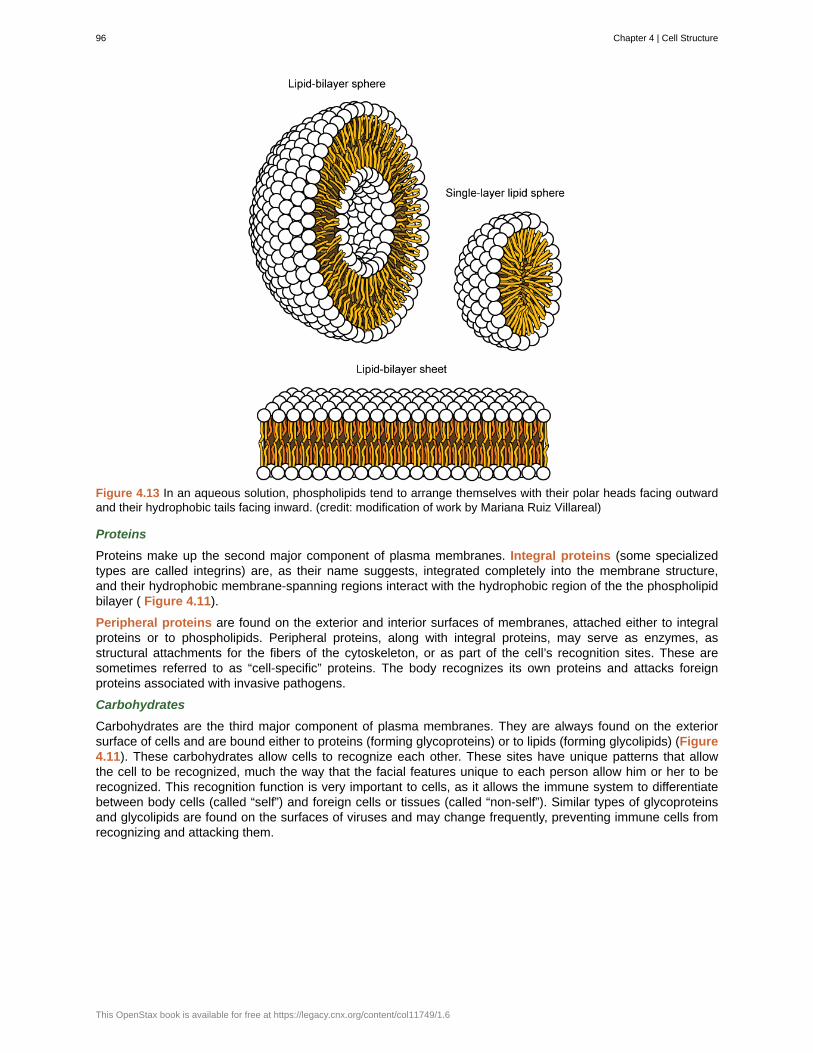
Figure 4.13 In an aqueous solution, phospholipids tend to arrange themselves with their polar heads facing outwardand their hydrophobic tails facing inward. (credit: modification of work by Mariana Ruiz Villareal)
Proteins
Proteins make up the second major component of plasma membranes. Integral proteins (some specializedtypes are called integrins) are, as their name suggests, integrated completely into the membrane structure,and their hydrophobic membrane-spanning regions interact with the hydrophobic region of the the phospholipidbilayer ( Figure 4.11).
Peripheral proteins are found on the exterior and interior surfaces of membranes, attached either to integralproteins or to phospholipids. Peripheral proteins, along with integral proteins, may serve as enzymes, asstructural attachments for the fibers of the cytoskeleton, or as part of the cell’s recognition sites. These aresometimes referred to as “cell-specific” proteins. The body recognizes its own proteins and attacks foreignproteins associated with invasive pathogens.
Carbohydrates
Carbohydrates are the third major component of plasma membranes. They are always found on the exteriorsurface of cells and are bound either to proteins (forming glycoproteins) or to lipids (forming glycolipids) (Figure4.11). These carbohydrates allow cells to recognize each other. These sites have unique patterns that allowthe cell to be recognized, much the way that the facial features unique to each person allow him or her to berecognized. This recognition function is very important to cells, as it allows the immune system to differentiatebetween body cells (called “self”) and foreign cells or tissues (called “non-self”). Similar types of glycoproteinsand glycolipids are found on the surfaces of viruses and may change frequently, preventing immune cells fromrecognizing and attacking them.
96 Chapter 4 | Cell Structure
This OpenStax book is available for free at https://legacy.cnx.org/content/col11749/1.6
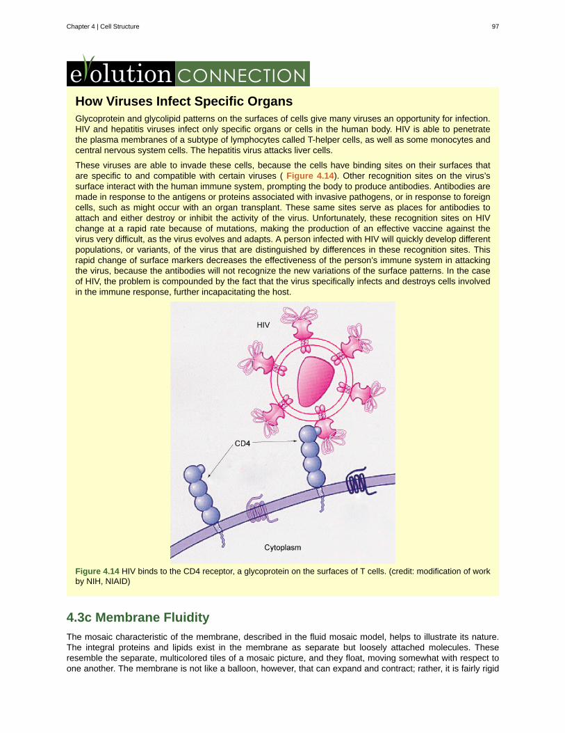
How Viruses Infect Specific OrgansGlycoprotein and glycolipid patterns on the surfaces of cells give many viruses an opportunity for infection.HIV and hepatitis viruses infect only specific organs or cells in the human body. HIV is able to penetratethe plasma membranes of a subtype of lymphocytes called T-helper cells, as well as some monocytes andcentral nervous system cells. The hepatitis virus attacks liver cells.
These viruses are able to invade these cells, because the cells have binding sites on their surfaces thatare specific to and compatible with certain viruses ( Figure 4.14). Other recognition sites on the virus’ssurface interact with the human immune system, prompting the body to produce antibodies. Antibodies aremade in response to the antigens or proteins associated with invasive pathogens, or in response to foreigncells, such as might occur with an organ transplant. These same sites serve as places for antibodies toattach and either destroy or inhibit the activity of the virus. Unfortunately, these recognition sites on HIVchange at a rapid rate because of mutations, making the production of an effective vaccine against thevirus very difficult, as the virus evolves and adapts. A person infected with HIV will quickly develop differentpopulations, or variants, of the virus that are distinguished by differences in these recognition sites. Thisrapid change of surface markers decreases the effectiveness of the person’s immune system in attackingthe virus, because the antibodies will not recognize the new variations of the surface patterns. In the caseof HIV, the problem is compounded by the fact that the virus specifically infects and destroys cells involvedin the immune response, further incapacitating the host.
Figure 4.14 HIV binds to the CD4 receptor, a glycoprotein on the surfaces of T cells. (credit: modification of workby NIH, NIAID)
4.3c Membrane Fluidity
The mosaic characteristic of the membrane, described in the fluid mosaic model, helps to illustrate its nature.The integral proteins and lipids exist in the membrane as separate but loosely attached molecules. Theseresemble the separate, multicolored tiles of a mosaic picture, and they float, moving somewhat with respect toone another. The membrane is not like a balloon, however, that can expand and contract; rather, it is fairly rigid
Chapter 4 | Cell Structure 97
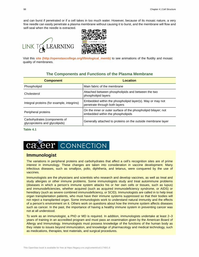
and can burst if penetrated or if a cell takes in too much water. However, because of its mosaic nature, a veryfine needle can easily penetrate a plasma membrane without causing it to burst, and the membrane will flow andself-seal when the needle is extracted.
Visit this site (http://openstaxcollege.org/l/biological_memb) to see animations of the fluidity and mosaicquality of membranes.
The Components and Functions of the Plasma Membrane
Component Location
Phospholipid Main fabric of the membrane
CholesterolAttached between phospholipids and between the twophospholipid layers
Integral proteins (for example, integrins)Embedded within the phospholipid layer(s). May or may notpenetrate through both layers
Peripheral proteinsOn the inner or outer surface of the phospholipid bilayer; notembedded within the phospholipids
Carbohydrates (components ofglycoproteins and glycolipids)
Generally attached to proteins on the outside membrane layer
Table 4.1
ImmunologistThe variations in peripheral proteins and carbohydrates that affect a cell’s recognition sites are of primeinterest in immunology. These changes are taken into consideration in vaccine development. Manyinfectious diseases, such as smallpox, polio, diphtheria, and tetanus, were conquered by the use ofvaccines.
Immunologists are the physicians and scientists who research and develop vaccines, as well as treat andstudy allergies or other immune problems. Some immunologists study and treat autoimmune problems(diseases in which a person’s immune system attacks his or her own cells or tissues, such as lupus)and immunodeficiencies, whether acquired (such as acquired immunodeficiency syndrome, or AIDS) orhereditary (such as severe combined immunodeficiency, or SCID). Immunologists are called in to help treatorgan transplantation patients, who must have their immune systems suppressed so that their bodies willnot reject a transplanted organ. Some immunologists work to understand natural immunity and the effectsof a person’s environment on it. Others work on questions about how the immune system affects diseasessuch as cancer. In the past, the importance of having a healthy immune system in preventing cancer wasnot at all understood.
To work as an immunologist, a PhD or MD is required. In addition, immunologists undertake at least 2–3years of training in an accredited program and must pass an examination given by the American Board ofAllergy and Immunology. Immunologists must possess knowledge of the functions of the human body asthey relate to issues beyond immunization, and knowledge of pharmacology and medical technology, suchas medications, therapies, test materials, and surgical procedures.
98 Chapter 4 | Cell Structure
This OpenStax book is available for free at https://legacy.cnx.org/content/col11749/1.6
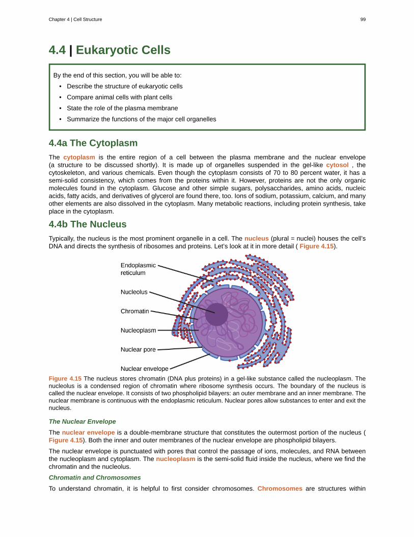
4.4 | Eukaryotic Cells
By the end of this section, you will be able to:
• Describe the structure of eukaryotic cells
• Compare animal cells with plant cells
• State the role of the plasma membrane
• Summarize the functions of the major cell organelles
4.4a The Cytoplasm
The cytoplasm is the entire region of a cell between the plasma membrane and the nuclear envelope(a structure to be discussed shortly). It is made up of organelles suspended in the gel-like cytosol , thecytoskeleton, and various chemicals. Even though the cytoplasm consists of 70 to 80 percent water, it has asemi-solid consistency, which comes from the proteins within it. However, proteins are not the only organicmolecules found in the cytoplasm. Glucose and other simple sugars, polysaccharides, amino acids, nucleicacids, fatty acids, and derivatives of glycerol are found there, too. Ions of sodium, potassium, calcium, and manyother elements are also dissolved in the cytoplasm. Many metabolic reactions, including protein synthesis, takeplace in the cytoplasm.
4.4b The Nucleus
Typically, the nucleus is the most prominent organelle in a cell. The nucleus (plural = nuclei) houses the cell’sDNA and directs the synthesis of ribosomes and proteins. Let’s look at it in more detail ( Figure 4.15).
Figure 4.15 The nucleus stores chromatin (DNA plus proteins) in a gel-like substance called the nucleoplasm. Thenucleolus is a condensed region of chromatin where ribosome synthesis occurs. The boundary of the nucleus iscalled the nuclear envelope. It consists of two phospholipid bilayers: an outer membrane and an inner membrane. Thenuclear membrane is continuous with the endoplasmic reticulum. Nuclear pores allow substances to enter and exit thenucleus.
The Nuclear Envelope
The nuclear envelope is a double-membrane structure that constitutes the outermost portion of the nucleus (Figure 4.15). Both the inner and outer membranes of the nuclear envelope are phospholipid bilayers.
The nuclear envelope is punctuated with pores that control the passage of ions, molecules, and RNA betweenthe nucleoplasm and cytoplasm. The nucleoplasm is the semi-solid fluid inside the nucleus, where we find thechromatin and the nucleolus.
Chromatin and Chromosomes
To understand chromatin, it is helpful to first consider chromosomes. Chromosomes are structures within
Chapter 4 | Cell Structure 99
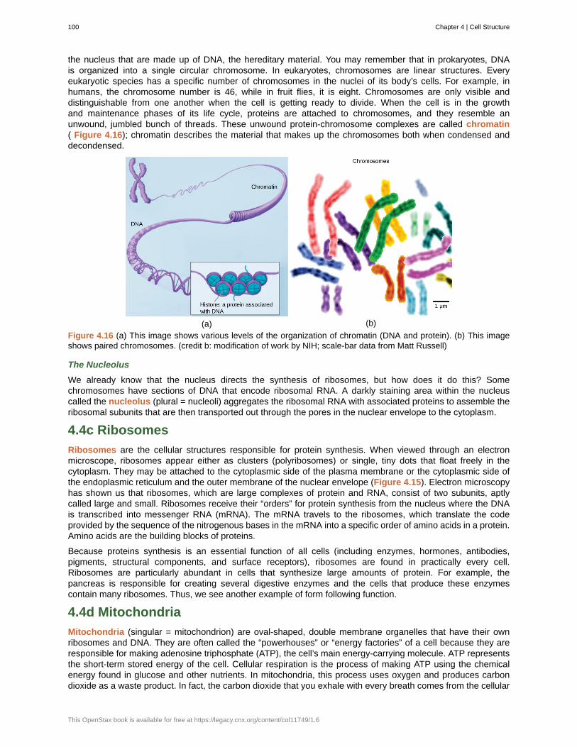
the nucleus that are made up of DNA, the hereditary material. You may remember that in prokaryotes, DNAis organized into a single circular chromosome. In eukaryotes, chromosomes are linear structures. Everyeukaryotic species has a specific number of chromosomes in the nuclei of its body’s cells. For example, inhumans, the chromosome number is 46, while in fruit flies, it is eight. Chromosomes are only visible anddistinguishable from one another when the cell is getting ready to divide. When the cell is in the growthand maintenance phases of its life cycle, proteins are attached to chromosomes, and they resemble anunwound, jumbled bunch of threads. These unwound protein-chromosome complexes are called chromatin( Figure 4.16); chromatin describes the material that makes up the chromosomes both when condensed anddecondensed.
(a) (b)
Figure 4.16 (a) This image shows various levels of the organization of chromatin (DNA and protein). (b) This imageshows paired chromosomes. (credit b: modification of work by NIH; scale-bar data from Matt Russell)
The Nucleolus
We already know that the nucleus directs the synthesis of ribosomes, but how does it do this? Somechromosomes have sections of DNA that encode ribosomal RNA. A darkly staining area within the nucleuscalled the nucleolus (plural = nucleoli) aggregates the ribosomal RNA with associated proteins to assemble theribosomal subunits that are then transported out through the pores in the nuclear envelope to the cytoplasm.
4.4c Ribosomes
Ribosomes are the cellular structures responsible for protein synthesis. When viewed through an electronmicroscope, ribosomes appear either as clusters (polyribosomes) or single, tiny dots that float freely in thecytoplasm. They may be attached to the cytoplasmic side of the plasma membrane or the cytoplasmic side ofthe endoplasmic reticulum and the outer membrane of the nuclear envelope (Figure 4.15). Electron microscopyhas shown us that ribosomes, which are large complexes of protein and RNA, consist of two subunits, aptlycalled large and small. Ribosomes receive their “orders” for protein synthesis from the nucleus where the DNAis transcribed into messenger RNA (mRNA). The mRNA travels to the ribosomes, which translate the codeprovided by the sequence of the nitrogenous bases in the mRNA into a specific order of amino acids in a protein.Amino acids are the building blocks of proteins.
Because proteins synthesis is an essential function of all cells (including enzymes, hormones, antibodies,pigments, structural components, and surface receptors), ribosomes are found in practically every cell.Ribosomes are particularly abundant in cells that synthesize large amounts of protein. For example, thepancreas is responsible for creating several digestive enzymes and the cells that produce these enzymescontain many ribosomes. Thus, we see another example of form following function.
4.4d Mitochondria
Mitochondria (singular = mitochondrion) are oval-shaped, double membrane organelles that have their ownribosomes and DNA. They are often called the “powerhouses” or “energy factories” of a cell because they areresponsible for making adenosine triphosphate (ATP), the cell’s main energy-carrying molecule. ATP representsthe short-term stored energy of the cell. Cellular respiration is the process of making ATP using the chemicalenergy found in glucose and other nutrients. In mitochondria, this process uses oxygen and produces carbondioxide as a waste product. In fact, the carbon dioxide that you exhale with every breath comes from the cellular
100 Chapter 4 | Cell Structure
This OpenStax book is available for free at https://legacy.cnx.org/content/col11749/1.6
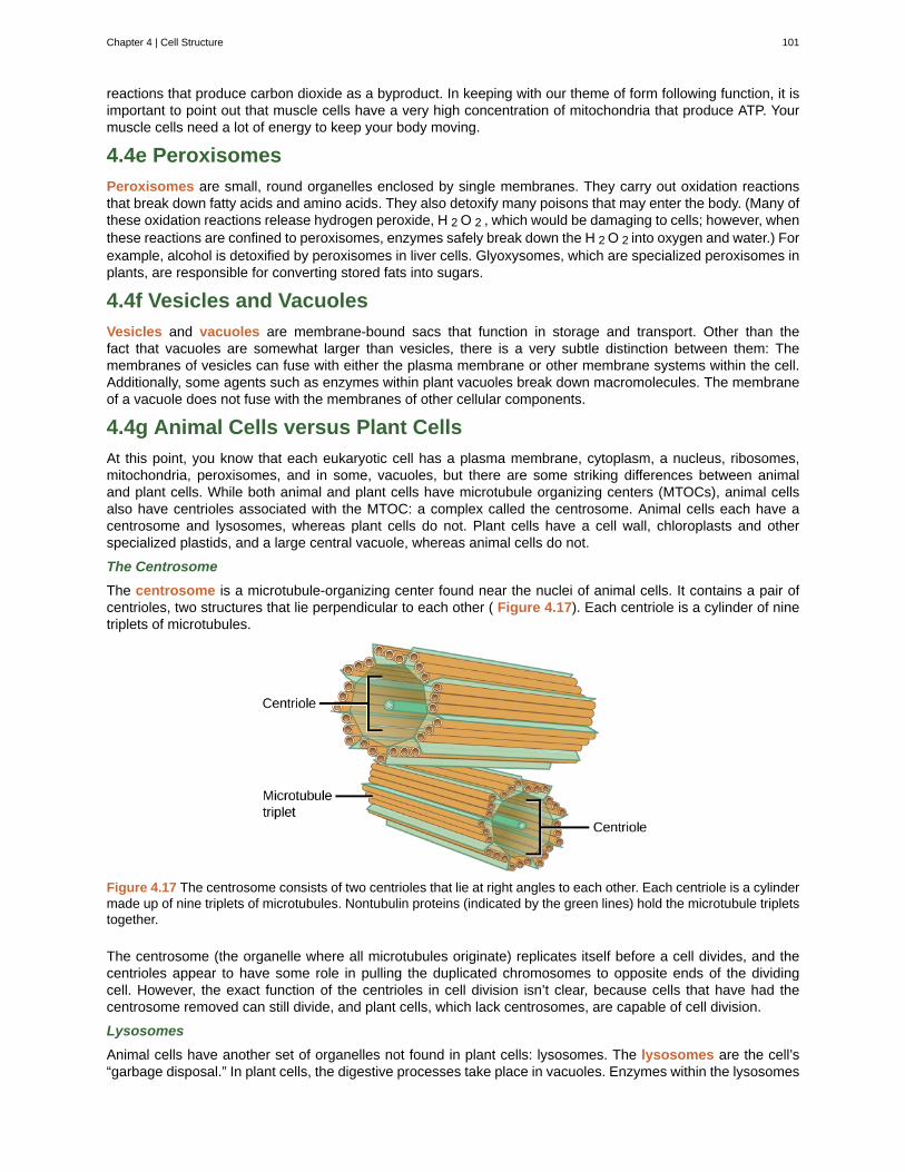
reactions that produce carbon dioxide as a byproduct. In keeping with our theme of form following function, it isimportant to point out that muscle cells have a very high concentration of mitochondria that produce ATP. Yourmuscle cells need a lot of energy to keep your body moving.
4.4e Peroxisomes
Peroxisomes are small, round organelles enclosed by single membranes. They carry out oxidation reactionsthat break down fatty acids and amino acids. They also detoxify many poisons that may enter the body. (Many ofthese oxidation reactions release hydrogen peroxide, H 2 O 2 , which would be damaging to cells; however, whenthese reactions are confined to peroxisomes, enzymes safely break down the H 2 O 2 into oxygen and water.) Forexample, alcohol is detoxified by peroxisomes in liver cells. Glyoxysomes, which are specialized peroxisomes inplants, are responsible for converting stored fats into sugars.
4.4f Vesicles and Vacuoles
Vesicles and vacuoles are membrane-bound sacs that function in storage and transport. Other than thefact that vacuoles are somewhat larger than vesicles, there is a very subtle distinction between them: Themembranes of vesicles can fuse with either the plasma membrane or other membrane systems within the cell.Additionally, some agents such as enzymes within plant vacuoles break down macromolecules. The membraneof a vacuole does not fuse with the membranes of other cellular components.
4.4g Animal Cells versus Plant Cells
At this point, you know that each eukaryotic cell has a plasma membrane, cytoplasm, a nucleus, ribosomes,mitochondria, peroxisomes, and in some, vacuoles, but there are some striking differences between animaland plant cells. While both animal and plant cells have microtubule organizing centers (MTOCs), animal cellsalso have centrioles associated with the MTOC: a complex called the centrosome. Animal cells each have acentrosome and lysosomes, whereas plant cells do not. Plant cells have a cell wall, chloroplasts and otherspecialized plastids, and a large central vacuole, whereas animal cells do not.
The Centrosome
The centrosome is a microtubule-organizing center found near the nuclei of animal cells. It contains a pair ofcentrioles, two structures that lie perpendicular to each other ( Figure 4.17). Each centriole is a cylinder of ninetriplets of microtubules.
Figure 4.17 The centrosome consists of two centrioles that lie at right angles to each other. Each centriole is a cylindermade up of nine triplets of microtubules. Nontubulin proteins (indicated by the green lines) hold the microtubule tripletstogether.
The centrosome (the organelle where all microtubules originate) replicates itself before a cell divides, and thecentrioles appear to have some role in pulling the duplicated chromosomes to opposite ends of the dividingcell. However, the exact function of the centrioles in cell division isn’t clear, because cells that have had thecentrosome removed can still divide, and plant cells, which lack centrosomes, are capable of cell division.
Lysosomes
Animal cells have another set of organelles not found in plant cells: lysosomes. The lysosomes are the cell’s“garbage disposal.” In plant cells, the digestive processes take place in vacuoles. Enzymes within the lysosomes
Chapter 4 | Cell Structure 101
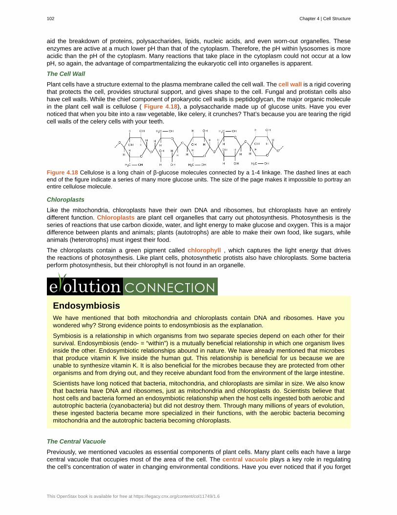
aid the breakdown of proteins, polysaccharides, lipids, nucleic acids, and even worn-out organelles. Theseenzymes are active at a much lower pH than that of the cytoplasm. Therefore, the pH within lysosomes is moreacidic than the pH of the cytoplasm. Many reactions that take place in the cytoplasm could not occur at a lowpH, so again, the advantage of compartmentalizing the eukaryotic cell into organelles is apparent.
The Cell Wall
Plant cells have a structure external to the plasma membrane called the cell wall. The cell wall is a rigid coveringthat protects the cell, provides structural support, and gives shape to the cell. Fungal and protistan cells alsohave cell walls. While the chief component of prokaryotic cell walls is peptidoglycan, the major organic moleculein the plant cell wall is cellulose ( Figure 4.18), a polysaccharide made up of glucose units. Have you evernoticed that when you bite into a raw vegetable, like celery, it crunches? That’s because you are tearing the rigidcell walls of the celery cells with your teeth.
Figure 4.18 Cellulose is a long chain of β-glucose molecules connected by a 1-4 linkage. The dashed lines at eachend of the figure indicate a series of many more glucose units. The size of the page makes it impossible to portray anentire cellulose molecule.
Chloroplasts
Like the mitochondria, chloroplasts have their own DNA and ribosomes, but chloroplasts have an entirelydifferent function. Chloroplasts are plant cell organelles that carry out photosynthesis. Photosynthesis is theseries of reactions that use carbon dioxide, water, and light energy to make glucose and oxygen. This is a majordifference between plants and animals; plants (autotrophs) are able to make their own food, like sugars, whileanimals (heterotrophs) must ingest their food.
The chloroplasts contain a green pigment called chlorophyll , which captures the light energy that drivesthe reactions of photosynthesis. Like plant cells, photosynthetic protists also have chloroplasts. Some bacteriaperform photosynthesis, but their chlorophyll is not found in an organelle.
EndosymbiosisWe have mentioned that both mitochondria and chloroplasts contain DNA and ribosomes. Have youwondered why? Strong evidence points to endosymbiosis as the explanation.
Symbiosis is a relationship in which organisms from two separate species depend on each other for theirsurvival. Endosymbiosis (endo- = “within”) is a mutually beneficial relationship in which one organism livesinside the other. Endosymbiotic relationships abound in nature. We have already mentioned that microbesthat produce vitamin K live inside the human gut. This relationship is beneficial for us because we areunable to synthesize vitamin K. It is also beneficial for the microbes because they are protected from otherorganisms and from drying out, and they receive abundant food from the environment of the large intestine.
Scientists have long noticed that bacteria, mitochondria, and chloroplasts are similar in size. We also knowthat bacteria have DNA and ribosomes, just as mitochondria and chloroplasts do. Scientists believe thathost cells and bacteria formed an endosymbiotic relationship when the host cells ingested both aerobic andautotrophic bacteria (cyanobacteria) but did not destroy them. Through many millions of years of evolution,these ingested bacteria became more specialized in their functions, with the aerobic bacteria becomingmitochondria and the autotrophic bacteria becoming chloroplasts.
The Central Vacuole
Previously, we mentioned vacuoles as essential components of plant cells. Many plant cells each have a largecentral vacuole that occupies most of the area of the cell. The central vacuole plays a key role in regulatingthe cell’s concentration of water in changing environmental conditions. Have you ever noticed that if you forget
102 Chapter 4 | Cell Structure
This OpenStax book is available for free at https://legacy.cnx.org/content/col11749/1.6
to water a plant for a few days, it wilts? That’s because as the water concentration in the soil becomes lowerthan the water concentration in the plant, water moves out of the central vacuoles and cytoplasm. As the centralvacuole shrinks, it leaves the cell wall unsupported. This loss of support to the cell walls of plant cells results inthe wilted appearance of the plant.
The central vacuole also supports the expansion of the cell. When the central vacuole holds more water, the cellgets larger without having to invest a lot of energy in synthesizing new cytoplasm.
4.5 | Endomembrane System and Proteins
By the end of this section, you will be able to:
• List the components of the endomembrane system
• Recognize the relationship between the endomembrane system and its functions
The endomembrane system (endo = “within”) is a group of membranes and organelles ( Figure 4.19) ineukaryotic cells that works together to modify, package, and transport lipids and proteins. It includes the nuclearenvelope, lysosomes, and vesicles, which we’ve already mentioned, and the endoplasmic reticulum and Golgiapparatus, which we will cover shortly. Although not technically within the cell, the plasma membrane is includedin the endomembrane system because, as you will see, it interacts with the other endomembranous organelles.The endomembrane system does not include the membranes of either mitochondria or chloroplasts.
Chapter 4 | Cell Structure 103
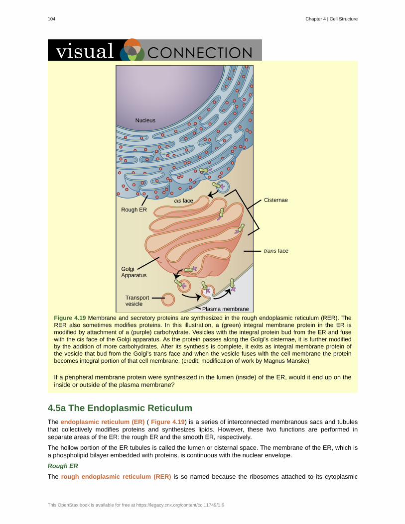
Figure 4.19 Membrane and secretory proteins are synthesized in the rough endoplasmic reticulum (RER). TheRER also sometimes modifies proteins. In this illustration, a (green) integral membrane protein in the ER ismodified by attachment of a (purple) carbohydrate. Vesicles with the integral protein bud from the ER and fusewith the cis face of the Golgi apparatus. As the protein passes along the Golgi’s cisternae, it is further modifiedby the addition of more carbohydrates. After its synthesis is complete, it exits as integral membrane protein ofthe vesicle that bud from the Golgi’s trans face and when the vesicle fuses with the cell membrane the proteinbecomes integral portion of that cell membrane. (credit: modification of work by Magnus Manske)
If a peripheral membrane protein were synthesized in the lumen (inside) of the ER, would it end up on theinside or outside of the plasma membrane?
4.5a The Endoplasmic Reticulum
The endoplasmic reticulum (ER) ( Figure 4.19) is a series of interconnected membranous sacs and tubulesthat collectively modifies proteins and synthesizes lipids. However, these two functions are performed inseparate areas of the ER: the rough ER and the smooth ER, respectively.
The hollow portion of the ER tubules is called the lumen or cisternal space. The membrane of the ER, which isa phospholipid bilayer embedded with proteins, is continuous with the nuclear envelope.
Rough ER
The rough endoplasmic reticulum (RER) is so named because the ribosomes attached to its cytoplasmic
104 Chapter 4 | Cell Structure
This OpenStax book is available for free at https://legacy.cnx.org/content/col11749/1.6
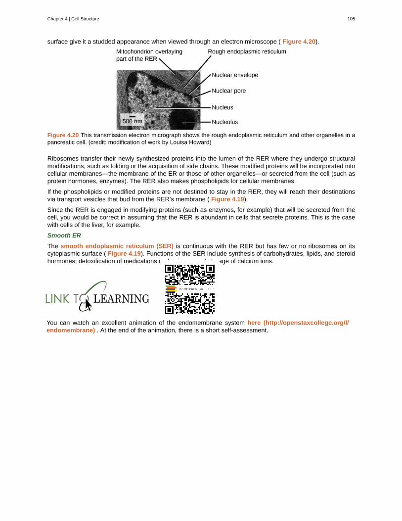
surface give it a studded appearance when viewed through an electron microscope ( Figure 4.20).
Figure 4.20 This transmission electron micrograph shows the rough endoplasmic reticulum and other organelles in apancreatic cell. (credit: modification of work by Louisa Howard)
Ribosomes transfer their newly synthesized proteins into the lumen of the RER where they undergo structuralmodifications, such as folding or the acquisition of side chains. These modified proteins will be incorporated intocellular membranes—the membrane of the ER or those of other organelles—or secreted from the cell (such asprotein hormones, enzymes). The RER also makes phospholipids for cellular membranes.
If the phospholipids or modified proteins are not destined to stay in the RER, they will reach their destinationsvia transport vesicles that bud from the RER’s membrane ( Figure 4.19).
Since the RER is engaged in modifying proteins (such as enzymes, for example) that will be secreted from thecell, you would be correct in assuming that the RER is abundant in cells that secrete proteins. This is the casewith cells of the liver, for example.
Smooth ER
The smooth endoplasmic reticulum (SER) is continuous with the RER but has few or no ribosomes on itscytoplasmic surface ( Figure 4.19). Functions of the SER include synthesis of carbohydrates, lipids, and steroidhormones; detoxification of medications and poisons; and storage of calcium ions.
You can watch an excellent animation of the endomembrane system here (http://openstaxcollege.org/l/endomembrane) . At the end of the animation, there is a short self-assessment.
Chapter 4 | Cell Structure 105
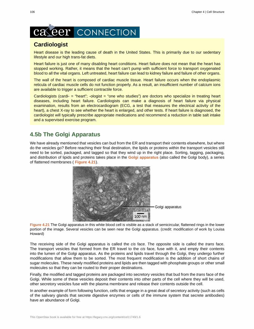
CardiologistHeart disease is the leading cause of death in the United States. This is primarily due to our sedentarylifestyle and our high trans-fat diets.
Heart failure is just one of many disabling heart conditions. Heart failure does not mean that the heart hasstopped working. Rather, it means that the heart can’t pump with sufficient force to transport oxygenatedblood to all the vital organs. Left untreated, heart failure can lead to kidney failure and failure of other organs.
The wall of the heart is composed of cardiac muscle tissue. Heart failure occurs when the endoplasmicreticula of cardiac muscle cells do not function properly. As a result, an insufficient number of calcium ionsare available to trigger a sufficient contractile force.
Cardiologists (cardi- = “heart”; -ologist = “one who studies”) are doctors who specialize in treating heartdiseases, including heart failure. Cardiologists can make a diagnosis of heart failure via physicalexamination, results from an electrocardiogram (ECG, a test that measures the electrical activity of theheart), a chest X-ray to see whether the heart is enlarged, and other tests. If heart failure is diagnosed, thecardiologist will typically prescribe appropriate medications and recommend a reduction in table salt intakeand a supervised exercise program.
4.5b The Golgi Apparatus
We have already mentioned that vesicles can bud from the ER and transport their contents elsewhere, but wheredo the vesicles go? Before reaching their final destination, the lipids or proteins within the transport vesicles stillneed to be sorted, packaged, and tagged so that they wind up in the right place. Sorting, tagging, packaging,and distribution of lipids and proteins takes place in the Golgi apparatus (also called the Golgi body), a seriesof flattened membranes ( Figure 4.21).
Figure 4.21 The Golgi apparatus in this white blood cell is visible as a stack of semicircular, flattened rings in the lowerportion of the image. Several vesicles can be seen near the Golgi apparatus. (credit: modification of work by LouisaHoward)
The receiving side of the Golgi apparatus is called the cis face. The opposite side is called the trans face.The transport vesicles that formed from the ER travel to the cis face, fuse with it, and empty their contentsinto the lumen of the Golgi apparatus. As the proteins and lipids travel through the Golgi, they undergo furthermodifications that allow them to be sorted. The most frequent modification is the addition of short chains ofsugar molecules. These newly modified proteins and lipids are then tagged with phosphate groups or other smallmolecules so that they can be routed to their proper destinations.
Finally, the modified and tagged proteins are packaged into secretory vesicles that bud from the trans face of theGolgi. While some of these vesicles deposit their contents into other parts of the cell where they will be used,other secretory vesicles fuse with the plasma membrane and release their contents outside the cell.
In another example of form following function, cells that engage in a great deal of secretory activity (such as cellsof the salivary glands that secrete digestive enzymes or cells of the immune system that secrete antibodies)have an abundance of Golgi.
106 Chapter 4 | Cell Structure
This OpenStax book is available for free at https://legacy.cnx.org/content/col11749/1.6
In plant cells, the Golgi apparatus has the additional role of synthesizing polysaccharides, some of which areincorporated into the cell wall and some of which are used in other parts of the cell.
GeneticistMany diseases arise from genetic mutations that prevent the synthesis of critical proteins. One such diseaseis Lowe disease (also called oculocerebrorenal syndrome, because it affects the eyes, brain, and kidneys).In Lowe disease, there is a deficiency in an enzyme localized to the Golgi apparatus. Children with Lowedisease are born with cataracts, typically develop kidney disease after the first year of life, and may haveimpaired mental abilities.
Lowe disease is a genetic disease caused by a mutation on the X chromosome. The X chromosome is oneof the two human sex chromosome, as these chromosomes determine a person's sex. Females possesstwo X chromosomes while males possess one X and one Y chromosome. In females, the genes on onlyone of the two X chromosomes are expressed. Therefore, females who carry the Lowe disease gene onone of their X chromosomes have a 50/50 chance of having the disease. However, males only have one Xchromosome and the genes on this chromosome are always expressed. Therefore, males will always haveLowe disease if their X chromosome carries the Lowe disease gene. The location of the mutated gene, aswell as the locations of many other mutations that cause genetic diseases, has now been identified. Throughprenatal testing, a woman can find out if the fetus she is carrying may be afflicted with one of several geneticdiseases.
Geneticists analyze the results of prenatal genetic tests and may counsel pregnant women on availableoptions. They may also conduct genetic research that leads to new drugs or foods, or perform DNA analysesthat are used in forensic investigations.
4.5c Lysosomes
In addition to their role as the digestive component and organelle-recycling facility of animal cells, lysosomes areconsidered to be parts of the endomembrane system. Lysosomes also use their hydrolytic enzymes to destroypathogens (disease-causing organisms) that might enter the cell. A good example of this occurs in a group ofwhite blood cells called macrophages, which are part of your body’s immune system. In a process known asphagocytosis or endocytosis, a section of the plasma membrane of the macrophage invaginates (folds in) andengulfs a pathogen. The invaginated section, with the pathogen inside, then pinches itself off from the plasmamembrane and becomes a vesicle. The vesicle fuses with a lysosome. The lysosome’s hydrolytic enzymes thendestroy the pathogen ( Figure 4.22).
Chapter 4 | Cell Structure 107
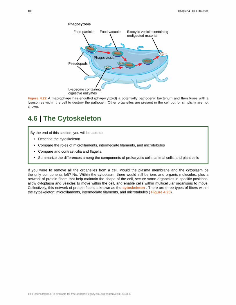
Figure 4.22 A macrophage has engulfed (phagocytized) a potentially pathogenic bacterium and then fuses with alysosomes within the cell to destroy the pathogen. Other organelles are present in the cell but for simplicity are notshown.
4.6 | The Cytoskeleton
By the end of this section, you will be able to:
• Describe the cytoskeleton
• Compare the roles of microfilaments, intermediate filaments, and microtubules
• Compare and contrast cilia and flagella
• Summarize the differences among the components of prokaryotic cells, animal cells, and plant cells
If you were to remove all the organelles from a cell, would the plasma membrane and the cytoplasm bethe only components left? No. Within the cytoplasm, there would still be ions and organic molecules, plus anetwork of protein fibers that help maintain the shape of the cell, secure some organelles in specific positions,allow cytoplasm and vesicles to move within the cell, and enable cells within multicellular organisms to move.Collectively, this network of protein fibers is known as the cytoskeleton . There are three types of fibers withinthe cytoskeleton: microfilaments, intermediate filaments, and microtubules ( Figure 4.23).
108 Chapter 4 | Cell Structure
This OpenStax book is available for free at https://legacy.cnx.org/content/col11749/1.6
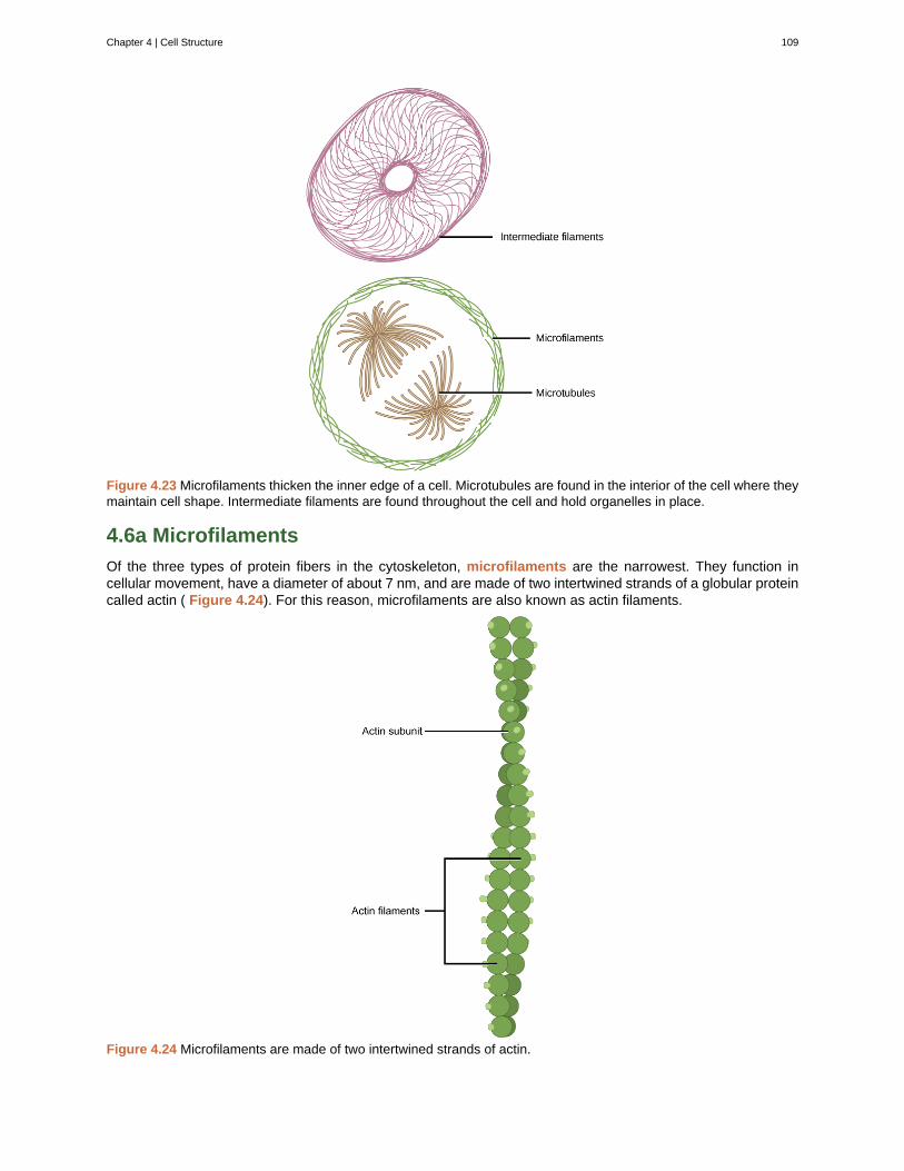
Figure 4.23 Microfilaments thicken the inner edge of a cell. Microtubules are found in the interior of the cell where theymaintain cell shape. Intermediate filaments are found throughout the cell and hold organelles in place.
4.6a Microfilaments
Of the three types of protein fibers in the cytoskeleton, microfilaments are the narrowest. They function incellular movement, have a diameter of about 7 nm, and are made of two intertwined strands of a globular proteincalled actin ( Figure 4.24). For this reason, microfilaments are also known as actin filaments.
Figure 4.24 Microfilaments are made of two intertwined strands of actin.
Chapter 4 | Cell Structure 109
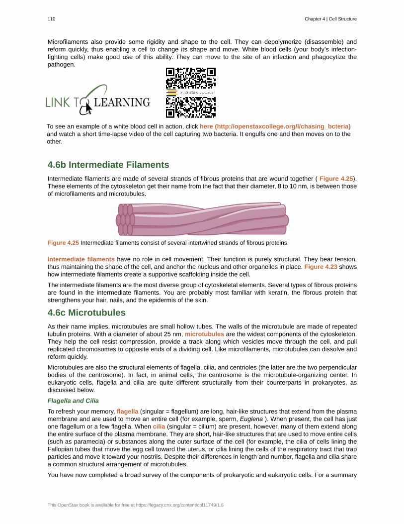
Microfilaments also provide some rigidity and shape to the cell. They can depolymerize (disassemble) andreform quickly, thus enabling a cell to change its shape and move. White blood cells (your body’s infection-fighting cells) make good use of this ability. They can move to the site of an infection and phagocytize thepathogen.
To see an example of a white blood cell in action, click here (http://openstaxcollege.org/l/chasing_bcteria)and watch a short time-lapse video of the cell capturing two bacteria. It engulfs one and then moves on to theother.
4.6b Intermediate Filaments
Intermediate filaments are made of several strands of fibrous proteins that are wound together ( Figure 4.25).These elements of the cytoskeleton get their name from the fact that their diameter, 8 to 10 nm, is between thoseof microfilaments and microtubules.
Figure 4.25 Intermediate filaments consist of several intertwined strands of fibrous proteins.
Intermediate filaments have no role in cell movement. Their function is purely structural. They bear tension,thus maintaining the shape of the cell, and anchor the nucleus and other organelles in place. Figure 4.23 showshow intermediate filaments create a supportive scaffolding inside the cell.
The intermediate filaments are the most diverse group of cytoskeletal elements. Several types of fibrous proteinsare found in the intermediate filaments. You are probably most familiar with keratin, the fibrous protein thatstrengthens your hair, nails, and the epidermis of the skin.
4.6c Microtubules
As their name implies, microtubules are small hollow tubes. The walls of the microtubule are made of repeatedtubulin proteins. With a diameter of about 25 nm, microtubules are the widest components of the cytoskeleton.They help the cell resist compression, provide a track along which vesicles move through the cell, and pullreplicated chromosomes to opposite ends of a dividing cell. Like microfilaments, microtubules can dissolve andreform quickly.
Microtubules are also the structural elements of flagella, cilia, and centrioles (the latter are the two perpendicularbodies of the centrosome). In fact, in animal cells, the centrosome is the microtubule-organizing center. Ineukaryotic cells, flagella and cilia are quite different structurally from their counterparts in prokaryotes, asdiscussed below.
Flagella and Cilia
To refresh your memory, flagella (singular = flagellum) are long, hair-like structures that extend from the plasmamembrane and are used to move an entire cell (for example, sperm, Euglena ). When present, the cell has justone flagellum or a few flagella. When cilia (singular = cilium) are present, however, many of them extend alongthe entire surface of the plasma membrane. They are short, hair-like structures that are used to move entire cells(such as paramecia) or substances along the outer surface of the cell (for example, the cilia of cells lining theFallopian tubes that move the egg cell toward the uterus, or cilia lining the cells of the respiratory tract that trapparticles and move it toward your nostrils. Despite their differences in length and number, flagella and cilia sharea common structural arrangement of microtubules.
You have now completed a broad survey of the components of prokaryotic and eukaryotic cells. For a summary
110 Chapter 4 | Cell Structure
This OpenStax book is available for free at https://legacy.cnx.org/content/col11749/1.6
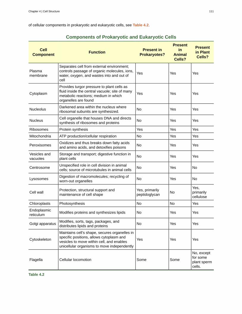
of cellular components in prokaryotic and eukaryotic cells, see Table 4.2.
Components of Prokaryotic and Eukaryotic Cells
CellComponent
FunctionPresent in
Prokaryotes?
Presentin
AnimalCells?
Presentin PlantCells?
Plasmamembrane
Separates cell from external environment;controls passage of organic molecules, ions,water, oxygen, and wastes into and out ofcell
Yes Yes Yes
Cytoplasm
Provides turgor pressure to plant cells asfluid inside the central vacuole; site of manymetabolic reactions; medium in whichorganelles are found
Yes Yes Yes
NucleolusDarkened area within the nucleus whereribosomal subunits are synthesized.
No Yes Yes
NucleusCell organelle that houses DNA and directssynthesis of ribosomes and proteins
No Yes Yes
Ribosomes Protein synthesis Yes Yes Yes
Mitochondria ATP production/cellular respiration No Yes Yes
PeroxisomesOxidizes and thus breaks down fatty acidsand amino acids, and detoxifies poisons
No Yes Yes
Vesicles andvacuoles
Storage and transport; digestive function inplant cells
No Yes Yes
CentrosomeUnspecified role in cell division in animalcells; source of microtubules in animal cells
No Yes No
LysosomesDigestion of macromolecules; recycling ofworn-out organelles
No Yes No
Cell wallProtection, structural support andmaintenance of cell shape
Yes, primarilypeptidoglycan
NoYes,primarilycellulose
Chloroplasts Photosynthesis No No Yes
Endoplasmicreticulum
Modifies proteins and synthesizes lipids No Yes Yes
Golgi apparatusModifies, sorts, tags, packages, anddistributes lipids and proteins
No Yes Yes
Cytoskeleton
Maintains cell’s shape, secures organelles inspecific positions, allows cytoplasm andvesicles to move within cell, and enablesunicellular organisms to move independently
Yes Yes Yes
Flagella Cellular locomotion Some Some
No, exceptfor someplant spermcells.
Table 4.2
Chapter 4 | Cell Structure 111
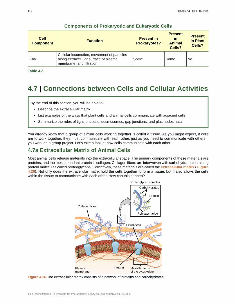
Components of Prokaryotic and Eukaryotic Cells
CellComponent
FunctionPresent in
Prokaryotes?
Presentin
AnimalCells?
Presentin PlantCells?
CiliaCellular locomotion, movement of particlesalong extracellular surface of plasmamembrane, and filtration
Some Some No
Table 4.2
4.7 | Connections between Cells and Cellular Activities
By the end of this section, you will be able to:
• Describe the extracellular matrix
• List examples of the ways that plant cells and animal cells communicate with adjacent cells
• Summarize the roles of tight junctions, desmosomes, gap junctions, and plasmodesmata
You already know that a group of similar cells working together is called a tissue. As you might expect, if cellsare to work together, they must communicate with each other, just as you need to communicate with others ifyou work on a group project. Let’s take a look at how cells communicate with each other.
4.7a Extracellular Matrix of Animal Cells
Most animal cells release materials into the extracellular space. The primary components of these materials areproteins, and the most abundant protein is collagen. Collagen fibers are interwoven with carbohydrate-containingprotein molecules called proteoglycans. Collectively, these materials are called the extracellular matrix ( Figure4.26). Not only does the extracellular matrix hold the cells together to form a tissue, but it also allows the cellswithin the tissue to communicate with each other. How can this happen?
Figure 4.26 The extracellular matrix consists of a network of proteins and carbohydrates.
112 Chapter 4 | Cell Structure
This OpenStax book is available for free at https://legacy.cnx.org/content/col11749/1.6
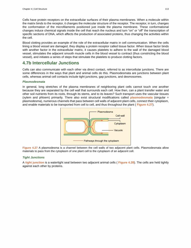
Cells have protein receptors on the extracellular surfaces of their plasma membranes. When a molecule withinthe matrix binds to the receptor, it changes the molecular structure of the receptor. The receptor, in turn, changesthe conformation of the microfilaments positioned just inside the plasma membrane. These conformationalchanges induce chemical signals inside the cell that reach the nucleus and turn “on” or “off” the transcription ofspecific sections of DNA, which affects the production of associated proteins, thus changing the activities withinthe cell.
Blood clotting provides an example of the role of the extracellular matrix in cell communication. When the cellslining a blood vessel are damaged, they display a protein receptor called tissue factor. When tissue factor bindswith another factor in the extracellular matrix, it causes platelets to adhere to the wall of the damaged bloodvessel, stimulates the adjacent smooth muscle cells in the blood vessel to contract (thus constricting the bloodvessel), and initiates a series of steps that stimulate the platelets to produce clotting factors.
4.7b Intercellular Junctions
Cells can also communicate with each other via direct contact, referred to as intercellular junctions. There aresome differences in the ways that plant and animal cells do this. Plasmodesmata are junctions between plantcells, whereas animal cell contacts include tight junctions, gap junctions, and desmosomes.
Plasmodesmata
In general, long stretches of the plasma membranes of neighboring plant cells cannot touch one anotherbecause they are separated by the cell wall that surrounds each cell. How then, can a plant transfer water andother soil nutrients from its roots, through its stems, and to its leaves? Such transport uses the vascular tissues(xylem and phloem) primarily. There also exist structural modifications called plasmodesmata (singular =plasmodesma), numerous channels that pass between cell walls of adjacent plant cells, connect their cytoplasm,and enable materials to be transported from cell to cell, and thus throughout the plant ( Figure 4.27).
Figure 4.27 A plasmodesma is a channel between the cell walls of two adjacent plant cells. Plasmodesmata allowmaterials to pass from the cytoplasm of one plant cell to the cytoplasm of an adjacent cell.
Tight Junctions
A tight junction is a watertight seal between two adjacent animal cells ( Figure 4.28). The cells are held tightlyagainst each other by proteins.
Chapter 4 | Cell Structure 113
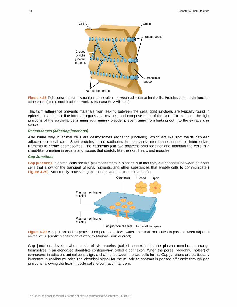
Figure 4.28 Tight junctions form watertight connections between adjacent animal cells. Proteins create tight junctionadherence. (credit: modification of work by Mariana Ruiz Villareal)
This tight adherence prevents materials from leaking between the cells; tight junctions are typically found inepithelial tissues that line internal organs and cavities, and comprise most of the skin. For example, the tightjunctions of the epithelial cells lining your urinary bladder prevent urine from leaking out into the extracellularspace.
Desmosomes (adhering junctions)
Also found only in animal cells are desmosomes (adhering junctions), which act like spot welds betweenadjacent epithelial cells. Short proteins called cadherins in the plasma membrane connect to intermediatefilaments to create desmosomes. The cadherins join two adjacent cells together and maintain the cells in asheet-like formation in organs and tissues that stretch, like the skin, heart, and muscles.
Gap Junctions
Gap junctions in animal cells are like plasmodesmata in plant cells in that they are channels between adjacentcells that allow for the transport of ions, nutrients, and other substances that enable cells to communicate (Figure 4.29). Structurally, however, gap junctions and plasmodesmata differ.
Figure 4.29 A gap junction is a protein-lined pore that allows water and small molecules to pass between adjacentanimal cells. (credit: modification of work by Mariana Ruiz Villareal)
Gap junctions develop when a set of six proteins (called connexins) in the plasma membrane arrangethemselves in an elongated donut-like configuration called a connexon. When the pores (“doughnut holes”) ofconnexons in adjacent animal cells align, a channel between the two cells forms. Gap junctions are particularlyimportant in cardiac muscle: The electrical signal for the muscle to contract is passed efficiently through gapjunctions, allowing the heart muscle cells to contract in tandem.
114 Chapter 4 | Cell Structure
This OpenStax book is available for free at https://legacy.cnx.org/content/col11749/1.6

To conduct a virtual microscopy lab and review the parts of a cell, work through the steps of this interactiveassignment (http://openstaxcollege.org/l/microscopy_lab) .
Chapter 4 | Cell Structure 115
amphiphilic
cell wall
central vacuole
centrosome
chlorophyll
Chloroplasts
chromatin
Chromosomes
cilia
cytoplasm
cytoskeleton
cytosol
electron microscopes
endomembrane system
endoplasmic reticulum (ER)
eukaryotic cells
extracellular matrix
flagella
fluid mosaic model
Gap junctions
glycolipids
glycoproteins
Golgi apparatus
KEY TERMS
molecule possessing a polar or charged area and a nonpolar or uncharged area capable ofinteracting with both hydrophilic and hydrophobic environments
rigid cell covering made of cellulose that protects the cell, provides structural support, and gives shapeto the cell
large plant cell organelle that regulates the cell’s storage compartment, holds water, and playsa significant role in cell growth as the site of macromolecule degradation
region in animal cells made of two centrioles
green pigment that captures the light energy that drives the light reactions of photosynthesis
plant cell organelle that carries out photosynthesis
protein-DNA complex that serves as the building material of chromosomes
structure within the nucleus that is made of chromatin that contains DNA, the hereditarymaterial
short, hair-like structures that extend from the plasma membrane in large numbers and are used to movecells and/or substances
entire region between the plasma membrane and the nuclear envelope, consisting of organellessuspended in the gel-like cytosol, the cytoskeleton, and various chemicals
network of protein fibers that collectively maintain the shape of the cell, secure some organellesin specific positions, allow cytoplasm and vesicles to move within the cell, and enable unicellular organismsto move independently
gel-like material of the cytoplasm in which cell structures are suspended
an instrument that magnifies an object using a beam of electrons passed and bentthrough a lens system to visualize a specimen
group of organelles and membranes in eukaryotic cells that work together modifying,packaging, and transporting lipids and proteins
series of interconnected membranous structures within eukaryotic cells thatcollectively modify proteins and synthesize lipids
cell that has a membrane-bound nucleus and several other membrane-bound compartments
material (primarily collagen, glycoproteins, and proteoglycans) secreted from animal cellsthat provides mechanical protection and anchoring for the cells in the tissue
long, hair-like structures that extend from the plasma membrane and are used to move a cell
describes the structure of the plasma membrane as a mosaic of components includingphospholipids, cholesterol, proteins, glycoproteins, and glycolipids (sugar chains attached to proteins orlipids, respectively), resulting in a fluid character (fluidity)
channel between two adjacent animal cells that allows ions, nutrients, and low molecular weightsubstances to pass between cells, enabling the cells to communicate
combination of carbohydrates and lipids
combination of carbohydrates and proteins
eukaryotic organelle made up of a series of stacked membranes that sorts, tags, and
116 Chapter 4 | Cell Structure
This OpenStax book is available for free at https://legacy.cnx.org/content/col11749/1.6
hydrophilic
hydrophobic
Integral proteins
Intermediate filaments
light microscopes
lysosomes
microfilaments
microscope
microtubules
mitochondria
nuclear envelope
nucleoid
nucleolus
nucleoplasm
nucleus
organelles
Peripheral proteins
Peroxisomes
plasma membrane
plasmodesmata
prokaryote
Ribosomes
rough endoplasmic reticulum (RER)
smooth endoplasmic reticulum (SER)
packages lipids and proteins for distribution
molecule with the ability to bond with water; “water-loving”
molecule that does not have the ability to bond with water; “water-hating”
protein integrated into the membrane structure that interacts exclusively with thehydrocarbon chains of membrane lipids and often spans the membrane
cytoskeletal component, composed of several intertwined strands of fibrous protein,that bears tension, supports cell-cell junctions, and anchors cells to extracellular structures
an instrument that magnifies an object using a beam of visible light passed and bentthrough a lens system to visualize a speciment
organelle in an animal cell that functions as the cell's digestive component; it breaks down proteins,polysaccharides, lipids, nucleic acids, and even worn-out organlles
narrowest element of the cytoskeleton; it provides rigidity and shape to the cell and enablescellular movements
an instrument that magnifies an object
widest element of the cytoskeleton; it helps the cell resist compression, provides a track alongwhich vesicles move through the cell, pulls replicated chromosomes to opposite ends of a dividing cell, andis the structural element of centrioles, cilia, and flagella
(singular = mitochondrion) cellular organelles responsible for carrying out cellular respiration,resulting in the production of ATP, the cell’s main energy-carrying molecule
double-membrane structure that constitutes the outermost portion of the nucleus
central part of a prokaryotic cell in which the chromosome is found
darkly staining body within the nucleus that is responsible for assembling the subunits of theribosomes
semi-solid fluid inside the nucleus that contains the chromatin and nucleolus
cell organelle that houses the cell’s DNA and directs the synthesis of ribosomes and proteins
compartment or sac within a cell
protein found at the surface of a plasma membrane either on its exterior or interior side
small, round organelle that contains hydrogen peroxide, oxidizes fatty acids and amino acids,and detoxifies many poisons
phospholipid bilayer with embedded (integral) or attached peripheral) proteins, andseparates the internal content of the cell from its surroundings
channel that passes between the cell walls of adjacent plant cells, connects their cytoplasm,and allows materials to be transported from cell to cell
unicellular organism that lacks a nucleus or any other membrane-bound organelle
cellular structure that carries out protein synthesis
region of the endoplasmic reticulum that is studded with ribosomes andengages in protein modification and phospholipid synthesis
region of the endoplasmic reticulum that has few or no ribosomes onits cytoplasmic surface and synthesizes carbohydrates, lipids, and steroid hormones; detoxifies certain
Chapter 4 | Cell Structure 117
tight junction
unified cell theory
vacuoles
Vesicles
chemicals (like pesticides, preservatives, medications, and environmental pollutants), and stores calciumions
firm seal between two adjacent animal cells created by protein adherence
a biological concept that states that all organisms are composed of one or more cells; thecell is the basic unit of life; and new cells arise from existing cells
membrane-bound sac, somewhat larger than a vescile, which functions in cellular storage andtransport
small, membrane-bound sac that functions in cellular storage and transport; its membrane is capableof fusing with the plasma membrane and the membranes of the ER and golgi
CHAPTER SUMMARY
4.1 Studying Cells
A cell is the smallest unit of life. Most cells are so tiny that they cannot be seen with the naked eye. Therefore,scientists use microscopes to study cells. Electron microscopes provide higher magnification, higher resolution,and more detail than light microscopes. The unified cell theory states that all organisms are composed of oneor more cells, the cell is the basic unit of life, and new cells arise from existing cells.
4.2 Prokaryotic Cells
Prokaryotes are predominantly single-celled organisms of the domains Bacteria and Archaea. All prokaryoteshave plasma membranes, cytoplasm, ribosomes, and DNA that is not membrane-bound. Most havepeptidoglycan cell walls and many have polysaccharide capsules. Prokaryotic cells range in diameter from 0.1to 5.0 μm.
As a cell increases in size, its surface area-to-volume ratio decreases. If the cell grows too large, the plasmamembrane will not have sufficient surface area to support the rate of diffusion required for the increasedvolume.
4.3 Components and Structure of Cell Membranes
Like a prokaryotic cell, a eukaryotic cell has a plasma membrane, cytoplasm, and ribosomes, but a eukaryoticcell is typically larger than a prokaryotic cell, has a true nucleus (meaning its DNA is surrounded by amembrane), and has other membrane-bound organelles that allow for compartmentalization of functions. Theplasma membrane is a phospholipid bilayer embedded with proteins.
The modern understanding of the plasma membrane is referred to as the fluid mosaic model. The plasmamembrane is composed of a bilayer of phospholipids, with their hydrophobic, fatty acid tails in contact witheach other. The landscape of the membrane is studded with proteins, some of which span the membrane.Some of these proteins serve to transport materials into or out of the cell. Carbohydrates are attached to someof the proteins and lipids on the outward-facing surface of the membrane, forming complexes that function toidentify the cell to other cells. The fluid nature of the membrane is due to temperature, the configuration of thefatty acid tails (some kinked by double bonds), the presence of cholesterol embedded in the membrane, andthe mosaic nature of the proteins and protein-carbohydrate combinations, which are not firmly fixed in place.Plasma membranes enclose and define the borders of cells, but rather than being a static bag, they aredynamic and constantly in flux.
4.4 Eukaryotic Cells
The nucleus’s nucleolus is the site of ribosome assembly. Ribosomes are either found in the cytoplasm orattached to the cytoplasmic side of the plasma membrane or endoplasmic reticulum. They perform proteinsynthesis. Mitochondria participate in cellular respiration; they are responsible for the majority of ATP producedin the cell. Peroxisomes hydrolyze fatty acids, amino acids, and some toxins. Vesicles and vacuoles arestorage and transport compartments. In plant cells, vacuoles also help break down macromolecules.
Animal cells also have a centrosome and lysosomes. The centrosome has two bodies perpendicular to eachother, the centrioles, and has an unknown purpose in cell division. Lysosomes are the digestive organelles of
118 Chapter 4 | Cell Structure
This OpenStax book is available for free at https://legacy.cnx.org/content/col11749/1.6
animal cells.
Plant cells and plant-like cells each have a cell wall, chloroplasts, and a central vacuole. The plant cell wall,whose primary component is cellulose, protects the cell, provides structural support, and gives shape to thecell. Photosynthesis takes place in chloroplasts. The central vacuole can expand without having to producemore cytoplasm.
4.5 Endomembrane System and Proteins
The endomembrane system includes the nuclear envelope, lysosomes, vesicles, the ER, and Golgi apparatus,as well as the plasma membrane. These cellular components work together to modify, package, tag, andtransport proteins and lipids that form the membranes.
The RER modifies proteins and synthesizes phospholipids used in cell membranes. The SER synthesizescarbohydrates, lipids, and steroid hormones; engages in the detoxification of medications and poisons; andstores calcium ions. Sorting, tagging, packaging, and distribution of lipids and proteins take place in the Golgiapparatus. Lysosomes are created by the budding of the membranes of the RER and Golgi. Lysosomes digestmacromolecules, recycle worn-out organelles, and destroy pathogens.
4.6 The Cytoskeleton
The cytoskeleton has three different types of protein elements. From narrowest to widest, they are themicrofilaments (actin filaments), intermediate filaments, and microtubules. Microfilaments are often associatedwith myosin. They provide rigidity and shape to the cell and facilitate cellular movements. Intermediatefilaments bear tension and anchor the nucleus and other organelles in place. Microtubules help the cell resistcompression, serve as tracks for motor proteins that move vesicles through the cell, and pull replicatedchromosomes to opposite ends of a dividing cell. They are also the structural element of centrioles, flagella,and cilia.
4.7 Connections between Cells and Cellular Activities
When protein receptors on the surface of the plasma membrane of an animal cell bind to a substance in theextracellular matrix, a chain of reactions begins that changes activities taking place within the cell.Plasmodesmata are channels between adjacent plant cells, while gap junctions are channels between adjacentanimal cells. However, their structures are quite different. A tight junction is a watertight seal between twoadjacent cells, while a desmosome acts like a spot weld.
Chapter 4 | Cell Structure 119

120 Chapter 4 | Cell Structure
This OpenStax book is available for free at https://legacy.cnx.org/content/col11749/1.6
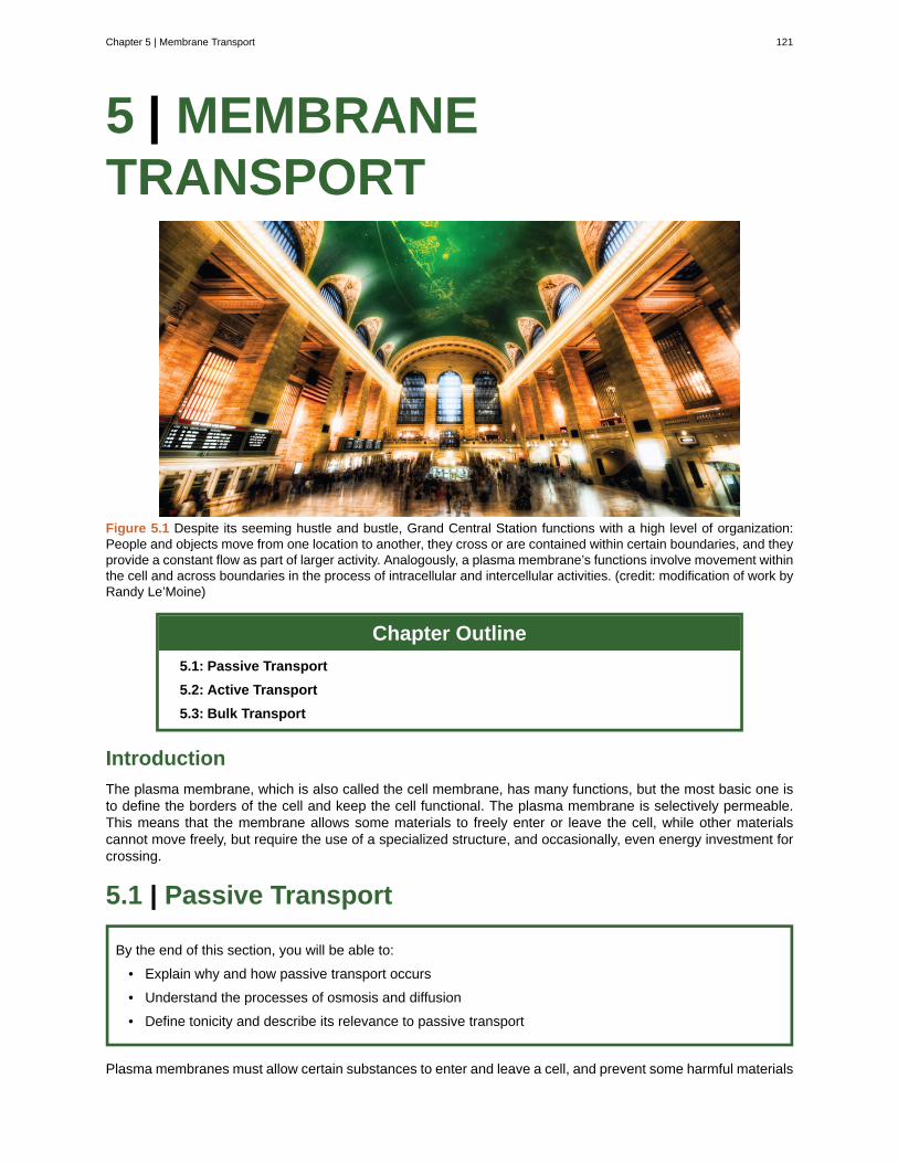
5 | MEMBRANETRANSPORT
Figure 5.1 Despite its seeming hustle and bustle, Grand Central Station functions with a high level of organization:People and objects move from one location to another, they cross or are contained within certain boundaries, and theyprovide a constant flow as part of larger activity. Analogously, a plasma membrane’s functions involve movement withinthe cell and across boundaries in the process of intracellular and intercellular activities. (credit: modification of work byRandy Le’Moine)
Chapter Outline
5.1: Passive Transport
5.2: Active Transport
5.3: Bulk Transport
Introduction
The plasma membrane, which is also called the cell membrane, has many functions, but the most basic one isto define the borders of the cell and keep the cell functional. The plasma membrane is selectively permeable.This means that the membrane allows some materials to freely enter or leave the cell, while other materialscannot move freely, but require the use of a specialized structure, and occasionally, even energy investment forcrossing.
5.1 | Passive Transport
By the end of this section, you will be able to:
• Explain why and how passive transport occurs
• Understand the processes of osmosis and diffusion
• Define tonicity and describe its relevance to passive transport
Plasma membranes must allow certain substances to enter and leave a cell, and prevent some harmful materials
Chapter 5 | Membrane Transport 121
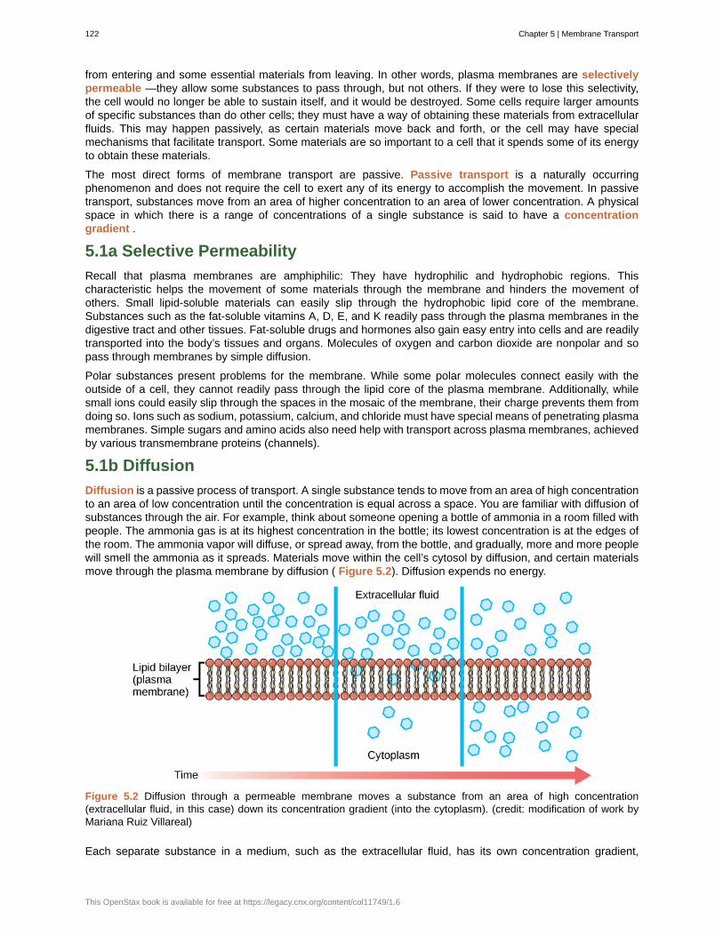
from entering and some essential materials from leaving. In other words, plasma membranes are selectivelypermeable —they allow some substances to pass through, but not others. If they were to lose this selectivity,the cell would no longer be able to sustain itself, and it would be destroyed. Some cells require larger amountsof specific substances than do other cells; they must have a way of obtaining these materials from extracellularfluids. This may happen passively, as certain materials move back and forth, or the cell may have specialmechanisms that facilitate transport. Some materials are so important to a cell that it spends some of its energyto obtain these materials.
The most direct forms of membrane transport are passive. Passive transport is a naturally occurringphenomenon and does not require the cell to exert any of its energy to accomplish the movement. In passivetransport, substances move from an area of higher concentration to an area of lower concentration. A physicalspace in which there is a range of concentrations of a single substance is said to have a concentrationgradient .
5.1a Selective Permeability
Recall that plasma membranes are amphiphilic: They have hydrophilic and hydrophobic regions. Thischaracteristic helps the movement of some materials through the membrane and hinders the movement ofothers. Small lipid-soluble materials can easily slip through the hydrophobic lipid core of the membrane.Substances such as the fat-soluble vitamins A, D, E, and K readily pass through the plasma membranes in thedigestive tract and other tissues. Fat-soluble drugs and hormones also gain easy entry into cells and are readilytransported into the body’s tissues and organs. Molecules of oxygen and carbon dioxide are nonpolar and sopass through membranes by simple diffusion.
Polar substances present problems for the membrane. While some polar molecules connect easily with theoutside of a cell, they cannot readily pass through the lipid core of the plasma membrane. Additionally, whilesmall ions could easily slip through the spaces in the mosaic of the membrane, their charge prevents them fromdoing so. Ions such as sodium, potassium, calcium, and chloride must have special means of penetrating plasmamembranes. Simple sugars and amino acids also need help with transport across plasma membranes, achievedby various transmembrane proteins (channels).
5.1b Diffusion
Diffusion is a passive process of transport. A single substance tends to move from an area of high concentrationto an area of low concentration until the concentration is equal across a space. You are familiar with diffusion ofsubstances through the air. For example, think about someone opening a bottle of ammonia in a room filled withpeople. The ammonia gas is at its highest concentration in the bottle; its lowest concentration is at the edges ofthe room. The ammonia vapor will diffuse, or spread away, from the bottle, and gradually, more and more peoplewill smell the ammonia as it spreads. Materials move within the cell’s cytosol by diffusion, and certain materialsmove through the plasma membrane by diffusion ( Figure 5.2). Diffusion expends no energy.
Figure 5.2 Diffusion through a permeable membrane moves a substance from an area of high concentration(extracellular fluid, in this case) down its concentration gradient (into the cytoplasm). (credit: modification of work byMariana Ruiz Villareal)
Each separate substance in a medium, such as the extracellular fluid, has its own concentration gradient,
122 Chapter 5 | Membrane Transport
This OpenStax book is available for free at https://legacy.cnx.org/content/col11749/1.6
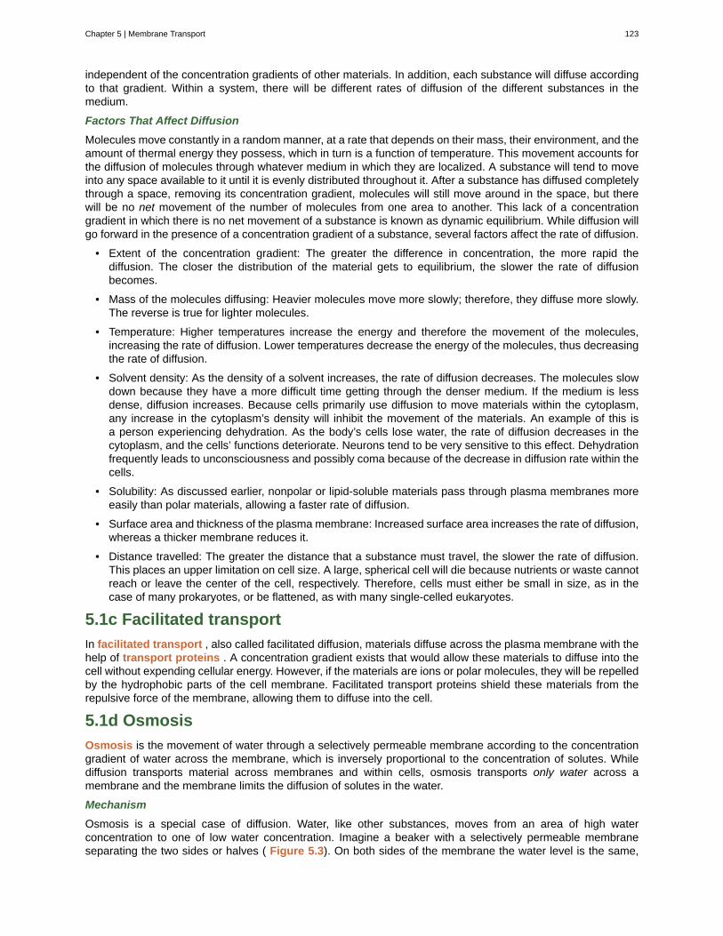
independent of the concentration gradients of other materials. In addition, each substance will diffuse accordingto that gradient. Within a system, there will be different rates of diffusion of the different substances in themedium.
Factors That Affect Diffusion
Molecules move constantly in a random manner, at a rate that depends on their mass, their environment, and theamount of thermal energy they possess, which in turn is a function of temperature. This movement accounts forthe diffusion of molecules through whatever medium in which they are localized. A substance will tend to moveinto any space available to it until it is evenly distributed throughout it. After a substance has diffused completelythrough a space, removing its concentration gradient, molecules will still move around in the space, but therewill be no net movement of the number of molecules from one area to another. This lack of a concentrationgradient in which there is no net movement of a substance is known as dynamic equilibrium. While diffusion willgo forward in the presence of a concentration gradient of a substance, several factors affect the rate of diffusion.
• Extent of the concentration gradient: The greater the difference in concentration, the more rapid thediffusion. The closer the distribution of the material gets to equilibrium, the slower the rate of diffusionbecomes.
• Mass of the molecules diffusing: Heavier molecules move more slowly; therefore, they diffuse more slowly.The reverse is true for lighter molecules.
• Temperature: Higher temperatures increase the energy and therefore the movement of the molecules,increasing the rate of diffusion. Lower temperatures decrease the energy of the molecules, thus decreasingthe rate of diffusion.
• Solvent density: As the density of a solvent increases, the rate of diffusion decreases. The molecules slowdown because they have a more difficult time getting through the denser medium. If the medium is lessdense, diffusion increases. Because cells primarily use diffusion to move materials within the cytoplasm,any increase in the cytoplasm’s density will inhibit the movement of the materials. An example of this isa person experiencing dehydration. As the body’s cells lose water, the rate of diffusion decreases in thecytoplasm, and the cells’ functions deteriorate. Neurons tend to be very sensitive to this effect. Dehydrationfrequently leads to unconsciousness and possibly coma because of the decrease in diffusion rate within thecells.
• Solubility: As discussed earlier, nonpolar or lipid-soluble materials pass through plasma membranes moreeasily than polar materials, allowing a faster rate of diffusion.
• Surface area and thickness of the plasma membrane: Increased surface area increases the rate of diffusion,whereas a thicker membrane reduces it.
• Distance travelled: The greater the distance that a substance must travel, the slower the rate of diffusion.This places an upper limitation on cell size. A large, spherical cell will die because nutrients or waste cannotreach or leave the center of the cell, respectively. Therefore, cells must either be small in size, as in thecase of many prokaryotes, or be flattened, as with many single-celled eukaryotes.
5.1c Facilitated transport
In facilitated transport , also called facilitated diffusion, materials diffuse across the plasma membrane with thehelp of transport proteins . A concentration gradient exists that would allow these materials to diffuse into thecell without expending cellular energy. However, if the materials are ions or polar molecules, they will be repelledby the hydrophobic parts of the cell membrane. Facilitated transport proteins shield these materials from therepulsive force of the membrane, allowing them to diffuse into the cell.
5.1d Osmosis
Osmosis is the movement of water through a selectively permeable membrane according to the concentrationgradient of water across the membrane, which is inversely proportional to the concentration of solutes. Whilediffusion transports material across membranes and within cells, osmosis transports only water across amembrane and the membrane limits the diffusion of solutes in the water.
Mechanism
Osmosis is a special case of diffusion. Water, like other substances, moves from an area of high waterconcentration to one of low water concentration. Imagine a beaker with a selectively permeable membraneseparating the two sides or halves ( Figure 5.3). On both sides of the membrane the water level is the same,
Chapter 5 | Membrane Transport 123
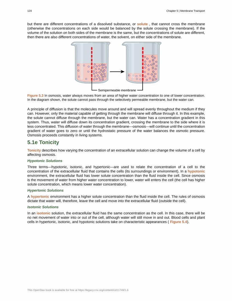
but there are different concentrations of a dissolved substance, or solute , that cannot cross the membrane(otherwise the concentrations on each side would be balanced by the solute crossing the membrane). If thevolume of the solution on both sides of the membrane is the same, but the concentrations of solute are different,then there are also different concentrations of water, the solvent, on either side of the membrane.
Figure 5.3 In osmosis, water always moves from an area of higher water concentration to one of lower concentration.In the diagram shown, the solute cannot pass through the selectively permeable membrane, but the water can.
A principle of diffusion is that the molecules move around and will spread evenly throughout the medium if theycan. However, only the material capable of getting through the membrane will diffuse through it. In this example,the solute cannot diffuse through the membrane, but the water can. Water has a concentration gradient in thissystem. Thus, water will diffuse down its concentration gradient, crossing the membrane to the side where it isless concentrated. This diffusion of water through the membrane—osmosis—will continue until the concentrationgradient of water goes to zero or until the hydrostatic pressure of the water balances the osmotic pressure.Osmosis proceeds constantly in living systems.
5.1e Tonicity
Tonicity describes how varying the concentration of an extracellular solution can change the volume of a cell byaffecting osmosis.
Hypotonic Solutions
Three terms—hypotonic, isotonic, and hypertonic—are used to relate the concentration of a cell to theconcentration of the extracellular fluid that contains the cells (its surroundings or environment). In a hypotonicenvironment, the extracellular fluid has lower solute concentration than the fluid inside the cell. Since osmosisis the movement of water from higher water concentration to lower, water will enters the cell (the cell has highersolute concentration, which means lower water concentration).
Hypertonic Solutions
A hypertonic environment has a higher solute concentration than the fluid inside the cell. The rules of osmosisdictate that water will, therefore, leave the cell and move into the extracellular fluid (outside the cell).
Isotonic Solutions
In an isotonic solution, the extracellular fluid has the same concentration as the cell. In this case, there will beno net movement of water into or out of the cell, although water will still move in and out. Blood cells and plantcells in hypertonic, isotonic, and hypotonic solutions take on characteristic appearances ( Figure 5.4).
124 Chapter 5 | Membrane Transport
This OpenStax book is available for free at https://legacy.cnx.org/content/col11749/1.6
Figure 5.4 Osmosis changes the shape of red blood cells in hypertonic, isotonic, and hypotonic solutions. (credit:Mariana Ruiz Villareal)
A doctor injects a patient with what the doctor thinks is an isotonic saline solution. The patient dies, andan autopsy reveals that many red blood cells have been destroyed. Do you think the solution the doctorinjected was really isotonic?
For a video illustrating the process of diffusion in solutions, visit this site (http://openstaxcollege.org/l/dispersion) .
5.1f Tonicity in Living Systems
In a hypotonic environment, water enters a cell, and the cell swells. In an isotonic condition, the relativeconcentrations of solute and solvent are equal on both sides of the membrane. There is no net water movement;therefore, there is no change in the size of the cell. In a hypertonic solution, water leaves a cell and the cellshrinks. If either the hypo- or hyper- condition goes to excess, the cell’s functions become compromised, andthe cell may be destroyed.
A red blood cell will burst, or lyse, when it swells beyond the plasma membrane’s capability to expand.Remember, the membrane resembles a mosaic, with discrete spaces between the molecules composing it. Ifthe cell swells, and the spaces between the lipids and proteins become too large, the cell will break apart.
In contrast, when excessive amounts of water leave a red blood cell, the cell shrinks, or crenates. This has theeffect of concentrating the solutes left in the cell, making the cytosol denser and interfering with diffusion withinthe cell. The cell’s ability to function will be compromised and may also result in the death of the cell.
Various living things have ways of controlling the effects of osmosis—a mechanism called osmoregulation.Some organisms, such as plants, fungi, bacteria, and some protists, have cell walls that surround the plasmamembrane and prevent cell lysis in a hypotonic solution. The plasma membrane can only expand to the limitof the cell wall, so the cell will not lyse. In fact, the cytoplasm in plants is always slightly hypertonic to thecellular environment, and water will always enter a cell if water is available. This inflow of water produces turgorpressure, which stiffens the cell walls of the plant ( Figure 5.5). In nonwoody plants, turgor pressure supportsthe plant. Conversely, if the plant is not watered, the extracellular fluid will become hypertonic, causing water toleave the cell. In this condition, the cell does not shrink because the cell wall is not flexible. However, the cellmembrane detaches from the wall and constricts the cytoplasm. This is called plasmolysis . Plants lose turgorpressure in this condition and wilt ( Figure 5.6).
Chapter 5 | Membrane Transport 125
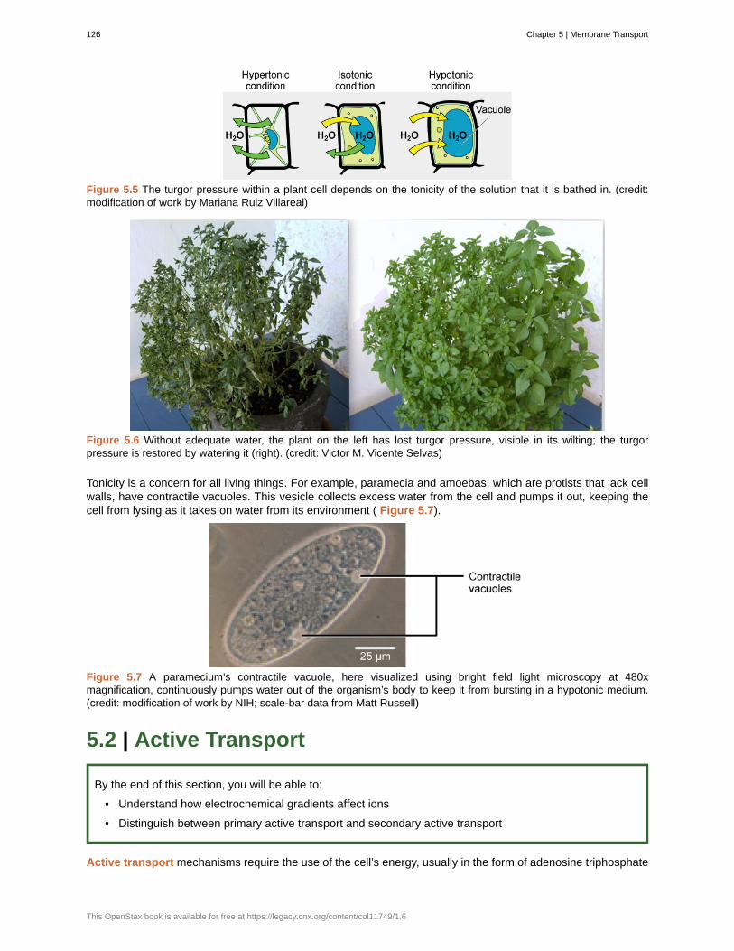
Figure 5.5 The turgor pressure within a plant cell depends on the tonicity of the solution that it is bathed in. (credit:modification of work by Mariana Ruiz Villareal)
Figure 5.6 Without adequate water, the plant on the left has lost turgor pressure, visible in its wilting; the turgorpressure is restored by watering it (right). (credit: Victor M. Vicente Selvas)
Tonicity is a concern for all living things. For example, paramecia and amoebas, which are protists that lack cellwalls, have contractile vacuoles. This vesicle collects excess water from the cell and pumps it out, keeping thecell from lysing as it takes on water from its environment ( Figure 5.7).
Figure 5.7 A paramecium’s contractile vacuole, here visualized using bright field light microscopy at 480xmagnification, continuously pumps water out of the organism’s body to keep it from bursting in a hypotonic medium.(credit: modification of work by NIH; scale-bar data from Matt Russell)
5.2 | Active Transport
By the end of this section, you will be able to:
• Understand how electrochemical gradients affect ions
• Distinguish between primary active transport and secondary active transport
Active transport mechanisms require the use of the cell’s energy, usually in the form of adenosine triphosphate
126 Chapter 5 | Membrane Transport
This OpenStax book is available for free at https://legacy.cnx.org/content/col11749/1.6
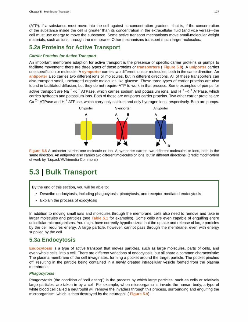
(ATP). If a substance must move into the cell against its concentration gradient—that is, if the concentrationof the substance inside the cell is greater than its concentration in the extracellular fluid (and vice versa)—thecell must use energy to move the substance. Some active transport mechanisms move small-molecular weightmaterials, such as ions, through the membrane. Other mechanisms transport much larger molecules.
5.2a Proteins for Active Transport
Carrier Proteins for Active Transport
An important membrane adaption for active transport is the presence of specific carrier proteins or pumps tofacilitate movement: there are three types of these proteins or transporters ( Figure 5.8). A uniporter carriesone specific ion or molecule. A symporter carries two different ions or molecules, both in the same direction. Anantiporter also carries two different ions or molecules, but in different directions. All of these transporters canalso transport small, uncharged organic molecules like glucose. These three types of carrier proteins are alsofound in facilitated diffusion, but they do not require ATP to work in that process. Some examples of pumps for
active transport are Na + -K + ATPase, which carries sodium and potassium ions, and H + -K + ATPase, whichcarries hydrogen and potassium ions. Both of these are antiporter carrier proteins. Two other carrier proteins are
Ca 2+ ATPase and H + ATPase, which carry only calcium and only hydrogen ions, respectively. Both are pumps.
Figure 5.8 A uniporter carries one molecule or ion. A symporter carries two different molecules or ions, both in thesame direction. An antiporter also carries two different molecules or ions, but in different directions. (credit: modificationof work by “Lupask”/Wikimedia Commons)
5.3 | Bulk Transport
By the end of this section, you will be able to:
• Describe endocytosis, including phagocytosis, pinocytosis, and receptor-mediated endocytosis
• Explain the process of exocytosis
In addition to moving small ions and molecules through the membrane, cells also need to remove and take inlarger molecules and particles (see Table 5.1 for examples). Some cells are even capable of engulfing entireunicellular microorganisms. You might have correctly hypothesized that the uptake and release of large particlesby the cell requires energy. A large particle, however, cannot pass through the membrane, even with energysupplied by the cell.
5.3a Endocytosis
Endocytosis is a type of active transport that moves particles, such as large molecules, parts of cells, andeven whole cells, into a cell. There are different variations of endocytosis, but all share a common characteristic:The plasma membrane of the cell invaginates, forming a pocket around the target particle. The pocket pinchesoff, resulting in the particle being contained in a newly created intracellular vesicle formed from the plasmamembrane.
Phagocytosis
Phagocytosis (the condition of “cell eating”) is the process by which large particles, such as cells or relativelylarge particles, are taken in by a cell. For example, when microorganisms invade the human body, a type ofwhite blood cell called a neutrophil will remove the invaders through this process, surrounding and engulfing themicroorganism, which is then destroyed by the neutrophil ( Figure 5.9).
Chapter 5 | Membrane Transport 127
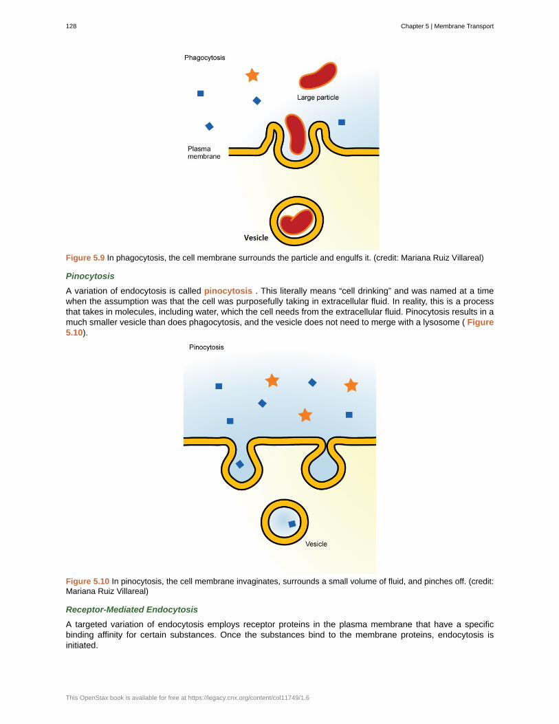
Figure 5.9 In phagocytosis, the cell membrane surrounds the particle and engulfs it. (credit: Mariana Ruiz Villareal)
Pinocytosis
A variation of endocytosis is called pinocytosis . This literally means “cell drinking” and was named at a timewhen the assumption was that the cell was purposefully taking in extracellular fluid. In reality, this is a processthat takes in molecules, including water, which the cell needs from the extracellular fluid. Pinocytosis results in amuch smaller vesicle than does phagocytosis, and the vesicle does not need to merge with a lysosome ( Figure5.10).
Figure 5.10 In pinocytosis, the cell membrane invaginates, surrounds a small volume of fluid, and pinches off. (credit:Mariana Ruiz Villareal)
Receptor-Mediated Endocytosis
A targeted variation of endocytosis employs receptor proteins in the plasma membrane that have a specificbinding affinity for certain substances. Once the substances bind to the membrane proteins, endocytosis isinitiated.
128 Chapter 5 | Membrane Transport
This OpenStax book is available for free at https://legacy.cnx.org/content/col11749/1.6
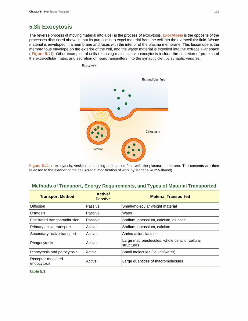
5.3b Exocytosis
The reverse process of moving material into a cell is the process of exocytosis. Exocytosis is the opposite of theprocesses discussed above in that its purpose is to expel material from the cell into the extracellular fluid. Wastematerial is enveloped in a membrane and fuses with the interior of the plasma membrane. This fusion opens themembranous envelope on the exterior of the cell, and the waste material is expelled into the extracellular space( Figure 5.11). Other examples of cells releasing molecules via exocytosis include the secretion of proteins ofthe extracellular matrix and secretion of neurotransmitters into the synaptic cleft by synaptic vesicles.
Figure 5.11 In exocytosis, vesicles containing substances fuse with the plasma membrane. The contents are thenreleased to the exterior of the cell. (credit: modification of work by Mariana Ruiz Villareal)
Methods of Transport, Energy Requirements, and Types of Material Transported
Transport MethodActive/Passive
Material Transported
Diffusion Passive Small-molecular weight material
Osmosis Passive Water
Facilitated transport/diffusion Passive Sodium, potassium, calcium, glucose
Primary active transport Active Sodium, potassium, calcium
Secondary active transport Active Amino acids, lactose
Phagocytosis ActiveLarge macromolecules, whole cells, or cellularstructures
Pinocytosis and potocytosis Active Small molecules (liquids/water)
Receptor-mediatedendocytosis
Active Large quantities of macromolecules
Table 5.1
Chapter 5 | Membrane Transport 129
active transport
antiporter
concentration gradient
diffusion
endocytosis
exocytosis
facilitated transport
hypertonic
hypotonic
isotonic
osmosis
passive transport
pinocytosis
plasmolysis
selectively permeable
solute
symporter
tonicity
transporters
uniporter
KEY TERMS
method of transporting material that requires energy
transporter that carries two ions or small molecules in different directions
area of high concentration adjacent to an area of low concentration
passive process of transport of low-molecular weight material according to its concentration gradient
type of active transport that moves substances, including fluids and particles, into a cell
process of passing bulk material out of a cell
process by which material moves down a concentration gradient (from high to lowconcentration) using integral membrane proteins
situation in which extracellular fluid has a higher osmolarity than the fluid inside the cell, resulting inwater moving out of the cell
situation in which extracellular fluid has a lower osmolarity than the fluid inside the cell, resulting inwater moving into the cell
situation in which the extracellular fluid has the same osmolarity as the fluid inside the cell, resulting inno net movement of water into or out of the cell
transport of water through a selectively permeable membrane according to the concentration gradientof water across the membrane that results from the presence of solute that cannot pass through themembrane
method of transporting material through a membrane that does not require energy
a variation of endocytosis that imports macromolecules that the cell needs from the extracellularfluid
detaching of the cell membrane from the cell wall and constriction of the cell membrane when aplant cell is in a hypertonic solution
characteristic of a membrane that allows some substances through but not others
substance dissolved in a liquid to form a solution
transporter that carries two different ions or small molecules, both in the same direction
amount of solute in a solution
specific carrier proteins or pumps that facilitate movement
transporter that carries one specific ion or molecule
CHAPTER SUMMARY
5.1 Passive Transport
The passive forms of transport, diffusion and osmosis, move materials of small molecular weight acrossmembranes. Substances diffuse from areas of high concentration to areas of lower concentration, and thisprocess continues until the substance is evenly distributed in a system. In solutions containing more than onesubstance, each type of molecule diffuses according to its own concentration gradient, independent of thediffusion of other substances. Many factors can affect the rate of diffusion, including concentration gradient,size of the particles that are diffusing, temperature of the system, and so on.
In living systems, diffusion of substances into and out of cells is mediated by the plasma membrane. Somematerials diffuse readily through the membrane, but others are hindered, and their passage is made possible
130 Chapter 5 | Membrane Transport
This OpenStax book is available for free at https://legacy.cnx.org/content/col11749/1.6
by specialized proteins, such as channels and transporters. The chemistry of living things occurs in aqueoussolutions, and balancing the concentrations of those solutions is an ongoing problem. In living systems,diffusion of some substances would be slow or difficult without membrane proteins that facilitate transport.
5.2 Active Transport
The combined gradient that affects an ion includes its concentration gradient and its electrical gradient. Apositive ion, for example, might tend to diffuse into a new area, down its concentration gradient, but if it isdiffusing into an area of net positive charge, its diffusion will be hampered by its electrical gradient. Whendealing with ions in aqueous solutions, a combination of the electrochemical and concentration gradients,rather than just the concentration gradient alone, must be considered. Living cells need certain substances thatexist inside the cell in concentrations greater than they exist in the extracellular space. Moving substances uptheir electrochemical gradients requires energy from the cell. Active transport uses energy stored in ATP to fuelthis transport. Active transport of small molecular-sized materials uses integral proteins in the cell membrane tomove the materials: These proteins are analogous to pumps. Some pumps, which carry out primary activetransport, couple directly with ATP to drive their action. In co-transport (or secondary active transport), energyfrom primary transport can be used to move another substance into the cell and up its concentration gradient.
5.3 Bulk Transport
Active transport methods require the direct use of ATP to fuel the transport. Large particles, such asmacromolecules, parts of cells, or whole cells, can be engulfed by other cells in a process called phagocytosis.In phagocytosis, a portion of the membrane invaginates and flows around the particle, eventually pinching offand leaving the particle entirely enclosed by an envelope of plasma membrane. Vesicle contents are brokendown by the cell, with the particles either used as food or dispatched. Pinocytosis is a similar process on asmaller scale. The plasma membrane invaginates and pinches off, producing a small envelope of fluid fromoutside the cell. Pinocytosis imports substances that the cell needs from the extracellular fluid. The cell expelswaste in a similar but reverse manner: it pushes a membranous vacuole to the plasma membrane, allowing thevacuole to fuse with the membrane and incorporate itself into the membrane structure, releasing its contents tothe exterior.
Chapter 5 | Membrane Transport 131

132 Chapter 5 | Membrane Transport
This OpenStax book is available for free at https://legacy.cnx.org/content/col11749/1.6
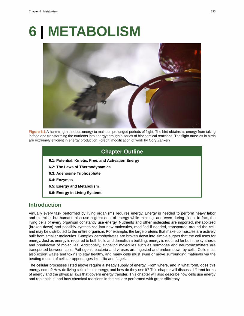
6 | METABOLISM
Figure 6.1 A hummingbird needs energy to maintain prolonged periods of flight. The bird obtains its energy from takingin food and transforming the nutrients into energy through a series of biochemical reactions. The flight muscles in birdsare extremely efficient in energy production. (credit: modification of work by Cory Zanker)
Chapter Outline
6.1: Potential, Kinetic, Free, and Activation Energy
6.2: The Laws of Thermodynamics
6.3: Adenosine Triphosphate
6.4: Enzymes
6.5: Energy and Metabolism
6.6: Energy in Living Systems
Introduction
Virtually every task performed by living organisms requires energy. Energy is needed to perform heavy laborand exercise, but humans also use a great deal of energy while thinking, and even during sleep. In fact, theliving cells of every organism constantly use energy. Nutrients and other molecules are imported, metabolized(broken down) and possibly synthesized into new molecules, modified if needed, transported around the cell,and may be distributed to the entire organism. For example, the large proteins that make up muscles are activelybuilt from smaller molecules. Complex carbohydrates are broken down into simple sugars that the cell uses forenergy. Just as energy is required to both build and demolish a building, energy is required for both the synthesisand breakdown of molecules. Additionally, signaling molecules such as hormones and neurotransmitters aretransported between cells. Pathogenic bacteria and viruses are ingested and broken down by cells. Cells mustalso export waste and toxins to stay healthy, and many cells must swim or move surrounding materials via thebeating motion of cellular appendages like cilia and flagella.
The cellular processes listed above require a steady supply of energy. From where, and in what form, does thisenergy come? How do living cells obtain energy, and how do they use it? This chapter will discuss different formsof energy and the physical laws that govern energy transfer. This chapter will also describe how cells use energyand replenish it, and how chemical reactions in the cell are performed with great efficiency.
Chapter 6 | Metabolism 133
6.1 | Potential, Kinetic, Free, and Activation Energy
By the end of this section, you will be able to:
• Define “energy”
• Explain the difference between kinetic and potential energy
• Discuss the concept of activation energy
• Describe endergonic and exergonic reactions
Energy is defined as the ability to do work. As you’ve learned, energy exists in different forms. For example,electrical energy, light energy, and heat energy are all different types of energy. While these are all familiar typesof energy that one can see or feel, there is another type of energy that is much less tangible. This energy isassociated with something as simple as an object held above the ground. In order to appreciate the way energyflows into and out of biological systems, it is important to understand more about the different types of energythat exist in the physical world.
6.1a Types of Energy
When an object is in motion, there is energy associated with that object. In the example of an airplane in flight,there is a great deal of energy associated with the motion of the airplane. This is because moving objects arecapable of enacting a change, or doing work. Think of a wrecking ball. Even a slow-moving wrecking ball can doa great deal of damage to other objects. However, a wrecking ball that is not in motion is incapable of performingwork. Energy associated with objects in motion is called kinetic energy . A speeding bullet, a walking person,the rapid movement of molecules in the air (which produces heat), and electromagnetic radiation like light allhave kinetic energy.
Now what if that same motionless wrecking ball is lifted two stories above a car with a crane? If the suspendedwrecking ball is unmoving, is there energy associated with it? The answer is yes. The suspended wrecking ballhas energy associated with it that is fundamentally different from the kinetic energy of objects in motion. Thisform of energy results from the fact that there is the potential for the wrecking ball to do work. If it is released,indeed it would do work. Because this type of energy refers to the potential to do work, it is called potentialenergy . Objects transfer their energy between kinetic and potential in the following way: As the wrecking ballhangs motionless, it has 0 kinetic and 100 percent potential energy. Once it is released, its kinetic energy beginsto increase because it builds speed due to gravity. At the same time, as it nears the ground, it loses potentialenergy. Somewhere mid-fall it has 50 percent kinetic and 50 percent potential energy. Just before it hits theground, the ball has nearly lost its potential energy and has near-maximal kinetic energy. Other examples ofpotential energy include the energy of water held behind a dam ( Figure 6.2), or a person about to skydive outof an airplane.
134 Chapter 6 | Metabolism
This OpenStax book is available for free at https://legacy.cnx.org/content/col11749/1.6
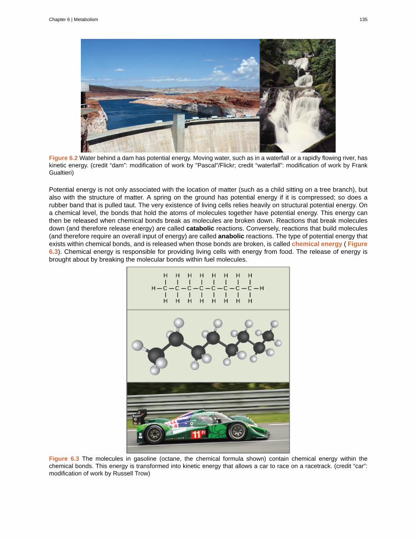
Figure 6.2 Water behind a dam has potential energy. Moving water, such as in a waterfall or a rapidly flowing river, haskinetic energy. (credit “dam”: modification of work by "Pascal"/Flickr; credit “waterfall”: modification of work by FrankGualtieri)
Potential energy is not only associated with the location of matter (such as a child sitting on a tree branch), butalso with the structure of matter. A spring on the ground has potential energy if it is compressed; so does arubber band that is pulled taut. The very existence of living cells relies heavily on structural potential energy. Ona chemical level, the bonds that hold the atoms of molecules together have potential energy. This energy canthen be released when chemical bonds break as molecules are broken down. Reactions that break moleculesdown (and therefore release energy) are called catabolic reactions. Conversely, reactions that build molecules(and therefore require an overall input of energy) are called anabolic reactions. The type of potential energy thatexists within chemical bonds, and is released when those bonds are broken, is called chemical energy ( Figure6.3). Chemical energy is responsible for providing living cells with energy from food. The release of energy isbrought about by breaking the molecular bonds within fuel molecules.
Figure 6.3 The molecules in gasoline (octane, the chemical formula shown) contain chemical energy within thechemical bonds. This energy is transformed into kinetic energy that allows a car to race on a racetrack. (credit “car”:modification of work by Russell Trow)
Chapter 6 | Metabolism 135
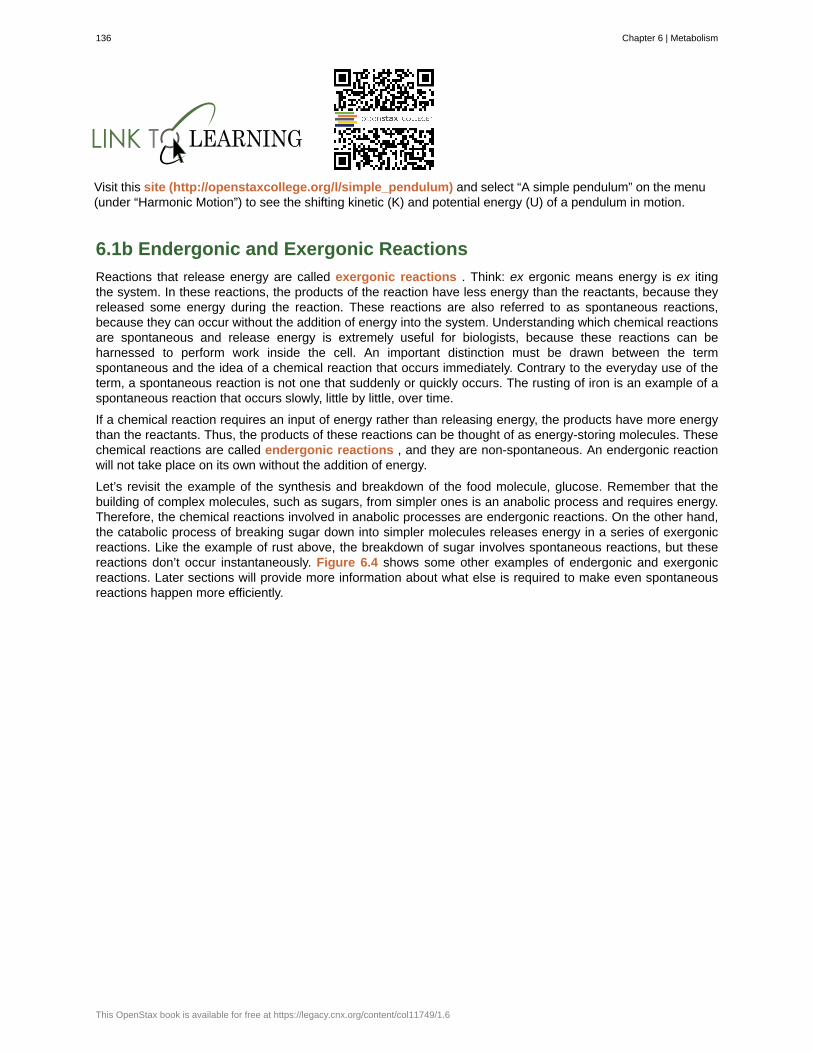
Visit this site (http://openstaxcollege.org/l/simple_pendulum) and select “A simple pendulum” on the menu(under “Harmonic Motion”) to see the shifting kinetic (K) and potential energy (U) of a pendulum in motion.
6.1b Endergonic and Exergonic Reactions
Reactions that release energy are called exergonic reactions . Think: ex ergonic means energy is ex itingthe system. In these reactions, the products of the reaction have less energy than the reactants, because theyreleased some energy during the reaction. These reactions are also referred to as spontaneous reactions,because they can occur without the addition of energy into the system. Understanding which chemical reactionsare spontaneous and release energy is extremely useful for biologists, because these reactions can beharnessed to perform work inside the cell. An important distinction must be drawn between the termspontaneous and the idea of a chemical reaction that occurs immediately. Contrary to the everyday use of theterm, a spontaneous reaction is not one that suddenly or quickly occurs. The rusting of iron is an example of aspontaneous reaction that occurs slowly, little by little, over time.
If a chemical reaction requires an input of energy rather than releasing energy, the products have more energythan the reactants. Thus, the products of these reactions can be thought of as energy-storing molecules. Thesechemical reactions are called endergonic reactions , and they are non-spontaneous. An endergonic reactionwill not take place on its own without the addition of energy.
Let’s revisit the example of the synthesis and breakdown of the food molecule, glucose. Remember that thebuilding of complex molecules, such as sugars, from simpler ones is an anabolic process and requires energy.Therefore, the chemical reactions involved in anabolic processes are endergonic reactions. On the other hand,the catabolic process of breaking sugar down into simpler molecules releases energy in a series of exergonicreactions. Like the example of rust above, the breakdown of sugar involves spontaneous reactions, but thesereactions don’t occur instantaneously. Figure 6.4 shows some other examples of endergonic and exergonicreactions. Later sections will provide more information about what else is required to make even spontaneousreactions happen more efficiently.
136 Chapter 6 | Metabolism
This OpenStax book is available for free at https://legacy.cnx.org/content/col11749/1.6
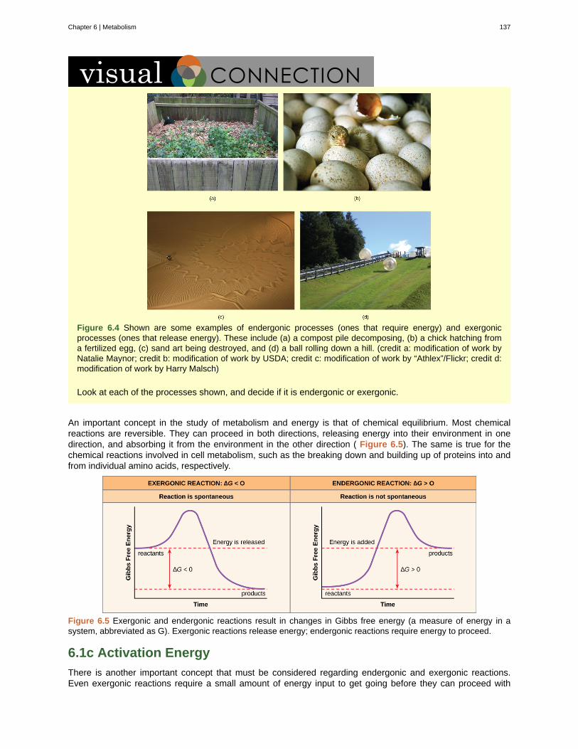
Figure 6.4 Shown are some examples of endergonic processes (ones that require energy) and exergonicprocesses (ones that release energy). These include (a) a compost pile decomposing, (b) a chick hatching froma fertilized egg, (c) sand art being destroyed, and (d) a ball rolling down a hill. (credit a: modification of work byNatalie Maynor; credit b: modification of work by USDA; credit c: modification of work by “Athlex”/Flickr; credit d:modification of work by Harry Malsch)
Look at each of the processes shown, and decide if it is endergonic or exergonic.
An important concept in the study of metabolism and energy is that of chemical equilibrium. Most chemicalreactions are reversible. They can proceed in both directions, releasing energy into their environment in onedirection, and absorbing it from the environment in the other direction ( Figure 6.5). The same is true for thechemical reactions involved in cell metabolism, such as the breaking down and building up of proteins into andfrom individual amino acids, respectively.
Figure 6.5 Exergonic and endergonic reactions result in changes in Gibbs free energy (a measure of energy in asystem, abbreviated as G). Exergonic reactions release energy; endergonic reactions require energy to proceed.
6.1c Activation Energy
There is another important concept that must be considered regarding endergonic and exergonic reactions.Even exergonic reactions require a small amount of energy input to get going before they can proceed with
Chapter 6 | Metabolism 137
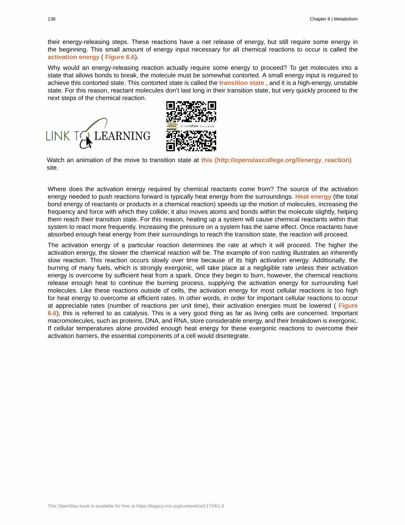
their energy-releasing steps. These reactions have a net release of energy, but still require some energy inthe beginning. This small amount of energy input necessary for all chemical reactions to occur is called theactivation energy ( Figure 6.6).
Why would an energy-releasing reaction actually require some energy to proceed? To get molecules into astate that allows bonds to break, the molecule must be somewhat contorted. A small energy input is required toachieve this contorted state. This contorted state is called the transition state , and it is a high-energy, unstablestate. For this reason, reactant molecules don’t last long in their transition state, but very quickly proceed to thenext steps of the chemical reaction.
Watch an animation of the move to transition state at this (http://openstaxcollege.org/l/energy_reaction)site.
Where does the activation energy required by chemical reactants come from? The source of the activationenergy needed to push reactions forward is typically heat energy from the surroundings. Heat energy (the totalbond energy of reactants or products in a chemical reaction) speeds up the motion of molecules, increasing thefrequency and force with which they collide; it also moves atoms and bonds within the molecule slightly, helpingthem reach their transition state. For this reason, heating up a system will cause chemical reactants within thatsystem to react more frequently. Increasing the pressure on a system has the same effect. Once reactants haveabsorbed enough heat energy from their surroundings to reach the transition state, the reaction will proceed.
The activation energy of a particular reaction determines the rate at which it will proceed. The higher theactivation energy, the slower the chemical reaction will be. The example of iron rusting illustrates an inherentlyslow reaction. This reaction occurs slowly over time because of its high activation energy. Additionally, theburning of many fuels, which is strongly exergonic, will take place at a negligible rate unless their activationenergy is overcome by sufficient heat from a spark. Once they begin to burn, however, the chemical reactionsrelease enough heat to continue the burning process, supplying the activation energy for surrounding fuelmolecules. Like these reactions outside of cells, the activation energy for most cellular reactions is too highfor heat energy to overcome at efficient rates. In other words, in order for important cellular reactions to occurat appreciable rates (number of reactions per unit time), their activation energies must be lowered ( Figure6.6); this is referred to as catalysis. This is a very good thing as far as living cells are concerned. Importantmacromolecules, such as proteins, DNA, and RNA, store considerable energy, and their breakdown is exergonic.If cellular temperatures alone provided enough heat energy for these exergonic reactions to overcome theiractivation barriers, the essential components of a cell would disintegrate.
138 Chapter 6 | Metabolism
This OpenStax book is available for free at https://legacy.cnx.org/content/col11749/1.6
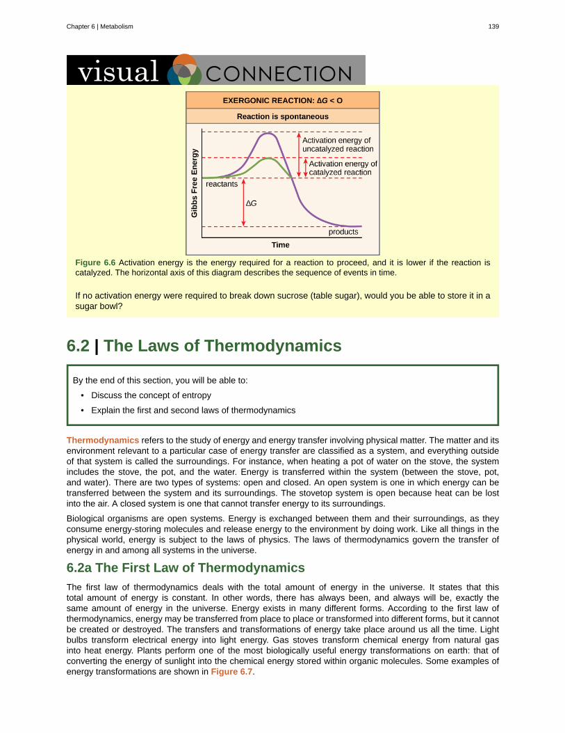
Figure 6.6 Activation energy is the energy required for a reaction to proceed, and it is lower if the reaction iscatalyzed. The horizontal axis of this diagram describes the sequence of events in time.
If no activation energy were required to break down sucrose (table sugar), would you be able to store it in asugar bowl?
6.2 | The Laws of Thermodynamics
By the end of this section, you will be able to:
• Discuss the concept of entropy
• Explain the first and second laws of thermodynamics
Thermodynamics refers to the study of energy and energy transfer involving physical matter. The matter and itsenvironment relevant to a particular case of energy transfer are classified as a system, and everything outsideof that system is called the surroundings. For instance, when heating a pot of water on the stove, the systemincludes the stove, the pot, and the water. Energy is transferred within the system (between the stove, pot,and water). There are two types of systems: open and closed. An open system is one in which energy can betransferred between the system and its surroundings. The stovetop system is open because heat can be lostinto the air. A closed system is one that cannot transfer energy to its surroundings.
Biological organisms are open systems. Energy is exchanged between them and their surroundings, as theyconsume energy-storing molecules and release energy to the environment by doing work. Like all things in thephysical world, energy is subject to the laws of physics. The laws of thermodynamics govern the transfer ofenergy in and among all systems in the universe.
6.2a The First Law of Thermodynamics
The first law of thermodynamics deals with the total amount of energy in the universe. It states that thistotal amount of energy is constant. In other words, there has always been, and always will be, exactly thesame amount of energy in the universe. Energy exists in many different forms. According to the first law ofthermodynamics, energy may be transferred from place to place or transformed into different forms, but it cannotbe created or destroyed. The transfers and transformations of energy take place around us all the time. Lightbulbs transform electrical energy into light energy. Gas stoves transform chemical energy from natural gasinto heat energy. Plants perform one of the most biologically useful energy transformations on earth: that ofconverting the energy of sunlight into the chemical energy stored within organic molecules. Some examples ofenergy transformations are shown in Figure 6.7.
Chapter 6 | Metabolism 139
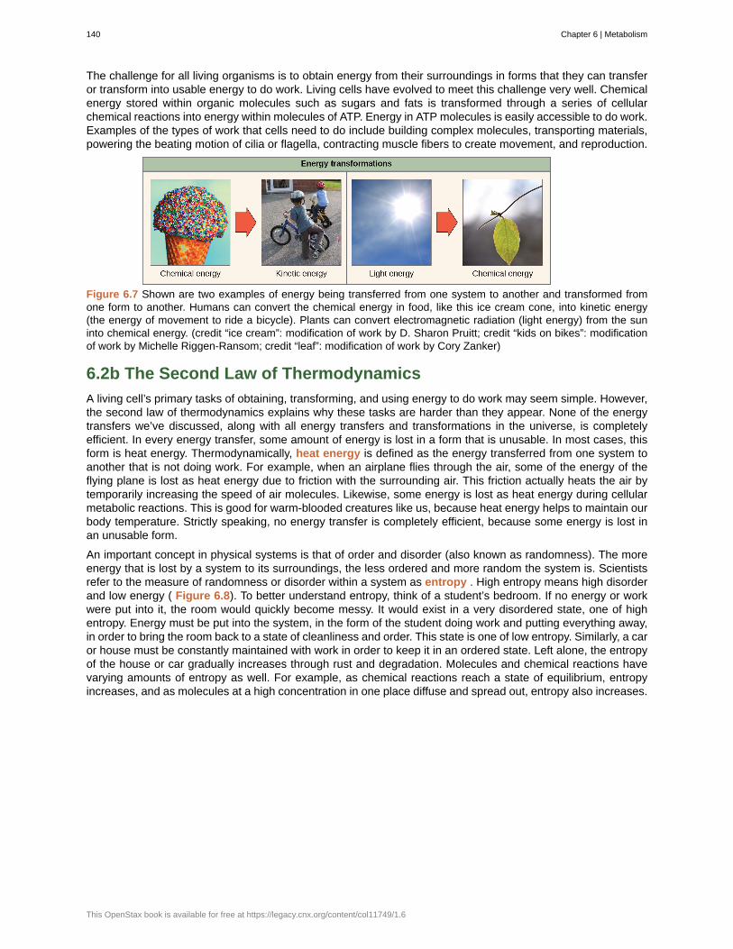
The challenge for all living organisms is to obtain energy from their surroundings in forms that they can transferor transform into usable energy to do work. Living cells have evolved to meet this challenge very well. Chemicalenergy stored within organic molecules such as sugars and fats is transformed through a series of cellularchemical reactions into energy within molecules of ATP. Energy in ATP molecules is easily accessible to do work.Examples of the types of work that cells need to do include building complex molecules, transporting materials,powering the beating motion of cilia or flagella, contracting muscle fibers to create movement, and reproduction.
Figure 6.7 Shown are two examples of energy being transferred from one system to another and transformed fromone form to another. Humans can convert the chemical energy in food, like this ice cream cone, into kinetic energy(the energy of movement to ride a bicycle). Plants can convert electromagnetic radiation (light energy) from the suninto chemical energy. (credit “ice cream”: modification of work by D. Sharon Pruitt; credit “kids on bikes”: modificationof work by Michelle Riggen-Ransom; credit “leaf”: modification of work by Cory Zanker)
6.2b The Second Law of Thermodynamics
A living cell’s primary tasks of obtaining, transforming, and using energy to do work may seem simple. However,the second law of thermodynamics explains why these tasks are harder than they appear. None of the energytransfers we’ve discussed, along with all energy transfers and transformations in the universe, is completelyefficient. In every energy transfer, some amount of energy is lost in a form that is unusable. In most cases, thisform is heat energy. Thermodynamically, heat energy is defined as the energy transferred from one system toanother that is not doing work. For example, when an airplane flies through the air, some of the energy of theflying plane is lost as heat energy due to friction with the surrounding air. This friction actually heats the air bytemporarily increasing the speed of air molecules. Likewise, some energy is lost as heat energy during cellularmetabolic reactions. This is good for warm-blooded creatures like us, because heat energy helps to maintain ourbody temperature. Strictly speaking, no energy transfer is completely efficient, because some energy is lost inan unusable form.
An important concept in physical systems is that of order and disorder (also known as randomness). The moreenergy that is lost by a system to its surroundings, the less ordered and more random the system is. Scientistsrefer to the measure of randomness or disorder within a system as entropy . High entropy means high disorderand low energy ( Figure 6.8). To better understand entropy, think of a student’s bedroom. If no energy or workwere put into it, the room would quickly become messy. It would exist in a very disordered state, one of highentropy. Energy must be put into the system, in the form of the student doing work and putting everything away,in order to bring the room back to a state of cleanliness and order. This state is one of low entropy. Similarly, a caror house must be constantly maintained with work in order to keep it in an ordered state. Left alone, the entropyof the house or car gradually increases through rust and degradation. Molecules and chemical reactions havevarying amounts of entropy as well. For example, as chemical reactions reach a state of equilibrium, entropyincreases, and as molecules at a high concentration in one place diffuse and spread out, entropy also increases.
140 Chapter 6 | Metabolism
This OpenStax book is available for free at https://legacy.cnx.org/content/col11749/1.6
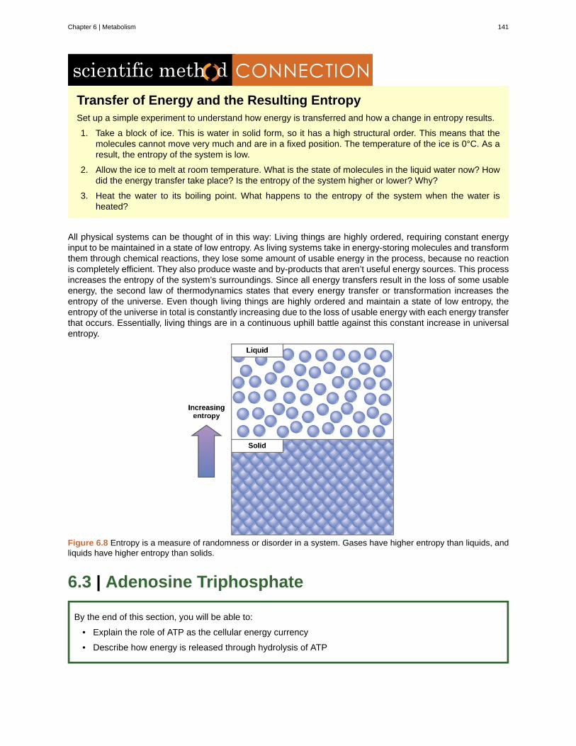
Transfer of Energy and the Resulting EntropySet up a simple experiment to understand how energy is transferred and how a change in entropy results.
1. Take a block of ice. This is water in solid form, so it has a high structural order. This means that themolecules cannot move very much and are in a fixed position. The temperature of the ice is 0°C. As aresult, the entropy of the system is low.
2. Allow the ice to melt at room temperature. What is the state of molecules in the liquid water now? Howdid the energy transfer take place? Is the entropy of the system higher or lower? Why?
3. Heat the water to its boiling point. What happens to the entropy of the system when the water isheated?
All physical systems can be thought of in this way: Living things are highly ordered, requiring constant energyinput to be maintained in a state of low entropy. As living systems take in energy-storing molecules and transformthem through chemical reactions, they lose some amount of usable energy in the process, because no reactionis completely efficient. They also produce waste and by-products that aren’t useful energy sources. This processincreases the entropy of the system’s surroundings. Since all energy transfers result in the loss of some usableenergy, the second law of thermodynamics states that every energy transfer or transformation increases theentropy of the universe. Even though living things are highly ordered and maintain a state of low entropy, theentropy of the universe in total is constantly increasing due to the loss of usable energy with each energy transferthat occurs. Essentially, living things are in a continuous uphill battle against this constant increase in universalentropy.
Figure 6.8 Entropy is a measure of randomness or disorder in a system. Gases have higher entropy than liquids, andliquids have higher entropy than solids.
6.3 | Adenosine Triphosphate
By the end of this section, you will be able to:
• Explain the role of ATP as the cellular energy currency
• Describe how energy is released through hydrolysis of ATP
Chapter 6 | Metabolism 141
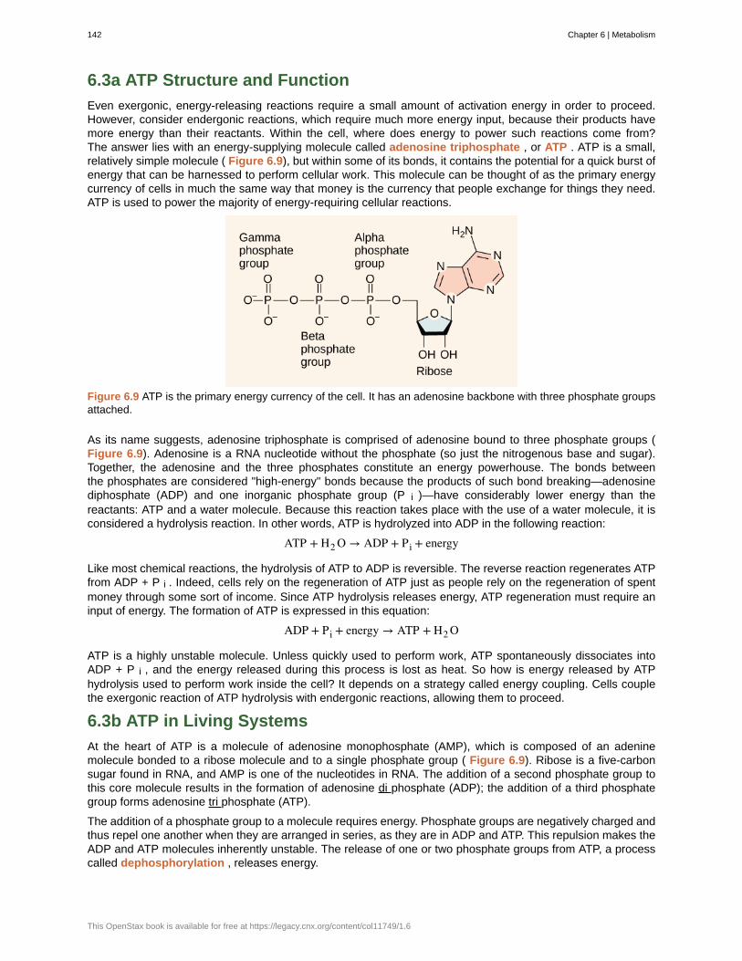
6.3a ATP Structure and Function
Even exergonic, energy-releasing reactions require a small amount of activation energy in order to proceed.However, consider endergonic reactions, which require much more energy input, because their products havemore energy than their reactants. Within the cell, where does energy to power such reactions come from?The answer lies with an energy-supplying molecule called adenosine triphosphate , or ATP . ATP is a small,relatively simple molecule ( Figure 6.9), but within some of its bonds, it contains the potential for a quick burst ofenergy that can be harnessed to perform cellular work. This molecule can be thought of as the primary energycurrency of cells in much the same way that money is the currency that people exchange for things they need.ATP is used to power the majority of energy-requiring cellular reactions.
Figure 6.9 ATP is the primary energy currency of the cell. It has an adenosine backbone with three phosphate groupsattached.
As its name suggests, adenosine triphosphate is comprised of adenosine bound to three phosphate groups (Figure 6.9). Adenosine is a RNA nucleotide without the phosphate (so just the nitrogenous base and sugar).Together, the adenosine and the three phosphates constitute an energy powerhouse. The bonds betweenthe phosphates are considered "high-energy" bonds because the products of such bond breaking—adenosinediphosphate (ADP) and one inorganic phosphate group (P i )—have considerably lower energy than thereactants: ATP and a water molecule. Because this reaction takes place with the use of a water molecule, it isconsidered a hydrolysis reaction. In other words, ATP is hydrolyzed into ADP in the following reaction:
ATP + H2 O → ADP + Pi + energy
Like most chemical reactions, the hydrolysis of ATP to ADP is reversible. The reverse reaction regenerates ATPfrom ADP + P i . Indeed, cells rely on the regeneration of ATP just as people rely on the regeneration of spentmoney through some sort of income. Since ATP hydrolysis releases energy, ATP regeneration must require aninput of energy. The formation of ATP is expressed in this equation:
ADP + Pi + energy → ATP + H2 O
ATP is a highly unstable molecule. Unless quickly used to perform work, ATP spontaneously dissociates intoADP + P i , and the energy released during this process is lost as heat. So how is energy released by ATPhydrolysis used to perform work inside the cell? It depends on a strategy called energy coupling. Cells couplethe exergonic reaction of ATP hydrolysis with endergonic reactions, allowing them to proceed.
6.3b ATP in Living Systems
At the heart of ATP is a molecule of adenosine monophosphate (AMP), which is composed of an adeninemolecule bonded to a ribose molecule and to a single phosphate group ( Figure 6.9). Ribose is a five-carbonsugar found in RNA, and AMP is one of the nucleotides in RNA. The addition of a second phosphate group tothis core molecule results in the formation of adenosine di phosphate (ADP); the addition of a third phosphategroup forms adenosine tri phosphate (ATP).
The addition of a phosphate group to a molecule requires energy. Phosphate groups are negatively charged andthus repel one another when they are arranged in series, as they are in ADP and ATP. This repulsion makes theADP and ATP molecules inherently unstable. The release of one or two phosphate groups from ATP, a processcalled dephosphorylation , releases energy.
142 Chapter 6 | Metabolism
This OpenStax book is available for free at https://legacy.cnx.org/content/col11749/1.6
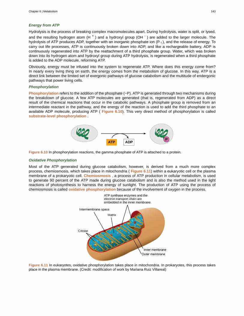
Energy from ATP
Hydrolysis is the process of breaking complex macromolecules apart. During hydrolysis, water is split, or lysed,
and the resulting hydrogen atom (H + ) and a hydroxyl group (OH - ) are added to the larger molecule. Thehydrolysis of ATP produces ADP, together with an inorganic phosphate ion (P i ), and the release of energy. Tocarry out life processes, ATP is continuously broken down into ADP, and like a rechargeable battery, ADP iscontinuously regenerated into ATP by the reattachment of a third phosphate group. Water, which was brokendown into its hydrogen atom and hydroxyl group during ATP hydrolysis, is regenerated when a third phosphateis added to the ADP molecule, reforming ATP.
Obviously, energy must be infused into the system to regenerate ATP. Where does this energy come from?In nearly every living thing on earth, the energy comes from the metabolism of glucose. In this way, ATP is adirect link between the limited set of exergonic pathways of glucose catabolism and the multitude of endergonicpathways that power living cells.
Phosphorylation
Phosphorylation refers to the addition of the phosphate (~P). ATP is generated through two mechanisms duringthe breakdown of glucose. A few ATP molecules are generated (that is, regenerated from ADP) as a directresult of the chemical reactions that occur in the catabolic pathways. A phosphate group is removed from anintermediate reactant in the pathway, and the energy of the reaction is used to add the third phosphate to anavailable ADP molecule, producing ATP ( Figure 6.10). This very direct method of phosphorylation is calledsubstrate-level phosphorylation .
Figure 6.10 In phosphorylation reactions, the gamma phosphate of ATP is attached to a protein.
Oxidative Phosphorylation
Most of the ATP generated during glucose catabolism, however, is derived from a much more complexprocess, chemiosmosis, which takes place in mitochondria ( Figure 6.11) within a eukaryotic cell or the plasmamembrane of a prokaryotic cell. Chemiosmosis , a process of ATP production in cellular metabolism, is usedto generate 90 percent of the ATP made during glucose catabolism and is also the method used in the lightreactions of photosynthesis to harness the energy of sunlight. The production of ATP using the process ofchemiosmosis is called oxidative phosphorylation because of the involvement of oxygen in the process.
Figure 6.11 In eukaryotes, oxidative phosphorylation takes place in mitochondria. In prokaryotes, this process takesplace in the plasma membrane. (Credit: modification of work by Mariana Ruiz Villareal)
Chapter 6 | Metabolism 143
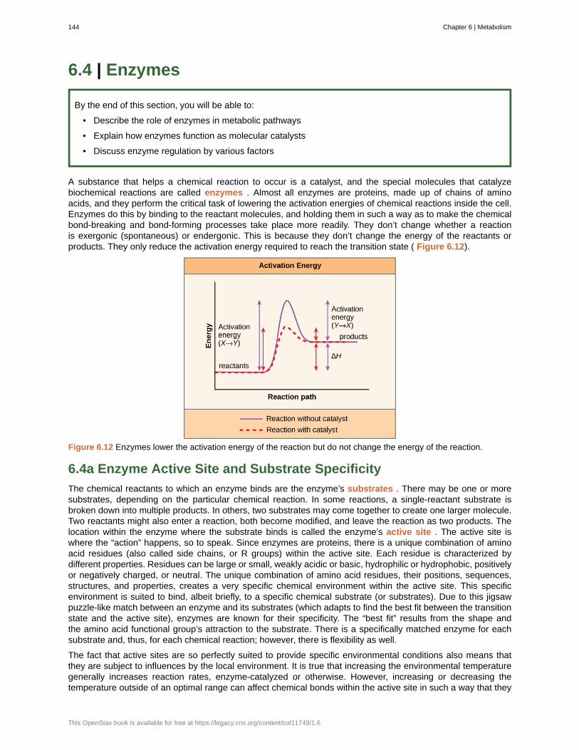
6.4 | Enzymes
By the end of this section, you will be able to:
• Describe the role of enzymes in metabolic pathways
• Explain how enzymes function as molecular catalysts
• Discuss enzyme regulation by various factors
A substance that helps a chemical reaction to occur is a catalyst, and the special molecules that catalyzebiochemical reactions are called enzymes . Almost all enzymes are proteins, made up of chains of aminoacids, and they perform the critical task of lowering the activation energies of chemical reactions inside the cell.Enzymes do this by binding to the reactant molecules, and holding them in such a way as to make the chemicalbond-breaking and bond-forming processes take place more readily. They don’t change whether a reactionis exergonic (spontaneous) or endergonic. This is because they don’t change the energy of the reactants orproducts. They only reduce the activation energy required to reach the transition state ( Figure 6.12).
Figure 6.12 Enzymes lower the activation energy of the reaction but do not change the energy of the reaction.
6.4a Enzyme Active Site and Substrate Specificity
The chemical reactants to which an enzyme binds are the enzyme’s substrates . There may be one or moresubstrates, depending on the particular chemical reaction. In some reactions, a single-reactant substrate isbroken down into multiple products. In others, two substrates may come together to create one larger molecule.Two reactants might also enter a reaction, both become modified, and leave the reaction as two products. Thelocation within the enzyme where the substrate binds is called the enzyme’s active site . The active site iswhere the “action” happens, so to speak. Since enzymes are proteins, there is a unique combination of aminoacid residues (also called side chains, or R groups) within the active site. Each residue is characterized bydifferent properties. Residues can be large or small, weakly acidic or basic, hydrophilic or hydrophobic, positivelyor negatively charged, or neutral. The unique combination of amino acid residues, their positions, sequences,structures, and properties, creates a very specific chemical environment within the active site. This specificenvironment is suited to bind, albeit briefly, to a specific chemical substrate (or substrates). Due to this jigsawpuzzle-like match between an enzyme and its substrates (which adapts to find the best fit between the transitionstate and the active site), enzymes are known for their specificity. The “best fit” results from the shape andthe amino acid functional group’s attraction to the substrate. There is a specifically matched enzyme for eachsubstrate and, thus, for each chemical reaction; however, there is flexibility as well.
The fact that active sites are so perfectly suited to provide specific environmental conditions also means thatthey are subject to influences by the local environment. It is true that increasing the environmental temperaturegenerally increases reaction rates, enzyme-catalyzed or otherwise. However, increasing or decreasing thetemperature outside of an optimal range can affect chemical bonds within the active site in such a way that they
144 Chapter 6 | Metabolism
This OpenStax book is available for free at https://legacy.cnx.org/content/col11749/1.6
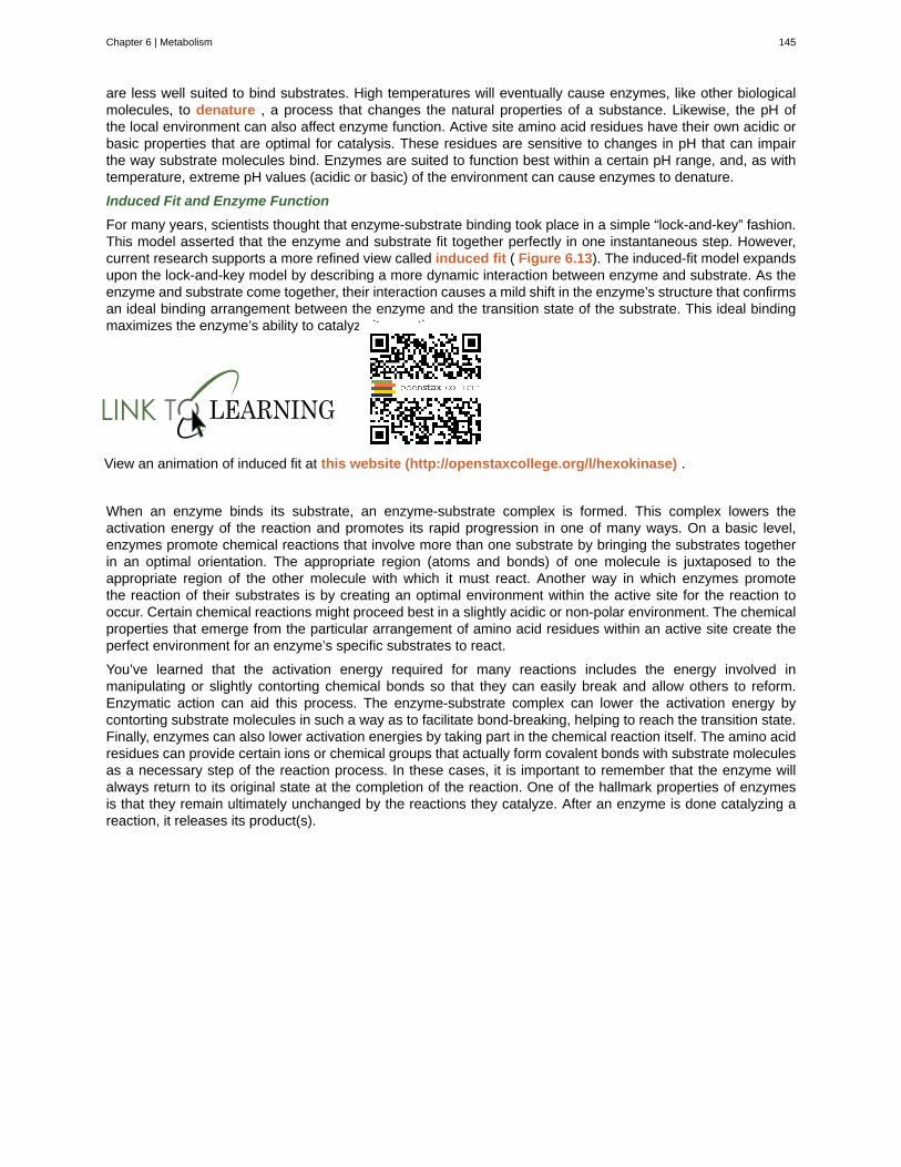
are less well suited to bind substrates. High temperatures will eventually cause enzymes, like other biologicalmolecules, to denature , a process that changes the natural properties of a substance. Likewise, the pH ofthe local environment can also affect enzyme function. Active site amino acid residues have their own acidic orbasic properties that are optimal for catalysis. These residues are sensitive to changes in pH that can impairthe way substrate molecules bind. Enzymes are suited to function best within a certain pH range, and, as withtemperature, extreme pH values (acidic or basic) of the environment can cause enzymes to denature.
Induced Fit and Enzyme Function
For many years, scientists thought that enzyme-substrate binding took place in a simple “lock-and-key” fashion.This model asserted that the enzyme and substrate fit together perfectly in one instantaneous step. However,current research supports a more refined view called induced fit ( Figure 6.13). The induced-fit model expandsupon the lock-and-key model by describing a more dynamic interaction between enzyme and substrate. As theenzyme and substrate come together, their interaction causes a mild shift in the enzyme’s structure that confirmsan ideal binding arrangement between the enzyme and the transition state of the substrate. This ideal bindingmaximizes the enzyme’s ability to catalyze its reaction.
View an animation of induced fit at this website (http://openstaxcollege.org/l/hexokinase) .
When an enzyme binds its substrate, an enzyme-substrate complex is formed. This complex lowers theactivation energy of the reaction and promotes its rapid progression in one of many ways. On a basic level,enzymes promote chemical reactions that involve more than one substrate by bringing the substrates togetherin an optimal orientation. The appropriate region (atoms and bonds) of one molecule is juxtaposed to theappropriate region of the other molecule with which it must react. Another way in which enzymes promotethe reaction of their substrates is by creating an optimal environment within the active site for the reaction tooccur. Certain chemical reactions might proceed best in a slightly acidic or non-polar environment. The chemicalproperties that emerge from the particular arrangement of amino acid residues within an active site create theperfect environment for an enzyme’s specific substrates to react.
You’ve learned that the activation energy required for many reactions includes the energy involved inmanipulating or slightly contorting chemical bonds so that they can easily break and allow others to reform.Enzymatic action can aid this process. The enzyme-substrate complex can lower the activation energy bycontorting substrate molecules in such a way as to facilitate bond-breaking, helping to reach the transition state.Finally, enzymes can also lower activation energies by taking part in the chemical reaction itself. The amino acidresidues can provide certain ions or chemical groups that actually form covalent bonds with substrate moleculesas a necessary step of the reaction process. In these cases, it is important to remember that the enzyme willalways return to its original state at the completion of the reaction. One of the hallmark properties of enzymesis that they remain ultimately unchanged by the reactions they catalyze. After an enzyme is done catalyzing areaction, it releases its product(s).
Chapter 6 | Metabolism 145
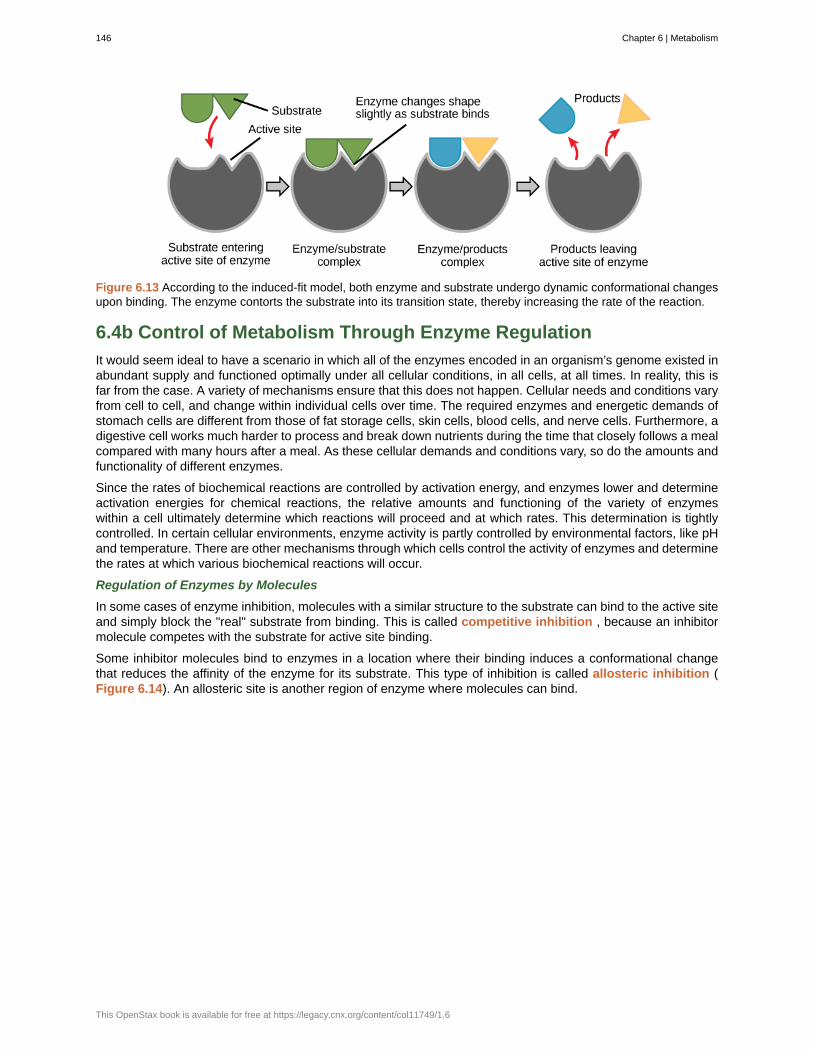
Figure 6.13 According to the induced-fit model, both enzyme and substrate undergo dynamic conformational changesupon binding. The enzyme contorts the substrate into its transition state, thereby increasing the rate of the reaction.
6.4b Control of Metabolism Through Enzyme Regulation
It would seem ideal to have a scenario in which all of the enzymes encoded in an organism’s genome existed inabundant supply and functioned optimally under all cellular conditions, in all cells, at all times. In reality, this isfar from the case. A variety of mechanisms ensure that this does not happen. Cellular needs and conditions varyfrom cell to cell, and change within individual cells over time. The required enzymes and energetic demands ofstomach cells are different from those of fat storage cells, skin cells, blood cells, and nerve cells. Furthermore, adigestive cell works much harder to process and break down nutrients during the time that closely follows a mealcompared with many hours after a meal. As these cellular demands and conditions vary, so do the amounts andfunctionality of different enzymes.
Since the rates of biochemical reactions are controlled by activation energy, and enzymes lower and determineactivation energies for chemical reactions, the relative amounts and functioning of the variety of enzymeswithin a cell ultimately determine which reactions will proceed and at which rates. This determination is tightlycontrolled. In certain cellular environments, enzyme activity is partly controlled by environmental factors, like pHand temperature. There are other mechanisms through which cells control the activity of enzymes and determinethe rates at which various biochemical reactions will occur.
Regulation of Enzymes by Molecules
In some cases of enzyme inhibition, molecules with a similar structure to the substrate can bind to the active siteand simply block the "real" substrate from binding. This is called competitive inhibition , because an inhibitormolecule competes with the substrate for active site binding.
Some inhibitor molecules bind to enzymes in a location where their binding induces a conformational changethat reduces the affinity of the enzyme for its substrate. This type of inhibition is called allosteric inhibition (Figure 6.14). An allosteric site is another region of enzyme where molecules can bind.
146 Chapter 6 | Metabolism
This OpenStax book is available for free at https://legacy.cnx.org/content/col11749/1.6
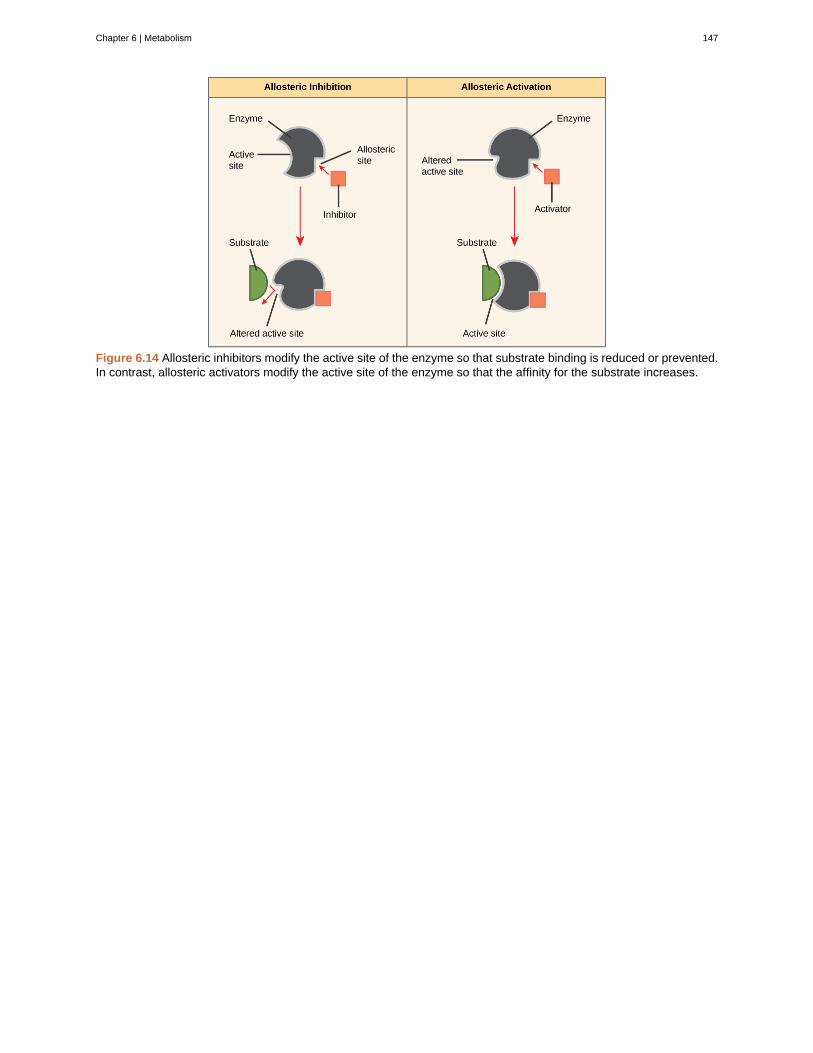
Figure 6.14 Allosteric inhibitors modify the active site of the enzyme so that substrate binding is reduced or prevented.In contrast, allosteric activators modify the active site of the enzyme so that the affinity for the substrate increases.
Chapter 6 | Metabolism 147
Figure 6.15 Have you ever wondered how pharmaceutical drugs are developed? (credit: Deborah Austin)
Drug Discovery by Looking for Inhibitors of Key Enzymes inSpecific PathwaysEnzymes are key components of metabolic pathways. Understanding how enzymes work and how they canbe regulated is a key principle behind the development of many of the pharmaceutical drugs ( Figure 6.15)on the market today. Biologists working in this field collaborate with other scientists, usually chemists, todesign drugs.
Consider statins for example—which is the name given to the class of drugs that reduces cholesterol levels.These compounds are essentially inhibitors of the enzyme HMG-CoA reductase. HMG-CoA reductase isthe enzyme that synthesizes cholesterol from lipids in the body. By inhibiting this enzyme, the levels ofcholesterol synthesized in the body can be reduced. Similarly, acetaminophen, popularly marketed underthe brand name Tylenol, is an inhibitor of the enzyme cyclooxygenase. While it is effective in providing relieffrom fever and inflammation (pain), its mechanism of action is still not completely understood.
How are drugs developed? One of the first challenges in drug development is identifying the specificmolecule that the drug is intended to target. In the case of statins, HMG-CoA reductase is the drug target.Drug targets are identified through painstaking research in the laboratory. Identifying the target alone is notsufficient; scientists also need to know how the target acts inside the cell and which reactions go awry inthe case of disease. Once the target and the pathway are identified, then the actual process of drug designbegins. During this stage, chemists and biologists work together to design and synthesize molecules thatcan either block or activate a particular reaction. However, this is only the beginning: both if and when a drugprototype is successful in performing its function, then it must undergo many tests from in vitro experimentsto clinical trials before it can get FDA approval to be on the market.
Many enzymes don’t work optimally, or even at all, unless bound to other specific non-protein helper molecules,either temporarily through ionic or hydrogen bonds or permanently through stronger covalent bonds. Two typesof helper molecules are cofactors and coenzymes . Binding to these molecules promotes optimal conformationand function for their respective enzymes. Cofactors are inorganic ions such as iron (Fe++) and magnesium(Mg++). One example of an enzyme that requires a metal ion as a cofactor is the enzyme that builds DNAmolecules, DNA polymerase, which requires bound zinc ion (Zn++) to function. Coenzymes are organic helpermolecules, with a basic atomic structure made up of carbon and hydrogen, which are required for enzymeaction. The most common sources of coenzymes are dietary vitamins ( Figure 6.16). Vitamin C is a coenzymefor multiple enzymes that take part in building the important connective tissue component, collagen. Therefore,enzyme function is, in part, regulated by an abundance of various cofactors and coenzymes, which are suppliedprimarily by the diets of most organisms.
148 Chapter 6 | Metabolism
This OpenStax book is available for free at https://legacy.cnx.org/content/col11749/1.6
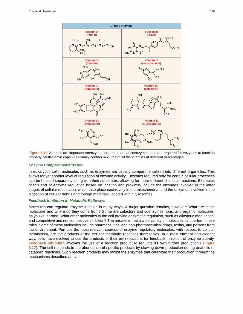
Figure 6.16 Vitamins are important coenzymes or precursors of coenzymes, and are required for enzymes to functionproperly. Multivitamin capsules usually contain mixtures of all the vitamins at different percentages.
Enzyme Compartmentalization
In eukaryotic cells, molecules such as enzymes are usually compartmentalized into different organelles. Thisallows for yet another level of regulation of enzyme activity. Enzymes required only for certain cellular processescan be housed separately along with their substrates, allowing for more efficient chemical reactions. Examplesof this sort of enzyme regulation based on location and proximity include the enzymes involved in the latterstages of cellular respiration, which take place exclusively in the mitochondria, and the enzymes involved in thedigestion of cellular debris and foreign materials, located within lysosomes.
Feedback Inhibition in Metabolic Pathways
Molecules can regulate enzyme function in many ways. A major question remains, however: What are thesemolecules and where do they come from? Some are cofactors and coenzymes, ions, and organic molecules,as you’ve learned. What other molecules in the cell provide enzymatic regulation, such as allosteric modulation,and competitive and noncompetitive inhibition? The answer is that a wide variety of molecules can perform theseroles. Some of these molecules include pharmaceutical and non-pharmaceutical drugs, toxins, and poisons fromthe environment. Perhaps the most relevant sources of enzyme regulatory molecules, with respect to cellularmetabolism, are the products of the cellular metabolic reactions themselves. In a most efficient and elegantway, cells have evolved to use the products of their own reactions for feedback inhibition of enzyme activity.Feedback inhibition involves the use of a reaction product to regulate its own further production ( Figure6.17). The cell responds to the abundance of specific products by slowing down production during anabolic orcatabolic reactions. Such reaction products may inhibit the enzymes that catalyzed their production through themechanisms described above.
Chapter 6 | Metabolism 149
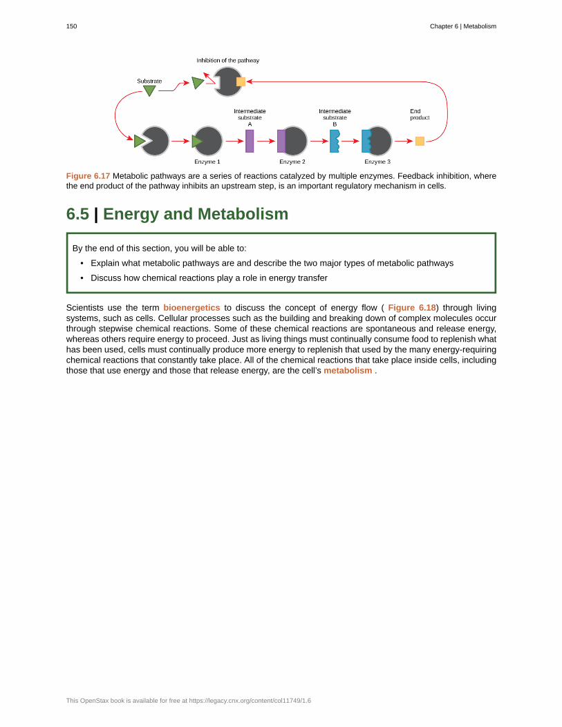
Figure 6.17 Metabolic pathways are a series of reactions catalyzed by multiple enzymes. Feedback inhibition, wherethe end product of the pathway inhibits an upstream step, is an important regulatory mechanism in cells.
6.5 | Energy and Metabolism
By the end of this section, you will be able to:
• Explain what metabolic pathways are and describe the two major types of metabolic pathways
• Discuss how chemical reactions play a role in energy transfer
Scientists use the term bioenergetics to discuss the concept of energy flow ( Figure 6.18) through livingsystems, such as cells. Cellular processes such as the building and breaking down of complex molecules occurthrough stepwise chemical reactions. Some of these chemical reactions are spontaneous and release energy,whereas others require energy to proceed. Just as living things must continually consume food to replenish whathas been used, cells must continually produce more energy to replenish that used by the many energy-requiringchemical reactions that constantly take place. All of the chemical reactions that take place inside cells, includingthose that use energy and those that release energy, are the cell’s metabolism .
150 Chapter 6 | Metabolism
This OpenStax book is available for free at https://legacy.cnx.org/content/col11749/1.6
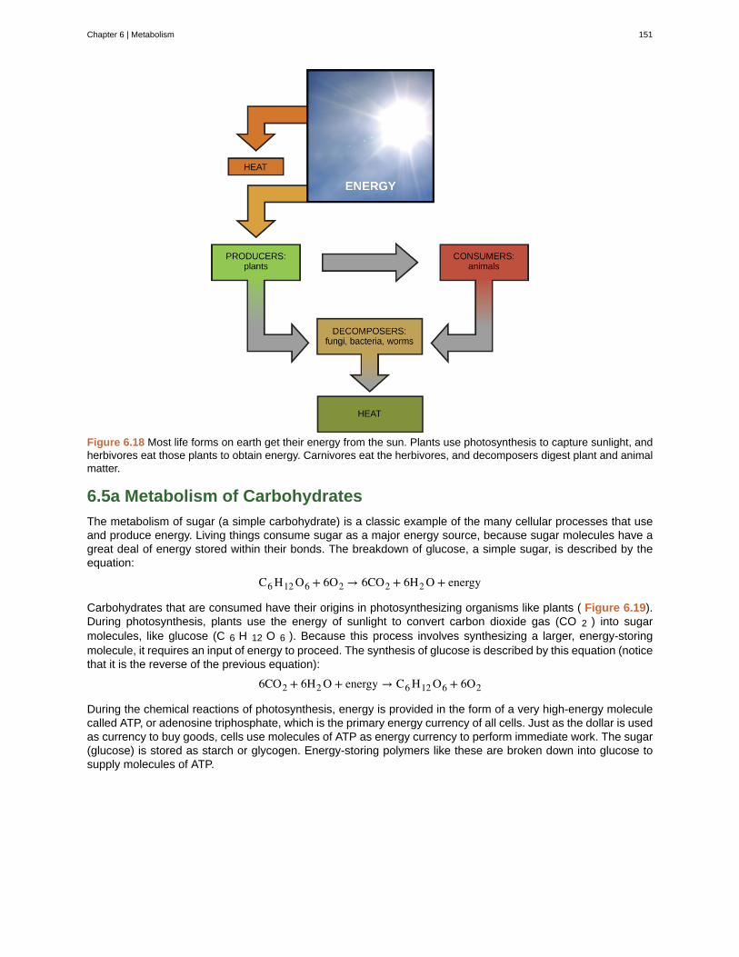
Figure 6.18 Most life forms on earth get their energy from the sun. Plants use photosynthesis to capture sunlight, andherbivores eat those plants to obtain energy. Carnivores eat the herbivores, and decomposers digest plant and animalmatter.
6.5a Metabolism of Carbohydrates
The metabolism of sugar (a simple carbohydrate) is a classic example of the many cellular processes that useand produce energy. Living things consume sugar as a major energy source, because sugar molecules have agreat deal of energy stored within their bonds. The breakdown of glucose, a simple sugar, is described by theequation:
C6 H12 O6 + 6O2 → 6CO2 + 6H2 O + energy
Carbohydrates that are consumed have their origins in photosynthesizing organisms like plants ( Figure 6.19).During photosynthesis, plants use the energy of sunlight to convert carbon dioxide gas (CO 2 ) into sugarmolecules, like glucose (C 6 H 12 O 6 ). Because this process involves synthesizing a larger, energy-storingmolecule, it requires an input of energy to proceed. The synthesis of glucose is described by this equation (noticethat it is the reverse of the previous equation):
6CO2 + 6H2 O + energy → C6 H12 O6 + 6O2
During the chemical reactions of photosynthesis, energy is provided in the form of a very high-energy moleculecalled ATP, or adenosine triphosphate, which is the primary energy currency of all cells. Just as the dollar is usedas currency to buy goods, cells use molecules of ATP as energy currency to perform immediate work. The sugar(glucose) is stored as starch or glycogen. Energy-storing polymers like these are broken down into glucose tosupply molecules of ATP.
Chapter 6 | Metabolism 151
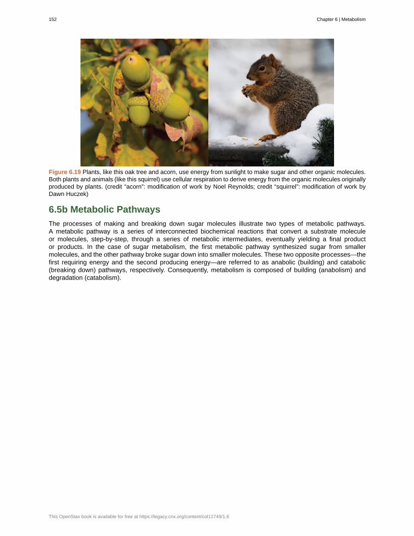
Figure 6.19 Plants, like this oak tree and acorn, use energy from sunlight to make sugar and other organic molecules.Both plants and animals (like this squirrel) use cellular respiration to derive energy from the organic molecules originallyproduced by plants. (credit “acorn”: modification of work by Noel Reynolds; credit “squirrel”: modification of work byDawn Huczek)
6.5b Metabolic Pathways
The processes of making and breaking down sugar molecules illustrate two types of metabolic pathways.A metabolic pathway is a series of interconnected biochemical reactions that convert a substrate moleculeor molecules, step-by-step, through a series of metabolic intermediates, eventually yielding a final productor products. In the case of sugar metabolism, the first metabolic pathway synthesized sugar from smallermolecules, and the other pathway broke sugar down into smaller molecules. These two opposite processes—thefirst requiring energy and the second producing energy—are referred to as anabolic (building) and catabolic(breaking down) pathways, respectively. Consequently, metabolism is composed of building (anabolism) anddegradation (catabolism).
152 Chapter 6 | Metabolism
This OpenStax book is available for free at https://legacy.cnx.org/content/col11749/1.6
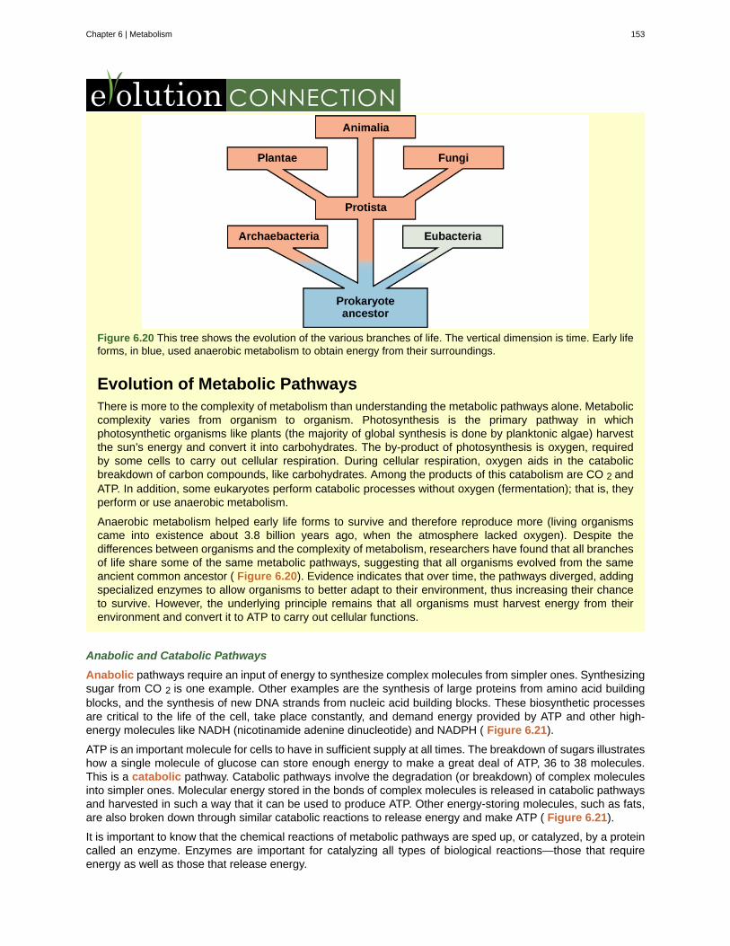
Figure 6.20 This tree shows the evolution of the various branches of life. The vertical dimension is time. Early lifeforms, in blue, used anaerobic metabolism to obtain energy from their surroundings.
Evolution of Metabolic PathwaysThere is more to the complexity of metabolism than understanding the metabolic pathways alone. Metaboliccomplexity varies from organism to organism. Photosynthesis is the primary pathway in whichphotosynthetic organisms like plants (the majority of global synthesis is done by planktonic algae) harvestthe sun’s energy and convert it into carbohydrates. The by-product of photosynthesis is oxygen, requiredby some cells to carry out cellular respiration. During cellular respiration, oxygen aids in the catabolicbreakdown of carbon compounds, like carbohydrates. Among the products of this catabolism are CO 2 andATP. In addition, some eukaryotes perform catabolic processes without oxygen (fermentation); that is, theyperform or use anaerobic metabolism.
Anaerobic metabolism helped early life forms to survive and therefore reproduce more (living organismscame into existence about 3.8 billion years ago, when the atmosphere lacked oxygen). Despite thedifferences between organisms and the complexity of metabolism, researchers have found that all branchesof life share some of the same metabolic pathways, suggesting that all organisms evolved from the sameancient common ancestor ( Figure 6.20). Evidence indicates that over time, the pathways diverged, addingspecialized enzymes to allow organisms to better adapt to their environment, thus increasing their chanceto survive. However, the underlying principle remains that all organisms must harvest energy from theirenvironment and convert it to ATP to carry out cellular functions.
Anabolic and Catabolic Pathways
Anabolic pathways require an input of energy to synthesize complex molecules from simpler ones. Synthesizingsugar from CO 2 is one example. Other examples are the synthesis of large proteins from amino acid buildingblocks, and the synthesis of new DNA strands from nucleic acid building blocks. These biosynthetic processesare critical to the life of the cell, take place constantly, and demand energy provided by ATP and other high-energy molecules like NADH (nicotinamide adenine dinucleotide) and NADPH ( Figure 6.21).
ATP is an important molecule for cells to have in sufficient supply at all times. The breakdown of sugars illustrateshow a single molecule of glucose can store enough energy to make a great deal of ATP, 36 to 38 molecules.This is a catabolic pathway. Catabolic pathways involve the degradation (or breakdown) of complex moleculesinto simpler ones. Molecular energy stored in the bonds of complex molecules is released in catabolic pathwaysand harvested in such a way that it can be used to produce ATP. Other energy-storing molecules, such as fats,are also broken down through similar catabolic reactions to release energy and make ATP ( Figure 6.21).
It is important to know that the chemical reactions of metabolic pathways are sped up, or catalyzed, by a proteincalled an enzyme. Enzymes are important for catalyzing all types of biological reactions—those that requireenergy as well as those that release energy.
Chapter 6 | Metabolism 153
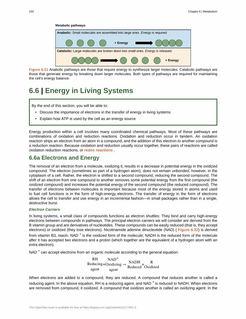
Figure 6.21 Anabolic pathways are those that require energy to synthesize larger molecules. Catabolic pathways arethose that generate energy by breaking down larger molecules. Both types of pathways are required for maintainingthe cell’s energy balance.
6.6 | Energy in Living Systems
By the end of this section, you will be able to:
• Discuss the importance of electrons in the transfer of energy in living systems
• Explain how ATP is used by the cell as an energy source
Energy production within a cell involves many coordinated chemical pathways. Most of these pathways arecombinations of oxidation and reduction reactions. Oxidation and reduction occur in tandem. An oxidationreaction strips an electron from an atom in a compound, and the addition of this electron to another compound isa reduction reaction. Because oxidation and reduction usually occur together, these pairs of reactions are calledoxidation reduction reactions, or redox reactions .
6.6a Electrons and Energy
The removal of an electron from a molecule, oxidizing it, results in a decrease in potential energy in the oxidizedcompound. The electron (sometimes as part of a hydrogen atom), does not remain unbonded, however, in thecytoplasm of a cell. Rather, the electron is shifted to a second compound, reducing the second compound. Theshift of an electron from one compound to another removes some potential energy from the first compound (theoxidized compound) and increases the potential energy of the second compound (the reduced compound). Thetransfer of electrons between molecules is important because most of the energy stored in atoms and usedto fuel cell functions is in the form of high-energy electrons. The transfer of energy in the form of electronsallows the cell to transfer and use energy in an incremental fashion—in small packages rather than in a single,destructive burst.
Electron Carriers
In living systems, a small class of compounds functions as electron shuttles: They bind and carry high-energyelectrons between compounds in pathways. The principal electron carriers we will consider are derived from theB vitamin group and are derivatives of nucleotides. These compounds can be easily reduced (that is, they acceptelectrons) or oxidized (they lose electrons). Nicotinamide adenine dinucleotide (NAD) ( Figure 6.22) is derived
from vitamin B3, niacin. NAD + is the oxidized form of the molecule; NADH is the reduced form of the moleculeafter it has accepted two electrons and a proton (which together are the equivalent of a hydrogen atom with anextra electron).
NAD + can accept electrons from an organic molecule according to the general equation:
RHReducing
agent+
NAD+
Oxidizingagent
→ NADHReduced
+ ROxidized
When electrons are added to a compound, they are reduced. A compound that reduces another is called a
reducing agent. In the above equation, RH is a reducing agent, and NAD + is reduced to NADH. When electronsare removed from compound, it oxidized. A compound that oxidizes another is called an oxidizing agent. In the
154 Chapter 6 | Metabolism
This OpenStax book is available for free at https://legacy.cnx.org/content/col11749/1.6

above equation, NAD + is an oxidizing agent, and RH is oxidized to R.
Similarly, flavin adenine dinucleotide (FAD + ) is derived from vitamin B 2 , also called riboflavin. Its reduced form
is FADH 2 . A second variation of NAD, NADP, contains an extra phosphate group. Both NAD + and FAD + areextensively used in energy extraction from sugars, and NADP plays an important role in anabolic reactions andphotosynthesis.
Figure 6.22 The oxidized form of the electron carrier (NAD + ) is shown on the left and the reduced form (NADH) is
shown on the right. The nitrogenous base in NADH has one more hydrogen ion and two more electrons than in NAD +
.
Chapter 6 | Metabolism 155
activation energy
active site
adenosine triphosphate
allosteric inhibition
anabolic
anabolic
ATP
bioenergetics
catabolic
catabolic
chemical energy
Chemiosmosis
coenzymes
cofactors
competitive inhibition
denature
dephosphorylation
endergonic reactions
entropy
enzymes
exergonic reactions
feedback inhibition
heat energy
heat energy
induced fit
kinetic energy
metabolism
KEY TERMS
energy necessary for reactions to occur
specific region of the enzyme to which the substrate binds
see ATP
inhibition by a binding event at a site different from the active site, which induces aconformational change and reduces the affinity of the enzyme for its substrate
reactions that use an overall input of energy to synthesize molecules
(also, anabolism) pathways that require an input of energy to synthesize complex molecules fromsimpler ones
adenosine triphosphate, the cell’s energy currency
study of energy flowing through living systems
reactions that break molecules down, and overall release energy
(also, catabolism) pathways in which complex molecules are broken down into simpler ones
potential energy in chemical bonds that is released when those bonds are broken
process in which ATP is produced during cellular metabolism by the involvement of a protongradient across a membrane
small organic molecule, such as a vitamin or its derivative, which is required to enhance the activityof an enzyme
inorganic ion, such as iron or magnesium, required for optimal regulation of enzyme activity
type of inhibition in which the inhibitor competes with the substrate molecule by bindingto the active site of the enzyme
process that changes the natural properties of a substance
removal of a phosphate group from a molecule
chemical reactions that require a net input of energy
measure of randomness or disorder within a system
molecules that speed up a chemical reaction
reactions that exhibit a net release of energy
effect of a product of a reaction sequence to decrease its further production by inhibitingthe activity of the first enzyme in the pathway that produces it
total bond energy of reactants or products in a chemical reaction
total bond energy of reactants or products in a chemical reacation
dynamic fit between the enzyme and its substrate, in which both components modify theirstructures to allow for ideal binding
type of energy associated with objects or particles in motion
all the chemical reactions that take place inside cells, including anabolism and catabolism
156 Chapter 6 | Metabolism
This OpenStax book is available for free at https://legacy.cnx.org/content/col11749/1.6
oxidative phosphorylation
Phosphorylation
potential energy
redox reactions
substrate-level phosphorylation
substrates
thermodynamics
transition state
production of ATP using the process of chemiosmosis and oxygen
addition of a high-energy phosphate to a compound, usually a metabolic intermediate, aprotein, or ADP
type of energy that has the potential to do work; stored energy
chemical reaction that consists of the coupling of an oxidation reaction to a reduction reaction
production of ATP from ADP using the excess energy from a chemicalreaction and a phosphate group from a reactant
molecules on which enzymes act
study of energy and energy transfer involving physical matter
high-energy, unstable state (an intermediate form between the substrate and the product)occurring during a chemical reaction
CHAPTER SUMMARY
6.1 Potential, Kinetic, Free, and Activation Energy
Energy comes in many different forms. Objects in motion do physical work, and kinetic energy is the energy ofobjects in motion. Objects that are not in motion may have the potential to do work, and thus, have potentialenergy. Molecules also have potential energy because the breaking of molecular bonds has the potential torelease energy. Living cells depend on the harvesting of potential energy from molecular bonds to performwork.
A reaction can exhibit a net release of energy or net consumption of energy. A reaction that gives off energy iscalled an exergonic reaction. One that requires energy input is called an endergonic reaction. Exergonicreactions are said to be spontaneous, because their products have less energy than their reactants. Theproducts of endergonic reactions have a higher energy state than the reactants, and so these arenonspontaneous reactions. However, all reactions (including spontaneous reactions) require an initial input ofenergy to reach the transition state, at which they’ll proceed. This initial input of energy is called the activationenergy.
6.2 The Laws of Thermodynamics
In studying energy, scientists use the term “system” to refer to the matter and its environment involved inenergy transfers. Everything outside of the system is called the surroundings. Single cells are biologicalsystems. Systems can be thought of as having a certain amount of order. It takes energy to make a systemmore ordered. The more ordered a system is, the lower its entropy. Entropy is a measure of the disorder of asystem. As a system becomes more disordered, the lower its energy and the higher its entropy become.
A series of laws, called the laws of thermodynamics, describe the properties and processes of energy transfer.The first law states that the total amount of energy in the universe is constant. This means that energy can’t becreated or destroyed, only transferred or transformed. The second law of thermodynamics states that everyenergy transfer involves some loss of energy in an unusable form, such as heat energy, resulting in a moredisordered system. In other words, no energy transfer is completely efficient and tends toward disorder.
6.3 Adenosine Triphosphate
ATP is the primary energy-supplying molecule for living cells. ATP is made up of a nucleotide, a five-carbonsugar, and three phosphate groups. The bonds that connect the phosphates (phosphoanhydride bonds) havehigh-energy content. The energy released from the hydrolysis of ATP into ADP + P i is used to perform cellularwork. Cells use ATP to perform work by coupling the exergonic reaction of ATP hydrolysis with endergonicreactions. ATP donates its phosphate group to another molecule via a process known as phosphorylation. Thephosphorylated molecule is at a higher-energy state and is less stable than its unphosphorylated form, and thisadded energy from the addition of the phosphate allows the molecule to undergo its endergonic reaction.
ATP functions as the energy currency for cells. It allows the cells to store energy briefly and transport it within
Chapter 6 | Metabolism 157

the cell to support endergonic chemical reactions. The structure of ATP is that of an RNA nucleotide with threephosphates attached. As ATP is used for energy, a phosphate group or two are detached, and either ADP orAMP is produced. Energy derived from glucose catabolism is used to convert ADP into ATP. When ATP is usedin a reaction, the third phosphate is temporarily attached to a substrate in a process called phosphorylation.The two processes of ATP regeneration that are used in conjunction with glucose catabolism are substrate-level phosphorylation and oxidative phosphorylation through the process of chemiosmosis.
6.4 Enzymes
Enzymes are chemical catalysts that accelerate chemical reactions at physiological temperatures by loweringtheir activation energy. Enzymes are usually proteins consisting of one or more polypeptide chains. Enzymeshave an active site that provides a unique chemical environment, made up of certain amino acid R groups(residues). This unique environment is perfectly suited to convert particular chemical reactants for that enzyme,called substrates, into unstable intermediates called transition states. Enzymes and substrates are thought tobind with an induced fit, which means that enzymes undergo slight conformational adjustments upon substratecontact, leading to full, optimal binding. Enzymes bind to substrates and catalyze reactions in four differentways: bringing substrates together in an optimal orientation, compromising the bond structures of substrates sothat bonds can be more easily broken, providing optimal environmental conditions for a reaction to occur, orparticipating directly in their chemical reaction by forming transient covalent bonds with the substrates.
Enzyme action must be regulated so that in a given cell at a given time, the desired reactions are beingcatalyzed and the undesired reactions are not. Enzymes are regulated by cellular conditions, such astemperature and pH. They are also regulated through their location within a cell, sometimes beingcompartmentalized so that they can only catalyze reactions under certain circumstances. Inhibition andactivation of enzymes via other molecules are other important ways that enzymes are regulated. Inhibitors canact competitively, noncompetitively, or allosterically; noncompetitive inhibitors are usually allosteric. Activatorscan also enhance the function of enzymes allosterically. The most common method by which cells regulate theenzymes in metabolic pathways is through feedback inhibition. During feedback inhibition, the products of ametabolic pathway serve as inhibitors (usually allosteric) of one or more of the enzymes (usually the firstcommitted enzyme of the pathway) involved in the pathway that produces them.
6.5 Energy and Metabolism
Cells perform the functions of life through various chemical reactions. A cell’s metabolism refers to the chemicalreactions that take place within it. There are metabolic reactions that involve the breaking down of complexchemicals into simpler ones, such as the breakdown of large macromolecules. This process is referred to ascatabolism, and such reactions are associated with a release of energy. On the other end of the spectrum,anabolism refers to metabolic processes that build complex molecules out of simpler ones, such as thesynthesis of macromolecules. Anabolic processes require energy. Glucose synthesis and glucose breakdownare examples of anabolic and catabolic pathways, respectively.
6.6 Energy in Living Systems
One of the ways energy is moved is by transporting electrons. The gain or loss of an electron can alter amolecule so it now behaves differently. A molecule that gained an electron is “reduced” because it becamemore negative. A molecule that lost an electron is “oxidized” -- it became more positive. Certain molecules’ onlyfunction is to transport electrons around a cell (ex. NAD, FAD+).
158 Chapter 6 | Metabolism
This OpenStax book is available for free at https://legacy.cnx.org/content/col11749/1.6
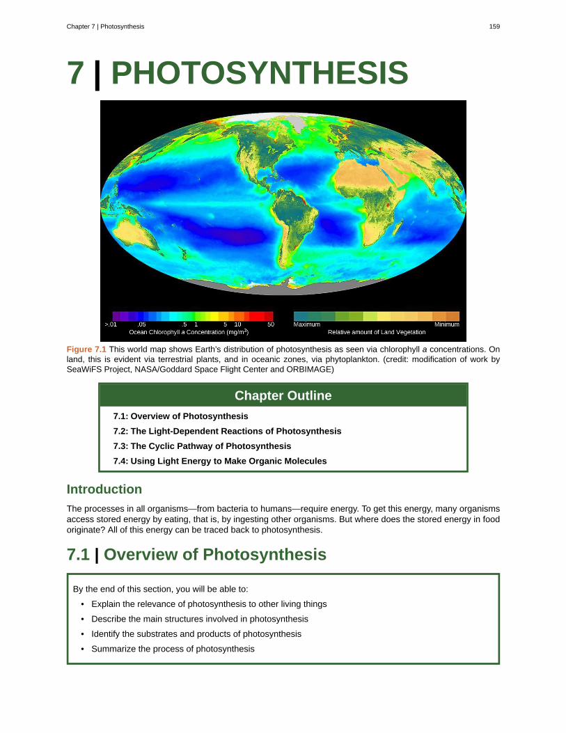
7 | PHOTOSYNTHESIS
Figure 7.1 This world map shows Earth’s distribution of photosynthesis as seen via chlorophyll a concentrations. Onland, this is evident via terrestrial plants, and in oceanic zones, via phytoplankton. (credit: modification of work bySeaWiFS Project, NASA/Goddard Space Flight Center and ORBIMAGE)
Chapter Outline
7.1: Overview of Photosynthesis
7.2: The Light-Dependent Reactions of Photosynthesis
7.3: The Cyclic Pathway of Photosynthesis
7.4: Using Light Energy to Make Organic Molecules
Introduction
The processes in all organisms—from bacteria to humans—require energy. To get this energy, many organismsaccess stored energy by eating, that is, by ingesting other organisms. But where does the stored energy in foodoriginate? All of this energy can be traced back to photosynthesis.
7.1 | Overview of Photosynthesis
By the end of this section, you will be able to:
• Explain the relevance of photosynthesis to other living things
• Describe the main structures involved in photosynthesis
• Identify the substrates and products of photosynthesis
• Summarize the process of photosynthesis
Chapter 7 | Photosynthesis 159
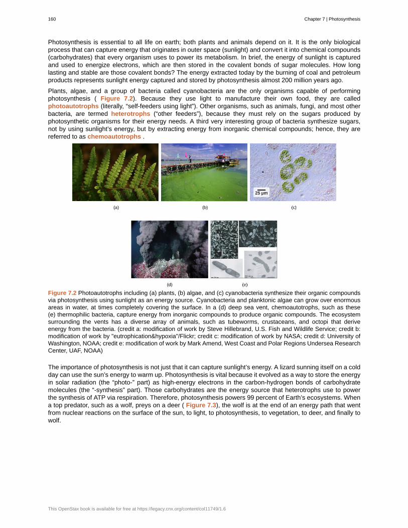
Photosynthesis is essential to all life on earth; both plants and animals depend on it. It is the only biologicalprocess that can capture energy that originates in outer space (sunlight) and convert it into chemical compounds(carbohydrates) that every organism uses to power its metabolism. In brief, the energy of sunlight is capturedand used to energize electrons, which are then stored in the covalent bonds of sugar molecules. How longlasting and stable are those covalent bonds? The energy extracted today by the burning of coal and petroleumproducts represents sunlight energy captured and stored by photosynthesis almost 200 million years ago.
Plants, algae, and a group of bacteria called cyanobacteria are the only organisms capable of performingphotosynthesis ( Figure 7.2). Because they use light to manufacture their own food, they are calledphotoautotrophs (literally, “self-feeders using light”). Other organisms, such as animals, fungi, and most otherbacteria, are termed heterotrophs (“other feeders”), because they must rely on the sugars produced byphotosynthetic organisms for their energy needs. A third very interesting group of bacteria synthesize sugars,not by using sunlight’s energy, but by extracting energy from inorganic chemical compounds; hence, they arereferred to as chemoautotrophs .
Figure 7.2 Photoautotrophs including (a) plants, (b) algae, and (c) cyanobacteria synthesize their organic compoundsvia photosynthesis using sunlight as an energy source. Cyanobacteria and planktonic algae can grow over enormousareas in water, at times completely covering the surface. In a (d) deep sea vent, chemoautotrophs, such as these(e) thermophilic bacteria, capture energy from inorganic compounds to produce organic compounds. The ecosystemsurrounding the vents has a diverse array of animals, such as tubeworms, crustaceans, and octopi that deriveenergy from the bacteria. (credit a: modification of work by Steve Hillebrand, U.S. Fish and Wildlife Service; credit b:modification of work by "eutrophication&hypoxia"/Flickr; credit c: modification of work by NASA; credit d: University ofWashington, NOAA; credit e: modification of work by Mark Amend, West Coast and Polar Regions Undersea ResearchCenter, UAF, NOAA)
The importance of photosynthesis is not just that it can capture sunlight’s energy. A lizard sunning itself on a coldday can use the sun’s energy to warm up. Photosynthesis is vital because it evolved as a way to store the energyin solar radiation (the “photo-” part) as high-energy electrons in the carbon-hydrogen bonds of carbohydratemolecules (the “-synthesis” part). Those carbohydrates are the energy source that heterotrophs use to powerthe synthesis of ATP via respiration. Therefore, photosynthesis powers 99 percent of Earth’s ecosystems. Whena top predator, such as a wolf, preys on a deer ( Figure 7.3), the wolf is at the end of an energy path that wentfrom nuclear reactions on the surface of the sun, to light, to photosynthesis, to vegetation, to deer, and finally towolf.
160 Chapter 7 | Photosynthesis
This OpenStax book is available for free at https://legacy.cnx.org/content/col11749/1.6
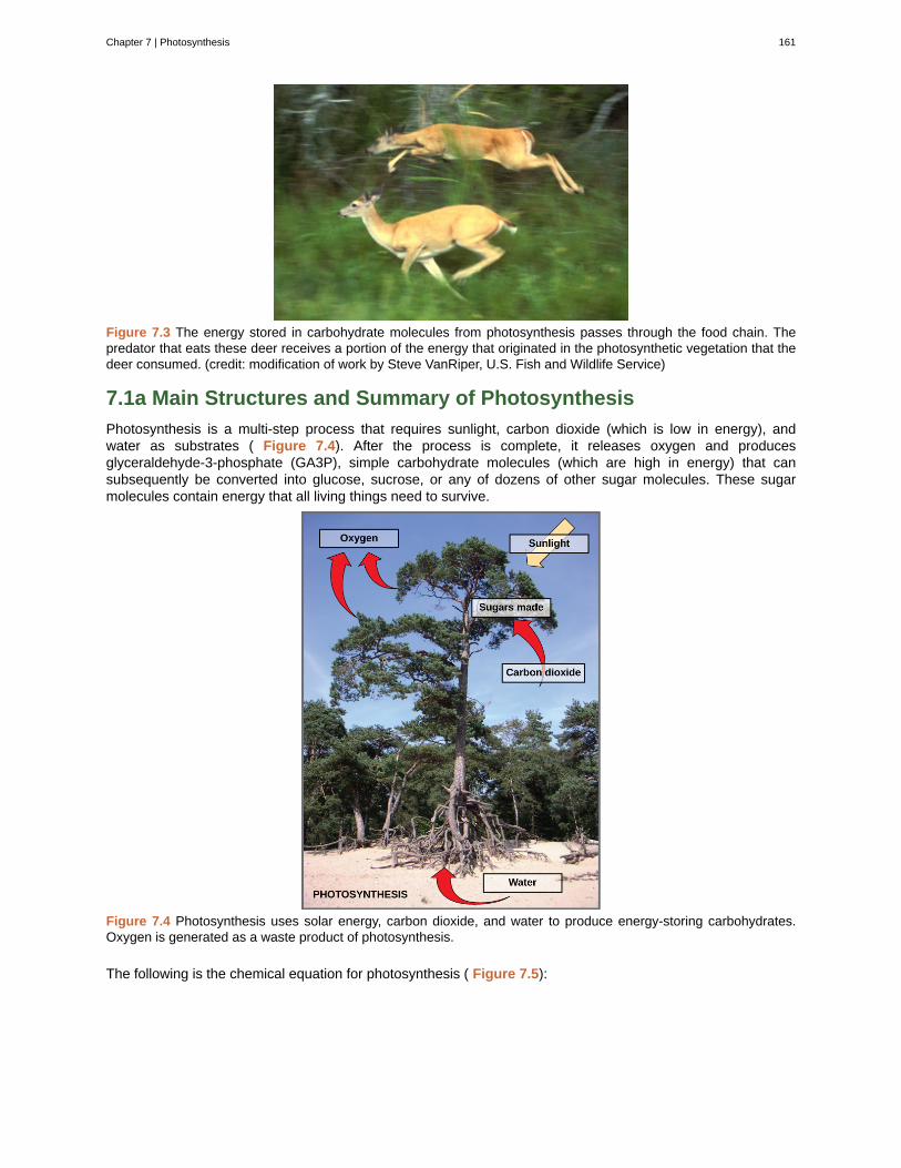
Figure 7.3 The energy stored in carbohydrate molecules from photosynthesis passes through the food chain. Thepredator that eats these deer receives a portion of the energy that originated in the photosynthetic vegetation that thedeer consumed. (credit: modification of work by Steve VanRiper, U.S. Fish and Wildlife Service)
7.1a Main Structures and Summary of Photosynthesis
Photosynthesis is a multi-step process that requires sunlight, carbon dioxide (which is low in energy), andwater as substrates ( Figure 7.4). After the process is complete, it releases oxygen and producesglyceraldehyde-3-phosphate (GA3P), simple carbohydrate molecules (which are high in energy) that cansubsequently be converted into glucose, sucrose, or any of dozens of other sugar molecules. These sugarmolecules contain energy that all living things need to survive.
Figure 7.4 Photosynthesis uses solar energy, carbon dioxide, and water to produce energy-storing carbohydrates.Oxygen is generated as a waste product of photosynthesis.
The following is the chemical equation for photosynthesis ( Figure 7.5):
Chapter 7 | Photosynthesis 161
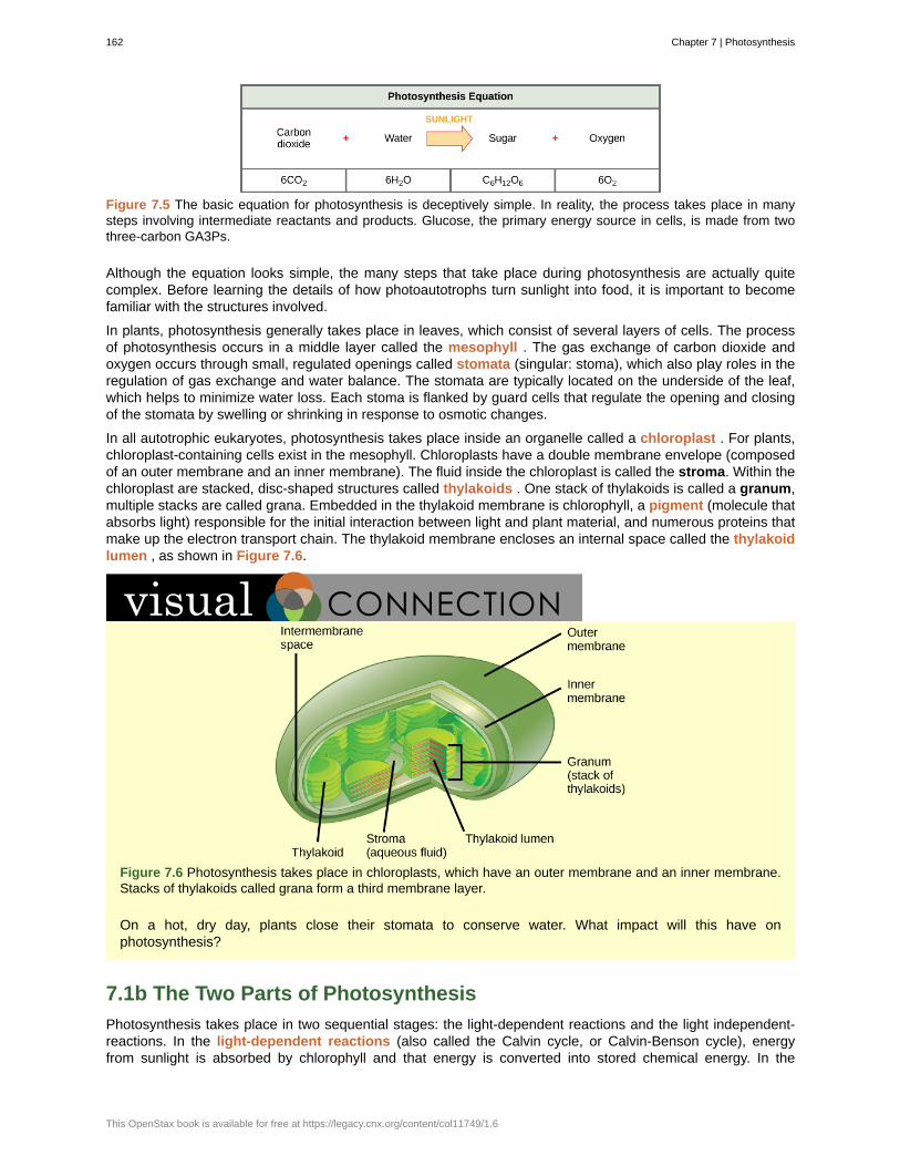
Figure 7.5 The basic equation for photosynthesis is deceptively simple. In reality, the process takes place in manysteps involving intermediate reactants and products. Glucose, the primary energy source in cells, is made from twothree-carbon GA3Ps.
Although the equation looks simple, the many steps that take place during photosynthesis are actually quitecomplex. Before learning the details of how photoautotrophs turn sunlight into food, it is important to becomefamiliar with the structures involved.
In plants, photosynthesis generally takes place in leaves, which consist of several layers of cells. The processof photosynthesis occurs in a middle layer called the mesophyll . The gas exchange of carbon dioxide andoxygen occurs through small, regulated openings called stomata (singular: stoma), which also play roles in theregulation of gas exchange and water balance. The stomata are typically located on the underside of the leaf,which helps to minimize water loss. Each stoma is flanked by guard cells that regulate the opening and closingof the stomata by swelling or shrinking in response to osmotic changes.
In all autotrophic eukaryotes, photosynthesis takes place inside an organelle called a chloroplast . For plants,chloroplast-containing cells exist in the mesophyll. Chloroplasts have a double membrane envelope (composedof an outer membrane and an inner membrane). The fluid inside the chloroplast is called the stroma. Within thechloroplast are stacked, disc-shaped structures called thylakoids . One stack of thylakoids is called a granum,multiple stacks are called grana. Embedded in the thylakoid membrane is chlorophyll, a pigment (molecule thatabsorbs light) responsible for the initial interaction between light and plant material, and numerous proteins thatmake up the electron transport chain. The thylakoid membrane encloses an internal space called the thylakoidlumen , as shown in Figure 7.6.
Figure 7.6 Photosynthesis takes place in chloroplasts, which have an outer membrane and an inner membrane.Stacks of thylakoids called grana form a third membrane layer.
On a hot, dry day, plants close their stomata to conserve water. What impact will this have onphotosynthesis?
7.1b The Two Parts of Photosynthesis
Photosynthesis takes place in two sequential stages: the light-dependent reactions and the light independent-reactions. In the light-dependent reactions (also called the Calvin cycle, or Calvin-Benson cycle), energyfrom sunlight is absorbed by chlorophyll and that energy is converted into stored chemical energy. In the
162 Chapter 7 | Photosynthesis
This OpenStax book is available for free at https://legacy.cnx.org/content/col11749/1.6
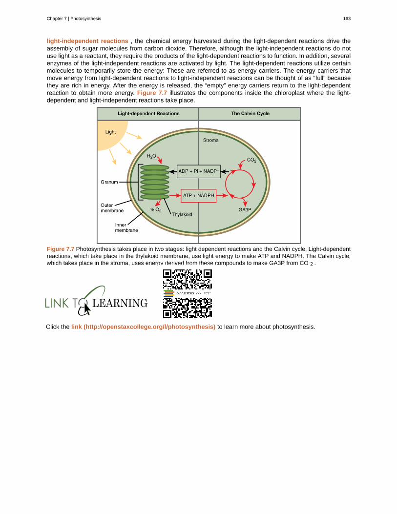
light-independent reactions , the chemical energy harvested during the light-dependent reactions drive theassembly of sugar molecules from carbon dioxide. Therefore, although the light-independent reactions do notuse light as a reactant, they require the products of the light-dependent reactions to function. In addition, severalenzymes of the light-independent reactions are activated by light. The light-dependent reactions utilize certainmolecules to temporarily store the energy: These are referred to as energy carriers. The energy carriers thatmove energy from light-dependent reactions to light-independent reactions can be thought of as “full” becausethey are rich in energy. After the energy is released, the “empty” energy carriers return to the light-dependentreaction to obtain more energy. Figure 7.7 illustrates the components inside the chloroplast where the light-dependent and light-independent reactions take place.
Figure 7.7 Photosynthesis takes place in two stages: light dependent reactions and the Calvin cycle. Light-dependentreactions, which take place in the thylakoid membrane, use light energy to make ATP and NADPH. The Calvin cycle,which takes place in the stroma, uses energy derived from these compounds to make GA3P from CO 2 .
Click the link (http://openstaxcollege.org/l/photosynthesis) to learn more about photosynthesis.
Chapter 7 | Photosynthesis 163
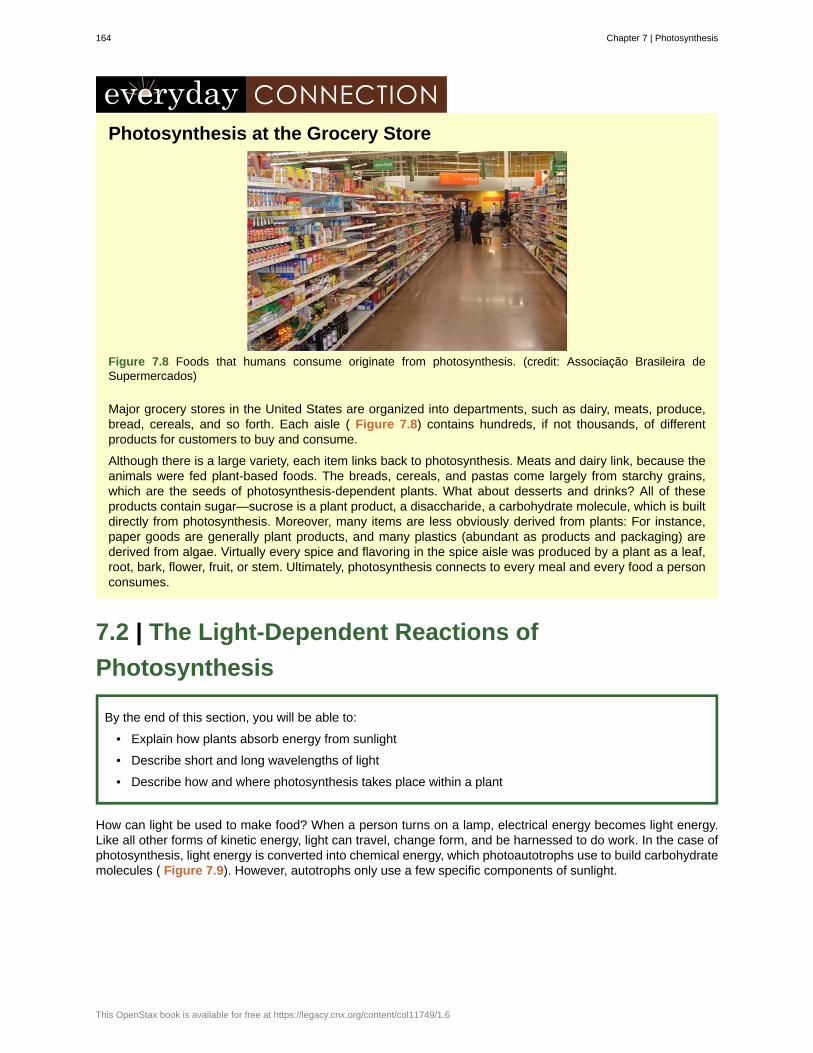
Photosynthesis at the Grocery Store
Figure 7.8 Foods that humans consume originate from photosynthesis. (credit: Associação Brasileira deSupermercados)
Major grocery stores in the United States are organized into departments, such as dairy, meats, produce,bread, cereals, and so forth. Each aisle ( Figure 7.8) contains hundreds, if not thousands, of differentproducts for customers to buy and consume.
Although there is a large variety, each item links back to photosynthesis. Meats and dairy link, because theanimals were fed plant-based foods. The breads, cereals, and pastas come largely from starchy grains,which are the seeds of photosynthesis-dependent plants. What about desserts and drinks? All of theseproducts contain sugar—sucrose is a plant product, a disaccharide, a carbohydrate molecule, which is builtdirectly from photosynthesis. Moreover, many items are less obviously derived from plants: For instance,paper goods are generally plant products, and many plastics (abundant as products and packaging) arederived from algae. Virtually every spice and flavoring in the spice aisle was produced by a plant as a leaf,root, bark, flower, fruit, or stem. Ultimately, photosynthesis connects to every meal and every food a personconsumes.
7.2 | The Light-Dependent Reactions of
Photosynthesis
By the end of this section, you will be able to:
• Explain how plants absorb energy from sunlight
• Describe short and long wavelengths of light
• Describe how and where photosynthesis takes place within a plant
How can light be used to make food? When a person turns on a lamp, electrical energy becomes light energy.Like all other forms of kinetic energy, light can travel, change form, and be harnessed to do work. In the case ofphotosynthesis, light energy is converted into chemical energy, which photoautotrophs use to build carbohydratemolecules ( Figure 7.9). However, autotrophs only use a few specific components of sunlight.
164 Chapter 7 | Photosynthesis
This OpenStax book is available for free at https://legacy.cnx.org/content/col11749/1.6
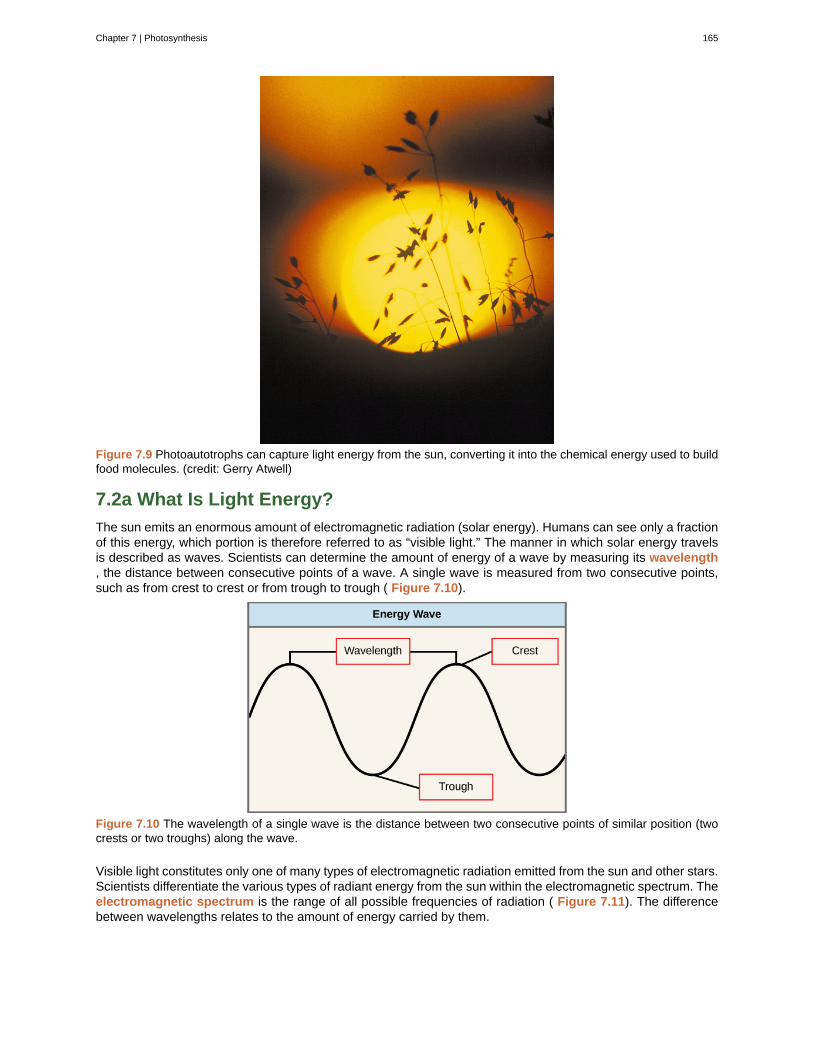
Figure 7.9 Photoautotrophs can capture light energy from the sun, converting it into the chemical energy used to buildfood molecules. (credit: Gerry Atwell)
7.2a What Is Light Energy?
The sun emits an enormous amount of electromagnetic radiation (solar energy). Humans can see only a fractionof this energy, which portion is therefore referred to as “visible light.” The manner in which solar energy travelsis described as waves. Scientists can determine the amount of energy of a wave by measuring its wavelength, the distance between consecutive points of a wave. A single wave is measured from two consecutive points,such as from crest to crest or from trough to trough ( Figure 7.10).
Figure 7.10 The wavelength of a single wave is the distance between two consecutive points of similar position (twocrests or two troughs) along the wave.
Visible light constitutes only one of many types of electromagnetic radiation emitted from the sun and other stars.Scientists differentiate the various types of radiant energy from the sun within the electromagnetic spectrum. Theelectromagnetic spectrum is the range of all possible frequencies of radiation ( Figure 7.11). The differencebetween wavelengths relates to the amount of energy carried by them.
Chapter 7 | Photosynthesis 165
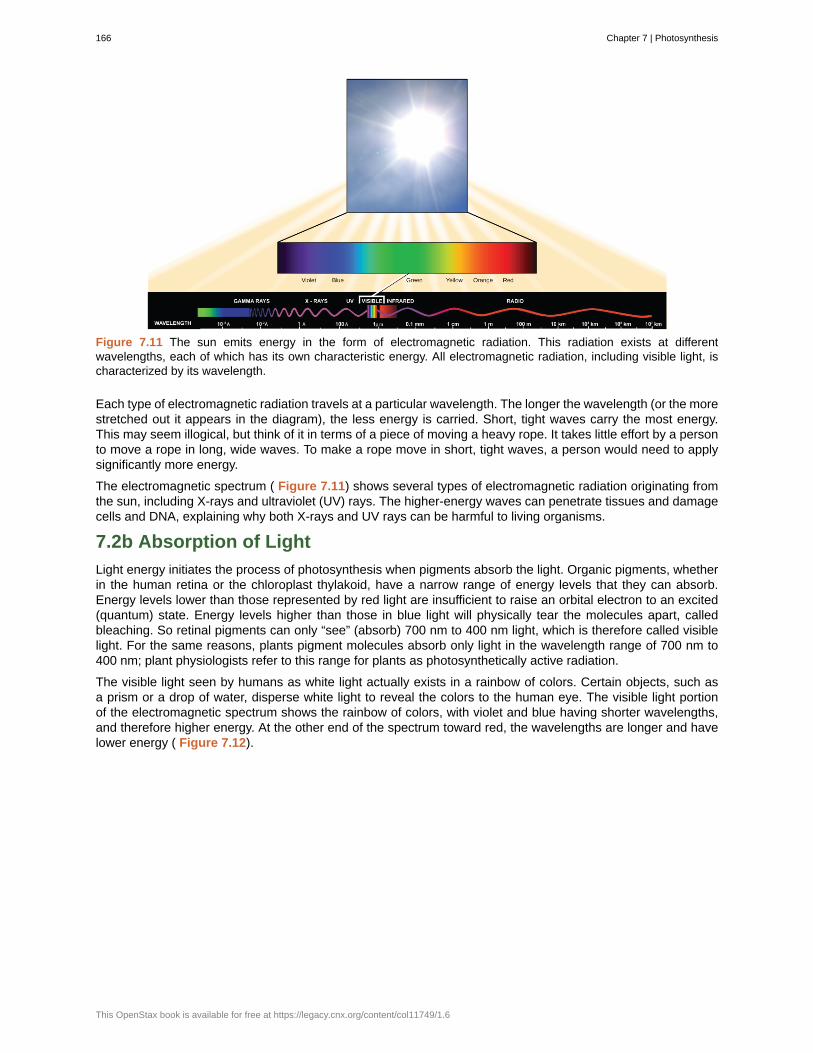
Figure 7.11 The sun emits energy in the form of electromagnetic radiation. This radiation exists at differentwavelengths, each of which has its own characteristic energy. All electromagnetic radiation, including visible light, ischaracterized by its wavelength.
Each type of electromagnetic radiation travels at a particular wavelength. The longer the wavelength (or the morestretched out it appears in the diagram), the less energy is carried. Short, tight waves carry the most energy.This may seem illogical, but think of it in terms of a piece of moving a heavy rope. It takes little effort by a personto move a rope in long, wide waves. To make a rope move in short, tight waves, a person would need to applysignificantly more energy.
The electromagnetic spectrum ( Figure 7.11) shows several types of electromagnetic radiation originating fromthe sun, including X-rays and ultraviolet (UV) rays. The higher-energy waves can penetrate tissues and damagecells and DNA, explaining why both X-rays and UV rays can be harmful to living organisms.
7.2b Absorption of Light
Light energy initiates the process of photosynthesis when pigments absorb the light. Organic pigments, whetherin the human retina or the chloroplast thylakoid, have a narrow range of energy levels that they can absorb.Energy levels lower than those represented by red light are insufficient to raise an orbital electron to an excited(quantum) state. Energy levels higher than those in blue light will physically tear the molecules apart, calledbleaching. So retinal pigments can only “see” (absorb) 700 nm to 400 nm light, which is therefore called visiblelight. For the same reasons, plants pigment molecules absorb only light in the wavelength range of 700 nm to400 nm; plant physiologists refer to this range for plants as photosynthetically active radiation.
The visible light seen by humans as white light actually exists in a rainbow of colors. Certain objects, such asa prism or a drop of water, disperse white light to reveal the colors to the human eye. The visible light portionof the electromagnetic spectrum shows the rainbow of colors, with violet and blue having shorter wavelengths,and therefore higher energy. At the other end of the spectrum toward red, the wavelengths are longer and havelower energy ( Figure 7.12).
166 Chapter 7 | Photosynthesis
This OpenStax book is available for free at https://legacy.cnx.org/content/col11749/1.6
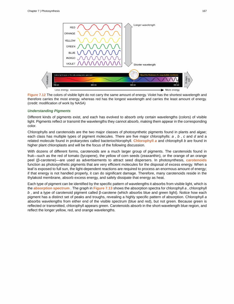
Figure 7.12 The colors of visible light do not carry the same amount of energy. Violet has the shortest wavelength andtherefore carries the most energy, whereas red has the longest wavelength and carries the least amount of energy.(credit: modification of work by NASA)
Understanding Pigments
Different kinds of pigments exist, and each has evolved to absorb only certain wavelengths (colors) of visiblelight. Pigments reflect or transmit the wavelengths they cannot absorb, making them appear in the correspondingcolor.
Chlorophylls and carotenoids are the two major classes of photosynthetic pigments found in plants and algae;each class has multiple types of pigment molecules. There are five major chlorophylls: a , b , c and d and arelated molecule found in prokaryotes called bacteriochlorophyll. Chlorophyll a and chlorophyll b are found inhigher plant chloroplasts and will be the focus of the following discussion.
With dozens of different forms, carotenoids are a much larger group of pigments. The carotenoids found infruit—such as the red of tomato (lycopene), the yellow of corn seeds (zeaxanthin), or the orange of an orangepeel (β-carotene)—are used as advertisements to attract seed dispersers. In photosynthesis, carotenoidsfunction as photosynthetic pigments that are very efficient molecules for the disposal of excess energy. When aleaf is exposed to full sun, the light-dependent reactions are required to process an enormous amount of energy;if that energy is not handled properly, it can do significant damage. Therefore, many carotenoids reside in thethylakoid membrane, absorb excess energy, and safely dissipate that energy as heat.
Each type of pigment can be identified by the specific pattern of wavelengths it absorbs from visible light, which isthe absorption spectrum . The graph in Figure 7.13 shows the absorption spectra for chlorophyll a , chlorophyllb , and a type of carotenoid pigment called β-carotene (which absorbs blue and green light). Notice how eachpigment has a distinct set of peaks and troughs, revealing a highly specific pattern of absorption. Chlorophyll aabsorbs wavelengths from either end of the visible spectrum (blue and red), but not green. Because green isreflected or transmitted, chlorophyll appears green. Carotenoids absorb in the short-wavelength blue region, andreflect the longer yellow, red, and orange wavelengths.
Chapter 7 | Photosynthesis 167
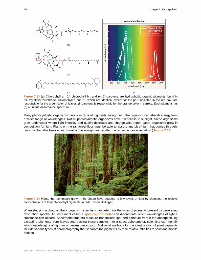
Figure 7.13 (a) Chlorophyll a , (b) chlorophyll b , and (c) β -carotene are hydrophobic organic pigments found inthe thylakoid membrane. Chlorophyll a and b , which are identical except for the part indicated in the red box, areresponsible for the green color of leaves. β -carotene is responsible for the orange color in carrots. Each pigment has(d) a unique absorbance spectrum.
Many photosynthetic organisms have a mixture of pigments; using them, the organism can absorb energy froma wider range of wavelengths. Not all photosynthetic organisms have full access to sunlight. Some organismsgrow underwater where light intensity and quality decrease and change with depth. Other organisms grow incompetition for light. Plants on the rainforest floor must be able to absorb any bit of light that comes through,because the taller trees absorb most of the sunlight and scatter the remaining solar radiation ( Figure 7.14).
Figure 7.14 Plants that commonly grow in the shade have adapted to low levels of light by changing the relativeconcentrations of their chlorophyll pigments. (credit: Jason Hollinger)
When studying a photosynthetic organism, scientists can determine the types of pigments present by generatingabsorption spectra. An instrument called a spectrophotometer can differentiate which wavelengths of light asubstance can absorb. Spectrophotometers measure transmitted light and compute from it the absorption. Byextracting pigments from leaves and placing these samples into a spectrophotometer, scientists can identifywhich wavelengths of light an organism can absorb. Additional methods for the identification of plant pigmentsinclude various types of chromatography that separate the pigments by their relative affinities to solid and mobilephases.
168 Chapter 7 | Photosynthesis
This OpenStax book is available for free at https://legacy.cnx.org/content/col11749/1.6
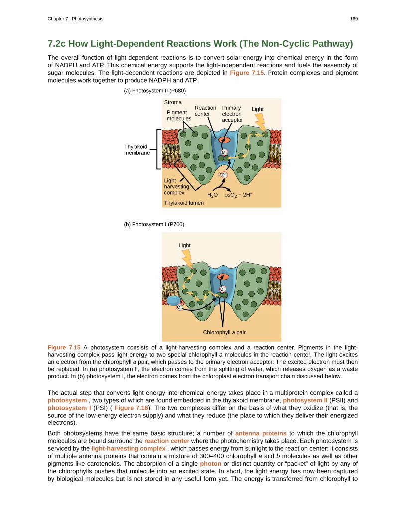
7.2c How Light-Dependent Reactions Work (The Non-Cyclic Pathway)
The overall function of light-dependent reactions is to convert solar energy into chemical energy in the formof NADPH and ATP. This chemical energy supports the light-independent reactions and fuels the assembly ofsugar molecules. The light-dependent reactions are depicted in Figure 7.15. Protein complexes and pigmentmolecules work together to produce NADPH and ATP.
Figure 7.15 A photosystem consists of a light-harvesting complex and a reaction center. Pigments in the light-harvesting complex pass light energy to two special chlorophyll a molecules in the reaction center. The light excitesan electron from the chlorophyll a pair, which passes to the primary electron acceptor. The excited electron must thenbe replaced. In (a) photosystem II, the electron comes from the splitting of water, which releases oxygen as a wasteproduct. In (b) photosystem I, the electron comes from the chloroplast electron transport chain discussed below.
The actual step that converts light energy into chemical energy takes place in a multiprotein complex called aphotosystem , two types of which are found embedded in the thylakoid membrane, photosystem II (PSII) andphotosystem I (PSI) ( Figure 7.16). The two complexes differ on the basis of what they oxidize (that is, thesource of the low-energy electron supply) and what they reduce (the place to which they deliver their energizedelectrons).
Both photosystems have the same basic structure; a number of antenna proteins to which the chlorophyllmolecules are bound surround the reaction center where the photochemistry takes place. Each photosystem isserviced by the light-harvesting complex , which passes energy from sunlight to the reaction center; it consistsof multiple antenna proteins that contain a mixture of 300–400 chlorophyll a and b molecules as well as otherpigments like carotenoids. The absorption of a single photon or distinct quantity or “packet” of light by any ofthe chlorophylls pushes that molecule into an excited state. In short, the light energy has now been capturedby biological molecules but is not stored in any useful form yet. The energy is transferred from chlorophyll to
Chapter 7 | Photosynthesis 169
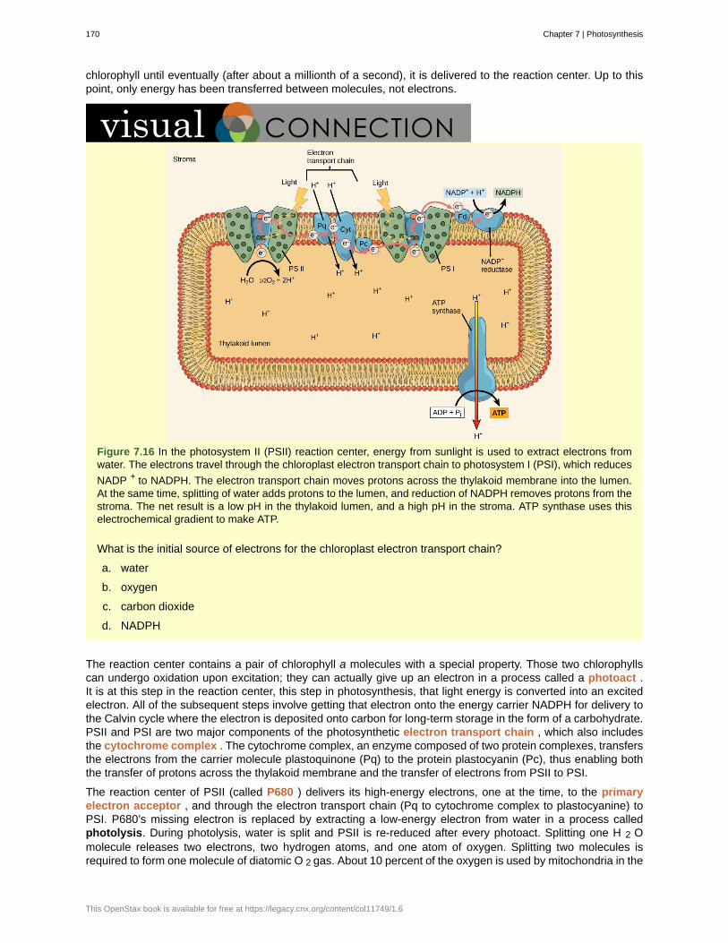
chlorophyll until eventually (after about a millionth of a second), it is delivered to the reaction center. Up to thispoint, only energy has been transferred between molecules, not electrons.
Figure 7.16 In the photosystem II (PSII) reaction center, energy from sunlight is used to extract electrons fromwater. The electrons travel through the chloroplast electron transport chain to photosystem I (PSI), which reduces
NADP + to NADPH. The electron transport chain moves protons across the thylakoid membrane into the lumen.At the same time, splitting of water adds protons to the lumen, and reduction of NADPH removes protons from thestroma. The net result is a low pH in the thylakoid lumen, and a high pH in the stroma. ATP synthase uses thiselectrochemical gradient to make ATP.
What is the initial source of electrons for the chloroplast electron transport chain?
a. water
b. oxygen
c. carbon dioxide
d. NADPH
The reaction center contains a pair of chlorophyll a molecules with a special property. Those two chlorophyllscan undergo oxidation upon excitation; they can actually give up an electron in a process called a photoact .It is at this step in the reaction center, this step in photosynthesis, that light energy is converted into an excitedelectron. All of the subsequent steps involve getting that electron onto the energy carrier NADPH for delivery tothe Calvin cycle where the electron is deposited onto carbon for long-term storage in the form of a carbohydrate.PSII and PSI are two major components of the photosynthetic electron transport chain , which also includesthe cytochrome complex . The cytochrome complex, an enzyme composed of two protein complexes, transfersthe electrons from the carrier molecule plastoquinone (Pq) to the protein plastocyanin (Pc), thus enabling boththe transfer of protons across the thylakoid membrane and the transfer of electrons from PSII to PSI.
The reaction center of PSII (called P680 ) delivers its high-energy electrons, one at the time, to the primaryelectron acceptor , and through the electron transport chain (Pq to cytochrome complex to plastocyanine) toPSI. P680’s missing electron is replaced by extracting a low-energy electron from water in a process calledphotolysis. During photolysis, water is split and PSII is re-reduced after every photoact. Splitting one H 2 Omolecule releases two electrons, two hydrogen atoms, and one atom of oxygen. Splitting two molecules isrequired to form one molecule of diatomic O 2 gas. About 10 percent of the oxygen is used by mitochondria in the
170 Chapter 7 | Photosynthesis
This OpenStax book is available for free at https://legacy.cnx.org/content/col11749/1.6
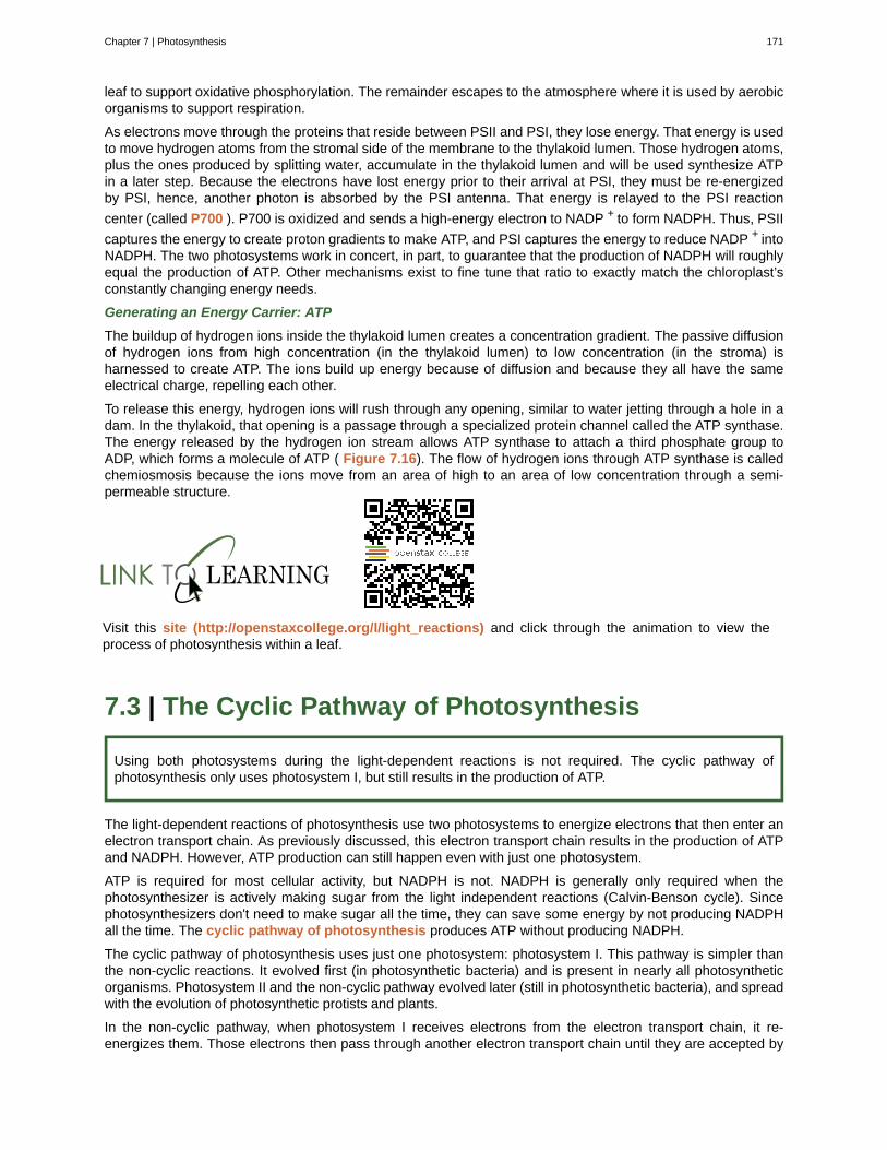
leaf to support oxidative phosphorylation. The remainder escapes to the atmosphere where it is used by aerobicorganisms to support respiration.
As electrons move through the proteins that reside between PSII and PSI, they lose energy. That energy is usedto move hydrogen atoms from the stromal side of the membrane to the thylakoid lumen. Those hydrogen atoms,plus the ones produced by splitting water, accumulate in the thylakoid lumen and will be used synthesize ATPin a later step. Because the electrons have lost energy prior to their arrival at PSI, they must be re-energizedby PSI, hence, another photon is absorbed by the PSI antenna. That energy is relayed to the PSI reaction
center (called P700 ). P700 is oxidized and sends a high-energy electron to NADP + to form NADPH. Thus, PSII
captures the energy to create proton gradients to make ATP, and PSI captures the energy to reduce NADP + intoNADPH. The two photosystems work in concert, in part, to guarantee that the production of NADPH will roughlyequal the production of ATP. Other mechanisms exist to fine tune that ratio to exactly match the chloroplast’sconstantly changing energy needs.
Generating an Energy Carrier: ATP
The buildup of hydrogen ions inside the thylakoid lumen creates a concentration gradient. The passive diffusionof hydrogen ions from high concentration (in the thylakoid lumen) to low concentration (in the stroma) isharnessed to create ATP. The ions build up energy because of diffusion and because they all have the sameelectrical charge, repelling each other.
To release this energy, hydrogen ions will rush through any opening, similar to water jetting through a hole in adam. In the thylakoid, that opening is a passage through a specialized protein channel called the ATP synthase.The energy released by the hydrogen ion stream allows ATP synthase to attach a third phosphate group toADP, which forms a molecule of ATP ( Figure 7.16). The flow of hydrogen ions through ATP synthase is calledchemiosmosis because the ions move from an area of high to an area of low concentration through a semi-permeable structure.
Visit this site (http://openstaxcollege.org/l/light_reactions) and click through the animation to view theprocess of photosynthesis within a leaf.
7.3 | The Cyclic Pathway of Photosynthesis
Using both photosystems during the light-dependent reactions is not required. The cyclic pathway ofphotosynthesis only uses photosystem I, but still results in the production of ATP.
The light-dependent reactions of photosynthesis use two photosystems to energize electrons that then enter anelectron transport chain. As previously discussed, this electron transport chain results in the production of ATPand NADPH. However, ATP production can still happen even with just one photosystem.
ATP is required for most cellular activity, but NADPH is not. NADPH is generally only required when thephotosynthesizer is actively making sugar from the light independent reactions (Calvin-Benson cycle). Sincephotosynthesizers don't need to make sugar all the time, they can save some energy by not producing NADPHall the time. The cyclic pathway of photosynthesis produces ATP without producing NADPH.
The cyclic pathway of photosynthesis uses just one photosystem: photosystem I. This pathway is simpler thanthe non-cyclic reactions. It evolved first (in photosynthetic bacteria) and is present in nearly all photosyntheticorganisms. Photosystem II and the non-cyclic pathway evolved later (still in photosynthetic bacteria), and spreadwith the evolution of photosynthetic protists and plants.
In the non-cyclic pathway, when photosystem I receives electrons from the electron transport chain, it re-energizes them. Those electrons then pass through another electron transport chain until they are accepted by
Chapter 7 | Photosynthesis 171
NADP+. However, if there is not enough NADP+ to accept those electrons, the cycle does not immediately stop.Instead, electrons can be transported back to photosystem I, which can re-energize them again. This is calledthe cyclic pathway because no new electrons are ever needed ( Figure 7.17). Electrons that leave photosystemI always return to it, so the same electrons are re-energized over and over again (as long as there is lighthitting the photosystem). As electrons pass through the second electron transport chain, they are powering thetransport of H+ ions across the membrane. As long as an H+ gradient exists, ATP can be made.
Figure 7.17 The Cyclic Pathway of Photosynthesis
If no new NADPH is made (because there are no NADP+ molecules to accept electrons), no new sugar can bemade. In certain situations, this is acceptable. During the winter, plants aren't actively making a lot of sugar, andtherefore wouldn't need a lot of new NADPH. With the cyclic pathway, plants can save some time and energy.
Since photosystem I is accepting electrons that are returned to it, it is not accepting electrons from the previouselectron transport chain. Therefore, the first electron transport chain will be backed up, which means thatphotolysis will not occur. Without photolysis, no water is broken down and therefore no oxygen is produced.
7.4 | Using Light Energy to Make Organic Molecules
By the end of this section, you will be able to:
• Describe the Calvin cycle
• Define carbon fixation
• Explain how photosynthesis works in the energy cycle of all living organisms
After the energy from the sun is converted into chemical energy and temporarily stored in ATP and NADPHmolecules, the cell has the fuel needed to build carbohydrate molecules for long-term energy storage. Theproducts of the light-dependent reactions, ATP and NADPH, have lifespans in the range of millionths of seconds,whereas the products of the light-independent reactions (carbohydrates and other forms of reduced carbon) cansurvive for hundreds of millions of years. The carbohydrate molecules made will have a backbone of carbonatoms. Where does the carbon come from? It comes from carbon dioxide, the gas that is a waste product ofrespiration in microbes, fungi, plants, and animals.
7.4a The Calvin Cycle
In plants, carbon dioxide (CO 2 ) enters the leaves through stomata, where it diffuses over short distancesthrough intercellular spaces until it reaches the mesophyll cells. Once in the mesophyll cells, CO 2 diffuses into
172 Chapter 7 | Photosynthesis
This OpenStax book is available for free at https://legacy.cnx.org/content/col11749/1.6
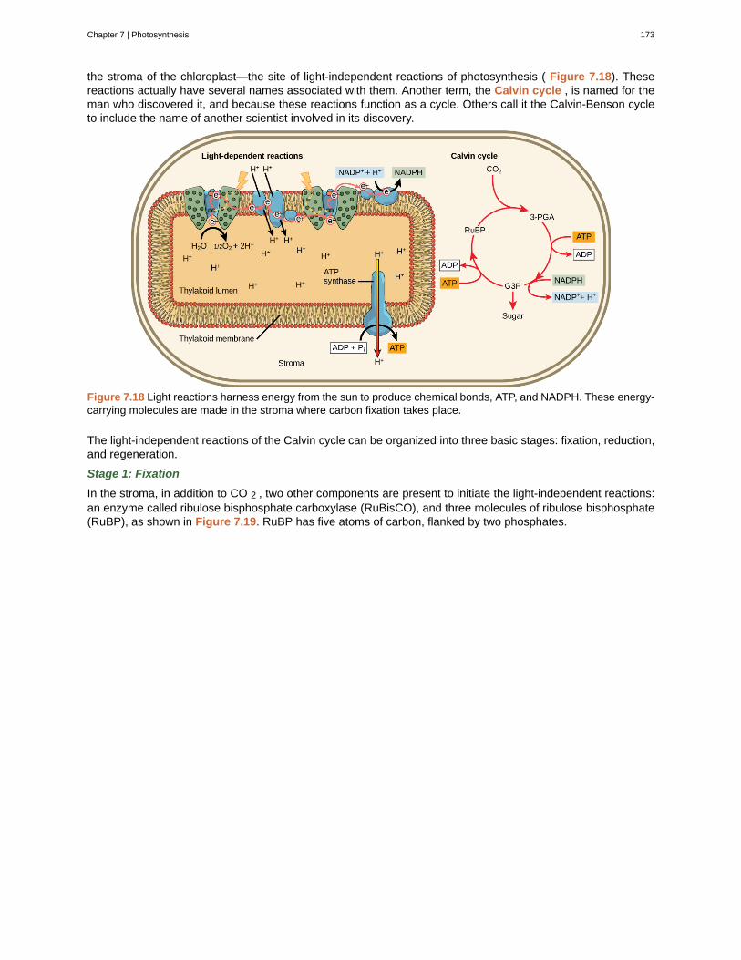
the stroma of the chloroplast—the site of light-independent reactions of photosynthesis ( Figure 7.18). Thesereactions actually have several names associated with them. Another term, the Calvin cycle , is named for theman who discovered it, and because these reactions function as a cycle. Others call it the Calvin-Benson cycleto include the name of another scientist involved in its discovery.
Figure 7.18 Light reactions harness energy from the sun to produce chemical bonds, ATP, and NADPH. These energy-carrying molecules are made in the stroma where carbon fixation takes place.
The light-independent reactions of the Calvin cycle can be organized into three basic stages: fixation, reduction,and regeneration.
Stage 1: Fixation
In the stroma, in addition to CO 2 , two other components are present to initiate the light-independent reactions:an enzyme called ribulose bisphosphate carboxylase (RuBisCO), and three molecules of ribulose bisphosphate(RuBP), as shown in Figure 7.19. RuBP has five atoms of carbon, flanked by two phosphates.
Chapter 7 | Photosynthesis 173
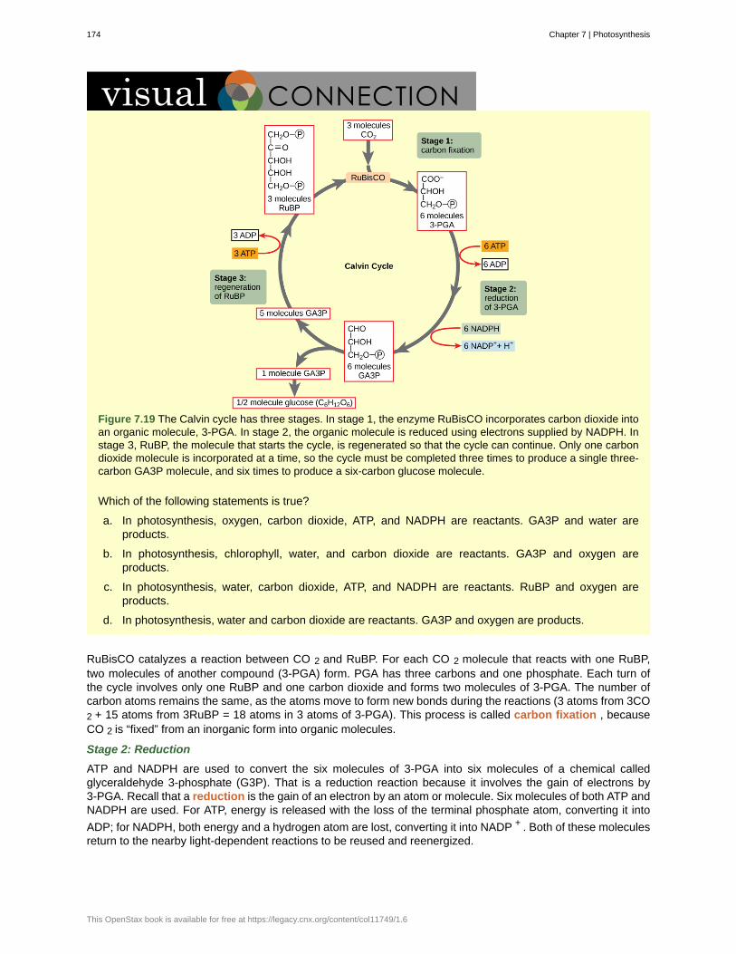
Figure 7.19 The Calvin cycle has three stages. In stage 1, the enzyme RuBisCO incorporates carbon dioxide intoan organic molecule, 3-PGA. In stage 2, the organic molecule is reduced using electrons supplied by NADPH. Instage 3, RuBP, the molecule that starts the cycle, is regenerated so that the cycle can continue. Only one carbondioxide molecule is incorporated at a time, so the cycle must be completed three times to produce a single three-carbon GA3P molecule, and six times to produce a six-carbon glucose molecule.
Which of the following statements is true?
a. In photosynthesis, oxygen, carbon dioxide, ATP, and NADPH are reactants. GA3P and water areproducts.
b. In photosynthesis, chlorophyll, water, and carbon dioxide are reactants. GA3P and oxygen areproducts.
c. In photosynthesis, water, carbon dioxide, ATP, and NADPH are reactants. RuBP and oxygen areproducts.
d. In photosynthesis, water and carbon dioxide are reactants. GA3P and oxygen are products.
RuBisCO catalyzes a reaction between CO 2 and RuBP. For each CO 2 molecule that reacts with one RuBP,two molecules of another compound (3-PGA) form. PGA has three carbons and one phosphate. Each turn ofthe cycle involves only one RuBP and one carbon dioxide and forms two molecules of 3-PGA. The number ofcarbon atoms remains the same, as the atoms move to form new bonds during the reactions (3 atoms from 3CO2 + 15 atoms from 3RuBP = 18 atoms in 3 atoms of 3-PGA). This process is called carbon fixation , becauseCO 2 is “fixed” from an inorganic form into organic molecules.
Stage 2: Reduction
ATP and NADPH are used to convert the six molecules of 3-PGA into six molecules of a chemical calledglyceraldehyde 3-phosphate (G3P). That is a reduction reaction because it involves the gain of electrons by3-PGA. Recall that a reduction is the gain of an electron by an atom or molecule. Six molecules of both ATP andNADPH are used. For ATP, energy is released with the loss of the terminal phosphate atom, converting it into
ADP; for NADPH, both energy and a hydrogen atom are lost, converting it into NADP + . Both of these moleculesreturn to the nearby light-dependent reactions to be reused and reenergized.
174 Chapter 7 | Photosynthesis
This OpenStax book is available for free at https://legacy.cnx.org/content/col11749/1.6
Stage 3: Regeneration
Interestingly, at this point, only one of the G3P molecules leaves the Calvin cycle and is sent to the cytoplasmto contribute to the formation of other compounds needed by the plant. Because the G3P exported from thechloroplast has three carbon atoms, it takes three “turns” of the Calvin cycle to fix enough net carbon to exportone G3P. But each turn makes two G3Ps, thus three turns make six G3Ps. One is exported while the remainingfive G3P molecules remain in the cycle and are used to regenerate RuBP, which enables the system to preparefor more CO 2 to be fixed. Three more molecules of ATP are used in these regeneration reactions.
This link (http://openstaxcollege.org/l/calvin_cycle) leads to an animation of the Calvin cycle. Click stage1, stage 2, and then stage 3 to see G3P and ATP regenerate to form RuBP.
PhotosynthesisDuring the evolution of photosynthesis, a major shift occurred from the bacterial type of photosynthesisthat involves only one photosystem and is typically anoxygenic (does not generate oxygen) into modernoxygenic (does generate oxygen) photosynthesis, employing two photosystems. This modern oxygenicphotosynthesis is used by many organisms—from giant tropical leaves in the rainforest to tinycyanobacterial cells—and the process and components of this photosynthesis remain largely the same.Photosystems absorb light and use electron transport chains to convert energy into the chemical energy ofATP and NADH. The subsequent light-independent reactions then assemble carbohydrate molecules withthis energy.
Photosynthesis in desert plants has evolved adaptations that conserve water. In the harsh dry heat, everydrop of water must be used to survive. Because stomata must open to allow for the uptake of CO 2 , waterescapes from the leaf during active photosynthesis. Desert plants have evolved processes to conservewater and deal with harsh conditions. A more efficient use of CO 2 allows plants to adapt to living with lesswater. Some plants such as cacti ( Figure 7.20) can prepare materials for photosynthesis during the night bya temporary carbon fixation/storage process, because opening the stomata at this time conserves water dueto cooler temperatures. In addition, cacti have evolved the ability to carry out low levels of photosynthesiswithout opening stomata at all, an extreme mechanism to face extremely dry periods.
Chapter 7 | Photosynthesis 175
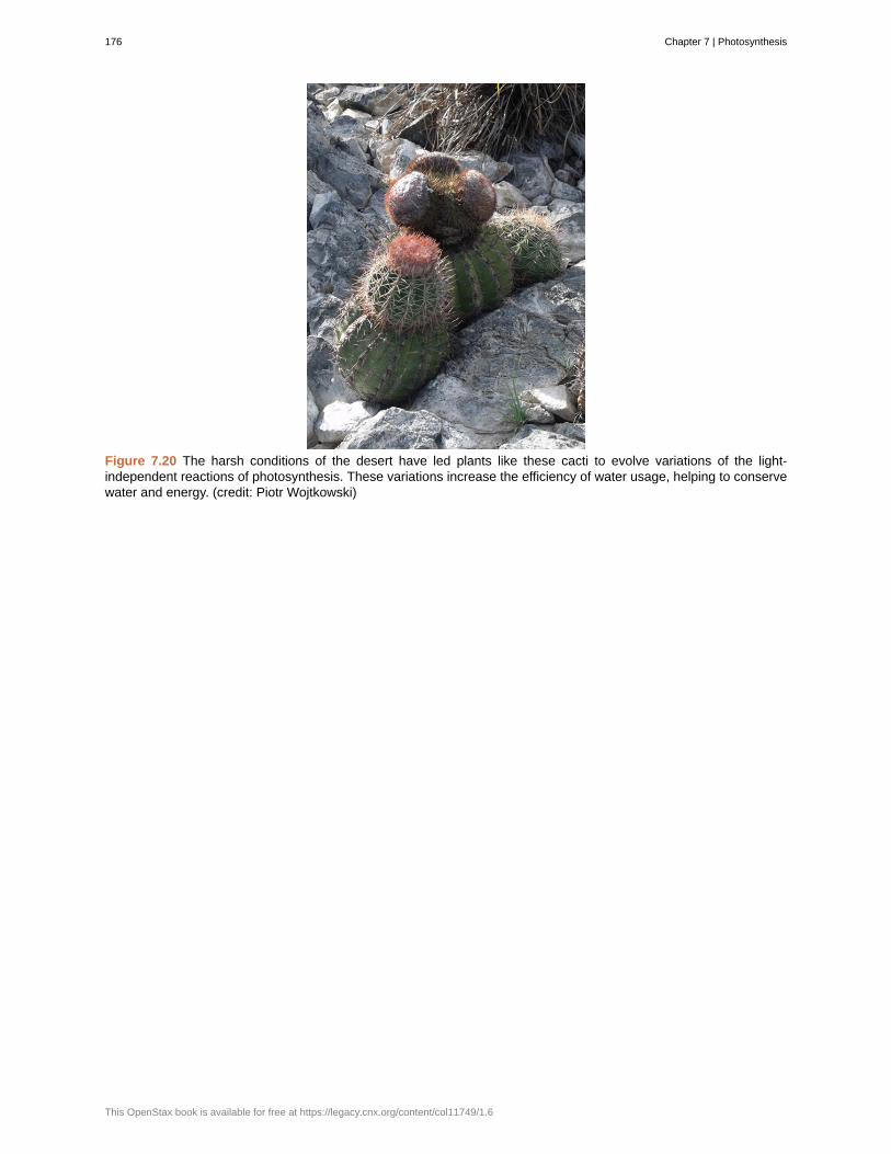
Figure 7.20 The harsh conditions of the desert have led plants like these cacti to evolve variations of the light-independent reactions of photosynthesis. These variations increase the efficiency of water usage, helping to conservewater and energy. (credit: Piotr Wojtkowski)
176 Chapter 7 | Photosynthesis
This OpenStax book is available for free at https://legacy.cnx.org/content/col11749/1.6
absorption spectrum
antenna proteins
Calvin cycle
carbon fixation
carotenoids
chemoautotrophs
chlorophyll a
chloroplast
cyclic pathway of photosynthesis
cytochrome complex
electromagnetic spectrum
electron transport chain
granum
heterotrophs
light-dependent reactions
light-harvesting complex
light-independent reactions
mesophyll
P680
P700
photoact
photoautotrophs
photolysis
photon
photosystem
KEY TERMS
range of wavelengths of electromagnetic radiation absorbed by a given substance
pigment molecule that directly absorbs light and transfers the energy absorbed to otherpigment molecules
light-independent reactions of photosynthesis that convert carbon dioxide from the atmosphereinto carbohydrates using the energy and reducing power of ATP and NADPH
process of converting inorganic CO2 gas into organic compounds
photosynthetic pigment that functions to dispose of excess energy
organism that can build organic molecules using energy derived from inorganic chemicalsinstead of sunlight
form of chlorophyll that absorbs violet-blue and red light and consequently has a bluish-greencolor; the only pigment molecule that performs the photochemistry by getting excited and losing an electronto the electron transport chain
organelle in which photosynthesis takes place
process of photosynthesis that only uses Photosystem I
group of reversibly oxidizable and reducible proteins that forms part of the electrontransport chain between photosystem II and photosystem I
range of all possible frequencies of radiation
group of proteins between PSII and PSI that pass energized electrons and use theenergy released by the electrons to move hydrogen ions against their concentration gradient into thethylakoid lumen
a stack of thylakoids inside a chloroplast
organism that consumes organic substances or other organisms for food
first stage of photosynthesis where certain wavelengths of visible light areabsorbed to form two energy-carrying molecules from ATP and NADPH
complex that passes energy from sunlight to the reaction center in eachphotosystem; it consists of multiple antenna proteins thst contain a mixture of 300-400 chlorophyll a and bmolecules as well as other pigments like carotenoids
second stage of photosynthesis through which carbon dioxide is used to buildcarbohydrate molecules using energy from ATP and NADPH
middle layer of chlorophyll-rich cells in a leaf
reaction center of photosystem II
reaction center of photosystem I
ejection of an electron from a reaction center using the energy of an absorbed photon
organism capable of producing its own organic compounds from sunlight
process of splitting water to release electrons that will reduce the oxidized PSII
distinct quantity or “packet” of light energy
group of proteins, chlorophyll, and other pigments that are used in the light-dependent reactions
Chapter 7 | Photosynthesis 177
photosystem I
photosystem II
pigment
primary electron acceptor
reaction center
reduction
spectrophotometer
stomata
stroma
thylakoid lumen
thylakoids
wavelength
of photosynthesis to absorb light energy and convert it into chemical energy
integral pigment and protein complex in thylakoid membranes that uses light energy to
transport electrons from plastocyanin to NADP + (which becomes reduced to NADPH in the process)
integral protein and pigment complex in thylakoid membranes that transports electrons fromwater to the electron transport chain; oxygen is a product of PSII
molecule that is capable of absorbing certain wavelengths of light and reflecting others (whichaccounts for its color)
pigment or other organic molecule in the reaction center that accepts an energizedelectron from the reaction center
complex of chlorophyll molecules and other organic molecules that is assembled around aspecial pair of chlorophyll molecules and a primary electron acceptor; capable of undergoing oxidation andreduction
gain of electron(s) by an atom or molecule
instrument that can measure transmitted light and compute the absorption
openings that regulate gas exchange and water evaporation between leaves and the environment,typically situated on the underside of leaves
internal fluid environment of a chloroplast
aqueous space bound by a thylakoid membrane where protons accumulate during light-driven electron transport
disc-shaped, membrane-bound structure inside a chloroplast where the light-dependent reactionsof photosynthesis take place
distance between consecutive points of equal position (two crests or two troughs) of a wave in agraphic representation; inversely proportional to the energy of the radiation
CHAPTER SUMMARY
7.1 Overview of Photosynthesis
The process of photosynthesis transformed life on Earth. By harnessing energy from the sun, photosynthesisevolved to allow living things access to enormous amounts of energy. Because of photosynthesis, living thingsgained access to sufficient energy that allowed them to build new structures and achieve the biodiversityevident today.
Only certain organisms, called photoautotrophs, can perform photosynthesis; they require the presence ofchlorophyll, a specialized pigment that absorbs certain portions of the visible spectrum and can capture energyfrom sunlight. Photosynthesis uses carbon dioxide and water to assemble carbohydrate molecules and releaseoxygen as a waste product into the atmosphere. Eukaryotic autotrophs, such as plants and algae, haveorganelles called chloroplasts in which photosynthesis takes place, and starch accumulates. In prokaryotes,such as cyanobacteria, the process is less localized and occurs within folded membranes, extensions of theplasma membrane, and in the cytoplasm.
7.2 The Light-Dependent Reactions of Photosynthesis
The pigments of the first part of photosynthesis, the light-dependent reactions, absorb energy from sunlight. Aphoton strikes the antenna pigments of photosystem II to initiate photosynthesis. The energy travels to thereaction center that contains chlorophyll a to the electron transport chain, which pumps hydrogen ions into thethylakoid interior. This action builds up a high concentration of ions. The ions flow through ATP synthase viachemiosmosis to form molecules of ATP, which are used for the formation of sugar molecules in the secondstage of photosynthesis. Photosystem I absorbs a second photon, which results in the formation of an NADPHmolecule, another energy and reducing power carrier for the light-independent reactions.
178 Chapter 7 | Photosynthesis
This OpenStax book is available for free at https://legacy.cnx.org/content/col11749/1.6
7.3 The Cyclic Pathway of Photosynthesis
The cyclic pathway of photosynthesis uses just photosystem I to generate ATP. This process occurs whensugar production is not immediately necessary and therefore no new NADPH is generated. Electrons arerecycled from photosystem I to be continually re-energized until they can be accepted by NADP+. The cyclicpathway generates ATP, but neither oxygen nor sugar.
7.4 Using Light Energy to Make Organic Molecules
Using the energy carriers formed in the first steps of photosynthesis, the light-independent reactions, or theCalvin cycle, take in CO 2 from the environment. An enzyme, RuBisCO, catalyzes a reaction with CO 2 andanother molecule, RuBP. After three cycles, a three-carbon molecule of G3P leaves the cycle to become part ofa carbohydrate molecule. The remaining G3P molecules stay in the cycle to be regenerated into RuBP, whichis then ready to react with more CO 2 . Photosynthesis forms an energy cycle with the process of cellularrespiration. Plants need both photosynthesis and respiration for their ability to function in both the light anddark, and to be able to interconvert essential metabolites. Therefore, plants contain both chloroplasts andmitochondria.
Chapter 7 | Photosynthesis 179

180 Chapter 7 | Photosynthesis
This OpenStax book is available for free at https://legacy.cnx.org/content/col11749/1.6
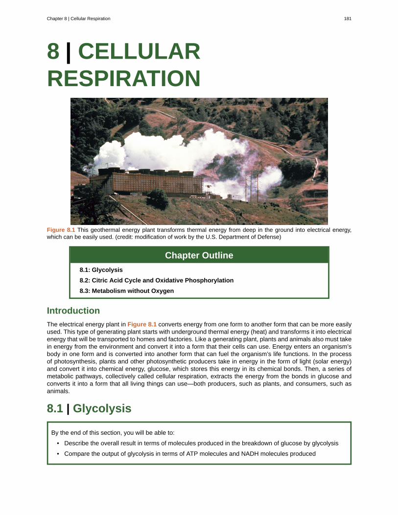
8 | CELLULARRESPIRATION
Figure 8.1 This geothermal energy plant transforms thermal energy from deep in the ground into electrical energy,which can be easily used. (credit: modification of work by the U.S. Department of Defense)
Chapter Outline
8.1: Glycolysis
8.2: Citric Acid Cycle and Oxidative Phosphorylation
8.3: Metabolism without Oxygen
Introduction
The electrical energy plant in Figure 8.1 converts energy from one form to another form that can be more easilyused. This type of generating plant starts with underground thermal energy (heat) and transforms it into electricalenergy that will be transported to homes and factories. Like a generating plant, plants and animals also must takein energy from the environment and convert it into a form that their cells can use. Energy enters an organism’sbody in one form and is converted into another form that can fuel the organism’s life functions. In the processof photosynthesis, plants and other photosynthetic producers take in energy in the form of light (solar energy)and convert it into chemical energy, glucose, which stores this energy in its chemical bonds. Then, a series ofmetabolic pathways, collectively called cellular respiration, extracts the energy from the bonds in glucose andconverts it into a form that all living things can use—both producers, such as plants, and consumers, such asanimals.
8.1 | Glycolysis
By the end of this section, you will be able to:
• Describe the overall result in terms of molecules produced in the breakdown of glucose by glycolysis
• Compare the output of glycolysis in terms of ATP molecules and NADH molecules produced
Chapter 8 | Cellular Respiration 181
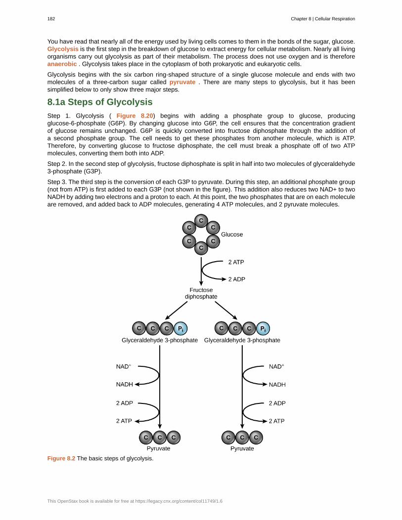
You have read that nearly all of the energy used by living cells comes to them in the bonds of the sugar, glucose.Glycolysis is the first step in the breakdown of glucose to extract energy for cellular metabolism. Nearly all livingorganisms carry out glycolysis as part of their metabolism. The process does not use oxygen and is thereforeanaerobic . Glycolysis takes place in the cytoplasm of both prokaryotic and eukaryotic cells.
Glycolysis begins with the six carbon ring-shaped structure of a single glucose molecule and ends with twomolecules of a three-carbon sugar called pyruvate . There are many steps to glycolysis, but it has beensimplified below to only show three major steps.
8.1a Steps of Glycolysis
Step 1. Glycolysis ( Figure 8.20) begins with adding a phosphate group to glucose, producingglucose-6-phosphate (G6P). By changing glucose into G6P, the cell ensures that the concentration gradientof glucose remains unchanged. G6P is quickly converted into fructose diphosphate through the addition ofa second phosphate group. The cell needs to get these phosphates from another molecule, which is ATP.Therefore, by converting glucose to fructose diphosphate, the cell must break a phosphate off of two ATPmolecules, converting them both into ADP.
Step 2. In the second step of glycolysis, fructose diphosphate is split in half into two molecules of glyceraldehyde3-phosphate (G3P).
Step 3. The third step is the conversion of each G3P to pyruvate. During this step, an additional phosphate group(not from ATP) is first added to each G3P (not shown in the figure). This addition also reduces two NAD+ to twoNADH by adding two electrons and a proton to each. At this point, the two phosphates that are on each moleculeare removed, and added back to ADP molecules, generating 4 ATP molecules, and 2 pyruvate molecules.
Figure 8.2 The basic steps of glycolysis.
182 Chapter 8 | Cellular Respiration
This OpenStax book is available for free at https://legacy.cnx.org/content/col11749/1.6
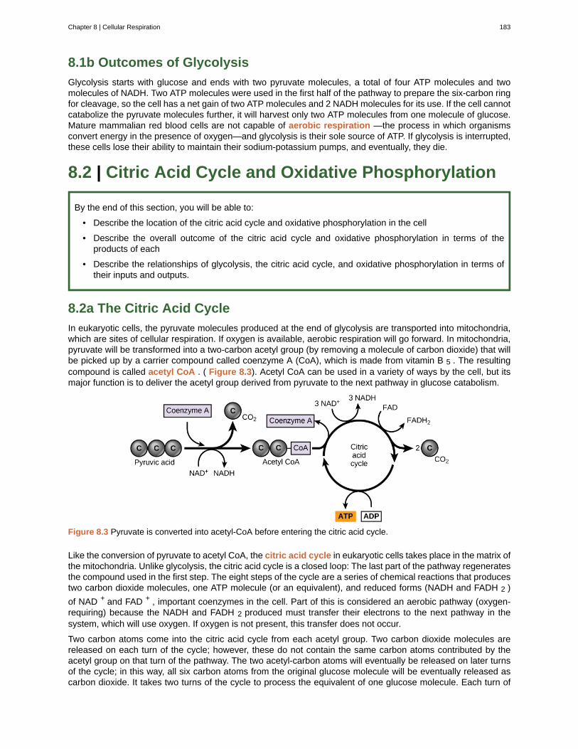
8.1b Outcomes of Glycolysis
Glycolysis starts with glucose and ends with two pyruvate molecules, a total of four ATP molecules and twomolecules of NADH. Two ATP molecules were used in the first half of the pathway to prepare the six-carbon ringfor cleavage, so the cell has a net gain of two ATP molecules and 2 NADH molecules for its use. If the cell cannotcatabolize the pyruvate molecules further, it will harvest only two ATP molecules from one molecule of glucose.Mature mammalian red blood cells are not capable of aerobic respiration —the process in which organismsconvert energy in the presence of oxygen—and glycolysis is their sole source of ATP. If glycolysis is interrupted,these cells lose their ability to maintain their sodium-potassium pumps, and eventually, they die.
8.2 | Citric Acid Cycle and Oxidative Phosphorylation
By the end of this section, you will be able to:
• Describe the location of the citric acid cycle and oxidative phosphorylation in the cell
• Describe the overall outcome of the citric acid cycle and oxidative phosphorylation in terms of theproducts of each
• Describe the relationships of glycolysis, the citric acid cycle, and oxidative phosphorylation in terms oftheir inputs and outputs.
8.2a The Citric Acid Cycle
In eukaryotic cells, the pyruvate molecules produced at the end of glycolysis are transported into mitochondria,which are sites of cellular respiration. If oxygen is available, aerobic respiration will go forward. In mitochondria,pyruvate will be transformed into a two-carbon acetyl group (by removing a molecule of carbon dioxide) that willbe picked up by a carrier compound called coenzyme A (CoA), which is made from vitamin B 5 . The resultingcompound is called acetyl CoA . ( Figure 8.3). Acetyl CoA can be used in a variety of ways by the cell, but itsmajor function is to deliver the acetyl group derived from pyruvate to the next pathway in glucose catabolism.
Figure 8.3 Pyruvate is converted into acetyl-CoA before entering the citric acid cycle.
Like the conversion of pyruvate to acetyl CoA, the citric acid cycle in eukaryotic cells takes place in the matrix ofthe mitochondria. Unlike glycolysis, the citric acid cycle is a closed loop: The last part of the pathway regeneratesthe compound used in the first step. The eight steps of the cycle are a series of chemical reactions that producestwo carbon dioxide molecules, one ATP molecule (or an equivalent), and reduced forms (NADH and FADH 2 )
of NAD + and FAD + , important coenzymes in the cell. Part of this is considered an aerobic pathway (oxygen-requiring) because the NADH and FADH 2 produced must transfer their electrons to the next pathway in thesystem, which will use oxygen. If oxygen is not present, this transfer does not occur.
Two carbon atoms come into the citric acid cycle from each acetyl group. Two carbon dioxide molecules arereleased on each turn of the cycle; however, these do not contain the same carbon atoms contributed by theacetyl group on that turn of the pathway. The two acetyl-carbon atoms will eventually be released on later turnsof the cycle; in this way, all six carbon atoms from the original glucose molecule will be eventually released ascarbon dioxide. It takes two turns of the cycle to process the equivalent of one glucose molecule. Each turn of
Chapter 8 | Cellular Respiration 183
the cycle forms three high-energy NADH molecules and one high-energy FADH 2 molecule. These high-energycarriers will connect with the last portion of aerobic respiration to produce ATP molecules. One ATP (or anequivalent) is also made in each cycle. Several of the intermediate compounds in the citric acid cycle can beused in synthesizing non-essential amino acids; therefore, the cycle is both anabolic and catabolic.
8.2b Oxidative Phosphorylation
You have just read about two pathways in glucose catabolism—glycolysis and the citric acid cycle—thatgenerate ATP. Most of the ATP generated during the aerobic catabolism of glucose, however, is not generateddirectly from these pathways. Rather, it derives from a process that begins with passing electrons through aseries of chemical reactions to a final electron acceptor, oxygen. These reactions take place in specializedprotein complexes located in the inner membrane of the mitochondria of eukaryotic organisms and on theinner part of the cell membrane of prokaryotic organisms. The energy of the electrons is harvested and usedto generate a electrochemical gradient across the inner mitochondrial membrane. The potential energy of thisgradient is used to generate ATP. The entirety of this process is called oxidative phosphorylation .
The electron transport chain ( Figure 8.4 a ) is the last component of aerobic respiration and is the onlypart of metabolism that uses atmospheric oxygen. Oxygen continuously diffuses into plants for this purpose.In animals, oxygen enters the body through the respiratory system. Electron transport is a series of chemicalreactions that resembles a bucket brigade in that electrons are passed rapidly from one component to the next,to the endpoint of the chain where oxygen is the final electron acceptor and water is produced. There arefour complexes composed of proteins, labeled I through IV in Figure 8.4 c , and the aggregation of these fourcomplexes, together with associated mobile, accessory electron carriers, is called the electron transport chain. The electron transport chain is present in multiple copies in the inner mitochondrial membrane of eukaryotesand in the plasma membrane of prokaryotes. In each transfer of an electron through the electron transportchain, the electron loses energy, but with some transfers, the energy is stored as potential energy by using itto pump hydrogen ions across the inner mitochondrial membrane into the intermembrane space, creating anelectrochemical gradient.
184 Chapter 8 | Cellular Respiration
This OpenStax book is available for free at https://legacy.cnx.org/content/col11749/1.6
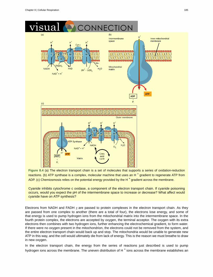
Figure 8.4 (a) The electron transport chain is a set of molecules that supports a series of oxidation-reduction
reactions. (b) ATP synthase is a complex, molecular machine that uses an H + gradient to regenerate ATP from
ADP. (c) Chemiosmosis relies on the potential energy provided by the H + gradient across the membrane.
Cyanide inhibits cytochrome c oxidase, a component of the electron transport chain. If cyanide poisoningoccurs, would you expect the pH of the intermembrane space to increase or decrease? What affect wouldcyanide have on ATP synthesis?
Electrons from NADH and FADH 2 are passed to protein complexes in the electron transport chain. As theyare passed from one complex to another (there are a total of four), the electrons lose energy, and some ofthat energy is used to pump hydrogen ions from the mitochondrial matrix into the intermembrane space. In thefourth protein complex, the electrons are accepted by oxygen, the terminal acceptor. The oxygen with its extraelectrons then combines with two hydrogen ions, further enhancing the electrochemical gradient, to form water.If there were no oxygen present in the mitochondrion, the electrons could not be removed from the system, andthe entire electron transport chain would back up and stop. The mitochondria would be unable to generate newATP in this way, and the cell would ultimately die from lack of energy. This is the reason we must breathe to drawin new oxygen.
In the electron transport chain, the energy from the series of reactions just described is used to pump
hydrogen ions across the membrane. The uneven distribution of H + ions across the membrane establishes an
Chapter 8 | Cellular Respiration 185
electrochemical gradient, owing to the H + ions’ positive charge and their higher concentration on one side of themembrane.
Hydrogen ions diffuse through the inner membrane through an integral membrane protein called ATP synthase( Figure 8.4 b ). This complex protein acts as a tiny generator, turned by the force of the hydrogen ions diffusingthrough it, down their electrochemical gradient from the intermembrane space, where there are many mutuallyrepelling hydrogen ions to the matrix, where there are few. The turning of the parts of this molecular machineregenerate ATP from ADP. This flow of hydrogen ions across the membrane through ATP synthase is calledchemiosmosis .
Chemiosmosis ( Figure 8.4 c ) is used to generate 90 percent of the ATP made during aerobic glucosecatabolism. The result of the reactions is the production of ATP from the energy of the electrons removed fromhydrogen atoms. These atoms were originally part of a glucose molecule. At the end of the electron transportsystem, the electrons are used to reduce an oxygen molecule to oxygen ions. The extra electrons on the oxygenions attract hydrogen ions (protons) from the surrounding medium, and water is formed. The electron transportchain and the production of ATP through chemiosmosis are collectively called oxidative phosphorylation.
8.2c ATP Yield
As mentioned in chapter 6, one molecule of glucose can be used to produce approximately 36 ATP. However,this number can vary. For example, the number of hydrogen ions that the electron transport chain complexescan pump through the membrane varies between species. Another source of variance stems from the shuttleof electrons across the mitochondrial membrane. The NADH generated from glycolysis cannot easily enter
mitochondria. Thus, electrons are picked up on the inside of the mitochondria by either NAD + or FAD + . Fewer
ATP molecules are generated when FAD + acts as a carrier. NAD + is used as the electron transporter in the liver
and FAD + in the brain, so ATP yield depends on the tissue being considered.
Another factor that affects the yield of ATP molecules generated from glucose is that intermediate compoundsin these pathways are used for other purposes. Glucose catabolism connects with the pathways that buildor break down all other biochemical compounds in cells, and the result is somewhat messier than the idealsituations described thus far. For example, sugars other than glucose are fed into the glycolytic pathway forenergy extraction. Other molecules that would otherwise be used to harvest energy in glycolysis or the citric acidcycle may be removed to form nucleic acids, amino acids, lipids, or other compounds. Overall, in living systems,these pathways of glucose catabolism extract about 34 percent of the energy contained in glucose.
Mitochondrial Disease PhysicianWhat happens when the critical reactions of cellular respiration do not proceed correctly? Mitochondrialdiseases are genetic disorders of metabolism. Mitochondrial disorders can arise from mutations in nuclearor mitochondrial DNA, and they result in the production of less energy than is normal in body cells.Symptoms of mitochondrial diseases can include muscle weakness, lack of coordination, stroke-likeepisodes, and loss of vision and hearing. Most affected people are diagnosed in childhood, although thereare some adult-onset diseases. Identifying and treating mitochondrial disorders is a specialized medicalfield. The educational preparation for this profession requires a college education, followed by medicalschool with a specialization in medical genetics. Medical geneticists can be board certified by the AmericanBoard of Medical Genetics and go on to become associated with professional organizations devoted to thestudy of mitochondrial disease, such as the Mitochondrial Medicine Society and the Society for InheritedMetabolic Disease.
186 Chapter 8 | Cellular Respiration
This OpenStax book is available for free at https://legacy.cnx.org/content/col11749/1.6
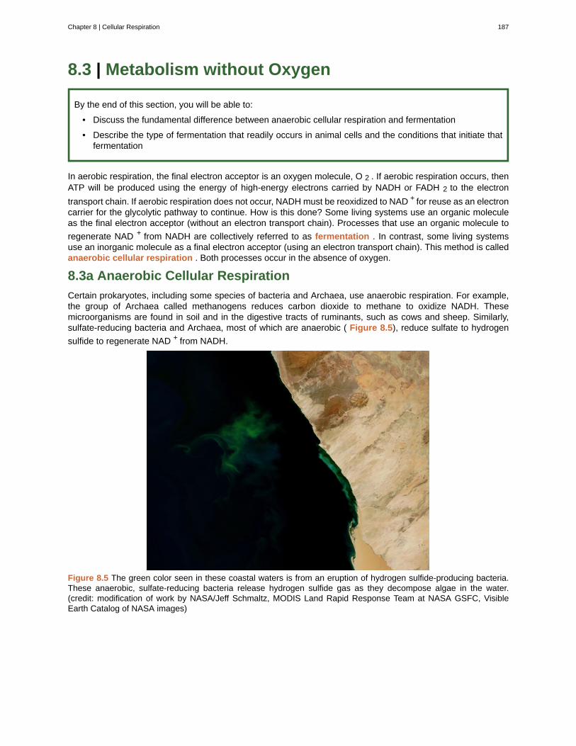
8.3 | Metabolism without Oxygen
By the end of this section, you will be able to:
• Discuss the fundamental difference between anaerobic cellular respiration and fermentation
• Describe the type of fermentation that readily occurs in animal cells and the conditions that initiate thatfermentation
In aerobic respiration, the final electron acceptor is an oxygen molecule, O 2 . If aerobic respiration occurs, thenATP will be produced using the energy of high-energy electrons carried by NADH or FADH 2 to the electron
transport chain. If aerobic respiration does not occur, NADH must be reoxidized to NAD + for reuse as an electroncarrier for the glycolytic pathway to continue. How is this done? Some living systems use an organic moleculeas the final electron acceptor (without an electron transport chain). Processes that use an organic molecule to
regenerate NAD + from NADH are collectively referred to as fermentation . In contrast, some living systemsuse an inorganic molecule as a final electron acceptor (using an electron transport chain). This method is calledanaerobic cellular respiration . Both processes occur in the absence of oxygen.
8.3a Anaerobic Cellular Respiration
Certain prokaryotes, including some species of bacteria and Archaea, use anaerobic respiration. For example,the group of Archaea called methanogens reduces carbon dioxide to methane to oxidize NADH. Thesemicroorganisms are found in soil and in the digestive tracts of ruminants, such as cows and sheep. Similarly,sulfate-reducing bacteria and Archaea, most of which are anaerobic ( Figure 8.5), reduce sulfate to hydrogen
sulfide to regenerate NAD + from NADH.
Figure 8.5 The green color seen in these coastal waters is from an eruption of hydrogen sulfide-producing bacteria.These anaerobic, sulfate-reducing bacteria release hydrogen sulfide gas as they decompose algae in the water.(credit: modification of work by NASA/Jeff Schmaltz, MODIS Land Rapid Response Team at NASA GSFC, VisibleEarth Catalog of NASA images)
Chapter 8 | Cellular Respiration 187

Link to Learning
Visit this site (http://openstaxcollege.org/l/fermentation) to see anaerobic cellular respiration in action.
8.3b Fermentation
Lactic Acid Fermentation
The fermentation method used by animals and certain bacteria, like those in yogurt, is lactic acid fermentation( Figure 8.6). This type of fermentation is used routinely in mammalian red blood cells and in skeletal musclethat has an insufficient oxygen supply to allow aerobic respiration to continue (that is, in muscles used to the
point of fatigue). Remember from chapter 2 that an acid is a molecule that adds H+ to a solution (also called
deprotonation). When lactic acid deprotonates (releases an H+, it is now called lactate. In muscles, accumulationof lactate (because many lactic acid molecules deprotonate) occurs. This excess lactate must be removed bythe blood circulation and the lactate brought to the liver for further metabolism (converted back to pyruvate). Thechemical reactions of lactic acid fermentation are the following:
Pyruvate + NADH ↔ lactic acid + NAD+ ↔ lactate + H+
The enzyme used in this reaction is lactate dehydrogenase (LDH). The reaction can proceed in either direction,but the reaction from left to right is inhibited by acidic conditions. Such acidic conditions was once believed tocause muscle stiffness, fatigue, and soreness, although more recent research disputes this hypothesis.
188 Chapter 8 | Cellular Respiration
This OpenStax book is available for free at https://legacy.cnx.org/content/col11749/1.6
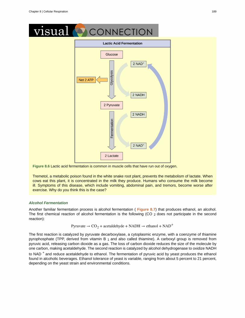
Figure 8.6 Lactic acid fermentation is common in muscle cells that have run out of oxygen.
Tremetol, a metabolic poison found in the white snake root plant, prevents the metabolism of lactate. Whencows eat this plant, it is concentrated in the milk they produce. Humans who consume the milk becomeill. Symptoms of this disease, which include vomiting, abdominal pain, and tremors, become worse afterexercise. Why do you think this is the case?
Alcohol Fermentation
Another familiar fermentation process is alcohol fermentation ( Figure 8.7) that produces ethanol, an alcohol.The first chemical reaction of alcohol fermentation is the following (CO 2 does not participate in the secondreaction):
Pyruvate → CO2 + acetaldehyde + NADH → ethanol + NAD+
The first reaction is catalyzed by pyruvate decarboxylase, a cytoplasmic enzyme, with a coenzyme of thiaminepyrophosphate (TPP, derived from vitamin B 1 and also called thiamine). A carboxyl group is removed frompyruvic acid, releasing carbon dioxide as a gas. The loss of carbon dioxide reduces the size of the molecule byone carbon, making acetaldehyde. The second reaction is catalyzed by alcohol dehydrogenase to oxidize NADH
to NAD + and reduce acetaldehyde to ethanol. The fermentation of pyruvic acid by yeast produces the ethanolfound in alcoholic beverages. Ethanol tolerance of yeast is variable, ranging from about 5 percent to 21 percent,depending on the yeast strain and environmental conditions.
Chapter 8 | Cellular Respiration 189
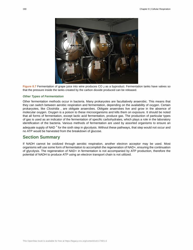
Figure 8.7 Fermentation of grape juice into wine produces CO 2 as a byproduct. Fermentation tanks have valves sothat the pressure inside the tanks created by the carbon dioxide produced can be released.
Other Types of Fermentation
Other fermentation methods occur in bacteria. Many prokaryotes are facultatively anaerobic. This means thatthey can switch between aerobic respiration and fermentation, depending on the availability of oxygen. Certainprokaryotes, like Clostridia , are obligate anaerobes. Obligate anaerobes live and grow in the absence ofmolecular oxygen. Oxygen is a poison to these microorganisms and kills them on exposure. It should be notedthat all forms of fermentation, except lactic acid fermentation, produce gas. The production of particular typesof gas is used as an indicator of the fermentation of specific carbohydrates, which plays a role in the laboratoryidentification of the bacteria. Various methods of fermentation are used by assorted organisms to ensure an
adequate supply of NAD + for the sixth step in glycolysis. Without these pathways, that step would not occur andno ATP would be harvested from the breakdown of glucose.
Section Summary
If NADH cannot be oxidized through aerobic respiration, another electron acceptor may be used. Mostorganisms will use some form of fermentation to accomplish the regeneration of NAD+, ensuring the continuationof glycolysis. The regeneration of NAD+ in fermentation is not accompanied by ATP production, therefore thepotential of NADH to produce ATP using an electron transport chain is not utilized.
190 Chapter 8 | Cellular Respiration
This OpenStax book is available for free at https://legacy.cnx.org/content/col11749/1.6
acetyl CoA
aerobic respiration
anaerobic
anaerobic cellular respiration
ATP synthase
chemiosmosis
citric acid cycle
electron transport chain
fermentation
glycolysis
oxidative phosphorylation
pyruvate
KEY TERMS
the combination of an acetyl group derived from pyruvic acid and coenzyme A which is made frompantothenic acid (a B-group vitamin)
process in which organisms convert energy in the presence of oxygen
process that does not use oxygen
process in which organisms convert energy for their use in the absence ofoxygen
a membrane-embedded protein complex that regenerates ATP from ADP with energy fromprotons diffusing through it
the movement of hydrogen ions down their electrochemical gradient across a membranethrough ATP synthase to generate ATP
a series of enzyme-catalyzed chemical reactions of central importance in all living cells thatharvests the energy in carbon-carbon bonds of sugar molecules to generate ATP; the citric acid cycle is anaerobic metabolic pathway because it requires oxygen in later reactions to proceed
a series of four large, multi-protein complexes embedded in the inner mitochondrialmembrane that accepts electrons from donor compounds and harvests energy from a series of chemicalreactions to generate a hydrogen ion gradient across the membrane
process of regenerating NAD + with either an inorganic or organic compound serving as the finalelectron accceptor, occurs in the absence; occurs in the absence of oxygen
process of breaking glucose into two three-carbon molecules with the production of ATP and NADH
the production of ATP by the transfer of electrons down the electron transportchain to create a proton gradient that is used by ATP synthase to add phosphate groups to ADP molecules
three-carbon sugar that can be decarboxylated and oxidized to make acetyl CoA, which enters thecitric acid cycle under aerobic conditions; the end product of glycolysis
CHAPTER SUMMARY
8.1 Glycolysis
Glycolysis is the first pathway used in the breakdown of glucose to extract energy. It was probably one of theearliest metabolic pathways to evolve and is used by nearly all of the organisms on earth. Glycolysis consists oftwo parts: the first part prepares the six-carbon ring of glucose for cleavage into two three-carbon sugars. ATPis invested in the process during this half to energize the separation. The second half of glycolysis extracts ATPand high-energy electrons from hydrogen atoms and attaches them to NAD+. Two ATP molecule are investedin the first half and four ATP molecules are formed by substrate phosphorylation during the second half. Thisproduces a net gain of two ATP and two NADH molecules for the cell.
8.2 Citric Acid Cycle and Oxidative Phosphorylation
In the presence of oxygen, pyruvate is transformed into an acetyl group attached to a carrier molecule ofcoenzyme A. The resulting acetyl CoA is delivered to the citric acid cycle for further catabolism. During theconversion of pyruvate into the acetyl group, a molecule of carbon dioxide and two high-energy electrons areremoved. The carbon dioxide accounts for two (conversion of two pyruvate molecules) of the six carbons of theoriginal glucose molecule. The electrons are picked up by NAD+, and the NADH carries the elctrons to theelectron transport chain.
The electron transport chain is the portion of aerobic respiration that uses free oxygen as the final electronacceptor. The electron transport chain is composed of four large, multiprotein complexes embedded in themitochondrial membrane and two small diffusible electron carriers shuttling electrons between them. The
Chapter 8 | Cellular Respiration 191

electrons are passed through a series of redox reactions as they move from complex to complex. This processcontributes to the gradient used in chemiosmosis, used to fuel production of ATP. The final products of theelectron transport chain are water and ATP.
A number of intermediate compounds of the citric acid cycle can be diverted into the anabolism of otherbiochemical molecules, such as nonessential amino acids, sugars, and lipids. These same molecules canserve as energy sources for the glucose pathways.
192 Chapter 8 | Cellular Respiration
This OpenStax book is available for free at https://legacy.cnx.org/content/col11749/1.6
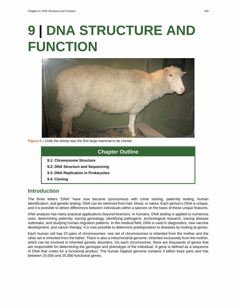
9 | DNA STRUCTURE ANDFUNCTION
Figure 9.1 Dolly the sheep was the first large mammal to be cloned.
Chapter Outline
9.1: Chromosome Structure
9.2: DNA Structure and Sequencing
9.3: DNA Replication in Prokaryotes
9.4: Cloning
Introduction
The three letters “DNA” have now become synonymous with crime solving, paternity testing, humanidentification, and genetic testing. DNA can be retrieved from hair, blood, or saliva. Each person’s DNA is unique,and it is possible to detect differences between individuals within a species on the basis of these unique features.
DNA analysis has many practical applications beyond forensics. In humans, DNA testing is applied to numeroususes: determining paternity, tracing genealogy, identifying pathogens, archeological research, tracing diseaseoutbreaks, and studying human migration patterns. In the medical field, DNA is used in diagnostics, new vaccinedevelopment, and cancer therapy. It is now possible to determine predisposition to diseases by looking at genes.
Each human cell has 23 pairs of chromosomes: one set of chromosomes is inherited from the mother and theother set is inherited from the father. There is also a mitochondrial genome, inherited exclusively from the mother,which can be involved in inherited genetic disorders. On each chromosome, there are thousands of genes thatare responsible for determining the genotype and phenotype of the individual. A gene is defined as a sequenceof DNA that codes for a functional product. The human haploid genome contains 3 billion base pairs and hasbetween 20,000 and 25,000 functional genes.
Chapter 9 | DNA Structure and Function 193
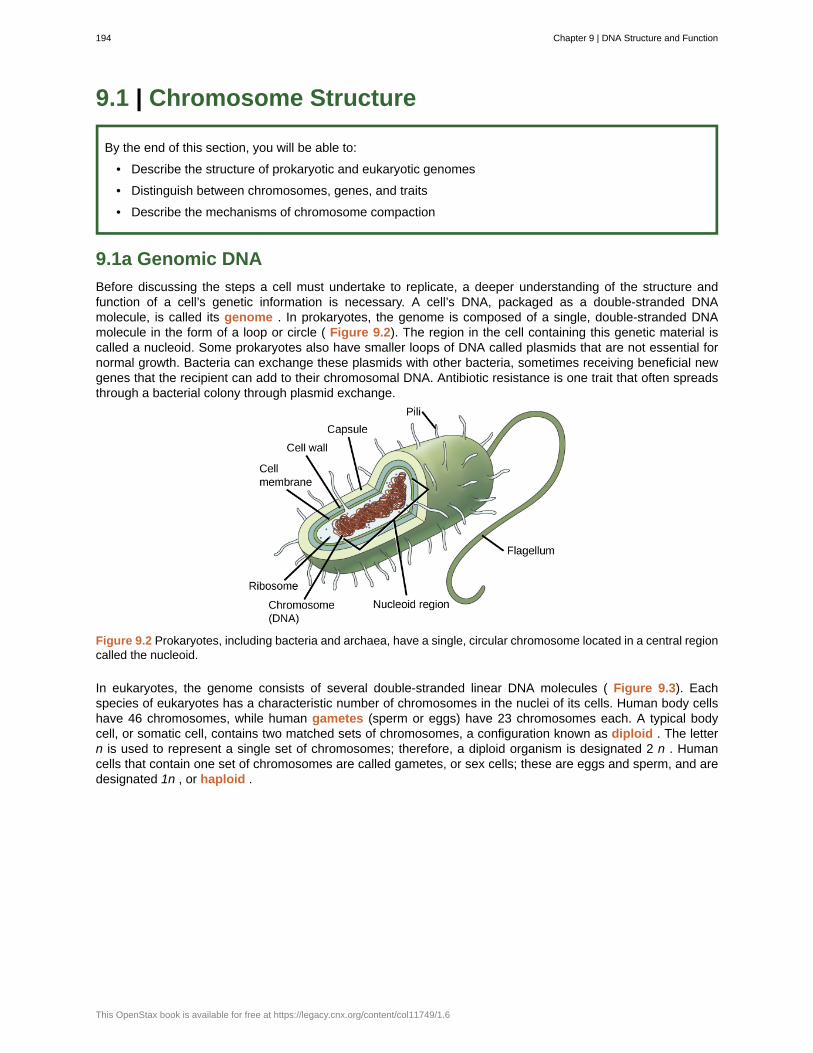
9.1 | Chromosome Structure
By the end of this section, you will be able to:
• Describe the structure of prokaryotic and eukaryotic genomes
• Distinguish between chromosomes, genes, and traits
• Describe the mechanisms of chromosome compaction
9.1a Genomic DNA
Before discussing the steps a cell must undertake to replicate, a deeper understanding of the structure andfunction of a cell’s genetic information is necessary. A cell’s DNA, packaged as a double-stranded DNAmolecule, is called its genome . In prokaryotes, the genome is composed of a single, double-stranded DNAmolecule in the form of a loop or circle ( Figure 9.2). The region in the cell containing this genetic material iscalled a nucleoid. Some prokaryotes also have smaller loops of DNA called plasmids that are not essential fornormal growth. Bacteria can exchange these plasmids with other bacteria, sometimes receiving beneficial newgenes that the recipient can add to their chromosomal DNA. Antibiotic resistance is one trait that often spreadsthrough a bacterial colony through plasmid exchange.
Figure 9.2 Prokaryotes, including bacteria and archaea, have a single, circular chromosome located in a central regioncalled the nucleoid.
In eukaryotes, the genome consists of several double-stranded linear DNA molecules ( Figure 9.3). Eachspecies of eukaryotes has a characteristic number of chromosomes in the nuclei of its cells. Human body cellshave 46 chromosomes, while human gametes (sperm or eggs) have 23 chromosomes each. A typical bodycell, or somatic cell, contains two matched sets of chromosomes, a configuration known as diploid . The lettern is used to represent a single set of chromosomes; therefore, a diploid organism is designated 2 n . Humancells that contain one set of chromosomes are called gametes, or sex cells; these are eggs and sperm, and aredesignated 1n , or haploid .
194 Chapter 9 | DNA Structure and Function
This OpenStax book is available for free at https://legacy.cnx.org/content/col11749/1.6
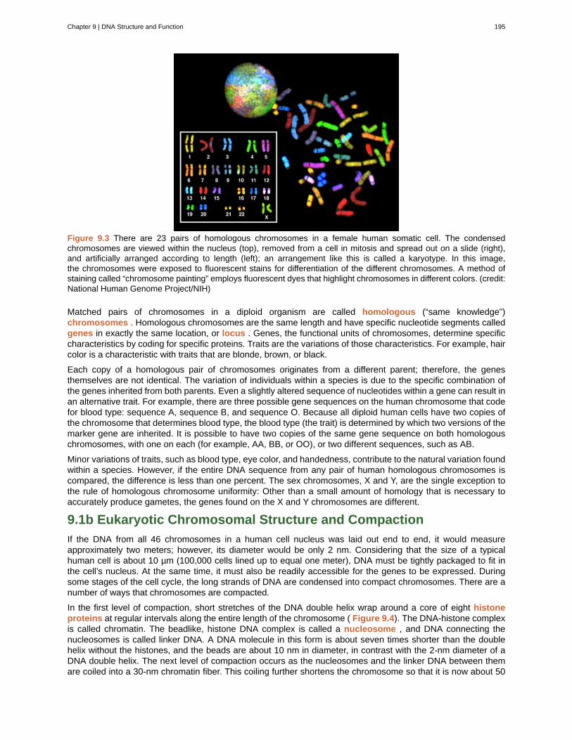
Figure 9.3 There are 23 pairs of homologous chromosomes in a female human somatic cell. The condensedchromosomes are viewed within the nucleus (top), removed from a cell in mitosis and spread out on a slide (right),and artificially arranged according to length (left); an arrangement like this is called a karyotype. In this image,the chromosomes were exposed to fluorescent stains for differentiation of the different chromosomes. A method ofstaining called “chromosome painting” employs fluorescent dyes that highlight chromosomes in different colors. (credit:National Human Genome Project/NIH)
Matched pairs of chromosomes in a diploid organism are called homologous (“same knowledge”)chromosomes . Homologous chromosomes are the same length and have specific nucleotide segments calledgenes in exactly the same location, or locus . Genes, the functional units of chromosomes, determine specificcharacteristics by coding for specific proteins. Traits are the variations of those characteristics. For example, haircolor is a characteristic with traits that are blonde, brown, or black.
Each copy of a homologous pair of chromosomes originates from a different parent; therefore, the genesthemselves are not identical. The variation of individuals within a species is due to the specific combination ofthe genes inherited from both parents. Even a slightly altered sequence of nucleotides within a gene can result inan alternative trait. For example, there are three possible gene sequences on the human chromosome that codefor blood type: sequence A, sequence B, and sequence O. Because all diploid human cells have two copies ofthe chromosome that determines blood type, the blood type (the trait) is determined by which two versions of themarker gene are inherited. It is possible to have two copies of the same gene sequence on both homologouschromosomes, with one on each (for example, AA, BB, or OO), or two different sequences, such as AB.
Minor variations of traits, such as blood type, eye color, and handedness, contribute to the natural variation foundwithin a species. However, if the entire DNA sequence from any pair of human homologous chromosomes iscompared, the difference is less than one percent. The sex chromosomes, X and Y, are the single exception tothe rule of homologous chromosome uniformity: Other than a small amount of homology that is necessary toaccurately produce gametes, the genes found on the X and Y chromosomes are different.
9.1b Eukaryotic Chromosomal Structure and Compaction
If the DNA from all 46 chromosomes in a human cell nucleus was laid out end to end, it would measureapproximately two meters; however, its diameter would be only 2 nm. Considering that the size of a typicalhuman cell is about 10 µm (100,000 cells lined up to equal one meter), DNA must be tightly packaged to fit inthe cell’s nucleus. At the same time, it must also be readily accessible for the genes to be expressed. Duringsome stages of the cell cycle, the long strands of DNA are condensed into compact chromosomes. There are anumber of ways that chromosomes are compacted.
In the first level of compaction, short stretches of the DNA double helix wrap around a core of eight histoneproteins at regular intervals along the entire length of the chromosome ( Figure 9.4). The DNA-histone complexis called chromatin. The beadlike, histone DNA complex is called a nucleosome , and DNA connecting thenucleosomes is called linker DNA. A DNA molecule in this form is about seven times shorter than the doublehelix without the histones, and the beads are about 10 nm in diameter, in contrast with the 2-nm diameter of aDNA double helix. The next level of compaction occurs as the nucleosomes and the linker DNA between themare coiled into a 30-nm chromatin fiber. This coiling further shortens the chromosome so that it is now about 50
Chapter 9 | DNA Structure and Function 195
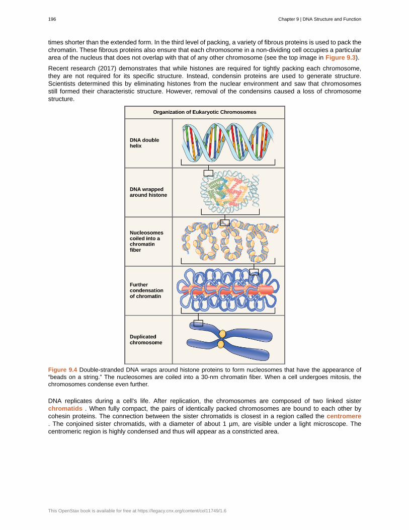
times shorter than the extended form. In the third level of packing, a variety of fibrous proteins is used to pack thechromatin. These fibrous proteins also ensure that each chromosome in a non-dividing cell occupies a particulararea of the nucleus that does not overlap with that of any other chromosome (see the top image in Figure 9.3).
Recent research (2017) demonstrates that while histones are required for tightly packing each chromosome,they are not required for its specific structure. Instead, condensin proteins are used to generate structure.Scientists determined this by eliminating histones from the nuclear environment and saw that chromosomesstill formed their characteristic structure. However, removal of the condensins caused a loss of chromosomestructure.
Figure 9.4 Double-stranded DNA wraps around histone proteins to form nucleosomes that have the appearance of“beads on a string.” The nucleosomes are coiled into a 30-nm chromatin fiber. When a cell undergoes mitosis, thechromosomes condense even further.
DNA replicates during a cell's life. After replication, the chromosomes are composed of two linked sisterchromatids . When fully compact, the pairs of identically packed chromosomes are bound to each other bycohesin proteins. The connection between the sister chromatids is closest in a region called the centromere. The conjoined sister chromatids, with a diameter of about 1 µm, are visible under a light microscope. Thecentromeric region is highly condensed and thus will appear as a constricted area.
196 Chapter 9 | DNA Structure and Function
This OpenStax book is available for free at https://legacy.cnx.org/content/col11749/1.6
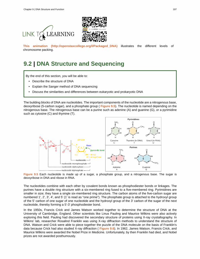
This animation (http://openstaxcollege.org/l/Packaged_DNA) illustrates the different levels ofchromosome packing.
9.2 | DNA Structure and Sequencing
By the end of this section, you will be able to:
• Describe the structure of DNA
• Explain the Sanger method of DNA sequencing
• Discuss the similarities and differences between eukaryotic and prokaryotic DNA
The building blocks of DNA are nucleotides. The important components of the nucleotide are a nitrogenous base,deoxyribose (5-carbon sugar), and a phosphate group ( Figure 9.5). The nucleotide is named depending on thenitrogenous base. The nitrogenous base can be a purine such as adenine (A) and guanine (G), or a pyrimidinesuch as cytosine (C) and thymine (T).
Figure 9.5 Each nucleotide is made up of a sugar, a phosphate group, and a nitrogenous base. The sugar isdeoxyribose in DNA and ribose in RNA.
The nucleotides combine with each other by covalent bonds known as phosphodiester bonds or linkages. Thepurines have a double ring structure with a six-membered ring fused to a five-membered ring. Pyrimidines aresmaller in size; they have a single six-membered ring structure. The carbon atoms of the five-carbon sugar arenumbered 1', 2', 3', 4', and 5' (1' is read as “one prime”). The phosphate group is attached to the hydroxyl groupof the 5' carbon of one sugar of one nucleotide and the hydroxyl group of the 3' carbon of the sugar of the nextnucleotide, thereby forming a 5'-3' phosphodiester bond.
In the 1950s, Francis Crick and James Watson worked together to determine the structure of DNA at theUniversity of Cambridge, England. Other scientists like Linus Pauling and Maurice Wilkins were also activelyexploring this field. Pauling had discovered the secondary structure of proteins using X-ray crystallography. InWilkins’ lab, researcher Rosalind Franklin was using X-ray diffraction methods to understand the structure ofDNA. Watson and Crick were able to piece together the puzzle of the DNA molecule on the basis of Franklin'sdata because Crick had also studied X-ray diffraction ( Figure 9.6). In 1962, James Watson, Francis Crick, andMaurice Wilkins were awarded the Nobel Prize in Medicine. Unfortunately, by then Franklin had died, and Nobelprizes are not awarded posthumously.
Chapter 9 | DNA Structure and Function 197

Figure 9.6 The work of pioneering scientists (a) James Watson, Francis Crick, and Maclyn McCarty led to our presentday understanding of DNA. Scientist Rosalind Franklin discovered (b) the X-ray diffraction pattern of DNA, whichhelped to elucidate its double helix structure. (credit a: modification of work by Marjorie McCarty, Public Library ofScience)
Watson and Crick proposed that DNA is made up of two strands that are twisted around each other to form aright-handed helix. Base pairing takes place between a purine and pyrimidine; namely, A pairs with T and G pairswith C. Adenine and thymine are complementary base pairs, and cytosine and guanine are also complementarybase pairs. The base pairs are stabilized by hydrogen bonds; adenine and thymine form two hydrogen bondsand cytosine and guanine form three hydrogen bonds. The two strands are anti-parallel in nature; that is, the3' end of one strand faces the 5' end of the other strand. The sugar and phosphate of the nucleotides formthe backbone of the structure, whereas the nitrogenous bases are stacked inside. Each base pair is separatedfrom the other base pair by a distance of 0.34 nm, and each turn of the helix measures 3.4 nm. Therefore, tenbase pairs are present per turn of the helix. The diameter of the DNA double helix is 2 nm, and it is uniformthroughout. Only the pairing between a purine and pyrimidine can explain the uniform diameter. The twisting ofthe two strands around each other results in the formation of uniformly spaced major and minor grooves ( Figure9.7).
Figure 9.7 DNA has (a) a double helix structure and (b) phosphodiester bonds. The (c) major and minor grooves arebinding sites for DNA binding proteins during processes such as transcription (the copying of RNA from DNA) andreplication.
9.2a DNA Sequencing Techniques
Until the 1990s, the sequencing of DNA (reading the sequence of DNA) was a relatively expensive and longprocess. Using radiolabeled nucleotides also compounded the problem through safety concerns. With currentlyavailable technology and automated machines, the process is cheap, safer, and can be completed in a matter ofhours. Fred Sanger developed the sequencing method used for the human genome sequencing project, whichis widely used today.
198 Chapter 9 | DNA Structure and Function
This OpenStax book is available for free at https://legacy.cnx.org/content/col11749/1.6
Visit this site (http://openstaxcollege.org/l/DNA_sequencing) to watch a video explaining the DNAsequence reading technique that resulted from Sanger’s work.
Sanger’s genome sequencing has led to a race to sequence human genomes at a rapid speed and lowcost, often referred to as the $1000 in one day sequence. Learn more by selecting the Sequencing at Speedanimation here (http://openstaxcollege.org/l/DNA_and_genomes) .
Neanderthal Genome: How Are We Related?The first draft sequence of the Neanderthal genome was recently published by Richard E. Green et al.
in 2010.[1]
Neanderthals are the closest ancestors of present-day humans. They were known to havelived in Europe and Western Asia before they disappeared from fossil records approximately 30,000 yearsago. Green’s team studied almost 40,000-year-old fossil remains that were selected from sites acrossthe world. Extremely sophisticated means of sample preparation and DNA sequencing were employedbecause of the fragile nature of the bones and heavy microbial contamination. In their study, the scientistswere able to sequence some four billion base pairs. The Neanderthal sequence was compared with thatof present-day humans from across the world. After comparing the sequences, the researchers foundthat the Neanderthal genome had 2 to 3 percent greater similarity to people living outside Africa thanto people in Africa. While current theories have suggested that all present-day humans can be traced toa small ancestral population in Africa, the data from the Neanderthal genome may contradict this view.Green and his colleagues also discovered DNA segments among people in Europe and Asia that aremore similar to Neanderthal sequences than to other contemporary human sequences. Another interestingobservation was that Neanderthals are as closely related to people from Papua New Guinea as to thosefrom China or France. This is surprising because Neanderthal fossil remains have been located onlyin Europe and West Asia. Most likely, genetic exchange took place between Neanderthals and modernhumans as modern humans emerged out of Africa, before the divergence of Europeans, East Asians, andPapua New Guineans.
Several genes seem to have undergone changes from Neanderthals during the evolution of present-day humans. These genes are involved in cranial structure, metabolism, skin morphology, and cognitivedevelopment. One of the genes that is of particular interest is RUNX2 , which is different in modern dayhumans and Neanderthals. This gene is responsible for the prominent frontal bone, bell-shaped rib cage,and dental differences seen in Neanderthals. It is speculated that an evolutionary change in RUNX2 wasimportant in the origin of modern-day humans, and this affected the cranium and the upper body.
1.Richard E. Green et al., “A Draft Sequence of the Neandertal Genome,” Science 328 (2010): 710-22.
Chapter 9 | DNA Structure and Function 199
Watch Svante Pääbo’s talk (http://openstaxcollege.org/l/neanderthal) explaining the Neanderthal genomeresearch at the 2011 annual TED (Technology, Entertainment, Design) conference.
9.3 | DNA Replication in Prokaryotes
By the end of this section, you will be able to:
• Explain the process of DNA replication in prokaryotes
• Discuss the role of different enzymes and proteins in supporting this process
DNA replication is the process of a cell making an additional copy of all of its DNA. DNA replication has beenextremely well studied in prokaryotes primarily because of the small size of the genome and the mutants thatare available. E. coli has 4.6 million base pairs in a single circular chromosome and all of it gets replicated inapproximately 42 minutes, starting from a single origin of replication and proceeding around the circle in bothdirections. This means that approximately 1000 nucleotides are added per second. The process is quite rapidand occurs without many mistakes.
DNA replication employs a large number of proteins and enzymes, each of which plays a critical role duringthe process. One of the key players is the enzyme DNA polymerase , also known as DNA pol, which addsnucleotides one by one to the growing DNA chain that are complementary to the template strand. The addition ofnucleotides requires energy; this energy is obtained from the nucleotides that have three phosphates attachedto them: ATP, GTP, TTP, and CTP. The ATP in the previous sentence is the same molecules used for energythroughout a cell. When the bond between the phosphates is broken, the energy released is used to form thephosphodiester bond between the incoming nucleotide and the growing chain. In prokaryotes, three main typesof polymerases are known: DNA pol I, DNA pol II, and DNA pol III. It is now known that DNA pol III is the enzymerequired for DNA synthesis; DNA pol I and DNA pol II are primarily required for repair.
How does the replication machinery know where to begin? It turns out that there are specific nucleotidesequences called origins of replication where replication begins. In E. coli, which has a single origin of replicationon its one chromosome (as do most prokaryotes), it is approximately 245 base pairs long and is rich in ATsequences. The origin of replication is recognized by certain proteins that bind to this site. An enzyme calledhelicase unwinds the DNA by breaking the hydrogen bonds between the nitrogenous base pairs. As the DNAopens up, Y-shaped structures called replication forks are formed. Two replication forks are formed at theorigin of replication and these get extended bi- directionally as replication proceeds. Single-strand bindingproteins coat the single strands of DNA near the replication fork to prevent the single-stranded DNA fromwinding back into a double helix. DNA polymerase is able to add nucleotides only in the 5' to 3' direction (anew DNA strand can be only extended in this direction). It also requires a free 3'-OH group to which it canadd nucleotides by forming a phosphodiester bond between the 3'-OH end and the 5' phosphate of the nextnucleotide. This essentially means that it cannot add nucleotides if a free 3'-OH group is not available. Thenhow does it add the first nucleotide? The problem is solved with the help of a primer that provides the free3'-OH end. Another enzyme, RNA primase , synthesizes an RNA primer that is about five to ten nucleotideslong and complementary to the DNA. Because this sequence primes the DNA synthesis, it is appropriatelycalled the primer . DNA polymerase can now extend this RNA primer, adding nucleotides one by one that arecomplementary to the template strand ( Figure 9.8).
200 Chapter 9 | DNA Structure and Function
This OpenStax book is available for free at https://legacy.cnx.org/content/col11749/1.6
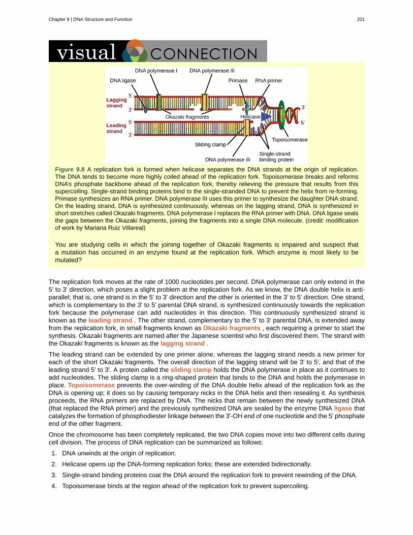
Figure 9.8 A replication fork is formed when helicase separates the DNA strands at the origin of replication.The DNA tends to become more highly coiled ahead of the replication fork. Topoisomerase breaks and reformsDNA’s phosphate backbone ahead of the replication fork, thereby relieving the pressure that results from thissupercoiling. Single-strand binding proteins bind to the single-stranded DNA to prevent the helix from re-forming.Primase synthesizes an RNA primer. DNA polymerase III uses this primer to synthesize the daughter DNA strand.On the leading strand, DNA is synthesized continuously, whereas on the lagging strand, DNA is synthesized inshort stretches called Okazaki fragments. DNA polymerase I replaces the RNA primer with DNA. DNA ligase sealsthe gaps between the Okazaki fragments, joining the fragments into a single DNA molecule. (credit: modificationof work by Mariana Ruiz Villareal)
You are studying cells in which the joining together of Okazaki fragments is impaired and suspect thata mutation has occurred in an enzyme found at the replication fork. Which enzyme is most likely to bemutated?
The replication fork moves at the rate of 1000 nucleotides per second. DNA polymerase can only extend in the5' to 3' direction, which poses a slight problem at the replication fork. As we know, the DNA double helix is anti-parallel; that is, one strand is in the 5' to 3' direction and the other is oriented in the 3' to 5' direction. One strand,which is complementary to the 3' to 5' parental DNA strand, is synthesized continuously towards the replicationfork because the polymerase can add nucleotides in this direction. This continuously synthesized strand isknown as the leading strand . The other strand, complementary to the 5' to 3' parental DNA, is extended awayfrom the replication fork, in small fragments known as Okazaki fragments , each requiring a primer to start thesynthesis. Okazaki fragments are named after the Japanese scientist who first discovered them. The strand withthe Okazaki fragments is known as the lagging strand .
The leading strand can be extended by one primer alone, whereas the lagging strand needs a new primer foreach of the short Okazaki fragments. The overall direction of the lagging strand will be 3' to 5', and that of theleading strand 5' to 3'. A protein called the sliding clamp holds the DNA polymerase in place as it continues toadd nucleotides. The sliding clamp is a ring-shaped protein that binds to the DNA and holds the polymerase inplace. Topoisomerase prevents the over-winding of the DNA double helix ahead of the replication fork as theDNA is opening up; it does so by causing temporary nicks in the DNA helix and then resealing it. As synthesisproceeds, the RNA primers are replaced by DNA. The nicks that remain between the newly synthesized DNA(that replaced the RNA primer) and the previously synthesized DNA are sealed by the enzyme DNA ligase thatcatalyzes the formation of phosphodiester linkage between the 3'-OH end of one nucleotide and the 5' phosphateend of the other fragment.
Once the chromosome has been completely replicated, the two DNA copies move into two different cells duringcell division. The process of DNA replication can be summarized as follows:
1. DNA unwinds at the origin of replication.
2. Helicase opens up the DNA-forming replication forks; these are extended bidirectionally.
3. Single-strand binding proteins coat the DNA around the replication fork to prevent rewinding of the DNA.
4. Topoisomerase binds at the region ahead of the replication fork to prevent supercoiling.
Chapter 9 | DNA Structure and Function 201
5. Primase synthesizes RNA primers complementary to the DNA strand.
6. DNA polymerase starts adding nucleotides to the 3'-OH end of the primer.
7. Elongation of both the lagging and the leading strand continues.
8. RNA primers are removed by exonuclease activity.
9. Gaps are filled by DNA pol by adding dNTPs.
10. The gap between the two DNA fragments is sealed by DNA ligase, which helps in the formation ofphosphodiester bonds.
Table 9.1 summarizes the enzymes involved in prokaryotic DNA replication and the functions of each.
Prokaryotic DNA Replication: Enzymes and Their Function
Enzyme/protein Specific Function
DNA pol IExonuclease activity removes RNA primer and replaces with newlysynthesized DNA
DNA pol II Repair function
DNA pol III Main enzyme that adds nucleotides in the 5'-3' direction
HelicaseOpens the DNA helix by breaking hydrogen bonds between the nitrogenousbases
LigaseSeals the gaps between the Okazaki fragments to create one continuous DNAstrand
Primase Synthesizes RNA primers needed to start replication
Sliding Clamp Helps to hold the DNA polymerase in place when nucleotides are being added
TopoisomeraseHelps relieve the stress on DNA when unwinding by causing breaks and thenresealing the DNA
Single-strand bindingproteins (SSB)
Binds to single-stranded DNA to avoid DNA rewinding back.
Table 9.1
Review the full process of DNA replication here (http://openstaxcollege.org/l/replication_DNA) .
The previous section has demonstrated how DNA replicates by breaking existing double-stranded pieces apart,then adding a new second strand to each existing one. This may seem very logical, but scientists weren'talways so certain how the process worked. All they knew was somehow a cell started with one piece of double-stranded DNA and then later it ended up with two pieces of double-stranded DNA. Scientists hypothesizedthree possibilities, one of which was the model described above, called semi-conservative replication. The othermodels (conservative replication and discontinuous replication) were rejected based on multiple experiments.
DNA replication is not a perfect process. The DNA polymerase sometimes makes mistakes. Cells have multipleways to fix mistakes: DNA polymerase has a proof-reading ability to check its own work and different types ofrepair processes exist to fix different kinds of mutations. However, not every mistake is fixed. If a mistake is notfixed, we call it a mutation. Mutations are passed on because a cell can only fix a mistake as soon as it happens,so if it's not fixed right away, it stays in the cell's DNA permanently.
202 Chapter 9 | DNA Structure and Function
This OpenStax book is available for free at https://legacy.cnx.org/content/col11749/1.6
9.4 | Cloning
By the end of this section, you will be able to:
• Explain molecular, cellular, and reproductive cloning
9.4a Molecular Cloning
In general, the word “cloning” means the creation of a perfect replica; however, in biology, the re-creation ofa whole organism is referred to as “reproductive cloning.” Long before attempts were made to clone an entireorganism, researchers learned how to reproduce desired regions or fragments of the genome, a process that isreferred to as molecular cloning .
9.4b Cellular Cloning
Unicellular organisms, such as bacteria and yeast, naturally produce clones of themselves when they replicateasexually by binary fission; this is known as cellular cloning . The nuclear DNA duplicates by the process ofmitosis, which creates an exact replica of the genetic material.
9.4c Reproductive Cloning
Reproductive cloning is a method used to make a clone or an identical copy of an entire multicellular organism.Most multicellular organisms undergo reproduction by sexual means, which involves genetic hybridization of twoindividuals (parents), making it impossible for generation of an identical copy or a clone of either parent. Recentadvances in biotechnology have made it possible to artificially induce asexual reproduction of mammals in thelaboratory.
Parthenogenesis, or “virgin birth,” occurs when an embryo grows and develops without the fertilization of theegg occurring; this is a form of asexual reproduction. An example of parthenogenesis occurs in species in whichthe female lays an egg and if the egg is fertilized, it is a diploid egg and the individual develops into a female;if the egg is not fertilized, it remains a haploid egg and develops into a male. The unfertilized egg is called aparthenogenic, or virgin, egg. Some insects and reptiles lay parthenogenic eggs that can develop into adults.
Sexual reproduction requires two cells; when the haploid egg and sperm cells fuse, a diploid zygote results.The zygote nucleus contains the genetic information to produce a new individual. However, early embryonicdevelopment requires the cytoplasmic material contained in the egg cell. This idea forms the basis forreproductive cloning. Therefore, if the haploid nucleus of an egg cell is replaced with a diploid nucleus from thecell of any individual of the same species (called a donor), it will become a zygote that is genetically identical tothe donor. Somatic cell nuclear transfer is the technique of transferring a diploid nucleus into an enucleatedegg. It can be used for either therapeutic cloning or reproductive cloning.
The first cloned animal was Dolly, a sheep who was born in 1996. The success rate of reproductive cloning atthe time was very low. Dolly lived for seven years and died of respiratory complications ( Figure 9.9). Thereis speculation that because the cell DNA belongs to an older individual, the age of the DNA may affect the lifeexpectancy of a cloned individual. Since Dolly, several animals such as horses, bulls, and goats have beensuccessfully cloned, although these individuals often exhibit facial, limb, and cardiac abnormalities. There havebeen attempts at producing cloned human embryos as sources of embryonic stem cells, sometimes referred toas cloning for therapeutic purposes. Therapeutic cloning produces stem cells to attempt to remedy detrimentaldiseases or defects (unlike reproductive cloning, which aims to reproduce an organism). Still, therapeutic cloningefforts have met with resistance because of bioethical considerations.
Chapter 9 | DNA Structure and Function 203
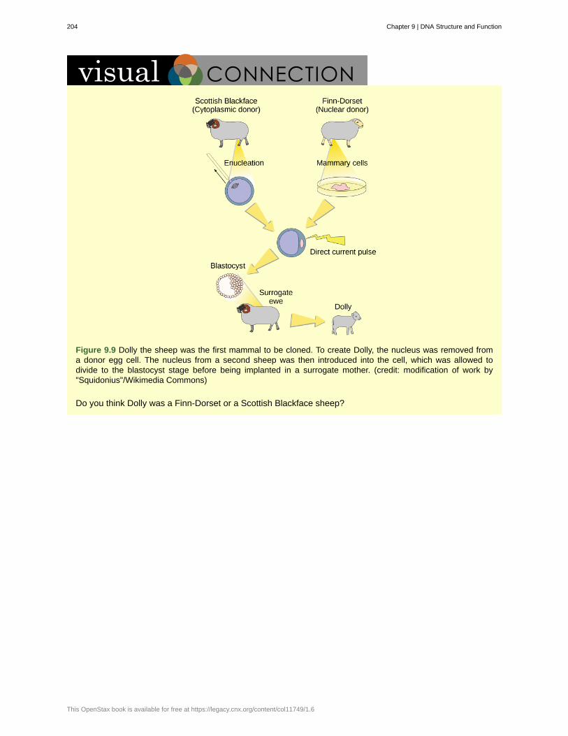
Figure 9.9 Dolly the sheep was the first mammal to be cloned. To create Dolly, the nucleus was removed froma donor egg cell. The nucleus from a second sheep was then introduced into the cell, which was allowed todivide to the blastocyst stage before being implanted in a surrogate mother. (credit: modification of work by"Squidonius"/Wikimedia Commons)
Do you think Dolly was a Finn-Dorset or a Scottish Blackface sheep?
204 Chapter 9 | DNA Structure and Function
This OpenStax book is available for free at https://legacy.cnx.org/content/col11749/1.6
cellular cloning
centromere
chromatids
chromosomes
diploid
DNA polymerase
DNA replication
gametes
genes
genome
haploid
helicase
histone proteins
homologous
lagging strand
leading strand
ligase
locus
molecular cloning
nucleosome
Okazaki fragments
primase
primer
replication forks
reproductive cloning
Single-strand binding proteins
KEY TERMS
production of identical cell populations by binary fission
region at which sister chromatids are bound together; a constricted area in condensedchromosomes
single DNA molecule of two strands of duplicated DNA and associated proteins held together atthe centromere
condensed chromatin; can be "replicated" or "unreplicated"
cell, nucleus, or organism containing two sets of chromosomes (2 n )
enzyme that replicates DNA
process of copying DNA
haploid reproductive cell or sex cell (sperm or egg)
physical and functional unit of heredity, a sequence of DNA that codes for a protein
total genetic information of a cell or organism
cell, nucleus, or organism containing one set of chromosomes ( n )
during replication, this enzyme helps to open up the DNA helix by breaking the hydrogen bonds
proteins found in the chromatin of all eukaryotic cells; associated with DNA to formnucleosomes
chromosomes of the same morphology with genes in the same location; diploid organisms havepairs of homologous chromosomes (homologs), with each homolog derived from a different parent
during replication, the strand that is replicated in short fragments and away from the replicationfork
strand that is synthesized continuously in the 5'-3' direction which is synthesized in the directionof the replication fork
enzyme that catalyzes the formation of a phosphodiester linkage between the 3' OH and 5' phosphateends of the DNA
position of a gene on a chromosome
cloning of DNA fragments
subunit of chromatin composed of a short length of DNA wrapped around a core of histoneproteins
DNA fragment synthesized in short stretches on the lagging strand
enzyme that synthesizes the RNA primer; the primer is needed for DNA pol to start synthesis of a newDNA strand
short stretch of nucleotides that is required to initiate replication; in the case of replication, the primerhas RNA nucleotides
Y-shaped structure formed during initiation of replication
cloning of entire organisms
during replication, protein that binds to the single-stranded DNA; his helps in
Chapter 9 | DNA Structure and Function 205
sliding clamp
Somatic cell nuclear transfer
Therapeutic cloning
topoisomerase
keeping the two strands of DNA apart so they may serve as templates
ring-shaped protein that holds the DNA pol on the DNA strand
transfer of a somatic (body) cell's nucleus into an egg cell missing its ownnucleus
production of stem cells used to treat disease
enzyme that causes underwinding or overwinding of DNA when DNA replication is taking place
CHAPTER SUMMARY
9.1 Chromosome Structure
A genome is all of an organism's DNA. Depending on the type of organism, the DNA can be organized into asingle circular chromosome, or multiple, linear chromosomes. One molecule of DNA is a double-stranded pieceof deoxyribonucleic acid, and is usually kept condensed in the form of chromatin.
9.2 DNA Structure and Sequencing
Nucleic acids are made of nucleotides. Nucleotides are made of a nitrogenous base, and one-three phosphategroups, all bonded to a sugar molecule. DNA uses four different nitrogenous bases, and the order of thesebases (the DNA sequence) determines what the DNA encodes. Determining the sequence of all the bases in acell is called "DNA sequencing," and is an important tool in scientific research.
9.3 DNA Replication in Prokaryotes
DNA replication is required for cells to pass on their DNA when they divide into new cells. Multiple proteins areinvolved in replicating DNA, and it happens in a semi-conservative manner. The two strands are split apart, anda new, complementary copy of each strand is built. DNA polymerase can only build in the 5' to 3' direction,which means that each strand is replicated in different directions. Unfixed errors during DNA replication aremutations, and can be passed on when that cell divides.
9.4 Cloning
Cloning is a process of making a copy of something's DNA. This copy could be just a section of DNA(molecular cloning), or an entire copy of a cell (cellular cloning), or an entire copy of an organism (molecularcloning).
206 Chapter 9 | DNA Structure and Function
This OpenStax book is available for free at https://legacy.cnx.org/content/col11749/1.6

10 | GENES ANDPROTEINS
Figure 10.1 Genes, which are carried on (a) chromosomes, are linearly organized instructions for making the RNAand protein molecules that are necessary for all of processes of life. The (b) interleukin-2 protein and (c) alpha-2u-globulin protein are just two examples of the array of different molecular structures that are encoded by genes. (credit“chromosome: National Human Genome Research Institute; credit “interleukin-2”: Ramin Herati/Created from PDB1M47 and rendered with Pymol; credit “alpha-2u-globulin”: Darren Logan/rendered with AISMIG)
Chapter Outline
10.1: The Genetic Code
10.2: Prokaryotic Transcription
10.3: RNA Processing in Eukaryotes
10.4: Ribosomes and Protein Synthesis
10.5: Mutations
Introduction
Since the rediscovery of Mendel’s work in 1900, the definition of the gene has progressed from an abstract unitof heredity to a tangible molecular entity capable of replication, expression, and mutation (Figure 10.1). Genesare composed of DNA and are linearly arranged on chromosomes. Genes specify the sequences of amino acids,which are the building blocks of proteins. In turn, proteins are responsible for orchestrating nearly every functionof the cell. Both genes and the proteins they encode are absolutely essential to life as we know it.
10.1 | The Genetic Code
By the end of this section, you will be able to:
• Explain the “central dogma” of protein synthesis
• Describe the genetic code and how the nucleotide sequence prescribes the amino acid and the proteinsequence
The cellular process of transcription generates messenger RNA (mRNA), a mobile molecular copy of one ormore genes with an alphabet of A, C, G, and uracil (U). Translation of the mRNA template converts nucleotide-based genetic information into a protein product. Protein sequences consist of 20 commonly occurring amino
Chapter 10 | Genes and Proteins 207
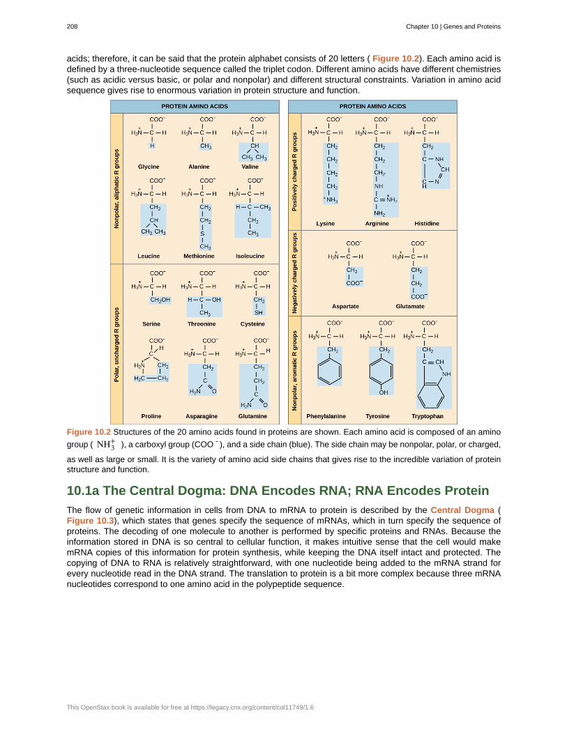
acids; therefore, it can be said that the protein alphabet consists of 20 letters ( Figure 10.2). Each amino acid isdefined by a three-nucleotide sequence called the triplet codon. Different amino acids have different chemistries(such as acidic versus basic, or polar and nonpolar) and different structural constraints. Variation in amino acidsequence gives rise to enormous variation in protein structure and function.
Figure 10.2 Structures of the 20 amino acids found in proteins are shown. Each amino acid is composed of an amino
group ( NH3+ ), a carboxyl group (COO - ), and a side chain (blue). The side chain may be nonpolar, polar, or charged,
as well as large or small. It is the variety of amino acid side chains that gives rise to the incredible variation of proteinstructure and function.
10.1a The Central Dogma: DNA Encodes RNA; RNA Encodes Protein
The flow of genetic information in cells from DNA to mRNA to protein is described by the Central Dogma (Figure 10.3), which states that genes specify the sequence of mRNAs, which in turn specify the sequence ofproteins. The decoding of one molecule to another is performed by specific proteins and RNAs. Because theinformation stored in DNA is so central to cellular function, it makes intuitive sense that the cell would makemRNA copies of this information for protein synthesis, while keeping the DNA itself intact and protected. Thecopying of DNA to RNA is relatively straightforward, with one nucleotide being added to the mRNA strand forevery nucleotide read in the DNA strand. The translation to protein is a bit more complex because three mRNAnucleotides correspond to one amino acid in the polypeptide sequence.
208 Chapter 10 | Genes and Proteins
This OpenStax book is available for free at https://legacy.cnx.org/content/col11749/1.6
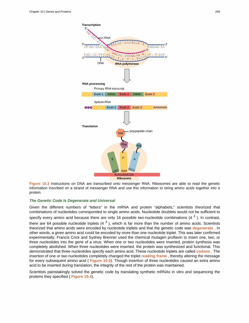
Figure 10.3 Instructions on DNA are transcribed onto messenger RNA. Ribosomes are able to read the geneticinformation inscribed on a strand of messenger RNA and use this information to string amino acids together into aprotein.
The Genetic Code Is Degenerate and Universal
Given the different numbers of “letters” in the mRNA and protein “alphabets,” scientists theorized thatcombinations of nucleotides corresponded to single amino acids. Nucleotide doublets would not be sufficient to
specify every amino acid because there are only 16 possible two-nucleotide combinations (4 2 ). In contrast,
there are 64 possible nucleotide triplets (4 3 ), which is far more than the number of amino acids. Scientiststheorized that amino acids were encoded by nucleotide triplets and that the genetic code was degenerate . Inother words, a given amino acid could be encoded by more than one nucleotide triplet. This was later confirmedexperimentally; Francis Crick and Sydney Brenner used the chemical mutagen proflavin to insert one, two, orthree nucleotides into the gene of a virus. When one or two nucleotides were inserted, protein synthesis wascompletely abolished. When three nucleotides were inserted, the protein was synthesized and functional. Thisdemonstrated that three nucleotides specify each amino acid. These nucleotide triplets are called codons . Theinsertion of one or two nucleotides completely changed the triplet reading frame , thereby altering the messagefor every subsequent amino acid ( Figure 10.5). Though insertion of three nucleotides caused an extra aminoacid to be inserted during translation, the integrity of the rest of the protein was maintained.
Scientists painstakingly solved the genetic code by translating synthetic mRNAs in vitro and sequencing theproteins they specified ( Figure 10.4).
Chapter 10 | Genes and Proteins 209
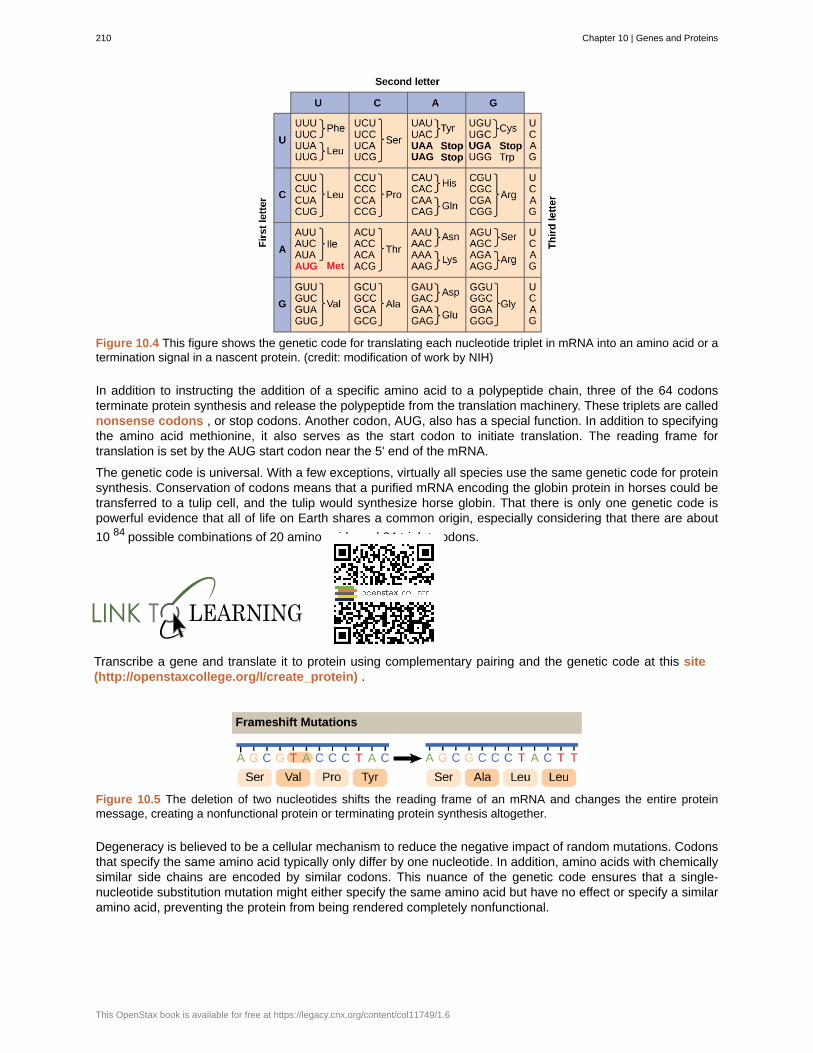
Figure 10.4 This figure shows the genetic code for translating each nucleotide triplet in mRNA into an amino acid or atermination signal in a nascent protein. (credit: modification of work by NIH)
In addition to instructing the addition of a specific amino acid to a polypeptide chain, three of the 64 codonsterminate protein synthesis and release the polypeptide from the translation machinery. These triplets are callednonsense codons , or stop codons. Another codon, AUG, also has a special function. In addition to specifyingthe amino acid methionine, it also serves as the start codon to initiate translation. The reading frame fortranslation is set by the AUG start codon near the 5' end of the mRNA.
The genetic code is universal. With a few exceptions, virtually all species use the same genetic code for proteinsynthesis. Conservation of codons means that a purified mRNA encoding the globin protein in horses could betransferred to a tulip cell, and the tulip would synthesize horse globin. That there is only one genetic code ispowerful evidence that all of life on Earth shares a common origin, especially considering that there are about
10 84 possible combinations of 20 amino acids and 64 triplet codons.
Transcribe a gene and translate it to protein using complementary pairing and the genetic code at this site(http://openstaxcollege.org/l/create_protein) .
Figure 10.5 The deletion of two nucleotides shifts the reading frame of an mRNA and changes the entire proteinmessage, creating a nonfunctional protein or terminating protein synthesis altogether.
Degeneracy is believed to be a cellular mechanism to reduce the negative impact of random mutations. Codonsthat specify the same amino acid typically only differ by one nucleotide. In addition, amino acids with chemicallysimilar side chains are encoded by similar codons. This nuance of the genetic code ensures that a single-nucleotide substitution mutation might either specify the same amino acid but have no effect or specify a similaramino acid, preventing the protein from being rendered completely nonfunctional.
210 Chapter 10 | Genes and Proteins
This OpenStax book is available for free at https://legacy.cnx.org/content/col11749/1.6
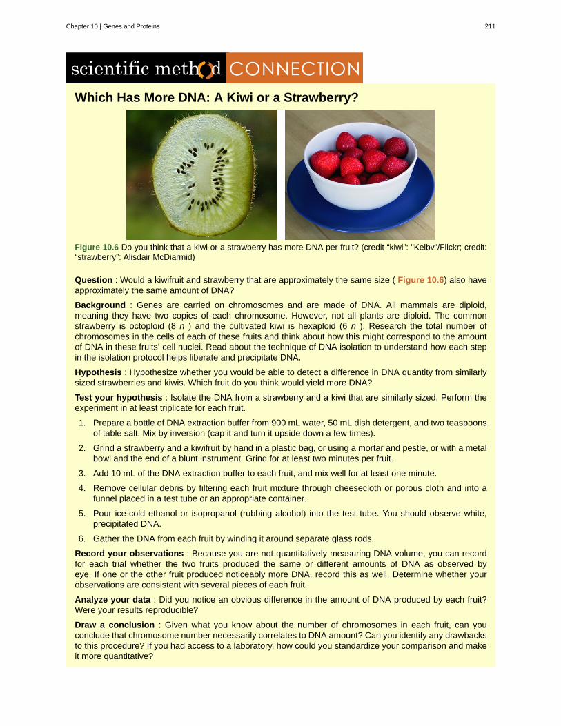
Which Has More DNA: A Kiwi or a Strawberry?
Figure 10.6 Do you think that a kiwi or a strawberry has more DNA per fruit? (credit “kiwi”: "Kelbv"/Flickr; credit:“strawberry”: Alisdair McDiarmid)
Question : Would a kiwifruit and strawberry that are approximately the same size ( Figure 10.6) also haveapproximately the same amount of DNA?
Background : Genes are carried on chromosomes and are made of DNA. All mammals are diploid,meaning they have two copies of each chromosome. However, not all plants are diploid. The commonstrawberry is octoploid (8 n ) and the cultivated kiwi is hexaploid (6 n ). Research the total number ofchromosomes in the cells of each of these fruits and think about how this might correspond to the amountof DNA in these fruits’ cell nuclei. Read about the technique of DNA isolation to understand how each stepin the isolation protocol helps liberate and precipitate DNA.
Hypothesis : Hypothesize whether you would be able to detect a difference in DNA quantity from similarlysized strawberries and kiwis. Which fruit do you think would yield more DNA?
Test your hypothesis : Isolate the DNA from a strawberry and a kiwi that are similarly sized. Perform theexperiment in at least triplicate for each fruit.
1. Prepare a bottle of DNA extraction buffer from 900 mL water, 50 mL dish detergent, and two teaspoonsof table salt. Mix by inversion (cap it and turn it upside down a few times).
2. Grind a strawberry and a kiwifruit by hand in a plastic bag, or using a mortar and pestle, or with a metalbowl and the end of a blunt instrument. Grind for at least two minutes per fruit.
3. Add 10 mL of the DNA extraction buffer to each fruit, and mix well for at least one minute.
4. Remove cellular debris by filtering each fruit mixture through cheesecloth or porous cloth and into afunnel placed in a test tube or an appropriate container.
5. Pour ice-cold ethanol or isopropanol (rubbing alcohol) into the test tube. You should observe white,precipitated DNA.
6. Gather the DNA from each fruit by winding it around separate glass rods.
Record your observations : Because you are not quantitatively measuring DNA volume, you can recordfor each trial whether the two fruits produced the same or different amounts of DNA as observed byeye. If one or the other fruit produced noticeably more DNA, record this as well. Determine whether yourobservations are consistent with several pieces of each fruit.
Analyze your data : Did you notice an obvious difference in the amount of DNA produced by each fruit?Were your results reproducible?
Draw a conclusion : Given what you know about the number of chromosomes in each fruit, can youconclude that chromosome number necessarily correlates to DNA amount? Can you identify any drawbacksto this procedure? If you had access to a laboratory, how could you standardize your comparison and makeit more quantitative?
Chapter 10 | Genes and Proteins 211
10.2 | Prokaryotic Transcription
By the end of this section, you will be able to:
• List the different steps in prokaryotic transcription
• Discuss the role of promoters in prokaryotic transcription
• Describe how and when transcription is terminated
The prokaryotes, which include bacteria and archaea, are mostly single-celled organisms that, by definition,lack membrane-bound nuclei and other organelles. A bacterial chromosome is a covalently closed circle that,unlike eukaryotic chromosomes, is not organized around histone proteins. The central region of the cell inwhich prokaryotic DNA resides is called the nucleoid. In addition, prokaryotes often have abundant plasmids, which are shorter circular DNA molecules that may only contain one or a few genes. Plasmids can betransferred independently of the bacterial chromosome during cell division and often carry traits such asantibiotic resistance.
Transcription in prokaryotes (and in eukaryotes) requires the DNA double helix to partially unwind in the region ofmRNA synthesis. The region of unwinding is called a transcription bubble. Transcription always proceeds fromthe same DNA strand for each gene, which is called the template strand . The mRNA product is complementaryto the template strand and is almost identical to the other DNA strand, called the nontemplate strand . The onlydifference is that in the RNA, all of the T nucleotides are replaced with U nucleotides. When a single strand ofRNA pairs with a single strand of DNA, those U nucleotides in the RNA are complementary to the A nucleotidesin the DNA. An A-U base pair occurs via two hydrogen bonds, just like A–T pairing in a DNA double helix.
The nucleotide pair in the DNA double helix that corresponds to the site from which the first 5' mRNA nucleotideis transcribed is called the +1 site, or the initiation site . Nucleotides preceding the initiation site are givennegative numbers and are designated upstream . Conversely, nucleotides following the initiation site aredenoted with “+” numbering and are called downstream nucleotides.
10.2a Initiation of Transcription in Prokaryotes
Prokaryotes do not have membrane-enclosed nuclei. Therefore, the processes of transcription, translation, andmRNA degradation can all occur simultaneously.
Our discussion here will exemplify transcription by describing this process in Escherichia coli , a well-studiedbacterial species. Although some differences exist between transcription in E. coli and transcription in archaea,an understanding of E. coli transcription can be applied to virtually all bacterial species.
Prokaryotic Promoters
A promoter is a DNA sequence onto which the transcription machinery binds and initiates transcription. Inmost cases, promoters exist upstream of the genes they regulate. The specific sequence of a promoter is veryimportant because it determines whether the corresponding gene is transcribed all the time, some of the time,or infrequently. Although promoters vary among prokaryotic genomes, a few elements are conserved.
View this MolecularMovies animation (http://openstaxcollege.org/l/transcription) to see the first part oftranscription and the base sequence repetition of the TATA box.
10.2b Elongation and Termination in Prokaryotes
The transcription elongation phase begins with RNA polymerase synthesizing mRNA in the 5' to 3' direction ata rate of approximately 40 nucleotides per second. As elongation proceeds, the DNA is continuously unwound
212 Chapter 10 | Genes and Proteins
This OpenStax book is available for free at https://legacy.cnx.org/content/col11749/1.6

ahead of the core enzyme and rewound behind it ( Figure 10.7). The base pairing between DNA and RNA isnot stable enough to maintain the stability of the mRNA synthesis components. Instead, the RNA polymeraseacts as a stable linker between the DNA template and the nascent RNA strands to ensure that elongation is notinterrupted prematurely.
Figure 10.7 During elongation, the prokaryotic RNA polymerase tracks along the DNA template, synthesizes mRNA inthe 5' to 3' direction, and unwinds and rewinds the DNA as it is read. Credit: Wikimedia Commons user Dovelike.
10.2c Prokaryotic Termination Signals
Once a gene is transcribed, the prokaryotic polymerase needs to be instructed to dissociate from the DNAtemplate and liberate the newly made mRNA. Depending on the gene being transcribed, there are two kindsof termination signals. One is protein-based and the other is RNA-based. Rho-dependent termination iscontrolled by the rho protein, which tracks along behind the polymerase on the growing mRNA chain. Near theend of the gene, the polymerase encounters a run of G nucleotides on the DNA template and it stalls. As a result,the rho protein collides with the polymerase. The interaction with rho releases the mRNA from the transcriptionbubble.
Rho-independent termination is controlled by specific sequences in the DNA template strand. As thepolymerase nears the end of the gene being transcribed, it encounters a region rich in C–G nucleotides. ThemRNA folds back on itself, and the complementary C–G nucleotides bind together. The result is a stable hairpinthat causes the polymerase to stall as soon as it begins to transcribe a region rich in A–T nucleotides. Thecomplementary U–A region of the mRNA transcript forms only a weak interaction with the template DNA. This,coupled with the stalled polymerase, induces enough instability for the core enzyme to break away and liberatethe new mRNA transcript.
Upon termination, the process of transcription is complete. By the time termination occurs, the prokaryotictranscript would already have been used to begin synthesis of numerous copies of the encoded proteinbecause these processes can occur concurrently. The unification of transcription, translation, and even mRNAdegradation is possible because all of these processes occur in the same 5' to 3' direction, and because thereis no membranous compartmentalization in the prokaryotic cell ( Figure 10.8). In contrast, the presence of anucleus in eukaryotic cells prevents simultaneous transcription and translation.
Figure 10.8 Multiple polymerases can transcribe a single bacterial gene while numerous ribosomes concurrentlytranslate the mRNA transcripts into polypeptides. In this way, a specific protein can rapidly reach a high concentrationin the bacterial cell.
Chapter 10 | Genes and Proteins 213
Visit this BioStudio animation (http://openstaxcollege.org/l/transcription2) to see the process ofprokaryotic transcription.
10.3 | RNA Processing in Eukaryotes
By the end of this section, you will be able to:
• Describe the different steps in RNA processing
• Understand the significance of exons, introns, and splicing
• Explain how tRNAs and rRNAs are processed
After transcription, eukaryotic pre-mRNAs must undergo several processing steps before they can be translated.Eukaryotic (and prokaryotic) tRNAs and rRNAs also undergo processing before they can function ascomponents in the protein synthesis machinery.
10.3a mRNA Processing
The eukaryotic pre-mRNA undergoes extensive processing before it is ready to be translated. The additionalsteps involved in eukaryotic mRNA maturation create a molecule with a much longer half-life than a prokaryoticmRNA. Eukaryotic mRNAs last for several hours, whereas the typical E. coli mRNA lasts no more than fiveseconds.
Pre-mRNAs are first coated in RNA-stabilizing proteins; these protect the pre-mRNA from degradation while itis processed and exported out of the nucleus. The three most important steps of pre-mRNA processing are theaddition of stabilizing and signaling factors at the 5' and 3' ends of the molecule, and the removal of interveningsequences that do not specify the appropriate amino acids. In rare cases, the mRNA transcript can be “edited”after it is transcribed.
214 Chapter 10 | Genes and Proteins
This OpenStax book is available for free at https://legacy.cnx.org/content/col11749/1.6
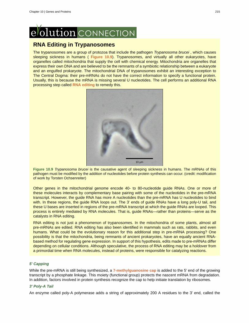
RNA Editing in TrypanosomesThe trypanosomes are a group of protozoa that include the pathogen Trypanosoma brucei , which causessleeping sickness in humans ( Figure 10.9). Trypanosomes, and virtually all other eukaryotes, haveorganelles called mitochondria that supply the cell with chemical energy. Mitochondria are organelles thatexpress their own DNA and are believed to be the remnants of a symbiotic relationship between a eukaryoteand an engulfed prokaryote. The mitochondrial DNA of trypanosomes exhibit an interesting exception toThe Central Dogma: their pre-mRNAs do not have the correct information to specify a functional protein.Usually, this is because the mRNA is missing several U nucleotides. The cell performs an additional RNAprocessing step called RNA editing to remedy this.
Figure 10.9 Trypanosoma brucei is the causative agent of sleeping sickness in humans. The mRNAs of thispathogen must be modified by the addition of nucleotides before protein synthesis can occur. (credit: modificationof work by Torsten Ochsenreiter)
Other genes in the mitochondrial genome encode 40- to 80-nucleotide guide RNAs. One or more ofthese molecules interacts by complementary base pairing with some of the nucleotides in the pre-mRNAtranscript. However, the guide RNA has more A nucleotides than the pre-mRNA has U nucleotides to bindwith. In these regions, the guide RNA loops out. The 3' ends of guide RNAs have a long poly-U tail, andthese U bases are inserted in regions of the pre-mRNA transcript at which the guide RNAs are looped. Thisprocess is entirely mediated by RNA molecules. That is, guide RNAs—rather than proteins—serve as thecatalysts in RNA editing.
RNA editing is not just a phenomenon of trypanosomes. In the mitochondria of some plants, almost allpre-mRNAs are edited. RNA editing has also been identified in mammals such as rats, rabbits, and evenhumans. What could be the evolutionary reason for this additional step in pre-mRNA processing? Onepossibility is that the mitochondria, being remnants of ancient prokaryotes, have an equally ancient RNA-based method for regulating gene expression. In support of this hypothesis, edits made to pre-mRNAs differdepending on cellular conditions. Although speculative, the process of RNA editing may be a holdover froma primordial time when RNA molecules, instead of proteins, were responsible for catalyzing reactions.
5' Capping
While the pre-mRNA is still being synthesized, a 7-methylguanosine cap is added to the 5' end of the growingtranscript by a phosphate linkage. This moiety (functional group) protects the nascent mRNA from degradation.In addition, factors involved in protein synthesis recognize the cap to help initiate translation by ribosomes.
3' Poly-A Tail
An enzyme called poly-A polymerase adds a string of approximately 200 A residues to the 3' end, called the
Chapter 10 | Genes and Proteins 215
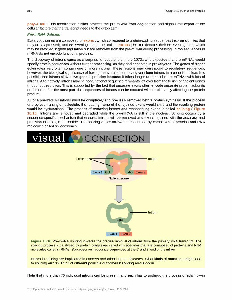
poly-A tail . This modification further protects the pre-mRNA from degradation and signals the export of thecellular factors that the transcript needs to the cytoplasm.
Pre-mRNA Splicing
Eukaryotic genes are composed of exons , which correspond to protein-coding sequences ( ex- on signifies thatthey are ex pressed), and int ervening sequences called introns ( int- ron denotes their int ervening role), whichmay be involved in gene regulation but are removed from the pre-mRNA during processing. Intron sequences inmRNA do not encode functional proteins.
The discovery of introns came as a surprise to researchers in the 1970s who expected that pre-mRNAs wouldspecify protein sequences without further processing, as they had observed in prokaryotes. The genes of highereukaryotes very often contain one or more introns. These regions may correspond to regulatory sequences;however, the biological significance of having many introns or having very long introns in a gene is unclear. It ispossible that introns slow down gene expression because it takes longer to transcribe pre-mRNAs with lots ofintrons. Alternatively, introns may be nonfunctional sequence remnants left over from the fusion of ancient genesthroughout evolution. This is supported by the fact that separate exons often encode separate protein subunitsor domains. For the most part, the sequences of introns can be mutated without ultimately affecting the proteinproduct.
All of a pre-mRNA’s introns must be completely and precisely removed before protein synthesis. If the processerrs by even a single nucleotide, the reading frame of the rejoined exons would shift, and the resulting proteinwould be dysfunctional. The process of removing introns and reconnecting exons is called splicing ( Figure10.10). Introns are removed and degraded while the pre-mRNA is still in the nucleus. Splicing occurs by asequence-specific mechanism that ensures introns will be removed and exons rejoined with the accuracy andprecision of a single nucleotide. The splicing of pre-mRNAs is conducted by complexes of proteins and RNAmolecules called spliceosomes.
Figure 10.10 Pre-mRNA splicing involves the precise removal of introns from the primary RNA transcript. Thesplicing process is catalyzed by protein complexes called spliceosomes that are composed of proteins and RNAmolecules called snRNAs. Spliceosomes recognize sequences at the 5' and 3' end of the intron.
Errors in splicing are implicated in cancers and other human diseases. What kinds of mutations might leadto splicing errors? Think of different possible outcomes if splicing errors occur.
Note that more than 70 individual introns can be present, and each has to undergo the process of splicing—in
216 Chapter 10 | Genes and Proteins
This OpenStax book is available for free at https://legacy.cnx.org/content/col11749/1.6
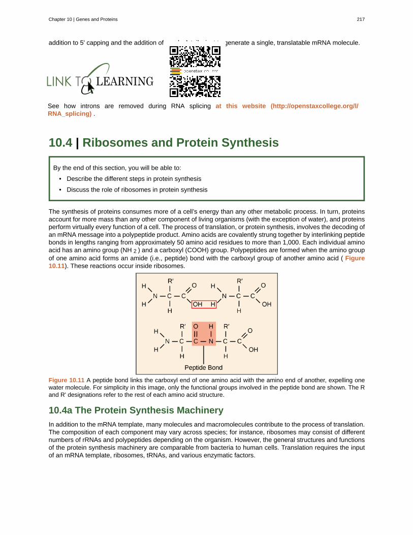
addition to 5' capping and the addition of a poly-A tail—just to generate a single, translatable mRNA molecule.
See how introns are removed during RNA splicing at this website (http://openstaxcollege.org/l/RNA_splicing) .
10.4 | Ribosomes and Protein Synthesis
By the end of this section, you will be able to:
• Describe the different steps in protein synthesis
• Discuss the role of ribosomes in protein synthesis
The synthesis of proteins consumes more of a cell’s energy than any other metabolic process. In turn, proteinsaccount for more mass than any other component of living organisms (with the exception of water), and proteinsperform virtually every function of a cell. The process of translation, or protein synthesis, involves the decoding ofan mRNA message into a polypeptide product. Amino acids are covalently strung together by interlinking peptidebonds in lengths ranging from approximately 50 amino acid residues to more than 1,000. Each individual aminoacid has an amino group (NH 2 ) and a carboxyl (COOH) group. Polypeptides are formed when the amino groupof one amino acid forms an amide (i.e., peptide) bond with the carboxyl group of another amino acid ( Figure10.11). These reactions occur inside ribosomes.
Figure 10.11 A peptide bond links the carboxyl end of one amino acid with the amino end of another, expelling onewater molecule. For simplicity in this image, only the functional groups involved in the peptide bond are shown. The Rand R' designations refer to the rest of each amino acid structure.
10.4a The Protein Synthesis Machinery
In addition to the mRNA template, many molecules and macromolecules contribute to the process of translation.The composition of each component may vary across species; for instance, ribosomes may consist of differentnumbers of rRNAs and polypeptides depending on the organism. However, the general structures and functionsof the protein synthesis machinery are comparable from bacteria to human cells. Translation requires the inputof an mRNA template, ribosomes, tRNAs, and various enzymatic factors.
Chapter 10 | Genes and Proteins 217
Click through the steps of this PBS interactive (http://openstaxcollege.org/l/prokary_protein) to seeprotein synthesis in action.
Ribosomes
Even before an mRNA is translated, a cell must invest energy to build each of its ribosomes. In E. coli , thereare between 10,000 and 70,000 ribosomes present in each cell at any given time. A ribosome is a complexmacromolecule composed of structural and catalytic rRNAs, and many distinct polypeptides. In eukaryotes, thenucleolus is completely specialized for the synthesis and assembly of rRNAs.
Ribosomes exist in the cytoplasm in prokaryotes and in the cytoplasm and rough endoplasmic reticulum ineukaryotes. Mitochondria and chloroplasts also have their own ribosomes in the matrix and stroma, which lookmore similar to prokaryotic ribosomes (and have similar drug sensitivities) than the ribosomes just outside theirouter membranes in the cytoplasm. Ribosomes dissociate into large and small subunits when they are notsynthesizing proteins and reassociate during the initiation of translation. The small subunit is responsible forbinding the mRNA template, whereas the large subunit sequentially binds tRNAs. Each mRNA molecule issimultaneously translated by many ribosomes, all synthesizing protein in the same direction: reading the mRNAfrom 5' to 3' and synthesizing the polypeptide from the N terminus (free amino end) to the C terminus (freecarboxyl end).
tRNAs
The tRNAs are structural RNA molecules that were transcribed from genes by RNA polymerase III. Dependingon the species, 40 to 60 types of tRNAs exist in the cytoplasm. Serving as adaptors, specific tRNAs bind tosequences on the mRNA template and add the corresponding amino acid to the polypeptide chain. Therefore,tRNAs are the molecules that actually “translate” the language of RNA into the language of proteins. The tRNAcan do this because of a specific region of the tRNA called the anticodon . The anticodon contains basescomplementary to a three-nucleotide codon on the mRNA.
Of the 64 possible mRNA codons—or triplet combinations of A, U, G, and C—three specify the termination ofprotein synthesis and 61 specify the addition of amino acids to the polypeptide chain. Of these 61, one codon(AUG) also encodes the initiation of translation. Each tRNA anticodon can base pair with one of the mRNAcodons and add an amino acid or terminate translation, according to the genetic code. For instance, if thesequence CUA occurred on an mRNA template in the proper reading frame, it would bind a tRNA expressingthe complementary sequence, GAU, which would be linked to the amino acid leucine.
10.4b The Mechanism of Protein Synthesis
As with mRNA synthesis, protein synthesis can be divided into three phases: initiation, elongation, andtermination.
Initiation of Translation
Eukaryotic initiation begins with initiation factor (IF) proteins recognizing the 7-methylguanosine cap at the 5' endof the mRNA. A cap-binding protein (CBP) and several other IFs assist the movement of the ribosome to the5' cap. Once at the cap, the initiation complex tracks along the mRNA in the 5' to 3' direction, searching for theAUG start codon .
Once the appropriate AUG is identified, the other proteins and CBP dissociate, and the large subunit binds to thecomplex of tRNA, mRNA, and the small subunit. This step completes the initiation of translation in eukaryotes.
Translation, Elongation, and Termination
The large ribosomal subunit consists of three compartments: the A (aminoacyl) site binds incoming chargedaminoacyl tRNAs. The P (peptidyl) site binds charged tRNAs carrying amino acids that have formed peptidebonds with the growing polypeptide chain but have not yet dissociated from their corresponding tRNA. The E(exit) site releases dissociated tRNAs so that they can be recharged with free amino acids.
218 Chapter 10 | Genes and Proteins
This OpenStax book is available for free at https://legacy.cnx.org/content/col11749/1.6
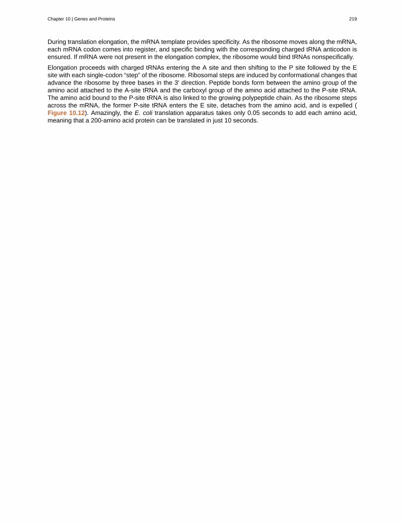
During translation elongation, the mRNA template provides specificity. As the ribosome moves along the mRNA,each mRNA codon comes into register, and specific binding with the corresponding charged tRNA anticodon isensured. If mRNA were not present in the elongation complex, the ribosome would bind tRNAs nonspecifically.
Elongation proceeds with charged tRNAs entering the A site and then shifting to the P site followed by the Esite with each single-codon “step” of the ribosome. Ribosomal steps are induced by conformational changes thatadvance the ribosome by three bases in the 3' direction. Peptide bonds form between the amino group of theamino acid attached to the A-site tRNA and the carboxyl group of the amino acid attached to the P-site tRNA.The amino acid bound to the P-site tRNA is also linked to the growing polypeptide chain. As the ribosome stepsacross the mRNA, the former P-site tRNA enters the E site, detaches from the amino acid, and is expelled (Figure 10.12). Amazingly, the E. coli translation apparatus takes only 0.05 seconds to add each amino acid,meaning that a 200-amino acid protein can be translated in just 10 seconds.
Chapter 10 | Genes and Proteins 219
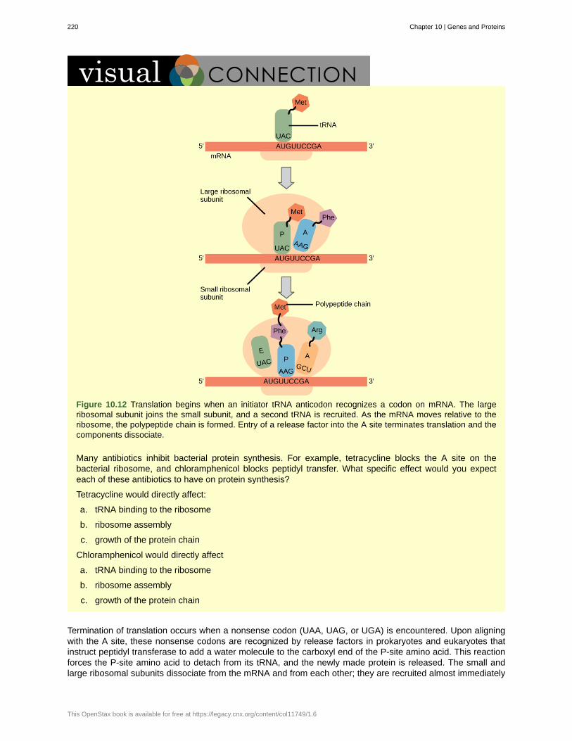
Figure 10.12 Translation begins when an initiator tRNA anticodon recognizes a codon on mRNA. The largeribosomal subunit joins the small subunit, and a second tRNA is recruited. As the mRNA moves relative to theribosome, the polypeptide chain is formed. Entry of a release factor into the A site terminates translation and thecomponents dissociate.
Many antibiotics inhibit bacterial protein synthesis. For example, tetracycline blocks the A site on thebacterial ribosome, and chloramphenicol blocks peptidyl transfer. What specific effect would you expecteach of these antibiotics to have on protein synthesis?
Tetracycline would directly affect:
a. tRNA binding to the ribosome
b. ribosome assembly
c. growth of the protein chain
Chloramphenicol would directly affect
a. tRNA binding to the ribosome
b. ribosome assembly
c. growth of the protein chain
Termination of translation occurs when a nonsense codon (UAA, UAG, or UGA) is encountered. Upon aligningwith the A site, these nonsense codons are recognized by release factors in prokaryotes and eukaryotes thatinstruct peptidyl transferase to add a water molecule to the carboxyl end of the P-site amino acid. This reactionforces the P-site amino acid to detach from its tRNA, and the newly made protein is released. The small andlarge ribosomal subunits dissociate from the mRNA and from each other; they are recruited almost immediately
220 Chapter 10 | Genes and Proteins
This OpenStax book is available for free at https://legacy.cnx.org/content/col11749/1.6
into another translation initiation complex. After many ribosomes have completed translation, the mRNA isdegraded so the nucleotides can be reused in another transcription reaction.
10.4c Protein Folding, Modification, and Targeting
During and after translation, individual amino acids may be chemically modified, signal sequences may beappended, and the new protein “folds” into a distinct three-dimensional structure as a result of intramolecularinteractions. A signal sequence is a short tail of amino acids that directs a protein to a specific cellularcompartment. These sequences at the amino end or the carboxyl end of the protein can be thought of as theprotein’s “train ticket” to its ultimate destination. Other cellular factors recognize each signal sequence and helptransport the protein from the cytoplasm to its correct compartment. For instance, a specific sequence at theamino terminus will direct a protein to the mitochondria or chloroplasts (in plants). Once the protein reaches itscellular destination, the signal sequence is usually clipped off.
Many proteins fold spontaneously, but some proteins require helper molecules, called chaperones, to preventthem from aggregating during the complicated process of folding. Even if a protein is properly specified byits corresponding mRNA, it could take on a completely dysfunctional shape if abnormal temperature or pHconditions prevent it from folding correctly.
10.5 | Mutations
By the end of this section, you will be able to:
• Discuss the different types of mutations in DNA
• Explain DNA repair mechanisms
DNA replication is a highly accurate process, but mistakes can occasionally occur, such as a DNA polymeraseinserting a wrong base. Uncorrected mistakes may sometimes lead to serious consequences, such as cancer.Repair mechanisms correct the mistakes. In rare cases, mistakes are not corrected, leading to mutations; inother cases, repair enzymes are themselves mutated or defective.
Most of the mistakes during DNA replication are promptly corrected by DNA polymerase by proofreading thebase that has been just added ( Figure 10.13). In proofreading , the DNA polymerase reads the newly addedbase before adding the next one, so a correction can be made. The polymerase checks whether the newlyadded base has paired correctly with the base in the template strand. If it is the right base, the next nucleotide isadded. If an incorrect base has been added, the enzyme makes a cut at the phosphodiester bond and releasesthe wrong nucleotide. This is performed by the exonuclease action of DNA pol III. Once the incorrect nucleotidehas been removed, a new one will be added again.
Figure 10.13 Proofreading by DNA polymerase corrects errors during replication.
Errors during DNA replication are not the only reason why mutations arise in DNA. Mutations , variations in thenucleotide sequence of a genome, can also occur because of damage to DNA. Such mutations may be of twotypes: induced or spontaneous. Induced mutations are those that result from an exposure to chemicals, UVrays, x-rays, or some other environmental agent. Spontaneous mutations occur without any exposure to anyenvironmental agent; they are a result of natural reactions taking place within the body.
Mutations may have a wide range of effects. Some mutations are not expressed; these are known as silentmutations . Point mutations are those mutations that affect a single base pair. The most common nucleotidemutations are substitutions, in which one base is replaced by another. These can be of two types, either
Chapter 10 | Genes and Proteins 221
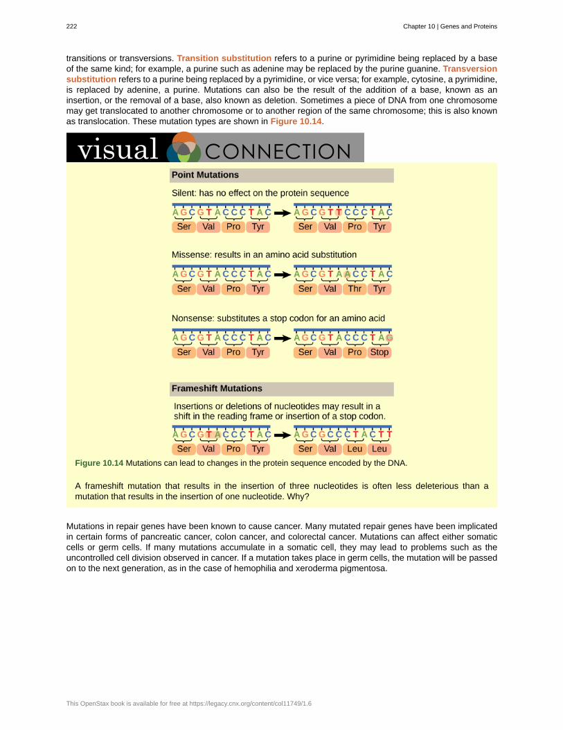
transitions or transversions. Transition substitution refers to a purine or pyrimidine being replaced by a baseof the same kind; for example, a purine such as adenine may be replaced by the purine guanine. Transversionsubstitution refers to a purine being replaced by a pyrimidine, or vice versa; for example, cytosine, a pyrimidine,is replaced by adenine, a purine. Mutations can also be the result of the addition of a base, known as aninsertion, or the removal of a base, also known as deletion. Sometimes a piece of DNA from one chromosomemay get translocated to another chromosome or to another region of the same chromosome; this is also knownas translocation. These mutation types are shown in Figure 10.14.
Figure 10.14 Mutations can lead to changes in the protein sequence encoded by the DNA.
A frameshift mutation that results in the insertion of three nucleotides is often less deleterious than amutation that results in the insertion of one nucleotide. Why?
Mutations in repair genes have been known to cause cancer. Many mutated repair genes have been implicatedin certain forms of pancreatic cancer, colon cancer, and colorectal cancer. Mutations can affect either somaticcells or germ cells. If many mutations accumulate in a somatic cell, they may lead to problems such as theuncontrolled cell division observed in cancer. If a mutation takes place in germ cells, the mutation will be passedon to the next generation, as in the case of hemophilia and xeroderma pigmentosa.
222 Chapter 10 | Genes and Proteins
This OpenStax book is available for free at https://legacy.cnx.org/content/col11749/1.6
7-methylguanosine cap
anticodon
Central Dogma
codons
degenerate
downstream
exons
hairpin
Induced mutations
initiation site
introns
Mutations
nonsense codons
nontemplate strand
plasmids
Point mutations
poly-A tail
promoter
proofreading
reading frame
Rho-dependent termination
Rho-independent termination
RNA editing
RNA polymerase
signal sequence
KEY TERMS
modification added to the 5' end of pre-mRNAs to protect mRNA from degradationand assist translation
three-nucleotide sequence in a tRNA molecule that corresponds to an mRNA codon
states that genes specify the sequence of mRNAs, which in turn specify the sequence ofproteins
three consecutive nucleotides in mRNA that specify the insertion of an amino acid or the release of apolypeptide chain during translation
when a given amino acid can be encoded by more than one nucleotide triplet
nucleotides following the initiation site in the direction of mRNA transcription; in general,sequences that are toward the 3' end relative to a site on the mRNA
sequence present in protein-coding mRNA after completion of pre-mRNA splciing
structure of RNA when it folds back on itself and forms intramolecular hydrogen bonds betweencomplementary nucleotides
mutation that results from exposure to chemicals or environmental agents
nucleotide from which mRNA synthesis proceeds in the 5' to 3' direction; denoted with a “+1”
non-protein-coding intervening sequences spliced from mRNA during processing
variation in the nucleotide sequence of a genome
one of the three mRNA codons that specifies termination of translation
strand of DNA that is not used to transcribe mRNA; this strand is identical to the mRNAexcept that T nucleotides in the DNA are replaced by U nucleotides in the mRNA
extrachromosomal, covalently closed, circular DNA molecule that may only contain one or a fewgenes; common in prokaryotes
mutation that affects a single base
modification added to the 3' end of pre-mRNAs to protect mRNA from degradation and assist mRNAexport from the nucleus
DNA sequence to which RNA polymerase and associated factors bind and initiate transcription
function of DNA pol in which it reads the newly added base before adding the next one
sequence of triplet codons in mRNA that specify a particular protein; a ribosome shift of one ortwo nucleotides in either direction completely abolishes synthesis of that protein
in prokaryotes, termination of transcription by an interaction between RNApolymerase and the rho protein at a run of G nucleotides on the DNA template
termination sequence-dependent termination of prokaryotic mRNA synthesis;caused by hairpin formation in the mRNA that stalls the polymerase
direct alteration of one or more nucleotides in an mRNA that has already been synthesized
enzyme used to build an RNA molecule from a complementary strand of DNA
short tail of amino acids that directs a protein to a specific cellular compartment
Chapter 10 | Genes and Proteins 223
silent mutations
splicing
Spontaneous mutations
start codon
template strand
transcription bubble.
transition substitution
transversion substitution
upstream
mutation that is not expressed
process of removing introns and reconnecting exons in a pre-mRNA
mutation that takes place in the cells as a result of chemical reactions taking placenaturally without exposure to any external agent
AUG (or rarely, GUG) on an mRNA from which translation begins; always specifies methionine
strand of DNA that specifies the complementary mRNA molecule
region of locally unwound DNA that allows for transcription of mRNA
when a purine is replaced with a purine or a pyrimidine is replaced with anotherpyrimidine
when a purine is replaced by a pyrimidine or a pyrimidine is replaced by a purine
nucleotides preceding the initiation site; in general, sequences toward the 5' end relative to a site onthe mRNA
CHAPTER SUMMARY
10.1 The Genetic Code
The genetic code refers to the DNA alphabet (A, T, C, G), the RNA alphabet (A, U, C, G), and the polypeptidealphabet (20 amino acids). The Central Dogma describes the flow of genetic information in the cell from genesto mRNA to proteins. Genes are used to make mRNA by the process of transcription; mRNA is used tosynthesize proteins by the process of translation. The genetic code is degenerate because 64 triplet codons inmRNA specify only 20 amino acids and three nonsense codons. Almost every species on the planet uses thesame genetic code.
10.2 Prokaryotic Transcription
In prokaryotes, mRNA synthesis is initiated at a promoter sequence on the DNA template comprising twoconsensus sequences that recruit RNA polymerase. The prokaryotic polymerase consists of a core enzyme offour protein subunits and a σ protein that assists only with initiation. Elongation synthesizes mRNA in the 5' to3' direction at a rate of 40 nucleotides per second. Termination liberates the mRNA and occurs either by rhoprotein interaction or by the formation of an mRNA hairpin.
10.3 RNA Processing in Eukaryotes
Eukaryotic pre-mRNAs are modified with a 5' methylguanosine cap and a poly-A tail. These structures protectthe mature mRNA from degradation and help export it from the nucleus. Pre-mRNAs also undergo splicing, inwhich introns are removed and exons are reconnected with single-nucleotide accuracy. Only finished mRNAsthat have undergone 5' capping, 3' polyadenylation, and intron splicing are exported from the nucleus to thecytoplasm. Pre-rRNAs and pre-tRNAs may be processed by intramolecular cleavage, splicing, methylation, andchemical conversion of nucleotides. Rarely, RNA editing is also performed to insert missing bases after anmRNA has been synthesized.
10.4 Ribosomes and Protein Synthesis
The players in translation include the mRNA template, ribosomes, tRNAs, and various enzymatic factors. Thesmall ribosomal subunit forms on the mRNA template either at the Shine-Dalgarno sequence (prokaryotes) orthe 5' cap (eukaryotes). Translation begins at the initiating AUG on the mRNA, specifying methionine. Theformation of peptide bonds occurs between sequential amino acids specified by the mRNA template accordingto the genetic code. Charged tRNAs enter the ribosomal A site, and their amino acid bonds with the amino acidat the P site. The entire mRNA is translated in three-nucleotide “steps” of the ribosome. When a nonsensecodon is encountered, a release factor binds and dissociates the components and frees the new protein.Folding of the protein occurs during and after translation.
224 Chapter 10 | Genes and Proteins
This OpenStax book is available for free at https://legacy.cnx.org/content/col11749/1.6
10.5 Mutations
DNA polymerase can make mistakes while adding nucleotides. It edits the DNA by proofreading every newlyadded base. Incorrect bases are removed and replaced by the correct base, and then a new base is added.Most mistakes are corrected during replication, although when this does not happen, the mismatch repairmechanism is employed. Mismatch repair enzymes recognize the wrongly incorporated base and excise it fromthe DNA, replacing it with the correct base. In yet another type of repair, nucleotide excision repair, the incorrectbase is removed along with a few bases on the 5' and 3' end, and these are replaced by copying the templatewith the help of DNA polymerase. The ends of the newly synthesized fragment are attached to the rest of theDNA using DNA ligase, which creates a phosphodiester bond.
Most mistakes are corrected, and if they are not, they may result in a mutation defined as a permanent changein the DNA sequence. Mutations can be of many types, such as substitution, deletion, insertion, andtranslocation. Mutations in repair genes may lead to serious consequences such as cancer. Mutations can beinduced or may occur spontaneously.
Chapter 10 | Genes and Proteins 225

226 Chapter 10 | Genes and Proteins
This OpenStax book is available for free at https://legacy.cnx.org/content/col11749/1.6
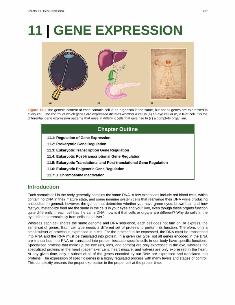
11 | GENE EXPRESSION
Figure 11.1 The genetic content of each somatic cell in an organism is the same, but not all genes are expressed inevery cell. The control of which genes are expressed dictates whether a cell is (a) an eye cell or (b) a liver cell. It is thedifferential gene expression patterns that arise in different cells that give rise to (c) a complete organism.
Chapter Outline
11.1: Regulation of Gene Expression
11.2: Prokaryotic Gene Regulation
11.3: Eukaryotic Transcription Gene Regulation
11.4: Eukaryotic Post-transcriptional Gene Regulation
11.5: Eukaryotic Translational and Post-translational Gene Regulation
11.6: Eukaryotic Epigenetic Gene Regulation
11.7: X Chromosome Inactivation
Introduction
Each somatic cell in the body generally contains the same DNA. A few exceptions include red blood cells, whichcontain no DNA in their mature state, and some immune system cells that rearrange their DNA while producingantibodies. In general, however, the genes that determine whether you have green eyes, brown hair, and howfast you metabolize food are the same in the cells in your eyes and your liver, even though these organs functionquite differently. If each cell has the same DNA, how is it that cells or organs are different? Why do cells in theeye differ so dramatically from cells in the liver?
Whereas each cell shares the same genome and DNA sequence, each cell does not turn on, or express, thesame set of genes. Each cell type needs a different set of proteins to perform its function. Therefore, only asmall subset of proteins is expressed in a cell. For the proteins to be expressed, the DNA must be transcribedinto RNA and the RNA must be translated into protein. In a given cell type, not all genes encoded in the DNAare transcribed into RNA or translated into protein because specific cells in our body have specific functions.Specialized proteins that make up the eye (iris, lens, and cornea) are only expressed in the eye, whereas thespecialized proteins in the heart (pacemaker cells, heart muscle, and valves) are only expressed in the heart.At any given time, only a subset of all of the genes encoded by our DNA are expressed and translated intoproteins. The expression of specific genes is a highly regulated process with many levels and stages of control.This complexity ensures the proper expression in the proper cell at the proper time.
Chapter 11 | Gene Expression 227
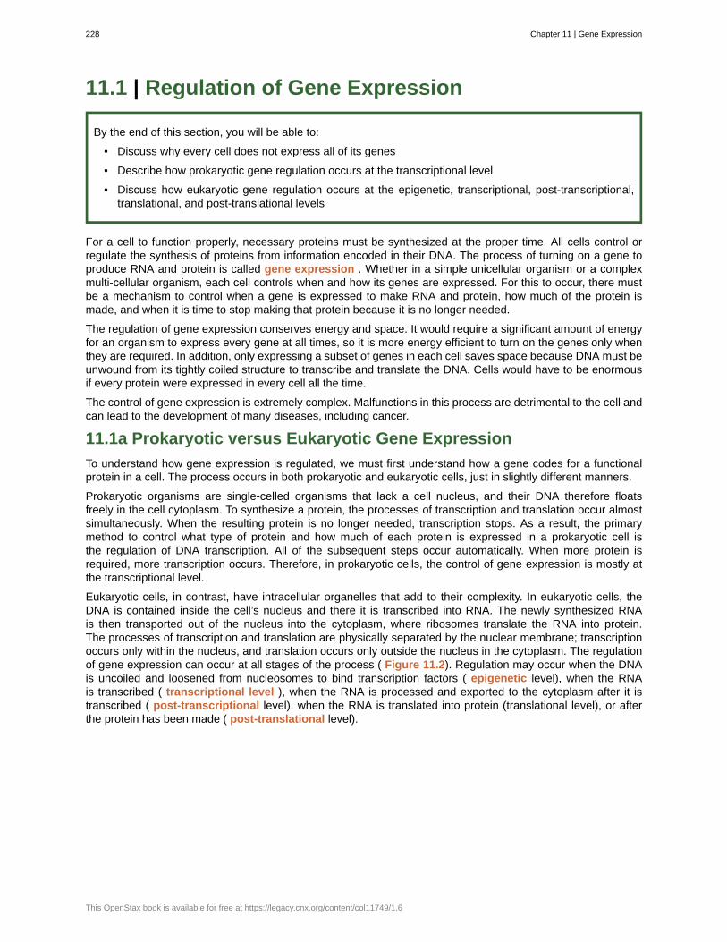
11.1 | Regulation of Gene Expression
By the end of this section, you will be able to:
• Discuss why every cell does not express all of its genes
• Describe how prokaryotic gene regulation occurs at the transcriptional level
• Discuss how eukaryotic gene regulation occurs at the epigenetic, transcriptional, post-transcriptional,translational, and post-translational levels
For a cell to function properly, necessary proteins must be synthesized at the proper time. All cells control orregulate the synthesis of proteins from information encoded in their DNA. The process of turning on a gene toproduce RNA and protein is called gene expression . Whether in a simple unicellular organism or a complexmulti-cellular organism, each cell controls when and how its genes are expressed. For this to occur, there mustbe a mechanism to control when a gene is expressed to make RNA and protein, how much of the protein ismade, and when it is time to stop making that protein because it is no longer needed.
The regulation of gene expression conserves energy and space. It would require a significant amount of energyfor an organism to express every gene at all times, so it is more energy efficient to turn on the genes only whenthey are required. In addition, only expressing a subset of genes in each cell saves space because DNA must beunwound from its tightly coiled structure to transcribe and translate the DNA. Cells would have to be enormousif every protein were expressed in every cell all the time.
The control of gene expression is extremely complex. Malfunctions in this process are detrimental to the cell andcan lead to the development of many diseases, including cancer.
11.1a Prokaryotic versus Eukaryotic Gene Expression
To understand how gene expression is regulated, we must first understand how a gene codes for a functionalprotein in a cell. The process occurs in both prokaryotic and eukaryotic cells, just in slightly different manners.
Prokaryotic organisms are single-celled organisms that lack a cell nucleus, and their DNA therefore floatsfreely in the cell cytoplasm. To synthesize a protein, the processes of transcription and translation occur almostsimultaneously. When the resulting protein is no longer needed, transcription stops. As a result, the primarymethod to control what type of protein and how much of each protein is expressed in a prokaryotic cell isthe regulation of DNA transcription. All of the subsequent steps occur automatically. When more protein isrequired, more transcription occurs. Therefore, in prokaryotic cells, the control of gene expression is mostly atthe transcriptional level.
Eukaryotic cells, in contrast, have intracellular organelles that add to their complexity. In eukaryotic cells, theDNA is contained inside the cell’s nucleus and there it is transcribed into RNA. The newly synthesized RNAis then transported out of the nucleus into the cytoplasm, where ribosomes translate the RNA into protein.The processes of transcription and translation are physically separated by the nuclear membrane; transcriptionoccurs only within the nucleus, and translation occurs only outside the nucleus in the cytoplasm. The regulationof gene expression can occur at all stages of the process ( Figure 11.2). Regulation may occur when the DNAis uncoiled and loosened from nucleosomes to bind transcription factors ( epigenetic level), when the RNAis transcribed ( transcriptional level ), when the RNA is processed and exported to the cytoplasm after it istranscribed ( post-transcriptional level), when the RNA is translated into protein (translational level), or afterthe protein has been made ( post-translational level).
228 Chapter 11 | Gene Expression
This OpenStax book is available for free at https://legacy.cnx.org/content/col11749/1.6
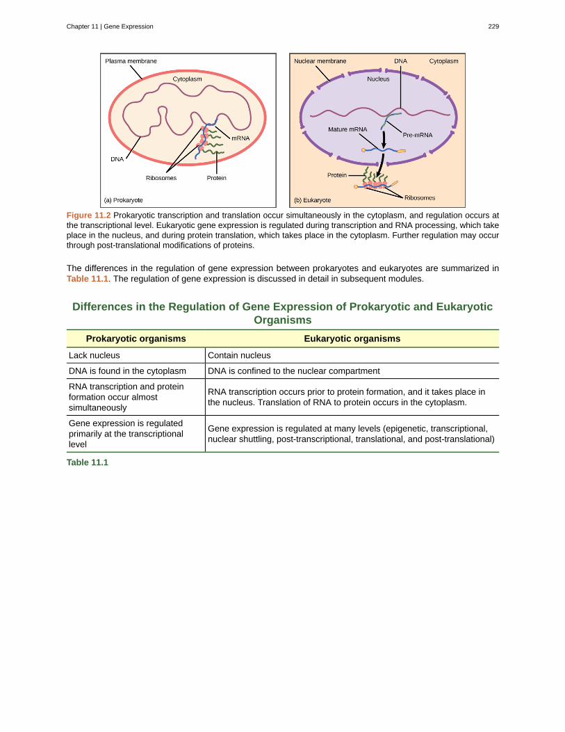
Figure 11.2 Prokaryotic transcription and translation occur simultaneously in the cytoplasm, and regulation occurs atthe transcriptional level. Eukaryotic gene expression is regulated during transcription and RNA processing, which takeplace in the nucleus, and during protein translation, which takes place in the cytoplasm. Further regulation may occurthrough post-translational modifications of proteins.
The differences in the regulation of gene expression between prokaryotes and eukaryotes are summarized inTable 11.1. The regulation of gene expression is discussed in detail in subsequent modules.
Differences in the Regulation of Gene Expression of Prokaryotic and EukaryoticOrganisms
Prokaryotic organisms Eukaryotic organisms
Lack nucleus Contain nucleus
DNA is found in the cytoplasm DNA is confined to the nuclear compartment
RNA transcription and proteinformation occur almostsimultaneously
RNA transcription occurs prior to protein formation, and it takes place inthe nucleus. Translation of RNA to protein occurs in the cytoplasm.
Gene expression is regulatedprimarily at the transcriptionallevel
Gene expression is regulated at many levels (epigenetic, transcriptional,nuclear shuttling, post-transcriptional, translational, and post-translational)
Table 11.1
Chapter 11 | Gene Expression 229
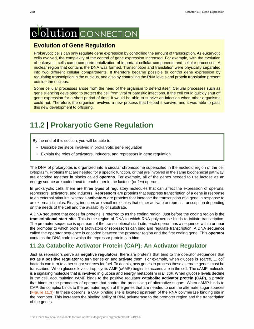
Evolution of Gene RegulationProkaryotic cells can only regulate gene expression by controlling the amount of transcription. As eukaryoticcells evolved, the complexity of the control of gene expression increased. For example, with the evolutionof eukaryotic cells came compartmentalization of important cellular components and cellular processes. Anuclear region that contains the DNA was formed. Transcription and translation were physically separatedinto two different cellular compartments. It therefore became possible to control gene expression byregulating transcription in the nucleus, and also by controlling the RNA levels and protein translation presentoutside the nucleus.
Some cellular processes arose from the need of the organism to defend itself. Cellular processes such asgene silencing developed to protect the cell from viral or parasitic infections. If the cell could quickly shut offgene expression for a short period of time, it would be able to survive an infection when other organismscould not. Therefore, the organism evolved a new process that helped it survive, and it was able to passthis new development to offspring.
11.2 | Prokaryotic Gene Regulation
By the end of this section, you will be able to:
• Describe the steps involved in prokaryotic gene regulation
• Explain the roles of activators, inducers, and repressors in gene regulation
The DNA of prokaryotes is organized into a circular chromosome supercoiled in the nucleoid region of the cellcytoplasm. Proteins that are needed for a specific function, or that are involved in the same biochemical pathway,are encoded together in blocks called operons. For example, all of the genes needed to use lactose as anenergy source are coded next to each other in the lactose (or lac) operon.
In prokaryotic cells, there are three types of regulatory molecules that can affect the expression of operons:repressors, activators, and inducers. Repressors are proteins that suppress transcription of a gene in responseto an external stimulus, whereas activators are proteins that increase the transcription of a gene in response toan external stimulus. Finally, inducers are small molecules that either activate or repress transcription dependingon the needs of the cell and the availability of substrate.
A DNA sequence that codes for proteins is referred to as the coding region. Just before the coding region is thetranscriptional start site. This is the region of DNA to which RNA polymerase binds to initiate transcription.The promoter sequence is upstream of the transcriptional start site; each operon has a sequence within or nearthe promoter to which proteins (activators or repressors) can bind and regulate transcription. A DNA sequencecalled the operator sequence is encoded between the promoter region and the first coding gene. This operatorcontains the DNA code to which the repressor protein can bind.
11.2a Catabolite Activator Protein (CAP): An Activator Regulator
Just as repressors serve as negative regulators, there are proteins that bind to the operator sequences thatact as a positive regulator to turn genes on and activate them. For example, when glucose is scarce, E. colibacteria can turn to other sugar sources for fuel. To do this, new genes to process these alternate genes must betranscribed. When glucose levels drop, cyclic AMP (cAMP) begins to accumulate in the cell. The cAMP moleculeis a signaling molecule that is involved in glucose and energy metabolism in E. coli. When glucose levels declinein the cell, accumulating cAMP binds to the positive regulator catabolite activator protein (CAP), a proteinthat binds to the promoters of operons that control the processing of alternative sugars. When cAMP binds toCAP, the complex binds to the promoter region of the genes that are needed to use the alternate sugar sources(Figure 11.3). In these operons, a CAP binding site is located upstream of the RNA polymerase binding site inthe promoter. This increases the binding ability of RNA polymerase to the promoter region and the transcriptionof the genes.
230 Chapter 11 | Gene Expression
This OpenStax book is available for free at https://legacy.cnx.org/content/col11749/1.6
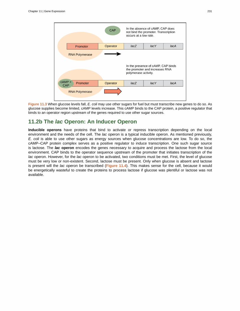
Figure 11.3 When glucose levels fall, E. coli may use other sugars for fuel but must transcribe new genes to do so. Asglucose supplies become limited, cAMP levels increase. This cAMP binds to the CAP protein, a positive regulator thatbinds to an operator region upstream of the genes required to use other sugar sources.
11.2b The lac Operon: An Inducer Operon
Inducible operons have proteins that bind to activate or repress transcription depending on the localenvironment and the needs of the cell. The lac operon is a typical inducible operon. As mentioned previously,E. coli is able to use other sugars as energy sources when glucose concentrations are low. To do so, thecAMP–CAP protein complex serves as a positive regulator to induce transcription. One such sugar sourceis lactose. The lac operon encodes the genes necessary to acquire and process the lactose from the localenvironment. CAP binds to the operator sequence upstream of the promoter that initiates transcription of thelac operon. However, for the lac operon to be activated, two conditions must be met. First, the level of glucosemust be very low or non-existent. Second, lactose must be present. Only when glucose is absent and lactoseis present will the lac operon be transcribed (Figure 11.4). This makes sense for the cell, because it wouldbe energetically wasteful to create the proteins to process lactose if glucose was plentiful or lactose was notavailable.
Chapter 11 | Gene Expression 231
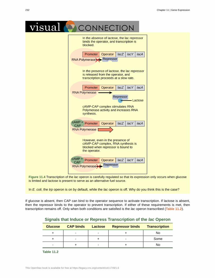
Figure 11.4 Transcription of the lac operon is carefully regulated so that its expression only occurs when glucoseis limited and lactose is present to serve as an alternative fuel source.
In E. coli, the trp operon is on by default, while the lac operon is off. Why do you think this is the case?
If glucose is absent, then CAP can bind to the operator sequence to activate transcription. If lactose is absent,then the repressor binds to the operator to prevent transcription. If either of these requirements is met, thentranscription remains off. Only when both conditions are satisfied is the lac operon transcribed (Table 11.2).
Signals that Induce or Repress Transcription of the lac Operon
Glucose CAP binds Lactose Repressor binds Transcription
+ - - + No
+ - + - Some
- + - + No
Table 11.2
232 Chapter 11 | Gene Expression
This OpenStax book is available for free at https://legacy.cnx.org/content/col11749/1.6
Signals that Induce or Repress Transcription of the lac Operon
Glucose CAP binds Lactose Repressor binds Transcription
- + + - Yes
Table 11.2
Watch an animated tutorial (http://openstaxcollege.org/l/lac_operon) about the workings of lac operonhere.
11.3 | Eukaryotic Transcription Gene Regulation
By the end of this section, you will be able to:
• Discuss the role of transcription factors in gene regulation
• Explain how enhancers and repressors regulate gene expression
Like prokaryotic cells, the transcription of genes in eukaryotes requires the actions of an RNA polymeraseto bind to a sequence upstream of a gene to initiate transcription. However, unlike prokaryotic cells, theeukaryotic RNA polymerase requires other proteins, or transcription factors, to facilitate transcription initiation.Transcription factors are proteins that bind to the promoter sequence and other regulatory sequences tocontrol the transcription of the target gene. RNA polymerase by itself cannot initiate transcription in eukaryoticcells. Transcription factors must bind to the promoter region first and recruit RNA polymerase to the site fortranscription to be established.
View the process of transcription—the making of RNA from a DNA template—at this site(http://openstaxcollege.org/l/transcript_RNA) .
11.3a Enhancers and Transcription
In some eukaryotic genes, there are regions that help increase or enhance transcription. These regions, calledenhancers, are not necessarily close to the genes they enhance. They can be located upstream of a gene,within the coding region of the gene, downstream of a gene, or may be thousands of nucleotides away.
Enhancer regions are binding sequences, or sites, for transcription factors. When a DNA-bending protein binds,the shape of the DNA changes (Figure 11.5). This shape change allows for the interaction of the activatorsbound to the enhancers with the transcription factors bound to the promoter region and the RNA polymerase.Whereas DNA is generally depicted as a straight line in two dimensions, it is actually a three-dimensional object.Therefore, a nucleotide sequence thousands of nucleotides away can fold over and interact with a specificpromoter.
Chapter 11 | Gene Expression 233
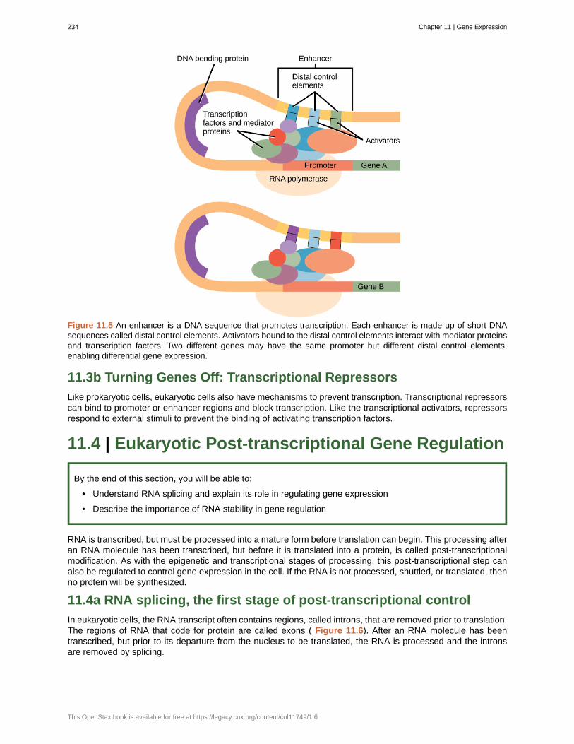
Figure 11.5 An enhancer is a DNA sequence that promotes transcription. Each enhancer is made up of short DNAsequences called distal control elements. Activators bound to the distal control elements interact with mediator proteinsand transcription factors. Two different genes may have the same promoter but different distal control elements,enabling differential gene expression.
11.3b Turning Genes Off: Transcriptional Repressors
Like prokaryotic cells, eukaryotic cells also have mechanisms to prevent transcription. Transcriptional repressorscan bind to promoter or enhancer regions and block transcription. Like the transcriptional activators, repressorsrespond to external stimuli to prevent the binding of activating transcription factors.
11.4 | Eukaryotic Post-transcriptional Gene Regulation
By the end of this section, you will be able to:
• Understand RNA splicing and explain its role in regulating gene expression
• Describe the importance of RNA stability in gene regulation
RNA is transcribed, but must be processed into a mature form before translation can begin. This processing afteran RNA molecule has been transcribed, but before it is translated into a protein, is called post-transcriptionalmodification. As with the epigenetic and transcriptional stages of processing, this post-transcriptional step canalso be regulated to control gene expression in the cell. If the RNA is not processed, shuttled, or translated, thenno protein will be synthesized.
11.4a RNA splicing, the first stage of post-transcriptional control
In eukaryotic cells, the RNA transcript often contains regions, called introns, that are removed prior to translation.The regions of RNA that code for protein are called exons ( Figure 11.6). After an RNA molecule has beentranscribed, but prior to its departure from the nucleus to be translated, the RNA is processed and the intronsare removed by splicing.
234 Chapter 11 | Gene Expression
This OpenStax book is available for free at https://legacy.cnx.org/content/col11749/1.6
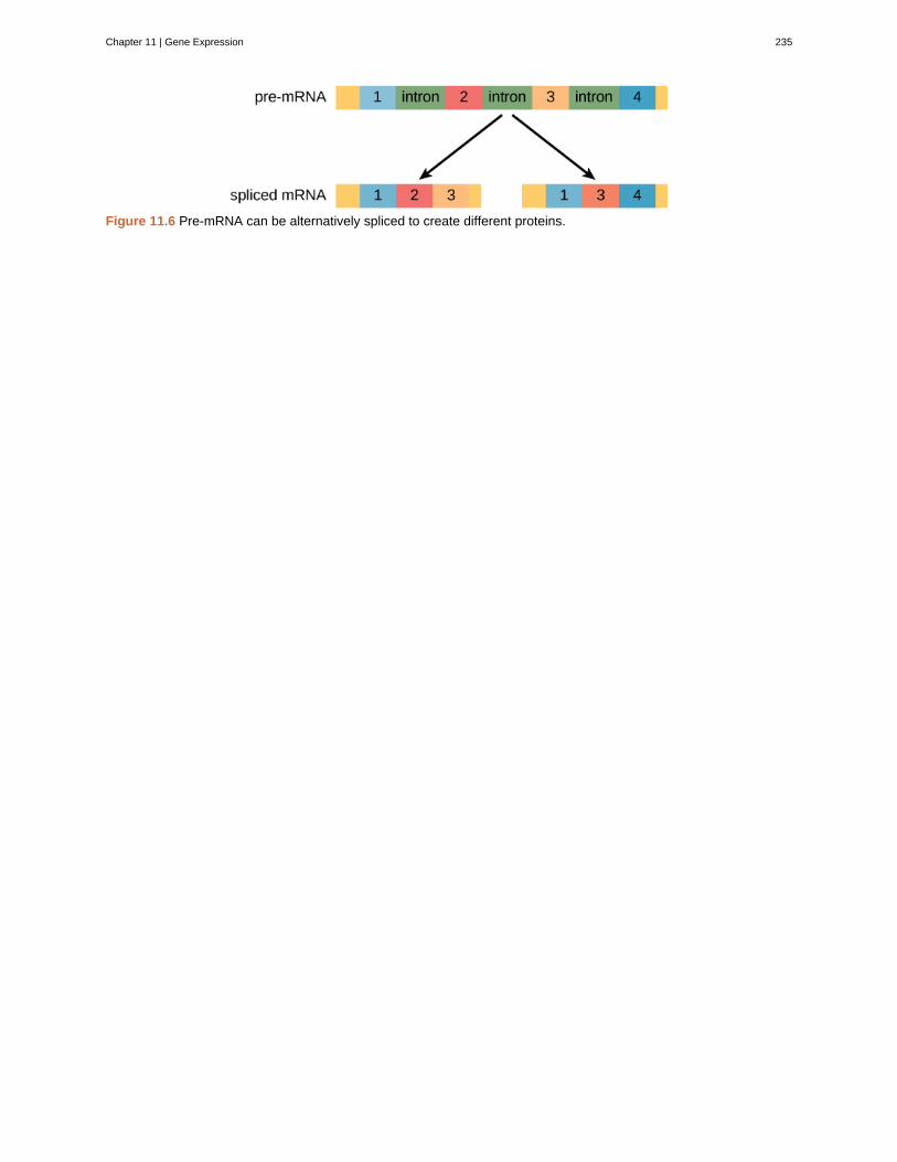
Figure 11.6 Pre-mRNA can be alternatively spliced to create different proteins.
Chapter 11 | Gene Expression 235
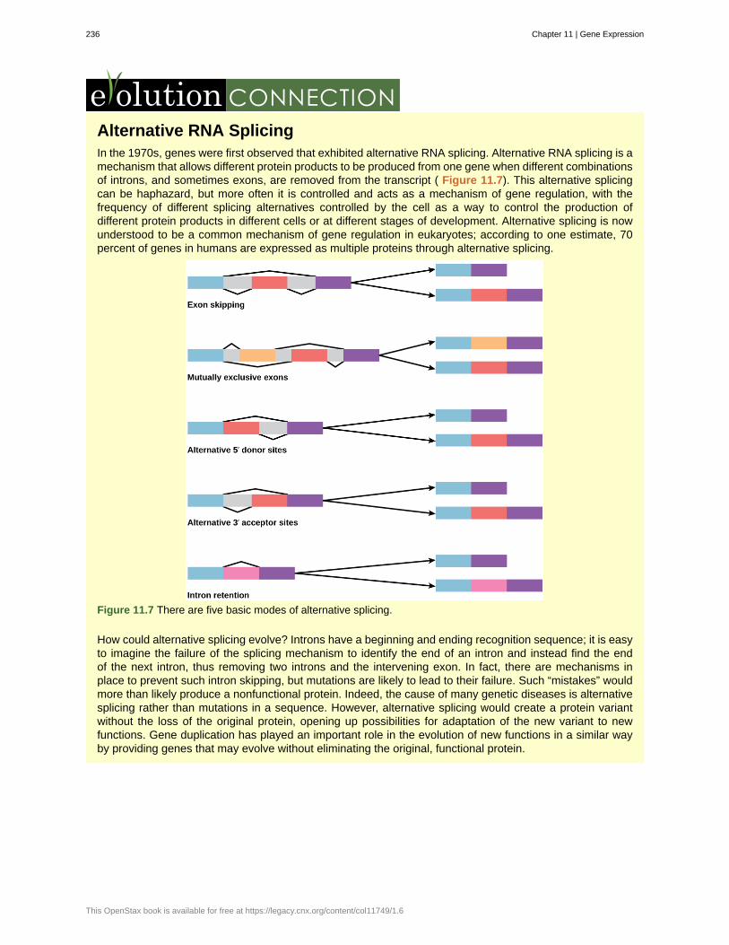
Alternative RNA SplicingIn the 1970s, genes were first observed that exhibited alternative RNA splicing. Alternative RNA splicing is amechanism that allows different protein products to be produced from one gene when different combinationsof introns, and sometimes exons, are removed from the transcript ( Figure 11.7). This alternative splicingcan be haphazard, but more often it is controlled and acts as a mechanism of gene regulation, with thefrequency of different splicing alternatives controlled by the cell as a way to control the production ofdifferent protein products in different cells or at different stages of development. Alternative splicing is nowunderstood to be a common mechanism of gene regulation in eukaryotes; according to one estimate, 70percent of genes in humans are expressed as multiple proteins through alternative splicing.
Figure 11.7 There are five basic modes of alternative splicing.
How could alternative splicing evolve? Introns have a beginning and ending recognition sequence; it is easyto imagine the failure of the splicing mechanism to identify the end of an intron and instead find the endof the next intron, thus removing two introns and the intervening exon. In fact, there are mechanisms inplace to prevent such intron skipping, but mutations are likely to lead to their failure. Such “mistakes” wouldmore than likely produce a nonfunctional protein. Indeed, the cause of many genetic diseases is alternativesplicing rather than mutations in a sequence. However, alternative splicing would create a protein variantwithout the loss of the original protein, opening up possibilities for adaptation of the new variant to newfunctions. Gene duplication has played an important role in the evolution of new functions in a similar wayby providing genes that may evolve without eliminating the original, functional protein.
236 Chapter 11 | Gene Expression
This OpenStax book is available for free at https://legacy.cnx.org/content/col11749/1.6
Visualize how mRNA splicing happens by watching the process in action in this video(http://openstaxcollege.org/l/mRNA_splicing) .
11.5 | Eukaryotic Translational and Post-translational
Gene Regulation
By the end of this section, you will be able to:
• Understand the process of translation and discuss its key factors
• Describe how the initiation complex controls translation
• Explain the different ways in which the post-translational control of gene expression takes place
After the RNA has been transported to the cytoplasm, it is translated into protein. Control of this process is largelydependent on the RNA molecule. As previously discussed, the stability of the RNA will have a large impact onits translation into a protein. As the stability changes, the amount of time that it is available for translation alsochanges.
11.5a Chemical Modifications, Protein Activity, and Longevity
Proteins can be chemically modified with the addition of groups including methyl, phosphate, acetyl, andubiquitin groups. The addition or removal of these groups from proteins regulates their activity or the length oftime they exist in the cell. Sometimes these modifications can regulate where a protein is found in the cell—forexample, in the nucleus, the cytoplasm, or attached to the plasma membrane.
Chemical modifications occur in response to external stimuli such as stress, the lack of nutrients, heat, orultraviolet light exposure. These changes can alter epigenetic accessibility, transcription, mRNA stability, ortranslation—all resulting in changes in expression of various genes. This is an efficient way for the cell to rapidlychange the levels of specific proteins in response to the environment. Because proteins are involved in everystage of gene regulation, the phosphorylation of a protein (depending on the protein that is modified) can alteraccessibility to the chromosome, can alter translation (by altering transcription factor binding or function), canchange nuclear shuttling (by influencing modifications to the nuclear pore complex), can alter RNA stability (bybinding or not binding to the RNA to regulate its stability), can modify translation (increase or decrease), or canchange post-translational modifications (add or remove phosphates or other chemical modifications).
The addition of an ubiquitin group to a protein marks that protein for degradation. Ubiquitin acts like a flagindicating that the protein lifespan is complete. These proteins are moved to the proteasome , an organelle thatfunctions to remove proteins, to be degraded ( Figure 11.8). One way to control gene expression, therefore, isto alter the longevity of the protein.
Chapter 11 | Gene Expression 237
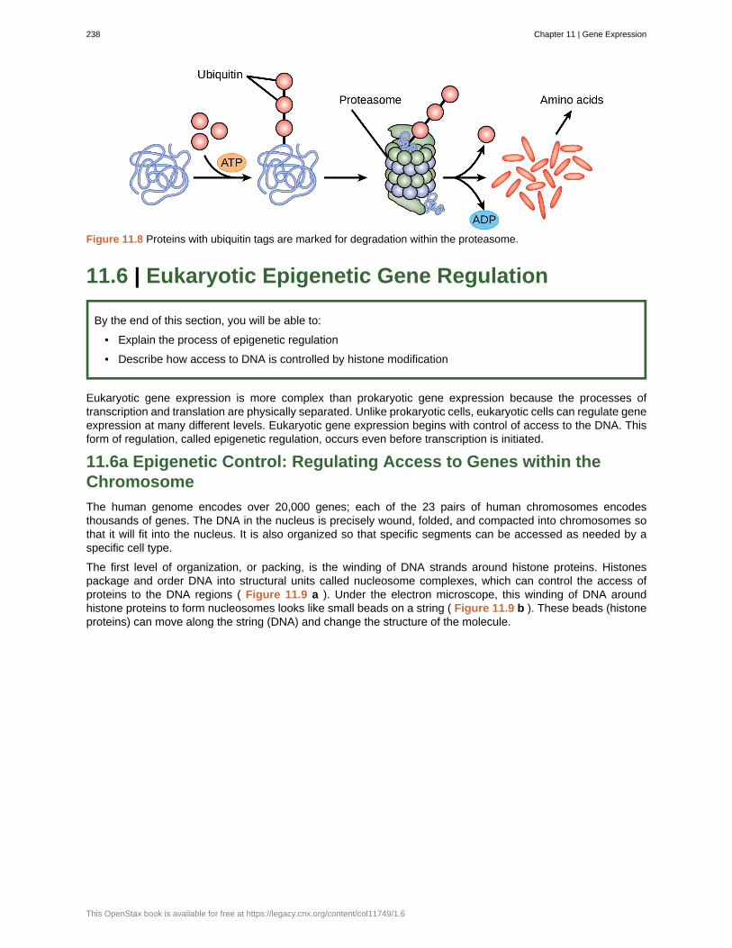
Figure 11.8 Proteins with ubiquitin tags are marked for degradation within the proteasome.
11.6 | Eukaryotic Epigenetic Gene Regulation
By the end of this section, you will be able to:
• Explain the process of epigenetic regulation
• Describe how access to DNA is controlled by histone modification
Eukaryotic gene expression is more complex than prokaryotic gene expression because the processes oftranscription and translation are physically separated. Unlike prokaryotic cells, eukaryotic cells can regulate geneexpression at many different levels. Eukaryotic gene expression begins with control of access to the DNA. Thisform of regulation, called epigenetic regulation, occurs even before transcription is initiated.
11.6a Epigenetic Control: Regulating Access to Genes within theChromosome
The human genome encodes over 20,000 genes; each of the 23 pairs of human chromosomes encodesthousands of genes. The DNA in the nucleus is precisely wound, folded, and compacted into chromosomes sothat it will fit into the nucleus. It is also organized so that specific segments can be accessed as needed by aspecific cell type.
The first level of organization, or packing, is the winding of DNA strands around histone proteins. Histonespackage and order DNA into structural units called nucleosome complexes, which can control the access ofproteins to the DNA regions ( Figure 11.9 a ). Under the electron microscope, this winding of DNA aroundhistone proteins to form nucleosomes looks like small beads on a string ( Figure 11.9 b ). These beads (histoneproteins) can move along the string (DNA) and change the structure of the molecule.
238 Chapter 11 | Gene Expression
This OpenStax book is available for free at https://legacy.cnx.org/content/col11749/1.6
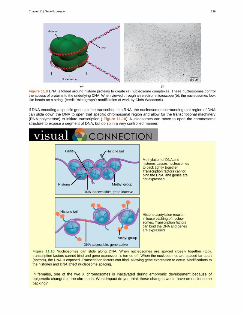
Figure 11.9 DNA is folded around histone proteins to create (a) nucleosome complexes. These nucleosomes controlthe access of proteins to the underlying DNA. When viewed through an electron microscope (b), the nucleosomes looklike beads on a string. (credit “micrograph”: modification of work by Chris Woodcock)
If DNA encoding a specific gene is to be transcribed into RNA, the nucleosomes surrounding that region of DNAcan slide down the DNA to open that specific chromosomal region and allow for the transcriptional machinery(RNA polymerase) to initiate transcription ( Figure 11.10). Nucleosomes can move to open the chromosomestructure to expose a segment of DNA, but do so in a very controlled manner.
Figure 11.10 Nucleosomes can slide along DNA. When nucleosomes are spaced closely together (top),transcription factors cannot bind and gene expression is turned off. When the nucleosomes are spaced far apart(bottom), the DNA is exposed. Transcription factors can bind, allowing gene expression to occur. Modifications tothe histones and DNA affect nucleosome spacing.
In females, one of the two X chromosomes is inactivated during embryonic development because ofepigenetic changes to the chromatin. What impact do you think these changes would have on nucleosomepacking?
Chapter 11 | Gene Expression 239
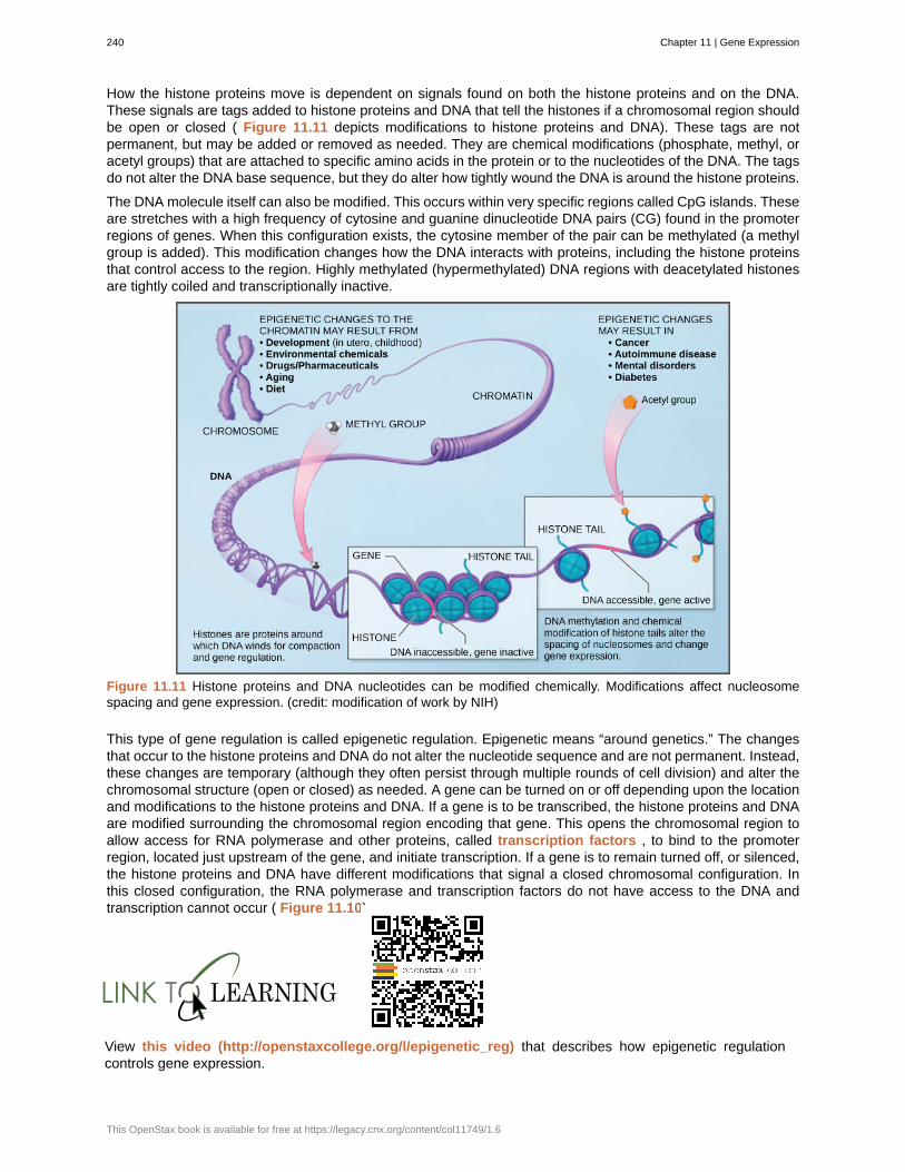
How the histone proteins move is dependent on signals found on both the histone proteins and on the DNA.These signals are tags added to histone proteins and DNA that tell the histones if a chromosomal region shouldbe open or closed ( Figure 11.11 depicts modifications to histone proteins and DNA). These tags are notpermanent, but may be added or removed as needed. They are chemical modifications (phosphate, methyl, oracetyl groups) that are attached to specific amino acids in the protein or to the nucleotides of the DNA. The tagsdo not alter the DNA base sequence, but they do alter how tightly wound the DNA is around the histone proteins.
The DNA molecule itself can also be modified. This occurs within very specific regions called CpG islands. Theseare stretches with a high frequency of cytosine and guanine dinucleotide DNA pairs (CG) found in the promoterregions of genes. When this configuration exists, the cytosine member of the pair can be methylated (a methylgroup is added). This modification changes how the DNA interacts with proteins, including the histone proteinsthat control access to the region. Highly methylated (hypermethylated) DNA regions with deacetylated histonesare tightly coiled and transcriptionally inactive.
Figure 11.11 Histone proteins and DNA nucleotides can be modified chemically. Modifications affect nucleosomespacing and gene expression. (credit: modification of work by NIH)
This type of gene regulation is called epigenetic regulation. Epigenetic means “around genetics.” The changesthat occur to the histone proteins and DNA do not alter the nucleotide sequence and are not permanent. Instead,these changes are temporary (although they often persist through multiple rounds of cell division) and alter thechromosomal structure (open or closed) as needed. A gene can be turned on or off depending upon the locationand modifications to the histone proteins and DNA. If a gene is to be transcribed, the histone proteins and DNAare modified surrounding the chromosomal region encoding that gene. This opens the chromosomal region toallow access for RNA polymerase and other proteins, called transcription factors , to bind to the promoterregion, located just upstream of the gene, and initiate transcription. If a gene is to remain turned off, or silenced,the histone proteins and DNA have different modifications that signal a closed chromosomal configuration. Inthis closed configuration, the RNA polymerase and transcription factors do not have access to the DNA andtranscription cannot occur ( Figure 11.10).
View this video (http://openstaxcollege.org/l/epigenetic_reg) that describes how epigenetic regulationcontrols gene expression.
240 Chapter 11 | Gene Expression
This OpenStax book is available for free at https://legacy.cnx.org/content/col11749/1.6
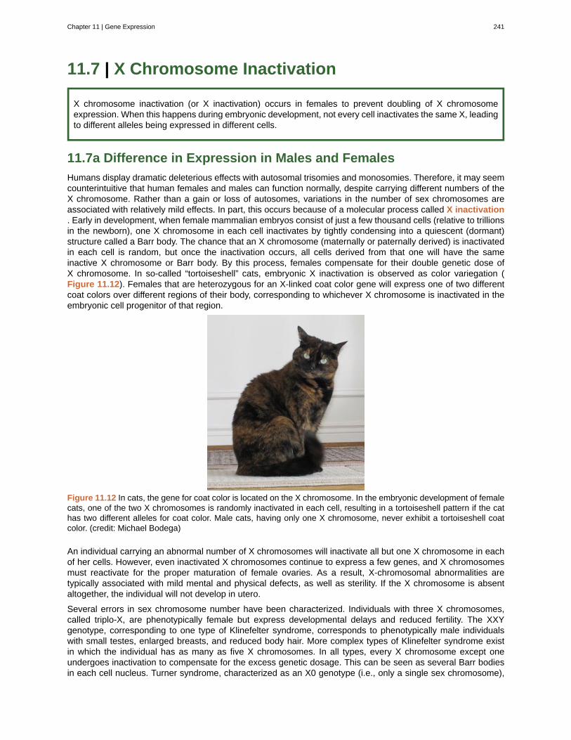
11.7 | X Chromosome Inactivation
X chromosome inactivation (or X inactivation) occurs in females to prevent doubling of X chromosomeexpression. When this happens during embryonic development, not every cell inactivates the same X, leadingto different alleles being expressed in different cells.
11.7a Difference in Expression in Males and Females
Humans display dramatic deleterious effects with autosomal trisomies and monosomies. Therefore, it may seemcounterintuitive that human females and males can function normally, despite carrying different numbers of theX chromosome. Rather than a gain or loss of autosomes, variations in the number of sex chromosomes areassociated with relatively mild effects. In part, this occurs because of a molecular process called X inactivation. Early in development, when female mammalian embryos consist of just a few thousand cells (relative to trillionsin the newborn), one X chromosome in each cell inactivates by tightly condensing into a quiescent (dormant)structure called a Barr body. The chance that an X chromosome (maternally or paternally derived) is inactivatedin each cell is random, but once the inactivation occurs, all cells derived from that one will have the sameinactive X chromosome or Barr body. By this process, females compensate for their double genetic dose ofX chromosome. In so-called “tortoiseshell” cats, embryonic X inactivation is observed as color variegation (Figure 11.12). Females that are heterozygous for an X-linked coat color gene will express one of two differentcoat colors over different regions of their body, corresponding to whichever X chromosome is inactivated in theembryonic cell progenitor of that region.
Figure 11.12 In cats, the gene for coat color is located on the X chromosome. In the embryonic development of femalecats, one of the two X chromosomes is randomly inactivated in each cell, resulting in a tortoiseshell pattern if the cathas two different alleles for coat color. Male cats, having only one X chromosome, never exhibit a tortoiseshell coatcolor. (credit: Michael Bodega)
An individual carrying an abnormal number of X chromosomes will inactivate all but one X chromosome in eachof her cells. However, even inactivated X chromosomes continue to express a few genes, and X chromosomesmust reactivate for the proper maturation of female ovaries. As a result, X-chromosomal abnormalities aretypically associated with mild mental and physical defects, as well as sterility. If the X chromosome is absentaltogether, the individual will not develop in utero.
Several errors in sex chromosome number have been characterized. Individuals with three X chromosomes,called triplo-X, are phenotypically female but express developmental delays and reduced fertility. The XXYgenotype, corresponding to one type of Klinefelter syndrome, corresponds to phenotypically male individualswith small testes, enlarged breasts, and reduced body hair. More complex types of Klinefelter syndrome existin which the individual has as many as five X chromosomes. In all types, every X chromosome except oneundergoes inactivation to compensate for the excess genetic dosage. This can be seen as several Barr bodiesin each cell nucleus. Turner syndrome, characterized as an X0 genotype (i.e., only a single sex chromosome),
Chapter 11 | Gene Expression 241

corresponds to a phenotypically female individual with short stature, webbed skin in the neck region, hearingand cardiac impairments, and sterility.
242 Chapter 11 | Gene Expression
This OpenStax book is available for free at https://legacy.cnx.org/content/col11749/1.6
activator
catabolite activator protein (CAP)
enhancer
epigenetic
gene expression
inducible operon
lac operon
negative regulator
operator
operon
positive regulator
post-transcriptional
post-translational
proteasome
repressor
transcription factor binding site
transcription factors
transcriptional level
transcriptional start site
X inactivation
KEY TERMS
protein that binds to prokaryotic operators to increase transcription
protein that complexes with cAMP to bind to the promoter sequences ofoperons that control sugar processing when glucose is not available
segment of DNA that is upstream, downstream, perhaps thousands of nucleotides away, or onanother chromosome that influence the transcription of a specific gene
heritable changes that do not involve changes in the DNA sequence
processes that control the turning on or turning off of a gene
operon that can be activated or repressed depending on cellular needs and the surroundingenvironment
operon in prokaryotic cells that encodes genes required for processing and intake of lactose
protein that prevents transcription
region of DNA outside of the promoter region that binds activators or repressors that control geneexpression in prokaryotic cells
collection of genes involved in a pathway that are transcribed together as a single mRNA in prokaryoticcells
protein that increases transcription
control of gene expression after the RNA molecule has been created but before it istranslated into protein
control of gene expression after a protein has been created
organelle that degrades proteins
protein that binds to the operator of prokaryotic genes to prevent transcription
sequence of DNA to which a transcription factor binds
protein that binds to the DNA at the promoter or enhancer region and that influencestranscription of a gene
control of gene expression before it has been transcribed into mRNA
site at which transcription begins
condensation of X chromosomes into Barr bodies during embryonic development in females tocompensate for the double genetic dose
CHAPTER SUMMARY
11.1 Regulation of Gene Expression
While all somatic cells within an organism contain the same DNA, not all cells within that organism express thesame proteins. Prokaryotic organisms express the entire DNA they encode in every cell, but not necessarily allat the same time. Proteins are expressed only when they are needed. Eukaryotic organisms express a subsetof the DNA that is encoded in any given cell. In each cell type, the type and amount of protein is regulated bycontrolling gene expression. To express a protein, the DNA is first transcribed into RNA, which is thentranslated into proteins. In prokaryotic cells, these processes occur almost simultaneously. In eukaryotic cells,transcription occurs in the nucleus and is separate from the translation that occurs in the cytoplasm. Geneexpression in prokaryotes is regulated only at the transcriptional level, whereas in eukaryotic cells, gene
Chapter 11 | Gene Expression 243
expression is regulated at the epigenetic, transcriptional, post-transcriptional, translational, and post-translational levels.
11.2 Prokaryotic Gene Regulation
The regulation of gene expression in prokaryotic cells occurs at the transcriptional level. There are three waysto control the transcription of an operon: repressive control, activator control, and inducible control. Repressivecontrol, typified by the trp operon, uses proteins bound to the operator sequence to physically prevent thebinding of RNA polymerase and the activation of transcription. Therefore, if tryptophan is not needed, therepressor is bound to the operator and transcription remains off. Activator control, typified by the action of CAP,increases the binding ability of RNA polymerase to the promoter when CAP is bound. In this case, low levels ofglucose result in the binding of cAMP to CAP. CAP then binds the promoter, which allows RNA polymerase tobind to the promoter better. In the last example—the lac operon—two conditions must be met to initiatetranscription. Glucose must not be present, and lactose must be available for the lac operon to be transcribed.If glucose is absent, CAP binds to the operator. If lactose is present, the repressor protein does not bind to itsoperator. Only when both conditions are met will RNA polymerase bind to the promoter to induce transcription.
11.3 Eukaryotic Transcription Gene Regulation
To start transcription, general transcription factors must first bind to the TATA box and recruit RNA polymeraseto that location. The binding of additional regulatory transcription factors will either increase or preventtranscription. In addition to promoter sequences, enhancer regions help augment transcription. Enhancers canbe upstream, downstream, within a gene itself, or on other chromosomes. Transcription factors bind toenhancer regions to increase or prevent transcription.
11.4 Eukaryotic Post-transcriptional Gene Regulation
Post-transcriptional control can occur at any stage after transcription, including RNA splicing, nuclear shuttling,and RNA stability. Once RNA is transcribed, it must be processed to create a mature RNA that is ready to betranslated. This involves the removal of introns that do not code for protein. Proteins bind to the signals thatmark the exon/intron border to remove the introns and ligate the exons together. Once this occurs, the RNA ismature and can be translated. RNA is created and spliced in the nucleus, but needs to be transported to thecytoplasm to be translated. RNA is transported to the cytoplasm through the nuclear pore complex. Once theRNA is in the cytoplasm, the length of time it resides there before being degraded, called RNA stability, canalso be altered to control the overall amount of protein that is synthesized. The RNA stability can be increased,leading to longer residency time in the cytoplasm, or decreased, leading to shortened time and less proteinsynthesis.
11.5 Eukaryotic Translational and Post-translational Gene Regulation
Changing the status of the RNA or the protein itself can affect the amount of protein, the function of the protein,or how long it is found in the cell. To translate the protein, a protein initiator complex must assemble on theRNA. Modifications (such as phosphorylation) of proteins in this complex can prevent proper translation fromoccurring. Once a protein has been synthesized, it can be modified (phosphorylated, acetylated, methylated, orubiquitinated). These post-translational modifications can greatly impact the stability, degradation, or function ofthe protein.
11.6 Eukaryotic Epigenetic Gene Regulation
In eukaryotic cells, the first stage of gene expression control occurs at the epigenetic level. Epigeneticmechanisms control access to the chromosomal region to allow genes to be turned on or off. Thesemechanisms control how DNA is packed into the nucleus by regulating how tightly the DNA is wound aroundhistone proteins. The addition or removal of chemical modifications (or flags) to histone proteins or DNA signalsto the cell to open or close a chromosomal region. Therefore, eukaryotic cells can control whether a gene isexpressed by controlling accessibility to transcription factors and the binding of RNA polymerase to initiatetranscription.
11.7 X Chromosome Inactivation
X chromosome inactivation occurs in females to prevent the X chromosome from being expressed twice asmuch. This process occurs during embryonic development, and different cells may inactivate a different X. This
244 Chapter 11 | Gene Expression
This OpenStax book is available for free at https://legacy.cnx.org/content/col11749/1.6

results in different expression in different cells.
Chapter 11 | Gene Expression 245

246 Chapter 11 | Gene Expression
This OpenStax book is available for free at https://legacy.cnx.org/content/col11749/1.6
12 | MENDEL'SEXPERIMENTS ANDHEREDITY
Figure 12.1 Experimenting with thousands of garden peas, Mendel uncovered the fundamentals of genetics. (credit:modification of work by Jerry Kirkhart)
Chapter Outline
12.1: Mendel’s Experiments and the Laws of Probability
12.2: Characteristics and Traits
12.3: Laws of Inheritance
12.4: Alternatives and Applications
Introduction
Genetics is the study of heredity. Johann Gregor Mendel set the framework for genetics long beforechromosomes or genes had been identified, at a time when meiosis was not well understood. Mendel selecteda simple biological system and conducted methodical, quantitative analyses using large sample sizes. Becauseof Mendel’s work, the fundamental principles of heredity were revealed. We now know that genes, carriedon chromosomes, are the basic functional units of heredity with the capability to be replicated, expressed, ormutated. Today, the postulates put forth by Mendel form the basis of classical, or Mendelian, genetics. Not allgenes are transmitted from parents to offspring according to Mendelian genetics, but Mendel’s experimentsserve as an excellent starting point for thinking about inheritance.
Chapter 12 | Mendel's Experiments and Heredity 247
12.1 | Mendel’s Experiments and the Laws of
Probability
By the end of this section, you will be able to:
• Describe the scientific reasons for the success of Mendel’s experimental work
• Describe the expected outcomes of monohybrid crosses involving dominant and recessive alleles
• Apply the sum and product rules to calculate probabilities
Figure 12.2 Johann Gregor Mendel is considered the father of genetics.
Johann Gregor Mendel (1822–1884) ( Figure 12.2) was a lifelong learner, teacher, scientist, and man offaith. As a young adult, he joined the Augustinian Abbey of St. Thomas in Brno in what is now the CzechRepublic. Supported by the monastery, he taught physics, botany, and natural science courses at the secondaryand university levels. In 1856, he began a decade-long research pursuit involving inheritance patterns inhoneybees and plants, ultimately settling on pea plants as his primary model system (a system with convenientcharacteristics used to study a specific biological phenomenon to be applied to other systems). In 1865, Mendelpresented the results of his experiments with nearly 30,000 pea plants to the local Natural History Society. Hedemonstrated that traits are transmitted faithfully from parents to offspring independently of other traits and in
dominant and recessive patterns. In 1866, he published his work, Experiments in Plant Hybridization,[1]
in theproceedings of the Natural History Society of Brünn. However, Mendel’s work went virtually unnoticed by thescientific community at the time, and he was not recognized for his extraordinary scientific contributions duringhis lifetime. In fact, it was not until 1900 that his work was rediscovered, reproduced, and revitalized by scientistson the brink of discovering the chromosomal basis of heredity.
12.1a Mendel’s Model System
Mendel’s work was accomplished using the garden pea, Pisum sativum , to study inheritance. By experimentingwith true-breeding pea plants (plants that always produce offspring that look like the parent), Mendel avoidedthe appearance of unexpected traits in offspring that might occur if the plants were not true breeding. The gardenpea also grows to maturity within one season, meaning that several generations could be evaluated over arelatively short time. Finally, large quantities of garden peas could be cultivated simultaneously, allowing Mendel
1.Johann Gregor Mendel, Versuche über Pflanzenhybriden Verhandlungen des naturforschenden Vereines in Brünn, Bd. IV für das Jahr , 1865Abhandlungen, 3–47. [for English translation see http://www.mendelweb.org/Mendel.plain.html]
248 Chapter 12 | Mendel's Experiments and Heredity
This OpenStax book is available for free at https://legacy.cnx.org/content/col11749/1.6
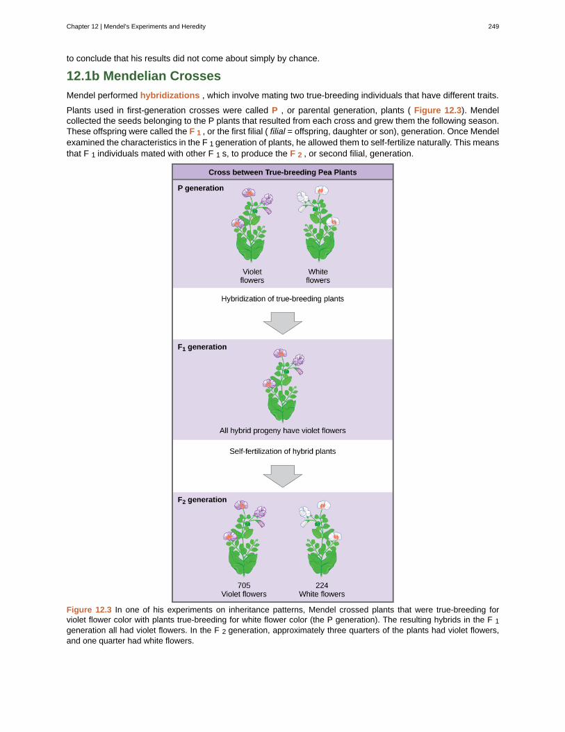
to conclude that his results did not come about simply by chance.
12.1b Mendelian Crosses
Mendel performed hybridizations , which involve mating two true-breeding individuals that have different traits.
Plants used in first-generation crosses were called P , or parental generation, plants ( Figure 12.3). Mendelcollected the seeds belonging to the P plants that resulted from each cross and grew them the following season.These offspring were called the F 1 , or the first filial ( filial = offspring, daughter or son), generation. Once Mendelexamined the characteristics in the F 1 generation of plants, he allowed them to self-fertilize naturally. This meansthat F 1 individuals mated with other F 1 s, to produce the F 2 , or second filial, generation.
Figure 12.3 In one of his experiments on inheritance patterns, Mendel crossed plants that were true-breeding forviolet flower color with plants true-breeding for white flower color (the P generation). The resulting hybrids in the F 1generation all had violet flowers. In the F 2 generation, approximately three quarters of the plants had violet flowers,and one quarter had white flowers.
Chapter 12 | Mendel's Experiments and Heredity 249
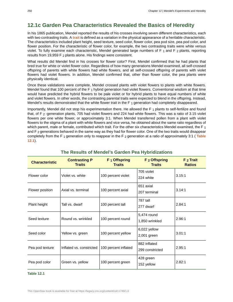
12.1c Garden Pea Characteristics Revealed the Basics of Heredity
In his 1865 publication, Mendel reported the results of his crosses involving seven different characteristics, eachwith two contrasting traits. A trait is defined as a variation in the physical appearance of a heritable characteristic.The characteristics included plant height, seed texture, seed color, flower color, pea pod size, pea pod color, andflower position. For the characteristic of flower color, for example, the two contrasting traits were white versusviolet. To fully examine each characteristic, Mendel generated large numbers of F 1 and F 2 plants, reportingresults from 19,959 F 2 plants alone. His findings were consistent.
What results did Mendel find in his crosses for flower color? First, Mendel confirmed that he had plants thatbred true for white or violet flower color. Regardless of how many generations Mendel examined, all self-crossedoffspring of parents with white flowers had white flowers, and all self-crossed offspring of parents with violetflowers had violet flowers. In addition, Mendel confirmed that, other than flower color, the pea plants werephysically identical.
Once these validations were complete, Mendel crossed plants with violet flowers to plants with white flowers.Mendel found that 100 percent of the F 1 hybrid generation had violet flowers. Conventional wisdom at that timewould have predicted the hybrid flowers to be pale violet or for hybrid plants to have equal numbers of whiteand violet flowers. In other words, the contrasting parental traits were expected to blend in the offspring. Instead,Mendel’s results demonstrated that the white flower trait in the F 1 generation had completely disappeared.
Importantly, Mendel did not stop his experimentation there. He allowed the F 1 plants to self-fertilize and foundthat, of F 2 -generation plants, 705 had violet flowers and 224 had white flowers. This was a ratio of 3.15 violetflowers per one white flower, or approximately 3:1. When Mendel transferred pollen from a plant with violetflowers to the stigma of a plant with white flowers and vice versa, he obtained about the same ratio regardless ofwhich parent, male or female, contributed which trait. For the other six characteristics Mendel examined, the F 1and F 2 generations behaved in the same way as they had for flower color. One of the two traits would disappearcompletely from the F 1 generation only to reappear in the F 2 generation at a ratio of approximately 3:1 ( Table12.1).
The Results of Mendel’s Garden Pea Hybridizations
CharacteristicContrasting P
TraitsF 1 Offspring
TraitsF 2 Offspring
TraitsF 2 TraitRatios
Flower color Violet vs. white 100 percent violet705 violet
224 white3.15:1
Flower position Axial vs. terminal 100 percent axial651 axial
207 terminal3.14:1
Plant height Tall vs. dwarf 100 percent tall787 tall
277 dwarf2.84:1
Seed texture Round vs. wrinkled 100 percent round5,474 round
1,850 wrinkled2.96:1
Seed color Yellow vs. green 100 percent yellow6,022 yellow
2,001 green3.01:1
Pea pod texture Inflated vs. constricted 100 percent inflated882 inflated
299 constricted2.95:1
Pea pod color Green vs. yellow 100 percent green428 green
152 yellow2.82:1
Table 12.1
250 Chapter 12 | Mendel's Experiments and Heredity
This OpenStax book is available for free at https://legacy.cnx.org/content/col11749/1.6
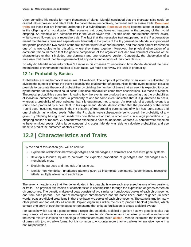
Upon compiling his results for many thousands of plants, Mendel concluded that the characteristics could bedivided into expressed and latent traits. He called these, respectively, dominant and recessive traits. Dominanttraits are those that are inherited unchanged in a hybridization. Recessive traits become latent, or disappear,in the offspring of a hybridization. The recessive trait does, however, reappear in the progeny of the hybridoffspring. An example of a dominant trait is the violet-flower trait. For this same characteristic (flower color),white-colored flowers are a recessive trait. The fact that the recessive trait reappeared in the F 2 generationmeant that the traits remained separate (not blended) in the plants of the F 1 generation. Mendel also proposedthat plants possessed two copies of the trait for the flower-color characteristic, and that each parent transmittedone of its two copies to its offspring, where they came together. Moreover, the physical observation of adominant trait could mean that the genetic composition of the organism included two dominant versions of thecharacteristic or that it included one dominant and one recessive version. Conversely, the observation of arecessive trait meant that the organism lacked any dominant versions of this characteristic.
So why did Mendel repeatedly obtain 3:1 ratios in his crosses? To understand how Mendel deduced the basicmechanisms of inheritance that lead to such ratios, we must first review the laws of probability.
12.1d Probability Basics
Probabilities are mathematical measures of likelihood. The empirical probability of an event is calculated bydividing the number of times the event occurs by the total number of opportunities for the event to occur. It is alsopossible to calculate theoretical probabilities by dividing the number of times that an event is expected to occurby the number of times that it could occur. Empirical probabilities come from observations, like those of Mendel.Theoretical probabilities come from knowing how the events are produced and assuming that the probabilitiesof individual outcomes are equal. A probability of one for some event indicates that it is guaranteed to occur,whereas a probability of zero indicates that it is guaranteed not to occur. An example of a genetic event is around seed produced by a pea plant. In his experiment, Mendel demonstrated that the probability of the event“round seed” occurring was one in the F 1 offspring of true-breeding parents, one of which has round seeds andone of which has wrinkled seeds. When the F 1 plants were subsequently self-crossed, the probability of anygiven F 2 offspring having round seeds was now three out of four. In other words, in a large population of F 2offspring chosen at random, 75 percent were expected to have round seeds, whereas 25 percent were expectedto have wrinkled seeds. Using large numbers of crosses, Mendel was able to calculate probabilities and usethese to predict the outcomes of other crosses.
12.2 | Characteristics and Traits
By the end of this section, you will be able to:
• Explain the relationship between genotypes and phenotypes in dominant and recessive gene systems
• Develop a Punnett square to calculate the expected proportions of genotypes and phenotypes in amonohybrid cross
• Explain the purpose and methods of a test cross
• Identify non-Mendelian inheritance patterns such as incomplete dominance, codominance, recessivelethals, multiple alleles, and sex linkage
The seven characteristics that Mendel evaluated in his pea plants were each expressed as one of two versions,or traits. The physical expression of characteristics is accomplished through the expression of genes carried onchromosomes. The genetic makeup of peas consists of two similar or homologous copies of each chromosome,one from each parent. Each pair of homologous chromosomes has the same linear order of genes. In otherwords, peas are diploid organisms in that they have two copies of each chromosome. The same is true for manyother plants and for virtually all animals. Diploid organisms utilize meiosis to produce haploid gametes, whichcontain one copy of each homologous chromosome that unite at fertilization to create a diploid zygote.
For cases in which a single gene controls a single characteristic, a diploid organism has two genetic copies thatmay or may not encode the same version of that characteristic. Gene variants that arise by mutation and exist atthe same relative locations on homologous chromosomes are called alleles . Mendel examined the inheritanceof genes with just two allele forms, but it is common to encounter more than two alleles for any given gene in anatural population.
Chapter 12 | Mendel's Experiments and Heredity 251
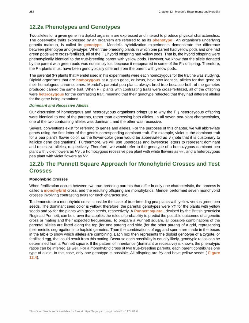
12.2a Phenotypes and Genotypes
Two alleles for a given gene in a diploid organism are expressed and interact to produce physical characteristics.The observable traits expressed by an organism are referred to as its phenotype . An organism’s underlyinggenetic makeup, is called its genotype . Mendel’s hybridization experiments demonstrate the differencebetween phenotype and genotype. When true-breeding plants in which one parent had yellow pods and one hadgreen pods were cross-fertilized, all of the F 1 hybrid offspring had yellow pods. That is, the hybrid offspring werephenotypically identical to the true-breeding parent with yellow pods. However, we know that the allele donatedby the parent with green pods was not simply lost because it reappeared in some of the F 2 offspring. Therefore,the F 1 plants must have been genotypically different from the parent with yellow pods.
The parental (P) plants that Mendel used in his experiments were each homozygous for the trait he was studying.Diploid organisms that are homozygous at a given gene, or locus, have two identical alleles for that gene ontheir homologous chromosomes. Mendel’s parental pea plants always bred true because both of the gametesproduced carried the same trait. When P 0 plants with contrasting traits were cross-fertilized, all of the offspringwere heterozygous for the contrasting trait, meaning that their genotype reflected that they had different allelesfor the gene being examined.
Dominant and Recessive Alleles
Our discussion of homozygous and heterozygous organisms brings us to why the F 1 heterozygous offspringwere identical to one of the parents, rather than expressing both alleles. In all seven pea-plant characteristics,one of the two contrasting alleles was dominant, and the other was recessive.
Several conventions exist for referring to genes and alleles. For the purposes of this chapter, we will abbreviategenes using the first letter of the gene’s corresponding dominant trait. For example, violet is the dominant traitfor a pea plant’s flower color, so the flower-color gene would be abbreviated as V (note that it is customary toitalicize gene designations). Furthermore, we will use uppercase and lowercase letters to represent dominantand recessive alleles, respectively. Therefore, we would refer to the genotype of a homozygous dominant peaplant with violet flowers as VV , a homozygous recessive pea plant with white flowers as vv , and a heterozygouspea plant with violet flowers as Vv .
12.2b The Punnett Square Approach for Monohybrid Crosses and TestCrosses
Monohybrid Crosses
When fertilization occurs between two true-breeding parents that differ in only one characteristic, the process iscalled a monohybrid cross, and the resulting offspring are monohybrids. Mendel performed seven monohybridcrosses involving contrasting traits for each characteristic.
To demonstrate a monohybrid cross, consider the case of true-breeding pea plants with yellow versus green peaseeds. The dominant seed color is yellow; therefore, the parental genotypes were YY for the plants with yellowseeds and yy for the plants with green seeds, respectively. A Punnett square , devised by the British geneticistReginald Punnett, can be drawn that applies the rules of probability to predict the possible outcomes of a geneticcross or mating and their expected frequencies. To prepare a Punnett square, all possible combinations of theparental alleles are listed along the top (for one parent) and side (for the other parent) of a grid, representingtheir meiotic segregation into haploid gametes. Then the combinations of egg and sperm are made in the boxesin the table to show which alleles are combining. Each box then represents the diploid genotype of a zygote, orfertilized egg, that could result from this mating. Because each possibility is equally likely, genotypic ratios can bedetermined from a Punnett square. If the pattern of inheritance (dominant or recessive) is known, the phenotypicratios can be inferred as well. For a monohybrid cross of two true-breeding parents, each parent contributes onetype of allele. In this case, only one genotype is possible. All offspring are Yy and have yellow seeds ( Figure12.4).
252 Chapter 12 | Mendel's Experiments and Heredity
This OpenStax book is available for free at https://legacy.cnx.org/content/col11749/1.6
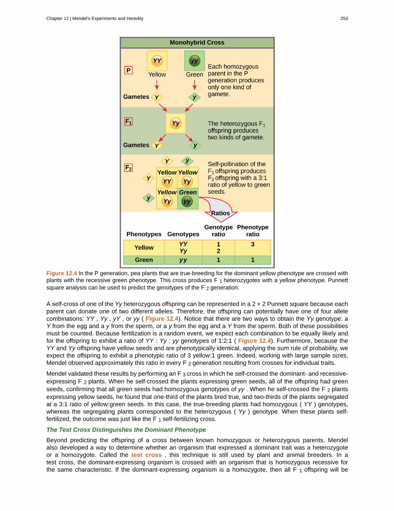
Figure 12.4 In the P generation, pea plants that are true-breeding for the dominant yellow phenotype are crossed withplants with the recessive green phenotype. This cross produces F 1 heterozygotes with a yellow phenotype. Punnettsquare analysis can be used to predict the genotypes of the F 2 generation.
A self-cross of one of the Yy heterozygous offspring can be represented in a 2 × 2 Punnett square because eachparent can donate one of two different alleles. Therefore, the offspring can potentially have one of four allelecombinations: YY , Yy , yY , or yy ( Figure 12.4). Notice that there are two ways to obtain the Yy genotype: aY from the egg and a y from the sperm, or a y from the egg and a Y from the sperm. Both of these possibilitiesmust be counted. Because fertilization is a random event, we expect each combination to be equally likely andfor the offspring to exhibit a ratio of YY : Yy : yy genotypes of 1:2:1 ( Figure 12.4). Furthermore, because theYY and Yy offspring have yellow seeds and are phenotypically identical, applying the sum rule of probability, weexpect the offspring to exhibit a phenotypic ratio of 3 yellow:1 green. Indeed, working with large sample sizes,Mendel observed approximately this ratio in every F 2 generation resulting from crosses for individual traits.
Mendel validated these results by performing an F 3 cross in which he self-crossed the dominant- and recessive-expressing F 2 plants. When he self-crossed the plants expressing green seeds, all of the offspring had greenseeds, confirming that all green seeds had homozygous genotypes of yy . When he self-crossed the F 2 plantsexpressing yellow seeds, he found that one-third of the plants bred true, and two-thirds of the plants segregatedat a 3:1 ratio of yellow:green seeds. In this case, the true-breeding plants had homozygous ( YY ) genotypes,whereas the segregating plants corresponded to the heterozygous ( Yy ) genotype. When these plants self-fertilized, the outcome was just like the F 1 self-fertilizing cross.
The Test Cross Distinguishes the Dominant Phenotype
Beyond predicting the offspring of a cross between known homozygous or heterozygous parents, Mendelalso developed a way to determine whether an organism that expressed a dominant trait was a heterozygoteor a homozygote. Called the test cross , this technique is still used by plant and animal breeders. In atest cross, the dominant-expressing organism is crossed with an organism that is homozygous recessive forthe same characteristic. If the dominant-expressing organism is a homozygote, then all F 1 offspring will be
Chapter 12 | Mendel's Experiments and Heredity 253
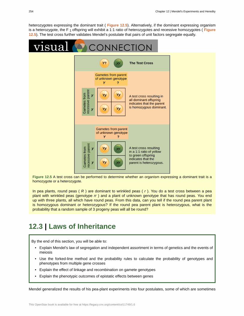
heterozygotes expressing the dominant trait ( Figure 12.5). Alternatively, if the dominant expressing organismis a heterozygote, the F 1 offspring will exhibit a 1:1 ratio of heterozygotes and recessive homozygotes ( Figure12.5). The test cross further validates Mendel’s postulate that pairs of unit factors segregate equally.
Figure 12.5 A test cross can be performed to determine whether an organism expressing a dominant trait is ahomozygote or a heterozygote.
In pea plants, round peas ( R ) are dominant to wrinkled peas ( r ). You do a test cross between a peaplant with wrinkled peas (genotype rr ) and a plant of unknown genotype that has round peas. You endup with three plants, all which have round peas. From this data, can you tell if the round pea parent plantis homozygous dominant or heterozygous? If the round pea parent plant is heterozygous, what is theprobability that a random sample of 3 progeny peas will all be round?
12.3 | Laws of Inheritance
By the end of this section, you will be able to:
• Explain Mendel’s law of segregation and independent assortment in terms of genetics and the events ofmeiosis
• Use the forked-line method and the probability rules to calculate the probability of genotypes andphenotypes from multiple gene crosses
• Explain the effect of linkage and recombination on gamete genotypes
• Explain the phenotypic outcomes of epistatic effects between genes
Mendel generalized the results of his pea-plant experiments into four postulates, some of which are sometimes
254 Chapter 12 | Mendel's Experiments and Heredity
This OpenStax book is available for free at https://legacy.cnx.org/content/col11749/1.6
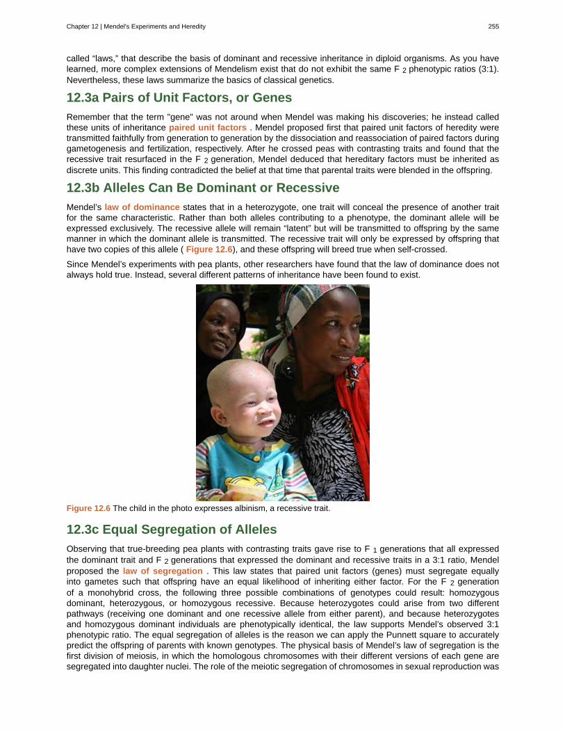
called “laws,” that describe the basis of dominant and recessive inheritance in diploid organisms. As you havelearned, more complex extensions of Mendelism exist that do not exhibit the same F 2 phenotypic ratios (3:1).Nevertheless, these laws summarize the basics of classical genetics.
12.3a Pairs of Unit Factors, or Genes
Remember that the term "gene" was not around when Mendel was making his discoveries; he instead calledthese units of inheritance paired unit factors . Mendel proposed first that paired unit factors of heredity weretransmitted faithfully from generation to generation by the dissociation and reassociation of paired factors duringgametogenesis and fertilization, respectively. After he crossed peas with contrasting traits and found that therecessive trait resurfaced in the F 2 generation, Mendel deduced that hereditary factors must be inherited asdiscrete units. This finding contradicted the belief at that time that parental traits were blended in the offspring.
12.3b Alleles Can Be Dominant or Recessive
Mendel’s law of dominance states that in a heterozygote, one trait will conceal the presence of another traitfor the same characteristic. Rather than both alleles contributing to a phenotype, the dominant allele will beexpressed exclusively. The recessive allele will remain “latent” but will be transmitted to offspring by the samemanner in which the dominant allele is transmitted. The recessive trait will only be expressed by offspring thathave two copies of this allele ( Figure 12.6), and these offspring will breed true when self-crossed.
Since Mendel’s experiments with pea plants, other researchers have found that the law of dominance does notalways hold true. Instead, several different patterns of inheritance have been found to exist.
Figure 12.6 The child in the photo expresses albinism, a recessive trait.
12.3c Equal Segregation of Alleles
Observing that true-breeding pea plants with contrasting traits gave rise to F 1 generations that all expressedthe dominant trait and F 2 generations that expressed the dominant and recessive traits in a 3:1 ratio, Mendelproposed the law of segregation . This law states that paired unit factors (genes) must segregate equallyinto gametes such that offspring have an equal likelihood of inheriting either factor. For the F 2 generationof a monohybrid cross, the following three possible combinations of genotypes could result: homozygousdominant, heterozygous, or homozygous recessive. Because heterozygotes could arise from two differentpathways (receiving one dominant and one recessive allele from either parent), and because heterozygotesand homozygous dominant individuals are phenotypically identical, the law supports Mendel’s observed 3:1phenotypic ratio. The equal segregation of alleles is the reason we can apply the Punnett square to accuratelypredict the offspring of parents with known genotypes. The physical basis of Mendel’s law of segregation is thefirst division of meiosis, in which the homologous chromosomes with their different versions of each gene aresegregated into daughter nuclei. The role of the meiotic segregation of chromosomes in sexual reproduction was
Chapter 12 | Mendel's Experiments and Heredity 255
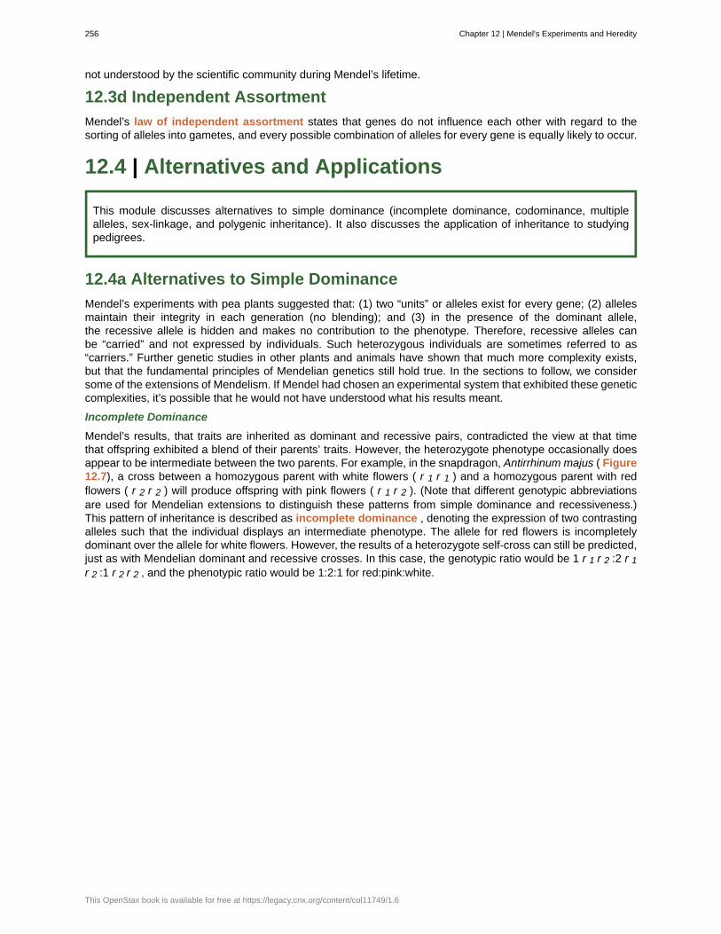
not understood by the scientific community during Mendel’s lifetime.
12.3d Independent Assortment
Mendel’s law of independent assortment states that genes do not influence each other with regard to thesorting of alleles into gametes, and every possible combination of alleles for every gene is equally likely to occur.
12.4 | Alternatives and Applications
This module discusses alternatives to simple dominance (incomplete dominance, codominance, multiplealleles, sex-linkage, and polygenic inheritance). It also discusses the application of inheritance to studyingpedigrees.
12.4a Alternatives to Simple Dominance
Mendel’s experiments with pea plants suggested that: (1) two “units” or alleles exist for every gene; (2) allelesmaintain their integrity in each generation (no blending); and (3) in the presence of the dominant allele,the recessive allele is hidden and makes no contribution to the phenotype. Therefore, recessive alleles canbe “carried” and not expressed by individuals. Such heterozygous individuals are sometimes referred to as“carriers.” Further genetic studies in other plants and animals have shown that much more complexity exists,but that the fundamental principles of Mendelian genetics still hold true. In the sections to follow, we considersome of the extensions of Mendelism. If Mendel had chosen an experimental system that exhibited these geneticcomplexities, it’s possible that he would not have understood what his results meant.
Incomplete Dominance
Mendel’s results, that traits are inherited as dominant and recessive pairs, contradicted the view at that timethat offspring exhibited a blend of their parents’ traits. However, the heterozygote phenotype occasionally doesappear to be intermediate between the two parents. For example, in the snapdragon, Antirrhinum majus ( Figure12.7), a cross between a homozygous parent with white flowers ( r 1 r 1 ) and a homozygous parent with redflowers ( r 2 r 2 ) will produce offspring with pink flowers ( r 1 r 2 ). (Note that different genotypic abbreviationsare used for Mendelian extensions to distinguish these patterns from simple dominance and recessiveness.)This pattern of inheritance is described as incomplete dominance , denoting the expression of two contrastingalleles such that the individual displays an intermediate phenotype. The allele for red flowers is incompletelydominant over the allele for white flowers. However, the results of a heterozygote self-cross can still be predicted,just as with Mendelian dominant and recessive crosses. In this case, the genotypic ratio would be 1 r 1 r 2 :2 r 1r 2 :1 r 2 r 2 , and the phenotypic ratio would be 1:2:1 for red:pink:white.
256 Chapter 12 | Mendel's Experiments and Heredity
This OpenStax book is available for free at https://legacy.cnx.org/content/col11749/1.6
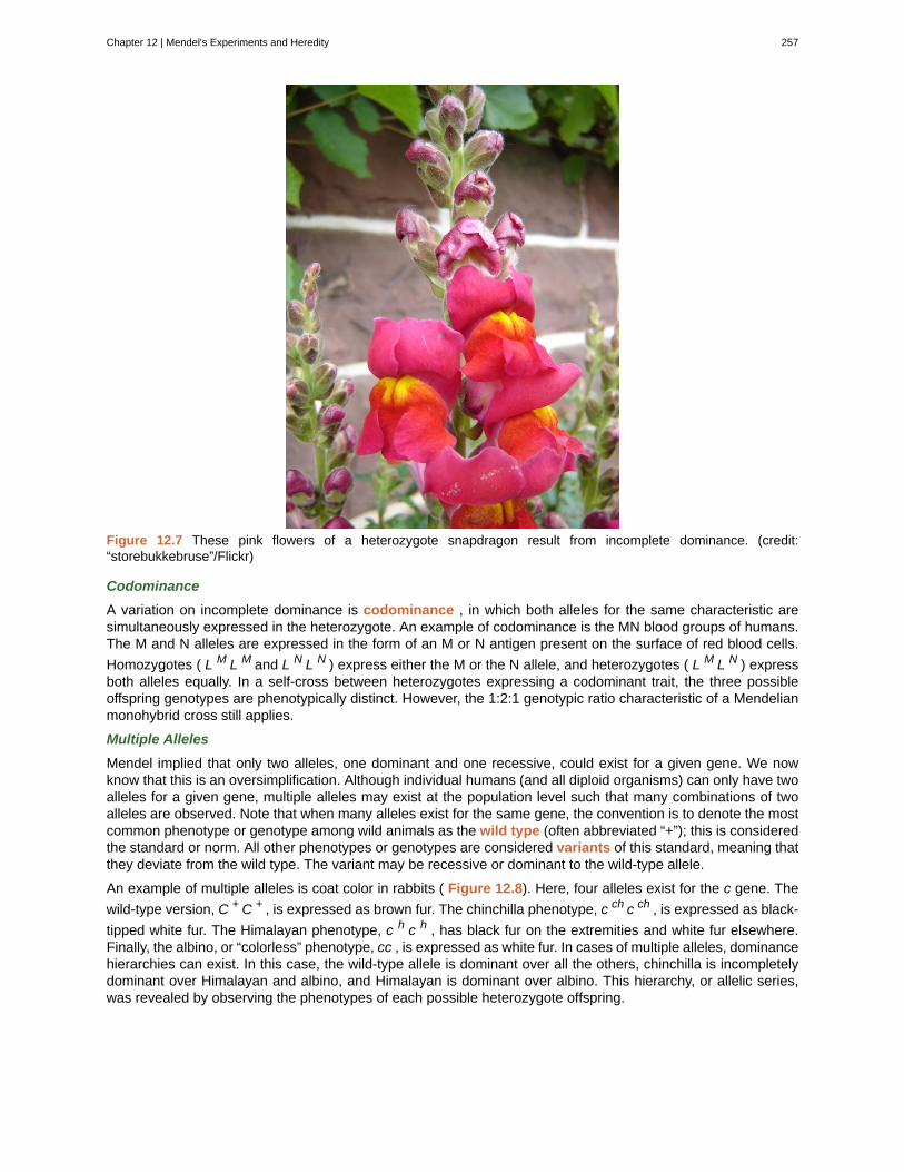
Figure 12.7 These pink flowers of a heterozygote snapdragon result from incomplete dominance. (credit:“storebukkebruse”/Flickr)
Codominance
A variation on incomplete dominance is codominance , in which both alleles for the same characteristic aresimultaneously expressed in the heterozygote. An example of codominance is the MN blood groups of humans.The M and N alleles are expressed in the form of an M or N antigen present on the surface of red blood cells.
Homozygotes ( L M L M and L N L N ) express either the M or the N allele, and heterozygotes ( L M L N ) expressboth alleles equally. In a self-cross between heterozygotes expressing a codominant trait, the three possibleoffspring genotypes are phenotypically distinct. However, the 1:2:1 genotypic ratio characteristic of a Mendelianmonohybrid cross still applies.
Multiple Alleles
Mendel implied that only two alleles, one dominant and one recessive, could exist for a given gene. We nowknow that this is an oversimplification. Although individual humans (and all diploid organisms) can only have twoalleles for a given gene, multiple alleles may exist at the population level such that many combinations of twoalleles are observed. Note that when many alleles exist for the same gene, the convention is to denote the mostcommon phenotype or genotype among wild animals as the wild type (often abbreviated “+”); this is consideredthe standard or norm. All other phenotypes or genotypes are considered variants of this standard, meaning thatthey deviate from the wild type. The variant may be recessive or dominant to the wild-type allele.
An example of multiple alleles is coat color in rabbits ( Figure 12.8). Here, four alleles exist for the c gene. The
wild-type version, C + C + , is expressed as brown fur. The chinchilla phenotype, c ch c ch , is expressed as black-
tipped white fur. The Himalayan phenotype, c h c h , has black fur on the extremities and white fur elsewhere.Finally, the albino, or “colorless” phenotype, cc , is expressed as white fur. In cases of multiple alleles, dominancehierarchies can exist. In this case, the wild-type allele is dominant over all the others, chinchilla is incompletelydominant over Himalayan and albino, and Himalayan is dominant over albino. This hierarchy, or allelic series,was revealed by observing the phenotypes of each possible heterozygote offspring.
Chapter 12 | Mendel's Experiments and Heredity 257
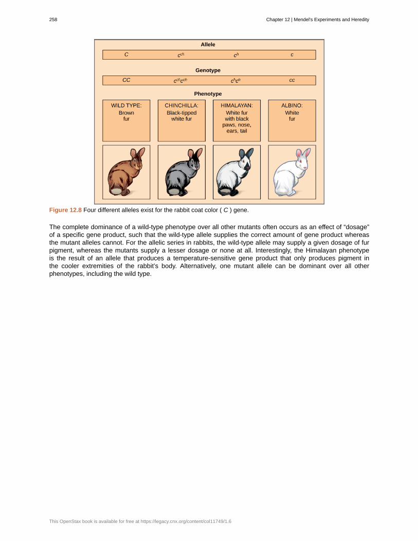
Figure 12.8 Four different alleles exist for the rabbit coat color ( C ) gene.
The complete dominance of a wild-type phenotype over all other mutants often occurs as an effect of “dosage”of a specific gene product, such that the wild-type allele supplies the correct amount of gene product whereasthe mutant alleles cannot. For the allelic series in rabbits, the wild-type allele may supply a given dosage of furpigment, whereas the mutants supply a lesser dosage or none at all. Interestingly, the Himalayan phenotypeis the result of an allele that produces a temperature-sensitive gene product that only produces pigment inthe cooler extremities of the rabbit’s body. Alternatively, one mutant allele can be dominant over all otherphenotypes, including the wild type.
258 Chapter 12 | Mendel's Experiments and Heredity
This OpenStax book is available for free at https://legacy.cnx.org/content/col11749/1.6
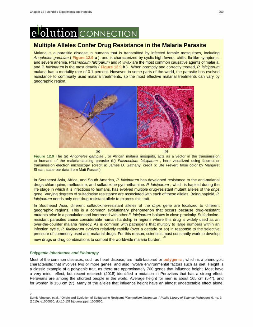
Multiple Alleles Confer Drug Resistance in the Malaria ParasiteMalaria is a parasitic disease in humans that is transmitted by infected female mosquitoes, includingAnopheles gambiae ( Figure 12.9 a ), and is characterized by cyclic high fevers, chills, flu-like symptoms,and severe anemia. Plasmodium falciparum and P. vivax are the most common causative agents of malaria,and P. falciparum is the most deadly ( Figure 12.9 b ) . When promptly and correctly treated, P. falciparummalaria has a mortality rate of 0.1 percent. However, in some parts of the world, the parasite has evolvedresistance to commonly used malaria treatments, so the most effective malarial treatments can vary bygeographic region.
(a) (b)Figure 12.9 The (a) Anopheles gambiae , or African malaria mosquito, acts as a vector in the transmissionto humans of the malaria-causing parasite (b) Plasmodium falciparum , here visualized using false-colortransmission electron microscopy. (credit a: James D. Gathany; credit b: Ute Frevert; false color by MargaretShear; scale-bar data from Matt Russell)
In Southeast Asia, Africa, and South America, P. falciparum has developed resistance to the anti-malarialdrugs chloroquine, mefloquine, and sulfadoxine-pyrimethamine. P. falciparum , which is haploid during thelife stage in which it is infectious to humans, has evolved multiple drug-resistant mutant alleles of the dhpsgene. Varying degrees of sulfadoxine resistance are associated with each of these alleles. Being haploid, P.falciparum needs only one drug-resistant allele to express this trait.
In Southeast Asia, different sulfadoxine-resistant alleles of the dhps gene are localized to differentgeographic regions. This is a common evolutionary phenomenon that occurs because drug-resistantmutants arise in a population and interbreed with other P. falciparum isolates in close proximity. Sulfadoxine-resistant parasites cause considerable human hardship in regions where this drug is widely used as anover-the-counter malaria remedy. As is common with pathogens that multiply to large numbers within aninfection cycle, P. falciparum evolves relatively rapidly (over a decade or so) in response to the selectivepressure of commonly used anti-malarial drugs. For this reason, scientists must constantly work to develop
new drugs or drug combinations to combat the worldwide malaria burden.[2]
Polygenic Inheritance and Pleiotropy
Most of the common diseases, such as heart disease, are multi-factored or polygenic , which is a phenotypiccharacteristic that involves two or more genes, and also involve environmental factors such as diet. Height isa classic example of a polygenic trait, as there are approximately 700 genes that influence height. Most havea very minor effect, but recent research (2018) identified a mutation in Peruvians that has a strong effect.Peruvians are among the shortest people in the world. Average height for men is about 165 cm (5'4"), andfor women is 153 cm (5'). Many of the alleles that influence height have an almost undetectable effect alone,
2.Sumiti Vinayak, et al., “Origin and Evolution of Sulfadoxine Resistant Plasmodium falciparum ,” Public Library of Science Pathogens 6, no. 3(2010): e1000830, doi:10.1371/journal.ppat.1000830.
Chapter 12 | Mendel's Experiments and Heredity 259
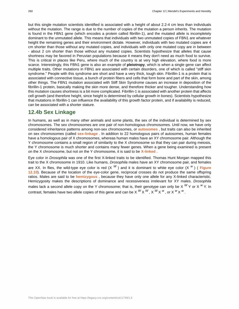
but this single mutation scientists identified is associated with a height of about 2.2-4 cm less than individualswithout the mutation. The range is due to the number of copies of the mutation a person inherits. The mutationis found in the FBN1 gene (which encodes a protein called fibrillin-1), and the mutated allele is incompletelydominant to the unmutated allele. This means that individuals with two unmutated copies of FBN1 are whateverheight the remaining genes and their environment dictate. However, individuals with two mutated copies are 4cm shorter than those without any mutated copies, and individuals with only one mutated copy are in between- about 2 cm shorter than those without any mutated copies. Scientists hypothesize that alleles that causeshortness may be favored in Peruvian populations because it means they don't need as much food to survive.This is critical in places like Peru, where much of the country is at very high elevation, where food is morescarce. Interestingly, this FBN1 gene is also an example of pleiotropy, which is when a single gene can affectmultiple traits. Other mutations in FBN1 are associated with certain disorders, one of which is called "stiff skinsyndrome." People with this syndrome are short and have a very thick, tough skin. Fibrillin-1 is a protein that isassociated with connective tissue, a bunch of protein fibers and cells that form bone and part of the skin, amongother things. The FBN1 mutation associated with Stiff Skin Syndrome causes an increase in deposition of thefibrillin-1 protein, basically making the skin more dense, and therefore thicker and tougher. Understanding howthis mutation causes shortness is a bit more complicated. Fibrillin-1 is associated with another protein that affectscell growth (and therefore height, since height is determined by cellular growth in bones). Scientists hypothesizethat mutations in fibrillin-1 can influence the availability of this growth factor protein, and if availability is reduced,can be associated with a shorter stature.
12.4b Sex Linkage
In humans, as well as in many other animals and some plants, the sex of the individual is determined by sexchromosomes. The sex chromosomes are one pair of non-homologous chromosomes. Until now, we have onlyconsidered inheritance patterns among non-sex chromosomes, or autosomes , but traits can also be inheritedon sex chromosomes (called sex-linkage . In addition to 22 homologous pairs of autosomes, human femaleshave a homologous pair of X chromosomes, whereas human males have an XY chromosome pair. Although theY chromosome contains a small region of similarity to the X chromosome so that they can pair during meiosis,the Y chromosome is much shorter and contains many fewer genes. When a gene being examined is presenton the X chromosome, but not on the Y chromosome, it is said to be X-linked .
Eye color in Drosophila was one of the first X-linked traits to be identified. Thomas Hunt Morgan mapped thistrait to the X chromosome in 1910. Like humans, Drosophila males have an XY chromosome pair, and females
are XX. In flies, the wild-type eye color is red (X W ) and it is dominant to white eye color (X w ) ( Figure12.10). Because of the location of the eye-color gene, reciprocal crosses do not produce the same offspringratios. Males are said to be hemizygous , because they have only one allele for any X-linked characteristic.Hemizygosity makes the descriptions of dominance and recessiveness irrelevant for XY males. Drosophila
males lack a second allele copy on the Y chromosome; that is, their genotype can only be X W Y or X w Y. In
contrast, females have two allele copies of this gene and can be X W X W , X W X w , or X w X w .
260 Chapter 12 | Mendel's Experiments and Heredity
This OpenStax book is available for free at https://legacy.cnx.org/content/col11749/1.6
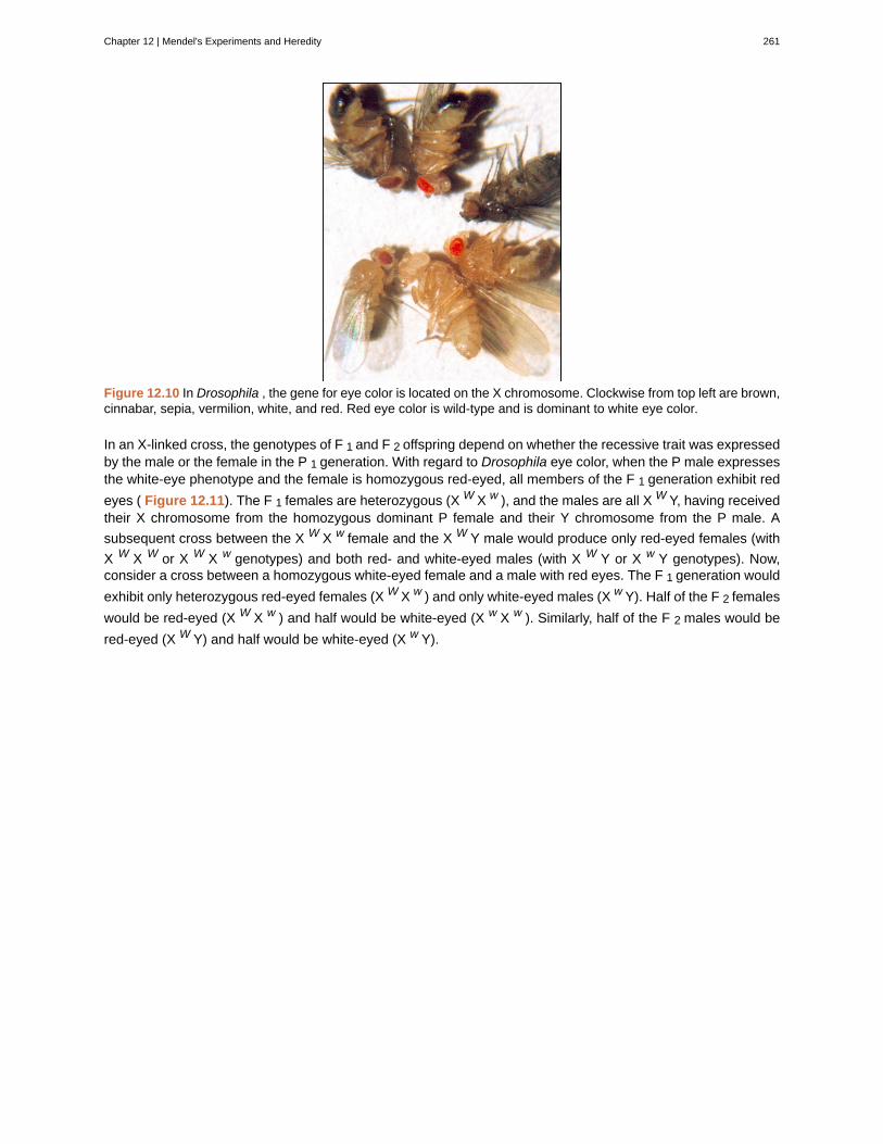
Figure 12.10 In Drosophila , the gene for eye color is located on the X chromosome. Clockwise from top left are brown,cinnabar, sepia, vermilion, white, and red. Red eye color is wild-type and is dominant to white eye color.
In an X-linked cross, the genotypes of F 1 and F 2 offspring depend on whether the recessive trait was expressedby the male or the female in the P 1 generation. With regard to Drosophila eye color, when the P male expressesthe white-eye phenotype and the female is homozygous red-eyed, all members of the F 1 generation exhibit red
eyes ( Figure 12.11). The F 1 females are heterozygous (X W X w ), and the males are all X W Y, having receivedtheir X chromosome from the homozygous dominant P female and their Y chromosome from the P male. A
subsequent cross between the X W X w female and the X W Y male would produce only red-eyed females (with
X W X W or X W X w genotypes) and both red- and white-eyed males (with X W Y or X w Y genotypes). Now,consider a cross between a homozygous white-eyed female and a male with red eyes. The F 1 generation would
exhibit only heterozygous red-eyed females (X W X w ) and only white-eyed males (X w Y). Half of the F 2 females
would be red-eyed (X W X w ) and half would be white-eyed (X w X w ). Similarly, half of the F 2 males would be
red-eyed (X W Y) and half would be white-eyed (X w Y).
Chapter 12 | Mendel's Experiments and Heredity 261
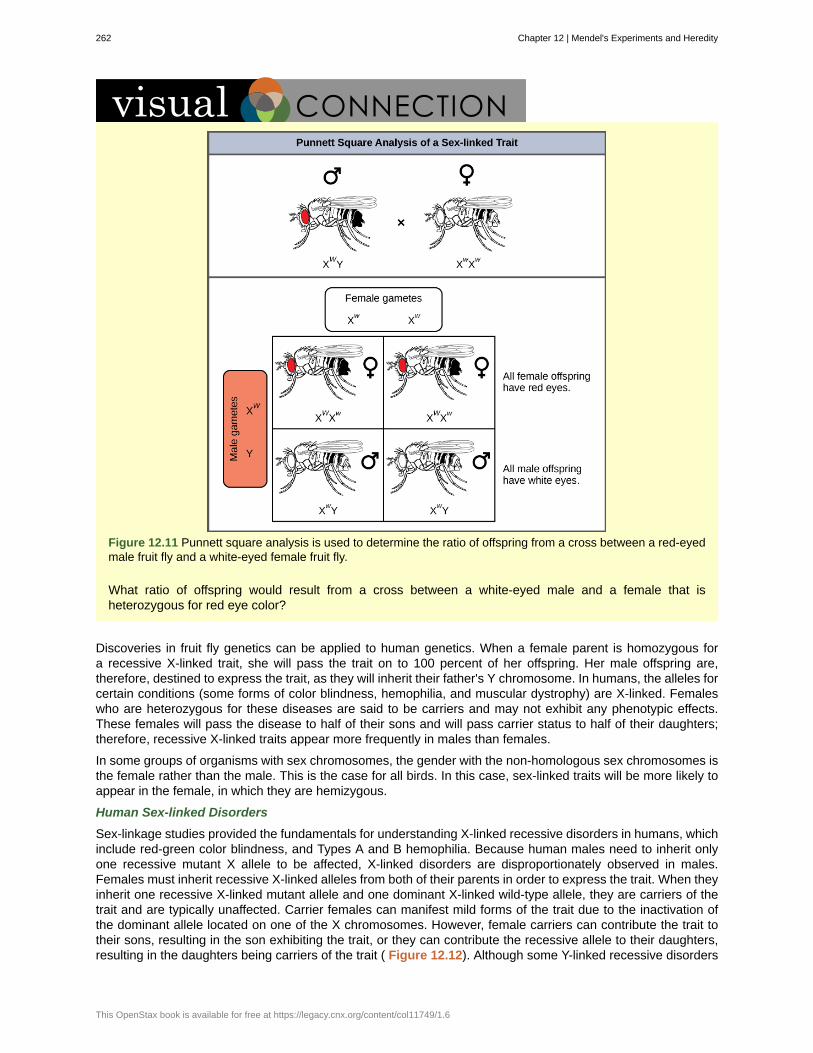
Figure 12.11 Punnett square analysis is used to determine the ratio of offspring from a cross between a red-eyedmale fruit fly and a white-eyed female fruit fly.
What ratio of offspring would result from a cross between a white-eyed male and a female that isheterozygous for red eye color?
Discoveries in fruit fly genetics can be applied to human genetics. When a female parent is homozygous fora recessive X-linked trait, she will pass the trait on to 100 percent of her offspring. Her male offspring are,therefore, destined to express the trait, as they will inherit their father's Y chromosome. In humans, the alleles forcertain conditions (some forms of color blindness, hemophilia, and muscular dystrophy) are X-linked. Femaleswho are heterozygous for these diseases are said to be carriers and may not exhibit any phenotypic effects.These females will pass the disease to half of their sons and will pass carrier status to half of their daughters;therefore, recessive X-linked traits appear more frequently in males than females.
In some groups of organisms with sex chromosomes, the gender with the non-homologous sex chromosomes isthe female rather than the male. This is the case for all birds. In this case, sex-linked traits will be more likely toappear in the female, in which they are hemizygous.
Human Sex-linked Disorders
Sex-linkage studies provided the fundamentals for understanding X-linked recessive disorders in humans, whichinclude red-green color blindness, and Types A and B hemophilia. Because human males need to inherit onlyone recessive mutant X allele to be affected, X-linked disorders are disproportionately observed in males.Females must inherit recessive X-linked alleles from both of their parents in order to express the trait. When theyinherit one recessive X-linked mutant allele and one dominant X-linked wild-type allele, they are carriers of thetrait and are typically unaffected. Carrier females can manifest mild forms of the trait due to the inactivation ofthe dominant allele located on one of the X chromosomes. However, female carriers can contribute the trait totheir sons, resulting in the son exhibiting the trait, or they can contribute the recessive allele to their daughters,resulting in the daughters being carriers of the trait ( Figure 12.12). Although some Y-linked recessive disorders
262 Chapter 12 | Mendel's Experiments and Heredity
This OpenStax book is available for free at https://legacy.cnx.org/content/col11749/1.6
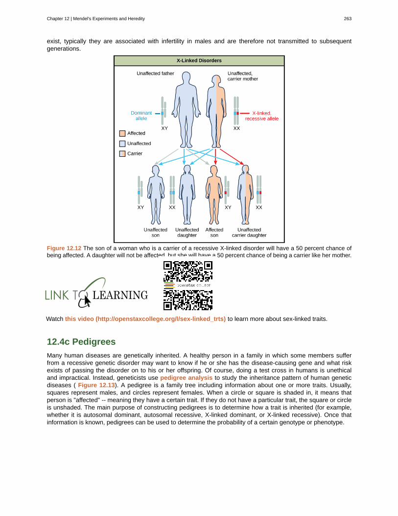
exist, typically they are associated with infertility in males and are therefore not transmitted to subsequentgenerations.
Figure 12.12 The son of a woman who is a carrier of a recessive X-linked disorder will have a 50 percent chance ofbeing affected. A daughter will not be affected, but she will have a 50 percent chance of being a carrier like her mother.
Watch this video (http://openstaxcollege.org/l/sex-linked_trts) to learn more about sex-linked traits.
12.4c Pedigrees
Many human diseases are genetically inherited. A healthy person in a family in which some members sufferfrom a recessive genetic disorder may want to know if he or she has the disease-causing gene and what riskexists of passing the disorder on to his or her offspring. Of course, doing a test cross in humans is unethicaland impractical. Instead, geneticists use pedigree analysis to study the inheritance pattern of human geneticdiseases ( Figure 12.13). A pedigree is a family tree including information about one or more traits. Usually,squares represent males, and circles represent females. When a circle or square is shaded in, it means thatperson is "affected" -- meaning they have a certain trait. If they do not have a particular trait, the square or circleis unshaded. The main purpose of constructing pedigrees is to determine how a trait is inherited (for example,whether it is autosomal dominant, autosomal recessive, X-linked dominant, or X-linked recessive). Once thatinformation is known, pedigrees can be used to determine the probability of a certain genotype or phenotype.
Chapter 12 | Mendel's Experiments and Heredity 263
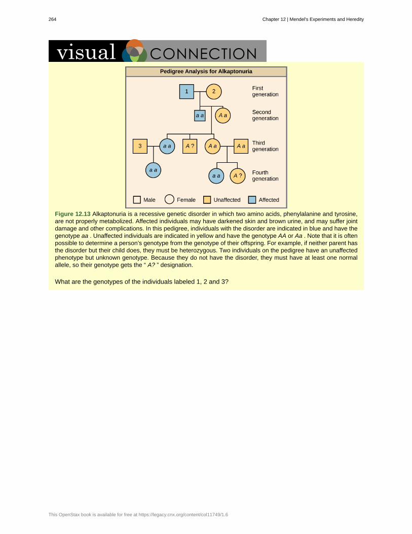
Figure 12.13 Alkaptonuria is a recessive genetic disorder in which two amino acids, phenylalanine and tyrosine,are not properly metabolized. Affected individuals may have darkened skin and brown urine, and may suffer jointdamage and other complications. In this pedigree, individuals with the disorder are indicated in blue and have thegenotype aa . Unaffected individuals are indicated in yellow and have the genotype AA or Aa . Note that it is oftenpossible to determine a person’s genotype from the genotype of their offspring. For example, if neither parent hasthe disorder but their child does, they must be heterozygous. Two individuals on the pedigree have an unaffectedphenotype but unknown genotype. Because they do not have the disorder, they must have at least one normalallele, so their genotype gets the “ A? ” designation.
What are the genotypes of the individuals labeled 1, 2 and 3?
264 Chapter 12 | Mendel's Experiments and Heredity
This OpenStax book is available for free at https://legacy.cnx.org/content/col11749/1.6

alleles
autosomes
codominance
Dominant traits
F 1
F 2
genotype
hemizygous
heterozygous
homozygous
hybridizations
incomplete dominance
law of dominance
law of independent assortment
law of segregation
model system
monohybrid
P
paired unit factors
pedigree analysis
phenotype
pleiotropy
polygenic
Punnett square
KEY TERMS
gene variations that arise by mutation and exist at the same relative locations on homologouschromosomes
any of the non-sex chromosomes
in a heterozygote, complete and simultaneous expression of both alleles for the samecharacteristic
trait that confers the same physical appearance whether an individual has two copies of thetrait or one
first filial generation in a cross; the offspring of the parental generation
second filial generation produced when F 1 individuals are self-crossed or fertilized with each other
underlying genetic makeup, consisting of both physically visible and non-expressed alleles, of anorganism
presence of only one allele for a characteristic, as in X-linkage; hemizygosity makes descriptionsof dominance and recessiveness irrelevant
having two different alleles for a given gene on the homologous chromosome
having two identical alleles for a given gene on the homologous chromosome
process of mating two individuals that differ with the goal of achieving a certain characteristic intheir offspring
in a heterozygote, expression of two contrasting alleles such that the individualdisplays an intermediate phenotype
in a heterozygote, one trait will conceal the presence of another trait for the samecharacteristic
genes do not influence each other with regard to sorting of alleles intogametes; every possible combination of alleles is equally likely to occur
paired unit factors (i.e., genes) segregate equally into gametes such that offspring have anequal likelihood of inheriting any combination of factors
species or biological system used to study a specific biological phenomenon to be applied toother different species
result of a cross between two true-breeding parents that express different traits for only onecharacteristic
parental generation in a cross
Term used by Mendel to describe units of inheritance; modern equivalent would be "gene"
uses family tree information to study inheritance
observable traits expressed by an organism
when a single gene affects multiple traits
one trait encoded by many genes
visual representation of a cross between two individuals in which the gametes of eachindividual are denoted along the top and side of a grid, respectively, and the possible zygotic genotypes arerecombined at each box in the grid
Chapter 12 | Mendel's Experiments and Heredity 265
Recessive traits
sex-linkage
test cross
trait
true-breeding
variants
wild type
X-linked
traits that appear "latent" or non-expressed when the individual also carries a dominant allelefor that same characteristic; when present as two identical copies, the recessive trait is expressed
when a trait is inherited by a gene located on the X or Y chromosome
cross between a dominant expressing individual with an unknown genotype and a homozygousrecessive individual; the offspring phenotypes indicate whether the unknown parent is heterozygous orhomozygous for the dominant trait
variation in the physical appearance of a heritable characteristic
individuals that always produce offspring with the same trait as the parents over multiplegenerations
any trait that differs from wild-type
most common trait found in the wild
gene present on the X, but not the Y chromosome
CHAPTER SUMMARY
12.1 Mendel’s Experiments and the Laws of Probability
Working with garden pea plants, Mendel found that crosses between parents that differed by one trait producedF 1 offspring that all expressed the traits of one parent. Observable traits are referred to as dominant, and non-expressed traits are described as recessive. When the offspring in Mendel’s experiment were self-crossed, theF 2 offspring exhibited the dominant trait or the recessive trait in a 3:1 ratio, confirming that the recessive traithad been transmitted faithfully from the original P 0 parent. Reciprocal crosses generated identical F 1 and F 2offspring ratios. By examining sample sizes, Mendel showed that his crosses behaved reproducibly accordingto the laws of probability, and that the traits were inherited as independent events.
12.2 Characteristics and Traits
When true-breeding or homozygous individuals that differ for a certain trait are crossed, all of the offspring willbe heterozygotes for that trait. If the traits are inherited as dominant and recessive, the F 1 offspring will allexhibit the same phenotype as the parent homozygous for the dominant trait. If these heterozygous offspringare self-crossed, the resulting F 2 offspring will be equally likely to inherit gametes carrying the dominant orrecessive trait, giving rise to offspring of which one quarter are homozygous dominant, half are heterozygous,and one quarter are homozygous recessive. Because homozygous dominant and heterozygous individuals arephenotypically identical, the observed traits in the F 2 offspring will exhibit a ratio of three dominant to onerecessive.
12.3 Laws of Inheritance
Mendel postulated that genes (characteristics) are inherited as pairs of alleles (traits) that behave in a dominantand recessive pattern. Alleles segregate into gametes such that each gamete is equally likely to receive eitherone of the two alleles present in a diploid individual. In addition, genes are assorted into gametesindependently of one another. That is, alleles are generally not more likely to segregate into a gamete with aparticular allele of another gene. A dihybrid cross demonstrates independent assortment when the genes inquestion are on different chromosomes or distant from each other on the same chromosome. For crossesinvolving more than two genes, use the forked line or probability methods to predict offspring genotypes andphenotypes rather than a Punnett square.
Although chromosomes sort independently into gametes during meiosis, Mendel’s law of independentassortment refers to genes, not chromosomes, and a single chromosome may carry more than 1,000 genes.When genes are located in close proximity on the same chromosome, their alleles tend to be inherited together.This results in offspring ratios that violate Mendel's law of independent assortment. However, recombinationserves to exchange genetic material on homologous chromosomes such that maternal and paternal allelesmay be recombined on the same chromosome. This is why alleles on a given chromosome are not alwaysinherited together. Recombination is a random event occurring anywhere on a chromosome. Therefore, genes
266 Chapter 12 | Mendel's Experiments and Heredity
This OpenStax book is available for free at https://legacy.cnx.org/content/col11749/1.6
that are far apart on the same chromosome are likely to still assort independently because of recombinationevents that occurred in the intervening chromosomal space.
Whether or not they are sorting independently, genes may interact at the level of gene products such that theexpression of an allele for one gene masks or modifies the expression of an allele for a different gene. This iscalled epistasis.
12.4 Alternatives and Applications
Alleles do not always behave in dominant and recessive patterns. Incomplete dominance describes situationsin which the heterozygote exhibits a phenotype that is intermediate between the homozygous phenotypes.Codominance describes the simultaneous expression of both of the alleles in the heterozygote. Althoughdiploid organisms can only have two alleles for any given gene, it is common for more than two alleles of agene to exist in a population. In humans, as in many animals and some plants, females have two Xchromosomes and males have one X and one Y chromosome. Genes that are present on the X but not the Ychromosome are said to be X-linked, such that males only inherit one allele for the gene, and females inherittwo.
Chapter 12 | Mendel's Experiments and Heredity 267

268 Chapter 12 | Mendel's Experiments and Heredity
This OpenStax book is available for free at https://legacy.cnx.org/content/col11749/1.6
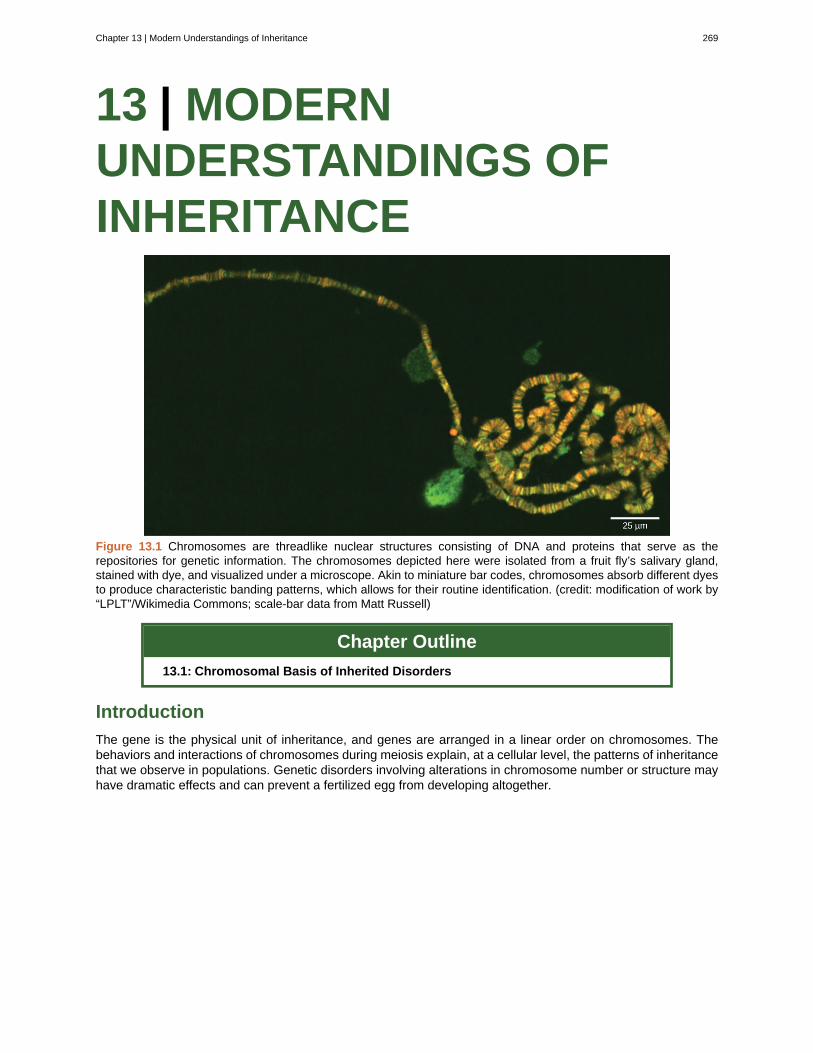
13 | MODERNUNDERSTANDINGS OFINHERITANCE
Figure 13.1 Chromosomes are threadlike nuclear structures consisting of DNA and proteins that serve as therepositories for genetic information. The chromosomes depicted here were isolated from a fruit fly’s salivary gland,stained with dye, and visualized under a microscope. Akin to miniature bar codes, chromosomes absorb different dyesto produce characteristic banding patterns, which allows for their routine identification. (credit: modification of work by“LPLT”/Wikimedia Commons; scale-bar data from Matt Russell)
Chapter Outline
13.1: Chromosomal Basis of Inherited Disorders
Introduction
The gene is the physical unit of inheritance, and genes are arranged in a linear order on chromosomes. Thebehaviors and interactions of chromosomes during meiosis explain, at a cellular level, the patterns of inheritancethat we observe in populations. Genetic disorders involving alterations in chromosome number or structure mayhave dramatic effects and can prevent a fertilized egg from developing altogether.
Chapter 13 | Modern Understandings of Inheritance 269
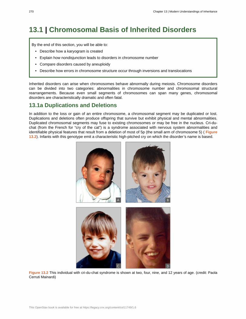
13.1 | Chromosomal Basis of Inherited Disorders
By the end of this section, you will be able to:
• Describe how a karyogram is created
• Explain how nondisjunction leads to disorders in chromosome number
• Compare disorders caused by aneuploidy
• Describe how errors in chromosome structure occur through inversions and translocations
Inherited disorders can arise when chromosomes behave abnormally during meiosis. Chromosome disorderscan be divided into two categories: abnormalities in chromosome number and chromosomal structuralrearrangements. Because even small segments of chromosomes can span many genes, chromosomaldisorders are characteristically dramatic and often fatal.
13.1a Duplications and Deletions
In addition to the loss or gain of an entire chromosome, a chromosomal segment may be duplicated or lost.Duplications and deletions often produce offspring that survive but exhibit physical and mental abnormalities.Duplicated chromosomal segments may fuse to existing chromosomes or may be free in the nucleus. Cri-du-chat (from the French for “cry of the cat”) is a syndrome associated with nervous system abnormalities andidentifiable physical features that result from a deletion of most of 5p (the small arm of chromosome 5) ( Figure13.2). Infants with this genotype emit a characteristic high-pitched cry on which the disorder’s name is based.
Figure 13.2 This individual with cri-du-chat syndrome is shown at two, four, nine, and 12 years of age. (credit: PaolaCerruti Mainardi)
270 Chapter 13 | Modern Understandings of Inheritance
This OpenStax book is available for free at https://legacy.cnx.org/content/col11749/1.6
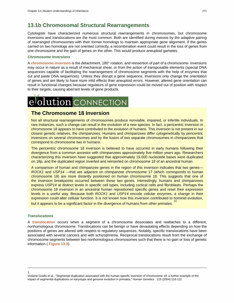
13.1b Chromosomal Structural Rearrangements
Cytologists have characterized numerous structural rearrangements in chromosomes, but chromosomeinversions and translocations are the most common. Both are identified during meiosis by the adaptive pairingof rearranged chromosomes with their former homologs to maintain appropriate gene alignment. If the genescarried on two homologs are not oriented correctly, a recombination event could result in the loss of genes fromone chromosome and the gain of genes on the other. This would produce aneuploid gametes.
Chromosome Inversions
A chromosome inversion is the detachment, 180° rotation, and reinsertion of part of a chromosome. Inversionsmay occur in nature as a result of mechanical shear, or from the action of transposable elements (special DNAsequences capable of facilitating the rearrangement of chromosome segments with the help of enzymes thatcut and paste DNA sequences). Unless they disrupt a gene sequence, inversions only change the orientationof genes and are likely to have more mild effects than aneuploid errors. However, altered gene orientation canresult in functional changes because regulators of gene expression could be moved out of position with respectto their targets, causing aberrant levels of gene products.
The Chromosome 18 InversionNot all structural rearrangements of chromosomes produce nonviable, impaired, or infertile individuals. Inrare instances, such a change can result in the evolution of a new species. In fact, a pericentric inversion inchromosome 18 appears to have contributed to the evolution of humans. This inversion is not present in ourclosest genetic relatives, the chimpanzees. Humans and chimpanzees differ cytogenetically by pericentricinversions on several chromosomes and by the fusion of two separate chromosomes in chimpanzees thatcorrespond to chromosome two in humans.
The pericentric chromosome 18 inversion is believed to have occurred in early humans following theirdivergence from a common ancestor with chimpanzees approximately five million years ago. Researcherscharacterizing this inversion have suggested that approximately 19,000 nucleotide bases were duplicatedon 18p, and the duplicated region inverted and reinserted on chromosome 18 of an ancestral human.
A comparison of human and chimpanzee genes in the region of this inversion indicates that two genes—ROCK1 and USP14 —that are adjacent on chimpanzee chromosome 17 (which corresponds to humanchromosome 18) are more distantly positioned on human chromosome 18. This suggests that one ofthe inversion breakpoints occurred between these two genes. Interestingly, humans and chimpanzeesexpress USP14 at distinct levels in specific cell types, including cortical cells and fibroblasts. Perhaps thechromosome 18 inversion in an ancestral human repositioned specific genes and reset their expressionlevels in a useful way. Because both ROCK1 and USP14 encode cellular enzymes, a change in theirexpression could alter cellular function. It is not known how this inversion contributed to hominid evolution,
but it appears to be a significant factor in the divergence of humans from other primates.[1]
Translocations
A translocation occurs when a segment of a chromosome dissociates and reattaches to a different,nonhomologous chromosome. Translocations can be benign or have devastating effects depending on how thepositions of genes are altered with respect to regulatory sequences. Notably, specific translocations have beenassociated with several cancers and with schizophrenia. Reciprocal translocations result from the exchange ofchromosome segments between two nonhomologous chromosomes such that there is no gain or loss of geneticinformation ( Figure 13.3).
1.Violaine Goidts et al., “Segmental duplication associated with the human-specific inversion of chromosome 18: a further example of theimpact of segmental duplications on karyotype and genome evolution in primates,” Human Genetics . 115 (2004):116-122
Chapter 13 | Modern Understandings of Inheritance 271
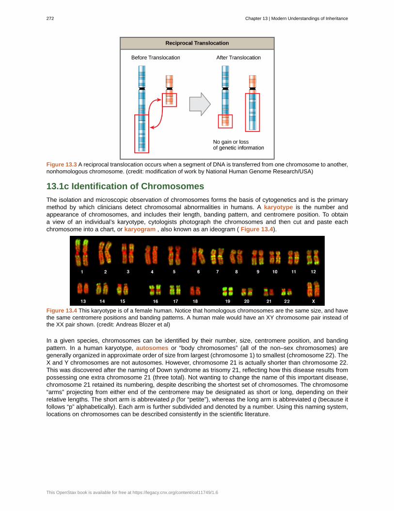
Figure 13.3 A reciprocal translocation occurs when a segment of DNA is transferred from one chromosome to another,nonhomologous chromosome. (credit: modification of work by National Human Genome Research/USA)
13.1c Identification of Chromosomes
The isolation and microscopic observation of chromosomes forms the basis of cytogenetics and is the primarymethod by which clinicians detect chromosomal abnormalities in humans. A karyotype is the number andappearance of chromosomes, and includes their length, banding pattern, and centromere position. To obtaina view of an individual’s karyotype, cytologists photograph the chromosomes and then cut and paste eachchromosome into a chart, or karyogram , also known as an ideogram ( Figure 13.4).
Figure 13.4 This karyotype is of a female human. Notice that homologous chromosomes are the same size, and havethe same centromere positions and banding patterns. A human male would have an XY chromosome pair instead ofthe XX pair shown. (credit: Andreas Blozer et al)
In a given species, chromosomes can be identified by their number, size, centromere position, and bandingpattern. In a human karyotype, autosomes or “body chromosomes” (all of the non–sex chromosomes) aregenerally organized in approximate order of size from largest (chromosome 1) to smallest (chromosome 22). TheX and Y chromosomes are not autosomes. However, chromosome 21 is actually shorter than chromosome 22.This was discovered after the naming of Down syndrome as trisomy 21, reflecting how this disease results frompossessing one extra chromosome 21 (three total). Not wanting to change the name of this important disease,chromosome 21 retained its numbering, despite describing the shortest set of chromosomes. The chromosome“arms” projecting from either end of the centromere may be designated as short or long, depending on theirrelative lengths. The short arm is abbreviated p (for “petite”), whereas the long arm is abbreviated q (because itfollows “p” alphabetically). Each arm is further subdivided and denoted by a number. Using this naming system,locations on chromosomes can be described consistently in the scientific literature.
272 Chapter 13 | Modern Understandings of Inheritance
This OpenStax book is available for free at https://legacy.cnx.org/content/col11749/1.6
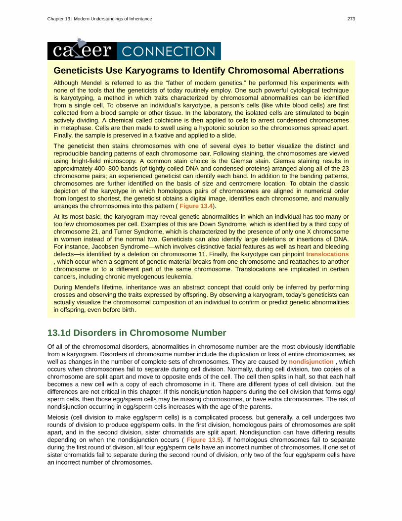
Geneticists Use Karyograms to Identify Chromosomal AberrationsAlthough Mendel is referred to as the “father of modern genetics,” he performed his experiments withnone of the tools that the geneticists of today routinely employ. One such powerful cytological techniqueis karyotyping, a method in which traits characterized by chromosomal abnormalities can be identifiedfrom a single cell. To observe an individual’s karyotype, a person’s cells (like white blood cells) are firstcollected from a blood sample or other tissue. In the laboratory, the isolated cells are stimulated to beginactively dividing. A chemical called colchicine is then applied to cells to arrest condensed chromosomesin metaphase. Cells are then made to swell using a hypotonic solution so the chromosomes spread apart.Finally, the sample is preserved in a fixative and applied to a slide.
The geneticist then stains chromosomes with one of several dyes to better visualize the distinct andreproducible banding patterns of each chromosome pair. Following staining, the chromosomes are viewedusing bright-field microscopy. A common stain choice is the Giemsa stain. Giemsa staining results inapproximately 400–800 bands (of tightly coiled DNA and condensed proteins) arranged along all of the 23chromosome pairs; an experienced geneticist can identify each band. In addition to the banding patterns,chromosomes are further identified on the basis of size and centromere location. To obtain the classicdepiction of the karyotype in which homologous pairs of chromosomes are aligned in numerical orderfrom longest to shortest, the geneticist obtains a digital image, identifies each chromosome, and manuallyarranges the chromosomes into this pattern ( Figure 13.4).
At its most basic, the karyogram may reveal genetic abnormalities in which an individual has too many ortoo few chromosomes per cell. Examples of this are Down Syndrome, which is identified by a third copy ofchromosome 21, and Turner Syndrome, which is characterized by the presence of only one X chromosomein women instead of the normal two. Geneticists can also identify large deletions or insertions of DNA.For instance, Jacobsen Syndrome—which involves distinctive facial features as well as heart and bleedingdefects—is identified by a deletion on chromosome 11. Finally, the karyotype can pinpoint translocations, which occur when a segment of genetic material breaks from one chromosome and reattaches to anotherchromosome or to a different part of the same chromosome. Translocations are implicated in certaincancers, including chronic myelogenous leukemia.
During Mendel’s lifetime, inheritance was an abstract concept that could only be inferred by performingcrosses and observing the traits expressed by offspring. By observing a karyogram, today’s geneticists canactually visualize the chromosomal composition of an individual to confirm or predict genetic abnormalitiesin offspring, even before birth.
13.1d Disorders in Chromosome Number
Of all of the chromosomal disorders, abnormalities in chromosome number are the most obviously identifiablefrom a karyogram. Disorders of chromosome number include the duplication or loss of entire chromosomes, aswell as changes in the number of complete sets of chromosomes. They are caused by nondisjunction , whichoccurs when chromosomes fail to separate during cell division. Normally, during cell division, two copies of achromosome are split apart and move to opposite ends of the cell. The cell then splits in half, so that each halfbecomes a new cell with a copy of each chromosome in it. There are different types of cell division, but thedifferences are not critical in this chapter. If this nondisjunction happens during the cell division that forms egg/sperm cells, then those egg/sperm cells may be missing chromosomes, or have extra chromosomes. The risk ofnondisjunction occurring in egg/sperm cells increases with the age of the parents.
Meiosis (cell division to make egg/sperm cells) is a complicated process, but generally, a cell undergoes tworounds of division to produce egg/sperm cells. In the first division, homologous pairs of chromosomes are splitapart, and in the second division, sister chromatids are split apart. Nondisjunction can have differing resultsdepending on when the nondisjunction occurs ( Figure 13.5). If homologous chromosomes fail to separateduring the first round of division, all four egg/sperm cells have an incorrect number of chromosomes. If one set ofsister chromatids fail to separate during the second round of division, only two of the four egg/sperm cells havean incorrect number of chromosomes.
Chapter 13 | Modern Understandings of Inheritance 273
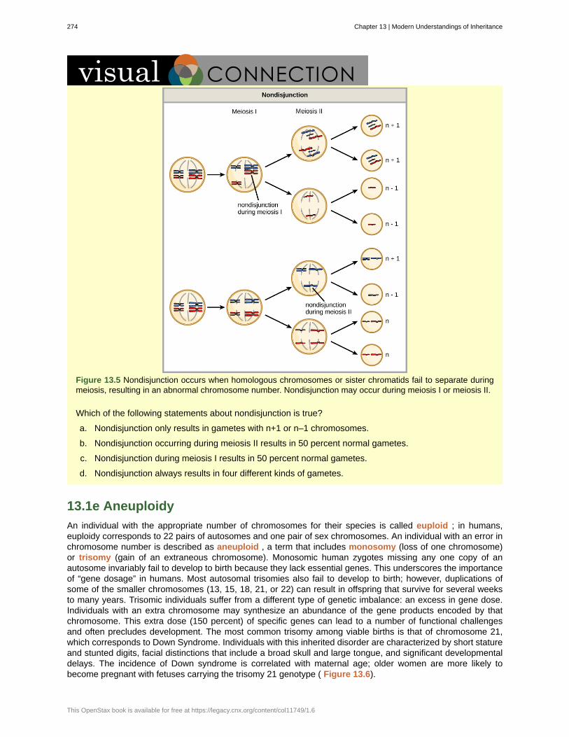
Figure 13.5 Nondisjunction occurs when homologous chromosomes or sister chromatids fail to separate duringmeiosis, resulting in an abnormal chromosome number. Nondisjunction may occur during meiosis I or meiosis II.
Which of the following statements about nondisjunction is true?
a. Nondisjunction only results in gametes with n+1 or n–1 chromosomes.
b. Nondisjunction occurring during meiosis II results in 50 percent normal gametes.
c. Nondisjunction during meiosis I results in 50 percent normal gametes.
d. Nondisjunction always results in four different kinds of gametes.
13.1e Aneuploidy
An individual with the appropriate number of chromosomes for their species is called euploid ; in humans,euploidy corresponds to 22 pairs of autosomes and one pair of sex chromosomes. An individual with an error inchromosome number is described as aneuploid , a term that includes monosomy (loss of one chromosome)or trisomy (gain of an extraneous chromosome). Monosomic human zygotes missing any one copy of anautosome invariably fail to develop to birth because they lack essential genes. This underscores the importanceof “gene dosage” in humans. Most autosomal trisomies also fail to develop to birth; however, duplications ofsome of the smaller chromosomes (13, 15, 18, 21, or 22) can result in offspring that survive for several weeksto many years. Trisomic individuals suffer from a different type of genetic imbalance: an excess in gene dose.Individuals with an extra chromosome may synthesize an abundance of the gene products encoded by thatchromosome. This extra dose (150 percent) of specific genes can lead to a number of functional challengesand often precludes development. The most common trisomy among viable births is that of chromosome 21,which corresponds to Down Syndrome. Individuals with this inherited disorder are characterized by short statureand stunted digits, facial distinctions that include a broad skull and large tongue, and significant developmentaldelays. The incidence of Down syndrome is correlated with maternal age; older women are more likely tobecome pregnant with fetuses carrying the trisomy 21 genotype ( Figure 13.6).
274 Chapter 13 | Modern Understandings of Inheritance
This OpenStax book is available for free at https://legacy.cnx.org/content/col11749/1.6
Figure 13.6 The incidence of having a fetus with trisomy 21 increases dramatically with maternal age.
Visualize the addition of a chromosome that leads to Down syndrome in this video simulation(http://openstaxcollege.org/l/down_syndrome) .
13.1f Polyploidy
An individual with more than the correct number of chromosome sets (two for diploid species) is calledpolyploid . For instance, fertilization of an abnormal diploid egg with a normal haploid sperm would yielda triploid zygote. Polyploid animals are extremely rare, with only a few examples among the flatworms,crustaceans, amphibians, fish, and lizards. Polyploid animals are sterile because meiosis cannot proceednormally and instead produces mostly aneuploid daughter cells that cannot yield viable zygotes. Rarely,polyploid animals can reproduce asexually by haplodiploidy, in which an unfertilized egg divides mitotically toproduce offspring. In contrast, polyploidy is very common in the plant kingdom, and polyploid plants tend to belarger and more robust than euploids of their species ( Figure 13.7).
Chapter 13 | Modern Understandings of Inheritance 275
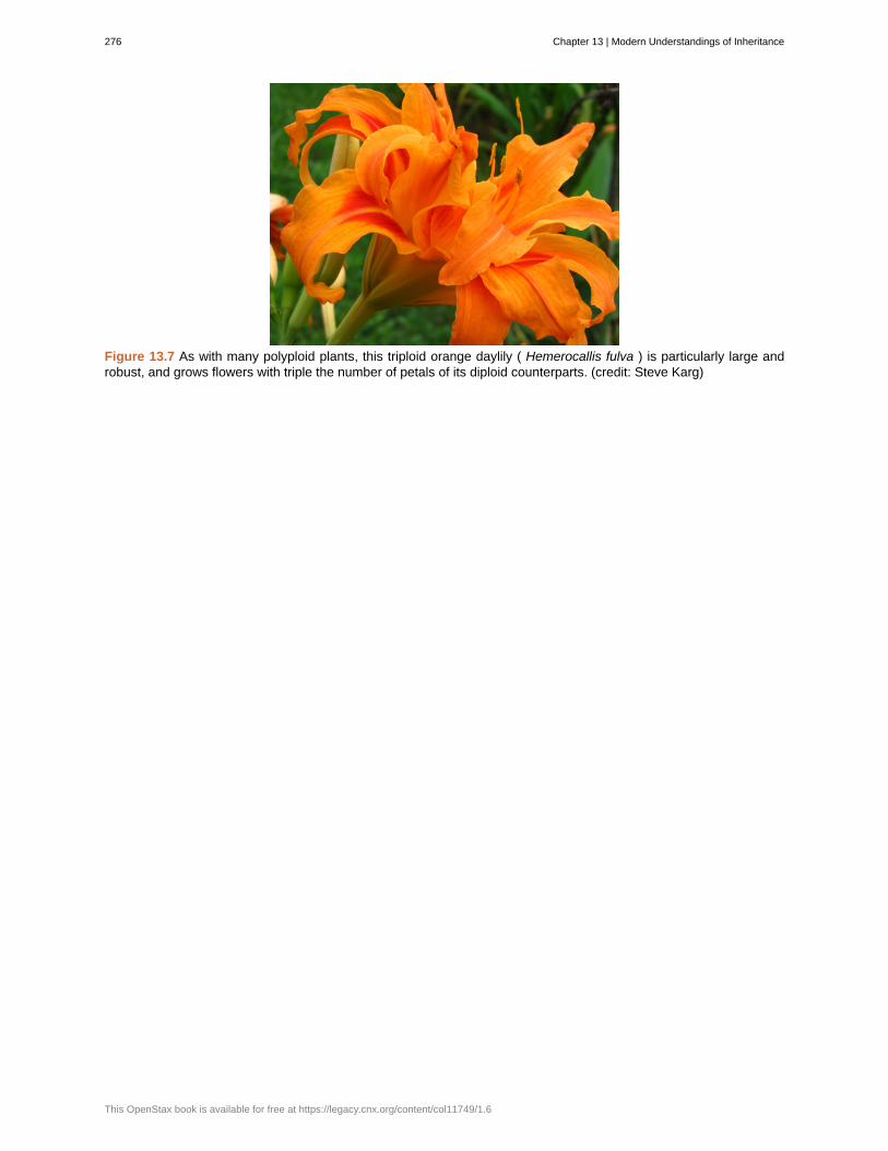
Figure 13.7 As with many polyploid plants, this triploid orange daylily ( Hemerocallis fulva ) is particularly large androbust, and grows flowers with triple the number of petals of its diploid counterparts. (credit: Steve Karg)
276 Chapter 13 | Modern Understandings of Inheritance
This OpenStax book is available for free at https://legacy.cnx.org/content/col11749/1.6
aneuploid
autosomes
chromosome inversion
euploid
karyogram
karyotype
monosomy
nondisjunction
polyploid
translocation
translocations
trisomy
KEY TERMS
individual with an error in chromosome number; includes deletions and duplications of chromosomesegments
any of the non-sex chromosomes
detachment, 180° rotation, and reinsertion of a chromosome arm
individual with the appropriate number of chromosomes for their species
photographic image of a karyotype
number and appearance of an individuals chromosomes; includes the size, banding patterns, andcentromere position
otherwise diploid genotype in which one chromosome is missing
failure of synapsed homologs to completely separate and migrate to separate poles during thefirst cell division of meiosis
individual with an incorrect number of chromosome sets
process by which one segment of a chromosome dissociates and reattaches to a different,nonhomologous chromosome
process by which one segment of a chromosome dissociates and reattaches to a different,nonhomologous chromosome
otherwise diploid genotype in which one entire chromosome is duplicated
CHAPTER SUMMARY
13.1 Chromosomal Basis of Inherited Disorders
The number, size, shape, and banding pattern of chromosomes make them easily identifiable in a karyogramand allows for the assessment of many chromosomal abnormalities. Disorders in chromosome number, oraneuploidies, are typically lethal to the embryo, although a few trisomic genotypes are viable. Because of Xinactivation, aberrations in sex chromosomes typically have milder phenotypic effects. Aneuploidies alsoinclude instances in which segments of a chromosome are duplicated or deleted. Chromosome structures mayalso be rearranged, for example by inversion or translocation. Both of these aberrations can result inproblematic phenotypic effects. Because they force chromosomes to assume unnatural topologies duringmeiosis, inversions and translocations are often associated with reduced fertility because of the likelihood ofnondisjunction.
Chapter 13 | Modern Understandings of Inheritance 277

278 Chapter 13 | Modern Understandings of Inheritance
This OpenStax book is available for free at https://legacy.cnx.org/content/col11749/1.6
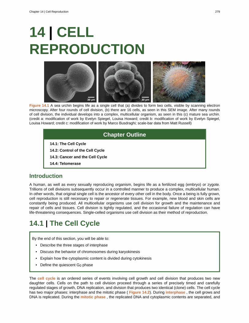
14 | CELLREPRODUCTION
Figure 14.1 A sea urchin begins life as a single cell that (a) divides to form two cells, visible by scanning electronmicroscopy. After four rounds of cell division, (b) there are 16 cells, as seen in this SEM image. After many roundsof cell division, the individual develops into a complex, multicellular organism, as seen in this (c) mature sea urchin.(credit a: modification of work by Evelyn Spiegel, Louisa Howard; credit b: modification of work by Evelyn Spiegel,Louisa Howard; credit c: modification of work by Marco Busdraghi; scale-bar data from Matt Russell)
Chapter Outline
14.1: The Cell Cycle
14.2: Control of the Cell Cycle
14.3: Cancer and the Cell Cycle
14.4: Telomerase
Introduction
A human, as well as every sexually reproducing organism, begins life as a fertilized egg (embryo) or zygote.Trillions of cell divisions subsequently occur in a controlled manner to produce a complex, multicellular human.In other words, that original single cell is the ancestor of every other cell in the body. Once a being is fully grown,cell reproduction is still necessary to repair or regenerate tissues. For example, new blood and skin cells areconstantly being produced. All multicellular organisms use cell division for growth and the maintenance andrepair of cells and tissues. Cell division is tightly regulated, and the occasional failure of regulation can havelife-threatening consequences. Single-celled organisms use cell division as their method of reproduction.
14.1 | The Cell Cycle
By the end of this section, you will be able to:
• Describe the three stages of interphase
• Discuss the behavior of chromosomes during karyokinesis
• Explain how the cytoplasmic content is divided during cytokinesis
• Define the quiescent G0 phase
The cell cycle is an ordered series of events involving cell growth and cell division that produces two newdaughter cells. Cells on the path to cell division proceed through a series of precisely timed and carefullyregulated stages of growth, DNA replication, and division that produces two identical (clone) cells. The cell cyclehas two major phases: interphase and the mitotic phase ( Figure 14.2). During interphase , the cell grows andDNA is replicated. During the mitotic phase , the replicated DNA and cytoplasmic contents are separated, and
Chapter 14 | Cell Reproduction 279
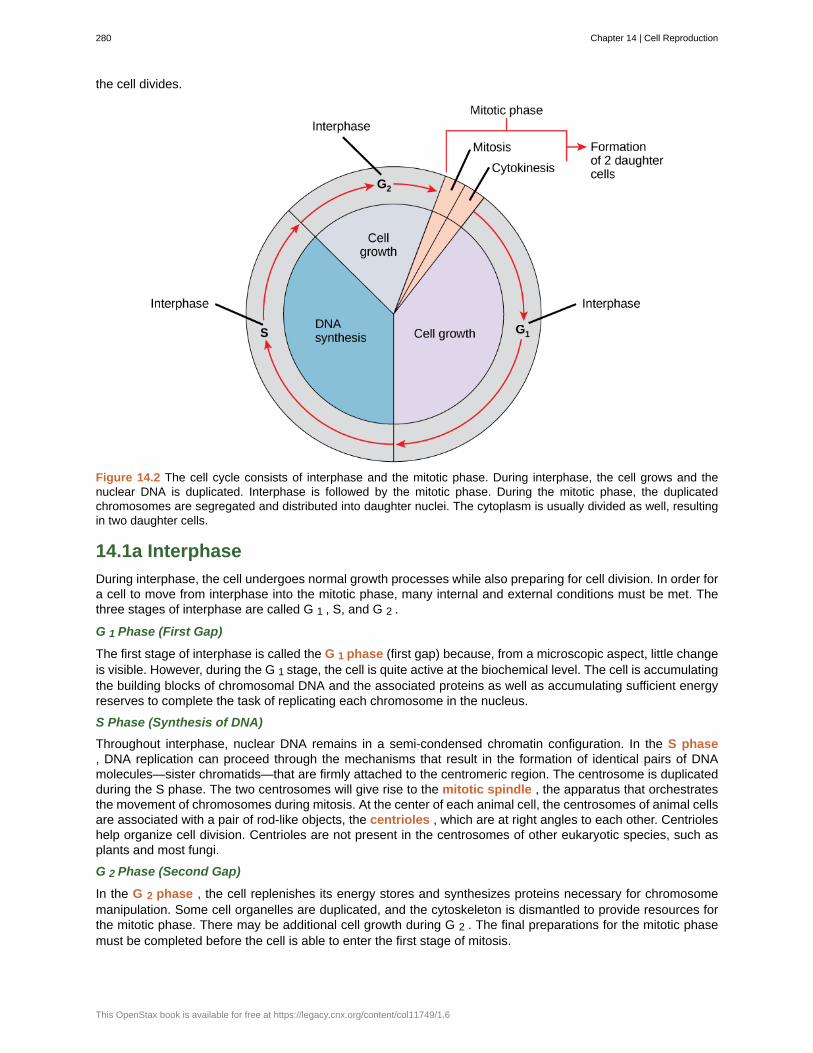
the cell divides.
Figure 14.2 The cell cycle consists of interphase and the mitotic phase. During interphase, the cell grows and thenuclear DNA is duplicated. Interphase is followed by the mitotic phase. During the mitotic phase, the duplicatedchromosomes are segregated and distributed into daughter nuclei. The cytoplasm is usually divided as well, resultingin two daughter cells.
14.1a Interphase
During interphase, the cell undergoes normal growth processes while also preparing for cell division. In order fora cell to move from interphase into the mitotic phase, many internal and external conditions must be met. Thethree stages of interphase are called G 1 , S, and G 2 .
G 1 Phase (First Gap)
The first stage of interphase is called the G 1 phase (first gap) because, from a microscopic aspect, little changeis visible. However, during the G 1 stage, the cell is quite active at the biochemical level. The cell is accumulatingthe building blocks of chromosomal DNA and the associated proteins as well as accumulating sufficient energyreserves to complete the task of replicating each chromosome in the nucleus.
S Phase (Synthesis of DNA)
Throughout interphase, nuclear DNA remains in a semi-condensed chromatin configuration. In the S phase, DNA replication can proceed through the mechanisms that result in the formation of identical pairs of DNAmolecules—sister chromatids—that are firmly attached to the centromeric region. The centrosome is duplicatedduring the S phase. The two centrosomes will give rise to the mitotic spindle , the apparatus that orchestratesthe movement of chromosomes during mitosis. At the center of each animal cell, the centrosomes of animal cellsare associated with a pair of rod-like objects, the centrioles , which are at right angles to each other. Centrioleshelp organize cell division. Centrioles are not present in the centrosomes of other eukaryotic species, such asplants and most fungi.
G 2 Phase (Second Gap)
In the G 2 phase , the cell replenishes its energy stores and synthesizes proteins necessary for chromosomemanipulation. Some cell organelles are duplicated, and the cytoskeleton is dismantled to provide resources forthe mitotic phase. There may be additional cell growth during G 2 . The final preparations for the mitotic phasemust be completed before the cell is able to enter the first stage of mitosis.
280 Chapter 14 | Cell Reproduction
This OpenStax book is available for free at https://legacy.cnx.org/content/col11749/1.6
14.1b The Mitotic Phase
The mitotic phase is a multistep process during which the duplicated chromosomes are aligned, separated, andmove into two new, identical daughter cells.
Revisit the stages of mitosis at this site (http://openstaxcollege.org/l/Cell_cycle_mito) .
Mitosis
Mitosis , is divided into a series of phases—prophase, metaphase, anaphase, and telophase—that result in thedivision of the cell nucleus ( Figure 14.3).
Chapter 14 | Cell Reproduction 281
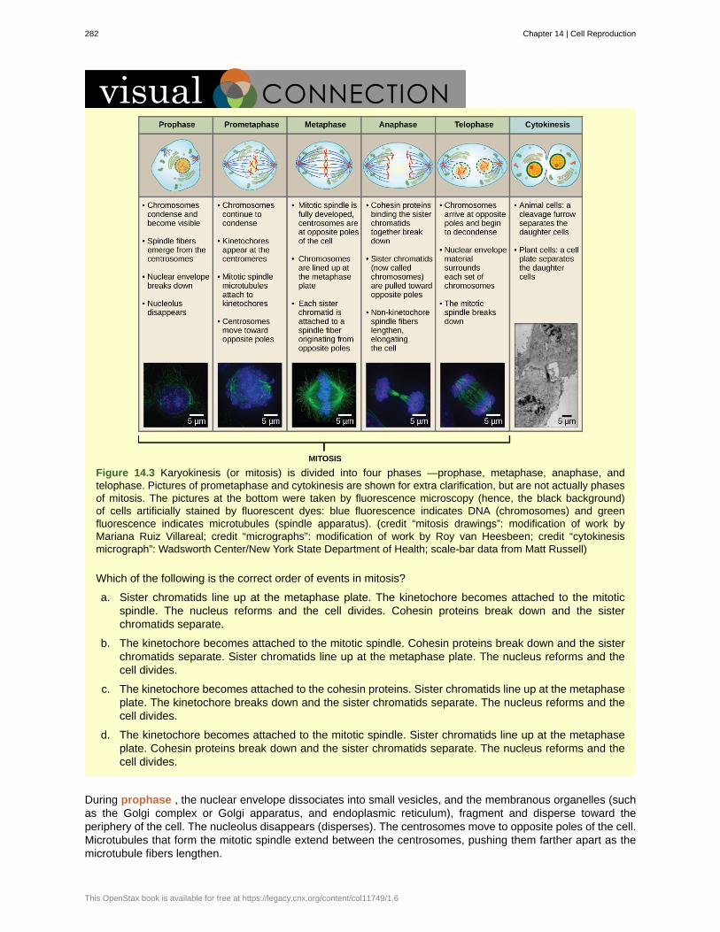
Figure 14.3 Karyokinesis (or mitosis) is divided into four phases —prophase, metaphase, anaphase, andtelophase. Pictures of prometaphase and cytokinesis are shown for extra clarification, but are not actually phasesof mitosis. The pictures at the bottom were taken by fluorescence microscopy (hence, the black background)of cells artificially stained by fluorescent dyes: blue fluorescence indicates DNA (chromosomes) and greenfluorescence indicates microtubules (spindle apparatus). (credit “mitosis drawings”: modification of work byMariana Ruiz Villareal; credit “micrographs”: modification of work by Roy van Heesbeen; credit “cytokinesismicrograph”: Wadsworth Center/New York State Department of Health; scale-bar data from Matt Russell)
Which of the following is the correct order of events in mitosis?
a. Sister chromatids line up at the metaphase plate. The kinetochore becomes attached to the mitoticspindle. The nucleus reforms and the cell divides. Cohesin proteins break down and the sisterchromatids separate.
b. The kinetochore becomes attached to the mitotic spindle. Cohesin proteins break down and the sisterchromatids separate. Sister chromatids line up at the metaphase plate. The nucleus reforms and thecell divides.
c. The kinetochore becomes attached to the cohesin proteins. Sister chromatids line up at the metaphaseplate. The kinetochore breaks down and the sister chromatids separate. The nucleus reforms and thecell divides.
d. The kinetochore becomes attached to the mitotic spindle. Sister chromatids line up at the metaphaseplate. Cohesin proteins break down and the sister chromatids separate. The nucleus reforms and thecell divides.
During prophase , the nuclear envelope dissociates into small vesicles, and the membranous organelles (suchas the Golgi complex or Golgi apparatus, and endoplasmic reticulum), fragment and disperse toward theperiphery of the cell. The nucleolus disappears (disperses). The centrosomes move to opposite poles of the cell.Microtubules that form the mitotic spindle extend between the centrosomes, pushing them farther apart as themicrotubule fibers lengthen.
282 Chapter 14 | Cell Reproduction
This OpenStax book is available for free at https://legacy.cnx.org/content/col11749/1.6
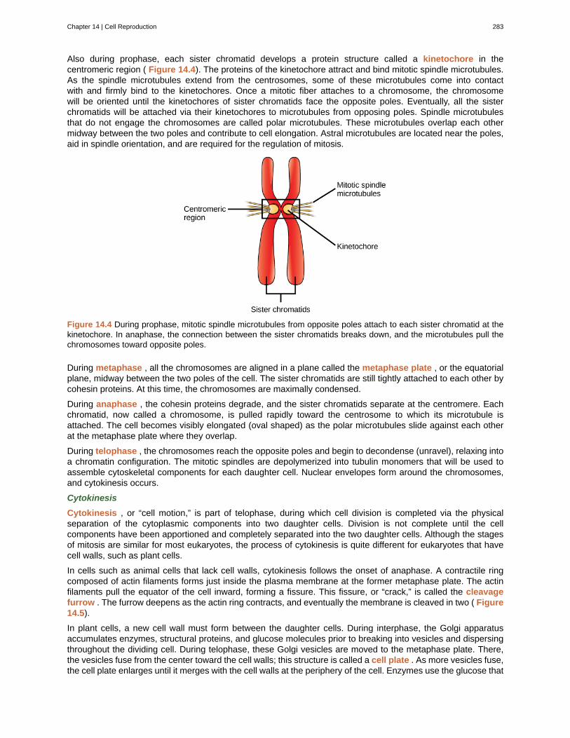
Also during prophase, each sister chromatid develops a protein structure called a kinetochore in thecentromeric region ( Figure 14.4). The proteins of the kinetochore attract and bind mitotic spindle microtubules.As the spindle microtubules extend from the centrosomes, some of these microtubules come into contactwith and firmly bind to the kinetochores. Once a mitotic fiber attaches to a chromosome, the chromosomewill be oriented until the kinetochores of sister chromatids face the opposite poles. Eventually, all the sisterchromatids will be attached via their kinetochores to microtubules from opposing poles. Spindle microtubulesthat do not engage the chromosomes are called polar microtubules. These microtubules overlap each othermidway between the two poles and contribute to cell elongation. Astral microtubules are located near the poles,aid in spindle orientation, and are required for the regulation of mitosis.
Figure 14.4 During prophase, mitotic spindle microtubules from opposite poles attach to each sister chromatid at thekinetochore. In anaphase, the connection between the sister chromatids breaks down, and the microtubules pull thechromosomes toward opposite poles.
During metaphase , all the chromosomes are aligned in a plane called the metaphase plate , or the equatorialplane, midway between the two poles of the cell. The sister chromatids are still tightly attached to each other bycohesin proteins. At this time, the chromosomes are maximally condensed.
During anaphase , the cohesin proteins degrade, and the sister chromatids separate at the centromere. Eachchromatid, now called a chromosome, is pulled rapidly toward the centrosome to which its microtubule isattached. The cell becomes visibly elongated (oval shaped) as the polar microtubules slide against each otherat the metaphase plate where they overlap.
During telophase , the chromosomes reach the opposite poles and begin to decondense (unravel), relaxing intoa chromatin configuration. The mitotic spindles are depolymerized into tubulin monomers that will be used toassemble cytoskeletal components for each daughter cell. Nuclear envelopes form around the chromosomes,and cytokinesis occurs.
Cytokinesis
Cytokinesis , or “cell motion,” is part of telophase, during which cell division is completed via the physicalseparation of the cytoplasmic components into two daughter cells. Division is not complete until the cellcomponents have been apportioned and completely separated into the two daughter cells. Although the stagesof mitosis are similar for most eukaryotes, the process of cytokinesis is quite different for eukaryotes that havecell walls, such as plant cells.
In cells such as animal cells that lack cell walls, cytokinesis follows the onset of anaphase. A contractile ringcomposed of actin filaments forms just inside the plasma membrane at the former metaphase plate. The actinfilaments pull the equator of the cell inward, forming a fissure. This fissure, or “crack,” is called the cleavagefurrow . The furrow deepens as the actin ring contracts, and eventually the membrane is cleaved in two ( Figure14.5).
In plant cells, a new cell wall must form between the daughter cells. During interphase, the Golgi apparatusaccumulates enzymes, structural proteins, and glucose molecules prior to breaking into vesicles and dispersingthroughout the dividing cell. During telophase, these Golgi vesicles are moved to the metaphase plate. There,the vesicles fuse from the center toward the cell walls; this structure is called a cell plate . As more vesicles fuse,the cell plate enlarges until it merges with the cell walls at the periphery of the cell. Enzymes use the glucose that
Chapter 14 | Cell Reproduction 283
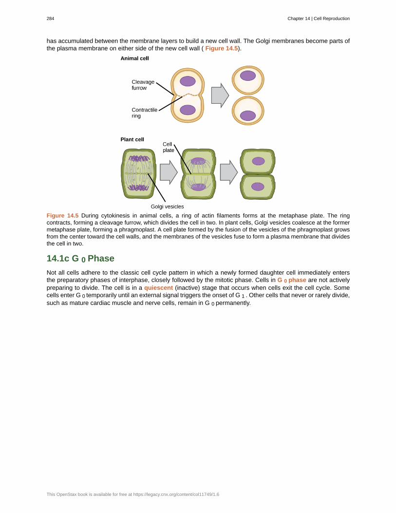
has accumulated between the membrane layers to build a new cell wall. The Golgi membranes become parts ofthe plasma membrane on either side of the new cell wall ( Figure 14.5).
Figure 14.5 During cytokinesis in animal cells, a ring of actin filaments forms at the metaphase plate. The ringcontracts, forming a cleavage furrow, which divides the cell in two. In plant cells, Golgi vesicles coalesce at the formermetaphase plate, forming a phragmoplast. A cell plate formed by the fusion of the vesicles of the phragmoplast growsfrom the center toward the cell walls, and the membranes of the vesicles fuse to form a plasma membrane that dividesthe cell in two.
14.1c G 0 Phase
Not all cells adhere to the classic cell cycle pattern in which a newly formed daughter cell immediately entersthe preparatory phases of interphase, closely followed by the mitotic phase. Cells in G 0 phase are not activelypreparing to divide. The cell is in a quiescent (inactive) stage that occurs when cells exit the cell cycle. Somecells enter G 0 temporarily until an external signal triggers the onset of G 1 . Other cells that never or rarely divide,such as mature cardiac muscle and nerve cells, remain in G 0 permanently.
284 Chapter 14 | Cell Reproduction
This OpenStax book is available for free at https://legacy.cnx.org/content/col11749/1.6
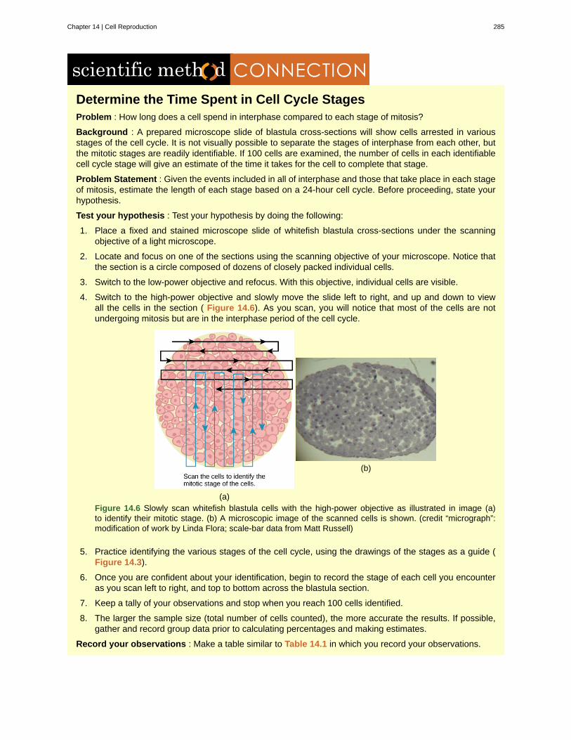
Determine the Time Spent in Cell Cycle StagesProblem : How long does a cell spend in interphase compared to each stage of mitosis?
Background : A prepared microscope slide of blastula cross-sections will show cells arrested in variousstages of the cell cycle. It is not visually possible to separate the stages of interphase from each other, butthe mitotic stages are readily identifiable. If 100 cells are examined, the number of cells in each identifiablecell cycle stage will give an estimate of the time it takes for the cell to complete that stage.
Problem Statement : Given the events included in all of interphase and those that take place in each stageof mitosis, estimate the length of each stage based on a 24-hour cell cycle. Before proceeding, state yourhypothesis.
Test your hypothesis : Test your hypothesis by doing the following:
1. Place a fixed and stained microscope slide of whitefish blastula cross-sections under the scanningobjective of a light microscope.
2. Locate and focus on one of the sections using the scanning objective of your microscope. Notice thatthe section is a circle composed of dozens of closely packed individual cells.
3. Switch to the low-power objective and refocus. With this objective, individual cells are visible.
4. Switch to the high-power objective and slowly move the slide left to right, and up and down to viewall the cells in the section ( Figure 14.6). As you scan, you will notice that most of the cells are notundergoing mitosis but are in the interphase period of the cell cycle.
(a)
(b)
Figure 14.6 Slowly scan whitefish blastula cells with the high-power objective as illustrated in image (a)to identify their mitotic stage. (b) A microscopic image of the scanned cells is shown. (credit “micrograph”:modification of work by Linda Flora; scale-bar data from Matt Russell)
5. Practice identifying the various stages of the cell cycle, using the drawings of the stages as a guide (Figure 14.3).
6. Once you are confident about your identification, begin to record the stage of each cell you encounteras you scan left to right, and top to bottom across the blastula section.
7. Keep a tally of your observations and stop when you reach 100 cells identified.
8. The larger the sample size (total number of cells counted), the more accurate the results. If possible,gather and record group data prior to calculating percentages and making estimates.
Record your observations : Make a table similar to Table 14.1 in which you record your observations.
Chapter 14 | Cell Reproduction 285
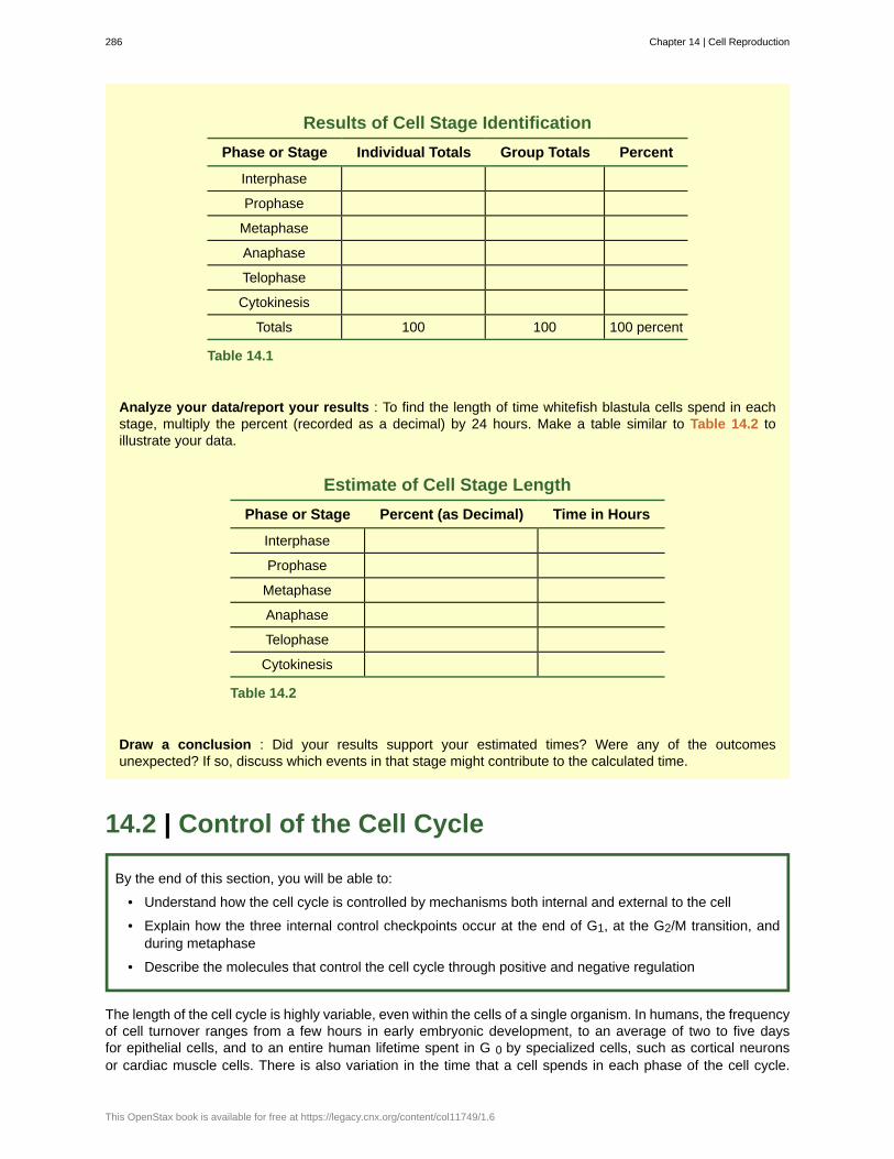
Results of Cell Stage Identification
Phase or Stage Individual Totals Group Totals Percent
Interphase
Prophase
Metaphase
Anaphase
Telophase
Cytokinesis
Totals 100 100 100 percent
Table 14.1
Analyze your data/report your results : To find the length of time whitefish blastula cells spend in eachstage, multiply the percent (recorded as a decimal) by 24 hours. Make a table similar to Table 14.2 toillustrate your data.
Estimate of Cell Stage Length
Phase or Stage Percent (as Decimal) Time in Hours
Interphase
Prophase
Metaphase
Anaphase
Telophase
Cytokinesis
Table 14.2
Draw a conclusion : Did your results support your estimated times? Were any of the outcomesunexpected? If so, discuss which events in that stage might contribute to the calculated time.
14.2 | Control of the Cell Cycle
By the end of this section, you will be able to:
• Understand how the cell cycle is controlled by mechanisms both internal and external to the cell
• Explain how the three internal control checkpoints occur at the end of G1, at the G2/M transition, andduring metaphase
• Describe the molecules that control the cell cycle through positive and negative regulation
The length of the cell cycle is highly variable, even within the cells of a single organism. In humans, the frequencyof cell turnover ranges from a few hours in early embryonic development, to an average of two to five daysfor epithelial cells, and to an entire human lifetime spent in G 0 by specialized cells, such as cortical neuronsor cardiac muscle cells. There is also variation in the time that a cell spends in each phase of the cell cycle.
286 Chapter 14 | Cell Reproduction
This OpenStax book is available for free at https://legacy.cnx.org/content/col11749/1.6
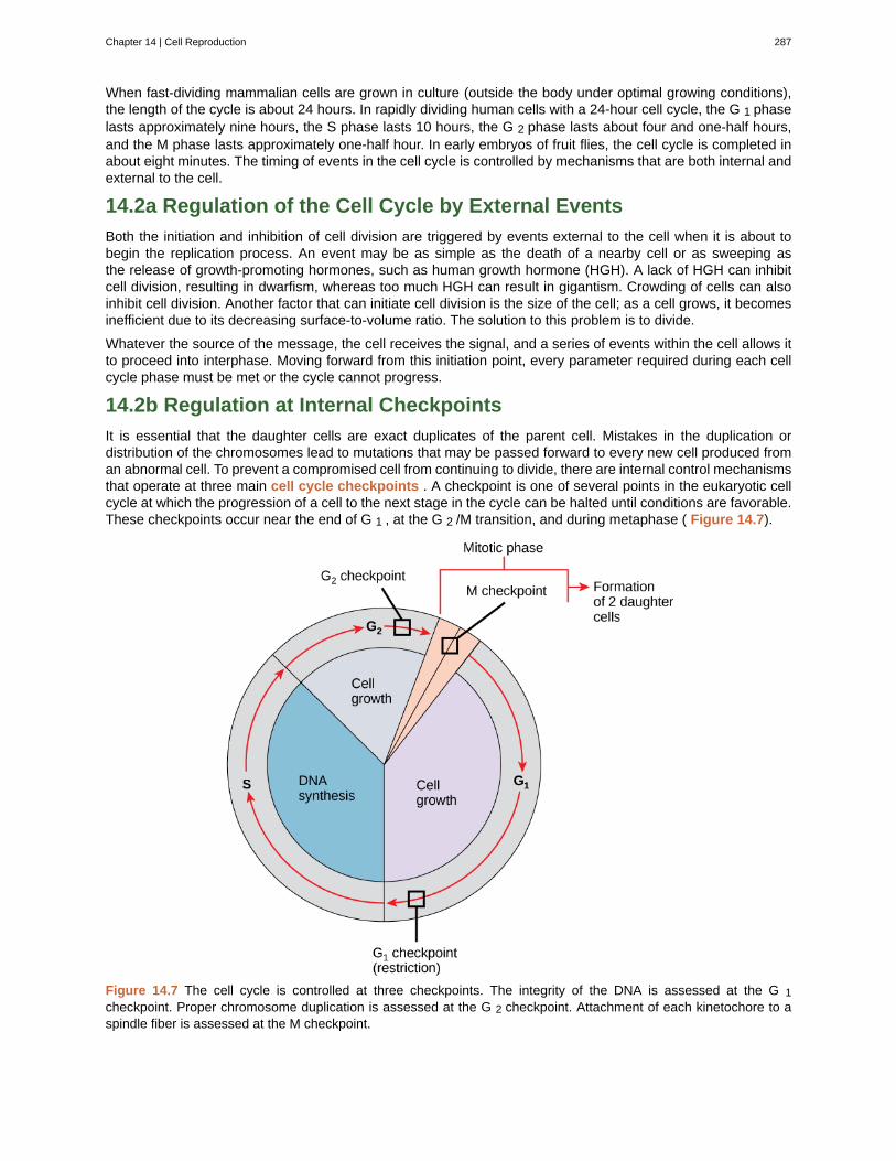
When fast-dividing mammalian cells are grown in culture (outside the body under optimal growing conditions),the length of the cycle is about 24 hours. In rapidly dividing human cells with a 24-hour cell cycle, the G 1 phaselasts approximately nine hours, the S phase lasts 10 hours, the G 2 phase lasts about four and one-half hours,and the M phase lasts approximately one-half hour. In early embryos of fruit flies, the cell cycle is completed inabout eight minutes. The timing of events in the cell cycle is controlled by mechanisms that are both internal andexternal to the cell.
14.2a Regulation of the Cell Cycle by External Events
Both the initiation and inhibition of cell division are triggered by events external to the cell when it is about tobegin the replication process. An event may be as simple as the death of a nearby cell or as sweeping asthe release of growth-promoting hormones, such as human growth hormone (HGH). A lack of HGH can inhibitcell division, resulting in dwarfism, whereas too much HGH can result in gigantism. Crowding of cells can alsoinhibit cell division. Another factor that can initiate cell division is the size of the cell; as a cell grows, it becomesinefficient due to its decreasing surface-to-volume ratio. The solution to this problem is to divide.
Whatever the source of the message, the cell receives the signal, and a series of events within the cell allows itto proceed into interphase. Moving forward from this initiation point, every parameter required during each cellcycle phase must be met or the cycle cannot progress.
14.2b Regulation at Internal Checkpoints
It is essential that the daughter cells are exact duplicates of the parent cell. Mistakes in the duplication ordistribution of the chromosomes lead to mutations that may be passed forward to every new cell produced froman abnormal cell. To prevent a compromised cell from continuing to divide, there are internal control mechanismsthat operate at three main cell cycle checkpoints . A checkpoint is one of several points in the eukaryotic cellcycle at which the progression of a cell to the next stage in the cycle can be halted until conditions are favorable.These checkpoints occur near the end of G 1 , at the G 2 /M transition, and during metaphase ( Figure 14.7).
Figure 14.7 The cell cycle is controlled at three checkpoints. The integrity of the DNA is assessed at the G 1checkpoint. Proper chromosome duplication is assessed at the G 2 checkpoint. Attachment of each kinetochore to aspindle fiber is assessed at the M checkpoint.
Chapter 14 | Cell Reproduction 287
The G 1 Checkpoint
The G 1 checkpoint determines whether all conditions are favorable for cell division to proceed. The G 1checkpoint is a point at which the cell irreversibly commits to the cell division process. External influences, suchas growth factors, play a large role in carrying the cell past the G 1 checkpoint. In addition to adequate reservesand cell size, there is a check for genomic DNA damage at the G 1 checkpoint. A cell that does not meet all therequirements will not be allowed to progress into the S phase. The cell can halt the cycle and attempt to remedythe problematic condition, or the cell can advance into G 0 and await further signals when conditions improve.
The G 2 Checkpoint
The G 2 checkpoint bars entry into the mitotic phase if certain conditions are not met. As at the G 1 checkpoint,cell size and protein reserves are assessed. However, the most important role of the G 2 checkpoint is to ensurethat all of the chromosomes have been replicated and that the replicated DNA is not damaged. If the checkpointmechanisms detect problems with the DNA, the cell cycle is halted, and the cell attempts to either complete DNAreplication or repair the damaged DNA.
The M Checkpoint
The M checkpoint occurs near the end of the metaphase stage of karyokinesis. The M checkpoint is also knownas the spindle checkpoint, because it determines whether all the sister chromatids are correctly attached to thespindle microtubules. Because the separation of the sister chromatids during anaphase is an irreversible step,the cycle will not proceed until the kinetochores of each pair of sister chromatids are firmly anchored to at leasttwo spindle fibers arising from opposite poles of the cell.
Watch what occurs at the G 1 , G 2 , and M checkpoints by visiting this website (http://openstaxcollege.org/l/cell_checkpnts) to see an animation of the cell cycle.
14.3 | Cancer and the Cell Cycle
By the end of this section, you will be able to:
• Describe how cancer is caused by uncontrolled cell growth
• Understand how proto-oncogenes are normal cell genes that, when mutated, become oncogenes
• Describe how tumor suppressors function
• Explain how mutant tumor suppressors cause cancer
Cancer comprises many different diseases caused by a common mechanism: uncontrolled cell growth. Despitethe redundancy and overlapping levels of cell cycle control, errors do occur. One of the critical processesmonitored by the cell cycle checkpoint surveillance mechanism is the proper replication of DNA during the Sphase. Even when all of the cell cycle controls are fully functional, a small percentage of replication errors(mutations) will be passed on to the daughter cells. If changes to the DNA nucleotide sequence occur within acoding portion of a gene and are not corrected, a gene mutation results. All cancers start when a gene mutationgives rise to a faulty protein that plays a key role in cell reproduction. The change in the cell that results fromthe malformed protein may be minor. Even minor mistakes, however, may allow subsequent mistakes to occurmore readily. Over and over, small uncorrected errors are passed from the parent cell to the daughter cells andamplified as each generation produces more non-functional proteins from uncorrected DNA damage. Eventually,the pace of the cell cycle speeds up as the effectiveness of the control and repair mechanisms decreases.Uncontrolled growth of the mutated cells outpaces the growth of normal cells in the area, and a tumor (“-oma”)can result.
288 Chapter 14 | Cell Reproduction
This OpenStax book is available for free at https://legacy.cnx.org/content/col11749/1.6
14.3a Proto-oncogenes
The genes that code for the positive cell cycle regulators are called proto-oncogenes . Proto-oncogenes arenormal genes that, when mutated in certain ways, become oncogenes , genes that cause a cell to becomecancerous. Consider what might happen to the cell cycle in a cell with a recently acquired oncogene. In mostinstances, the alteration of the DNA sequence will result in a less functional (or non-functional) protein. The resultis detrimental to the cell and will likely prevent the cell from completing the cell cycle; however, the organismis not harmed because the mutation will not be carried forward. If a cell cannot reproduce, the mutation isnot propagated and the damage is minimal. Occasionally, however, a gene mutation causes a change thatincreases the activity of a positive regulator, thereby promoting cell division. If the resulting daughter cells aretoo damaged to undergo further cell divisions, the mutation would not be propagated and no harm would cometo the organism. However, if the atypical daughter cells are able to undergo further cell divisions, subsequentgenerations of cells will probably accumulate even more mutations, some possibly in additional genes thatregulate the cell cycle.
Many genes can be considered proto-oncogenes. In addition to the cell cycle regulatory proteins, any proteinthat influences the cycle can be altered in such a way as to override cell cycle checkpoints. An oncogene is anygene that, when altered, leads to an increase in the rate of cell cycle progression.
14.3b Tumor Suppressor Genes
Like proto-oncogenes, many of the negative cell cycle regulatory proteins were discovered in cells that hadbecome cancerous. Tumor suppressor genes are segments of DNA that code for negative regulator proteins,the type of regulators that, when activated, can prevent the cell from undergoing uncontrolled division. Thecollective function of the best-understood tumor suppressor gene proteins, Rb, p53, and p21, is to put up aroadblock to cell cycle progression until certain events are completed. A cell that carries a mutated form of anegative regulator might not be able to halt the cell cycle if there is a problem. Tumor suppressors are similar tobrakes in a vehicle: Malfunctioning brakes can contribute to a car crash.
Mutated p53 genes have been identified in more than one-half of all human tumor cells. This discovery is notsurprising in light of the multiple roles that the p53 protein plays at the G 1 checkpoint. A cell with a faulty p53 mayfail to detect errors present in the genomic DNA ( Figure 14.8). Even if a partially functional p53 does identifythe mutations, it may no longer be able to signal the necessary DNA repair enzymes. Either way, damaged DNAwill remain uncorrected. At this point, a functional p53 will deem the cell unsalvageable and trigger programmedcell death (apoptosis). The damaged version of p53 found in cancer cells, however, cannot trigger apoptosis.
Chapter 14 | Cell Reproduction 289
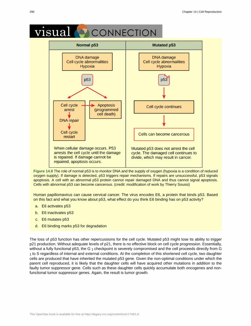
Figure 14.8 The role of normal p53 is to monitor DNA and the supply of oxygen (hypoxia is a condition of reducedoxygen supply). If damage is detected, p53 triggers repair mechanisms. If repairs are unsuccessful, p53 signalsapoptosis. A cell with an abnormal p53 protein cannot repair damaged DNA and thus cannot signal apoptosis.Cells with abnormal p53 can become cancerous. (credit: modification of work by Thierry Soussi)
Human papillomavirus can cause cervical cancer. The virus encodes E6, a protein that binds p53. Basedon this fact and what you know about p53, what effect do you think E6 binding has on p53 activity?
a. E6 activates p53
b. E6 inactivates p53
c. E6 mutates p53
d. E6 binding marks p53 for degradation
The loss of p53 function has other repercussions for the cell cycle. Mutated p53 might lose its ability to triggerp21 production. Without adequate levels of p21, there is no effective block on cell cycle progression. Essentially,without a fully functional p53, the G 1 checkpoint is severely compromised and the cell proceeds directly from G1 to S regardless of internal and external conditions. At the completion of this shortened cell cycle, two daughtercells are produced that have inherited the mutated p53 gene. Given the non-optimal conditions under which theparent cell reproduced, it is likely that the daughter cells will have acquired other mutations in addition to thefaulty tumor suppressor gene. Cells such as these daughter cells quickly accumulate both oncogenes and non-functional tumor suppressor genes. Again, the result is tumor growth.
290 Chapter 14 | Cell Reproduction
This OpenStax book is available for free at https://legacy.cnx.org/content/col11749/1.6
Go to this website (http://openstaxcollege.org/l/cancer) to watch an animation of how cancer results fromerrors in the cell cycle.
14.4 | Telomerase
By the end of this section, you will be able to:
• Discuss the similarities and differences between DNA replication in eukaryotes and prokaryotes
• State the role of telomerase in DNA replication
Eukaryotic genomes are much more complex and larger in size than prokaryotic genomes. The human genomehas three billion base pairs per haploid set of chromosomes, and 6 billion base pairs are replicated during the Sphase of the cell cycle.
14.4a Telomere replication
Unlike prokaryotic chromosomes, eukaryotic chromosomes are linear. As you’ve learned, the enzyme DNA polcan add nucleotides only in the 5' to 3' direction. In the leading strand, synthesis continues until the end of thechromosome is reached. On the lagging strand, DNA is synthesized in short stretches, each of which is initiatedby a separate primer. When the replication fork reaches the end of the linear chromosome, there is no place fora primer to be made for the DNA fragment to be copied at the end of the chromosome. These ends thus remainunpaired, and over time these ends may get progressively shorter as cells continue to divide.
The ends of the linear chromosomes are known as telomeres , which have repetitive sequences that codefor no particular gene. In a way, these telomeres protect the genes from getting deleted as cells continue todivide. In humans, a six base pair sequence, TTAGGG, is repeated 100 to 1000 times. The discovery of theenzyme telomerase ( Figure 14.10) helped in the understanding of how chromosome ends are maintained.The telomerase enzyme contains a catalytic part and a built-in RNA template. It attaches to the end of thechromosome, and complementary bases to the RNA template are added on the 3' end of the DNA strand. Oncethe 3' end of the lagging strand template is sufficiently elongated, DNA polymerase can add the nucleotidescomplementary to the ends of the chromosomes. Thus, the ends of the chromosomes are replicated.
Chapter 14 | Cell Reproduction 291
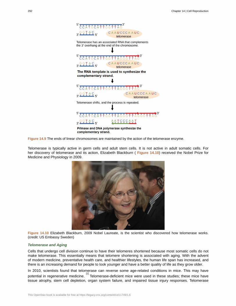
Figure 14.9 The ends of linear chromosomes are maintained by the action of the telomerase enzyme.
Telomerase is typically active in germ cells and adult stem cells. It is not active in adult somatic cells. Forher discovery of telomerase and its action, Elizabeth Blackburn ( Figure 14.10) received the Nobel Prize forMedicine and Physiology in 2009.
Figure 14.10 Elizabeth Blackburn, 2009 Nobel Laureate, is the scientist who discovered how telomerase works.(credit: US Embassy Sweden)
Telomerase and Aging
Cells that undergo cell division continue to have their telomeres shortened because most somatic cells do notmake telomerase. This essentially means that telomere shortening is associated with aging. With the adventof modern medicine, preventative health care, and healthier lifestyles, the human life span has increased, andthere is an increasing demand for people to look younger and have a better quality of life as they grow older.
In 2010, scientists found that telomerase can reverse some age-related conditions in mice. This may have
potential in regenerative medicine.[1]
Telomerase-deficient mice were used in these studies; these mice havetissue atrophy, stem cell depletion, organ system failure, and impaired tissue injury responses. Telomerase
292 Chapter 14 | Cell Reproduction
This OpenStax book is available for free at https://legacy.cnx.org/content/col11749/1.6
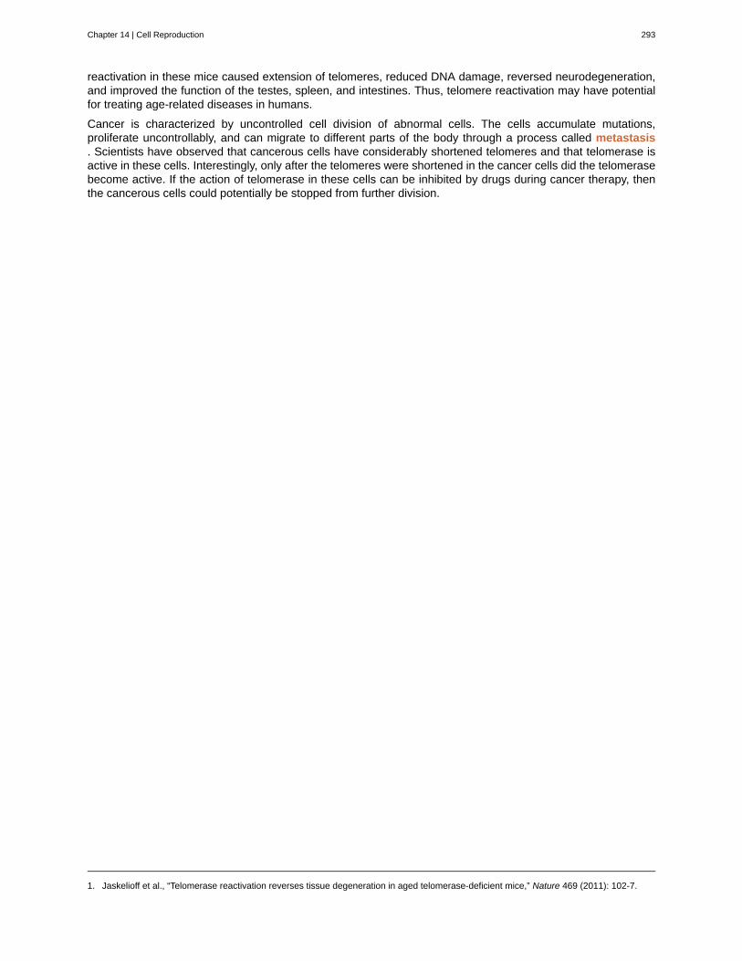
reactivation in these mice caused extension of telomeres, reduced DNA damage, reversed neurodegeneration,and improved the function of the testes, spleen, and intestines. Thus, telomere reactivation may have potentialfor treating age-related diseases in humans.
Cancer is characterized by uncontrolled cell division of abnormal cells. The cells accumulate mutations,proliferate uncontrollably, and can migrate to different parts of the body through a process called metastasis. Scientists have observed that cancerous cells have considerably shortened telomeres and that telomerase isactive in these cells. Interestingly, only after the telomeres were shortened in the cancer cells did the telomerasebecome active. If the action of telomerase in these cells can be inhibited by drugs during cancer therapy, thenthe cancerous cells could potentially be stopped from further division.
1. Jaskelioff et al., “Telomerase reactivation reverses tissue degeneration in aged telomerase-deficient mice,” Nature 469 (2011): 102-7.
Chapter 14 | Cell Reproduction 293
anaphase
cell cycle
cell cycle checkpoints
cell plate
centrioles
cleavage furrow
cytokinesis
G 0 phase
G 1 phase
G 2 phase
interphase
kinetochore
metaphase
metaphase plate
metastasis
mitosis
mitotic phase
mitotic spindle
oncogenes
prophase
proto-oncogenes
quiescent
S phase
telomerase
KEY TERMS
stage of mitosis during which sister chromatids are separated from each other
ordered series of events involving cell growth and cell division that produces two new daughter cells
mechanism that monitors the preparedness of a eukaryotic cell to advance through thevarious cell cycle stages
structure formed during plant cell cytokinesis by Golgi vesicles, forming a temporary structure(phragmoplast) and fusing at the metaphase plate; ultimately leads to the formation of cell walls thatseparate the two daughter cells
rod-like structure constructed of microtubules at the center of each animal cell centrosome
constriction formed by an actin ring during cytokinesis in animal cells that leads to cytoplasmicdivision
division of the cytoplasm following mitosis that forms two daughter cells.
distinct from the G 1 phase of interphase; a cell in G 0 is not preparing to divide
(also, first gap) first phase of interphase centered on cell growth during mitosis
(also, second gap) third phase of interphase during which the cell undergoes final preparations formitosis
period of the cell cycle leading up to mitosis; includes G 1 , S, and G 2 phases (the interim periodbetween two consecutive cell divisions
protein structure associated with the centromere of each sister chromatid that attracts and bindsspindle microtubules during prometaphase
stage of mitosis during which chromosomes are aligned at the metaphase plate
equatorial plane midway between the two poles of a cell where the chromosomes alignduring metaphase
the spread of cancer cells from an initial tumor to other parts of the body
(also, karyokinesis) period of the cell cycle during which the duplicated chromosomes are separatedinto identical nuclei; includes prophase, prometaphase, metaphase, anaphase, and telophase
period of the cell cycle during which duplicated chromosomes are distributed into two nuclei andcytoplasmic contents are divided; includes karyokinesis (mitosis) and cytokinesis
apparatus composed of microtubules that orchestrates the movement of chromosomes duringmitosis
mutated version of a normal gene involved in the positive regulation of the cell cycle
stage of mitosis during which chromosomes condense and the mitotic spindle begins to form
normal gene that when mutated becomes an oncogene
refers to a cell that is performing normal cell functions and has not initiated preparations for celldivision
second, or synthesis, stage of interphase during which DNA replication occurs
enzyme that contains a catalytic part and an inbuilt RNA template; it functions to maintaintelomeres at chromosome ends
294 Chapter 14 | Cell Reproduction
This OpenStax book is available for free at https://legacy.cnx.org/content/col11749/1.6
telomeres
telophase
Tumor suppressor genes
DNA at the end of linear chromosomes
stage of mitosis during which chromosomes arrive at opposite poles, decondense, and aresurrounded by a new nuclear envelope
segment of DNA that codes for regulator proteins that prevent the cell fromundergoing uncontrolled division
CHAPTER SUMMARY
14.1 The Cell Cycle
The cell cycle is an orderly sequence of events. Cells on the path to cell division proceed through a series ofprecisely timed and carefully regulated stages. In eukaryotes, the cell cycle consists of a long preparatoryperiod, called interphase. Interphase is divided into G 1 , S, and G 2 phases. The mitotic phase consists of fourphases: prophase, metaphase, anaphase, and telophase.
14.2 Control of the Cell Cycle
Each step of the cell cycle is monitored by internal controls called checkpoints. There are three majorcheckpoints in the cell cycle: one near the end of G 1 , a second at the G 2 /M transition, and the third duringmetaphase. Positive regulator molecules allow the cell cycle to advance to the next stage. Negative regulatormolecules monitor cellular conditions and can halt the cycle until specific requirements are met.
14.3 Cancer and the Cell Cycle
Cancer is the result of unchecked cell division caused by a breakdown of the mechanisms that regulate the cellcycle. The loss of control begins with a change in the DNA sequence of a gene that codes for one of theregulatory molecules. Faulty instructions lead to a protein that does not function as it should. Any disruption ofthe monitoring system can allow other mistakes to be passed on to the daughter cells. Each successive celldivision will give rise to daughter cells with even more accumulated damage. Eventually, all checkpointsbecome nonfunctional, and rapidly reproducing cells crowd out normal cells, resulting in a tumor or leukemia(blood cancer).
14.4 Telomerase
When cells divide, they need to first replicate their DNA. In earlier sections, we discussed how this happens inS phase of interphase. When the chromosomes replicate, one strand of each chromatid is a little shorter than itused to be. This happens because of the directionality of the DNA polymerase. Certain cells (stem cells, somecancer cells) produce an enzyme (telomerase) that can lengthen those shorter strands. However, most cells donot express telomerase, and therefore the DNA gets shorter and shorter with every S phase. Eventually, thestrands get short enough to trigger the cell to undergo apoptosis.
Chapter 14 | Cell Reproduction 295

296 Chapter 14 | Cell Reproduction
This OpenStax book is available for free at https://legacy.cnx.org/content/col11749/1.6
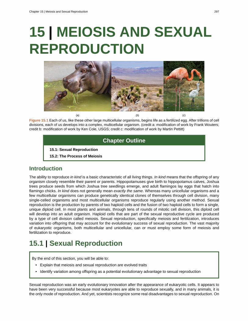
15 | MEIOSIS AND SEXUALREPRODUCTION
Figure 15.1 Each of us, like these other large multicellular organisms, begins life as a fertilized egg. After trillions of celldivisions, each of us develops into a complex, multicellular organism. (credit a: modification of work by Frank Wouters;credit b: modification of work by Ken Cole, USGS; credit c: modification of work by Martin Pettitt)
Chapter Outline
15.1: Sexual Reproduction
15.2: The Process of Meiosis
Introduction
The ability to reproduce in kind is a basic characteristic of all living things. In kind means that the offspring of anyorganism closely resemble their parent or parents. Hippopotamuses give birth to hippopotamus calves, Joshuatrees produce seeds from which Joshua tree seedlings emerge, and adult flamingos lay eggs that hatch intoflamingo chicks. In kind does not generally mean exactly the same. Whereas many unicellular organisms and afew multicellular organisms can produce genetically identical clones of themselves through cell division, manysingle-celled organisms and most multicellular organisms reproduce regularly using another method. Sexualreproduction is the production by parents of two haploid cells and the fusion of two haploid cells to form a single,unique diploid cell. In most plants and animals, through tens of rounds of mitotic cell division, this diploid cellwill develop into an adult organism. Haploid cells that are part of the sexual reproductive cycle are producedby a type of cell division called meiosis. Sexual reproduction, specifically meiosis and fertilization, introducesvariation into offspring that may account for the evolutionary success of sexual reproduction. The vast majorityof eukaryotic organisms, both multicellular and unicellular, can or must employ some form of meiosis andfertilization to reproduce.
15.1 | Sexual Reproduction
By the end of this section, you will be able to:
• Explain that meiosis and sexual reproduction are evolved traits
• Identify variation among offspring as a potential evolutionary advantage to sexual reproduction
Sexual reproduction was an early evolutionary innovation after the appearance of eukaryotic cells. It appears tohave been very successful because most eukaryotes are able to reproduce sexually, and in many animals, it isthe only mode of reproduction. And yet, scientists recognize some real disadvantages to sexual reproduction. On
Chapter 15 | Meiosis and Sexual Reproduction 297
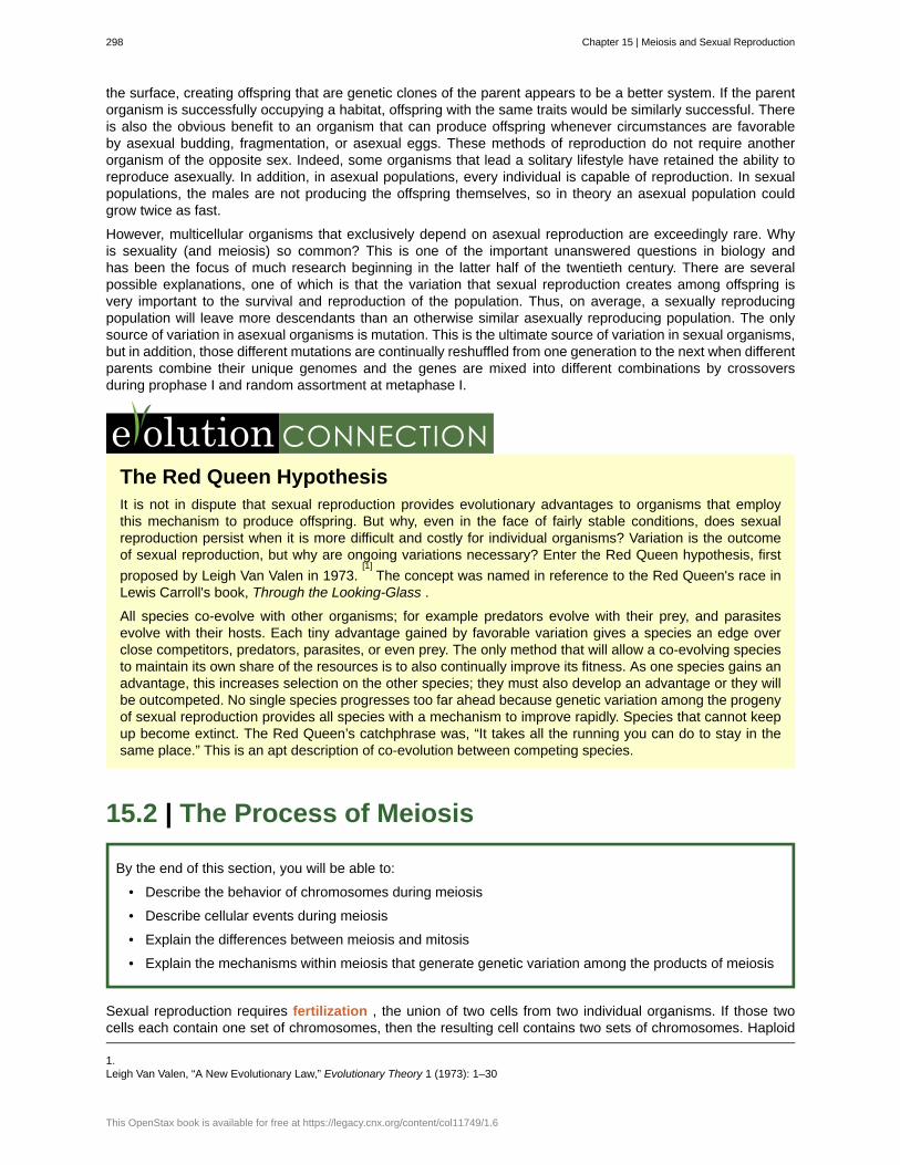
the surface, creating offspring that are genetic clones of the parent appears to be a better system. If the parentorganism is successfully occupying a habitat, offspring with the same traits would be similarly successful. Thereis also the obvious benefit to an organism that can produce offspring whenever circumstances are favorableby asexual budding, fragmentation, or asexual eggs. These methods of reproduction do not require anotherorganism of the opposite sex. Indeed, some organisms that lead a solitary lifestyle have retained the ability toreproduce asexually. In addition, in asexual populations, every individual is capable of reproduction. In sexualpopulations, the males are not producing the offspring themselves, so in theory an asexual population couldgrow twice as fast.
However, multicellular organisms that exclusively depend on asexual reproduction are exceedingly rare. Whyis sexuality (and meiosis) so common? This is one of the important unanswered questions in biology andhas been the focus of much research beginning in the latter half of the twentieth century. There are severalpossible explanations, one of which is that the variation that sexual reproduction creates among offspring isvery important to the survival and reproduction of the population. Thus, on average, a sexually reproducingpopulation will leave more descendants than an otherwise similar asexually reproducing population. The onlysource of variation in asexual organisms is mutation. This is the ultimate source of variation in sexual organisms,but in addition, those different mutations are continually reshuffled from one generation to the next when differentparents combine their unique genomes and the genes are mixed into different combinations by crossoversduring prophase I and random assortment at metaphase I.
The Red Queen HypothesisIt is not in dispute that sexual reproduction provides evolutionary advantages to organisms that employthis mechanism to produce offspring. But why, even in the face of fairly stable conditions, does sexualreproduction persist when it is more difficult and costly for individual organisms? Variation is the outcomeof sexual reproduction, but why are ongoing variations necessary? Enter the Red Queen hypothesis, first
proposed by Leigh Van Valen in 1973.[1]
The concept was named in reference to the Red Queen's race inLewis Carroll's book, Through the Looking-Glass .
All species co-evolve with other organisms; for example predators evolve with their prey, and parasitesevolve with their hosts. Each tiny advantage gained by favorable variation gives a species an edge overclose competitors, predators, parasites, or even prey. The only method that will allow a co-evolving speciesto maintain its own share of the resources is to also continually improve its fitness. As one species gains anadvantage, this increases selection on the other species; they must also develop an advantage or they willbe outcompeted. No single species progresses too far ahead because genetic variation among the progenyof sexual reproduction provides all species with a mechanism to improve rapidly. Species that cannot keepup become extinct. The Red Queen’s catchphrase was, “It takes all the running you can do to stay in thesame place.” This is an apt description of co-evolution between competing species.
15.2 | The Process of Meiosis
By the end of this section, you will be able to:
• Describe the behavior of chromosomes during meiosis
• Describe cellular events during meiosis
• Explain the differences between meiosis and mitosis
• Explain the mechanisms within meiosis that generate genetic variation among the products of meiosis
Sexual reproduction requires fertilization , the union of two cells from two individual organisms. If those twocells each contain one set of chromosomes, then the resulting cell contains two sets of chromosomes. Haploid
1.Leigh Van Valen, “A New Evolutionary Law,” Evolutionary Theory 1 (1973): 1–30
298 Chapter 15 | Meiosis and Sexual Reproduction
This OpenStax book is available for free at https://legacy.cnx.org/content/col11749/1.6
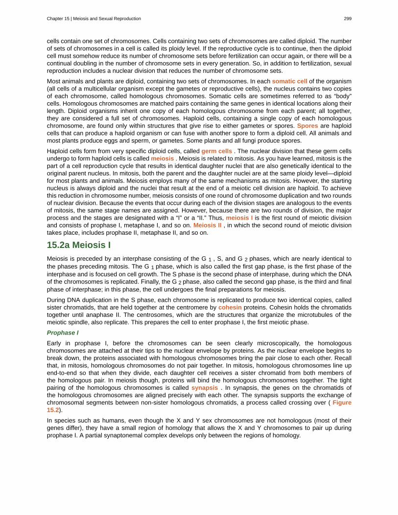
cells contain one set of chromosomes. Cells containing two sets of chromosomes are called diploid. The numberof sets of chromosomes in a cell is called its ploidy level. If the reproductive cycle is to continue, then the diploidcell must somehow reduce its number of chromosome sets before fertilization can occur again, or there will be acontinual doubling in the number of chromosome sets in every generation. So, in addition to fertilization, sexualreproduction includes a nuclear division that reduces the number of chromosome sets.
Most animals and plants are diploid, containing two sets of chromosomes. In each somatic cell of the organism(all cells of a multicellular organism except the gametes or reproductive cells), the nucleus contains two copiesof each chromosome, called homologous chromosomes. Somatic cells are sometimes referred to as “body”cells. Homologous chromosomes are matched pairs containing the same genes in identical locations along theirlength. Diploid organisms inherit one copy of each homologous chromosome from each parent; all together,they are considered a full set of chromosomes. Haploid cells, containing a single copy of each homologouschromosome, are found only within structures that give rise to either gametes or spores. Spores are haploidcells that can produce a haploid organism or can fuse with another spore to form a diploid cell. All animals andmost plants produce eggs and sperm, or gametes. Some plants and all fungi produce spores.
Haploid cells form from very specific diploid cells, called germ cells . The nuclear division that these germ cellsundergo to form haploid cells is called meiosis . Meiosis is related to mitosis. As you have learned, mitosis is thepart of a cell reproduction cycle that results in identical daughter nuclei that are also genetically identical to theoriginal parent nucleus. In mitosis, both the parent and the daughter nuclei are at the same ploidy level—diploidfor most plants and animals. Meiosis employs many of the same mechanisms as mitosis. However, the startingnucleus is always diploid and the nuclei that result at the end of a meiotic cell division are haploid. To achievethis reduction in chromosome number, meiosis consists of one round of chromosome duplication and two roundsof nuclear division. Because the events that occur during each of the division stages are analogous to the eventsof mitosis, the same stage names are assigned. However, because there are two rounds of division, the majorprocess and the stages are designated with a “I” or a “II.” Thus, meiosis I is the first round of meiotic divisionand consists of prophase I, metaphase I, and so on. Meiosis II , in which the second round of meiotic divisiontakes place, includes prophase II, metaphase II, and so on.
15.2a Meiosis I
Meiosis is preceded by an interphase consisting of the G 1 , S, and G 2 phases, which are nearly identical tothe phases preceding mitosis. The G 1 phase, which is also called the first gap phase, is the first phase of theinterphase and is focused on cell growth. The S phase is the second phase of interphase, during which the DNAof the chromosomes is replicated. Finally, the G 2 phase, also called the second gap phase, is the third and finalphase of interphase; in this phase, the cell undergoes the final preparations for meiosis.
During DNA duplication in the S phase, each chromosome is replicated to produce two identical copies, calledsister chromatids, that are held together at the centromere by cohesin proteins. Cohesin holds the chromatidstogether until anaphase II. The centrosomes, which are the structures that organize the microtubules of themeiotic spindle, also replicate. This prepares the cell to enter prophase I, the first meiotic phase.
Prophase I
Early in prophase I, before the chromosomes can be seen clearly microscopically, the homologouschromosomes are attached at their tips to the nuclear envelope by proteins. As the nuclear envelope begins tobreak down, the proteins associated with homologous chromosomes bring the pair close to each other. Recallthat, in mitosis, homologous chromosomes do not pair together. In mitosis, homologous chromosomes line upend-to-end so that when they divide, each daughter cell receives a sister chromatid from both members ofthe homologous pair. In meiosis though, proteins will bind the homologous chromosomes together. The tightpairing of the homologous chromosomes is called synapsis . In synapsis, the genes on the chromatids ofthe homologous chromosomes are aligned precisely with each other. The synapsis supports the exchange ofchromosomal segments between non-sister homologous chromatids, a process called crossing over ( Figure15.2).
In species such as humans, even though the X and Y sex chromosomes are not homologous (most of theirgenes differ), they have a small region of homology that allows the X and Y chromosomes to pair up duringprophase I. A partial synaptonemal complex develops only between the regions of homology.
Chapter 15 | Meiosis and Sexual Reproduction 299
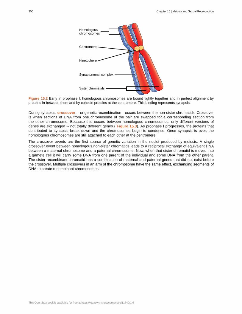
Figure 15.2 Early in prophase I, homologous chromosomes are bound tightly together and in perfect alignment byproteins in between them and by cohesin proteins at the centromere. This binding represents synapsis.
During synapsis, crossover —or genetic recombination—occurs between the non-sister chromatids. Crossoveris when sections of DNA from one chromosome of the pair are swapped for a corresponding section fromthe other chromosome. Because this occurs between homologous chromosomes, only different versions ofgenes are exchanged -- not totally different genes ( Figure 15.3). As prophase I progresses, the proteins thatcontributed to synapsis break down and the chromosomes begin to condense. Once synapsis is over, thehomologous chromosomes are still attached to each other at the centromere.
The crossover events are the first source of genetic variation in the nuclei produced by meiosis. A singlecrossover event between homologous non-sister chromatids leads to a reciprocal exchange of equivalent DNAbetween a maternal chromosome and a paternal chromosome. Now, when that sister chromatid is moved intoa gamete cell it will carry some DNA from one parent of the individual and some DNA from the other parent.The sister recombinant chromatid has a combination of maternal and paternal genes that did not exist beforethe crossover. Multiple crossovers in an arm of the chromosome have the same effect, exchanging segments ofDNA to create recombinant chromosomes.
300 Chapter 15 | Meiosis and Sexual Reproduction
This OpenStax book is available for free at https://legacy.cnx.org/content/col11749/1.6
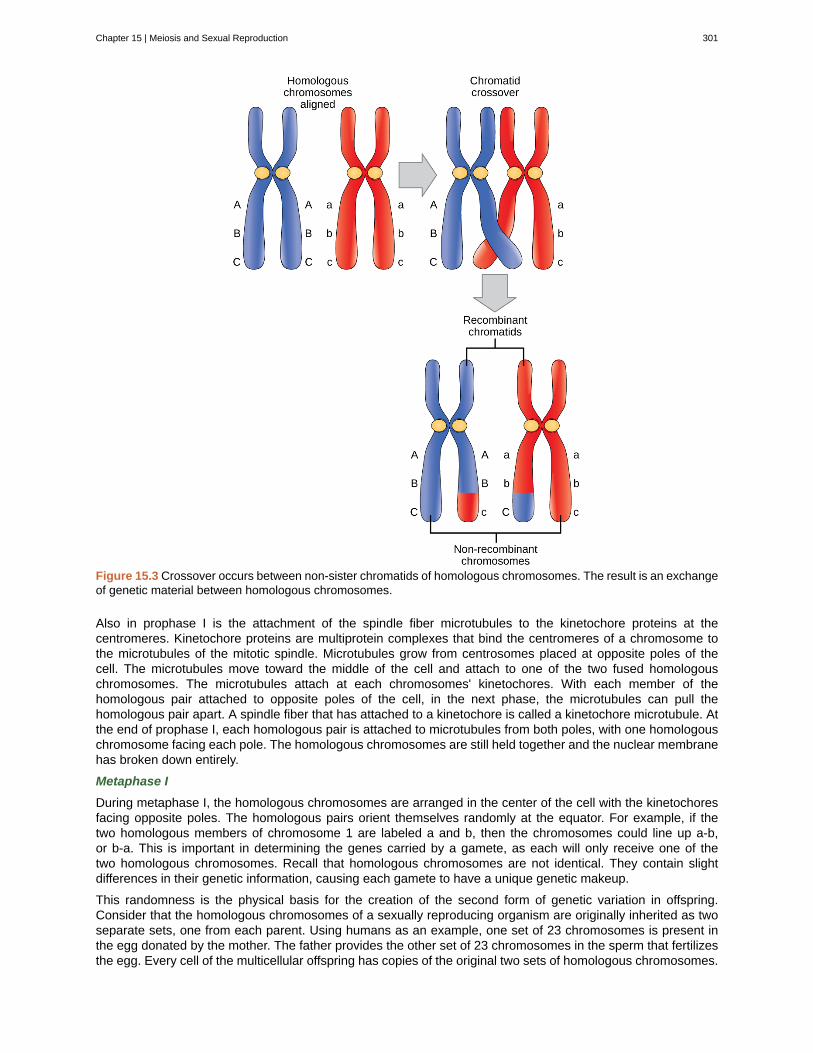
Figure 15.3 Crossover occurs between non-sister chromatids of homologous chromosomes. The result is an exchangeof genetic material between homologous chromosomes.
Also in prophase I is the attachment of the spindle fiber microtubules to the kinetochore proteins at thecentromeres. Kinetochore proteins are multiprotein complexes that bind the centromeres of a chromosome tothe microtubules of the mitotic spindle. Microtubules grow from centrosomes placed at opposite poles of thecell. The microtubules move toward the middle of the cell and attach to one of the two fused homologouschromosomes. The microtubules attach at each chromosomes' kinetochores. With each member of thehomologous pair attached to opposite poles of the cell, in the next phase, the microtubules can pull thehomologous pair apart. A spindle fiber that has attached to a kinetochore is called a kinetochore microtubule. Atthe end of prophase I, each homologous pair is attached to microtubules from both poles, with one homologouschromosome facing each pole. The homologous chromosomes are still held together and the nuclear membranehas broken down entirely.
Metaphase I
During metaphase I, the homologous chromosomes are arranged in the center of the cell with the kinetochoresfacing opposite poles. The homologous pairs orient themselves randomly at the equator. For example, if thetwo homologous members of chromosome 1 are labeled a and b, then the chromosomes could line up a-b,or b-a. This is important in determining the genes carried by a gamete, as each will only receive one of thetwo homologous chromosomes. Recall that homologous chromosomes are not identical. They contain slightdifferences in their genetic information, causing each gamete to have a unique genetic makeup.
This randomness is the physical basis for the creation of the second form of genetic variation in offspring.Consider that the homologous chromosomes of a sexually reproducing organism are originally inherited as twoseparate sets, one from each parent. Using humans as an example, one set of 23 chromosomes is present inthe egg donated by the mother. The father provides the other set of 23 chromosomes in the sperm that fertilizesthe egg. Every cell of the multicellular offspring has copies of the original two sets of homologous chromosomes.
Chapter 15 | Meiosis and Sexual Reproduction 301
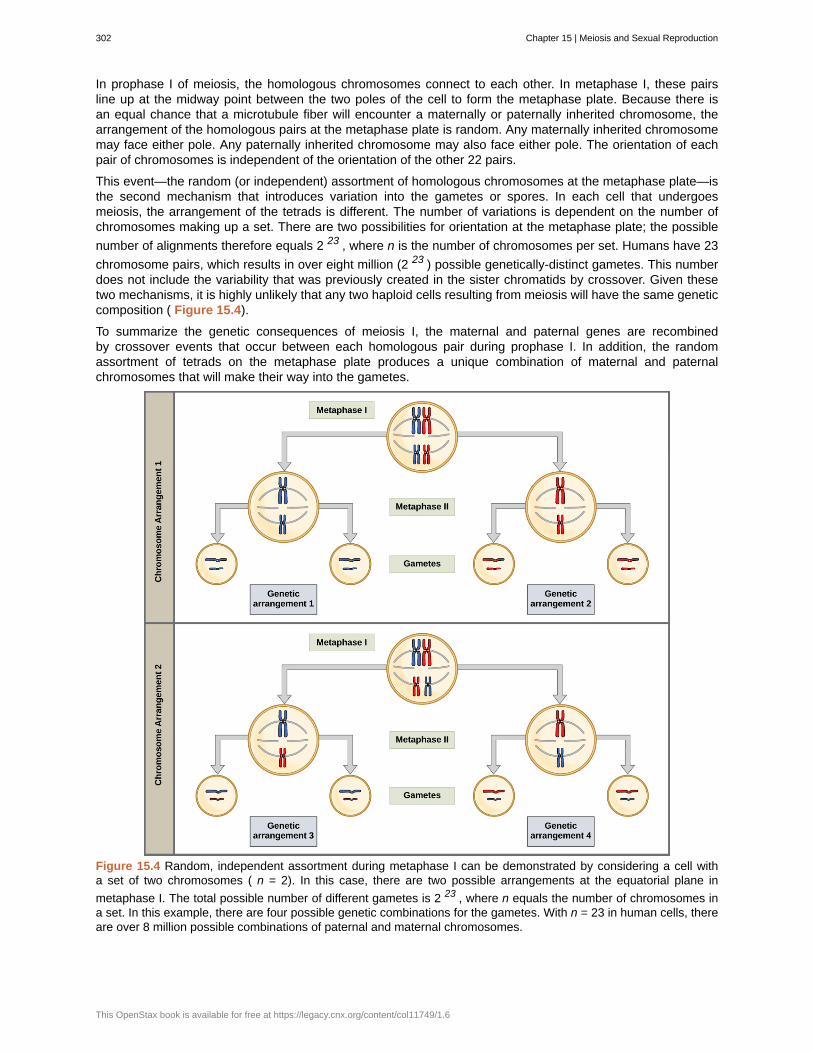
In prophase I of meiosis, the homologous chromosomes connect to each other. In metaphase I, these pairsline up at the midway point between the two poles of the cell to form the metaphase plate. Because there isan equal chance that a microtubule fiber will encounter a maternally or paternally inherited chromosome, thearrangement of the homologous pairs at the metaphase plate is random. Any maternally inherited chromosomemay face either pole. Any paternally inherited chromosome may also face either pole. The orientation of eachpair of chromosomes is independent of the orientation of the other 22 pairs.
This event—the random (or independent) assortment of homologous chromosomes at the metaphase plate—isthe second mechanism that introduces variation into the gametes or spores. In each cell that undergoesmeiosis, the arrangement of the tetrads is different. The number of variations is dependent on the number ofchromosomes making up a set. There are two possibilities for orientation at the metaphase plate; the possible
number of alignments therefore equals 2 23 , where n is the number of chromosomes per set. Humans have 23
chromosome pairs, which results in over eight million (2 23 ) possible genetically-distinct gametes. This numberdoes not include the variability that was previously created in the sister chromatids by crossover. Given thesetwo mechanisms, it is highly unlikely that any two haploid cells resulting from meiosis will have the same geneticcomposition ( Figure 15.4).
To summarize the genetic consequences of meiosis I, the maternal and paternal genes are recombinedby crossover events that occur between each homologous pair during prophase I. In addition, the randomassortment of tetrads on the metaphase plate produces a unique combination of maternal and paternalchromosomes that will make their way into the gametes.
Figure 15.4 Random, independent assortment during metaphase I can be demonstrated by considering a cell witha set of two chromosomes ( n = 2). In this case, there are two possible arrangements at the equatorial plane in
metaphase I. The total possible number of different gametes is 2 23 , where n equals the number of chromosomes ina set. In this example, there are four possible genetic combinations for the gametes. With n = 23 in human cells, thereare over 8 million possible combinations of paternal and maternal chromosomes.
302 Chapter 15 | Meiosis and Sexual Reproduction
This OpenStax book is available for free at https://legacy.cnx.org/content/col11749/1.6
Anaphase I
In anaphase I, the microtubules pull the linked chromosomes apart. The sister chromatids remain tightly boundtogether at the centromere ( Figure 15.5).
Telophase I
In telophase, the separated chromosomes arrive at opposite poles. The remainder of the typical telophaseevents may or may not occur, depending on the species. In some organisms, the chromosomes decondenseand nuclear envelopes form around the chromatids in telophase I. In other organisms, cytokinesis—the physicalseparation of the cytoplasmic components into two daughter cells—occurs without reformation of the nuclei.In nearly all species of animals and some fungi, cytokinesis separates the cell contents via a cleavage furrow(constriction of the actin ring that leads to cytoplasmic division). In plants, a cell plate is formed during cellcytokinesis by Golgi vesicles fusing at the metaphase plate. This cell plate will ultimately lead to the formation ofcell walls that separate the two daughter cells.
Two haploid cells are the end result of the first meiotic division. The cells are haploid because at each pole,there is just one of each pair of the homologous chromosomes. Therefore, only one full set of the chromosomesis present. This is why the cells are considered haploid—there is only one chromosome set, even though eachhomolog still consists of two sister chromatids. Recall that sister chromatids are merely duplicates of one of thetwo homologous chromosomes (except for changes that occurred during crossing over). In meiosis II, these twosister chromatids will separate, creating four haploid daughter cells.
Review the process of meiosis, observing how chromosomes align and migrate, at Meiosis: An InteractiveAnimation (http://openstaxcollege.org/l/animal_meiosis) .
15.2b Meiosis II
In some species, cells enter a brief interphase, or interkinesis , before entering meiosis II. Interkinesis lacksan S phase, so chromosomes are not duplicated. The two cells produced in meiosis I go through the events ofmeiosis II in synchrony. During meiosis II, the sister chromatids within the two daughter cells separate, formingfour new haploid gametes. The mechanics of meiosis II is similar to mitosis, except that each dividing cell hasonly one set of homologous chromosomes. Therefore, each cell has half the number of sister chromatids toseparate out as a diploid cell undergoing mitosis.
Prophase II
If the chromosomes decondensed in telophase I, they condense again. If nuclear envelopes were formed, theyfragment into vesicles. The centrosomes that were duplicated during interkinesis move away from each othertoward opposite poles, and new spindles are formed. Each sister chromatid forms an individual kinetochore thatattaches to microtubules from opposite poles.
Metaphase II
The sister chromatids are maximally condensed and aligned at the equator of the cell.
Anaphase II
The sister chromatids are pulled apart by the kinetochore microtubules and move toward opposite poles. Non-kinetochore microtubules elongate the cell.
Chapter 15 | Meiosis and Sexual Reproduction 303
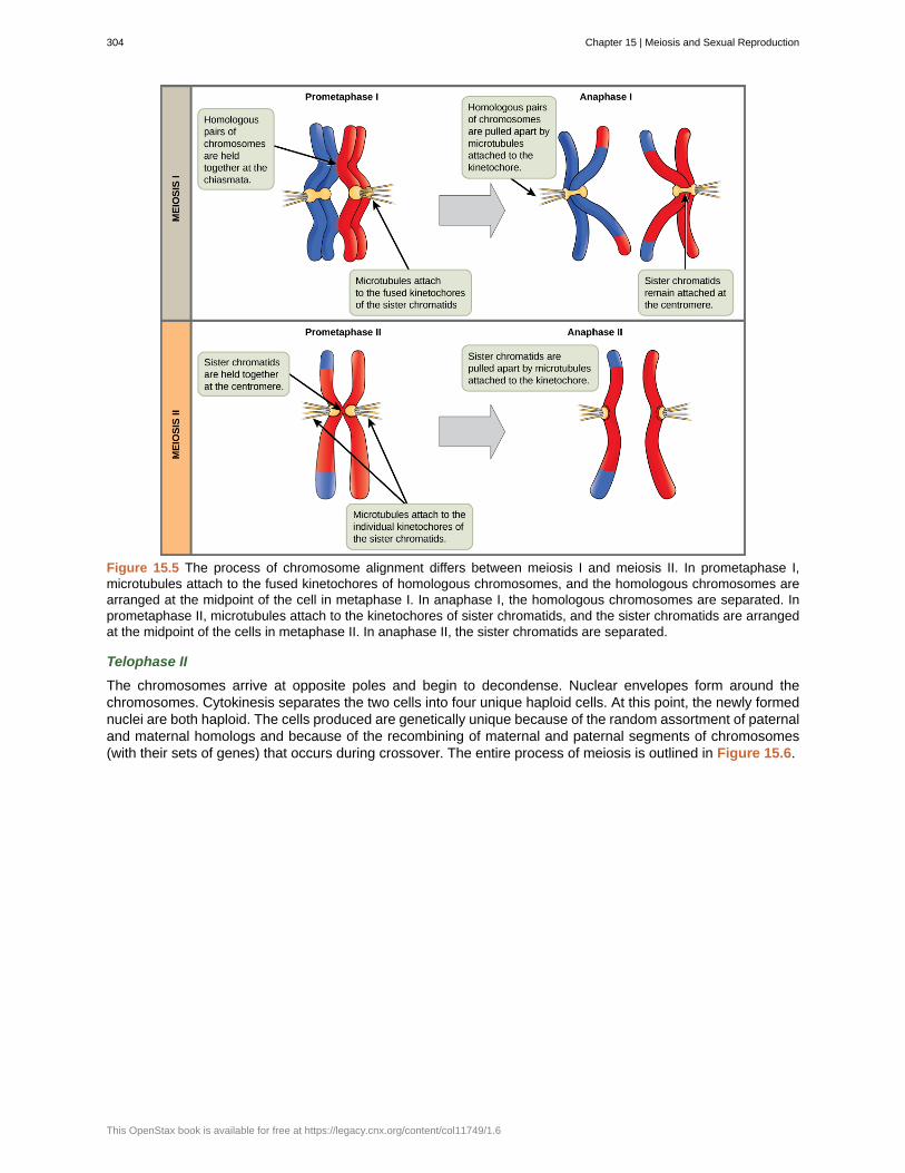
Figure 15.5 The process of chromosome alignment differs between meiosis I and meiosis II. In prometaphase I,microtubules attach to the fused kinetochores of homologous chromosomes, and the homologous chromosomes arearranged at the midpoint of the cell in metaphase I. In anaphase I, the homologous chromosomes are separated. Inprometaphase II, microtubules attach to the kinetochores of sister chromatids, and the sister chromatids are arrangedat the midpoint of the cells in metaphase II. In anaphase II, the sister chromatids are separated.
Telophase II
The chromosomes arrive at opposite poles and begin to decondense. Nuclear envelopes form around thechromosomes. Cytokinesis separates the two cells into four unique haploid cells. At this point, the newly formednuclei are both haploid. The cells produced are genetically unique because of the random assortment of paternaland maternal homologs and because of the recombining of maternal and paternal segments of chromosomes(with their sets of genes) that occurs during crossover. The entire process of meiosis is outlined in Figure 15.6.
304 Chapter 15 | Meiosis and Sexual Reproduction
This OpenStax book is available for free at https://legacy.cnx.org/content/col11749/1.6
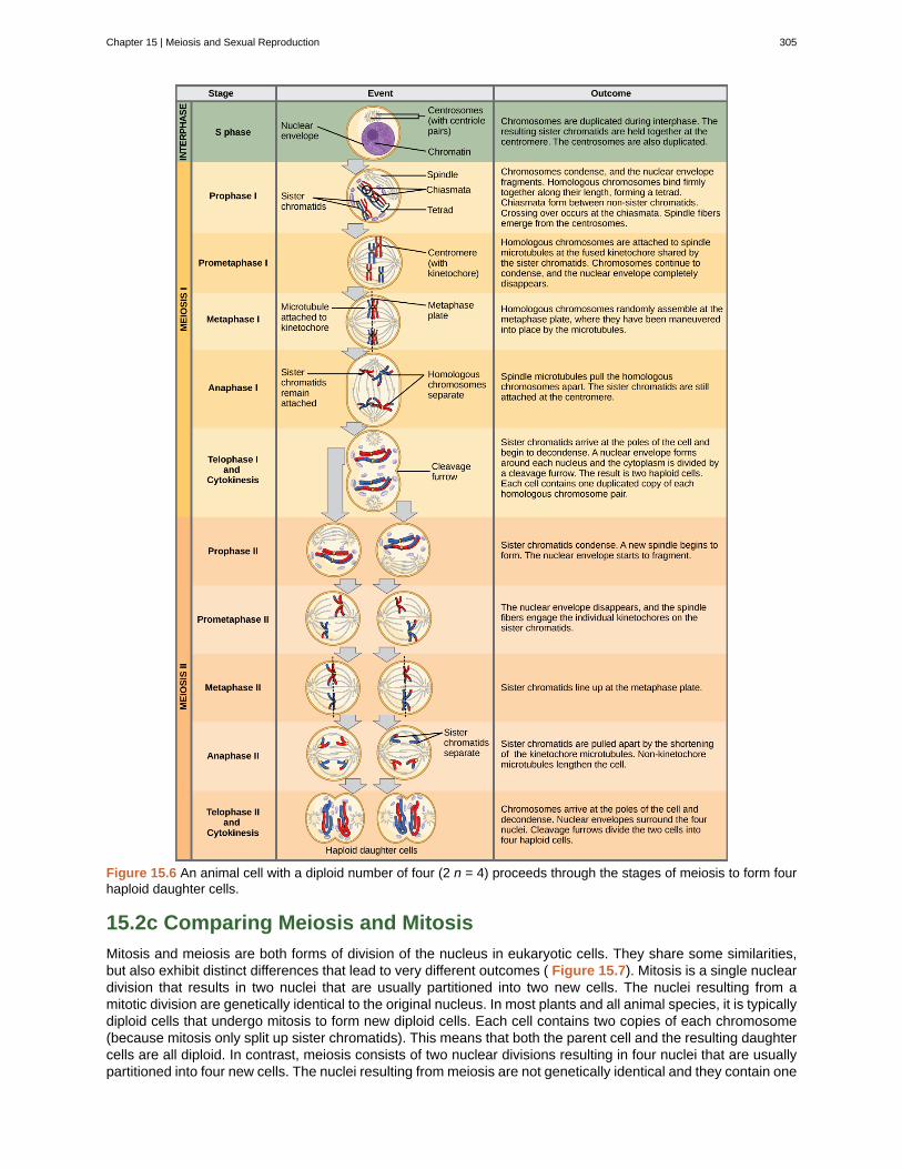
Figure 15.6 An animal cell with a diploid number of four (2 n = 4) proceeds through the stages of meiosis to form fourhaploid daughter cells.
15.2c Comparing Meiosis and Mitosis
Mitosis and meiosis are both forms of division of the nucleus in eukaryotic cells. They share some similarities,but also exhibit distinct differences that lead to very different outcomes ( Figure 15.7). Mitosis is a single nucleardivision that results in two nuclei that are usually partitioned into two new cells. The nuclei resulting from amitotic division are genetically identical to the original nucleus. In most plants and all animal species, it is typicallydiploid cells that undergo mitosis to form new diploid cells. Each cell contains two copies of each chromosome(because mitosis only split up sister chromatids). This means that both the parent cell and the resulting daughtercells are all diploid. In contrast, meiosis consists of two nuclear divisions resulting in four nuclei that are usuallypartitioned into four new cells. The nuclei resulting from meiosis are not genetically identical and they contain one
Chapter 15 | Meiosis and Sexual Reproduction 305
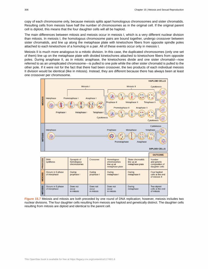
copy of each chromosome only, because meiosis splits apart homologous chromosomes and sister chromatids.Resulting cells from meiosis have half the number of chromosomes as in the original cell. If the original parentcell is diploid, this means that the four daughter cells will all be haploid.
The main differences between mitosis and meiosis occur in meiosis I, which is a very different nuclear divisionthan mitosis. In meiosis I, the homologous chromosome pairs are bound together, undergo crossover betweensister chromatids, and line up along the metaphase plate with kinetochore fibers from opposite spindle polesattached to each kinetochore of a homolog in a pair. All of these events occur only in meiosis I.
Meiosis II is much more analogous to a mitotic division. In this case, the duplicated chromosomes (only one setof them) line up on the metaphase plate with divided kinetochores attached to kinetochore fibers from oppositepoles. During anaphase II, as in mitotic anaphase, the kinetochores divide and one sister chromatid—nowreferred to as an unreplicated chromosome—is pulled to one pole while the other sister chromatid is pulled to theother pole. If it were not for the fact that there had been crossover, the two products of each individual meiosisII division would be identical (like in mitosis). Instead, they are different because there has always been at leastone crossover per chromosome.
Figure 15.7 Meiosis and mitosis are both preceded by one round of DNA replication; however, meiosis includes twonuclear divisions. The four daughter cells resulting from meiosis are haploid and genetically distinct. The daughter cellsresulting from mitosis are diploid and identical to the parent cell.
306 Chapter 15 | Meiosis and Sexual Reproduction
This OpenStax book is available for free at https://legacy.cnx.org/content/col11749/1.6
The Mystery of the Evolution of MeiosisSome characteristics of organisms are so widespread and fundamental that it is sometimes difficult toremember that they evolved like other simpler traits. Meiosis is such an extraordinarily complex seriesof cellular events that biologists have had trouble hypothesizing and testing how it may have evolved.Although meiosis is inextricably entwined with sexual reproduction and its advantages and disadvantages,it is important to separate the questions of the evolution of meiosis and the evolution of sex, because earlymeiosis may have been advantageous for different reasons than it is now. Thinking outside the box andimagining what the early benefits from meiosis might have been is one approach to uncovering how it mayhave evolved.
Meiosis and mitosis share obvious cellular processes and it makes sense that meiosis evolved from mitosis.
The difficulty lies in the clear differences between meiosis I and mitosis. Adam Wilkins and Robin Holliday[2]
summarized the unique events that needed to occur for the evolution of meiosis from mitosis. These stepsare homologous chromosome pairing, crossover exchanges, sister chromatids remaining attached duringanaphase, and suppression of DNA replication in interphase. They argue that the first step is the hardestand most important, and that understanding how it evolved would make the evolutionary process clearer.They suggest genetic experiments that might shed light on the evolution of synapsis.
There are other approaches to understanding the evolution of meiosis in progress. Different forms of meiosisexist in single-celled protists. Some appear to be simpler or more “primitive” forms of meiosis. Comparingthe meiotic divisions of different protists may shed light on the evolution of meiosis. Marilee Ramesh and
colleagues[3]
compared the genes involved in meiosis in protists to understand when and where meiosismight have evolved. Although research is still ongoing, recent scholarship into meiosis in protists suggeststhat some aspects of meiosis may have evolved later than others. This kind of genetic comparison can tellus what aspects of meiosis are the oldest and what cellular processes they may have borrowed from inearlier cells.
Click through the steps of this interactive animation to compare the meiotic process of cell division to that ofmitosis: How Cells Divide (http://openstaxcollege.org/l/how_cells_dvide) .
2.Adam S. Wilkins and Robin Holliday, “The Evolution of Meiosis from Mitosis,” Genetics 181 (2009): 3–12.3.Marilee A. Ramesh, Shehre-Banoo Malik and John M. Logsdon, Jr, “A Phylogenetic Inventory of Meiotic Genes: Evidence for Sex in Giardiaand an Early Eukaryotic Origin of Meiosis,” Current Biology 15 (2005):185–91.
Chapter 15 | Meiosis and Sexual Reproduction 307
cohesin
crossover
fertilization
germ cells
interkinesis
meiosis
meiosis I
meiosis II
somatic cell
Spores
synapsis
KEY TERMS
proteins that form a complex that seals sister chromatids together at their centromeres until anaphaseII of meiosis
exchange of genetic material between non-sister chromatids resulting in chromosomes thatincorporate genes from both parents of the organism
union of two haploid cells from two individual organisms
specialized cell line that produces gametes, such as eggs or sperm
(also, interphase II ) brief period of rest between meiosis I and meiosis II
a nuclear division process that results in four haploid cells
first round of meiotic cell division; referred to as reduction division because the ploidy level is reducedfrom diploid to haploid
second round of meiotic cell division following meiosis I; sister chromatids are separated intoindividual chromosomes, and the result is four unique haploid cells
all the cells of a multicellular organism except the gametes or reproductive cells
haploid cell that can produce a haploid multicellular organism or can fuse with another spore to form adiploid cell
formation of a close association between homologous chromosomes during prophase I
CHAPTER SUMMARY
15.1 Sexual Reproduction
Nearly all eukaryotes undergo sexual reproduction. The variation introduced into the reproductive cells bymeiosis appears to be one of the advantages of sexual reproduction that has made it so successful. Meiosisand fertilization alternate in sexual life cycles. The process of meiosis produces unique reproductive cells calledgametes, which have half the number of chromosomes as the parent cell. Fertilization, the fusion of haploidgametes from two individuals, restores the diploid condition. Thus, sexually reproducing organisms alternatebetween haploid and diploid stages.
15.2 The Process of Meiosis
Sexual reproduction requires that diploid organisms produce haploid cells that can fuse during fertilization toform diploid offspring. As with mitosis, DNA replication occurs prior to meiosis during the S-phase of the cellcycle. Meiosis is a series of events that arrange and separate chromosomes and chromatids into daughtercells. During the interphases of meiosis, each chromosome is duplicated. In meiosis, there are two rounds ofnuclear division resulting in four nuclei and usually four daughter cells, each with half the number ofchromosomes as the parent cell. The first separates homologs, and the second—like mitosis—separateschromatids into individual chromosomes. During meiosis, variation in the daughter nuclei is introduced becauseof crossover in prophase I and random alignment of tetrads at metaphase I. The cells that are produced bymeiosis are genetically unique.
Meiosis and mitosis share similarities, but have distinct outcomes. Mitotic divisions are single nuclear divisionsthat produce daughter nuclei that are genetically identical and have the same number of chromosome sets asthe original cell. Meiotic divisions include two nuclear divisions that produce four daughter nuclei that aregenetically different and have one chromosome set instead of the two sets of chromosomes in the parent cell.The main differences between the processes occur in the first division of meiosis, in which homologouschromosomes are paired and exchange non-sister chromatid segments. The homologous chromosomesseparate into different nuclei during meiosis I, causing a reduction of ploidy level in the first division. Thesecond division of meiosis is more similar to a mitotic division, except that the daughter cells do not containidentical genomes because of crossover.
308 Chapter 15 | Meiosis and Sexual Reproduction
This OpenStax book is available for free at https://legacy.cnx.org/content/col11749/1.6
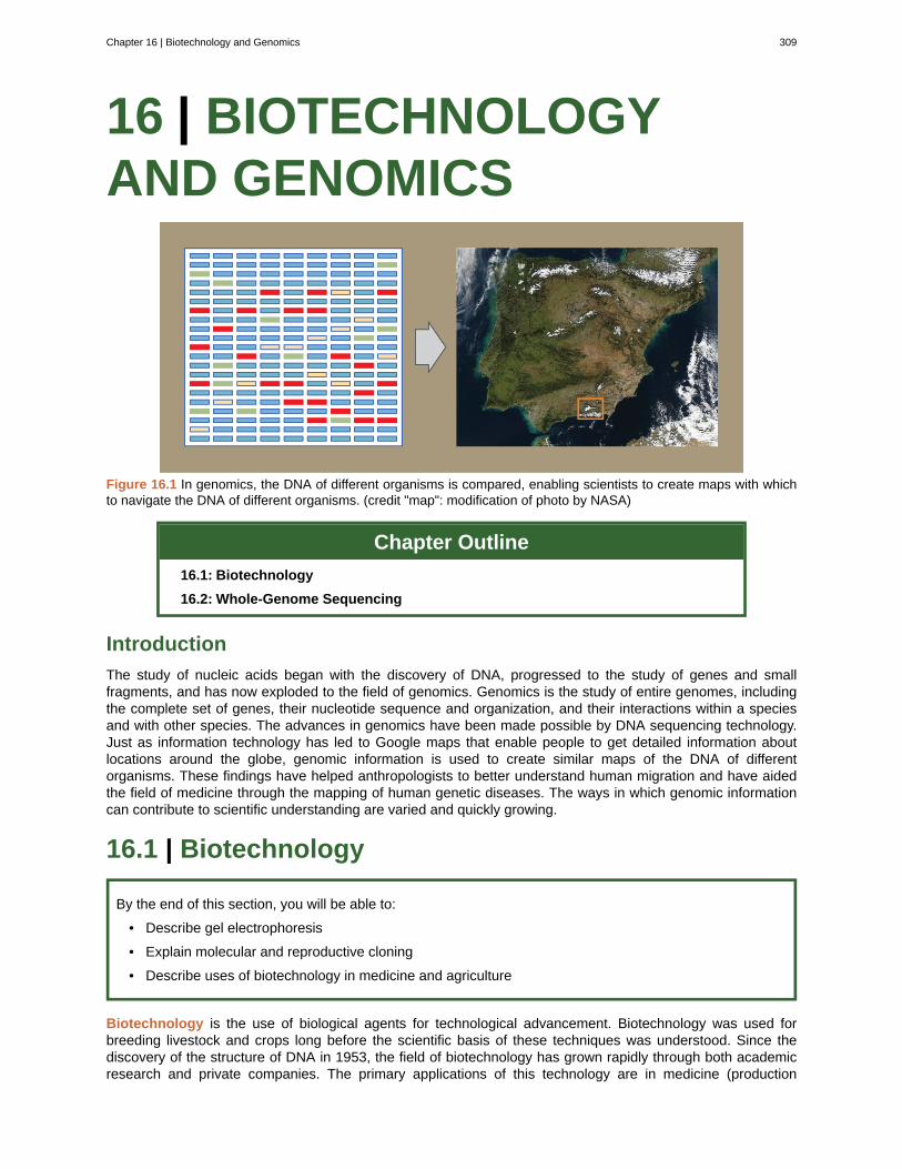
16 | BIOTECHNOLOGYAND GENOMICS
Figure 16.1 In genomics, the DNA of different organisms is compared, enabling scientists to create maps with whichto navigate the DNA of different organisms. (credit "map": modification of photo by NASA)
Chapter Outline
16.1: Biotechnology
16.2: Whole-Genome Sequencing
Introduction
The study of nucleic acids began with the discovery of DNA, progressed to the study of genes and smallfragments, and has now exploded to the field of genomics. Genomics is the study of entire genomes, includingthe complete set of genes, their nucleotide sequence and organization, and their interactions within a speciesand with other species. The advances in genomics have been made possible by DNA sequencing technology.Just as information technology has led to Google maps that enable people to get detailed information aboutlocations around the globe, genomic information is used to create similar maps of the DNA of differentorganisms. These findings have helped anthropologists to better understand human migration and have aidedthe field of medicine through the mapping of human genetic diseases. The ways in which genomic informationcan contribute to scientific understanding are varied and quickly growing.
16.1 | Biotechnology
By the end of this section, you will be able to:
• Describe gel electrophoresis
• Explain molecular and reproductive cloning
• Describe uses of biotechnology in medicine and agriculture
Biotechnology is the use of biological agents for technological advancement. Biotechnology was used forbreeding livestock and crops long before the scientific basis of these techniques was understood. Since thediscovery of the structure of DNA in 1953, the field of biotechnology has grown rapidly through both academicresearch and private companies. The primary applications of this technology are in medicine (production
Chapter 16 | Biotechnology and Genomics 309
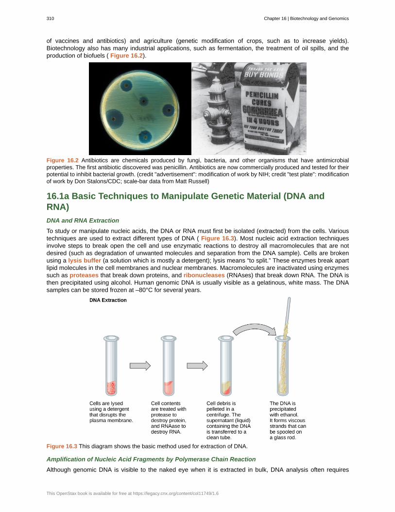
of vaccines and antibiotics) and agriculture (genetic modification of crops, such as to increase yields).Biotechnology also has many industrial applications, such as fermentation, the treatment of oil spills, and theproduction of biofuels ( Figure 16.2).
Figure 16.2 Antibiotics are chemicals produced by fungi, bacteria, and other organisms that have antimicrobialproperties. The first antibiotic discovered was penicillin. Antibiotics are now commercially produced and tested for theirpotential to inhibit bacterial growth. (credit "advertisement": modification of work by NIH; credit "test plate": modificationof work by Don Stalons/CDC; scale-bar data from Matt Russell)
16.1a Basic Techniques to Manipulate Genetic Material (DNA andRNA)
DNA and RNA Extraction
To study or manipulate nucleic acids, the DNA or RNA must first be isolated (extracted) from the cells. Varioustechniques are used to extract different types of DNA ( Figure 16.3). Most nucleic acid extraction techniquesinvolve steps to break open the cell and use enzymatic reactions to destroy all macromolecules that are notdesired (such as degradation of unwanted molecules and separation from the DNA sample). Cells are brokenusing a lysis buffer (a solution which is mostly a detergent); lysis means “to split.” These enzymes break apartlipid molecules in the cell membranes and nuclear membranes. Macromolecules are inactivated using enzymessuch as proteases that break down proteins, and ribonucleases (RNAses) that break down RNA. The DNA isthen precipitated using alcohol. Human genomic DNA is usually visible as a gelatinous, white mass. The DNAsamples can be stored frozen at –80°C for several years.
Figure 16.3 This diagram shows the basic method used for extraction of DNA.
Amplification of Nucleic Acid Fragments by Polymerase Chain Reaction
Although genomic DNA is visible to the naked eye when it is extracted in bulk, DNA analysis often requires
310 Chapter 16 | Biotechnology and Genomics
This OpenStax book is available for free at https://legacy.cnx.org/content/col11749/1.6
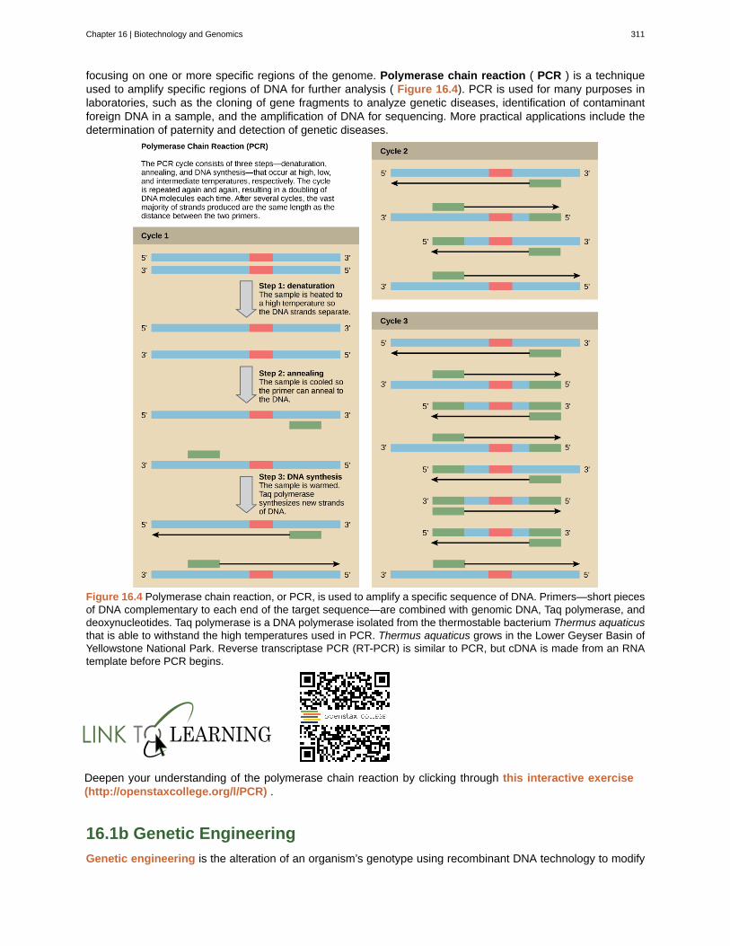
focusing on one or more specific regions of the genome. Polymerase chain reaction ( PCR ) is a techniqueused to amplify specific regions of DNA for further analysis ( Figure 16.4). PCR is used for many purposes inlaboratories, such as the cloning of gene fragments to analyze genetic diseases, identification of contaminantforeign DNA in a sample, and the amplification of DNA for sequencing. More practical applications include thedetermination of paternity and detection of genetic diseases.
Figure 16.4 Polymerase chain reaction, or PCR, is used to amplify a specific sequence of DNA. Primers—short piecesof DNA complementary to each end of the target sequence—are combined with genomic DNA, Taq polymerase, anddeoxynucleotides. Taq polymerase is a DNA polymerase isolated from the thermostable bacterium Thermus aquaticusthat is able to withstand the high temperatures used in PCR. Thermus aquaticus grows in the Lower Geyser Basin ofYellowstone National Park. Reverse transcriptase PCR (RT-PCR) is similar to PCR, but cDNA is made from an RNAtemplate before PCR begins.
Deepen your understanding of the polymerase chain reaction by clicking through this interactive exercise(http://openstaxcollege.org/l/PCR) .
16.1b Genetic Engineering
Genetic engineering is the alteration of an organism’s genotype using recombinant DNA technology to modify
Chapter 16 | Biotechnology and Genomics 311
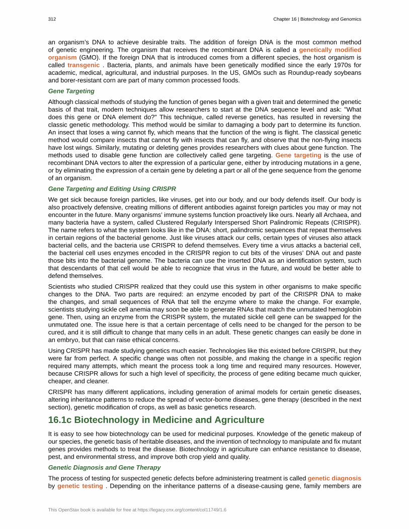
an organism’s DNA to achieve desirable traits. The addition of foreign DNA is the most common methodof genetic engineering. The organism that receives the recombinant DNA is called a genetically modifiedorganism (GMO). If the foreign DNA that is introduced comes from a different species, the host organism iscalled transgenic . Bacteria, plants, and animals have been genetically modified since the early 1970s foracademic, medical, agricultural, and industrial purposes. In the US, GMOs such as Roundup-ready soybeansand borer-resistant corn are part of many common processed foods.
Gene Targeting
Although classical methods of studying the function of genes began with a given trait and determined the geneticbasis of that trait, modern techniques allow researchers to start at the DNA sequence level and ask: "Whatdoes this gene or DNA element do?" This technique, called reverse genetics, has resulted in reversing theclassic genetic methodology. This method would be similar to damaging a body part to determine its function.An insect that loses a wing cannot fly, which means that the function of the wing is flight. The classical geneticmethod would compare insects that cannot fly with insects that can fly, and observe that the non-flying insectshave lost wings. Similarly, mutating or deleting genes provides researchers with clues about gene function. Themethods used to disable gene function are collectively called gene targeting. Gene targeting is the use ofrecombinant DNA vectors to alter the expression of a particular gene, either by introducing mutations in a gene,or by eliminating the expression of a certain gene by deleting a part or all of the gene sequence from the genomeof an organism.
Gene Targeting and Editing Using CRISPR
We get sick because foreign particles, like viruses, get into our body, and our body defends itself. Our body isalso proactively defensive, creating millions of different antibodies against foreign particles you may or may notencounter in the future. Many organisms’ immune systems function proactively like ours. Nearly all Archaea, andmany bacteria have a system, called Clustered Regularly Interspersed Short Palindromic Repeats (CRISPR).The name refers to what the system looks like in the DNA: short, palindromic sequences that repeat themselvesin certain regions of the bacterial genome. Just like viruses attack our cells, certain types of viruses also attackbacterial cells, and the bacteria use CRISPR to defend themselves. Every time a virus attacks a bacterial cell,the bacterial cell uses enzymes encoded in the CRISPR region to cut bits of the viruses’ DNA out and pastethose bits into the bacterial genome. The bacteria can use the inserted DNA as an identification system, suchthat descendants of that cell would be able to recognize that virus in the future, and would be better able todefend themselves.
Scientists who studied CRISPR realized that they could use this system in other organisms to make specificchanges to the DNA. Two parts are required: an enzyme encoded by part of the CRISPR DNA to makethe changes, and small sequences of RNA that tell the enzyme where to make the change. For example,scientists studying sickle cell anemia may soon be able to generate RNAs that match the unmutated hemoglobingene. Then, using an enzyme from the CRISPR system, the mutated sickle cell gene can be swapped for theunmutated one. The issue here is that a certain percentage of cells need to be changed for the person to becured, and it is still difficult to change that many cells in an adult. These genetic changes can easily be done inan embryo, but that can raise ethical concerns.
Using CRISPR has made studying genetics much easier. Technologies like this existed before CRISPR, but theywere far from perfect. A specific change was often not possible, and making the change in a specific regionrequired many attempts, which meant the process took a long time and required many resources. However,because CRISPR allows for such a high level of specificity, the process of gene editing became much quicker,cheaper, and cleaner.
CRISPR has many different applications, including generation of animal models for certain genetic diseases,altering inheritance patterns to reduce the spread of vector-borne diseases, gene therapy (described in the nextsection), genetic modification of crops, as well as basic genetics research.
16.1c Biotechnology in Medicine and Agriculture
It is easy to see how biotechnology can be used for medicinal purposes. Knowledge of the genetic makeup ofour species, the genetic basis of heritable diseases, and the invention of technology to manipulate and fix mutantgenes provides methods to treat the disease. Biotechnology in agriculture can enhance resistance to disease,pest, and environmental stress, and improve both crop yield and quality.
Genetic Diagnosis and Gene Therapy
The process of testing for suspected genetic defects before administering treatment is called genetic diagnosisby genetic testing . Depending on the inheritance patterns of a disease-causing gene, family members are
312 Chapter 16 | Biotechnology and Genomics
This OpenStax book is available for free at https://legacy.cnx.org/content/col11749/1.6
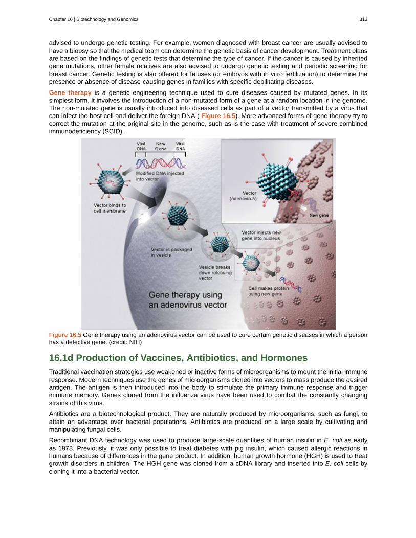
advised to undergo genetic testing. For example, women diagnosed with breast cancer are usually advised tohave a biopsy so that the medical team can determine the genetic basis of cancer development. Treatment plansare based on the findings of genetic tests that determine the type of cancer. If the cancer is caused by inheritedgene mutations, other female relatives are also advised to undergo genetic testing and periodic screening forbreast cancer. Genetic testing is also offered for fetuses (or embryos with in vitro fertilization) to determine thepresence or absence of disease-causing genes in families with specific debilitating diseases.
Gene therapy is a genetic engineering technique used to cure diseases caused by mutated genes. In itssimplest form, it involves the introduction of a non-mutated form of a gene at a random location in the genome.The non-mutated gene is usually introduced into diseased cells as part of a vector transmitted by a virus thatcan infect the host cell and deliver the foreign DNA ( Figure 16.5). More advanced forms of gene therapy try tocorrect the mutation at the original site in the genome, such as is the case with treatment of severe combinedimmunodeficiency (SCID).
Figure 16.5 Gene therapy using an adenovirus vector can be used to cure certain genetic diseases in which a personhas a defective gene. (credit: NIH)
16.1d Production of Vaccines, Antibiotics, and Hormones
Traditional vaccination strategies use weakened or inactive forms of microorganisms to mount the initial immuneresponse. Modern techniques use the genes of microorganisms cloned into vectors to mass produce the desiredantigen. The antigen is then introduced into the body to stimulate the primary immune response and triggerimmune memory. Genes cloned from the influenza virus have been used to combat the constantly changingstrains of this virus.
Antibiotics are a biotechnological product. They are naturally produced by microorganisms, such as fungi, toattain an advantage over bacterial populations. Antibiotics are produced on a large scale by cultivating andmanipulating fungal cells.
Recombinant DNA technology was used to produce large-scale quantities of human insulin in E. coli as earlyas 1978. Previously, it was only possible to treat diabetes with pig insulin, which caused allergic reactions inhumans because of differences in the gene product. In addition, human growth hormone (HGH) is used to treatgrowth disorders in children. The HGH gene was cloned from a cDNA library and inserted into E. coli cells bycloning it into a bacterial vector.
Chapter 16 | Biotechnology and Genomics 313
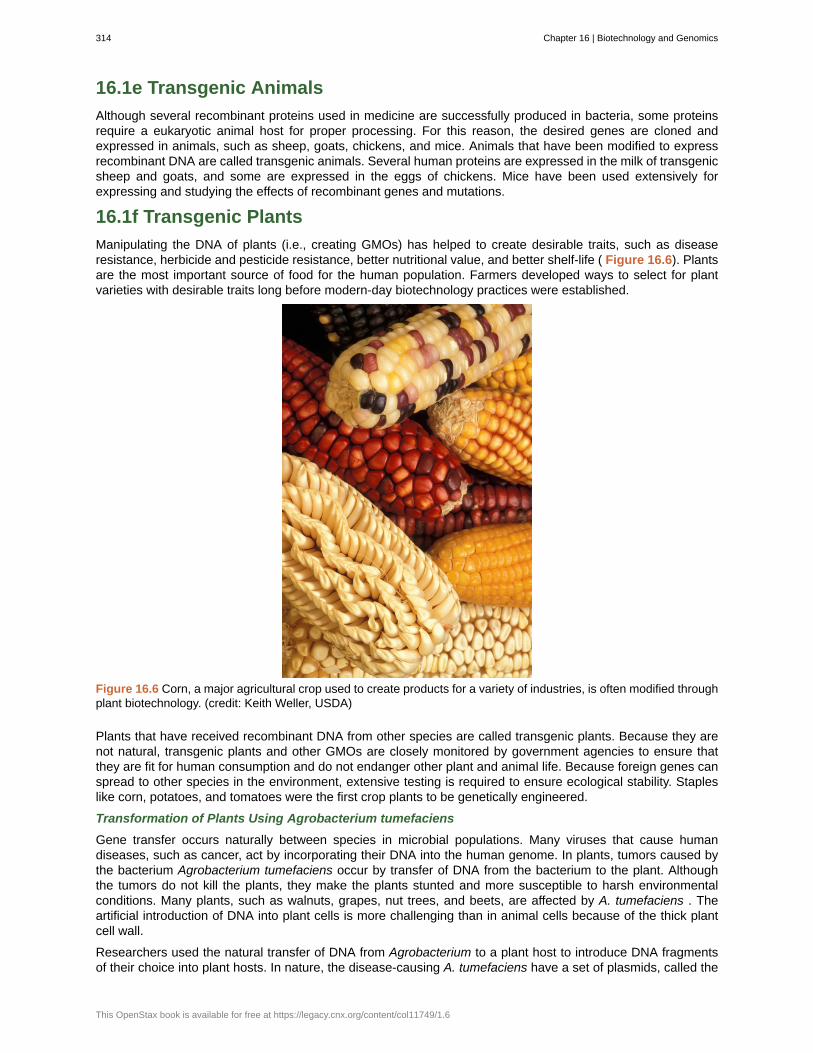
16.1e Transgenic Animals
Although several recombinant proteins used in medicine are successfully produced in bacteria, some proteinsrequire a eukaryotic animal host for proper processing. For this reason, the desired genes are cloned andexpressed in animals, such as sheep, goats, chickens, and mice. Animals that have been modified to expressrecombinant DNA are called transgenic animals. Several human proteins are expressed in the milk of transgenicsheep and goats, and some are expressed in the eggs of chickens. Mice have been used extensively forexpressing and studying the effects of recombinant genes and mutations.
16.1f Transgenic Plants
Manipulating the DNA of plants (i.e., creating GMOs) has helped to create desirable traits, such as diseaseresistance, herbicide and pesticide resistance, better nutritional value, and better shelf-life ( Figure 16.6). Plantsare the most important source of food for the human population. Farmers developed ways to select for plantvarieties with desirable traits long before modern-day biotechnology practices were established.
Figure 16.6 Corn, a major agricultural crop used to create products for a variety of industries, is often modified throughplant biotechnology. (credit: Keith Weller, USDA)
Plants that have received recombinant DNA from other species are called transgenic plants. Because they arenot natural, transgenic plants and other GMOs are closely monitored by government agencies to ensure thatthey are fit for human consumption and do not endanger other plant and animal life. Because foreign genes canspread to other species in the environment, extensive testing is required to ensure ecological stability. Stapleslike corn, potatoes, and tomatoes were the first crop plants to be genetically engineered.
Transformation of Plants Using Agrobacterium tumefaciens
Gene transfer occurs naturally between species in microbial populations. Many viruses that cause humandiseases, such as cancer, act by incorporating their DNA into the human genome. In plants, tumors caused bythe bacterium Agrobacterium tumefaciens occur by transfer of DNA from the bacterium to the plant. Althoughthe tumors do not kill the plants, they make the plants stunted and more susceptible to harsh environmentalconditions. Many plants, such as walnuts, grapes, nut trees, and beets, are affected by A. tumefaciens . Theartificial introduction of DNA into plant cells is more challenging than in animal cells because of the thick plantcell wall.
Researchers used the natural transfer of DNA from Agrobacterium to a plant host to introduce DNA fragmentsof their choice into plant hosts. In nature, the disease-causing A. tumefaciens have a set of plasmids, called the
314 Chapter 16 | Biotechnology and Genomics
This OpenStax book is available for free at https://legacy.cnx.org/content/col11749/1.6
Ti plasmids (tumor-inducing plasmids), that contain genes for the production of tumors in plants. DNA from theTi plasmid integrates into the infected plant cell’s genome. Researchers manipulate the Ti plasmids to removethe tumor-causing genes and insert the desired DNA fragment for transfer into the plant genome.
The Organic Insecticide Bacillus thuringiensis
Bacillus thuringiensis (Bt) is a bacterium that produces protein crystals during sporulation that are toxic to manyinsect species that affect plants. Bt toxin has to be ingested by insects for the toxin to be activated. Insects thathave eaten Bt toxin stop feeding on the plants within a few hours. After the toxin is activated in the intestines ofthe insects, death occurs within a couple of days. Modern biotechnology has allowed plants to encode their owncrystal Bt toxin that acts against insects. The crystal toxin genes have been cloned from Bt and introduced intoplants. Bt toxin has been found to be safe for the environment, non-toxic to humans and other mammals, and isapproved for use by organic farmers as a natural insecticide.
Flavr Savr Tomato
The first GM crop to be introduced into the market was the Flavr Savr Tomato produced in 1994. Geneticengineering was used to slow down the process of softening and rotting caused by fungal infections, which ledto increased shelf life of the GM tomatoes. Additional genetic modification improved the flavor of this tomato.The Flavr Savr tomato did not successfully stay in the market because of problems maintaining and shipping thecrop.
16.2 | Whole-Genome Sequencing
By the end of this section, you will be able to:
• Describe three types of sequencing
• Define whole-genome sequencing
Although there have been significant advances in the medical sciences in recent years, doctors are stillconfounded by some diseases, and they are using whole-genome sequencing to get to the bottom of theproblem. Whole-genome sequencing is a process that determines the DNA sequence of an entire genome.Whole-genome sequencing is a brute-force approach to problem solving when there is a genetic basis atthe core of a disease. Several laboratories now provide services to sequence, analyze, and interpret entiregenomes.
For example, whole-exome sequencing is a lower-cost alternative to whole genome sequencing. In exomesequencing, only the coding, exon-producing regions of the DNA are sequenced. In 2010, whole-exomesequencing was used to save a young boy whose intestines had multiple mysterious abscesses. The childhad several colon operations with no relief. Finally, whole-exome sequencing was performed, which revealeda defect in a pathway that controls apoptosis (programmed cell death). A bone-marrow transplant was used toovercome this genetic disorder, leading to a cure for the boy. He was the first person to be successfully treatedbased on a diagnosis made by whole-exome sequencing. Today, human genome sequencing is more readilyavailable and can be completed in a day or two for about $1000.
Chapter 16 | Biotechnology and Genomics 315
Comparing SequencesA sequence alignment is an arrangement of proteins, DNA, or RNA; it is used to identify regions of similaritybetween cell types or species, which may indicate conservation of function or structures. Sequencealignments may be used to construct phylogenetic trees. The following website uses a software programcalled BLAST (basic local alignment search tool) (http://blast.ncbi.nlm.nih.gov/Blast.cgi) .
Under “Basic Blast,” click “Nucleotide Blast.” Input the following sequence into the large "query sequence"box: ATTGCTTCGATTGCA. Below the box, locate the "Species" field and type "human" or "Homo sapiens".Then click “BLAST” to compare the inputted sequence against known sequences of the human genome.The result is that this sequence occurs in over a hundred places in the human genome. Scroll down belowthe graphic with the horizontal bars and you will see short description of each of the matching hits. Pick oneof the hits near the top of the list and click on "Graphics". This will bring you to a page that shows wherethe sequence is found within the entire human genome. You can move the slider that looks like a green flagback and forth to view the sequences immediately around the selected gene. You can then return to yourselected sequence by clicking the "ATG" button.
316 Chapter 16 | Biotechnology and Genomics
This OpenStax book is available for free at https://legacy.cnx.org/content/col11749/1.6
biotechnology
gene targeting
gene therapy
genetic diagnosis
genetic engineering
genetic testing
genetically modified organism lysis buffer
proteases
ribonucleases
Ti plasmids
transgenic
whole-genome sequencing
KEY TERMS
use of biological agents for technological advancement
method for altering the sequence of a specific gene by introducing the modified version on avector
technique used to cure inheritable diseases by replacing mutant genes with good genes
diagnosis of the potential for disease development by analyzing disease-causing genes
alteration of the genetic makeup of an organism
process of testing for the presence of disease-causing genes
solution used to break the cell membrane and release cellcontents
enzyme that breaks down proteins
enzyme that breaks down RNA
plasmid system derived from Agrobacterium tumefaciens that has been used by scientists tointroduce foreign DNA into plant cells
organism that receives DNA from a different species
process that determines the DNA sequence of an entire genome
CHAPTER SUMMARY
16.1 Biotechnology
Biotechnology is using DNA. RNA, or protein to advance technology in some way. Biotechnology has beenused in medicine, agriculture by the production of genetics tests, vaccines, antibiotics, treatments, and todevelop new plant varieties.
16.2 Whole-Genome Sequencing
Whole-genome sequencing is a process used to determine the order of every nucleotide on every chromosomein a particular individual. Versions of this technology are becoming cheaper and more commonly used indisease diagnostics, as well as in general science research.
Chapter 16 | Biotechnology and Genomics 317

318 Chapter 16 | Biotechnology and Genomics
This OpenStax book is available for free at https://legacy.cnx.org/content/col11749/1.6
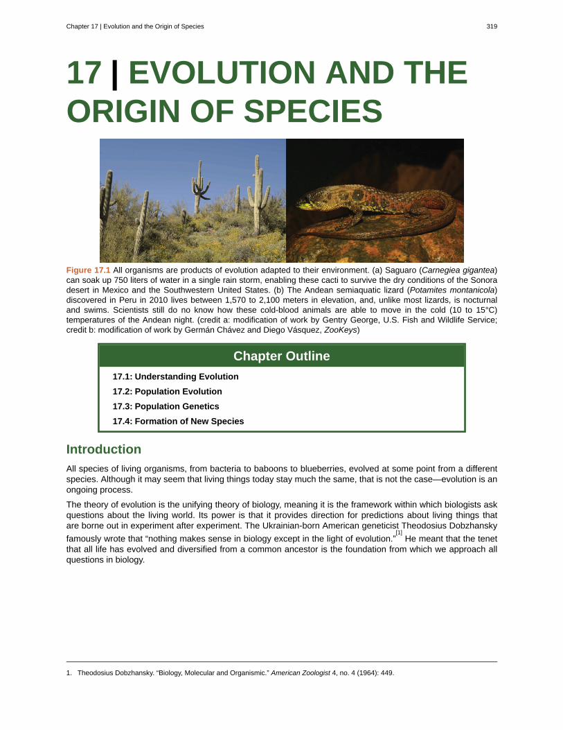
17 | EVOLUTION AND THEORIGIN OF SPECIES
Figure 17.1 All organisms are products of evolution adapted to their environment. (a) Saguaro (Carnegiea gigantea)can soak up 750 liters of water in a single rain storm, enabling these cacti to survive the dry conditions of the Sonoradesert in Mexico and the Southwestern United States. (b) The Andean semiaquatic lizard (Potamites montanicola)discovered in Peru in 2010 lives between 1,570 to 2,100 meters in elevation, and, unlike most lizards, is nocturnaland swims. Scientists still do no know how these cold-blood animals are able to move in the cold (10 to 15°C)temperatures of the Andean night. (credit a: modification of work by Gentry George, U.S. Fish and Wildlife Service;credit b: modification of work by Germán Chávez and Diego Vásquez, ZooKeys)
Chapter Outline
17.1: Understanding Evolution
17.2: Population Evolution
17.3: Population Genetics
17.4: Formation of New Species
Introduction
All species of living organisms, from bacteria to baboons to blueberries, evolved at some point from a differentspecies. Although it may seem that living things today stay much the same, that is not the case—evolution is anongoing process.
The theory of evolution is the unifying theory of biology, meaning it is the framework within which biologists askquestions about the living world. Its power is that it provides direction for predictions about living things thatare borne out in experiment after experiment. The Ukrainian-born American geneticist Theodosius Dobzhansky
famously wrote that “nothing makes sense in biology except in the light of evolution.”[1]
He meant that the tenetthat all life has evolved and diversified from a common ancestor is the foundation from which we approach allquestions in biology.
1. Theodosius Dobzhansky. “Biology, Molecular and Organismic.” American Zoologist 4, no. 4 (1964): 449.
Chapter 17 | Evolution and the Origin of Species 319
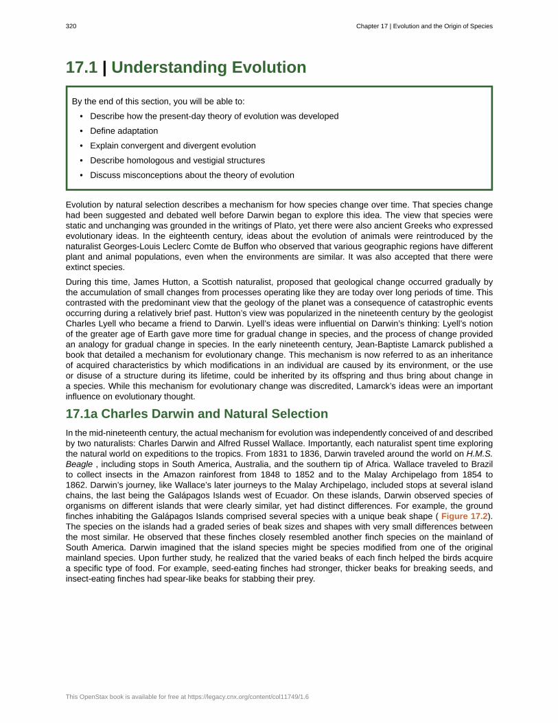
17.1 | Understanding Evolution
By the end of this section, you will be able to:
• Describe how the present-day theory of evolution was developed
• Define adaptation
• Explain convergent and divergent evolution
• Describe homologous and vestigial structures
• Discuss misconceptions about the theory of evolution
Evolution by natural selection describes a mechanism for how species change over time. That species changehad been suggested and debated well before Darwin began to explore this idea. The view that species werestatic and unchanging was grounded in the writings of Plato, yet there were also ancient Greeks who expressedevolutionary ideas. In the eighteenth century, ideas about the evolution of animals were reintroduced by thenaturalist Georges-Louis Leclerc Comte de Buffon who observed that various geographic regions have differentplant and animal populations, even when the environments are similar. It was also accepted that there wereextinct species.
During this time, James Hutton, a Scottish naturalist, proposed that geological change occurred gradually bythe accumulation of small changes from processes operating like they are today over long periods of time. Thiscontrasted with the predominant view that the geology of the planet was a consequence of catastrophic eventsoccurring during a relatively brief past. Hutton’s view was popularized in the nineteenth century by the geologistCharles Lyell who became a friend to Darwin. Lyell’s ideas were influential on Darwin’s thinking: Lyell’s notionof the greater age of Earth gave more time for gradual change in species, and the process of change providedan analogy for gradual change in species. In the early nineteenth century, Jean-Baptiste Lamarck published abook that detailed a mechanism for evolutionary change. This mechanism is now referred to as an inheritanceof acquired characteristics by which modifications in an individual are caused by its environment, or the useor disuse of a structure during its lifetime, could be inherited by its offspring and thus bring about change ina species. While this mechanism for evolutionary change was discredited, Lamarck’s ideas were an importantinfluence on evolutionary thought.
17.1a Charles Darwin and Natural Selection
In the mid-nineteenth century, the actual mechanism for evolution was independently conceived of and describedby two naturalists: Charles Darwin and Alfred Russel Wallace. Importantly, each naturalist spent time exploringthe natural world on expeditions to the tropics. From 1831 to 1836, Darwin traveled around the world on H.M.S.Beagle , including stops in South America, Australia, and the southern tip of Africa. Wallace traveled to Brazilto collect insects in the Amazon rainforest from 1848 to 1852 and to the Malay Archipelago from 1854 to1862. Darwin’s journey, like Wallace’s later journeys to the Malay Archipelago, included stops at several islandchains, the last being the Galápagos Islands west of Ecuador. On these islands, Darwin observed species oforganisms on different islands that were clearly similar, yet had distinct differences. For example, the groundfinches inhabiting the Galápagos Islands comprised several species with a unique beak shape ( Figure 17.2).The species on the islands had a graded series of beak sizes and shapes with very small differences betweenthe most similar. He observed that these finches closely resembled another finch species on the mainland ofSouth America. Darwin imagined that the island species might be species modified from one of the originalmainland species. Upon further study, he realized that the varied beaks of each finch helped the birds acquirea specific type of food. For example, seed-eating finches had stronger, thicker beaks for breaking seeds, andinsect-eating finches had spear-like beaks for stabbing their prey.
320 Chapter 17 | Evolution and the Origin of Species
This OpenStax book is available for free at https://legacy.cnx.org/content/col11749/1.6
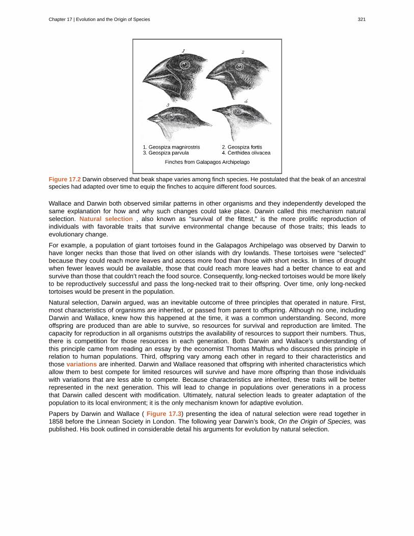
Figure 17.2 Darwin observed that beak shape varies among finch species. He postulated that the beak of an ancestralspecies had adapted over time to equip the finches to acquire different food sources.
Wallace and Darwin both observed similar patterns in other organisms and they independently developed thesame explanation for how and why such changes could take place. Darwin called this mechanism naturalselection. Natural selection , also known as “survival of the fittest,” is the more prolific reproduction ofindividuals with favorable traits that survive environmental change because of those traits; this leads toevolutionary change.
For example, a population of giant tortoises found in the Galapagos Archipelago was observed by Darwin tohave longer necks than those that lived on other islands with dry lowlands. These tortoises were “selected”because they could reach more leaves and access more food than those with short necks. In times of droughtwhen fewer leaves would be available, those that could reach more leaves had a better chance to eat andsurvive than those that couldn’t reach the food source. Consequently, long-necked tortoises would be more likelyto be reproductively successful and pass the long-necked trait to their offspring. Over time, only long-neckedtortoises would be present in the population.
Natural selection, Darwin argued, was an inevitable outcome of three principles that operated in nature. First,most characteristics of organisms are inherited, or passed from parent to offspring. Although no one, includingDarwin and Wallace, knew how this happened at the time, it was a common understanding. Second, moreoffspring are produced than are able to survive, so resources for survival and reproduction are limited. Thecapacity for reproduction in all organisms outstrips the availability of resources to support their numbers. Thus,there is competition for those resources in each generation. Both Darwin and Wallace’s understanding ofthis principle came from reading an essay by the economist Thomas Malthus who discussed this principle inrelation to human populations. Third, offspring vary among each other in regard to their characteristics andthose variations are inherited. Darwin and Wallace reasoned that offspring with inherited characteristics whichallow them to best compete for limited resources will survive and have more offspring than those individualswith variations that are less able to compete. Because characteristics are inherited, these traits will be betterrepresented in the next generation. This will lead to change in populations over generations in a processthat Darwin called descent with modification. Ultimately, natural selection leads to greater adaptation of thepopulation to its local environment; it is the only mechanism known for adaptive evolution.
Papers by Darwin and Wallace ( Figure 17.3) presenting the idea of natural selection were read together in1858 before the Linnean Society in London. The following year Darwin’s book, On the Origin of Species, waspublished. His book outlined in considerable detail his arguments for evolution by natural selection.
Chapter 17 | Evolution and the Origin of Species 321
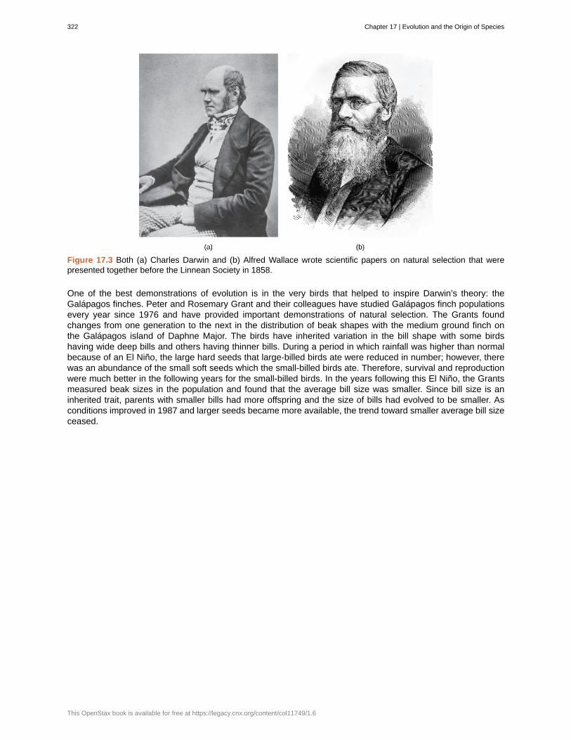
Figure 17.3 Both (a) Charles Darwin and (b) Alfred Wallace wrote scientific papers on natural selection that werepresented together before the Linnean Society in 1858.
One of the best demonstrations of evolution is in the very birds that helped to inspire Darwin’s theory: theGalápagos finches. Peter and Rosemary Grant and their colleagues have studied Galápagos finch populationsevery year since 1976 and have provided important demonstrations of natural selection. The Grants foundchanges from one generation to the next in the distribution of beak shapes with the medium ground finch onthe Galápagos island of Daphne Major. The birds have inherited variation in the bill shape with some birdshaving wide deep bills and others having thinner bills. During a period in which rainfall was higher than normalbecause of an El Niño, the large hard seeds that large-billed birds ate were reduced in number; however, therewas an abundance of the small soft seeds which the small-billed birds ate. Therefore, survival and reproductionwere much better in the following years for the small-billed birds. In the years following this El Niño, the Grantsmeasured beak sizes in the population and found that the average bill size was smaller. Since bill size is aninherited trait, parents with smaller bills had more offspring and the size of bills had evolved to be smaller. Asconditions improved in 1987 and larger seeds became more available, the trend toward smaller average bill sizeceased.
322 Chapter 17 | Evolution and the Origin of Species
This OpenStax book is available for free at https://legacy.cnx.org/content/col11749/1.6
Field BiologistMany people hike, explore caves, scuba dive, or climb mountains for recreation. People often participatein these activities hoping to see wildlife. Experiencing the outdoors can be incredibly enjoyable andinvigorating. What if your job was to be outside in the wilderness? Field biologists by definition work outdoorsin the “field.” The term field in this case refers to any location outdoors, even under water. A field biologisttypically focuses research on a certain species, group of organisms, or a single habitat ( Figure 17.4).
Figure 17.4 A field biologist tranquilizes a polar bear for study. (credit: Karen Rhode)
One objective of many field biologists includes discovering new species that have never been recorded.Not only do such findings expand our understanding of the natural world, but they also lead to importantinnovations in fields such as medicine and agriculture. Plant and microbial species, in particular, can revealnew medicinal and nutritive knowledge. Other organisms can play key roles in ecosystems or be consideredrare and in need of protection. When discovered, these important species can be used as evidence forenvironmental regulations and laws.
17.1b Processes and Patterns of Evolution
Natural selection can only take place if there is variation , or differences, among individuals in a population.Importantly, these differences must have some genetic basis; otherwise, the selection will not lead to change inthe next generation. This is critical because variation among individuals can be caused by non-genetic reasonssuch as an individual being taller because of better nutrition rather than different genes.
Genetic diversity in a population comes from two main mechanisms: mutation and sexual reproduction. Mutation,a change in DNA, is the ultimate source of new alleles, or new genetic variation in any population. Thegenetic changes caused by mutation can have one of three outcomes on the phenotype. A mutation affectsthe phenotype of the organism in a way that gives it reduced fitness—lower likelihood of survival or feweroffspring. A mutation may produce a phenotype with a beneficial effect on fitness. And, many mutations will alsohave no effect on the fitness of the phenotype; these are called neutral mutations. Mutations may also have awhole range of effect sizes on the fitness of the organism that expresses them in their phenotype, from a smalleffect to a great effect. Sexual reproduction also leads to genetic diversity: when two parents reproduce, uniquecombinations of alleles assemble to produce the unique genotypes and thus phenotypes in each of the offspring.
A heritable trait that helps the survival and/or reproduction of an organism in its present environment is called anadaptation . Scientists describe groups of organisms becoming adapted to their environment when a changein the range of genetic variation occurs over time that increases or maintains the “fit” of the population to itsenvironment. The webbed feet of platypuses are an adaptation for swimming. The snow leopards’ thick fur is anadaptation for living in the cold. The cheetahs’ fast speed is an adaptation for catching prey.
Chapter 17 | Evolution and the Origin of Species 323
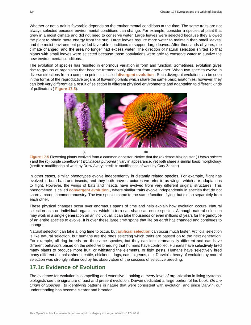
Whether or not a trait is favorable depends on the environmental conditions at the time. The same traits are notalways selected because environmental conditions can change. For example, consider a species of plant thatgrew in a moist climate and did not need to conserve water. Large leaves were selected because they allowedthe plant to obtain more energy from the sun. Large leaves require more water to maintain than small leaves,and the moist environment provided favorable conditions to support large leaves. After thousands of years, theclimate changed, and the area no longer had excess water. The direction of natural selection shifted so thatplants with small leaves were selected because those populations were able to conserve water to survive thenew environmental conditions.
The evolution of species has resulted in enormous variation in form and function. Sometimes, evolution givesrise to groups of organisms that become tremendously different from each other. When two species evolve indiverse directions from a common point, it is called divergent evolution . Such divergent evolution can be seenin the forms of the reproductive organs of flowering plants which share the same basic anatomies; however, theycan look very different as a result of selection in different physical environments and adaptation to different kindsof pollinators ( Figure 17.5).
Figure 17.5 Flowering plants evolved from a common ancestor. Notice that the (a) dense blazing star ( Liatrus spicata) and the (b) purple coneflower ( Echinacea purpurea ) vary in appearance, yet both share a similar basic morphology.(credit a: modification of work by Drew Avery; credit b: modification of work by Cory Zanker)
In other cases, similar phenotypes evolve independently in distantly related species. For example, flight hasevolved in both bats and insects, and they both have structures we refer to as wings, which are adaptationsto flight. However, the wings of bats and insects have evolved from very different original structures. Thisphenomenon is called convergent evolution , where similar traits evolve independently in species that do notshare a recent common ancestry. The two species came to the same function, flying, but did so separately fromeach other.
These physical changes occur over enormous spans of time and help explain how evolution occurs. Naturalselection acts on individual organisms, which in turn can shape an entire species. Although natural selectionmay work in a single generation on an individual, it can take thousands or even millions of years for the genotypeof an entire species to evolve. It is over these large time spans that life on earth has changed and continues tochange.
Natural selection can take a long time to occur, but artificial selection can occur much faster. Artificial selectionis like natural selection, but humans are the ones selecting which traits are passed on to the next generation.For example, all dog breeds are the same species, but they can look dramatically different and can havedifferent behaviors based on the selective breeding that humans have controlled. Humans have selectively bredmany plants to produce more fruit, or withstand the elements, or fight pests. Humans have selectively bredmany different animals: sheep, cattle, chickens, dogs, cats, pigeons, etc. Darwin's theory of evolution by naturalselection was strongly influenced by his observation of the success of selective breeding.
17.1c Evidence of Evolution
The evidence for evolution is compelling and extensive. Looking at every level of organization in living systems,biologists see the signature of past and present evolution. Darwin dedicated a large portion of his book, On theOrigin of Species , to identifying patterns in nature that were consistent with evolution, and since Darwin, ourunderstanding has become clearer and broader.
324 Chapter 17 | Evolution and the Origin of Species
This OpenStax book is available for free at https://legacy.cnx.org/content/col11749/1.6
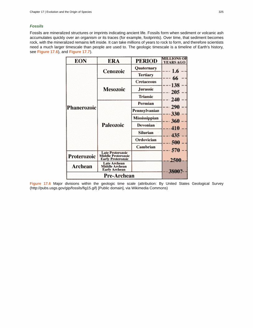
Fossils
Fossils are mineralized structures or imprints indicating ancient life. Fossils form when sediment or volcanic ashaccumulates quickly over an organism or its traces (for example, footprints). Over time, that sediment becomesrock, with the mineralized remains left inside. It can take millions of years to rock to form, and therefore scientistsneed a much larger timescale than people are used to. The geologic timescale is a timeline of Earth's history,see Figure 17.6), and Figure 17.7).
Figure 17.6 Major divisions within the geologic time scale (attribution: By United States Geological Survey(http://pubs.usgs.gov/gip/fossils/fig15.gif) [Public domain], via Wikimedia Commons)
Chapter 17 | Evolution and the Origin of Species 325
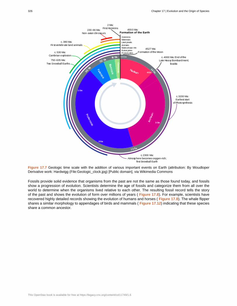
Figure 17.7 Geologic time scale with the addition of various important events on Earth (attribution: By WoudloperDerivative work: Hardwigg (File:Geologic_clock.jpg) [Public domain], via Wikimedia Commons
Fossils provide solid evidence that organisms from the past are not the same as those found today, and fossilsshow a progression of evolution. Scientists determine the age of fossils and categorize them from all over theworld to determine when the organisms lived relative to each other. The resulting fossil record tells the storyof the past and shows the evolution of form over millions of years ( Figure 17.8). For example, scientists haverecovered highly detailed records showing the evolution of humans and horses ( Figure 17.8). The whale flippershares a similar morphology to appendages of birds and mammals ( Figure 17.12) indicating that these speciesshare a common ancestor.
326 Chapter 17 | Evolution and the Origin of Species
This OpenStax book is available for free at https://legacy.cnx.org/content/col11749/1.6
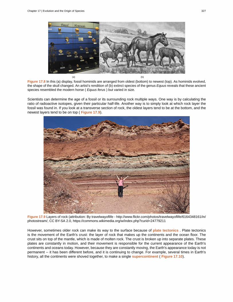
Figure 17.8 In this (a) display, fossil hominids are arranged from oldest (bottom) to newest (top). As hominids evolved,the shape of the skull changed. An artist’s rendition of (b) extinct species of the genus Equus reveals that these ancientspecies resembled the modern horse ( Equus ferus ) but varied in size.
Scientists can determine the age of a fossil or its surrounding rock multiple ways. One way is by calculating theratio of radioactive isotopes, given their particular half-life. Another way is to simply look at which rock layer thefossil was found in. If you look at a transverse section of rock, the oldest layers tend to be at the bottom, and thenewest layers tend to be on top ( Figure 17.9).
Figure 17.9 Layers of rock (attribution: By travelwayoflife - http://www.flickr.com/photos/travelwayoflife/6164348161/in/photostream/, CC BY-SA 2.0, https://commons.wikimedia.org/w/index.php?curid=24779211
However, sometimes older rock can make its way to the surface because of plate tectonics . Plate tectonicsis the movement of the Earth's crust: the layer of rock that makes up the continents and the ocean floor. Thecrust sits on top of the mantle, which is made of molten rock. The crust is broken up into separate plates. Theseplates are constantly in motion, and their movement is responsible for the current appearance of the Earth'scontinents and oceans today. However, because they are constantly moving, the Earth's appearance today is notpermanent -- it has been different before, and it is continuing to change. For example, several times in Earth'shistory, all the continents were shoved together, to make a single supercontinent ( Figure 17.10).
Chapter 17 | Evolution and the Origin of Species 327
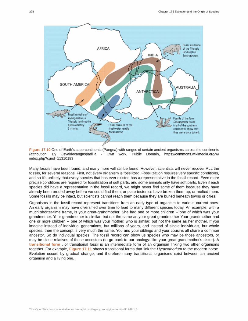
Figure 17.10 One of Earth’s supercontinents (Pangea) with ranges of certain ancient organisms across the continents(attribution: By Osvaldocangaspadilla - Own work, Public Domain, https://commons.wikimedia.org/w/index.php?curid=11310183
Many fossils have been found, and many more will still be found. However, scientists will never recover ALL thefossils, for several reasons. First, not every organism is fossilized. Fossilization requires very specific conditions,and so it's unlikely that every species that has ever existed has a representative in the fossil record. Even moreprecise conditions are required for fossilization of soft parts, and some animals only have soft parts. Even if eachspecies did have a representative in the fossil record, we might never find some of them because they havealready been eroded away before we could find them, or plate tectonics have broken them up, or melted them.Some fossils may be intact, but scientists cannot reach them because they are buried beneath towns or cities.
Organisms in the fossil record represent transitions from an early type of organism to various current ones.An early organism may have diversified over time to lead to many different species today. An example, with amuch shorter-time frame, is your great-grandmother. She had one or more children -- one of which was yourgrandmother. Your grandmother is similar, but not the same as your great-grandmother Your grandmother hadone or more children -- one of which was your mother, who is similar, but not the same as her mother. If youimagine instead of individual generations, but millions of years, and instead of single individuals, but wholespecies, then the concept is very much the same. You and your siblings and your cousins all share a commonancestor. So do individual species. The fossil record can show us species who may be those ancestors, ormay be close relatives of those ancestors (to go back to our analogy: like your great-grandmother's sister). Atransitional form , or transitional fossil is an intermediate form of an organism linking two other organismstogether. For example, Figure 17.11 shows transitional forms that link the Hyracotherium to the modern horse.Evolution occurs by gradual change, and therefore many transitional organisms exist between an ancientorganism and a living one.
328 Chapter 17 | Evolution and the Origin of Species
This OpenStax book is available for free at https://legacy.cnx.org/content/col11749/1.6
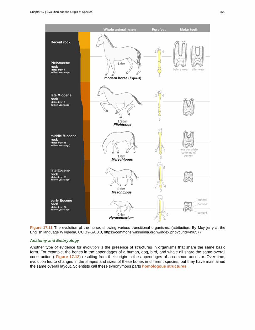
Figure 17.11 The evolution of the horse, showing various transitional organisms. (attribution: By Mcy jerry at theEnglish language Wikipedia, CC BY-SA 3.0, https://commons.wikimedia.org/w/index.php?curid=496577
Anatomy and Embryology
Another type of evidence for evolution is the presence of structures in organisms that share the same basicform. For example, the bones in the appendages of a human, dog, bird, and whale all share the same overallconstruction ( Figure 17.12) resulting from their origin in the appendages of a common ancestor. Over time,evolution led to changes in the shapes and sizes of these bones in different species, but they have maintainedthe same overall layout. Scientists call these synonymous parts homologous structures .
Chapter 17 | Evolution and the Origin of Species 329
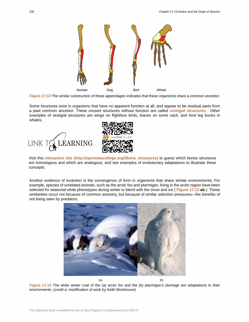
Figure 17.12 The similar construction of these appendages indicates that these organisms share a common ancestor.
Some structures exist in organisms that have no apparent function at all, and appear to be residual parts froma past common ancestor. These unused structures without function are called vestigial structures . Otherexamples of vestigial structures are wings on flightless birds, leaves on some cacti, and hind leg bones inwhales.
Visit this interactive site (http://openstaxcollege.org/l/bone_structures) to guess which bones structuresare homologous and which are analogous, and see examples of evolutionary adaptations to illustrate theseconcepts.
Another evidence of evolution is the convergence of form in organisms that share similar environments. Forexample, species of unrelated animals, such as the arctic fox and ptarmigan, living in the arctic region have beenselected for seasonal white phenotypes during winter to blend with the snow and ice ( Figure 17.13 ab ). Thesesimilarities occur not because of common ancestry, but because of similar selection pressures—the benefits ofnot being seen by predators.
Figure 17.13 The white winter coat of the (a) arctic fox and the (b) ptarmigan’s plumage are adaptations to theirenvironments. (credit a: modification of work by Keith Morehouse)
330 Chapter 17 | Evolution and the Origin of Species
This OpenStax book is available for free at https://legacy.cnx.org/content/col11749/1.6
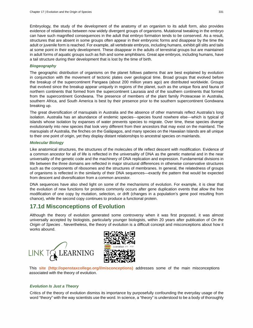
Embryology, the study of the development of the anatomy of an organism to its adult form, also providesevidence of relatedness between now widely divergent groups of organisms. Mutational tweaking in the embryocan have such magnified consequences in the adult that embryo formation tends to be conserved. As a result,structures that are absent in some groups often appear in their embryonic forms and disappear by the time theadult or juvenile form is reached. For example, all vertebrate embryos, including humans, exhibit gill slits and tailsat some point in their early development. These disappear in the adults of terrestrial groups but are maintainedin adult forms of aquatic groups such as fish and some amphibians. Great ape embryos, including humans, havea tail structure during their development that is lost by the time of birth.
Biogeography
The geographic distribution of organisms on the planet follows patterns that are best explained by evolutionin conjunction with the movement of tectonic plates over geological time. Broad groups that evolved beforethe breakup of the supercontinent Pangaea (about 200 million years ago) are distributed worldwide. Groupsthat evolved since the breakup appear uniquely in regions of the planet, such as the unique flora and fauna ofnorthern continents that formed from the supercontinent Laurasia and of the southern continents that formedfrom the supercontinent Gondwana. The presence of members of the plant family Proteaceae in Australia,southern Africa, and South America is best by their presence prior to the southern supercontinent Gondwanabreaking up.
The great diversification of marsupials in Australia and the absence of other mammals reflect Australia’s longisolation. Australia has an abundance of endemic species—species found nowhere else—which is typical ofislands whose isolation by expanses of water prevents species to migrate. Over time, these species divergeevolutionarily into new species that look very different from their ancestors that may exist on the mainland. Themarsupials of Australia, the finches on the Galápagos, and many species on the Hawaiian Islands are all uniqueto their one point of origin, yet they display distant relationships to ancestral species on mainlands.
Molecular Biology
Like anatomical structures, the structures of the molecules of life reflect descent with modification. Evidence ofa common ancestor for all of life is reflected in the universality of DNA as the genetic material and in the nearuniversality of the genetic code and the machinery of DNA replication and expression. Fundamental divisions inlife between the three domains are reflected in major structural differences in otherwise conservative structuressuch as the components of ribosomes and the structures of membranes. In general, the relatedness of groupsof organisms is reflected in the similarity of their DNA sequences—exactly the pattern that would be expectedfrom descent and diversification from a common ancestor.
DNA sequences have also shed light on some of the mechanisms of evolution. For example, it is clear thatthe evolution of new functions for proteins commonly occurs after gene duplication events that allow the freemodification of one copy by mutation, selection, or drift (changes in a population’s gene pool resulting fromchance), while the second copy continues to produce a functional protein.
17.1d Misconceptions of Evolution
Although the theory of evolution generated some controversy when it was first proposed, it was almostuniversally accepted by biologists, particularly younger biologists, within 20 years after publication of On theOrigin of Species . Nevertheless, the theory of evolution is a difficult concept and misconceptions about how itworks abound.
This site (http://openstaxcollege.org/l/misconceptions) addresses some of the main misconceptionsassociated with the theory of evolution.
Evolution Is Just a Theory
Critics of the theory of evolution dismiss its importance by purposefully confounding the everyday usage of theword “theory” with the way scientists use the word. In science, a “theory” is understood to be a body of thoroughly
Chapter 17 | Evolution and the Origin of Species 331
tested and verified explanations for a set of observations of the natural world. Scientists have a theory of theatom, a theory of gravity, and the theory of relativity, each of which describes understood facts about the world.In the same way, the theory of evolution describes facts about the living world. As such, a theory in science hassurvived significant efforts to discredit it by scientists. In contrast, a “theory” in common vernacular is a wordmeaning a guess or suggested explanation; this meaning is more akin to the scientific concept of “hypothesis.”When critics of evolution say evolution is “just a theory,” they are implying that there is little evidence supportingit and that it is still in the process of being rigorously tested. This is a mischaracterization.
Individuals Evolve
Evolution is the change in genetic composition of a population over time, specifically over generations, resultingfrom differential reproduction of individuals with certain alleles. Individuals do change over their lifetime,obviously, but this is called development and involves changes programmed by the set of genes the individualacquired at birth in coordination with the individual’s environment. When thinking about the evolution of acharacteristic, it is probably best to think about the change of the average value of the characteristic in thepopulation over time. For example, when natural selection leads to bill-size change in medium-ground finchesin the Galápagos, this does not mean that individual bills on the finches are changing. If one measures theaverage bill size among all individuals in the population at one time and then measures the average bill size inthe population several years later, this average value will be different as a result of evolution. Although someindividuals may survive from the first time to the second, they will still have the same bill size; however, there willbe many new individuals that contribute to the shift in average bill size.
Evolution Explains the Origin of Life
It is a common misunderstanding that evolution includes an explanation of life’s origins. Conversely, some of thetheory’s critics believe that it cannot explain the origin of life. The theory does not try to explain the origin of life.The theory of evolution explains how populations change over time and how life diversifies the origin of species.It does not shed light on the beginnings of life including the origins of the first cells, which is how life is defined.The mechanisms of the origin of life on Earth are a particularly difficult problem because it occurred a very longtime ago, and presumably it just occurred once. Importantly, biologists believe that the presence of life on Earthprecludes the possibility that the events that led to life on Earth can be repeated because the intermediate stageswould immediately become food for existing living things.
However, once a mechanism of inheritance was in place in the form of a molecule like DNA either within a cellor pre-cell, these entities would be subject to the principle of natural selection. More effective reproducers wouldincrease in frequency at the expense of inefficient reproducers. So while evolution does not explain the originof life, it may have something to say about some of the processes operating once pre-living entities acquiredcertain properties.
Organisms Evolve on Purpose
Statements such as “organisms evolve in response to a change in an environment” are quite common, butsuch statements can lead to two types of misunderstandings. First, the statement must not be understood tomean that individual organisms evolve. The statement is shorthand for “a population evolves in response to achanging environment.” However, a second misunderstanding may arise by interpreting the statement to meanthat the evolution is somehow intentional. A changed environment results in some individuals in the population,those with particular phenotypes, benefiting and therefore producing proportionately more offspring than otherphenotypes. This results in change in the population if the characteristics are genetically determined.
It is also important to understand that the variation that natural selection works on is already in a populationand does not arise in response to an environmental change. For example, applying antibiotics to a population ofbacteria will, over time, select a population of bacteria that are resistant to antibiotics. The resistance, which iscaused by a gene, did not arise by mutation because of the application of the antibiotic. The gene for resistancewas already present in the gene pool of the bacteria, likely at a low frequency. The antibiotic, which kills thebacterial cells without the resistance gene, strongly selects individuals that are resistant, since these would bethe only ones that survived and divided. Experiments have demonstrated that mutations for antibiotic resistancedo not arise as a result of antibiotic.
In a larger sense, evolution is not goal directed. Species do not become “better” over time; they simply tracktheir changing environment with adaptations that maximize their reproduction in a particular environment at aparticular time. Evolution has no goal of making faster, bigger, more complex, or even smarter species, despitethe commonness of this kind of language in popular discourse. What characteristics evolve in a species are afunction of the variation present and the environment, both of which are constantly changing in a non-directionalway. What trait is fit in one environment at one time may well be fatal at some point in the future. This holds
332 Chapter 17 | Evolution and the Origin of Species
This OpenStax book is available for free at https://legacy.cnx.org/content/col11749/1.6
equally well for a species of insect as it does the human species.
17.2 | Population Evolution
By the end of this section, you will be able to:
• Define population genetics and describe how population genetics is used in the study of the evolution ofpopulations
• Define the Hardy-Weinberg principle and discuss its importance
The mechanisms of inheritance, or genetics, were not understood at the time Charles Darwin and Alfred RusselWallace were developing their idea of natural selection. This lack of understanding was a stumbling block tounderstanding many aspects of evolution. In fact, the predominant (and incorrect) genetic theory of the time,blending inheritance, made it difficult to understand how natural selection might operate. Darwin and Wallacewere unaware of the genetics work by Austrian monk Gregor Mendel, which was published in 1866, not longafter publication of Darwin's book, On the Origin of Species . Mendel’s work was rediscovered in the earlytwentieth century at which time geneticists were rapidly coming to an understanding of the basics of inheritance.Initially, the newly discovered particulate nature of genes made it difficult for biologists to understand how gradualevolution could occur. But over the next few decades genetics and evolution were integrated in what becameknown as the modern synthesis —the coherent understanding of the relationship between natural selectionand genetics that took shape by the 1940s and is generally accepted today. In sum, the modern synthesisdescribes how evolutionary processes, such as natural selection, can affect a population’s genetic makeup, and,in turn, how this can result in the gradual evolution of populations and species. The theory also connects thischange of a population over time, called microevolution , with the processes that gave rise to new species andhigher taxonomic groups with widely divergent characters, called macroevolution .
Evolution and Flu VaccinesEvery fall, the media starts reporting on flu vaccinations and potential outbreaks. Scientists, health experts,and institutions determine recommendations for different parts of the population, predict optimal productionand inoculation schedules, create vaccines, and set up clinics to provide inoculations. You may think of theannual flu shot as a lot of media hype, an important health protection, or just a briefly uncomfortable prick inyour arm. But do you think of it in terms of evolution?
The media hype of annual flu shots is scientifically grounded in our understanding of evolution. Each year,scientists across the globe strive to predict the flu strains that they anticipate being most widespread andharmful in the coming year. This knowledge is based in how flu strains have evolved over time and overthe past few flu seasons. Scientists then work to create the most effective vaccine to combat those selectedstrains. Hundreds of millions of doses are produced in a short period in order to provide vaccinations to keypopulations at the optimal time.
Because viruses, like the flu, evolve very quickly (especially in evolutionary time), this poses quite achallenge. Viruses mutate and replicate at a fast rate, so the vaccine developed to protect against last year’sflu strain may not provide the protection needed against the coming year’s strain. Evolution of these virusesmeans continued adaptions to ensure survival, including adaptations to survive previous vaccines.
17.2a Population Genetics
Recall that a gene for a particular character may have several alleles, or variants, that code for different traitsassociated with that character. For example, in the ABO blood type system in humans, three alleles determinethe particular blood-type protein on the surface of red blood cells. Each individual in a population of diploidorganisms can only carry two alleles for a particular gene, but more than two may be present in the individualsthat make up the population. Mendel followed alleles as they were inherited from parent to offspring. In the earlytwentieth century, biologists in a field of study known as population genetics began to study how selective
Chapter 17 | Evolution and the Origin of Species 333
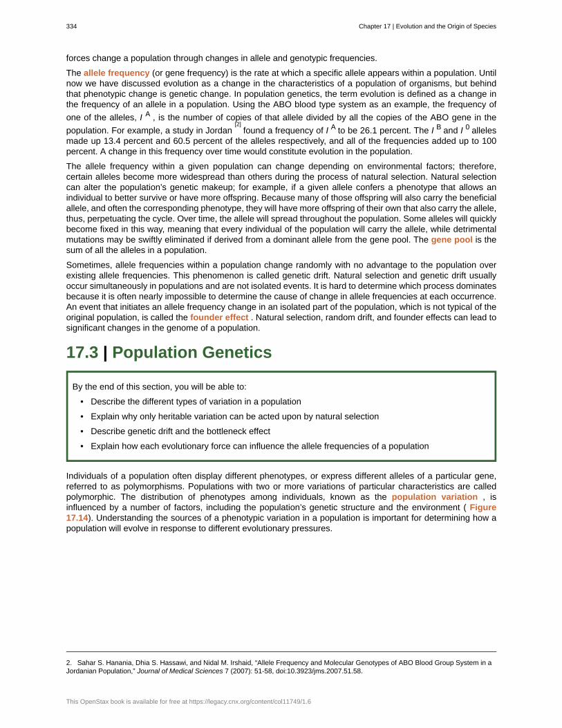
forces change a population through changes in allele and genotypic frequencies.
The allele frequency (or gene frequency) is the rate at which a specific allele appears within a population. Untilnow we have discussed evolution as a change in the characteristics of a population of organisms, but behindthat phenotypic change is genetic change. In population genetics, the term evolution is defined as a change inthe frequency of an allele in a population. Using the ABO blood type system as an example, the frequency of
one of the alleles, I A , is the number of copies of that allele divided by all the copies of the ABO gene in the
population. For example, a study in Jordan[2]
found a frequency of I A to be 26.1 percent. The I B and I 0 allelesmade up 13.4 percent and 60.5 percent of the alleles respectively, and all of the frequencies added up to 100percent. A change in this frequency over time would constitute evolution in the population.
The allele frequency within a given population can change depending on environmental factors; therefore,certain alleles become more widespread than others during the process of natural selection. Natural selectioncan alter the population’s genetic makeup; for example, if a given allele confers a phenotype that allows anindividual to better survive or have more offspring. Because many of those offspring will also carry the beneficialallele, and often the corresponding phenotype, they will have more offspring of their own that also carry the allele,thus, perpetuating the cycle. Over time, the allele will spread throughout the population. Some alleles will quicklybecome fixed in this way, meaning that every individual of the population will carry the allele, while detrimentalmutations may be swiftly eliminated if derived from a dominant allele from the gene pool. The gene pool is thesum of all the alleles in a population.
Sometimes, allele frequencies within a population change randomly with no advantage to the population overexisting allele frequencies. This phenomenon is called genetic drift. Natural selection and genetic drift usuallyoccur simultaneously in populations and are not isolated events. It is hard to determine which process dominatesbecause it is often nearly impossible to determine the cause of change in allele frequencies at each occurrence.An event that initiates an allele frequency change in an isolated part of the population, which is not typical of theoriginal population, is called the founder effect . Natural selection, random drift, and founder effects can lead tosignificant changes in the genome of a population.
17.3 | Population Genetics
By the end of this section, you will be able to:
• Describe the different types of variation in a population
• Explain why only heritable variation can be acted upon by natural selection
• Describe genetic drift and the bottleneck effect
• Explain how each evolutionary force can influence the allele frequencies of a population
Individuals of a population often display different phenotypes, or express different alleles of a particular gene,referred to as polymorphisms. Populations with two or more variations of particular characteristics are calledpolymorphic. The distribution of phenotypes among individuals, known as the population variation , isinfluenced by a number of factors, including the population’s genetic structure and the environment ( Figure17.14). Understanding the sources of a phenotypic variation in a population is important for determining how apopulation will evolve in response to different evolutionary pressures.
2. Sahar S. Hanania, Dhia S. Hassawi, and Nidal M. Irshaid, “Allele Frequency and Molecular Genotypes of ABO Blood Group System in aJordanian Population,” Journal of Medical Sciences 7 (2007): 51-58, doi:10.3923/jms.2007.51.58.
334 Chapter 17 | Evolution and the Origin of Species
This OpenStax book is available for free at https://legacy.cnx.org/content/col11749/1.6
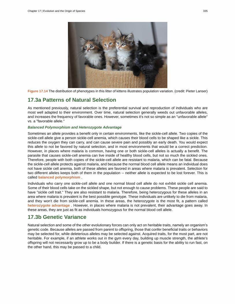
Figure 17.14 The distribution of phenotypes in this litter of kittens illustrates population variation. (credit: Pieter Lanser)
17.3a Patterns of Natural Selection
As mentioned previously, natural selection is the preferential survival and reproduction of individuals who aremost well adapted to their environment. Over time, natural selection generally weeds out unfavorable alleles,and increases the frequency of favorable ones. However, sometimes it's not so simple as an "unfavorable allele"vs. a "favorable allele."
Balanced Polymorphism and Heterozygote Advantage
Sometimes an allele provides a benefit only in certain environments, like the sickle-cell allele. Two copies of thesickle-cell allele give a person sickle-cell anemia, which causes their blood cells to be shaped like a sickle. Thisreduces the oxygen they can carry, and can cause severe pain and possibly an early death. You would expectthis allele to not be favored by natural selection, and in most environments that would be a correct prediction.However, in places where malaria is common, having one or both sickle-cell alleles is actually a benefit. Theparasite that causes sickle-cell anemia can live inside of healthy blood cells, but not so much the sickled ones.Therefore, people with both copies of the sickle-cell allele are resistant to malaria, which can be fatal. Becausethe sickle-cell allele protects against malaria, and because the normal blood cell allele means an individual doesnot have sickle cell anemia, both of these alleles are favored in areas where malaria is prevalent. Selection fortwo different alleles keeps both of them in the population -- neither allele is expected to be lost forever. This iscalled balanced polymorphism .
Individuals who carry one sickle-cell allele and one normal blood cell allele do not exhibit sickle cell anemia.Some of their blood cells take on the sickled shape, but not enough to cause problems. These people are said tohave "sickle cell trait." They are also resistant to malaria. Therefore, being heterozygous for these alleles in anarea where malaria is prevalent is the best possible genotype. These individuals are unlikely to die from malaria,and they won't die from sickle-cell anemia. In these areas, the heterozygote is the most fit, a pattern calledheterozygote advantage . However, in places where malaria is not prevalent, their advantage goes away. Inthese areas, they are just as fit as individuals homozygous for the normal blood cell allele.
17.3b Genetic Variance
Natural selection and some of the other evolutionary forces can only act on heritable traits, namely an organism’sgenetic code. Because alleles are passed from parent to offspring, those that confer beneficial traits or behaviorsmay be selected for, while deleterious alleles may be selected against. Acquired traits, for the most part, are notheritable. For example, if an athlete works out in the gym every day, building up muscle strength, the athlete’soffspring will not necessarily grow up to be a body builder. If there is a genetic basis for the ability to run fast, onthe other hand, this may be passed to a child.
Chapter 17 | Evolution and the Origin of Species 335
Before Darwinian evolution became the prevailing theory of the field, French naturalist Jean-Baptiste Lamarcktheorized that acquired traits could, in fact, be inherited; while this hypothesis has largely been unsupported,scientists have recently begun to realize that Lamarck was not completely wrong. Visit this site(http://openstaxcollege.org/l/epigenetic) to learn more.
Heritability is the fraction of phenotype variation that can be attributed to genetic differences, or geneticvariance, among individuals in a population. The greater the hereditability of a population’s phenotypic variation,the more susceptible it is to the evolutionary forces that act on heritable variation.
The diversity of alleles and genotypes within a population is called genetic variance . When scientists areinvolved in the breeding of a species, such as with animals in zoos and nature preserves, they try to increase apopulation’s genetic variance to preserve as much of the phenotypic diversity as they can. This also helps reducethe risks associated with inbreeding , the mating of closely related individuals, which can have the undesirableeffect of bringing together deleterious recessive mutations that can cause abnormalities and susceptibility todisease. For example, a disease that is caused by a rare, recessive allele might exist in a population, but it willonly manifest itself when an individual carries two copies of the allele. Because the allele is rare in a normal,healthy population with unrestricted habitat, the chance that two carriers will mate is low, and even then, only25 percent of their offspring will inherit the disease allele from both parents. While it is likely to happen at somepoint, it will not happen frequently enough for natural selection to be able to swiftly eliminate the allele from thepopulation, and as a result, the allele will be maintained at low levels in the gene pool. However, if a family ofcarriers begins to interbreed with each other, this will dramatically increase the likelihood of two carriers matingand eventually producing diseased offspring, a phenomenon known as inbreeding depression .
Changes in allele frequencies that are identified in a population can shed light on how it is evolving. In additionto natural selection, there are other evolutionary forces that could be in play: genetic drift, gene flow, mutation,nonrandom mating, and environmental variances.
17.3c Genetic Drift
The theory of natural selection stems from the observation that some individuals in a population are more likelyto survive longer and have more offspring than others; thus, they will pass on more of their alleles to the nextgeneration. A big, powerful male gorilla, for example, is much more likely than a smaller, weaker one to becomethe population’s silverback, the pack’s leader who mates far more than the other males of the group. The packleader will father more offspring, who share half of his alleles, and are likely to also grow bigger and stronger liketheir father. Over time, the alleles for bigger size will increase in frequency in the population, and the populationwill, as a result, grow larger on average. That is, this would occur if this particular selection pressure , or drivingselective force, were the only one acting on the population. In other examples, better camouflage or a strongerresistance to drought might pose a selection pressure.
Another way a population’s allele and genotype frequencies can change is genetic drift ( Figure 17.15), whichis simply the effect of chance. By chance, some individuals will have more offspring than others—not due to anadvantage conferred by some genetically-encoded trait, but just because one male happened to be in the rightplace at the right time (when the receptive female walked by) or because the other one happened to be in thewrong place at the wrong time (when a fox was hunting).
336 Chapter 17 | Evolution and the Origin of Species
This OpenStax book is available for free at https://legacy.cnx.org/content/col11749/1.6
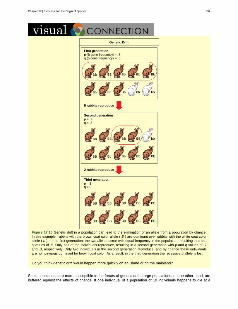
Figure 17.15 Genetic drift in a population can lead to the elimination of an allele from a population by chance.In this example, rabbits with the brown coat color allele ( B ) are dominant over rabbits with the white coat colorallele ( b ). In the first generation, the two alleles occur with equal frequency in the population, resulting in p andq values of .5. Only half of the individuals reproduce, resulting in a second generation with p and q values of .7and .3, respectively. Only two individuals in the second generation reproduce, and by chance these individualsare homozygous dominant for brown coat color. As a result, in the third generation the recessive b allele is lost.
Do you think genetic drift would happen more quickly on an island or on the mainland?
Small populations are more susceptible to the forces of genetic drift. Large populations, on the other hand, arebuffered against the effects of chance. If one individual of a population of 10 individuals happens to die at a
Chapter 17 | Evolution and the Origin of Species 337
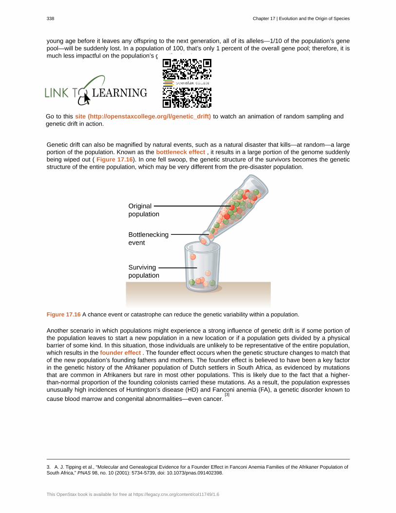
young age before it leaves any offspring to the next generation, all of its alleles—1/10 of the population’s genepool—will be suddenly lost. In a population of 100, that’s only 1 percent of the overall gene pool; therefore, it ismuch less impactful on the population’s genetic structure.
Go to this site (http://openstaxcollege.org/l/genetic_drift) to watch an animation of random sampling andgenetic drift in action.
Genetic drift can also be magnified by natural events, such as a natural disaster that kills—at random—a largeportion of the population. Known as the bottleneck effect , it results in a large portion of the genome suddenlybeing wiped out ( Figure 17.16). In one fell swoop, the genetic structure of the survivors becomes the geneticstructure of the entire population, which may be very different from the pre-disaster population.
Figure 17.16 A chance event or catastrophe can reduce the genetic variability within a population.
Another scenario in which populations might experience a strong influence of genetic drift is if some portion ofthe population leaves to start a new population in a new location or if a population gets divided by a physicalbarrier of some kind. In this situation, those individuals are unlikely to be representative of the entire population,which results in the founder effect . The founder effect occurs when the genetic structure changes to match thatof the new population’s founding fathers and mothers. The founder effect is believed to have been a key factorin the genetic history of the Afrikaner population of Dutch settlers in South Africa, as evidenced by mutationsthat are common in Afrikaners but rare in most other populations. This is likely due to the fact that a higher-than-normal proportion of the founding colonists carried these mutations. As a result, the population expressesunusually high incidences of Huntington’s disease (HD) and Fanconi anemia (FA), a genetic disorder known to
cause blood marrow and congenital abnormalities—even cancer.[3]
3. A. J. Tipping et al., “Molecular and Genealogical Evidence for a Founder Effect in Fanconi Anemia Families of the Afrikaner Population ofSouth Africa,” PNAS 98, no. 10 (2001): 5734-5739, doi: 10.1073/pnas.091402398.
338 Chapter 17 | Evolution and the Origin of Species
This OpenStax book is available for free at https://legacy.cnx.org/content/col11749/1.6
Watch this short video (http://openstaxcollege.org/l/founder_bottle) to learn more about the founder andbottleneck effects.
Testing the Bottleneck EffectQuestion: How do natural disasters affect the genetic structure of a population?
Background: When much of a population is suddenly wiped out by an earthquake or hurricane, theindividuals that survive the event are usually a random sampling of the original group. As a result, thegenetic makeup of the population can change dramatically. This phenomenon is known as the bottleneckeffect.
Hypothesis: Repeated natural disasters will yield different population genetic structures; therefore, eachtime this experiment is run, the results will vary.
Test the hypothesis: Count out the original population using different colored beads. For example, red,blue, and yellow beads might represent red, blue, and yellow individuals. After recording the number ofeach individual in the original population, place them all in a bottle with a narrow neck that will only allow afew beads out at a time. Then, pour 1/3 of the bottle’s contents into a bowl. This represents the survivingindividuals after a natural disaster kills a majority of the population. Count the number of the different coloredbeads in the bowl, and record it. Then, place all of the beads back in the bottle and repeat the experimentfour more times.
Analyze the data: Compare the five populations that resulted from the experiment. Do the populations allcontain the same number of different colored beads, or do they vary? Remember, these populations allcame from the same exact parent population.
Form a conclusion: Most likely, the five resulting populations will differ quite dramatically. This is becausenatural disasters are not selective—they kill and spare individuals at random. Now think about how thismight affect a real population. What happens when a hurricane hits the Mississippi Gulf Coast? How do theseabirds that live on the beach fare?
17.3d Gene Flow
Another important evolutionary force is gene flow : the flow of alleles in and out of a population due to themigration of individuals or gametes ( Figure 17.17). While some populations are fairly stable, others experiencemore flux. Many plants, for example, send their pollen far and wide, by wind or by bird, to pollinate otherpopulations of the same species some distance away. Even a population that may initially appear to be stable,such as a pride of lions, can experience its fair share of immigration and emigration as developing males leavetheir mothers to seek out a new pride with genetically unrelated females. This variable flow of individuals in andout of the group not only changes the gene structure of the population, but it can also introduce new geneticvariation to populations in different geological locations and habitats.
Chapter 17 | Evolution and the Origin of Species 339
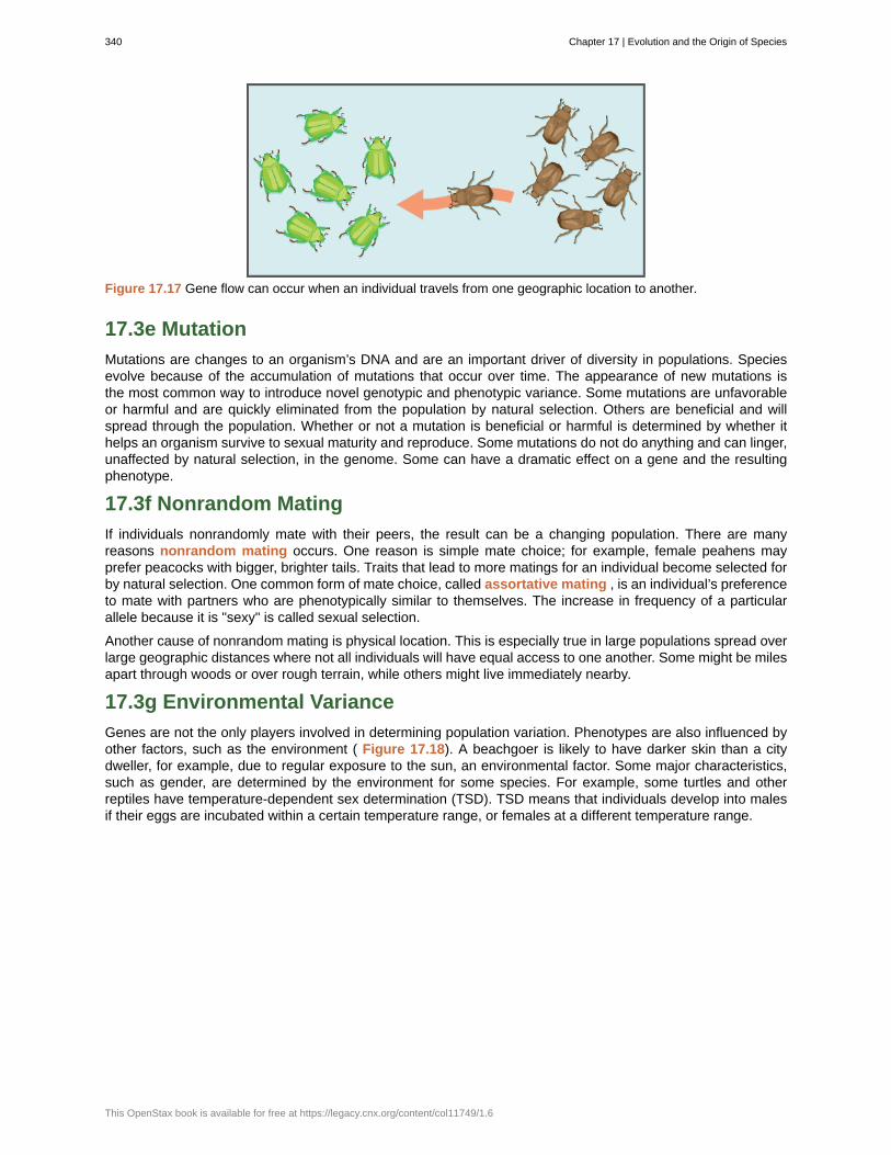
Figure 17.17 Gene flow can occur when an individual travels from one geographic location to another.
17.3e Mutation
Mutations are changes to an organism’s DNA and are an important driver of diversity in populations. Speciesevolve because of the accumulation of mutations that occur over time. The appearance of new mutations isthe most common way to introduce novel genotypic and phenotypic variance. Some mutations are unfavorableor harmful and are quickly eliminated from the population by natural selection. Others are beneficial and willspread through the population. Whether or not a mutation is beneficial or harmful is determined by whether ithelps an organism survive to sexual maturity and reproduce. Some mutations do not do anything and can linger,unaffected by natural selection, in the genome. Some can have a dramatic effect on a gene and the resultingphenotype.
17.3f Nonrandom Mating
If individuals nonrandomly mate with their peers, the result can be a changing population. There are manyreasons nonrandom mating occurs. One reason is simple mate choice; for example, female peahens mayprefer peacocks with bigger, brighter tails. Traits that lead to more matings for an individual become selected forby natural selection. One common form of mate choice, called assortative mating , is an individual’s preferenceto mate with partners who are phenotypically similar to themselves. The increase in frequency of a particularallele because it is "sexy" is called sexual selection.
Another cause of nonrandom mating is physical location. This is especially true in large populations spread overlarge geographic distances where not all individuals will have equal access to one another. Some might be milesapart through woods or over rough terrain, while others might live immediately nearby.
17.3g Environmental Variance
Genes are not the only players involved in determining population variation. Phenotypes are also influenced byother factors, such as the environment ( Figure 17.18). A beachgoer is likely to have darker skin than a citydweller, for example, due to regular exposure to the sun, an environmental factor. Some major characteristics,such as gender, are determined by the environment for some species. For example, some turtles and otherreptiles have temperature-dependent sex determination (TSD). TSD means that individuals develop into malesif their eggs are incubated within a certain temperature range, or females at a different temperature range.
340 Chapter 17 | Evolution and the Origin of Species
This OpenStax book is available for free at https://legacy.cnx.org/content/col11749/1.6
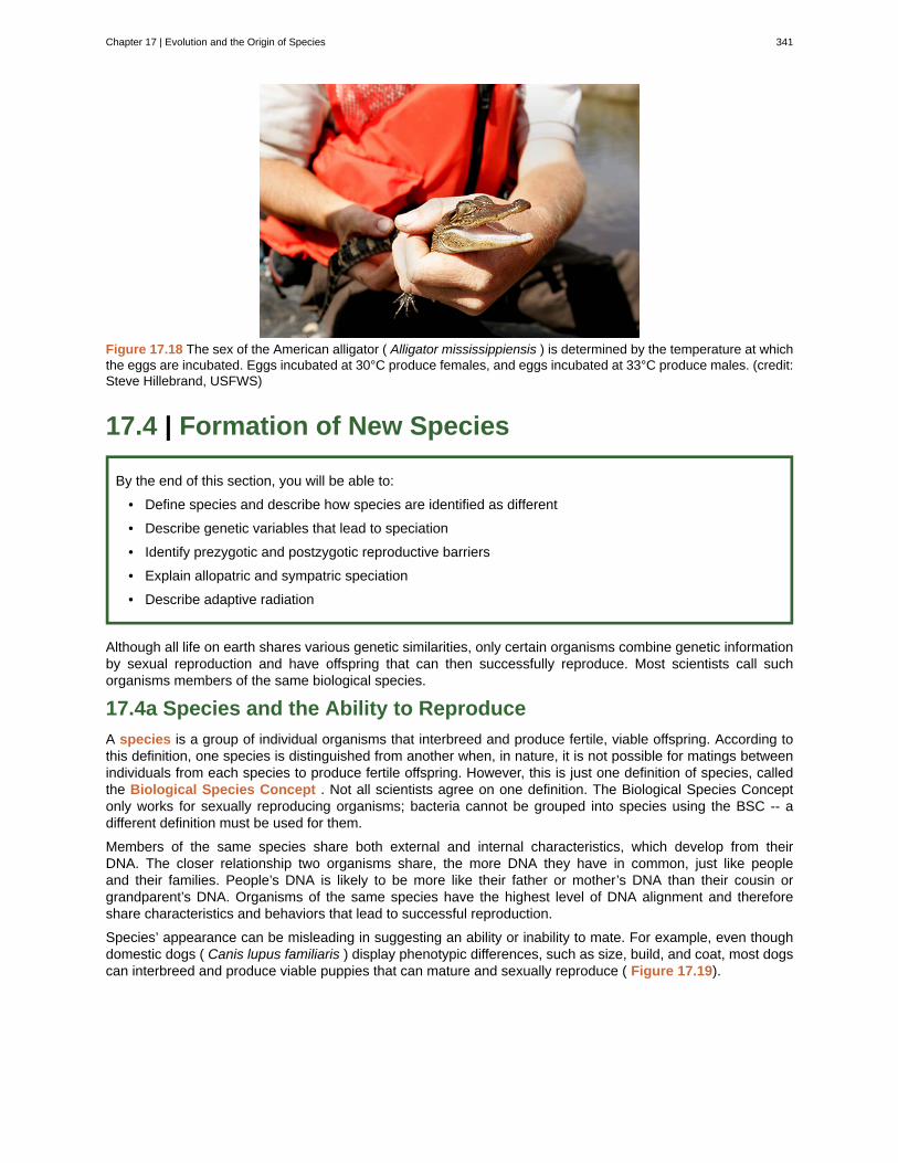
Figure 17.18 The sex of the American alligator ( Alligator mississippiensis ) is determined by the temperature at whichthe eggs are incubated. Eggs incubated at 30°C produce females, and eggs incubated at 33°C produce males. (credit:Steve Hillebrand, USFWS)
17.4 | Formation of New Species
By the end of this section, you will be able to:
• Define species and describe how species are identified as different
• Describe genetic variables that lead to speciation
• Identify prezygotic and postzygotic reproductive barriers
• Explain allopatric and sympatric speciation
• Describe adaptive radiation
Although all life on earth shares various genetic similarities, only certain organisms combine genetic informationby sexual reproduction and have offspring that can then successfully reproduce. Most scientists call suchorganisms members of the same biological species.
17.4a Species and the Ability to Reproduce
A species is a group of individual organisms that interbreed and produce fertile, viable offspring. According tothis definition, one species is distinguished from another when, in nature, it is not possible for matings betweenindividuals from each species to produce fertile offspring. However, this is just one definition of species, calledthe Biological Species Concept . Not all scientists agree on one definition. The Biological Species Conceptonly works for sexually reproducing organisms; bacteria cannot be grouped into species using the BSC -- adifferent definition must be used for them.
Members of the same species share both external and internal characteristics, which develop from theirDNA. The closer relationship two organisms share, the more DNA they have in common, just like peopleand their families. People’s DNA is likely to be more like their father or mother’s DNA than their cousin orgrandparent’s DNA. Organisms of the same species have the highest level of DNA alignment and thereforeshare characteristics and behaviors that lead to successful reproduction.
Species’ appearance can be misleading in suggesting an ability or inability to mate. For example, even thoughdomestic dogs ( Canis lupus familiaris ) display phenotypic differences, such as size, build, and coat, most dogscan interbreed and produce viable puppies that can mature and sexually reproduce ( Figure 17.19).
Chapter 17 | Evolution and the Origin of Species 341
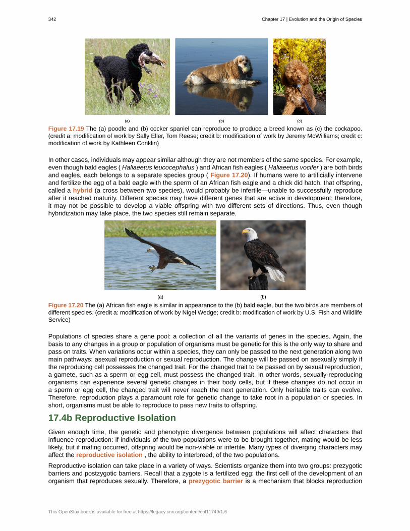
Figure 17.19 The (a) poodle and (b) cocker spaniel can reproduce to produce a breed known as (c) the cockapoo.(credit a: modification of work by Sally Eller, Tom Reese; credit b: modification of work by Jeremy McWilliams; credit c:modification of work by Kathleen Conklin)
In other cases, individuals may appear similar although they are not members of the same species. For example,even though bald eagles ( Haliaeetus leucocephalus ) and African fish eagles ( Haliaeetus vocifer ) are both birdsand eagles, each belongs to a separate species group ( Figure 17.20). If humans were to artificially interveneand fertilize the egg of a bald eagle with the sperm of an African fish eagle and a chick did hatch, that offspring,called a hybrid (a cross between two species), would probably be infertile—unable to successfully reproduceafter it reached maturity. Different species may have different genes that are active in development; therefore,it may not be possible to develop a viable offspring with two different sets of directions. Thus, even thoughhybridization may take place, the two species still remain separate.
Figure 17.20 The (a) African fish eagle is similar in appearance to the (b) bald eagle, but the two birds are members ofdifferent species. (credit a: modification of work by Nigel Wedge; credit b: modification of work by U.S. Fish and WildlifeService)
Populations of species share a gene pool: a collection of all the variants of genes in the species. Again, thebasis to any changes in a group or population of organisms must be genetic for this is the only way to share andpass on traits. When variations occur within a species, they can only be passed to the next generation along twomain pathways: asexual reproduction or sexual reproduction. The change will be passed on asexually simply ifthe reproducing cell possesses the changed trait. For the changed trait to be passed on by sexual reproduction,a gamete, such as a sperm or egg cell, must possess the changed trait. In other words, sexually-reproducingorganisms can experience several genetic changes in their body cells, but if these changes do not occur ina sperm or egg cell, the changed trait will never reach the next generation. Only heritable traits can evolve.Therefore, reproduction plays a paramount role for genetic change to take root in a population or species. Inshort, organisms must be able to reproduce to pass new traits to offspring.
17.4b Reproductive Isolation
Given enough time, the genetic and phenotypic divergence between populations will affect characters thatinfluence reproduction: if individuals of the two populations were to be brought together, mating would be lesslikely, but if mating occurred, offspring would be non-viable or infertile. Many types of diverging characters mayaffect the reproductive isolation , the ability to interbreed, of the two populations.
Reproductive isolation can take place in a variety of ways. Scientists organize them into two groups: prezygoticbarriers and postzygotic barriers. Recall that a zygote is a fertilized egg: the first cell of the development of anorganism that reproduces sexually. Therefore, a prezygotic barrier is a mechanism that blocks reproduction
342 Chapter 17 | Evolution and the Origin of Species
This OpenStax book is available for free at https://legacy.cnx.org/content/col11749/1.6
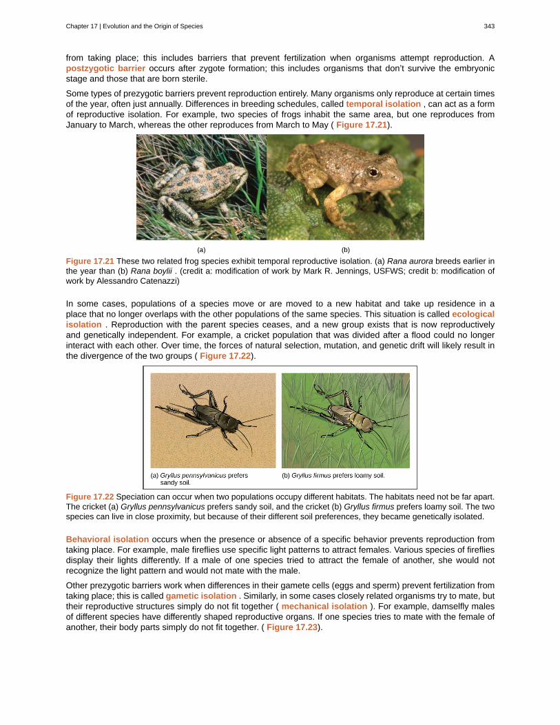
from taking place; this includes barriers that prevent fertilization when organisms attempt reproduction. Apostzygotic barrier occurs after zygote formation; this includes organisms that don’t survive the embryonicstage and those that are born sterile.
Some types of prezygotic barriers prevent reproduction entirely. Many organisms only reproduce at certain timesof the year, often just annually. Differences in breeding schedules, called temporal isolation , can act as a formof reproductive isolation. For example, two species of frogs inhabit the same area, but one reproduces fromJanuary to March, whereas the other reproduces from March to May ( Figure 17.21).
Figure 17.21 These two related frog species exhibit temporal reproductive isolation. (a) Rana aurora breeds earlier inthe year than (b) Rana boylii . (credit a: modification of work by Mark R. Jennings, USFWS; credit b: modification ofwork by Alessandro Catenazzi)
In some cases, populations of a species move or are moved to a new habitat and take up residence in aplace that no longer overlaps with the other populations of the same species. This situation is called ecologicalisolation . Reproduction with the parent species ceases, and a new group exists that is now reproductivelyand genetically independent. For example, a cricket population that was divided after a flood could no longerinteract with each other. Over time, the forces of natural selection, mutation, and genetic drift will likely result inthe divergence of the two groups ( Figure 17.22).
Figure 17.22 Speciation can occur when two populations occupy different habitats. The habitats need not be far apart.The cricket (a) Gryllus pennsylvanicus prefers sandy soil, and the cricket (b) Gryllus firmus prefers loamy soil. The twospecies can live in close proximity, but because of their different soil preferences, they became genetically isolated.
Behavioral isolation occurs when the presence or absence of a specific behavior prevents reproduction fromtaking place. For example, male fireflies use specific light patterns to attract females. Various species of firefliesdisplay their lights differently. If a male of one species tried to attract the female of another, she would notrecognize the light pattern and would not mate with the male.
Other prezygotic barriers work when differences in their gamete cells (eggs and sperm) prevent fertilization fromtaking place; this is called gametic isolation . Similarly, in some cases closely related organisms try to mate, buttheir reproductive structures simply do not fit together ( mechanical isolation ). For example, damselfly malesof different species have differently shaped reproductive organs. If one species tries to mate with the female ofanother, their body parts simply do not fit together. ( Figure 17.23).
Chapter 17 | Evolution and the Origin of Species 343
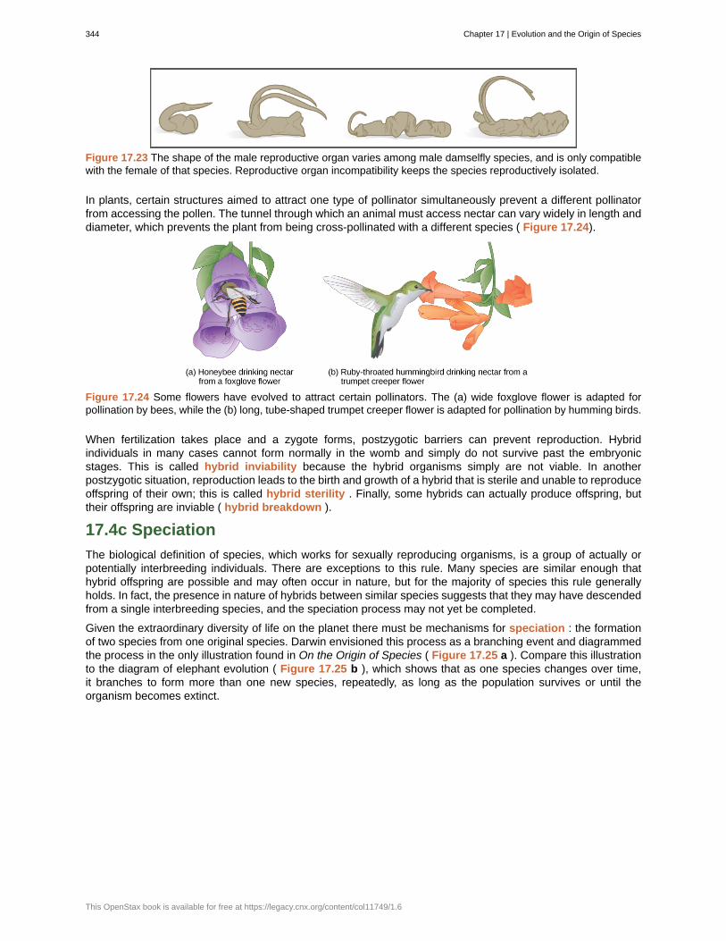
Figure 17.23 The shape of the male reproductive organ varies among male damselfly species, and is only compatiblewith the female of that species. Reproductive organ incompatibility keeps the species reproductively isolated.
In plants, certain structures aimed to attract one type of pollinator simultaneously prevent a different pollinatorfrom accessing the pollen. The tunnel through which an animal must access nectar can vary widely in length anddiameter, which prevents the plant from being cross-pollinated with a different species ( Figure 17.24).
Figure 17.24 Some flowers have evolved to attract certain pollinators. The (a) wide foxglove flower is adapted forpollination by bees, while the (b) long, tube-shaped trumpet creeper flower is adapted for pollination by humming birds.
When fertilization takes place and a zygote forms, postzygotic barriers can prevent reproduction. Hybridindividuals in many cases cannot form normally in the womb and simply do not survive past the embryonicstages. This is called hybrid inviability because the hybrid organisms simply are not viable. In anotherpostzygotic situation, reproduction leads to the birth and growth of a hybrid that is sterile and unable to reproduceoffspring of their own; this is called hybrid sterility . Finally, some hybrids can actually produce offspring, buttheir offspring are inviable ( hybrid breakdown ).
17.4c Speciation
The biological definition of species, which works for sexually reproducing organisms, is a group of actually orpotentially interbreeding individuals. There are exceptions to this rule. Many species are similar enough thathybrid offspring are possible and may often occur in nature, but for the majority of species this rule generallyholds. In fact, the presence in nature of hybrids between similar species suggests that they may have descendedfrom a single interbreeding species, and the speciation process may not yet be completed.
Given the extraordinary diversity of life on the planet there must be mechanisms for speciation : the formationof two species from one original species. Darwin envisioned this process as a branching event and diagrammedthe process in the only illustration found in On the Origin of Species ( Figure 17.25 a ). Compare this illustrationto the diagram of elephant evolution ( Figure 17.25 b ), which shows that as one species changes over time,it branches to form more than one new species, repeatedly, as long as the population survives or until theorganism becomes extinct.
344 Chapter 17 | Evolution and the Origin of Species
This OpenStax book is available for free at https://legacy.cnx.org/content/col11749/1.6
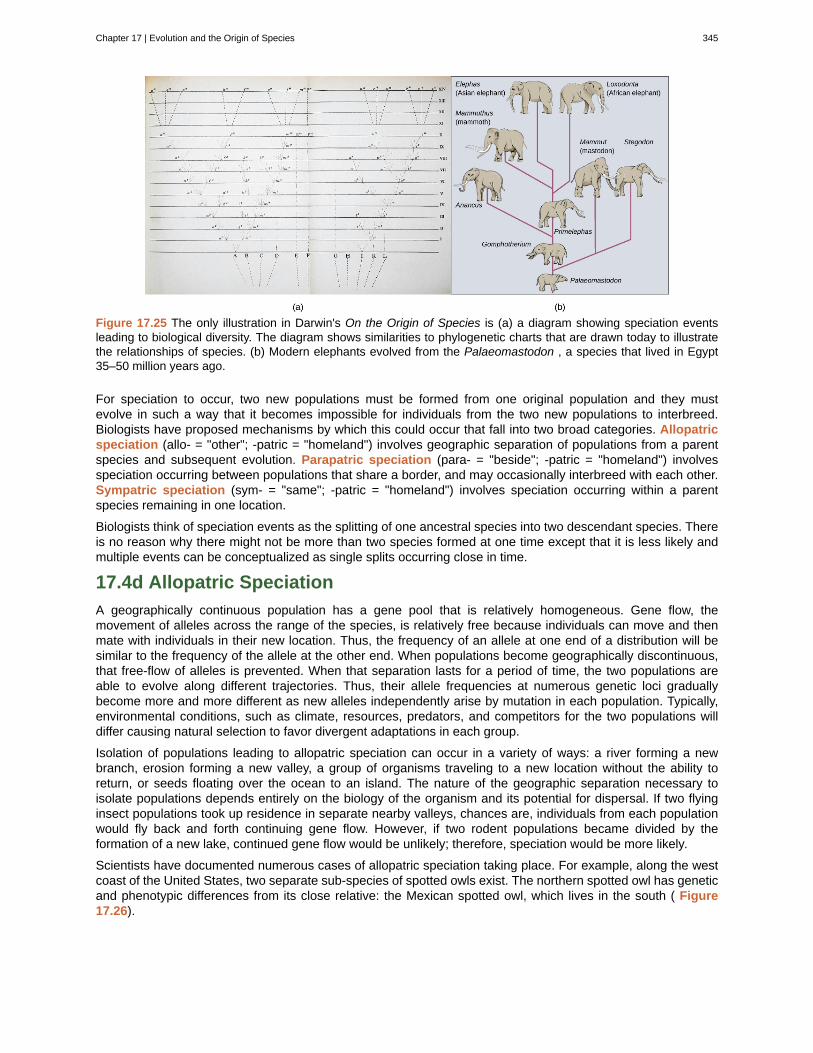
Figure 17.25 The only illustration in Darwin's On the Origin of Species is (a) a diagram showing speciation eventsleading to biological diversity. The diagram shows similarities to phylogenetic charts that are drawn today to illustratethe relationships of species. (b) Modern elephants evolved from the Palaeomastodon , a species that lived in Egypt35–50 million years ago.
For speciation to occur, two new populations must be formed from one original population and they mustevolve in such a way that it becomes impossible for individuals from the two new populations to interbreed.Biologists have proposed mechanisms by which this could occur that fall into two broad categories. Allopatricspeciation (allo- = "other"; -patric = "homeland") involves geographic separation of populations from a parentspecies and subsequent evolution. Parapatric speciation (para- = "beside"; -patric = "homeland") involvesspeciation occurring between populations that share a border, and may occasionally interbreed with each other.Sympatric speciation (sym- = "same"; -patric = "homeland") involves speciation occurring within a parentspecies remaining in one location.
Biologists think of speciation events as the splitting of one ancestral species into two descendant species. Thereis no reason why there might not be more than two species formed at one time except that it is less likely andmultiple events can be conceptualized as single splits occurring close in time.
17.4d Allopatric Speciation
A geographically continuous population has a gene pool that is relatively homogeneous. Gene flow, themovement of alleles across the range of the species, is relatively free because individuals can move and thenmate with individuals in their new location. Thus, the frequency of an allele at one end of a distribution will besimilar to the frequency of the allele at the other end. When populations become geographically discontinuous,that free-flow of alleles is prevented. When that separation lasts for a period of time, the two populations areable to evolve along different trajectories. Thus, their allele frequencies at numerous genetic loci graduallybecome more and more different as new alleles independently arise by mutation in each population. Typically,environmental conditions, such as climate, resources, predators, and competitors for the two populations willdiffer causing natural selection to favor divergent adaptations in each group.
Isolation of populations leading to allopatric speciation can occur in a variety of ways: a river forming a newbranch, erosion forming a new valley, a group of organisms traveling to a new location without the ability toreturn, or seeds floating over the ocean to an island. The nature of the geographic separation necessary toisolate populations depends entirely on the biology of the organism and its potential for dispersal. If two flyinginsect populations took up residence in separate nearby valleys, chances are, individuals from each populationwould fly back and forth continuing gene flow. However, if two rodent populations became divided by theformation of a new lake, continued gene flow would be unlikely; therefore, speciation would be more likely.
Scientists have documented numerous cases of allopatric speciation taking place. For example, along the westcoast of the United States, two separate sub-species of spotted owls exist. The northern spotted owl has geneticand phenotypic differences from its close relative: the Mexican spotted owl, which lives in the south ( Figure17.26).
Chapter 17 | Evolution and the Origin of Species 345
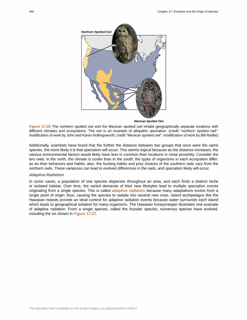
Figure 17.26 The northern spotted owl and the Mexican spotted owl inhabit geographically separate locations withdifferent climates and ecosystems. The owl is an example of allopatric speciation. (credit "northern spotted owl":modification of work by John and Karen Hollingsworth; credit "Mexican spotted owl": modification of work by Bill Radke)
Additionally, scientists have found that the further the distance between two groups that once were the samespecies, the more likely it is that speciation will occur. This seems logical because as the distance increases, thevarious environmental factors would likely have less in common than locations in close proximity. Consider thetwo owls: in the north, the climate is cooler than in the south; the types of organisms in each ecosystem differ,as do their behaviors and habits; also, the hunting habits and prey choices of the southern owls vary from thenorthern owls. These variances can lead to evolved differences in the owls, and speciation likely will occur.
Adaptive Radiation
In some cases, a population of one species disperses throughout an area, and each finds a distinct nicheor isolated habitat. Over time, the varied demands of their new lifestyles lead to multiple speciation eventsoriginating from a single species. This is called adaptive radiation because many adaptations evolve from asingle point of origin; thus, causing the species to radiate into several new ones. Island archipelagos like theHawaiian Islands provide an ideal context for adaptive radiation events because water surrounds each islandwhich leads to geographical isolation for many organisms. The Hawaiian honeycreeper illustrates one exampleof adaptive radiation. From a single species, called the founder species, numerous species have evolved,including the six shown in Figure 17.27.
346 Chapter 17 | Evolution and the Origin of Species
This OpenStax book is available for free at https://legacy.cnx.org/content/col11749/1.6
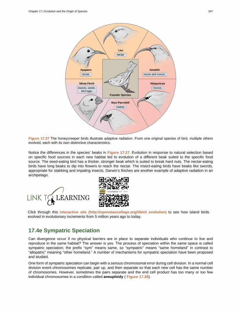
Figure 17.27 The honeycreeper birds illustrate adaptive radiation. From one original species of bird, multiple othersevolved, each with its own distinctive characteristics.
Notice the differences in the species’ beaks in Figure 17.27. Evolution in response to natural selection basedon specific food sources in each new habitat led to evolution of a different beak suited to the specific foodsource. The seed-eating bird has a thicker, stronger beak which is suited to break hard nuts. The nectar-eatingbirds have long beaks to dip into flowers to reach the nectar. The insect-eating birds have beaks like swords,appropriate for stabbing and impaling insects. Darwin’s finches are another example of adaptive radiation in anarchipelago.
Click through this interactive site (http://openstaxcollege.org/l/bird_evolution) to see how island birdsevolved in evolutionary increments from 5 million years ago to today.
17.4e Sympatric Speciation
Can divergence occur if no physical barriers are in place to separate individuals who continue to live andreproduce in the same habitat? The answer is yes. The process of speciation within the same space is calledsympatric speciation; the prefix “sym” means same, so “sympatric” means “same homeland” in contrast to“allopatric” meaning “other homeland.” A number of mechanisms for sympatric speciation have been proposedand studied.
One form of sympatric speciation can begin with a serious chromosomal error during cell division. In a normal celldivision event chromosomes replicate, pair up, and then separate so that each new cell has the same numberof chromosomes. However, sometimes the pairs separate and the end cell product has too many or too fewindividual chromosomes in a condition called aneuploidy ( Figure 17.28).
Chapter 17 | Evolution and the Origin of Species 347
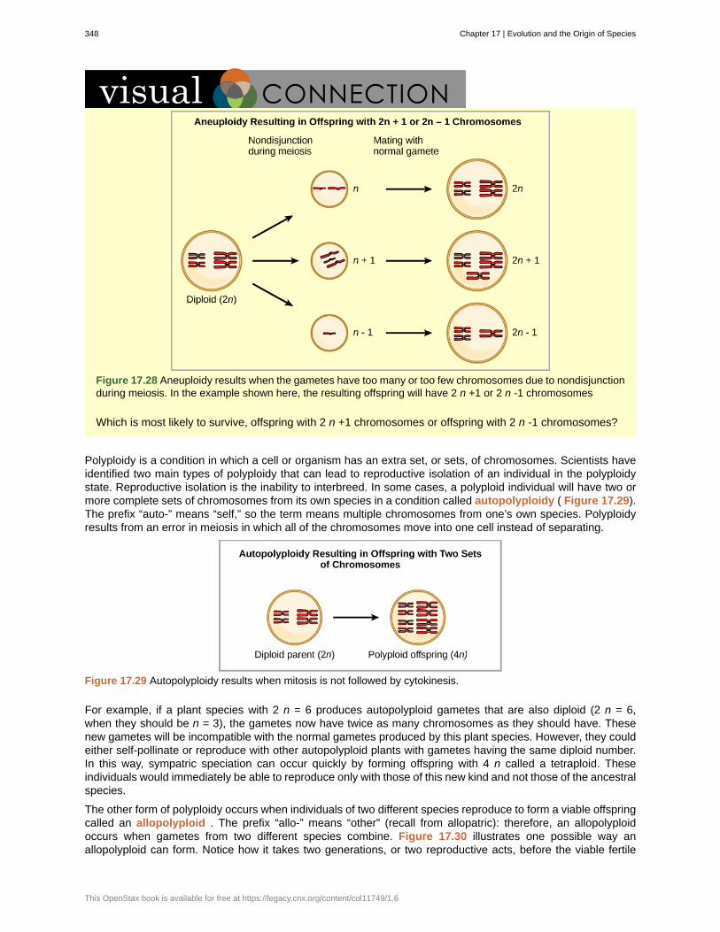
Figure 17.28 Aneuploidy results when the gametes have too many or too few chromosomes due to nondisjunctionduring meiosis. In the example shown here, the resulting offspring will have 2 n +1 or 2 n -1 chromosomes
Which is most likely to survive, offspring with 2 n +1 chromosomes or offspring with 2 n -1 chromosomes?
Polyploidy is a condition in which a cell or organism has an extra set, or sets, of chromosomes. Scientists haveidentified two main types of polyploidy that can lead to reproductive isolation of an individual in the polyploidystate. Reproductive isolation is the inability to interbreed. In some cases, a polyploid individual will have two ormore complete sets of chromosomes from its own species in a condition called autopolyploidy ( Figure 17.29).The prefix “auto-” means “self,” so the term means multiple chromosomes from one’s own species. Polyploidyresults from an error in meiosis in which all of the chromosomes move into one cell instead of separating.
Figure 17.29 Autopolyploidy results when mitosis is not followed by cytokinesis.
For example, if a plant species with 2 n = 6 produces autopolyploid gametes that are also diploid (2 n = 6,when they should be n = 3), the gametes now have twice as many chromosomes as they should have. Thesenew gametes will be incompatible with the normal gametes produced by this plant species. However, they couldeither self-pollinate or reproduce with other autopolyploid plants with gametes having the same diploid number.In this way, sympatric speciation can occur quickly by forming offspring with 4 n called a tetraploid. Theseindividuals would immediately be able to reproduce only with those of this new kind and not those of the ancestralspecies.
The other form of polyploidy occurs when individuals of two different species reproduce to form a viable offspringcalled an allopolyploid . The prefix “allo-” means “other” (recall from allopatric): therefore, an allopolyploidoccurs when gametes from two different species combine. Figure 17.30 illustrates one possible way anallopolyploid can form. Notice how it takes two generations, or two reproductive acts, before the viable fertile
348 Chapter 17 | Evolution and the Origin of Species
This OpenStax book is available for free at https://legacy.cnx.org/content/col11749/1.6
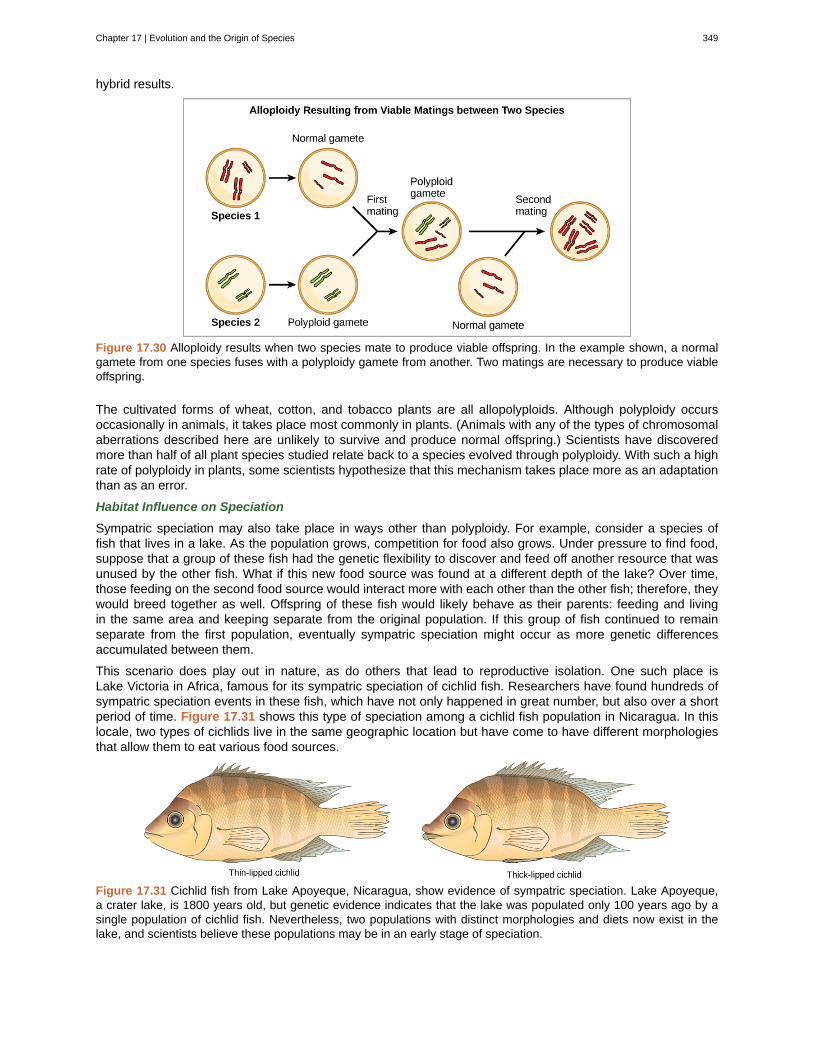
hybrid results.
Figure 17.30 Alloploidy results when two species mate to produce viable offspring. In the example shown, a normalgamete from one species fuses with a polyploidy gamete from another. Two matings are necessary to produce viableoffspring.
The cultivated forms of wheat, cotton, and tobacco plants are all allopolyploids. Although polyploidy occursoccasionally in animals, it takes place most commonly in plants. (Animals with any of the types of chromosomalaberrations described here are unlikely to survive and produce normal offspring.) Scientists have discoveredmore than half of all plant species studied relate back to a species evolved through polyploidy. With such a highrate of polyploidy in plants, some scientists hypothesize that this mechanism takes place more as an adaptationthan as an error.
Habitat Influence on Speciation
Sympatric speciation may also take place in ways other than polyploidy. For example, consider a species offish that lives in a lake. As the population grows, competition for food also grows. Under pressure to find food,suppose that a group of these fish had the genetic flexibility to discover and feed off another resource that wasunused by the other fish. What if this new food source was found at a different depth of the lake? Over time,those feeding on the second food source would interact more with each other than the other fish; therefore, theywould breed together as well. Offspring of these fish would likely behave as their parents: feeding and livingin the same area and keeping separate from the original population. If this group of fish continued to remainseparate from the first population, eventually sympatric speciation might occur as more genetic differencesaccumulated between them.
This scenario does play out in nature, as do others that lead to reproductive isolation. One such place isLake Victoria in Africa, famous for its sympatric speciation of cichlid fish. Researchers have found hundreds ofsympatric speciation events in these fish, which have not only happened in great number, but also over a shortperiod of time. Figure 17.31 shows this type of speciation among a cichlid fish population in Nicaragua. In thislocale, two types of cichlids live in the same geographic location but have come to have different morphologiesthat allow them to eat various food sources.
Figure 17.31 Cichlid fish from Lake Apoyeque, Nicaragua, show evidence of sympatric speciation. Lake Apoyeque,a crater lake, is 1800 years old, but genetic evidence indicates that the lake was populated only 100 years ago by asingle population of cichlid fish. Nevertheless, two populations with distinct morphologies and diets now exist in thelake, and scientists believe these populations may be in an early stage of speciation.
Chapter 17 | Evolution and the Origin of Species 349
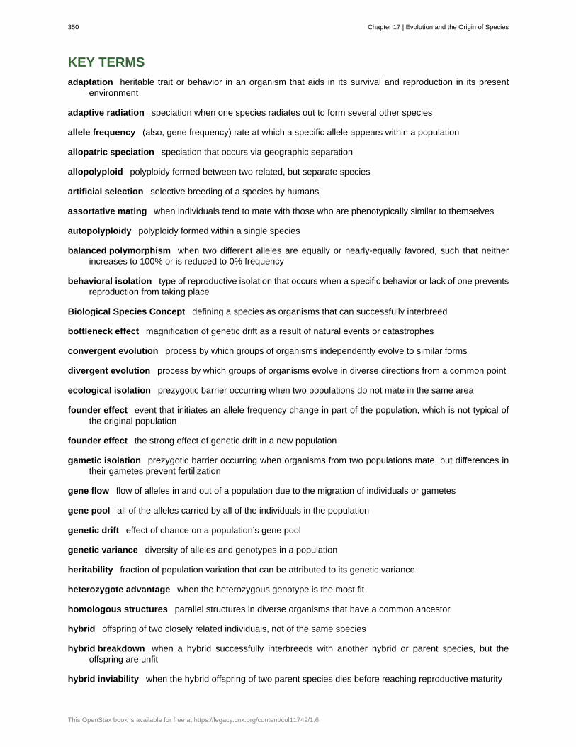
adaptation
adaptive radiation
allele frequency
allopatric speciation
allopolyploid
artificial selection
assortative mating
autopolyploidy
balanced polymorphism
behavioral isolation
Biological Species Concept
bottleneck effect
convergent evolution
divergent evolution
ecological isolation
founder effect
founder effect
gametic isolation
gene flow
gene pool
genetic drift
genetic variance
heritability
heterozygote advantage
homologous structures
hybrid
hybrid breakdown
hybrid inviability
KEY TERMS
heritable trait or behavior in an organism that aids in its survival and reproduction in its presentenvironment
speciation when one species radiates out to form several other species
(also, gene frequency) rate at which a specific allele appears within a population
speciation that occurs via geographic separation
polyploidy formed between two related, but separate species
selective breeding of a species by humans
when individuals tend to mate with those who are phenotypically similar to themselves
polyploidy formed within a single species
when two different alleles are equally or nearly-equally favored, such that neitherincreases to 100% or is reduced to 0% frequency
type of reproductive isolation that occurs when a specific behavior or lack of one preventsreproduction from taking place
defining a species as organisms that can successfully interbreed
magnification of genetic drift as a result of natural events or catastrophes
process by which groups of organisms independently evolve to similar forms
process by which groups of organisms evolve in diverse directions from a common point
prezygotic barrier occurring when two populations do not mate in the same area
event that initiates an allele frequency change in part of the population, which is not typical ofthe original population
the strong effect of genetic drift in a new population
prezygotic barrier occurring when organisms from two populations mate, but differences intheir gametes prevent fertilization
flow of alleles in and out of a population due to the migration of individuals or gametes
all of the alleles carried by all of the individuals in the population
effect of chance on a population’s gene pool
diversity of alleles and genotypes in a population
fraction of population variation that can be attributed to its genetic variance
when the heterozygous genotype is the most fit
parallel structures in diverse organisms that have a common ancestor
offspring of two closely related individuals, not of the same species
when a hybrid successfully interbreeds with another hybrid or parent species, but theoffspring are unfit
when the hybrid offspring of two parent species dies before reaching reproductive maturity
350 Chapter 17 | Evolution and the Origin of Species
This OpenStax book is available for free at https://legacy.cnx.org/content/col11749/1.6
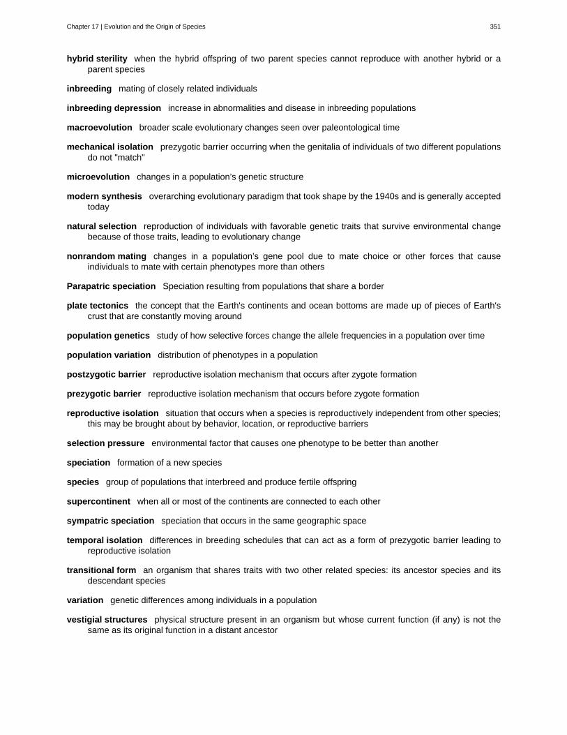
hybrid sterility
inbreeding
inbreeding depression
macroevolution
mechanical isolation
microevolution
modern synthesis
natural selection
nonrandom mating
Parapatric speciation
plate tectonics
population genetics
population variation
postzygotic barrier
prezygotic barrier
reproductive isolation
selection pressure
speciation
species
supercontinent
sympatric speciation
temporal isolation
transitional form
variation
vestigial structures
when the hybrid offspring of two parent species cannot reproduce with another hybrid or aparent species
mating of closely related individuals
increase in abnormalities and disease in inbreeding populations
broader scale evolutionary changes seen over paleontological time
prezygotic barrier occurring when the genitalia of individuals of two different populationsdo not "match"
changes in a population’s genetic structure
overarching evolutionary paradigm that took shape by the 1940s and is generally acceptedtoday
reproduction of individuals with favorable genetic traits that survive environmental changebecause of those traits, leading to evolutionary change
changes in a population’s gene pool due to mate choice or other forces that causeindividuals to mate with certain phenotypes more than others
Speciation resulting from populations that share a border
the concept that the Earth's continents and ocean bottoms are made up of pieces of Earth'scrust that are constantly moving around
study of how selective forces change the allele frequencies in a population over time
distribution of phenotypes in a population
reproductive isolation mechanism that occurs after zygote formation
reproductive isolation mechanism that occurs before zygote formation
situation that occurs when a species is reproductively independent from other species;this may be brought about by behavior, location, or reproductive barriers
environmental factor that causes one phenotype to be better than another
formation of a new species
group of populations that interbreed and produce fertile offspring
when all or most of the continents are connected to each other
speciation that occurs in the same geographic space
differences in breeding schedules that can act as a form of prezygotic barrier leading toreproductive isolation
an organism that shares traits with two other related species: its ancestor species and itsdescendant species
genetic differences among individuals in a population
physical structure present in an organism but whose current function (if any) is not thesame as its original function in a distant ancestor
Chapter 17 | Evolution and the Origin of Species 351
CHAPTER SUMMARY
17.1 Understanding Evolution
Evolution is the process of adaptation through mutation which allows more desirable characteristics to bepassed to the next generation. Over time, organisms evolve more characteristics that are beneficial to theirsurvival. For living organisms to adapt and change to environmental pressures, genetic variation must bepresent. With genetic variation, individuals have differences in form and function that allow some to survivecertain conditions better than others. These organisms pass their favorable traits to their offspring. Eventually,environments change, and what was once a desirable, advantageous trait may become an undesirable traitand organisms may further evolve. Evolution may be convergent with similar traits evolving in multiple speciesor divergent with diverse traits evolving in multiple species that came from a common ancestor. Evidence ofevolution can be observed by means of DNA code and the fossil record, and also by the existence ofhomologous and vestigial structures.
17.2 Population Evolution
The modern synthesis of evolutionary theory grew out of the cohesion of Darwin’s, Wallace’s, and Mendel’sthoughts on evolution and heredity, along with the more modern study of population genetics. It describes theevolution of populations and species, from small-scale changes among individuals to large-scale changes overpaleontological time periods. To understand how organisms evolve, scientists can track populations’ allelefrequencies over time. If they differ from generation to generation, scientists can conclude that the population isnot in Hardy-Weinberg equilibrium, and is thus evolving.
17.3 Population Genetics
Both genetic and environmental factors can cause phenotypic variation in a population. Different alleles canconfer different phenotypes, and different environments can also cause individuals to look or act differently.Only those differences encoded in an individual’s genes, however, can be passed to its offspring and, thus, bea target of natural selection. Natural selection works by selecting for alleles that confer beneficial traits orbehaviors, while selecting against those for deleterious qualities. Genetic drift stems from the chanceoccurrence that some individuals in the germ line have more offspring than others. When individuals leave orjoin the population, allele frequencies can change as a result of gene flow. Mutations to an individual’s DNAmay introduce new variation into a population. Allele frequencies can also be altered when individuals do notrandomly mate with others in the group.
17.4 Formation of New Species
Speciation occurs along two main pathways: geographic separation (allopatric speciation) and throughmechanisms that occur within a shared habitat (sympatric speciation). Both pathways isolate a populationreproductively in some form. Mechanisms of reproductive isolation act as barriers between closely relatedspecies, enabling them to diverge and exist as genetically independent species. Prezygotic barriers blockreproduction prior to formation of a zygote, whereas postzygotic barriers block reproduction after fertilizationoccurs. For a new species to develop, something must cause a breach in the reproductive barriers. Sympatricspeciation can occur through errors in meiosis that form gametes with extra chromosomes (polyploidy).Autopolyploidy occurs within a single species, whereas allopolyploidy occurs between closely related species.
352 Chapter 17 | Evolution and the Origin of Species
This OpenStax book is available for free at https://legacy.cnx.org/content/col11749/1.6

ANSWER KEY
<ext:end-of-book-solutions-placeholder></ext:end-of-book-solutions-placeholder>
Answer Key 353

354 Answer Key
This OpenStax book is available for free at https://legacy.cnx.org/content/col11749/1.6
INDEXSymbols
7-methylguanosine cap , 215,223α-helix, 73β-pleated sheet, 73
A
absorption spectrum , 167, 177abstract , 22, 23acetyl CoA , 183, 191acid , 44, 47activation energy , 138, 156activator, 243activators, 230active site , 144, 156Active transport , 126active transport , 130adaptation , 323, 350adaptive radiation , 346, 350adenosine triphosphate , 142,156adhesion , 43, 47aerobic respiration , 183, 191allele frequency , 334, 350alleles , 251, 265Allopatric speciation , 345allopatric speciation , 350allopolyploid , 348, 350allosteric inhibition , 146, 156alpha-helix structure (α-helix),80amino acid, 80Amino acids, 69amphiphilic , 94, 116anabolic, 135, 156, 156Anabolic , 153anaerobic , 182, 191anaerobic cellular respiration ,187, 191anaphase , 283, 294aneuploid , 274, 277antenna proteins , 169, 177anticodon , 218, 223antiporter , 127, 130applied science , 20, 23artificial selection , 324, 350assortative mating , 340, 350atom , 9, 23, 28, 47atomic mass , 29, 47atomic number , 29, 47ATP , 142, 156ATP synthase , 186, 191
autopolyploidy , 348, 350autosomes , 260, 265, 272, 277
B
balanced chemical equation ,35, 47balanced polymorphism , 335,350base , 44, 47Basic science , 20basic science , 23Behavioral isolation , 343behavioral isolation , 350beta-pleated sheet (β-pleated),80bioenergetics , 150, 156biological macromolecules , 54,80Biological Species Concept ,341, 350biology , 14, 23biosphere , 10, 23Biotechnology , 309biotechnology , 317bottleneck effect , 338, 350Buffers , 45, 47
C
calorie , 41, 47Calvin cycle , 173, 177capillary action , 43, 47Carbohydrates , 56, 80carbon fixation , 174, 177carotenoids , 167, 177catabolic, 135, 153, 156, 156catabolite activator protein(CAP), 230, 243cell , 10, 23cell cycle , 279, 294cell cycle checkpoints , 287, 294cell plate , 283, 294cell wall , 102, 116cellular cloning , 203, 205Cellulose , 59cellulose , 80Central Dogma , 208, 223central vacuole , 102, 116centrioles , 280, 294centromere , 196, 205centrosome , 101, 116chaperone, 80chaperones, 75chemical bonds , 34, 47chemical energy , 135, 156Chemical reactions , 35, 47
Chemiosmosis , 143, 156chemiosmosis , 186, 191chemoautotrophs , 160, 177chitin , 60, 80chlorophyll , 102, 116Chlorophyll a , 167chlorophyll a , 177chloroplast , 162, 177Chloroplasts , 102, 116chromatids , 196, 205chromatin , 100, 116chromosome inversion , 271,277Chromosomes , 99, 116chromosomes , 195, 205cilia , 110, 116citric acid cycle , 183, 191cleavage furrow , 283, 294codominance , 257, 265codons , 209, 223coenzymes , 148, 156cofactors , 148, 156cohesin , 299, 308cohesion , 42, 47community , 10, 23competitive inhibition , 146, 156compounds , 35, 47concentration gradient , 122,130conclusion , 22, 23control group , 18, 23controlled variables , 18, 23convergent evolution , 324, 350covalent bonds , 36, 47crossover , 300, 308cyclic pathway ofphotosynthesis , 171, 177cytochrome complex , 170, 177Cytokinesis , 283cytokinesis , 294cytoplasm , 99, 116cytoskeleton , 108, 116cytosol , 99, 116
D
Deductive reasoning , 16deductive reasoning , 23degenerate , 209, 223dehydration synthesis , 54, 80denaturation, 69, 75, 80denature , 145, 156deoxyribonucleic acid (DNA) ,76, 80dependent variable , 18, 23dephosphorylation , 142, 156
Index 355
Descriptive (or discovery)science , 16, 23Diffusion , 122diffusion , 130diploid , 194, 205Disaccharides , 57, 80discussion , 22, 23dissociation , 42, 47divergent evolution , 324, 350DNA polymerase , 200, 205DNA replication , 200, 205Dominant traits , 251, 265downstream , 212, 223
E
ecological isolation , 343, 350ecosystem , 10, 23electrolytes , 36, 47electromagnetic spectrum , 165,177electron microscopes , 86, 116electron transfer , 36, 47electron transport chain , 170,177, 184, 191electronegativity , 37, 47Electrons , 29, 47Elements , 28, 47endergonic reactions , 136, 156Endocytosis , 127endocytosis , 130endomembrane system , 103,116endoplasmic reticulum (ER) ,104, 116enhancer, 243enhancers, 233entropy , 140, 156enzyme, 80Enzymes, 68enzymes , 144, 156epigenetic , 228, 243eukaryotes , 10, 23eukaryotic cells , 91, 116euploid , 274, 277evaporation , 41, 47evolution , 11, 23exergonic reactions , 136, 156Exocytosis , 129exocytosis , 130exons , 216, 223extracellular matrix , 112, 116
F
F 1 , 249, 265F 2 , 249, 265
facilitated transport , 123, 130falsifiable , 17, 23Feedback inhibition , 149feedback inhibition , 156fermentation , 187, 191fertilization , 298, 308flagella , 110, 116fluid mosaic model , 94, 116founder effect , 334, 338, 350,350Functional groups , 52, 80
G
G 0 phase , 284, 294G 1 phase , 280, 294G 2 phase , 280, 294gametes , 194, 205gametic isolation , 343, 350Gap junctions , 114, 116gene expression , 228, 243gene flow , 339, 350gene pool , 334, 350Gene targeting , 312gene targeting , 317Gene therapy , 313gene therapy , 317genes , 195, 205genetic diagnosis , 312, 317genetic drift , 336, 350Genetic engineering , 311genetic engineering , 317genetic testing , 312, 317genetic variance , 336, 350genetically modified organism ,312, 317genome , 194, 205genotype , 252, 265germ cells , 299, 308Glycogen , 59glycogen , 80glycolipids , 94, 116Glycolysis , 182glycolysis , 191glycoproteins , 94, 116Golgi apparatus , 106, 116granum, 162, 177
H
hairpin , 213, 223haploid , 194, 205Heat energy , 138heat energy , 140, 156, 156heat of vaporization , 41, 47helicase , 200, 205hemizygous , 260, 265
Heritability , 336heritability , 350heterotrophs , 160, 177heterozygote advantage , 335,350heterozygous , 252, 265histone proteins , 195, 205homeostasis , 8, 23homologous , 195, 205homologous structures , 329,350homozygous , 252, 265hormone, 80Hormones, 68hybrid , 342, 350hybrid breakdown , 344, 350hybrid inviability , 344, 350hybrid sterility , 344, 351hybridizations , 249, 265Hydrocarbons , 52, 80hydrogen bond , 38, 47hydrolysis, 42, 47, 55, 80hydrophilic , 39, 48, 94, 117hydrophobic , 39, 48, 117Hydrophobic , 94hypertonic , 124, 130hypothesis , 15, 23hypothesis-based science , 16,23hypotonic , 124, 130
I
inbreeding , 336, 351inbreeding depression , 336,351incomplete dominance , 256,265independent variable , 18, 23induced fit , 145, 156Induced mutations , 221, 223inducible operon, 243Inducible operons, 231Inductive reasoning , 15inductive reasoning , 23inert gases , 34, 48initiation site , 212, 223Integral proteins , 96, 117interkinesis , 303, 308Intermediate filaments , 110,117interphase , 279, 294introduction , 22, 23introns , 216, 223Ionic bonds , 36, 48ions , 34, 48
356 Index
This OpenStax book is available for free at https://legacy.cnx.org/content/col11749/1.6
isotonic , 124, 130Isotopes , 30, 48
K
karyogram , 272, 277karyotype , 272, 277kinetic energy , 134, 156kinetochore , 283, 294
L
lac operon, 231, 243lagging strand , 201, 205law of dominance , 255, 265law of independent assortment ,256, 265law of segregation , 255, 265leading strand , 201, 205ligase , 201, 205light microscopes , 86, 117light-dependent reactions , 162,177light-harvesting complex , 169,177light-independent reactions ,163, 177Lipids , 62, 80litmus paper , 44, 48locus , 195, 205lysis buffer , 310, 317lysosomes , 101, 117
M
macroevolution , 333, 351macromolecules , 9, 23mass number , 29, 48materials and methods , 22, 23Matter , 28matter , 48mechanical isolation , 343, 351meiosis , 299, 308meiosis I , 299, 308Meiosis II , 299meiosis II , 308mesophyll , 162, 177messenger RNA (mRNA) , 76,80metabolism , 150, 156metaphase , 283, 294metaphase plate , 283, 294metastasis , 293, 294microevolution , 333, 351microfilaments , 109, 117microscope , 86, 117microtubules , 110, 117
Mitochondria , 100mitochondria , 117Mitosis , 281mitosis , 294mitotic phase , 279, 294mitotic spindle , 280, 294model system , 248, 265modern synthesis , 333, 351molecular cloning , 203, 205molecule , 9, 23Molecules , 32, 48monohybrid , 252, 265monomers , 54, 80Monosaccharides , 56, 80monosomy , 274, 277Mutations , 221, 223
N
Natural selection , 321natural selection , 351negative control , 18, 24negative regulator, 243negative regulators, 230neutral, 44, 48neutron , 29, 48noble gases , 34, 48nondisjunction , 273, 277Nonpolar covalent bonds , 37,48nonrandom mating , 340, 351nonsense codons , 210, 223nontemplate strand , 212, 223nuclear envelope , 99, 117Nucleic acids , 76, 80nucleoid , 90, 117nucleolus , 100, 117nucleoplasm , 99, 117nucleosome , 195, 205nucleotides , 76, 80nucleus , 28, 48, 99, 117
O
octet rule , 33, 48Okazaki fragments , 201, 205oncogenes , 289, 294operator, 230, 243operon, 243operons, 230orbitals , 32, 48organ system , 10, 24organelles , 9, 24, 91, 117organic molecules , 52, 80Organisms , 10, 24Organs , 10, 24Osmosis , 123
osmosis , 130oxidative phosphorylation , 143,157, 184, 191
P
P , 249, 265P680 , 170, 177P700 , 171, 177paired unit factors , 255, 265Parapatric speciation , 345, 351Passive transport , 122passive transport , 130pedigree analysis , 263, 265Peer-reviewed manuscripts , 21,24peptide bond, 70, 80periodic table , 32, 48Peripheral proteins , 96, 117Peroxisomes , 101, 117pH scale , 44, 48phenotype , 252, 265phosphodiester , 77, 81Phospholipids , 65, 81Phosphorylation , 143, 157photoact , 170, 177photoautotrophs , 160, 177photolysis, 170, 177photon , 169, 177photosystem , 169, 177photosystem I , 169, 178photosystem II , 169, 178phylogenetic tree , 11, 24pigment , 162, 178pinocytosis , 128, 130placebo , 18, 24plagiarism , 22, 24plasma membrane , 92, 117plasmids , 212, 223plasmodesmata , 113, 117plasmolysis , 125, 130plate tectonics , 327, 351pleiotropy, 260, 265Point mutations , 221, 223polar covalent bond , 37, 48poly-A tail , 216, 223polygenic , 259, 265polymers , 54, 81polypeptide, 71, 81polyploid , 275, 277polysaccharide , 59, 81population , 10, 24population genetics , 333, 351population variation , 334, 351positive control , 18, 24positive regulator, 230, 243
Index 357
post-transcriptional , 228, 243post-translational , 228, 243postzygotic barrier , 343, 351potential energy , 134, 157prezygotic barrier , 342, 351primary electron acceptor , 170,178primary structure, 72, 81primase , 200, 205primer , 200, 205products , 35, 48prokaryote , 90, 117Prokaryotes , 10, 24promoter , 212, 223proofreading , 221, 223prophase , 282, 294proteases , 310, 317proteasome , 237, 243protein, 81Proteins, 68proto-oncogenes , 289, 294proton , 29, 48Punnett square , 252, 265pyruvate , 182, 191
Q
quaternary structure, 74, 81quiescent , 284, 294
R
radioisotopes , 30, 48reactants , 35, 48reaction center , 169, 178reading frame , 209, 223Recessive traits , 251, 266redox reactions , 154, 157reduction , 174, 178replication forks , 200, 205repressor, 243Repressors, 230Reproductive cloning , 203reproductive cloning , 205reproductive isolation , 342, 351results , 22, 24reversible chemical reaction ,35, 48Review articles , 22, 24Rho-dependent termination ,213, 223Rho-independent termination ,213, 223ribonucleases , 310, 317ribonucleic acid (RNA) , 76, 81Ribosomes , 100, 117RNA editing , 215, 223
RNA polymerase , 212, 223rough endoplasmic reticulum(RER) , 104, 117
S
S phase , 280, 294saturated fatty acid , 63, 81Science , 15science , 24scientific method , 15, 24secondary structure, 73, 81selection pressure , 336, 351selectively permeable , 122, 130serendipity , 21, 24sex-linkage , 260, 266signal sequence , 221, 223silent mutations , 221, 224Single-strand binding proteins ,200, 205sliding clamp , 201, 206smooth endoplasmic reticulum(SER) , 105, 117solute , 124, 130solvent , 42, 48somatic cell , 299, 308Somatic cell nuclear transfer ,203, 206speciation , 344, 351species , 341, 351specific heat capacity , 41, 48spectrophotometer , 168, 178sphere of hydration , 42, 49splicing , 216, 224Spontaneous mutations , 221,224Spores , 299, 308starch , 56, 81start codon , 218, 224steroids , 67, 81stomata , 162, 178stroma, 162, 178substituted hydrocarbons , 53,81substrate-level phosphorylation ,143, 157substrates , 144, 157supercontinent , 327, 351surface tension , 42, 49Sympatric speciation , 345sympatric speciation , 351symporter , 127, 130synapsis , 299, 308
T
telomerase , 291, 294
telomeres , 291, 295telophase , 283, 295template strand , 212, 224temporal isolation , 343, 351tertiary structure, 73, 81test cross , 253, 266theory , 15, 24Therapeutic cloning , 203, 206Thermodynamics , 139thermodynamics , 157thylakoid lumen , 162, 178thylakoids , 162, 178Ti plasmids , 315, 317tight junction , 113, 118tissues , 10, 24Tonicity , 124tonicity , 130Topoisomerase , 201topoisomerase , 206trait , 250, 266trans fat , 64, 81transcription bubble. , 212, 224transcription factor binding site,243transcription factors , 240, 243transcriptional level , 228, 243transcriptional start site, 230,243transgenic , 312, 317transition state , 138, 157Transition substitution , 222transition substitution , 224transitional form , 328, 351translocation , 271, 277translocations , 273, 277transport proteins , 123transporters , 127, 130Transversion substitution , 222transversion substitution , 224triacylglycerols , 63, 81triglycerides , 63, 81trisomy , 274, 277true-breeding , 248, 266Tumor suppressor genes , 289,295
U
unified cell theory , 87, 118uniporter , 127, 130unsaturated , 64, 81upstream , 212, 224
V
vacuoles , 101, 118valence shell , 33, 49
358 Index
This OpenStax book is available for free at https://legacy.cnx.org/content/col11749/1.6
variable , 18, 24variants , 257, 266variation , 323, 351variations , 321Vesicles , 101, 118vestigial structures , 330, 351
W
wavelength , 165, 178Wax , 66wax , 81Whole-genome sequencing ,315whole-genome sequencing ,317wild type , 257, 266
X
X inactivation , 241, 243X-linked , 260, 266
Index 359

360 Index
This OpenStax book is available for free at https://legacy.cnx.org/content/col11749/1.6
ATTRIBUTIONS
Collection: General Biology Part I - Mixed MajorsEdited by: Shannon McDermottURL: https://legacy.cnx.org/content/col11749/1.6/Copyright: Shannon McDermottLicense: http://creativecommons.org/licenses/by/4.0/
Module: PrefaceBy: OpenStaxURL: https://legacy.cnx.org/content/m46078/1.13/Copyright: Rice UniversityLicense: http://creativecommons.org/licenses/by/4.0/
Module: IntroductionBy: OpenStaxURL: https://legacy.cnx.org/content/m44386/1.2/Copyright: Rice UniversityLicense: http://creativecommons.org/licenses/by/3.0/
Module: 1.1 Themes and Concepts of BiologyUsed here as: Themes and Concepts of BiologyBy: Shannon McDermottURL: https://legacy.cnx.org/content/m52512/1.3/Copyright: Shannon McDermottLicense: http://creativecommons.org/licenses/by/4.0/Based on: Themes and Concepts of Biology <http://legacy.cnx.org/content/m44388/1.13> by OpenStax.
Module: 1.2 The Science of BiologyUsed here as: The Science of BiologyBy: Shannon McDermottURL: https://legacy.cnx.org/content/m51993/1.3/Copyright: Shannon McDermottLicense: http://creativecommons.org/licenses/by/4.0/Based on: The Science of Biology <http://legacy.cnx.org/content/m44387/1.11> by OpenStax.
Module: IntroductionBy: OpenStaxURL: https://legacy.cnx.org/content/m44389/1.2/Copyright: Rice UniversityLicense: http://creativecommons.org/licenses/by/3.0/
Module: 2.1 Atoms, Isotopes, Ions, and Molecules: The Building BlocksUsed here as: Atoms, Isotopes, Ions, and Molecules: The Building BlocksBy: Shannon McDermottURL: https://legacy.cnx.org/content/m52513/1.3/Copyright: Shannon McDermottLicense: http://creativecommons.org/licenses/by/4.0/Based on: Atoms, Isotopes, Ions, and Molecules: The Building Blocks <http://legacy.cnx.org/content/m44390/1.10>by OpenStax.
Module: 2.2 WaterUsed here as: WaterBy: Shannon McDermottURL: https://legacy.cnx.org/content/m52522/1.3/Copyright: Shannon McDermottLicense: http://creativecommons.org/licenses/by/4.0/
Index 361
Based on: Water <http://legacy.cnx.org/content/m44392/1.9> by OpenStax.
Module: IntroductionBy: OpenStaxURL: https://legacy.cnx.org/content/m44395/1.4/Copyright: Rice UniversityLicense: http://creativecommons.org/licenses/by/3.0/
Module: 2.3 CarbonUsed here as: CarbonBy: Shannon McDermottURL: https://legacy.cnx.org/content/m52525/1.3/Copyright: Shannon McDermottLicense: http://creativecommons.org/licenses/by/4.0/Based on: Carbon <http://legacy.cnx.org/content/m44393/1.5> by OpenStax.
Module: 3.1 Synthesis of Biological MacromoleculesUsed here as: Synthesis of Biological MacromoleculesBy: Shannon McDermottURL: https://legacy.cnx.org/content/m52526/1.4/Copyright: Shannon McDermottLicense: http://creativecommons.org/licenses/by/4.0/Based on: Synthesis of Biological Macromolecules <http://legacy.cnx.org/content/m44397/1.12> by OpenStax.
Module: 3.2 CarbohydratesUsed here as: CarbohydratesBy: Shannon McDermottURL: https://legacy.cnx.org/content/m52531/1.3/Copyright: Shannon McDermottLicense: http://creativecommons.org/licenses/by/4.0/Based on: Carbohydrates <http://legacy.cnx.org/content/m44400/1.6> by OpenStax.
Module: 3.3 LipidsUsed here as: LipidsBy: Shannon McDermottURL: https://legacy.cnx.org/content/m52532/1.3/Copyright: Shannon McDermottLicense: http://creativecommons.org/licenses/by/4.0/Based on: Lipids <http://legacy.cnx.org/content/m44401/1.8> by OpenStax.
Module: 3.4 ProteinsUsed here as: ProteinsBy: Shannon McDermottURL: https://legacy.cnx.org/content/m52535/1.4/Copyright: Shannon McDermottLicense: http://creativecommons.org/licenses/by/4.0/Based on: Proteins <http://legacy.cnx.org/content/m44402/1.7> by OpenStax.
Module: 3.6 Nucleic AcidsUsed here as: Nucleic AcidsBy: Shannon McDermottURL: https://legacy.cnx.org/content/m52536/1.4/Copyright: Shannon McDermottLicense: http://creativecommons.org/licenses/by/4.0/Based on: Nucleic Acids <http://legacy.cnx.org/content/m44403/1.8> by OpenStax.
Module: IntroductionBy: OpenStaxURL: https://legacy.cnx.org/content/m44404/1.2/
362 Index
This OpenStax book is available for free at https://legacy.cnx.org/content/col11749/1.6
Copyright: Rice UniversityLicense: http://creativecommons.org/licenses/by/3.0/
Module: 4.1 Studying CellsUsed here as: Studying CellsBy: Shannon McDermottURL: https://legacy.cnx.org/content/m52539/1.3/Copyright: Shannon McDermottLicense: http://creativecommons.org/licenses/by/4.0/Based on: Studying Cells <http://legacy.cnx.org/content/m44405/1.8> by OpenStax.
Module: 4.2 Prokaryotic CellsUsed here as: Prokaryotic CellsBy: Shannon McDermottURL: https://legacy.cnx.org/content/m52541/1.4/Copyright: Shannon McDermottLicense: http://creativecommons.org/licenses/by/4.0/Based on: Prokaryotic Cells <http://legacy.cnx.org/content/m44406/1.11> by OpenStax.
Module: 6.1 Components and StructureUsed here as: Components and Structure of Cell MembranesBy: Shannon McDermottURL: https://legacy.cnx.org/content/m52577/1.4/Copyright: Shannon McDermottLicense: http://creativecommons.org/licenses/by/4.0/Based on: Components and Structure <http://legacy.cnx.org/content/m44416/1.8> by OpenStax.
Module: 4.3 Eukaryotic CellsUsed here as: Eukaryotic CellsBy: Shannon McDermottURL: https://legacy.cnx.org/content/m52542/1.4/Copyright: Shannon McDermottLicense: http://creativecommons.org/licenses/by/4.0/Based on: Eukaryotic Cells <http://legacy.cnx.org/content/m44407/1.13> by OpenStax.
Module: 4.4 Endomembrane System and ProteinsUsed here as: Endomembrane System and ProteinsBy: Shannon McDermottURL: https://legacy.cnx.org/content/m52546/1.4/Copyright: Shannon McDermottLicense: http://creativecommons.org/licenses/by/4.0/Based on: The Endomembrane System and Proteins <http://legacy.cnx.org/content/m44435/1.11> by OpenStax.
Module: 4.5 The CytoskeletonUsed here as: The CytoskeletonBy: Shannon McDermottURL: https://legacy.cnx.org/content/m52548/1.3/Copyright: Shannon McDermottLicense: http://creativecommons.org/licenses/by/4.0/Based on: The Cytoskeleton <http://legacy.cnx.org/content/m44412/1.8> by OpenStax.
Module: 4.6 Connections between Cells and Cellular ActivitiesUsed here as: Connections between Cells and Cellular ActivitiesBy: Shannon McDermottURL: https://legacy.cnx.org/content/m52564/1.3/Copyright: Shannon McDermottLicense: http://creativecommons.org/licenses/by/4.0/Based on: Connections between Cells and Cellular Activities <http://legacy.cnx.org/content/m44413/1.7> by
Index 363
OpenStax.
Module: IntroductionBy: OpenStaxURL: https://legacy.cnx.org/content/m44415/1.2/Copyright: Rice UniversityLicense: http://creativecommons.org/licenses/by/3.0/
Module: 6.2 Passive TransportUsed here as: Passive TransportBy: Shannon McDermottURL: https://legacy.cnx.org/content/m52570/1.4/Copyright: Shannon McDermottLicense: http://creativecommons.org/licenses/by/4.0/Based on: Passive Transport <http://legacy.cnx.org/content/m44417/1.12> by OpenStax.
Module: 6.3 Active TransportUsed here as: Active TransportBy: Shannon McDermottURL: https://legacy.cnx.org/content/m52572/1.3/Copyright: Shannon McDermottLicense: http://creativecommons.org/licenses/by/4.0/Based on: Active Transport <http://legacy.cnx.org/content/m44418/1.5> by OpenStax.
Module: 5.3 Bulk TransportUsed here as: Bulk TransportBy: Shannon McDermottURL: https://legacy.cnx.org/content/m52584/1.5/Copyright: Shannon McDermottLicense: http://creativecommons.org/licenses/by/4.0/Based on: Bulk Transport <http://legacy.cnx.org/content/m44419/1.7> by OpenStax.
Module: IntroductionBy: OpenStaxURL: https://legacy.cnx.org/content/m44421/1.2/Copyright: Rice UniversityLicense: http://creativecommons.org/licenses/by/3.0/
Module: 6.1 Potential, Kinetic, and Activation EnergyUsed here as: Potential, Kinetic, Free, and Activation EnergyBy: Shannon McDermottURL: https://legacy.cnx.org/content/m52653/1.3/Copyright: Shannon McDermottLicense: http://creativecommons.org/licenses/by/4.0/Based on: Potential, Kinetic, Free, and Activation Energy <http://legacy.cnx.org/content/m44425/1.6> by OpenStax.
Module: 7.3 The Laws of ThermodynamicsUsed here as: The Laws of ThermodynamicsBy: Shannon McDermottURL: https://legacy.cnx.org/content/m52658/1.3/Copyright: Shannon McDermottLicense: http://creativecommons.org/licenses/by/4.0/Based on: The Laws of Thermodynamics <http://legacy.cnx.org/content/m44424/1.7> by OpenStax.
Module: 6.3 Adenosine TriphosphateUsed here as: Adenosine TriphosphateBy: Shannon McDermottURL: https://legacy.cnx.org/content/m52659/1.4/Copyright: Shannon McDermott
364 Index
This OpenStax book is available for free at https://legacy.cnx.org/content/col11749/1.6
License: http://creativecommons.org/licenses/by/4.0/Based on: ATP: Adenosine Triphosphate <http://legacy.cnx.org/content/m44427/1.7> by OpenStax.
Module: 6.4 EnzymesUsed here as: EnzymesBy: Shannon McDermottURL: https://legacy.cnx.org/content/m52660/1.4/Copyright: Shannon McDermottLicense: http://creativecommons.org/licenses/by/4.0/Based on: Enzymes <http://legacy.cnx.org/content/m44429/1.7> by OpenStax.
Module: 7.1 Energy and MetabolismUsed here as: Energy and MetabolismBy: Shannon McDermottURL: https://legacy.cnx.org/content/m52616/1.3/Copyright: Shannon McDermottLicense: http://creativecommons.org/licenses/by/4.0/Based on: Energy and Metabolism <http://legacy.cnx.org/content/m44422/1.8> by OpenStax.
Module: 6.6 Energy in Living SystemsUsed here as: Energy in Living SystemsBy: Shannon McDermottURL: https://legacy.cnx.org/content/m52664/1.3/Copyright: Shannon McDermottLicense: http://creativecommons.org/licenses/by/4.0/Based on: Energy in Living Systems <http://legacy.cnx.org/content/m44431/1.8> by OpenStax.
Module: IntroductionBy: OpenStaxURL: https://legacy.cnx.org/content/m44446/1.2/Copyright: Rice UniversityLicense: http://creativecommons.org/licenses/by/3.0/
Module: 7.1 Overview of PhotosynthesisUsed here as: Overview of PhotosynthesisBy: Shannon McDermottURL: https://legacy.cnx.org/content/m52661/1.3/Copyright: Shannon McDermottLicense: http://creativecommons.org/licenses/by/4.0/Based on: Overview of Photosynthesis <http://legacy.cnx.org/content/m44447/1.8> by OpenStax.
Module: 7.2 The Light-Dependent Reactions of PhotosynthesisUsed here as: The Light-Dependent Reactions of PhotosynthesisBy: Shannon McDermottURL: https://legacy.cnx.org/content/m52662/1.3/Copyright: Shannon McDermottLicense: http://creativecommons.org/licenses/by/4.0/Based on: The Light-Dependent Reactions of Photosynthesis <http://legacy.cnx.org/content/m44448/1.11> byOpenStax.
Module: The Cyclic Pathway of PhotosynthesisBy: Shannon McDermottURL: https://legacy.cnx.org/content/m55514/1.6/Copyright: Shannon McDermottLicense: http://creativecommons.org/licenses/by/4.0/
Module: 8.3 Using Light Energy to Make Organic MoleculesUsed here as: Using Light Energy to Make Organic MoleculesBy: Shannon McDermott
Index 365
URL: https://legacy.cnx.org/content/m52663/1.4/Copyright: Shannon McDermottLicense: http://creativecommons.org/licenses/by/4.0/Based on: Using Light Energy to Make Organic Molecules <http://legacy.cnx.org/content/m44449/1.7> byOpenStax.
Module: IntroductionBy: OpenStaxURL: https://legacy.cnx.org/content/m44430/1.4/Copyright: Rice UniversityLicense: http://creativecommons.org/licenses/by/3.0/
Module: 9.2 GlycolysisUsed here as: GlycolysisBy: Shannon McDermottURL: https://legacy.cnx.org/content/m52665/1.3/Copyright: Shannon McDermottLicense: http://creativecommons.org/licenses/by/4.0/Based on: Glycolysis <http://legacy.cnx.org/content/m44432/1.6> by OpenStax.
Module: 8.2 Citric Acid Cycle and Oxidative PhosphorylationUsed here as: Citric Acid Cycle and Oxidative PhosphorylationBy: Shannon McDermottURL: https://legacy.cnx.org/content/m54705/1.2/Copyright: Shannon McDermottLicense: http://creativecommons.org/licenses/by/4.0/Based on: Cell Energy: Citric Acid Cycle and Oxidative Phosphorylation <http://legacy.cnx.org/content/m47947/1.1> by Jeffrey Mahr.
Module: 8.3 Metabolism without OxygenUsed here as: Metabolism without OxygenBy: Shannon McDermottURL: https://legacy.cnx.org/content/m52668/1.4/Copyright: Shannon McDermottLicense: http://creativecommons.org/licenses/by/4.0/Based on: Metabolism without Oxygen <http://legacy.cnx.org/content/m44444/1.6> by OpenStax.
Module: IntroductionBy: OpenStaxURL: https://legacy.cnx.org/content/m44484/1.2/Copyright: Rice UniversityLicense: http://creativecommons.org/licenses/by/3.0/
Module: 9.1 Chromosome StructureUsed here as: Chromosome StructureBy: Shannon McDermottURL: https://legacy.cnx.org/content/m52671/1.4/Copyright: Shannon McDermottLicense: http://creativecommons.org/licenses/by/4.0/Based on: Cell Division <http://legacy.cnx.org/content/m44459/1.8> by OpenStax.
Module: 14.2 DNA Structure and SequencingUsed here as: DNA Structure and SequencingBy: Shannon McDermottURL: https://legacy.cnx.org/content/m52683/1.4/Copyright: Shannon McDermottLicense: http://creativecommons.org/licenses/by/4.0/Based on: DNA Structure and Sequencing <http://legacy.cnx.org/content/m44486/1.7> by OpenStax.
366 Index
This OpenStax book is available for free at https://legacy.cnx.org/content/col11749/1.6
Module: 9.3 DNA Replication in ProkaryotesUsed here as: DNA Replication in ProkaryotesBy: Shannon McDermottURL: https://legacy.cnx.org/content/m52688/1.4/Copyright: Shannon McDermottLicense: http://creativecommons.org/licenses/by/4.0/Based on: DNA Replication in Prokaryotes <http://legacy.cnx.org/content/m44488/1.6> by OpenStax.
Module: Derived copy of BiotechnologyUsed here as: CloningBy: Shannon McDermottURL: https://legacy.cnx.org/content/m54732/1.5/Copyright: Shannon McDermottLicense: http://creativecommons.org/licenses/by/4.0/Based on: Biotechnology <http://legacy.cnx.org/content/m44552/1.7> by OpenStax.
Module: IntroductionBy: OpenStaxURL: https://legacy.cnx.org/content/m44518/1.3/Copyright: Rice UniversityLicense: http://creativecommons.org/licenses/by/3.0/
Module: 10.1 The Genetic CodeUsed here as: The Genetic CodeBy: Shannon McDermottURL: https://legacy.cnx.org/content/m52694/1.3/Copyright: Shannon McDermottLicense: http://creativecommons.org/licenses/by/4.0/Based on: The Genetic Code <http://legacy.cnx.org/content/m44522/1.7> by OpenStax.
Module: 15.2 Prokaryotic TranscriptionUsed here as: Prokaryotic TranscriptionBy: Shannon McDermottURL: https://legacy.cnx.org/content/m52697/1.4/Copyright: Shannon McDermottLicense: http://creativecommons.org/licenses/by/4.0/Based on: Prokaryotic Transcription <http://legacy.cnx.org/content/m44523/1.6> by OpenStax.
Module: 15.3 RNA Processing in EukaryotesUsed here as: RNA Processing in EukaryotesBy: Shannon McDermottURL: https://legacy.cnx.org/content/m52698/1.3/Copyright: Shannon McDermottLicense: http://creativecommons.org/licenses/by/4.0/Based on: RNA Processing in Eukaryotes <http://legacy.cnx.org/content/m44532/1.5> by OpenStax.
Module: 15.4 Ribosomes and Protein SynthesisUsed here as: Ribosomes and Protein SynthesisBy: Shannon McDermottURL: https://legacy.cnx.org/content/m52700/1.3/Copyright: Shannon McDermottLicense: http://creativecommons.org/licenses/by/4.0/Based on: Ribosomes and Protein Synthesis <http://legacy.cnx.org/content/m44529/1.7> by OpenStax.
Module: 14.6 DNA RepairUsed here as: MutationsBy: Shannon McDermottURL: https://legacy.cnx.org/content/m52690/1.3/
Index 367
Copyright: Shannon McDermottLicense: http://creativecommons.org/licenses/by/4.0/Based on: DNA Repair <http://legacy.cnx.org/content/m44513/1.6> by OpenStax.
Module: IntroductionBy: OpenStaxURL: https://legacy.cnx.org/content/m44533/1.3/Copyright: Rice UniversityLicense: http://creativecommons.org/licenses/by/3.0/
Module: 16.1 Regulation of Gene ExpressionUsed here as: Regulation of Gene ExpressionBy: Shannon McDermottURL: https://legacy.cnx.org/content/m52701/1.3/Copyright: Shannon McDermottLicense: http://creativecommons.org/licenses/by/4.0/Based on: Regulation of Gene Expression <http://legacy.cnx.org/content/m44534/1.6> by OpenStax.
Module: Derived copy of Prokaryotic Gene RegulationUsed here as: Prokaryotic Gene RegulationBy: Shannon McDermottURL: https://legacy.cnx.org/content/m54733/1.3/Copyright: Shannon McDermottLicense: http://creativecommons.org/licenses/by/4.0/Based on: Prokaryotic Gene Regulation <http://legacy.cnx.org/content/m44535/1.5> by OpenStax.
Module: 16.4 Eukaryotic Transcription Gene RegulationUsed here as: Eukaryotic Transcription Gene RegulationBy: Shannon McDermottURL: https://legacy.cnx.org/content/m52704/1.3/Copyright: Shannon McDermottLicense: http://creativecommons.org/licenses/by/4.0/Based on: Eukaryotic Transcription Gene Regulation <http://legacy.cnx.org/content/m44538/1.5> by OpenStax.
Module: 16.5 Eukaryotic Post-transcriptional Gene RegulationUsed here as: Eukaryotic Post-transcriptional Gene RegulationBy: Shannon McDermottURL: https://legacy.cnx.org/content/m52707/1.3/Copyright: Shannon McDermottLicense: http://creativecommons.org/licenses/by/4.0/Based on: Eukaryotic Post-transcriptional Gene Regulation <http://legacy.cnx.org/content/m44539/1.7> byOpenStax.
Module: 11.5 Eukaryotic Translational and Post-translational Gene RegulationUsed here as: Eukaryotic Translational and Post-translational Gene RegulationBy: Shannon McDermottURL: https://legacy.cnx.org/content/m52708/1.3/Copyright: Shannon McDermottLicense: http://creativecommons.org/licenses/by/4.0/Based on: Eukaryotic Translational and Post-translational Gene Regulation <http://legacy.cnx.org/content/m44542/1.5> by OpenStax.
Module: 16.3 Eukaryotic Epigenetic Gene RegulationUsed here as: Eukaryotic Epigenetic Gene RegulationBy: Shannon McDermottURL: https://legacy.cnx.org/content/m52703/1.4/Copyright: Shannon McDermottLicense: http://creativecommons.org/licenses/by/4.0/
368 Index
This OpenStax book is available for free at https://legacy.cnx.org/content/col11749/1.6
Based on: Eukaryotic Epigenetic Gene Regulation <http://legacy.cnx.org/content/m44536/1.5> by OpenStax.
Module: X Chromosome InactivationBy: Shannon McDermottURL: https://legacy.cnx.org/content/m61617/1.1/Copyright: Shannon McDermottLicense: http://creativecommons.org/licenses/by/4.0/
Module: IntroductionBy: OpenStaxURL: https://legacy.cnx.org/content/m44474/1.2/Copyright: Rice UniversityLicense: http://creativecommons.org/licenses/by/3.0/
Module: 12.1 Mendel’s Experiments and the Laws of ProbabilityUsed here as: Mendel’s Experiments and the Laws of ProbabilityBy: Shannon McDermottURL: https://legacy.cnx.org/content/m52677/1.4/Copyright: Shannon McDermottLicense: http://creativecommons.org/licenses/by/4.0/Based on: Mendel’s Experiments and the Laws of Probability <http://legacy.cnx.org/content/m44476/1.6> byOpenStax.
Module: 12.2 Characteristics and TraitsUsed here as: Characteristics and TraitsBy: Shannon McDermottURL: https://legacy.cnx.org/content/m52678/1.5/Copyright: Shannon McDermottLicense: http://creativecommons.org/licenses/by/4.0/Based on: Characteristics and Traits <http://legacy.cnx.org/content/m44478/1.6> by OpenStax.
Module: 12.3 Laws of InheritanceUsed here as: Laws of InheritanceBy: Shannon McDermottURL: https://legacy.cnx.org/content/m52680/1.4/Copyright: Shannon McDermottLicense: http://creativecommons.org/licenses/by/4.0/Based on: Laws of Inheritance <http://legacy.cnx.org/content/m44479/1.3> by OpenStax.
Module: Alternatives and ApplicationsBy: Shannon McDermottURL: https://legacy.cnx.org/content/m61618/1.2/Copyright: Shannon McDermottLicense: http://creativecommons.org/licenses/by/4.0/
Module: IntroductionBy: OpenStaxURL: https://legacy.cnx.org/content/m44480/1.2/Copyright: Rice UniversityLicense: http://creativecommons.org/licenses/by/3.0/
Module: 13.1 Chromosomal Basis of Inherited DisordersUsed here as: Chromosomal Basis of Inherited DisordersBy: Shannon McDermottURL: https://legacy.cnx.org/content/m52679/1.3/Copyright: Shannon McDermottLicense: http://creativecommons.org/licenses/by/4.0/Based on: Chromosomal Basis of Inherited Disorders <http://legacy.cnx.org/content/m44483/1.7> by OpenStax.
Index 369

Module: IntroductionBy: OpenStaxURL: https://legacy.cnx.org/content/m44457/1.2/Copyright: Rice UniversityLicense: http://creativecommons.org/licenses/by/3.0/
Module: 10.2 The Cell CycleUsed here as: The Cell CycleBy: Shannon McDermottURL: https://legacy.cnx.org/content/m52672/1.3/Copyright: Shannon McDermottLicense: http://creativecommons.org/licenses/by/4.0/Based on: The Cell Cycle <http://legacy.cnx.org/content/m44460/1.7> by OpenStax.
Module: 10.3 Control of the Cell CycleUsed here as: Control of the Cell CycleBy: Shannon McDermottURL: https://legacy.cnx.org/content/m52673/1.3/Copyright: Shannon McDermottLicense: http://creativecommons.org/licenses/by/4.0/Based on: Control of the Cell Cycle <http://legacy.cnx.org/content/m44466/1.6> by OpenStax.
Module: 10.4 Cancer and the Cell CycleUsed here as: Cancer and the Cell CycleBy: Shannon McDermottURL: https://legacy.cnx.org/content/m52674/1.3/Copyright: Shannon McDermottLicense: http://creativecommons.org/licenses/by/4.0/Based on: Cancer and the Cell Cycle <http://legacy.cnx.org/content/m44463/1.6> by OpenStax.
Module: 14.5 DNA Replication in EukaryotesUsed here as: TelomeraseBy: Shannon McDermottURL: https://legacy.cnx.org/content/m52689/1.5/Copyright: Shannon McDermottLicense: http://creativecommons.org/licenses/by/4.0/Based on: DNA Replication in Eukaryotes <http://legacy.cnx.org/content/m44517/1.5> by OpenStax.
Module: IntroductionBy: OpenStaxURL: https://legacy.cnx.org/content/m44468/1.2/Copyright: Rice UniversityLicense: http://creativecommons.org/licenses/by/3.0/
Module: 15.1 Sexual ReproductionUsed here as: Sexual ReproductionBy: Shannon McDermottURL: https://legacy.cnx.org/content/m52676/1.3/Copyright: Shannon McDermottLicense: http://creativecommons.org/licenses/by/4.0/Based on: Sexual Reproduction <http://legacy.cnx.org/content/m44470/1.5> by OpenStax.
Module: 15.2 The Process of MeiosisUsed here as: The Process of MeiosisBy: Shannon McDermottURL: https://legacy.cnx.org/content/m52675/1.3/Copyright: Shannon McDermottLicense: http://creativecommons.org/licenses/by/4.0/
370 Index
This OpenStax book is available for free at https://legacy.cnx.org/content/col11749/1.6
Based on: The Process of Meiosis <http://legacy.cnx.org/content/m44469/1.8> by OpenStax.
Module: IntroductionBy: OpenStaxURL: https://legacy.cnx.org/content/m44551/1.2/Copyright: Rice UniversityLicense: http://creativecommons.org/licenses/by/3.0/
Module: 17.1 BiotechnologyUsed here as: BiotechnologyBy: Shannon McDermottURL: https://legacy.cnx.org/content/m52709/1.4/Copyright: Shannon McDermottLicense: http://creativecommons.org/licenses/by/4.0/Based on: Biotechnology <http://legacy.cnx.org/content/m44552/1.7> by OpenStax.
Module: 17.2 Whole-Genome SequencingUsed here as: Whole-Genome SequencingBy: Shannon McDermottURL: https://legacy.cnx.org/content/m52710/1.3/Copyright: Shannon McDermottLicense: http://creativecommons.org/licenses/by/4.0/Based on: Whole-Genome Sequencing <http://legacy.cnx.org/content/m44555/1.7> by OpenStax.
Module: IntroductionBy: OpenStaxURL: https://legacy.cnx.org/content/m44561/1.2/Copyright: Rice UniversityLicense: http://creativecommons.org/licenses/by/3.0/
Module: 18.1 Understanding EvolutionUsed here as: Understanding EvolutionBy: Shannon McDermottURL: https://legacy.cnx.org/content/m52715/1.3/Copyright: Shannon McDermottLicense: http://creativecommons.org/licenses/by/4.0/Based on: Understanding Evolution <http://legacy.cnx.org/content/m44568/1.5> by OpenStax.
Module: 19.1 Population EvolutionUsed here as: Population EvolutionBy: Shannon McDermottURL: https://legacy.cnx.org/content/m52718/1.3/Copyright: Shannon McDermottLicense: http://creativecommons.org/licenses/by/4.0/Based on: Population Evolution <http://legacy.cnx.org/content/m44582/1.5> by OpenStax.
Module: 19.2 Population GeneticsUsed here as: Population GeneticsBy: Shannon McDermottURL: https://legacy.cnx.org/content/m52722/1.4/Copyright: Shannon McDermottLicense: http://creativecommons.org/licenses/by/4.0/Based on: Population Genetics <http://legacy.cnx.org/content/m44584/1.5> by OpenStax.
Module: 18.2 Formation of New SpeciesUsed here as: Formation of New SpeciesBy: Shannon McDermottURL: https://legacy.cnx.org/content/m52720/1.3/Copyright: Shannon McDermott
Index 371

License: http://creativecommons.org/licenses/by/4.0/Based on: Formation of New Species <http://legacy.cnx.org/content/m44574/1.5> by OpenStax.
372 Index
This OpenStax book is available for free at https://legacy.cnx.org/content/col11749/1.6

ABOUT CONNEXIONSSince 1999, Connexions has been pioneering a global system where anyone can create course materials and makethem fully accessible and easily reusable free of charge. We are a Web-based authoring, teaching and learningenvironment open to anyone interested in education, including students, teachers, professors and lifelong learners. Weconnect ideas and facilitate educational communities. Connexions's modular, interactive courses are in use worldwideby universities, community colleges, K-12 schools, distance learners, and lifelong learners. Connexions materials arein many languages, including English, Spanish, Chinese, Japanese, Italian, Vietnamese, French, Portuguese, andThai.
Index 373
Related Documents


