BIOTECHNOLOGY METHODS Gene cloning and enzymatic characterization of an endoprotease Endo-Pro-Aspergillus niger Chao Kang • Xiao-Wei Yu • Yan Xu Received: 5 March 2013 / Accepted: 29 April 2013 / Published online: 18 May 2013 Ó Society for Industrial Microbiology and Biotechnology 2013 Abstract A novel endoprotease Endo-Pro-Aspergillus niger (endoprotease EPR) was first successfully expressed at high level in the methylotrophic yeast Pichia pastoris and the purification procedure was established. The endo- protease EPR is 95 % identity with proline specific endo- peptidase from A. niger CBS513.88 (EMBL; AX458699), while sharing low identity with those from other microor- ganisms. The purified endoprotease EPR was a monomer of 60 kDa. Furthermore, the peptide mass fingerprinting (PMF) analysis confirmed that the purified protein was an endoprotease Endo-Pro-Aspergillus niger. A three-dimen- sional model revealed that the active site of the enzyme was located in Ser (179) -Asp (458) -His (491) , based on template 3n2zB with sequence identity of 17.6 %. The optimum pH and temperature of the endoprotease EPR were pH 4–5 and 35 °C, and the stabilities were pH 3–7 and 15–60 °C, respectively. Furthermore, the endoprotease EPR had the ability to digest peptides with the C-terminal of proline as well as alanine, and was also capable of hydrolyzing larger peptides. The properties of the endoprotease EPR made it a highly promising candidate for future application in the field of brewing and food process. Keywords Aspergillus niger Proline-specific endoprotease Endo-Pro-Aspergillus niger Expression Pichia pastoris Introduction Prolyl endopeptidase (EC 3. 4. 21. 26), also called proline- specific endoprotease, belongs to the serine protease family and has the ability to cleave peptides at internal proline residues [30]. As is known, the cyclic amino acid proline, due to its unique structural properties, plays a key physi- ological role by protecting peptides from enzymatic deg- radation, and links to a wide range of diseases [2, 13, 17, 18], including depression, Parkinson’s disease, and celiac sprue, as well as other diseases (blood pressure regulation, anorexia, bulimia nervosa, et al.). In contrast, the currently available proteolytic enzymes cannot efficiently cleave the peptide bond involving proline residues of proline-rich proteins, such as casein, gluten, collagen, and gelatin. However, many researchers found that the prolyl endo- peptidase can overcome these problems. Prolyl endopep- tidase also attracted numerous medical researchers and was proposed as a potential therapeutic approach because of its highly efficient degradation in gluten [14, 27]. The microbial prolyl endopeptidase was first purified from Flavobacterium meningosepticum and then classified as a serine protease on the basic of its inhibition by DFP [31]. Since then, prolyl endopeptidase activity was also found in Xanthomonas sp. [28], Aeromonas hydrophilic [11], Pseudomonas sp. KU-22 [15], Sphingomonas capsu- late [10], Lactobacillus helveticus [25], Halobacterium halobium S9 [3], Myxococcus xanthus [6], Aspergillus niger [4, 12, 32], A. oryzae [19]. However, from an application point of view, F. meningosepticum, C. Kang X.-W. Yu (&) Y. Xu (&) State Key Laboratory of Food Science and Technology, The Key Laboratory of Industrial Biotechnology of Ministry of Education, School of Biotechnology, Jiangnan University, 1800 Lihu Ave, Wuxi 214122 Jiangsu, People’s Republic of China e-mail: [email protected] Y. Xu e-mail: [email protected] C. Kang e-mail: [email protected] 123 J Ind Microbiol Biotechnol (2013) 40:855–864 DOI 10.1007/s10295-013-1284-4 Downloaded from https://academic.oup.com/jimb/article/40/8/855/5994704 by guest on 24 June 2022

Welcome message from author
This document is posted to help you gain knowledge. Please leave a comment to let me know what you think about it! Share it to your friends and learn new things together.
Transcript
BIOTECHNOLOGY METHODS
Gene cloning and enzymatic characterization of an endoproteaseEndo-Pro-Aspergillus niger
Chao Kang • Xiao-Wei Yu • Yan Xu
Received: 5 March 2013 / Accepted: 29 April 2013 / Published online: 18 May 2013
� Society for Industrial Microbiology and Biotechnology 2013
Abstract A novel endoprotease Endo-Pro-Aspergillus
niger (endoprotease EPR) was first successfully expressed
at high level in the methylotrophic yeast Pichia pastoris
and the purification procedure was established. The endo-
protease EPR is 95 % identity with proline specific endo-
peptidase from A. niger CBS513.88 (EMBL; AX458699),
while sharing low identity with those from other microor-
ganisms. The purified endoprotease EPR was a monomer
of 60 kDa. Furthermore, the peptide mass fingerprinting
(PMF) analysis confirmed that the purified protein was an
endoprotease Endo-Pro-Aspergillus niger. A three-dimen-
sional model revealed that the active site of the enzyme
was located in Ser(179)-Asp(458)-His(491), based on template
3n2zB with sequence identity of 17.6 %. The optimum pH
and temperature of the endoprotease EPR were pH 4–5 and
35 �C, and the stabilities were pH 3–7 and 15–60 �C,
respectively. Furthermore, the endoprotease EPR had the
ability to digest peptides with the C-terminal of proline as
well as alanine, and was also capable of hydrolyzing larger
peptides. The properties of the endoprotease EPR made it a
highly promising candidate for future application in the
field of brewing and food process.
Keywords Aspergillus niger � Proline-specific
endoprotease � Endo-Pro-Aspergillus niger �Expression � Pichia pastoris
Introduction
Prolyl endopeptidase (EC 3. 4. 21. 26), also called proline-
specific endoprotease, belongs to the serine protease family
and has the ability to cleave peptides at internal proline
residues [30]. As is known, the cyclic amino acid proline,
due to its unique structural properties, plays a key physi-
ological role by protecting peptides from enzymatic deg-
radation, and links to a wide range of diseases [2, 13, 17,
18], including depression, Parkinson’s disease, and celiac
sprue, as well as other diseases (blood pressure regulation,
anorexia, bulimia nervosa, et al.). In contrast, the currently
available proteolytic enzymes cannot efficiently cleave the
peptide bond involving proline residues of proline-rich
proteins, such as casein, gluten, collagen, and gelatin.
However, many researchers found that the prolyl endo-
peptidase can overcome these problems. Prolyl endopep-
tidase also attracted numerous medical researchers and was
proposed as a potential therapeutic approach because of its
highly efficient degradation in gluten [14, 27].
The microbial prolyl endopeptidase was first purified
from Flavobacterium meningosepticum and then classified
as a serine protease on the basic of its inhibition by DFP
[31]. Since then, prolyl endopeptidase activity was also
found in Xanthomonas sp. [28], Aeromonas hydrophilic
[11], Pseudomonas sp. KU-22 [15], Sphingomonas capsu-
late [10], Lactobacillus helveticus [25], Halobacterium
halobium S9 [3], Myxococcus xanthus [6], Aspergillus
niger [4, 12, 32], A. oryzae [19]. However, from an
application point of view, F. meningosepticum,
C. Kang � X.-W. Yu (&) � Y. Xu (&)
State Key Laboratory of Food Science and Technology,
The Key Laboratory of Industrial Biotechnology of
Ministry of Education, School of Biotechnology, Jiangnan
University, 1800 Lihu Ave, Wuxi 214122 Jiangsu,
People’s Republic of China
e-mail: [email protected]
Y. Xu
e-mail: [email protected]
C. Kang
e-mail: [email protected]
123
J Ind Microbiol Biotechnol (2013) 40:855–864
DOI 10.1007/s10295-013-1284-4
Dow
nloaded from https://academ
ic.oup.com/jim
b/article/40/8/855/5994704 by guest on 24 June 2022
Xanthomonas sp., A. hydrophila, Pseudomonas sp. KU-22,
Myxococcus xanthus, which belong to pathogenic bacteria,
are obviously not good choices for food processing
industry, while H. halobium S9 also showed numerous
serious drawbacks, such as culture condition with an
extremely high salt concentration and preference for
cleaving Pro in the penultimate position combination with
hydrophobic amino acid in the C-terminal side of peptides.
On the contrary, Aspergillus sp., known as the food-grade
microorganisms, may be considered as safe and attractive
microorganisms for producing prolyl endopeptidase [14,
27]. In addition, it was observed that using low levels of an
acid proline-specific endoprotease from A. niger in bottled
beer could effectively prevent chill-haze formation, but
leaving the beer form almost unaffected [12]. Since then,
this enzyme has been widely applied in brewing industry
for beer stabilization. At the same time, the first proline-
specific protease gene from A. niger was cloned and
expressed in A. niger CBS513.88. The overexpressed
proline-specific protease was of good debittering effect to
the peptides responsible for the bitter taste of casein
hydrolysate containing several proline residues [4]. Fur-
thermore, genome of A. niger CBS518.13 was sequenced
and analyzed, which revealed that the endoprotease Endo-
Pro-Aspergillus niger (endoprotease EPR) was highly
homologous to the proline-specific endoprotease with the
same particular conserved catalytic triad Ser-Asp-His of
serine protease. At the same time, the proline-specific
endoprotease was also confirmed to be the S28 family of
clan SC of serine proteases [16]. Endoprotease EPR was
also considered to be proline-specific endoprotease. How-
ever, to our knowledge, few researchers have reported the
production of A. niger proline-specific endoprotease with
low yield and activity [4, 5, 16].
In recent years, the yeast Pichia pastoris, known as a
powerful expression system for the production of high
levels of various recombinant heterologous proteins,
together with its economic use, has applied for both basic
laboratory research and industrial manufacture [9]. There-
fore, in this study, we constructed recombinant P. pastoris
strains capable of producing endoprotease Endo-Pro-
Aspergillus niger at high levels and the purified endopro-
tease EPR from the culture supernatant was characterized.
Materials and methods
Strains, vectors, reagents, and enzymes
A. niger 2.169 strains, Escherichia coli JM 109 strains, the
plasmid expression vector pPIC9 K, and strain for protein
expression P. pastoris GS115, were stored in our library. The
plasmid vector pMD19-T, restriction endonucleases, Taq
DNA polymerase and T4 DNA ligase, were purchased from
TaKaRa Biotechnology. The Z-Gly-Pro-pNA substrate was
obtained from Bachem (King of Prussia, PA, USA). The
standard mini Plasmid Prep Kit and the DNA gel extraction
kit were purchased from Omega (OMEGA bio-tek, USA).
DNA sequencing was performed using an ABI377 sequencer
(Applied Biosystems, Foster City, CA, USA). The Amicon
Ultra 30, 000 MWCO membrane was from Millipore
(Billerica, MA, USA), and the HiTrap DEAE FF column was
from Amersham Biosciences (Piscataway, NJ, USA).
Cloning of endoprotease EPR gene and construction
of the expression plasmid
A. niger 2.169 was grown in a medium containing 1.0 g of
K2HPO4, 0.4 g of KH2PO4, 0.5 g of KCl, 0.5 g of
MgSO4.7H2O, 0.01 g of FeSO4.7H2O, 5 g of glucose, and
15 g of collagen (Sigma), as described by Edens et al. The
collagen was used as the sole carbon source to induce the
expression of the gene encoding for endoprotease EPR.
Young mycelia were harvested after 48 h grown at 30 �C.
Total RNA from A. niger was isolated using the TRIzol
reagent exactly as described by the supplier (Sangon Bio-
tech. Shanghai, China) and its purity was evaluated by
electrophoresis on 2 % agarose gel.
Reverse transcription was performed by using 2 lg
totals RNA, 1X Prime Script Buffer, 25 pmol Oligo dT
Primer (50 lM) and 50 pmol Random 6 mers (100 lM).
Reactions were carried out at 37 �C for 15 min, 85 �C for
5 s, and 4 �C for 10 min. The resulting cDNA from the A.
niger was then used for PCR. PCR mixture contained
0.5 lM final concentrations of sense (P1: 50-CCGGA
ATTCGCTCGCCCCCGTCTTGT) and antisense (P2: 50-CCGCGGCCGCTCAAGCATAATACTCCTCCACCC) prim-
ers, which contained added sites for the restriction enzymes
EcoRI and NotI (underline), respectively, TaKaRa Taq
DNA polymerase 1.25 U, 10* PCR Buffer (Mg2? plus)
5 ll, dNTP Mixture (each 2.5 mM) 4 ll, primer 1 (20 lM)
1 ll, primer 2 (20 lM) 1 ll, and cDNA 0.5 lg, in a 50-ll
volume PCR amplification was performed by incubating
the samples at 94 �C for 3 min of preheating, followed by
30 cycles at 94 �C for 30 s, at 63 �C for 60 s, and at 72 �C
for 60 s, with a final extension at 72 �C for 10 min. At the
end of amplification, samples were submitted to electro-
phoresis on 1.5 % agarose gel with a 2,000-bp DNA ladder
as a size marker. The amplified bands of about 1,530 bp
were visualized by Golden View I staining.
The PCR-amplified fragment encoding endoprotease
EPR was cloned into the pMD19-T cloning vector and
subjected to double-stranded DNA sequencing. After
EcoRI-NotI digestion, the endoprotease EPR gene was
cloned in the pPIC9 K vector between the EcoRI (50 end)
and NotI (30 end) restriction sites to generate the plasmid
856 J Ind Microbiol Biotechnol (2013) 40:855–864
123
Dow
nloaded from https://academ
ic.oup.com/jim
b/article/40/8/855/5994704 by guest on 24 June 2022
pPIC9 K-EPR. The resulting plasmid (pPIC9 K-EPR) was
transformed into E. coli DH5a, and then the recombinant
E. coli cells were selected on ampicillin containing LB
plates and screened by PCR using the AOX1, P1, P2
primers. Plasmid DNA was purified from the recombinant
E. coli DH5a and subjected to DNA sequence analysis to
confirm the endoprotease EPR cDNA fragment.
Transformation and expression of endoprotease EPR
in P. pastoris
Pichia pastoris wild-type strain GS115 was used as a host
for the expression of the gene encoding endoprotease EPR.
The expression vector pPIC9 K-EPR described above was
linearized by digestion with restriction enzyme BglII and
introduced into P. pastoris wild-type strain GS115 by
electroporation using a Micropulser (Bio-Rad, Hercules,
CA, USA). According to the manufacturer’s recommen-
dations (Invitrogen), the following culture media: minimal
dextrose medium (MD), buffered glycerol-complex med-
ium (BMGY), and buffered minimal methanol (BMMY),
were prepared for the transformation of P. pastoris,
selection of recombinant clones, and expression of endo-
protease EPR. For cultures in liquid BMMY, which contain
methanol as an inducer and carbon source, methanol was
added every 24 h to a final concentration of 1 % (v/v). All
cultures were carried out at 28 �C with shaking 250 rpm.
Every day (0, 24, 48, 72, and 96 h), just before the addition
of 1 % methanol, a 1-ml sample of the expression medium
was collected to measure endoprotease EPR activity and
for expression analysis by SDS-PAGE.
Enzymes activity
Endoprotease EPR activity was determined using the
methodology reported by Edens et al. Firstly, the substrate
(benzyloxycarbonyl-glycine-proline-p-nitroanilide, Z-Gly-
Pro-pNA), was dissolved in 1, 4-dioxane (40 %, v/v in
water) at 60 �C, to prepare a 250-lM solution. The
expression endoprotease EPR activity was determined by
using Z-Gly-Pro-pNA as substrate at 37 �C in a citrate/
disodium phosphate buffer (pH 5.0). The reaction products
were monitored spectrophotometrically at 410 nm. One
unit of the endoprotease EPR activity was defined as the
quantity of enzyme that releases 1 lmol of p-nitroanilide
per minute under the conditions specified.
Purification of the endoprotease EPR
After 96 h of culture, the entire medium was harvested by
centrifugation at 10,000 9 g for 20 min. The endoprotease
EPR secreted in the supernatant was purified by a four-step
procedure consisting of ammonium sulfate precipitation,
dialysis, Amicon Ultra 30, 000 MWCO membrane (Milli-
pore), and ion-exchange chromatography. The following
steps were carried out at 4 �C unless otherwise described.
Briefly, the supernatant was fractionated with ammonium
sulfate (60–80 % saturation) over night, and after centri-
fugation the precipitate was dialyzed in water, then con-
centrated with Amicon Ultra 30,000 MWCO membrane
(Millipore), and applied to a HiTrap DEAE FF column
(Amersham Biosciences) pre-equilibrated with 20 mM
Tris–HCl (pH 5.0). The column was washed with the same
buffer with a linear gradient from 0 to 0.5 M NaCl, and the
enzyme eluted at around 0.3 M NaCl; the fractions con-
taining activity were pooled and kept at 4 �C. The eluted
proteins were also analyzed by SDS-PAGE.
Peptide mass fingerprinting analysis
of the endoprotease EPR
PMF analysis of the endoprotease EPR was analyzed using
MALDI-TOF–MS as follows. The sample was first analyzed
by 12 % SDS-PAGE and then stained with silver nitrate.
The recombinant endoprotease EPR band was cut out of the
gel to be a 1-mm3 rubber block by homemade cut-off
device, decolorized in 200–400 ll 100 mM NH4HCO3/
30 % acetonitrile solutions for about 1 h, and then the
supernatant was removed. Then, the rubber block was
incubated in 90 ll 100 mM NH4HCO3, 10 ll 100 mM DTT
at 56 �C for 30 min, and stored in 70 ll 100 mM
NH4HCO3, 30 ll 200 mM IAM3 at dark for 20 min; and
then washed with 100 ll 100 mM NH4HCO3 for 15 min,
100 ll 100 % ACN for 5 min, respectively. The above
sample without any liquid was reacted with 5 ll 2.5–10 ng/ll
trypsin at 4 �C for 1 h. After removal of the supernatant, the
sample (without any trypsin) was incubated in 20–30 ll
25 mM NH4HCO3 at 37 �C for about 20 h, and then the
enzymatic hydrolysates that resulted from digestion of the
protein with trypsin were introduced into a mass spectrom-
eter. The positive-ion mode was employed and the mass
spectrometer with the application of a spray voltage was set
at 3.2 kV. MASCOT search tool (URL, http://www.matrix
science.com) was used for identification of tryptic maps.
Determination of pH and thermal optima
To establish the pH activity profiles of the endoprotease
EPR, the citrate/disodium phosphate buffers with different
pH values were prepared. A synthetic chromogenic peptide
Z-Gly-Pro-pNA was used as the substrate for the enzyme.
Endoprotease EPR activity dependence in terms of pH
was determined using 0.1 M citrate–phosphate buffers (pH
of 2, 3, 4, 5, 6, 7, 8, and 9). Enzyme stability against pH
was determined after incubating the enzyme for 30 min at
37 �C. Thermal dependence of endoprotease EPR activity
J Ind Microbiol Biotechnol (2013) 40:855–864 857
123
Dow
nloaded from https://academ
ic.oup.com/jim
b/article/40/8/855/5994704 by guest on 24 June 2022
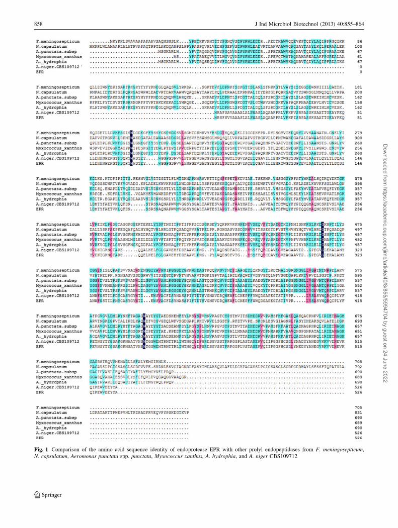
Fig. 1 Comparison of the amino acid sequence identity of endoprotease EPR with other prolyl endopeptidases from F. meningosepticum,
N. capsulatum, Aeromonas punctata spp. punctata, Myxococcus xanthus, A. hydrophia, and A. niger CBS109712
858 J Ind Microbiol Biotechnol (2013) 40:855–864
123
Dow
nloaded from https://academ
ic.oup.com/jim
b/article/40/8/855/5994704 by guest on 24 June 2022
was determined incubating the reaction mixture (enzyme in
0.1 M citrate–phosphate buffer pH 5 and the substrate) at
temperatures between 15 and 80 �C. To evaluate thermal
stability, 50 ll of the enzyme solution and 500 ll of 0.1 M
citrate–phosphate buffer pH 5 were incubated for 30 min at
different temperatures (15, 20, 25, 30, 35, 40, 45, 50, 55,
60, 65, 70, 75, and 80 �C). Then the activity was deter-
mined according to the standard enzyme activity test.
Inhibitors
Various inhibitors such as phenylmethylsulfonyl fluoride
(PMSF, typical serine protease inhibitor, 0.1 M), EDTA
(metal ions, 0.1 M), aprotinin (0.1 M), and leupeptin
hemisulfate (0.1 M) were added to the enzyme and incu-
bated for 30 min at 37 �C followed by enzyme assay under
standard condition at 37 �C and pH 4.0. The sample
without any inhibitor was taken as control (100 %).
Degradation of peptides and hydrolysis of intact protein
by endoprotease EPR
The potential of endoprotease EPR in degrading various
peptides and whole protein was given below. Two peptides
with the sequences N-SKETTMPLW-OH (400 mg/ml) and
N-SKETTMALW-OH (240 mg/ml) were incubated with
the purified endoprotease EPR, at 35 �C for 30 min,
respectively. The resulting peptide sequences were con-
firmed by LC/MS/MS. The ability of endoprotease EPR to
hydrolyze the whole protein, such as b-casein, bovine
serum albumin (BSA) and collagen, was studied. A total of
1 U of endoprotease EPR was incubated with 1 ml dif-
ferent proteins (1 g/l) in 50 mM sodium phosphate buffer
(pH 5.0) at 35 �C for 24 h. Their hydrolysates were ana-
lyzed by RP-HPLC.
Results and discussion
Analysis of transformed clones and expression
of the endoprotease EPR
Total RNA from A. niger was isolated and submitted to the
reverse transcription. The resultant cDNA was used for
PCR and yielded a 1-530-bp DNA fragment containing the
whole coding region with the expected signal endoprotease
EPR sequence. The homology alignment is shown in
Fig. 1. Sequences of several reported prolyl endopeptidases
from F. meningosepticum, N. capsulatum, Aeromonas
punctata spp. punctata, Myxococcus xanthus, A. hydro-
phia, and A. niger were aligned with endoprotease EPR,
which showed 10.19, 11.86, 10.71, 12.46, 11.27, and
95.82 % identity to the endoprotease Endo-Pro Aspergillus
niger, respectively. Although the homology from different
sources was very low, the sequence Gly-X-Ser-X-Gly was
conserved among them.
The recombinant plasmid pPIC9 K-EPR containing the
endoprotease EPR gene was transformed into the P. pas-
toris wild-type strain GS115. Some of the colonies selected
on MD plates (ten of 34 colonies) were tested, and trans-
formants were selected with G418 (2.0 mg/ml) by PCR
using primers specific for endoprotease EPR to confirm the
integration of the endoprotease EPR coding region into the
P. pastoris genome and six positive clones were selected.
The clone with the highest activity and one negative con-
trol, pPIC9 K (without the insert), was initially inoculated
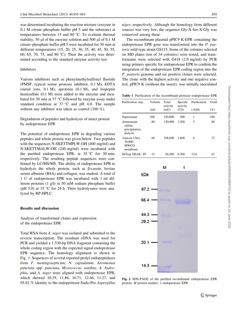
Table 1 Purification of the recombinant protease endoprotease EPR
Purification step Volume Total
activity
Specific
activity
Purification Yield
(ml) (mU) (U/l) (-fold) (%)
Supernatant 300 150,000 500 1 100
Ammonium
sulfate
precipitation,
dialysis
80 120,000 1,500 3 80
Amicon Ultra
30,000
MWCO
membrane
60 108,000 1,800 6 72
HiTrap DEAE- FF 15 94,500 6,300 12.6 63
Fig. 2 SDS-PAGE of the purified recombinant endoprotease EPR
protein. M protein marker; 1 endoprotease EPR
J Ind Microbiol Biotechnol (2013) 40:855–864 859
123
Dow
nloaded from https://academ
ic.oup.com/jim
b/article/40/8/855/5994704 by guest on 24 June 2022
into BMGY and later into BMMY. The cells and the
supernatant of these clones were then collected by
centrifugation.
Purification and peptide mass fingerprinting (PMF)
analysis of the endoprotease EPR
To confirm the expression efficiency of the target protein,
the concentration of induction methanol (0.5 and 1 %) and
the inducing period were optimized. The supernatants were
taken at various time points (0, 12, 24, 48, 72, 80, 96 h).
The results showed that the endoprotease EPR activity
increased in a time-dependent manner within 80 h, but then
slowed down at 96 h, which implied that the optimized
inducing period of the transformant was around 80 h. Also,
the final methanol concentration of 1 % was better than
0.5 %. After 80 h of methanol induction, cultures were
harvested and the supernatants were collected for purifi-
cation of endoprotease EPR.
The prolyl endopeptidase was purified 12.6-fold with
63 % yield from the crude enzyme extract (specific activity
500 U/l). A summary for this purification step is given in
Table 1. Firstly, the proteins in the crude extract precipi-
tated with 30–70 % (NH4)2SO4 achieved threefold enzyme
purification. Fractions exhibiting prolyl endopeptidase
activity purified 12.6-fold (6,300 U/l) were pooled. The
SDS-PAGE result suggested that the enzyme appeared as a
monomer with molecular weight of about 60 kDa (Fig. 2).
174.828
285.808
471.860
212.851
314.797
908.207
439.883
245.788
390.803
1109.731
352.867111.853
651.986
135.822
592.948
503.880
810.116739.043521.88985.892
330.838
412.855
876.398
549.883
1065.470693.963944.821
779.048
0
2
4
6
8
4x10
Inte
ns. [
a.u.
]
200 400 600 800 1000 1200m/z
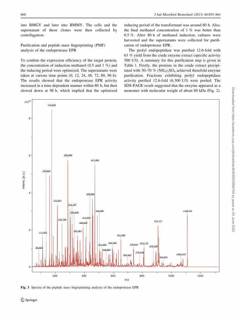
Fig. 3 Spectra of the peptide mass fingerprinting analysis of the endoprotease EPR
860 J Ind Microbiol Biotechnol (2013) 40:855–864
123
Dow
nloaded from https://academ
ic.oup.com/jim
b/article/40/8/855/5994704 by guest on 24 June 2022
Peptide mass fingerprinting (PMF) is known to be an
excellent, fast, and powerful search engine to differentiate
peptidase even with highly similar properties. Therefore, the
identification of the recombinant endoprotease EPR was
analyzed by PMF analysis. As shown in Fig. 3, mass values
of the peptides resulted from the endoprotease EPR diges-
tion ranged from 877 to 3,600. The peptide mass finger-
printing data were analyzed using the MASCOT search tool
(http://ww.matrixscience.com) and showed that a unique
MS/MS fragmentation of LVSASYWQR matches with the
recombinant endoprotease EPR, which further confirmed
that the purified recombinant protein is what we want.
Proline-specific endopeptidases (PEPs) are a unique
class of serine proteases, and most of their structures are
unknown, excluding the structures from pig PEP, S. cap-
sulate PEP, and Myxococcus xanthus PEP [21]. A web-
based tool for protein structure homology modeling-Swiss
Model and the software Discovery Studio 3.1 were used for
predicting the simulated structure of the endoprotease EPR,
which revealed that the active site was located in Ser(179)-
Asp(458)-His(491), based on the template 3n2zB with
sequence identity of 17.6 %. The result indicated that the
endoprotease EPR shared the same active site Ser-Asp-His
with human prolylcarboxypeptidase belonging to S28
protease family [20, 23].
Properties of endoprotease EPR produced
by P. pastoris
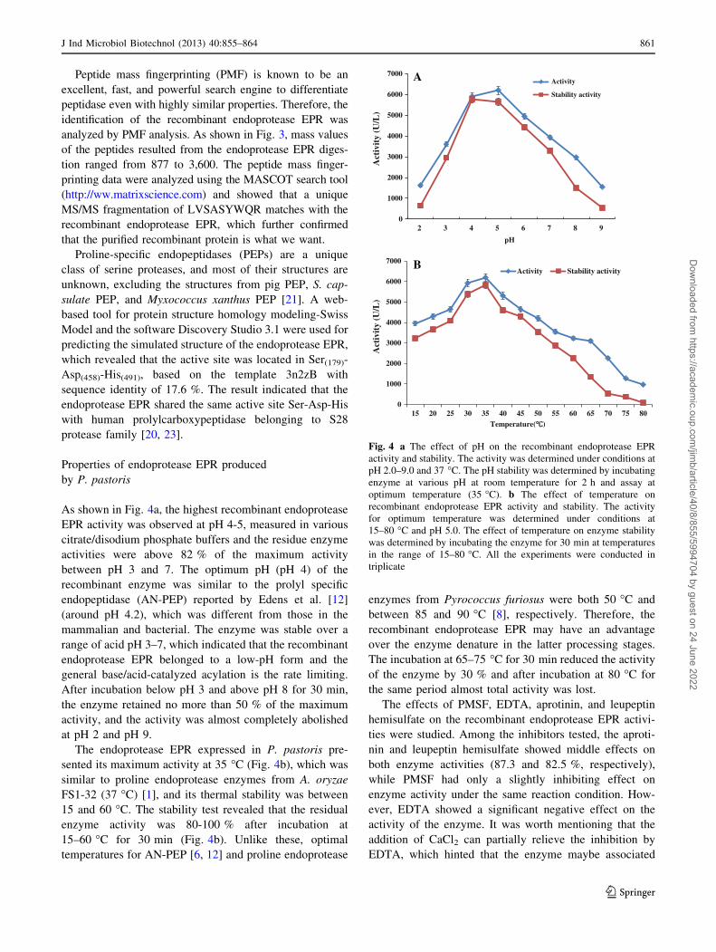
As shown in Fig. 4a, the highest recombinant endoprotease
EPR activity was observed at pH 4-5, measured in various
citrate/disodium phosphate buffers and the residue enzyme
activities were above 82 % of the maximum activity
between pH 3 and 7. The optimum pH (pH 4) of the
recombinant enzyme was similar to the prolyl specific
endopeptidase (AN-PEP) reported by Edens et al. [12]
(around pH 4.2), which was different from those in the
mammalian and bacterial. The enzyme was stable over a
range of acid pH 3–7, which indicated that the recombinant
endoprotease EPR belonged to a low-pH form and the
general base/acid-catalyzed acylation is the rate limiting.
After incubation below pH 3 and above pH 8 for 30 min,
the enzyme retained no more than 50 % of the maximum
activity, and the activity was almost completely abolished
at pH 2 and pH 9.
The endoprotease EPR expressed in P. pastoris pre-
sented its maximum activity at 35 �C (Fig. 4b), which was
similar to proline endoprotease enzymes from A. oryzae
FS1-32 (37 �C) [1], and its thermal stability was between
15 and 60 �C. The stability test revealed that the residual
enzyme activity was 80-100 % after incubation at
15–60 �C for 30 min (Fig. 4b). Unlike these, optimal
temperatures for AN-PEP [6, 12] and proline endoprotease
enzymes from Pyrococcus furiosus were both 50 �C and
between 85 and 90 �C [8], respectively. Therefore, the
recombinant endoprotease EPR may have an advantage
over the enzyme denature in the latter processing stages.
The incubation at 65–75 �C for 30 min reduced the activity
of the enzyme by 30 % and after incubation at 80 �C for
the same period almost total activity was lost.
The effects of PMSF, EDTA, aprotinin, and leupeptin
hemisulfate on the recombinant endoprotease EPR activi-
ties were studied. Among the inhibitors tested, the aproti-
nin and leupeptin hemisulfate showed middle effects on
both enzyme activities (87.3 and 82.5 %, respectively),
while PMSF had only a slightly inhibiting effect on
enzyme activity under the same reaction condition. How-
ever, EDTA showed a significant negative effect on the
activity of the enzyme. It was worth mentioning that the
addition of CaCl2 can partially relieve the inhibition by
EDTA, which hinted that the enzyme maybe associated
0
1000
2000
3000
4000
5000
6000
7000
2 3 4 5 6 7 8 9
Act
ivit
y (U
/L)
pH
Activity
Stability activity
0
1000
2000
3000
4000
5000
6000
7000
15 20 25 30 35 40 45 50 55 60 65 70 75 80
Act
ivit
y (U
/L)
Temperature( )
Activity Stability activity
A
B
Fig. 4 a The effect of pH on the recombinant endoprotease EPR
activity and stability. The activity was determined under conditions at
pH 2.0–9.0 and 37 �C. The pH stability was determined by incubating
enzyme at various pH at room temperature for 2 h and assay at
optimum temperature (35 �C). b The effect of temperature on
recombinant endoprotease EPR activity and stability. The activity
for optimum temperature was determined under conditions at
15–80 �C and pH 5.0. The effect of temperature on enzyme stability
was determined by incubating the enzyme for 30 min at temperatures
in the range of 15–80 �C. All the experiments were conducted in
triplicate
J Ind Microbiol Biotechnol (2013) 40:855–864 861
123
Dow
nloaded from https://academ
ic.oup.com/jim
b/article/40/8/855/5994704 by guest on 24 June 2022
with metal ions and required calcium ions for its optimal
activity. This phenomena might be attributed to calcium
ions involved in the stabilization of the enzyme molecular
structure. In fact, calcium ions are known to be inducers
and stabilizers of many enzymes [26, 29] and also protect
them from conformational changes [24].
In comparison with chromogenic substrates Ala-Pro-
pNA, Ala–Ala-Pro-pNA, Z-Gly-Pro-pNA, and Z-Ala-Ala–
Ala-Pro-pNA, according to the kinetic assay, the recom-
binant endoprotease EPR toward the dipeptide Ala-Pro-
pNA showed almost no activity, while Ala–Ala-Pro-pNA
and Z-Ala-Ala–Ala-Pro-pNA were better substrates for the
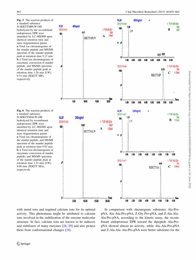
Fig. 5 The reaction products of
a standard substance
N-SEKTTMPLW-OH
hydrolyzed by the recombinant
endoprotease EPR were
identified by LC–MS/MS upon
identical retention time and
mass fragmentation patent.
a Total ion chromatograms of
the stander peptide and MS/MS
spectrum of the stander peptide
peak at retention time 9.12 min.
b, c Total ion chromatograms of
enzymatic conversion of stander
peptide, and MS/MS spectrum
of the stander peptide peak at
retention time 1.26 min (LW),
6.74 min (SEKTT MP),
respectively
Fig. 6 The reaction products of
a standard substance
N-SEKTTMALW-OH
hydrolyzed by recombinant
endoprotease EPR were
identified by LC–MS/MS upon
identical retention time and
mass fragmentation patent.
a Total ion chromatograms of
the stander peptide, and MS/MS
spectrum of the stander peptide
peak at retention time 9.02 min.
b, c Total ion chromatograms of
enzymatic conversion of stander
peptide, and MS/MS spectrum
of the stander peptide peak at
retention time 1.23 min (LW),
6.06 min (SEKTT MA),
respectively
862 J Ind Microbiol Biotechnol (2013) 40:855–864
123
Dow
nloaded from https://academ
ic.oup.com/jim
b/article/40/8/855/5994704 by guest on 24 June 2022
endoprotease EPR. The research implied that the enzyme
preferred larger substrates. Two peptides with the sequen-
ces N-SKETTMPLW-OH (Mw 1092.29) and N-SKETT-
MALW-OH (Mw 1066.25) were incubated with the
purified recombinant endoprotease EPR, respectively. The
resulting peptide sequences N-SKETTMP-OH (Mw 792)
(Fig. 5) and N-SKETTMA-OH (Mw 766.7) were con-
firmed by LC/MS/MS (Fig. 6). Until recently, researchers
found that certain proline-specific endoprotease could
degrade a 33-mer gluten-derived and intact protein [22].
What limits the enzyme specificity is the substrate acces-
sibility to the proline-specific endoprotease activity site
instead of the chain length specificity [7]. In addition,
HPLC analysis in our study showed that the pure recom-
binant endoprotease EPR also could digest the b-casein and
bovine serum albumin.
Acknowledgments Financial support from the National High
Technology Research and Development Program of China (863
Program) (No. 2012AA022207 and 2008AA10Z304), the Funda-
mental Research Funds for the Central Universities (JUSRP11014),
and the Ministry of Education, R.P. China, and from NSFC
(20802027) is greatly appreciated.
References
1. Araki H, Ouchi H, Uesugi S, Hashimoto Y, Shimoda T (1993)
Prolyl endopeptidase and production thereof. EPO patent
EP19920111124
2. Arentz-Hansen H, McAdam SN, Molberg Ø, Fleckenstein B,
Lundin KE, Jørgensen TJ, Jung G, Roepstorff P, Sollid LM
(2002) Celiac lesion T cells recognize epitopes that cluster in
regions of gliadins rich in proline residues. Gastroenterology
123:803–809
3. Capiralla H, Hiroi T, Hirokawa T, Maeda S (2002) Purification
and characterization of a hydrophobic amino acid specific
endopeptidase from Halobacterium halobium S9 with potential.
Process Biochem 38:571–579
4. Edens L, Dekker P, van der Hoeven R, Deen F, de Roos A, Floris
R (2005) Extracellular prolyl endoprotease from Aspergillus
niger and its use in the debittering of protein hydrolysates.
J Agric Food Chem 53:7950–7957
5. Esparza Y, Huaiquil A, Neira L, Leyton A, Rubilar M, Salazar L,
Shene C (2011) Optimization of process conditions for the pro-
duction of a prolylendopeptidase by Aspergillus niger ATCC
11414 in solid-state fermentation. Food Sci Biotechnol
20:1323–1330
6. Gass J, Ehren J, Strohmeier G, Isaacs I, Khosla C (2005) Fer-
mentation, purification, formulation, and pharmacological eval-
uation of a prolyl endopeptidase from Myxococcus xanthus:
implications for celiac sprue therapy. Biotechnol Bioeng
92:674–684
7. Gass J, Khosla C (2007) Prolyl endopeptidases. Cell Mol Life Sci
64:345–355
8. Harwood VJ, Denson JD, Robinson-Bidle KA, Schreier HJ
(1997) Overexpression and characterization of a prolyl endo-
peptidase from the hyperthermophilic archaeon Pyrococcus
furiosus. J Bacteriol 179:3613–3618
9. Higgins DR, Cregg JM (1998) Methods in molecular biology:
Pichia protocols. Totowa, NJ
10. Kabashima T, Fujii M, Meng Y, Ito K, Yoshimoto T (1998)
Prolyl endopeptidase from Sphingomonas capsulata: isolation
and characterization of the enzyme and nucleotide sequence of
the gene. Arch Biochem Biophys 358:141–148
11. Kanatani A, Yoshimoto T, Kitazono A, Kokubo T, Tsuru D
(1993) Prolyl endopeptidase from Aeromonas hydrophila:
cloning, sequencing, and expression of the enzyme gene, and
characterization of the expressed enzyme. J Biochem 113:
790–796
12. Lopez M, Edens L (2005) Effective prevention of chill-haze in
beer using an acid proline-specific endoprotease from Aspergillus
niger. J Agric Food Chem 53:7944–7949
13. Mentlein R (1988) Proline residues in the maturation and deg-
radation of peptide hormones and neuropeptides. FEBS Lett
234:251–256
14. Mitea C, Havenaar R, Drijfhout JW, Edens L, Dekking L, Koning
F (2008) Efficient degradation of gluten by a prolyl endoprotease
in a gastrointestinal model: implications for coeliac disease. Gut
57:25–32
15. Oyama H, Aoki H, Amano M, Mizuki E, Yoshimoto T, Tsuru D,
Murao S (1997) Purification and characterization of a prolyl
endopeptidase from Pseudomonas sp. KU-22. J Ferment Bioeng
84:538–542
16. Pel HJ, de Winde JH, Archer DB, Dyer PS, Hofmann G, Schaap
PJ, Turner G, de Vries RP, Albang R, Albermann K et al (2007)
Genome sequencing and analysis of the versatile cell factory
Aspergillus niger CBS 513.88. Nat Biotechnol 25:221–231
17. Polgar L (2002) The prolyl oligopeptidase family. Cell Mol Life
Sci 59:349–362
18. Polgar L (2002) Structure-function of prolyl oligopeptidase and
its role in neurological disorders. Curr Med Chem 2:251–257
19. Riggle HM, Fisher MA (2009) Purification of a prolyl endopep-
tidase from Aspergillus oryzae and evaluation of its ability to
digest gluten. http://oasys2.confex.com/acs/237nm/techprogram/
P1239602.HTM
20. Schwede T, Kopp J, Guex N, Peitsch MC (2003) SWISS-
MODEL: an automated protein homology-modeling server.
Nucleic Acids Res 31:3381–3385
21. Shan L, Mathew II, Khosla C (2005) Structure and mechanistic
analysis of two prolyl endopeptidase: role of interdomain
dynamics in catalysis and specificity. PNAS 102:3599–3604
22. Shan L, Molberg Ø, Parrot I, Hausch F, Filiz F, Gray GM, Sollid
LM, Khosla C (2002) Structural basis for gluten intolerance in
celiac sprue. Science 297:2275–2279
23. Soisson SM, Patel SB, Abeywickrema PD, Byrne NJ, Diehl RE,
Hall DL, Forf RE, Reid JC, Rickert KW, Shipman JM, Sharma S,
Lumb KJ (2010) Structural definition and substrate specificity of
the S28 protease family: the crystal structure of human prolyl-
carboxypeptidase. BMC Struct Biol 10:16
24. Sousa F, Ju S, Erbel A, Kokol V, Cavaco-Paulo A, Gubitz GM
(2007) A novel metalloprotease from Bacillus cereus for protein
fibre processing. Enzyme Microb Technol 40:772–781
25. Sridhar VR, Hughes JE, Welke DL, Broadbent JR, Steele JL
(2005) Identification of endopeptidase genes from the genomic
sequence of Lactobacillus helveticus CNRZ32 and the role of
these genes in hydrolysis of model bitter peptides. Appl EnvironMicrobiol 71:3025–3032
26. Subba C, Sathish T, Ravichandra P, Prakasham RS (2009)
Characterization of thermo- and detergent stable serine protease
from isolated Bacillus circulans and evaluation of eco-friendly
applications. Process Biochem 44:262–269
27. Stepniak D, Spaenij-Dekking L, Mitea C, Moester M, de RuA,
Baak-Pablo R, van VP, Edens L, Koning F (2006) Highly effi-
cient gluten degradation with a newly identified prolyl endopro-
tease: implications for celiac disease. Am J Physiol Gastrointest
Liver Physiol 291:621–629
J Ind Microbiol Biotechnol (2013) 40:855–864 863
123
Dow
nloaded from https://academ
ic.oup.com/jim
b/article/40/8/855/5994704 by guest on 24 June 2022

28. Szwajcer-Dey E, Rasmussen J, Meldal M, Breddam K (1992)
Proline-specific endopeptidases from microbial sources: isolation
of an enzyme from a Xanthomonas sp. J Bacteriol 174:2454–2459
29. Takii Y, Kurlyama N, Suzuki Y (1990) Alkaline serine protease
produced-from citric acid by Bacillus alcalophilus subsp. halo-
durans KP 1239. Appl Microbial Biotechnol 34:57–62
30. Walter R, Simmons WH, Yoshimoto T (1980) Proline-specific
endo- and exopeptidases. Mol Cell Biochem 30:111–127
31. Yoshimoto T, Walter R, Tsuru D (1980) Proline-specific endo-
peptidase from Flavobacterium: purification and properties.
J Biol Chem 255:4786–4792
32. Xianchang X, Di Y, Fuping L, Yanlin G, Yu L (2009) Cloning of
proline-specific endoprotease gene of Aspergillus niger and
expression in Pichia pastoris. Ind Microbiol (in Chinese) 39:7–12
864 J Ind Microbiol Biotechnol (2013) 40:855–864
123
Dow
nloaded from https://academ
ic.oup.com/jim
b/article/40/8/855/5994704 by guest on 24 June 2022
Related Documents