Special issue: Research report Gender differences in the neural network of facial mimicry of smiles e An rTMS study Sebastian Korb a,*,1 , Jennifer Malsert b,c,1 , Vincent Rochas d,e , Tonia A. Rihs d,e , Sebastian W. Rieger b,d , Samir Schwab c , Paula M. Niedenthal f and Didier Grandjean b,c a Neuroscience Area, SISSA, Trieste, Italy b Swiss Center for Affective Sciences, Geneva, Switzerland c Neuroscience of Emotion and Affective Dynamics Laboratory, Department of Psychology and Educational Sciences, University of Geneva, Switzerland d Department of Fundamental Neuroscience, University of Geneva, Switzerland e Functional Brain Mapping Laboratory, Department of Fundamental Neuroscience, University of Geneva, Switzerland f Department of Psychology, University of Wisconsin, Madison, USA article info Article history: Received 7 October 2014 Reviewed 26 February 2015 Revised 7 April 2015 Accepted 24 June 2015 Published online xxx Keywords: Facial mimicry Gender differences TMS Somatosensory cortex Motor cortex abstract Under theories of embodied emotion, exposure to a facial expression triggers facial mimicry. Facial feedback is then used to recognize and judge the perceived expression. However, the neural bases of facial mimicry and of the use of facial feedback remain poorly understood. Furthermore, gender differences in facial mimicry and emotion recognition suggest that different neural substrates might accompany the production of facial mim- icry, and the processing of facial feedback, in men and women. Here, repetitive trans- cranial magnetic stimulation (rTMS) was applied to the right primary motor cortex (M1), the right primary somatosensory cortex (S1), or, in a control condition, the vertex (VTX). Facial mimicry of smiles and emotion judgments were recorded in response to video clips depicting changes from neutral or angry to happy facial expressions. While in females rTMS over M1 and S1 compared to VTX led to reduced mimicry and, in the case of M1, delayed detection of smiles, there was no effect of TMS condition for males. We conclude that in female participants M1 and S1 play a role in the mimicry and in the use of facial feedback for accurate processing of smiles. © 2015 Elsevier Ltd. All rights reserved. * Corresponding author. Neuroscience Area, SISSA, Via Bonomea 265, 34136 Trieste, Italy. E-mail address: [email protected] (S. Korb). 1 These authors contributed equally to this work. Available online at www.sciencedirect.com ScienceDirect Journal homepage: www.elsevier.com/locate/cortex cortex xxx (2015) 1 e14 http://dx.doi.org/10.1016/j.cortex.2015.06.025 0010-9452/© 2015 Elsevier Ltd. All rights reserved. Please cite this article in press as: Korb, S., et al., Gender differences in the neural network of facial mimicry of smiles e An rTMS study, Cortex (2015), http://dx.doi.org/10.1016/j.cortex.2015.06.025

Welcome message from author
This document is posted to help you gain knowledge. Please leave a comment to let me know what you think about it! Share it to your friends and learn new things together.
Transcript

Special issue: Research report
Gender differences in the neural network of facialmimicry of smiles e An rTMS study
Sebastian Korb a,*,1, Jennifer Malsert b,c,1, Vincent Rochas d,e,Tonia A. Rihs d,e, Sebastian W. Rieger b,d, Samir Schwab c,Paula M. Niedenthal f and Didier Grandjean b,c
a Neuroscience Area, SISSA, Trieste, Italyb Swiss Center for Affective Sciences, Geneva, Switzerlandc Neuroscience of Emotion and Affective Dynamics Laboratory, Department of Psychology and Educational Sciences,University of Geneva, Switzerlandd Department of Fundamental Neuroscience, University of Geneva, Switzerlande Functional Brain Mapping Laboratory, Department of Fundamental Neuroscience, University of Geneva,Switzerlandf Department of Psychology, University of Wisconsin, Madison, USA
a r t i c l e i n f o
Article history:
Received 7 October 2014
Reviewed 26 February 2015
Revised 7 April 2015
Accepted 24 June 2015
Published online xxx
Keywords:
Facial mimicry
Gender differences
TMS
Somatosensory cortex
Motor cortex
a b s t r a c t
Under theories of embodied emotion, exposure to a facial expression triggers facial
mimicry. Facial feedback is then used to recognize and judge the perceived expression.
However, the neural bases of facial mimicry and of the use of facial feedback remain poorly
understood. Furthermore, gender differences in facial mimicry and emotion recognition
suggest that different neural substrates might accompany the production of facial mim-
icry, and the processing of facial feedback, in men and women. Here, repetitive trans-
cranial magnetic stimulation (rTMS) was applied to the right primary motor cortex (M1),
the right primary somatosensory cortex (S1), or, in a control condition, the vertex (VTX).
Facial mimicry of smiles and emotion judgments were recorded in response to video clips
depicting changes from neutral or angry to happy facial expressions. While in females
rTMS over M1 and S1 compared to VTX led to reduced mimicry and, in the case of M1,
delayed detection of smiles, there was no effect of TMS condition for males. We conclude
that in female participants M1 and S1 play a role in the mimicry and in the use of facial
feedback for accurate processing of smiles.
© 2015 Elsevier Ltd. All rights reserved.
* Corresponding author. Neuroscience Area, SISSA, Via Bonomea 265, 34136 Trieste, Italy.E-mail address: [email protected] (S. Korb).
1 These authors contributed equally to this work.
Available online at www.sciencedirect.com
ScienceDirectJournal homepage: www.elsevier.com/locate/cortex
c o r t e x x x x ( 2 0 1 5 ) 1e1 4
http://dx.doi.org/10.1016/j.cortex.2015.06.0250010-9452/© 2015 Elsevier Ltd. All rights reserved.
Please cite this article in press as: Korb, S., et al., Gender differences in the neural network of facial mimicry of smiles e An rTMS study,Cortex (2015), http://dx.doi.org/10.1016/j.cortex.2015.06.025
1. Introduction
The human face is one of the most expressive channels of
emotional and social communication. Accurate interpretationof clues to affective states and behavioral intentions displayedon the face are crucial abilities for smooth social interactionand successful goal pursuit in society. Indeed, impairedemotion recognition and reduced empathy are major factorsleading to difficulties in social communication that charac-terize, for example, people with autism spectrum disorder(ASD). Tangible differences in the display and perception ofemotional expressions also exist between healthy male andfemale individuals. It is therefore of scientific and societalinterest to understand the processes and neural correlates
that support emotion recognition. The present study investi-gated gender differences in the role of motor and somato-sensory cortices in facial mimicry and emotion perception.
An influential theoretical account, which builds upon along and prominent tradition in biology, philosophy, andpsychology (Darwin, 1872; James, 1950; Lipps, 1903), suggeststhat emotional information is processed through somato-visceral and motoric re-experiencing (Barsalou, 2008;Iacoboni, 2009; Niedenthal, 2007). A component of thisembodied emotion theory is the facial feedback hypothesis, ac-cording to which information from one's own facial expres-
sions feeds back into the brain and triggers or colorsemotional responses, and influences emotional judgments(Adelmann & Zajonc, 1989; Buck, 1980; Hatfield, Cacioppo, &Rapson, 1993; McIntosh, 1996; Strack, Martin, & Stepper,1988). Support for this hypothesis comes from researchshowing that voluntarily producing emotional facial expres-sions results in specific physiological activity patterns(Ekman, Levenson,& Friesen, 1983) and shapes correspondingsubjective feelings. Actively facilitating or inhibiting smiling,by holding a pen either between the teeth or the lips, in-fluences the appraisal of humorous stimuli (Soussignan, 2002;
Strack et al., 1988). Similarly, recent clinical trials suggest thatindividuals suffering from depression may benefit from pro-cedures leading to the paralysis of the Corrugator muscles(involved in frowning and sadness), possibly by impeding thisspecific facial feedback that may contribute to the build-up ofnegative emotions (Finzi & Rosenthal, 2014; Wollmer et al.,2012).
Another component of embodied emotion theory is theobservation that people spontaneously engage in motormimicry. The perception of a smile, for example, causes theobserver to smile in return. The observer's own smile is hy-
pothesized to facilitate the recognition of the observedexpression through afferent feedback to the brain. Indeed,mimicry of happy faces increases the accuracy of judgmentsof smile authenticity (Korb, With, Niedenthal, Kaiser, &
Grandjean, 2014; but see Hess & Blairy, 2001), and the block-ing of facial mimicry reduces the speed and the accuracy ofrecognizing emotional facial expressions. For example,blocking facial mimicry slows the recognition of positive andnegative facial expressions (Stel & van Knippenberg, 2008),impairs the distinction between true and false smiles(Maringer, Krumhuber, Fischer, & Niedenthal, 2011;Rychlowska et al., 2014), delays the perception of the offset
of happy and sad facial expressions (Niedenthal, Brauer,
Halberstadt, & Innes-Ker, 2001), and interferes with therecognition of happiness (Oberman, Winkielman, &
Ramachandran, 2007). Furthermore, paralysis of the Corru-gator muscle through injections of botulinum toxin decreasesresponses to angry faces in emotion centers of the brain suchas the amygdala, and reduces the functional coupling be-tween the amygdala and brain stem regions implicated inautonomic emotional responses (Hennenlotter et al., 2009).
The hypothesis that facial mimicry occurs both sponta-neously and unconsciously is supported by findings thatmimicry can occur in the absence of conscious perception of
the stimulus face (Dimberg, Thunberg, & Elmehed, 2000;Mathersul, McDonald, & Rushby, 2013), and that it is difficultto suppress voluntarily (Dimberg, Thunberg,&Grunedal, 2002;Korb, Grandjean, & Scherer, 2010). Facial mimicry may becrucial for the development of empathy, which requires thedetection and the representation of another person'semotional state. Indeed, facial mimicry is increased in in-dividuals high in self-reported trait empathy (Dimberg,Andr!easson, & Thunberg, 2011; Sonnby-Borgstrom, 2002).However, emotion recognition can also occur without facialmimicry, for example in individuals with facial paralysis
(Rives Bogart&Matsumoto, 2010), and the simulation ofmotorand somatosensory events linked to facial expressions canoccur in the brain only, that is, in the absence of an overtperipheral response.
Which systems of the brain are responsible for the spon-taneous production of facial mimicry, and which ones utilizethe resulting facial feedback, or provide a visceral and somaticsimulation, during the processing of facial expressions? Toanswer these questions we turn to the neuroscientific litera-ture, where current models of social cognition are built uponthe notion that motor, somatosensory, and emotional brain
regions simulate other people's actions, sensations, andemotions, and by doing so contribute to the their perceptionand interpretation (Iacoboni, 2009; Keysers, Kaas, & Gazzola,2010). Studies using neuroscientific or neuropsychologicalmethods largely suggest that perceiving another person per-forming a motor action, displaying a facial expression, orbeing touched on their body, results in increased neural ac-tivity in the perceiver's motor, emotional, and somatosensoryareas.
The mirror neuron system (MNS) provides a putativeneural basis for facial mimicry. It includes the inferior frontalgyrus (IFG), the posterior parietal cortex, but also primary and
secondary somatosensory cortices (S1 and S2), and the insula(Di Pellegrino, Fadiga, Fogassi, Gallese, & Rizzolatti, 1992;Gazzola & Keysers, 2009; Molenberghs, Cunnington, &
Mattingley, 2012; Mukamel, Ekstrom, Kaplan, Iacoboni, &
Fried, 2010; Rizzolatti & Craighero, 2004; Rizzolatti, Fogassi,& Gallese, 2001). Brain imaging studies have found substan-tial overlap in the brain activity accompanying the productionand observation of facial expressions (Van der Gaag,Minderaa, & Keysers, 2007). However, only few studies havespecifically investigated the neural correlates of spontaneousfacial mimicry. Schilbach, Eickhoff, Mojzisch, and Vogeley
(2008) reported increased brain activity, likely accompanyingfacial mimicry, in the face area of the left primary motorcortex (M1) and in the bilateral posterior cingulate gyrus.
c o r t e x x x x ( 2 0 1 5 ) 1e1 42
Please cite this article in press as: Korb, S., et al., Gender differences in the neural network of facial mimicry of smiles e An rTMS study,Cortex (2015), http://dx.doi.org/10.1016/j.cortex.2015.06.025

Likowski et al. (2012) reported significant correlations between
the amplitude of facial mimicry and brain activity in variousareas that belong, or are functionally connected, to the MNS,including the IFG, the SMA, the insula, the medial temporalgyrus (MTG) and the superior temporal sulcus (STS). Also ofinterest, the disruption of medial premotor cortices withevent-related rTMS interferes with the recognition of facialexpressions (Balconi & Bortolotti, 2013a, 2013b; Rochas et al.,2013), while activation of a more fronto-polar area (BA9) in-creases facial mimicry (Balconi & Canavesio, 2013).
If motor and premotor areas of the MNS (M1, IFG, medialpremotor cortices) might constitute the “output” center of
facial mimicry, somatosensory cortices (S1, S2) could be thetargets of the “input” for ensuing facial feedback (be it real orsimulated). This assumption is based on several lines ofempirical evidence. First, as reviewed above, these areas showincreased activity during the perception of facial expressions(Molenberghs et al., 2012; Van der Gaag et al., 2007). Second, asshown in a sample of 108 patients, brain lesions affecting theright somatosensory cortices are associatedwith performancedeficits on tasks requiring the recognition of facial expres-sions of emotion (Adolphs, Damasio, Tranel, Cooper, &
Damasio, 2000). Finally, inhibition of the right somatosen-
sory cortex with transcranial magnetic stimulation (TMS)leads to slower responses and reduced accuracy in emotion-matching tasks (Pitcher, Garrido, Walsh, & Duchaine, 2008;Pourtois et al., 2004). Therefore, the somatosensory cortexhas been suggested to simulate internally, or to be an efferenttarget of, tactile and proprioceptive facial feedback, whichaccompanies facial mimicry (Adolphs et al., 2000; Sims, VanReekum, Johnstone, & Chakrabarti, 2012).
In summary, the neural circuitry underlying the simulationof facial expressions perceived in others (be this an overtmimicry leading to measurable changes in the facial electro-
myography (EMG), or a “brain-only” internal simulation ofmotor output), and the processing of (real or simulated) so-matosensory facial feedback, are not fully understood. Motorand premotor areas of the MNS (M1, IFG, medial premotorcortices) are likely substrates for the production of facialmimicry (the “output”). Somatosensory cortices (S1, S2),especially in the right hemisphere, are likely involved in theprocessing of facial feedback, and in the analysis of the resultof facial mimicry (the “input”).
However, men and women differ in their behavioral,subjective, and neural responses to emotional stimuli. Thus,the putative neural network responsible for generating (or
simulating) facial mimicry and processing facial feedback islikely to differ by gender. Important gender differences existin the production, perception, and regulation of facialexpression. Women are more emotionally expressive andmore empathic than men (Eisenberg & Lennon, 1983; Kring &
Gordon, 1998); more accurate and/or efficient in processingfacial expressions of emotion (Hall, 1978; Hall & Matsumoto,2004; Hoffmann, Kessler, Eppel, Rukavina, & Traue, 2010);show more facial mimicry than men (Dimberg & Lundquist,1990); and are more susceptible to emotional contagion, asrevealed both in self-report and dyadic interaction (Doherty,
Orimoto, Singelis, Hatfield, & Hebb, 1995). Gender differ-ences have also been found in the effects of pacifier useduring infancy, which arguably blocks facial mimicry, on
facial mimicry recorded at age seven. Specifically, pacifier use
is associated with reduced facial mimicry in boys, but notgirls (Niedenthal et al., 2012). These findings suggest thatmen and women may utilize a different set of neural struc-tures during the recognition and embodiment of emotionalfacial expressions, or at least that the degree to which ele-ments of this network can be modulated through externalinfluences varies by gender. The assumption that importantdifferences may exist in the neural substrate of facial mim-icry and emotion recognition seems particularly plausible ifone considers that “sex influences on brain function areubiquitous, found at every level of neuroscience from the
behaving human to the ion channel” (Cahill, 2012, p. 2542). Inline with this, meta-analyses of brain imaging studies haverevealed that brain activation to emotional stimuli greatlydiffers by gender (Stevens & Hamann, 2012).
The present experiment investigated gender differences inthe neural circuitry underlying the simulation/production offacial mimicry of smiles and the integration of facial feedbackin the recognition process of facial expressions of happiness.In a within-subjects design, rTMS was delivered in separatesessions to inhibit the activity of the Zygomaticus/cheek re-gion of the right M1 or S1. Delivery of rTMS over the vertex
(VTX, midline midpoint between inion and nasion) served asan active control condition. Participants then completed twotasks that involved rating the intensity of emotional facialexpressions (Intensity task), and detecting the offset of a facialexpression of emotion (Offset task). Both tasks were inspiredby previous research reporting the occurrence of facial mim-icry during the perception of movie clips of neutral-to-emotional morphs (Achaibou, Pourtois, Schwartz, &
Vuilleumier, 2008), and changes in the time of detection ofthe offset of emotional expressions after blocking partici-pants' facial mimicry (Niedenthal et al., 2001). Although both
tasks included expressions of anger and happiness, analysesfocused on trials with the latter emotion because fMRI andrTMS served to localize and subsequently target motor andsomatosensory cortices innervating the cheek region(involved in smiling).
To measure facial mimicry, EMG was recorded over bilat-eral Corrugator and Zygomaticus muscles. To guarantee in-dividual coil positioning using a frameless stereotaxic system,locations of the Zygomaticus area in M1 and of the cheek areain S1 were determined in a first session through fMRI, duringwhich participants were asked to smile or experienced a lighttouch on their cheek.
Fourmain hypotheses were tested.We hypothesized a roleof M1 in the production of mimicry of smiles, and a role of S1in the perception of the resulting facial feedback. In otherwords, 1) rTMS over M1was expected to reduce facial mimicryof smiles and to affect behavioral responses in both tasks, and2) rTMS over S1 was expected to affect behavioral responseswithout reducing facial mimicry by impairing the processingand integration of facial feedback information.We expected 3)facial mimicry of smiles to be correlated with measures ofmood (Moody, McIntosh, Mann, & Weisser, 2007) and traitempathy (Dimberg et al., 2011; Sonnby-Borgstrom, 2002).
Finally, we expected 4) rTMS to have different effects onbehavioral responses to and facial mimicry of smiles in maleversus female participants.
c o r t e x x x x ( 2 0 1 5 ) 1e1 4 3
Please cite this article in press as: Korb, S., et al., Gender differences in the neural network of facial mimicry of smiles e An rTMS study,Cortex (2015), http://dx.doi.org/10.1016/j.cortex.2015.06.025
2. Methods
2.1. Participants
Thirty healthy participants (17 females, mean age ¼ 22.7years, SD ¼ 3.4, range 19e31 years) were recruited throughadvertisements on campus at the University of Geneva. Allparticipants reported to be right-handed and free of psychi-atric disorders, and had normal or corrected-to-normal vision.They were screened to ensure they were free from neurolog-ical, psychiatric, and medical problems, as well as from con-traindications for TMS and MRI. Participants gave informedconsent and were paid for their participation.
2.2. Stimuli
In both the Offset and Intensity tasks stimuli consisted ofshort video clips showing gradual changes in emotional ex-pressions displayed by adult faces. Stimuli were constructed
inmorphing software (Morpheus PhotoMorpher, version 3.17)using happy, angry and neutral expressions by 14 adult faces(50% male). The same photos have been successfully used tocreate similar stimuli in previous studies (Halberstadt &
Niedenthal, 2001; Niedenthal et al., 2001). Movie clips in theOffset task always showed a full-blown expression of happi-ness or anger that gradually morphed into the other expres-sion. In the Intensity task, neutral facial expressions morphedinto an expression of happiness or anger. The movie clips,which were shown at 60 frames per second, lasted five sec-onds in the Offset task and two seconds in the Intensity task.
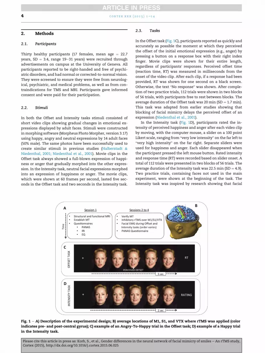
2.3. Tasks
In theOffset task (Fig. 1C), participants reported as quickly andaccurately as possible the moment at which they perceivedthe offset of the initial emotional expression (e.g., anger) bypressing a button on a response box with their right indexfinger. Movie clips were shown for their entire length,regardless of participants' responses. Perceived offset time(reaction time, RT) was measured in milliseconds from theonset of the video clip. After each clip, if a response had been
provided, RT was shown for one second on a black screen.Otherwise, the text “No response” was shown. After comple-tion of two practice trials, 112 trials were shown in two blocksof 56 trials, with participants free to rest between blocks. Theaverage duration of the Offset task was 20 min (SD ¼ 1.7 min).This task was adapted from earlier studies showing thatblocking of facial mimicry delays the perceived offset of anexpression (Niedenthal et al., 2001).
In the Intensity task (Fig. 1D), participants rated the in-tensity of perceived happiness and anger after each video clipby moving, with the computer mouse, a slider on a 100 pointLikert scale, ranging from “very low intensity” on the far left to
“very high intensity” on the far right. Separate sliders wereused for happiness and anger. Each slider disappeared whenthe participant pressed the left mouse button. Rated intensityand response time (RT) were recorded based on slider onset. Atotal of 112 trials were presented in two blocks of 56 trials. Theaverage duration of the Intensity task was 22.5 min (SD ¼ 4.9).Two practice trials, containing faces not used in the mainexperiment, were shown at the beginning of the task. TheIntensity task was inspired by research showing that facial
Fig. 1 e A) Description of the experimental design; B) average locations of M1, S1, and VTX where rTMS was applied (colorindicates pre- and post-central gyrus); C) example of an Angry-To-Happy trial in the Offset task; D) example of a Happy trialin the Intensity task.
c o r t e x x x x ( 2 0 1 5 ) 1e1 44
Please cite this article in press as: Korb, S., et al., Gender differences in the neural network of facial mimicry of smiles e An rTMS study,Cortex (2015), http://dx.doi.org/10.1016/j.cortex.2015.06.025

mimicry can reliably be induced by dynamic expressions of
happiness and anger (Achaibou et al., 2008).
2.4. Procedure
A within-subjects design was used. The experiment wascomposed of four separate laboratory sessions, which were
completed no fewer than five and no more than seven daysapart (Fig. 1A).
In the first session, participants completed two safetyquestionnaires to verify that MRI and TMS procedures werenot contraindicated, signed informed consent, and completedstructural and functional scanning in the MRI (see below). Atthe end of the first session, participants completed threepersonality and affect questionnaires: The InterpersonalReactivity Index (IRI; Davis, 1983) and Empathy Quotient (EQ;Baron-Cohen & Wheelwright, 2004) were used to measuretrait-like empathy, while the Positive and Negative Affect
Schedule (PANAS; Watson, Clark, & Tellegen, 1988) measuredparticipants' current mood. The PANAS was moreover filledout at the end of sessions two, three, and four, to control forparticipants' mood.
Sessions two to four were identical, except for the locationof the application of TMS (M1, S1, VTX in semi-random orderacross participants), and in that the order of the Intensity andOffset tasks, as well as of the trials in these tasks, weredifferent across participants and sessions (Fig. 1B). At thebeginning of sessions two to four participants received singlepulse TMS over their right motor cortex to establish their
motor threshold (MT) based on the EMG of their left thumb.The MT was defined as the minimal intensity that inducedmotor evoked potentials (MEPs) greater or equal to 50 mV peak-to-peak amplitude in five out of ten trials. EMG for the MTmeasurement was recorded bipolarly on the left AbductorPollicis Brevis muscle with electrodes connected to the Mag-stim EMG module. The stimulation intensity for the rTMSsequence (see below) was adjusted based on the measuredMT. Next, facial EMG electrodes were attached (see below),and rTMS was applied. Immediately following the rTMSsequence, participants completed the Intensity and Offset
tasks, while EMG was recorded. After their last session par-ticipants were debriefed and paid.
2.5. Facial EMG recording
Facial EMG was bipolarly recorded from the left and right
Corrugator Supercilii and Zygomaticus Major muscles, ac-cording to guidelines (Fridlund & Cacioppo, 1986). We used aBiosemi (www.biosemi.com) ActiveTwo amplifier systemwithAg/AgCl active electrodes, a sampling rate of 2048 Hz and abandwidth of DC-1600 Hz. The common mode sense andright-driven-leg electrodes (serving as ground and reference)were placed below the hairline on the center of the forehead.
2.6. rTMS stimulation
Magnetic stimulation was performed with a Rapid2 generatorconnected to a 70 mm figure-of-eight coil producing biphasic
pulses (The Magstim Company Ltd, Whitland, Wales, UK). A
modified continuous Theta burst stimulation procedure(mcTBS) was used, consisting of 200 bursts of three TMS pul-ses each delivered at 30 Hz, the bursts being delivered at 6 Hz.Thus, over a period of 33.3 sec, a total of 600 pulses wereadministered at an intensity of 80% of the MT. This type ofprotocol has been employed before over M1 and the frontaleye fields, and has been shown to induce cortical inhibitionlasting more than 30 min (Goldsworthy, Pitcher, & Ridding,2012; Nyffeler et al., 2006). Precise positioning of the coil totarget the right M1 or S1 cortices, or the VTX as an activecontrol, was achieved using a neuro-navigation system (see
below) based on participant's structural MRI and functionallocalizer results.
2.7. Structural MRI and fMRI localizer
MRI images were acquired using a 3T whole body MRI scanner(Trio TIM, Siemens, Germany) with the product 12 channel
head coil. Anatomical imaging was carried out using a T1
weighted MPRAGE sequence (TR/TI/TE ¼ 1900/900/2.27 msec,flip angle ¼ 9", PAT factor ¼ 2, voxel dimensions: 1 mmisotropic, 256 # 256 # 192 voxel). A functional localizerexperiment was then performed using a BOLD contrast opti-mized EPI sequence (TR/TE ¼ 2000/30 msec, flip angle ¼ 80",PAT factor¼ 2, 64# 64 pixel, 3.2# 3.2mm, 35 slices acquired indescending order, 3.2 mm slice thickness, 20% slice gap). Onerun comprising 250 volumes was acquired for each partici-pant. Participants watched a back projection screen placedinside the scanner bore, in the center of which a cross or a
circle was displayed. They were instructed to maintainattention to the center of the screen, to rest when a cross wasdisplayed, to smile whenever the circle was present, and toavoid moving their head. Compliance with the motor taskinstruction wasmonitored with an MRI compatible movie clipcamera (12M-i, MRC SystemsGmbH, Germany). Tactile stimuliwere applied to both lower cheeks of the participant with acustom built device comprising non-metallic pneumatic cyl-inders (TA-AC-PVC-1.0-EP, Teqcom Industries, Inc., USA)(Rieger, Domınguez-Borr"as, & Vuilleumier, 2011). Tactilestimulation occurred only while the cross was shown on the
screen, and so did not coincide with the motor task. The cyl-inders were controlled by electromagnetic valves, which wereplaced outside the MRI scanner room and connected to aprogrammable control unit. The cylinders were adjusted to bein permanent contact with the skin and traveled forward andback by five mm once per second when activated. Stimuliwere timed according to a mini-block design, consisting of 53rest periods of two to 12 sec duration, interspersed with 26somatosensory blocks of four or six seconds and 26 motorblocks of four or six seconds in pseudo-randomized order.
The MR data were analyzed using BrainVoyager QX (Brain
Innovation, The Netherlands). The functional images weremotion corrected and co-registered to the T1 image at theindividual level (no spatial normalization was performed).Activation maps were calculated based on the stimulus tim-ings convolved with a canonical HRF. The motion parameterswere included as regressors of no interest.
c o r t e x x x x ( 2 0 1 5 ) 1e1 4 5
Please cite this article in press as: Korb, S., et al., Gender differences in the neural network of facial mimicry of smiles e An rTMS study,Cortex (2015), http://dx.doi.org/10.1016/j.cortex.2015.06.025
2.8. Neuronavigation and coil placement
On each session of TMS, the coil was placed over the right M1or S1, or over the VTX with the help of a frameless neuro-navigation system based on the individual anatomical andfunctional MRI data. The system combines the neuro-navigation module of BrainVoyager Qx software with the ul-trasound CMS20 measuring system for navigation (ZebrisGmbH, Tubingen, Germany). Target coordinates for TMS inthe motor and somatosensory cortices were determined by
locating in the functional MRI data the peak of the largestcluster of activation in themotor and somatosensory cortices,respectively. The VTX target area was defined as themidpointbetween inion and nasion in the antero-posterior axis and onthemidline with the help of the neuronavigation. The averageTalairach (MNI) coordinates at which rTMS was applied were52.2; $8; 39.8 (52.7, $10.3, 42.7) for the right M1, 55; $19.8; 39.6(55.5,$22.4, 41.9) for the right S1 and 0;$31.9; 65.2 (0,$36.2, 69)for the VTX (Fig. 1B). On average, the target areas forM1 and S1were thus 1.5 cm apart. The first phase of the biphasic cur-rents flowing into the coil was oriented in an antero-medialdirection for the stimulation of M1 or S1 e perpendicularly
to the central sulcus. For stimulation of the VTX, the currentswere oriented forward.
2.9. Data analysis
The study was designed to investigate the role of M1 and S1 inthe perception and mimicry of smiles. Therefore, data ana-lyses focused on behavioral responses and EMG of the Zygo-maticus muscle to Angry-To-Happy trials in the Offset task,and Happy trials in the Intensity task (see Supplementarymaterial for graphs including EMG of the Corrugator and re-sponses to Happy-To-Angry trials). Statistical analyses wereperformed in SPSS (version 20) and R (www.r-project.org). Incases of non-sphericity, corrected p values and uncorrecteddegrees of freedom are reported.
2.10. Questionnaires, TMS-Intensity, TMS-Time
Possible differences in mood across sessions and gender were
investigated with a repeated measures analysis of variance(rmANOVA) with the between-subjects factor Gender and thewithin-subjects factors Valence (positive, negative mood) andTMS (M1, SI, VTX) and followed up with post hoc t-tests.Gender differences for IRI and EQ questionnaires wereexplored with independent-samples t-tests.
The intensity at which rTMS was applied was based oneach participant's MT, and could vary between sessions of thesame participant. In addition, the order of tasks was coun-terbalanced, and duration of experimental procedures (e.g.,switching between tasks) and task durations could vary.Therefore, two rmANOVAswere computed with the between-
subjects factor Gender, and the within-subjects factor TMS, totest if sessions differed in terms of TMS intensity (i.e., ma-chine output) and TMS-Time (i.e., time in minutes passedbetween rTMS and the beginning of each task). Both factorswere also included as covariates in analyses of covariance(ANCOVAs) of behavioral and EMG data.
2.11. Behavior
For the Offset task, trials without response (<1%), or on whichthe perceived offset time was more than two SDs above orbelow the mean, were removed. On average, 4% of the trialswere removed (SD ¼ 5.8). Remaining Angry-To-Happy trialswere analyzed in an rmANOVA with the factors TMS andGender, and with t-tests based on hypotheses.
In the Intensity task data from one participant were lostduring the VTX session due to technical problems, and the
participant was excluded from analyses. The remaining 29participants reported, on average, higher emotional intensityon the appropriate rating scale in 98.1% of the trials (SD ¼ 1.7).The number of Happy trials with correct ratings (i.e., higherratings of happiness than anger) was analyzed in a 3 # 2rmANOVA with the factors TMS and Gender, as well as withplanned contrasts. Trials were removed if they had incorrectratings (<2% of all trials across participants), or if their RT wasmore than two SDs over the mean of all participants (<7%).The remaining data included (across both emotions) anaverage of 51.9 trials (SD ¼ 7.4) per condition and participant.Intensity and RTs of happiness ratings to Happy trials were
analyzed with the same rmANOVA and planned contrasts asabove.
In order to investigate the impact of potentially con-founding factors, behavioral data from both tasks were alsoanalyzed in ANCOVAs with the between-subjects factorGender, the within-subjects factor TMS, and the covariatesTMS-Time and TMS-Intensity.
Offset times were non-parametrically correlated (uncor-rected) with the questionnaire scores to explore the effects ofmood (PANAS) and trait empathy (IRI, EQ) on emotionperception (Table 1). Similarly, two-tailed non-parametric
Spearman correlations were computed between the ratingson the Intensity task and the questionnaire scores (Table 3).
2.12. EMG data
Offline, EMG data were preprocessed in Matlab (versionR2012a; www.mathworks.com) partially using the EEGLABtoolbox (Delorme & Makeig, 2004). Data were submitted tobipolar montage, bandpass-filtered from 20 to 400 Hz, and
segmented from one second before to five seconds (Offsettask) or two seconds (Intensity task) after stimulus onset(SO). Data were then rectified and smoothed with a 40 Hzlow-pass filter. For each participant, we excluded trials basedon behavioral analyses (see above), and trials in which theaverage amplitude in the baseline period ($1 sec to SO) ofeither muscle exceeded by more than two SDs the averageamplitude over all trials' baselines for the respective muscle.For the Offset task (across all three sessions, which on totalincluded 336 trials), an average of 23.1% of trials wereexcluded per participant (number of excluded trials M ¼ 77.5,SD ¼ 17.2). For the Intensity task, an average of 19.3% trials
were excluded per participant (number of excluded trialsM ¼ 64.8, SD ¼ 10.9). After this artifact rejection procedure,data from SO onwards were expressed as percentage of theaverage of the one-second long baseline (for a similar pro-cedure see De Wied, van Boxtel, Zaalberg, Goudena, &
Matthys, 2006; Korb et al., 2014). For statistics, valid EMG
c o r t e x x x x ( 2 0 1 5 ) 1e1 46
Please cite this article in press as: Korb, S., et al., Gender differences in the neural network of facial mimicry of smiles e An rTMS study,Cortex (2015), http://dx.doi.org/10.1016/j.cortex.2015.06.025

data were transformed to the natural logarithm and averaged
over both sides of the face and over two time windows,which lasted one second for the Intensity task, and 2.5 sec forthe Offset task.
To investigate the modulation of the amplitude of facialmimicry by TMS, analyses focused on Zygomaticus EMG dataduring the second time-window of Angry-To-Happy andHappy trials, respectively in the Offset and Intensity tasks.Wechose the Zygomaticus because inhibitory rTMS was appliedover motor (M1) and somatosensory (S1) areas innervating thecheek (which comprises the Zygomaticus but not the Corru-gator muscle). The second time window was chosen since it
contained the apex (Intensity task) and the rise to apex (Offsettask) of the happiness expressions, and therefore was mostlikely to reveal facial mimicry of smiles.2 These data wereanalyzed with rmANOVAs including the factors TMS andGender, with hypothesis-driven t-tests and, to check forpossible confounds, in mixed ANCOVAs with the factorsGender and TMS and the covariates TMS-Intensity and TMS-Time.
To test whether participants' facial mimicry was linked totheir positive and negative mood (assessed after each sessionvia the PANAS), or to their empathy traits (assessed once with
the IRI and EQ), we calculated non-parametric Spearmancorrelations between these questionnaires and facial mimicry(defined as above) both in the Offset task (Table 2) and theIntensity task (Table 4).
3. Results
3.1. Questionnaires
An rmANOVA on the PANAS data with the factors TMS,Valence and Gender revealed a significant main effect ofValence [F(1,28) ¼ 261.4, p < .001, hp
2 ¼ .9], due to higher scoreson the positive than the negative mood subscale, a significantTMS # Gender interaction [F(2,56) ¼ 4.2, p ¼ .023, hp
2 ¼ .13],and a trend for Valence # Gender interaction [F(1,28) ¼ 3.9,p ¼ .06, hp
2 ¼ .12]. No other effects reached significance ortrend level (other Fs < 2.2, ps > .14). Post hoc t tests (uncor-
rected) revealed significantly higher negative mood in male(means 23.7 and 21.8) compared to female participants(means 13.2 and 14.5) in the M1 [t(28) ¼ 2.4, p ¼ .02] and S1condition [t(28) ¼ 2.5, p ¼ .02], and in the VTX (M ¼ 15.2)compared to the M1 condition (M ¼ 13.2) in female partici-pants [t(16) ¼ 2.7, p ¼ .02].
The average score on the EQ was 37.6 (SD ¼ 7.8, range24e62). There was no difference [t(28) ¼ $.21, p ¼ .84] betweenmen (M ¼ 37.3, SD ¼ 5.6) and women (M ¼ 37.9, SD ¼ 9.3). Theaverage scores on the IRI subscales were 13.3 (SD ¼ 4.8) forPerspective taking, 15.5 (SD¼ 4.6) for Fantasy, 7.7 (SD¼ 3.6) for
Empathic Concern, and 8.9 (SD ¼ 4.6) for Personal Distress. Nodifferences betweenmen andwomenwere observed (all t < .8,all p > .4).
3.2. TMS-Intensity and TMS-Time
Mean TMS-Intensity for conditions M1, S1, and VTXwere, respectively, 46.2, 45.2, 45.6 (SDs were 7.6, 8.1, 8.4)percent of the maximal TMS stimulator output. An rmA-NOVA with the factors Gender and TMS revealed a signifi-cant interaction [F(2,56) ¼ 3.34, p < .04, hp
2 ¼ .11], which wasfollowed up with independent-samples t-tests to comparegender groups across conditions, and with paired-samples t-tests to compare conditions amongst gender groups. In fe-
males, TMS-Intensity in the M1 condition was found[t(16) ¼ 2.3, p ¼ .04] to be significantly lower (M ¼ 44.8,SD ¼ 7.6) than in the VTX condition (M ¼ 45.4, SD ¼ 8.5). Noother comparison reached significance or trend level (allt < 1.77, all p > .1).
An rmANOVA with factors Gender and TMS on the vari-able TMS-Time in the Offset task showed a trend for an effectof Gender [F(1,28) ¼ 3.53, p ¼ .07, hp
2 ¼ .11]. On average, menstarted the Offset task 15.35 min after rTMS, while womenstarted it after 20.34 min. The same AVOVA also resulted in atrend for an effect of Gender in the Intensity task[F(1,28) ¼ 2.94, p ¼ .09, hp
2 ¼ .09], with slightly earlier task
onsets in women (M ¼ 15.4 min) compared to men(M ¼ 20.23 min).
3.3. Offset task
3.3.1. BehaviorRTs were analyzed in an rmANOVA with the factors TMS andGender, resulting in a significant main effect of Gender[F(1,28)¼ 8.4, p ¼ .007, hp
2 ¼ .23], due to later perceived offset in
male (M ¼ 2610 msec) compared to female participants(M ¼ 2143 msec), and a strong trend for a TMS # Genderinteraction [F(2,56) ¼ 2.9, p ¼ .06, hp
2 ¼ .1]. Planned contrastsrevealed that females perceived (Fig. 2) offsets in Angry-To-Happy trials significantly [t(16) ¼ 2.2, p ¼ .03] later in the M1condition (M ¼ 2241.1 msec, SD ¼ 468) compared to the VTXcondition (M ¼ 2075.4 msec, SD ¼ 487.7), and to the S1(M ¼ 2113.2 msec, SD ¼ 499.9) condition [t(16) ¼ 2.3, p ¼ .02].RTs for VTX and S1 did not differ significantly [t(16)¼ .5, p¼ .6].The same comparisons in male participants were not signifi-cant (all t < 1.5, all p > .17).
An ANCOVA with the factors Gender and TMS and thecovariates TMS-Time and TMS-Intensity revealed a significantmain effect of Gender [F(1,26) ¼ 5.97, p ¼ .02, hp
2 ¼ .19], suchthat males perceived anger offsets later (M ¼ 2586.7 msec,SD ¼ 460.4) than did female participants (M ¼ 2161.2 msec,SD ¼ 456.6.7). There was also a significant interaction ofTMS # Gender [F(2,52) ¼ 3.97, p ¼ .03, hp
2 ¼ .13], characterizedby increasing RTs from VTX over S1 to M1 in females, but nosubstantial differences in RTs between conditions in maleparticipants. Finally, there was a trend for a TMS # TMS-Timeinteraction [F(2,52) ¼ 3.1, p ¼ .055, hp
2 ¼ .11]. In order to furtherinvestigate the role played by TMS-Time, the RTs of each TMS
conditionwere regressed onto their respective TMS-Time, andresiduals were entered into an rmANOVA with the factorsGender and TMS. As for the ANCOVA, a significantmain effectof Gender [F(1,28) ¼ 14.7, p ¼ .001, hp
2 ¼ .34] and a significantGender# TMS interaction [F(2,56)¼ 3.52, p¼ .04, hp
2 ¼ .11] werefound. It can therefore be concluded that the different effect of
2 This was confirmed through analyses including both musclesand time-windows, which are not shown due to space limits, butcan be provided upon request. See also graphs in Supplementarymaterial.
c o r t e x x x x ( 2 0 1 5 ) 1e1 4 7
Please cite this article in press as: Korb, S., et al., Gender differences in the neural network of facial mimicry of smiles e An rTMS study,Cortex (2015), http://dx.doi.org/10.1016/j.cortex.2015.06.025
seba
seba

TMS conditions in male and female participants cannot beexplained by a difference in time delay between application ofrTMS and beginning of the Offset task.
The influence of the personality and mood questionnaires
(see Table 1) was explored with Spearman correlations. Theperspective taking subscale of the IRI was significantlynegatively correlated with perceived offset times in the S1condition for all participants, and in the M1 and S1 conditionsin female participants only. The empathic concern subscaleof the IRI was significantly negatively correlated withperceived offset times in the S1 condition over all partici-pants, and in males and females separately. Trends fornegative correlations with empathic concern were found inthe M1 condition across all participants and in females only.In female participants negative correlations with the EQ were
significant in the M1 condition, and at trend level in the S1condition.
In summary, females showed delayed detection ofexpression offset in Angry-To-Happy trials after TMS over M1compared to VTX and S1. RTs for S1 went into the same di-rection as for M1, but did not differ significantly from VTX. Nomodulation of perceived offset by TMS was found in males.The perspective taking and empathic concern subscales of theIRI, and the scores of the EQ showed in females, and partiallyalso inmale participants, negative correlations with perceivedoffset times in the M1 and S1 conditions.
3.3.2. EMGEMG data of the Zygomaticus (Fig. 3) were analyzed in anrmANOVAwith the factors TMS and Gender. This resulted in atrend for a significant interaction [F(2,56) ¼ 3.2, p ¼ .07, hp
2 ¼ .1]
and non significant main effects of TMS or Gender (both F < .5,both p > .5). Based on hypotheses, effects of TMS on mimicrywere also investigated with paired-samples t-tests. For female
participants, we found significantly reduced [t(16) ¼ $2.9,p ¼ .011] mimicry of smiles after TMS over M1 (M ¼ 4.59,SD ¼ .34) compared to VTX (M ¼ 4.65, SD ¼ .51), and after TMSover S1 (M ¼ 4.62, SD ¼ .36) compared to VTX [t(16) ¼ $2.7,p ¼ .014]. Conditions M1 and S1 did not differ from each other[t(16) ¼ $1.38, p ¼ .18, nsec]. The same t-tests were not sig-nificant in male participants (all t < .8, all p > .4). The ANCOVAdid not reveal significant main or interaction effects, sug-gesting neither TMS-Intensity nor TMS-Time played a role inthe modulation of mimicry by rTMS. A significant correlation(see Table 2) was found in the VTX condition betweenmimicry
of smiles and negative affect (rho ¼ .39, p ¼ .03).In summary, reduced facialmimicry of smileswas found in
females after both rTMS over M1 and S1 in comparison to theVTX control condition. Male participants, on the other hand,showed no modulation of facial mimicry by rTMS. As testedwith an ANCOVA, TMS-Intensity and TMS-Time played nosignificant roles in these results.
3.4. Intensity task
3.4.1. BehaviorThree separate ANOVAs with the factors TMS and Genderwere carried out on the percentages of correct rating, the in-tensity ratings, and RTs of ratings. No main or interactioneffect reached significance (all F < 1.8, all p > .18). Plannedcomparisons of the effects of TMS were all not significant (all
Fig. 2 e Perceived offset times across Gender and TMSconditions. Error bars indicate SE. Female participantsshowed a significantly delayed perception of offset timesafter rTMS over M1 compared to S1 and VTX. No effect ofrTMS on perceived offset times was found in maleparticipants.
Fig. 3 e EMG (log-transformed) of the Zygomaticus duringthe Offset task in response to Anger-To-Happy trialsduring the second time-window of analyses (2.5 sece5 secafter stimulus onset). Error bars indicate SE. Womenshowed reduced mimicry of smiles after TMS over M1 andS1 compared to VTX. Men showed no modulation ofmimicry by rTMS.
c o r t e x x x x ( 2 0 1 5 ) 1e1 48
Please cite this article in press as: Korb, S., et al., Gender differences in the neural network of facial mimicry of smiles e An rTMS study,Cortex (2015), http://dx.doi.org/10.1016/j.cortex.2015.06.025
seba
seba

t < 1.4, all p > .19). The ANCOVA on percentage of correct
ratings revealed a main effect of TMS-Time [F(1,25) ¼ 6.5,p ¼ .02, hp
2 ¼ .21], and a trend for an effect of Gender[F(1,25) ¼ 2.9, p ¼ .09, hp
2 ¼ .11], but no interactions. Regressingout TMS-Time from each condition's percentages of correctratings and running an rmANOVAwith the factors Gender andTMS did not lead to any significant results (all F < 1.4, allp > .25). The ANCOVAs on perceived intensity and on RTs didnot reveal any trends or significant effects (all F < 2.3, allp > .14).
A significant positive correlation was found across allparticipants between percentages of correct ratings and RTs in
Happy trials of the VTX condition (rho¼ .69, p< .001), but not ofthe M1 or S1 condition (both rho < .16, p > .42). The samepattern of results was found in male and female participantsseparately.
Negative affect and intensity ratings were negativelycorrelated in the VTX condition for females, and positivelycorrelated in the S1 condition for males (see Table 3).Empathic concern was significantly positively correlated withratings of happiness in the M1 and S1 conditions in femaleparticipants, and at trend level across all participants. A trendfor a correlation between personal distress and happiness
ratings was also found for female participants in the VTXcondition. Empathy measured with the EQ showed a signifi-cant correlation with happiness ratings in the M1 and S1
conditions for females, and at trend level across all
participants.In summary, no effect of rTMS on perceived emotional
intensity was found in the Intensity task. Correlations ofbehavior and questionnaires went in the expected directionfor female participants, with greater empathy and lowernegative affect being associated with greater perceivedhappiness in happy faces, during rTMS-induced disruption ofmotor and somatosensory areas. An unexpected positivecorrelation in males was found between ratings of happinessand negative affect.
3.4.2. EMGAn rmANOVAwith the factors TMS and Gender did not revealany significant or trend-level effects (all F < 1.9, all p > .17).Similarly, using planned contrasts, no significant results werefound in female participants (all t < 1.2, all p > .28) nor in maleparticipants (all t < 1.3, all p > .22). The ANCOVA did not revealany significant effects (all F < 2.5, all p > .12).
In female participants facial mimicry of smiles wassignificantly correlated (see Table 4) with the EQ in the M1condition (rho¼ .57, p¼ .02), andwith perspective taking in theS1 condition (rho ¼ .51, p ¼ .04). A trend for a correlation be-
tween mimicry and perspective taking was also found in theVTX condition across all participants.
Table 2 e Offset task e Spearman's rho (and p) values for mimicry of smiles and questionnaires.
PA NA IRI-fan IRI-per IRI-emp IRI-dis EQ
Females (n ¼ 17) M1 .24 (.35) .08 (.77) .12 (.64) .18 (.49) $.12 (.64) $.03 (.89) .21 (.42)S1 .01 (.97) .03 (.90) .09 (.72) .22 (.39) $.21 (.42) $.18 (.50) $.09 (.72)VTX $.29 (.26) .41 (.11) .23 (.37) $.05 (.85) $.27 (.29) $.13 (.62) $.03 (.91)
Males (n ¼ 13) M1 $.01 (.96) .12 (.7) .32 (.28) $.35 (.25) $.09 (.75) .21 (.49) $.52 (.07)#S1 $.19 (.53) $.12 (.7) .43 (.14) $.37 (.21) $.37 (.21) .01 (.98) $.43 (.14)VTX $.33 (.27) .38 (.19) $.02 (.93) $.56 (.047) $.45 (.12) .14 (.65) $.59 (.03)
All (n ¼ 30) M1 .2 (.28) .14 (.47) .18 (.34) $.05 (.79) $.09 (.63) .07 (.71) $.06 (.75)S1 $.03 (.87) $.04 (.82) .24 (.21) $.10 (.59) $.23 (.22) $.07 (.69) $.19 (.32)VTX $.3 (.11) .39 (.03)* .18 (.35) $.23 (.21) $.31 (.09)# .02 (.89) $.24 (.2)
Note: EMG of the Zygomaticus to Angry-To-Happy trials at time 2, averaged over the right and left sides, was used. PA and NA ¼ Positive andNegative Affect subscales of the PANAS; IRI-fan ¼ Fantasy subscale of the IRI; IRI-per ¼ Perspective Taking subscale; IRI-emp ¼ EmpathicConcern subscale; IRI-dis ¼ Personal Distress subscale; EQ ¼ Empathy Quotient. **p < .01; *p < .05; #p < .1 (trend).
Table 1 e Offset task e Spearman's rho (and p) values between perceived offset times in Angry-To-Happy trials andquestionnaires.
PA NA IRI-fan IRI-per IRI-emp IRI-dis EQ
Females (n ¼ 17) M1 $.38 (.13) .11 (.69) $.16 (.54) $.71 (.001)** $.43 (.08)# .04 (.89) $.50 (.04)*S1 $.39 (.11) .04 (.88) $.16 (.58) $.64 (005)** $.51 (.04)* .16 (.53) $.42 (.09)#VTX $.19 (.45) .22 (.39) $.15 (.58) $.15 (.57) $.01 (.96) .09 (.72) $.09 (.74)
Males (n ¼ 13) M1 $.36 (.23) $.36 (.22) $.06 (.85) .14 (.64) .17 (.58) $.14 (.64) .06 (.84)S1 $.21 (.48) $.29 (.34) $.18 (.55) $.29 (.33) $.57 (.04)* $.14 (.65) $.42 (.15)VTX .07 (.83) $.28 (.24) $.13 (.68) $.07 (.81) $.17 (.57) $.27 (.34) $.29 (.32)
All (n ¼ 30) M1 $.32 (.08)# $.07 (.70) $.16 (.39) $.25 (.17) $.35 (.06)# $.07 (.72) $.24 (.21)S1 $.19 (.32) .10 (.59) $.21 (.27) $.44 (.01)* $.42 (.02)* .05 (.79) $.33 (.07)VTX $.19 (.32) .06 (.77) $.19 (.32) $.20 (.28) $.15 (.41) $.03 (.88) $.15 (.43)
Note: For the PANAS, which participants filled out at every session, the positive (PA) and negative (NA) affect scales of the corresponding sessionwere used. IRI-fan¼ Fantasy subscale of the IRI; IRI-per¼ Perspective Taking subscale; IRI-emp¼ Empathic Concern subscale; IRI-dis¼ PersonalDistress subscale; EQ ¼ Empathy Quotient. **p < .01; *p < .05; #p < .1 (trend).
c o r t e x x x x ( 2 0 1 5 ) 1e1 4 9
Please cite this article in press as: Korb, S., et al., Gender differences in the neural network of facial mimicry of smiles e An rTMS study,Cortex (2015), http://dx.doi.org/10.1016/j.cortex.2015.06.025

In summary, the amplitude of mimicry in the Intensitytask was not modulated by rTMS over M1 or S1 in eithergender group. In line with hypotheses, significant correla-tions were found in female participants between mimicry ofsmiles and empathy scales (EQ and perspective taking ofthe IRI).
4. Discussion
The aim of this experiment was to investigate the neuralcorrelates of facial mimicry of smiles in males and females byinhibiting with rTMS, over three separate sessions, the rightprimary motor cortex (M1), the right somatosensory cortex(S1), or e in a control condition e the VTX. Over two tasks,facial mimicry of smiles was measured bilaterally via facialEMG. Gender differences were found for the effects of rTMS onthe intensity of facial mimicry of smiles and on the perceptionof changes between angry and happy facial expressions. Infemale participants, as expected, inhibition of M1 reduced the
amount of smile mimicry and delayed the perception ofsmiles in dynamically unfolding Angry-To-Happy morphs.Inhibition of S1, similarly toM1, significantly reducedmimicryof smiles and delayed smile recognition (the latter effect didnot reach statistical significance). No effects of rTMS onmimicry or behavior were found in male participants. In the
following, these results, as well as the study's limitations, willbe discussed in more detail.
We tested the role ofM1 in the production of facialmimicryof smiles by functionally locating with fMRI, and then inhib-iting with rTMS, the specific area of the right M1 that in-nervates the Zygomaticus muscle and which is active duringsmiling. Reduced facial mimicry of happy expressions,defined as weaker Zygomaticus activation, was expected tooccur in the M1 compared to the control condition, in which
rTMS was applied over the VTX. In line with the hypothesis ofgender differences in the neural network of facial mimicry,female but not male participants' mimicry of smiles wassignificantly reduced in the Offset task after rTMS over M1,compared to VTX (Fig. 3).
Since facial mimicry can influence the processing of andjudgments about emotional facial expressions (Korb et al.,2014; Niedenthal et al., 2001; Rychlowska et al., 2014), inhibi-tion of M1 was also expected to be associated with lowerratings of perceived happiness in happy faces of the Intensitytask, and delayed perception of happiness in Angry-To-Happy
videos in the Offset task. This hypothesis was partiallyconfirmed, and results differed by gender. Only in femaleparticipants was the perception of change from angry tohappy expressions delayed in the M1 compared to the VTXcondition (Fig. 2). No effects were found for either gendergroup in the Intensity task.
Table 4 e Intensity task e Spearman's rho (and p) values for mimicry of smiles and questionnaires.
PA NA IRI-fan IRI-per IRI-emp IRI-dis EQ
Females (n ¼ 17) M1 .09 (.75) $.24 (.36) .08 (.78) .28 (.29) .40 (.12) .26 (.33) .57 (.02)*S1 .03 (.90) .17 (.54) .03 (.92) .51 (.04)* .22 (.40) $.03 (.92) .39 (.13)VTX .28 (.29) .32 (.23) .36 (.17) .32 (.23) $.17 (.52) $.16 (.54) .13 (.64)
Males (n ¼ 13) M1 $.24 (.43) $.26 (.40) .18 (.55) $.14 (.66) $.08 (.79) .04 (.89) $.29 (.34)S1 $.30 (.31) .07 (.82) .12 (.70) .03 (.93) $.08 (.80) .24 (.42) .05 (.86)VTX $.15 (.63) .12 (.70) .03 (.91) .35 (.25) $.46 (.11) .17 (.57) .20 (.50)
All (n ¼ 30) M1 $.1 (.60) $.3 (.11) .22 (.25) .12 (.53) .15 (.43) .20 (.30) .19 (.31)S1 $.15 (.43) .07 (.70) .11 (.58) .28 (.14) .16 (.4) .19 (.32) .27 (.16)VTX .09 (.65) .07 (.73) .17 (.37) .34 (.07)# $.3 (.11) $.01 (.97) .13 (.49)
Note: EMG of the Zygomaticus to Happy stimuli, at time 2, averaged over the right and left sides, was used. PA and NA ¼ Positive and NegativeAffect subscales of the PANAS; IRI-fan ¼ Fantasy subscale of the IRI; IRI-per ¼ Perspective Taking subscale; IRI-emp ¼ Empathic Concernsubscale; IRI-dis ¼ Personal Distress subscale; EQ ¼ Empathy Quotient. **p < .01; *p < .05; #p < .1 (trend).
Table 3 e Intensity task e Spearman's rho (and p) values between ratings of Happy trials and questionnaires.
PA NA IRI-fan IRI-per IRI-emp IRI-dis EQ
Females (n ¼ 17) M1 .01 (.96) $.39 (.14) $.01 (.98) .21 (.42) .69 (.003)** .32 (.22) .56 (.02)*S1 .01 (.99) $.32 (.23) .07 (.79) .23 (.39) .58 (.02)* .37 (.15) .57 (.02)*VTX $.08 (.78) $.63 (.01)* .10 (.71) .07 (.79) .40 (.12) .48 (.06)# .36 (.17)
Males (n ¼ 13) M1 .29 (.33) .32 (.29) .14 (.64) .15 (.63) $.07 (.82) .16 (.60) $.06 (.85)S1 .17 (.58) .62 (.02)* .12 (.70) .11 (.72) .01 (.96) .29 (.33) $.02 (.96)VTX .46 (.11) $.01 (.97) $.27 (.38) .25 (.40) $.07 (.81) $.23 (.46) .09 (.77)
All (n ¼ 30) M1 .14 (.46) .09 (.65) $.07 (.72) .13 (.50) .36 (.05)# .24 (.21) .35 (.06)#S1 .08 (.67) .17 (.38) $.03 (.88) .10 (.61) .31 (.09)# .32 (.09) .36 (.05)#VTX .21 (.28) $.23 (.24) $.14 (.48) .08 (.68) .13 (.50) .09 (.65) .24 (.21)
Note: For the PANAS, the Positive (PA) and Negative (NA) affect scales of the corresponding session were used. IRI-fan ¼ Fantasy subscale of theIRI; IRI-per ¼ Perspective Taking subscale; IRI-emp ¼ Empathic Concern subscale; IRI-dis ¼ Personal Distress subscale; EQ ¼ Empathy Quotient.**p < .01; *p < .05; #p < .1 (trend).
c o r t e x x x x ( 2 0 1 5 ) 1e1 410
Please cite this article in press as: Korb, S., et al., Gender differences in the neural network of facial mimicry of smiles e An rTMS study,Cortex (2015), http://dx.doi.org/10.1016/j.cortex.2015.06.025

The reduction of smile mimicry and the delay in the
perception of smiles, found after inhibition of the primarymotor cortex in female participants, implicate M1 in thegeneration of spontaneous mimicry to happy expressions,strengthen the hypothesis that facial mimicry plays a role inthe detection of subtle changes in facial expressions(Niedenthal et al., 2001), and suggest differences in the neuralcircuitry underlying facial mimicry in men and women.
The right somatosensory cortex (S1)was targetedwith rTMSin order to reduce the processing of the “input” of facial mim-icry, that is, the facial feedback, and/or to suppress the internalsimulation of somato-visceral states typically associated with
emotional expressions. We expected rTMS over S1 to affectparticipants' behavioral responses (which should rely on theprocessing of the incoming facial feedback, or on its internalsimulation or representation), but to leave facial mimicryintact. For females, inhibition of S1 resulted in a significantreduction of mimicry of smiles (Fig. 3), and a non-significantdelay in the perception of smile onsets. No modulation ofeither behavior or facial mimicry was observed for males.
The reduction of facial mimicry in the S1 condition seemsintriguing at first. If S1 were only responsible for processingfacial feedback, it should not be involved in the production or
simulation of motor output. These findings might beexplained by the fact that while S1 is not a motor output area,it receives expected sensory representations before and dur-ing movement execution (Gazzola & Keysers, 2009). Thesepredicted sensory feedbacks play a role in the planning andcarrying out of the action. Therefore, rTMS may have alteredactual or expected somatosensory feedback in S1, which inturn may have reduced facial mimicry. Cortico-subcorticalloops between S1 and the motor territories of the basalganglia may also have impacted on the mimicry level.
In summary, a significant reduction of smile mimicry was
found after inhibition of S1 in female participants. This resultsuggests that S1, as expected, plays a role in the mimicry offacial expressions. However, rather than solely being a site inwhich facial feedback is processed, or a somatosensorysimulation is produced, S1 may interact with M1 in the pro-duction of spontaneous facial mimicry through direct corticalconnections between S1 andM1 and through subcortical loopsinvolving the basal ganglia.
The gender differences in the effects of inhibitory rTMS onM1 and S1 are unlikely to be due to differences in mood,empathy, specific TMS-Intensity (based on the individuallyestablished MT), or TMS-Time (the time delay between rTMS
administration and task presentation mainly due to task coun-terbalancing). First, negative mood was significantly higher inmales compared to females in some conditions, yet malesshowedapositiveornocorrelationwithmimicryandbehavioralresponses to smiles (see Tables 2 and 3). Second, no genderdifferences were found in trait empathy, measured with the IRIand EQ questionnaires (Baron-Cohen & Wheelwright, 2004;Davis, 1983). Third, TMS-Intensity was actually lower in the M1compared to theVTX condition in female participants, which, ifanything, shouldhave led to less cortical inhibitionofM1.Whenincluded as a covariate, TMS-Intensity did not result inmain or
interaction effects. Fourth, non-significant trends were foundfor TMS-Time in men compared to women in the Offset task,and for women compared to men in the Intensity task. A trend
for aTMS#TMS-Time interaction also emerged in theANCOVA
on perceived offset times, but regressing out TMS-Time did notchange themain finding of a TMS # Gender interaction.
Consistent with reports of greater facial mimicry inempathic participants (Dimberg et al., 2011; Sonnby-Borgstrom, 2002), females showed correlations betweenfacial mimicry, perspective taking and EQ scores (Table 4).Perceived smile onsets correlated, mostly for females in theM1 and S1 conditions, with greater perspective taking,empathic accuracy, and overall empathy as measured by theEQ (Table 1). In the Intensity task (Table 3), ratings of happi-nesswere positively correlatedwith empathic concern and EQ
scores in the M1 and S1 conditions.Summarizing across both tasks, behavioral measures
correlated in the expected direction with empathy, especiallythe perspective-taking and empathic concern subscales of theIRI, and the EQ. These correlations suggest, consistent withNiedenthal et al. (2001), that participants reporting high levelsof empathy perceived the changes between anger andhappiness expressions more rapidly. It is interesting to notethat correlations with empathy-measuring personality scalesoccurred exclusively in the M1 and S1 conditions. This sug-gests that the “advantage” that these personality traits pro-
vide for recognizing facial expressions only really comes intoplay when parts of the neural infrastructure relevant for theseprocesses are inhibited. The sparseness of the mood bybehavior correlations (in females, smile mimicry in the VTXcondition was inversely related to negative mood; mimicry inmale participants was positively correlated with negativemood in the M1 condition) suggests that one should interpretthem with caution.
The absence or weak pattern of correlations betweenempathy and facial mimicry (Tables 2 and 4) stands incontrast to a stronger pattern of correlations between
empathy and behavioral measures of expression perception(Tables 1 and 3). This suggests that the self-reportmeasures ofempathy and mood used in the present study may be more inconcordance with voluntary motor responses (button pressesto indicate a consciously perceived change in emotionalexpression) than with spontaneous (and arguably uncon-scious) facial mimicry.
5. Conclusion
Using rTMS to induce targeted cortical inhibition, M1 and S1were shown to play a role in the production of smile mimicry,and (in the case of M1) in the perception of Angry-To-Happychanges in facial expressions. Importantly, these resultsoccurred only in female participants, pointing to potentialdisparities between genders in the neural circuitry underlying
the perception and mimicry of facial expressions.
Acknowledgments
The Swiss National Science Foundation supported the work ofSebastian Korb (Early Postdoc Mobility scholarship, PBGEP1-139870 and NCCR Affective Sciences grant 51NF40-104897-
c o r t e x x x x ( 2 0 1 5 ) 1e1 4 11
Please cite this article in press as: Korb, S., et al., Gender differences in the neural network of facial mimicry of smiles e An rTMS study,Cortex (2015), http://dx.doi.org/10.1016/j.cortex.2015.06.025

DG), and Vincent Rochas (Grant No. 310030_132952 to Chris-
toph M. Michel). Paula Niedenthal was supported by a grant(BCS-1251101) from the National Science Foundation, and agrant from Open Research Area (ORA) (ANR-10-ORAR-010-01)in Europe for the Social Sciences (ANR-DFG-ESRC-NOW). Wethank the two anonymous reviewers for providing construc-tive criticisms, which have contributed to the improvement ofthe paper.
Supplementary material
Supplementary material related to this article can be found athttp://dx.doi.org/10.1016/j.cortex.2015.06.025.
r e f e r e n c e s
Achaibou, A., Pourtois, G., Schwartz, S., & Vuilleumier, P. (2008).Simultaneous recording of EEG and facial muscle reactionsduring spontaneous emotional mimicry. Neuropsychologia,46(4), 1104e1113. http://dx.doi.org/10.1016/j.neuropsychologia.2007.10.019.
Adelmann, P. K., & Zajonc, R. B. (1989). Facial efference and theexperience of emotion. Annual Review of Psychology, 40(1),249e280. http://dx.doi.org/10.1146/annurev.ps.40.020189.001341.
Adolphs, R., Damasio, H., Tranel, D., Cooper, G., &Damasio, A. R. (2000). A role for somatosensory cortices inthe visual recognition of emotion as revealed by three-dimensional lesion mapping. Journal of Neuroscience, 20(7),2683e2690.
Balconi, M., & Bortolotti, A. (2013a). Conscious and unconsciousface recognition is improved by high-frequency rTMS on pre-motor cortex. Consciousness and Cognition, 22(3), 771e778.http://dx.doi.org/10.1016/j.concog.2013.04.013.
Balconi, M., & Bortolotti, A. (2013b). The “simulation” of the facialexpression of emotions in case of short and long stimulusduration. The effect of pre-motor cortex inhibition by rTMS.Brain and Cognition, 83(1), 114e120. http://dx.doi.org/10.1016/j.bandc.2013.07.003.
Balconi, M., & Canavesio, Y. (2013). High-frequency rTMSimproves facial mimicry and detection responses in anempathic emotional task. Neuroscience, 236, 12e20. http://dx.doi.org/10.1016/j.neuroscience.2012.12.059.
Baron-Cohen, S., & Wheelwright, S. (2004). The empathyquotient: an investigation of adults with aspergersyndrome or high functioning autism, and normal sexdifferences. Journal of Autism and Developmental Disorders,34(2), 163e175.
Barsalou, L. W. (2008). Grounded cognition. Annual Review ofPsychology, 59(1), 617e645.
Buck, R. (1980). Nonverbal behavior and the theory of emotion:the facial feedback hypothesis. Journal of Personality and SocialPsychology, 38(5), 811e824. http://dx.doi.org/10.1037/0022-3514.38.5.811.
Cahill, L. (2012). A half-truth is a whole lie: on the necessity ofinvestigating sex influences on the brain. Endocrinology, 153(6),2541e2543. http://dx.doi.org/10.1210/en.2011-2167.
Darwin, C. (1872). The expression of the emotions in man and animals.UK: John Murray.
Davis, M. H. (1983). Measuring individual differences in empathy:evidence for a multidimensional approach. Journal ofPersonality and Social Psychology, 44(1), 113e126. http://dx.doi.org/10.1037/0022-3514.44.1.113.
De Wied, M., van Boxtel, A., Zaalberg, R., Goudena, P. P., &Matthys, W. (2006). Facial EMG responses to dynamicemotional facial expressions in boys with disruptive behaviordisorders. Journal of Psychiatric Research, 40(2), 112e121. http://dx.doi.org/10.1016/j.jpsychires.2005.08.003 (PII: S0022-3956(05)00099-3).
Delorme, A., & Makeig, S. (2004). EEGLAB: an open source toolboxfor analysis of single-trial EEG dynamics includingindependent component analysis. Journal of NeuroscienceMethods, 134(1), 9e21. http://dx.doi.org/10.1016/j.jneumeth.2003.10.009.
Di Pellegrino, G., Fadiga, L., Fogassi, L., Gallese, V., & Rizzolatti, G.(1992). Understanding motor events: a neurophysiologicalstudy. Experimental Brain Research, 91(1), 176e180.
Dimberg, U., Andr!easson, P., & Thunberg, M. (2011). Emotionalempathy and facial reactions to facial expressions. Journal ofPsychophysiology, 25(1), 26e31. http://dx.doi.org/10.1027/0269-8803/a000029.
Dimberg, U., & Lundquist, L. O. (1990). Gender differences in facialreactions to facial expressions. Biological Psychology, 30(2),151e159.
Dimberg, U., Thunberg, M., & Elmehed, K. (2000). Unconsciousfacial reactions to emotional facial expressions. PsychologicalScience, 11(1), 86e89.
Dimberg, U., Thunberg, M., & Grunedal, S. (2002). Facial reactionsto emotional stimuli: automatically controlled emotionalresponses. Cognition and Emotion, 16(4), 449e472.
Doherty, R. W., Orimoto, L., Singelis, T. M., Hatfield, E., & Hebb, J.(1995). Emotional contagion: gender and occupationaldifferences. Psychology of Women Quarterly, 19(3), 355e371.http://dx.doi.org/10.1111/j.1471-6402.1995.tb00080.x.
Eisenberg, N., & Lennon, R. (1983). Sex differences in empathy andrelated capacities. Psychological Bulletin, 94(1), 100e131.
Ekman, P., Levenson, R. W., & Friesen, W. V. (1983). Autonomicnervous system activity distinguishes among emotions.Science, 221(4616), 1208e1210.
Finzi, E., & Rosenthal, N. E. (2014). Treatment of depression withonabotulinumtoxinA: a randomized, double-blind, placebocontrolled trial. Journal of Psychiatric Research, 52, 1e6. http://dx.doi.org/10.1016/j.jpsychires.2013.11.006.
Fridlund, A. J., & Cacioppo, J. T. (1986). Guidelines for humanelectromyographic research. Psychophysiology, 23(5), 567e589.http://dx.doi.org/10.1111/j.1469-8986.1986.tb00676.x.
Gazzola, V., & Keysers, C. (2009). The observation andexecution of actions share motor and somatosensory voxelsin all tested subjects: single-subject analyses of unsmoothedfMRI data. Cerebral Cortex (New York, N.Y.: 1991), 19(6),1239e1255.
Goldsworthy, M. R., Pitcher, J. B., & Ridding, M. C. (2012). Acomparison of two different continuous theta burststimulation paradigms applied to the human primary motorcortex. Clinical Neurophysiology: Official Journal of theInternational Federation of Clinical Neurophysiology, 123(11),2256e2263. http://dx.doi.org/10.1016/j.clinph.2012.05.001.
Halberstadt, J., & Niedenthal, P. M. (2001). Effects of emotionconcepts on perceptual memory for emotional expressions.Journal of Personality and Social Psychology, 81(4), 587e598. http://dx.doi.org/10.1037//0022-3514.81.4.587.
Hall, J. A. (1978). Gender effects in decoding nonverbal cues.Psychological Bulletin, 85(4), 845e857. http://dx.doi.org/10.1037/0033-2909.85.4.845.
Hall, J. A., & Matsumoto, D. (2004). Gender differences injudgments of multiple emotions from facial expressions.Emotion (Washington, D.C.), 4(2), 201e206. http://dx.doi.org/10.1037/1528-3542.4.2.201.
Hatfield, E., Cacioppo, J. T., & Rapson, R. L. (1993). Emotionalcontagion. Current Directions in Psychological Science, 2(3),96e100. http://dx.doi.org/10.1111/1467-8721.ep10770953.
c o r t e x x x x ( 2 0 1 5 ) 1e1 412
Please cite this article in press as: Korb, S., et al., Gender differences in the neural network of facial mimicry of smiles e An rTMS study,Cortex (2015), http://dx.doi.org/10.1016/j.cortex.2015.06.025

Hennenlotter, A., Dresel, C., Castrop, F., Ceballos-Baumann, A. O., Baumann, A. O. C., Wohlschl€ager, A. M.,et al. (2009). The link between facial feedback and neuralactivity within central circuitries of emotionenew insightsfrom botulinum toxin-induced denervation of frownmuscles. Cerebral Cortex, 19(3), 537e542. http://dx.doi.org/10.1093/cercor/bhn104.
Hess, U., & Blairy, S. (2001). Facial mimicry and emotionalcontagion to dynamic emotional facial expressions and theirinfluence on decoding accuracy. International Journal ofPsychophysiology, 40(2), 129e141.
Hoffmann, H., Kessler, H., Eppel, T., Rukavina, S., & Traue, H. C.(2010). Expression intensity, gender and facial emotionrecognition: women recognize only subtle facial emotionsbetter than men. Acta Psychologica, 135(3), 278e283. http://dx.doi.org/10.1016/j.actpsy.2010.07.012.
Iacoboni, M. (2009). Imitation, empathy, and mirror neurons.Annual Review of Psychology, 60, 653e670. http://dx.doi.org/10.1146/annurev.psych.60.110707.163604.
James, W. (1950). The principles of psychology. New York: Dover.Keysers, C., Kaas, J. H., & Gazzola, V. (2010). Somatosensation in
social perception. Nature Reviews Neuroscience, 11(6), 417e428.http://dx.doi.org/10.1038/nrn2833.
Korb, S., Grandjean, D., & Scherer, K. R. (2010). Timing andvoluntary suppression of facial mimicry to smiling faces in aGo/NoGo taskean EMG study. Biological Psychology, 85(2),347e349. http://dx.doi.org/10.1016/j.biopsycho.2010.07.012.
Korb, S., With, S., Niedenthal, P. M., Kaiser, S., & Grandjean, D.(2014). The perception and mimicry of facial movementspredict judgments of smile authenticity. PLoS One, 9(6), e99194.http://dx.doi.org/10.1371/journal.pone.0099194.
Kring, A. M., & Gordon, A. H. (1998). Sex differences in emotion:expression, experience, and physiology. Journal of Personalityand Social Psychology, 74(3), 686e703.
Likowski, K. U., Muhlberger, A., Gerdes, A. B. M., Wieser, M. J.,Pauli, P., & Weyers, P. (2012). Facial mimicry and the mirrorneuron system: simultaneous acquisition of facialelectromyography and functional magnetic resonanceimaging. Frontiers in Human Neuroscience, 6(214). http://dx.doi.org/10.3389/fnhum.2012.00214.
Lipps, T. (1903). Einfuhlung, innere Nachahmung, undOrganempfindungen. Archiv Fur Die Gesamte Psychologie, 1,185e204.
Maringer, M., Krumhuber, E. G., Fischer, A. H., & Niedenthal, P. M.(2011). Beyond smile dynamics: mimicry and beliefs injudgments of smiles. Emotion (Washington, D.C.), 11(1),181e187. http://dx.doi.org/10.1037/a0022596.
Mathersul, D., McDonald, S., & Rushby, J. A. (2013). Automaticfacial responses to briefly presented emotional stimuli inautism spectrum disorder. Biological Psychology. http://dx.doi.org/10.1016/j.biopsycho.2013.08.004.
McIntosh, D. N. (1996). Facial feedback hypotheses: evidence,implications, and directions. Motivation and Emotion, 20(2),121e147. http://dx.doi.org/10.1007/BF02253868.
Molenberghs, P., Cunnington, R., & Mattingley, J. B. (2012). Brainregions with mirror properties: a meta-analysis of 125 humanfMRI studies. Neuroscience and Biobehavioral Reviews, 36(1),341e349. http://dx.doi.org/10.1016/j.neubiorev.2011.07.004.
Moody, E. J., McIntosh, D. N., Mann, L. J., & Weisser, K. R. (2007).More than mere mimicry? The influence of emotion on rapidfacial reactions to faces. Emotion (Washington, D.C.), 7(2),447e457.
Mukamel, R., Ekstrom, A. D., Kaplan, J., Iacoboni, M., & Fried, I.(2010). Single-neuron responses in humans during executionand observation of actions. Current Biology, 20(8), 750e756.http://dx.doi.org/10.1016/j.cub.2010.02.045.
Niedenthal, P. M. (2007). Embodying emotion. Science, 316(5827),1002e1005.
Niedenthal, P. M., Augustinova, M., Rychlowska, M., Droit-Volet, S., Zinner, L., Knafo, A., et al. (2012). Negative relationsbetween pacifier use and emotional competence. Basic andApplied Social Psychology, 34(5), 387e394. http://dx.doi.org/10.1080/01973533.2012.712019.
Niedenthal, P. M., Brauer, M., Halberstadt, J. B., & Innes-Ker, A. H. (2001). When did her smile drop? Facial mimicryand the influences of emotional state on the detection ofchange in emotional expression. Cognition and Emotion, 15(16),853e864.
Nyffeler, T., Wurtz, P., Luscher, H.-R., Hess, C. W., Senn, W.,Pflugshaupt, T., et al. (2006). Repetitive TMS over the humanoculomotor cortex: comparison of 1-Hz and theta burststimulation. Neuroscience Letters, 409(1), 57e60. http://dx.doi.org/10.1016/j.neulet.2006.09.011.
Oberman, L. M., Winkielman, P., & Ramachandran, V. S. (2007).Face to face: blocking facial mimicry can selectively impairrecognition of emotional expressions. Social Neuroscience,2(3e4), 167e178.
Pitcher, D., Garrido, L., Walsh, V., & Duchaine, B. C. (2008).Transcranial magnetic stimulation disrupts the perceptionand embodiment of facial expressions. Journal of Neuroscience,28(36), 8929e8933.
Pourtois, G., Sander, D., Andres, M., Grandjean, D., Reveret, L.,Olivier, E., et al. (2004). Dissociable roles of the humansomatosensory and superior temporal cortices for processingsocial face signals. European Journal of Neuroscience, 20(12),3507e3515.
Rieger, S. W., Domınguez-Borr"as, J., & Vuilleumier, P. (2011). AnMRI compatible somatosensory stimulation device for theinvestigation of crossmodal effects of emotion on the processing oftactile, visual, and auditory input. Presented at the ESMRMBAnnual Meeting, Leipzig.
Rives Bogart, K., & Matsumoto, D. (2010). Facial mimicry is notnecessary to recognize emotion: facial expression recognitionby people with Moebius syndrome. Social Neuroscience, 5(2),241. http://dx.doi.org/10.1080/17470910903395692.
Rizzolatti, G., & Craighero, L. (2004). The mirror-neuron system.Annual Review of Neuroscience, 27, 169e192. http://dx.doi.org/10.1146/annurev.neuro.27.070203.144230.
Rizzolatti, G., Fogassi, L., & Gallese, V. (2001). Neurophysiologicalmechanisms underlying the understanding and imitation ofaction. Nature Reviews Neuroscience, 2(9), 661e670.
Rochas, V., Gelmini, L., Krolak-Salmon, P., Poulet, E., Saoud, M.,Brunelin, J., et al. (2013). Disrupting pre-SMA activity impairsfacial happiness recognition: an event-related TMS study.Cerebral Cortex (New York, N.Y.: 1991), 23(7), 1517e1525. http://dx.doi.org/10.1093/cercor/bhs133.
Rychlowska, M., Ca~nadas, E., Wood, A., Krumhuber, E. G.,Fischer, A., & Niedenthal, P. M. (2014). Blocking mimicrymakes true and false smiles look the same. PLoS One, 9(3),e90876. http://dx.doi.org/10.1371/journal.pone.0090876.
Schilbach, L., Eickhoff, S. B., Mojzisch, A., & Vogeley, K.(2008). What's in a smile? Neural correlates of facialembodiment during social interaction. Social Neuroscience,3(1), 37e50.
Sims, T. B., Van Reekum, C. M., Johnstone, T., & Chakrabarti, B.(2012). How reward modulates mimicry: EMG evidence ofgreater facial mimicry of more rewarding happy faces.Psychophysiology, 49(7), 998e1004. http://dx.doi.org/10.1111/j.1469-8986.2012.01377.x.
Sonnby-Borgstrom, M. (2002). Automatic mimicry reactions asrelated to differences in emotional empathy. ScandinavianJournal of Psychology, 43(5), 433e443.
Soussignan, R. (2002). Duchenne smile, emotional experience,and autonomic reactivity: a test of the facial feedbackhypothesis. Emotion (Washington, D.C.), 2(1), 52e74. http://dx.doi.org/10.1037/1528-3542.2.1.52.
c o r t e x x x x ( 2 0 1 5 ) 1e1 4 13
Please cite this article in press as: Korb, S., et al., Gender differences in the neural network of facial mimicry of smiles e An rTMS study,Cortex (2015), http://dx.doi.org/10.1016/j.cortex.2015.06.025

Stel, M., & van Knippenberg, A. (2008). The role of facial mimicryin the recognition of affect. Psychological Science, 19(10),984e985.
Stevens, J. S., & Hamann, S. (2012). Sex differences inbrain activation to emotional stimuli: a meta-analysisof neuroimaging studies. Neuropsychologia, 50(7),1578e1593. http://dx.doi.org/10.1016/j.neuropsychologia.2012.03.011.
Strack, F., Martin, L. L., & Stepper, S. (1988). Inhibiting andfacilitating conditions of the human smile: a nonobtrusive testof the facial feedback hypothesis. Journal of Personality andSocial Psychology, 54(5), 768e777.
Van der Gaag, C., Minderaa, R. B., & Keysers, C. (2007). Facialexpressions: what the mirror neuron system can and cannottell us. Social Neuroscience, 2(3e4), 179e222.
Watson, D., Clark, L. A., & Tellegen, A. (1988). Development andvalidation of brief measures of positive and negative affect:the PANAS scales. Journal of Personality and Social Psychology,54(6), 1063e1070.
Wollmer, M. A., de Boer, C., Kalak, N., Beck, J., G€otz, T.,Schmidt, T., et al. (2012). Facing depression with botulinumtoxin: a randomized controlled trial. Journal of PsychiatricResearch, 46(5), 574e581. http://dx.doi.org/10.1016/j.jpsychires.2012.01.027.
c o r t e x x x x ( 2 0 1 5 ) 1e1 414
Please cite this article in press as: Korb, S., et al., Gender differences in the neural network of facial mimicry of smiles e An rTMS study,Cortex (2015), http://dx.doi.org/10.1016/j.cortex.2015.06.025
Related Documents