Review GDSL family of serine esterases/lipases Casimir C. Akoh a,b,1 , Guan-Chiun Lee b , Yen-Chywan Liaw c , Tai-Huang Huang d , Jei-Fu Shaw b, * a Department of Food Science and Technology, University of Georgia, Athens, GA 30602-7610, USA b Institute of Botany, Academia Sinica, Nankang, Taipei 11529, Taiwan, ROC c Institute of Molecular Biology, Academia Sinica, Nankang, Taipei, 11529, Taiwan d Institute of Biomedical Sciences, Academia Sinica, Nankang, Taipei, 11529, Taiwan Abstract GDSL esterases and lipases are hydrolytic enzymes with multifunctional properties such as broad sub- strate specificity and regiospecificity. They have potential for use in the hydrolysis and synthesis of impor- tant ester compounds of pharmaceutical, food, biochemical, and biological interests. This new subclass of lipolytic enzymes possesses a distinct GDSL sequence motif different from the GxSxG motif found in many lipases. Unlike the common lipases, GDSL enzymes do not have the so called nucleophile elbow. Studies show that GDSL hydrolases have a flexible active site that appears to change conformation with the pres- ence and binding of the different substrates, much like the induced fit mechanism proposed by Koshland. Some of the GDSL enzymes have thioesterase, protease, arylesterase, and lysophospholipase activity, yet they appear to be the same protein with similar molecular weight (22–60 kDa for most esterases), although some have multiple glycosylation sites with higher apparent molecular weight. GDSL enzymes have five consensus sequence (I–V) and four invariant important catalytic residues Ser, Gly, Asn, and His in blocks I, II, III, and V, respectively. The oxyanion structure led to a new designation of these enzymes as SGNH-hydrolase superfamily or subfamily. Phylogenetic analysis revealed that block IIA which belonged to the SGNH-hydrolases was found only in clade I. Therefore, this family of hydrolases represents a new example of convergent evolution of lipolytic enzymes. These enzymes have little sequence homology to true lipases. Another important differentiating feature of GDSL subfamily of lipolytic 0163-7827/$ - see front matter Ó 2004 Elsevier Ltd. All rights reserved. doi:10.1016/j.plipres.2004.09.002 * Corresponding author. Tel.: +886 2 2789 9590x260 or 226; fax: +886 2 2782 7954. E-mail address: [email protected] (J.-F. Shaw). 1 Professor Akoh ([email protected]) is on leave from the Department of Food Science and Technology, University of Georgia and currently a visiting Professor in Professor ShawÕs laboratory. Progress in Lipid Research 43 (2004) 534–552 Progress in Lipid Research www.elsevier.com/locate/plipres

Welcome message from author
This document is posted to help you gain knowledge. Please leave a comment to let me know what you think about it! Share it to your friends and learn new things together.
Transcript
Progress in Lipid Research 43 (2004) 534–552
Progress inLipid Research
www.elsevier.com/locate/plipres
Review
GDSL family of serine esterases/lipases
Casimir C. Akoh a,b,1, Guan-Chiun Lee b, Yen-Chywan Liaw c,Tai-Huang Huang d, Jei-Fu Shaw b,*
a Department of Food Science and Technology, University of Georgia, Athens, GA 30602-7610, USAb Institute of Botany, Academia Sinica, Nankang, Taipei 11529, Taiwan, ROC
c Institute of Molecular Biology, Academia Sinica, Nankang, Taipei, 11529, Taiwand Institute of Biomedical Sciences, Academia Sinica, Nankang, Taipei, 11529, Taiwan
Abstract
GDSL esterases and lipases are hydrolytic enzymes with multifunctional properties such as broad sub-
strate specificity and regiospecificity. They have potential for use in the hydrolysis and synthesis of impor-tant ester compounds of pharmaceutical, food, biochemical, and biological interests. This new subclass of
lipolytic enzymes possesses a distinct GDSL sequence motif different from the GxSxG motif found in many
lipases. Unlike the common lipases, GDSL enzymes do not have the so called nucleophile elbow. Studies
show that GDSL hydrolases have a flexible active site that appears to change conformation with the pres-
ence and binding of the different substrates, much like the induced fit mechanism proposed by Koshland.
Some of the GDSL enzymes have thioesterase, protease, arylesterase, and lysophospholipase activity, yet
they appear to be the same protein with similar molecular weight (�22–60 kDa for most esterases),
although some have multiple glycosylation sites with higher apparent molecular weight. GDSL enzymeshave five consensus sequence (I–V) and four invariant important catalytic residues Ser, Gly, Asn, and
His in blocks I, II, III, and V, respectively. The oxyanion structure led to a new designation of these
enzymes as SGNH-hydrolase superfamily or subfamily. Phylogenetic analysis revealed that block IIA
which belonged to the SGNH-hydrolases was found only in clade I. Therefore, this family of hydrolases
represents a new example of convergent evolution of lipolytic enzymes. These enzymes have little sequence
homology to true lipases. Another important differentiating feature of GDSL subfamily of lipolytic
0163-7827/$ - see front matter � 2004 Elsevier Ltd. All rights reserved.
doi:10.1016/j.plipres.2004.09.002
* Corresponding author. Tel.: +886 2 2789 9590x260 or 226; fax: +886 2 2782 7954.
E-mail address: [email protected] (J.-F. Shaw).1 Professor Akoh ([email protected]) is on leave from the Department of Food Science and Technology, University of
Georgia and currently a visiting Professor in Professor Shaw�s laboratory.

C.C. Akoh et al. / Progress in Lipid Research 43 (2004) 534–552 535
enzymes is that the serine-containing motif is closer to the N-terminus unlike other lipases where the
GxSxG motif is near the center. Since the first classification of these subclass or subfamily of lipases as
GDSL(S) hydrolase, progress has been made in determining the consensus sequence, crystal structure,
active site and oxyanion residues, secondary structure, mechanism of catalysis, and understanding the con-
formational changes. Nevertheless, much still needs to be done to gain better understanding of in vivo bio-
logical function, 3-D structure, how this group of enzymes evolved to utilize many different substrates, and
the mechanism of reactions. Protein engineering is needed to improve the substrate specificity, enantiose-
lectivity, specific activity, thermostability, and heterologous expression in other hosts (especially food grademicroorganisms) leading to eventual large scale production and applications. We hope that this review will
rekindle interest among researchers and the industry to study and find uses for these unique enzymes.
� 2004 Elsevier Ltd. All rights reserved.
Keywords: Arylesterases; GDSL- family; Esterases; Genetic engineering; Lipases; Lysophospholipase 1; Protease I;
Phylogenetic analysis; Recombinant DNA; SGNH-hydrolase family; Site-directed mutagenesis; Thioesterase I; TAP;
TEP-I
Contents
1. Introduction . . . . . . . . . . . . . . . . . . . . . . . . . . . . . . . . . . . . . . . . . . . . . . . . . . . . . . . . . . . . . . . . . . 536
2. Crystal and secondary structure of GDSL esterases and lipases . . . . . . . . . . . . . . . . . . . . . . . . . . . . . . 538
3. Conformational changes due to substrate binding . . . . . . . . . . . . . . . . . . . . . . . . . . . . . . . . . . . . . . . . 540
4. Unique mechanism of catalysis . . . . . . . . . . . . . . . . . . . . . . . . . . . . . . . . . . . . . . . . . . . . . . . . . . . . . 541
5. Multiple functions and potential applications of GDSL enzymes . . . . . . . . . . . . . . . . . . . . . . . . . . . . . 542
No
ACAH
5.1. Microbial esterases/lipases . . . . . . . . . . . . . . . . . . . . . . . . . . . . . . . . . . . . . . . . . . . . . . . . . . . . 542
5.2. Plant esterases/lipases . . . . . . . . . . . . . . . . . . . . . . . . . . . . . . . . . . . . . . . . . . . . . . . . . . . . . . . 544
5.3. GDSL esterases/lipases with medical and health implications . . . . . . . . . . . . . . . . . . . . . . . . . . . 545
6. Phylogenetic analysis of the GDSL family of serine esterases/lipases . . . . . . . . . . . . . . . . . . . . . . . . . . . 547
7. Conclusion . . . . . . . . . . . . . . . . . . . . . . . . . . . . . . . . . . . . . . . . . . . . . . . . . . . . . . . . . . . . . . . . . . . 549
Acknowledgements . . . . . . . . . . . . . . . . . . . . . . . . . . . . . . . . . . . . . . . . . . . . . . . . . . . . . . . . . . . . . 549
Appendix A. Supplementary material . . . . . . . . . . . . . . . . . . . . . . . . . . . . . . . . . . . . . . . . . . . . . . . . 549
References. . . . . . . . . . . . . . . . . . . . . . . . . . . . . . . . . . . . . . . . . . . . . . . . . . . . . . . . . . . . . . . . . . . . 549
menclature
P acyl carrier proteinL N-acyl-homoserine lactone
DENP diethyl p-nitrophenyl phosphateDEP diethyl phosphono moietyDFP diisopropylflurophosphateEE enantiomeric excessGDSL motif consensus amino acid sequence of Gly, Asp, Ser, and Leu around the active site
SerGPI glycerophosphatidylinositolHPTS 1-hydroxy-pyrene-3,6,8-trisulfonic acidIBK infectious bovine keratoconjuctivitisLyso-PC lysophosphatidylcholineMC Michaelis complexMD molecular dynamicsMIR multiple isomorphous replacementMR molecular replacementNAPNE N-acetyl-D,LD,L-phenylalanine b-naphthyl esterNBPNPE N-carbobenzoxy-LL-phenylalanine p-nitrophenyl esterNMR nuclear magnetic resonanceNOE nuclear Overhauser effectORF open reading framePE phosphatidylethanolaminePG phosphatidylglycerolPLA phospholipase APMSF phenylmethylsulfonyl fluorideRGAE rhamnogalacturonan acetylesteraseRT-PCR reverse transcription polymerase chain reactionSGNH motif consensus amino acid sequence of Ser, Gly, Asn, and His where Gly and Asn
donate proton to the oxyanion hole with Ser at the active siteSRS sequence retrieve systemTAP thioesterase/protease I/lysophospholipase L1
TC tetrahedral complexTEP-I thioesterase/protease I
536 C.C. Akoh et al. / Progress in Lipid Research 43 (2004) 534–552
1. Introduction
Many lipase (EC 3.1.1.3) and esterase (EC 3.1.1.1) sequences possess the pentapeptide GxSxGmotif with S as the active site serine situated near the center of the conserved sequence. However,not all lipolytic enzymes have this common motif. A new subfamily of hydrolytic/lipolytic en-zymes show a different motif, GDSL (previously known as GDSLS) with the active site serine lo-cated near the N-terminus [1]. Selected members of this class include Aeromonas hydrophilialipases/acyltransferase, Vibrio parahemolyticus hemolysin/phospholipase, Xenorhabdus lumines-
cens lipase, Pseudomonas putida ORF in the trpE–trpG region, Arabidopsis thaliana proline-rich

Table 1
Selected GDSL- and SGNH-hydrolases of known functions
Organism isolated from Function Accession Number Ref.
GDSL-hydrolases
Aeromonas hydrophila Lipase/acyltransferase P10480 [53]
Vibrio parahaemolyticus Hemolysin Q99289 [54]
Xenorhabdus luminescens Lipase P40601 [55]
Pseudomonas putida ORF in trpE-tripG region P40604 [56]
Arabidopsis thaliana Proline-rich protein P40602 [57]
Brassica napus Proline-rich protein P40603 [57]
Escherichia coli Thioesterase/protease P29679 [7]
Vibrio mimicus Arylesterase Q07792 [13]
Vibrio mimicus Arylesterase/thioesterase/
Chymotrypsin-like activity
NAa [30]
Salmonella typhimurium Esterase for naphthyl esters NA [33]
Streptomyces rimosus Lipase Q93MW7 [34]
Neocallimastix patriciarum Acetylxylan esterase U66251–66253 [4]
Moraxella bovis Phospholipase B/lipase NA [43]
Legionella pneumophila Phospholipase A NA [45]
Trypanosoma brucei Glycosylphosphoinositol
(GPI) deacylase
NA [47]
Hevea brasiliensis latex Lipase/esterase NA [41]
SGNH-hydrolases
Escherichia coli Thioesterase/protease/
lysophospholipase
P77125, P29679 [5,7]
Streptomyces scabies Suberin esterase, SsEst 1ESC [58]
Bovine brain platelet activating
factor, PAF-AH(Ib)a1
Acetylhydrolase 1WAB [59]
Aspergillus aculeatus Rhamnogalacturonan
acetylesterase, RGAE
1DEO, Q00017 [2,60]
a NA, not available.
C.C. Akoh et al. / Progress in Lipid Research 43 (2004) 534–552 537
protein, Brassica napus proline-rich protein, Vibrio mimicus arylesterase, Escherichia coli thioest-erase/protease I/lysophospholipase L1, Streptomyces rimosus lipase, and Streptomyces scabiessuberin esterase (Table 1). Recently, a subgroup of this GDSL family was further classified asSGNH-hydrolase due to the presence of four strictly conserved residues Ser-Gly-Asn-His in fourconserved blocks I, II, III, and V, respectively [2–4]. Each of the four residues plays a key role inthe catalytic function of the enzyme. The catalytic Ser in block I serve as the nucleophile and aproton donor to the oxyanion hole. The Gly residue in block II and the Asn in block III serveas two other proton donors to the oxyanion hole. Histidine residue in block V acts as a baseto make active site Ser more nucleophilic by deprotonating the hydroxyl group. Another featurein block V is the presence of Asp located at the third amino acid preceding His (i.e., DxxH servesas the third member of the catalytic triad). Representative members of the SGNH family includeE. coli thioesterase/protease I/lysophospholipase L1 [5] and thioesterase/protease I, TEP-I [6](Table 1).

538 C.C. Akoh et al. / Progress in Lipid Research 43 (2004) 534–552
2. Crystal and secondary structure of GDSL esterases and lipases
Our group was the first to report on the arylesterase (EC 3.1.1.2) from V. mimicus and to solvethe crystal and secondary structures of thioesterase/protease I from E. coli. Thioesterase I (EC3.1.2.2) specifically catalyzes the deacylation of fatty acyl-CoA thioesters and fatty acyl–acyl car-rier protein (ACP), especially those with long acyl groups (C12–C18) such as the palmitoyl group.This enzyme has multifunctional activity in that it possess protease, lipase, and arylesterase func-tion [7,8]. Thioesterase I is encoded by the tesA gene and consists of 183 amino acid residues witha molecular mass of 20.5 kDa [7]. From sequence alignment and biochemical assay, the enzymewas shown to contain a novel Gly-Asp-Ser-Leu-Ser motif located close to the N-terminal regionand differs from the Gly-Xaa-Ser-Xaa-Gly motif found in the middle regions of other typical lip-ases and esterases [9–11], suggesting a unique three-dimensional folding of thioesterase I. Singlecrystals were obtained in 21 days using methoxypolyethylene glycol 5000 (PEG-MME 5K) asthe precipitant [12]. The crystals were found to belong to the tetragonal space group P41212 orits enantiomorph P43212, with unit-cell parameters a = b = 50.87(7), c = 171.5 (1) A. The crystalsdiffracted to beyond 2.4 A resolution. Only one monomer with the molecular mass of 20.5 kDacontaining 55% solvent was detected [12]. Sequence comparison of thioesterase I with other serineprotease crystal structures indicated very low sequence homology, and therefore, molecularreplacement trials may not be justified. On further sequence similarity searches of the SWISS-PROT sequence database and EMBL database, we found that there was 51.7% homology be-tween E. coli thioesterase I and V. mimicus arylesterase [13]. The enzyme protein forms a tetramerat pH values above 6.5 and exists as a monomer at low pH values. Both monomer and tetramerare catalytically active.
The secondary structure of TEP-I was deduced from the consensus chemical shift indices, back-bone short- and medium-range NOEs, and amide proton exchange rates in nuclear magnetic res-onance, NMR [14]. The protein consisted of four b-strands and seven a-helices, arranged inalternate order. The four b-strands form a parallel b-sheet. The topological arrangement of theb-strands of �1x, +2x, +1x appeared to resemble that of the core region of the a/b hydrolasesuperfamily, typically found in common lipases and esterases [14]. However, crystallographic 3-D structural studies are still needed to elucidate the structural differences between thioesterase Iand other related esterases, and to enable us understand more fully the substrate specificity andstereoselectivity of the GDSL enzymes. The crystal structure of a different type of arylesterasefrom Pseudomonas fluorescens is available [15]. Knowing the complete structure will also benefitscientists who are interested in the protein engineering and applications of these enzymes such asin the stereospecific synthesis and hydrolysis of important esters. For example, arylesterases showpreferential substrate specificity for aromatic esters, and in mammals, they are involved in thedetoxification of organophosphorous compounds and also used as diagnostic indicator for livercirrhosis [16].
E. coli thioesterase I (TAP), formerly TEP-I, belongs to the SGNH-hydrolase family as was firstclassified by Upton and Buckley [1]. However, a clear definition of SGNH-hydrolase family wasdepicted by four invariant residues, Ser, Gly, Asn, and His in blocks I, II, III, and V, respectively[2]. Initially, three structures were found to belong to this family (S. scabies esterase, bovine brainplatelet-activating factor acetylhydrolase, and Aspergillus aculeatus rhamnogalacturonan acety-lesterase). As seen in Table 1, more enzymes are being discovered that belong to this family.

C.C. Akoh et al. / Progress in Lipid Research 43 (2004) 534–552 539
We found that E. coli TAP also belong to the SGNH-hydrolase family based on similar topologyof the catalytic triad, absence of a nucleophilic elbow, sharing of identical invariant residues, andstructural homology to known SGNH-hydrolases [5]. Recent studies showed that the gene se-quences of E. coli thioesterase I (tesA), protease I (apeA), and lysophospholipases L1 (pldC) wereidentical and located at the same genomic position [17,18]. Substrate competition assays [18]showed that thioesterase I and protease I utilize the same active site and all were identified as peri-plasmic proteins [7,17,19]. The gene product, TAP, was designated as TesA/ApeA/PldC based onthe chronological order of discovery [5]. The initial structure of TAP was solved by the multipleisomorphous replacement (MIR) method. The crystal structure of native TAP was determined at1.9 A, revealing a minimal SGNH-hydrolase fold. The sequence alignment of the four conservedblocks in the SGNH-hydrolases family is shown in Fig. 1. The structure of TAP in complex with adiethyl phosphono moiety (DEP) was determined by the molecular replacement (MR) methodand its catalytic triad identified as Ser10-Asp154-His157 and oxyanion hole as Ser10-Gly44-Asn73 [5]. An asymmetric unit of the TAP crystal contains one monomer of TAP, folded intoa single a/b domain. It has a single compact domain without a lid, an important domain of mostlipases, which have been extensively applied in industry. Only few structures of small lipases (in
Fig. 1. Sequence alignment of four conserved blocks in the SGNH-hydrolases family. Sequence alignment of conserved
blocks and flanking residues in TAP, structurally characterized as SGNH-hydrolases and members identified by site-
directed mutagenesis was prepared by ALSCRIPT. PAF-AH: PAF-AH(Ib)a1 [61]; EtpA: Vibrio mimicus arylesterase
(SWISS-PROT Q07792) [20]; GCAT: Aeromonas hydrophila lipase/acyltransferase (SWISS-PROT P10480) [62]; EstA:
Lactobacillus helveticus CNRZ32 arylesterase (TrEMBL Q9LAH7) [63]. Four consensus blocks I, II, III, and V are
boxed by a black line. Conserved residues are masked in red (absolutely conserved) or yellow (in four or more
sequences). The number in parentheses indicates the location of the end residue. The secondary element assignment,
above the alignment, corresponds to the TAP structure. The catalytic triad and oxyanion hole residues are indicated by
red-filled down triangles and blue-filled circles, respectively. The putative residues specific for acid moiety are shown in
magenta triangles or boxed by magenta lines. The blue residues indicate that the activities of the site-directed
mutagenesis mutants were abolished. The cyan residues indicate that the activities of the mutants were less than 50% of
wild-type. The mutants retaining the activities are shaded in gray. Source. Ref. [5].

540 C.C. Akoh et al. / Progress in Lipid Research 43 (2004) 534–552
terms of molecular mass) are available. Therefore, the structure of TAP is valuable for its poten-tial industrial applications.
Site-directed mutagenesis of V. mimicus arylesterase suggested the catalytic triad was Ser29,His153, and Asp96 (Ser10, His134, and Asp77 in mature protein without the signal peptide)[20]. The mutagenesis also revealed that Asp28 was important for the esterase activity, beingthe first time that a residue immediately preceding the active site Ser in esterase was shown bio-chemically to possess such a property. Asp28 (Asp10 in mature enzyme) also plays a role in sub-strate binding while Ser31 (Ser12 in mature enzyme) was shown to be involved in the binding ofN-acetyl-D,LD,L-phenylalanine b-naphthyl ester (NAPNE). N-terminal analysis showed that Ser20was the first amino acid of the mature secreted protein suggesting that the N-terminal 19 hydro-phobic amino acids served as a signal peptide [13]. Its optimum reaction temperature was 50 �Cand optimum pH around 8.0 [13]. Computer modeling of the arylesterase structure with E. coli
thioesterase as template suggests that Asp131 instead of Asp77 is a member of the catalytic triad(Lee and Shaw, unpublished result).
3. Conformational changes due to substrate binding
The active site environment of TEP-I is found to be highly flexible [21]. Using 15N nuclear mag-netic spin relaxation rates, the steady-state 1H–15N heteronuclear Overhauser effect, and analysisof the data with model-free formalism (with axially symmetric rotational diffusion anisotropy) toextract the backbone dynamics of TEP-I, it was found that the core structure of the central b-sheet and the long a-helices were rigid, while the binding pocket appeared to be flexible [21].Two of the putative catalytic triads, Ser10 and His157, and their neighbors showed motion onthe ls/ms timescale, suggesting that their slow motion may have a role in catalysis, in additionto their role in ligand (substrate binding). It was suggested that the presence of a flexible sub-strate-binding pocket may also facilitate binding to a wide range of substrates and confer the ver-satile functional property to the TEP-I protein [21]. Subsequent to this finding, NMR was used tostudy the hydrogen bonds involved in the catalytic triad of TEP-I. Based on the comparison of thelow-field 1H NMR spectra of two mutants (S10G and S12G) and two transition state analog com-plexes, the exchangeable proton resonances were assigned at 16.3, 14.3, and 12.8 ppm at pH 3.5 toHis157-Nd2H, Ser10-OcH, and His157-Ne2H, respectively [22]. Ser10 was found to form a hydro-gen bond with a residue other than His157. The oxyanion hole of TAP consists of three residueseach separated from the other by more than 3.5 A, implying that all are highly polarized whensubstrate binds. The existence of a highly tri-residue-constituted oxyanion hole was proposedto compensate for the lack of a (His)Ce1–H� � �O@C hydrogen bond [5], implying a different cat-alytic mechanism for SGNH-hydrolases from normal serine proteases. A unique hydrogen bondnetwork was thought to stabilize the catalytic center of SGNH-hydrolases [5].
Enzyme function, for the most part, involves some form of conformational change as substratebinds and this is important in enzyme catalysis. Conformational changes associated with loops areequally important. In fungal lipases, for example, the movement of a helical loop opens up thehydrophobic active site which is otherwise effectively shielded from the solvent [23]. The loops thattrigger and modulate movements was studied in b-1,4-galactosyltransferase and found that theamino-acid sequence of a long loop that undergoes a large conformational change upon substrate

C.C. Akoh et al. / Progress in Lipid Research 43 (2004) 534–552 541
binding was not well conserved [24]. Molecular dynamics simulations was used to show that thelarge conformational change in the long loop was brought about by a second, interacting loop andthat the sequence of the second loop (particularly glycine residues) was highly conserved [24]. Thiswas apparently brought about through communication of flexibility by triggering loops that haveseveral glycine residues as in GDSL-family of hydrolases.
4. Unique mechanism of catalysis
The existence of flexible active site or flexible substrate-binding pocket in GDSL-hydrolases hasimplication on catalysis. The Koshland induced-fit theory proposes that the original structure ofthe enzyme active site does not fit the substrate exactly, but that the presence of substrate inducesstructural changes in the active site to fit the substrate binding [25]. It follows that the GDSL en-zymes� active site must be flexible if they are to follow the induced-fit theory. That means that theactive site changes shape only after the substrate is bound. Detailed dynamic information on thebackbone of TEP-I indicated a rigid core structure comprising the b-sheet and a-helices uponwhich was a flexible binding pocket. The flexibility at the active site allowed different substratesto bind in optimal conformations for catalysis to occur [21]. Therefore, TEP-I exhibits a highlyversatile functional role of being a protease, thioesterase, arylesterase, and lysophospholipase.It is not known, however, whether all GDSL enzymes have this general property. Several modesexist for the conformational changes observed in enzymes, such as induced-fit, lock-and-key, andthe generalized model [26]. Molecular dynamics (MD) was used to show that b-1,4-galctosyl-transferase-I, enolase, and lipase undergo a conformational change upon ligand binding [24].The substrate-binding crevise of TEP-1 was recently investigated by co-crystallizing with octanoicacid (C8) substrate (Lo and Liaw, unpublished personal communication, 2004). The flexible loop(loop 75–80) of TEP-1-C8 structure undergoes a conformational change by moving toward thesubstrate-binding crevise and loop 111–120, thereby enhancing the hydrophobicity around thecrevise by triggering ‘‘switch loop’’ movement. This leads to the formation of a hydrophobicbridge which covers the C4–C8 carbon atoms of octanoic acid. They postulated that the effectof the switch loop movement was to stabilize transition state intermediates with acyl chain lengthsgreater than C4, and this is essential for TEP-1 substrate preference for long acyl chain thioesters(C16 or C18). Whether site-directed mutagenesis of switch loop will change TEP-1 substrate pref-erence requires further studies.
The general catalytic sequence of serine proteases includes formation of a noncovalent Micha-elis complex (MC). Nucleophilic attack by the serine hydroxyl group of the peptide carbonylgroup results in the formation of a tetrahedral oxyanion intermediate, which subsequently re-leases the C-terminal product fragment to form the acyl-enzyme at the Ser residue. In the seconddeacylation reaction, hydrolytic attack by water molecule on the ester carbonyl group leads tothe formation of the second tetrahedral intermediate, which collapses to yield an N-terminalproduct fragment and the restoration of free enzyme. NMR was used to observe the formationof the Michaelis complex between TEP-I and diethyl p-nitrophenyl phosphate (DENP), an activesite-directed inhibitor of serine protease, and its subsequent conversion to the tetrahedral com-plex (TC). TEP-I was found to bind in two steps, a fast formation of the DENP/TEP-I MC fol-lowed by a slow conversion to TC [6]. It was proposed, based on the X-ray crystallographic
542 C.C. Akoh et al. / Progress in Lipid Research 43 (2004) 534–552
structure of TEP-I in complex with diethyl phosphate (DEP), that the amide protons of Ser10and Gly44 together with the Hd of Asn73, served as the oxyanion hole proton donors that sta-bilized the tetrahedral adduct. The pattern of the residues perturbed in both steps suggested asequential, stepwise structural change upon substrate binding of DENP [6]. This observationwas the first in using direct NMR observation to study the formation of MC and its conversionto the tetrahedral complex of serine proteases and SGNH-hydrolases. Because the binding ofDENP to TEP-I was very slow and irreversible, it allowed the characterization of the structuralchanges involved in the formation of the DENP/TEP-I Michaelis constant. How the oxyanioninteract with the proton donors has far-reaching consequences for the catalytic mechanismand should be explored. A mechanism of binding and catalysis of esters by the arylesterase fromV. mimicus was proposed which includes the unique role of Ser31 for aromatic (hydrophobic)acyl-binding [20]. The mechanism involves a two-step-reaction (acylation and deacylation) sim-ilar to those proposed for lipases and serine proteases [27]. In this mechanism, Ser29, His153,and Asp96 were designated as components of the catalytic triad, and Ser31 was considered anessential element for the aromatic (hydrophobic) acyl substrate binding. The first step is the acy-lation leading to the formation of a tetrahedral transition state, cleavage of the ester bond, dif-fusion of the alcohol component, and formation of an acyl-enzyme intermediate. The secondstep (deacylation) involves the hydrolysis of the acyl-enzyme intermediate to release the enzyme.Esterolytic antibodies was used as model for the study of hydrolase mechanisms and the in-duced-fit conformational rearrangement compared with serine esterases with tyrosine-based oxy-anion hole [28].
5. Multiple functions and potential applications of GDSL enzymes
Table 1 lists the sources and potential applications of GDSL enzymes. These applications andphysiological functions were further separated, based on the sources, into microbial and plant est-erases/lipases. In addition, those with medical or health implications were also discussed below.
5.1. Microbial esterases/lipases
The TEP-I gene was recloned and sequenced from E. coli strain JM109 and the overexpressed,mature enzyme purified to homogeneity [8]. The enzyme had broad hydrolytic activity towardthree kinds of substrates including acyl-CoAs, esters, and amino acid derivatives. As an arylest-erase, it hydrolyzed several aromatic esters such as a-naphthyl acetate, a-naphthyl butyrate, phenylacetate, benzyl acetate, and eight p-nitrophenyl esters. It also hydrolyzed a protease substrate(chymotrypsin-like substrate), N-carbobenzoxy-LL-phenylalanine p-nitrophenyl ester (LL-NBPNPE)preferentially with a catalytic efficiency (kcat/Km) of 4.00 s
�1 lM�1, which was 23 times higher thanthat of the enantiomer DD-NBPNPE [8]. Therefore, TEP-I possesses substrate stereospecificity.Thioesterases are believed to exist in cytoplasm, where they release free fatty acids from theacyl-ACP complex as the last step of fatty acid biosynthesis in vivo. However, E. coli TEP-I wasa periplasmic enzyme that hydrolyzed palmitoyl-ACP very slowly, suggesting that E. coli TEP-Iwas not a thioesterase enzyme of fatty acid biosynthesis in vivo but may find applications in thefats and oils industry or flavor and fragrances. E. coli thioesterase I was tagged with His on the

C.C. Akoh et al. / Progress in Lipid Research 43 (2004) 534–552 543
C-terminal and compared with non-His tagged enzyme. The addition of six hydrophilic His resi-dues resulted on a change in substrate specificity of TEP-I toward more hydrophilic substrates [29].
V. mimicus arylesterase has thioesterase activity for benzoyl-CoA and chymotrypsin-like activ-ity on NBPNPE [30]. When Ser31 of the enzyme was substituted with a Gly or an Ala, by site-directed mutagenesis, its activity was altered compared with the wild-type enzyme. The S31Aenzyme showed a 5-fold increase and 57% decrease in the catalytic efficiency for benzoyl-CoAand NBPNPE, respectively, and the S31G enzyme showed a 3.6-fold increase and 43% decreasefor the same substrates, respectively [30]. The mutagenesis results demonstrated that Ser31 playsan important role in the substrate specificity or catalysis of this arylesterase. Evolutionally speak-ing, the V. mimicus etpA gene may have been an ancestor of many modern esterases, thioesterases,and serine proteases. The eptA gene and its homolog, the E. coli thioesterase/protease gene, mayrepresent a case of divergent evolution. However, the V. mimicus eptA gene and the genes for ser-ine proteases [31] may be considered an example of convergent evolution, because the overall ami-no-acid sequence homology between the V. mimicus enzyme and serine proteases is low.
TAP is found exclusively in the periplasm, a major source of phospholipids such as phospha-tidylethanolamine (PE) and phosphatidylglycerol (PG). It is not a natural protease, despite pos-sessing proteolytic activities for amino acid derivatives, in which the scissile bond is an ester bond[18]. The most possible physiological role of TAP is that of lysophospholipase L1 (EC 3.1.1.5),since TAP shows specificity towards lysophospholipids and its activity on 1-acyl lysophosphat-idylcholine (lyso-PC) follows typical Michaelis–Menten kinetics [17,32]. TAP exhibits stereoselec-tivity for amino-acid derivatives such as LL-NBPNPE as shown for TEP-I above (TAP wasformerly known as TEP-I until the discovery of its lysophospholipase activity). Therefore, TAPis potentially useful in the synthesis and hydrolysis of lysophospholipids which are importantemulsifiers in foods and bakery process and in the kinetic resolution of racemic mixtures of indus-trial chemicals [8]. The apeE gene of Salmonella typhimurium encodes an outer membrane esterase,not found in E. coli K-12, that catalyzes the hydrolysis of a variety of naphthyl esters and of C6–C16 fatty acid p-nitrophenyl esters but unable to hydrolyze peptide bonds [33]. The molecularmass after signal peptide cleavage was 67 kDa and the amino acid sequence was similar to thelipase excreted by the entomopathogenic bacterium Photorhabdus luminescens.
A purified extracellular lipase from S. rimosus R6-554W with no overall amino-acid sequencesimilarity to other lipases but contain the GDS(L)-like consensus motif was reported [34]. Homol-ogous expression of the lipase gene in a S. rimosus lipase-deficient strain harboring the lipase geneon a high-copy-number vector resulted in lipase activity 22-fold higher than the original strain.The enzyme was phenylmethylsulfonyl fluoride (PMSF)-resistant contrary to the PMSF-sensitiveesterases of the GDS(L)-family, but showed hydrolysis of p-nitrophenyl butyrate and palmitate[34]. The crystal structure of this enzyme remains to be determined and it may help explain thereported differences. Gram-negative bacteria utilize the autotransporter secretion system to exportproteins (passenger) across the membrane. Pseudomonas sp. autotransporter lipolytic protein(PalA) is a GDSL lipases of 66 kDa with N-terminal region composed of Ala1-Ala296. The C-ter-minal region Leu320-Phe612 is similar to the autotransporter. The pore size of the C-terminal b-barrel was reported to be important in delivering the N-terminal domain to the cell surface [35]. Itwas hypothesized or assumed that the outer membrane translocation was an energy independentprocess. However, further studies are needed to better understand how the features of b-domainregulate the proper folding of the N-terminal domain. A new esterase gene from Xanthomonas

544 C.C. Akoh et al. / Progress in Lipid Research 43 (2004) 534–552
vesicatoria (formerly X. campestris) DSM 50861, a gram-negative bacteria, was cloned and over-expressed in E. coli [36]. The open reading frame (ORF) of the DNA fragment contained 1,818base pairs (bp) that encoded a GDSL esterase of 67 kDa (63 kDa for mature protein), namedXv_EstE, with a catalytic triad of Gly-Asp-Ser. The protein also contained a C-terminal b-barrel(autotransporter) membrane domain that facilitated the esterase binding to the outer membranedirecting the passenger, Xv_EstE protein, into the secretory pathway. It contained an aromaticamino acid, Phe, at its very C-terminus, which is a typical feature of the porins – outer membraneb-barrels. The esterase showed preferential hydrolysis of short chain (up to C8) and p-substitutednitrophenylesters. The esterase was unable to hydrolyze triacylglycerols of long chain fatty acidswhich are typical lipase substrates. It was able to hydrolyze only long chain ester, 1-hydroxy-pyr-ene-3,6,8-trisulfonic acid (HPTS)-fatty acid esters and showed enantioselectivity for 1-methyl-prop-2-ynyl acetate (93% EE) [36]. Though a plant pathogen, this organism was able to secretean esterase with industrial potential such as in the resolution of racemic mixtures and synthesisof enantiomerically pure and flavor compounds. The physiological function of this periplasmicesterase still needs further studies. Work is needed to determine if this esterase can show enantio-selectivity with polar esters. Serratia liquefaciens MGI is a microorganism that can cause rot ofvegetables. Strain MGI secrets many hydrolytic enzymes such as lipase, protease, phospholipase,nuclease, and chitinase. A gene, estA, which encodes a GDSL esterase was heterologously ex-pressed in E. coli. Under certain growth conditions, EstA protein may provide the cell with pre-cursors for the biosynthesis of N-acyl-homoserine lactone (AHL). The EstA protein (molecularmass 69.35 kDa for mature protein) had 48% identity with ApeE from Salmonella enterica serovarTyphimurium, 42% identity with Lip1 from P. luminescens, and 24% identity with EstA fromPseudomonas aeruginosa [37]. The physiological function of the esterase was to provide accessto certain carbon sources such as Tween 20 or Tween 80 and to supply the cell with precursorsrequired for AHL biosynthesis. The esterase was specific for the hydrolysis of p-nitrophenylC2–C4 esters and triacylglycerols of less than C10. No activity was obtained with nitrophenyl es-ters greater than C6 [37]. This esterase may have industrial application in the synthesis of shortchain esters and flavor and fragrance esters. However, the role of S. liquefaciens in AHL biosyn-thesis under natural life conditions such as during colonization of plant roots remains to bedetermined.
5.2. Plant esterases/lipases
GDSL esterases/lipases might play an important role in the regulation of plant developmentand morphogenesis. A lipolytic enzyme with members in rice, Arabidopsis, and maize was isolated,sequenced, and translated. There was a significant similarity to sequences in the lipase family overthe entire open reading frame of Arab-1 and when expressed in E. coli, the gene product was lipo-lytic [38]. Arab-1 and genes for some of the other plant proteins appear to be differentially ex-pressed. No activity was assigned to other plant proteins in this family except the Arabidopsisenzyme and future research is warranted for the other plant proteins. Another GDSL enzymefrom post-germinated sunflower (Helianthus annuus L.) seeds was isolated, purified, and shownto have fatty acyl-ester hydrolase activity [39]. Recently, we discovered one member of rice GDSLesterase family that might be involved in production yield (manuscript in preparation). Nonspe-cific acyl-ester hydrolases or esterases have been purified from bean leaves, potato tuber and rice

C.C. Akoh et al. / Progress in Lipid Research 43 (2004) 534–552 545
bran [40]. Three Neocallimastix patriciarum esterases involved in the degradation of complex poly-saccharides were found to belong to the GDSL-family [4]. They have acetylxylan esterase activity– able to remove O-acetyl groups from xylose residues in xylan and xylo-oligomers. Deacetylationof xylans with this enzyme may have application in food by increasing the biodegradability of xy-lans and render cellulose more accessible, or it may enhance the breakdown or hydrolysis of ace-tylxylan by other enzymes. The acetylxylan esterase may participate in the degradation of plantcell walls in association with xylanases, cellulases, and mannanases. As. aculeatus producesrhamnogalacturonan acetylesterase (RGAE), a member of the SGNH-family, that is able to per-form a synergistic degradation of rhamnogalacturonan [2]. The enzyme has two potential N-gly-cosylation sites in the primary sequence at residues Asn104 and Asn182. Plant cell walls containpectic polysaccharides in the middle lamella. The main backbone contain highly branchedrhamnogalacturonan (hairy) regions consisting of repeating a-(1,2)-LL-Rha-a-(1,4)-DD-GalUAdisaccharide units and this requires a complex set of enzymes for its degradation. Recently, theearly nodule specific protein homolog (Hev b 13), an allergenic GDS-type lipolytic esterase wasisolated and characterized from Hevea brasiliensis latex [41]. The protein has a molecular massof 42.98 kDa and had three predicted N-glycosylation sites. It was also thought to be involvedin plant defense mechanism.
Many plant genes are expressed predominantly in the developing and/or mature root nodule.Some are involved with nodule structure while others are associated with infection of the devel-oping nodule. Early nodulins (Enods) are expressed before the onset of nitrogen fixation. Enod8 isa Medicago nodule specific gene of the GDSL family of lipases and esterases found in Medicago
sativa (alfalfa) and M. truncatula (model legume) as well as in bacteria. RNA blot analysis andgene-specific reverse transcription polymerase chain reaction (RT-PCR) showed that two of theEnod8 genes were transcriptionally active. No Enod8 proteins were found in the roots. PurifiedEnod8 proteins were found to possess esterase activity on acetyl and butyrl esters but not longerchain aliphatic esters [42]. There protein sequence was similar to an exopolygalacturonase/EP4/iEP4 and lanatoside 15 0-O-acetylesterase. The esterase activity obeyed the Michaelis–Mentenkinetics. The Enod8 protein may be N-glycosylated. It remains to be determined if the Enod8 pro-teins are targeted to the cell wall.
5.3. GDSL esterases/lipases with medical and health implications
Phospholipase B (PLB) was cloned and characterized from Moraxella bovis as member of theGDSL family of lipolytic enzymes. It has a molecular mass of 65.8 kDa with phospholipase Bactivity [43]. It has 28% identity and 42% similarity to a lipase or esterase from the plant pathogenXylella fastidiosa (Accession No. D82761) and may play a role in the pathogenesis of the infec-tious bovine keratoconjunctivitis (IBK) infection. IBK is the most common ocular disease thatoccurs in cattle, very contagious and, if untreated, can result in corneal ulceration and temporaryor permanent blindness [43]. Therefore, this enzyme has medical and pharmaceutical implicationand we need to know more about it possibly by constructing plbmutant and testing in bovine IBKmodel system [44]. Legionella pneumophila secrets phospholipase A (PLA), a member of theGDSL family that cleaves fatty acids from lysophospholipids. The plaA gene encoded a 309-ami-no acid protein (PLaA) which had homology to the GDSL lipolytic enzymes [45]. Overexpressionof the plaA completely protected L. pneumophila from the toxic effects of lysoPC, suggesting a role

Fig. 2. Multiple amino acid sequence alignment of the GDSL family of serine esterases/lipases. Abbreviations for taxa
are shown in supplement data. Taxa belonging to the same clade shown in Fig. 3 are indicated. Conserved blocks were
marked above the alignment. The catalytic triads were indicated by red down triangles. Shaded amino acids indicate
60% conservation. Consensus sequences are aligned at the bottom lines of each block.
546 C.C. Akoh et al. / Progress in Lipid Research 43 (2004) 534–552
C.C. Akoh et al. / Progress in Lipid Research 43 (2004) 534–552 547
for PlaA in bacterial detoxification of lysophospholipids. Glycosylphosphatidyl inositol (GPI)membrane anchors are found in eukaryotes. They may be involved in signal transduction and pro-tein targeting, through association with membrane microdomains of ‘‘lipid rafts’’ [46]. The tsetse-transmitted African trypanosomes that cause human sleeping sickness are protected in the humanbloodstream by a surface coat of GPI-anchored variant of glycoprotein (VSG) and GPI biosyn-thesis can help with therapy. The GPI anchors of Trypanosoma brucei variant VSGs undergorounds of inositol acylation and deacylation during GPI biosynthesis and deacylation reactionsare inhibited by diisopropylflurophosphate (DFP). The GDSL-lipase from T. brucei has inositol(GPI-specific) deacylase activity [47].
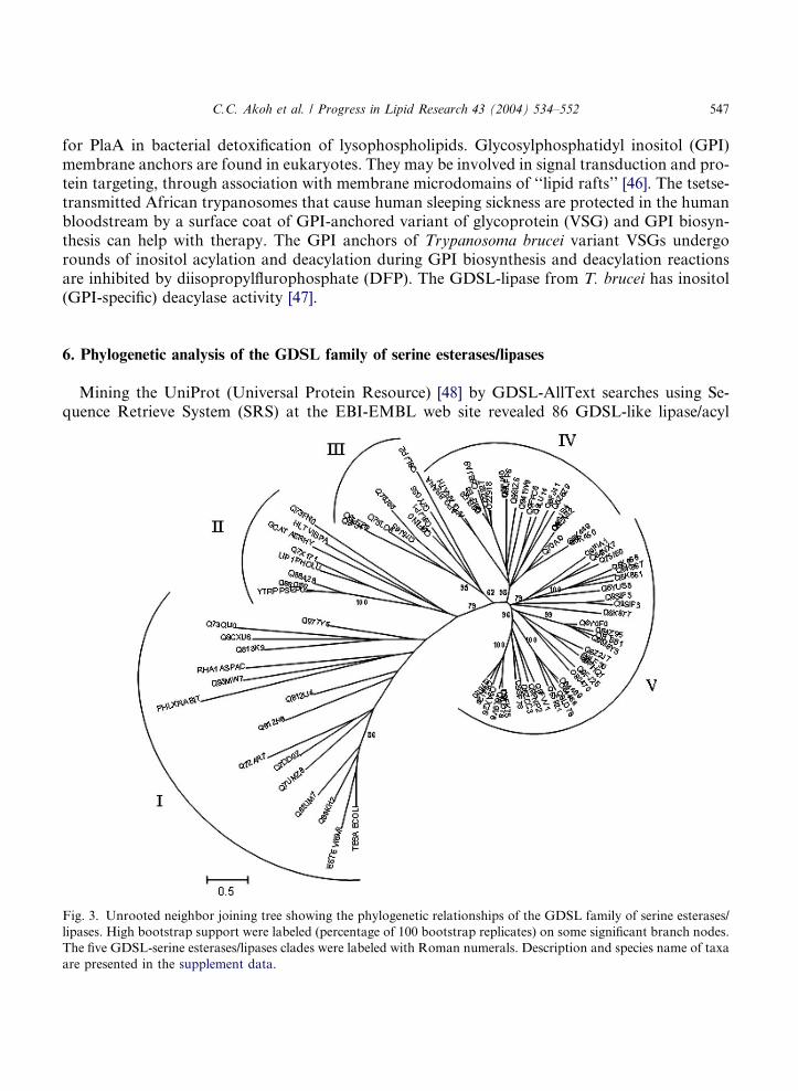
6. Phylogenetic analysis of the GDSL family of serine esterases/lipases
Mining the UniProt (Universal Protein Resource) [48] by GDSL-AllText searches using Se-quence Retrieve System (SRS) at the EBI-EMBL web site revealed 86 GDSL-like lipase/acyl
Fig. 3. Unrooted neighbor joining tree showing the phylogenetic relationships of the GDSL family of serine esterases/
lipases. High bootstrap support were labeled (percentage of 100 bootstrap replicates) on some significant branch nodes.
The five GDSL-serine esterases/lipases clades were labeled with Roman numerals. Description and species name of taxa
are presented in the supplement data.

548 C.C. Akoh et al. / Progress in Lipid Research 43 (2004) 534–552
hydrolase sequences (see supplemental data). To prevent the inclusion of undesired fragment se-quences, only sequences with lengths longer than 100 amino acid residues were chosen. These taxacontain 38 A. thaliana genes, 20 Oryza sativa genes, 4 other green plant genes, 22 bacterial genes,one rabbit�s gene, and one As. aculeatus�s gene. In addition to some functionally characterizedgenes, most of these taxa are putative GDSL-motif lipase/hydrolase from genomics analysis.Much more taxa which are not annotated could be found if Blast program is applied to do se-quence similarity search. A multiple sequence alignment was created with DIALIGN 2.2.1 [49]using the default settings at Bielefeld University Bioinformatics Server. DIALIGN is a novel localmultiple alignment program based on segment-to-segment comparison and was found to be thebest method to detect local similarities among distantly related sequences [50]. According tothe previous assignment [5], Fig. 2 shows multiple sequence alignments of five blocks in a well con-served region. This well conserved region between blocks I and V was used to conduct phylo-genetic analysis using the PHYLIP software package (v. 3.6b) [51]. Terminal gaps in thepeptide alignment produced using DIALIGN (above) were removed prior to running the analysis,while the internal alignment gaps were left and analysis conducted scoring gaps as characters or asmissing characters. Distances were calculated using the Jones-Taylor-Thornton matrix (PROT-DIST), and the neighbor joining method was used to create the tree (NEIGHBOR). Bootstrapanalysis was conducted using 100 bootstrap replicates [51]. TreeView (v.1.6.6) was used to displayresulting trees [52].
This distance-based phylogenetic analysis of the GDSL esterase/lipase genes revealed five dis-tinct clades (Fig. 3), numbered arbitrarily in a clockwise manner. The groupings were well sup-ported by bootstrap analysis. The assignment of the GDSL esterase/lipase genes to the fivegroups from trees built using data obtained from parsimony analysis was the same as for theneighbor joining analysis (data not shown). The bacterial GDSL lipolytic enzymes are representedin clades I and II. The higher green plant GDSL esterases/lipases, mainly from A. thaliana and O.
sativa, were found in three of the five groups (clades III–V). All five clades possess the conservedamino acid sequence blocks I, III, and V (Fig. 2). However, block IIA which belongs to theSGNH-hydrolases family was found only in clade I, and neither block IIA or IIB was found inclade II.
From sequence similarity analysis in block I, clade II and III possess a special pattern betweenclade I and clades IV–V. It is possible that clade II and III genes might be the transition statesduring evolution from clade I to clades IV–V. Two members (APG_ARATH and APG_BRANA)of clade IV were anther-specific proline-rich APG-like proteins. The largest group was clade V,consisting entirely of putative GDSL-motif lipase/hydrolase mainly from A. thaliana and O. sati-
va. It possesses 58% (19 of 33) of the A. thaliana genes and 39% (13 of 33) of the O. sativa genes.Given the degree of sequence conservation and short branch lengths, it seems likely that sub-groups within clade V have recently undergone duplication and expansion. For example, duplica-tions of a Q6YUS8-like ancestor have given rise to Q6K861, 867, and 868. The sequencealignment data (Figs. 1 and 2) and phylogenic tree (Fig. 3) indicate that GDSL- and SGNH-hydrolase family of esterases and lipases are a new example of convergent evolution with low se-quence homology. They probably evolved differently from the forces that constrained and moldedthe GDSL-motif. Therefore, despite shared sequence conservation, the GDSL and SGNH motifsmost likely resulted from convergent evolution instead of divergent evolution as found in commonlipases with the GxSxG sequence motif and nucleophile elbow.

C.C. Akoh et al. / Progress in Lipid Research 43 (2004) 534–552 549
7. Conclusion
The activity, catalytic efficiency, substrate selectivity, and enantioselectivity of many of theGDSL and SGNH-family enzymes can be increased by His-tagging, directed evolution, saturationmutagenesis, and rational approaches. The 3-D and crystal structures of many of the enzymes re-mains to be determined and therefore insights into the substrate-binding mechanisms and catal-ysis will still be subjects of further investigations. To understand how one enzyme will havemultisubstrate or multifunctional properties points to the need for active site flexibility studies.Obviously, there is curiosity and interest on how genetic or protein engineering and heterologousexpression will affect the production (in industrial scale), activity and potential applications of theGDSL-family of enzymes in the food, flavor, fragrance, cosmetics, textile, pharmaceutical, anddetergent industry, to name a few. The biochemical functions (structure–function relationships)of many of the enzymes are still unknown and require further investigation.
Acknowledgments
We acknowledge financial support by a Grant NSC-92-2313-B-001-025 from the National Sci-ence Council, Republic of China. We thank the University of Georgia, USA, for granting Profes-sor Akoh a leave of absence to Academia Sinica, Taipei, Taiwan.
Appendix A. Supplementary material
Supplemental data showing the accession number, enzyme description and source, sequencelength, and the references for the GDSL esterases and lipases. The sequences were used for theconstruction of the phylogenetic tree depicted in Fig. 3. Supplementary data associated with thisarticle can be found, in the online version at doi:10.1016/j.plipres.2004.09.002.
References
[1] Upton C, Buckley JT. A new family of lipolytic enzymes?. Trends Biochem Sci 1995;20:178–9.
[2] Molgaard A, Kauppinen S, Larsen S. Rhamnogalacturonan acetylesterase elucidates the structure and function of
a new family of hydrolases. Struct Fold Des 2000;8:373–83.
[3] Li J, Derewenda U, Dauter Z, Smith S, Derewenda ZS. Crystal structure of the Escherichia coli thioesterase II, a
homolog of the human Nef binding enzyme. Nat Struct Biol 2000;7:555–9.
[4] Dalrymple BP, Cybinski DH, Layton I, McSweeney CS, Xue GP, Swadling YJ, et al. Three Neocallimastix
patriciarum esterases associated with the degradation of complex polysaccharides are members of a new family of
hydrolases. Microbiology 1997;143(Pt 8):2605–14.
[5] Lo YC, Lin SC, Shaw JF, Liaw YC. Crystal structure of Escherichia coli thioesterase I/protease I/lysophosp-
holipase L1: consensus sequence blocks constitute the catalytic center of SGNH-hydrolases through a conserved
hydrogen bond network. J Mol Biol 2003;330:539–51.
[6] Tyukhtenko SI, Litvinchuk AV, Chang CF, Lo YC, Lee SJ, Shaw JF, et al. Sequential structural changes of
Escherichia coli thioesterase/protease I in the serial formation of Michaelis and tetrahedral complexes with diethyl
p-nitrophenyl phosphate. Biochemistry 2003;42:8289–97.

550 C.C. Akoh et al. / Progress in Lipid Research 43 (2004) 534–552
[7] Cho H, Cronan Jr JE. Escherichia coli thioesterase I, molecular cloning and sequencing of the structural gene and
identification as a periplasmic enzyme. J Biol Chem 1993;268:9238–45.
[8] Lee YL, Chen JC, Shaw JF. The thioesterase I of Escherichia coli has arylesterase activity and shows
stereospecificity for protease substrates. Biochem Biophys Res Commun 1997;231:452–6.
[9] Brzozowski AM, Derewenda U, Derewenda ZS, Dodson GG, Lawson DM, Turkenburg JP, et al. A model for
interfacial activation in lipases from the structure of a fungal lipase-inhibitor complex. Nature 1991;351:491–4.
[10] Schrag JD, Li YG, Wu S, Cygler M. Ser-His-Glu triad forms the catalytic site of the lipase from Geotrichum
candidum. Nature 1991;351:761–4.
[11] Winkler FK, D�Arcy A, Hunziker W. Structure of human pancreatic lipase. Nature 1990;343:771–4.
[12] Lo YC, Lee YL, Shaw JF, Liaw YC. Crystallization and preliminary X-ray crystallographic analysis of
thioesterase I from Escherichia coli. Acta Crystallogr D 2000;56(Pt 6):756–7.
[13] Shaw JF, Chang RC, Chuang KH, Yen YT, Wang YJ, Wang FG. Nucleotide sequence of a novel arylesterase gene
from Vibro mimicus and characterization of the enzyme expressed in Escherichia coli. Biochem J 1994;298(Pt
):675–80.
[14] Lin TH, Chen C, Huang RF, Lee YL, Shaw JF, Huang TH. Multinuclear NMR resonance assignments and the
secondary structure of Escherichia coli thioesterase/protease I: a member of a new subclass of lipolytic enzymes. J
Biomol NMR 1998;11:363–80.
[15] Kim KK, Hwang KY, Choi KD, Kang JH, Yoo OJ, Suh SW. Crystallization and preliminary X-ray
crystallographic analysis of arylesterase from Pseudomonas fluorescens. Proteins 1993;15:213–5.
[16] Kawai H, Yomoda S, Inoue Y. ELISA using monoclonal antibody to human serum arylesterase. Clin Chim Acta
1991;202:219–25.
[17] Karasawa K, Yokoyama K, Setaka M, Nojima S. The Escherichia coli pldC gene encoding lysophospholipase L
(1) is identical to the apeA and tesA genes encoding protease I and thioesterase I, respectively. J Biochem (Tokyo)
1999;126:445–8.
[18] Cho H, Cronan Jr JE. ‘‘Protease I’’ of Escherichia coli functions as a thioesterase in vivo. J Bacteriol
1994;176:1793–5.
[19] Doi O, Nojima S. Lysophospholipase of Escherichia coli. J Biol Chem 1975;250:5208–14.
[20] Chang RC, Chen JC, Shaw JF. Site-directed mutagenesis of a novel serine arylesterase from Vibrio mimicus
identifies residues essential for catalysis. Biochem Biophys Res Commun 1996;221:477–83.
[21] Huang YT, Liaw YC, Gorbatyuk VY, Huang TH. Backbone dynamics of Escherichia coli thioesterase/protease I:
evidence of a flexible active-site environment for a serine protease. J Mol Biol 2001;307:1075–90.
[22] Tyukhtenko SI, Litvinchuk AV, Chang CF, Leu RJ, Shaw JF, Huang TH. NMR studies of the hydrogen bonds
involving the catalytic triad of Escherichia coli thioesterase/protease I. FEBS Lett 2002;528:203–6.
[23] Derewenda ZS. Structure and function of lipases. Adv Protein Chem 1994;45:1–52.
[24] Gunasekaran K, Ma B, Nussinov R. Triggering loops and enzyme function: identification of loops that trigger and
modulate movements. J Mol Biol 2003;332:143–59.
[25] Koshland DE. Application of a theory of enzyme specificity to protein synthesis (in Symposium on Amino Acid
Activation). Proc Natl Acad Sci USA 1958;44:98–104.
[26] Hammes GG. Multiple conformational changes in enzyme catalysis. Biochemistry 2002;41:8221–8.
[27] Grochulski P, Bouthillier F, Kazlauskas RJ, Serreqi AN, Schrag JD, Ziomek E, et al. Analogs of reaction
intermediates identify a unique substrate binding site in Candida rugosa lipase. Biochemistry
1994;33:3494–500.
[28] Lindner AB, Kim SH, Schindler DG, Eshhar Z, Tawfik DS. Esterolytic antibodies as mechanistic and structural
models of hydrolases-a quantitative analysis. J Mol Biol 2002;320:559–72.
[29] Lee YL, Su MS, Huang TH, Shaw JF. C-terminal His-tagging results in substrate specificity changes of the
thioesterase I from E. coli. J Am Oil Chem Soc 1999;76:1113–8.
[30] Chang RC, Chen JC, Shaw JF. Vibrio mimicus arylesterase has thioesterase and chymotrypsin-like activity.
Biochem Biophys Res Commun 1995;213:475–83.
[31] Brenner S. The molecular evolution of genes and proteins: a tale of two serines. Nature 1988;334:528–30.
[32] Karasawa K, Kudo I, Kobayashi T, Homma H, Chiba N, Mizushima H, et al. Lysophospholipase L1 from
Escherichia coli K-12 overproducer. J Biochem (Tokyo) 1991;109:288–93.

C.C. Akoh et al. / Progress in Lipid Research 43 (2004) 534–552 551
[33] Carinato ME, Collin-Osdoby P, Yang X, Knox TM, Conlin CA, Miller CG. The apeE gene of Salmonella
typhimurium encodes an outer membrane esterase not present in Escherichia coli. J Bacteriol 1998;180:
3517–21.
[34] Vujaklija D, Schroder W, Abramic M, Zou P, Lescic I, Franke P, et al. A novel streptomycete lipase: cloning,
sequencing and high-level expression of the Streptomyces rimosus GDS (L)-lipase gene. Arch Microbiol
2002;178:124–30.
[35] Lee HW, Byun SM. The pore size of the autotransporter domain is critical for the active translocation of the
passenger domain. Biochem Biophys Res Commun 2003;307:820–5.
[36] Talker-Huiber D, Jose J, Glieder A, Pressnig M, Stubenrauch G, Schwab H. Esterase EstE from Xanthomonas
vesicatoria (Xv_EstE) is an outer membrane protein capable of hydrolyzing long-chain polar esters. Appl
Microbiol Biotechnol 2003;61:479–87.
[37] Riedel K, Talker-Huiber D, Givskov M, Schwab H, Eberl L. Identification and characterization of a GDSL
esterase gene located proximal to the swr quorum-sensing system of Serratia liquefaciens MG1. Appl Environ
Microbiol 2003;69:3901–10.
[38] Brick DJ, Brumlik MJ, Buckley JT, Cao JX, Davies PC, Misra S, et al. A new family of lipolytic plant enzymes
with members in rice, Arabidopsis and maize. FEBS Lett 1995;377:475–80.
[39] Beisson F, Gardies AM, Teissere M, Ferte N, Noat G. An esterase neosynthesized in post-germinated sunflower
seeds is related to a new family of lipolytic enzymes. Plant Physiol Biochem 1997;35:761–5.
[40] Mukherjee KD. Plant lipases and their application in lipid biotransformations. Progr Lipid Res 1994;33:165–74.
[41] Arif SA, Hamilton RG, Yusof F, Chew NP, Loke YH, Nimkar S, et al. Isolation and characterization of the early
nodule-specific protein homologue (Hev b 13), an allergenic lipolytic esterase from Hevea brasiliensis latex. J Biol
Chem 2004;279:23933–41.
[42] Pringle D, Dickstein R. Purification of ENOD8 proteins from Medicago sativa root nodules and their
characterization as esterases. Plant Physiol Biochem 2004;42:73–9.
[43] Farn JL, Strugnell RA, Hoyne PA, Michalski WP, Tennent JM. Molecular characterization of a secreted enzyme
with phospholipase B activity from Moraxella bovis. J Bacteriol 2001;183:6717–20.
[44] Lepper AW, Power BE. Infectivity and virulence of Australian strains of Moraxella bovis for the murine and
bovine eye in relation to pilus serogroup sub-unit size and degree of piliation. Aust Vet J 1988;65:305–9.
[45] Flieger A, Neumeister B, Cianciotto NP. Characterization of the gene encoding the major secreted lysophosp-
holipase A of Legionella pneumophila and its role in detoxification of lysophosphatidylcholine. Infect Immun
2002;70:6094–106.
[46] Simons K, Ikonen E. Functional rafts in cell membranes. Nature 1997;387:569–72.
[47] Guther ML, Leal S, Morrice NA, Cross GA, Ferguson MA. Purification, cloning and characterization of a GPI
inositol deacylase from Trypanosoma brucei. EMBO J 2001;20:4923–34.
[48] Apweiler R, Bairoch A, Wu CH, Barker WC, Boeckmann B, Ferro S, et al. UniProt: the Universal Protein
knowledgebase. Nucleic Acids Res 2004;32(Database issue):D115–9.
[49] Morgenstern B. DIALIGN 2: improvement of the segment-to-segment approach to multiple sequence alignment.
Bioinformatics 1999;15:211–8.
[50] Thompson JD, Plewniak F, Poch O. A comprehensive comparison of multiple sequence alignment programs.
Nucleic Acids Res 1999;27:2682–90.
[51] Felsenstein J. PHYLIP (PHYLogeny Inference Package) is a package of programs for inferring phylogenies
(evolutionary trees), 1980. Available from: http://evolution.genetics.was1980.
[52] Page RD. TreeView: an application to display phylogenetic trees on personal computers. Comput Appl Biosci
1996;12:357–8.
[53] Robertson DL, Hilton S, Wong KR, Koepke A, Buckley JT. Influence of active site and tyrosine modification on
the secretion and activity of the Aeromonas hydrophila lipase/acyltransferase. J Biol Chem 1994;269:2146–50.
[54] Shinoda S, Matsuoka H, Tsuchie T, Miyoshi S, Yamamoto S, Taniguchi H, et al. Purification and
characterization of a lecithin-dependent haemolysin from Escherichia coli transformed by a Vibrio parahaemo-
lyticus gene. J Gen Microbiol 1991;137(Pt 12):2705–11.
[55] Wang H, Dowds BC. Phase variation in Xenorhabdus luminescens: cloning and sequencing of the lipase gene and
analysis of its expression in primary and secondary phases of the bacterium. J Bacteriol 1993;175:1665–73.

552 C.C. Akoh et al. / Progress in Lipid Research 43 (2004) 534–552
[56] Essar DW, Eberly L, Crawford IP. Evolutionary differences in chromosomal locations of four early genes of the
tryptophan pathway in fluorescent pseudomonads: DNA sequences and characterization of Pseudomonas putida
trpE and trpGDC. J Bacteriol 1990;172:867–83.
[57] Roberts MR, Foster GD, Blundell RP, Robinson SW, Kumar A, Draper J, et al. Gametophytic and sporophytic
expression of an anther-specific Arabidopsis thaliana gene. Plant J 1993;3:111–20.
[58] Wei Y, Schottel JL, Derewenda U, Swenson L, Patkar S, Derewenda ZS. A novel variant of the catalytic triad in
the Streptomyces scabies esterase. Nat Struct Biol 1995;2:218–23.
[59] Ho YS, Swenson L, Derewenda U, Serre L, Wei Y, Dauter Z, et al. Brain acetylhydrolase that inactivates platelet-
activating factor is a G-protein-like trimer. Nature 1997;385:89–93.
[60] Kauppinen S, Christgau S, Kofod LV, Halkier T, Dorreich K, Dalboge H. Molecular cloning and characterization
of a rhamnogalacturonan acetylesterase from Aspergillus aculeatus. Synergism between rhamnogalacturonan
degrading enzymes. J Biol Chem 1995;270:27172–8.
[61] Ho YS, Sheffield PJ, Masuyama J, Arai H, Li J, Aoki J, et al. Probing the substrate specificity of the intracellular
brain platelet-activating factor acetylhydrolase. Protein Eng 1999;12:693–700.
[62] Brumlik MJ, Buckley JT. Identification of the catalytic triad of the lipase/acyltransferase from Aeromonas
hydrophila. J Bacteriol 1996;178:2060–4.
[63] Fenster KM, Parkin KL, Steele JL. Characterization of an arylesterase from Lactobacillus helveticus CNRZ32.
J Appl Microbiol 2000;88:572–83.
Related Documents
![CHEMISTRY & BIOLOGY INTERFACE...cinnamoyl ester hydrolases, include ferulic / para-coumaric acid esterases [6].Moreover, ferulic acid esterases (FAEs; EC 3.1.1.73) and microorganisms](https://static.cupdf.com/doc/110x72/5e62fc0fd110d451973c6f71/chemistry-biology-cinnamoyl-ester-hydrolases-include-ferulic-para-coumaric.jpg)