Hindawi Publishing Corporation Evidence-Based Complementary and Alternative Medicine Volume 2012, Article ID 404012, 9 pages doi:10.1155/2012/404012 Research Article Gastroprotective Activity of Polygonum chinense Aqueous Leaf Extract on Ethanol-Induced Hemorrhagic Mucosal Lesions in Rats Iza Farhana Ismail, 1 Shahram Golbabapour, 2, 3 Pouya Hassandarvish, 2 Maryam Hajrezaie, 2, 3 Nazia Abdul Majid, 3 Farkaad A. Kadir, 4 Fouad Al-Bayaty, 5 Khalijah Awang, 1 Hazrina Hazni, 1 and Mahmood Ameen Abdulla 2 1 Department of Chemistry, Faculty of Science, University of Malaya, 50603 Kuala Lumpur, Malaysia 2 Department of Molecular Medicine, Faculty of Medicine, University of Malaya, 50603 Kuala Lumpur, Malaysia 3 Institute of Biological Science, Faculty of Science, University of Malaya, 50603 Kuala Lumpur, Malaysia 4 Department of Anatomy, Faculty of Medicine, Cyberjaya University College of Medical Scinces, 6300 Cyberjaya, Selangor Darul Ehsan, Malaysia 5 Faculty of Dentistry, Universiti Teknologi Mara, 40450 Shah Alam, Malaysia Correspondence should be addressed to Mahmood Ameen Abdulla, [email protected] Received 1 July 2012; Revised 3 November 2012; Accepted 5 December 2012 Academic Editor: Virginia S. Martino Copyright © 2012 Iza Farhana Ismail et al. This is an open access article distributed under the Creative Commons Attribution License, which permits unrestricted use, distribution, and reproduction in any medium, provided the original work is properly cited. Polygonum chinense is a Malaysian ethnic plant with various healing effects. This study was to determine preventive effect of aque- ous leaf extract of P. chinense against ethanol-induced gastric mucosal injury in rats. Sprague Dawley rats were divided into seven groups. The normal and ulcer control groups were orally administered with distilled water. The reference group was orally administered with 20 mg/kg omeprazole. The experimental groups received the extracts 62.5, 125, 250, and 500 mg/kg, accordingly. After sixty minutes, distilled water and absolute ethanol were given (5mL/kg) to the normal control and the others, respectively. In addition to histology, immunohistochemical and periodic acid schiff (PAS) stains, levels of lipid peroxidation, malondialdehyde (MDA), antioxidant enzymes, and superoxide dismutase (SOD) were measured. The ulcer group exhibited severe mucosal damages. The experimental groups significantly reduced gastric lesions and MDA levels and increased SOD level. Immunohistochemistry of the experimental groups showed upregulation and downregulation of Hsp70 and Bax proteins, respectively. PAS staining in these groups exhibited intense staining as compared to the ulcer group. Acute toxicity study revealed the nontoxic nature of the extract. Our data provide first evidence that P. chinense extract could significantly prevent gastric ulcer. 1. Introduction Gastritis is an inflammation, irritation, or erosion that occurs when the endogenous defensive mechanisms of mucosal barrier cannot properly protect the organ. Usually, expo- sure to exceed acid and pepsin causes insult on the gas- trointestinal wall [1]. Several factors increase the inci- dence of peptic ulcer diseases, including Helicobacter pylori infection and rarely other infections such as tuberculosis, syphilis, viral infections, fungal infections, bacteria parasites, and worms. Some medications, such as NSAIDs (such as aspirin, steroids, and nonsteroidal anti-inflammatory), potassium, and iron supplements, have been reported risky for prevalence of peptic ulcer disease. In addition, med- ical andsurgical conditions, illnesses like chronic pancre- atitis, autoimmune disease, pernicious anemia, and perni- cious anemia, emotional or physical distress, foods (heavy drinking alcohol), and smoking increase the chance of the commonness of the diseases among population [2]. Many products are used commonly to treat gastritis. General protocol for the treatment of gastritis is to reduce acid secretion [3]. Traditionally, plants are used for medicinal purposes in different countries. Nowadays, researchers and companies are inclined to investigate more about herbal

Welcome message from author
This document is posted to help you gain knowledge. Please leave a comment to let me know what you think about it! Share it to your friends and learn new things together.
Transcript

Hindawi Publishing CorporationEvidence-Based Complementary and Alternative MedicineVolume 2012, Article ID 404012, 9 pagesdoi:10.1155/2012/404012
Research Article
Gastroprotective Activity of Polygonum chinenseAqueous Leaf Extract on Ethanol-Induced HemorrhagicMucosal Lesions in Rats
Iza Farhana Ismail,1 Shahram Golbabapour,2, 3 Pouya Hassandarvish,2
Maryam Hajrezaie,2, 3 Nazia Abdul Majid,3 Farkaad A. Kadir,4 Fouad Al-Bayaty,5
Khalijah Awang,1 Hazrina Hazni,1 and Mahmood Ameen Abdulla2
1 Department of Chemistry, Faculty of Science, University of Malaya, 50603 Kuala Lumpur, Malaysia2 Department of Molecular Medicine, Faculty of Medicine, University of Malaya, 50603 Kuala Lumpur, Malaysia3 Institute of Biological Science, Faculty of Science, University of Malaya, 50603 Kuala Lumpur, Malaysia4 Department of Anatomy, Faculty of Medicine, Cyberjaya University College of Medical Scinces, 6300 Cyberjaya,Selangor Darul Ehsan, Malaysia
5 Faculty of Dentistry, Universiti Teknologi Mara, 40450 Shah Alam, Malaysia
Correspondence should be addressed to Mahmood Ameen Abdulla, [email protected]
Received 1 July 2012; Revised 3 November 2012; Accepted 5 December 2012
Academic Editor: Virginia S. Martino
Copyright © 2012 Iza Farhana Ismail et al. This is an open access article distributed under the Creative Commons AttributionLicense, which permits unrestricted use, distribution, and reproduction in any medium, provided the original work is properlycited.
Polygonum chinense is a Malaysian ethnic plant with various healing effects. This study was to determine preventive effect of aque-ous leaf extract of P. chinense against ethanol-induced gastric mucosal injury in rats. Sprague Dawley rats were divided intoseven groups. The normal and ulcer control groups were orally administered with distilled water. The reference group wasorally administered with 20 mg/kg omeprazole. The experimental groups received the extracts 62.5, 125, 250, and 500 mg/kg,accordingly. After sixty minutes, distilled water and absolute ethanol were given (5 mL/kg) to the normal control and the others,respectively. In addition to histology, immunohistochemical and periodic acid schiff (PAS) stains, levels of lipid peroxidation,malondialdehyde (MDA), antioxidant enzymes, and superoxide dismutase (SOD) were measured. The ulcer group exhibitedsevere mucosal damages. The experimental groups significantly reduced gastric lesions and MDA levels and increased SODlevel. Immunohistochemistry of the experimental groups showed upregulation and downregulation of Hsp70 and Bax proteins,respectively. PAS staining in these groups exhibited intense staining as compared to the ulcer group. Acute toxicity study revealedthe nontoxic nature of the extract. Our data provide first evidence that P. chinense extract could significantly prevent gastric ulcer.
1. Introduction
Gastritis is an inflammation, irritation, or erosion that occurswhen the endogenous defensive mechanisms of mucosalbarrier cannot properly protect the organ. Usually, expo-sure to exceed acid and pepsin causes insult on the gas-trointestinal wall [1]. Several factors increase the inci-dence of peptic ulcer diseases, including Helicobacter pyloriinfection and rarely other infections such as tuberculosis,syphilis, viral infections, fungal infections, bacteria parasites,and worms. Some medications, such as NSAIDs (suchas aspirin, steroids, and nonsteroidal anti-inflammatory),
potassium, and iron supplements, have been reported riskyfor prevalence of peptic ulcer disease. In addition, med-ical andsurgical conditions, illnesses like chronic pancre-atitis, autoimmune disease, pernicious anemia, and perni-cious anemia, emotional or physical distress, foods (heavydrinking alcohol), and smoking increase the chance ofthe commonness of the diseases among population [2].Many products are used commonly to treat gastritis. Generalprotocol for the treatment of gastritis is to reduce acidsecretion [3]. Traditionally, plants are used for medicinalpurposes in different countries. Nowadays, researchers andcompanies are inclined to investigate more about herbal
2 Evidence-Based Complementary and Alternative Medicine
medicines, and many plants with antiulcerogenic propertieshave been found by different groups [4–7].
The genus Polygonum belongs to Polygonaceae andcomprises about 150 species. It contains various bioactivecompounds and has phytopharmaceutical importance [8].However, there was no published research about gastropro-tective effect of P. chinense in rats. This study was undertakento evaluate the gastroprotective potential of aqueous extractof this plant against ethanol-induced gastric mucosal hemor-rhage in rats.
2. Materials and Methods
2.1. Omeprazole. In this study, omeprazole was used as a ref-erence antiulcer drug and was obtained from the UniversityMalaya Medical Centre (UMMC) Pharmacy. The drug wasdissolved in distilled water and administered orally to the ratsat a dosage of 20 mg/kg body weight (5 mL/kg) according tothe recommendation of Mahmood et al. [9].
2.2. Plant Specimen and Preparation of Extraction. Fresh P.chinense leaves were obtained from Ethno Resources SdnBhd, Selangor, Malaysia, deposited at the Herbarium ofRimba Ilmu, Institute of Science Biology, University ofMalaya, Kuala Lumpur (Voucher Specimen no. KLU 47125).The leaves were dried in shade for one week, and thedried leaves were powdered using electrical blender. The finepowder (200 g) was soaked in 1000 mL of distilled water in aconical flask and heated 1 hour on a hot plate (85◦C). Whileits temperature was decreasing to 40◦± 5◦C, it was filteredtwice through cotton wool. Then, the extract was stored in afreezer. The frozen extract underwent freeze-drying processto yield dark brown powder (23.04 g, 11.52%). The extractwas then dissolved in distilled water and administered orally(5 mL/kg) to rats at dosages of 62.5, 125, 250, and 500 mg/kgaccording to the recommendation of Mahmood et al. [10].
2.3. Experimental Animals and Acute Toxicity Studies. Theacute toxicity study was used to determine a safe dose for P.chinense. Thirty-six healthy Sprague Dawley rats (18 malesand 18 females) were obtained from the Experimental Ani-mal House, Faculty of Medicine, University of Malaya. Theywere assigned equally into 3 groups: vehicle (distilled water),2 g/kg, and 5 g/kg of the extract of P. chinense, respectively.The animals were fasted overnight (food but not water) priorto the dosing. Food was withheld for further 3 to 4 h afterdosing. The animals were observed for 30 min and 2, 4, 24,and 48 hours after the administration for the onset of anyclinical or toxicological symptoms for 2 weeks. The animalswere sacrificed on the 15th day. Serum biochemical andhistological (liver and kidney) parameters were determined[11]. The study was approved by the Ethics Committee forAnimal Experimentation, Faculty of Medicine, University ofMalaya, Malaysia with the Ethic no. PM/07/05/2011/MMA(a) (R). Throughout the experiments, all animals receivedhuman care according to the criteria outlined in the “Guidefor the Care and Use of Laboratory Animals” prepared by the
National Academy of Sciences and published by the NationalInstitute of Health [11].
2.4. Experimental Animals for Gastric Ulcer. Healthy adultSprague Dawley rats of both genders (aged between 6–8weeks and weighed between 200–220 g) were prepared fromAnimal House Faculty of Medicine, University of Malaya,Kuala Lumpur, Malaysia. The animals were kept understandard laboratory conditions, caged in stainless steel cageswith raised floors of wide wire mesh to prevent coprophagia,and housed at temperature of 25 ± 2◦C in a 12-hour light-dark cycle. They were fed with standard laboratory pelletand water ad libitum. The animals were fasted for 24 hoursbefore treatment to ensure their stomachs are empty. Duringthe fasting period, the rats were allowed to have free accessof water. Their access to water was inhibited for 2 hoursbefore the experiment triggered. This protocol was basedon the guidelines of Animal Care and Use Committee fromLaboratory Animal Science Centre, Faculty of Medicine,University of Malaya, Kuala Lumpur, Malaysia.
2.4.1. Induction of Gastric Ulcer. The following pretreatmentwas according to the recommendations of Mahmood et al.[9]. The fasted rats were divided randomly into 7 groups of6 rats. The groups were numbered 1–7 and the pretreatmentbegan accordingly. Group 1 served as the normal control andreceived distilled water orally. Group 2 labeled “ulcer controlgroup” received distilled water orally as a pre-treatment.Group 3, the reference group, received 20 mg/kg omeprazoleorally. The experimental groups (group 4–7) received aque-ous leaf extract of P. Chinense, at dosages of 62.5, 125, 250,and 500 mg/kg, respectively. An hour after the pre-treatment,distilled water (5 mL/kg) was orally administered to thegroup 1, and absolute ethanol (5 mL/kg) was orally givento groups 2–7. After 1 hour, all animals were euthanized byan overdose of xylazine and ketamine anesthesia followedby cervical dislocation technique to assure their euthanasia.Their stomachs were immediately excised and were kept incontainers of normal saline [6].
2.4.2. Determination of Gastric Wall Mucus. The gastric wallmucus was evaluated according to the modified procedure ofCorne et al. [12].
2.4.3. Gross Evaluation of Gastric Lesions. Ulcers of the gastricmucosa appear as elongated bands of hemorrhagic lesionsparallel to the long axis of the stomach. Gastric mucosa ofeach rat was examined for estimate damage. The length andwidth of the ulcer (mm) were measured with a planimeter(10 × 10 mm2 = ulcer area) under a dissecting microscope(1.8x). The ulcerated area was calculated through totalingthe number of small squares (2 × 2 mm2) covering an ulcerband. The sum of the lesions areas for each stomach wasapplied in the calculation of the ulcer area (UA), where“the sum of small squares × 4 × 1.8 = UA (mm2)” accord-ing to the recommendation of Mahmood et al. [10]. The
Evidence-Based Complementary and Alternative Medicine 3
inhibition percentage (I%) was calculated by the followingformula:
(I%) =[
(UAcontrol −UAtreated)UAcontrol
]× 100%. (1)
2.4.4. Preparation of Homogenate. The gastric tissue sampleswere washed thoroughly with ice-cold saline. Homogenates(10% (w/v)) were then prepared with ice-cold 50 mM phos-phate buffer (pH 7.4) containing mammalian proteaseinhibitor cocktail. The homogenates were centrifuged at10,000×g for 30 min (4◦C). The supernatant was used forfurther experiments.
2.4.5. Measurement of SOD Activity. SOD activity was mea-sured according to the protocol of Sun et al. [13].
2.4.6. Measurement of MDA. Tissue MDA (mMol/L) wasdetermined based on the method developed by Draper andHadley [14].
2.4.7. Measurement of Protein Concentration. Biuret reactionwas the main protocol to measure the protein concentra-tions, as described by Gornall et al. [15].
2.5. Histological Studies of the Gastric Mucosa
2.5.1. Preparation of Tissue Sections. Specimens of the gastricwalls were fixed in 10% buffered formalin for 18 hours atroom temperature and processed by a tissue-processingmachine (Leica, Germany). Sections of the stomach wereadopted at a thickness of 5 µm.
2.5.2. Hematoxylin and Eosin. Stomach sections were stainedwith Hematoxylin and Eosin for histological evaluation [16].
2.5.3. Study of Mucosal Glycoproteins. The glandular portionof stomach was stained with periodic acid schiff (PAS) foreach rat in each group [17].
2.6. Immunohistochemical Staining. Tissue section slideswere heated at 60◦C for approximately 25 min in a hot airoven (Venticell, MMM, Einrichtungen, Germany). The tissuesections were deparaffinized in xylene and rehydrated withgraded alcohol. Antigen retrieval process was performedin a 10 mM sodium citrate buffer. Immunohistochemicalstaining was conducted according to manufacturer’s protocol(Dakocytomation, USA). Briefly, endogenous peroxidase wasblocked by peroxidase block (0.03% hydrogen peroxide con-taining sodium azide) for 5 min. Tissue sections were washedgently with wash buffer and then incubated with Hsp70(1 : 500) and Bax (1 : 200) biotinylated primary antibodiesfor 15 min. The sections were rinsed gently with wash bufferand placed in buffer bath. The slides were then placed in ahumidified chamber and sufficient amount of streptavidin—HRP (streptavidin conjugated to horseradish peroxidase inPBS containing an antimicrobial agent) was added andincubated for 15 min. Then, tissue sections were rinsed gently
in wash buffer and placed in buffer bath. Diaminobenzidine-substrate-chromagen was added to the tissue sections andincubated further for 5 min following washing and counter-staining with hematoxylin for 5 seconds. The sections werethen dipped in weak ammonia (0.037 Mol/L) 10 times andthen rinsed with distilled water and cover slipped. Positivefindings of the immunohistochemical staining should beseen as brown stains under light microscope.
2.7. Statistical Analysis. All values were reported as mean ±S.E.M. The statistical significance of differences betweengroups was assessed with one-way ANOVA (post hoc anal-ysis). A value of P < 0.05 was considered significant.
3. Results
3.1. Acute Toxicity Study. To determine the acute toxicity ofthe P. chinense extract, the animals were treated with theextract at a dose of 2 g/kg or 5 g/kg. Their health conditionswere screened for a period of 14 days. All of the animals werehealthy and did not manifest any sign of toxicity at thesedoses. Serum biochemical and histologic indicators of liverand kidney did not show any abnormalities.
3.2. Gross Evaluation of Gastric Lesions. The anti-ulcer activ-ity of P. chinense extract in ethanol-induced hemorrhagicmucosal lesions model is shown in Figure 1 and Table 1. Thelesions were long, hemorrhagic, and confined to the glandu-lar portions. Results showed that those rats pre-treated withthe extract significantly reduced areas of gastric ulcer forma-tion when compared with the ulcer control group (Figure 1).Absolute ethanol caused extensive and visible hemorrhagiclesions to gastric mucosa. The P. chinense extract significantlysuppressed the formation of the ulcers, especially at itshighest dosage, and astoundingly flattened gastric mucosalfolds at the dosage of 500 mg/kg of the extract (Figure 1).The extract significantly reduced the ulcer size and severity ina dose-dependent manner. The protection properties of theextract at its highest dosage (500 mg/kg) appeared similar tothe reference group (Figure 1).
3.3. Effect of P. chinense on Ethanol-Induced Changes in GastricWall Mucus. In the ulcer control group, in comparison tothe normal control group, ethanol significantly decreasedthe Alcian-blue-binding capacity of gastric wall mucus. Inthe experimental groups, pre-treatment with the P. chi-nense extract significantly enhanced the Alcian-blue-bindingcapacity of the gastric mucosa (Table 1).
3.4. Effect of P. chinense on Measurements of SOD Activity.Ethanol was able to reduce SOD activity as shown in theulcer control group, compared to normal control group. Inthe experimental groups, the extract of P. chinense caused asignificant increase in the enzyme activity of SOD (Table 1).
3.5. Effect of P. chinense Extract on Tissue MDA. Ethanolincreased the MDA activity significantly when comparedwith the normal group. However, the extract of P. chinense

4 Evidence-Based Complementary and Alternative Medicine
(a) (b) (c) (d)
(e) (f) (g)
Figure 1: Macroscopic appearance of gastric mucosal lesions of the rats in different groups. (a) Rats in the normal control group showedintact gastric mucosa. (b) The ulcer control group (pretreated with 5 mL/kg absolute alcohol) showed severe injuries to the gastric mucosa(white arrow). Absolute ethanol imposed extensive hemorrhagic necrosis to gastric mucosa. (c) The reference group (omeprazole 20 mg/kg)showed milder injuries to the gastric mucosa (white arrow) compared with the ulcer control rats. (d) Rat pre-treated with P. chinense(62.50 mg/kg) showed moderate injuries on the gastric mucosa (white arrow). (e) Pre-treated with 125 mg/kg of P. chinense extractsuppressed lesions to a mild-moderate condition (white arrow). (f) Mild injuries of the gastric mucosa were found in those rats pre-treatedwith 250 mg/kg of P. chinense extract. (g) Pre-treatment with 500 mg/kg of the extract protected injuries at a mild condition and flattenedthe gastric mucosa observed (black arrow).
Table 1: The ulcer area, percentage of inhibition, gastric wall mucus (mg Alcian blue/g tissue), MDA (µmol/g protein), SOD (U/g protein),and protein concentration (mg/mL tissue) of the groups.
Groups Ulcer area Inhibition % GWM MDA Protein SOD
1 — — 245.13 ± 2.2∗ 70 ± 2.93∗ 10 ± 0.16∗ 236.13 ± 4.29∗
2 921.90 ± 16.68 — 125.10 ± 2.3 228 ± 4.53 5.17 ± 0.12 121.15 ± 2.50
3 159.30 ± 5.04∗ 82.72 320.11 ± 2.4∗ 95.08 ± 3.3∗ 8.45 ± 0.16∗ 423.00 ± 5.83∗
4 455 ± 12.16∗ 50.64 282.00 ± 1.8∗ 110 ± 4.23∗ 7.22 ± 0.06∗ 285.00 ± 4.83∗
5 315.23 ± 9.17∗ 65.81 303.1 ± 2.5∗ 108 ± 3.65∗ 7.86 ± 0.04∗ 350.10 ± 4.29∗
6 235.61 ± 7.39∗ 74.44 315.08 ± 2∗ 102.1 ± 3.7∗ 9.40 ± 0.12∗ 402.42 ± 5.95∗
7 114.00 ± 5.41∗ 87.63 325.11 ± 4.4∗ 92.15 ± 3.2∗ 9.70 ± 0.14∗ 415.13 ± 6.90∗
Values are assumed as mean ± S.E.M. The statistical analysis was assessed with one-way ANOVA (post hoc analysis) with P < 0.05. ∗Significant differenceswhen compared to group 2. (1) The normal control group. (2) the ulcer control group. (3) the reference control. (4) 62.50 mg/kg of the plant extract; (5)125 mg/kg of the plant extract. (6) 250 mg/kg of the plant extract. (7) 500 mg/kg of the plant extract.
significantly decreased MDA activity, similar to the referencegroup (Table 1).
3.6. Effect of P. chinense Extract on Protein Concentration. Inthe ulcer control group, ethanol significantly decreased theprotein concentration of the gastric mucosal homogenatewhen compared with the normal control group. However,increase in the protein concentration was evident in theexperimental groups as it was in those rats receiving omepra-zole (Table 1).
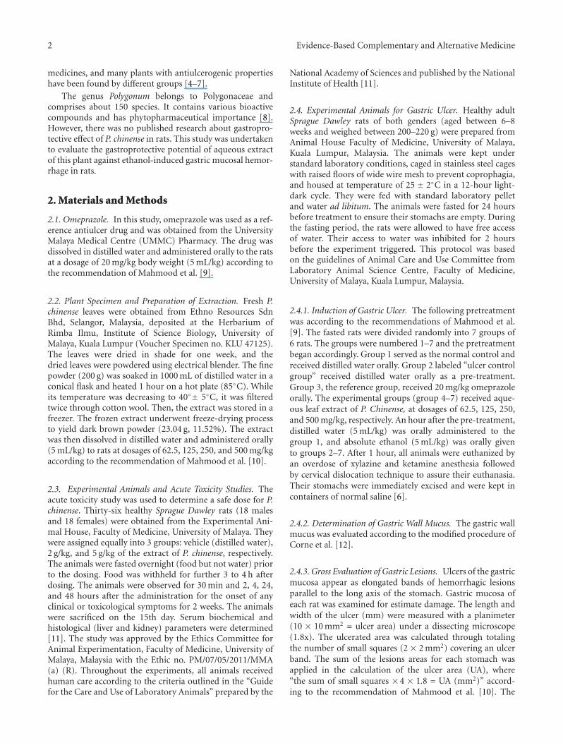
3.7. Histological Evaluation of Gastric Lesions. Histologi-cal observation of gastric lesions in the ulcer control
group showed comparatively extensive damage of the gas-tric mucosa, and necrotic lesions penetrated deeply into themucosa. Also, extensive oedema and leucocytes infiltrationsof the submucosal layer appeared microscopically (Figure 2).Rats receiving the extract of P. chinense had better protec-tion for the gastric mucosa from reduction or absence ofulcer area, submucosal oedema, and leucocytes infiltration(Figure 2). The extract showed its potential to exert thegastroprotective effect in a dose-dependent manner.
3.8. Periodic Acid Schiff (PAS) of Mucosal Glycoproteins.The extract of P. chinense increased the PAS staining ofgastric mucosa, as evident in Figure 3 (the magenta color),
Evidence-Based Complementary and Alternative Medicine 5
(a) (b) (c) (d)
(e) (f) (g)
Figure 2: Histology evaluation of gastric mucosal lesions of the rats in different groups (H&E staining 10x). (a) Rats in the normal controlgroup showed intact gastric mucosa. (b) The ulcer control group (pre-treated with 5 mL/kg absolute alcohol) showed severe disruptionon the epithelium. The necrotic lesions penetrated deeply into mucosa (white arrow). Also, extensive edema and leucocyte infiltration ofsubmucosal layer were seen (black arrow). (c) The reference group (omeprazole 20 mg/kg) showed a mild disruption of the epithelium withedema and leucocyte infiltration of submucosal layer. (d) Rat pre-treated with P. chinense (62.50 mg/kg) showed a moderate disruptionof epithelium with edema and leucocytes infiltration of submucosal layer. (e) Pre-treated with 125 mg/kg of P. chinense extract suppresseddisruption of surface epithelium with edema and leucocyte infiltration of submucosal layer to a mild-moderate condition. (f) Mild disruptionof the epithelium was found in those rats pre-treated with 250 mg/kg of P. chinense extract. (g) Pre-treatment with 500 mg/kg of the extractprotected the epithelium from disruption at a mild condition along with a mild edema and leucocytes infiltration of the submucosal layer.
in comparison to the ulcer control group, indicating theincrease in glycoprotein content of gastric mucosa. In theother word, the P. chinense extract reserved the decrease inPAS staining induced by absolute ethanol (Figure 3).
3.9. Immunohistochemistry. Immunohistochemistry resultsshowed that the pre-treatment with the P. chinense extractcaused upregulation of Hsp70 protein. Similarly, omeprazoleenhanced the expression of Hsp70. The expression of Hsp70protein in the ulcer control group underwent downreg-ulation when compared to that of experimental groups(Figure 4). Bax protein, on the other hand, in the animalspre-treated with the P. chinense extract (as well as thoserats in the reference group) was downregulated. In contrast,the expression of Bax protein in the ulcer control group wasupregulated (Figure 5). Bax positive cells are characterized bythe brown staining in the cytoplasm of epithelial cells in thegastric glands.
4. Discussions
The acute toxicity test did not show any signs of toxicityor mortality in the applied dosages. Oral administrationof absolute ethanol was noxious to the stomach. It causedtopical disruption of gastric mucosa barrier and provokedremarkable vascular changes within a few minutes [16].Absolute ethanol also produced linear hemorrhagic lesions,extensive submucosal edema, mucosal friability, inflamma-tory cells infiltration, and epithelial cell loss into the gastric
lumen, typical characteristics of alcoholic injuries [18].Mucus secretion was assumed a crucial defensive factor toprotect the gastric mucosa from gastric lesions [19].
Administration of ethanol, in ulcer control group,reduced protein concentration, but oral pre-treatment withthe plant extract maintained protein concentration in thegastric homogenates. Ethanol could damage epithelial cells,leading to reduction of protein concentration [20]. Mucusmembrane, the first defending layer of the stomach tissue,could be eroded by ethanol. Gastric mucosa prevented directcontact to the digestive enzymes [20]. The extract of P.chinense could enhance generation of epithelial cells whichin turn significantly increased the protein concentration ofthe gastric homogenates.
Superoxide and hydroxyl radicals are important media-tors of oxidative stress that play vital role in some clinical dis-orders. Any compounds (natural or synthetic) with antioxi-dant activities might contribute towards the total/partial alle-viation of such damage. Therefore, eliminating superoxideand hydroxyl radical could contribute to defend a living bodyagainst disease [21]. SOD converts superoxide to hydro-gen peroxide, and subsequently catalase converts hydrogenperoxide to water. The gastric mucosal homogenate in theulcer control group reduced the activity of SOD. This mightbe resulted from their utilization for the decomposition ofsuperoxide anion, generated by lipid peroxidation. Loweredactivity of this enzyme may end up to a number ofdeleterious results. The pre-treatment with the P. chinenseextract increased the activity of SOD. In the stomach

6 Evidence-Based Complementary and Alternative Medicine
(a) (b) (c) (d)
(e) (f) (g)
Figure 3: PAS staining evaluation of gastric mucosal lesions of the rats in different groups (PAS staining 20x): (a) the normal control group;(b) the ulcer control group; (c) the reference group (omeprazole 20 mg/kg); (d). rat pre-treated with P. chinense (62.50 mg/kg); (e) ratpre-treated with P. chinense (125 mg/kg); (f) rat pre-treated with P. chinense (250 mg/kg); (g) rat pre-treated with P. chinense (500 mg/kg).
(a) (b) (c) (d)
(e) (f) (g)
Figure 4: Immunohistochemical evaluation of expression of Hsp70 protein appearance of gastric mucosal lesions of the rats in differentgroups (20x): (a) the normal control group; (b) the ulcer control group; (c) the reference group (omeprazole 20 mg/kg); (d). rat pre-treatedwith P. chinense (62.50 mg/kg); (e) rat pre-treated with P. chinense (125 mg/kg); (f) rat pre-treated with P. chinense (250 mg/kg); (g) ratpre-treated with P. chinense (500 mg/kg).
homogenates, the P. chinense extract significantly decreasedthe concentration of malondialdehyde, an indicator for lipidperoxidation. Lipid peroxidation is a recognized example ofoxidative damage that affects cell membranes. It is causedby an imbalance between oxidative damage and antioxidantdefense systems. The reduction in lipid peroxidation bythe extract pointed to its antioxidant activity. Moreover,
P. chinense extract might protect/prevent significant changesin biochemical parameters and morphologic changes ofgastric mucosa when absolute ethanol was administrated.MDA level was significantly increased in the ulcer groupalong with reduction in SOD antioxidant enzyme activity.
Histological evaluation of the gastric tissue showed thatthe characteristics of ethanol-induced lesions consist of

Evidence-Based Complementary and Alternative Medicine 7
(a) (b) (c) (d)
(e) (f) (g)
Figure 5: Immunohistochemical evaluation of expression of Bax protein appearance of gastric mucosal lesions of the rats in different groups(20x): (a) the normal control group; (b) the ulcer control group; (c) the reference group (omeprazole 20 mg/kg); (d) rat pre-treated with P.chinense (62.50 mg/kg); (e) rat pre-treated with P. chinense (125 mg/kg); (f) rat pre-treated with P. chinense (250 mg/kg); (g) rat pre-treatedwith P. chinense (500 mg/kg).
hemorrhage, edema, inflammatory infiltrate, and loss ofepithelial cells as previously reported by other studies [22,23]. Result of the present study proved the gastroprotectiveproperty of the P. chinense extract in the company of inhi-bition of leucocytes infiltration to preserve the gastric wall.Absolute alcohol could extensively damage the gastricmucosa and could increase neutrophil infiltration into thegastric mucosa. Oxygen free radicals, derived from infiltratedneutrophils in ulcerated gastric tissues, have inhibitory effecton gastric ulcers healing in rats. Neutrophils also mediatelipid peroxidation through the production of superoxideanions [24]. Neutrophils as a main source of inflammatorymediators can release potent reactive oxygen species (suchas superoxide, hydrogen peroxide, and myeloperoxidasederived oxidants). These reactive oxygen species are highlycytotoxic and can induce tissue damage [25].
In this study, the flattening of the mucosal folds appearedas an utmost gastroprotective effect of the P. chinense leafextract which might be due to the reduced motility of thestomach. Changes in the gastric motility might influence theincidence of gastric lesions [4]. Relaxation of circular musclescould protect the gastric mucosa through the flattening ofthe mucosal folds which might increase the mucosal areaexposed to necrotizing agents and would reduce the volumeof the gastric irritants on rugal crest [6]. Ethanol triggereda remarkable contraction of the circular muscles and led tomucosal compression at the crests of mucosal folds, leadingto necrosis and ulceration [10].
The PAS staining method exhibited characteristic car-mine staining of stomach regions that secreted mucopoly-saccharides. Our study clarified that the P. chinense extractenhanced intense secretion of the mucus from gastric glands.The mucus production is one of the main mechanisms oflocal gastric mucosal defense [24]. Among various factors
enhancing ulcer prevention in stomach, mucus and bicar-bonate secretion might be extremely important in the ulcerpreventing process as they produced a mucus/bicarbonatelayer protecting newly formed cells from irritations (such asacid and peptic injury) [26].
Apoptosis, programmed death of cells through DNAfragmentation, cell shrinkage, and dilation of endoplasmicreticulum are normally followed by cell degeneration andthe formation of membrane vesicles, called apoptosis bodies.Ethanol by itself is able to induce apoptosis in the gastricepithelium in the late phase of its imposition. The upregula-tion of proapoptotic factor, Bax protein, and downregulationof antiapoptotic Bcl-2 are two main indicators for theapoptosis. Hsp70, on the other hand, is the most conservedand abundantly produced protein in response to differentforms of stress [27], such as heat, toxic agents, infection,and proliferation [28]. Interaction between these proteins isimportant in maintaining the cellular homeostatic state [29].Hsp70 proteins defend cells from oxidative stress or heatshock. Ethanol-generated reactive oxygen species normallyact to inhibit the expression of Hsp70 and increase theexpression of Bax. Hsp70 prevents these partially denaturedproteins from aggregating and allows them to refold. Theoverexpression of Hsp70 noticed in this study could suggestthat the P. chinense protected the gastric tissues through theup-regulation of Hsp70.
The Hsp70 family functions as a molecular chaperoneand reduces stress-induced denaturation and aggregation ofintracellular proteins. In addition to its chaperoning activi-ties, Hsp70 has been suggested to exert its gastroprotectiveaction by protecting mitochondria and by interfering withthe stress-induced apoptotic program [30]. Animals pre-treated with P. chinense extract showed down expressionof Bax protein. Bax protein was found to be up-regulated

8 Evidence-Based Complementary and Alternative Medicine
in ulcer control group. Bax is one of the Bcl-2 family andknown to be a key protein relating to apoptosis throughmitochondrial injury [31]. The susceptibility of a cell toapoptosis depends on the balance between apoptosis-pro-moting and -suppressing factors [32]. Apoptotic cell deathplays significant roles in the loss of gastric mucosal integritycaused by various aggressive factors [33]. Significant induc-tion of gastroprotective Hsp70 was found in the P. chinense-administrated rats, suggesting that the restoration of theproteins might contribute to prevention of ethanol-inducedgastric hemorrhagic mucosal lesions. It is, therefore, sug-gested that this plant has a protective effect against ethanol-induced gastric damages by induction of Hsp70.
5. Conclusions
Our study reveals that P. chinense leaf extract could signifi-cantly protect gastric mucosa against ethanol-induced gastricmucosal injury. Such protection was shown as ascertain bygross appearance, histology, PAS, and immunohistochem-istry staining gastric tissue homogenate, which is relatedpartly to a preservation of gastric mucus secretion and to theantioxidant activity.
Authors’ Contribution
I. F. Ismail and S. Golbabapour contributed equally to thiswork.
Acknowledgments
The authors would like to thank University of Malaya, KualaLumpur, Malaysia for the PPP Grant (PS338/2010A), UMRGGrant (RG040/10BIO), and High Impact Research Grant(HIR Grant no. F000009-21001).
References
[1] M. Khazaei and H. Salehi, “Protective effect of Falcaria vulgarisextract on ethanol induced gastric ulcer in rat,” Iranian Journalof Pharmacology and Therapeutics, vol. 5, no. 1, pp. 43–46,2006.
[2] O. A. Salawu, A. Y. Tijani, I. C. Obidike, H. A. Rafindadi,and M. Emeje, “Anti-ulcerogenic properties of methanolicroot extract of Piliostigma reticulatum (DC) Hoechst (Syn.Bauhinia reticulate DC) -leguminosae in rats,” African Journalof Pharmacy and Pharmacology, vol. 3, no. 5, pp. 252–258,2009.
[3] J. K. Grover, G. Adiga, V. Vats, and S. S. Rathi, “Extracts of Ben-incasa hispida prevent development of experimental ulcers,”Journal of Ethnopharmacology, vol. 78, no. 2-3, pp. 159–164,2001.
[4] M. A. Abdulla, K. A. A. Ahmed, F. H. Al-Bayaty, and Y.Masood, “Gastroprotective effect of Phyllanthus niruri leafextract against ethanol-induced gastric mucosal injury in rats,”African Journal of Pharmacy and Pharmacology, vol. 4, no. 5,pp. 226–230, 2010.
[5] S. I. Abdelwahab, S. Mohan, M. A. Abdulla et al., “The metha-nolic extract of Boesenbergia rotunda (L.) Mansf. and its majorcompound pinostrobin induces anti-ulcerogenic property in
vivo: possible involvement of indirect antioxidant action,”Journal of Ethnopharmacology, vol. 137, no. 2, pp. 963–970,2011.
[6] S. Q. Wasman, A. A. Mahmood, H. Salehhuddin, A. A. Zahra,and I. Salmah, “Cytoprotective activities of Polygonum minusaqueous leaf extract on ethanol-induced gastric ulcer in rats,”Journal of Medicinal Plant Research, vol. 4, no. 24, pp. 2658–2665, 2010.
[7] S. W. Qader, M. A. Abdulla, L. S. Chua et al., “Pharmacolog-ical mechanisms underlying gastroprotective activities of thefractions obtained from Polygonum minus in sprague dawleyrats,” International Journal of Molecular Sciences, vol. 13, no. 2,pp. 1481–1496, 2012.
[8] B. P. Ezhilan and R. Neelamegam, “GC-MS analysis of phyto-components in the ethanol extract of Polygonum chinense L,”Pharmacognosy Research, vol. 4, no. 1, pp. 11–14, 2012.
[9] A. A. Mahmood, A. A. Mariod, F. Al-Bayaty, and S. I. Abdel-Wahab, “Anti-ulcerogenic activity of Gynura procumbens leafextract against experimentally-induced gastric lesions in rats,”Journal of Medicinal Plant Research, vol. 4, no. 8, pp. 685–691,2010.
[10] A. A. Mahmood, A. A. Fard, H. Harita, Z. A. Amin, and I.Salmah, “Evaluation of gastroprotective effects of Strobianthescrispus leaf extract on ethanol-induced gastric mucosal injuryin rats,” Scientific Research and Essays, vol. 6, no. 11, pp. 2306–2314, 2011.
[11] D. O. F. O. CHEMICALS, “OECD Guideline for testing ofchemicals,” 2005.
[12] S. J. Corne, S. M. Morrissey, and R. J. Woods, “Proceedings:a method for the quantitative estimation of gastric barriermucus,” Journal of Physiology, vol. 242, no. 2, pp. 116P–117P,1974.
[13] Y. Sun, L. W. Oberley, and Y. Li, “A simple method for clinicalassay of superoxide dismutase,” Clinical Chemistry, vol. 34, no.3, pp. 497–500, 1988.
[14] H. H. Draper and M. Hadley, “Malondialdehyde determina-tion as index of lipid peroxidation,” Methods in Enzymology,vol. 186, pp. 421–431, 1990.
[15] A. G. Gornall, C. J. Bardawill, and M. M. David, “Determina-tion of serum proteins by means of the biuret reaction,” TheJournal of Biological Chemistry, vol. 177, no. 2, pp. 751–766,1949.
[16] F. C. Moleiro, M. A. Andreo, R. D. C. dos Santos et al., “Mouririelliptica: validation of gastroprotective, healing and anti-Helicobacter pylori effects,” Journal of Ethnopharmacology, vol.123, no. 3, pp. 359–368, 2009.
[17] J. McManus and R. Mowry, “PAS reaction staining,” inStaining Methods, Histological and Histochemical, p. 126, AHoeber International Reprint Harpoer & Row, New York, NY,USA, 1st edition, 1964.
[18] A. Franke, S. Teyssen, and M. V. Singer, “Alcohol-related dis-eases of the esophagus and stomach,” Digestive Diseases, vol.23, no. 3-4, pp. 204–213, 2005.
[19] M. Jainu and C. S. S. Devi, “Gastroprotective action of Cissusquadrangularis extract against NSAID induced gastric ulcer:role of proinflammatory cytokines and oxidative damage,”Chemico-Biological Interactions, vol. 161, no. 3, pp. 262–270,2006.
[20] J. D. Kaunitz, “Barrier function of gastric mucus,” Keio Journalof Medicine, vol. 48, no. 2, pp. 63–68, 1999.
[21] G. Cao, S. L. Booth, J. A. Sadowski, and R. L. Prior, “Increasesin human plasma antioxidant capacity after consumption ofcontrolled diets high in fruit and vegetables,” American Journalof Clinical Nutrition, vol. 68, no. 5, pp. 1081–1087, 1998.

Evidence-Based Complementary and Alternative Medicine 9
[22] J. V. R. Medeiros, G. G. Gadelha, S. J. Lima et al., “Role of theNO/cGMP/KATP pathway in the protective effects of sildenafilagainst ethanol-induced gastric damage in rats,” British Jour-nal of Pharmacology, vol. 153, no. 4, pp. 721–727, 2008.
[23] E. R. Vasconcelos Rios, N. F. Moura Rocha, E. T. Venancioet al., “Mechanisms involved in the gastroprotective activityof esculin on acute gastric lesions in mice,” Chemico-BiologicalInteractions, vol. 188, no. 1, pp. 246–254, 2010.
[24] T. Kobayashi, Y. Ohta, J. Yoshino, and S. Nakazawa, “Tepre-none promotes the healing of acetic acid-induced chronicgastric ulcers in rats by inhibiting neutrophil infiltration andlipid peroxidation in ulcerated gastric tissues,” Pharmacologi-cal Research, vol. 43, no. 1, pp. 23–30, 2001.
[25] C. L. Cheng and M. W. L. Koo, “Effects of Centella asiatica onethanol induced gastric mucosal lesions in rats,” Life Sciences,vol. 67, no. 21, pp. 2647–2653, 2000.
[26] A. Tarnawski, I. L. Szabo, S. S. Husain, and B. Soreghan,“Regeneration of gastric mucosa during ulcer healing is trig-gered by growth factors and signal transduction pathways,”Journal of Physiology-Paris, vol. 95, no. 1–6, pp. 337–344, 2001.
[27] K. Shichijo, M. Ihara, M. Matsuu, M. Ito, Y. Okumura, and I.Sekine, “Overexpression of heat shock protein 70 in stomachof stress-induced gastric ulcer-resistant rats,” Digestive Dis-eases and Sciences, vol. 48, no. 2, pp. 340–348, 2003.
[28] M. Oberringer, H. P. Baum, V. Jung et al., “Differential expres-sion of heat shock protein 70 in well healing and chronichuman wound tissue,” Biochemical and Biophysical ResearchCommunications, vol. 214, no. 3, pp. 1009–1014, 1995.
[29] H. P. Kim, D. Morse, and A. M. K. Choi, “Heat-shock proteins:new keys to the development of cytoprotective therapies,”Expert Opinion on Therapeutic Targets, vol. 10, no. 5, pp. 759–769, 2006.
[30] K. Rokutan, “Role of heat shock proteins in gastric mucosalprotection,” Journal of Gastroenterology and Hepatology, vol.15, pp. D12–D19, 2000.
[31] A. Nechushtan, C. L. Smith, I. Lamensdorf, S. H. Yoon, and R.J. Youle, “Bax and Bak coalesce into novel mitochondria-associated clusters during apoptosis,” Journal of Cell Biology,vol. 153, no. 6, pp. 1265–1276, 2001.
[32] C. M. Rudin and C. B. Thompson, “Apoptosis and disease: reg-ulation and clinical relevance of programmed cell death,”Annual Review of Medicine, vol. 48, pp. 267–281, 1997.
[33] P. C. Konturek, T. Brzozowski, S. J. Konturek et al., “Apoptosisin gastric mucosa with stress-induced gastric ulcers,” Journalof Physiology and Pharmacology, vol. 50, no. 2, pp. 211–225,1999.
Related Documents