Gastrointestinal Radiology

Welcome message from author
This document is posted to help you gain knowledge. Please leave a comment to let me know what you think about it! Share it to your friends and learn new things together.
Transcript

Gastrointestinal Radiology
GI001-EB-X
Acute Mesenteric Ischaemia: Imaging Presentation of Common and Rare Findings and Differential Diagnoses
All Day Room: GI Community, Learning Center
ParticipantsMarie-Luise Kromrey, MD, Greifswald, Germany (Presenter) Nothing to DiscloseNorbert Hosten, MD, Greifswald, Germany (Abstract Co-Author) Institutional research agreement, Siemens AG; Institutional researchagreement, Bayer AG; Stockholder, Siemens AG
TEACHING POINTS
1. Review etiology, diagnosis and therapeutic options of acute mesenteric ischaemia.2. Outline its common direct and indirectimaging presentation.3. Illustrate case-based rare findings and differential diagnoses.
TABLE OF CONTENTS/OUTLINE
Background on Acute Mesenteric Ischaemia- Etiology- Imaging Techniques- Therapeutic OptionsCommon Imaging Presentation ofAcute Mesenteric Ischaemia- Direct Signs of Acute Mesenteric Ischaemia- Indirect Signs of Acute Mesenteric IschaemiaRareFindings – Differential Diagnoses Radiologists Should ConsiderConclusion

GI002-EB-X
Inside Out: Ductal Morphology in Characterization of Pancreatic Pathology
All Day Room: GI Community, Learning Center
ParticipantsAli Pirasteh, MD, Dallas, TX (Presenter) Nothing to DiscloseGaurav Khatri, MD, Dallas, TX (Abstract Co-Author) Nothing to DiscloseAlberto Diaz de Leon, MD, Dallas, TX (Abstract Co-Author) Nothing to DiscloseNisa Kubiliun, MD, Dallas, TX (Abstract Co-Author) Nothing to DiscloseIvan Pedrosa, MD, Dallas, TX (Abstract Co-Author) Nothing to Disclose
TEACHING POINTS
Different pancreatic duct appearances are predictive of pancreas disease processes. Multiple ancillary findings associated withductal anomalies help with pancreatic disease diagnosis.
TABLE OF CONTENTS/OUTLINE
Embryology, normal anatomy, and variants of the pancreatic ductal system. Pattern recognition Inflammatory Acute pancreatitis:invisible/compressed duct secondary to an inflamed pancreas Chronic pancreatitis: areas of duct stricture and dilatation in anatrophied pancreas +/- parenchymal and intraductal calcifications Autoimmune pancreatitis: multifocal duct narrowing and loss ofpancreatic lobulations +/- pseudocapsule Neoplastic Primary ductal – 1) Main-duct Intraductal Papillary Mucinous Neoplasm (IPMN):dilated main duct without strictures +/- side branch dilatation and intraductal papillary projections; 2) Branch-type IPMN; 3) Mixed-type IPMN Adenocarcinoma: duct dilatation upstream to the infiltrative lesion and normal duct downstream +/- common bile ductdilatation for pancreatic head lesions Neuroendocrine and other rare variants: mild dilation from extrinsic mass effect without directinvasion. Atypical presentation with intraductal masses Metastases – e.g. renal cell, lung, breast: mild irregularity of the ductwithout dilation Post-surgical – e.g. Post Whipple and Fray
GI003-EB-X
Identification of Atypical Presentation of Pancreatic Neuroendocrine Tumor (NET)
All Day Room: GI Community, Learning Center
ParticipantsWu S. Liu, DO, Tucson, AZ (Presenter) Nothing to DiscloseBobby T. Kalb, MD, Tucson, AZ (Abstract Co-Author) Nothing to DiscloseHina Arif Tiwari, MD, Tucson, AZ (Abstract Co-Author) Nothing to DiscloseFerenc Czeyda-Pommersheim, MD, Pittsburgh, PA (Abstract Co-Author) Nothing to DiscloseIva Petkovska, MD, Tucson, AZ (Abstract Co-Author) Nothing to DiscloseDiego R. Martin, MD, PhD, Tucson, AZ (Abstract Co-Author) Nothing to DiscloseJames R. Costello, MD, PhD, Tucson, AZ (Abstract Co-Author) Nothing to Disclose
TEACHING POINTS
NETs with atypical imaging features challenge the most experienced radiologists. By sharing atypical presentations of NETs, wehope to increase imaging accuracy and early diagnosis. The spectrum of NET presentations include: typical solid NET, atypicalsolid NET, cystic NET. Dynamic contrast-enhanced magnetic resonance imaging and diffusion weighted imaging provide powerfulimaging tools. Most NETs are hyper-vascular with typical MRI features: hypointense to background pancreas on pre-contrastimaging, robust enhancement on arterial phase, marked restricted diffusion on DWI. Atypical presentations of NETs can include bothsolid and cystic variants. Solid tumors can demonstrate hypointensity on pre-contrast imaging which becomes isointense tobackground pancreas on arterial phase. Since expectations predict the mass to be hyperintense to background pancreas, thisenhancement pattern can confound even the most skilled radiologists. Equally challenging are cystic neuroendocrine tumors whichcan closely resemble such mimics as pseudocysts, solid pseudopapillary tumors, ductal adenocarcinomas with cystic features,pancreatic IPMNs, and mucinous cystadenomas.
TABLE OF CONTENTS/OUTLINE
- MRI techniques- Solid typical NET MRI features- Atypical NET MRI features- Cystic NET MRI features- MRI features of NETmimickers
GI004-EB-X
Clinical Challenges and Images of Incidental Splenic Masses: How Much Do You Know Regarding SplenicTumors and Mimickers?
All Day Room: GI Community, Learning Center
FDA Discussions may include off-label uses.
ParticipantsJin Woong Kim, MD, Jeollanamdo, Korea, Republic Of (Presenter) Nothing to DiscloseSang Soo Shin, MD, Gwangju, Korea, Republic Of (Abstract Co-Author) Nothing to DiscloseHyo Soon Lim, MD, Jeollanam-Do, Korea, Republic Of (Abstract Co-Author) Nothing to DiscloseHyun Ju Seon, MD, Hwasun-Eup, Korea, Republic Of (Abstract Co-Author) Nothing to DiscloseSuk Hee Heo, MD, Hwasun-Gun, Korea, Republic Of (Abstract Co-Author) Nothing to DiscloseYoung Hoe Hur, Jeollanam-Do, Korea, Republic Of (Abstract Co-Author) Nothing to DiscloseYong-Yeon Jeong, MD, Chonnam, Korea, Republic Of (Abstract Co-Author) Nothing to Disclose
TEACHING POINTS
1. To overview imaging findings of various splenic tumors and mimickers2. To illustrate radiologic-pathologic correlation in varioussplenic tumors and mimickers3. To discuss differential points to help discriminate among various splenic tumors and mimickers
TABLE OF CONTENTS/OUTLINE
1. Classification of splenic neoplastic and non-neoplastic lesions2. Clinical challenges and images 1) Case 1: Metastasis 2) Case 2:Pseudocyst 3) Case 3: Hamartoma 4) Case 4: Hemangioma 5) Case 5: Microabscess 6) Case 6: Angiosarcoma 7) Case 7: Epithelialcyst 8) Case 8: Lymphangioma 9) Case 9: Splenic abscess 10) Case 10: Hemangiomatosis 11) Case 11: Splenic infarction 12) Case12: Malignant lymphoma 13) Case 13: Invasive aspergillosis 14) Case 14: Intraparenchymal hematoma 15) Case 15: Malignantfibrous histiocytoma 16) Case 16: Sclerosing angiomatoid nodular transformation3. Review of cases 1~16 with radiologic-pathologiccorrelation4. Summary of useful radiologic findings to help discriminate among various splenic tumors and mimickers5. Suggestedalgorithms for narrowing differential diagnosis of various splenic tumors and mimickers
GI005-EB-X
How To Differentiate Cystic Pancreatic Lesions: A Pictorial Review with Pathologic Correlation
All Day Room: GI Community, Learning Center
ParticipantsSuraj J. Kabadi, MD, Charlottesville, VA (Presenter) Nothing to DiscloseArun Krishnaraj, MD, MPH, Charlottesville, VA (Abstract Co-Author) Nothing to DiscloseEduard E. DeLange, MD, Charlottesville, VA (Abstract Co-Author) Nothing to Disclose
TEACHING POINTS
Illustrate the imaging features of various cystic pancreatic lesions, specifically pseudocyst, serous cystadenoma, mucinous cysticneoplasm, solid and pseudopapillary neoplasm, and side-branch and main duct intraductal papillary mucinous neoplasm Highlight thedemographics, pathologic appearance, and cyst aspiration profiles characteristic of each lesion Review the malignant potential andmanagement of each lesion
TABLE OF CONTENTS/OUTLINE
1. A review of the imaging features on CT and MRI of the following cystic pancreatic lesions with a focus on how demographics andother contributory patient data can help favor a diagnosis Pseudocyst Serous cystadenoma Mucinous cystic neoplasm Solid andpseudopapillary neoplasm Side-branch intraductal papillary mucinous neoplasm Main duct intraductal papillary mucinous neoplasm Other miscellaneous cystic lesions2. A brief review of the gross pathologic features and microscopic features of each lesion3.Correlation of cyst aspiration profiles associated with each lesion, with an emphasis on CEA, CA 19-9, mucin, and amylase/lipase4.A review of the natural history of each lesion, malignant potential, and management algorithms
GI006-EB-X
Imaging Presentations of Pancreatic Neuroendocrine Neoplasms
All Day Room: GI Community, Learning Center
ParticipantsMirko D'onofrio, MD, Verona, Italy (Presenter) Nothing to DiscloseValentina Ciaravino, MD, Verona, Italy (Abstract Co-Author) Nothing to DiscloseNicolo Cardobi, Verona, Italy (Abstract Co-Author) Nothing to DisclosePaolo Tinazzi Martini, MD, Peschiera del Garda, Italy (Abstract Co-Author) Nothing to DiscloseRiccardo De Robertis, MD, Verona, Italy (Abstract Co-Author) Nothing to Disclose
TEACHING POINTS
To show different typical and atypical, common and uncommon, rare and very rare imaging presentations of pancreaticneuroendocrine neoplasms. The images cases will be presented with multimodalities approaches and pathologic correlations tojustify and explain the presented features.
TABLE OF CONTENTS/OUTLINE
Pancreatic neuroendocrine neoplasms usually present as hypervascular masses. More rarely can be hypovascular making thedifferential diagnosis in respect to ductal adenocarcinoma very difficult or impossible. Many other presentations of pancreaticneuroendocrine neoplasms are possible because the lesions can be cystic, necrotic or calcified. Moreover these tumors can besingle or multiple involving different site of the pancreatic gland. Regarding the growth patterns the pancreatic neuroendocrineneoplasms can involve vessels by infiltration or colonization of the vascular lumen that is quite typical of this pancreatic tumorhystoptype. Finally the pancreatic neuroendocrine tumors can spread to other sites with few organ or multi-organ metastaticdiffusion.All these aspects will be shown and explained to improve radiologist knowledge to diagnose and stage pancreaticneuroendocrine neoplasms.The radiologist should be aware of all these possible presentations to better diagnosis, report andmanage each every single case.
GI007-EB-X
Evaluation of Subepithelial Lesions of Stomach with Three-dimensional Multi-detector CT Gastrography:Emphasis on Differential Diagnosis
All Day Room: GI Community, Learning Center
FDA Discussions may include off-label uses.
ParticipantsJin Woong Kim, MD, Jeollanamdo, Korea, Republic Of (Abstract Co-Author) Nothing to DiscloseSang Soo Shin, MD, Gwangju, Korea, Republic Of (Presenter) Nothing to DiscloseHyo Soon Lim, MD, Jeollanam-Do, Korea, Republic Of (Abstract Co-Author) Nothing to DiscloseHyun Ju Seon, MD, Hwasun-Eup, Korea, Republic Of (Abstract Co-Author) Nothing to DiscloseSuk Hee Heo, MD, Hwasun-Gun, Korea, Republic Of (Abstract Co-Author) Nothing to DiscloseYoung Hoe Hur, Jeollanam-Do, Korea, Republic Of (Abstract Co-Author) Nothing to DiscloseYong-Yeon Jeong, MD, Chonnam, Korea, Republic Of (Abstract Co-Author) Nothing to Disclose
TEACHING POINTS
1. To list common and uncommon gastric subepithelial lesions (SELs)2. To correlate imaging features of various gastric SELs in CTgastrography with endoscopy and EUS3. To discuss differential diagnosis of various gastric SELs
TABLE OF CONTENTS/OUTLINE
A. IntroductionB. Frequency and location of various SELs in stomachC. Diagnostic approach to gastric SELs 1. Comparison amongconventional endoscopy, EUS and CT gastrography 2. Role of CT gastrography for evaluation of gastric SELs 3. Comprehensiveguidelines for imaging diagnosis of SELsD. Imaging findings of various SELs in CT gastrography with endoscopic and EUScorrelation 1. True neoplasiaa. GIST, b. leiomyoma, c. schwannoma, d. glomus tumor, e. lipoma, f. hemangioma, g. inflammatoryfibroid polyp, h. carcinoid tumor 2. Non-neoplastic lesionsa. ectopic pancreas, b. gastritis cystica profunda, C. varices3. DiagnosticpitfallsE. Summary of useful imaging findings in differential diagnosis of various SELs in stomachF. Suggested diagnostic algorithmsfor various gastric SELs
GI008-EB-X
Accurate Magnetic Resonance Imaging Diagnosis of Splenic Pathology
All Day Room: GI Community, Learning Center
ParticipantsViral Patel, DO, Tucson, AZ (Presenter) Nothing to DiscloseBobby T. Kalb, MD, Tucson, AZ (Abstract Co-Author) Nothing to DiscloseHina Arif Tiwari, MD, Tucson, AZ (Abstract Co-Author) Nothing to DiscloseFerenc Czeyda-Pommersheim, MD, Pittsburgh, PA (Abstract Co-Author) Nothing to DiscloseIva Petkovska, MD, Tucson, AZ (Abstract Co-Author) Nothing to DiscloseDiego R. Martin, MD, PhD, Tucson, AZ (Abstract Co-Author) Nothing to DiscloseJames R. Costello, MD, PhD, Tucson, AZ (Abstract Co-Author) Nothing to Disclose
TEACHING POINTS
Advanced magnetic resonance imaging (MRI) provides a powerful imaging tool for the accurate diagnosis of splenic pathology.Splenic findings present frequently as an incidental observation, and advanced MRI techniques provide valuable insight to generatean accurate imaging description. Advanced MRI techniques rely upon traditional T2w and T1w imaging in addition to such functionalimaging techniques as diffusion weighted imaging and spectroscopic evaluation of fat and iron content. MRI frequently provides anadditional component of soft tissue contrast which eludes traditional CT imaging. More importantly, MRI helps not only to distinguishbetween benign and malignant etiologies but also to provide an accurate and specific description of often confused diagnosticconsiderations.
TABLE OF CONTENTS/OUTLINE
Advanced MRI techniquesCongenital variants of the spleen on commonly acquired MR sequencesOverview of disease processes affecting the spleen: Heterotaxy Syndrome with Left Isomerism Abscess & MicroabscessesSarcoidosis Hematologic (Sickle Cell Disease, Extramedullary Hematopoiesis, Gaucher Disease, Hemosiderosis) Chronic liver Diseasewith Stigmata of Portal Hypertension Laceration Infarct Splenic Cyst Hydatid Cyst Hemangioma Hamartoma LymphangiomaLymphoma Angiosarcoma Metastatic disease including periserosal implants
GI009-EB-X
Cystic Lesions of Upper Gastrointestinal Tract
All Day Room: GI Community, Learning Center
ParticipantsYoungseo Cho, MD, Kuri, Korea, Republic Of (Presenter) Nothing to DiscloseYong-Soo Kim, MD, PhD, Guri City, Korea, Republic Of (Abstract Co-Author) Nothing to DiscloseSanghyeok Lim, MD, Gyeonggi-Do, Korea, Republic Of (Abstract Co-Author) Nothing to Disclose
TEACHING POINTS
1. To review CT findings of various cystic lesion involving stomach and duodenum2. To correlated with CT, endoscopic, endoscopic ultrasonography and pathologic findings of various cystic diseases of stomachand duodenum
TABLE OF CONTENTS/OUTLINE
Cystic lesion in the stomach and duodenum are rare disease entity. Most of cystic lesions are benign lesions and their imagingfeatures look very similar. Patients have various chief complains such as nonspecific abdominal pain, bowel obstruction, bleeding,and associated malignancy. We will show the CT and endoscopic ultrasonographic findings of cystic lesion in the upper GI andcorrelate these features with pathologic findings. We demonstrate diverse cystic lesions in stomach and duodenum classified asfollows;1. Congenital lesions (bronchogenic cyst, duplication cyst and ectopic pancreas)2. Inflammatory lesions (gastritis cystica profunda, Tuberculosis, pancreatic pseudocyst and anisakiasis)3. Neoplasms (lymphangioma, cystic degeneration of GIST and mucinous adenocarcinoma)4. Miscellaneous lesions (Brunner’s gland cyst, trauma related submucosal hematoma and gossypiboma)
GI010-EB-X
Radiology-Pathology Correlation of Rectal Cancer with High Resolution Magnetic Resonance (MR) Imagingand Whole-mount Pathologic Specimen; General Review of Rectal MR Imaging and Clinical Implications
All Day Room: GI Community, Learning Center
ParticipantsSung Kyoung Moon, Seoul, Korea, Republic Of (Presenter) Nothing to DiscloseHyug-Gi Kim, Suwon, Korea, Republic Of (Abstract Co-Author) Nothing to DiscloseSung Eun Ahn, Seoul, Korea, Republic Of (Abstract Co-Author) Nothing to DiscloseSeong Jin Park, MD, PhD, Seoul, Korea, Republic Of (Abstract Co-Author) Nothing to DiscloseJoo Won Lim, MD, PhD, Seoul, Korea, Republic Of (Abstract Co-Author) Nothing to DiscloseDong Ho Lee, MD, Seoul, Korea, Republic Of (Abstract Co-Author) Nothing to DiscloseYoun Wha Kim, Seoul, Korea, Republic Of (Abstract Co-Author) Nothing to DiscloseKil Yeon Lee, Seoul, Korea, Republic Of (Abstract Co-Author) Nothing to Disclose
TEACHING POINTS
1. To present the adequate image acquisition of the rectal MR Imaging for rectal cancer.2. To display the anatomical landmarks anddiscuss its clinical implications.3. To educate the interpretation method of rectal MR imaging according to TNM staging.4. Todemonstrate the educational cases with rectal MR imaging and whole-mount specimen for radiology-pathology correlation.5. Todiscuss on-going issues of guidelines of rectal cancer treatment.
TABLE OF CONTENTS/OUTLINE
1. MR imaging techniques for rectal cancer High Resolution MR Imaging techniques Diffusion-Weighted Imaging2. Anatomicallandmarks of the rectum and anus. Rectum, mesorectal fascia, and peritoneal reflection Sphincter anatomy Surgical outlines forTME, ultralow anterior resection, and intersphincteric resection Beyond TME3. Interpretation of rectal MRI with TNM staging andsubstaging AJCC Staging system and various substaging with T staging N staging and distant metastasis evaluation4.Representative cases with rectal MR imaging and whole-mount specimen T staging issue Extramural vascular invasion (EMVI) inrectal cancer5. Ongoing issues in diagnosis and treatment guidelines Review of various treatment guidelines; NCCN, ESMO, UK NICE,and Japanese guidelines Clinical issues and recent researches
GI011-EB-X
Yellow Alert Simply Noting at the Bedside: Early Detection of Complication Following Liver or PancreasTransplantation Using Contrast-enhanced US
All Day Room: GI Community, Learning Center
FDA Discussions may include off-label uses.
ParticipantsRan Kim, Seoul, Korea, Republic Of (Presenter) Nothing to DiscloseWoo Kyoung Jeong, MD, Seoul, Korea, Republic Of (Abstract Co-Author) Nothing to DiscloseJi Hye Min, Seoul, Korea, Republic Of (Abstract Co-Author) Nothing to DiscloseTae Wook Kang, MD, Seoul, Korea, Republic Of (Abstract Co-Author) Nothing to DiscloseKyoung Doo Song, MD, Seoul, Korea, Republic Of (Abstract Co-Author) Nothing to DiscloseWon Jae Lee, MD, Seoul, Korea, Republic Of (Abstract Co-Author) Research Grant, Samsung Electronics Co, Ltd
TEACHING POINTS
Contrast-enhanced US (CEUS) is very useful to detect early vascular complications after transplantation at the bedside and enableto take action immediately. The purposes of this exhibit are :1. To review early complications of liver and pancreastransplantations.2. To understand the complexity of vascular anatomy after pancreas transplantation.3. To demonstrate variousearly complications after liver or pancreas transplantation using CEUS.
TABLE OF CONTENTS/OUTLINE
A. Liver transplantation - Strength of CEUS - Indication/protocol of CEUS after LT - Early complications of liver transplantation *Hepatic artery thrombosis * Hepatic artery stenosis * Liver ischemia and infarction * Portal vein thrombosis or stenosis * Inferiorvena cava and hepatic vein thrombosis or stenosis * Hyperemia or congestion due to hepatic vein occlusion * Hematoma withactive bleedingB. Pancreas transplantation - Complexity of vascular anatomy following the pancreas transplantation - Earlycomplications of pancreas transplantation * Vascular graft thrombosis or stenosis * Stricture of duodeno-cystostomy * Graftrejection
GI012-EB-X
Variable Injection Parameters in Contrast-Enhanced CT: Theory and Clinical Application of the Variable-Injection Method
All Day Room: GI Community, Learning Center
ParticipantsKazuaki Terasawa, PhD, Saitama-Shi, Japan (Presenter) Nothing to DiscloseTomoyuki Ogata, RT, Saitama, Japan (Abstract Co-Author) Nothing to DiscloseTomohiro Tsukimata, RT, Nagano-Shi, Japan (Abstract Co-Author) Nothing to Disclose
TEACHING POINTS
A contrast method can change around weight and time only with two parameters, the amount of iodine (mgI/kg/s), and injectionduration time (s). From this, in order to change the contrast enhancement effect, the clinical application by the theory of avariable-injection method and it which used the variation factor (an injection end rate / injection start rate) as the 3rd parameterwas considered.
TABLE OF CONTENTS/OUTLINE
The components of the contrast material flow phantom (TEC phantom).1) Plastic water tank; Contrast material is injected into(1)2) Flow pump3) Flow meter4) Water-filled acrylic cylinder5) Closed metallic tank6) Connecting tubeContrast material wascirculated with the pulsatile flow pump at a rate of 60 pulses per minute.
GI013-EB-X
Staging and Response Assessment in Pancreatic Cancer: Images Advances and Updates
All Day Room: GI Community, Learning Center
ParticipantsVinit Baliyan, MBBS, MD, Boston, MA (Presenter) Nothing to DiscloseHamed Kordbacheh, MD, Boston, MA (Abstract Co-Author) Nothing to DiscloseDushyant V. Sahani, MD, Boston, MA (Abstract Co-Author) Research support, General Electric Company; Medical Advisory Board,Allena Pharmaceuticals, IncAvinash R. Kambadakone, MD, Boston, MA (Abstract Co-Author) Nothing to Disclose
TEACHING POINTS
The purpose of this educational exhibit is: To review the current role of imaging in staging of pancreatic cancer Imaging Role ofadvanced Imaging techniques in pancreatic cancer To discuss recent advances and updates in imaging of pancreatic cancer Tohighlight key imaging features of pancreatic cancer staging using an interactive quiz based format
TABLE OF CONTENTS/OUTLINE
Updates of technical advances (Dual energy CT, Diffusion weighted MRI) Discuss current criteria and updates in assessment ofpancreatic cancer resectability Prognostic markers and predictors of treatment response on Imaging Interactive case-based imagingquiz to highlight role of imaging in pancreatic cancer Answers and explanations
Honored Educators
Presenters or authors on this event have been recognized as RSNA Honored Educators for participating in multiple qualifyingeducational activities. Honored Educators are invested in furthering the profession of radiology by delivering high-qualityeducational content in their field of study. Learn how you can become an honored educator by visiting the website at:https://www.rsna.org/Honored-Educator-Award/
Dushyant V. Sahani, MD - 2012 Honored EducatorDushyant V. Sahani, MD - 2015 Honored EducatorDushyant V. Sahani, MD - 2016 Honored Educator

GI014-EB-X
The Dilemma and Pitfalls in the CT Staging of Gastric Cancer: Do We Have 'Stage Fright'?
All Day Room: GI Community, Learning Center
ParticipantsLei Tang, MD, Beijing, China (Presenter) Nothing to DiscloseZi-Yu Li, Beijing, China (Abstract Co-Author) Nothing to DiscloseJia-Fu Ji, Beijing, China (Abstract Co-Author) Nothing to Disclose
TEACHING POINTS
1. To illustrate the dilemma and pitfalls during the CT staging of gastric cancer.2. To introduce the possible ways to improve thestaging accuracy.
TABLE OF CONTENTS/OUTLINE
1. The dilemma and possible pitfalls for CT staging of gastric cancer: (1) If the submucosal layer is not displayed, it can only takethe indirect “50% thickness ratio” to distinguish T1 and T2, which rely on the enhancement of mucosa and easily cause bias (Fig1). (2) T3 tumors may be overstaged as T4a, in the case of the inflammatory and fibrosis strands and spiculations near the serosawhich are similar as tumor infiltration (Fig 2). (3) The CT criteria for T4b are still in controversial, and the resectable signs are notuniform. Some significantly infiltrated unresectable tumors lack typical CT signs (Fig 3). (4) Size is still the widely-used criterion forthe diagnosis of lymph node metastasis. (5) It lacks the typical signs for the diagnosis of early peritoneal metastasis, with adiagnostic sensitivity of only 50%.2. The ways to improve the CT staging accuracy: (1) Standardized examination and reportingprocesses. (2) Exploration of new signs: “hyperattenuating serosa sign” to discriminate T4a from T3 (Fig 4); “smudge sign” todetect early peritoneal metastasis (Fig 5). (3) Development of new modalities such as monochromatic CT images.
GI015-EB-X
X-Ray Defecography 'The Forgotten Buddy'
All Day Room: GI Community, Learning Center
ParticipantsDaniel A. Rodriguez Quintero Sr, MD, Mexico City D.F., Mexico (Presenter) Nothing to DiscloseMaria Rebeca Arizaga Ramirez, MD, Mexico City, Mexico (Abstract Co-Author) Nothing to DiscloseJulieta Viridiana Galicia, MD, Mexico City, Mexico (Abstract Co-Author) Nothing to DiscloseJaime A. Saavedra-Abril, MD, Mexico, Mexico (Abstract Co-Author) Nothing to Disclose
TEACHING POINTS
After the exhibit the reader would be able to:- Describe the radiologic technique of x-ray defecography.- Recognize the indications to perform this procedure.- Depict the anatomical structures through this method.- Identify the different pathologies through this method.-Evaluate the advantages of x-ray defecography over MRI.The X-ray defecography remains as an important diagnostic tool in theevaluation of patients with functional and morphological abnormalities of the anorectal region.It is an effective method with low radiation dose, cost and widespread accessibility. In addition, it evaluates the evacuation in anatural position.X- ray defecography screens for other potential causes of symptoms and helps determine wether to perform another imagingmodality
TABLE OF CONTENTS/OUTLINE
IntroductionIndicationsTechniqueNormal Anatomy Pictorial review of the anorectal abnormalities through the X-ray defecographyConclusions

GI016-EB-X
Calcification Contained Peritoneal Diseases
All Day Room: GI Community, Learning Center
ParticipantsYong-Soo Kim, MD, PhD, Guri City, Korea, Republic Of (Presenter) Nothing to DiscloseYoungseo Cho, MD, Kuri, Korea, Republic Of (Abstract Co-Author) Nothing to DiscloseSanghyeok Lim, MD, Gyeonggi-Do, Korea, Republic Of (Abstract Co-Author) Nothing to Disclose
TEACHING POINTS
1. To illustrate calcification containing peritoneal diseases2. To differentiated the imaging characteristics of peritoneal diseases by using the morphology and distribution of calcifications
TABLE OF CONTENTS/OUTLINE
We retrospectively reviewed CT findings in pathologically confirmed calcification contained peritoneal diseases. We classifiedperitoneal disease as follows:1. Central calcification; carcinoid tumor, sclerosing mesenteritis, spillage of gallstones, Gossypiboma2. Peripheral rim calcification; Sclerosing peritonitis, peritoneal carcinomatosis with calcification, ruptured ovarian teratoma,ruptured hydatid cysts3. Clustered calcification; Tbc lymphadenitis, parasite infestation
GI017-EB-X
Ultrasonography Guided Percutaneous Injection of Anal Bulking Agents in the Treatment of FecalIncontinence
All Day Room: GI Community, Learning Center
ParticipantsAlejandro Moreira Grecco, Ciudad De Buenos Aires, Argentina (Abstract Co-Author) Nothing to DiscloseMariangeles Gomez, Ciudad de Buenos Aires, Argentina (Abstract Co-Author) Nothing to DisclosePatricia E. Farias, MD, Buenos Aires, Argentina (Presenter) Nothing to DiscloseMaria M. Ramirez Sanchez, MD, Buenos Aires, Argentina (Abstract Co-Author) Nothing to DiscloseMaria Marta Piskorz, Ciudad De Buenos Aires, Argentina (Abstract Co-Author) Nothing to DiscloseJorge A. Olmos, Ciudad De Buenos Aires, Argentina (Abstract Co-Author) Nothing to Disclose
TEACHING POINTS
To review treatements available of anal incontinence To understand the advantage of transperineal ultrasound to disclose analsphincter anatomy To show a new way to guide bulkin agent injection in anal incontinence treatment and its advantages
TABLE OF CONTENTS/OUTLINE
Definition of anal incontinence Causes and predisposing factors of anal incontinence Anal sphincter ultrasonographic anatomyAdvantages of transperineal ultrasound versus 360 endoanal sonography guidance in the procedure. Tratement options focusing inbulkin agent injection (technique description)
GI018-EB-X
Tumors or Non-tumorous Focal Masses of the Spleen: Imaging Findings and Differential Diagnosis
All Day Room: GI Community, Learning Center
FDA Discussions may include off-label uses.
ParticipantsYoung Hwan Lee, MD, Iksan, Korea, Republic Of (Presenter) Nothing to DiscloseYoue Ree Kim, MD, Iksan, Korea, Republic Of (Abstract Co-Author) Nothing to DiscloseKwon-Ha Yoon, MD, PhD, Iksan, Korea, Republic Of (Abstract Co-Author) Nothing to DiscloseHaeji Rue, Iksan, Korea, Republic Of (Abstract Co-Author) Nothing to Disclose
TEACHING POINTS
The purpose of this exhibit is1. To review the imaging findings of benign or malignant splenic tumors and non-tumorous focalmasses.2. To know helpful imaging features of ultrasound, contrast-enhanced US, CT and MRI for differential diagnosis of splenic masses.3. To discuss radiologic and pathologic correlation of surgically resected solid or cystic splenic masses.
TABLE OF CONTENTS/OUTLINE
Developmental anomaly that mimic splenic or perisplenic tumorsClassification of cystic and solid masses of the spleenImagingfeatures for differential diagnosis of various splenic tumors and tumor like focal splenic lesions - Tumor like non-tumorous lesions(Pseudocyst, infected cyst, abscess, hematoma, pseudoaneurysm, infarction, granuloma, sarcoidosis, gamna-gandy bodies,inflammatory pseudotumors, Splenic angiomatoid nodular transformation) - Benign splenic tumors (Cyst, lymphangioma, hemangioma, harmatoma) - Malignant splenic tumors (lymphoma, leukemia, metastasis, angiosarcoma)Radiologic-pathologic correlation of surgically resectedsolid or cystic splenic masses
GI019-EB-X
Accurate Diagnosis of Acute and Chronic Cholecystitis using Magnetic Resonance Imaging
All Day Room: GI Community, Learning Center
ParticipantsHaramrit Hansra, MD, Tucson, AZ (Presenter) Nothing to DiscloseBobby T. Kalb, MD, Tucson, AZ (Abstract Co-Author) Nothing to DiscloseIva Petkovska, MD, Tucson, AZ (Abstract Co-Author) Nothing to DiscloseFerenc Czeyda-Pommersheim, MD, Pittsburgh, PA (Abstract Co-Author) Nothing to DiscloseHina Arif Tiwari, MD, Tucson, AZ (Abstract Co-Author) Nothing to DiscloseJames R. Costello, MD, PhD, Tucson, AZ (Abstract Co-Author) Nothing to Disclose
TEACHING POINTS
Right upper quadrant pain is a common presentation in the emergency department. Clinicians are often faced with a lengthydifferential of potential contributing etiologies with acute and/or chronic cholecystitis frequently emerging as an importantdiagnostic consideration. Advanced MRI techniques easily identify features of acute inflammation which can be distinguished fromcharacteristic findings of chronic inflammation. T2 fat saturation techniques readily identify acute inflammation which isdifferentiated from the progressive delayed enhancement of chronic inflammation on dynamic T1w imaging. MRI can accuratelyevaluate confounding cases of acute on chronic inflammation of the gallbladder in addition to gangrenous cholecystitis,emphysematous cholecystitis, and gallbladder perforation. This represents a significant improvement over traditional ultrasound andcomputed tomography techniques. Ultimately, MRI evaluation can help to facilitate clinical management.
TABLE OF CONTENTS/OUTLINE
Discuss advanced MRI techniques Review pathology of acute and chronic cholecystititis Detail MRI features of acute cholecystitis,chronic cholecystitis, and acute on chronic cholecystitis Illustrate MRI features of gangrenous cholecystis, emphysematouscholecystitis, and gallbladder perforation Summary
GI020-EB-X
Tomographic Assessment of Pancreatic Fibrosis to Predict Pancreatic Fistula after Pancreaticoduodenectomy
All Day Room: GI Community, Learning Center
ParticipantsCecilia Carrera, Caba, Argentina (Presenter) Nothing to DiscloseMaria de los Milagros Di Cecco, MD, Buenos Aires, Argentina (Abstract Co-Author) Nothing to DiscloseTatiana Gillanders, Buenos Aires, Argentina (Abstract Co-Author) Nothing to DiscloseEduardo P. Eyheremendy, MD, Buenos Aires, Argentina (Abstract Co-Author) Nothing to DisclosePablo Capitanich, Buenos Aires, Argentina (Abstract Co-Author) Nothing to DiscloseJose Alvarez Galesio, Buenos Aires, Argentina (Abstract Co-Author) Nothing to DiscloseMariela Barreto, MD, Buenos Aires, Argentina (Abstract Co-Author) Nothing to Disclose
TEACHING POINTS
Pancreatic fistulas are caused by disruption of the main pancreatic duct, smaller ducts or lesions of the parenchyma. The mostcommon cause of pancreatic fistula is pancreatic surgery. In this exhibit we aim to: 1) Review the factors associated withpostoperative pancreatic fistulas. 2) Establish the utility of preoperative computer tomography (CT) as a predictor of pancreaticfibrosis (PF) and the risk of pancreatic fistula. 3) Discuss parameters that can be used as indicators of PF.
TABLE OF CONTENTS/OUTLINE
1) Introduction:- a. Pathophysiology of pancreatic fistulas and fibrosis. b. Predictors of pancreatic fistulas c. Todays use ofpreoperative CT scans. 2) Materials and methods:- a. Preoperative CT parameters used to predict pancreatic fibrosis 3) Review ofcases 4) Conclusion
GI021-EB-X
Intestinal Intussusception: What's the Leading Cause?
All Day Room: GI Community, Learning Center
ParticipantsJinyoung Chang, MD, Anyang, Korea, Republic Of (Presenter) Nothing to DiscloseMin-Jeong Kim, Anyang, Korea, Republic Of (Abstract Co-Author) Nothing to DiscloseHong-Il Ha, MD, Anyang-Si, Korea, Republic Of (Abstract Co-Author) Nothing to DiscloseKwanseop Lee, Anyang, Korea, Republic Of (Abstract Co-Author) Nothing to Disclose
TEACHING POINTS
1. To present pathognomonic radiologic findings of intestinal intussusception such as a target-like or sausage-shaped mass.2. To identify various disorders at the leading point of intussusception according to patient’s age, location of intussusception, andleading causes.3. To decide whether there is the leading cause of intussusception and to illustrate characteristic imaging findings of leadingcauses.
TABLE OF CONTENTS/OUTLINE
1. Pathophysiology2. Pathognomonic radiologic findings of intussusception3. Classification of intussusception - Age: Child, Adult - Location: Gastroduodenal, Enteroenteric, Ileocolic, Colocolic, Colorectal - Leading cause: Transient peristalsis Benign (Lipoma, Leiomyoma, Inflammatory fibroid polyp , GIST, Hamartoma, Meckel diverticulum, Duplication , Anastomosissite) Malignant (Adenocarcinoma, Lymphoma, Malignant GIST, Metastasis,…) Idiopathic4. Summary
GI022-EB-X
Four Solid Primary Pancreatic Neoplasms and Their Mimickers
All Day Room: GI Community, Learning Center
ParticipantsArielle A. Bauer, MD, Aurora, CO (Presenter) Spouse, Employee, Medtronic plc; Spouse, Stockholder, Medtronic plcJeffrey Meier, MD, Aurora, CO (Abstract Co-Author) Nothing to Disclose
TEACHING POINTS
Solid neoplasms of the pancreas are increasingly encountered in radiology practice due to growing volume and improved diagnosticcapabilities of CT imaging. Although correctly identifying these neoplasms aids in clinical management, many pancreatic lesionshave similar imaging characteristics, making their differentiation difficult. The differential diagnosis for a solid pancreatic lesionencountered on CT can be narrowed by considering 1) typical CT imaging features of four relatively common solid primarypancreatic neoplasms (pancreatic adenocarcinoma, pancreatic neuroendocrine tumors (PNET), lymphoma, and solid pseudopapillarytumor (SPT)) and 2) pancreatic lesions that mimic the appearances of these four primary neoplasms.
TABLE OF CONTENTS/OUTLINE
BackgroundSolid primary pancreatic neoplasms and their mimickersPNET * Intrapancreatic splenule * Serous cystadenoma *Hemangioma * MetastasisLymphoma * Adenocarcinoma * Pancreatitis * MetastasisSPT * PNET * Acinar cell carcinoma * Serouscystadenoma * MetastasisPancreatic adenocarcinoma * Lymphoma * Pancreatitis * Acinar cell carcinoma * MetastasisDecisionTree: Three key imaging features to focus the differential of pancreatic masses: Contrast enhancement, diffuse involvement, andpresence of cystic or calcified components.References
GI023-EB-X
Are You Sure of FNH? What about β-catenin Activated Hepatocellular Adenoma?
All Day Room: GI Community, Learning Center
ParticipantsSamuel Chang, MD, Aurora, CO (Presenter) Nothing to DiscloseJeffrey Meier, MD, Aurora, CO (Abstract Co-Author) Nothing to DiscloseKavita Garg, MD, Denver, CO (Abstract Co-Author) Nothing to DiscloseJeffrey Kaplan, MD, Aurora, CO (Abstract Co-Author) Nothing to DiscloseGerald D. Dodd III, MD, Aurora, CO (Abstract Co-Author) Nothing to Disclose
TEACHING POINTS
Up to 80% of β-catenin activated hepatocellular adenomas (β-HCAs) uptake Gd-EOB-DTPA and mimic focal nodular hyperplasia(FNH). Inflammatory HCAs (I-HCA) also often mimic FNH on Gd-EOB-DTPA liver MR due to background hepatic steatosis up to 30%.β-HCAs has a higher malignant transformation rate up to 46%. Radiologically I-HCA may also have β-catenin activation in up to20%. The radiologists should understand and discern similar findings of HCAs to FNH, not to misdiagnose HCA as FNH, byrecommending biopsy or surgery.
TABLE OF CONTENTS/OUTLINE
Classification and Clinical Characteristics of 4 Subtypes of HCAs Molecular Basis and Immunohistochemical Staining of 4 Subtypes ofHCAs Key MR Imaging Characteristics of 4 Subtypes of HCAs Differential Imaging Points of β-HCAs and I-HCA versus FNHRecommended Brief Management Scheme
GI025-EC-X
The Fat and the Curious
All Day Room: GI Community, Learning Center
ParticipantsSamir A. Khwaja, Meng, MBBS, Cambridge, United Kingdom (Presenter) Nothing to DiscloseSiobhan A. Whitley, MBBS, Toronto, AB (Abstract Co-Author) Nothing to DiscloseSara S. Upponi, MBBS, Cambridge, United Kingdom (Abstract Co-Author) Nothing to DiscloseAshley S. Shaw, MBBCh, Cambridge, United Kingdom (Abstract Co-Author) Nothing to DiscloseDavid Bowden, MBBChir, Toronto, ON (Abstract Co-Author) Nothing to DiscloseDavid J. Lomas, MD, Cambridge, United Kingdom (Abstract Co-Author) Nothing to DiscloseEdmund M. Godfrey, MBBCh, FRCR, Cambridge, United Kingdom (Abstract Co-Author) Nothing to Disclose
TEACHING POINTS
Diagnosing hepatic steatosis is usually straightforward however atypical patterns of fat deposition can cause diagnosticuncertainty.Fat containing liver lesions vary greatly in their imaging features and biological behaviour.MR imaging is the mostspecific technique for demonstrating microscopic and macroscopic fat.
TABLE OF CONTENTS/OUTLINE
Hepatic steatosis is one of the commonest findings in cross-sectional imaging. Fat may be identified diffusely or regionally withinthe liver or more unusually within focal liver lesions.In our case based multi-modality review you will: 1) Tackle real clinical cases where lesion characterisation presented a diagnostic dilemma2) Recognise typical and atypical patterns of fat deposition such as multifocal, subcapsular, perilesional and perivascular deposition3) Identify imaging features of liver lesions which can contain fat such as hepatocellular adenoma, focal nodular hyperplasia,angiomyolipoma, hepatocellular carcinoma, pseudolipoma of Glisson’s capsule and fat-containing liver metastases4) Learn strategies to reach a confident diagnosis where the presence of fat makes lesion characterisation difficult.This review willenable radiologists to distinguish patterns of focal fat deposition from each other and from fat-containing benign and malignant liverlesions which may require further management.
GI026-EC-X
CT Evaluation of the Suspected Pancreatic Mass: The Use of an iPad as an Interface to a Knowledge BasedSystem for the Differential Diagnosis of Pancreas Tumors
All Day Room: GI Community, Learning Center
ParticipantsElliot K. Fishman, MD, Baltimore, MD (Presenter) Institutional Grant support, Siemens AG; Institutional Grant support, GeneralElectric Company; Rachel B. Thomas, Baltimore, MD (Abstract Co-Author) Nothing to DiscloseSara Raminpour, BS, Baltimore, MD (Abstract Co-Author) Nothing to DiscloseHannah Ahn, Baltimore, MD (Abstract Co-Author) Nothing to DisclosePamela T. Johnson, MD, Baltimore, MD (Abstract Co-Author) Nothing to Disclose
TEACHING POINTS
After using the exhibit (iPad App) the user will have a more complete understanding of the analysis of pancreatic tumors by;1. usinga checklist to better define the etiology of a pancreatic mass in a systematic fashion with a detailed and proven pathway2.reviewing the teaching file of over 2000 pathologically proven cases across a range of pathologies3. reviewing a series of pearls onthe various pancreatic lesions4. listening to lectures about pancreatic tumors including lectures on solid and cystic pancreaticlesions5. reviewing a series of illustrations about pancreatic anatomy and surgeries used for management of pancreatic tumors
TABLE OF CONTENTS/OUTLINE
The program contains eight main sections that are accessed through the app.Each section can be accessed with a search tool.The program is easy to use and available for free on the Apple App store.1. checklist2. teaching file3. pearls 4. journal club withkey articles summarized5. lectures on pancreatic tumors6. pancreatic imaging quiz7. pancreatic illustations8. search tool
Honored Educators
Presenters or authors on this event have been recognized as RSNA Honored Educators for participating in multiple qualifyingeducational activities. Honored Educators are invested in furthering the profession of radiology by delivering high-qualityeducational content in their field of study. Learn how you can become an honored educator by visiting the website at:https://www.rsna.org/Honored-Educator-Award/
Elliot K. Fishman, MD - 2012 Honored EducatorElliot K. Fishman, MD - 2014 Honored EducatorElliot K. Fishman, MD - 2016 Honored EducatorPamela T. Johnson, MD - 2016 Honored Educator

GI100-ED-X
What Radiologist should Note about Molecular Targeting Therapy; Special Focused on SorafenibAdministration for Advanced HCC
All Day Room: GI Community, Learning Center
ParticipantsKenichiro Okumura, Kanazawa, Japan (Presenter) Nothing to DiscloseChiaki Ueshima, MD, Kanazawa, Japan (Abstract Co-Author) Nothing to DiscloseKazuto Kozaka, MD, Kanazawa, Japan (Abstract Co-Author) Nothing to DiscloseTetsuya Minami, MD, Kanazawa, Japan (Abstract Co-Author) Nothing to DiscloseDai Inoue, Kanazawa, Japan (Abstract Co-Author) Nothing to DiscloseSatoshi Kobayashi, MD, Kanazawa, Japan (Abstract Co-Author) Nothing to DiscloseNorihide Yoneda, Kanazawa, Japan (Abstract Co-Author) Nothing to DiscloseWataru Koda, Kanazawa, Japan (Abstract Co-Author) Nothing to DiscloseKotaro Yoshida, MD, Kanazawa, Japan (Abstract Co-Author) Nothing to DiscloseAzusa Kitao, MD, Kanazawa, Japan (Abstract Co-Author) Nothing to DiscloseOsamu Matsui, MD, Kanazawa, Japan (Abstract Co-Author) Nothing to DiscloseToshifumi Gabata, MD, PhD, Kanazawa, Japan (Abstract Co-Author) Nothing to Disclose
TEACHING POINTS
After administration of Sorafenib to advanced HCC cases, the tumor shows various radiological changes such as necrosis, bloodflow decrease, pseudo-aneurysm formation, and tumor shrinkage. Sometimes it is difficult to assess the therapeutic effect ofSorafenib with ordinary criteria for tumor therapeutic effect evaluation such as RECIST. Radiologists should be familiar with noveltherapeutic effect assessment methods such as CT perfusion, Choi’s criteria, and Iodine map for molecular targeting therapy ofHCC.The purpose of this exhibit is to:1. Illustrate the current concept of molecular targeting therapy for HCC.2. Describe themorphological and functional alteration of HCC after Sorafenib administration.3. Review the various methods to evaluate thetherapeutic effect after administration of Sorafenib to advanced HCC.
TABLE OF CONTENTS/OUTLINE
Current indication of molecular targeting therapy for advanced HCC. Merit of Sorafenib administration to advanced HCC cases.Imaging evaluation of the side effects on Sorafenib administration to HCC Imaging evaluation of morphological and functionalalteration on HCC after Sorafenib administration. How to evaluate the therapeutic effect of Sorafenib administration to HCC

GI101-ED-X
Oops I Drained it Again
All Day Room: GI Community, Learning Center
ParticipantsRyan D. Clayton, MD, Richmond, VA (Abstract Co-Author) Nothing to DiscloseLauren Moomjian, MD, Richmond, VA (Presenter) Nothing to DiscloseLaura R. Carucci, MD, Midlothian, VA (Abstract Co-Author) Nothing to Disclose
TEACHING POINTS
A variety of entities may mimic drainable abscesses This can lead to misdiagnosis of these entities, unnecessary percutaneouspigtail drainage catheter placement and other complications Misdiagnosis can lead to delay in appropriate treatment of thepatientPurpose/Aim: To expose radiologists to a variety of entities for which percutaneous drainage may be requested, but is notindicated, to facilitate greater diagnostic accuracy and treatment in their practice
TABLE OF CONTENTS/OUTLINE
Indications for percutaneous drainage Methods of percutaneous drainage Entities which may mimic drainable abscesses Neoplasm-GIST, Necrotic rectal Ca, Gallbladder Ca, Lymphoma involving the pancreas/splenic hilum, Recurrent malignancy-in a surgical bed oralong a staple line Ischemia/Infarction- Liquefactive infarction of the spleen, Infarcted intrapancreatic splenule Congenital-Meckel’s Diverticulum, Obstructed upper pole moiety, True Giant Colonic Diverticulum Postoperative- Distended or partitionedexcluded stomach post RYGB, Small bowel anastomosis, Indiana Pouch urinary diversion Non pyogenic infection- XGP involving thecolon, TB, MAI, Echinococcal Cyst Inflammatory/Other- Gossypiboma, Ruptured Dermoid Cyst, Necrotic Lymph nodes How to avoiddraining abscess mimics

GI103-ED-X
A Primer on Giant Incisional Hernia Repair: What the Radiologist should Know
All Day Room: GI Community, Learning Center
ParticipantsKushal Parikh, MD, Ann Arbor, MI (Presenter) Nothing to DiscloseMahmoud M. Al-Hawary, MD, Ann Arbor, MI (Abstract Co-Author) Nothing to DiscloseRichard Burney, MD, Ann Arbor, MI (Abstract Co-Author) Nothing to DiscloseKatherine E. Maturen, MD, Ann Arbor, MI (Abstract Co-Author) Nothing to Disclose
TEACHING POINTS
After reviewing this exhibit, learners will: Be aware of the prevalence and associated morbidity of large/giant incisional abdominalwall hernias Become familiar with the uses of pre-operative imaging and the pertinent findings Be familiar with operative techniquesand post-operative complications, in particular, the component separation technique (CST)
TABLE OF CONTENTS/OUTLINE
Background of giant incisional hernia Definition and current data from the surgical literature Review of myofascial anatomy alongwith relevant physiologic and anatomic considerations Unique features of giant incisional hernias Operative techniques reviewPrimary suture repair Mesh repair Extraperitoneal Sublay/inlay With or without component separation technique (CST) Choosing theright repair Role of Imaging Indications Role of different modalities Our institutional imaging protocol Proposed CT reporting criteriaEmerging imaging methods Complications of incisional hernia repairs with pictorial examples
Honored Educators
Presenters or authors on this event have been recognized as RSNA Honored Educators for participating in multiple qualifyingeducational activities. Honored Educators are invested in furthering the profession of radiology by delivering high-qualityeducational content in their field of study. Learn how you can become an honored educator by visiting the website at:https://www.rsna.org/Honored-Educator-Award/
Katherine E. Maturen, MD - 2014 Honored Educator
GI104-ED-X
Focal Liver Lesions and Pseudolesions after Liver Transplantation: Spectrum of Imaging Findings andDifferential Diagnosis
All Day Room: GI Community, Learning Center
ParticipantsRonald H. Wachsberg, MD, Newark, NJ (Presenter) Nothing to DiscloseBaburao Koneru, MD, Newark, NJ (Abstract Co-Author) Nothing to Disclose
TEACHING POINTS
Viewers of this exhibit will learn that:1. The differential diagnosis of focal liver lesions in a liver transplant recipient is moreextensive than the usual considerations of infarct, abscess, biloma and tumor2. Pre-existing lesions in the liver graft, sequelae ofthe trauma that caused the death of the donor, specific findings unique to this population, and various pseudolesions can mimicpathology in the liver graft.3. While the imaging findings alone may be nonspecific in many cases, attention to the clinical history,prior imaging studies, and specific imaging findings can often lead to a specific diagnosis
TABLE OF CONTENTS/OUTLINE
Conventional differential diagnosis: infarct, abscess, biloma, malignancyLess commonly considered liver lesions: pre-existing lesion inthe donor liver, hematoma, AV fistula Pseudolesions: loculated intrafissural collection, donor IVC thrombus mimicking tumor, bileduct filled with hyperechoic sludge mimicking tumor
GI105-ED-X
Complications in Biliary Atresia Patients after Treatment; What Radiologists Need to Know
All Day Room: GI Community, Learning Center
ParticipantsHarsha V. Nalluri, MD, Cincinnati, OH (Presenter) Nothing to DiscloseJoanna Jeong, MD, Cincinnati, OH (Abstract Co-Author) Nothing to DiscloseNabeel Porbandarwala, MD, Kansas City, MO (Abstract Co-Author) Nothing to DiscloseSaad Ranginwala, MD, Cincinnati, OH (Abstract Co-Author) Nothing to DiscloseAndrew T. Trout, MD, Cincinnati, OH (Abstract Co-Author) Advisory Board, Koninklijke Philips NV; Travel support, Koninklijke PhilipsNV ; Author, Reed Elsevier; Research Grant, Siemens AGJonathan R. Dillman, MD, Cincinnati, OH (Abstract Co-Author) Research Grant, Siemens AG; Research Grant, Guerbet SA; Travelsupport, Koninklijke Philips NVChandana G. Lall, MD, Orange, CA (Abstract Co-Author) Nothing to DiscloseSadhna Verma, MD, Cincinnati, OH (Abstract Co-Author) Nothing to Disclose
TEACHING POINTS
Understand the pathology of biliary atresia (BA), how it is treated, and when a liver transplant should be considered. Illustrate aspectrum of complications in BA patients of varying severity prior to and after liver transplant on CT and MR. Discuss factorsaffecting prognosis in BA patients after treatment and the clinical implications of complications found on imaging.
TABLE OF CONTENTS/OUTLINE
Background information and pathophysiology of biliary atresia (BA)Kasai hepatoportoenterostomy; initial treatment forBAComplications in BA patients post Kasai and pre liver transplant - Biliary tree - Hepatic parenchyma - VasculatureFactorsaffecting outcomes in BA post KasaiLiver transplant; definitive treatment for BACriteria for liver transplant in BAComplications postliver transplant specific to BA patientsConclusion
Honored Educators
Presenters or authors on this event have been recognized as RSNA Honored Educators for participating in multiple qualifyingeducational activities. Honored Educators are invested in furthering the profession of radiology by delivering high-qualityeducational content in their field of study. Learn how you can become an honored educator by visiting the website at:https://www.rsna.org/Honored-Educator-Award/
Jonathan R. Dillman, MD - 2016 Honored EducatorChandana G. Lall, MD - 2013 Honored EducatorSadhna Verma, MD - 2013 Honored Educator
GI106-ED-X
Advanced Imaging Techniques: How Much can They Help You in Colorectal Cancer?
All Day Room: GI Community, Learning Center
ParticipantsRoberto Garcia Figueiras, MD, PhD, Santiago de Compostela, Spain (Presenter) Nothing to DiscloseSandra Baleato Gonzalez, MD, PhD, Santiago, Spain (Abstract Co-Author) Nothing to DiscloseAnwar R. Padhani, MD, FRCR, Northwood, United Kingdom (Abstract Co-Author) Advisory Board, Siemens AG; Speakers Bureau,Siemens AG; Researcher, Siemens AG; Speakers Bureau, Johnson & JohnsonVicky J. Goh, MBBCh, London, United Kingdom (Abstract Co-Author) Research Grant, Siemens AG Speaker, Siemens AGAntonio Luna, MD, Jaen, Spain (Abstract Co-Author) Nothing to DiscloseAna Marhuenda, MD, Valencia, Spain (Abstract Co-Author) Nothing to DiscloseJoan C. Vilanova, MD, PhD, Girona, Spain (Abstract Co-Author) Nothing to Disclose
TEACHING POINTS
1. Imaging techniques play a key role in the management of patients with colorectal cancer.2. Advanced anatomical, functional andmolecular imaging (FMI) techniques may improve the assessment of diagnosis, prognosis, planning therapy and assessment ofresponse to treatment of these patients.3. FMI techniques may allow the assessment of tumor specific characteristics and tumorheterogeneity
TABLE OF CONTENTS/OUTLINE
1. Introduction2. Anatomical imaging techniques in colorectal cancer (CRC):2.1. 3D-Imaging: CT colonography, tumor volumetry,liver segmentation for surgical planning2.2. Emerging imaging techniques: Dual-energy CT, texture analysis, magnetization transferMRI.3. FMI for the evaluation of tumor hallmarks:3.1. Angiogenesis in CRC: MR-based imaging techniques (DCE-MRI, IVIM), PerfusionCT, DCE-US.3.2. Tumor proliferation and cellularity in CRC:*Functional imaging of cellularity (Diffusion-weighted imaging (DWI),whole-Body DW-MRI, Kurtosis)*Molecular imaging of tumor proliferation with PET3.3. Tumor metabolism in CRC: 18F-FDG-PET andMR spectroscopy3.4. Imaging oxygenation and hypoxia: BOLD-MRI3.5. Functional imaging of lymph nodes (DWI, USPIO)3.6.Functional imaging in liver metastases (hepatocyte-specific MRI contrast media)3.6. Others4. The value of multiparametric imagingin CRC5. Conclusions
Honored Educators
Presenters or authors on this event have been recognized as RSNA Honored Educators for participating in multiple qualifyingeducational activities. Honored Educators are invested in furthering the profession of radiology by delivering high-qualityeducational content in their field of study. Learn how you can become an honored educator by visiting the website at:https://www.rsna.org/Honored-Educator-Award/
Anwar R. Padhani, MD, FRCR - 2012 Honored Educator
GI107-ED-X
Bariatric Surgery: The Normal and Abnormal Post-operative Appearance on CT
All Day Room: GI Community, Learning Center
ParticipantsJonathan Weinstein, MD, Philadelphia, PA (Presenter) Nothing to DiscloseRoshan Y. Modi, MD, Hartford, CT (Abstract Co-Author) Nothing to DiscloseFlavius F. Guglielmo, MD, Philadelphia, PA (Abstract Co-Author) Nothing to Disclose
TEACHING POINTS
After conservative obesity treatment measures have failed, bariatric surgery has become the next line of treatment. This primerreviews the most common bariatric surgeries performed and the normal and abnormal post-operative imaging appearances on CTscan.
TABLE OF CONTENTS/OUTLINE
The 3 most common types of bariatric surgery include:-Roux-en-Y Gastric Bypass: gastrojejunal (GJ) anastomosis· Surgicaltechnique· New anatomyComplications:Immediate post-op: Anastomotic leak, Roux limb ischemiaAfter immediate post-op: GJanastomosis narrowing, marginal ulcers, Roux limb ischemia, small bowel obstruction, internal hernia, gastrogastric fistula-AdjustableGastric Banding· Surgical technique, banding systems· Satisfactory band positioning: evaluate the phiangleComplications:Immediate post-op: Gastric perforation, band malposition, tubing disconnectionAfter immediate post-op:Posterior slippage “O sign” and phi angle, port malfunction, gastric erosions, concentric dilatation of pouch due toadhesions/excessively tightened stoma-Sleeve Gastrectomy: suture/staple line along greater curvature· Surgicaltechnique· New anatomyComplications:Immediate post-op: Gastrectomy site leakAfter immediate post-op: Antral scarringresulting in gastric outlet type symptoms, reflux
GI109-ED-X
Possible Efficacy of Lubiprostone on CT Colonography: Improvement in the Examination Accuracy forDefecation and Colon Expansion
All Day Room: GI Community, Learning Center
ParticipantsHideki Horigome, RT, Choshi, Japan (Presenter) Nothing to DiscloseNaoki Katori, RT, Choshi, Japan (Abstract Co-Author) Nothing to DiscloseHirokazu Yatagawa, RT, Choshi, Japan (Abstract Co-Author) Nothing to DiscloseMasahiro Suzuki, Chuo-Ku, Japan (Abstract Co-Author) Nothing to DiscloseHidenobu Nakajo, MD, Inzai, Japan (Abstract Co-Author) Nothing to DiscloseKazuo Narushima, MD, PhD, Chiba, Japan (Abstract Co-Author) Nothing to DiscloseKazuo Narushima, MD, PhD, Chiba, Japan (Abstract Co-Author) Nothing to Disclose
TEACHING POINTS
Although sufficient colon expansion is essential for maintaining CT colonography (CTC) accuracy, sometimes insufficient expansionis also observed on the image despite adequate care.The purpose of this exhibit is :Since lubiprostone does not result in colonirritation and enables defecation, it can be used for CTC of the participant who underwent examination without fear of colonexpansion; it also improves examination accuracy.
TABLE OF CONTENTS/OUTLINE
Understanding about the mechanism of action of lubiprostone Understanding of the factor inhibiting colon expansion Utility oflubiprostone to confirm with a CTC image
GI110-ED-X
LI-RADS v2014 - Illustration of Imaging Features Favoring Malignancy Other than HCC (LR-M)
All Day Room: GI Community, Learning Center
ParticipantsInes Nikolovski, MBBS, New York, NY (Presenter) Nothing to DiscloseNiamh M. Long, MD, New York, NY (Abstract Co-Author) Nothing to DiscloseTara A. Morgan, MD, San Francisco, CA (Abstract Co-Author) Nothing to DiscloseRobert M. Marks, MD, San Diego, CA (Abstract Co-Author) Nothing to DiscloseRichard Kinh Gian Do, MD, PhD, New York, NY (Abstract Co-Author) Nothing to Disclose
TEACHING POINTS
Identify ancillary imaging features that favor the LR-M Category over LR-4 or LR-5 categories in LI-RADS v2014. Recognize imagingfeatures of intrahepatic cholangiocarcinomas, the second most common primary liver malignancy. List overlap in risk factors forhepatocellular carcinomas and intrahepatic cholangiocarcinomas.
TABLE OF CONTENTS/OUTLINE
Definition of LR-M category in LI-RADS v2014 Illustration of ancillary imaging features Clinical risk factors and tumor markers for HCCand intrahepatic cholangiocarcinoma Imaging features of the following non-HCC malignancies, and distinguishing characteristics fromHCC; Intrahepatic cholangiocarcinoma Hepatocholangiocarcinoma Metastatic disease Sample cases

GI111-ED-X
Splenomegaly: Useful Clues to Expanding the Differential Diagnosis
All Day Room: GI Community, Learning Center
ParticipantsHyungjoon Cho, Seoul, Korea, Republic Of (Presenter) Nothing to DiscloseNa Yeon Han, Seoul, Korea, Republic Of (Abstract Co-Author) Nothing to DiscloseDeuk Jae Sung, MD, Seoul, Korea, Republic Of (Abstract Co-Author) Nothing to DiscloseMin-Ju Kim, MD, Seoul, Korea, Republic Of (Abstract Co-Author) Nothing to DiscloseBeom Jin Park, MD, Seoul, Korea, Republic Of (Abstract Co-Author) Nothing to DiscloseKi Choon Sim, Seoul, Korea, Republic Of (Abstract Co-Author) Nothing to DiscloseSung Bum Cho, Seoul, Korea, Republic Of (Abstract Co-Author) Nothing to Disclose
TEACHING POINTS
The size of the spleen can increase owing to the disease of spleen itself, however in most cases, splenomegaly occurs inconnection with pathologic conditions of other organ systems including blood, liver and heart. Therefore, splenomegaly can be usedas a first indicator of other diseases, and it should not be ignored. CT not only enables to measure splenic size accurately, but alsomay provide important clues for diagnosis of various diseases which are associated with splenomegaly.1. To understand theanatomy and function of the spleen.2. To compare suggested methods to measure the splenic size.3. To understandthe pathophysiology of splenomegaly and accordingly classify the splenomegaly.4. To demonstrate image findings of eachdiseases and to suggest useful points for diagnosis.
TABLE OF CONTENTS/OUTLINE
Anatomy, structure and function of the spleen Measurement of the spleen and determination of splenomegaly Classifications ofsplenomegaly by mechanism and their image features Congestive disease Hematologic disease Infectious disease Storage diseasesImmunologic-inflammatory disease Primary neoplasms and cysts Secondary neoplasms Miscellaneous Disorders
GI112-ED-X
Hepatic Regenerative Nodules in Budd-Chiari Syndrome: Enhancement Pattern on Gd-BOPTA MagneticResonance Imaging, with Emphasis of Hepatobiliary Phase
All Day Room: GI Community, Learning Center
ParticipantsGiuseppe Mamone, MD, Palermo, Italy (Presenter) Nothing to DiscloseSettimo Caruso, Palermo, Italy (Abstract Co-Author) Nothing to DiscloseGianluca Marrone, MD, Palermo, Italy (Abstract Co-Author) Nothing to DiscloseGiovanni Gentile, Palermo, Italy (Abstract Co-Author) Nothing to DiscloseRoberto Miraglia, MD, Palermo, Italy (Abstract Co-Author) Nothing to DiscloseAngelo Luca, MD, Palermo, Italy (Abstract Co-Author) Nothing to Disclose
TEACHING POINTS
MRI findings of Budd-Chiari Syndrome Dynamic enhancement patterns of hepatic regenerative nodules with Gd-BOPTA Hepatobiliarypatterns of hepatic regenerative nodules with Gd-BOPTA
TABLE OF CONTENTS/OUTLINE
PURPOSE/AIMThe aim of this exhibit is to evaluate the Gd-BOPTA magnetic resonance imaging findings of hepatic regenerativenodules in Budd-Chiari syndrome, with emphasis on the hepatobiliary phase.CONTENT ORGANIZATION Introduction to Budd-ChiariSyndrome. Clinical presentation of the disease. Role of MRI and contraindications. Functional features of Gd-BOPTA and role of thehepatobiliary phase. MRI diagnosis of Budd-Chiari Syndrome. Dynamic enhancement of hepatic regenerative nodules: exhibit ofdifferent patterns. Hepatobiliary pattern of hepatic regenerative nodules: exhibit of different patterns. Differential diagnosisbetween hepatic regenerative nodules and hepatocellular carcinoma. Conclusions.
GI114-ED-X
Diagnostic and Management of Acute Mesenteric Ischemia: Central Role of Diagnostic and InterventionalRadiologists
All Day Room: GI Community, Learning Center
ParticipantsPauline Copin, MD, Clichy, France (Presenter) Nothing to DiscloseSophie Beranger-Gibert, Paris, France (Abstract Co-Author) Nothing to DiscloseMatthieu Lagadec, MD, Clichy, France (Abstract Co-Author) Nothing to DiscloseWassef Khaled, Paris, France (Abstract Co-Author) Nothing to DiscloseIsabelle Boulay-Coletta, MD, Paris, France (Abstract Co-Author) Nothing to DiscloseMarc Zins, MD, Paris, France (Abstract Co-Author) Nothing to DiscloseValerie Vilgrain, MD, Clichy, France (Abstract Co-Author) Nothing to DiscloseMaxime Ronot, MD, Clichy, France (Abstract Co-Author) Nothing to Disclose
TEACHING POINTS
To know the causes and clinical presentations of acute mesenteric ischemia (AMI)To differentiate the imaging features of early and late-stage forms of AMITo identify complications and pitfalls.To understand the multidisciplinary treatment, and the central role of interventional radiologists.
TABLE OF CONTENTS/OUTLINE
Clinical presentations and risks factorsRole/utility of biological testsDifference between early and late-stageImaging features ofAMI: bowel ischemia: decreased or absent wall enhancement, spontaneous wall hyperattenuation, thickened of thin bowel wall,feces sign, fat standing, ascites vascular insufficiency: arterial or venous occlusion or thrombosis, vascular engorgement,atheromatosis. complications: extraluminal gas, collections and abscesses, pneumatosis, portal venous gas underlying cause: spleenand kidney infarctions, atheromatosis, vasculitis or venous abnormalities.Sensitivity/specificity and inter-reader agreement of thedifferent featuresPresentation of multidisciplinary treatment algorithm, role of interventional radiologyIllustration of simple andcomplex casesOutlineDiagnostic and interventional radiologists play a central role in the up-to-date management of AMI. Wepresent an extensive and illustrated overview of the experience of a dedicated mesenteric stroke center unit.

GI115-ED-X
Hepatic Morphology Abnormalities: Beyond Cirrhosis
All Day Room: GI Community, Learning Center
ParticipantsGiuseppe Mamone, MD, Palermo, Italy (Presenter) Nothing to DiscloseVincenzo Carollo, MD, Palermo, Italy (Abstract Co-Author) Nothing to DiscloseMariapina Milazzo, Palermo, Italy (Abstract Co-Author) Nothing to DiscloseSettimo Caruso, Palermo, Italy (Abstract Co-Author) Nothing to DiscloseSarah Aquilina, Naxxar, Malta (Abstract Co-Author) Nothing to DiscloseKelvin Cortis, MD, FRCR, Msida, Malta (Abstract Co-Author) Nothing to DiscloseAngelo Luca, MD, Palermo, Italy (Abstract Co-Author) Nothing to Disclose
TEACHING POINTS
The aim of this exhibit is to review the CT and MRI appearances of hepatic morphology abnormalities in the cirrhotic liver and otherdiseases, describing pathologic conditions that can mimic cirrhosis, with useful tips for the differential diagnosis. So the majorteaching points are:- To review the imaging of the cirrhotic liver.- To review and describe the pathological entities that causechanges in the normal liver morphology, resulting in a radiological appearance that mimics cirrhosis. - To provide useful tips todifferentiate these conditions from cirrhosis on CT and MRI.
TABLE OF CONTENTS/OUTLINE
CONTENT ORGANIZATION a) Introduction to hepatic morphology abnormalities.b) Hepatic signs of cirrhosis.c) Extrahepatic signs ofcirrhosis: imaging of portal hypertension.d) Imaging findings of diseases that can cause liver morphology changes and mimic livercirrhosis:e) Congenital hepatic fibrosis.f) Caroli disease.g) Budd-Chiari Syndrome.h) Hepatoportal sclerosis.i) Cavernoustransformation of the portal vein.j) Metastatic disease and pseudocirrhosis.k) Subacute liver failure.l) Posttherapeutic morphologicchanges in the liver.m) Conclusions.
GI116-ED-X
Pancreatic Masses: The Weird and the Wild
All Day Room: GI Community, Learning Center
ParticipantsLuyao Shen, MD, Los Angeles, CA (Presenter) Nothing to DiscloseTaylor J. Choy, MD, Los Angeles, CA (Abstract Co-Author) Nothing to DiscloseEly R. Felker, MD, Los Angeles, CA (Abstract Co-Author) Nothing to DiscloseDaniel J. Margolis, MD, Los Angeles, CA (Abstract Co-Author) Nothing to DiscloseDavid S. Lu, MD, Los Angeles, CA (Abstract Co-Author) Consultant, Medtronic, Inc Speaker, Medtronic, Inc Consultant, Johnson &Johnson Research Grant, Johnson & Johnson Consultant, Bayer AG Research Grant, Bayer AG Speaker, Bayer AG Steven S. Raman, MD, Santa Monica, CA (Abstract Co-Author) Nothing to DiscloseBarbara M. Kadell, MD, Los Angeles, CA (Abstract Co-Author) Nothing to Disclose
TEACHING POINTS
1) To study the uncommon masses in the pancreas, which include rare path-proven lesions, weird presentations of commonmasses, and co-existent 2 or 3 separate pathologic entities in one pancreas.2) To study the complications of the pancreaticmasses.
TABLE OF CONTENTS/OUTLINE
1) Common masses gone wild: A) Main duct intraductal papillary mucinous neoplasm (IPMN) causing biliary dilatation. B) Multiplegrowing cysts: IPMN vs. mucinous cystic neoplasm on path. C) Solitary papillary epithelial neoplasm (SPEN) with widespreadmetastasis. D) Adenocarcinoma with drop pelvic metastasis.2) Rare path-proven masses: A) mixed acinar-endocrine carcinoma. B)myofibroblastic sarcoma. C) intraductal oncocytic papillary neoplasm (IOPN). D) multifocal acinar cell cystadenoma.3) Multipleentities in one pancreas: A) serous cystadenoma and IPMN. B) adenocarcinoma and IPMN. C) serous cystadenoma andadenocarcinoma. D) neuroendocrine, serous cystadenoma, and simple cysts in Von Hippel-Lindau (VHL)4) Secondary involvement:A) plasmacytoma. B) B-cell lymphoma. C) lung metastasis.
GI117-ED-X
The Good, the Bad, and the Ugly of Liver Transplantation: What You Need to Know!
All Day Room: GI Community, Learning Center
ParticipantsNeema J. Patel, MD, Jacksonville, FL (Presenter) Nothing to DiscloseMelanie P. Caserta, MD, Jacksonville, FL (Abstract Co-Author) Nothing to Disclose
TEACHING POINTS
Orthotopic liver transplant (OLT) is a complex procedure performed at select medical centers. An understanding of the basics ofOLT and knowledge of expected postoperative findings and potential complications are important to appropriately managetransplant patients in the acute and long-term setting. Ultrasound (US) is routinely used to follow OLTs; however, multimodalityimaging with CT, MR, and angiography can be complementary to each other in evaluating and managing patients with OLT In thisexhibit, we will present a multimodality case based review of liver transplantation and its complications.
TABLE OF CONTENTS/OUTLINE
Review the basics of OLT including surgical technique Normal post-OLT imaging findings Post-OLT complications and treatmentVascular Structures Hepatic artery Hepatic vein / Inferior vena cava Portal vein Abnormalities Stenosis Thrombosis Other OLTParenchyma Infarction Cirrhosis Liver lesions / hepatocellular carcinoma Rejection Biliary System Biliary stricture / obstruction Bileleak Ischemic cholangiopathy Primary sclerosing cholangitis Extrahepatic Fluid collection – hematoma, seroma, or abscess Summary:what you need to know!
GI118-ED-X
Diagnostic Errors When Interpreting Abdominopelvic Computed Tomography: How Often and Why Do TheyOccur?
All Day Room: GI Community, Learning Center
ParticipantsSeong Jong Yun, Cheongwon-gun, Korea, Republic Of (Presenter) Nothing to DiscloseHyun Cheol Kim, Seoul, Korea, Republic Of (Abstract Co-Author) Nothing to DiscloseDal Mo Yang, Seoul, Korea, Republic Of (Abstract Co-Author) Nothing to DiscloseSang Won Kim, MD, Seoul, Korea, Republic Of (Abstract Co-Author) Nothing to Disclose
TEACHING POINTS
1. To review the frequency and categorization of diagnostic errors2. To demonstrate the significance of the perceptual/interpretiveerrors of diagnostic errors during interpretation of abdominopelvic CT3. To discuss the various causes and subcategorization ofperceptual/interpretive errors during interpretation of abdominopelvic CT4. To illustrate the various perceptual/interpretive errors foreach subcategory5. To discuss possible solutions for minimizing perceptual/interpretive errors
TABLE OF CONTENTS/OUTLINE
1. Frequency and Categorization of Diagnostic Errors2. Significance of Perceptual/Interpretive Errors of Diagnostic Errors duringInterpretation of Abdominopelvic CT3. Perceptual Error 1) Overlooking Other Lesions due to a Conspicuous Lesion 2) Missing aSmall Lesion due to a Superimposed Lesion 3) Oversight of a Lesion Located in a Blind Spot 4) Failure to Detect Lesions due toImproper Use of the CT Window Settings4. Solutions for Minimizing Perceptual Errors5. Interpretive Error (Cognitive Error) 1)Misinterpretation due to Improper Patient Information 2) Mistaking a Non-Significant Finding for a Lesion 3) Diagnostic Difficultydue to Atypical Manifestation of a Lesion 4) Mistaking the Origination of a Lesion6. Solutions for Minimizing Interpretive Errors
GI120-ED-X
What's Your Gut Feeling? Acute, Emergent and Post-Surgical Disorders or Complications Involving theJejunum
All Day Room: GI Community, Learning Center
ParticipantsDeborah D. Brahee, MD, Pittsburgh, PA (Presenter) Nothing to DiscloseDennis C. Monks Jr, MD, Pittsburgh, PA (Abstract Co-Author) Nothing to DiscloseMatthew S. Hartman, MD, Pittsburgh, PA (Abstract Co-Author) Nothing to DisclosePaul R. Klepchick, MD, Pittsburgh, PA (Abstract Co-Author) Nothing to DiscloseRishi K. Maheshwary, MD, Pittsburgh, PA (Abstract Co-Author) Nothing to DiscloseKamyar Ilkhanipour, MD, Pittsburgh, PA (Abstract Co-Author) Nothing to Disclose
TEACHING POINTS
1. The jejunum is an area of bowel easily overlooked on imaging. Review of pertinent cases demonstrating acute pathology of thejejunum may improve detection, understanding, and often the treatment/outcome of these pathologies.2. Quiz cases are intended to challenge the radiologist, to improve diagnostic skill in interpretation, and knowledge of jejunalpathology.3. Pertinent imaging, pathophysiology and post-diagnosis options will be discussed.
TABLE OF CONTENTS/OUTLINE
Multimodality imaging with pathologic examples of acute, emergent and post-surgical disorders or complications involving thejejunum will be presented in a quiz format. The following cases will be included.Post-operative:a) Gastrojejunal intussusceptionb) Afferent loop syndromec) Marginal ulcerd) Anastomotic leake) Graft versus host diseasePost-traumatic:a) Lacerationb) HematomaInflammatory:a) Dieulafoy lesion with gastrointestinal bleedb) Acute diverticulitis with perforationc) Bowel ischemia as a result of acute thrombosisd) Ulcer with perforatione) Diverticulosis with benign pneumatosisf) Enteritisg) IntussusceptionCongenital:a) Paraduodenal hernia
Honored Educators
Presenters or authors on this event have been recognized as RSNA Honored Educators for participating in multiple qualifyingeducational activities. Honored Educators are invested in furthering the profession of radiology by delivering high-qualityeducational content in their field of study. Learn how you can become an honored educator by visiting the website at:https://www.rsna.org/Honored-Educator-Award/
Matthew S. Hartman, MD - 2016 Honored Educator
GI121-ED-X
Fluid in the Peritoneal Space: Role of MRI in Characterizing Peritoneal Fluid and Differential Diagnoses
All Day Room: GI Community, Learning Center
ParticipantsGustavo O. da Cunha, MD , Rio de Janeiro, Brazil (Presenter) Nothing to DiscloseAlice d. Queiroz, MD , Rio de Janeiro, Brazil (Abstract Co-Author) Nothing to DiscloseLeonardo K. Bittencourt, MD, PhD, Rio De Janeiro, Brazil (Abstract Co-Author) Nothing to DiscloseRomulo Varella, MD, Rio de Janeiro, Brazil (Abstract Co-Author) Nothing to DiscloseSabrina O. Bernal, MD, Rio de Janeiro, Brazil (Abstract Co-Author) Nothing to Disclose
TEACHING POINTS
Fluid within the peritoneal space is fairly common in the daily practice, and can be caused by a multitude of factors, ranging fromphysiologic, observed in the normal female ovulatory cycle, to poor prognosis predictors, as usually seen in ovarian serouscistoadenocarcinoma.Its nature may vastly vary as well, from transudative composition to blood or even bile collections.MRI plays avery important role in characterizing peritoneal fluid, due to its capability of discerning components that no other imaging techniquehas. T1-weighted images with and without fat supression are useful for characterizing blood products and fat. DWI is fundamentalto study abscesses, as well as gadolinium-enhanced images.
TABLE OF CONTENTS/OUTLINE
INTRODUCTIONTYPES OF PERITONEAL LIQUID WIITH SOME EXAMPLES AND ILLUSTRATIVE CASES- transudative composition: inpatients with chronic liver disease.- hemoperitoneum: ruptured ovarian cyst, ectopic pregnancy, endometriosis or even rutpuredaneurysm.- chiloperitoneum: iatrogenic biliary duct lesion.- fat: ruptured teratoma.- exsudative: pelvic inflammatory disease.-other: pseudomixoma peritonei, carcinomatosis.MRI IMAGING FEATURES OF PERITONEAL LIQUIDSTABLE WITH MAIN CAUSES OFLIQUIDS BASED ON MRI CHARACTERISTICS AS WELL AS LOCALIZATIONSUMMARY AND PRACTICAL TIPS

GI122-ED-X
Small Bowel, Big Problems - Acute Entities of the Small Bowel
All Day Room: GI Community, Learning Center
FDA Discussions may include off-label uses.
ParticipantsMark D. Sugi, MD, Phoenix, AZ (Presenter) Nothing to DiscloseDouglas S. Katz, MD, Mineola, NY (Abstract Co-Author) Nothing to DiscloseMatt H. Kwon, Mineola, NY (Abstract Co-Author) Nothing to DiscloseMeghan G. Lubner, MD, Madison, WI (Abstract Co-Author) Grant, Koninklijke Philips NV; Grant, Johnson & Johnson; Vincent M. Mellnick, MD, Saint Louis, MO (Abstract Co-Author) Nothing to DiscloseSanjeev Bhalla, MD, Saint Louis, MO (Abstract Co-Author) Nothing to DiscloseChristine O. Menias, MD, Scottsdale, AZ (Abstract Co-Author) Nothing to Disclose
TEACHING POINTS
The purpose of this exhibit is: To review the types of abdominal emergencies of the small bowel beyond simple obstruction. Toillustrate, through case-based review, pertinent imaging findings in each of the categories of small bowel emergencies. To highlightthe key clinical findings and differential diagnoses associated with each entity.
TABLE OF CONTENTS/OUTLINE
Types of Acute Entities of the Small Bowel Acute Mesenteric Ischemia Vasculitis Shock Bowel / Hypoperfusion ComplexAngioedema Graft-Versus-Host-Disease Acute Radiation Enteritis Chemotherapy-Induced Enteritis Small Bowel Neoplasms Crohn’sDisease (Perforation) Foreign Body Perforation
Honored Educators
Presenters or authors on this event have been recognized as RSNA Honored Educators for participating in multiple qualifyingeducational activities. Honored Educators are invested in furthering the profession of radiology by delivering high-qualityeducational content in their field of study. Learn how you can become an honored educator by visiting the website at:https://www.rsna.org/Honored-Educator-Award/
Christine O. Menias, MD - 2013 Honored EducatorChristine O. Menias, MD - 2014 Honored EducatorChristine O. Menias, MD - 2015 Honored EducatorChristine O. Menias, MD - 2016 Honored EducatorSanjeev Bhalla, MD - 2014 Honored EducatorSanjeev Bhalla, MD - 2016 Honored EducatorDouglas S. Katz, MD - 2013 Honored EducatorDouglas S. Katz, MD - 2015 Honored EducatorVincent M. Mellnick, MD - 2016 Honored EducatorMeghan G. Lubner, MD - 2014 Honored EducatorMeghan G. Lubner, MD - 2015 Honored Educator
GI123-ED-X
CT Evaluation of Gastrostomy Tube Complications: A Guide for the Non-Interventionalist
All Day Room: GI Community, Learning Center
ParticipantsMichael Baad, MD, Chicago, IL (Presenter) Nothing to DiscloseDanial Jilani, MD, Chicago, IL (Abstract Co-Author) Nothing to DisclosePritesh Patel, MD, Chicago, IL (Abstract Co-Author) Nothing to Disclose
TEACHING POINTS
1. Review differences between radiologic, endoscopic and surgically placed G-Tubes with respect to technique, tube type, andcomplications.2. Review the CT appearance of early and delayed G-Tube complications.3. Provide recommendations which may beused by the non-interventional radiologist as a guide when these complications are encountered.
TABLE OF CONTENTS/OUTLINE
Gastrostomy tube placement Placement techniques and variations in technique with endoscopic, fluoroscopic, or surgicalplacement Type of tubes and retention devices CT appearance of a properly placed G-TubeComplications Early Procedure RelatedComplications Massive pneumoperitoneum Subcutaneous emphysema Ileus Antral placement Colonic placement Displaced T-tack’sHemorrhage: Rectus and Intraabdominal Stomach not flush with anterior abdominal wall Late Complications Buried bumper syndromeStomal tumor seeding in head and neck cancer Deflated balloon Distal migration of balloon to duodenum and jejunumIntussusception around migrated balloon Balloon and tube displaced within peritoneum Balloon not flush against stomach wall Entericfeeds into peritoneum Leakage around tube into peritoneum Peritonitis Subcutaneous infection and abscess formationIntraabdominal abscess formation
GI124-ED-X
Pathophysiology and CT Imaging Features of Non Cirrhotic Portal Fibrosis
All Day Room: GI Community, Learning Center
ParticipantsSharad Maheshwari, MD, Mumbai, India (Presenter) Nothing to DiscloseAbhijit A. Raut, MD, Mumbai, India (Abstract Co-Author) Nothing to Disclose
TEACHING POINTS
1. Introduce entity known as non-cirrhotic portal fibrosis (NCPF) and its etiopathogenesis.2. Different entity from cirrhosis as thereis no liver parenchymal disease. 3. Describe incidence and regional variation in the disease process. It is more comon in the east ina setting of low socio-economic background. Primarily seen in young males in third and fourth decade. 3. Describe the clinicalguidelines to diagnose based on Ascia pacific Association for the study of liver (APASL).4. Describe Imaging Feature.5. Managementincluding role of liver transplantation.
TABLE OF CONTENTS/OUTLINE
1. Defination of the entity2. Incidence and regional variation. 3. Etiopathogenesis.4. Differentiation from cirrhosis.5. Imagingfeatures6. Clinical management.

GI125-ED-X
Biphenotypic Primary Liver Neoplasm: A Diagnostic Challenge, and How to Better Differentiate BiphenotypicPrimary Liver Neoplasm from Hepatocellular Carcinoma and Cholangiocarcinoma
All Day Room: GI Community, Learning Center
ParticipantsQiong Han, MD, Lexington, KY (Presenter) Nothing to DiscloseJames T. Lee, MD, Lexington, KY (Abstract Co-Author) Nothing to DiscloseEun Y. Lee, MD, Lexington, KY (Abstract Co-Author) Nothing to DiscloseAdrian A. Dawkins, MD, Lexington, KY (Abstract Co-Author) Nothing to DiscloseHalemane S. Ganesh, MD, Lexington, KY (Abstract Co-Author) Nothing to DiscloseRashmi T. Nair, MD, Lexington, KY (Abstract Co-Author) Nothing to DiscloseJoseph W. Owen, MD, Saint Louis, MO (Abstract Co-Author) Nothing to DiscloseScott D. Stevens, MD, Lexington, KY (Abstract Co-Author) Nothing to DiscloseAndres R. Ayoob, MD, Lexington, KY (Abstract Co-Author) Nothing to Disclose
TEACHING POINTS
Initially described in 1949, biphenotypic primary hepatic tumors comprise minority of primary hepatic neoplasm, while primaryhepatocellular carcinoma and cholangiocarcinoma are the most common primary hepatic neoplasm.Biphenotypic primary liverneoplasm, also called hepatocholangiocarcinoma (HCC-CC), is a rare entity that imposes diagnosing challenges upon radiologists,because it often presents with image features that resemble both hepatocellular carcinoma (HCC) and cholangiocarcinoma (CC).Differentiating HCC-CC from other primary liver neoplasm can potentially alter patient’s management: for example, while patientswith HCC are considered candidate for liver transplant, patients with HCC-CC or CC are usually not treated with transplant. Thiseducational exhibit will focus on the diagnostic challenge in differentiating biphenotypic primary liver neoplasm from primaryhepatocellular carcinoma and intrahepatic cholangiocarcinoma.
TABLE OF CONTENTS/OUTLINE
Background. Review common imaging features that distinguish HCC and CC. Pathogenesis of mixed HCC-CC. Challenges in imagingdiagnosis of mixed HCC-CC from HCC and CC. Clinical implications of mixed tumors. Summary.
GI126-ED-X
The Spectrum of Ischemic Colitis: Varying Etiologies, Manifestations and Radiographic-pathologic Findings
All Day Room: GI Community, Learning Center
ParticipantsDae Hee Kim, MD, Burlington , MA (Presenter) Nothing to DiscloseWalter Champion III, MD, Burlington, MA (Abstract Co-Author) Nothing to DiscloseFrancis J. Scholz, MD, Burlington, MA (Abstract Co-Author) Owner, FSpoon Company
TEACHING POINTS
First described by Boley in 1963, ischemic colitis is a common cause of acute abdominal pain and gastrointestinal bleeding. It ismost commonly attributed to a non-occlusive, low-flow vascular state but can also be induced by numerous arterial or venousinsults. It can have varying radiographic manifestations. It may be transient, segmental or regional. It may cause bowelperforation. It may heal without sequelae or with stricture formation. The purpose of this exhibit is to review the varying etiologiesand imaging manifestations of ischemic colitis.
TABLE OF CONTENTS/OUTLINE
1. Introduction.2. Overview of colonic vasculature.3. Literature review of epidemiology and etiology of ischemic colitis.4. Earlyversus late acute ischemic colitis.5. Unusual distribution: A case of right colon ischemia.6. Unusual etiologies: Tumor, Hernia,endovascular abdominal aortic aneurysm repair and AVM.7. Chronic sequelae of ischemic colitis. 8. Summary.
GI127-ED-X
Abdominal Manifestations of Cystic Fibrosis: Role of Diagnostic and Interventional Radiology
All Day Room: GI Community, Learning Center
ParticipantsSarah Aquilina, Naxxar, Malta (Abstract Co-Author) Nothing to DiscloseSettimo Caruso, Palermo, Italy (Abstract Co-Author) Nothing to DiscloseKelvin Cortis, MD, FRCR, Msida, Malta (Abstract Co-Author) Nothing to DiscloseChristine J. Cannataci, MD, Msida, Malta (Abstract Co-Author) Nothing to DiscloseGiuseppe Mamone, MD, Palermo, Italy (Abstract Co-Author) Nothing to DiscloseRoberto Miraglia, MD, Palermo, Italy (Abstract Co-Author) Nothing to DiscloseAmbra di Piazza, MD, Palermo, Italy (Abstract Co-Author) Nothing to DiscloseVincenzo Carollo, MD, Palermo, Italy (Presenter) Nothing to Disclose
TEACHING POINTS
Advances in the management of the pulmonary complications of cystic fibrosis have led to increased patient survival in cysticfibrosis (CF), with more patients exhibiting CF related abdominal findings. Review the clinico-radiological spectrum of abdominalmanifestations of CF, illustrated through multimodality imaging. Review the current data regarding the role of TransjugularIntrahepatic Portosystemic Shunt (TIPS) creation in CF patients.
TABLE OF CONTENTS/OUTLINE
Introduction: Pathophysiology of the abdominal complications of CF Hepatobiliary manifestations Hepatic steatosis Hepatic fibrosisand/or cirrhosis with imaging features of portal hypertension Biliary manifestations Focal biliary plugging and focal biliary cirrhosisSecondary sclerosing cholangitis Cholelithiasis TIPS in CF patients Pancreatic manifestations Complete fatty replacement,pancreatic duct strictures, and pancreatic exocrine insufficiency Pancreatic cysts Gastrointestinal manifestations Meconium ileusand meconium ileus equivalent Intussusception Colonic wall redundancy Pericolonic/mesenteric fat infiltration
GI128-ED-X
Mind the Gap: Pre and Post Operative Abdominal Wall Hernia Imaging - Are You Doing Justice?
All Day Room: GI Community, Learning Center
ParticipantsAruna R. Patil, MD, FRCR, Bangalore, India (Presenter) Nothing to DiscloseHimansu S. Mohanty, MBBS, Bangalore, India (Abstract Co-Author) Nothing to DiscloseShrivalli Nandikoor, MBBS, Bangalore, India (Abstract Co-Author) Nothing to DiscloseGovindarajan J. Mallarajapatna, MBBS, MD, Bangalore, India (Abstract Co-Author) Nothing to DiscloseRavishankar Bhat, Bangalore, India (Abstract Co-Author) Nothing to Disclose
TEACHING POINTS
Abdominal wall hernias are encountered frequently and repair with or without mesh placement is a commonly performedprocedure. This exhibit will provide an overview on the structured reporting of different abdominal wall hernias from the surgeonsperspective. Pitfalls and subtleties in interpretation of abdominal wall hernias with focus on mesh apperances on imaging. Acquaintthe radiologist with imaging features of various complications post repair .
TABLE OF CONTENTS/OUTLINE
The exhibit will cover the following :- Pre operative imaging assessment of various types of abdominal wall hernia with emphasis ona formatted reporting based on surgeons need – ventral hernia, lumbar hernia, inguinal, femoral hernias, traumatic and incisionalhernias and associated complications such as bowel obstruction, strangulation and incarceration. Pitfalls / mimics of hernia – fattyinguinal canal, recti divarication, parietal wall lipomas, umbilical sinus, bladder ear , rectus hematoma, tumors etc. Imagingappearances of various types of mesh, a knowledge of which is very important to avoid misinterpretation. Post op complicationsincluding, a. Hematoma / Seroma, b.Mesh infection, sinus formation c.Migrated mesh, d.Recurrent hernia , e. Fistulization withadjacent viscera.
GI129-ED-X
The Problematic Pancreas: Pitfalls in Pancreatic Imaging
All Day Room: GI Community, Learning Center
ParticipantsCarrie K. Gomez, DO, Rochester, NY (Presenter) Nothing to DiscloseDeborah J. Rubens, MD, Rochester, NY (Abstract Co-Author) Nothing to DiscloseLuann T. Jones, MD, Pittsford, NY (Abstract Co-Author) Nothing to Disclose
TEACHING POINTS
Pancreatic adenocarcinoma constitutes 80-90% of all pancreatic tumors. Recognition of early imaging features for de novodiagnosis and disease recurrence is of paramount importance. Loss of the mottled/lobular appearance and contour deformities ofthe pancreas, subtle dilation of the pancreatic duct, and changes in shape or caliber of the SMV are subtle or indirect signs ofdisease. Reversal of pancreatic atrophy, while not commonly seen, is typically an unrecognized sign of infiltrative metastaticrecurrence. Alternate diagnoses must be considered for large tumors which do not cause pancreatic ductal dilatation, includingundifferentiated carcinoma of the pancreas with osteoclast-like giant cells, solid pseudopapillary epithelial neoplasm (SPEN), acinarcell carcinoma, and lymphoma.
TABLE OF CONTENTS/OUTLINE
Portray subtle findings in subsequently proven cases of pancreatic adenocarcinoma that went unrecognized. Show frequentlymissed early signs of disease recurrence post Whipple. Portray examples of less common pancreatic tumors initially misinterpretedas pancreatic adenocarcinoma including undifferentiated carcinoma of the pancreas with osteoclast-like giant cells, SPEN, acinarcell carcinoma, lymphoma, and metastatic disease..

GI131-ED-X
Integration of Bremsstrahlung SPECT/CT in the Interpretation of Post-SIRT Imaging
All Day Room: GI Community, Learning Center
ParticipantsJoanna Jeong, MD, Cincinnati, OH (Presenter) Nothing to DiscloseHarsha V. Nalluri, MD, Cincinnati, OH (Abstract Co-Author) Nothing to DiscloseCharles P. Young, MD, Cincinnati, OH (Abstract Co-Author) Nothing to DiscloseRoss L. Ristagno, MD, Cincinnati, OH (Abstract Co-Author) Nothing to DiscloseAboulemagd Makramalla, cincinnati, OH (Abstract Co-Author) Nothing to DiscloseJennifer L. Scheler, MD, Cincinnati, OH (Abstract Co-Author) Nothing to DiscloseBruce Mahoney, MD, Cincinnati, OH (Abstract Co-Author) Nothing to DiscloseShimul Shah, Cincinnati, OH (Abstract Co-Author) Nothing to DiscloseKyuran A. Choe, MD, Cincinnati, OH (Abstract Co-Author) Nothing to Disclose
TEACHING POINTS
Review of Bremstrahlung (BS) SPECT/CT following Y-90 SIRT. Demonstrate the role of BS SPECT/CT in identifying the region of Y-90 therapy. Demonstrate the role of BS SPECT/CT in identifying regions of non target Y-90 distribution and effects. Discuss the roleof BS SPECT/CT in the interpretation of post therapy imaging and differentiation of post treatment changes from disease recurrenceor progression.
TABLE OF CONTENTS/OUTLINE
Background information on SIRT Background information on Bremsstrahlung SPECT/CT Review of Post SIRT imaging findings In thetumor bed change in tumor size peripheral hyperenhancement Beyond the tumor bed geographic area of hypoattenuationparenchymal hyperattenuation transient perivascular edema perihepatic ascites volumetric changes in the liver complicationsSummary and conclusion
GI132-ED-X
Synoptic Reporting for MRI of Perianal Fistulas
All Day Room: GI Community, Learning Center
ParticipantsSeng Thipphavong, MD, Toronto, ON (Presenter) Nothing to DiscloseAndreu F. Costa, MD, FRCPC, Halifax, NS (Abstract Co-Author) Nothing to DiscloseBoraiah Sreeharsha, FRCPC, Liverpool, United Kingdom (Abstract Co-Author) Nothing to Disclose
TEACHING POINTS
Proper characterization of perianal fistula disease on MRI is essential for guiding appropriate surgical management and monitoringtreatment response. A standardized assessment and reporting method for perianal fistulas on MRI ensures that essential informationaffecting treatment are communicated to surgeons in a concise, consistent, and complete manner.
TABLE OF CONTENTS/OUTLINE
Background. Review MRI protocol. Review perianal MRI anatomy. Parks classification. St James MRI classification. Presentation ofsynoptic reporting for perianal fistulas, with illustrative how-to-do-it examples.
GI133-ED-X
Bowel Preparation in CT Colonography: Interactive Case Based Presentation
All Day Room: GI Community, Learning Center
ParticipantsNasir M. Jaffer, MD, Toronto, ON (Presenter) Nothing to DisclosePatrik Rogalla, MD, Toronto, ON (Abstract Co-Author) Research Grant, Toshiba Corporation; Speakers Bureau, Bayer AGTanya P. Chawla, MBBS, Toronto, ON (Abstract Co-Author) Nothing to DiscloseLawrence A. Stein, MD, Montreal, QC (Abstract Co-Author) Nothing to DiscloseLuis S. Guimaraes, MD, Toronto, ON (Abstract Co-Author) Nothing to Disclose
TEACHING POINTS
•Learn Principles,types & pitfalls of bowel preparation
•Learn different regimens of Bowel Preparation in CT Colonography
•Discuss unique Bowel Preparation for patients with granulomatous inflammatory bowel disease
TABLE OF CONTENTS/OUTLINE
•This interactive presentation will use clinical scenarios to assess your background knowledge of and discuss the types, physiology,utility and limitations of different laxatives.INSTRUCTIONS FOR INTERACTIVE SESSSION:• Hyperlinks on possible choices for eachquestion, and Selecting correct answer will allow to move the next side CASES WILL INCLUDE THE FOLLOWING CLINICAL SCENARIOSCASE 1 Anxious Patient requiring information about safety, andcomfort of CTC procedure CASE 2: Pt with medical comorbiditiesCASE 3: Severely constipated patient CASE 4: Pt withcolostomy CASE 5: Pt with IBD and stenosis CASE 6: CTC immediately after Incomplete colonoscopeCASE 7: Minimum volumesi(Laxative and fecal agents) ingested by PT undergoing CTC

GI134-ED-X
Magnetic Resonance Imaging of Liver Fibrosis: A Pictorial Essay
All Day Room: GI Community, Learning Center
FDA Discussions may include off-label uses.
ParticipantsLeonie Petitclerc, MSc, Montreal, QC (Presenter) Nothing to DiscloseGuillaume Gilbert, PhD, Montreal, QC (Abstract Co-Author) Employee, Koninklijke Philips NVJessica Murphy-Lavallee, MD, Montreal, QC (Abstract Co-Author) Nothing to DiscloseDamien Olivie, MD, MS, Montreal, QC (Abstract Co-Author) Nothing to DiscloseGuy Cloutier, PhD, Montreal, QC (Abstract Co-Author) Nothing to DiscloseAn Tang, MD, Montreal, QC (Abstract Co-Author) Advisory Board, Imagia Cybernetics Inc
TEACHING POINTS
To review imaging-based techniques for the quantification of liver fibrosis. To compare the diagnostic accuracy of these techniquesfor staging liver fibrosis. To discuss the advantages and limitations of these imaging methods.
TABLE OF CONTENTS/OUTLINE
Clinical relevance: epidemiology and etiology of liver fibrosis. Liver biopsy: advantages and limitations for diagnosis and staging ofliver fibrosis. Clinical need: imaging techniques for staging and monitoring of liver fibrosis. Classification of techniques by imagingmodality: US, CT and MRI. Review of imaging methods, with focus on MR-based methods: elastography, diffusion, perfusion, textureanalysis, hepatocellular function, T1ρ, cine-tagging. For each imaging method: summary of concept, diagnostic accuracy,advantages and limitations. Future directions: unmet clinical needs, confounding effects of fat and inflammation, MR sequencesmore robust to iron deposition in liver, need for faster methods and the emerging role of multi-parametric MRI.

GI135-ED-X
Don't Always Go with Your Gut: A Case-Based Review of Gastrointestinal Mimics of Malignancy
All Day Room: GI Community, Learning Center
ParticipantsChristina Ma, MD, Los Angeles, CA (Presenter) Nothing to DiscloseMonica Deshmukh, MD, Los Angeles, CA (Abstract Co-Author) Nothing to DiscloseAnokh Pahwa, MD, Los Angeles, CA (Abstract Co-Author) Nothing to DiscloseShaden F. Mohammad, MD, Sylmar, CA (Abstract Co-Author) Nothing to DiscloseCecilia M. Jude, MD, Los Angeles, CA (Abstract Co-Author) Author, UpToDate, IncMaitraya K. Patel, MD, Sylmar, CA (Abstract Co-Author) Nothing to Disclose
TEACHING POINTS
Malignancy in the gastrointestinal tract is one of the top differential diagnoses, presenting with a variety of focal or diffuse imagingmanifestations. However, many non-neoplastic diseases can show similar patterns. An understanding of normal variants andalternate diagnoses can reduce the frequency of malignancy misdiagnoses. This case-based review will assist the radiologist at alllevels of training in identifying key clinical and imaging features that help identify these non- neoplastic conditions and makeappropriate recommendations to the referring clinician.
TABLE OF CONTENTS/OUTLINE
Multimodality case-based review of inflammatory and infectious diseases and normal variants mimicking gastrointestinal malignancy.The following cases will be presented with discussion of pertinent clinical presentation, treatment, and differential diagnosis:1.Liver: fulminant liver failure, sarcoidosis, biliary hamartomas2. Gallbladder: xanthogranulomatous cholecystitis, adenomyomatosis,tumefactive sludge3. Spleen: sarcoidosis, candidiasis, siderotic nodules, splenosis4. Pancreas: focal, chronic, and autoimmunepancreatitis, fat saponification, focal pancreaticatrophy, intrapancreatic splenule5. Bowel: inflammatory entities, bezoar6.Peritoneum and mesentery: endometriosis, gossypiboma

GI136-ED-X
Imaging of the Greater Omentum: Protective Mechanism and Pathologic Condition
All Day Room: GI Community, Learning Center
ParticipantsAkitoshi Inoue, MD, Shiga, Japan (Presenter) Nothing to DiscloseShinichi Ota, MD,PhD, Otsu, Japan (Abstract Co-Author) Nothing to DiscloseKenji Furuichi, Osaka, Japan (Abstract Co-Author) Nothing to DiscloseHisayasu Matsuo, MD, Shiga, Japan (Abstract Co-Author) Nothing to DiscloseMichio Yamasaki, MD, Ritto, Japan (Abstract Co-Author) Nothing to DiscloseYuki Tomozawa, MD, Otsu, Japan (Abstract Co-Author) Nothing to DiscloseShobu Watanabe, MD, Otsu, Japan (Abstract Co-Author) Nothing to DiscloseAkira Furukawa, MD, PhD, Tokyo, Japan (Abstract Co-Author) Nothing to DiscloseNorihisa Nitta, MD, Kyoto, Japan (Abstract Co-Author) Nothing to DiscloseKiyoshi Murata, MD, Otsu, Japan (Abstract Co-Author) Nothing to Disclose
TEACHING POINTS
The greater omentum constructed by four-layered peritoneum, fat tissue, lymph system, and vessels attaches to the greatercurvature of stomach and the transverse colon. The omentum is mobile and protect spread of inflammation or tumor, therefore, it isalso known as “Police in Abdomen”.The aim of this presentation include the following:1, To review anatomy and histology of thegreater omentum2, To learn protective mechanism for inflammatory and neoplastic process and omental plug for treatment ofgastrointestinal perforation and inflammatory disorder3, To describe various disease of the greater omentum
TABLE OF CONTENTS/OUTLINE
1, Introduction2, Gross anatomy and imaging anatomy3, Protective mechanism4, Omental plug5, Various disease 5-1, Abnormallocation such as external hernia 5-2, Bowel obstruction due to the greater omentum 5-3, Infarction 5-4, Inflammation 5-5, Vascularlesion 5-6, Neoplasm6, Summary
GI137-ED-X
Compartments of the Anterior Pararenal Space - Path to a Focused Differential Diagnosis
All Day Room: GI Community, Learning Center
ParticipantsMichael Oliphant, MD, Winston-Salem, NC (Presenter) Nothing to Disclose
TEACHING POINTS
Teach pictorially the compartmentalization of the anterior pararenal space (APRS) by showing the development of the pancreasand duodenum within the dorsal mesentery followed by fusion onto the native APRS forming the pancreaticoduodenal compartment and subsequent fusion of the right and left colon and their mesenteries forming the colonic compartment.Demonstrate thesecompartments by cadaver dissection.Show CT scans of proven cases involving these compartments.Correlating the anatomic andclinical information can lead to a focused differential and occasionally a specific diagnosis.Understanding the the anatomy thespread of pathology can be recognized and anticipated
TABLE OF CONTENTS/OUTLINE
Purpose: to explain the value of recognizing the compartments of the APRSEmbryology of the compartments of the APRS - text andfiguresAnatomy of compartments of the APRS - cadaver dissectionsPathophysiology of trauma, inflammation, perforation, and tumorspread within these compartmentsClinicopathologic correlation leading to a tailored differential or a specific diagnosis
GI138-ED-X
Stercoral Colitis: A Review of Imaging Features and Complications
All Day Room: GI Community, Learning Center
ParticipantsAmandeep Singh, MD, New Hyde Park, NY (Presenter) Nothing to DiscloseJohn J. Hines Jr, MD, New Hyde Park, NY (Abstract Co-Author) Nothing to DiscloseBarak Friedman, MD, New York, NY (Abstract Co-Author) Nothing to DiscloseDouglas S. Katz, MD, Mineola, NY (Abstract Co-Author) Nothing to Disclose
TEACHING POINTS
The learner will accomplish the following goals:1.) Understand the basic pathophysiology and risk factors of stercoral colitis such aspatients with chronic constipation and fecal impaction in elderly, institutionalized patients, diabetics, narcotic, anticholinergicusers.2.) Familiarize themselves with the imaging findings of stercoral colitis and its associated complications such as colonicischemia, perforation, feculent peritonitis and death, with the help of relevant cases.3.) Review the imaging mimics of stercoralcolitis, which in general are more common; these include diverticulitis, infectious colitis, inflammatory bowel disease, and ischemiccolitis, and understand the distinguishing features.4.) Learn the importance of early diagnosis and timely management of stercoralcolitis.
TABLE OF CONTENTS/OUTLINE
- Risk factors, clinical presentation and pathophysiology of stercoral colitis- Case-based review of imaging findings of uncomplicatedand complicated stercoral colitis- Imaging mimics of stercoral colitis and distinguishing features- Management of stercoral colitis-Summary
Honored Educators
Presenters or authors on this event have been recognized as RSNA Honored Educators for participating in multiple qualifyingeducational activities. Honored Educators are invested in furthering the profession of radiology by delivering high-qualityeducational content in their field of study. Learn how you can become an honored educator by visiting the website at:https://www.rsna.org/Honored-Educator-Award/
Douglas S. Katz, MD - 2013 Honored EducatorDouglas S. Katz, MD - 2015 Honored Educator

GI139-ED-X
Laparoscopic Sleeve Gastrectomy: Everything the Radiologist Needs to Know
All Day Room: GI Community, Learning Center
ParticipantsTulay Ekinci, MD , Bronx, NY (Presenter) Nothing to DiscloseMarjorie W. Stein, MD, New Rochelle, NY (Abstract Co-Author) Nothing to DiscloseFernanda S. Mazzariol, MD, Bronxville, NY (Abstract Co-Author) Nothing to DiscloseEllen L. Wolf, MD, New York, NY (Abstract Co-Author) Nothing to Disclose
TEACHING POINTS
1. Sleeve gastrectomy is an effective weight loss surgery due to gastric restriction in addition to metabolic and hormonal factors.2.UGI series with water-soluble contrast is generally performed in the early post-operative period to detect a leak and may befollowed with barium if a leak is not demonstrated.3. The most common site of leakage is from the proximal staple line. Contrastmay be seen in a track, collection, and/or drain or free in the peritoneal cavity4. CT scan is useful to detect leak, hemorrhage,splenic injury, abscess, and thromboembolic disease.5. Focal strictures may be treated with endoscopic dilatation, but longstrictures often require surgical revision.
TABLE OF CONTENTS/OUTLINE
1. Review of the surgical technique and mechanisms of weight loss in sleeve gastrectomy.2. Normal radiographic anatomy3.Multimodality review of complications of laparascopic sleeve gastrectomy including: A. LeakB. AbscessC. FistulaD. HemorrhageE.StrictureF. Splenic injuryG.Gastric dilatationH.Gastroesophageal reflux diseaseI.Wound complicationsJ.VenousthromboembolismK.Trocar Hernia

GI140-ED-X
CT Findings of Blister Pack Ingestion
All Day Room: GI Community, Learning Center
ParticipantsAoi Uchida, MD, Tokyo, Japan (Presenter) Nothing to DiscloseTakashi Katayama, MD, PhD, Tokyo, Japan (Abstract Co-Author) Nothing to DiscloseJuri Miyaji, MD, Tokyo, Japan (Abstract Co-Author) Nothing to DiscloseMayuko Sato, Ome-Shi, Japan (Abstract Co-Author) Nothing to DiscloseEtsuo Yamazaki, Tokyo, Japan (Abstract Co-Author) Nothing to Disclose
TEACHING POINTS
Blister pack ingestion is a common problem in emergency rooms. Computed tomography (CT) can confirm the location, size and thestate of ingested object and can be used to guide further patient management, such as an endoscopy, or surgery. The sensitivityof CT depends on the material from which the blister pack is made. Several kinds of materials can be used to form such films,including polyvinyl chloride (PVC) and polypropylene (PP). The pills themselves also exhibit their own attenuations, such findings canbe helpful for identifying an ingested blister pack in cases where the packaging cannot be visualized using CT. We will report 14cases of blister pack ingestion and describe the in vitro CT findings for both the pills and the blister packs. We discuss theradiologic feature and pitfalls of blister pack ingestion.
TABLE OF CONTENTS/OUTLINE
1. Introduction: Clinical importance of the ingestion of blister packs Structural feature and materials of blister packs Commonfindings of blister pack ingestion on CT2. In vitro study of blister pill packs on CT: packaging (including 7 kinds of materials) pills(60 tablets and 10 capsules)3. Case series: 14 cases of ingested blister packs4. Discussion: CT findings and pitfalls for diagnosis ofblister back ingestion. Information about the materials of blister packs
GI141-ED-X
Hepatocellular Carcinoma with Venous Invasion: Imaging Features and Implications on Management
All Day Room: GI Community, Learning Center
ParticipantsScott M. Thompson, MD, PhD, Rochester, MN (Presenter) Research collaboration, Synta Pharmaceuticals CorpMichael L. Wells, MD, Rochester, MN (Abstract Co-Author) Nothing to DiscloseEric C. Ehman, MD, Pacifica, CA (Abstract Co-Author) Nothing to DiscloseJames C. Andrews, MD, Rochester, MN (Abstract Co-Author) Nothing to DiscloseChristine O. Menias, MD, Scottsdale, AZ (Abstract Co-Author) Nothing to DiscloseChristopher L. Hallemeier, MD, Rochester, MN (Abstract Co-Author) Nothing to DiscloseLewis R. Roberts, MBChB, PhD, Rochester, MN (Abstract Co-Author) Research Grant, Ariad Pharmaceuticals, Inc Research Grant,Bayer AG Research Grant, Bristol-Myers Squibb Company Research Grant, Gilead Sciences, Inc Consultant, Gilead Sciences, IncResearch Grant, Inova Diagnostics, Inc Consultant, Inova Diagnostics, Inc Consultant, Nordion, Inc Research Grant, Nordion, IncResearch Grant, Wako Life Sciences, Inc Consultant, Wako Life Sciences, IncSudhakar K. Venkatesh, MD, FRCR, Rochester, MN (Abstract Co-Author) Nothing to Disclose
TEACHING POINTS
1. Learn the imaging appearance of hepatocellular carcinoma (HCC) with venous invasion on ultrasound (US), computed tomography(CT), magnetic resonance imaging (MRI) and angiography.2. Understand the implications of venous invasion on management of HCC including locoregional, surgical, radiation and medicaltherapy.
TABLE OF CONTENTS/OUTLINE
1. Imaging appearance of HCC with venous invasion: Enhancement of HCC in the dynamic phase differs from classical HCCDifferentiation of bland thrombus from tumor thrombus Identification of arterioportal and arteriovenous shunting2. Implications ofHCC with venous invasion on management: An arterioportal or arteriovenous shunt increases risk of non-target embolization withinliver and/or abdomen in patients undergoing transarterial chemoembolization (TACE) or radioembolization (TARE). Select patientsmay be a candidate for super-selective TACE. An arteriovenous shunt increases the risk of lung irradiation with TARE Alternativetreatment options –surgical resection, transplant, stereotactic body radiation therapy (SBRT), molecular-targeted therapy withSorafenib.
Honored Educators
Presenters or authors on this event have been recognized as RSNA Honored Educators for participating in multiple qualifyingeducational activities. Honored Educators are invested in furthering the profession of radiology by delivering high-qualityeducational content in their field of study. Learn how you can become an honored educator by visiting the website at:https://www.rsna.org/Honored-Educator-Award/
Christine O. Menias, MD - 2013 Honored EducatorChristine O. Menias, MD - 2014 Honored EducatorChristine O. Menias, MD - 2015 Honored EducatorChristine O. Menias, MD - 2016 Honored Educator
GI142-ED-X
MR Imaging Techniques of Chronic Pancreatitis; How to Do Quantitative, Qualitative and Dynamic Imaging
All Day Room: GI Community, Learning Center
ParticipantsTemel Tirkes, MD, Indianapolis, IN (Presenter) Nothing to DiscloseSudhakar K. Venkatesh, MD, FRCR, Rochester, MN (Abstract Co-Author) Nothing to DiscloseKumaresan Sandrasegaran, MD, Carmel, IN (Abstract Co-Author) Nothing to Disclose
TEACHING POINTS
Conventional and new MR imaging techniques of fibrosis How to do dynamic MR imaging of pancreas? How to do quantitativeimaging of pancreas? Criteria used for quantitative and qualitative analysis of chronic pancreatitis How to determine tissue stiffnessof the pancreas How do we detect steatosis in the pancreas? Criteria used for grading of chronic pancreatitis
TABLE OF CONTENTS/OUTLINE
Viewers of this presentation will have knowledge of these imaging techniques of the pancreas What is qualitative and quantitativeimaging? T1-weighted gradient echo imaging for measurement of T1 signal T1-weighted DIXON imaging for fat quantification T1mapping for quantitative imaging of pancreas Secretin enhanced MRCP (S-MRCP) for pancreatic exocrine function and ductalimaging Cambridge classification for interpretation of ductal changes Diffusion weighted imaging (DWI) for assessing fibrosis MRelastography (MRE) for measurement of pancreatic tissue stiffness
Honored Educators
Presenters or authors on this event have been recognized as RSNA Honored Educators for participating in multiple qualifyingeducational activities. Honored Educators are invested in furthering the profession of radiology by delivering high-qualityeducational content in their field of study. Learn how you can become an honored educator by visiting the website at:https://www.rsna.org/Honored-Educator-Award/
Temel Tirkes, MD - 2013 Honored EducatorTemel Tirkes, MD - 2014 Honored EducatorKumaresan Sandrasegaran, MD - 2013 Honored EducatorKumaresan Sandrasegaran, MD - 2014 Honored EducatorKumaresan Sandrasegaran, MD - 2016 Honored Educator
GI143-ED-X
GI Motility Assessment: Techniques and Clinical Relevance
All Day Room: GI Community, Learning Center
ParticipantsSamita Garg, MD, Cleveland, OH (Abstract Co-Author) Nothing to DiscloseMichael C. Gates, MD, Shreveport, LA (Abstract Co-Author) Nothing to DiscloseJames Walton, Aurora, CO (Abstract Co-Author) Nothing to DiscloseKavita Garg, MD, Denver, CO (Presenter) Nothing to Disclose
TEACHING POINTS
The purpose of this exhibit is:1. To review various GI motility disorders2. To illustrate the role of various imaging techniques(including fluorocopic, nuclear medicine, CT or MR enterography or defecography) using sample cases from clinical practice3. Brieflydiscuss newer imaging techniques and their clinical relevance including smart pill to diagnose functional abnormalities
TABLE OF CONTENTS/OUTLINE
Overview of GI motility: normal vs abnormalPathophysiology, causes and imaging of gastroparesisNewer imaging techniques includingCT or MR entrography and defecography to rule out structural abnormalities such as inflammatory bowel disease, enterocele, rectalprolapse, dyssynergiaBrief illustration of endoscopic evaluation using Smart Pill and clinical relevance
GI144-ED-X
Cross Sectional Imaging of Intrahepatic Cholangiocarcinoma: Development, Growth, Spread, and Prognosis
All Day Room: GI Community, Learning Center
ParticipantsNieun Seo, MD, Seoul, Korea, Republic Of (Presenter) Nothing to DiscloseJin-Young Choi, Seoul, Korea, Republic Of (Abstract Co-Author) Nothing to DiscloseYong Eun Chung, MD, PhD, Seoul, Korea, Republic Of (Abstract Co-Author) Nothing to DiscloseMi-Suk Park, MD, Seoul, Korea, Republic Of (Abstract Co-Author) Nothing to DiscloseMyeong-Jin Kim, MD, PhD, Seoul, Korea, Republic Of (Abstract Co-Author) Research Grant, Bayer AGKi Whang Kim, MD, Seoul, Korea, Republic Of (Abstract Co-Author) Nothing to Disclose
TEACHING POINTS
1. To introduce recent updates in epidemiology and pathologic concepts of intrahepatic cholangiocarcinoma (ICC)2. To present an imaging atlas of ICC with radiologic-pathologic correlation and differential diagnosis3. To describe imaging features of ICC using advanced MR technology4. To discuss the prognostic imaging features of ICC
TABLE OF CONTENTS/OUTLINE
1. Epidemiology2. Pathologic considerations and cholangiocarcinogenesis (1) Gross morphology (2) New pathologicsubclassification i. Perihilar large duct type ICC ii. Peripheral small duct type ICC (3) Premalignant lesions of ICC (4)Relationship with pancreatic counterparts3. Updated imaging features and differential diagnosis (1) Technical considerations of CTand MRI (2) Typical imaging features and its differential diagnosis i. Mass-forming ICC ii. Periductal infiltrating ICC iii.Intraductal growing ICC4. Prognostic implications of imaging features (1) Prominent arterial enhancement (2) Delayedenhancement

GI145-ED-X
Bariatric Surgery: Normal Findings and Complications
All Day Room: GI Community, Learning Center
ParticipantsCARLOS GALVEZ GARCIA, PhD, SEVILLE, Spain (Presenter) Nothing to DiscloseAntonio Talegon Melendez, Seville, Spain (Abstract Co-Author) Nothing to DiscloseRafael F. Ocete, MD, Seville, Spain (Abstract Co-Author) Nothing to DiscloseBlanca Vargas Serrano, Seville, Spain (Abstract Co-Author) Nothing to DiscloseJavier Castell, MD, Valencina De La Concepcion, Spain (Abstract Co-Author) Nothing to DiscloseCarlos Gutierrez, Seville, Spain (Abstract Co-Author) Nothing to DiscloseAlberto J. Rodriguez, Sevilla, Spain (Abstract Co-Author) Nothing to DiscloseJuan Jurado Serrano, MBBS, Seville, Spain (Abstract Co-Author) Nothing to DiscloseAlejandro Ramiro Cueva, Seville, Spain (Abstract Co-Author) Nothing to Disclose
TEACHING POINTS
Revise de main surgical techniques for weight reduction in patient with morbid obesity ( laparoscopic Roux-en-Y gastric bypass,laparoscopic adjustable gastric banding, and laparoscopic sleeve gastrectomy). Describe the normal imaging findings and surgical anatomy of these techniques in TC and fluoroscopic upper gastrointestinalexaminations. Describe the early and late major complications after these procedures.
TABLE OF CONTENTS/OUTLINE
Surgical technique: we will analyse these procedures using pictures and diagrams. Surgical anatomy: we will describe the surgicalanatomy and normal imaging findings for these bariatric procedures at fluoroscopic examinations and CT. Complications: theimaging features of complications of bariatric surgery are discussed and illustrated: Laparoscopic Roux-en-Y Gastric Bypass:Leaks;Anastomotic narrowing and strictures (gastrojejunal anastomosis and the Roux limb), Small bowel obstruction (adhesions vs internalhernias), Intussusception; Recurrent weight gain. Laparoscopic Sleeve Gastrectomy: Gastric leaks; Gastric dilation,Gastricstrictures and gastroesophageal reflux. Laparoscopic Adjustable Gastric Banding: Malpositioned band and Pouch dilation.
GI146-ED-X
Liver Transplant Evaluation: A Review of Technique, Normal Imaging Appearance and Complications
All Day Room: GI Community, Learning Center
ParticipantsAmit B. Desai, MD, Rochester, NY (Presenter) Nothing to DiscloseShweta Bhatt, MD, MBBS, Rochester, NY (Abstract Co-Author) Nothing to DiscloseAkshya Gupta, MD, Rochester, NY (Abstract Co-Author) Nothing to Disclose
TEACHING POINTS
By viewing this exhibit, the viewer will be able to :1. Understand the normal expected appearances on imaging in a liver transplant.2. Identify the complications in liver transplant in the early and chronic post operative period.3. Identify the etiology for thecomplications
TABLE OF CONTENTS/OUTLINE
1. Surgical technique2. Normal expected findings in liver transplant on imaging, with emphasis on ultrasound 3. Complications -Arterial(obstructive disease, aneurysm, AVF) -Venous (porta, hepatic, IVC) - Biliary -Neoplastic ( recurrence of HCC, PTLD)Parenchyma and perihepaticSample cases and discussion of each
GI147-ED-X
Radiologic Evaluation of Pancreas Transplant and Its Complications
All Day Room: GI Community, Learning Center
ParticipantsPaula Gallego Ferrero, MD, Santander, Spain (Presenter) Nothing to DiscloseJuan Crespo Del Pozo, Santander, Spain (Abstract Co-Author) Nothing to DiscloseVictor Fernandez Lobo, Santander, Spain (Abstract Co-Author) Nothing to DiscloseBeatriz Garcia Martinez, BDS, Santander, Spain (Abstract Co-Author) Nothing to DiscloseAmaya Iturralde Garriz, Santander, Spain (Abstract Co-Author) Nothing to DiscloseFrancisco Gonzalez Sanchez, Santander, Spain (Abstract Co-Author) Nothing to Disclose
TEACHING POINTS
To review the most commonly used pancreas transplantation surgical procedures. To illustrate the normal post-operativeanatomy and the imaging appearance of the pancreatic graft. To recognize the radiographic features of the most frequentcomplications following a pancreas transplantation.
TABLE OF CONTENTS/OUTLINE
Introduction to the pancreas transplant, different surgical techniques and normal postoperative imaging appearance. Most commonpostoperative complications: Anastomotic leakage. Hemorrhage. Infection. Vascular thrombosis. Most common nonsurgical complica tions: immunologic complications, being the graft rejection one of them. The evaluation of these patients can include ultrasound,computed tomography and magnetic resonance imaging. Main teaching point: an accurate and early diagnosis ofpancreas transplant complications is essential to start the adequate treatment and increase the chances of graft survival.

GI148-ED-X
Imaging of Non-Neoplastic True Pancreatic Cysts: Diagnostic Key for the Distinction from Pancreatic CysticNeoplasms
All Day Room: GI Community, Learning Center
ParticipantsKousei Ishigami, MD, Iowa City, IA (Presenter) Nothing to DiscloseAkihiro Nishie, MD, Fukuoka, Japan (Abstract Co-Author) Nothing to DiscloseYoshiki Asayama, MD, Fukuoka, Japan (Abstract Co-Author) Nothing to DiscloseYasuhiro Ushijima, MD, Fukuoka, Japan (Abstract Co-Author) Nothing to DiscloseYukihisa Takayama, MD, Fukuoka, Japan (Abstract Co-Author) Research Grant, FUJIFILM Holdings CorporationHiroshi Honda, MD, Fukuoka, Japan (Abstract Co-Author) Nothing to DiscloseDaisuke Okamoto, MD, Fukuoka City, Japan (Abstract Co-Author) Nothing to DiscloseNobuhiro Fujita, MD, PhD, Fukuoka, Japan (Abstract Co-Author) Nothing to DiscloseMasakazu Hirakawa, MD, Beppu, Japan (Abstract Co-Author) Nothing to Disclose
TEACHING POINTS
Lymphoepithelial cyst (LEC) commonly presents as an exophytic multilocular cystic mass, containing high intensity component onT1-weighted and diffusion weighted (DW) images. The presence of microscopic fat may be specific for LEC. Epidermoid cyst (EDC)commonly presents as a uni- or multilocular cystic mass at the edge of the pancreatic tail, and the cyst wall of EDC is typicallyuniform. DW image with and without superparamagnetic iron oxide (SPIO) is helpful to confirm the presence of an accessory splenictissue, from which EDC originates. Multilobulated shape is more suggestive for cystic neoplasms (e.g., mucinous cystic neoplasm)rather than EDC.
TABLE OF CONTENTS/OUTLINE
LEC of the pancreas and its mimickers: serous cystic neoplasm, mucinous cystic neoplasm, tuberculous lymphadenitis, and hepato-biliary cystic neoplasms. EDC with accessory splenic tissue: usefulness of the combination of DW image with and without SPIO. EDCwithout solid component and its mimickers: mucinous cystic neoplasm and cystic neuroendocrine neoplasm. Differential diagnosis ofpancreatic cysts with T1-shortening and restricted diffusion. Diagnostic keys for the differential diagnosis between non-neoplasticpancreatic true cysts and cystic neoplasms of the pancreas.
GI149-ED-X
A Low-hanging Fruit from the Biliary Tree: Biliary Imaging Post Intervention
All Day Room: GI Community, Learning Center
ParticipantsAlmamoon I. Justaniah, MD, Boston, MA (Presenter) Nothing to DiscloseOlga R. Brook, MD, Boston, MA (Abstract Co-Author) Nothing to Disclose
TEACHING POINTS
The learner will be able to:-Identify the etiology of recurrent cholangitis in liver transplant patients-Understand the significance of pneumobilia post biliary intervention and how does it pertain to patient’s care-Recognize that hemobilia can masquerade as hematemesis, melena or hematochazia and the fastest route to diagnosis is to have ahigh index of suspicion-Suspect and identify the source of biliary-related bleeding post intervention-Diagnose post TACE chemical cholecystitis and help care providers with the management plan-Appreciate the importance of careful catheters and stents evaluation
TABLE OF CONTENTS/OUTLINE
Images will be shown. Questions will be asked. Explanation and discussion will follow each case including tips andtricks. Management plans will be provided when possible. Supportive points will be drawn from the literature.The cases include:-Ischemic cholangiopathy post liver transplant due to hepatic arterial thrombosis-Recurrent cholangitis due to anastomotic stricture at the hepaticojejunostomy-Hemobilia post percutaneous biliary drain placement due to hepatic artery pseudoaneurysm-Hemoperitoneum post percutaneous biliary drain placement due to hepatic arterial injury-Post TACE chemical cholecystitis-Multiple cases of dislodged, migrated, or removed biliary catheters that have gone unnoticed
GI150-ED-X
Ultrasound of Hyperechoic Masses in the Liver: Pathologically Categorized Differential Diagnosis
All Day Room: GI Community, Learning Center
ParticipantsYumi Matsui, Kashihara, Japan (Presenter) Nothing to DiscloseMasayo Ogawa, Kashihara, Japan (Abstract Co-Author) Nothing to DiscloseToshiko Hirai, MD, Kashihara, Japan (Abstract Co-Author) Nothing to DiscloseNagaaki Marugami, Kashihara, Japan (Abstract Co-Author) Nothing to DiscloseKimihiko Kichikawa, MD, Kashihara, Japan (Abstract Co-Author) Nothing to Disclose
TEACHING POINTS
1) To understand why the hepatic masses show high echo on US in terms of pathology.2) To illustrate the spectrum of hepatichyperechoic masses with CT and MR images to make a diagnosis.
TABLE OF CONTENTS/OUTLINE
We classify hepatic hyperechoic masses according to pathological features as follows; 1) multiple micro cystic muss and vascularmass: Bile duct hamartoma, and Hemangioma.2) infectious or fibrous masses: inflammatory pseudotumor, abscess, and hepaticcontusion.3) fat-containing masses: Focal fatty change, Angiomyolipoma, Focal nodular hyperplasia, Liver adenoma, GranularEchinococcosis, and Hepatocellular carcinoma (well-differentiated cancer, moderately-differentiated cancer, and small nodule).4)calcified mass, coagulative necrosis, and heterogeneous mass: metastatic tumors (sigmoid cancer, renal pelvis cancer, andcarcinoid cancer).Hepatic hyperechoic masses are produced by the multiple reflection effect. We examine pathological features ofhepatic hyperechoic masses and summarize their radiological diagnostic flow chart.
GI151-ED-X
Liver Imaging Reporting and Data System: An Interactive Tutorial
All Day Room: GI Community, Learning Center
ParticipantsVictoria Chernyak, MD, Bronx, NY (Presenter) Nothing to DiscloseCynthia S. Santillan, MD, San Diego, CA (Abstract Co-Author) Consultant, Robarts Clinical Trials, IncJonathan C. Hooker, BS, San Diego, CA (Abstract Co-Author) Nothing to DiscloseClaude B. Sirlin, MD, San Diego, CA (Abstract Co-Author) Research Grant, General Electric Company; Research Grant, Siemens AG;Research Grant, Guerbet SA; ;
TEACHING POINTS
Hepatocellular carcinoma (HCC) is the most common primary liver neoplasm and the most rapidly rising cause of cancer death in theUnited States. While various guidelines for imaging-based diagnosis of HCC exist, the Liver Imaging Reporting and Data System (LI-RADS) is the most comprehensive and the only one providing standardized terminology for interpretation and reporting. The purposeof this exhibit is to familiarize radiologists with the LI-RADS algorithm and its key terminology and to increase their comfort levelwith using LI-RADS. At the conclusion of this live activity, participants will be able to: Define key LI-RADS terminology includingmajor features and ancillary features Apply major features, ancillary features, and tie-breaking rules using the LI-RADS algorithm toassign categories
TABLE OF CONTENTS/OUTLINE
This presentation will begin with review slides highlighting key points of LI-RADS algorithm. After that, illustrative cases covering aspectrum of LI-RADS categories will be presented in a quiz format.The presentation outline is as follows: LI-RADS Major featuresArterial phase iso-, hypo- and hyperenhancement Diameter "Washout" "Capsule" Threshold growth LI-RADS Ancillary features LI-RADS Algorithm, including use of ancillary features and tie-breaking rules Practice cases: LR 1-5, LR-M, LR-V

GI152-ED-X
Six-step Predisposal to Achieve High Quality CT Images of Gastric Cancer: How We to Do
All Day Room: GI Community, Learning Center
ParticipantsLei Tang, MD, Beijing, China (Presenter) Nothing to DiscloseYan-Xia Sun, Beijing, China (Abstract Co-Author) Nothing to DiscloseJia Fu, Beijing, China (Abstract Co-Author) Nothing to DiscloseXiao-Ting Li, Peking City, China (Abstract Co-Author) Nothing to DiscloseXiao-Ting Li, Peking City, China (Abstract Co-Author) Nothing to DiscloseYing-Shi Sun, MD, PhD, Beijing, China (Abstract Co-Author) Nothing to Disclose
TEACHING POINTS
1. To introduce the six-step predisposal to improve the imaging quality of gastric cancer CT. 2. To explain the reasons for the set-up of crucial steps.3. To introduce the quality control (QC) procedures to guarantee the image quality.
TABLE OF CONTENTS/OUTLINE
1. Predisposal procedures:Step 1: 20 mg anisodamine was administered 15-20 min prior to CT examination.Step 2: ask if thepatients feel thirsty and flushing, to make sure the hypotonic effect.Step 3: breath-hold training for 2-3 times, using both eye(observe) and hand (touch), to make sure motionless abdominal wall.Step 4: moisten the oral cavity, to relieve the thirsty causedby hypotonic and facilitate the chew of effervescent granules.Step 5: the patients sit on the scanning table, swallow theeffervescent granules, avoid the spurt out of the gas.Step 6: the patients drink 2ml water and immediately change to supine forscanning, to facilitate the uniform distribution of the gas.2. Key points for three-level QC:(1) The nurses should record if thepatients feel thirsty and have flushing.(2) The technologists should judge the filling degree and determine whether to supplementeffervescent agent.(3) The radiologists should evaluate the image quality, to judge if the gastric lumen fully expanded, and ifperistalsis and stair step artifacts eliminated.

GI153-ED-X
Imaging Features of Gastrointestinal Neoplasm in Patients with Inflammatory Bowel Disease
All Day Room: GI Community, Learning Center
ParticipantsHussein Jaffer, Toronto, ON (Presenter) Nothing to DiscloseLuis S. Guimaraes, MD, Toronto, ON (Abstract Co-Author) Nothing to DiscloseSeng Thipphavong, MD, Toronto, ON (Abstract Co-Author) Nothing to DiscloseRichard Kirsch, MD, PhD, Toronto, ON (Abstract Co-Author) Nothing to DiscloseNasir M. Jaffer, MD, Toronto, ON (Abstract Co-Author) Nothing to Disclose
TEACHING POINTS
The learning objectives of this educational presentation are as follows: 1) To learn types of intestinal neoplasms that occur inCrohn’s disease and ulcerative colitis patients, specifically colorectal cancer, small bowel adenocarcinoma, and intestinallymphoma;2) To learn distinctive radiographic appearances of neoplasms in IBD, and their histopathological features; and 3) Tounderstand the unique surveillance and management of neoplasms in IBD.
TABLE OF CONTENTS/OUTLINE
IBD and Cancer: Epidemiology and Surveillance Colorectal Cancer in IBD: Risk Factors Imaging Modalities and Indications ColorectalCancer in Ulcerative Colitis: Imaging Findings Small Bowel Neoplasms in Crohn's Disease Pitfalls of Imaging in IBD with Lesions OtherNeoplasms in IBD: Anal Cancer & Lymphoma Cases: A series of cases from our institution of gastrointestinal neoplasms in IBDpatients History Imaging Findings Pathology Conclusion.
GI154-ED-X
Looking beyond the 'GIST' of Gastrointestinal Stromal Tumors: A Review of Associated Tumors
All Day Room: GI Community, Learning Center
ParticipantsJoshua K. Kuroiwa, DO, San Antonio, TX (Abstract Co-Author) Nothing to DiscloseChristopher J. Lisanti, MD, Schertz, TX (Presenter) Royalties, Wolters Kluwer nvNathaniel E. Smith, MD, Ft. Sam Houston, TX (Abstract Co-Author) Nothing to DiscloseRyan B. Schwope, MD, San Antonio, TX (Abstract Co-Author) Nothing to DiscloseKyle R. Walker, DO, Universal City, TX (Abstract Co-Author) Nothing to DiscloseLiem T. Mansfield, MD, San Antonio, TX (Abstract Co-Author) Nothing to Disclose
TEACHING POINTS
1. Once GIST is diagnosed, radiologists must search for synchronous tumors due to: A. Higher incidence of non-syndromictumors at and after GIST diagnosis, and B. GIST occur in syndromes (5% of GIST) associated with NF-1, Carney triad, orCarney-Stratakis Syndrome2. Goals of post-therapy imaging are: A. Evaluate for response to treatment B. Evaluate forrecurrent or metastatic disease C. Search for metachronous tumors
TABLE OF CONTENTS/OUTLINE
1. Review GIST epidemiology, pathophysiology and common imaging findings2. Definitions of synchronous, metachronous, andmetastatic tumor3. Syndromes involving GIST: 5% occur in patients with NF-1 syndrome, Carney triad, or Carney-Stratakis4. Non-syndromic synchronous and metachronous tumors occur at higher rates in patients with GISTa. Standardized Incidence Ratio (SIR)≥ 6: ovarian carcinomab. SIR >/= 4, < 6: sarcomas, neuroendocrine-carcinoid, small intestine adenocarcinoma, papillary thyroidcancer, renal cell carcinomaSIR >/= 2, < 4: adenocarcinomas (colorectal, hepatobiliary, gastric, pancreatic)SIR >1, < 2: Non-Hodgkin lymphoma, uterine adenocarcinoma, non-small cell lung cancer, urothelial carcinoma of the bladder5. Review c-KITexpression of GIST tumors and their related syndromic and non-syndromic tumors
GI155-ED-X
US of the Bowel in Inflammatory Bowel Disease: The Recent Contributions of Contrast Enhanced Ultrasound(CEUS) and Shear Wave Elastography (SWE)
All Day Room: GI Community, Learning Center
FDA Discussions may include off-label uses.
ParticipantsChristine Merrill, Calgary, AB (Abstract Co-Author) Nothing to DiscloseCathy Lu, MD, Edmonton, AB (Abstract Co-Author) Nothing to DiscloseAlexandra Medellin-Kowalewski, MD, Calgary, AB (Abstract Co-Author) Nothing to DiscloseKerri Novak, MD, Calgary, AB (Abstract Co-Author) Research Grant, AbbVie Inc Research Grant, Johnson & JohnsonStephanie R. Wilson, MD, Calgary, AB (Presenter) Equipment support, Siemens AG; Equipment support, Koninklijke Philips NV
TEACHING POINTS
Sonographic assessmsent of IBD is a safe, cost effective and accurate method allowing exquisite spatial and temporal resolutionfor diagnosis, determination of disease activity and identification of common complications. .Evaluation of wall thickness, inflammatory fat, and blood flow allows for an ultrasound global assessment by grading each parameterfrom 0, no observation, through to 3, showing severe abnormality.CEUS is more sensitive than color Doppler imaging for the identification of mural blood flow as a reflection of disease activity,showing blood flow at the perfusion level. CEUS is invaluable for differentiation of inflammatory masses with avascularity shown for abscess and hyperenhancement forphlegmon. SWE and CEUS are objective biomarkers improving the management of strictures..SWE values correlate inversely with CEUS values and directly with smooth muscle hypertrophy in strictures in our own populationwith surgical resection.
TABLE OF CONTENTS/OUTLINE
Images of tandard parameters on greyscale and Doppler imaging for assessing disease activity, Fig 1 Our activity Assessments: USGA grading chart, Fig 2.Contribution of CEUS to disease activity assessment and differentiation of inflammatory masses, Fig3 and 4Stricture evaluation including stiffness measurements on SWE and activity assessments on CEUS, Fig 5

GI156-ED-X
Imaging of Anomalous Pancreatobiliary Ductal Junction in Adult Patients and Its Concomitant Diseases
All Day Room: GI Community, Learning Center
ParticipantsTakaharu Tsuda, MD, PhD, Toon, Japan (Presenter) Nothing to DiscloseMegumi Matsuda, MD, Toon, Japan (Abstract Co-Author) Nothing to DiscloseHiroaki Tanaka, MD, Onsen-Gun, Japan (Abstract Co-Author) Nothing to DiscloseTeruhito Mochizuki, MD, Toon, Japan (Abstract Co-Author) Nothing to DiscloseYoshifumi Sugawara, MD, Matsuyama, Japan (Abstract Co-Author) Nothing to DiscloseYousuke Mizuno, Toon, Ehime Prefecture, Japan (Abstract Co-Author) Nothing to DiscloseSohei Kitazawa, Toon, Ehime Prefecture, Japan (Abstract Co-Author) Nothing to Disclose
TEACHING POINTS
Anomalous pancreatobiliary ductal junction (APBDJ) is an congenital anomaly in which the main pancreatic duct and common bileduct are joined outside the duodenal wall and form a long common channel. Recent advancement of CT and MRI has enabled us todetect this anomaly preoperatively in patients with gall bladder and bile duct diseases, who does not show significant dilatation ofbile duct. Several studies have shown that non-dilated type patients with APBDJ have high risk of gall bladder cancer. In thisexhibit, we show both benign and malignant conditions in bile duct and gall bladder diseases in patients with APBDJTeaching point ofthis exhibit is:1. To know types of APBDJ (based on Todani's classification).2. To know imaging techniques using MDCT (includingdrip infusion cholangiography CT and contrast-enhanced CT) and MRCP.3. To know various disease conditions derive from APBDJ inadult patients with choledochal cyst and without dilated common bile duct (non-dilated type).
TABLE OF CONTENTS/OUTLINE
Table of Content1. Type of APBDJ.2. Data acquisition and imaging technique of CT and MRI3. Choledochal cyst in adult patients4.Disease conditions of gall bladder and bile duct derive from APBDJ
GI157-ED-X
A New Era in the Pancreas: Functional Imaging
All Day Room: GI Community, Learning Center
ParticipantsSandra Baleato Gonzalez, MD, PhD, Santiago, Spain (Presenter) Nothing to DiscloseRoberto Garcia Figueiras, MD, PhD, Santiago de Compostela, Spain (Abstract Co-Author) Nothing to DiscloseMaria Antonietta Bali, MD, PhD, Brussel, Belgium (Abstract Co-Author) Nothing to DiscloseGabriel C. Fernandez-Perez, PhD, MD, Avila, Spain (Abstract Co-Author) Nothing to DiscloseJoan C. Vilanova, MD, PhD, Girona, Spain (Abstract Co-Author) Nothing to DiscloseAntonio Luna, MD, Jaen, Spain (Abstract Co-Author) Nothing to DiscloseJulio Iglesias, Santiago de Compostela, Spain (Abstract Co-Author) Nothing to DiscloseConcepcion M. Martinez, MD, Vigo, Spain (Abstract Co-Author) Nothing to DiscloseJose Larino, Santiago de Compostela, Spain (Abstract Co-Author) Nothing to Disclose
TEACHING POINTS
The evaluation of pancreas has traditionally been made based on morphologic imaging. Recent developments in imaging techniqueshave improved the ability to evaluate pancreatic entities. Beside this, functional and molecular imaging (FMI) improves detectionand characterization of different pancreatic pathologies.The aim of this exhibit is:- To illustrate the added value of FMI of thepancreas- To show the imaging findings of benign and malignant pancreatic entities using these techniques.- To discuss thetradeoffs and limitations of FMI techniques.- To review the added value of FMI in clinical practice.
TABLE OF CONTENTS/OUTLINE
1. Anatomical imaging2. FMI modalities1.1. Ultrasound Elastography1.2. Dual energy CT1.3. DCE imaging techniques: Perfusion CT,DCE-MRI, DCE-US1.4. Diffusion1.4.1. Monoexponential model1.4.2. Biexponential model: IVIM, kurtosis 1.5. MR Spectroscopy 1.6.Nuclear Imaging2. Clinical setting:2.1. Acute pancreatitis2.2. Chronic pancreatitis2.3. Cystic tumors2.4. Solid tumors2.5.Metastases2.6. Others
GI158-ED-X
Uncommon Localized Cystic Lesions of Liver: Clue to Diagnosis
All Day Room: GI Community, Learning Center
ParticipantsJooae Choe, MD, Seoul, Korea, Republic Of (Presenter) Nothing to DiscloseKyoung Won Kim, Seoul, Korea, Republic Of (Abstract Co-Author) Nothing to DiscloseYong Moon Shin, Seoul, Korea, Republic Of (Abstract Co-Author) Nothing to DiscloseEunsil Yu, MD, Seoul, Korea, Republic Of (Abstract Co-Author) Nothing to DiscloseJihun Kim, Seoul, Korea, Republic Of (Abstract Co-Author) Nothing to DiscloseSung Gyu Lee, MD, Seoul, Korea, Republic Of (Abstract Co-Author) Nothing to Disclose
TEACHING POINTS
1. To illustrate multimodality imaging features of uncommon localized cystic lesions of the liver, with some radiologic-pathologiccorrelation.2. To discuss the clues to radiologic differential diagnosis in atypical cystic liver lesions.
TABLE OF CONTENTS/OUTLINE
Uncommon Localized Cystic Lesions of the Liver: Multimodality imaging featuresI. Nontumorous cystic lesions:1. Localized biliaryhamartoma2. Localized Caroli disease3. Mesenchymal hamartoma4. Hepatic foregut cyst5. Hydatid cyst6. Post-traumaticpseudocystII. Tumorous cysts:1. Biliary cystadenoma/carcinoma2. Intraductal papillary neoplasm of bile duct3. Cholangiocarcinomaarising from biliary hamartoma4. Cystic hepatocellular carcinoma5. Embryonal sarcoma6. Cystic metastasis7. Metastaticgastrointestinal stromal tumor with cystic change8. Atypical benign liver tumors with cystic changeIII. Summary: clues to thedifferential diagnosis
GI159-ED-X
Cystic Pancreatic Lesions: Appearance and Characteristics on Cross-Sectional Imaging, and When EndoscopicUltrasound Correlation is Appropriate
All Day Room: GI Community, Learning Center
ParticipantsSopo Lin, MD, Hershey, PA (Presenter) Nothing to DiscloseJeffrey S. Chen, MD, Hershey, PA (Abstract Co-Author) Nothing to DiscloseChristine M. Peterson, MD, Hummelstown, PA (Abstract Co-Author) Nothing to Disclose
TEACHING POINTS
- Review cross-sectional imaging features of pancreatic cystic lesions, as accurate characterization is essential for guidance offurther management- Understand limitations of cross-sectional imaging, and identify the select group of lesions for whichendoscopic ultrasound evaluation may be appropriate
TABLE OF CONTENTS/OUTLINE
IntroductionReview of cross sectional imaging characteristics of cystic pancreatic lesions- Mucinous cystic neoplasm- Serouscystadenoma- IPMNs- Pancreatic adenocarcinoma with cystic degeneration- Pseudocyst- Other Identification of cases in whichrecommendation with endoscopic ultrasound is appropriate- Macrocystic lesions- Cyst with septations- Difficult to characterizelesions- Endoscopic ultrasound image correlation Summary & Conclusions
GI160-ED-X
Splenomegaly: A Systematic Clinicoradiologic Approach to the Differential Diagnosis
All Day Room: GI Community, Learning Center
ParticipantsBrett Sjoberg, MD, Salt Lake City, UT (Abstract Co-Author) Nothing to DiscloseMeghan G. Lubner, MD, Madison, WI (Abstract Co-Author) Grant, Koninklijke Philips NV; Grant, Johnson & Johnson; Christine O. Menias, MD, Scottsdale, AZ (Abstract Co-Author) Nothing to DiscloseVincent M. Mellnick, MD, Saint Louis, MO (Abstract Co-Author) Nothing to DisclosePerry J. Pickhardt, MD, Madison, WI (Presenter) Co-founder, VirtuoCTC, LLC; Stockholder, Cellectar Biosciences, Inc; Stockholder,SHINE Medical Technologies, Inc; Research Grant, Koninklijke Philips NV
TEACHING POINTS
- Review the major pathophysiological causes of splenomegaly in adults- Present an approach to diagnosis that incorporates allrelevant clinical and radiologic data- Demonstrate a series of clinical imaging examples of splenomegaly by the various underlyingcauses
TABLE OF CONTENTS/OUTLINE
Definition of splenomegaly - Imaging modalities - Issues related to measurement and diagnosisCategorical causes ofsplenomegaly - Portal hypertension, infections, neoplasms, hemolytic anemia, extramedullary hematopoiesis, storage diseases,sequestration, trauma, idiopathic - Sample casesSystemic clinico-radiologic approach to the patient with splenomegaly
Honored Educators
Presenters or authors on this event have been recognized as RSNA Honored Educators for participating in multiple qualifyingeducational activities. Honored Educators are invested in furthering the profession of radiology by delivering high-qualityeducational content in their field of study. Learn how you can become an honored educator by visiting the website at:https://www.rsna.org/Honored-Educator-Award/
Perry J. Pickhardt, MD - 2014 Honored EducatorMeghan G. Lubner, MD - 2014 Honored EducatorMeghan G. Lubner, MD - 2015 Honored EducatorChristine O. Menias, MD - 2013 Honored EducatorChristine O. Menias, MD - 2014 Honored EducatorChristine O. Menias, MD - 2015 Honored EducatorChristine O. Menias, MD - 2016 Honored EducatorVincent M. Mellnick, MD - 2016 Honored Educator
GI161-ED-X
Cholangiocarcinogenesis: Role of Imaging for Premalignant Detection with Pathologic Correlation
All Day Room: GI Community, Learning Center
ParticipantsSurachate Siripongsakun, MD, Laksi, Thailand (Presenter) Nothing to Disclose
TEACHING POINTS
1. New WHO classification of dysplastic bile duct changes includes Biliary intraepithelial neoplasia (Bil-IN) and Intraductal PapillaryNeoplasm of Biliary tract (IPNB). Imaging findings of BIl-INs appear non-specific and present as thickened and enhancing bile ductwall. High grade lesions may associate with bile duct narrowing and obstruction. 2. In pathological specimen Bil-IN showspsuedostratification of bile duct epithelium, increase nuclear cytoplasmic ratio and atypia therefore present as thickening of bileduct wall. Bil-IN typically progress into periductal infiltrating type growth pattern of cholangiocarcinoma.3. IPNB presents aspolypoid intraductal growth, with pathological hallmark of fibrovascular core covered by dysplastic epithelium and often producemucin. The imaging patterns includes polypoid nodule, flat or pedenculated type, oftently produce mucin content that causemucinous obstruction.
TABLE OF CONTENTS/OUTLINE
Risk factors for Cholangiocarcinomas (CCA)Evolution of CCAWHO classification of premalignant lesions of CCA including Bil-IN and IPNBBil-IN: imaging and pathological correlationImaging patterns of Bil-IN IPNB: imaging and pathological correlationImaging patterns of IPNB Natural history of Bil-IN and IPNBGrown pattern of Cholangiocarcinoma (CCA), Bil-IN and IPNBSummary: imaging pattern of Bil-IN, IPNB & CCA
GI162-ED-X
Radiologic Evaluation in Acute and Chronic Complications of Percutaneous Feeding Tubes
All Day Room: GI Community, Learning Center
ParticipantsCharles Nhan, MD, Montreal, QC (Presenter) Nothing to DiscloseNasir M. Jaffer, MD, Toronto, ON (Abstract Co-Author) Nothing to DiscloseRobert Beecroft, MD, Toronto, ON (Abstract Co-Author) Nothing to DiscloseSeng Thipphavong, MD, Toronto, ON (Abstract Co-Author) Nothing to DiscloseLuis S. Guimaraes, MD, Toronto, ON (Abstract Co-Author) Nothing to Disclose
TEACHING POINTS
Review indications and the types of gastrostomy and gastrojejunostomy tubes Review percutaneous placement technique offeeding tubes Discuss the common acute and chronic complications of gastrostomy and gastrojejunostomy tubes Discuss the utilityof different radiographic techniques in the assessment of tube complications
TABLE OF CONTENTS/OUTLINE
Background Indications for percutaneous feeding tubes Types of percutaneous feeding tubes Percutaneousgastrostomy/gastrojejunostomy technique Acute and chronic complications Radiologic assessment of tube complications Cases Case1: Malposition of gastrostomy tube Case 2: Dislodgement of catheter into peritoneal space Case 3: Unrecognized dislodgement ofgastrostomy tube Case 4: Retrograde retraction of gastrojejunostomy tube Case 5: Gastrostomy tube migration causing acutepancreatitis Case 6: Occlusion of gastrojejunostomy tube Case 7: Kinked gastrojejunostomy tube Case 8: Fractured gastrostomytube Case 9: Gastric perforation Case 10: Gastrojejunostomy tube causing small bowel insussusception Case 11: Intussusceptionrecognized as "flipped" gastrostomy tube Case 12: Pseudo-obstruction complicated by colonic ischemia Case 13: Pseudo free airafter gastric pull up
GI163-ED-X
Duct Tales: The Wide Spectrum of Intraluminal Pancreatic Ductal Pathology
All Day Room: GI Community, Learning Center
ParticipantsMaxine E. Darke, MD, Columbia, MO (Presenter) Nothing to DiscloseVenkata S. Katabathina, MD, San Antonio, TX (Abstract Co-Author) Nothing to DiscloseVaraha Tammisetti, MD, Houston, TX (Abstract Co-Author) Nothing to DiscloseChristine O. Menias, MD, Scottsdale, AZ (Abstract Co-Author) Nothing to DiscloseMatthew T. Heller, MD, Pittsburgh, PA (Abstract Co-Author) Author, Reed Elsevier; Consultant, Reed ElsevierKa-Kei Ngan, MD, Pittsburgh, PA (Abstract Co-Author) Nothing to DiscloseAnil K. Dasyam, MD, Pittsburgh, PA (Abstract Co-Author) Book contract, Reed Elsevier
TEACHING POINTS
To review abnormal contents/filling defects in the pancreatic ductal lumen including those that form within the duct andextraneous elements that gain access to the duct To discuss the role of imaging modalities such as CT, MRI/MRCP, EUS and ERCPin assessing the duct and in identifying the abnormal intraluminal contents as well as the etiology of the underlying pathologicalprocess To discuss clinical implications of the altered pancreatic ductal luminal environment
TABLE OF CONTENTS/OUTLINE
Normal pancreatic ductal anatomy and histology, and major anatomical aberrations of the pancreatic duct Role of imaging inassessing pancreatic duct morphology – pros and cons of the major imaging modalities including CT, MRI/MRCP, EUS and ERCPRichly illustrated spectrum of pathological entities affecting the pancreatic duct with emphasis on alteration of the intraluminalenvironment Abnormal contents forming in the lumen of the pancreatic duct - protein plugs, calculi, mucin, Pan-INs, mural nodulesin IPMN Extraneous elements gaining access to the pancreatic ductal lumen – gas, hemosuccus pancreaticus from intra orperipancreatic aneurysms and pseudocysts, tumors invading and thriving in the ductal lumen, therapeutic stent etc Clinicalimplications of the alteration in the pancreatic luminal content Summary
Honored Educators
Presenters or authors on this event have been recognized as RSNA Honored Educators for participating in multiple qualifyingeducational activities. Honored Educators are invested in furthering the profession of radiology by delivering high-qualityeducational content in their field of study. Learn how you can become an honored educator by visiting the website at:https://www.rsna.org/Honored-Educator-Award/
Venkata S. Katabathina, MD - 2012 Honored EducatorChristine O. Menias, MD - 2013 Honored EducatorChristine O. Menias, MD - 2014 Honored EducatorChristine O. Menias, MD - 2015 Honored EducatorChristine O. Menias, MD - 2016 Honored Educator
GI164-ED-X
Depiction of 2D Shear Wave Color Mapping Artifacts Using Shear Wave Propogation Imaging
All Day Room: GI Community, Learning Center
ParticipantsTheodore J. Dubinsky, MD, Seattle, WA (Presenter) Stockholder, Global Cancer Technology; Grant, Toshiba Corporation
TEACHING POINTS
1. To explain what shear wave propagation images demonstrate.2. To demonstrate how propagation images correlate with what is seen on 2D shear wave color map images.3. To present a variety of 2D shear wave artifacts and the explanation for them as demonstrated on propagation images.4. To demonstrate how 2D shear wave measurments of liver stiffness can be over or underestimated..
TABLE OF CONTENTS/OUTLINE
1. Normal propation images and a review of what they demonstrate2. Under and overestimation of liver stiffness; an explanation using shear wave propagation images3. A variety of 2D shear wave artifacts and the explanation for them using propagation images4. Conclusions regarding the utility of shear wave propagation imaging as a complement to 2D shear wave imaging
Honored Educators
Presenters or authors on this event have been recognized as RSNA Honored Educators for participating in multiple qualifyingeducational activities. Honored Educators are invested in furthering the profession of radiology by delivering high-qualityeducational content in their field of study. Learn how you can become an honored educator by visiting the website at:https://www.rsna.org/Honored-Educator-Award/
Theodore J. Dubinsky, MD - 2012 Honored EducatorTheodore J. Dubinsky, MD - 2013 Honored Educator

GI166-ED-X
A Bump in the Wall-Palpable Masses in the Abdominal Wall
All Day Room: GI Community, Learning Center
ParticipantsNirvikar Dahiya, MD, Saint Louis, MO (Presenter) Nothing to DiscloseMark D. Sugi, MD, Phoenix, AZ (Abstract Co-Author) Nothing to DiscloseScott W. Young, MD, Scottsdale, AZ (Abstract Co-Author) Nothing to DiscloseMaitray D. Patel, MD, Phoenix, AZ (Abstract Co-Author) Nothing to DiscloseYania M. Lopez-Alvarez, MD, San Juan, PR (Abstract Co-Author) Nothing to Disclose
TEACHING POINTS
1. Palpable masses in abdominal wall may arise from the skin, subcutaneous fat, muscles or fascia. The reader will be able to list thedifferntial diagnosis for these masses and their charachteristic differentiating features on ultrasound. 2. Palpable masses in theabdominal wall could also be secondary to masses within the parietal peritoneum or due to a defect in the wall, hernias. Thereaders of the exhibit will be able to list these masses and their differentiating features including the use of dynamic ultrasound tocategorize different type of hernias.
TABLE OF CONTENTS/OUTLINE
1. Anatomy of the abdominal wall. 2. Sonographic appearance of different layers of the abdominal wall with and without contractionof muscles. 3. Illustrative examples of abdominal wall lesions will include a discussion on the following: Epidermoid cyst with itsclassic dermal imprint pattern, Lipomas Vs Liposarcoma, Desmoid tumors, metatstatic lesions,lymphoma,neuroma, scarendometrioma, suture granuloma and others, 4. Dynamic evaluation for masses that are hernias. 5. Algorithm for furtherevaluation/management6. Conclusion
GI167-ED-X
Imaging of Peritoneal Carcinomatosis: Review the Common and Critical Findings to Improve the RadiologicalReport
All Day Room: GI Community, Learning Center
ParticipantsSabrina O. Bernal, MD, Rio de Janeiro, Brazil (Abstract Co-Author) Nothing to DiscloseDafne D. Melquiades, Rio de Janeiro, Brazil (Abstract Co-Author) Nothing to DiscloseNatalia Sabaneeff, MD, Rio de Janeiro, Brazil (Abstract Co-Author) Nothing to DiscloseRafael M. Cobo, MD, Rio de Janeiro, Brazil (Abstract Co-Author) Nothing to DiscloseLeonardo K. Bittencourt, MD, PhD, Rio De Janeiro, Brazil (Presenter) Nothing to DiscloseRoberto Blasbalg, MD, Sao Paulo, Brazil (Abstract Co-Author) Nothing to DiscloseGustavo O. da Cunha, MD , Rio de Janeiro, Brazil (Abstract Co-Author) Nothing to DiscloseAlice d. Queiroz, MD , Rio de Janeiro, Brazil (Abstract Co-Author) Nothing to Disclose
TEACHING POINTS
Review the anatomy of peritoneal cavity and the main peritoneal malignancies.Describe CT and MR appearance of peritonealcarcinomatosis and its pitfalls.Highlight the critical findings and their importance to treatment decision.
TABLE OF CONTENTS/OUTLINE
. Peritoneal anatomy and the dynamic flow of peritoneal fluid.. The most common malignancies with peritoneal involvement..Patterns of peritoneal carcinomatosis: ascites, diffuse and nodular peritoneal thickening, omental cake, mass like and infiltration ofthe mesentery.. Morphological types of implants: cystic, solid, calcified, mucinous.. The common and critical sites of peritonealcarcinomatosis. Imaging pitfalls: benign conditions that mimic peritoneal carcinomatosis.
GI168-ED-X
Multimodality Imaging of Pancreatic Transplants: A Review
All Day Room: GI Community, Learning Center
ParticipantsJonathan Steinman, MD, New York, NY (Presenter) Nothing to DiscloseSherelle L. Laifer-Narin, MD, Englewood, NJ (Abstract Co-Author) Nothing to Disclose
TEACHING POINTS
To summarize the various types of pancreatic transplant procedures To delineate indications and contraindications for pancreatictransplant To discuss multimodality evaluation (Ultrasound, CT) of the post-transplant pancreas To review patient outcomes andcomplications following transplant
TABLE OF CONTENTS/OUTLINE
Types of pancreatic transplant Pancreas and kidney tranpslant Simultaneous pancreas-kidney(SPK) transplantation Living donorkidney transplant alone (LDKTA) followed by pancreas after kidney (PAK) transplant Simultaneous pancreas and living donor kidneytransplantation (SPLK) Pancreas transplant Pancreas transplant alone (PTA)- Indications for transplant Type 1 diabetes mellitusEvidence of at least one type of irreversible secondary complication Extremely labile glycemic control Consider kidney transplant ifpatient has End-Stage Renal Disease (ESRD) Type 2 diabetes mellitus Rare, in select cases Contraindications for pancreastransplant Absolute Relative Imaging modalities for post-transplant evaluation Computed tomography (CT) Ultrasound Complicationsof pancreas transplant Rejection Graft-related Anastomotic leak Infection Pancreatitis Hemorrhage
GI169-ED-X
Therapy Evaluation of Hepatocellular Carcinoma after Radio-frequency Ablation and Stereotactic BodyRadiotherapy by CT, MR and US: Updated Interpretation
All Day Room: GI Community, Learning Center
ParticipantsMasahiro Okada, MD, Nishihara-Cho, Japan (Presenter) Nothing to DiscloseKazushi Numata, Yokohama, Japan (Abstract Co-Author) Nothing to DiscloseAtsuya Takeda, Tokyo, Japan (Abstract Co-Author) Nothing to DiscloseKatsuaki Tanaka, Yokohama, Japan (Abstract Co-Author) Nothing to DiscloseTakamichi Murakami, MD, PhD, Osakasayama, Japan (Abstract Co-Author) Nothing to DiscloseSadayuki Murayama, MD, PhD, Nishihara-Cho, Japan (Abstract Co-Author) Research Grant, Toshiba Corporation
TEACHING POINTS
1. To know the methods of HCC therapies, such as radio-frequency ablation (RFA) and stereotactic body radiotherapy (SBRT).2. Toknow guidelines of response evaluation after HCC therapies.3. To define diagnostic accuracy and difficulty for HCC recurrence onCT, US and MRI.4. To know pitfalls and limitations of the imaging evaluation after RFA/SBRT.
TABLE OF CONTENTS/OUTLINE
1 General information of local therapy of HCC a) RFA, b) SBRT, c) Complications of RFA and SBRT2 Modality of the reccurencediagnosis of HCC a) Dynamic CT, b) Contrast-enhanced US, c) Gd-EOB-DTPA enhanced MRI3 Important findings of HCCrecurrence a) Early stain at arterial phase, b) Washout at the portal-dominant or equilibrium phase, c) Decreased uptake at thehepatocyte phase / Kupffer phase4 Confusing liver parenchymal change (around HCC) after RFA and SBRT a) Arterial-portalshunting, b) Focal liver reaction (FLR), Radiation-induced liver disease (RILD)5 Guidelines of response evaluation a) ModifiedResponse Evaluation Criteria in Solid Tumors (RECIST), b) Response Evaluation Criteria in Cancer of the Liver (RECICL)6 Pitfalls andlimitations of the evaluation by CT, US and MRI after therapy of HCC

GI170-ED-X
Essential Items for Structured Reporting of Rectal Cancer MRI: 2016 Consensus from the Korean Society ofAbdominal Radiology (KSAR) Study Group for Rectal Cancer
All Day Room: GI Community, Learning Center
ParticipantsYoungseo Cho, MD, Kuri, Korea, Republic Of (Presenter) Nothing to DiscloseSeong Ho Park, MD, Seoul, Korea, Republic Of (Abstract Co-Author) Research Grant, DONGKOOK Pharmaceutical Co, Ltd
TEACHING POINTS
1. To present an evidence-based expert consensus on how to report rectal MRI performed to stage a rectal cancer.2. To listessential imaging findings on rectal MRI and to explain how they should be described in a structured format.3. To explain therationale and clincial relevance regarding each reporting items and its format.
TABLE OF CONTENTS/OUTLINE
1. How KSAR expert consensus was made? Explanations on the modified Delphi process.2. Table to show the structured reportingform recommended by KSAR.3. Explanations on each essential reporting item (1 through 12) with example figures, each in a unifiedformat that includes a) suggested reporting form with % agreement among the experts, b) example figures with appropriatesurgical/pathological correlation, and c) rationale and clinical relevance. Distance of the lowest tumor margin from the anal vergeDistance of the lowest tumor margin from the anorectal junction Tumor relationship to the anterior peritoneal reflectionCircumferential tumor location Longitudinal tumor size T stage (AJCC) Maximum extramural depth of tumor invasion Shortest tumordistance from the mesorectal fascia or the levator Anal sphincter involvement Mesorectal lymph node spread Extramesorectal lymphnode spread Extramural venous invasion (EMVI)4. Interactive quiz cases for self test.

GI171-ED-X
The Ins and Outs of Hepatic Capsular Retraction
All Day Room: GI Community, Learning Center
ParticipantsRyan Ward, MD, Cleveland, OH (Presenter) Nothing to DiscloseChristopher P. Coppa, MD, Cleveland, OH (Abstract Co-Author) Nothing to DisclosePeter S. Liu, MD, Cleveland, OH (Abstract Co-Author) Nothing to Disclose
TEACHING POINTS
1. Define hepatic capsular retraction and describe its utility in identifying liver lesions.2. Provide multimodality examples of common and uncommon causes of hepatic capsular retraction.3. Describe key imaging findings to differentiate between look-a-like lesions.
TABLE OF CONTENTS/OUTLINE
Background - define capsular retraction, explain underlying pathophysiology Categorical approach to etiology of capsular retraction– describe several broad categories of disease and disease-mimics that can result in capsular retraction Case examples –multimodality case examples which illustrate the various categories/etiologies, including capsular retraction related to: Malignanthepatic tumors including hepatocellular carcinoma, intrahepatic cholangiocarcinoma, and epitheliod hemangioendothelioma Benignhepatic tumors including sclerosed hemangioma Prior therapy – post-chemotherapy, prior TIPS or chemoembolization Confluenthepatic fibrosis Mimics/pseudoretraction – accessory fissure, exophytic masses simulating cleft of retraction Conclusion

GI172-ED-X
Imaging Features of Crohn Disease: Correlation of Ultrasound and MR-Enterography
All Day Room: GI Community, Learning Center
ParticipantsTomas Ripolles, MD, Valencia, Spain (Presenter) Nothing to DiscloseGregorio Martin, MD, Valencia, Spain (Abstract Co-Author) Nothing to DiscloseMj Martinez-Perez, MD, Valencia, Spain (Abstract Co-Author) Nothing to DiscloseFructuoso Delgado, MD, Valencia, Spain (Abstract Co-Author) Nothing to DiscloseJose Vizuete, Valencia, Spain (Abstract Co-Author) Nothing to DiscloseJose M. Paredes, MD, PhD, Valencia, Spain (Abstract Co-Author) Nothing to DiscloseRonny Rodelo, MD, Valencia, Spain (Abstract Co-Author) Nothing to Disclose
TEACHING POINTS
The purpose of this exhibit is:1- To describe the main imaging features of Crohn's disease (CD) with ultrasound (US) 2.- To showthe correlation between US and MR-enterography findings in patients who underwent both imaging techniques on the same day3-To illustrate features by examples of imaging-pathologic correlation
TABLE OF CONTENTS/OUTLINE
1.-Representative bowel US and correlative MRE images will be presented based on our experience in 150 patients who underwentthe same day both imaging techniques.2.-Bowel US technique will be presented.3.-The main features of CD that will be presentedare: a) Mural findings:-parietal thickening, -deep ulcers, -increased Doppler flow, -mural enhancement after contrast administration,-strictures, -bowel dilatation, b) Mesenteric features-fibrofatty proliferation, -perienteric inflammation or hyperemia,-regionallymphadenopathy c) Transmural complications:-sinus tracts, -fistulas, -phlegmon,-abscess.ConclusionsOur exhibit shows that bowelUS is a reliable technique to demonstrate bowel and mesenteric features in patients with Crohn disease and that US findings have ahigh degree of correlation with MR imaging findings.
GI173-ED-X
Spectrum of Imaging Findings in Non Cirrhotic Portal Hypertension: 'When Cirrhosis is Not Guilty'
All Day Room: GI Community, Learning Center
ParticipantsDiego M. Haberman, MD, Buenos Aires, Argentina (Presenter) Nothing to DiscloseLucrecia Micheli, MD, Buenos Aires, Argentina (Abstract Co-Author) Nothing to DiscloseLucia Pavlove, Caba, Argentina (Abstract Co-Author) Nothing to DiscloseVictoria Franco, Buenos Aires, Argentina (Abstract Co-Author) Nothing to DiscloseVictor Llanquipacha, Buenos Aires, Argentina (Abstract Co-Author) Nothing to DiscloseMariano Volpacchio, MD, Buenos Aires, Argentina (Abstract Co-Author) Nothing to Disclose
TEACHING POINTS
Non Cirrhotic Portal Hypertension (NCPH) encompass an heterogeneus group of disorders, usually of vascular etiology, resultingin portal hypertension in the abscense of liver cirrhosis.These disorders tend to mimic the clinical presentation of cirrhosis.However, substantial differences exists both in their ethiopathogenesis as well as in their treatment management and prognosis.Todetermine a proper differential diagnosis of each of these condtions is critical and imaging plays a relevant role in both diagnosisand management.The purpose of this exhibit is:1) To review pathophysiologic concepts underlying different conditions causing NonCirrhotic Portal Hypertension.2) To present an imaging overview of NCPH using Multidetector Computed Tomography (MDCT) and Magnetic Resonance Imaging(MRI) with an emphasis on findings relevant to suggest a specific ethiology.
TABLE OF CONTENTS/OUTLINE
Pre-hepatic disorders.Extrahepatic portal vein obstruction (Portal cavernoma).Intrahepatic disorders.Presinusoidal: Arterioportal fistula.Congenital hepatic fibrosis.Sclerosing Cholangitis.Obliterative portal venopathy (hepatoportalsclerosis)Sinusoidal: Gaucher disease.Postsinusoidal:Budd Chiari Syndrome.Sarcoidosis.Post-hepatic disorders.Constrictivepericarditis.Severe right heart failure.
GI175-ED-X
Follow Your Gut: Review of the Small Bowel Follow Through with Pathologic Correlation
All Day Room: GI Community, Learning Center
ParticipantsVivek Patel, MD, New Haven, CT (Presenter) Nothing to DiscloseAditi Vyas, MD, Norwalk, CT (Abstract Co-Author) Nothing to DiscloseTekchand Ramchand, MD, New Haven, CT (Abstract Co-Author) Nothing to DiscloseMahan Mathur, MD, New Haven, CT (Abstract Co-Author) Nothing to DiscloseJonathan D. Kirsch, MD, New Haven, CT (Abstract Co-Author) Nothing to DiscloseJay K. Pahade, MD, New Haven, CT (Abstract Co-Author) Consultant, Precision Imaging Metrics, LLCMike Spektor, MD, New Haven, CT (Abstract Co-Author) Nothing to Disclose
TEACHING POINTS
Discuss indications, relative/absolute contraindications, advantages, disadvantages, and limitations of the small bowel followthrough (SBFT). Understand the best practices for the SBFT technique. Review the spectrum of entities seen on SBFT with clinical,multimodality imaging and pathologic correlations. There will be additional relevant discussion about the pathology’sprevalence/epidemiology, pathophysiology, clinical presentation, treatments and compilations/outcomes.
TABLE OF CONTENTS/OUTLINE
Background Indications/contraindications, advantages/disadvantages, and limitations with comparison to alternate imaging.Technique Imaging or normal small bowel on SBFT with anatomical and embryologic correlation Pathologies to be reviewed:Inflammatory bowel disease Intestinal obstruction and strictures Intussusception Ischemia Malabsorption syndromes (Celiac,Whipple’s, tropical sprue) Post-surgical changes and adhesions Sacculations and pseudosacculations Scleroderma Tumors,lymphoma, polyps and other masses Wall abnormalities (diverticula including Meckel, ulcers, etc.) Brief discussion with imaging offluoroscopic enteroclysis Discussion of alternate uses for SBFT (motility and anatomy)
GI176-ED-X
Multiple Endocrine Neoplasia (MEN): Spectrum of Radiological Findings of Abdominal Manifestations
All Day Room: GI Community, Learning Center
ParticipantsKinan Alhalabi, Scottsdale, AZ (Presenter) Nothing to DiscloseChristine O. Menias, MD, Scottsdale, AZ (Abstract Co-Author) Nothing to DiscloseMeghan G. Lubner, MD, Madison, WI (Abstract Co-Author) Grant, Koninklijke Philips NV; Grant, Johnson & Johnson; Venkata S. Katabathina, MD, San Antonio, TX (Abstract Co-Author) Nothing to DiscloseAmy K. Hara, MD, Scottsdale, AZ (Abstract Co-Author) Royalties, General Electric Company; Khaled M. Elsayes, MD, Ann Arbor, MI (Abstract Co-Author) Nothing to DisclosePerry J. Pickhardt, MD, Madison, WI (Abstract Co-Author) Co-founder, VirtuoCTC, LLC; Stockholder, Cellectar Biosciences, Inc;Stockholder, SHINE Medical Technologies, Inc; Research Grant, Koninklijke Philips NV
TEACHING POINTS
1. Discuss the common and uncommon abdominal neoplasms associated with Multiple Endocrine Neoplasia (MEN) subtypes.2.Describe the imaging features of abdominal manifestations of Multiple Endocrine Neoplasia (MEN) and the importance of differentimaging modalities in the diagnosis.3. Discuss clinical presentation and treatment options for the different neoplasms.
TABLE OF CONTENTS/OUTLINE
Multiple Endocrine Neoplasia (MEN) is a family of tumor syndromes that involve two or more organs of the endocrine system in thebody. All MEN syndromes are genetic disorders and have autosomal dominant inheritance. Two major subtypes of MEN arerecognized, MEN type 1 (Wermer’s Syndrome), and MEN type 2, which is further subdivided into MEN type 2A (Sipple’s Syndrome),and MEN type 2B (also called MEN type 3). Imaging plays a significant role in diagnosing MEN syndromes along with laboratorytests. This exhibit will focus on the abdominal neoplasms associated with all subtypes of MEN, describing the characteristic imagingfeatures, presentation, and management for each manifestation.
Honored Educators
Presenters or authors on this event have been recognized as RSNA Honored Educators for participating in multiple qualifyingeducational activities. Honored Educators are invested in furthering the profession of radiology by delivering high-qualityeducational content in their field of study. Learn how you can become an honored educator by visiting the website at:https://www.rsna.org/Honored-Educator-Award/
Meghan G. Lubner, MD - 2014 Honored EducatorMeghan G. Lubner, MD - 2015 Honored EducatorChristine O. Menias, MD - 2013 Honored EducatorChristine O. Menias, MD - 2014 Honored EducatorChristine O. Menias, MD - 2015 Honored EducatorChristine O. Menias, MD - 2016 Honored EducatorVenkata S. Katabathina, MD - 2012 Honored EducatorAmy K. Hara, MD - 2015 Honored EducatorKhaled M. Elsayes, MD - 2014 Honored EducatorPerry J. Pickhardt, MD - 2014 Honored Educator
GI177-ED-X
Masses Behaving Badly: The Ruptured Abdominopelvic Mass-Test Your Knowledge!
All Day Room: GI Community, Learning Center
ParticipantsSpencer T. Lake, MD, San Francisco, CA (Presenter) Nothing to DiscloseBenjamin M. Yeh, MD, San Francisco, CA (Abstract Co-Author) Research Grant, General Electric Company; Author with royalties,Oxford University Press; Shareholder, Nextrast, Inc; Spencer C. Behr, MD, Burlingame, CA (Abstract Co-Author) Research Grant, General Electric Company; Consultant, General ElectricCompany; Consultant, Navidea Biopharmaceuticals, Inc; Grant, Navidea Biopharmaceuticals, Inc
TEACHING POINTS
Rupture of abdominal masses can be a catastrophic event leading to complications including hemorrhage, peritoneal soilage andsepsis, and peritoneal tumor spread, as well as dramatic change in clinical stability, staging, and prognosis. The presence of aruptured mass can also be a key diagnostic finding, thereby narrowing the differential diagnosis, as some tumor types are prone torupture. Our exhibit will provide illustrative cases that demonstrate the clinical consequences of mass rupture and highlight thediagnostic information it can provide.
TABLE OF CONTENTS/OUTLINE
The exhibit will be presented in case-based quiz format. Focused didactics will highlight the key diagnostic points and clinicalsignificance of rupture in each case. Categories include:Liver tumors - Focus on HCC, adenoma, angiosarcoma. Highlight featuresthat make a mass prone to rupture (e.g. location in liver, prior treatment such as TACE/RFA), and consequences of rupture.Mucinous tumors - Focus on appearance and clinical consequences of pseudomyxoma peritonei, and which tumors can give rise toit.Gynecologic tumors- Focus on complications of rupture, such as chemical peritonitis from ruptured ovarian dermoid.Associated witheach case will be images from our institution demonstrating findings of mass rupture on CT and MRI. When appropriate,iatrogenic rupture will be addressed.

GI178-ED-X
MRI in the Fields of Research on Obesity and Bariatric Surgery (Blinded)
All Day Room: GI Community, Learning Center
ParticipantsNicolas Linder, Leipzig, Germany (Presenter) Nothing to DiscloseAlexander Schaudinn, MD, Leipzig, Germany (Abstract Co-Author) Nothing to DiscloseNikita Garnov, Leipzig, Germany (Abstract Co-Author) Nothing to DiscloseThomas Rakete, Leipzig, Germany (Abstract Co-Author) Nothing to DiscloseSophia Michel, Leipzig, Germany (Abstract Co-Author) Nothing to DiscloseNora Dipper, Leipzig, Germany (Abstract Co-Author) Nothing to DiscloseKilian Solty, Leipzig, Germany (Abstract Co-Author) Nothing to DiscloseThomas K. Kahn, MD, Leipzig, Germany (Abstract Co-Author) Nothing to DiscloseHarald F. Busse, PhD, Leipzig, Germany (Abstract Co-Author) Nothing to Disclose
TEACHING POINTS
The purpose of this educational exhibit is to present and discuss MR-biomarkers for characterization of obesity and its comorbiditiesSemiautomatic segmentation for quantification of adipose tissue in obesity Clinical application of liver spectroscopy in hepaticsteatosis
TABLE OF CONTENTS/OUTLINE
1. Background: Obesity and the metabolic syndrome2. Compartments of adipose tissue and ectopic storage: Visceral adipose tissue(VAT), subcutaneous adipose tissue (SAT)3. Bariatric surgery and other treatment options: Concept, procedure and efficacy ofcurrent conventional and surgical therapies4. Imaging adipose tissue4.1 Technical aspects: MRI techniques for characterization ofobesity: Chemical shift, mDixon, Proton-Sprectroscopy4.2 Quantification of VAT, SAT and other ectopic fat depots4.3 Principlesand pitfalls of segmentation (manual, semi- or fully-automated)4.4 Evaluation of hepatic lipid storage and NAFLD: Multi-Echo Dixon,Proton Spectroscopy, Ultrasound5. Clinical application of imaging parameters: Phenotyping in clinical studies, therapy monitoring,limitations6. Beyond established techniques: New image derived biomarkers (sarcopenic obesity) and interventiional treatment ofobesity (embolization of gastric artery), overcoming current limitations (SAT quantification at limited FOV)
GI179-ED-X
Radiologic Features of High Grade Liver Injuries and Those Complications
All Day Room: GI Community, Learning Center
FDA Discussions may include off-label uses.
ParticipantsSo Hyun Park, MD, Incheon, Korea, Republic Of (Abstract Co-Author) Nothing to DiscloseSeung Joon Choi, Incheon, Korea, Republic Of (Presenter) Nothing to DiscloseYoung Sup Shim, MD, Incheon, Korea, Republic Of (Abstract Co-Author) Nothing to DiscloseHyung Sik Kim, MD, Incheon, Korea, Republic Of (Abstract Co-Author) Nothing to DiscloseBohyun Kim, MD, Suwon, Korea, Republic Of (Abstract Co-Author) Nothing to DiscloseKyoung Won Kim, Seoul, Korea, Republic Of (Abstract Co-Author) Nothing to DiscloseSo Yeon Kim, MD, Seoul, Korea, Republic Of (Abstract Co-Author) Nothing to Disclose
TEACHING POINTS
1. To introduce high grade liver injuries and complexity of those various complications2. To emphasize the importance of reviewingradiologic features of biliary, venous and infectious complications in high grade liver injuries3. To explain multidisciplinary approachof treatment of liver related complications
TABLE OF CONTENTS/OUTLINE
1. Introduction of high grade liver injuries2. Radiologic and clinical features of high grade liver injuries and those complications (1)high grade liver injuries with active bleeding and hemoperitoneum (2) biliary complications: leaks, necrosis, strictures, biloma (3)infectious complications: hepatic, perihepatic abscess (4) delayed bleeding (5) hepatic failure3. Benefits of multidisciplinaryapproach of treatment of high grade liver injuries and those complications in angiointervention, endoscopic retrogradecholangiopancratography (ERCP), percutaneous transhepatic biliary drainage (PTBD), percutaneous drainage (PCD)
GI180-ED-X
Tumor Quantification and Response Criteria for Assessment of Treatment Response in HepatocellularCarcinoma Treated with Targeted Therapy
All Day Room: GI Community, Learning Center
ParticipantsHong Guobin, MD, ZHUHAI, China (Presenter) Nothing to DiscloseWenli Cai, PhD, Boston, MA (Abstract Co-Author) Nothing to DiscloseAnand K. Singh, MD, Boston, MA (Abstract Co-Author) Nothing to DiscloseGordon J. Harris, PhD, Boston, MA (Abstract Co-Author) Medical Advisory Board, Fovia, Inc; Stockholder, IQ Medical Imaging LLC;
TEACHING POINTS
In this exhibit,we present an overview about the state-of-the-art and future trends of tumor quantification and response criteriafor assessment of hepatocellular carcinoma (HCC) treated with targeted therapies.The teaching points of this exhibit are:1.Tumorstreated with targeted therapies do not necessarily demonstrate the same imaging findings as those treated with cytotoxic therapy.2.Conventional linear measurement and its response criteria such as RECIST and WHO unveil their inadequacy to accurateassessment of HCC treated with targeted therapies.3.Revised versions of linear measurement and response criteria such as mRECIST or EASL remedy the deficiency in conventionalversions.4.Volumetric quantification is sensitive to detect early change of HCC and to accurately assess treatment response.
TABLE OF CONTENTS/OUTLINE
1.Comparison of imaging findings between HCCs treated with targeted therapy and cytotoxic therapy.2.Review of the existing linear measurement and the response criteria (mRECIST, EASL) in HCC, and discussion of the limitationsand the clinical challenges.3.Recent technical advances of HCC volumetric quantification and clinical evaluations for assessment of treatment response andprediction of patient survivals. 4.Recommended clinical guidelines using volumetric assessment for HCC treated with targetedtherapy.
GI181-ED-X
How to Interpret Post-Radioembolization Findings in the Liver on CT and MRI: Everything You Need to Knowin 10 Cases
All Day Room: GI Community, Learning Center
ParticipantsMathew P. Bligh, MD, BEng, Halifax, NS (Presenter) Nothing to DiscloseJohn Paul A. King, MD, Halifax, NS (Abstract Co-Author) Nothing to DiscloseRobert F. Berry, MD, Halifax, NS (Abstract Co-Author) Nothing to DiscloseSteven C. Burrell, MD, Halifax, NS (Abstract Co-Author) Nothing to DiscloseJudy A. Rowe, MD, Halifax, NS (Abstract Co-Author) Nothing to Disclose
TEACHING POINTS
Develop a confident, comprehensive multimodality approach to assessing tumor response on CT and MRI following transarterialradioembolization (TARE). Understand defining features of necrosis and viable tumor and accurately classify response usingcurrently accepted criteria. Recognize common pitfalls that can lead to misdiagnosis of tumor progression or response. Identifycommon complications and other sequelae of non-target radioembolization.
TABLE OF CONTENTS/OUTLINE
Follow-up imaging strategies post-TARE: roles of CT, MRI and PET/CT. Spectrum of post-TARE tumor findings at early and latefollow-up. Beyond RECIST: How to accurately characterize tumor response using current objective assessment tools and how toavoid pitfalls. Identifying and managing non-target sequelae of TARE. Review of 10 clinical cases demonstrating a variety ofpathology, focusing on key imaging findings and response classification.
GI182-ED-X
I ain't No Nice Guy: Imaging of Meckels Diverticulum Related Complications
All Day Room: GI Community, Learning Center
ParticipantsAruna R. Patil, MD, FRCR, Bangalore, India (Presenter) Nothing to DiscloseShrivalli Nandikoor, MBBS, Bangalore, India (Abstract Co-Author) Nothing to DiscloseSuprabath HN, Bangalore, India (Abstract Co-Author) Nothing to DiscloseHema Tadimari, Bangalore, India (Abstract Co-Author) Nothing to Disclose
TEACHING POINTS
To review the embryology of Meckels diverticulum To discuss the various complications of Meckels diverticulum, its imaging mimics.
TABLE OF CONTENTS/OUTLINE
Meckels diverticulum is a true diverticulum which is a remnant of the omphalomesentric duct. The contents of the exhibit are :Embryology of the Meckels diverticulum Multimodality imaging (conventional contrast , ultrasound, CT, MRI, nuclear medicine andangiography) apperances of diverticulum and related complications such as: Diverticulitis Perforation Gastrointestinal bleed Bezoarformation Inversion with intussception Internal hernia, content in inguinal hernia. Malignancy Mimics include Appendicitis, Epiploicappendagitis, Ileitis, Non meckel diverticulitis, lymphadenitis, mesentric panniculitis
GI184-ED-X
Beyond Diverticulitis: Complications and Pitfalls Related to Colonic Diverticular Disease
All Day Room: GI Community, Learning Center
ParticipantsBrandon W. Welsh, MD, Kansas City, KS (Abstract Co-Author) Nothing to DiscloseMeghan G. Lubner, MD, Madison, WI (Abstract Co-Author) Grant, Koninklijke Philips NV; Grant, Johnson & Johnson; Christine O. Menias, MD, Scottsdale, AZ (Abstract Co-Author) Nothing to DiscloseVincent M. Mellnick, MD, Saint Louis, MO (Abstract Co-Author) Nothing to DisclosePerry J. Pickhardt, MD, Madison, WI (Presenter) Co-founder, VirtuoCTC, LLC; Stockholder, Cellectar Biosciences, Inc; Stockholder,SHINE Medical Technologies, Inc; Research Grant, Koninklijke Philips NV
TEACHING POINTS
- Colonic diverticular disease (CDD) is a highly prevalent condition throughout the developed world- In addition to typicaluncomplicated acute diverticulitis, a host of less common acute, subacute, and chronic complications can be seen at CT and otherimaging modalities- Distinction between colonic diverticular disease and colorectal cancer can be challenging at CT, even withcolonography technique
TABLE OF CONTENTS/OUTLINE
Colonic diverticular disease (CDD)- Prevalence and demographics- Typical imaging featuresAcute uncomplicated diverticulitis- Left-sided- Right-sidedComplications of diverticulitis and CDD- Peridiverticular abscess- Peritonitis- Pylephlebitis and hepaticabscess- Fistula formation- Diverticular bleeding- Diverticular stricture and obstruction- Giant sigmoid diverticulum- Impacteddiverticulum simulating submucosal massCT- and CTC-related pitfalls- Distinguishing CDD from colorectal cancer- Other mimics ofdiverticulitis (focal colitis, epiploic appendagitis)- Inadequate distention from diverticulosis at CTC- CDD simulating polyps at CTC(impacted/inverted diverticula, mucosal prolapse)- Unsuspected diverticulitis at CTC
Honored Educators
Presenters or authors on this event have been recognized as RSNA Honored Educators for participating in multiple qualifyingeducational activities. Honored Educators are invested in furthering the profession of radiology by delivering high-qualityeducational content in their field of study. Learn how you can become an honored educator by visiting the website at:https://www.rsna.org/Honored-Educator-Award/
Perry J. Pickhardt, MD - 2014 Honored EducatorMeghan G. Lubner, MD - 2014 Honored EducatorMeghan G. Lubner, MD - 2015 Honored EducatorChristine O. Menias, MD - 2013 Honored EducatorChristine O. Menias, MD - 2014 Honored EducatorChristine O. Menias, MD - 2015 Honored EducatorChristine O. Menias, MD - 2016 Honored EducatorVincent M. Mellnick, MD - 2016 Honored Educator
GI185-ED-X
When Gallbladder Met Abdominal Wall: Tensile Gallbladder Fundus Sign in Acute Cholecystitis
All Day Room: GI Community, Learning Center
ParticipantsBedro Jin, MD, Santa Barbara, CA (Abstract Co-Author) Nothing to DiscloseTurner Fishpaw, MD, Santa Barbara, CA (Presenter) Nothing to DiscloseSteven Hartzman, MD, Santa Barbara, CA (Abstract Co-Author) Nothing to DiscloseBernard Chow, MD, Santa Barbara, CA (Abstract Co-Author) Nothing to Disclose
TEACHING POINTS
Described by Chansik et al (AJR) in 2013, the tensile gallbladder fundus sign is a moderately sensitive and highly specific CT sign inthe diagnosis of acute cholecystitis. Our educational exhibit aims to illustrate the meaningful use of the tensile gallbladder fundussign in clinical practice. We will use multiple pathology proven cases from our institution to help characterize the tensile gallbladderfundus sign in detail. An additional goal is to incorporate this sign into an imaging diagnosis algorithm for acute cholecystitis. Ineffect, we believe that incorporating this CT finding in the radiologist's repertoire could potentially avoid delay in diagnosis with itsassociated complications, discomfort from ultrasound examinations and unnecessary radiation exposure from HIDA scans.
TABLE OF CONTENTS/OUTLINE
Introduction Radiologic features of acute cholecystitis Limitations of CT in the acute cholecystitis diagnosis Tensile GallbladderFundus sign on CT a. Definition b. Usefulness c. Limitations Incorporation of this CT sign into an algorithm for the diagnosis of acute cholecystitis Conclusion

GI186-ED-X
Revisiting Traumatic Bowel Injury on MDCT: Lessons Learned
All Day Room: GI Community, Learning Center
ParticipantsAmeya J. Baxi, MBBS, DMRD, San Antonio, TX (Presenter) Nothing to DiscloseAbhijit Sunnapwar, MD, San Antonio, TX (Abstract Co-Author) Nothing to DiscloseJulia L. Humphrey, MD, San Antonio, TX (Abstract Co-Author) Nothing to DiscloseKedar N. Chintapalli, MD, San Antonio, TX (Abstract Co-Author) Nothing to DiscloseShaile Philips, MD, San Antonio, TX (Abstract Co-Author) Nothing to DiscloseVijayanadh Ojili, MD, San Antonio, TX (Abstract Co-Author) Nothing to Disclose
TEACHING POINTS
Study specific & non specific MDCT signs of bowel & mesenteric traumaDifferentiate between surgical & non-surgical traumaticlesions
TABLE OF CONTENTS/OUTLINE
Bowel & mesenteric injury is seen in up to 5% of blunt abdominal trauma. Clinical diagnosis of is often unreliable & negativelaparotomy rate is up to 40% when clinical assessment is used for treatment. Delay in diagnosis or missed injuries can howeverincrease mortality & morbidity from hollow viscus perforation, hemorrhage, peritonitis, or sepsis.Signs of bowel/mesenteric injury arefrequently subtle & require meticulous attention & thorough search in all the images. The significance of each individual sign varies.MDCT is the imaging modality of choice in evaluating blunt trauma to bowel & mesentery.Specific signs of bowel injury includediscontinuity of wall, pneumoperitoneum, retropneumoperitoneum, extravasation of oral contrast, & last abnormal bowel wallenhancement, segmental lack of enhancement, or decreased nhancement. Nonspecific signs include bowel wall thickening & freeintraperitoneal fluid. MDCT signs of mesenteric injury include extravasation of contrast from mesenteric vessels, abrupt termination,vascular beading & fat stranding.In this exhibit we will discuss MDCT findings of oesophagus, stomach, small and large bowelinjury including rectum as well as mesenteric injury.
GI187-ED-X
CT Findings of Drug-induced Gastrointestinal Tract Disorders
All Day Room: GI Community, Learning Center
FDA Discussions may include off-label uses.
ParticipantsEliko Tanaka, MD, Yokohama, Japan (Presenter) Nothing to DiscloseNobuyuki Takeyama, MD, Yokohama, Japan (Abstract Co-Author) Nothing to DiscloseAkira Furukawa, MD, PhD, Tokyo, Japan (Abstract Co-Author) Nothing to DiscloseYoshitaka Sato, MD, Ebina, Japan (Abstract Co-Author) Nothing to DiscloseJun Watari, MD, PhD, Zama, Japan (Abstract Co-Author) Nothing to DiscloseToshi Hashimoto, MD, Yokohama, Japan (Abstract Co-Author) Nothing to DiscloseTakaki Hayashi, MD, Yokohama, Japan (Abstract Co-Author) Nothing to DiscloseYuko Ono, Tokyo, Japan (Abstract Co-Author) Nothing to DiscloseShuzo Kanasaki, MD, PhD, Kyoto, Japan (Abstract Co-Author) Nothing to DiscloseSyerikjan Tulyeubai, MD, Tokyo, Japan (Abstract Co-Author) Nothing to DiscloseLinh D. Nguyen, MD, Arakawa-Ku, Japan (Abstract Co-Author) Nothing to Disclose
TEACHING POINTS
Drug-induced GI tract disorders are caused by various drugs and lead to variety of symptoms and conditions. Correct diagnosis ofthe condition for adequate therapy is important, however, differential diagnosis is sometimes difficult. Knowledge of causative drugsand characteristic pathology of each drug-induced GI tract disorder and mechanism of pathogenesis, as well as, characteristic CTfindings will help in correct diagnosis. The purposes of this presentation are: To know drugs that may cause drug-induced GI tractdisorders To learn clinical and pathological conditions and mechanisms of pathogenesis in each drug-induced GI tract disorder Torecognize characteristic CT images and key findings of drug-induced GI tract disorders depending on causative drugs
TABLE OF CONTENTS/OUTLINE
General knowledge of drug-induced GI tract disordersSummary of well documented drugs as causes of drug-induced GI tractdisordersDemonstration of drug-induced GI tract disorders depending on each drug including clinical condition, mechanisms ofpathogenesis, pathology, and CT images with emphasis on key findings Causative drugs discussed are: Antibiotics, NSAIDS,Chemotherapy agents, Others, including resin, barium, corticosteroids, opioid, α-glucosidase inhibitor, Chinese herbal medicine, PPI,ACE inhibitors, etc Discussion of differential diagnosis
GI188-ED-X
Cross Sectional Imaging of the Gallbladder; Pearls and Pitfalls
All Day Room: GI Community, Learning Center
ParticipantsKhaled M. Elsayes, MD, Ann Arbor, MI (Presenter) Nothing to DiscloseAkram M. Shaaban, MBBCh, Salt Lake City, UT (Abstract Co-Author) Nothing to DiscloseAhmed M. Khalaf, Houston, TX (Abstract Co-Author) Nothing to DiscloseAyman H. Gaballah, MD, FRCR, Columbia, MO (Abstract Co-Author) Nothing to DiscloseCorey T. Jensen, MD, Houston, TX (Abstract Co-Author) Nothing to DiscloseSarah M. Rothan, MD, Pearland, TX (Abstract Co-Author) Nothing to DiscloseAjaykumar C. Morani, MD, Houston, TX (Abstract Co-Author) Nothing to DiscloseChristine O. Menias, MD, Scottsdale, AZ (Abstract Co-Author) Nothing to Disclose
TEACHING POINTS
- Describe most commonly encountered pearls pathognomonic for specific gallbladder pathologies.- Illustrate various imaging pitfallsof the gallbladder that can lead to erroneous diagnoses.- Describe relevant technical background. pathophysiology andhemodynamics of these pitfalls.
TABLE OF CONTENTS/OUTLINE
1. Pearls2. Diagnostic pitfalls a. Mistaking benign lesions for malignant lesions b. Mistaking malignant lesions for benignlesions2. Technical pitfalls a. CT, US, MR specific issues that create difficulties in diagnosis b. Technique pitfalls3.Atypical presentations of common benign lesions4. Atypical presentations of common malignant lesions5. Organization according toimaging findings
Honored Educators
Presenters or authors on this event have been recognized as RSNA Honored Educators for participating in multiple qualifyingeducational activities. Honored Educators are invested in furthering the profession of radiology by delivering high-qualityeducational content in their field of study. Learn how you can become an honored educator by visiting the website at:https://www.rsna.org/Honored-Educator-Award/
Khaled M. Elsayes, MD - 2014 Honored EducatorAkram M. Shaaban, MBBCh - 2015 Honored EducatorAkram M. Shaaban, MBBCh - 2016 Honored EducatorChristine O. Menias, MD - 2013 Honored EducatorChristine O. Menias, MD - 2014 Honored EducatorChristine O. Menias, MD - 2015 Honored EducatorChristine O. Menias, MD - 2016 Honored Educator

GI189-ED-X
CT Evaluation of Challenging Cases of Distal Small Bowel Pathology: Pearls, Pitfalls and Imaging Tips for theDifferential Diagnosis
All Day Room: GI Community, Learning Center
ParticipantsChristopher R. Bailey, MD, Baltimore, MD (Presenter) Nothing to DisclosePamela T. Johnson, MD, Baltimore, MD (Abstract Co-Author) Nothing to DiscloseElliot K. Fishman, MD, Baltimore, MD (Abstract Co-Author) Institutional Grant support, Siemens AG; Institutional Grant support,General Electric Company;
TEACHING POINTS
1. Small bowel pathology can be difficult to detect and when detected forming a specific differential diagnosis is critical.This exhibitwill define a series of imaging findings that will help one analyze cases of small bowel disease and separate cases into likelydiagnosis be it inflammatory or neoplastic.2.Understand the pitfalls in the analysis of distal small bowel pathology and how a wellstructured systematic approach including the use of multiplayer and 3D imaging will help one reach the correct diagnosis3.Complications of chemotherapy or bone marrow transplant may be the source of small bowel pathology and must be considered inthe differential diagnosis
TABLE OF CONTENTS/OUTLINE
1. Clinical Presentation and clinical history as a guide to scan protocol2. Scan protocols with post processing protocols3. SmallBowel Patterns of Involvement a. bowel wall thickness and enhancement pattern b. length of involvement including extension tocecum c. presence of adjacent extension into fat or enteroenteric fistulae d. mesenteric involvement including adenopathy orfibrofatty proliferation e. involvement of other organs or organ systems f. distinguishing tumor vs inflammatory/ischemia g. definingthe various features of select inflammatory processes4. Role of post processing including MPR and 3D MIP/VRT5. final key pointsand pearls
Honored Educators
Presenters or authors on this event have been recognized as RSNA Honored Educators for participating in multiple qualifyingeducational activities. Honored Educators are invested in furthering the profession of radiology by delivering high-qualityeducational content in their field of study. Learn how you can become an honored educator by visiting the website at:https://www.rsna.org/Honored-Educator-Award/
Elliot K. Fishman, MD - 2012 Honored EducatorElliot K. Fishman, MD - 2014 Honored EducatorElliot K. Fishman, MD - 2016 Honored EducatorPamela T. Johnson, MD - 2016 Honored Educator
GI190-ED-X
Focal Liver Lesion Assessment with Contrast-enhanced Ultrasound (CEUS) and CEUS LI-RADS (Liver ImagingReporting and Data System)
All Day Room: GI Community, Learning Center
ParticipantsAlexander M. Vezeridis, MD, PhD, La Jolla, CA (Presenter) Nothing to DiscloseYuko Kono, MD, PhD, San Diego, CA (Abstract Co-Author) Nothing to DiscloseDavid O. Cosgrove, MBBCh, FRCR, London, United Kingdom (Abstract Co-Author) Research Consultant, SuperSonic Imagine ResearchConsultant, Bracco Group Speakers Bureau, Toshiba CorporationChristoph F. Dietrich, MD, Frankfurt Am Main, Germany (Abstract Co-Author) Nothing to DiscloseHyun-Jung Jang, MD, Toronto, ON (Abstract Co-Author) Nothing to DiscloseAvinash R. Kambadakone, MD, Boston, MA (Abstract Co-Author) Nothing to DiscloseTae Kyoung Kim, MD, PhD, Toronto, ON (Abstract Co-Author) Nothing to DiscloseAndrej Lyshchik, MD, PhD, Philadelphia, PA (Abstract Co-Author) Nothing to DiscloseDonald G. Mitchell, MD, Philadelphia, PA (Abstract Co-Author) Consultant, CMC Contrast ABFabio Piscaglia, Bologna, Italy (Abstract Co-Author) Research support, Bracco Group Speaker, Bayer AG Advisory Board, Bayer AGSpeaker, Siemens AGCynthia S. Santillan, MD, San Diego, CA (Abstract Co-Author) Consultant, Robarts Clinical Trials, IncClaude B. Sirlin, MD, San Diego, CA (Abstract Co-Author) Research Grant, General Electric Company; Research Grant, Siemens AG;Research Grant, Guerbet SA; ; Juergen K. Willmann, MD, Stanford, CA (Abstract Co-Author) Research Consultant, Bracco Group; Research Grant, Siemens AG;Research Grant, Bracco Group; Research Grant, Koninklijke Philips NV; Research Grant, General Electric Company; Advisory Board,Lantheus Medical Imaging, Inc; Advisory Board, Bracco GroupStephanie R. Wilson, MD, Calgary, AB (Abstract Co-Author) Equipment support, Siemens AG; Equipment support, Koninklijke PhilipsNV
TEACHING POINTS
1. Learn how CEUS LI-RADS will standardize technique, data collection, interpretation, and reporting of CEUS exams of the liver inpatients at risk for hepatocellular carcinoma (HCC).2. Understand CEUS LI-RADS major features, and the importance of washoutfeatures to diagnose LI-RADS 5 lesions (definitely HCC).3. Learn CEUS LI-RADS categories and how to classify focal liverobservations according to CEUS LI-RADS.
TABLE OF CONTENTS/OUTLINE
1. Introduction on CEUS LI-RADS: background, history2. Indications for, benefits and limitations of, and common referral patternsfor CEUS of focal liver lesions3. Brief overview of how a CEUS examination of the liver is performed, and sources for furtherinformation and instruction4. Description of CEUS LI-RADS with specific examples of observation categorization according to CEUSimaging findings: a. Major features, arterial phase enhancement and washout features b. Tie-breaking rules and ancillaryfeatures c. LI-RADS 1 -- definitely benign, including cysts, classic hemangiomas, or definite focal hepatic fat deposition orsparing d. LI-RADS 2 -- probably benign e. LI-RADS 3 -- intermediate probability for HCC f. LI-RADS 4 -- probablyHCC g. LI-RADS 5 -- definitely HCC h. LI-RADS 5V -- definite tumor in vein i. LI-RADS M -- probably malignant, notspecific for HCC
GI191-ED-X
Villous Gastrointestinal Tumors: Multimodality Imaging with Pathologic Correlation
All Day Room: GI Community, Learning Center
ParticipantsRashad Johnson, Houston, TX (Abstract Co-Author) Nothing to DiscloseChristine O. Menias, MD, Scottsdale, AZ (Abstract Co-Author) Nothing to DiscloseAyman H. Gaballah, MD, FRCR, Columbia, MO (Abstract Co-Author) Nothing to DisclosePriya R. Bhosale, MD, Bellaire, TX (Abstract Co-Author) Nothing to DiscloseHarmeet Kaur, MD, Houston, TX (Abstract Co-Author) Nothing to DiscloseAkram M. Shaaban, MBBCh, Salt Lake City, UT (Abstract Co-Author) Nothing to DiscloseKhaled M. Elsayes, MD, Ann Arbor, MI (Presenter) Nothing to Disclose
TEACHING POINTS
- Review the epidemiology, etiology, and pathophysiology of gastrointestinal villous tumors- Describe the distribution of the commonand uncommon gastrointestinal villous tumors- Discuss the utility of cross sectional imaging modalities in detection, diagnosis anddifferential diagnosis of these lesions- Illustrate cross sectional imaging features of these lesions with pathological correlation
TABLE OF CONTENTS/OUTLINE
Epidemiology, etiology, pathogenesis, and clinical presentationsCommon and uncommon locationsImaging characteristics withpathologic correlationDifferential diagnosisSummary:According to WHO criteria, villous adenomas comprise 5% of all gastrointestinaladenomas and are associated with more severe degrees of dysplasia. Although benign, they have malignant potential and can beprecursors of adenocarcinomas. Polyps are generally sessile structures that appear as velvety or cauliflower like projections and areoften asymptomatic but may occasionally ulcerate and bleed; and uncommonly, they may result in obstruction if very large.Villousadenomas occur more frequently in the rectum and rectosigmoid, although they may occur anywhere in the colon. Rarely, villousadenomas can occur in the duodenum and the small bowel, particularly at the ampulla.
Honored Educators
Presenters or authors on this event have been recognized as RSNA Honored Educators for participating in multiple qualifyingeducational activities. Honored Educators are invested in furthering the profession of radiology by delivering high-qualityeducational content in their field of study. Learn how you can become an honored educator by visiting the website at:https://www.rsna.org/Honored-Educator-Award/
Khaled M. Elsayes, MD - 2014 Honored EducatorChristine O. Menias, MD - 2013 Honored EducatorChristine O. Menias, MD - 2014 Honored EducatorChristine O. Menias, MD - 2015 Honored EducatorChristine O. Menias, MD - 2016 Honored EducatorAkram M. Shaaban, MBBCh - 2015 Honored EducatorAkram M. Shaaban, MBBCh - 2016 Honored EducatorPriya R. Bhosale, MD - 2012 Honored Educator
GI193-ED-X
Pancreatic Ductal Adenocarcinoma with CT Spectral Imaging: What's New?
All Day Room: GI Community, Learning Center
ParticipantsXiao Zhu Lin, MD, Shanghai, China (Presenter) Nothing to DiscloseHuanhuan Xie, Shanghai, China (Abstract Co-Author) Nothing to Disclose
TEACHING POINTS
1) To illustrate the characteristics of CT spectral imaging of pancreatic ductal adenocarcinoma (PDAC)2) To explore the optimalmonochromatic energy level for evaluating PDAC and peri-lesional vessels3) To demonstrate optimal monochromatic imaging withspectral CT in the preoperative staging of PDAC by presenting clinical data and images
TABLE OF CONTENTS/OUTLINE
1) Multi-parametric imaging of PDAC with spectral CT (113 cases)・CT spectral HU curves of PDAC, pancreatic parenchyma andaorta with dynamic enhanced imaging・Normalized CT spectral HU curves of PDAC to pancreatic parenchyma・Iodine/waterconcentration, Effective anatomic number (Eff-Z), and normalized values of PDAC2) Optimal monochromatic imaging of PDAC withspectral CT・Optimal monochromatic energy level and CNR value of PDAC to pancreatic parenchyma, and of peri-lesional vessels toPDAC3) Preoperative staging of PDAC with spectral CT optimal monochromatic imaging
GI194-ED-X
You Place it, You Own it! What the Interventional Radiologist Needs to Know about the Longitudinal Care ofGastrostomy Tubes
All Day Room: GI Community, Learning Center
FDA Discussions may include off-label uses.
ParticipantsStephen R. Lee, MD, Boston, MA (Presenter) Nothing to DiscloseNathan Elie Frenk, MD, Boston, MA (Abstract Co-Author) Nothing to DiscloseCatherine Grams, NP, Boston, MA (Abstract Co-Author) Nothing to DiscloseEmily Mertz, PA, Boston, MA (Abstract Co-Author) Nothing to DiscloseMeredith Preziosi, NP, Boston, MA (Abstract Co-Author) Nothing to DiscloseKara Stasko, PA, Boston, MA (Abstract Co-Author) Nothing to DisclosePeter R. Mueller, MD, Boston, MA (Abstract Co-Author) Consultant, Cook Group Incorporated
TEACHING POINTS
Although much has been published on the technique of percutaneous gastrostomy tube placement and management ofperiprocedural complications, there is a dearth in the IR literature regarding longitudinal care of patients with gastrostomy tubes. Ascontemporary IR practices become increasingly “clinical”, much of the responsibility in long-term management of gastrostomycatheters has appropriately been shifted back on to many IR practices. This case-based educational exhibit will review the work-upand treatment of common presenting problems encountered in clinic or on rounds after gastrostomy tube placement. Treatment ofpericatheter cellulitis and local skin care regimens to address skin breakdown related to pericatheter leakage will be discussed.Common problems associated with gastropexy T-tack anchors and suggestions for preventing them will be discussed in detail.Finally, technical refinements to prevent pain with use and frequent catheter clogging will be discussed.
TABLE OF CONTENTS/OUTLINE
1) Skin infections2) Gastropexy T-tack problems3) Pericatheter leakage4) Pain with use5) Catheter clogging
Honored Educators
Presenters or authors on this event have been recognized as RSNA Honored Educators for participating in multiple qualifyingeducational activities. Honored Educators are invested in furthering the profession of radiology by delivering high-qualityeducational content in their field of study. Learn how you can become an honored educator by visiting the website at:https://www.rsna.org/Honored-Educator-Award/
Peter R. Mueller, MD - 2012 Honored EducatorPeter R. Mueller, MD - 2013 Honored Educator
GI195-ED-X
Esophageal Mucosal Resection: A New Treatment Option for Advanced Megaesophagus-What EveryRadiologists Should Know?
All Day Room: GI Community, Learning Center
ParticipantsFernanda M. Costa, MD, Boa Vista, Brazil (Presenter) Nothing to DiscloseLarissa S. Cavalcanti, Boa Vista, Brazil (Abstract Co-Author) Nothing to DiscloseRainier L. Reis, Recife, Brazil (Abstract Co-Author) Nothing to DiscloseCarla Serpa, Recife, Brazil (Abstract Co-Author) Nothing to DiscloseAndrea F. Melo, MD, Recife, Brazil (Abstract Co-Author) Nothing to DiscloseEduardo J. Silva, MD, Recife, Brazil (Abstract Co-Author) Nothing to DiscloseJorge Elias Jr, MD, PhD, Ribeirao Preto, Brazil (Abstract Co-Author) Nothing to DiscloseValdair F. Muglia, MD, PhD, Ribeirao Preto, Brazil (Abstract Co-Author) Nothing to Disclose
TEACHING POINTS
* To understand the indications/contraindications, advantage/disadvantage and the surgical anatomy/techniques of EsophagealMucosal Resection (EMR), which is considered a new treatment option for advanced megaesophagus;* To learn EMR follow-up withdiagnostic imaging methods.* To enfasize EMR normal postoperative aspects and mainly its complications (acute and chronic),important on the radiologist practice.
TABLE OF CONTENTS/OUTLINE
1. Advanced megaesophagus: major causes (including Chagas Disease) and its pathophysiology2.Esophageal mucosal resection2.1. Indications and Contraindications2.2. Advantages and Disadvantages3. Surgical anatomy and techniques4. Postoperative diagnostic imaging method4.1. Radiography, Endoscopy, Tomography follow-up4.2. Normal postoperative aspects5. Acute complications (hemo-hydropneumothorax, esophagogastric anastomose dehiscence or stenosis, fistula, mediastinal fluidcollection)6. Cronic complications (esophagogastric anastomose isquemia, swallowing/regurgitation disorders, gastric compression, fluidcollection between the brought-up stomach and esofagic muscular layer)7. Conclusion

GI196-ED-X
Don't Call it an Abscess: Clinical and CT/MRI Characteristics of Neoplasms Arising in Chronic Anal Fistulas
All Day Room: GI Community, Learning Center
ParticipantsPauley Chea, MD, Burlington, MA (Presenter) Nothing to DiscloseChristopher D. Scheirey, MD, Burlington, MA (Abstract Co-Author) Nothing to DiscloseJennifer C. Broder, MD, Burlington, MA (Abstract Co-Author) Nothing to Disclose
TEACHING POINTS
1. Mucinous or squamous cell carcinoma can arise in chronic anal fistulas in patients with IBD2. Clinical presentation of these lesionsis often delayed due to location in the ischioanal fossa. 3. Imaging characteristics can mimic abscess and are susceptible tomisinterpretation and delay in diagnosis.4. Understanding CT and MRI features of these challenging lesions can assist in prospectivediagnosis.5. Treatment can include chemotherapy, radiation and surgical intervention.
TABLE OF CONTENTS/OUTLINE
1) Pathophysiology/Presentation:Rare.IBD can lead to chronic fistulas, creating environment for neoplasm development.Location andmimicry of abscess can lead to delay in diagnosis.2) Clinical, radiologic and pathologic characteristics: Examples from 7 patients (5mucinous and 2 squamous carcinomas):CT/MRI imaging characteristics of mucinous and squamous lesions with images:Physicalexam characteristics with images.Pathology with images.3) Management Options and Outcomes:Neoadjuvantchemoradiation.Surgical abdominoperineal resection.Adjuvant chemotherapy.Prognosis.
GI197-ED-X
Calcified Liver Masses; Pattern Recognition Approach on Computed Tomography with Pathologic Correlation
All Day Room: GI Community, Learning Center
ParticipantsMohamed I. Elshikh, MBBCh, Houston, TX (Abstract Co-Author) Nothing to DiscloseChristine O. Menias, MD, Scottsdale, AZ (Abstract Co-Author) Nothing to DiscloseNaveen Garg, MD, Houston, TX (Abstract Co-Author) Consultant, Document Storage Systems, Inc CEO, Garglet LLC Akram M. Shaaban, MBBCh, Salt Lake City, UT (Abstract Co-Author) Nothing to DiscloseSanaz Javadi, MD, Houston, TX (Abstract Co-Author) Nothing to DiscloseAyman H. Gaballah, MD, FRCR, Columbia, MO (Abstract Co-Author) Nothing to DiscloseKhaled M. Elsayes, MD, Ann Arbor, MI (Presenter) Nothing to Disclose
TEACHING POINTS
Discuss the spectrum of common and uncommon liver masses that can demonstrate calcificationDescribe typical and atypicalimaging features of these masses with pathologic correlationIllustrate a pattern recognition approach to help reach a specificdiagnosis.
TABLE OF CONTENTS/OUTLINE
- Pathophysiology and etiology- Typical and atypical imaging features of calcific liver pathologies: inflammatory (e.g. granulomatousinfection and Echinococcal disease), traumatic (e.g. calcified organizing hematoma), vascular calcification, benign and malignantneoplasms (e.g. biliary cystadenoma/cystadenocarcinoma, fibrolamellar carcinoma and cholangiocarcinoma), and metastatic (e.g.mucinous adenocarcinoma, osteosarcoma and chondrosarcoma)- Current management- Differential diagnosticapproachSummaryCalcifications of the liver are frequently encountered at CT. Entities such as chronic granulomatous infectiontypically is the source. However, there are certain patterns of calcifications that manifest with liver masses. The purpose of thisexhibit is therefore to illustrate the key imaging features and patterns of liver calcifications with an emphasis on CT. In addition,this exhibit will explain the implications of these various imaging findings on patient management.
Honored Educators
Presenters or authors on this event have been recognized as RSNA Honored Educators for participating in multiple qualifyingeducational activities. Honored Educators are invested in furthering the profession of radiology by delivering high-qualityeducational content in their field of study. Learn how you can become an honored educator by visiting the website at:https://www.rsna.org/Honored-Educator-Award/
Khaled M. Elsayes, MD - 2014 Honored EducatorChristine O. Menias, MD - 2013 Honored EducatorChristine O. Menias, MD - 2014 Honored EducatorChristine O. Menias, MD - 2015 Honored EducatorChristine O. Menias, MD - 2016 Honored EducatorAkram M. Shaaban, MBBCh - 2015 Honored EducatorAkram M. Shaaban, MBBCh - 2016 Honored Educator
GI198-ED-X
When Stool Becomes a Tool: Characterizing Bowel Contents as a Key to Differential Diagnosis
All Day Room: GI Community, Learning Center
ParticipantsRenata S. Assis, MD, Niteroi, Brazil (Abstract Co-Author) Nothing to DiscloseFlavio T. Andrade, MD, Niteroi, Brazil (Abstract Co-Author) Nothing to DiscloseAlessandro S. Melo, MD, PHD, Niteroi, Brazil (Abstract Co-Author) Nothing to DiscloseLeonardo K. Bittencourt, MD, PhD, Rio De Janeiro, Brazil (Presenter) Nothing to DiscloseAlair Augusto S. Santos, MD, Niteroi, Brazil (Abstract Co-Author) Nothing to DiscloseFernanda T. de Andrade, MD, FRCR, Rio de janeiro, Brazil (Abstract Co-Author) Nothing to Disclose
TEACHING POINTS
A wide range of luminal contents may be detected in the gastrointestinal tract on CT and MRI exams, with important diagnosticimplications.For instance, when dealing with bowel distension, the small bowel feces sign may help to locate the obstruction site.Moreover, many conditions may present with typical luminal contents, like blood products in bowel hemorrhage, calculus in gallstoneileus and fat in intussusceptions.Also, it is crucial to understand the effects of ingested substances, such as contrast media,certain fruits and iron reposition, on the signal characteristics of bowel contents.
TABLE OF CONTENTS/OUTLINE
1) Introduction: why characterize the bowel contents?2) CT and MRI appearances of intraluminal substances including blood,calcium, mucin, fat, feces, fluid, air, iron, contrast media and certain juices/fruits.3) Review the features of intestinal contents inbowel obstruction: air-fluid level small bowel feces sign fecaloma mesenteric fat lipoma gallstone foreign body bezoar4) Intraluminalmaterial in other conditions, such as foreign objects in bowel perforation and appendicolith in appendicitis.5) Table organized in adidactic fashion, dividing luminal contents into pathological and non-pathological, categorizing them by each material and mainassociated conditions.
GI199-ED-X
Defining the Characteristics of Lipomas on CT Colonography
All Day Room: GI Community, Learning Center
ParticipantsJudy Yee, MD, San Francisco, CA (Presenter) Research Grant, EchoPixel, IncCharmaine Santos, San Francisco, CA (Abstract Co-Author) Nothing to DiscloseStefanie Weinstein, MD, San Francisco, CA (Abstract Co-Author) Nothing to DiscloseSusana Candia, MD, San Francisco, CA (Abstract Co-Author) Nothing to DiscloseThomas A. Hope, MD, San Francisco, CA (Abstract Co-Author) Research Grant, Consultant, GE HealtcareBenjamin M. Yeh, MD, San Francisco, CA (Abstract Co-Author) Research Grant, General Electric Company; Author with royalties,Oxford University Press; Shareholder, Nextrast, Inc; Priyanka Jha, MBBS, Sacramento, CA (Abstract Co-Author) Nothing to Disclose
TEACHING POINTS
Colonic lipomas are benign fatty tumors that are most commonly found incidentally. The majority of colonic lipomas are reported tobe asymptomatic and typically located in the right colon. These lesions have been found more frequently in females than in malesand during the fifth and sixth decades of life. Diagnosis of these tumors can be accurately and easily made using the 2D views onCT colonography. The purpose of this study is to define the current characteristics of colonic lipomas using CT colonographyincluding: 1) patient demographics 2) imaging features and 3) segmental distribution.
TABLE OF CONTENTS/OUTLINE
This exhibit presents current features of colonic lipomas using CT colonography and will compare these findings with previouslyreported characteristics using other colonic tests. The incidence of lipomas of the colon in a large (>1000) patient cohort will bepresented. Patient demographics, including symptomatology, gender, and age will be used to emphasize teaching points. Imagingfeatures, including morphology (sessile, pedunculated, flat) and the mean and range of attenuation values (Hounsfield Units) will bepresented. The size (mean and range) of lipomas and segmental distribution will be reported. Additionally, specific for CTcolonography the usefulness of coating or adherence of tagging agents of colonic lipomas will be discussed.
GI200-ED-X
Secondary Signs of Small Iso-Attenuating Pancreatic Adenocarcinoma
All Day Room: GI Community, Learning Center
ParticipantsMegumi Matsuda, MD, Toon, Japan (Presenter) Nothing to DiscloseTakaharu Tsuda, MD, PhD, Toon, Japan (Abstract Co-Author) Nothing to DiscloseHiroaki Tanaka, MD, Onsen-Gun, Japan (Abstract Co-Author) Nothing to DiscloseYoshifumi Sugawara, MD, Matsuyama, Japan (Abstract Co-Author) Nothing to DiscloseHitoshi Miki, MD, PhD, Matsuyama, Japan (Abstract Co-Author) Nothing to DiscloseTeruhito Mochizuki, MD, Toon, Japan (Abstract Co-Author) Nothing to DiscloseYousuke Mizuno, Toon, Ehime Prefecture, Japan (Abstract Co-Author) Nothing to DiscloseSohei Kitazawa, Toon, Ehime Prefecture, Japan (Abstract Co-Author) Nothing to Disclose
TEACHING POINTS
Pancreatic adenocarcinoma is highly malignant tumor and its prognosis remains very poor in spite of advancement of CT and MRI.Early detection and curable resection is an only way to save patients from this disease. When small pancreatic adenocarcinomasshow iso-attenuating, secondary signs are very important for detection of the tumor. In this exhibit we demonstrate small iso-attenuating pancreatic adenocarcinomas showing secondary signs. We also compare the images of iso-attenuating pancreaticadenocarcinomas with their PET/CT findings and pathological findings. We believe this knowledge would lead to early detection ofthe tumor in routine work up of abdominal CT which is not performed by dynamic CT protocol for pancreatic imaging.Teaching Pointsof this exhibit are:1. To learn secondary signs which indicate pancreatic adenocarcinoma.2. To learn pathological characteristics ofiso-attenuating pancreatic adenocarcinoma.
TABLE OF CONTENTS/OUTLINE
1.Secondary signs of iso-attenuating pancreatic adenocarcinoma Ductal dilatation and abrupt cut off sign Local atrophic changeUnbalanced Proportion Amorphous change in fatty infiltration Presence of cyst2. Comparison with PET/CT findings3. Comparisonwith pathological findings

GI201-ED-X
Acute Mesenteric Ischemia on MR Imaging: Pearls and Pitfalls
All Day Room: GI Community, Learning Center
ParticipantsMohamad El Hawari, MD, Wichita, KS (Presenter) Nothing to DiscloseAkash C. Joshi, MD, Wichita, KS (Abstract Co-Author) Nothing to DiscloseKamran Ali, MD, Wichita, KS (Abstract Co-Author) Nothing to DiscloseKelsey S. Bourm, MD, Wichita, KS (Abstract Co-Author) Nothing to DisclosePatrick Craig, DO, Wichita, KS (Abstract Co-Author) Nothing to DiscloseSamuel Ornelas, MD, Wichita, KS (Abstract Co-Author) Nothing to Disclose
TEACHING POINTS
Acute mesenteric ischemia is a life threatening emergency with high mortality. Prompt diagnosis is crucial. Multidetector ComputedTomography (MDCT) is the main modality used to diagnose acute mesenteric ischemia.Familiarity with MR appearances of acutemesenteric ischemia is important as the diagnosis may be unsuspected with MR abdomen obtained for unrelated reasons. MRIwould be the preferred modality in pregnancy and in patients with severe contrast allergy. In this exhibit, we illustrate the spectrumof imaging findings of acute mesenteric ischemia and identify key MR sequences for optimal visualization of specificfindings. TEACHING POINTS:- Discuss the indications and role of MRI/MRA in evaluating acute mesenteric ischemia.- Depict the imaging manifestations of acute mesenteric ischemia on MRI/MRA- Identify potential pitfalls and how to avoid them
TABLE OF CONTENTS/OUTLINE
Introduction. Clinical features and pathophysiology of acute mesenteric ischemia. MR imaging features of acute mesentericischemia. Potential pitfalls and protocol optimization. Conclusion
GI202-ED-X
Complex Abdominal Defects: Avoiding a Hot Mesh
All Day Room: GI Community, Learning Center
ParticipantsMarc Michael D. Lim, MD, Valhalla, NY (Presenter) Nothing to DiscloseAnthony G. Gilet, MD, New York, NY (Abstract Co-Author) Nothing to DiscloseEdward Esses, MD, Valhalla, NY (Abstract Co-Author) Nothing to Disclose
TEACHING POINTS
- Ventral hernias, enterocutaneous fistulas, and other ventral abdominal defects are feared complications of many surgicalprocedures and have a high rate of recurrence.- Size, extent, recurrence, and types of previous repairs are radiographic findingsthat can effect subsequent surgical planning for complex abdominal defects.
TABLE OF CONTENTS/OUTLINE
Introduction Etiology of complex abdominal defects Patient risk factors Epidemiology Surgical management: Hernias Types of meshBiologic Synthetic Rationale behind use Comparative analysis Types of repair Mesh vs Tissue Open Laparoscopic Surgicalmanagement: Enterocutaneous fistulas Imaging Preoperative evaluation Postoperative complications Evaluation of recurrenceSummary

GI203-ED-X
CT of Small Bowel Obstruction: An Update
All Day Room: GI Community, Learning Center
ParticipantsEdouard J. Herin, MD, Creteil, France (Presenter) Nothing to DiscloseIngrid Millet, MD, Montpellier, France (Abstract Co-Author) Nothing to DiscloseWassef Khaled, Paris, France (Abstract Co-Author) Nothing to DiscloseLucie Corno, MD, Paris, France (Abstract Co-Author) Nothing to DiscloseIsabelle Boulay-Coletta, MD, Paris, France (Abstract Co-Author) Nothing to DiscloseStephane Silvera, MD, Paris, France (Abstract Co-Author) Nothing to DisclosePatrice Taourel, MD, Montpellier, France (Abstract Co-Author) Nothing to DiscloseMarc Zins, MD, Paris, France (Abstract Co-Author) Nothing to Disclose
TEACHING POINTS
- To review the pathophysiology of the two types of small bowel obstruction (SBO) (simple vs closed loop)- To know how todifferentiate these two types on CT, and what differences it makes on patient treatment and outcome- To review CT findingspredicting success or failure of medical treatment versus surgery- To learn which CT technique (including spectral CT) should beused for assessment of ischemia associated with SBO- To know the clinical value of the small-bowel feces sign (SBFS)- To learnrecent insights in assessment and management of SBO from the literature
TABLE OF CONTENTS/OUTLINE
- Pathophysiology of SBO- Treatment options and prognosis- CT findings predicting treatment outcome- Is ischemia present andwhich are the most specific CT signs? Improving detection of ischemia with adequate CT technique- SBFS: How to use it?- Samplecases- Future directions and summary

GI204-ED-X
Uncommon Evolution and Complications of Common Benign Liver Lesions
All Day Room: GI Community, Learning Center
ParticipantsFederica Vernuccio, MD, Palermo, Italy (Presenter) Nothing to DiscloseMaxime Ronot, MD, Clichy, France (Abstract Co-Author) Nothing to DiscloseMarco Dioguardi Burgio, MD, Paris, France (Abstract Co-Author) Nothing to DiscloseWasim Allaham, MD, Paris, France (Abstract Co-Author) Nothing to DiscloseGiuseppe Brancatelli, MD, Palermo, Italy (Abstract Co-Author) Speaker, Bayer AGValerie Vilgrain, MD, Clichy, France (Abstract Co-Author) Nothing to Disclose
TEACHING POINTS
To describe unusual evolution of common benign liver lesions (BLL) To describe possible complications of BLL To discuss the clinicalimplications
TABLE OF CONTENTS/OUTLINE
Hemangioma Unusual evolution Size change Calcification Complications Spontaneous rupture Major thrombosis Kasabach-MerrittSyndrome Volvulus of pedunculated type Extrinsic compression Hepatic cyst Unusual evolution Size change Granuloma formationComplications Hemorrhage Infection Spontaneous rupture Extrinsic compression Focal nodular hyperplasia Unusual evolution Sizechange Fibrous regression Complications Vascular compression Hepatocellular adenomas Unusual evolution Size changeContent/aspect changes Changes in vascularization Complications Rupture Hemorrhage Malignancy Unusual evolution orcomplications of BLLs are more and more encountered due to the increasing number of CT and MRI performed. In some cases,changes in lesion structure lead to diagnostic challenge; the comparison with previous imaging is often useful. This exhibit reviewsthe most common complications and unusual evolution of BLL and gives some diagnostic clues.
GI205-ED-X
Small Bowel Crohn's Disease at the Present Time: Is MR Enterography as Good as Barium Studies on theDiagnosis and Management?
All Day Room: GI Community, Learning Center
ParticipantsMaria Esther Gomez San Martin, MD, Valladolid, Spain (Presenter) Nothing to DiscloseNuria Andres Garcia, MD, Valladolid, Spain (Abstract Co-Author) Nothing to DiscloseRebeca Siguenza Gonzalez, Valladolid, Spain (Abstract Co-Author) Nothing to DiscloseReyes Petruzzella Lacave, Valladolid, Spain (Abstract Co-Author) Nothing to DiscloseMaria Pina Pallin, MD, Valladolid, Spain (Abstract Co-Author) Nothing to DiscloseIsabel Jimenez Cuenca, MD, Valladolid, Spain (Abstract Co-Author) Nothing to DiscloseMaria Antonia Udaondo Cascante, Valladolid, Spain (Abstract Co-Author) Nothing to Disclose
TEACHING POINTS
- Describe the radiological signs of Crohn´s disease, both during activity and signs of chronicity and its potential complications,using two imaging tests such as barium studies and MR enterography. - Recognize MR enterography as radiological test of choice in the study of the acute phase and follow-up of the Crohn´s disease.
TABLE OF CONTENTS/OUTLINE
Through this study we will describe the radiological findings in Crohn´s disease, both signs of activity and chronicity, and we willstress upon the current importance of MR enterography as it is is becoming in exploring "frontline" to study patients with Crohndisease.Barium study of the small bowel is the classic method of radiological diagnosis and monitoring of Crohn's disease. It will stillbe used for a long time, so it is important to know which are its implementation protocols and image findings.However, newtechniques such as MR enterography are getting imposed, especially in monitoring, allowing assess the presence of active disease.This is important in order to modify treatment guidelines that present a big economic cost, with the advantage of reducing theradiation dose delivered to the patient, who is subsidiary of frequent controls owing to the typical disease evolution.
GI206-ED-X
MR Imaging of Non-Gynecologic Pelvic Lesions
All Day Room: GI Community, Learning Center
ParticipantsKyeong Ah Kim, MD, Seoul, Korea, Republic Of (Presenter) Nothing to DiscloseYang Shin Park, MD, Seoul, Korea, Republic Of (Abstract Co-Author) Nothing to DiscloseJong Mee Lee, Seoul, Korea, Republic Of (Abstract Co-Author) Nothing to DiscloseJae Woong Choi, MD, Seoul, Korea, Republic Of (Abstract Co-Author) Nothing to DiscloseChang Hee Lee, MD, Seoul, Korea, Republic Of (Abstract Co-Author) Nothing to DiscloseCheol Min Park, MD, Seoul, Korea, Republic Of (Abstract Co-Author) Nothing to Disclose
TEACHING POINTS
1. There is a wide spectrum of diseases that arise in the female pelvic cavity, however many pelvic masses are misinterpreted as agynecologic origin.2. Non-gynecologic pelvic lesions encompass various conditions from benign lesions to highly aggressivemalignant lesions. They include diseases (benign and malignant tumor, inflammatory and infectious disease, etc.) of various origins,such as gastrointestinal tract, peritoneum, and retroperitoneum.3. MR imaging can provide valuable information in differentialdiagnosis of not only gynecologic lesions, but also non-gynecologic lesions encountered in female pelvis.
TABLE OF CONTENTS/OUTLINE
1. GASTROINTESTINAL TRACT: Tumors arising from GI tract may mimic the appearance of ovarian or uterine masses. Such tumorsinclude colorectal cancer, gastrointestinal tumor or lymphoma of small bowel, and appendiceal mucocele.2. PERITONEUM: Benign ormalignant disease involving the peritoneum may also mimic the appearance of reproductive organ diseases. These peritoneal lesionsinclude peritoneal serous papillary carcinoma, and tuberculous peritonitis.3. RETROPERITONEUM: Variety of retroperitoneal mass alsooccurs in the female pelvis and can simulate gynecologic disease. These include cystic lymphangioma, parasitic leiomyoma, andneurogenic tumor.

GI207-ED-X
Pancreatic Metastatic Lesions from Renal Cell Carcinomas (RCC): CT and MR Patterns
All Day Room: GI Community, Learning Center
ParticipantsGianpiero Cardone, MD, Milano, Italy (Presenter) Nothing to DiscloseMaurizio Papa, MD, Milan, Italy (Abstract Co-Author) Nothing to DisclosePaola Mangili, PhD, Milano, Italy (Abstract Co-Author) Nothing to DiscloseFraco Gaboardi, MD, Milan, Italy (Abstract Co-Author) Nothing to DiscloseGiuseppe Balconi, Ornago, Italy (Abstract Co-Author) Nothing to Disclose
TEACHING POINTS
1) To illustrate the most frequent CT and MR patterns of metastatic pancreatic lesions from RCC. 2) To evaluate the most effectiveCT and MR imaging examination techniques in the diagnosis of metastatic pancreatic lesions from RCC.
TABLE OF CONTENTS/OUTLINE
1) Epidemiology2) CT and MR imaging techniquesThe most effective CT technique was multiphasic acquisition. The most effectiveMR techniques were TSE T2w and dynamic ce-FS-GRE T1w sequences evaluated before and after digital subtraction procedure.3)CT and MR patterns of metastatic pancreatic lesions from RCCa) size b) morphologyc) densitometry (CT) and signal intensity (MR)d) contrast enhancement patterns e) DWI (MR)The most frequent CT patterns were hypodensity on pre-contrast images andintense contrast enhancement, being hyperdense compared with normal surrounding pancreatic parenchyma. The most frequent MRpatterns were high signal intensity on T2w images, restricted diffusion and hypervascularization on ce- images. The most effectiveCT technique was multiphasic acquisition. The most effective MR techniques were TSE T2w and dynamic ce-FS-GRE T1wsequences evaluated before and after digital subtraction procedure.
GI208-ED-X
Pitfalls and Pearls in the CT Diagnosis of Pancreatic Cancer: Lessons Learned from a Weekly MultidisciplinaryConference to Improve Diagnostic Accuracy
All Day Room: GI Community, Learning Center
ParticipantsElliot K. Fishman, MD, Baltimore, MD (Presenter) Institutional Grant support, Siemens AG; Institutional Grant support, GeneralElectric Company; Pamela T. Johnson, MD, Baltimore, MD (Abstract Co-Author) Nothing to DiscloseKaren M. Horton, MD, Baltimore, MD (Abstract Co-Author) Nothing to Disclose
TEACHING POINTS
This exhibit presents valuable educational lessions learned from participation in a pancreatic cancer multidisciplinary conferencestaffed by oncologists, surgeons, radiation oncologists, and pathologists. Detection of pancreatic cancer is often delayed becauseof specific errors in diagnosis. MDCT protocol is a critical determinate in the accurate diagnosis of pancreatic cancer, especially forearly diagnosis. Reviewing earlier CT scans is valuable in developing a knowledge base for sources of errors. Understand subtle CTfindingsto optimize reader accuracy
TABLE OF CONTENTS/OUTLINE
Importance ofpancreatic cancer multidisciplinary conference Role of the radiologist Common sources of error in the CT diagnosis ofpancreatic cancer errors in protocol design single venous phase scan limits detection of PNET (pancreatic neuroendocrine tumors)and metastases slow contrast injection rates reduces vascular lesion conspicuity low resolution multiplanar reconstructions or noMPRs intepretative errors dismissing pancreatic duct dilation due to isodense pancreatic cancer missing subtle enhancementabnormalities with small tumors incorrect diagnosis of pancreatic cancer in autoimmune pancreatitis mistakingperipancreaticadenopathy from GI PNETas a pancreatic mass duodenal GIST mimicking pancreatic cancer
Honored Educators
Presenters or authors on this event have been recognized as RSNA Honored Educators for participating in multiple qualifyingeducational activities. Honored Educators are invested in furthering the profession of radiology by delivering high-qualityeducational content in their field of study. Learn how you can become an honored educator by visiting the website at:https://www.rsna.org/Honored-Educator-Award/
Elliot K. Fishman, MD - 2012 Honored EducatorElliot K. Fishman, MD - 2014 Honored EducatorElliot K. Fishman, MD - 2016 Honored EducatorPamela T. Johnson, MD - 2016 Honored Educator
GI209-ED-X
MRI Characteristics of Pancreatic Tumors and Imitators
All Day Room: GI Community, Learning Center
ParticipantsNikhar Kinger, MD, Atlanta, GA (Presenter) Nothing to DiscloseHsiang-Jer Tseng, MA, MD, Atlanta, GA (Abstract Co-Author) Nothing to DiscloseCourtney A. Coursey Moreno, MD, Suwanee, GA (Abstract Co-Author) Nothing to DiscloseFrank H. Miller, MD, Chicago, IL (Abstract Co-Author) Research Grant, Siemens AGSadhna Nandwana, MD, Atlanta, GA (Abstract Co-Author) Nothing to DiscloseKelly L. Cox, DO, Atlanta, GA (Abstract Co-Author) Nothing to DisclosePardeep K. Mittal, MD, Atlanta, GA (Abstract Co-Author) Nothing to Disclose
TEACHING POINTS
1. To identify pancreatic tumors and their characteristics2. To demonstrate imitators of pancreatic tumors3. To compare pancreatictumors and their imitators
TABLE OF CONTENTS/OUTLINE
Pancreatic cancer is a leading cause of mortality from cancer as it is often discovered late in its clinical course. The pancreas hasbeen associated with many cystic and solid pathologies such as pancreatic adenocarcinoma, acinar type pancreatic cancer,neuroendocrine tumors, and serous or mucinous cystic neoplasms. Imitators of pancreatic cancer include, but are not limited to,lymphoma, autoimmune pancreatitis, paraduodenal pancreatitis, metastatic disease from the kidneys and colon. We identifypancreatic tumors and their characteristics. We describe imitators of pancreatic tumors and their characteristics. We compare theirimaging characteristics.
Honored Educators
Presenters or authors on this event have been recognized as RSNA Honored Educators for participating in multiple qualifyingeducational activities. Honored Educators are invested in furthering the profession of radiology by delivering high-qualityeducational content in their field of study. Learn how you can become an honored educator by visiting the website at:https://www.rsna.org/Honored-Educator-Award/
Courtney A. Coursey Moreno, MD - 2016 Honored EducatorPardeep K. Mittal, MD - 2016 Honored EducatorFrank H. Miller, MD - 2012 Honored EducatorFrank H. Miller, MD - 2014 Honored Educator
GI210-ED-X
The Role of MRI Diffusion Weighted Imaging in Peritoneal Carcinomatosis Evaluation: Where Are We Now?
All Day Room: GI Community, Learning Center
ParticipantsJavier V. de la Calle Lorenzo, Santander, Spain (Presenter) Nothing to DiscloseAlexandra de Diego Diez, Santander, Spain (Abstract Co-Author) Nothing to DiscloseAmaya Iturralde Garriz, Santander, Spain (Abstract Co-Author) Nothing to DiscloseBeatriz Garcia Martinez, BDS, Santander, Spain (Abstract Co-Author) Nothing to DisclosePaula Gallego Ferrero, MD, Santander, Spain (Abstract Co-Author) Nothing to DiscloseHector Vidal Trueba, Santander, Spain (Abstract Co-Author) Nothing to Disclose
TEACHING POINTS
To illustraste the features of diffusion weighted imaging (DWI) and conventional MRI sequences in peritoneal carcinomatosis Todetermine its utility in the asessment of preoperative peritoneal cancer index (PCI) To be aware of the potential DWI pitfalls
TABLE OF CONTENTS/OUTLINE
Peritoneal carcinomatosis implies a poor survival rate in patients suffering from ovarian, colonic, appendicular or gastric neoplasms.Cytoreductive surgical procedure has improved outcome over the last years but its succes relies on an accurate preoperative PCI.MDCT is not sensitive enough in peritoneal lesions < 5 mm, therefore further techniques are needed. DWI with ADC maps andcontrast-enhanced sequences combined improve the detection of malignant lesions. Sequence protocol Hotspots for peritonealimplants MRI findings Morphologic classification: Solid Cystic Mixed Patterns of presentation Micronodular Nodular Plaque-likeOmental cake Mass-like Teca aspect Ascites Pitfalls of DWI
GI211-ED-X
MRI Characteristics of Pancreatic Cystic Lesions and Management Based on Pathological Classification
All Day Room: GI Community, Learning Center
ParticipantsPardeep K. Mittal, MD, Atlanta, GA (Presenter) Nothing to DiscloseCamila L. Vendrami, Santo Andre, Brazil (Abstract Co-Author) Nothing to DiscloseJay Patel, MD, Morristown, NJ (Abstract Co-Author) Nothing to DiscloseCourtney A. Coursey Moreno, MD, Suwanee, GA (Abstract Co-Author) Nothing to DiscloseCaroline Kato, DO, Chicago, IL (Abstract Co-Author) Nothing to DiscloseLauren F. Alexander, MD, Atlanta, GA (Abstract Co-Author) Spouse, Stockholder, Abbott Laboratories; Spouse, Stockholder, AbbVieInc; Spouse, Stockholder, General Electric CompanyPeter A. Harri, MD, Atlanta, GA (Abstract Co-Author) Nothing to DiscloseFrank H. Miller, MD, Chicago, IL (Abstract Co-Author) Research Grant, Siemens AG
TEACHING POINTS
-Discuss pathological classification of pancreatic cysts according to line of origin and MRI characteristics -Describe MR imagingfeatures that help differentiate pancreatic cystic lesions -Role of endoscopic ultrasound in differentiating benign v/s malignantcystic lesions will also be discussed
TABLE OF CONTENTS/OUTLINE
Image based characterization of pancreatic cystic lesions is necessary for appropriate management, as some lesions require surgicalintervention or follow-up, whereas others require no action. Differential diagnosis based on pathological classification, patient profileand pertinent imaging patterns such as location, calcification, internal septations or mural nodularity and communication with ductwill be discussed Discuss MRI features of various pancreatic cystic Serous cyst adenoma Mucinous cystic pancreatic tumor(adenoma and carcinoma) IPMN Adenocarcinoma Solid pseudopapillary neoplasm Cystic neuroendocrine Lesions mimicking primarypancreatic cystic masses Pseudocyst Lymphoepithelial cyst Squamoid cyst Paraduodenal pancreatitisMRI allows clear depiction ofthe internal features of pancreatic cysts such as septa and other cyst contents as well as the pancreatic ductal system, thereforemore specific diagnosis can be achieved required for management
Honored Educators
Presenters or authors on this event have been recognized as RSNA Honored Educators for participating in multiple qualifyingeducational activities. Honored Educators are invested in furthering the profession of radiology by delivering high-qualityeducational content in their field of study. Learn how you can become an honored educator by visiting the website at:https://www.rsna.org/Honored-Educator-Award/
Pardeep K. Mittal, MD - 2016 Honored EducatorCourtney A. Coursey Moreno, MD - 2016 Honored EducatorFrank H. Miller, MD - 2012 Honored EducatorFrank H. Miller, MD - 2014 Honored Educator

GI212-ED-X
Gallbladder Cancer on MRI-What Surgeon's Expect to See in the Report
All Day Room: GI Community, Learning Center
ParticipantsNikhar Kinger, MD, Atlanta, GA (Presenter) Nothing to DiscloseHsiang-Jer Tseng, MA, MD, Atlanta, GA (Abstract Co-Author) Nothing to DiscloseCourtney A. Coursey Moreno, MD, Suwanee, GA (Abstract Co-Author) Nothing to DiscloseFrank H. Miller, MD, Chicago, IL (Abstract Co-Author) Research Grant, Siemens AGJay Patel, MD, Morristown, NJ (Abstract Co-Author) Nothing to DisclosePardeep K. Mittal, MD, Atlanta, GA (Abstract Co-Author) Nothing to Disclose
TEACHING POINTS
1. To demonstrate findings of gallbladder cancer on MRI2. To define classification and staging of gallbladder cancer on MRI3. Tohighlight important findings for diagnosis and surgical management
TABLE OF CONTENTS/OUTLINE
Gallbladder cancer is a rare but deadly malignancy that is often difficult to diagnose early. MRI, an excellent modality for evaluationof soft tissues, plays an important role in identifying gallbladder pathology and management. We demonstrate examples ofgallbladder cancer and also discuss primary gallbladder cancer staging according to the AJCC TNM system. We also discuss mimicsof gallbladder cancer including metastatic disease, lymphoma, xanthogranulomatous cholecystitis, and tumefactive sludge. Wehighlight important findings for diagnosis and surgical management.
Honored Educators
Presenters or authors on this event have been recognized as RSNA Honored Educators for participating in multiple qualifyingeducational activities. Honored Educators are invested in furthering the profession of radiology by delivering high-qualityeducational content in their field of study. Learn how you can become an honored educator by visiting the website at:https://www.rsna.org/Honored-Educator-Award/
Courtney A. Coursey Moreno, MD - 2016 Honored EducatorPardeep K. Mittal, MD - 2016 Honored EducatorFrank H. Miller, MD - 2012 Honored EducatorFrank H. Miller, MD - 2014 Honored Educator
GI213-ED-X
An Entity of Many Faces: The Typical and Atypical Appearances of Pancreatic Serous Cystadenomas
All Day Room: GI Community, Learning Center
ParticipantsTaemee Pak, Carle Place, NY (Presenter) Nothing to DiscloseJohn J. Hines Jr, MD, New Hyde Park, NY (Abstract Co-Author) Nothing to DiscloseMatt H. Kwon, Mineola, NY (Abstract Co-Author) Nothing to DiscloseDouglas S. Katz, MD, Mineola, NY (Abstract Co-Author) Nothing to DiscloseVictoria Khalef, MD, New Hyde Park, NY (Abstract Co-Author) Nothing to DiscloseBarak Friedman, MD, New York, NY (Abstract Co-Author) Nothing to DiscloseRamit Lamba, MD, Sacramento, CA (Abstract Co-Author) Nothing to Disclose
TEACHING POINTS
1. Review the epidemiology, natural history, pathologic findings, and medical management of pancreatic serous cystadenomas(SCA).2. Delineate the typical and atypical imaging findings of pancreatic SCAs on CT and MRI, with emphasis on potentialmisdiagnosis.3. Consider the role of ancillary imaging, especially endoscopic ultrasound (EUS), in making a correct diagnosis.4.Examine radiologic mimics and morphologic overlaps that may lead the radiologist to an incorrect diagnosis of pancreatic SCA,including other cystic pancreatic neoplasms and pancreatic adenocarcinomas.5. Review characteristic imaging findings of SCA thatmay aid the radiologist in making the correct diagnosis, or suggest additional imaging to better evaluate.
TABLE OF CONTENTS/OUTLINE
1. Epidemiology and natural history of pancreatic SCA2. Pathologic findings3. Management4. Classic imaging findings of SCAMicrocysts Honeycomb appearance Calcified central scar (CT) T2 dark central scar (MR) Lobular shape5. Less common imagingfindings of SCA Unilocular cyst Microcysts Oligocystic Upstream pancreatic ductal dilatation6. Role of ancillary imaging7. Case basedreview of mimics/differential diagnoses Mucinous cystic neoplasms Intraductal papillary mucinous neoplasm8. Conclusions
Honored Educators
Presenters or authors on this event have been recognized as RSNA Honored Educators for participating in multiple qualifyingeducational activities. Honored Educators are invested in furthering the profession of radiology by delivering high-qualityeducational content in their field of study. Learn how you can become an honored educator by visiting the website at:https://www.rsna.org/Honored-Educator-Award/
Douglas S. Katz, MD - 2013 Honored EducatorDouglas S. Katz, MD - 2015 Honored EducatorRamit Lamba, MD - 2014 Honored Educator
GI214-ED-X
Colon-cidentals: Detection, Characterization and Management of Colon Incidentalomas
All Day Room: GI Community, Learning Center
ParticipantsChristopher D. Sanders, MD, Fairfield, CA (Presenter) Nothing to DiscloseJared D. Kirkland, MD, Fairfield, CA (Abstract Co-Author) Nothing to DiscloseDell P. Dunn, MD, Newton, MA (Abstract Co-Author) Nothing to Disclose
TEACHING POINTS
-CT is the work horse of diagnostic medicine with identification of incidental abnormalities reported in up to 24% of CT scans.-Numerous ACR “White Papers” now exist offering recommendations to help radiologist interpret the clinical relevance of such“incidentalomas”; however, no such guidelines currently exist for incidental colonic masses.-As radiology transitions from volume-based to value-based practice, it is important to know: -When incidentalomas can be left alone -When further workup or intervention is necessary -The optimal next step in management-Lifetime incidence of colorectal cancer (CRC) is approximately 4-5%-Screening programs are effective in early detection of CRC and reducing the morbidity and mortality -However, a large portion of the population doesn't receive screening and is at risk for incidentally discovered CRC -Screening rate is ~57-78% based on 2010-2012 data, leaving 22-43% of patients >50yo unscreened and at risk to present withan incidental CRC. -10-14% of CRC occurs in patients <50yo, below the recommended age to begin screening
TABLE OF CONTENTS/OUTLINE
Adenocarcinoma-Colon-RectalGISTCarcinoidMetastasisDesmoidAdenomaAppendiceal MucoceleLeiomyomaHemangiomaLipomaCrohn’s diseaseVascular malformationDuplication CystGiant Sigmoid DiverticulumRetrorectal HamartomaLymphomaLynch Syndrome
GI215-ED-X
Added Value of MRI in the Staging of Pseudomyxoma Peritonei
All Day Room: GI Community, Learning Center
ParticipantsAnthony Dohan, MD, Montreal, QC (Presenter) Nothing to DiscloseMichel Abihanna, Lyon, France (Abstract Co-Author) Nothing to DiscloseRaphael Dautry, MD, Paris, France (Abstract Co-Author) Nothing to DiscloseVinciane Place, MD, Paris, France (Abstract Co-Author) Nothing to DiscloseClarisse Eveno, MD, Paris, France (Abstract Co-Author) Nothing to DiscloseOlivier Glehen, Pierre Benite, France (Abstract Co-Author) Nothing to DiscloseMarc Pocard, MD, Paris, France (Abstract Co-Author) Nothing to DisclosePhilippe A. Soyer, MD, PhD, Paris, France (Abstract Co-Author) Consultant, Guerbet SA; Pascal Rousset, Pierre Benite Cedex, France (Abstract Co-Author) Nothing to Disclose
TEACHING POINTS
The goals of this exhibit are: To review the specific MR imaging features of pseudomyxoma peritonei (PMP). To illustratehow specific sequences allow to better depict tumor spreading compared to computed tomography (CT). To explain what thesurgeon needs to know before deciding cytoreductive surgery with hyperthermic intraperitoneal chemotherapy (HIPEC) To highlightthe added value of the new functional imaging techniques
TABLE OF CONTENTS/OUTLINE
PMP: histopathological features and classification. Overview of MR imaging features of PMP with illustrative cases and intraoperativecorrelation obtained during cytoreductive surgery with HIPEC. Sample cases of PMP with various degrees of organ involvement.Added value of diffusion-weighted MR sequences by comparison with other imaging modalities. New functional imaging techniques:mucinous deposits volumetry and 3D texture analysis.
GI217-ED-X
It's My Belly Button: Cross-sectional Imaging Spectrum of Diseases of the Umbilicus and Remnants of theUmbilical Cord Revisited
All Day Room: GI Community, Learning Center
ParticipantsRobert J. Jackson, MD, San Antonio, TX (Presenter) Nothing to DiscloseCarlos S. Restrepo, MD, San Antonio, TX (Abstract Co-Author) Nothing to DiscloseChristine O. Menias, MD, Scottsdale, AZ (Abstract Co-Author) Nothing to DiscloseVenkata S. Katabathina, MD, San Antonio, TX (Abstract Co-Author) Nothing to Disclose
TEACHING POINTS
Discuss embryology & anatomy of the umbilicus and remnants of the umbilical cord.Review common and rare diseases of the umbilicus, urachal, and omphalomesenteric duct remnants.Describe cross-sectional imaging findings of these diseases and the role of imaging in the diagnosis, treatment, follow-up &surveillance.
TABLE OF CONTENTS/OUTLINE
IntroductionEmbryology and anatomy of umbilicus with special emphasis on umbilical cord remnantsUmbilicus: Omphalitis, abscess, hematoma, endometriosis, and neoplasms (Sister Joseph nodule)Umbilical ring: Omphalocele, gastroschisis, umbilical and Spigelian herniaUrachus: Patent Urachus, cysts, sinus, diverticulum & adenocarcinoma; abnormalities of extraperitoneal perivesical spaceOmphalomesenteric duct: Meckel diverticulumRecanalization of umbilical veinConclusion
Honored Educators
Presenters or authors on this event have been recognized as RSNA Honored Educators for participating in multiple qualifyingeducational activities. Honored Educators are invested in furthering the profession of radiology by delivering high-qualityeducational content in their field of study. Learn how you can become an honored educator by visiting the website at:https://www.rsna.org/Honored-Educator-Award/
Venkata S. Katabathina, MD - 2012 Honored EducatorCarlos S. Restrepo, MD - 2012 Honored EducatorCarlos S. Restrepo, MD - 2014 Honored EducatorChristine O. Menias, MD - 2013 Honored EducatorChristine O. Menias, MD - 2014 Honored EducatorChristine O. Menias, MD - 2015 Honored EducatorChristine O. Menias, MD - 2016 Honored Educator
GI218-ED-X
Intestinal Diverticula: Pathogenesis and Radiographic Findings
All Day Room: GI Community, Learning Center
ParticipantsNanae Tsuchiya, Okinawa, Japan (Presenter) Nothing to DiscloseMaho Tsubakimoto, MD, Nishihara, Japan (Abstract Co-Author) Nothing to DiscloseTsuneo Yamashiro, MD, Nishihara, Japan (Abstract Co-Author) Research Grant, Toshiba CorporationRin Iraha, Okinawa, Japan (Abstract Co-Author) Nothing to DiscloseYuko Iraha, Nishihara-cho, Japan (Abstract Co-Author) Nothing to DiscloseMasahiro Okada, MD, Nishihara-Cho, Japan (Abstract Co-Author) Nothing to DiscloseSadayuki Murayama, MD, PhD, Nishihara-Cho, Japan (Abstract Co-Author) Research Grant, Toshiba Corporation
TEACHING POINTS
1. To know the definition, pathogenesis and differential diagnosis in diverticulum 2. To review the radiographic findings indiverticulum, including new technologies of CT colonography and gastric 3D-CT 3. To know the important complications and informto emergency room doctors by submitting actual CT images
TABLE OF CONTENTS/OUTLINE
1. Definition of Diverticulum- true or false diverticulum- traction or pulsion diverticulum2.Technologies of intestinal diverticulum- CTcolonography- Gastric 3D-CT3. Various Diverticulum - Pathogenesis - Image finding - Complications - Differential diagnosis(A) Esophageal diverticulum - Zenker’s diverticulum - Killian-Jamieson diverticulum - Mid-Esophageal Diverticulum (traction or pulsion type ) - Epiphrenic diverticulum(B) Gastric diverticulum(C) Small bowel diverticulum - Duodenal diverticulum - Jejunoileal diverticulum - Meckel's diverticulum(D) Colonic diverticulum - Colonic diverticulum - Appendicular diverticulum
GI219-ED-X
Complications of Laparoscopic Cholecystectomy, Including Bile Duct Injury
All Day Room: GI Community, Learning Center
ParticipantsNikhar Kinger, MD, Atlanta, GA (Presenter) Nothing to DiscloseCourtney A. Coursey Moreno, MD, Suwanee, GA (Abstract Co-Author) Nothing to DiscloseKelly L. Cox, DO, Atlanta, GA (Abstract Co-Author) Nothing to DiscloseSadhna Nandwana, MD, Atlanta, GA (Abstract Co-Author) Nothing to DisclosePardeep K. Mittal, MD, Atlanta, GA (Abstract Co-Author) Nothing to Disclose
TEACHING POINTS
1. To identify complications of laparoscopic cholecystectomy2. To define classification of bile duct injuries3. To demonstrate theutility of MRI in diagnosis and classification of bile duct injuries
TABLE OF CONTENTS/OUTLINE
Laparoscopic cholecystectomy is an innovative method of surgery that has demonstrated improved morbidity and mortality whencompared to other methods of gallbladder surgery. However, complications remain a possibility of all surgeries. Bile duct injuries area serious consequence of surgery, particularly laparoscopic cholecystectomy. Bile duct injury can result in immediate complicationssuch as jaundice, biliary peritonitis, and cholangitis as a result of bile leak, bile duct transection, or bile duct ligation. Long termissues of bile duct injury also include recurrent cholangitis and secondary biliary cirrhosis. MRI is an excellent modality for evaluationof soft tissues. The Bismuth system for describing bile duct injuries is widely used. We demonstrate examples of injuries related tolaparoscopic cholecystectomy and evaluate gallbladder injuries with current classification methods. We demonstrate the role ofhepatobiliary agents in classification of bile duct injuries.
Honored Educators
Presenters or authors on this event have been recognized as RSNA Honored Educators for participating in multiple qualifyingeducational activities. Honored Educators are invested in furthering the profession of radiology by delivering high-qualityeducational content in their field of study. Learn how you can become an honored educator by visiting the website at:https://www.rsna.org/Honored-Educator-Award/
Courtney A. Coursey Moreno, MD - 2016 Honored EducatorPardeep K. Mittal, MD - 2016 Honored Educator

GI220-ED-X
Bariatric Surgery: Complications on Imaging
All Day Room: GI Community, Learning Center
ParticipantsNikhar Kinger, MD, Atlanta, GA (Presenter) Nothing to DiscloseCourtney A. Coursey Moreno, MD, Suwanee, GA (Abstract Co-Author) Nothing to DiscloseJay Patel, MD, Morristown, NJ (Abstract Co-Author) Nothing to DisclosePardeep K. Mittal, MD, Atlanta, GA (Abstract Co-Author) Nothing to Disclose
TEACHING POINTS
1. To identify types of bariatric surgery2. To identify the role of imaging in preoperative and postoperative bariatric surgeryevaluation3. To demonstrate the complications of bariatric surgery on imaging
TABLE OF CONTENTS/OUTLINE
An increase in obesity and its associated comorbidities is a phenomenon that has been described in many countries around theworld. Many public health initiatives attempt to counteract the individual cost of obesity. One of these many methods includebariatric surgery. Bariatric surgery has many different forms such as Roux-en-Y gastric bypass and sleeve gastrectomy. Wedemonstrate examples of bariatric surgery and identify the role of imaging in preoperative and postoperative evaluation. Wedemonstrate the imaging characteristics of potential complications from bariatric surgery.
Honored Educators
Presenters or authors on this event have been recognized as RSNA Honored Educators for participating in multiple qualifyingeducational activities. Honored Educators are invested in furthering the profession of radiology by delivering high-qualityeducational content in their field of study. Learn how you can become an honored educator by visiting the website at:https://www.rsna.org/Honored-Educator-Award/
Courtney A. Coursey Moreno, MD - 2016 Honored EducatorPardeep K. Mittal, MD - 2016 Honored Educator
GI221-ED-X
Gastric Cancer Recurrence at MDCT: 'Getting to Know the Enemy'
All Day Room: GI Community, Learning Center
ParticipantsFelipe A. Sanchez, MD, Santiago, Chile (Presenter) Nothing to DiscloseCarolina A. Weitz, MD, Santiago, Chile (Abstract Co-Author) Nothing to DiscloseJose Gutierrez Chacoff, MD, Santiago, Chile (Abstract Co-Author) Nothing to DiscloseJorge Ortiz Vega, MD, Santiago, Chile (Abstract Co-Author) Nothing to DiscloseFreddy A. Jalil, MD, Santiago, Chile (Abstract Co-Author) Nothing to DiscloseCristian Varela, MD, Santiago, Chile (Abstract Co-Author) Nothing to DiscloseIgnacio Maldonado, MD, Santiago, Chile (Abstract Co-Author) Nothing to Disclose
TEACHING POINTS
After viewing this exhibit, the learner will be able to: Recall the postsurgical anatomy and expected findings after oncologic gastricsurgery. Recognize the appearance of main recurrence patterns, such as locoregional involvement, hepatic metastases, peritonealseeding and distant metastases. Recognize the early signs that are highly suggestive of neoplastic recurrence.
TABLE OF CONTENTS/OUTLINE
1.- Epidemiology of gastric cancer2.- Curative surgical procedures: postsurgical usual findings Indications Oncologic surgerytechniques, with schematic representation Normal postsurgical findings at MDCT (case based)3.- Patterns of recurrence IncidenceTypes of recurrence and its patterns. Frequency of each pattern of recurrence in literature and in our local database. Clinicalimplications4.- Representative imaging findings of recurrence at MDCT Sutile key findings that suggest tumor recurrenceLocoregional involvement (including surgical seeding) Hepatic metastases Peritoneal seeding Distant metastases Diagnosticpitfalls 5.- Schematic evaluation approach6.- Summary and recommendations
GI222-ED-X
Not Such a Pain in the Butt: MR Imaging of Rectal Cancer what the Surgeon Needs to Know
All Day Room: GI Community, Learning Center
ParticipantsVanessa Lewis, MD, Chicago, IL (Presenter) Nothing to DiscloseFrank H. Miller, MD, Chicago, IL (Abstract Co-Author) Research Grant, Siemens AGPaul Nikolaidis, MD, Chicago, IL (Abstract Co-Author) Nothing to DiscloseHarmeet Kaur, MD, Houston, TX (Abstract Co-Author) Nothing to DiscloseScott Strong, Chicago, IL (Abstract Co-Author) Nothing to DiscloseCarolina Parada Villavicencio, MD, Chicago , IL (Abstract Co-Author) Nothing to Disclose
TEACHING POINTS
1. MRI plays a key role in the preoperative staging of rectal cancer, including evaluation of nodal disease, involvement of themesorectal fascia, and invasion of the pelvic side wall or local orgams.2. MRI is the best modality for evaluating pelvic anatomy forthe surgeon, including the anal sphincter complex and peritoneal reflection.3. Radiologists play a role in evaluating post-treatmentcomplications.4. Structured reporting may help with surgical planning.
TABLE OF CONTENTS/OUTLINE
Pelvic anatomy, including the anal sphincter complex and peritoneal reflection. Importance of high resolution small FOV imaging inorthogonal planes for diagnosis. Role of MRI in TNM staging. DIagnostic dilemmas, such as T2 vs. early T3 disease. Evaluating keyareas to guide surgical management, including iliac chain nodal metastases, peritoneal reflection, and anal sphincter. Post-treatment complications, including recurrence and fistulas. Structured reporting geared toward the surgeon.
Honored Educators
Presenters or authors on this event have been recognized as RSNA Honored Educators for participating in multiple qualifyingeducational activities. Honored Educators are invested in furthering the profession of radiology by delivering high-qualityeducational content in their field of study. Learn how you can become an honored educator by visiting the website at:https://www.rsna.org/Honored-Educator-Award/
Frank H. Miller, MD - 2012 Honored EducatorFrank H. Miller, MD - 2014 Honored Educator

GI223-ED-X
The Abnormal Stomach-Gastric Twists and Turns
All Day Room: GI Community, Learning Center
ParticipantsSatheesh Krishna, MD, Ottawa, ON (Presenter) Nothing to DiscloseSabarish Narayanasamy, MBBS,MD, Aligarh, India (Abstract Co-Author) Nothing to DiscloseRaman Verma, MD, Ottawa, ON (Abstract Co-Author) Nothing to DiscloseAnia Z. Kielar, MD, Ottawa, ON (Abstract Co-Author) Nothing to DiscloseAdnan M. Sheikh, MD, Ottawa, ON (Abstract Co-Author) Nothing to Disclose
TEACHING POINTS
1. Review the imaging landmarks of normal stomach 2. Discuss the varied abnormalities in gastric orientation and salient imagingfeatures which help identify each of them. 3. Discuss the imaging features of complications resulting from gastric ‘twists and turns’4. To present a “take home” systematic imaging approach to an abnormally oriented stomach to arrive at the correct diagnosis toensure timely management.
TABLE OF CONTENTS/OUTLINE
1. Revisiting normal anatomic landmarks on CT 2. CT protocol considerations 3. Dextrogastria with embryological considerations. 4.Diaphragmatic hernias- types, evolution of from intraabdominal to completely intrathoracic stomach 5. ‘Organoaxial’ intrathoracicpositioning of the “upside down” stomach. 6. Complications of organoaxial positioning -‘floppy fundus’ and gastric ischemia. 7.Complications of chronic hernia with ischemia, strictures and perforation. 8. Differentiation of organoaxial and mesenteroaxialvolvulus. 9. Differentiation of primary and secondary mesenteroaxial volvulus 10. Gastric intussusception secondary to masses. 11.Retrograde jejunogastric intussusception in patients with partial gastrectomy. 12. Gastric herniation secondary to diaphragmaticrupture. 13. Pseudo-malrotation secondary to gastric distension 14. Finally, a “Take Home” systematic imaging approach to anabnormally oriented stomach will be presented.
GI224-ED-X
From Inguinal to Spermatic Cord Lipoma; Pearls, Pitfalls and Mimics of Pelvic Hernias
All Day Room: GI Community, Learning Center
ParticipantsMiguel C. Cabarrus, MD, San Francisco, CA (Presenter) Nothing to DiscloseSpencer C. Behr, MD, Burlingame, CA (Abstract Co-Author) Research Grant, General Electric Company; Consultant, General ElectricCompany; Consultant, Navidea Biopharmaceuticals, Inc; Grant, Navidea Biopharmaceuticals, IncMichael A. Ohliger, MD, PhD, San Francisco, CA (Abstract Co-Author) Nothing to DiscloseJao J. Ou, MD, Louisville, KY (Abstract Co-Author) Nothing to DiscloseBenjamin M. Yeh, MD, San Francisco, CA (Abstract Co-Author) Research Grant, General Electric Company; Author with royalties,Oxford University Press; Shareholder, Nextrast, Inc;
TEACHING POINTS
The purpose of this exhibit is: To review the relevant anatomy and important anatomic landmarks in the abdominal wall and pelvis.Provide illustrative examples of common and uncommon abdominopelvic hernias that may be encountered in clinical practice.Discuss how to identify and differentiate hernia types through the use of anatomic landmarks. Identify mimics and pitfalls in thediagnosis of abdominopelvic hernias.
TABLE OF CONTENTS/OUTLINE
Review relevant CT anatomy and anatomic landmarks of the abdominal wall and pelvis Inguinal canal and contents VasculatureMuscles Ligaments Fascial layersCase based review of hernia types Groin Inguinal (direct, indirect) Femoral Ventral UmbilicalParaumbilical Incisional/parastomal Spigelian Pelvic Sciatic Ischiorectal Obturator LumbarReview of imaging findings related totreatment and complications Bowel obstruction and strangulation Postoperative appearance of hernia repairHernia mimics andpitfalls Spermatic cord lipoma Canal of nuck cyst Abdominal wall tumors Hematoma Undescended testis Varicocele
GI225-ED-X
Scope out the Periphery of the Focal Hepatic Lesions: What Do They Tell Us?
All Day Room: GI Community, Learning Center
ParticipantsTae Un Kim, MD, Yangsan, Korea, Republic Of (Presenter) Nothing to DiscloseNam Kyung Lee, MD, Busan, Korea, Republic Of (Abstract Co-Author) Nothing to DiscloseSuk Kim, MD, Pusan, Korea, Republic Of (Abstract Co-Author) Nothing to DiscloseSeung Hyun Lee, Busan, Korea, Republic Of (Abstract Co-Author) Nothing to DiscloseGa Jin Han, Busan, Korea, Republic Of (Abstract Co-Author) Nothing to DiscloseSang Jeong Ahn, Busan, Korea, Republic Of (Abstract Co-Author) Nothing to DiscloseDo Youn Park, Busan, Korea, Republic Of (Abstract Co-Author) Nothing to DiscloseJeong Heo, Busan, Korea, Republic Of (Abstract Co-Author) Nothing to Disclose
TEACHING POINTS
To learn about imaging findings of various focal hepatic lesions with focus on the periphery of lesions for arriving at the diagnosis.To understand the corresponding histopathology of different disease entities with special emphasis on the periphery of the lesions.To discuss the imaging characteristics for differentiating features between benign and malignant hepatic lesions..
TABLE OF CONTENTS/OUTLINE
Introduction Terms of periphery features: lesional vs. perilesionalImaging features with focus on the periphery of the focalhepatic lesions 1. Infectious Abscess (double target sign) 2. Inflammatory Inflammatory hepatic pseudotumor: targetsign - IgG4-related Inflammatory pseudotumor 3. Benign neoplasm Hemangioma: interrupted nodularenhancement Adenoma: Atoll sign Focal nodular hyperplasia: spoke-wheel pattern 4. Malignant neoplasm HCC : capsularenhancement Cholangiocarcinoma: target sign Metastasis: ring enhancement Others: hemangioendothelioma, lymphomaClues forthe differential diagnosis between benign and malignant hepatic lesionsSummary
GI226-ED-X
Not Common but not Forgotten: Non-malignant Esophageal Diseases on CT
All Day Room: GI Community, Learning Center
ParticipantsBrett W. Cerniglia, MD, MPH, Philadelphia, PA (Presenter) Nothing to DiscloseHongju Son, MD, Philadelphia, PA (Abstract Co-Author) Nothing to Disclose
TEACHING POINTS
The esophagus is involved by a wide range of pathologic processes that can be detected, defined, and staged with CT inconjunction with other imaging modalities or endoscopy. While either primary malignancy or metastasis from the adjacent organs ismuch more common, benign diseases/conditions can also be detected on CT. We review benign neoplasms, inflammatory/infectiousdiseases, trauma and additional miscellaneous conditions. It is important to compare and collaborate with other imaging modalitiesand/or endoscopic findings in order to formulate accurate diagnoses. Familiarity with radiographic features of these conditions isessential to guide appropriate patient management.
TABLE OF CONTENTS/OUTLINE
1. Benign tumors: Leiomyoma, squamous papilloma, duplication cyst, fibrovascular polyp2. Inflammatory or infectious diseases: Esophagitis, scleroderma3. Trauma: Perforation, fistula, hiatal hernia, intramural esophageal dissection4. Miscellaneousconditions: Achalasia, varices, transient intraluminal diverticulum

GI227-ED-X
Diseases of the Peribiliary Gland
All Day Room: GI Community, Learning Center
ParticipantsTakashi Matsubara, Kanazawa, Japan (Presenter) Nothing to DiscloseKazuto Kozaka, MD, Kanazawa, Japan (Abstract Co-Author) Nothing to DiscloseOsamu Matsui, MD, Kanazawa, Japan (Abstract Co-Author) Nothing to DiscloseYasuni Nakanuma, MD, Kanazawa, Japan (Abstract Co-Author) Nothing to DiscloseSatoshi Kobayashi, MD, Kanazawa, Japan (Abstract Co-Author) Nothing to DiscloseWataru Koda, Kanazawa, Japan (Abstract Co-Author) Nothing to DiscloseTetsuya Minami, MD, Kanazawa, Japan (Abstract Co-Author) Nothing to DiscloseAzusa Kitao, MD, Kanazawa, Japan (Abstract Co-Author) Nothing to DiscloseDai Inoue, Kanazawa, Japan (Abstract Co-Author) Nothing to DiscloseNorihide Yoneda, Kanazawa, Japan (Abstract Co-Author) Nothing to DiscloseKotaro Yoshida, MD, Kanazawa, Japan (Abstract Co-Author) Nothing to DiscloseTakahiro Ogi, MD, Kanazawa, Japan (Abstract Co-Author) Nothing to DiscloseToshifumi Gabata, MD, PhD, Kanazawa, Japan (Abstract Co-Author) Nothing to Disclose
TEACHING POINTS
Recently embryological and anatomic studies have shown that stem cells that are capable of differentiating into hepatocytes,cholangiocytes, and pancreatic cells are distributed along the biliary tree, particularly in the peribiliary glands. In addition, previousstudies showed that exocrine acini were intermingled with peribiliary glands expressed pancreatic enzymes. Thus the biliary tractcould be regarded as an incomplete pancreas. There are various diseases such as inflammation, neoplasms and congenital anomalyoriginate from the peribiliary glands.The purpose of this exhibit is to:1. Illustrate an anatomy of biliary tree including peribiliaryglands.2. Illustrate various diseases or pathological conditions related to peribiliary glands.
TABLE OF CONTENTS/OUTLINE
Biliary Tract is an Incomplete Pancreas and Peribiliary Glands have Biliary Tree Stem CellsDiseases of the Peribiliary GlandInflammation -Peribiliary Cysts and IgG4-Sclerosing CholangitisNeoplasms -Cholangiocellular Carcinoma and Intraductal Papillary NeoplasmsCongenital Anomaly -Autosomal Dominant Polycystic Kidney Disease
GI228-ED-X
Evaluating the Fatty Liver (Stetosis) in Spectral CT-A Future Prospect of the Quantification of Intrahepatic FatDensity
All Day Room: GI Community, Learning Center
ParticipantsTomohiro Arai, RT, Tokyo, Japan (Presenter) Nothing to DiscloseTsuyoshi Tajima, MD, PhD, Tokyo, Japan (Abstract Co-Author) Nothing to DiscloseMasaki Misawa, Tsukuba Ibaraki, Japan (Abstract Co-Author) Nothing to DiscloseMasafumi Shinozaki, RT, Tokyo, Japan (Abstract Co-Author) Nothing to DiscloseKayo Sakamoto, RT, Tokyo, Japan (Abstract Co-Author) Nothing to DiscloseYoshinobu Yajima, Tokyo, Japan (Abstract Co-Author) Nothing to DiscloseMasanori Sato, Ota, Japan (Abstract Co-Author) Nothing to Disclose
TEACHING POINTS
Because the conventional CT image is reconstructed of the relative CT values that have no unit to be measured, it cannot reflectthe composition of materials on images. On the other hand, the spectral CT is possible to acquire images constructed of units ofphysical density of materials, such as fat densities within the hepatic cells of human body.This educational exhibit shows theadvantages of spectral CT in quantification of the fatty liver. In this section, we will introduce the application of the fat-weightedimages and the density analysis using spectral CT.
TABLE OF CONTENTS/OUTLINE
1. To show the results of phantom study to evaluate the accuracy using the spectral CT.2. To describe the measurement accuracyand the information amount of spectral CT in comparison with the conventional CT in comparison with the reference standard, thepathological quantification of fat using liver biopsy.3. To exhibit the future prospect of less-invasive imaging evaluation of fatty liverusing the spectral CT.
GI229-ED-X
Avoiding Pitfalls in the Interpretation of Gadoxetic Acid-enhanced MR Imaging
All Day Room: GI Community, Learning Center
ParticipantsMarco Dioguardi Burgio, MD, Paris, France (Presenter) Nothing to DiscloseLuisa Paulatto, Geneva, Switzerland (Abstract Co-Author) Nothing to DiscloseMaxime Ronot, MD, Clichy, France (Abstract Co-Author) Nothing to DiscloseSylvain Terraz, MD, Geneva, Switzerland (Abstract Co-Author) Nothing to DiscloseValerie Vilgrain, MD, Clichy, France (Abstract Co-Author) Nothing to DiscloseGiuseppe Brancatelli, MD, Palermo, Italy (Abstract Co-Author) Speaker, Bayer AG
TEACHING POINTS
To review pharmacokinetic properties of gadoxetic acid (GA) To describe situations in which use of GA may lead to incorrectdiagnosis To emphasize those diagnostic clues that may help reach proper diagnoses
TABLE OF CONTENTS/OUTLINE
Pharmacokinetics of GA (vs. extracellular contrast agents) Possible pitfalls in different clinical scenarios Nodules in cirrhotic patientsOverdiagnosis of HCC Hemangioma Focal confluent fibrosis Cholangiocarcinoma Atypical HCC Lack of capsule visualizationIso/hypertensity on HBP Extrahepatic cancers staging Overdiagnosis of metastases Cholangiocarcinoma Hemangioma Fat depositionAtypical mets Target appearance Fortuitous discovery FNH vs. adenoma Hemangioma Unusual situations (splenosis) GA is widelyused in three main clinical situations: characterization of small nodules in cirrhotic patients, staging of extrahepatic cancers, andcharacterization of fortuitously discovered focal liver lesions. In all cases, the use of GA may lead to over- or misdiagnosis becauseof atypical presentations, or misidentification of mimickers. Cautious analysis of imaging features and clinical context helps reachinga proper diagnosis in most cases.
GI230-ED-X
Stomach Pseudolesions Due to External Compression that Mimic Gastric Subepithelial Tumor on Upper GIEndoscopy: Correlation with EUS and CT Findings
All Day Room: GI Community, Learning Center
ParticipantsJoon Chul Ra, Seoul, Korea, Republic Of (Presenter) Nothing to DiscloseEun Sun Lee, MD, PhD, Seoul, Korea, Republic Of (Abstract Co-Author) Nothing to DiscloseJong Beum Lee, Seoul, Korea, Republic Of (Abstract Co-Author) Nothing to DiscloseHyun Jeong Park, Seoul, Korea, Republic Of (Abstract Co-Author) Nothing to DiscloseSung Bin Park, MD, Seoul, Korea, Republic Of (Abstract Co-Author) Nothing to DiscloseByung Ihn Choi, MD, PhD, Seoul, Korea, Republic Of (Abstract Co-Author) Nothing to Disclose
TEACHING POINTS
1. Compression of stomach from external adjacent structures(e.g. liver, pancreas, spleen, normal splenic artery or aneurysm, crusof diaphragm) can mimic gastric subepithelial tumor on upper GI endoscopy.2. Endoscopic ultrasound and CT can be helpful fordifferentiation between external compression and true lesions.
TABLE OF CONTENTS/OUTLINE
1. Gastric subepithelial tumors(SETs)- Definition- Common cause- Etiology2. Diagnosis of gastric SETs- Upper GI endoscopy-Endoscopic ultrasonogrphy(EUS)- Computed tomography(CT) 3. Mimickers of gastric SETs: External compression from adjacentstructures.- Pancreas- Liver(or hepatic cysts)- Spleen- Normal splenic artery or splenic artery aneurysm- Crus of diaphragm

GI231-ED-X
Accidental Findings in Abdominal CT: How to Detect and Quantify Cholesterol Stones in Gallbladder with Non-enhanced Dual-Energy Spectral CT
All Day Room: GI Community, Learning Center
ParticipantsChuangbo Yang, MMed, Xianyang City, China (Presenter) Nothing to DiscloseJianying Li, Beijing, China (Abstract Co-Author) Employee, General Electric CompanyTaiping He, Xianyang, China (Abstract Co-Author) Nothing to DiscloseYong Yu, Xianyang City, China (Abstract Co-Author) Nothing to DiscloseHaifeng Duan, Xianyang City, China (Abstract Co-Author) Nothing to DiscloseYongjun Jia, MMed, Xianyang City, China (Abstract Co-Author) Nothing to Disclose
TEACHING POINTS
1) To illustrate the limitations of detecting cholesterol stones in conventional CT imaging2) To illustrate the advantages of virtualmonochromatic images with extended energy range and fat-based material decomposition images in dual-energy Spectral CT3) Todemonstrate strategies to maximize the detection of cholesterol stones
TABLE OF CONTENTS/OUTLINE
1) Limitations of conventional CT・CT number affected by averaging and beam hardening・Limited tube voltage range・Cholesterolstones in gallbladder have similar CT number as bile in 80-140kVp2) Advantages of Spectral CT imaging・Spectral CT generates bothmonochromatic image sets from 40-140keV and fat-based material decomposition images・Monochromatic images at 40keV and140keV increase the CT number differences between bile and cholesterol stones for better separation・Cholesterol stones with fatas major component appear bright in Fat-based MD images3) Strategies to maximize the detection of cholesterol stones・Use Fat-based MD images to locate cholesterol stones in gallbladder, and quantify fat concentration in the stone for positive identificationand differentiation・Combine monochromatic images at 40keV and 140keV for overall image analysis
GI233-ED-X
Imaging the J-pouch (Ileal Pouch-Anal Anastomosis or IPAA): Surgical Variations, Natural History andCommon Complications
All Day Room: GI Community, Learning Center
ParticipantsJoshua D. Reber, MD, Rochester , MN (Presenter) Nothing to DiscloseAmy L. Lightner, Rochester, IA (Abstract Co-Author) Nothing to DiscloseShannon P. Sheedy, MD, Rochester, MN (Abstract Co-Author) Nothing to DiscloseDavid Bruining, MD, Rochester, MN (Abstract Co-Author) Research Grant, Given Imaging Ltd Consultant, Bracco Group Stephen W. Trenkner, MD, Edina, MN (Abstract Co-Author) Nothing to DiscloseJohn M. Barlow, MD, Rochester, MN (Abstract Co-Author) Nothing to DiscloseChristine O. Menias, MD, Scottsdale, AZ (Abstract Co-Author) Nothing to DiscloseMellena D. Bridges, MD, Jacksonville, FL (Abstract Co-Author) Nothing to DiscloseJoel G. Fletcher, MD, Rochester, MN (Abstract Co-Author) Grant, Siemens AG; ;
TEACHING POINTS
Surgical formation of an ileal pouch-anal anastomosis (IPAA or “J-Pouch”) following colectomy is commonly utilized in medicallyrefractory ulcerative colitis and FAP. Typical outcomes following IPAA are excellent, but complications can substantially reducepatient quality of life and functionality. Fluoroscopy, CT, and MRI are often employed for evaluation of these conditions. Structuralcomplications include anastomotic leak, stricture, retained rectum with inflammation or dysplasia, pouch prolapse, and chronicpostoperative fistulas or sinus tracts. De novo or recurrent Crohn’s disease can result in fistulizing disease to the pouch, pouchitis,or pre-pouch ileal disease. Radiologist familiarity with postoperative complications may hasten accurate diagnosis and inform propertreatment.
TABLE OF CONTENTS/OUTLINE
J pouch surgical variations Natural history of J pouch Pouchitis vs. Crohn’s Features suggeseting pouchitis vs. Crohn's 4. Diarrhea Causes – compliance, overflow, sphincters 5. Structural Complications Leak –pouch-anal anastomosis and blind endStricture at IPAA Retained rectum Prolapse Post-operative fistulas 6. Recurrent Crohn’s Pre-pouch ileum or pouch FistulizingCrohn’s disease of the pouch Perianal and ano-vaginal fistulas 7. Other complications Ischemia Malignancy
Honored Educators
Presenters or authors on this event have been recognized as RSNA Honored Educators for participating in multiple qualifyingeducational activities. Honored Educators are invested in furthering the profession of radiology by delivering high-qualityeducational content in their field of study. Learn how you can become an honored educator by visiting the website at:https://www.rsna.org/Honored-Educator-Award/
Christine O. Menias, MD - 2013 Honored EducatorChristine O. Menias, MD - 2014 Honored EducatorChristine O. Menias, MD - 2015 Honored EducatorChristine O. Menias, MD - 2016 Honored Educator

GI234-ED-X
Hepatocarcinogenesis, MRI, and LI-RADS-It's Not Always HCC!
All Day Room: GI Community, Learning Center
ParticipantsCynthia S. Santillan, MD, San Diego, CA (Presenter) Consultant, Robarts Clinical Trials, IncIsabel G. Newton, MD, PhD, San Diego, CA (Abstract Co-Author) Nothing to DiscloseAdrija Mamidipalli, MBBS, San Diego, CA (Abstract Co-Author) Nothing to DiscloseAmol Shah, MD, La Jolla, CA (Abstract Co-Author) Nothing to DiscloseThomas A. Hope, MD, San Francisco, CA (Abstract Co-Author) Research Grant, Consultant, GE HealtcareElhamy R. Heba, MBBCh, MD, San Diego, CA (Abstract Co-Author) Nothing to DiscloseMark Valasek, San Diego, CA (Abstract Co-Author) Nothing to DiscloseAn Tang, MD, Montreal, QC (Abstract Co-Author) Advisory Board, Imagia Cybernetics IncEduardo A. Costa, MD, San Diego, CA (Abstract Co-Author) Nothing to DiscloseClaude B. Sirlin, MD, San Diego, CA (Abstract Co-Author) Research Grant, General Electric Company; Research Grant, Siemens AG;Research Grant, Guerbet SA; ;
TEACHING POINTS
The purpose of this exhibit is1)To familiarize radiologists with the possible pathways leading to malignancy in patients with cirrhosisor chronic hepatitis B virus infection 2)To explain the differences between cirrhotic/regenerative nodules, low grade dysplasticnodules, high grade dysplastic nodules, early hepatocellular carcinoma (HCC), and progressed HCC 3)To explain the associationbetween cirrhosis and non-hepatocellular tumors such as cholangiocarcinoma (CCA) and hepatocholangiocarcinoma (HCA) tumors4)To demonstrate some of the LI-RADS imaging features associated with these entities with an explanation of the underlyingpathophysiology
TABLE OF CONTENTS/OUTLINE
1)Background: Spectrum of hepatocellular nodules in cirrhosis, Concept of clonal expansion leading from nonmalignant nodules toprogressed HCC (Histologic features associated with progressed HCC such as expansive growth & vascular invasion), and Conceptof stem cells leading to HCC, CCA, or HCA tumors 2)Discussion of imaging features & associated pathophysiology:Major LI-RADSfeatures (Arterial phase hyperenhancement, Capsule appearance, Threshold growth) & Ancillary features (Hepatobiliary &transitional phase hypointensity, Mild-moderate T2 hyperintensity, Restricted diffusion, Corona enhancement, Intralesional fat,Lesional iron sparing, Mosaic architecture) 3)Summary

GI235-ED-X
Shear Wave Elastography: The Painless Liver 'Biopsy'
All Day Room: GI Community, Learning Center
ParticipantsPauley Chea, MD, Burlington, MA (Presenter) Nothing to DiscloseShalin M. Soni, MD, Burlington, MA (Abstract Co-Author) Nothing to DiscloseKaren L. Reuter, MD, Wellesley Hls, MA (Abstract Co-Author) Nothing to Disclose
TEACHING POINTS
- Chronic liver disease (CLD) may lead to hepatocyte injury, fibrotic regeneration, and cirrhosis- The current gold standard formonitoring the progression of CLD is the non-focal liver biopsy. - Shear wave elastography is a non-invasive means ofsupplementing and possibly replacing liver biopsy in the staging of liver fibrosis and the diagnosis of cirrhosis.- Shear waveelastography exploits a natural physical property of advancing CLD: As fibrosis progresses, liver tissue hardens. Quantified values ofliver stiffness are obtained in kilopascals (kPA), a unit of pressure measurement which can then converted to a METAVIR F stage,traditionally obtained histologically.
TABLE OF CONTENTS/OUTLINE
-Introduction to Chronic Liver Disease (Etiology,Pathophysiology) -"Gold Standard" Histopathologic Staging & Pitfalls.- Use of non-invasive techniques in staging fibrosis (Fibrotest,Fibroscan,Elastography).- Introduction to shear wave elastography (BasicPhysics).- Case examples demonstrating current use (Question of fibrosis, Cirrhosis) .- Pitfalls of elastography including confoundingfactors.- Clinical implications. (HCV prescription coverage. Treatment response monitoring.)-Conclusion and Future Directions.
GI236-ED-X
Cyst or Missed? Differentiating Challenging Cystic Lesions of the Liver
All Day Room: GI Community, Learning Center
ParticipantsYachao Zhang, MD, Valhalla, NY (Presenter) Nothing to DiscloseZeah Venitelli, MD, New York, NY (Abstract Co-Author) Nothing to DisclosePerry S. Gerard, MD, Valhalla, NY (Abstract Co-Author) Nothing to DiscloseAnthony G. Gilet, MD, New York, NY (Abstract Co-Author) Nothing to Disclose
TEACHING POINTS
Cystic hepatic lesions cross the spectrum of pathophysiology, from the benign to the highly malignant; the indolent to the urgent;the congenital to the acquired. The challenge arises in that many of these conditions exhibit substantial overlap in clinical andimaging characteristics.We will demonstrate imaging examples with pathologic correlate of challenging cystic hepatic lesions, manyof which were misdiagnosed initially on a clinical or imaging basis. We will highlight important imaging pearls that will helpdifferentiate ‘look alike’ entities. We will point out several potential pitfalls that can help avoid errors of interpretation.
TABLE OF CONTENTS/OUTLINE
Benign cysts: Simple hepatic cysts, von-meyenberg complexes, polycystic liver diseaseInfectious and acquired cysts: pyogeniccysts, echinoccocal cysts, amebic cysts, intrahepatic pancreatic pseudocystNeoplasm: Hepatic intraductal oncocytic papillarycarcinoma, biliary cystadenoma/cystadenocarcinoma, cystic metastases from ovarian and pancreatic primariesPearls and Pitfalls:Cystic metastasis mimicking benign cysts; peri-biliary cysts mimicking intrahepatic biliary ductal dilatation; Caroli’s disease mimickingcholangiocarcinoma; recurrent pyogenic cholangitis mimicking cholangiocarcinoma.Differentiating bilomas from other hepatic fluidcollections,
GI237-ED-X
Contribution of Radiology for the Diagnosis of Gastroesophageal Reflux Disease (GERD)
All Day Room: GI Community, Learning Center
ParticipantsAlberto I. Carbo, MD, Shreveport, LA (Presenter) Nothing to DiscloseAya Al Asfari, MD, Shreveport, LA (Abstract Co-Author) Nothing to DiscloseThomas R. Gates, MD, Shreveport, LA (Abstract Co-Author) Nothing to DiscloseDean D. Maglinte, MD, Indianapolis, IN (Abstract Co-Author) Nothing to Disclose
TEACHING POINTS
The purpose of this exhibit is:To review the pathophysiology of GERDTo explain the correlation between the pathologic changesproduced by reflux and radiologic imagesTo describe the radiologic technique used for the diagnosis of GERD including theradiologic contrasts, use of premedication, patient positions To illustrate the imaging findings associated with GERD like hiatalhernia, mucosal abnormalities, feline esophagus, thickened folds, inflammatory esophagogastric polyps, ulcerations, scarringand strictures, dysmotilities of the cricopharyngeus and esophagus, Barrett's esophagusTo explain the differential diagnosis ofradiologic findingsTo discuss the pros and cons of the option: Radiology versus Endoscopy on a patient with symptoms of reflux
TABLE OF CONTENTS/OUTLINE
Pathophysiology of GERDRadiologic techniqueReview of imaging findings Sample cases and mimicsRadiology versus Endoscopy
Honored Educators
Presenters or authors on this event have been recognized as RSNA Honored Educators for participating in multiple qualifyingeducational activities. Honored Educators are invested in furthering the profession of radiology by delivering high-qualityeducational content in their field of study. Learn how you can become an honored educator by visiting the website at:https://www.rsna.org/Honored-Educator-Award/
Alberto I. Carbo, MD - 2015 Honored Educator
GI238-ED-X
Multi-modality Imaging Features of HCC Post-RFA Therapy
All Day Room: GI Community, Learning Center
ParticipantsBijan Bijan, MD, Sacramento, CA (Presenter) Nothing to DiscloseEllen Cheang, MD, Sacramento, CA (Abstract Co-Author) Nothing to DiscloseJonah S. Hirschbein, MD, Sacramento, CA (Abstract Co-Author) Nothing to DiscloseJustin S. Lee, MD, Sacramento, CA (Abstract Co-Author) Nothing to DiscloseGiselle M. Melendres, MD, Davis, CA (Abstract Co-Author) Nothing to Disclose
TEACHING POINTS
1. Compare and contrast radiofrequency ablation (RFA), microwave ablation, and cryoablation as treatment options forhepatocellular carcinoma (HCC).2. Review the appropriate post-ablation imaging follow up and expected findings with an emphasison MRI.3. Illustrate the appearance of post-ablation disease recurrence.4. Discuss the potential pitfalls of post-ablation imagingfollow up.
TABLE OF CONTENTS/OUTLINE
1. The Application of Percutaneous Ablation to HCC.Radiofrequency ablation, microwave ablation, and cryoablation will be reviewedwith an emphasis on the advantages and disadvantages of each technique in the setting of HCC.2. Post-AblationImagingAppropriate post-ablation imaging follow up will be discussed and expected findings will be presented with examples from ourown institution. Examples will include ultrasound, CT, MR, and PET followup. Cases of disease recurrence will be presented as well.3.DWI and PET Correlation.The role and utility of these techniques will be explored.4. Post-Ablation Imaging Pitfalls.Potential pitfalls ofpost-ablation imaging will be illustrated with an emphasis on steps that can be taken to help ensure accurate interpretation ofthese studies.
GI239-ED-X
A Life Threatening Outcome: Colonic Complications in Acute Pancreatitis
All Day Room: GI Community, Learning Center
ParticipantsJavier V. de la Calle Lorenzo, Santander, Spain (Presenter) Nothing to DiscloseAmaya Iturralde Garriz, Santander, Spain (Abstract Co-Author) Nothing to DiscloseRaul Pellon Daben, Santander, Spain (Abstract Co-Author) Nothing to DiscloseFrancisco Gonzalez Sanchez, Santander, Spain (Abstract Co-Author) Nothing to DiscloseVictor Fernandez Lobo, Santander, Spain (Abstract Co-Author) Nothing to DiscloseSara Sanchez Bernal, MD, Santander, Spain (Abstract Co-Author) Nothing to Disclose
TEACHING POINTS
- To remember that although colonic complications in acute pancreatitis are not frequent, they ensure high severity- To remark the importance of identifying these types of complications (especially colon necrosis) in thecontrols performed using as example our hospital cases.- To emphasize the important role we have as radiologists in thefinal patient evolution.
TABLE OF CONTENTS/OUTLINE
1. Introduction: types of complications in acute pancreatitis 1.1 Gastrointestinal complications 1.2 Vascularcomplications 1.3 Inflammatory complications2. Types of gastrointestinal complications 2.1 Minorcomplications (Ogilvie, mechanical obstruction..) 2.2 Major complications (Necrosis, perforation, fistula, stenosis, necrotizingcolitis)3. Series of cases about major complications and its radiological features on CT4. Conclusion: CT is usefulfor early detection of colonic necrosis that despite being an infrequent complication it causes high mortality index inthis patients.

GI240-ED-X
Pancreatic Neuroendocrine Tumors: Interplay between CT and Treatment
All Day Room: GI Community, Learning Center
ParticipantsKyung Jin Lee, MD, Seoul, Korea, Republic Of (Presenter) Nothing to DiscloseHyoung Jung Kim, MD, Seoul, Korea, Republic Of (Abstract Co-Author) Nothing to DiscloseKyung Won Kim, MD, Seoul, Korea, Republic Of (Abstract Co-Author) Nothing to DiscloseSang Hyun Choi, Seoul, Korea, Republic Of (Abstract Co-Author) Nothing to DiscloseDong Wook Kim, MD, Seoul, Korea, Republic Of (Abstract Co-Author) Nothing to Disclose
TEACHING POINTS
The purpose of this exhibit is:1. To know the 2010 WHO classification and NCCN guideline for pancreatic neuroendocrine tumors. 2.To differentiate high grade pancreatic neuroendocrine tumor from low grade tumor on dynamic CT. 3. To know the complications ofmolecular targeted agent in patients with pancreatic neuroendocrine tumors. .
TABLE OF CONTENTS/OUTLINE
A. Introduction: various clinical and imaging features of pancreatic neuroendocrine tumors. B. 2010 WHO classification of pancreaticneuroendocrine tumors. C. 2015 NCCN guidelines for pancreatic neuroendocrine tumors. D. CT information that might be obtainedbefore surgery: beyond the TNM staging. 1. Differentiation of grade 3 pancreatic neuroendocrine carcinoma from grade 1/2pancreatic neuroendocrine tumors. 2. Candidates for oncological surgery versus candidates for conservative surgery in patientswith pancreatic neuroendocrine tumors. 3. Assessment of the prognosis in patients with pancreatic neuroendocrine tumors, basedon dynamic CT. E. Tumor response and complications of molecular targeted agent (Sunitinib and Everolimus)
GI241-ED-X
Anterior Extraperitoneal Space: Comprehensive Anatomical Overview and Evaluation of Pathologies Using CTand MRI
All Day Room: GI Community, Learning Center
ParticipantsFrank K. Chen, MD, Los Angeles, CA (Presenter) Nothing to DiscloseArvin Saremi, BS, Los Angeles, CA (Abstract Co-Author) Nothing to Disclosepetros Mirilas, MD,PhD, Atlanta, GA (Abstract Co-Author) Nothing to DiscloseFarhood Saremi, MD, Los Angeles, CA (Abstract Co-Author) Nothing to Disclose
TEACHING POINTS
The purpose of this exhibit is:Review the relevant anatomy of the structures participating in formation of the anteriorextraperitoneal spaceReview characteristic imaging findings of common and uncommon pathologies involving this region
TABLE OF CONTENTS/OUTLINE
Normal embryology and anatomy of the anterior extraperitoneal spaceDevelopmentTransversalis fasciaUmbilical prevesical fascia Umbilicovesical fasciaFascia around the round ligamentImaging characteristics with cadaveric correlationAbdominal above the umbilicusRetropubic or prevesical space of RetziusPrevesical, perivesical, and paravesical spacesThe retroinguinal space of BogrosConnections to Subperitoneal pelvic space, presacral space and retoperitoneumRelationship to the vessels, median and medialumbilical ligamentsAnatomical variationsAbnormalities of the anterior extraperitoneal spaceInfection and abscessPost trauma changesMalignanciesRelevance for extraperitoneal laparoscopic surgeryRelevant considerations for percutaneous interventions
Honored Educators
Presenters or authors on this event have been recognized as RSNA Honored Educators for participating in multiple qualifyingeducational activities. Honored Educators are invested in furthering the profession of radiology by delivering high-qualityeducational content in their field of study. Learn how you can become an honored educator by visiting the website at:https://www.rsna.org/Honored-Educator-Award/
Farhood Saremi, MD - 2015 Honored Educator
GI242-ED-X
Fecal Incontinence: Findings at MRI Defecography
All Day Room: GI Community, Learning Center
ParticipantsAdrian A. Dawkins, MD, Lexington, KY (Presenter) Nothing to DiscloseNanditha George, MBBS, Lexington, KY (Abstract Co-Author) Nothing to DiscloseRashmi T. Nair, MD, Lexington, KY (Abstract Co-Author) Nothing to DiscloseAndres R. Ayoob, MD, Lexington, KY (Abstract Co-Author) Nothing to DiscloseHalemane S. Ganesh, MD, Lexington, KY (Abstract Co-Author) Nothing to DiscloseScott D. Stevens, MD, Lexington, KY (Abstract Co-Author) Nothing to DiscloseJames T. Lee, MD, Lexington, KY (Abstract Co-Author) Nothing to Disclose
TEACHING POINTS
The reviewer of this exhibit will1) be familiarized with MR defecography as a technique for evaluating abnormal defecation.2) gaininsight into how MR defecography is performed at our institution.2) be familiarized with the spectrum of abnormalities seen in fecalincontinence and management implications.
TABLE OF CONTENTS/OUTLINE
MR Defecography: Indications and contraindicationsMR Defecography: TechniqueMR Defecography: Spectrum of findings in fecalincontinence
GI243-ED-X
Pancreatography: Radiologists Should Know Endoscopic Retrograde Pancreatography (ERP) as well asMagnetic Resonance Cholangiopancreatography (MRCP)
All Day Room: GI Community, Learning Center
ParticipantsShinichi Ota, MD,PhD, Otsu, Japan (Presenter) Nothing to DiscloseAkitoshi Inoue, MD, Shiga, Japan (Abstract Co-Author) Nothing to DiscloseYuki Tomozawa, MD, Otsu, Japan (Abstract Co-Author) Nothing to DiscloseShobu Watanabe, MD, Otsu, Japan (Abstract Co-Author) Nothing to DiscloseNorihisa Nitta, MD, Kyoto, Japan (Abstract Co-Author) Nothing to DiscloseKiyoshi Murata, MD, Otsu, Japan (Abstract Co-Author) Nothing to DiscloseAkira Furukawa, MD, PhD, Tokyo, Japan (Abstract Co-Author) Nothing to Disclose
TEACHING POINTS
To be familiar with ERP, although radiologists are so familiar with MRCP. To understand the advantages / disadvantages andusefulness / limitation of MRCP and ERP.
TABLE OF CONTENTS/OUTLINE
Overview of ERP and MRCP Merits and demerits of ERP and MRCP Indication and role of pancreatography in the diagnosis ofpancreatic disease Normal pancreatography on ERP and MRCP Cases in which MRCP contributes well to the diagnosis Cases in whichERP plays a key role for the diagnosis Cases in which pancreatography misleads the diagnosis Cases with complication caused byERP Summary
GI244-ED-X
Radiologic Manifestations at Abdominal Imaging and Approach to Diagnosis of Amyloidosis as a SystemicDisease
All Day Room: GI Community, Learning Center
ParticipantsSuk Kim, MD, Pusan, Korea, Republic Of (Presenter) Nothing to DiscloseGa Jin Han, Busan, Korea, Republic Of (Abstract Co-Author) Nothing to DiscloseNam Kyung Lee, MD, Busan, Korea, Republic Of (Abstract Co-Author) Nothing to DiscloseTae Un Kim, MD, Yangsan, Korea, Republic Of (Abstract Co-Author) Nothing to Disclose
TEACHING POINTS
Amyloidosis is defined as an extracellular deposition of protein fibrils with a β-sheet fibrillar structures and characteristic propertiesafter staining with Congo Red stain and classified several subgroup according to pathology and underlying disease. However,imaging findings at abdomen CT have been relatively nonspecific and unknown associated with various types and clinical settings.Therefore, we present here all about abdominal involvement of amyloidosis focused on various underlying disease or plasma celldisorder with 73 amyloidosis patients at our institution during the past 5 years.
TABLE OF CONTENTS/OUTLINE
1. Clinical characteristics of amyloidosis(1) Frequency(2) Pathology(3) Classification2. Diagnostic approach of amyloidosis(1)Imaging findings of amyloidosis at abdomen CT (2) Correlation with endoscopic, laboratory, and pathologic findings(3) Variousconcomitant systemic disease and imaging findings(4) Post-treatment changes and prognosis3. Mimickers of amyloidosis anddifferential diagnosis4. Importance of abdominal involvement in diagnosing amyloidosis in various clinical setting
GI245-ED-X
Unintended Consequences: A Pictorial Review to Identifying Surgical and Post-endoscopy Biliary InterventionComplications
All Day Room: GI Community, Learning Center
ParticipantsJoseph C. DeMarco, DO, Valhalla, NY (Presenter) Nothing to DiscloseKimberly Scherer, DO, Morristown, NJ (Abstract Co-Author) Nothing to DiscloseTzyy Shyang Chao, MD, Morristown, NJ (Abstract Co-Author) Nothing to DiscloseSean K. Calhoun, DO, Long Valley, NJ (Abstract Co-Author) Nothing to DiscloseThaddeus M. Yablonsky, MD, Morristown, NJ (Abstract Co-Author) Nothing to DiscloseScott Sosin, DO, Morristown, NJ (Abstract Co-Author) Nothing to Disclose
TEACHING POINTS
Surgery and ERCP have become mainstays of treatment for both benign and malignant pancreaticobiliary disease. At the end ofthis Educational Exhibit, the reader should: Be able to identify on imaging the various complications following surgical or endoscopicbiliary intervention. Be able to discuss management options including interventional radiology solutions for each complication.
TABLE OF CONTENTS/OUTLINE
1. Stent Complications: Unstable/unsuitable stent positioning Lumen occlusion Stent migration Tumor overgrowth of the stentPancreatitis Bile leak Hemorrhage Cholangitis Cholecystitis Pseudoaneurysm2. Interventional solutions: Percutaneous transhepaticcholecystostomy Percutaneous stent placement/restenting Stent retrieval Embolization i.Coils ii.N-butylcyanoacrylate iii.Gelfoam
GI246-ED-X
FDG PET/MR in Hepatobiliary Malignancies: Is there an Indication?
All Day Room: GI Community, Learning Center
ParticipantsKarin A. Herrmann, MD, PhD, Cleveland, OH (Abstract Co-Author) Nothing to DiscloseUliyana Yankevich, MD, Cleveland, OH (Abstract Co-Author) Nothing to DiscloseJohn B. Ammori, MD, Cleveland, OH (Abstract Co-Author) Nothing to DiscloseChristopher Siegel, MD, PhD, Cleveland, OH (Abstract Co-Author) Nothing to DiscloseRong Rong, MD, Beijing, China (Abstract Co-Author) Nothing to DiscloseNegin Rassouli, MD, Cleveland, OH (Abstract Co-Author) Institutional Grant support, Koninklijke Philips NVRaj M. Paspulati, MD, Cleveland, OH (Presenter) Nothing to Disclose
TEACHING POINTS
To illustrate and discuss the diagnostic potential of combined FDG PET/MR in the workup and follow up of primary and secondaryhepatic malignancies and address the benefits and limitations of this technique
TABLE OF CONTENTS/OUTLINE
Review the current established role of MRI and FDG PET in staging hepatocellular carcinoma (HCC) including various PET tracersDemonstrate examples of incremental diagnostic value of combined FDG PET/MR in selected cases increasing the diagnosticconfidence of metastatic HCC Review the current applications of MRI and FDG PET in diagnosis and staging of cholangio-cellularcarcinoma with focus on the intrahepatic type and illustrate added value of combined FDG PET/MRI in nodal and distal stagingDemonstrate the benefit of combined PET/MR in the preoperative workup of gallbladder neoplasms Review the role of MRI in thefollow up of secondary hepatic neoplasms and demonstrate the added value of combined PET/MR in assessing response to treatment. Highlight the pitfalls and limitations of FDG PET/MR in the evaluation of malignant liver lesions
GI247-ED-X
Unveiling the Mysteries of Congenital, Benign and Malignant Splenic Pathologies, Utilizing US, CT and MRI
All Day Room: GI Community, Learning Center
FDA Discussions may include off-label uses.
ParticipantsKrystal C. Buchanan, MD, New Haven, CT (Presenter) Nothing to DiscloseRahmat Ali, MD, New Haven, CT (Abstract Co-Author) Nothing to DiscloseLeslie M. Scoutt, MD, New Haven, CT (Abstract Co-Author) Consultant, Koninklijke Philips NVShweta Bhatt, MD, MBBS, Rochester, NY (Abstract Co-Author) Nothing to DiscloseMahan Mathur, MD, New Haven, CT (Abstract Co-Author) Nothing to DiscloseMargarita V. Revzin, MD, Wilton, CT (Abstract Co-Author) Nothing to Disclose
TEACHING POINTS
The goal of this study is to familiarize radiologists with applications of ultrasound in evaluation of congenital, benign and malignantsplenic pathology. This will serve to enhance pattern recognition and help radiologists create a focused, useful differentialdiagnosis for various splenic conditions.
TABLE OF CONTENTS/OUTLINE
A. A description of splenic anatomy and peritoneal ligaments. B. A review of ultrasound protocol for splenic evaluation, emphasizing optimization of parameters to improve splenic parenchymalassessment.C. A review of sonographic features of splenic pathologies in 5 categories: 1. Congenital abnormalities (accessory spleen, asplenia,polysplenia, true cysts aka epidermoid, wandering spleen), 2. Infectious/Inflammatory pathology (bacterial abscess, hydatid cyst,mononucleosis/CMV, sarcoidosis, granulomatous disease), 3. Benign neoplasms (hemangioma, hamartoma, lymphangioma,hemangiomatosis, lymphangiomatosis, littoral cell angioma), 4. Malignant neoplasms (lymphoma, angiosarcoma, metastases by directlocal invasion and hematogenous spread), 5. Other (extramedullary erythropoiesis)D. A discussion of imaging features characteristic of certain splenic pathologies, to help in narrowing differential diagnoses.E. Management of the presented splenic conditions will be reviewed.
Honored Educators
Presenters or authors on this event have been recognized as RSNA Honored Educators for participating in multiple qualifyingeducational activities. Honored Educators are invested in furthering the profession of radiology by delivering high-qualityeducational content in their field of study. Learn how you can become an honored educator by visiting the website at:https://www.rsna.org/Honored-Educator-Award/
Leslie M. Scoutt, MD - 2014 Honored Educator
GI248-ED-X
Decoding the Spleen Code: Vascular Splenic Pathologies on Cross-sectional Imaging
All Day Room: GI Community, Learning Center
FDA Discussions may include off-label uses.
ParticipantsRahmat Ali, MD, New Haven, CT (Presenter) Nothing to DiscloseGary M. Israel, MD, Madison, CT (Abstract Co-Author) Nothing to DiscloseJonathan D. Kirsch, MD, New Haven, CT (Abstract Co-Author) Nothing to DiscloseArtur Velcani, MD, Fairfield, CT (Abstract Co-Author) Nothing to DiscloseMichael Spektor, MD, New Haven, CT (Abstract Co-Author) Nothing to DiscloseMargarita V. Revzin, MD, Wilton, CT (Abstract Co-Author) Nothing to Disclose
TEACHING POINTS
The goal of this study is to familiarize radiologists with applications of ultrasound in the evaluation of splenic vascular pathologies. This will serve to enhance the ability of radiologists to look for particular vascular compromises when dealing with a specific clinicalsituation.
TABLE OF CONTENTS/OUTLINE
(1) Brief review of splenic vascular anatomy. (2) Ultrasound assessment of splenic vasculature, emphasizing utilization of differentapproaches and modes, as well as optimization of parameters. (3) Sonographic features of vascular pathologies in two majorcategories: (A) Traumatic; splenic laceration, fracture, subcapsular and intraparenchymal hematoma, ruptured splenic hematoma,splenic infarct, splenosis. (B) Nontraumatic: (a) Congenital (splenic artery aneurysm, AVM/AVF) (b) Infectious/inflammatory(splenic artery pseudoaneurysm, splenic vein occlusion, intraparenchymal hemorrhage and infarct in CMV infection andmononucleosis, splenic emboli from sepsis, peliosis (c) Splenic pathology related to portal hypertension including splenorenal shunt,patent and partially thrombosed short gastric varices, Gamna Gandy bodies (d) Hematologic disease including sickle cell relatedvascular abnormalities, autosplenosis, regenerating splenic nodules in the setting of a nonfunctioning spleen. (4) A review of themanagement of presented splenic vascular pathologies.
GI249-ED-X
Arterial Hyperenhancement and Washout in Cirrhosis-Pitfalls in Application of these Imaging Features
All Day Room: GI Community, Learning Center
ParticipantsCynthia S. Santillan, MD, San Diego, CA (Presenter) Consultant, Robarts Clinical Trials, IncEduardo A. Costa, MD, San Diego, CA (Abstract Co-Author) Nothing to DiscloseAn Tang, MD, Montreal, QC (Abstract Co-Author) Advisory Board, Imagia Cybernetics IncAdrija Mamidipalli, MBBS, San Diego, CA (Abstract Co-Author) Nothing to DiscloseShaun R. Best, MD, San Diego, CA (Abstract Co-Author) Nothing to DiscloseClaude B. Sirlin, MD, San Diego, CA (Abstract Co-Author) Research Grant, General Electric Company; Research Grant, Siemens AG;Research Grant, Guerbet SA; ;
TEACHING POINTS
Purpose of exhibit is 1.To familiarize radiologists with LI-RADS definitions for arterial hyperenhancement & washout appearanceusing imaging examples & schematics 2.To emphasize that in patients with cirrhosis, arterial hyperenhancement & washout can beassociated with not only hepatocellular carcinoma (HCC) but also non-HCC malignancies such as intrahepatic cholangiocarcinoma &metastatic disease 3.To demonstrate differences in arterial hyperenhancement & washout patterns that help in the differentialdiagnosis of HCC & non-HCC malignancies 4.To present pitfalls in the assessment of these features, such as the appearance withhepatobiliary contrast agents & post-treatment changes
TABLE OF CONTENTS/OUTLINE
A.Background:1.Importance of distinguishing HCC from other hepatic malignancies in patients with cirrhosis 2.LI-RADS definitions forarterial hyperenhancement & washout appearance 3.Different temporal & spatial patterns of these features that aid in thedifferential diagnosis of HCC & non-HCC malignancies 4.Pathophysiological basis for these patternsB.Quiz: Radiologists will bepresented with imaging examples of hepatic lesions demonstrating arterial hyperenhancement & washout appearance & will be askedto select the most likely diagnosis (HCC or non-HCC malignancy) after assessing enhancement patterns

GI250-ED-X
Reporting Pancreatic Ductal Adenocarcinoma: How to Highlight the Critical Findings
All Day Room: GI Community, Learning Center
FDA Discussions may include off-label uses.
ParticipantsFernanda G. Velloni, MD, Sao Paulo, Brazil (Presenter) Travel support, Bracco GroupSabrina O. Bernal, MD, Rio de Janeiro, Brazil (Abstract Co-Author) Nothing to DiscloseMarcelo S. Granja, MD, Pouso Alegre, Brazil (Abstract Co-Author) Nothing to DiscloseGustavo Pinto, MD, Sao Paulo, Brazil (Abstract Co-Author) Nothing to DiscloseRogerio Z. Pinetti, MD, Sao Paulo, Brazil (Abstract Co-Author) Nothing to DiscloseRoberto Blasbalg, MD, Sao Paulo, Brazil (Abstract Co-Author) Nothing to Disclose
TEACHING POINTS
- To review the epidemiology, diagnostic approach and staging of pancreatic ductal adenocarcinoma (PDA).- To review theradiological staging and its impact on the choice of therapeutic option and subsequent prognosis.- To expose the main differencesbetween structured and conventional reporting patterns.- To emphasize that the adoption of a standardized template for radiologyreporting using universally accepted terminology improves the decision-making process for the management of patients with PDA.
TABLE OF CONTENTS/OUTLINE
Pancreatic ductal adenocarcinoma (PDA):A. Epidemiology B.Diagnosis C.StagingD.Staging x Treatment x PrognosisE. Conventional xStructured ReportsF. Using structured report of multiphasic computed tomography (CT) or magnetic resonance imaging (MRI) forstaging pancreatic ductal adenocarcinoma (PDA) in practice.
GI251-ED-X
CT Evaluation of Patients Presenting with Acute Abdominal Pain and Diarrhea
All Day Room: GI Community, Learning Center
ParticipantsEugene Y. Kim, MD, Saint Louis, MO (Abstract Co-Author) Nothing to DiscloseGayatri Joshi, MD, Atlanta, GA (Abstract Co-Author) Nothing to DiscloseRyan E. Tade, MS, MD, Atlanta, GA (Abstract Co-Author) Nothing to DiscloseDavid M. Theriot, MD, Atlanta, GA (Presenter) Nothing to DiscloseDennis M. Balfe, MD, Saint Louis, MO (Abstract Co-Author) Nothing to DiscloseChristine O. Menias, MD, Scottsdale, AZ (Abstract Co-Author) Nothing to DiscloseSanjeev Bhalla, MD, Saint Louis, MO (Abstract Co-Author) Nothing to Disclose
TEACHING POINTS
This exhibit will: Familiarize the radiologist with the CT appearance of a broad spectrum of clinical entities that present withabdominal pain and diarrhea Illustrate the specific imaging features to aid the radiologist in narrowing the differential diagnosesReview the pathophysiology and relevant management for each entity, when applicable
TABLE OF CONTENTS/OUTLINE
In patients with abdominal pain and diarrhea, infection should be one of the first considerations. However, there is a wide spectrumof causes that can cause similar presenting signs and symptoms in the right clinical setting.In addition reviewing a host ofinfectious etiologies, this exhibit will systematically review the spectrum of other underlying processes that result in abdominal painand diarrhea, including inflammatory conditions, ischemic bowel, neutropenic enterocolitis, graft-versus-host disease, autoimmunedisorders, chemotherapy related enterocolitis, and radiation related enterocolitis. Each condition will be presented with an emphasison CT findings with multimodal imaging correlates. Brief pathophysiology and relevant treatment will be discussed for each entity, toaid the radiologist in effectively directing management.
Honored Educators
Presenters or authors on this event have been recognized as RSNA Honored Educators for participating in multiple qualifyingeducational activities. Honored Educators are invested in furthering the profession of radiology by delivering high-qualityeducational content in their field of study. Learn how you can become an honored educator by visiting the website at:https://www.rsna.org/Honored-Educator-Award/
Christine O. Menias, MD - 2013 Honored EducatorChristine O. Menias, MD - 2014 Honored EducatorChristine O. Menias, MD - 2015 Honored EducatorChristine O. Menias, MD - 2016 Honored EducatorSanjeev Bhalla, MD - 2014 Honored EducatorSanjeev Bhalla, MD - 2016 Honored Educator
GI252-ED-X
Retroperitoneal Masses: Spectrum of Imaging Findings
All Day Room: GI Community, Learning Center
ParticipantsMohamed E. Abdelhady, MBBS, MBBCh, Doha, Qatar (Presenter) Nothing to DiscloseHassan A. Aboughalia, MBBCH, Doha, Qatar (Abstract Co-Author) Nothing to DiscloseMustafa A. Mafraji, MBBS, Doha, Qatar (Abstract Co-Author) Nothing to DiscloseHanan Sherif, MD, Doha, Qatar (Abstract Co-Author) Nothing to DiscloseAhmed-Emad Mahfouz, MD, Doha, Qatar (Abstract Co-Author) Nothing to Disclose
TEACHING POINTS
1. To review retroperitoneal radiological anatomy.2. To spot features associated with aggressive retroperitoneal masses.3. Toanalyze imaging features of different diseases affecting the retro-peritoneum.4. To correlate radiological features associated withretro-peritoneal diseases with their histopathological diagnosis.
TABLE OF CONTENTS/OUTLINE
1. Radiological anatomy of retro-peritoneum.2. Imaging features of retro-peritoneal diseases with histopathological diagnosiscorrelation:a. Retro-peritoneal Fibrosis.b. Retro-peritoneal Lipomatosis.c. Retro-peritoneal Leiomyosarcoma.d. Retro-peritonealSchwannoma.e. Retro-peritoneal Lymphoma.f. Retro-peritoneal Neuroblastoma.g. Retro-peritoneal Ganglioneuromah. Retro-peritoneal Lymphangiomai. Retro-peritoneal Desmoplastic Round Blue Cell Tumourj. Retro-peritoneal Hematoma
GI253-ED-X
Read between the Images-Hepatic and Renal Manifestations of Systemic Disease and Generalized Conditions:Beyond the Usual
All Day Room: GI Community, Learning Center
ParticipantsRikiya Yamashita, MD, Kyoto, Japan (Presenter) Nothing to DiscloseShigeki Arizono, MD, Kyoto, Japan (Abstract Co-Author) Nothing to DiscloseAkihiro Furuta, Kyoto, Japan (Abstract Co-Author) Nothing to DiscloseAyako Ono, MD, Osaka-Shi, Japan (Abstract Co-Author) Nothing to DiscloseHiroyoshi Isoda, MD, Kyoto, Japan (Abstract Co-Author) Nothing to DiscloseKaori Togashi, MD, PhD, Kyoto, Japan (Abstract Co-Author) Research Grant, Bayer AG Research Grant, DAIICHI SANKYO GroupResearch Grant, Eisai Co, Ltd Research Grant, FUJIFILM Holdings Corporation Research Grant, Nihon Medi-Physics Co, Ltd ResearchGrant, Shimadzu Corporation Research Grant, Toshiba Corporation Research Grant, Covidien AG
TEACHING POINTS
Hepatic and renal manifestations are frequent in patients with systemic diseases and conditions, but it is often challenging toachieve a unifying diagnosis. Knowledge of a variety of imaging appearances of rare systemic diseases and conditions can helpinterpreting radiologists play a pivotal role in timely diagnosis and appropriate treatment selection. The purpose of this exhibit is: 1.To describe typical hepatic and renal manifestations of rare systemic diseases and conditions. 2. To present the role of imaging inthe diagnosis and evaluation of underlying systemic diseases and conditions. 3. To expose radiologists to a series of challengingcases in order to help improve the radiologist’s diagnostic performance when facing such cases.
TABLE OF CONTENTS/OUTLINE
We present a series of challenging cases in a quiz format. Imaging findings and associated clinical implications will be highlightedwith emphasis on key differentiating features in the discussion of each case.
GI254-ED-X
Utility of Spectral Analysis of Hepatic Ultrasound in Detecting Cardiac Pathology
All Day Room: GI Community, Learning Center
ParticipantsHugo Rodriguez Requena, MD, San Sebastian De Los Reyes, Spain (Presenter) Nothing to DiscloseMercedes Ibanez Moya, Madrid, Spain (Abstract Co-Author) Nothing to DiscloseIsabel Salmeron Beliz, MD, Madrid, Spain (Abstract Co-Author) Nothing to DiscloseIsidro Cogollos Agruna, MD, Madrid, Spain (Abstract Co-Author) Nothing to Disclose
TEACHING POINTS
-To review technical issues and parameters of colour and spectral Doppler to optimize the examination-To describe the normalfeatures of both hepatic and portal veins, and then summarize those pathologic findings that may rise a suspicion for heartconditions
TABLE OF CONTENTS/OUTLINE
Brief anatomical and physiology reminder of both hepatic and portal veinsTechnical considerations for an appropriate Dopplerevaluation of the liverNormal and abnormal Doppler waveforms in different heart conditionsSample cases Limitations andsummary CONCLUSION:The major teaching point of this exhibit are:-Doppler ultrasound is a simple but cost-effective tool thatprovides great amount of information if performed by a trained radiologist-A careful spectral analysis of hepatic and portal veins willreflect documented cardicac conditions. It also can help in detecting unknown heart morbidities, and therefore in preventingsubsequent complications
GI255-ED-X
Pre-transplantation Radiologic Assessment in Recipients with Hepatocellular Carcinoma
All Day Room: GI Community, Learning Center
ParticipantsKyoung Won Kim, Seoul, Korea, Republic Of (Presenter) Nothing to DiscloseJihyun An, Seoul, Korea, Republic Of (Abstract Co-Author) Nothing to DiscloseJu Hyun Shim, Seoul, Korea, Republic Of (Abstract Co-Author) Nothing to DiscloseSo Yeon Kim, MD, Seoul, Korea, Republic Of (Abstract Co-Author) Nothing to DiscloseGi Won Song, Seoul, Korea, Republic Of (Abstract Co-Author) Nothing to DiscloseSung Gyu Lee, MD, Seoul, Korea, Republic Of (Abstract Co-Author) Nothing to Disclose
TEACHING POINTS
1. To present a comprehensive and illustrative review about radiologic pre-transplantation assessment of hepatocellular carcinomain recipient candidates waiting for liver transplantation.2. To discuss about radiologic assessment of hepatocellular carcinoma afterbridging and downstaging treatments while waiting for liver transplantation.3. To illustrate radiologic features (beyond tumorstaging) that may be related with prognosis after liver transplantation.
TABLE OF CONTENTS/OUTLINE
1. Increasing tendency of liver transplantation for recipients with hepatocellular carcinoma2. Hepatocellular carcinoma forecastchart and metroticket model to estimate survival after liver transplantation3. Comparison of Milan, UCSF, and several local criteriaof large liver transplant centers4. Different issues in cadaveric and living donor liver transplantations5. Pre-transplantation radiologicstaging for hepatocellular carcinoma-How accurate?6. Bridging and downstaging treatments while waiting for liver transplantation7.Clinical practice guidelines-current recommendations for bridging and downstaging treatments before liver transplantation8.Radiologic features (beyond tumor staging) that may be related with prognosis after liver transplantation9. Summary
GI256-ED-X
Review of Virtual Non-contrast CT Scan from Spectral CT: What the Radiologist Needs to Know
All Day Room: GI Community, Learning Center
ParticipantsGuanfu Liu, MD, Guangzhou, China (Presenter) Nothing to DiscloseYing Guo, Guangzhou, China (Abstract Co-Author) Nothing to DiscloseHanwei Chen, Guangzhou, China (Abstract Co-Author) Nothing to Disclose
TEACHING POINTS
Teaching Points: To review traditional virtual non-contrast CT scan and novel techniques of spectral CT images in liver. A phantomstudy on image comparison among kinds of virtual non-contrast CT scan of spectral CT and liver clinical results sharing.To demonstrate workflow using these techniques by presenting clinical images.
TABLE OF CONTENTS/OUTLINE
1)Kinds of virtual non-contrast CT scan of spectral CT ・water base (WB) images ・140keV monochromatic images ・material suppressed iodine (MSI) images2)Comparison of kinds of virtual non-contrast CT scan of spectral CT in liver ・MSI value was found slightly increased with CT enhancement. The correlation coefficient between MSI value and CTattenuation value of 70kev monochromatic images were higher than that of 120kVp polychromatic images. MSI value can bepredicted by corresponding formulas of CT value of enhancement images. ・ Normalized MSI images on delay phase can be a potential substitute for TNC images images in live (normalized MSI CT valuesof liver=MSI CT values of liver-(MSI CT values of abdominal aorta- CT values of abdominal aorta)). ・Virtual non-contrast CT scan of spectral CT water base images and material suppressed iodine images has the same diagnosisvalue in liver.3)Workflow of tumor diagnosis using these techniques
GI257-ED-X
MR Defecography in Obstructive Defecation Syndrome: Imaging Findings and Impact on Surgical Management
All Day Room: GI Community, Learning Center
ParticipantsSeema Sud, MBBS, New Delhi, India (Presenter) Nothing to DiscloseTarvinder B. Buxi, MD, New Delhi, India (Abstract Co-Author) Nothing to DiscloseSamarjit S. Ghuman, MBBS, MD, Noida, India (Abstract Co-Author) Nothing to DiscloseBrijbhushan Aggarwal, New Delhi, India (Abstract Co-Author) Nothing to Disclose
TEACHING POINTS
Describe the procedure, sequences and interpretation of obstructive defecation syndrome Illutrate the imaging findings inobstructive defecation syndrome Impact on surgical management
TABLE OF CONTENTS/OUTLINE
MR defecography procedure and sequences Pelvic floor anatomy on MRI with description of the pubo-coccygeal , 'H' and 'M' linesand the anorectal angle Describing and grading the various pathologies like rectocoele, rectalprolapse, rectal intussusception,spastic perineum syndrome,enterocoeles and descending perineum The impact of the imagingfindings on the management and surgical procedures like STARR and POPSTARR.

GI258-ED-X
Imaging of Unusual Rectal Neoplasms? From Benign to Malignant
All Day Room: GI Community, Learning Center
ParticipantsHongliang Sun, MD, Beijing, China (Presenter) Nothing to DiscloseYanyan Xu, Beijing, China (Abstract Co-Author) Nothing to DiscloseWu Wang, MD, PhD, Beijing, China (Abstract Co-Author) Nothing to Disclose
TEACHING POINTS
To describe and illustrate unusual neoplasms that can affect the rectum in addition to adenocarcinoma with CT and MRI.Tointroduce critical clinical features, key radiologic findings of various unusual benign and malignant rectal neoplasms correlated withpathologic findings.
TABLE OF CONTENTS/OUTLINE
Epidemiology of various neoplasmsClinical featuresHistology of various neoplasmsReview of imaging findings CT, Conventional MRI –Diffusion weighted imaging, DCESample casesBenign lesionsa. Rectal duplication cyst (Figure.1)b. Rectal angiolipoma (Figure 2)c.Villous adenomad. Gastrointestinal stromaltumor (GIST) (Figure 3) Malignant lesionsa. Anal cancerb. Primary Lymphoma (Figure 4)c.Neuroendocrine tumor (NET) (Figure 5)
GI259-ED-X
Early Detection of Intrahepatic Cholangiocarcinoma
All Day Room: GI Community, Learning Center
ParticipantsMami Hamaoka, Kanazawa, Japan (Presenter) Nothing to DiscloseKazuto Kozaka, MD, Kanazawa, Japan (Abstract Co-Author) Nothing to DiscloseTakashi Matsubara, Kanazawa, Japan (Abstract Co-Author) Nothing to DiscloseNorihide Yoneda, Kanazawa, Japan (Abstract Co-Author) Nothing to DiscloseKotaro Yoshida, MD, Kanazawa, Japan (Abstract Co-Author) Nothing to DiscloseDai Inoue, Kanazawa, Japan (Abstract Co-Author) Nothing to DiscloseAzusa Kitao, MD, Kanazawa, Japan (Abstract Co-Author) Nothing to DiscloseTakahiro Ogi, MD, Kanazawa, Japan (Abstract Co-Author) Nothing to DiscloseTetsuya Minami, MD, Kanazawa, Japan (Abstract Co-Author) Nothing to DiscloseWataru Koda, Kanazawa, Japan (Abstract Co-Author) Nothing to DiscloseKenichi Harada, Kanazawa City, Japan (Abstract Co-Author) Nothing to DiscloseSatoshi Kobayashi, MD, Kanazawa, Japan (Abstract Co-Author) Nothing to DiscloseOsamu Matsui, MD, Kanazawa, Japan (Abstract Co-Author) Nothing to DiscloseToshifumi Gabata, MD, PhD, Kanazawa, Japan (Abstract Co-Author) Nothing to Disclose
TEACHING POINTS
Early diagnosis for intrahepatic cholangiocarcinoma (ICC) is important because the curative treatment option of cholangiocarcinomais surgical resection. But the results of surgery are still not satisfied. To improve patients’ survival ratio, early detection ismandatory. To know precursor or preinvasive lesion of ICC such as biliary intraepithelial neoplasm (BilIN), intraductal papillaryneoplasm of bile duct (IPNB), and mucinous neoplasm (MCN) of the liver and high risk group such as primary sclerosing cholangitis,hepatolithiasis, congenital biliary disease is essential to make early diagnosis of ICC. In addition, small sized mass forming ICCusually shows atypical enhancement and it sometimes misdiagnosed as hepatocellular carcinoma, resulting in inappropriatetherapy.The purpose of this exhibit is to: Illustrate the anatomical classification of biliary tract. Illustrate high risk group of ICC andearly lesions of bile duct cancer such as BilIN, IPNB and small mass forming ICC. Discuss differential diagnosis
TABLE OF CONTENTS/OUTLINE
Anatomical classification of biliary tract and gross classification and growth manner of ICC High risk group of ICC Early lesion of ICCSmall mass forming ICC (small duct/bile ductular cancer) Differential diagnosis
GI260-ED-X
'It's Not a Tumor'-Pancreatic Pitfalls and Cancer Mimics
All Day Room: GI Community, Learning Center
ParticipantsD A. Paratore, DO, Cleveland, OH (Presenter) Nothing to DiscloseNamita S. Gandhi, MD, Cleveland, OH (Abstract Co-Author) Research support, Siemens AGMyra K. Feldman, MD, Cleveland, OH (Abstract Co-Author) Nothing to Disclose
TEACHING POINTS
Teaching Points-There are several mimics and imaging pitfalls one must consider when evaluating for pancreatic adenocarcinoma.-Dedicated pancreatic imaging protocols are essential for accurate lesion detection and characterization of pancreatic lesions. -Structured radiologic reports ensure that the information required to accurately stage and manage pancreatic cancer is presentedin a clear and reproducible manner.
TABLE OF CONTENTS/OUTLINE
Review of pancreatic cancer imaging algorithm and imaging protocols (CT, MRI, EUS, PET). Sample cases of mimics/pitfalls/atypicalpresentations Focal fat, AIP, Lymphoma, Xanthogranulomatous disease, hypovascular NET, SPT
GI261-ED-X
MR Elastography: Its Added Value over Conventional MR Imaging of the Liver
All Day Room: GI Community, Learning Center
ParticipantsArgha Chatterjee, MBBS,MD, Chicago, IL (Abstract Co-Author) Nothing to DiscloseCarolina Parada Villavicencio, MD, Chicago , IL (Presenter) Nothing to DiscloseCamila L. Vendrami, Santo Andre, Brazil (Abstract Co-Author) Nothing to DiscloseSahil Gupta, Chicago, IL (Abstract Co-Author) Nothing to DiscloseSharon Z. Adam, MD, Chicago, IL (Abstract Co-Author) Nothing to DiscloseSudhakar K. Venkatesh, MD, FRCR, Rochester, MN (Abstract Co-Author) Nothing to DiscloseFrank H. Miller, MD, Chicago, IL (Abstract Co-Author) Research Grant, Siemens AG
TEACHING POINTS
Discuss the value of MR elastography in the diagnosis of hepatic fibrosis and cirrhosis compared to conventional imaging. Discussthe strengths and limitations of MR elastography and compare it with other MR and ultrasonography based techniques for fibrosisevaluation. Demonstrate uncommon causes of elevated stiffness values.
TABLE OF CONTENTS/OUTLINE
MR elastography is a novel technique that should be used more frequently in clinical practice. In this exhibit the role of MRelastography in the diagnosis of hepatic fibrosis will be discussed. Technique Review of literature including prior meta-analysis Roleof MRE for hepatic fibrosis Comparison with anatomic features and other techniques Role of MRE when assessing liver massesGradient echo and spin echo techniques Limitations of MRE Uncommon causes of elevated hepatic stiffness mimicking fibrosis Futureapplications
Honored Educators
Presenters or authors on this event have been recognized as RSNA Honored Educators for participating in multiple qualifyingeducational activities. Honored Educators are invested in furthering the profession of radiology by delivering high-qualityeducational content in their field of study. Learn how you can become an honored educator by visiting the website at:https://www.rsna.org/Honored-Educator-Award/
Frank H. Miller, MD - 2012 Honored EducatorFrank H. Miller, MD - 2014 Honored Educator
GI262-ED-X
Colorectal Laterally Spreading Tumor: CT Colonography Findings and Pitfalls
All Day Room: GI Community, Learning Center
ParticipantsNarumi Taguchi, Kumamoto, Japan (Presenter) Nothing to DiscloseMasanori Imuta, MD, Kumamoto, Japan (Abstract Co-Author) Nothing to DiscloseSeitaro Oda, MD, Kumamoto, Japan (Abstract Co-Author) Nothing to DiscloseSadahiro Yamamura, Kumamoto-Shi, Japan (Abstract Co-Author) Nothing to DiscloseTadatoshi Tsuchigame, MD, Kumamoto, Japan (Abstract Co-Author) Nothing to DiscloseHiromi Arima, Kumamoto, Japan (Abstract Co-Author) Nothing to DiscloseTakayuki Maezaki, Kumamoto, Japan (Abstract Co-Author) Nothing to DiscloseKazutaka Yamada, Kumamoto, Japan (Abstract Co-Author) Nothing to DiscloseYasuyuki Yamashita, MD, Kumamoto, Japan (Abstract Co-Author) Consultant, DAIICHI SANKYO Group
TEACHING POINTS
1. Laterally spreading tumor (LST) is the flat lesion well known among the endoscopists worldwide, and it is clearly defined as alesion of ≥10 mm in diameter with a low vertical axis extending laterally along the interior luminal wall.2. The LST is thought to beone of the important precursor lesion of colorectal cancer and, therefore, clinically important to detect in earlier stage.3. LST onCT colonography (CTC) is generally less conspicuous than polypoid lesion of a similar size but may still detectable with appropriatetechnique and awareness.
TABLE OF CONTENTS/OUTLINE
1. What is LST? -Background and definition of LST -Subclassification2. Clinical significance of LST -Prevalence andmalignant potential3. Diagnostic imaging in detection of LST -Endoscopy -Barium enema -CTC: comparison of 2D and 3Dviews for evaluation of LST4. Comparing CTC features with pathological findings5. Diagnostic pitfalls in CTC detection of LST -False positive and false negative -Limitations of CTC assessment6. Future perspective of LST assessment

GI263-ED-X
State-in-Art of Monochromatic CT in the Evaluation of Gastric Cancer
All Day Room: GI Community, Learning Center
ParticipantsLei Tang, MD, Beijing, China (Presenter) Nothing to DiscloseJia Fu, Beijing, China (Abstract Co-Author) Nothing to DiscloseZi-Yu Li, Beijing, China (Abstract Co-Author) Nothing to DiscloseXiao-Ting Li, Peking City, China (Abstract Co-Author) Nothing to DiscloseYing-Shi Sun, MD, PhD, Beijing, China (Abstract Co-Author) Nothing to DiscloseYing-Shi Sun, MD, PhD, Beijing, China (Abstract Co-Author) Nothing to DiscloseJia-Fu Ji, Beijing, China (Abstract Co-Author) Nothing to Disclose
TEACHING POINTS
1. To introduce the limitations of conventional polychromatic CT in the evaluation of gastric cancer.2. To illustrate the newmethods provided by monochromatic CT in the resolution improvement and quantitative evaluation of gastric cancer.3. To highlightthe specific utilizations of monochromatic CT on gastric cancer.
TABLE OF CONTENTS/OUTLINE
1. Limitations of polychromatic CT:(1) soft tissue resolution restricts the detection and staging.(2) unfixed shape of the hollowviscera influences the response evaluation.2. Potentials provided by monochromatic CT:(1) monochromatic images can yieldimproved CNR without increasing the noise.(2) spectral curves to discriminate the components of the tumors.(3) iodineconcentrations (IC) can be used to evaluate the tissue components quantitatively.3. Specific utilizations:(1) 50 keV images tohighlight the blood vessels, and bright-line sign to help T staging (Fig 1).(2) 70 keV images to highlight the soft tissue differences,for the favor of exact staging (Fig 2).(3) Spectral curves combined with IC to evaluate the fat space infiltration (Fig 3)(4) Spectralcurves combined with IC to discriminate the components of cancers (Fig 4)(5) IC to predict the response of gastric cancer toneoadjuvant chemotherapy (Fig 5).
GI264-ED-X
Current Role of CT Colonography in Preoperative Evaluation for Patients with Colorectal Cancer
All Day Room: GI Community, Learning Center
ParticipantsNarumi Taguchi, Kumamoto, Japan (Presenter) Nothing to DiscloseSeitaro Oda, MD, Kumamoto, Japan (Abstract Co-Author) Nothing to DiscloseMasanori Imuta, MD, Kumamoto, Japan (Abstract Co-Author) Nothing to DiscloseDaisuke Utsunomiya, MD, Kumamoto, Japan (Abstract Co-Author) Nothing to DiscloseTakeshi Nakaura, MD, Kumamoto, Japan (Abstract Co-Author) Nothing to DiscloseHideaki Yuki, MD, Kumamoto, Japan (Abstract Co-Author) Nothing to DiscloseMasafumi Kidoh, Kumamoto, Japan (Abstract Co-Author) Nothing to DiscloseKenichiro Hirata, Kumamoto, Japan (Abstract Co-Author) Nothing to DiscloseSadahiro Yamamura, Kumamoto-Shi, Japan (Abstract Co-Author) Nothing to DiscloseTadatoshi Tsuchigame, MD, Kumamoto, Japan (Abstract Co-Author) Nothing to DiscloseYasuyuki Yamashita, MD, Kumamoto, Japan (Abstract Co-Author) Consultant, DAIICHI SANKYO Group
TEACHING POINTS
1. CTC offers a comprehensive preoperative evaluation in patients with colorectal cancer, including a definition of the tumorlocation, presence or absence of synchronous lesions, evaluation of vascular system, and fairly accurate TNM staging.2. The 3Dfusion images of the mesenteric arteriogram and venogram with CTC images clearly revealed individual vascular anatomies aroundthe lesion and could play an important role in safely facilitating the surgical procedure as the preoperative 3D mapping.
TABLE OF CONTENTS/OUTLINE
1. Current status for CTC2. Preoperative evaluation by CTC - Tumor localization - TNM staging - Synchronous lesions -Evaluation of vascular systems3. Preoperative CTC acquisition techniques - Scanning protocols - Preoperative 3D mapping4. Radiation dose reduction techniques for preoperative CTC - Iterative reconstruction algorithms - Low tube voltagetechniques5. Future perspective of the preoperative CTC
GI265-ED-X
An Imaging Atlas of Gallbladder and Bile Ducts Pathology with Contrast Enhanced Ultrasound
All Day Room: GI Community, Learning Center
ParticipantsDemosthenes D. Cokkinos, MD, Athens, Greece (Presenter) Speaker, Bracco GroupEleni Antypa, Athens, Greece (Abstract Co-Author) Nothing to DiscloseSofia Tsolaki, Athens, Greece (Abstract Co-Author) Nothing to DiscloseAlkmini Skoura, MD, Chalkida, Greece (Abstract Co-Author) Nothing to DiscloseAnnabelle H. Hopkins, MD, Athens, Greece (Abstract Co-Author) Nothing to DisclosePloutarhos A Piperopoulos, MD, PhD, Athens, Greece (Abstract Co-Author) Nothing to Disclose
TEACHING POINTS
To explain the use of Contrast Enhanced Ultrasound (CEUS) in imaging gallbladder and bile ducts pathology. To review indications,imaging findings, limitation and pitfalls. To study the way in which CEUS increases the diagnostic value of baseline, unenhanced US.
TABLE OF CONTENTS/OUTLINE
Basic knowledge of US contrast agents is refreshed. Pathology in the gallbladder and bile ducts with indication for CEUS is outlined.Various indications for CEUS of the gallbladder include elucidating normal variants, differentiating dense biliary sludge from masses,assessment of cholecystitis, abscess and wall rupture. CEUS of the biliary tree is useful for distinguishing sludge from intraluminalmasses, as well as detecting and characterising cholangiocarcinoma. All pathological entities are explained with imaging findings onCEUS, correlated with CT or MR when these were performed. CEUS findings are compared to those of baseline, unenhanced US toassess the increase in US diagnostic value post contrast injection.
GI266-ED-X
DWI of Focal Splenic Lesions
All Day Room: GI Community, Learning Center
ParticipantsAntonio Luna, MD, Jaen, Spain (Presenter) Nothing to DiscloseLuis Luna, PhD, Jaen, Spain (Abstract Co-Author) Nothing to DiscloseLidia Alcala, MD, Jaen, Spain (Abstract Co-Author) Nothing to DiscloseJordi Broncano, MD, Cordoba, Spain (Abstract Co-Author) Nothing to DiscloseRoberto Garcia Figueiras, MD, PhD, Santiago de Compostela, Spain (Abstract Co-Author) Nothing to DiscloseChristine O. Menias, MD, Scottsdale, AZ (Abstract Co-Author) Nothing to Disclose
TEACHING POINTS
- To analyze the different appearances on DWI and ADC maps of normal spleen- To review the presentation of tumor and nontumor splenic focal masses on DWI- To enhance the role of DWI in the differential diagnosis and management of focal spleniclesions
TABLE OF CONTENTS/OUTLINE
The use of DWI in the spleen has been limited, as in normal conditions, this organ shows restriction of water motion. This exhibitreviews the characteristic on DWI of the most common splenic tumor and non tumor focal lesions, focusing on clinical features,pathology and MRI features with sample cases. The complementary role of DWI to MRI is enhanced, as in general infectious andmalignant lesions show a higher grade of restriction on DWI than benign entities.Outline1. Introduction2. DWI optimization forspleen3. Appearance on DWI of normal spleen4. Benign lesions: infection, cysts, vascular conditions, vascular tumors (hemangioma,lymphangioma, others), hamartoma, storage diseases, sarcoidosis, Gamna-Gandy bodies, inflammatory pseudotumor, extramedullaryhematopoiesis5. Malignant tumors: metastasis, lymphoma, leukemia, sarcomas6. Conclusions
Honored Educators
Presenters or authors on this event have been recognized as RSNA Honored Educators for participating in multiple qualifyingeducational activities. Honored Educators are invested in furthering the profession of radiology by delivering high-qualityeducational content in their field of study. Learn how you can become an honored educator by visiting the website at:https://www.rsna.org/Honored-Educator-Award/
Christine O. Menias, MD - 2013 Honored EducatorChristine O. Menias, MD - 2014 Honored EducatorChristine O. Menias, MD - 2015 Honored EducatorChristine O. Menias, MD - 2016 Honored Educator
GI267-ED-X
CT-Colonography (CTC) in the Diagnosis of Rectal Pathology: Spectrum of Findings, Endoscopic Correlationand Management Algorithm
All Day Room: GI Community, Learning Center
ParticipantsSusana A. Otero Muinelo, MD, A Coruna, Spain (Presenter) Nothing to DiscloseMaria J. Martinez-Sapina Llanas, MD, La Coruna, Spain (Abstract Co-Author) Nothing to DiscloseTania Perez Ramos, MD, A Coruna, Spain (Abstract Co-Author) Nothing to DiscloseConcepcion Crespo Garcia, A Coruna, Spain (Abstract Co-Author) Nothing to DisclosePablo Fernandez Armendariz, MD, A Coruna, Spain (Abstract Co-Author) Nothing to DiscloseEnrique Flores, MD, Santiago De Compostela, Spain (Abstract Co-Author) Nothing to Disclose
TEACHING POINTS
To review the semiology of the different rectal lesions presented at CT-Colonography (CTC). To propose an algorithm for furtherhandling of the lesions.
TABLE OF CONTENTS/OUTLINE
Colorectal carcinoma is a significant cause of death and early detection and treatment are critical. CTC is a noninvasive procedurerecently implemented as a screening technique for colorectal cancer when Optical Colonoscopy (OC) is contraindicated orincomplete.Rectal area can be especially challenging at CTC due to several factors related to anatomic conditions, intestinalpreparation and the technique itself which may obscure the pathology and difficult the diagnosis. The rectum is the single mostcommon location of missed cancers at CTC.Although the most important pathology in this area is cancer, in most cases we will findunsuspected and benign entities: varices, polyps, papillae, hemorrhoids, inflammatory diseases, extramucosal lesions, postsurgicalchanges or artifacts.In order to avoid misdiagnosis we must pay attention to technical requirements, be familiar to the spectrum ofrectal pathology and its features in CTC, perform a careful 3D flying-through and 2D assessment, and be concerned of thesubsequent management each patient will undergo, always correlating our findings with OC when performed, pathologic examinationresults and clinical evolution.

GI268-ED-X
A Radiologist's Ready Reckoner for Imaging Assessment of Pancreatic Tumors
All Day Room: GI Community, Learning Center
ParticipantsAshish Verma, MBBS, MD, Varanasi, India (Presenter) Nothing to DiscloseIshan Kumar, Varanasi, India (Abstract Co-Author) Nothing to DiscloseNimisha Verma, MBBS, Varanasi, India (Abstract Co-Author) Nothing to DiscloseShivi Jain, MD, Varanasi, India (Abstract Co-Author) Nothing to DiscloseMallika Tewari, MBBS,MChir, Varanasi, India (Abstract Co-Author) Nothing to DiscloseRitu Ojha, Varanasi, India (Abstract Co-Author) Nothing to DiscloseRam C. Shukla, MD, MBBS, Varansi, India (Abstract Co-Author) Nothing to Disclose
TEACHING POINTS
A systematic algorithm for an evidence based imaging evaluation of pancreatic tumors relies primarily on a pancreatic protocolCT/MR scan and targets those specific questions which a surgeon usually poses while planning a management protocol. Defining thethree aspects in every lesion may simplify reporting of pancreatic tumor in everyday clinical practice.Is the lesion in pancreas? What is the lesion?What all the surgeon wants?
TABLE OF CONTENTS/OUTLINE
Each of the three aspects extrapolates to a specific problem encountered while management of a pancreatic lesion.Is the lesion inpancreas? – Lesion localization. To make sure we are not mistaking another periampullary malignancy.What is the lesion? – Lesion characterization. To decide whether surgery, surgery with chemoradiation or watchful observationshould be chosen as the approach.What all the surgeon wants? – Lesion resectability. To assess the resectability criteria as perNCCN guidelines.

GI270-ED-X
Secondary Sclerosing Cholangitis: Uncommon Causes for a Familiar Disease
All Day Room: GI Community, Learning Center
ParticipantsAmr S. Elnayal, MD, Montreal, QC (Presenter) Nothing to DiscloseMaria Tsatoumas, MD, Outremont, QC (Abstract Co-Author) Nothing to DiscloseMorooj Al Subhi, MD, Montreal, QC (Abstract Co-Author) Nothing to DiscloseRahul B. Gujrathi, MD, Montreal, QC (Abstract Co-Author) Nothing to DiscloseIbrahim Al Basha, Montreal, QC (Abstract Co-Author) Nothing to DiscloseCaroline Reinhold, MD, MSc, Montreal, QC (Abstract Co-Author) Consultant, GlaxoSmithKline plc
TEACHING POINTS
The purpose of this exhibit is:1. To review the imaging characteristics of potential diseases leading to sclerosing cholangitis andthe complications of each disease entity.2. To clarify the differentiating imaging features between primary and secondary sclerosingcholangitis.3. To detect the early signs of cholangiocarcinoma and its different imaging presentations.
TABLE OF CONTENTS/OUTLINE
1. Causes of secondary sclerosing cholangitis: pathophysiology and imaging features of each disease with emphasis upon the MRIand MRCP findings.2. Characteristic imaging findings that may help in the differential diagnosis between various types of secondary sclerosingcholangitis and primary sclerosing cholangitis (PSC).3. The use of MRI for early detection of cholangiocarcinoma in patients with sclerosing cholangitis and the impact on the futuremanagement including potential liver transplantation.
Honored Educators
Presenters or authors on this event have been recognized as RSNA Honored Educators for participating in multiple qualifyingeducational activities. Honored Educators are invested in furthering the profession of radiology by delivering high-qualityeducational content in their field of study. Learn how you can become an honored educator by visiting the website at:https://www.rsna.org/Honored-Educator-Award/
Caroline Reinhold, MD, MSc - 2013 Honored EducatorCaroline Reinhold, MD, MSc - 2014 Honored Educator
GI271-ED-X
The Gamut of Cystic Liver Lesions: A Pictorial Review
All Day Room: GI Community, Learning Center
ParticipantsElise E. Saddleton, MD, Chicago, IL (Presenter) Nothing to DisclosePaul Nikolaidis, MD, Chicago, IL (Abstract Co-Author) Nothing to DiscloseHelena Gabriel, MD, Chicago, IL (Abstract Co-Author) Nothing to Disclose
TEACHING POINTS
The purpose of this exhibit is to review:Review a wide spectrum of cystic liver lesions, including infectious, inflammatory, congenitaland neoplastic.Characteristic imaging appearance of cystic lesions of the liver.When is follow-up imaging warranted?Which lesionshave malignant potential?
TABLE OF CONTENTS/OUTLINE
Imaging appearance of benign cystic liver lesions.Imaging appearance of malignant cystic liver lesions.Differential considerations forsingle versus multiple cystic liver lesions.Characteristics that warrant follow-up for particular cystic lesions.Controversial caseexamples.
GI272-ED-X
Pulp Fiction and Facts-Differentiating Splenic Lesions
All Day Room: GI Community, Learning Center
ParticipantsLi Tianyang, MD, Valhalla, NY (Presenter) Nothing to DiscloseZeenia Irani, MD, MS, Valhalla, NY (Abstract Co-Author) Nothing to DiscloseAngela Tong, MD, MS, Valhalla, NY (Abstract Co-Author) Nothing to DisclosePerry S. Gerard, MD, Valhalla, NY (Abstract Co-Author) Nothing to DiscloseAnthony G. Gilet, MD, New York, NY (Abstract Co-Author) Nothing to Disclose
TEACHING POINTS
1. Embryology of the spleen and how it relates to congenital splenic lesions.2. Imaging pearls of benign and malignant lesions of the spleen with pathologic correlation.3. How to avoid common imaging pitfalls of the spleen.
TABLE OF CONTENTS/OUTLINE
In this educational presentation, we will present illustration of the embryology and anatomy of the spleen. We will also review theCT and MRI imaging characteristics of the following common and unusual splenic lesions. We will demonstrate key imaging findings inthe following cases: Congenital and anatomic variants:- Polyspenia and asplenia syndromes, including situs abnormalities- Wandering spleen and splenic torsion Focal splenic lesions:- Hemangioma- Hamartoma- Pseudocyst - Lymphangioma- Splenic infarcts and abscesses - Splenic lymphoma- Splenic metastasis- Angiosarcoma- Sclerosing angiomatoid nodular transformation (SANT)Systemic diseases:- Sarcoidosis- Amyloidosis- Gaucher’s diseaseChallenging cases and pitfalls:- Intrapancreatic and intrapulmonary splenules mimicking neoplasm- Infarcted splenules- Difficult traumas: Pseudoaneurysms, lacerations without free fluid- Isolated splenic vein thrombosis and left gastric varices as only imaging findings of occult pancreatic malignancy

GI273-ED-X
Swallowable Gastric Balloons: Overview of Normal Post Procedural Imaging and Complications
All Day Room: GI Community, Learning Center
FDA Discussions may include off-label uses.
ParticipantsJohn Braun, MD, Pittsburgh, PA (Presenter) Nothing to DiscloseMatthew S. Hartman, MD, Pittsburgh, PA (Abstract Co-Author) Nothing to DiscloseGeorge Eid, MD, Pittsburgh, PA (Abstract Co-Author) Nothing to DiscloseGino Sartori, BS, Jeannette, PA (Abstract Co-Author) Nothing to DiscloseMark Welnick, MD, Pittsburgh, PA (Abstract Co-Author) Nothing to Disclose
TEACHING POINTS
Teaching Points:1. Discuss indication and reasoning behind swallowable gastric balloons with an overview of how the procedure isperformed. 2. Review normal post procedural findings of gastric balloon placement.3. Discuss complications of swallowable gastricballoons and review associated radiographic findings.
TABLE OF CONTENTS/OUTLINE
Table of Contents:1. Overview of Gastric Balloonsa. Clinical reasoning and managementb. Indication for placement2. ProcedureOverview3. Post Procedural Assessmenta. Normal follow up imagingb. Complications
Honored Educators
Presenters or authors on this event have been recognized as RSNA Honored Educators for participating in multiple qualifyingeducational activities. Honored Educators are invested in furthering the profession of radiology by delivering high-qualityeducational content in their field of study. Learn how you can become an honored educator by visiting the website at:https://www.rsna.org/Honored-Educator-Award/
Matthew S. Hartman, MD - 2016 Honored Educator
GI274-ED-X
Living in a Bad Neighborhood-Unusual Local Complications of Acute Pancreatitis: What the Radiologist UsuallyDoesn't Expect
All Day Room: GI Community, Learning Center
ParticipantsIgnacio Maldonado, MD, Santiago, Chile (Presenter) Nothing to DiscloseEduardo Sina Zepeda, MD, Santiago, Chile (Abstract Co-Author) Nothing to DiscloseDaniel I. Lazo, MD, Santiago, Chile (Abstract Co-Author) Nothing to DiscloseAlberto J. Rojas Astorga, MD, Santiago, Chile (Abstract Co-Author) Nothing to DiscloseJose Gutierrez Chacoff, MD, Santiago, Chile (Abstract Co-Author) Nothing to DiscloseCristian Varela, MD, Santiago, Chile (Abstract Co-Author) Nothing to DiscloseJorge Ortiz Vega, MD, Santiago, Chile (Abstract Co-Author) Nothing to Disclose
TEACHING POINTS
Local complications of acute pancreatitis can be categorized in fluid collections, gastrointestinal and biliary complications, solidorgan involvement and vascular injuries. Gastrointestinal and biliary complications affect mainly the stomach, duodenum andtransverse colon. In these cases, sinus tracts, fistulas and strictures may develop. When solid organs are affected, the liver,spleen and kidneys are frequently compromised. Vascular complications generally occur months or years after the episode, andinclude hemorrhage, arteriovenous fistula (AVF), portomesenteric vein thrombosis, ischemia and infarction. Massive hemorrhages areuncommon and associated with high mortality rate, mostly due to the rupture of pseudoaneurysms of the splenic artery. AVF arerare complications. In such cases angiographic images show hypervascular with arteria-venous shunts. Portomesenteric veinthrombosis is relatively rare. The splenic vein is involved more frequently. Secondary portal hypertension may develop.
TABLE OF CONTENTS/OUTLINE
General concepts of acute pancreatitis.New Atlanta classification, fluid collections.Other local complications of acute pancreatitis.Pictorial essay of unusual local complications of acute pancreatitis Gastrointestinal and biliary complications. Solid organinvolvement. Vascular complications.Teaching points.

GI275-ED-X
'Now You See Me, Now You Don't!'-Mobile Abdominal Masses: A Review
All Day Room: GI Community, Learning Center
ParticipantsVitor Hugo Fialho Lopes, MBChB, London, United Kingdom (Presenter) Nothing to DiscloseKumar Subramanian, Luton, United Kingdom (Abstract Co-Author) Nothing to DiscloseRachana Shukla, Abergavenny, United Kingdom (Abstract Co-Author) Nothing to Disclose
TEACHING POINTS
- Certain abdominal masses can be highly mobile, especially those arising from or attached to the mesentery/intraperitoneal bowelloops- This has to be taken into consideration when reporting interval cross-sectional imaging; rather than representing a newpathology, such known lesions may have solely moved in position- Utmost importance needs to be given when performing interventional or therapeutic procedures of such masses (namely planningradiotherapy or targeted biopsies)
TABLE OF CONTENTS/OUTLINE
The present exhibit will display cases in which patients underwent CT examination of the abdomen and pelvis at our institution for avariety of reasons, and abdominal or mesenteric masses were identified. These masses were noted to be in a different position onsubsequent CT scans. Such mobile masses we identified in our practice include:- Gastrointestinal Stromal Tumour (with a peduncle)- Small bowel lymphoma- Metastatic melanoma Images on interval imaging showing the different positions these masses assumed will be displayed. In therelevant cases, the implications in patient management of having a mobile abdominal mass will be explored. Other intra-abdominallesions (both intra and extraluminal) known to display mobility will be described and reviewed.
GI276-ED-X
CT and MRI of Neoplastic Solid Pancreatic Lesions: Key Findings to Preoperative Assessment
All Day Room: GI Community, Learning Center
FDA Discussions may include off-label uses.
ParticipantsMariana De Freitas Portela, Rio De Janeiro, Brazil (Presenter) Nothing to DiscloseDaniella B. Parente, MD, Rio de Janeiro, Brazil (Abstract Co-Author) Nothing to DiscloseRoberta Arruda, Rio de Janeiro, Brazil (Abstract Co-Author) Nothing to DiscloseJaime A. Oliveira Neto, MD, Rio De Janeiro, Brazil (Abstract Co-Author) Nothing to DiscloseAntonio Luis Eiras de Araujo, Rio de Janeiro, Brazil (Abstract Co-Author) Nothing to DiscloseSergio Altino de Oliveira, Rio de Janeiro, Brazil (Abstract Co-Author) Nothing to DiscloseIngrid C. de Holanda, Rio de Janeiro, Brazil (Abstract Co-Author) Nothing to Disclose
TEACHING POINTS
To describe the key findings of neoplastic solid pancreatic lesions on CT and MRI during staging in order to improve decision-makingprocess and optimize treatment.Discuss the contribution of PET-CT on neoplastic solid pancreatic lesions.
TABLE OF CONTENTS/OUTLINE
1) Describe the key CT and MRI findings used to access ressecability to be mentioned to stage neoplastic solid pancreatic lesion 2) Discuss the contribution of PET-CT on neoplastic solid pancreatic lesion3) Discuss and illustrate differential diagnoses4) Discuss potential pitfalls

GI277-ED-X
Ascites: CT and US Correlation-What Every Radiologist Should Know
All Day Room: GI Community, Learning Center
ParticipantsYuri C. Neves, MD, Sao Paulo, Brazil (Presenter) Nothing to DiscloseErina M. Nagaya, MD, Sao Paulo, Brazil (Abstract Co-Author) Nothing to DiscloseBarbara D. Nunes, MD, Sao Paulo, Brazil (Abstract Co-Author) Nothing to DiscloseMauro M. Hanaoka, MD, Sao Paulo, Brazil (Abstract Co-Author) Nothing to DiscloseCecilia S. Goldman, MD, Sao Paulo, Brazil (Abstract Co-Author) Nothing to DiscloseOsmar C. Saito, MD, PhD, Sao Paulo, Brazil (Abstract Co-Author) Nothing to DiscloseSandra M. Tochetto, MD, Sao Paulo, Brazil (Abstract Co-Author) Nothing to DiscloseAndrea C. Gomes, MD, Sao Paulo, Brazil (Abstract Co-Author) Nothing to DiscloseMaria Cristina Chammas, MD, Sao Paulo, Brazil (Abstract Co-Author) Nothing to Disclose
TEACHING POINTS
1. Discuss the importance of peritoneal anatomy and flow pathways in assessing disease spread through peritoneal spaces;2.Review the main causes of ascites and understand its physiopathological mechanisms;3, Describe the most important findings ofascites on ultrasonography (US) and computed tomography (CT);4. Point out what is important to search on US and CT, includingancillary features with diagnostic value;5. Recognize the advantages and limitations of each imaging modality in depicting ascites.
TABLE OF CONTENTS/OUTLINE
Background Definition of ascites Main causes of ascites Physiopathological mechanisms of fluid accumulation Available imagingmodalitiesAnatomic Considerations Peritoneal cavity anatomy (folds - ligaments and mesenteries - and peritonealspaces/compartments) Flow pathways of peritoneal free fluid and its importance to the pathologic processes.Findings Mostimportant findings (transudative versus exsudative) Additional findings that may help elucidate the correct diagnosis. Comparisonbetween US and CT - advantages and limitations

GI279-ED-X
Future Gold Standard of Imaging in Acute Pancreatitis
All Day Room: GI Community, Learning Center
ParticipantsSandra Baleato Gonzalez, MD, PhD, Santiago, Spain (Presenter) Nothing to DiscloseRoberto Garcia Figueiras, MD, PhD, Santiago de Compostela, Spain (Abstract Co-Author) Nothing to DiscloseMaria Antonietta Bali, MD, PhD, Brussel, Belgium (Abstract Co-Author) Nothing to DiscloseJulio Iglesias, Santiago de Compostela, Spain (Abstract Co-Author) Nothing to DiscloseJoan C. Vilanova, MD, PhD, Girona, Spain (Abstract Co-Author) Nothing to DiscloseGabriel C. Fernandez-Perez, PhD, MD, Avila, Spain (Abstract Co-Author) Nothing to DiscloseAnxo Martinez De Alegria, MD, Santiago de Compostela, Spain (Abstract Co-Author) Nothing to DiscloseAna Alvarez, Santiago de Compostela, Spain (Abstract Co-Author) Nothing to Disclose
TEACHING POINTS
-To establish the severity and scoring of acute pancreatitis on imaging: -To differentiate between Interstitial Edematouspancreatitis and necrotizing pancreatitis-To identify the etiology-To evalutate their complications collections, pseudocyst, abscess,pseudoanervysm-To discuss the role of new imaging techniques: Dual energy CT, Diffusion, IVIM, secretin-enhanced MRCP-To knowthe interventional therapeutic options
TABLE OF CONTENTS/OUTLINE
1. Index severity2. Etiology: 2.1.Biliary Litiasis 2.2.functional pathology 2.3.Anatomic variants2.4.Tumors 2.5.Drugs 2.6. Others3.Complications3.1.collections3.2.pseudocysts3.3.Abscess: 3.4. Disruption of wirsung:4.Imaging modalities4.1.US4.2 CT4.3.Dualenergy CT4.4. MR4.4.1.Diffusion4.4.2. PErfusion4.5 MRCP4.6. Secretin-Enhanced MRCP5.Therapeutic options
GI280-ED-X
Imaging Evaluation of Pancreatic and Peripancreatic Cystic Lesions: Pseudocysts, Tumors, Mimickers, andBeyond
All Day Room: GI Community, Learning Center
ParticipantsSergey Leshchinskiy, MD, Burlington, VT (Presenter) Nothing to DiscloseScott E. Potenta, MD, PhD, Burlington, VT (Abstract Co-Author) Nothing to DiscloseJudy K. Tam, MD, Burlington, VT (Abstract Co-Author) Nothing to DiscloseMark B. Johnson, MD, MS, Winooski, VT (Abstract Co-Author) Nothing to Disclose
TEACHING POINTS
The purpose of this exhibit is to:1. Identify pancreatic and peripancreatic lesions, as well as several common mimickers based ontheir morphology, size, and characteristic imaging features.2. Summarize current follow-up recommendations for discussed lesions.
TABLE OF CONTENTS/OUTLINE
Inflammatory Pseudocyst Hemorrhagic pseudocyst Infected pseudocyst/peripancreatic collectionNeoplastic Serous cystadenomaMucinous cystic neoplasm Solid pseudopapillary neoplasm Intraductal papillary mucinous neoplasm (IPMN) Mucinous adenocarcinomaarising from IMPN Pancreatic Adenocarcinoma Pancreatic neuroendocrine tumor (NET)Syndromes Causing Multiple True PancreaticCysts Von Hippel-Lindau Cystic fibrosis Autosomal dominant polycystic kidney diseasePotential Mimickers Fluid in lesser sac Biliarycystadenoma True cyst Dilated pancreatic duct Duodenal diverticulum Trauma Post surgical changes
GI281-ED-X
Barium Enema: A Review of the Colon Demonstrating Normal-Pathologic and Postoperative Findings
All Day Room: GI Community, Learning Center
ParticipantsRachel Z. Bass, MD, Birmingham, AL (Abstract Co-Author) Nothing to DiscloseJason A. Pietryga, MD, Riverside, RI (Abstract Co-Author) Nothing to DiscloseMark D. Little, MD, Birmingham, AL (Abstract Co-Author) Nothing to DiscloseMichelle M. McNamara, MD, Birmingham, AL (Abstract Co-Author) Nothing to DiscloseRupan Sanyal, MD, Birmingham, AL (Abstract Co-Author) Nothing to DiscloseJessica G. Zarzour, MD, Birmingham, AL (Presenter) Nothing to Disclose
TEACHING POINTS
Technique and review of a normal barium enemaCommon pathologic conditions with cross sectional correlatesPostoperative imagingappearances with associated complications
TABLE OF CONTENTS/OUTLINE
Review the technique of a routine double contrast barium enema and water-soluble enemas including the preparation, materials(barium, water soluble contrast agents and selection of enema tube tips), instillation of barium and air, fluoroscopic spot images,and overhead radiographsReview common indications for the double contrast barium enemaReview the common pathologicconditions visualized by the double contrast barium enema, the differential diagnoses, and the cross sectional correlates includinghemorrhoids, diverticula, polyps, adenocarcinoma, volvulus, intussusception, inflammatory bowel disease, hernias, and externalcompression.Review the post-operative imaging appearance of commonly performed colonic surgeries including abdominoperinealresection, anterior resection, protocolectomy with ileal J pouch-anal anastomosis, Hartmann procedure, and primary anastomosiswith diverting loop ileostomy as well as associated complications.

GI282-ED-X
Role of MRI in Local Staging of Recurrent Rectal Cancer; Correlation with Surgical Outcome
All Day Room: GI Community, Learning Center
ParticipantsRuben Waardhuizen van, MD, Rotterdam, Netherlands (Presenter) Nothing to DiscloseRoy S. Dwarkasing, MD, PhD, Rotterdam, Netherlands (Abstract Co-Author) Nothing to DiscloseFrancois Willemssen, MD, Hoogstraten, Belgium (Abstract Co-Author) Nothing to DiscloseMarianne De Vries, MD, Maastricht, Netherlands (Abstract Co-Author) Nothing to DiscloseSebastiaan v. Koeverden, Rotterdam, Netherlands (Abstract Co-Author) Nothing to Disclose
TEACHING POINTS
Describe dilemma’s on imaging of local recurrent rectal cancer(LRRC) Present our experiences/recommendations from the literaturefor diagnosis of LRRC on MRI,using Diffusion Weighted Imaging(DWI),Dynamic contrast enhanced(DCE) images as tools to increasesensitivity/specificity Demonstrate the added value of post-processing image evaluation of DCE series for diagnosis of LRRC Identify findings on MRI of LRRC which are decisive for surgical outcome/patient prognosis Describe current treatment options
TABLE OF CONTENTS/OUTLINE
Introduction Imaging challenges for diagnosis/local staging of LRRC Evaluation of our patients cohort(n=51).Describe/illustrate theadded value of DWI/DCE to improve sensitivy/specificity for diagnosis of LRRC Describe/illustrate key-findings on MRI wich aredecisive for surgical resection margins Treament options for LRRC Conclusions/recommendations ReferencesEarly detection of LRRCmay lead to improved chance of radical surgery.MRI is currently the preferred modality for pre-surgical workup.Postoperative/postradiation scarring and inflammation however make it hard to detect recurrent tumor. This exhibit will be based onan evaluation of our patient cohort/recommendations from the current literature. We present our experiences/analysis of patientswith LRRC who had MRI,surgery,histopathology assessment for LRRC

GI283-ED-X
Liver Transplant: Imaging of Postoperative Course
All Day Room: GI Community, Learning Center
ParticipantsNerea Hormaza, MD, Bilbao, Spain (Presenter) Nothing to DiscloseAna Rosa Gil Martin, Bilbao, Spain (Abstract Co-Author) Nothing to DiscloseRoque Oca, MD, Bilbao, Spain (Abstract Co-Author) Nothing to DiscloseRicardo Ituarte Uriarte, Barakaldo, Spain (Abstract Co-Author) Nothing to DiscloseJose Maria Pena, Barakaldo, Spain (Abstract Co-Author) Nothing to DiscloseJose Ignacio Lopez-Vidaur, Barakaldo, Spain (Abstract Co-Author) Nothing to DiscloseAnder Garcia Etxebarria, Barakaldo, Spain (Abstract Co-Author) Nothing to Disclose
TEACHING POINTS
- To identify normal imaging findings after liver transplant, and thus avoid possible misdiagnosis. - To describe the wide spectrum ofcomplications and their imaging features. - To discuss the adequacy of each imaging modality and emphasize the role of imagingnot only in diagnosis, but also in follow-up and in therapeutic process.
TABLE OF CONTENTS/OUTLINE
Liver transplantation has become the principal treatment for end-stage liver disease. In recent years, operative technique andimmunosuppressive agents have been improved. However, postoperative complications are not uncommon and they may limit thesuccess of transplantation. Complications include vascular and biliary disorders, fluid collections, infection, graft versus hostdisease, post-transplantation lymphoproliferative disorders and disease recurrence among others. Early diagnosis is crucial tominimize morbi-mortality and to improve outcomes. Radiologists play a pivotal role in this process. They should select the mostadequate imaging modality and recognize pathological conditions. In this exhibit, we show normal postoperative findings and imagingfeatures of possible complications in multiple imaging modalities. We also present an imaging follow-up algorithm and therapeuticapplications of radiology.
GI284-ED-X
Normal and Abnormal Opening of the Upper Esophageal Sphincter during the Swallowing: RadiologicClassification and Therapeutic Consequences
All Day Room: GI Community, Learning Center
ParticipantsAlberto I. Carbo, MD, Shreveport, LA (Presenter) Nothing to DiscloseStacie Odom, Shreveport, LA (Abstract Co-Author) Nothing to DiscloseDean D. Maglinte, MD, Indianapolis, IN (Abstract Co-Author) Nothing to Disclose
TEACHING POINTS
To describe the anatomy and mechanisms of the normal opening and closing of the Upper Esophageal Sphincter (UES)To explain the causes, pathophysiology and mechanisms of abnormal function of the UESTo describe the Videofluroscopy technique of swallowTo review the radiologic findings and describe a radiologic classification of theUES dysmotilities including diminished opening due to UES decreased elasticity or non-relaxation of sphincter, reduced opening dueto diminished contraction of the pharyngeal walls, delayed opening due to late UES relaxation or tardy elevation of hyolaryngealcomplex (HLC), and early closing due to premature UES contraction or early return of HLC.To discuss the consequences of the radiologic classification on patient treatment
TABLE OF CONTENTS/OUTLINE
Anatomy of the UES. Opening and closing musclesPhysiology and neurologic control of UES functionCauses and mechanisms of UES dysmotilitiesVideofluoroscopic techniqueRadiologic findings and classification of UES dysmotilitiesTherapeutic consequencesSample cases of each category documented by rapid digital sequences
Honored Educators
Presenters or authors on this event have been recognized as RSNA Honored Educators for participating in multiple qualifyingeducational activities. Honored Educators are invested in furthering the profession of radiology by delivering high-qualityeducational content in their field of study. Learn how you can become an honored educator by visiting the website at:https://www.rsna.org/Honored-Educator-Award/
Alberto I. Carbo, MD - 2015 Honored Educator

GI285-ED-X
Perfusional Disorders of the Liver: What Every Radiologist Should Know
All Day Room: GI Community, Learning Center
ParticipantsFrancisco Jose V. E Silva Filho, MD, Sao Paulo, Brazil (Presenter) Nothing to DiscloseRenata F. Pereira, MD, Sao Paulo, Brazil (Abstract Co-Author) Nothing to DiscloseAnthony C. Souza, Sao Paulo, Brazil (Abstract Co-Author) Nothing to DiscloseDavi d. Romao, MD, Sao Paulo, Brazil (Abstract Co-Author) Nothing to DisclosePedro Panizza, Sao Paulo, Brazil (Abstract Co-Author) Nothing to DiscloseNatally d. Horvat, MD, Sao Paulo, Brazil (Abstract Co-Author) Nothing to DisclosePublio C. Viana, MD, Sao Paulo, Brazil (Abstract Co-Author) Nothing to DiscloseClaudia D. Leite, MD, PhD, Sao Paulo, Brazil (Abstract Co-Author) Research Grant, General Electric Company
TEACHING POINTS
TEACHING POINTS:The purpose of this exhibit is:To detail and discuss the most important liver perfusional disorders, highlightingits main imaging features found in clinical practice.
TABLE OF CONTENTS/OUTLINE
TABLE OF CONTENS / OUTLINESMany different pathologies: traumatic, inflammatory, neoplastic, hematologic and compressive areable to affect liver perfusion, taking to specific imaging patterns. The spectrum of perfusional liver diseases comprises disturbancesof the great hepatic vessels and other clinical conditions like fistulas and bleeding tumors. Without timely and precise diagnosis,there is an important raise in morbidity and mortality rates. Introduction explaining the normal blood supply of the liver and how itis affected in different vascular disorders; A pictorial essay with computed tomography (CT) and magnetic resonance imaging (MRI)of different cases of perfusional hepatic disorders assessed in our institution, including portal vein thrombosis, hepatic arterypseudoaneurysm, Budd-Chiari Syndrome, Sinusoidal Obstruction Syndrome, peliosis hepatis and others. Summary and conclusions.Bibliographical references.

GI287-ED-X
Primary Sclerosing Cholangitis: What You Should Know
All Day Room: GI Community, Learning Center
ParticipantsJuan C. Spina Jr, MD, Capital Federal, Argentina (Presenter) Nothing to DiscloseAlejandra Villamil, Bs As, Argentina (Abstract Co-Author) Nothing to DiscloseEduardo Mullen, Bs As, Argentina (Abstract Co-Author) Nothing to DiscloseMartin De Santibanez, Caba, Argentina (Abstract Co-Author) Nothing to DiscloseJesica L. Savluk, MD, Capital Federal, Argentina (Abstract Co-Author) Nothing to Disclose
TEACHING POINTS
The purpose of this exhibit is:1- To illustrate the evolution of primary sclerosing cholangitis from diagnosis to transplantation2- To describe the most frequent complications.
TABLE OF CONTENTS/OUTLINE
Introduction: Diagnosis and evolutionDescription of typical imaging findingsDominant strictures vs cholangiocarcinoma.Complications:cholangitis / biliary sepsisIndications for liver transplantationPost transplant evaluation: recurrence in liver graft and othercomplications.Example cases using MDCT and MRI with histologic correlation
GI288-ED-X
Beyond Pancreatic Adenocarcinoma: Atypical Pancreatic Masses
All Day Room: GI Community, Learning Center
ParticipantsMariano Volpacchio, MD, Buenos Aires, Argentina (Presenter) Nothing to DiscloseGerman Espil, MD, Buenos Aires, Argentina (Abstract Co-Author) Nothing to DiscloseDiego M. Haberman, MD, Buenos Aires, Argentina (Abstract Co-Author) Nothing to DiscloseVictor Llanquipacha, Buenos Aires, Argentina (Abstract Co-Author) Nothing to DiscloseMario G. Santamarina, MD, Valparaiso, Chile (Abstract Co-Author) Nothing to DiscloseChristine O. Menias, MD, Scottsdale, AZ (Abstract Co-Author) Nothing to Disclose
TEACHING POINTS
Pancreatic adenocarcinoma is a common tumor with a very poor prognosis. Although its typical imaging appearance has beenextensively described a variety of both benign and malignant tumors as well as nonneoplastic lesions may involve the pancreas andpose a diagnostic challenge on imaging studies. The purpose of this exhibit is: To review unusual pancreatic masses clinical,pathologic and imaging features with a case-based approach To provide diagnostic clues useful to assist in proper diagnosis andmanagement
TABLE OF CONTENTS/OUTLINE
Introduction Pancreatic ductal adenocarcinoma imaging features Atypical and unusual pancreatic masses: Epithelial (exocrine,endocrine), mesenchymal, hematologic, secondary tumors Inflammatory conditions: Focal chronic pancreatitis, autoimmunepancreatitis, groove pancreatitis, esinophilia Pseudotumors Imaging-based algorithm approach to diagnosis
Honored Educators
Presenters or authors on this event have been recognized as RSNA Honored Educators for participating in multiple qualifyingeducational activities. Honored Educators are invested in furthering the profession of radiology by delivering high-qualityeducational content in their field of study. Learn how you can become an honored educator by visiting the website at:https://www.rsna.org/Honored-Educator-Award/
Christine O. Menias, MD - 2013 Honored EducatorChristine O. Menias, MD - 2014 Honored EducatorChristine O. Menias, MD - 2015 Honored EducatorChristine O. Menias, MD - 2016 Honored Educator
GI289-ED-X
10 Diagnosis you Need to Know about Pancreas
All Day Room: GI Community, Learning Center
ParticipantsTassia R. Yamanari, MD, Sao Paulo, Brazil (Presenter) Nothing to DisclosePublio C. Viana, MD, Sao Paulo, Brazil (Abstract Co-Author) Nothing to DiscloseNatally d. Horvat, MD, Sao Paulo, Brazil (Abstract Co-Author) Nothing to DiscloseValter R. Dos Santos Junior, MD, Sao Paulo, Brazil (Abstract Co-Author) Nothing to DiscloseThais C. Lima, Sao Paulo, Brazil (Abstract Co-Author) Nothing to DiscloseAna Isabella De Oliveira, MD, Sao Paulo, Brazil (Abstract Co-Author) Nothing to DiscloseClaudia D. Leite, MD, PhD, Sao Paulo, Brazil (Abstract Co-Author) Research Grant, General Electric CompanyGiovanni G. Cerri, MD, PhD, Sao Paulo, Brazil (Abstract Co-Author) Nothing to DiscloseMarcos R. Menezes, MD, Sao Paulo, Brazil (Abstract Co-Author) Nothing to Disclose
TEACHING POINTS
Pancreatic lesions are often detected at imaging as an expected finding in patients with clinically suspected pancreatic disease oras an incidental imaging finding in an asymptomatic patient. The spectrum of pancreatic lesions is broad and they can be classifiedas neoplastic, such as solid or cystic, and nonneoplastic lesions, such as acute, chronic, autoimmune, or focal pancreatitis. Adetailed knowledge of the radiologic features of 10 most important pancreatic conditions with a good understanding of the clinicalbackground is essential for correct diagnosis.
TABLE OF CONTENTS/OUTLINE
Introduction with the broad classification of pancreatic conditions as neoplastic and nonneoplastic lesions. Review of the spectrumthe 10 important conditions of the pancreas with special attention to key cross-sectional imaging features and clinical managementsuch as adenocarcinoma, neuroendocrine tumors, intraductal papillary mucinous neoplasm, serous and mucinous cystic neoplasms,Frantz tumor, metastasis, acute, chronic and focal pancreatitis. Illustration of uncommon lesions that must be known such asosteoclast giant cell tumor, lymphoma and groove pancreatitis.
GI290-ED-X
Abdominal Manifestations of Acute Graft-versus-Host Disease: What Radiologists Need to Know
All Day Room: GI Community, Learning Center
ParticipantsTassia R. Yamanari, MD, Sao Paulo, Brazil (Presenter) Nothing to DisclosePublio C. Viana, MD, Sao Paulo, Brazil (Abstract Co-Author) Nothing to DiscloseNatally d. Horvat, MD, Sao Paulo, Brazil (Abstract Co-Author) Nothing to DiscloseDavi d. Romao, MD, Sao Paulo, Brazil (Abstract Co-Author) Nothing to DiscloseValter R. Dos Santos Junior, MD, Sao Paulo, Brazil (Abstract Co-Author) Nothing to DiscloseClaudia D. Leite, MD, PhD, Sao Paulo, Brazil (Abstract Co-Author) Research Grant, General Electric CompanyMarcos R. Menezes, MD, Sao Paulo, Brazil (Abstract Co-Author) Nothing to DiscloseGiovanni G. Cerri, MD, PhD, Sao Paulo, Brazil (Abstract Co-Author) Nothing to Disclose
TEACHING POINTS
To explain the basic principles and indications of hematopoietic steam cells transplantation (HSCT). To review the pathophysiologyof Graft-versus-host disease (GVHD). To recognize the abdominal imaging findings of acute GVHD. To describe the differentialdiagnosis of the imaging findings in the early post transplantation setting.
TABLE OF CONTENTS/OUTLINE
Introduction with overview of pathophysiology of GvHD. Most common abdominal imaging features and clinical manifestations ofacute GvHD: intestinal and extra-intestinal disease. Differential diagnosis: mucositis, typhlitis, infectious enterocolitis(pseudomembranous colitis, CMV and other virus, fungi) and thrombotic microangiopathy.
GI291-ED-X
Imaging of Pancreatic Transplant: Review of Common Surgical Techniques, Normal Imaging Findings andComplications
All Day Room: GI Community, Learning Center
ParticipantsRahul B. Gujrathi, MD, Montreal, QC (Presenter) Nothing to DiscloseAnne Roy, MD, Westmount, QC (Abstract Co-Author) Nothing to DiscloseVipul Bist, Montreal, QC (Abstract Co-Author) Nothing to DiscloseAmr S. Elnayal, MD, Montreal, QC (Abstract Co-Author) Nothing to DiscloseMorooj Al Subhi, MD, Montreal, QC (Abstract Co-Author) Nothing to Disclose
TEACHING POINTS
Review common surgical techniques for pancreatic transplant. Normal imaging findings and complications in in pancreas transplant.
TABLE OF CONTENTS/OUTLINE
Pancreatic transplant is established treatment for patients with diabetes and renal failure. Over the years there have been changesin the surgical technique and immunosuppressive regimens which has improved the rate of graft survival. It is critical to knownormal imaging appearance of the transplant pancreas for evaluation of postoperative complications.While imaging for a transplantpancreas it is vital to be aware of prevalent surgical techniques like systemic venous anastomosis - bladder drainage and portal-venous anastomosis - enteric drainage. It’s rewarding to be aware of normal imaging characteristics on ultrasound, CT scan andMRI in emergency situations, especially ultrasound, as it is the most commonly asked for diagnostic imaging modality for assessmentof transplant in postoperative period. And while imaging one should be aware of common complications related to transplantpancreas. Complications include vascular thrombosis, vascular stenosis, rejection, pancreatitis and rarely post-transplantlymphoproliferative disorder.
GI292-ED-X
The Crying Pancreas: When to Suspect and How to Report Pancreatic Fistulas
All Day Room: GI Community, Learning Center
ParticipantsTiago O. Morita, MD, Sao Paulo, Brazil (Presenter) Nothing to DiscloseNatally d. Horvat, MD, Sao Paulo, Brazil (Abstract Co-Author) Nothing to DiscloseFernanda L. Mazzucato, MD, Sao Paulo, Brazil (Abstract Co-Author) Nothing to DiscloseCinthia D. Ortega, MD, Sao Paulo, Brazil (Abstract Co-Author) Nothing to DiscloseManoel S. Rocha, MD, PhD, Sao Paulo, Brazil (Abstract Co-Author) Nothing to Disclose
TEACHING POINTS
To review the main causes and the clinical importance of pancreatic fistulaTo overview imaging findings of pancreatic fistula atMDCT and MRI in patients with chronic and acute pancreatitis, neoplasm, upper abdominal surgery and trauma with a series ofillustrated teaching cases from our institutionTo discuss the relevance of different imaging modalities and the role of the radiologistin the diagnosis and management of pancreatic fistula
TABLE OF CONTENTS/OUTLINE
Normal pancreatic anatomy and postoperative anatomyEpidemiology and diagnosis of pancreatic fistulaImaging findingsof pancreatic fistula at MDCT and MRI in various clinical settings. When to suspect? How to report?Follow-up studies aftertreatment through surgical drainage, percutaneous drainage or clinical management, and outcomesRelevance of different imagingmodalities in the diagnosis and treatment of pancreatic fistulaRole of the radiologist in the multidisciplinary teamSummary and take-home messages
GI293-ED-X
Pictorial Review of Vascular Involvement in Borderline and Locally Advanced Pancreatic DuctalAdenocarcinomas and Complex Vascular Reconstructions used in Aggressive Surgical Treatments
All Day Room: GI Community, Learning Center
ParticipantsDonald L. Ly, MD, BEng, Toronto, ON (Presenter) Nothing to DiscloseSeng Thipphavong, MD, Toronto, ON (Abstract Co-Author) Nothing to DiscloseBoraiah Sreeharsha, FRCPC, Liverpool, United Kingdom (Abstract Co-Author) Nothing to Disclose
TEACHING POINTS
1. Review borderline resectability criteria for pancreatic ductal adenocarcionmas.2. Show examples of surgically treated borderlineresectable and locally advanced pancreatic ductal adenocarcinomas.3. Highlight the imaging appearances and potentialcomplications of complex arterial and venous reconstructions used in treatment for these pancreatic malignancies.
TABLE OF CONTENTS/OUTLINE
Pancreatic ductal adenocarcinomas in borderline to minimally advanced stages can be treated with aggressive surgical approachesin specialist centres. For abdominal radiologists, it is essential to properly identify and appropriately stage these tumours. Further, itis important to be well versed with the various advanced vascular reconstructions used in surgical treatment.We will shows variousexamples of resected borderline resectable and locally advanced pancreatic adenocarcinomas, highlight the crucial vascularinvolvement for reporting, describe various types of advanced vascular reconstructions and review their standard imagingappearances. We will also describe common vascular complications to emphasize the importance of early imaging surveillance in thepostoperative period.

GI294-ED-X
In and Out: Internal Hernias in the Acute Setting
All Day Room: GI Community, Learning Center
ParticipantsDennis P. Gilroy, DO, Rochester, NY (Abstract Co-Author) Nothing to DiscloseKevin C. He, MD, Rochester, NY (Presenter) Nothing to DiscloseShweta Bhatt, MD, MBBS, Rochester, NY (Abstract Co-Author) Nothing to Disclose
TEACHING POINTS
The purpose of this exhibit is:1. To review normal and abnormal peritoneal apertures and their associated internal hernias.2. Topractice identifying ED cases of complications.
TABLE OF CONTENTS/OUTLINE
1. Review of the different types of internal hernias:- Foramen of Winslow.- Transmesenteric.- Paraduodenal (right and left).-Pericecal.- Intersigmoid.2. ED cases of acute presentations. Discussion of potential complications. Examples include:- Closed loopobstruction.- Transmural necrosis.- Others.
GI295-ED-X
Cholangiocarcinoma Diagnosis and Staging: Update for Practicing Radiologist
All Day Room: GI Community, Learning Center
ParticipantsNajmeh Rohani, MD, Darby, PA (Presenter) Nothing to DiscloseOleg Teytelboym, MD, Philadelphia, PA (Abstract Co-Author) Nothing to Disclose
TEACHING POINTS
Risk factors for biliary tract malignanciesSpectrum of clinical presentation of patients with cholangiocarcinomaChallenges inestablishing tissue diagnosisRadiological features of cholangiocarcinoma on various imaging modalities including US, CT, MRI and FDGPET/CTMorphological classification of cholangiocarcinomaResectability criteriaDifferential diagnosis of cholangiocarcinoma
TABLE OF CONTENTS/OUTLINE
Epidemiology, pathogenesis and clinical features of cholangiocarcinomaMorphologic spectrum of cholangiocarcinoma based onpathology and radiological findingsDifferential diagnoses in malignant biliary obstructionIllustration of different imaging modalities indiagnosis of cholangiocarcinomaReview diagnostic yields of various biopsy techniques.Radiological staging and preoperativeassessment of patients with cholangiocarcinomaResectability evaluation in hilar cholangiocarcinomaTreatment and palliation optionsfor cholangiocarcinomaTNM staging of cholangiocarcinoma

GI296-ED-X
Uncommon Colorectal Tumors beyond Adenocarcinoma
All Day Room: GI Community, Learning Center
FDA Discussions may include off-label uses.
ParticipantsAngela H. Caiado, MD, Sao Paulo, Brazil (Presenter) Nothing to DiscloseGisele Warmbrand, MD, Sao Paulo, Brazil (Abstract Co-Author) Nothing to DiscloseLuciana P. Chamie, MD, PhD, Sao Paulo, Brazil (Abstract Co-Author) Nothing to DiscloseCarlos A. Matsumoto, MD, Sao Paulo, Brazil (Abstract Co-Author) Nothing to DiscloseGustavo S. Meirelles, MD, PhD, Sao Paulo, Brazil (Abstract Co-Author) Partner, DICOM Grid; Stockholder, Fleury SA; Advisory Board,Boehringer Ingelheim GmbH; Advisor, F. Hoffmann-La Roche LtdRogerio Caldana, MD, PhD, Sao Paulo, Brazil (Abstract Co-Author) Nothing to DiscloseDario A. Tiferes, MD, Sao Paulo, Brazil (Abstract Co-Author) Nothing to Disclose
TEACHING POINTS
To know the epidemiological aspects and clinical behavior of uncommon neoplasms of the colon and rectum.To illustrate using acase-based approach, the typical imaging characteristics of rare colorectal tumors and their differential diagnosis.To discuss therole of radiological and nuclear imaging techniques in diagnosis, evaluation of spread and response to therapy.
TABLE OF CONTENTS/OUTLINE
A. Introduction: The majority of colorectal neoplasms are adenomas and adenocarcinomas, nevertheless the incidence of raretumours has increased in the last decades.B. Clinical and epidemiological aspects, as well as, the imaging findings of the uncommoncolorretal tumors such as hemangiomas, neuroendocrine tumors, gastrointestinal stromal tumor (GIST), leiomyomas and sarcomas,lymphomas, melanomas, metastases and squamous cell carcinomas will be discussed and illustrated.C. Discussion of the radiologicaland nuclear imaging techniques role including CT, MRI, tumorspecific radiolabeled somatostatin receptor analogs and PET CT indiagnosis, evaluation of spread and response to therapy.D. SummaryE. Conclusion: Precocious diagnosis and the development ofmore efficient treatment remains the key to improve quality of life and survival for this relatively rare but progressively increasinggroup of colon and rectum tumors.

GI297-ED-X
Transjugular Intrahepatic Portosystemic Shunt (TIPS)-What Radiologists Should Know
All Day Room: GI Community, Learning Center
ParticipantsErina M. Nagaya, MD, Sao Paulo, Brazil (Presenter) Nothing to DiscloseFabio T. Jojima, MD, BEng, Sao Paulo, Brazil (Abstract Co-Author) Nothing to DiscloseVitor C. Zanetta, MD, Sao Paulo, Brazil (Abstract Co-Author) Nothing to DiscloseBarbara D. Nunes, MD, Sao Paulo, Brazil (Abstract Co-Author) Nothing to DiscloseOsmar C. Saito, MD, PhD, Sao Paulo, Brazil (Abstract Co-Author) Nothing to DiscloseMaria Cristina Chammas, MD, Sao Paulo, Brazil (Abstract Co-Author) Nothing to DiscloseSandra M. Tochetto, MD, Sao Paulo, Brazil (Abstract Co-Author) Nothing to Disclose
TEACHING POINTS
The purpose of this exhibit is to:- Review the imaging evaluation in the pre- and post-operative of TIPS;- Discuss the imagingcriteria for diagnosis of device dysfunction;- Show examples of earlier and later complications of TIPS.
TABLE OF CONTENTS/OUTLINE
- Describe the procedure technique and the current indications for TIPS.- Review the imaging evaluation, particularly ultrasoundand computed tomography, before and after TIPS. The assessment of the portal vein and its main branches, the splenic andmesenteric vein, the hepatic veins, the inferior vena cava, the collateral vessels (paraumbilical, left gastric, splenorenal shunt) andascites will be discussed.- Describe the expected adaptations in the portal hemodynamic after the implantation of TIPS.- Discussthe imaging criteria for the diagnosis of device dysfunction.- Cases of device stenosis (on portal anastomosis, hepatic veinanastomosis, related to device fold or device retraction) will be reviewed. Examples of device, portal vein and inferior vena cavaocclusion resulting in device dysfunction will be discussed.
GI299-ED-X
An A in Anus Anatomy and Abnormalities: Resident's Guide to Understanding Imaging of the Anus
All Day Room: GI Community, Learning Center
ParticipantsStephanie Channual, MD, Los Angeles, CA (Presenter) Nothing to DiscloseSteven S. Raman, MD, Santa Monica, CA (Abstract Co-Author) Nothing to Disclose
TEACHING POINTS
While the imaging of the anus may appear straightforward, most residents are not comfortable in the imaging interpretation of theanal pathology until late in their training. MR imaging is also rapidly emerging as a powerful tool for pre-surgical planning of the analregion. Therefore, it is important for residents to be familiar with anal anatomy and pathology early on in their career.At the end ofthe exhibit, the reader will:1. Understand the anatomy of the normal anal sphincter. 2. Learn about the various anal pathology thatexists and its appearances on fluoroscopy, US, CT, PET-CT, and MRI. 3. Be familiar with the optimal MRI protocols and post-processing techniques in imaging the anus.
TABLE OF CONTENTS/OUTLINE
Descriptions on the anatomy of the anal canal. Pictorial review of various anal pathology: Congenital (anorectal malformations), Traumatic, Iatrogenic, Inflammatory (ulcerativecolitis, Crohn’s disease), Infectious (perianal abscess, pilonidal sinus disease), and Neoplastic processes (primary cancer, lymphoma,metastases). MR imaging protocols and post-processing technique recommendations.

GI301-ED-X
Clinical and MR Indices of Inflammatory Bowel Disease Activity
All Day Room: GI Community, Learning Center
ParticipantsCynthia S. Santillan, MD, San Diego, CA (Presenter) Consultant, Robarts Clinical Trials, IncJordi Rimola, MD, Barcelona, Spain (Abstract Co-Author) Consultant, Robarts Clinical TrialsBarrett G. Levesque, MD, San Diego, CA (Abstract Co-Author) Nothing to DiscloseDavid Bruining, MD, Rochester, MN (Abstract Co-Author) Research Grant, Given Imaging Ltd Consultant, Bracco Group William J. Sandborn, MD, Rochester, MN (Abstract Co-Author) Research support, Johnson & Johnson Consultant, Johnson & JohnsonResearch support, Abbott Laboratories Consultant, Abbott Laboratories Research support, UCB SA Consultant, UCB SA Consultant,Elan Corporation, plc
TEACHING POINTS
The purpose of this exhibit is1) To familiarize radiologists with clinical indices of Crohn’s disease used by gastroenterologists, suchas the Harvey Bradshaw Index, Crohn’s Disease Activity Index, Crohn’s Disease Endoscopic Index of Severity, and SimpleEndoscopic Score for Crohn’s Disease.2) To describe, compare, and contrast indices for Crohn’s disease activity that use MRenterography findings, such as the Magnetic Resonance Index of Activity (MaRIA), Sailer index for anastomotic recurrence, Crohn'sDisease MRI Index (CDMRI), and Clermont score. 3) To discuss concept of cumulative damage as assessed with Lemann index.
TABLE OF CONTENTS/OUTLINE
1)Background and significance of Crohn’s disease2)Describe the need for reproducible indices of Crohn’s disease activity: Reliablydistinguish active from inactive disease, Guide therapy decisions, & Importance in research3)Review clinical indices of Crohn’sdisease: Symptom based vs Endoscopy based4)Review imaging based indices of Crohn’s disease using MR enterography: Describeindices & Provide examples of the relevant features and measurements5)Future directions and summary
GI302-ED-X
Pulp Fiction: Imaging Characteristics and Pathologic Correlates of Splenic Lesions
All Day Room: GI Community, Learning Center
ParticipantsDipan Danda, MD, Hamden, CT (Presenter) Nothing to DiscloseMike Spektor, MD, New Haven, CT (Abstract Co-Author) Nothing to DiscloseMahan Mathur, MD, New Haven, CT (Abstract Co-Author) Nothing to DiscloseMargarita V. Revzin, MD, Wilton, CT (Abstract Co-Author) Nothing to DisclosePatricia Balcacer, MD, Detroit, MI (Abstract Co-Author) Nothing to DiscloseJonathan D. Kirsch, MD, New Haven, CT (Abstract Co-Author) Nothing to Disclose
TEACHING POINTS
To review the normal appearance of the spleen on CT and MRI Provide an overview of normal variants, vascular and inflammatorydisorders of the spleen. Differentiate between benign and malignant splenic lesions with pathologic correlation. Be able to form adifferential diagnosis of splenic lesions
TABLE OF CONTENTS/OUTLINE
Introduction Normal Imaging Appearance of the Spleen Normal Splenic Variants Benign Splenic Lesions Splenic Cysts Hydatid CystsAbscesses Gamna-Gandy bodies Splenic Infarcts Hemangioma Hamartoma Lymphangioma Sclerosing angiomatoid nodulartransformation of the splee Littoral cell angioma Malignant Splenic Lesions Lymphoma Angiosarcoma Metasatases Conclusion
GI303-ED-X
Pancreatic Neuroendocrine Tumors: Spectrum of Imaging Findings with Emphasis in Atypical Presentationsand Differential Diagnosis
All Day Room: GI Community, Learning Center
ParticipantsJuan C. Spina Jr, MD, Capital Federal, Argentina (Presenter) Nothing to DiscloseStephanie Gross, MD, Buenos Aires, Argentina (Abstract Co-Author) Nothing to DiscloseJimena ossana, Bs As, Argentina (Abstract Co-Author) Nothing to DiscloseOscar Mazza, Buenos Aires, Argentina (Abstract Co-Author) Nothing to DiscloseEduardo Mullen, Bs As, Argentina (Abstract Co-Author) Nothing to DiscloseJulio R. Coronil, MD, La Plata, Argentina (Abstract Co-Author) Nothing to Disclose
TEACHING POINTS
The purpose of this exhibit is:1- To illustrate the spectrum of presentation of pancreatic neuroendocrine tumors with atypicalimaging findings2- To describe tips for the correct differential diagnosis with other lesions.
TABLE OF CONTENTS/OUTLINE
Introduction and clinical presentation (functional / non fuctional, sporadic or associated with different syndromes)Description oftypical imaging findingsDescription of atypical presentation and mimics.Imaging findings for differential diagnosisExample cases usingMDCT and MRI with histologic correlation
GI304-ED-X
Flat and Serrated Colorectal Lesions at CT Colonography: Appearance, Relevance, Pitfalls
All Day Room: GI Community, Learning Center
ParticipantsAshley R. Cahoon, MD, Houston, TX (Abstract Co-Author) Nothing to DiscloseDavid H. Kim, MD, Middleton, WI (Abstract Co-Author) Consultant, Viatronix, Inc; Co-founder, VirtuoCTC, LLC; Medical AdvisoryBoard, Digital ArtForms, Inc; Stockholder, Cellectar Biosciences, IncMeghan G. Lubner, MD, Madison, WI (Presenter) Grant, Koninklijke Philips NV; Grant, Johnson & Johnson; Perry J. Pickhardt, MD, Madison, WI (Abstract Co-Author) Co-founder, VirtuoCTC, LLC; Stockholder, Cellectar Biosciences, Inc;Stockholder, SHINE Medical Technologies, Inc; Research Grant, Koninklijke Philips NV
TEACHING POINTS
Flat colorectal lesions are shallow, plaque-like lesions less than 3 mm in height. Flat lesions/masses include a spectrum of histologicentities including adenomas and serrated neoplasms.Serrated neoplasms of the colon and rectum represent a subgroup of flatlesions that have the potential to transform into colorectal cancer, distinct from the traditional adenoma-carcinomapathway.Serrated neoplasms include sessile serrated polyps (SSP), traditional serrated adenomas (TSA) and hyperplastic polyps(HP).Target serrated neoplasms for removal are large (>1cm), flat, and proximal in location.Carpet lesions are flat laterally spreadingmasses >3 cm that tend to occur in older patients in rectal or cecal locations.Oral contrast often coats the surface of flatcolorectal neoplasms at CT colonography, which can aid in detection and diagnostic confidence.
TABLE OF CONTENTS/OUTLINE
Definition/general characteristics of flat polypsSpectrum of flat polyps/masses Adenomas Serrated lesionsSerratedneoplasms Incidence, epidemiology, clinical significance Alternative cancer pathway Imaging characteristics andoptimizing detection Management Case examples with optical colonoscopy correlationCarpet lesionsOptimal CTCpreparation for detection of flat lesions Surface coatingMimics/pitfallsSummary
Honored Educators
Presenters or authors on this event have been recognized as RSNA Honored Educators for participating in multiple qualifyingeducational activities. Honored Educators are invested in furthering the profession of radiology by delivering high-qualityeducational content in their field of study. Learn how you can become an honored educator by visiting the website at:https://www.rsna.org/Honored-Educator-Award/
Meghan G. Lubner, MD - 2014 Honored EducatorMeghan G. Lubner, MD - 2015 Honored EducatorPerry J. Pickhardt, MD - 2014 Honored Educator
GI305-ED-X
What a Tangled Web We Weave: MRE of Crohn's Disease and Its Complications
All Day Room: GI Community, Learning Center
ParticipantsAnup J. Alexander, MD, Elmhurst, IL (Abstract Co-Author) Nothing to DiscloseEdwina Chang, MD, Chicago, IL (Abstract Co-Author) Nothing to DiscloseAytekin Oto, MD, Chicago, IL (Abstract Co-Author) Research Grant, Koninklijke Philips NV Melvy S. Mathew, MD, Chicago, IL (Presenter) Nothing to Disclose
TEACHING POINTS
1. To review the typical MR enterography protocol and the utility of each of the respective imaging sequences. 2. To discuss areading algorithm that may be followed when presented with an MR enterography exam as evaluation and detection of all of thepertinent findings can be challenging.3. To illustrate MR enterography findings of different types of Crohn’s disease (activeinflammation, fibrostenotic and stricturing) and its complications.
TABLE OF CONTENTS/OUTLINE
Typical MR enterography protocol. Pathophysiology of Crohn’s disease including patterns of involvement.How and when MRE issuperior to CT enterography.Characteristic imaging findings related to Crohn’s disease, with emphasis on advanced disease statesand associated complications. Active Inflammation. Intramural bowel wall abscess formation. Fistulae formation (e.g., coloenteric,colocolonic, fistulae to vagina or bladder). Intraabdominopelvic abscess formation. Identification of perianal fistulae and abscesseson MRE. Fibrostenotic disease resulting in bowel obstruction.How radiological findings play a pivotal role in subsequent therapy andintervention.
Honored Educators
Presenters or authors on this event have been recognized as RSNA Honored Educators for participating in multiple qualifyingeducational activities. Honored Educators are invested in furthering the profession of radiology by delivering high-qualityeducational content in their field of study. Learn how you can become an honored educator by visiting the website at:https://www.rsna.org/Honored-Educator-Award/
Aytekin Oto, MD - 2013 Honored Educator

GI306-ED-X
Twists and Turns: Developmental Anomalies of the Pancreas
All Day Room: GI Community, Learning Center
ParticipantsAnup J. Alexander, MD, Elmhurst, IL (Abstract Co-Author) Nothing to DiscloseEdwina Chang, MD, Chicago, IL (Abstract Co-Author) Nothing to DiscloseAytekin Oto, MD, Chicago, IL (Abstract Co-Author) Research Grant, Koninklijke Philips NV Melvy S. Mathew, MD, Chicago, IL (Presenter) Nothing to Disclose
TEACHING POINTS
1. To review the normal embryologic development of the pancreas and discuss instances where the expected steps in pancreaticdevelopment go awry, resulting in anomalous appearances and conditions.2. To describe the benefits of secretin-enhanced MRCP,including assessment of pancreatic ductal anatomy and exocrine function and detection of complications related to chronicpancreatitis.3. To discuss the clinical relevance of recognizing congenital pancreatic anomalies, e.g., being aware of thecorresponding higher propensity of affected patients to develop pancreatitis, also taking care to check for other congenitalabnormalities on imaging exams.
TABLE OF CONTENTS/OUTLINE
Normal Embryologic Development of Pancreas.Abnormal Embryologic Development of Pancreas, giving rise to: Parenchymal Agenesisand Hypoplasia. (Potential Mimics: Uneven Fatty Atrophy, Cystitis Fibrosis-Related Fatty Replacement, Sequelae of PriorPancreatitis resulting in Severe Parenchymal Necrosis, Post-Operative Appearance) Pancreas Divisum. Ansa Pancreatica. AnnularPancreas. Ectopic Pancreatic Tissue.Normal and Abnormal Pancreatic Parenchyma, Ductal Anatomy and Function onImaging.Clinically Relevant Associations.
Honored Educators
Presenters or authors on this event have been recognized as RSNA Honored Educators for participating in multiple qualifyingeducational activities. Honored Educators are invested in furthering the profession of radiology by delivering high-qualityeducational content in their field of study. Learn how you can become an honored educator by visiting the website at:https://www.rsna.org/Honored-Educator-Award/
Aytekin Oto, MD - 2013 Honored Educator
GI307-ED-X
Studding, Seeding and Caking: Peritoneal Carcinomatosis on MRI
All Day Room: GI Community, Learning Center
ParticipantsMelvy S. Mathew, MD, Chicago, IL (Presenter) Nothing to DiscloseAnup J. Alexander, MD, Elmhurst, IL (Abstract Co-Author) Nothing to DiscloseEdwina Chang, MD, Chicago, IL (Abstract Co-Author) Nothing to DiscloseAytekin Oto, MD, Chicago, IL (Abstract Co-Author) Research Grant, Koninklijke Philips NV
TEACHING POINTS
1. To gain an awareness of the myriad of ways in which peritoneal carcinomatosis may manifest on MRI. 2. To review whichimaging sequences are the most useful in the detection and assessment of extent of peritoneal carcinomatosis.3. To discuss theMR findings of peritoneal carcinomatosis with pathological correlation.
TABLE OF CONTENTS/OUTLINE
Pathophysiology of peritoneal carcinomatosis.Most common primary malignancies giving rise to forms of carcinomatosisdiscussed.Various manifestations of carcinomatosis on imaging with emphasis on MRI appearances: Large masses involving thegreater omentum, or “caking.” Subtle and overt mesenteric soft tissue nodularity, including plaque-like or stellate patterns orbulky masslesions. Sister Mary Joseph nodule, abdominal wall involvement. Diffuse or focal irregular peritoneal thickening andenhancement. Subcapsular implants. Diaphragmatic involvement. Seeding or serosal implants along small and large bowel;bowel obstruction or stenosis secondary to tumoral encasement.
Honored Educators
Presenters or authors on this event have been recognized as RSNA Honored Educators for participating in multiple qualifyingeducational activities. Honored Educators are invested in furthering the profession of radiology by delivering high-qualityeducational content in their field of study. Learn how you can become an honored educator by visiting the website at:https://www.rsna.org/Honored-Educator-Award/
Aytekin Oto, MD - 2013 Honored Educator
GI308-ED-X
Recent Development of Dual-energy Electronic Cleansing in Fecal-tagging CT Colonography
All Day Room: GI Community, Learning Center
ParticipantsWenli Cai, PhD, Boston, MA (Presenter) Nothing to DiscloseMichael E. Zalis, MD, Boston, MA (Abstract Co-Author) Co-founder, QPID Health Inc Chief Medical Officer, QPID Health IncStockholder, QPID Health Inc
TEACHING POINTS
Electronic cleansing (EC) is an image post-processing technique for removal of tagged fecal materials in fecal tagging CTC (ftCTC)for virtual fly-through of the colon. In this exhibit, we present an overview about the recent developments of EC technology appliedon dual-energy CT colonography (DE-CTC), so called dual-energy EC (DE-CE).The teaching points of this exhibits are:1. DE-CTCacquired by different DE-CT scanners and different imaging protocols may unveil various imaging artifacts and pitfalls. 2.Conventional EC technologies tends to generate severe EC artifacts when applying to non-cathartic ftCTC. 3. Recent technicaldevelopment on DE-EC demonstrated a substantial progress on obviating cleansing artifacts in non-cathartic ftCTC.
TABLE OF CONTENTS/OUTLINE
1. Introduction of the imaging artifacts in DE-CTC caused by DE-CT scanner and imaging protocols.2. Review of existing ECsolutions to conventional single-energy ftCTC and the major EC artifacts and pitfalls casued. 3. Introduction of DE-CT physics formaterial differentiation and its impacts on DE-EC.4. Overview of recent technical development of DE-EC methods such as usingmaterial decomposition model, and effective atomic number, etc.5. Comparisons of EC quality between DE-EC and conventionalEC.6. Future trend of DE-EC development in non-cathartic DE-CTC.
GI309-ED-X
Kidney-Pancreas Transplant: What Every Radiologist Should Know About it?
All Day Room: GI Community, Learning Center
ParticipantsLarissa S. Cavalcanti, Boa Vista, Brazil (Presenter) Nothing to DiscloseKarllos D. Santos, Recife, Brazil (Abstract Co-Author) Nothing to DiscloseRenato D. Pereira, Teresina, Brazil (Abstract Co-Author) Nothing to DiscloseAlice A. Leite, Recife, Brazil (Abstract Co-Author) Nothing to DiscloseLara Biller Teixeira Fernandes de Araujo, Boa Vista, Recife, Brazil (Abstract Co-Author) Nothing to DiscloseAugusto Saulo Ribeiro Bezerra, Boa Vista, Recife, Brazil (Abstract Co-Author) Nothing to DiscloseCarla Serpa, Recife, Brazil (Abstract Co-Author) Nothing to DiscloseMariana Grando, Boa Vista, Brazil (Abstract Co-Author) Nothing to DiscloseAndrea F. Melo, MD, Recife, Brazil (Abstract Co-Author) Nothing to DiscloseEduardo J. Silva, MD, Recife, Brazil (Abstract Co-Author) Nothing to DiscloseValdair F. Muglia, MD, PhD, Ribeirao Preto, Brazil (Abstract Co-Author) Nothing to Disclose
TEACHING POINTS
The purpose of this exhibit is: * To understand the indications/contraindications, advantage/disadvantage and the surgicalanatomy/techniques of Kidney-Pancreas Transplant (KPT), which is considered an effective treatment for patients withlongstanding Diabetes Mellitus, associated with diabetic nephropathy. * To learn KPT follow-up with diagnostic imaging methods.*To enfasize KPT normal postoperative aspects and mainly its complications (acute and cronic), important on the radiologistpractice.
TABLE OF CONTENTS/OUTLINE
1. Kidney-Pancreas Transplant (KPT): 1.1. Definition1.2. Indications and Contraindications 1.3. Advantages and Disadvantages2.Surgical anatomy and techniques 3. Postoperative diagnostic imaging methods4.1. Ultrasonography, Tomography, Resonance,Angiography follow-up4.2. Normal postoperative aspects 5. Vascular complications (arterial and venous thrombosis)5. Pancreaticallograft complications (pancreatitis, Infection with abscess formation, pseudoaneurysm, fistula, anastomose alterations).6.Conclusion
GI310-ED-X
Going for the Cure: Update on Imaging and Management of Colorectal Metastases
All Day Room: GI Community, Learning Center
ParticipantsChristopher M. Coleman, MD, Jacksonville, FL (Presenter) Nothing to DiscloseMellena D. Bridges, MD, Jacksonville, FL (Abstract Co-Author) Nothing to Disclose
TEACHING POINTS
Resection is the gold standard treatment for colorectal liver metastases (CRLM).Surgical approaches to CRLM have become morevaried and sophisticated over the past two decades, ranging from simple metastectomy, to staged surgeries, frequently incollaboration with IR.Critical to surgical success are detection and precise localization of CRLM, along with knowledge of the rangeof normal and variant vascular and biliary anatomy, bringing the role of the diagnostic radiologist front and center.Since high-qualityMRI has improved sensitivity over other imaging modalities for liver metastases, well-designed protocols are critical for treatmentsuccess.
TABLE OF CONTENTS/OUTLINE
Introduction to surgical approaches to CRLMNon-anatomic metastectomy, at times in partnership with interventionalradiologyLobectomy/segmentectomyFuture liver remnant must be adequate to sustain liver function which may require Portal veinembolization 2-stage hepatectomy (ALPPS)What the surgeon needs from pre-operative imagingExact tumor locations andrelationshipsPertinent segmental, vascular and biliary anatomyVolumetric analysisAppropriate MR techniqueSequencesChoice ofcontrast agentSubtraction:often criticalComplications, pre and post-operativeVascular and biliarycompromiseInfarctionInfectionLeaksTumor recurrence

GI311-ED-X
Terminal Ileitis, It's Not Always Crohn's Disease
All Day Room: GI Community, Learning Center
ParticipantsEdwina Chang, MD, Chicago, IL (Abstract Co-Author) Nothing to DiscloseAnup J. Alexander, MD, Elmhurst, IL (Abstract Co-Author) Nothing to DiscloseAytekin Oto, MD, Chicago, IL (Abstract Co-Author) Research Grant, Koninklijke Philips NV Melvy S. Mathew, MD, Chicago, IL (Presenter) Nothing to Disclose
TEACHING POINTS
1. To gain awareness of the common and not-so-common entities that have a predilection for the terminal ileum.2. To review theclinical and imaging findings of various etiologies that can involve the terminal ileum with an emphasis on their differentiatingcharacteristics.
TABLE OF CONTENTS/OUTLINE
Anatomy of Terminal Ileum.Entities Involving the Terminal Ileum, including: Crohn’s Disease. Backwash Ileitis and UlcerativeColitis. Infection. Lymphoid Hyperplasia. Lymphoma. Carcinoid Tumor. Metastatic Disease. NSAID-associated. Radiation Enteritis. Secondary Involvement (Appendicitis, Cecal or Appendiceal Neoplasm, Cecal Diverticulitis,Typhlitis)Differentiating Characteristics on Imaging.
Honored Educators
Presenters or authors on this event have been recognized as RSNA Honored Educators for participating in multiple qualifyingeducational activities. Honored Educators are invested in furthering the profession of radiology by delivering high-qualityeducational content in their field of study. Learn how you can become an honored educator by visiting the website at:https://www.rsna.org/Honored-Educator-Award/
Aytekin Oto, MD - 2013 Honored Educator

GI312-ED-X
Large Bowel Obstruction: Not a Small Problem
All Day Room: GI Community, Learning Center
ParticipantsVictor F. Sai, MD, Los Angeles, CA (Presenter) Nothing to DiscloseKatrina R. Beckett, MD, Pittsburgh, PA (Abstract Co-Author) Nothing to Disclose
TEACHING POINTS
Learn about the often tricky to diagnose causes of large bowel obstruction Review many missed cases of large bowel obstruction
TABLE OF CONTENTS/OUTLINE
Quiz Format with many tricky cases of large bowel obstruction to allow the learner to see many cases of large bowel obstructionand try to diagnose the etiology. Multiple CT cine clips with surgical and pathological correlation to be shown.

GI313-ED-X
Vascular Lesions with Gastrointestinal Implications: An Overview of Imaging Features
All Day Room: GI Community, Learning Center
ParticipantsDaniela Canaviri, MD, Mexico City, Mexico (Presenter) Nothing to DiscloseSandra Ortiz, Mexico City, Mexico (Abstract Co-Author) Nothing to DiscloseNayeli Soriano Melendez, MD, Mexico City, Mexico (Abstract Co-Author) Nothing to DiscloseEnrique Miguel Cruz, MD, Mexico City, Mexico (Abstract Co-Author) Nothing to DiscloseOralia C. Rico Rodriguez, MD, Ciudad De Mexico, Mexico (Abstract Co-Author) Nothing to Disclose
TEACHING POINTS
In abdominal vascular imaging we can find lesions of large an medium-sized vessels that traduces a gastrointestinal abnormalitysuch as perfusion disorders, parenchymal lesions and also wall bowel abnormalities that´s why it is important to know vascularanatomy and congenital abnormalities in order to recognize alterations on gastrointestinal system.Angiography, CT, US and MRI areuseful to demonstrate the vascular lesion and we can use more than one diagnostic method in order to evaluate organs affectedand try to improve its perfusion.There are some features of each organ with vascular lesion that we can evaluate with imagingmethods and it is important to recognize them because if there is not an evident vascular lesion at least we can suspect it
TABLE OF CONTENTS/OUTLINE
To review normal abdominal vascularity and congenital anomaliesTo show vascular lesions and its specific gastrointestinalrepercussionTo describe each feature for an specific vascular lesion and its gastrointestinal repercussionTo demonstrate imagingfindings at gastrointestinal system for vascular lesions

GI314-ED-X
Detection and Inspection of Liver Infections and Collections: The Relevance and Role of Imaging in theManagement of Hepatic Infections and Abscesses
All Day Room: GI Community, Learning Center
ParticipantsYachao Zhang, MD, Valhalla, NY (Presenter) Nothing to DiscloseZeah Venitelli, MD, New York, NY (Abstract Co-Author) Nothing to DiscloseJudah Goldschmiedt, MD, Bronx, NY (Abstract Co-Author) Nothing to DiscloseShekher Maddineni, MD, Valhalla, NY (Abstract Co-Author) Nothing to DiscloseGregory Veillette, MD, Valhalla, NY (Abstract Co-Author) Nothing to DiscloseAnthony G. Gilet, MD, New York, NY (Abstract Co-Author) Nothing to DisclosePerry S. Gerard, MD, Valhalla, NY (Abstract Co-Author) Nothing to DiscloseGrigory N. Rozenblit, MD, Eastchester, NY (Abstract Co-Author) Nothing to Disclose
TEACHING POINTS
With biliary tract manipulation and liver transplantation becoming more commonplace, the importance of imaging hepatic infectionshas increased. These infections can range from phlegmon to organized abscesses and are potentially lethal. Prompt and accuratediagnosis is key in guiding proper treatment in these cases. While a majority of hepatic infections are pyogenic, fungal, parasiticand amebic etiologies may be encountered as well. Proper treatment plans include antibiotic therapy, percutaneous drainage andsurgery with an overall shift to minimally invasive therapy observed, which will vary based on the causative organism and extent ofdisease. Imaging diagnosis is often critical in evaluation of hepatic infections and a sophisticated and specific diagnosis will often becrucial in guiding patient management. We will provide comprehensive review of management of different types of liver abscess withrelevant imaging examples. We will highlight important imaging pearls that will help aid in treatment planning.
TABLE OF CONTENTS/OUTLINE
-Introduction-Imaging pearls of hepatic infections (I. Specificity II.Differentiating from other etiologies including malignancy)-Specific imaging patterns of pyogenic, fungal, parasitic and amebic abscesses with examples-Discussion of up-to-date diseasespecific treatment options
GI315-ED-X
All about Classification Systems for the Severity of Acute Pancreatitis: from Past to Future
All Day Room: GI Community, Learning Center
ParticipantsJimi Huh, MD, Seoul, Korea, Republic Of (Presenter) Nothing to DiscloseKyung Won Kim, MD, Seoul, Korea, Republic Of (Abstract Co-Author) Nothing to DiscloseJae Ho Byun, MD, Seoul, Korea, Republic Of (Abstract Co-Author) Nothing to DiscloseJong Hwa Lee, Pusan, Korea, Republic Of (Abstract Co-Author) Nothing to DiscloseYoong K. Jeong, MD, Seoul, Korea, Republic Of (Abstract Co-Author) Nothing to Disclose
TEACHING POINTS
1. To review evolution of clinical and radiological classification systems for severity of acute pancreatitis.2. To discuss the role ofseverity classifications for triage patients and guide management.
TABLE OF CONTENTS/OUTLINE
1. History of severity classification systems of acute pancreatitis.2. Clinical severity classification systems: Atlanta, Determinantbased, and Revised Atlanta classification.3. Radiological severity classification systems: CT severity index (CTSI) and modified CTseverity index (MCTSI)4. Comparative evaluation of radiological and clinical severity classifications.- CTSI versus MCTSI.- Clinicalversus Radiological classification system.5. Management of patients with acute pancreatitis based on severity classificationsystems. - Based on Revised Atlanta classification. - Interventional treatment: Percutaneous versus Endoscopic approaches.
GI316-ED-X
MRI with Liver-specific Contrast Agent for Diagnosis of Hepatocellular Adenoma (HCA) and Focal NodularHyperplasia (FNH); Typical and Atypical (or uncertain) Cases
All Day Room: GI Community, Learning Center
ParticipantsFrancois Willemssen, MD, Hoogstraten, Belgium (Presenter) Nothing to DiscloseBibiche Gest, MD, Rotterdam, Netherlands (Abstract Co-Author) Nothing to DiscloseRazvan L. Miclea, MD, PhD, Rotterdam, Netherlands (Abstract Co-Author) Nothing to DiscloseGabriel P. Krestin, MD, PhD, Rotterdam, Netherlands (Abstract Co-Author) Research Grant, General Electric Company; ResearchGrant, Bayer AG; Research Grant, Siemens AG; Consultant, Bracco Group; Scientific Advisor, Zebra Medical Vision Ltd; AdvisoryBoard, Quantib BVRoy S. Dwarkasing, MD, PhD, Rotterdam, Netherlands (Abstract Co-Author) Nothing to Disclose
TEACHING POINTS
➢ Describe the appropriate use of hepatobiliary contrast agents in liver imaging, including recommendations for a state-of-the-artMRI liver imaging protocol for routine clinical use.➢ Describe and illustrate typical imaging features of HCA and FNH for confidentdiagnosis.➢ Demonstrate cases with imaging features which make diagnosis challenging.➢ Present a step-wise approach for properdiagnosis in atypical (or uncertain) cases. ➢ Describe current clinical insights in management of HCA and FNH.
TABLE OF CONTENTS/OUTLINE
1) Introduction: Hepatic adenoma (including subclassification) and FNH2) Hepatobiliary MRI contrast agents3) Typical imagingfeatures of HCA and FNH with liver-specific MRI contrast.4) Challenging cases with atypical findings on MRI. 5) Clinical managementof HCA and FNH.6) Conclusions and recommendations7) References
GI317-ED-X
Applied Peritoneal Anatomy Using CT Peritoneography: A Pictorial Review
All Day Room: GI Community, Learning Center
ParticipantsNarendra B. Gutta, MBBS, MD, Little Rock, AR (Presenter) Nothing to DiscloseJohn C. Faircloth, DO, Sherwood, AR (Abstract Co-Author) Nothing to DiscloseJustin White, DO, Little Rock, AR (Abstract Co-Author) Nothing to DisclosePhilip J. Kenney, MD, Little Rock, AR (Abstract Co-Author) Nothing to DiscloseKedar Jambhekar, MD, Little Rock, AR (Abstract Co-Author) Nothing to DiscloseHemendra R. Shah, MD, Little Rock, AR (Abstract Co-Author) Nothing to Disclose
TEACHING POINTS
To present a simplified version of the complex applied anatomy of the peritoneum, its reflections as well as attachments by CTperitoneography. To provide an understanding of the pathway of spread and localization of intra- and extra-peritoneal diseases.
TABLE OF CONTENTS/OUTLINE
Define peritoneum, mesentery, omentum and peritoneal ligaments.Normal findings and applied anatomy.Mesentery:Small bowel mesentery Transverse mesocolon Sigmoid mesocolonOmentum: The lesser omentum: The gastrohepaticand hepatoduodenal ligament. The greater omentum: The gastrocolic, gastrosplenic and gastrophrenic ligament.Peritonealspaces/compartments: The lesser peritoneal sac: contains 3 recesses - Superior, splenic and inferior recess. The greaterperitoneal sac: The supramesocolic compartment: it includes - Right and left subphrenic, right and left perihepatic, right and leftsubhepatic and perisplenic spaces. The inframesocolic compartment: it includes - Right and left paracolic gutter and abdominalcentral space. Pelvic cavity: Rectovesical space (Male) and rectouterine space/ pouch of Douglas (Female).Peritoneal cavitycommunications with other spaces: the pathways of spread and localization of intra- and extra-peritoneal diseases. Theabdominal wall The retroperitoneum The pleural cavity The mediastinum

GI318-ED-X
CT and MRI Features of Autoimmune Pancreatitis: An Update
All Day Room: GI Community, Learning Center
ParticipantsAnne-Marie Tardivel, Paris, France (Presenter) Nothing to DiscloseLucie Corno, MD, Paris, France (Abstract Co-Author) Nothing to DiscloseIsabelle Boulay Coletta, Paris, France (Abstract Co-Author) Nothing to DiscloseSouhail Bennani, Paris, France (Abstract Co-Author) Nothing to DiscloseMarc Zins, MD, Paris, France (Abstract Co-Author) Nothing to Disclose
TEACHING POINTS
Autoimmune pancreatitis (AIP) is a form of chronic pancreatitis characterized by high responsiveness to corticosteroidadministration, with a histologic background of lymphoplasmacytic sclerosing pancreatitis for type 1 and idiopathic duct-centricpancreatitis for type 2.Multimodality non-invasive imaging plays a large part in the diagnosis of AIP, and in the differentiationbetween AIP subtypes and alternative diagnosis.Making the diagnosis of AIP can be challenging, often misdiagnosed as pancreaticcancer. Conversely, imaging when suggestive of the diagnosis of AIP, may be useful to exclude the possibility of malignancy andprevent unnecessary surgery.
TABLE OF CONTENTS/OUTLINE
Enlargement of the pancreatic gland with sausage shape appearance, irregular narrowing of the main pancreatic duct (MPD) andcapsule-like rim are common CT or MR features for both AIP types.Type 1 appears more often as a diffuse form or localized to thehead, whereas pancreatic tail cut off sign was only seen in type 2.Focal AIP may be differentiated from pancreatic cancer, due todelayed enhancement on both the portal and late phases, longer stenosed MPD, without significant dilation (5mm) of theMPD.Type 1 is a multisystemic IgG4 related-sclerosing disease, while type 2 is confined to the pancreas and is frequentlyassociated with inflammatory bowel disease.
GI319-ED-X
Hepatoid Carcinoma of Pancreas: The Perfect Mimicker-Keys to Not Forget it
All Day Room: GI Community, Learning Center
ParticipantsAndrea I. Fuentealba Cargill, MD, Santiago, Chile (Abstract Co-Author) Nothing to DiscloseNatalia Rossel, MD, Santiago, Chile (Presenter) Nothing to DisclosePaulina Sepulveda Pinto, MD, Santiago, Chile (Abstract Co-Author) Nothing to DiscloseDiego Ramirez Montes, MD, Santiago, Chile (Abstract Co-Author) Nothing to DiscloseDavid Herquinigo, MD, Santiago, Chile (Abstract Co-Author) Nothing to DiscloseAlejandra E. Pizarro, MD, Santiago, Chile (Abstract Co-Author) Nothing to DiscloseMarcelo Castro, Santiago, Chile (Abstract Co-Author) Nothing to Disclose
TEACHING POINTS
To describe the main characteristics of Hepatoid Carcinoma of Pancreas (HCP) in Computed Tomography (CT) and MagneticResonance Imaging (MRI).To show the importance of Alpha-fetoprotein (AFP) as a neoplasm marker to diagnose HCP.To review the main features that differentiate Hepatoid Carcinoma of Hepatocellular carcinoma metastatic tumor.To enphasize the importance to consider HCP in the differential diagnosis of pancreatic tumors.
TABLE OF CONTENTS/OUTLINE
We describe the pathogenesis of Hepatoid Tumor. Characterization of the clinical presentation of HCP. We correlated thehistological and immunohistochemical findings with imaging findings. Review of imaging findings on CT and MRI. Diagnostic criteria forhepatoid adenocarcinoma. Differential diagnosis of HCP. We realize a pictographic review of metastatic HCP.
GI320-ED-X
Small Bowel Crohn's Disease: Its Many Faces
All Day Room: GI Community, Learning Center
FDA Discussions may include off-label uses.
ParticipantsCatarina A. Silva, MD, Senhora da Hora, Portugal (Presenter) Nothing to DiscloseDean D. Maglinte, MD, Indianapolis, IN (Abstract Co-Author) Nothing to Disclose
TEACHING POINTS
1.Review the different phenotypes of Crohn´s Disease (CD) according to the recently updated Montreal classification with rad-pathcorrelation2. Learn the imaging characteristics of the complications of chronic CD 2. Elucidate the proper terms to address and useat CT and MR enterography/enteroclysis reports when diagnosing and staging CD.
TABLE OF CONTENTS/OUTLINE
Classification of Crohn´s disease (CD) activity based solely on clinical and laboratory parameters has not been clinicallyreproducible. Montreal classification of inflammatory bowel disease (CD): controversies, consensus, and implicationsImagingphenotypes at CT and MRI enterography/enteroclysis with pathological correlation:•Active inflammatory subtype•Fibrostenotic subtype (stricturing phenotype)•Perforation/fistulizing subtype•Quiescent disease•Perianal Disease•Reparative or regenerative subtypeMost frequent long term complications of chronic Crohns disease Neoplasms Small bowelobstruction Malabsorption Gallstones Musculoskeletal complications OthersRecommended Lexicon to use in the diagnosis and stagingof Crohn´s DiseaseSummary
GI321-ED-X
Computed Tomography Evaluation of Acute Gastrointestinal Bleeding
All Day Room: GI Community, Learning Center
ParticipantsMichael L. Wells, MD, Rochester, MN (Presenter) Nothing to DiscloseJeff L. Fidler, MD, Rochester, MN (Abstract Co-Author) Nothing to DiscloseAdam Froemming, MD, Rochester, MN (Abstract Co-Author) Nothing to DiscloseJohn M. Barlow, MD, Rochester, MN (Abstract Co-Author) Nothing to DiscloseRyan Lowe, Rochester, MN (Abstract Co-Author) Nothing to DiscloseJoel G. Fletcher, MD, Rochester, MN (Abstract Co-Author) Grant, Siemens AG; ;
TEACHING POINTS
- Appropriateness and technique used for CT evaluation of acute overt GI bleeding depends on patient factors including suspectedsite and rate of bleeding, comorbidities, hemodynamic stability, surgical/medical history.- CT must be used appropriately in the context of alternative diagnostic and therapeutic modalities including endoscopy, pillendoscopy, fluoroscopic angiography, nuclear imaging and surgery.- Acute overt brisk GI bleeding in a hemodynamically stable patient with suspected source distal to the ligament of Treitz can beevaluated rapidly with CT angiography.- Intermittent or slow overt or occult small bowel bleeding can be evaluated with CT enterography.
TABLE OF CONTENTS/OUTLINE
GI bleeding definitionsInitial clinical management of acute GI bleedingEvaluation of upper GI bleeding-Describe role of endoscopy as primary diagnostic/therapeutic tool-Define situations in which radiologic modalities for are most usefulEvaluation of overt lower GI bleeding-Discuss patient factors which influence initial choice of diagnostic modality or intervention.-Compare endoscopy, CT angiography, nuclear imaging and fluoroscopic angiography as primary tools for assessment.Suspected small bowel bleeding and occult bleeding-Discuss factors influencing choice between pill endoscopy and CT enterography.-Discuss use of extended endoscopy

GI322-ED-X
The Gut Buster: Bowel Injuries in Blunt Trauma
All Day Room: GI Community, Learning Center
ParticipantsMarc Michael D. Lim, MD, Valhalla, NY (Presenter) Nothing to DiscloseStephanie Jankovic, BS, Valhalla, NY (Abstract Co-Author) Nothing to DiscloseJeffrey M. Gnerre, MD, MS, Valhalla, NY (Abstract Co-Author) Nothing to DiscloseAnthony G. Gilet, MD, New York, NY (Abstract Co-Author) Nothing to Disclose
TEACHING POINTS
- Blunt traumatic bowel injuries are rife with downstream complications; prompt radiologic attention to and diagnosis of these at thetime of presentation can significantly affect the immediate management and long-term prognosis for the patient- In the absence ofevidence of traumatic injury to the liver, spleen, kidneys, or bladder, free fluid within the abdomen and/or pelvis should raise highsuspicion for bowel injury.
TABLE OF CONTENTS/OUTLINE
Introduction Epidemiology Mechanisms Bowel injuries: Imaging findings and management Perforation Serosal tear Intramuralhematoma Mesenteric hematoma Mesenteric laceration Mesenteric avulsion Ischemia/Shock bowel Summary
GI323-ED-X
Transarterial Embolization of Neuroendocrine Tumor Hepatic Metastases: Can Pre-therapy EnhancementPatterns Predict Response to Treatment?
All Day Room: GI Community, Learning Center
ParticipantsKevin M. Kuppler, MD, BA, Tampa, FL (Presenter) Nothing to DiscloseBrian G. Morse, MD, Tampa, FL (Abstract Co-Author) Nothing to DiscloseGhassan El-Haddad, MD, Tampa, FL (Abstract Co-Author) Nothing to DiscloseJunsung Choi, MD, Tampa, FL (Abstract Co-Author) Nothing to DiscloseBela Kis, MD, PhD, Boston, MA (Abstract Co-Author) Nothing to DiscloseBenjamin A. Biebel, MD, Tampa, FL (Abstract Co-Author) Nothing to DiscloseDaniel Jeong, MD, Madison, WI (Abstract Co-Author) Nothing to Disclose
TEACHING POINTS
1. 80% of NET hepatic metastases are hypervascular and 20% are hypovascular.2. For hypovascular NET mets, DWI and Octreoscan/FDG-PET correlation are important for diagnosis.3. There is relatively decreased arterial enhancement and increased possibility of transient hypoxia related motionwith Gadoxetate compared to full renally excreted agents.4. Hypervascular colorectal liver mets respond better to transarterial embolization (increased ratio arterial:portal blood flow allowsmore efficient targeting of tumor). Corresponding studies are unavailable for NET metastases.5. When monitoring response to targeted therapy (RECIST is relatively insensitive), enhancement, restricted diffusion, andsomatostatin receptor activity are most sensitive.
TABLE OF CONTENTS/OUTLINE
1. Neuroendocrine tumor (NET) overview, background, multimodality radiologic review2. Dynamic enhancement patterns of NET hepatic metastases3. Functional imaging correlation to MRI enhancement pattern4. Review NET biomarkers and dynamic enhancement patterns5. Review pre targeted therapy dynamic enhancement and post transarterial embolization (bland, chemo, or Y-90) response6. Present patterns of enhancement/response from our institution: > 50 consecutive transarterial embolizations of NET hepaticmetastases7. Discuss the potential for improved response with hypervascular metastases
GI324-ED-X
Portal Biliopathy: A Multitechnique Imaging Approach
All Day Room: GI Community, Learning Center
ParticipantsMaria Clara F. Jorge, Rio de Janeiro, Brazil (Presenter) Nothing to DiscloseAntonio Eiras-Araujo, MD, Rio De Janeiro, Brazil (Abstract Co-Author) Nothing to DiscloseDaniella B. Parente, MD, Rio de Janeiro, Brazil (Abstract Co-Author) Nothing to DiscloseJaime A. Oliveira Neto, MD, Rio De Janeiro, Brazil (Abstract Co-Author) Nothing to DiscloseHelena C. Fortes, MD, Rio de Janeiro, Brazil (Abstract Co-Author) Nothing to DiscloseBrainner C. Barbosa, MD, Rio de Janeiro, Brazil (Abstract Co-Author) Nothing to Disclose
TEACHING POINTS
Portal biliopathy refers to biliary obstruction secundary to chronic portal vein thrombosis. The mechanism may envolve extrinseccompression of the common bile duct by collateral vessels, fibrotic tissue, or a combination of these two factors. It may beclassified in three patterns according to the main factor envolved: varicoid, fibrotic or mixed pattern. Ocasionally the biliaryobstruction may be mistaken for malignant conditions and consequently the patient may undergo biopsy or surgery. Therefore, it isimportant to understand and recognize the imaging findings in portal biliopathy in order to avoid unnecessary invasive proceduresand thus provide a better treatment strategy for the patient.
TABLE OF CONTENTS/OUTLINE
Mechanisms and patterns of portal biliopathy.Radiological findings in this condition and the main characteristics that can help in thedifferentiation from other pathologies, mainly from the malignant ones.Illustration of some cases of different patterns of portalbiliopathy.
GI325-ED-X
Big Problems While Getting Small: Imaging Appearance of the Complications Associated with Bariatric Surgery
All Day Room: GI Community, Learning Center
ParticipantsChristina Boyd, MD, Morristown, NJ (Presenter) Nothing to DiscloseJay Patel, MD, Morristown, NJ (Abstract Co-Author) Nothing to DiscloseNeil Anand, MD, Morristown, NJ (Abstract Co-Author) Nothing to DiscloseMarci R. Handler, MD, Morristown, NJ (Abstract Co-Author) Nothing to DiscloseScott Sosin, DO, Morristown, NJ (Abstract Co-Author) Nothing to DiscloseKimberly Scherer, DO, Morristown, NJ (Abstract Co-Author) Nothing to DiscloseCourtney A. Coursey Moreno, MD, Suwanee, GA (Abstract Co-Author) Nothing to DisclosePardeep K. Mittal, MD, Atlanta, GA (Abstract Co-Author) Nothing to Disclose
TEACHING POINTS
1. Common bariatric surgery procedures include gastric lap band, gastric sleeve and gastric bypass2. Knowledge of surgical technique is key to recognize normal post-operative radiographic appearance after bariatric surgery3. Imaging plays an important role in identifying complications associated with bariatric surgery
TABLE OF CONTENTS/OUTLINE
Review the common surgical technique associated with bariatric surgery procedures such as gastric lap band, gastric sleeve andgastric bypass procedures. Learn the normal post-operative radiographic appearance after these procedures. Discuss variouscomplications associated with each procedure and learn key imaging characteristics.Cases to be presented include:Gastric LapBand:Normal Phi angleAnterior Band slippagePosterior Band slippage Intra-gastric erosionPort site infectionGastric Sleeve:Gastric obstructionIntra-abdominal abscess and empyemaGastric perforation and leakGastric Bypass:Small bowel obstruction of the Roux limb: Internal hernia, VolvulusAfferent Loop Syndrome: Obstruction of the biliary limb, StrictureIntra-abdominal fistulaGastric staple line dehiscenceGastric- Gastric fistulaVentral and Internal herniasIntussusceptionRetroperitoneal abscessGastric pouch Bezoar
Honored Educators
Presenters or authors on this event have been recognized as RSNA Honored Educators for participating in multiple qualifyingeducational activities. Honored Educators are invested in furthering the profession of radiology by delivering high-qualityeducational content in their field of study. Learn how you can become an honored educator by visiting the website at:https://www.rsna.org/Honored-Educator-Award/
Courtney A. Coursey Moreno, MD - 2016 Honored EducatorPardeep K. Mittal, MD - 2016 Honored Educator
GI326-ED-X
Orphan in the Abdomen: Cross-Sectional Imaging Spectrum of Non-Traumatic Splenic Emergencies
All Day Room: GI Community, Learning Center
ParticipantsHardik U. Shah, MBBS, MD, SAN ANTONIO, TX (Presenter) Nothing to DiscloseVijayanadh Ojili, MD, San Antonio, TX (Abstract Co-Author) Nothing to DiscloseAmeya J. Baxi, MBBS, DMRD, San Antonio, TX (Abstract Co-Author) Nothing to DiscloseSaurabh Rohatgi, MD, Atlanta, GA (Abstract Co-Author) Nothing to DiscloseBhagya Sannananja, MD, San Antonio , TX (Abstract Co-Author) Nothing to DiscloseAmol S. Katkar, MD, San Antonio, CO (Abstract Co-Author) Nothing to DiscloseArpit M. Nagar, MBBS, Columbus, OH (Abstract Co-Author) Nothing to Disclose
TEACHING POINTS
1. To describe the imaging manifestations of non-traumatic splenic emergencies and discuss the clinical implications of specificimaging findings.2. To discuss the role of imaging and image-guided interventions in the management of these patients.
TABLE OF CONTENTS/OUTLINE
Introduction, etiopathogenesis and clinical presentation of splenic emergencies.Role of cross-sectional imaging modalities(particularly CT)Imaging spectrum of non-traumatic splenic emergencies (splenic infarct, abscess, infective endocarditis, fungalinfection, splenic vein thrombosis, splenic artery aneurysm, torsion and spontaneous splenic rupture from a variety of causesincluding malaria, thrombocytopenia, anticoagulation, Osler Weber Rendu syndrome, lymhoma etc.)Rare splenic emergencies such aspeliosis of the spleen with rupture and hemoperitoneum are also illustrated.

GI327-ED-X
Solid Pseudopapillary Neoplasm of the Pancreas: Clinical Features, Common and Uncommon Findings, andRadiologic-pathologic Correlation
All Day Room: GI Community, Learning Center
ParticipantsYUKI HARA, Iruma-gun, Japan (Presenter) Nothing to DiscloseKatsuhiro Sano, MD, PhD, Chuo, Japan (Abstract Co-Author) Nothing to DiscloseHiroyuki Morisaka, MD, Kofu, Japan (Abstract Co-Author) Nothing to DiscloseYoshitaka Okada, MD, Hidaka, Japan (Abstract Co-Author) Nothing to DiscloseTomoaki Ichikawa, Hidaka-shi, Japan (Abstract Co-Author) Nothing to DiscloseMamoru Niitsu, MD, Saitama, Japan (Abstract Co-Author) Nothing to Disclose
TEACHING POINTS
a. Solid pseudopapillary neoplasm (SPN) rarely occurs in pancreas. Most SPNs are low grade malignant tumors, but sometimes takean aggressive behavior. b. In pathological diagnosis, the well-defined tumor has fibrous capsule, and frequently contains varyingamounts of necrosis, hemorrhage, calcification and cystic change. c. Common imaging findings are as follows: 1) well-encapsulated mass with varying solid and cystic components due to hemorrhagic degeneration 2) solid areas are slightly enhancedin contrast-enhanced CT/MRI 3) calcifications may be present at the periphery of the mass 4) MRI is sensitive of hemorrhagicchange 5) FDG uptake in solid component d. While most lesions are benign prognosis, up to 15% of SPN can be malignantprognosis. Complete resection is recommended for long-term survival even in the presence of metastatic disease.
TABLE OF CONTENTS/OUTLINE
a. Clinical features, prognosis and treatment of SPN b. Criteria for pathological diagnosis and common pathological findings ofSPN c. Characteristic imaging findings (ultrasonography, multiphasic contrast-enhanced CT and MRI and FDG-PET) and differentialdiagnosis d. Representative imaging findings of aggressive SPN: local invasion, metastasis and dissemination
GI328-ED-X
Hepatobiliary Pancreatic Lesions Depiction Performance of Ultra-high-resolution CT with 0.25-mm SliceThickness-128 Detector Rows
All Day Room: GI Community, Learning Center
FDA Discussions may include off-label uses.
ParticipantsHirobumi Nagasawa, RT, Tokyo, Japan (Presenter) Nothing to DiscloseYuto Kakuta, RT, Tokyo, Japan (Abstract Co-Author) Nothing to DiscloseTomohiko Aso, RT, Tokyo, Japan (Abstract Co-Author) Nothing to DiscloseYuzuru Kono, Tokyo, Japan (Abstract Co-Author) Nothing to DiscloseMasahiro Suzuki, Chuo-Ku, Japan (Abstract Co-Author) Nothing to DiscloseYusuke Miyamoto, Tokyo, Japan (Abstract Co-Author) Nothing to DiscloseYasuo Saito, BS, Otawara-Shi, Japan (Abstract Co-Author) Employee, Toshiba Corporation
TEACHING POINTS
The spatial resolution of Ultra-High-Resolution CT with 0.25-mm Slice Thickness * 128 Detector Rows is significantly higher thanthat of conventional Multi-Detector row CT (MDCT), which is expected to advance greatly in CT imaging of the various areas ofclinical, particularly in peripheral regions.This presentation shows the diagnostic performance change and usefulness of hepatobiliarypancreatic lesions in the ultra-high-resolution CT image while comparing the postoperative specimen image.
TABLE OF CONTENTS/OUTLINE
1.Introduction of hepatobiliary pancreatic lesions of features.2.To show a comparison data scanned the surgically-resectedspecimen of hepatobiliary pancreatic lesions in the ultra-high-resolution CT and MDCT image.3.To compare the capability ofvisualizing lesions ranging from surgical specimen image to ultra-high-resolution CT image.

GI329-ED-X
Advanced Imaging Techniques in Improving Image Quality of CT Abdomen
All Day Room: GI Community, Learning Center
ParticipantsKenneth K. Lau, MBBS, FRANZCR, Melbourne, Australia (Presenter) Nothing to DiscloseDana M. Jackson, RT, Clayton, Australia (Abstract Co-Author) Nothing to DiscloseAhilan Kuganesan, Clayton, Australia (Abstract Co-Author) Nothing to Disclose
TEACHING POINTS
Superior image quality improves diagnostic accuracy in CT of abdomen and pelvis (CTAP). CT image quality is defined by contrastresolution, spatial resolution, image noise, and artefacts. The abdominal pathology assessment can also be limited by motion, beam-hardening artifact from calcifications and devices and poor contrast opacification of organs. The aim of this exhibit is to reviewvarious latest imaging techniques that can improve the image quality and diagnostic utility of CTAP.
TABLE OF CONTENTS/OUTLINE
Latest techniques that are available: a) water equivalent diameter for radiation dose appropriation, b) improved temporal resolutionfor motion artifact reduction, c) lowering KVp for better contrast visibility, d) model-based iterative reconstruction for noisereduction and better low subject contrast organ visibility, e) fine focal spot to minimize x-ray penumbra effect for improved lesionclarity and calcium blooming artifact reduction, f) liver detection algorithm for increased dose distribution to upper abdomen, g) dualenergy for organ contrast optimization and calcium/metal artifact removal, h) single photon metal artifact reduction technique formetallic device artifact reduction. These latest CTAP imaging techniques are shown to improve the organ and lesion clarity.They aid more accurate abdominal pathology assessment.
GI330-ED-X
Vascular Disorders of the Liver - A Systematic Approach
All Day Room: GI Community, Learning Center
ParticipantsBrunna Oliveira, MD, Sao Paulo, Brazil (Abstract Co-Author) Nothing to DiscloseHugo C. Carneiro, MD, Sao Paulo, Brazil (Abstract Co-Author) Nothing to DiscloseCamila C. Tavares, MD, Sao Paulo Sp, Brazil (Abstract Co-Author) Nothing to DisclosePedro J. Pereira, MD, Sao Paulo, Brazil (Abstract Co-Author) Nothing to DiscloseNatally d. Horvat, MD, Sao Paulo, Brazil (Presenter) Nothing to DiscloseManoel S. Rocha, MD, PhD, Sao Paulo, Brazil (Abstract Co-Author) Nothing to Disclose
TEACHING POINTS
To review the hepatic vascular anatomy and its main anatomic variants;To review the imaging findings at multi-imaging modalitiesof hepatic vascular disorders, illustrated with clinical cases from our radiology department;To discuss its main differential diagnosis.
TABLE OF CONTENTS/OUTLINE
Hepatic vascular anatomy and its main anatomic variants; Didactic approach of the main hepatic vascular disorders with illustratedteaching cases (abdominal US, CT and MR), emphasizing the images features that may contribute to the diagnosis, including: Portal disorders: Decrease of portal flow or portal obstruction; Arterioportal shunts; Obliterative portal venopathy; Congenitalabsence of the portal vein; Arterial disorders: Decrease or disparition of arterial blood flow; Hepatic artery aneurysm; Hepatic veinsabnormalities: Budd-Chiari syndrome; Veno-occlusive disease; Right-sided heart failure; Intrahepatic vascular communication:Porto-hepatic and arterio-hepatic shunts; Hepatic lesions and perfusional disorders: Liver cirrhosis; Hepatic neoplasms; Hepatictrauma; Hereditary hemorrhagic telangiectasia; Steal phenomenon by hypervascular tumors; Inflammatory changes; Other causes.
GI331-ED-X
Wirsungocele and Santorinicele: MRCP Findings and Clinical Outcomes
All Day Room: GI Community, Learning Center
ParticipantsTemel Tirkes, MD, Indianapolis, IN (Presenter) Nothing to DiscloseFatih Akisik, MD, Indianapolis, IN (Abstract Co-Author) Nothing to DiscloseKumaresan Sandrasegaran, MD, Carmel, IN (Abstract Co-Author) Nothing to Disclose
TEACHING POINTS
Define Wirsungocele and Santorinicele Pathophysiology Imaging features on MRCP Significance and associations Clinical outcomes
TABLE OF CONTENTS/OUTLINE
MRCP can detect the presence of a Santorinicele, which is a focal distention of distal dorsal (Santorini) duct and Wirsungocele atthe ventral duct (duct of Wirsung). The accuracy of diagnosing Wirsungocele and Santorinicele with MRCP is increased with use ofsecretin. Probable pathophysiology for Santorinicele is impaired flow though the minor papilla. It has been suggested that patientswith pancreas divisum and a santorinicele are at higher risk of recurrent pancreatitis.
Honored Educators
Presenters or authors on this event have been recognized as RSNA Honored Educators for participating in multiple qualifyingeducational activities. Honored Educators are invested in furthering the profession of radiology by delivering high-qualityeducational content in their field of study. Learn how you can become an honored educator by visiting the website at:https://www.rsna.org/Honored-Educator-Award/
Temel Tirkes, MD - 2013 Honored EducatorTemel Tirkes, MD - 2014 Honored EducatorKumaresan Sandrasegaran, MD - 2013 Honored EducatorKumaresan Sandrasegaran, MD - 2014 Honored EducatorKumaresan Sandrasegaran, MD - 2016 Honored EducatorFatih Akisik, MD - 2014 Honored Educator

GI332-ED-X
Vascular Compression Syndromes in Abdomen and Pelvis: Current Concepts and Controversies
All Day Room: GI Community, Learning Center
ParticipantsAliaksei Salei, MD, Darby, PA (Presenter) Nothing to DiscloseVivek Gowdra Halappa, MD, Philadelphia, PA (Abstract Co-Author) Nothing to DiscloseKarl H. Schuleri, MD, Darby, PA (Abstract Co-Author) Nothing to DiscloseOleg Teytelboym, MD, Philadelphia, PA (Abstract Co-Author) Nothing to DiscloseStanley U. Chan, MD, Philadelphia, PA (Abstract Co-Author) Nothing to Disclose
TEACHING POINTS
Review imaging appearances of vascular compression syndromes in abdomen and pelvis. Learn clinical and imaging criteria helping todifferentiate compression syndromes from normal variants. Illustrate differential diagnosis and diagnostic pitfalls. Highlight currentapproaches to work-up and treatment.
TABLE OF CONTENTS/OUTLINE
Epidemiology and pathophysiology of vascular compression syndromes in abdomen and pelvis. Clinical presentation of vascularcompression syndromes. Pictorial review of imaging appearances of vascular compression syndromes, including median arcuateligament syndrome, superior mesenteric artery syndrome, Nutcracker syndrome, ureteric compression syndromes and May-Thurnersyndrome. Diagnostic workup of patients with vascular compression syndromes. Diagnostic controversies and pitfalls. Imaging cluesto differentiate from normal anatomic variants. Current approaches to treatment of vascular compression syndromes. Review ofcommonly used interventional and surgical procedures.
GI333-ED-X
CT Spectrum of Small Bowel Gastrointestinal Stromal Tumors
All Day Room: GI Community, Learning Center
ParticipantsSteven Dolan, MD, Rochester, MN (Presenter) Nothing to DiscloseRogerio Vasconcelos, MD, Rochester, MN (Abstract Co-Author) Nothing to DiscloseStephanie Hansel, MD, Rochester, MN (Abstract Co-Author) Research support, Given Imaging Ltd Advisory Board, Medtronic, IncMadeline Miller, Rochester, MN (Abstract Co-Author) Nothing to DiscloseJoel G. Fletcher, MD, Rochester, MN (Abstract Co-Author) Grant, Siemens AG; ; John M. Barlow, MD, Rochester, MN (Abstract Co-Author) Nothing to Disclose
TEACHING POINTS
Gastrointestinal Stromal Tumor (GIST) is the most common mesenchymal tumor of the GI tract. Small bowel (SB) GISTs are lesscommon than gastric GISTs. GI bleeding is most common clinical presentation of SB GIST. SB GISTs are typically solitary, well-marginated, heterogenously enhancing, mostly exophytic, proximal SB masses by CT. When an SB mass demonstrates typical CTfindings of GIST, this mass is highly likely to be classified pathologically as GIST after resection.
TABLE OF CONTENTS/OUTLINE
1. Epidemiology Most common mesenchymal tumor of the GI tract 4- 5,000 GIST diagnoses in USA annually 2.Pathophysiology Gastric GIST (60%) more common than SB GIST (30%) SB GIST more aggressive than gastric GIST 3. Clinicalpresentation Abdominal pain Gastrointestinal bleeding 4. Common CT findings of SB GIST Single mass Well-marginatedHeterogeneously enhancing Mostly exophytic Proximal SB 5. Less common CT findings of SB GIST Multiple masses Poorly marginatedHomogeneously enhancing Mostly endophytic Distal SB 6. Common sites of metastases (rare) Liver Peritoneum
GI334-ED-X
MRI Evaluation for Acute Appendicitis during Pregnancy
All Day Room: GI Community, Learning Center
ParticipantsVarun Mehta, BA, MD, Staten Island, NY (Presenter) Nothing to DiscloseVarun Chowdhary, MD, BS, Staten Island, NY (Abstract Co-Author) Nothing to DiscloseMelody Mar, BA, MD, Staten Island, NY (Abstract Co-Author) Nothing to DiscloseJesse Chen, MD, Staten Island, NY (Abstract Co-Author) Nothing to DiscloseSteven Peti, MD, Staten Island, NY (Abstract Co-Author) Nothing to DiscloseGail Yarmish, MD, New York, NY (Abstract Co-Author) Nothing to Disclose
TEACHING POINTS
The purpose of this exhibit is:1. To discuss the workup of suspected appendicitis in pregnant women, with a focused review of the literature.2. To review the MRI technique.3. To illustrate the MRI findings of the normal appendix and the inflamed appendix.4. To review the differential diagnosis of right lower quadrant pain in a pregnant patient.
TABLE OF CONTENTS/OUTLINE
1. Introduction2. Imaging Techniques3. Normal Appendix4. Acute Appendicitis5. Differential Diagnosis6. Summary

GI335-ED-X
Focal Hepatic Lesions; Pitfalls, Pseudolesions and Misdiagnoses
All Day Room: GI Community, Learning Center
ParticipantsKhaled M. Elsayes, MD, Ann Arbor, MI (Presenter) Nothing to DiscloseJanio Szklaruk, MD, PhD, Bala Cynwyd, PA (Abstract Co-Author) Nothing to DiscloseRichard L. Baron, MD, Chicago, IL (Abstract Co-Author) Nothing to Disclose
TEACHING POINTS
- Describe most commonly encountered imaging pitfalls, pseudolesions and misdiagnoses which can mimic other pathologies- Discussrelevant technical background. pathophysiology and hemodynamics of these pitfalls.- Correlate imaging features of these masseswith clinical and pathologic findings.- Provide useful pearls and clues to reach a specific diagnosis
TABLE OF CONTENTS/OUTLINE
1. Technical pitfalls a. CT, US, MR specific issues that create difficulties in diagnosis b. Technique pitfalls2.Diagnostic pitfalls a. Specific issues for cirrhotic and non-cirrhotic liver b. Mistaking benign lesions for malignantlesions c. Mistaking malignant lesions for benign lesions3. Atypical presentations of common lesions4. Organization accordingto imaging findingsSUMMARYThere is a wide range of common pitfalls and pseudo-lesions encountered in liver imaging, which canlead to incorrect diagnoses mainly because many radiologists are not completely familiar with anatomical, morphological,physiological, hemodynamic and biological principles. This leads to common misinterpretations which would further results in wrongmanagement with potentially negative outcome. In this exhibit, we will discuss the spectrum of these pathologies and provide cluesto correct diagnoses
Honored Educators
Presenters or authors on this event have been recognized as RSNA Honored Educators for participating in multiple qualifyingeducational activities. Honored Educators are invested in furthering the profession of radiology by delivering high-qualityeducational content in their field of study. Learn how you can become an honored educator by visiting the website at:https://www.rsna.org/Honored-Educator-Award/
Khaled M. Elsayes, MD - 2014 Honored Educator

GI336-ED-X
MR-Enterography and Cine-sequences: A Complete Evaluation in Small Bowel Diseases
All Day Room: GI Community, Learning Center
ParticipantsFilippo Barbiera, Sciacca, Italy (Abstract Co-Author) Nothing to DiscloseElena Murmura, MD, Sciacca, Italy (Presenter) Nothing to DiscloseMaria Accardi, Sciacca, Italy (Abstract Co-Author) Nothing to DiscloseSalvatore Serraino, Palermo, Italy (Abstract Co-Author) Nothing to DiscloseBruno Murmura, Sciacca, Italy (Abstract Co-Author) Nothing to DiscloseGiuseppe Maggio, Palermo, Italy (Abstract Co-Author) Nothing to DiscloseAntonella Botta, Palermo, Italy (Abstract Co-Author) Nothing to DiscloseLudovico La Grutta, MD, Palermo, Italy (Abstract Co-Author) Nothing to Disclose
TEACHING POINTS
To outline how to perform and interpret cine MR-Enterography (MRE)To suggest including cine-sequences in MRE protocol
TABLE OF CONTENTS/OUTLINE
BackgroundMultiphasic cine imaging is a useful component of MRE evaluation because it provides functional information about bowel motility.Currently, cine sequences remain underutilized, even if the evaluation of intestinal motility has been incorporated into activityscoring systems for Crohn disease(CD).Materials and MethodsIn addition to static sequences(FIESTA T2, LAVA T1), we use FIESTA T2 cine-sequences (FOV 40, matrix 200x200, thickness26mm, 1 NEX, 300 phases in 1 minute and 46 seconds). Between March 2015 and January 2016, 27 cine-MRE were acquired. Wecompared the diameter of lumen in static and cine sequences and gastro-intestinal motilityImaging Findings and ResultsIn all cases, cine-sequence allowed an optimal evaluation of intestinal motility, strictures, endoluminal lesions. We evaluated thediameter of lumen comparing static and cine sequences to distinguish strictures than functional narrowing: there is a statisticallysignificant difference (p<0.05) of lumen caliber between the sequences. In all cases, motility alterations in cine sequence wererelated to MR findings in static sequences.ConclusionsCine-sequence should be included in MRE protocol
GI337-ED-X
To Drain or Not to Drain? What Radiologists Need to Know about Management of Abscesses
All Day Room: GI Community, Learning Center
ParticipantsAliaksei Salei, MD, Darby, PA (Abstract Co-Author) Nothing to DisclosePouya Ziai, MD, Darby, PA (Abstract Co-Author) Nothing to DiscloseNajmeh Rohani, MD, Darby, PA (Presenter) Nothing to DiscloseSalmi Simmons, MD, Darby, PA (Abstract Co-Author) Nothing to DiscloseAaron Brandis, MD, Philadelphia, PA (Abstract Co-Author) Nothing to Disclose
TEACHING POINTS
Review predisposing factors, microbiology, and pathophysiology of abscesses in chest, abdomen and pelvis. Illustrate differentialdiagnosis and diagnostic pitfalls. Learn general treatment approaches based on location and morphology. Review percutaneoustechniques utilized in the management of abscesses.
TABLE OF CONTENTS/OUTLINE
Epidemiology and pathophysiology of abscess formation. Pictorial review of imaging appearances of abscesses in chest, abdomen,and pelvis. Differential diagnosis. Indications for medical management, aspiration, percutaneous drainage, and surgery. Advantagesand disadvantages of percutaneous drainage in comparison to surgical procedures. Pertinent clinical guidelines. Pitfalls andchallenges in percutaneous management of abscesses. Complications encountered in percutaneous drainage of abscesses. Tubemanagement, signs of successful drainage, and timing of tube removal. Signs of treatment failure and techniques to improvesuccess of percutaneous drainage.
GI338-ED-X
Diagnostic Approach of Enteropathies: What the Gastroenterologist Needs to Know
All Day Room: GI Community, Learning Center
ParticipantsPedro Panizza, Sao Paulo, Brazil (Presenter) Nothing to DiscloseFelipe L. Costa, MD , Sao Paulo, Brazil (Abstract Co-Author) Nothing to DiscloseDaniela F. Vieira Vendramini, Sao Paulo, Brazil (Abstract Co-Author) Nothing to DiscloseNatalia S. Queiroz, MD, Sao Paulo, Brazil (Abstract Co-Author) Nothing to DiscloseNatally d. Horvat, MD, Sao Paulo, Brazil (Abstract Co-Author) Nothing to DiscloseMarcelo A. Queiroz, MD, Sao Paulo, Brazil (Abstract Co-Author) Nothing to DiscloseAntonio S. Marcelino, MD, Sao Paulo, Brazil (Abstract Co-Author) Nothing to DisclosePublio C. Viana, MD, Sao Paulo, Brazil (Abstract Co-Author) Nothing to DiscloseClaudia D. Leite, MD, PhD, Sao Paulo, Brazil (Abstract Co-Author) Research Grant, General Electric CompanyMarcos R. Menezes, MD, Sao Paulo, Brazil (Abstract Co-Author) Nothing to DiscloseGiovanni G. Cerri, MD, PhD, Sao Paulo, Brazil (Abstract Co-Author) Nothing to Disclose
TEACHING POINTS
- To review the distinct and overlapping clinical, radiological and pathologic characteristics of the enterophaties.- The objective ofthis exhibit is demonstrate the role of radiology as a tool for diagnosis, staging and therapeutic assessment of patients withdiseases related to the small bowel.- It will be illustrated cases of our database, emphasizing on clinical, surgical and radiologicalfeatures of the enteropathies.- The diseases will be categorized according to the etiology and it will be presented the relevantinformation for the radiologist and the request physician.
TABLE OF CONTENTS/OUTLINE
- Morphostructural changes have important clinical connotation as they may be responsible for the bacterial overgrowth syndromeor atypical causes of abdominal pain.- Infectious diseases may have presumed agents according to the distribution in the smallintestine.Inflammatory bowel diseases currently have radiological examinations as a key tool in the evaluation of disease activity.- Iatrogenicenteropathy may result from iodinated contrast, ACE inhibitors and radiation therapy.- Vascular enteropathy diagnosis requiresgreat expertise and it is crucial to patient management due its high mortality.- Main neoplastic enteropathy related to the smallintestine are adenocarcinoma, neuroendocrine tumors, lymphoma and GIST.
GI340-ED-X
Don't MESH This Up!: Spectrum of Imaging Findings in Abdominal Hernia Repair with Mesh, from Common toObscure
All Day Room: GI Community, Learning Center
ParticipantsSagine A. Berry-Tony, MD, New Hyde Park, NY (Presenter) Nothing to DiscloseRuth D. Sarmiento, MD, New Hyde Park, NY (Abstract Co-Author) Nothing to DiscloseEkta Gupta, MD, Floral Park, NY (Abstract Co-Author) Nothing to DisclosePriya K. Shah, MD, Manhasset Hills, NY (Abstract Co-Author) Nothing to DiscloseSuzanne McElligott, MD, Manhasset, NY (Abstract Co-Author) Nothing to Disclose
TEACHING POINTS
More than one million hernia repairs are performed yearly in the US with an increasing percentage involving mesh. Radiologistsshould be aware of the most common types of mesh and the normal imaging appearance on CT, US, MR, and x-ray. Suspectedcomplications of hernia repairs are best evaluated with CT. Early normal post-operative appearance can overlap with that ofpathologic processes; knowledge of expected post-operative findings and clinical correlation is crucial. Complications after herniarepair occur in up to 50% of cases and may be related to the mesh as a foreign body or to the surgery itself. CT diagnosis canfacilitate treatment and prevent later, potentially more serious complications.
TABLE OF CONTENTS/OUTLINE
I.IntroductionII.Imaging features Most common types of mesh used and surgical placement Normal imaging findings in the early andlate post-operative periods.III.Early and late complicationsIV.Common mesh related complications Hernia recurrence- Most commoncomplication after hernia repair Infection/abscess HematomaV.Rare chronic mesh related complications Adhesion to bowel/Smallbowel obstruction/enterocutaneous fistula Studding with implants Detachment from wall/migration/crenulation MeshdefectVI.Conclusion
GI341-ED-X
Emerging Concepts on Oral Contrast Media for CT
All Day Room: GI Community, Learning Center
ParticipantsManuel Patino, MD, Boston, MA (Presenter) Nothing to DiscloseDiana Murcia, MD, Boston, MA (Abstract Co-Author) Nothing to DiscloseAvinash R. Kambadakone, MD, Boston, MA (Abstract Co-Author) Nothing to DiscloseDushyant V. Sahani, MD, Boston, MA (Abstract Co-Author) Research support, General Electric Company; Medical Advisory Board,Allena Pharmaceuticals, Inc
TEACHING POINTS
1. Positive oral contrast media (OCM) use continues to grow in outpatient indications and after major abdominal surgery; however,it is not commonly used in emergency settings. 2. Low X-ray photon energy CT imaging increases considerably the attenuation ofpositive OCM, which may obscure bowel wall, and predispose streak artifacts that affect surrounding organs. 3. Use of positiveOCM is associated with increased tube current demand on automatic exposure control systems. 4. It is desirable to optimizepositive OCM dose for new CT technology to improve image quality, 5. A combination of material-density and virtual monochromaticimage datasets from DECT enables differentiation among different contrast materials administered at the same time. 6. Materialssuch as tungsten, and bismuth can be used for opacification of the gastrointestinal tract
TABLE OF CONTENTS/OUTLINE
1. Review the current utilization of positive, neutral and negative OCM in outpatient facilities and emergency settings. 2. Discussthe influence of clinical indication on the selection of OCM. 3. Describe new CT technologies, including dual energy CT and low tubevoltage CT scanning, and review their effect on oral contrast media. 4. Discuss strategies for optimization of oral contrast mediausing new CT technologies. 5. Discuss the current status of alternative contrast media in abdominal CT.
Honored Educators
Presenters or authors on this event have been recognized as RSNA Honored Educators for participating in multiple qualifyingeducational activities. Honored Educators are invested in furthering the profession of radiology by delivering high-qualityeducational content in their field of study. Learn how you can become an honored educator by visiting the website at:https://www.rsna.org/Honored-Educator-Award/
Dushyant V. Sahani, MD - 2012 Honored EducatorDushyant V. Sahani, MD - 2015 Honored EducatorDushyant V. Sahani, MD - 2016 Honored Educator
GI342-ED-X
Cross Sectional Imaging of Pancreatic Masses; Pearls, Mimics and Pitfalls
All Day Room: GI Community, Learning Center
ParticipantsKhaled M. Elsayes, MD, Ann Arbor, MI (Presenter) Nothing to DiscloseAkram M. Shaaban, MBBCh, Salt Lake City, UT (Abstract Co-Author) Nothing to DiscloseAjaykumar C. Morani, MD, Houston, TX (Abstract Co-Author) Nothing to DisclosePriya R. Bhosale, MD, Bellaire, TX (Abstract Co-Author) Nothing to DiscloseMohamed I. Elshikh, MBBCh, Houston, TX (Abstract Co-Author) Nothing to DiscloseNaveen Garg, MD, Houston, TX (Abstract Co-Author) Consultant, Document Storage Systems, Inc CEO, Garglet LLC Brinda Rao, MD, Houston, TX (Abstract Co-Author) Nothing to Disclose
TEACHING POINTS
- Illustrate various imaging pitfalls and misinterpretations frequently encountered with pancreatic masses that can lead toerroneous diagnoses.- Illustrate most commonly encountered pearls pathognomonic for pancreatic masses.- Describe relevanttechnical background. pathophysiology and hemodynamics of these pitfalls.
TABLE OF CONTENTS/OUTLINE
- Variants: Lateral Contour Lobulations of the pancreatic head mimicking exophytic mass- Technical pitfalls leading to misdiagnoses-Mimics of pancreatic ductal adenocarcinoma (such as focal fat infiltration, focal chronic fibrosis and focal autoimmune pancreatitis)-Features favoring Malignancy- Atypical findings suggesting diagnoses other than adenocarcinoma- Mimics of neuroendocrine tumor(such as intrapancreatic accessory spleen, aneurysm, solid variant of serous cystadenoma and pancreatic intraductal calculus)-Classic signs: - Double duct sign - Duct cutoff sign -The duct Penetrating sign - Eggshell calcification - Central scar, calcification,honeycomb pattern- Sausage Pancreas - Hypo-attenuating rim
Honored Educators
Presenters or authors on this event have been recognized as RSNA Honored Educators for participating in multiple qualifyingeducational activities. Honored Educators are invested in furthering the profession of radiology by delivering high-qualityeducational content in their field of study. Learn how you can become an honored educator by visiting the website at:https://www.rsna.org/Honored-Educator-Award/
Khaled M. Elsayes, MD - 2014 Honored EducatorAkram M. Shaaban, MBBCh - 2015 Honored EducatorAkram M. Shaaban, MBBCh - 2016 Honored EducatorPriya R. Bhosale, MD - 2012 Honored Educator

ED005-SU
Gastrointestinal Sunday Case of the Day
Sunday, Nov. 27 7:00AM - 11:59PM Room: Case of Day, Learning Center
GI
AMA PRA Category 1 Credit ™: .50
ParticipantsChristine M. Peterson, MD, Hummelstown, PA (Presenter) Nothing to DisclosePerry J. Pickhardt, MD, Madison, WI (Abstract Co-Author) Co-founder, VirtuoCTC, LLC; Stockholder, Cellectar Biosciences, Inc;Stockholder, SHINE Medical Technologies, Inc; Research Grant, Koninklijke Philips NVConstantine A. Raptis, MD, Saint Louis, MO (Abstract Co-Author) Nothing to DiscloseChristine O. Menias, MD, Scottsdale, AZ (Abstract Co-Author) Nothing to DiscloseRex A. Parker III, MD, Los Angeles, CA (Abstract Co-Author) Nothing to DiscloseElizabeth B. Maddox, Madison, WI (Abstract Co-Author) Nothing to Disclose
TEACHING POINTS
1) Each GI case of the day will be taken from disorders of the luminal GI tract as well as the liver, spleen, pancreas, and biliarysystem. The findings may be uncommon manifestations of common diseases or common manifestations of uncommon diseases.
Honored Educators
Presenters or authors on this event have been recognized as RSNA Honored Educators for participating in multiple qualifyingeducational activities. Honored Educators are invested in furthering the profession of radiology by delivering high-qualityeducational content in their field of study. Learn how you can become an honored educator by visiting the website at:https://www.rsna.org/Honored-Educator-Award/
Perry J. Pickhardt, MD - 2014 Honored EducatorChristine O. Menias, MD - 2013 Honored EducatorChristine O. Menias, MD - 2014 Honored EducatorChristine O. Menias, MD - 2015 Honored EducatorChristine O. Menias, MD - 2016 Honored Educator
SPIO11A Pretreatment Imaging Evaluation of Liver Cancer
SPIO11B Surgical Intervention for Liver Cancer
SPIO11C Interventional Radiology Procedures for Liver Cancer
SPIO11D Radiotherapy for Liver Cancer and Post-RT Evaluation of Response
SPIO11
Oncodiagnosis Panel: Liver Cancer
Sunday, Nov. 27 10:45AM - 12:15PM Room: E353C
GI IR OI RO
AMA PRA Category 1 Credits ™: 1.50ARRT Category A+ Credits: 1.50
ParticipantsKathryn J. Fowler, MD, Chesterfield, MO (Moderator) Nothing to Disclose
Sub-Events
ParticipantsAshkan A. Malayeri, MD, Bethesda, MD, ([email protected] ) (Presenter) Nothing to Disclose
ParticipantsSam G. Pappas, MD, FACS, Maywood, IL (Presenter) Nothing to Disclose
ParticipantsSandeep Vaidya, MD, Seattle, WA (Presenter) Nothing to Disclose
ParticipantsMichael I. Lock, MD, FRCPC, London, ON (Presenter) Nothing to Disclose
LEARNING OBJECTIVES
1) Overview of technical details and options for radiation of liver cancer. 2) Review the evidence. 3) Review an approach to liverimage interpretation post radiation.
ABSTRACT
Radiation for liver is becoming common. However, the literature reveals a large variation in practice and outcome. This review ofthe data will provide an organized summary of the evidence and an understanding of the various methods of radiating livercancers. The primary objective is to review an approach to liver image interpretation post radiation. The presentation will coverexpected imaging changes with time after radiation, provide predictive imaging tools to identify i) which patients will progress ii)when patients will progress and iii) which patients will survive.
URL
none
SSA07-01 Gastrointestinal Keynote Speaker: Evolving Expectations from Imaging in the Management of RectalCancer
Sunday, Nov. 27 10:45AM - 10:55AM Room: E353A
SSA07-02 Clinical Impact of Preoperative Gadoxetic Acid-enhanced Liver MRI in the Evaluation of SynchronousLiver Metastasis of Colon Cancer
Sunday, Nov. 27 10:55AM - 11:05AM Room: E353A
SSA07-03 Tumor Enhancement on Gadoxetate-enhanced MRI is Associated with Long-term Survival in Patientswith Colorectal Liver Metastases
Sunday, Nov. 27 11:05AM - 11:15AM Room: E353A
SSA07
Science Session with Keynote: Gastrointestinal (Rectal Cancer)
Sunday, Nov. 27 10:45AM - 12:15PM Room: E353A
CT GI MR MI
AMA PRA Category 1 Credits ™: 1.50ARRT Category A+ Credits: 1.50
ParticipantsKedar Jambhekar, MD, Little Rock, AR (Moderator) Nothing to DiscloseMukesh G. Harisinghani, MD, Boston, MA (Moderator) Nothing to Disclose
Sub-Events
ParticipantsGina Brown, MD, MBBS, Sutton, United Kingdom (Presenter) Nothing to Disclose
AwardsStudent Travel Stipend Award
ParticipantsCherry Kim, MD, Seoul, Korea, Republic Of (Presenter) Nothing to DiscloseSo Yeon Kim, MD, Seoul, Korea, Republic Of (Abstract Co-Author) Nothing to DiscloseMin-Ju Kim, Seoul, Korea, Republic Of (Abstract Co-Author) Nothing to DiscloseSeung Soo Lee, MD, Seoul, Korea, Republic Of (Abstract Co-Author) Nothing to DiscloseSeong Ho Park, MD, Seoul, Korea, Republic Of (Abstract Co-Author) Research Grant, DONGKOOK Pharmaceutical Co, LtdKyu-Pyo Kim, Pittsburgh, PA (Abstract Co-Author) Nothing to DiscloseYong Sik Yoon, Seoul, Korea, Republic Of (Abstract Co-Author) Nothing to DiscloseChan Wook Kim, Seoul, Korea, Republic Of (Abstract Co-Author) Nothing to DiscloseJae Hoon Lee, Seoul, Korea, Republic Of (Abstract Co-Author) Nothing to DiscloseMoon-Gyu Lee, MD, Seoul, Korea, Republic Of (Abstract Co-Author) Nothing to Disclose
PURPOSE
To investigate whether additional MRI with gadoxetic acid increases the survival rate of patients with synchronous liver metastasisof colon cancer (sCLM) planning to undergo curative-intended treatment, compared with patients assessed only with CT.
METHOD AND MATERIALS
We retrospectively identified 117 patients with sCLM who underwent curative-intended treatment for colon cancer with resectablesCLM from 2006 to 2010 and who were initially evaluated using CT. Among these patients, 65 underwent additional gadoxetic acid-enhanced MRI (CT+MRI group) before surgery. The remaining 52 patients who were assessed using only CT constituted the CTgroup. We compared the baseline characteristics, including pathologic staging and the surgical margin status in the two groups. Inthe CT+MRI group, we analyzed patients with discrepancy between CT and MRI. We correlated the detected sCLM with thepathologic findings. The 5-year survival rate was compared between the two groups, and multivariable analyses were performedusing a Cox proportional hazard model. The inverse probability treatment weighting analysis (IPTW) adjusted by propensity scoreswas done to reduce the effect of selection bias.
RESULTS
The CT and CT+MRI groups were comparable regarding the baseline characteristics. In the CT+MRI group, 43 patients (66.2%,43/65) showed a discrepancy in the numbers of sCLM between CT and MRI and MRI detected 25 additional sCLM (38.5%, 25/65)which were initially missed on CT. The numbers of detected sCLM were correlated with the pathologic findings better in the CT+MRIgroup (86.2%) than in the CT group (65.4%) (P=0.014). The 5-year survival rate was significantly higher in the CT+MRI group thanin the CT group (70.8% vs. 48.1%, P=0.003). On multivariate analyses, the CT+MRI group showed a lower mortality rate (HR,0.413; 95% CI, 0.256 - 0.830) than the CT group. After the IPTW, the CT+MRI group was associated with a significantly lower riskof overall mortality (HR, 0.434; 95% CI, 0.226 - 0.831) than the CT group.
CONCLUSION
In patients with colon cancer and sCLM who underwent CT, additional preoperative evaluation by gadoxetic acid-enhanced MRIallowed us to detect sCLM more precisely and consequently increased the overall survival.
CLINICAL RELEVANCE/APPLICATION
Optimal preoperative staging using gadoxetic acid-enhanced liver MRI for synchronous liver metastasis in patients with colon cancercan improve the overall survival.
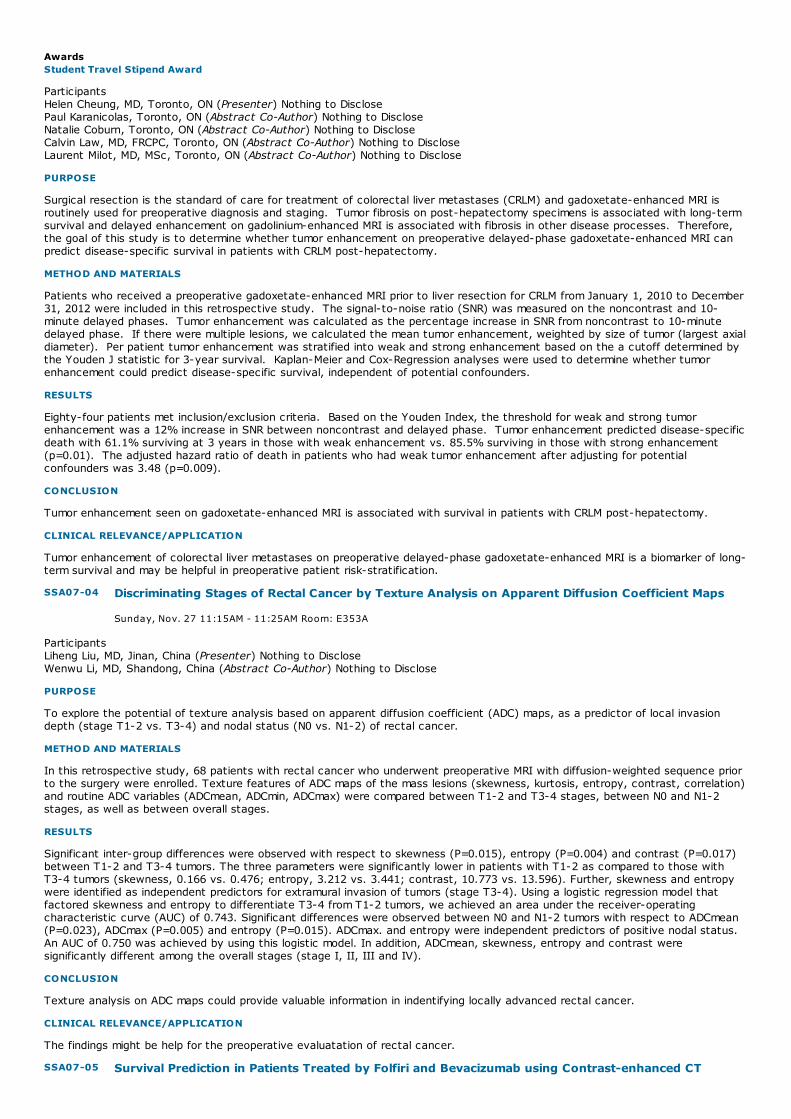
SSA07-04 Discriminating Stages of Rectal Cancer by Texture Analysis on Apparent Diffusion Coefficient Maps
Sunday, Nov. 27 11:15AM - 11:25AM Room: E353A
SSA07-05 Survival Prediction in Patients Treated by Folfiri and Bevacizumab using Contrast-enhanced CT
AwardsStudent Travel Stipend Award
ParticipantsHelen Cheung, MD, Toronto, ON (Presenter) Nothing to DisclosePaul Karanicolas, Toronto, ON (Abstract Co-Author) Nothing to DiscloseNatalie Coburn, Toronto, ON (Abstract Co-Author) Nothing to DiscloseCalvin Law, MD, FRCPC, Toronto, ON (Abstract Co-Author) Nothing to DiscloseLaurent Milot, MD, MSc, Toronto, ON (Abstract Co-Author) Nothing to Disclose
PURPOSE
Surgical resection is the standard of care for treatment of colorectal liver metastases (CRLM) and gadoxetate-enhanced MRI isroutinely used for preoperative diagnosis and staging. Tumor fibrosis on post-hepatectomy specimens is associated with long-termsurvival and delayed enhancement on gadolinium-enhanced MRI is associated with fibrosis in other disease processes. Therefore,the goal of this study is to determine whether tumor enhancement on preoperative delayed-phase gadoxetate-enhanced MRI canpredict disease-specific survival in patients with CRLM post-hepatectomy.
METHOD AND MATERIALS
Patients who received a preoperative gadoxetate-enhanced MRI prior to liver resection for CRLM from January 1, 2010 to December31, 2012 were included in this retrospective study. The signal-to-noise ratio (SNR) was measured on the noncontrast and 10-minute delayed phases. Tumor enhancement was calculated as the percentage increase in SNR from noncontrast to 10-minutedelayed phase. If there were multiple lesions, we calculated the mean tumor enhancement, weighted by size of tumor (largest axialdiameter). Per patient tumor enhancement was stratified into weak and strong enhancement based on the a cutoff determined bythe Youden J statistic for 3-year survival. Kaplan-Meier and Cox-Regression analyses were used to determine whether tumorenhancement could predict disease-specific survival, independent of potential confounders.
RESULTS
Eighty-four patients met inclusion/exclusion criteria. Based on the Youden Index, the threshold for weak and strong tumorenhancement was a 12% increase in SNR between noncontrast and delayed phase. Tumor enhancement predicted disease-specificdeath with 61.1% surviving at 3 years in those with weak enhancement vs. 85.5% surviving in those with strong enhancement(p=0.01). The adjusted hazard ratio of death in patients who had weak tumor enhancement after adjusting for potentialconfounders was 3.48 (p=0.009).
CONCLUSION
Tumor enhancement seen on gadoxetate-enhanced MRI is associated with survival in patients with CRLM post-hepatectomy.
CLINICAL RELEVANCE/APPLICATION
Tumor enhancement of colorectal liver metastases on preoperative delayed-phase gadoxetate-enhanced MRI is a biomarker of long-term survival and may be helpful in preoperative patient risk-stratification.
ParticipantsLiheng Liu, MD, Jinan, China (Presenter) Nothing to DiscloseWenwu Li, MD, Shandong, China (Abstract Co-Author) Nothing to Disclose
PURPOSE
To explore the potential of texture analysis based on apparent diffusion coefficient (ADC) maps, as a predictor of local invasiondepth (stage T1-2 vs. T3-4) and nodal status (N0 vs. N1-2) of rectal cancer.
METHOD AND MATERIALS
In this retrospective study, 68 patients with rectal cancer who underwent preoperative MRI with diffusion-weighted sequence priorto the surgery were enrolled. Texture features of ADC maps of the mass lesions (skewness, kurtosis, entropy, contrast, correlation)and routine ADC variables (ADCmean, ADCmin, ADCmax) were compared between T1-2 and T3-4 stages, between N0 and N1-2stages, as well as between overall stages.
RESULTS
Significant inter-group differences were observed with respect to skewness (P=0.015), entropy (P=0.004) and contrast (P=0.017)between T1-2 and T3-4 tumors. The three parameters were significantly lower in patients with T1-2 as compared to those withT3-4 tumors (skewness, 0.166 vs. 0.476; entropy, 3.212 vs. 3.441; contrast, 10.773 vs. 13.596). Further, skewness and entropywere identified as independent predictors for extramural invasion of tumors (stage T3-4). Using a logistic regression model thatfactored skewness and entropy to differentiate T3-4 from T1-2 tumors, we achieved an area under the receiver-operatingcharacteristic curve (AUC) of 0.743. Significant differences were observed between N0 and N1-2 tumors with respect to ADCmean(P=0.023), ADCmax (P=0.005) and entropy (P=0.015). ADCmax. and entropy were independent predictors of positive nodal status.An AUC of 0.750 was achieved by using this logistic model. In addition, ADCmean, skewness, entropy and contrast weresignificantly different among the overall stages (stage I, II, III and IV).
CONCLUSION
Texture analysis on ADC maps could provide valuable information in indentifying locally advanced rectal cancer.
CLINICAL RELEVANCE/APPLICATION
The findings might be help for the preoperative evaluatation of rectal cancer.
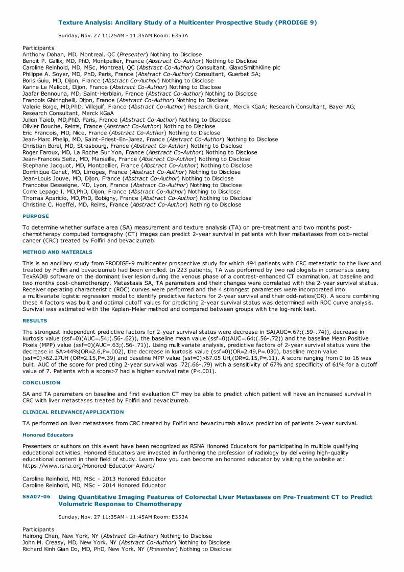
Texture Analysis: Ancillary Study of a Multicenter Prospective Study (PRODIGE 9)
Sunday, Nov. 27 11:25AM - 11:35AM Room: E353A
SSA07-06 Using Quantitative Imaging Features of Colorectal Liver Metastases on Pre-Treatment CT to PredictVolumetric Response to Chemotherapy
Sunday, Nov. 27 11:35AM - 11:45AM Room: E353A
ParticipantsAnthony Dohan, MD, Montreal, QC (Presenter) Nothing to DiscloseBenoit P. Gallix, MD, PhD, Montpellier, France (Abstract Co-Author) Nothing to DiscloseCaroline Reinhold, MD, MSc, Montreal, QC (Abstract Co-Author) Consultant, GlaxoSmithKline plcPhilippe A. Soyer, MD, PhD, Paris, France (Abstract Co-Author) Consultant, Guerbet SA; Boris Guiu, MD, Dijon, France (Abstract Co-Author) Nothing to DiscloseKarine Le Malicot, Dijon, France (Abstract Co-Author) Nothing to DiscloseJaafar Bennouna, MD, Saint-Herblain, France (Abstract Co-Author) Nothing to DiscloseFrancois Ghiringhelli, Dijon, France (Abstract Co-Author) Nothing to DiscloseValerie Boige, MD,PhD, Villejuif, France (Abstract Co-Author) Research Grant, Merck KGaA; Research Consultant, Bayer AG;Research Consultant, Merck KGaAJulien Taieb, MD,PhD, Paris, France (Abstract Co-Author) Nothing to DiscloseOlivier Bouche, Reims, France (Abstract Co-Author) Nothing to DiscloseEric Francois, MD, Nice, France (Abstract Co-Author) Nothing to DiscloseJean-Marc Phelip, MD, Saint-Priest-En-Jarez, France (Abstract Co-Author) Nothing to DiscloseChristian Borel, MD, Strasbourg, France (Abstract Co-Author) Nothing to DiscloseRoger Faroux, MD, La Roche Sur Yon, France (Abstract Co-Author) Nothing to DiscloseJean-Francois Seitz, MD, Marseille, France (Abstract Co-Author) Nothing to DiscloseStephane Jacquot, MD, Montpellier, France (Abstract Co-Author) Nothing to DiscloseDominique Genet, MD, Limoges, France (Abstract Co-Author) Nothing to DiscloseJean-Louis Jouve, MD, Dijon, France (Abstract Co-Author) Nothing to DiscloseFrancoise Desseigne, MD, Lyon, France (Abstract Co-Author) Nothing to DiscloseCome Lepage I, MD,PhD, Dijon, France (Abstract Co-Author) Nothing to DiscloseThomas Aparicio, MD,PhD, Bobigny, France (Abstract Co-Author) Nothing to DiscloseChristine C. Hoeffel, MD, Reims, France (Abstract Co-Author) Nothing to Disclose
PURPOSE
To determine whether surface area (SA) measurement and texture analysis (TA) on pre-treatment and two months post-chemotherapy computed tomography (CT) images can predict 2-year survival in patients with liver metastases from colo-rectalcancer (CRC) treated by Folfiri and bevacizumab.
METHOD AND MATERIALS
This is an ancillary study from PRODIGE-9 multicenter prospective study for which 494 patients with CRC metastatic to the liver andtreated by Folfiri and bevacizumab had been enrolled. In 223 patients, TA was performed by two radiologists in consensus usingTexRAD® software on the dominant liver lesion during the venous phase of a contrast-enhanced CT examination, at baseline andtwo months post-chemotherapy. Metastasis SA, TA parameters and their changes were correlated with the 2-year survival status.Receiver operating characteristic (ROC) curves were performed and the 4 strongest parameters were incorporated intoa multivariate logistic regression model to identify predictive factors for 2-year survival and their odd-ratios(OR). A score combiningthese 4 factors was built and optimal cutoff values for predicting 2-year survival status was determined with ROC curve analysis.Survival was estimated with the Kaplan-Meier method and compared between groups with the log-rank test.
RESULTS
The strongest independent predictive factors for 2-year survival status were decrease in SA(AUC=.67;(.59-.74)), decrease inkurtosis value (ssf=0)(AUC=.54;(.56-.62)), the baseline mean value (ssf=0)(AUC=.64;(.56-.72)) and the baseline Mean PositivePixels (MPP) value (ssf=0)(AUC=.63;(.56-.71)). Using multivariate analysis, predictive factors of 2-year survival status were thedecrease in SA>44%(OR=2.6,P=.002), the decrease in kurtosis value (ssf=0)(OR=2.49,P=.030), baseline mean value(ssf=0)>62.27UH (OR=2.15,P=.39) and baseline MPP value (ssf=0)>67.05 UH,(OR=2.15,P=.11). A score ranging from 0 to 16 wasbuilt. AUC of the score for predicting 2-year survival was .72(.66-.79) with a sensitivity of 67% and specificity of 61% for a cutoffvalue of 7. Patients with a score>7 had a higher survival rate (P<.001).
CONCLUSION
SA and TA parameters on baseline and first evaluation CT may be able to predict which patient will have an increased survival inCRC with liver metastases treated by Folfiri and bevacizumab.
CLINICAL RELEVANCE/APPLICATION
TA performed on liver metastases from CRC treated by Folfiri and bevacizumab allows prediction of patients 2-year survival.
Honored Educators
Presenters or authors on this event have been recognized as RSNA Honored Educators for participating in multiple qualifyingeducational activities. Honored Educators are invested in furthering the profession of radiology by delivering high-qualityeducational content in their field of study. Learn how you can become an honored educator by visiting the website at:https://www.rsna.org/Honored-Educator-Award/
Caroline Reinhold, MD, MSc - 2013 Honored EducatorCaroline Reinhold, MD, MSc - 2014 Honored Educator
ParticipantsHairong Chen, New York, NY (Abstract Co-Author) Nothing to DiscloseJohn M. Creasy, MD, New York, NY (Abstract Co-Author) Nothing to DiscloseRichard Kinh Gian Do, MD, PhD, New York, NY (Presenter) Nothing to Disclose
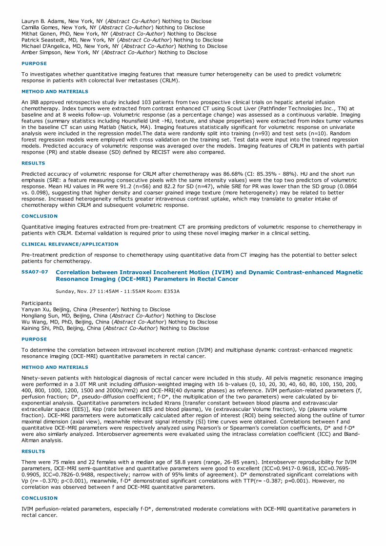
SSA07-07 Correlation between Intravoxel Incoherent Motion (IVIM) and Dynamic Contrast-enhanced MagneticResonance Imaging (DCE-MRI) Parameters in Rectal Cancer
Sunday, Nov. 27 11:45AM - 11:55AM Room: E353A
Lauryn B. Adams, New York, NY (Abstract Co-Author) Nothing to DiscloseCamilla Gomes, New York, NY (Abstract Co-Author) Nothing to DiscloseMithat Gonen, PhD, New York, NY (Abstract Co-Author) Nothing to DisclosePatrick Seastedt, MD, New York, NY (Abstract Co-Author) Nothing to DiscloseMichael D'Angelica, MD, New York, NY (Abstract Co-Author) Nothing to DiscloseAmber Simpson, New York, NY (Abstract Co-Author) Nothing to Disclose
PURPOSE
To investigates whether quantitative imaging features that measure tumor heterogeneity can be used to predict volumetricresponse in patients with colorectal liver metastases (CRLM).
METHOD AND MATERIALS
An IRB approved retrospective study included 103 patients from two prospective clinical trials on hepatic arterial infusionchemotherapy. Index tumors were extracted from contrast enhanced CT using Scout Liver (Pathfinder Technologies Inc., TN) atbaseline and at 8 weeks follow-up. Volumetric response (as a percentage change) was assessed as a continuous variable. Imagingfeatures (summary statistics including Hounsfield Unit -HU, texture, and shape properties) were extracted from index tumor volumesin the baseline CT scan using Matlab (Natick, MA). Imaging features statistically significant for volumetric response on univariateanalysis were included in the regression model.The data were randomly split into training (n=93) and test sets (n=10). Randomforest regression models were employed with cross validation on the training set. Test data were input into the trained regressionmodels. Predicted accuracy of volumetric response was averaged over the models. Imaging features of CRLM in patients with partialresponse (PR) and stable disease (SD) defined by RECIST were also compared.
RESULTS
Predicted accuracy of volumetric response for CRLM after chemotherapy was 86.68% (CI: 85.35% - 88%). HU and the short runemphasis (SRE: a feature measuring consecutive pixels with the same intensity values) were the top two predictors of volumetricresponse. Mean HU values in PR were 91.2 (n=56) and 82.2 for SD (n=47), while SRE for PR was lower than the SD group (0.0864vs. 0.098), suggesting that higher density and coarser grained image texture (more heterogeneity) may be related to betterresponse. Increased heterogeneity reflects greater intravenous contrast uptake, which may translate to greater intake ofchemotherapy within CRLM and subsequent volumetric response.
CONCLUSION
Quantitative imaging features extracted from pre-treatment CT are promising predictors of volumetric response to chemotherapy inpatients with CRLM. External validation is required prior to using these novel imaging marker in a clinical setting.
CLINICAL RELEVANCE/APPLICATION
Pre-treatment prediction of response to chemotherapy using quantitative data from CT imaging has the potential to better selectpatients for chemotherapy.
ParticipantsYanyan Xu, Beijing, China (Presenter) Nothing to DiscloseHongliang Sun, MD, Beijing, China (Abstract Co-Author) Nothing to DiscloseWu Wang, MD, PhD, Beijing, China (Abstract Co-Author) Nothing to DiscloseKaining Shi, PhD, Beijing, China (Abstract Co-Author) Nothing to Disclose
PURPOSE
To determine the correlation between intravoxel incoherent motion (IVIM) and multiphase dynamic contrast-enhanced magneticresonance imaging (DCE-MRI) quantitative parameters in rectal cancer.
METHOD AND MATERIALS
Ninety-seven patients with histological diagnosis of rectal cancer were included in this study. All pelvis magnetic resonance imagingwere performed in a 3.0T MR unit including diffusion-weighted imaging with 16 b-values (0, 10, 20, 30, 40, 60, 80, 100, 150, 200,400, 800, 1000, 1200, 1500 and 2000s/mm2) and DCE-MRI(40 dynamic phases) as reference. IVIM perfusion-related parameters (f,perfusion fraction; D*, pseudo-diffusion coefficient; f·D*, the multiplication of the two parameters) were calculated by bi-exponential analysis. Quantitative parameters included Ktrans [transfer constant between blood plasma and extravascularextracellular space (EES)], Kep (rate between EES and blood plasma), Ve (extravascular Volume fraction), Vp (plasma volumefraction). DCE-MRI parameters were automatically calculated after region of interest (ROI) being selected along the outline of tumormaximal dimension (axial view), meanwhile relevant signal intensity (SI) time curves were obtained. Correlations between f andquantitative DCE-MRI parameters were respectively analyzed using Pearson’s or Spearman’s correlation coefficients, D* and f·D*were also similarly analyzed. Interobserver agreements were evaluated using the intraclass correlation coefficient (ICC) and Bland-Altman analysis.
RESULTS
There were 75 males and 22 females with a median age of 58.8 years (range, 26-85 years). Interobserver reproducibility for IVIMparameters, DCE-MRI semi-quantitative and quantitative parameters were good to excellent (ICC=0.9417-0.9618, ICC=0.7695-0.9905, ICC=0.7826-0.9488, respectively; narrow with of 95% limits of agreement). D* demonstrated significant correlations withVp (r= -0.370; p<0.001), meanwhile, f·D* demonstrated significant correlations with TTP(r= -0.387; p=0.001). However, nocorrelation was observed between f and DCE-MRI quantitative parameters.
CONCLUSION
IVIM perfusion-related parameters, especially f·D*, demonstrated moderate correlations with DCE-MRI quantitative parameters inrectal cancer.
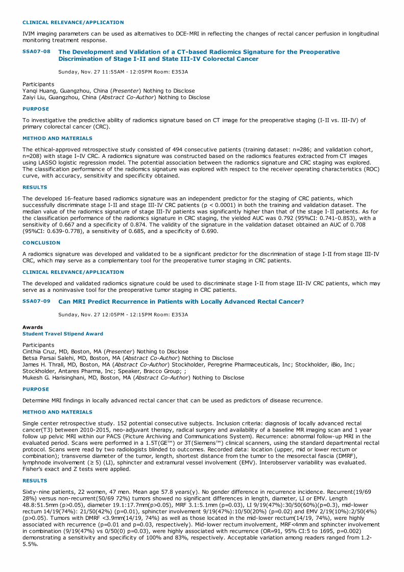
SSA07-08 The Development and Validation of a CT-based Radiomics Signature for the PreoperativeDiscrimination of Stage I-II and State III-IV Colorectal Cancer
Sunday, Nov. 27 11:55AM - 12:05PM Room: E353A
SSA07-09 Can MRI Predict Recurrence in Patients with Locally Advanced Rectal Cancer?
Sunday, Nov. 27 12:05PM - 12:15PM Room: E353A
CLINICAL RELEVANCE/APPLICATION
IVIM imaging parameters can be used as alternatives to DCE-MRI in reflecting the changes of rectal cancer perfusion in longitudinalmonitoring treatment response.
ParticipantsYanqi Huang, Guangzhou, China (Presenter) Nothing to DiscloseZaiyi Liu, Guangzhou, China (Abstract Co-Author) Nothing to Disclose
PURPOSE
To investigative the predictive ability of radiomics signature based on CT image for the preoperative staging (I-II vs. III-IV) ofprimary colorectal cancer (CRC).
METHOD AND MATERIALS
The ethical-approved retrospective study consisted of 494 consecutive patients (training dataset: n=286; and validation cohort,n=208) with stage I–IV CRC. A radiomics signature was constructed based on the radiomics features extracted from CT imagesusing LASSO logistic regression model. The potential association between the radiomics signature and CRC staging was explored.The classification performance of the radiomics signature was explored with respect to the receiver operating characteristics (ROC)curve, with accuracy, sensitivity and specificity obtained.
RESULTS
The developed 16-feature based radiomics signature was an independent predictor for the staging of CRC patients, whichsuccessfully discriminate stage I-II and stage III-IV CRC patients (p < 0.0001) in both the training and validation dataset. Themedian value of the radiomics signature of stage III-IV patients was significantly higher than that of the stage I-II patients. As forthe classification performance of the radiomics signature in CRC staging, the yielded AUC was 0.792 (95%CI: 0.741-0.853), with asensitivity of 0.667 and a specificity of 0.874. The validity of the signature in the validation dataset obtained an AUC of 0.708(95%CI: 0.639-0.778), a sensitivity of 0.685, and a specificity of 0.690.
CONCLUSION
A radiomics signature was developed and validated to be a significant predictor for the discrimination of stage I-II from stage III-IVCRC, which may serve as a complementary tool for the preoperative tumor staging in CRC patients.
CLINICAL RELEVANCE/APPLICATION
The developed and validated radiomics signature could be used to discriminate stage I-II from stage III-IV CRC patients, which mayserve as a noninvasive tool for the preoperative tumor staging in CRC patients.
AwardsStudent Travel Stipend Award
ParticipantsCinthia Cruz, MD, Boston, MA (Presenter) Nothing to DiscloseBetsa Parsai Salehi, MD, Boston, MA (Abstract Co-Author) Nothing to DiscloseJames H. Thrall, MD, Boston, MA (Abstract Co-Author) Stockholder, Peregrine Pharmaceuticals, Inc; Stockholder, iBio, Inc;Stockholder, Antares Pharma, Inc; Speaker, Bracco Group; ; Mukesh G. Harisinghani, MD, Boston, MA (Abstract Co-Author) Nothing to Disclose
PURPOSE
Determine MRI findings in locally advanced rectal cancer that can be used as predictors of disease recurrence.
METHOD AND MATERIALS
Single center retrospective study. 152 potential consecutive subjects. Inclusion criteria: diagnosis of locally advanced rectalcancer(T3) between 2010-2015, neo-adjuvant therapy, radical surgery and availability of a baseline MR imaging scan and 1 yearfollow up pelvic MRI within our PACS (Picture Archiving and Communications System). Recurrence: abnormal follow-up MRI in theevaluated period. Scans were performed in a 1.5T(GE™) or 3T(Siemens™) clinical scanners, using the standard departmental rectalprotocol. Scans were read by two radiologists blinded to outcomes. Recorded data: location (upper, mid or lower rectum orcombination); transverse diameter of the tumor, length, shortest distance from the tumor to the mesorectal fascia (DMRF),lymphnode involvement (≥ 5) (LI), sphincter and extramural vessel involvement (EMV). Interobserver variability was evaluated. Fisher’s exact and Z tests were applied.
RESULTS
Sixty-nine patients, 22 women, 47 men. Mean age 57.8 years(y). No gender difference in recurrence incidence. Recurrent(19/6928%) versus non-recurrent(50/69 72%) tumors showed no significant differences in length, diameter, LI or EMV. Length48.8:51.5mm (p>0.05), diameter 19.1:17.7mm(p>0.05), MRF 3.1:5.1mm (p=0.03), LI 9/19(47%):30/50(60%)(p=0.3), mid-lowerrectum 14/19(74%): 21/50(42%) (p=0.01), sphincter involvement 9/19(47%):10/50(20%) (p=0.02) and EMV 2/19(10%):2/50(4%)(p>0.05). Tumors with DMRF <3.9mm(14/19, 74%) as well as those located in the mid-lower rectum(14/19, 74%), were highlyassociated with recurrence (p=0.01 and p=0.03, respectively). Mid-lower rectum involvement, MRF<4mm and sphincter involvementin combination (9/19(47%) vs 0/50(0) p=0.03), were highly associated with recurrence (OR=91, 95% CI:5 to 1695, p=0.002)demonstrating a sensitivity and specificity of 100% and 83%, respectively. Acceptable variation among readers ranged from 1.2-5.5%.

CONCLUSION
Mid-lower rectum involvement and MRF<4mm are highly associated with rectal cancer recurrence when found on 1 year follow-upMRI. In combination with sphincter involvement, such tumors demonstrated a 91-fold greater chance of recurring.
CLINICAL RELEVANCE/APPLICATION
MRI features and tumor location may be used to predict recurrence and markers of worst prognosis in locally advanced rectalcancer at 1-year-follow up scans.

SSA08-01 Gastrointestinal Keynote Speaker: Novel CT Image Reconstruction Techniques
Sunday, Nov. 27 10:45AM - 10:55AM Room: E450A
SSA08-02 Image Noise and CNR: Are These Meaningful Parameters to Assess the Potential for Dose Reduction for Abdominal CT at Different Tube Voltages in the Era ofAdvanced Modeled Iterative Reconstruction? A 12-reader Phantom Study
Sunday, Nov. 27 10:55AM - 11:05AM Room: E450A
SSA08-03 Iterative Model Reconstruction (IMR) Technique with Low Dose CT- Enterography Protocol in Evaluation of Crohn's Disease Patients: Comparison with the 4thGeneration Iterative Reconstruction Algorithm
Sunday, Nov. 27 11:05AM - 11:15AM Room: E450A
SSA08-04 Comparison of Abdomen CT Radiation Doses (CTDIvol, DLP, and SSDE) from ACR Dose Index Registry based on Age and Type of Medical Facility in DifferentGeographical Regions of US
Sunday, Nov. 27 11:15AM - 11:25AM Room: E450A
SSA08
Science Session with Keynote: Gastrointestinal (CT Dose)
Sunday, Nov. 27 10:45AM - 12:15PM Room: E450A
CT GI
AMA PRA Category 1 Credits ™: 1.50ARRT Category A+ Credits: 1.50
ParticipantsWilliam P. Shuman, MD, Seattle, WA (Moderator) Research Grant, General Electric CompanyAndrea Laghi, MD, Rome, Italy (Moderator) Speaker, Bracco Group Speaker, Bayer AG Speaker, General Electric Company Speaker, Koninklijke Philips NVJeong Min Lee, MD, Seoul, Korea, Republic Of (Moderator) Grant, Guerbet SA; Support, Siemens AG; Grant, Bayer AG; Grant, General Electric Company; Grant, STARmed Co, Ltd; Grant, RF Medical Co, Ltd; Grant,Toshiba Corporation; Grant, Samsung Medical Healthcare
Sub-Events
ParticipantsWilliam P. Shuman, MD, Seattle, WA (Presenter) Research Grant, General Electric Company
AwardsStudent Travel Stipend Award
ParticipantsAndre Euler, MD, Basel, Switzerland (Presenter) Nothing to DiscloseZsolt Szucs-Farkas, MD, PhD, Berne, Switzerland (Abstract Co-Author) Nothing to DiscloseBram Stieltjes, MD, Basel, Switzerland (Abstract Co-Author) Nothing to DiscloseReto M. Eichenberger, MD, Basel , Switzerland (Abstract Co-Author) Nothing to DiscloseClemens Reisinger, MD, Basel, Switzerland (Abstract Co-Author) Nothing to DiscloseAnna Hirschmann, MD, Basel, Switzerland (Abstract Co-Author) Nothing to DiscloseCaroline Zahringer, Basel, Switzerland (Abstract Co-Author) Nothing to DiscloseAchim Kircher, Basel, Switzerland (Abstract Co-Author) Nothing to DiscloseSebastien Kopp, Basel, Switzerland (Abstract Co-Author) Nothing to DiscloseSebastian T. Schindera, MD, Basel, Switzerland (Abstract Co-Author) Research Grant, Siemens AG; Research Grant, Ulrich GmbH & Co KG; Research Grant, Bayer AG; Speakers Bureau, Bayer AG
PURPOSE
To assess the impact of changes in image noise (IN) and contrast-to-noise-ratio (CNR) on low-contrast-detectability by applying advanced modeled iterative reconstruction algorithm and low-kVp abdominal CT inphantoms of different phantom sizes with simulated liver lesions.
METHOD AND MATERIALS
A multi-energy liver phantom with 45 hypoattenuating, iodine-containing lesions (diameters of 5, 10, 15 mm; lesion-to-liver contrast of 10, 25, 50 HU) was placed in water containers mimicking a medium and largepatient (diameters: 30 and 40 cm). The phantom was scanned at 70, 80, 100 and 120 kVp (SOMATOM Force, Siemens). The 120 kVp-protocol served as the reference protocol and the mAs settings of the otherprotocols were adjusted to keep the CTDIvol constant. The datasets were reconstructed with advanced modeled iterative reconstruction (ADMIRE, strength 3) and filtered back projection (FBP). IN was measuredand CNR was calculated. Low-contrast lesion detection was independently assessed by twelve radiologists. Descriptive statistics and Fisher exact test were applied.
RESULTS
Applying iterative reconstruction, the IN decreased on average by 24% and by 27% and the CNR increased on average by 28% and by 34% for the medium and the large phantom, respectively. Lowering the tubevoltage, the CNR increased on average by 72%, 59%, 35% at 70, 80, 100 kVp, respectively compared with 120 kVp in the medium phantom and by 4%, 16%, 7% in the large phantom.Despite the improvement ofquantitative image quality by applying iterative reconstruction or low-kVp-imaging, there was no significant difference in overall detection rate within each phantom size (medium: 79% to 82%; large: 57% to 65%;P-value range: 0.37 to 1). Interobserver agreement was very good for both groups (κ-range of 0.8 to 0.9).
CONCLUSION
Improvement of quantitative image quality by advanced modeled iterative reconstruction or low-kVp-imaging did not impact low-contrast lesion detection in phantoms simulating medium and large patients.
CLINICAL RELEVANCE/APPLICATION
IN and CNR are well accepted parameters to grade quantitative image quality. However, they do not encompass the effects of dose optimization. More sophisticated image quality parameters are needed.
ParticipantsYu y. Gao, MD, Shenyang, China (Presenter) Nothing to DiscloseXiao Mei Lu, MMed, Shenyang, China (Abstract Co-Author) Nothing to Disclose
PURPOSE
To evaluate the diagnostic efficacy, in terms of radiation dose reduction, image quality and diagnostic confidence of low dose CTE protocol combined with Iterative model reconstruction (IMR) technique, incomparison with iterative reconstruction algorithm (iDose4) in patients with known Crohn's disease (CD)
METHOD AND MATERIALS
Twenty-three patients with CD underwent low-dose CTE scan in a single venous phase on 256 MDCT scanner with 100 kV, 120~150mAs and slice thickness 1 mm, using IMR reconstruction algorithm as well asiterative reconstruction algorithm (iDose4) of the same data sets.Two radiologists, blinded to clinical and pathological findings,independently observed the changes bowel wall and presence of CD activity features(mural thickening and enhancement pattern, perianal changes(if present), comb sign, lymphoadenopathy and complications such as fistulas and stenosis). Image noise, standard deviation (SD) and contrast-to-noise(CNR), diagnostic quality were evaluated using a 4-point scale.DLP and CTDI were recorded and compared between both reconstruction algorithm.
RESULTS
Total DLP and CTDI were lower in low-dose CTE examinations in our studies.(347.47 ~423.45 mGy*cm and overall around 6.37 mGy), allowing an overall dose reduction of 67.3% compared to prior dose in ourinstitution. IMR recontructed Low-dose CTE protocol showed high diagnostic quality in assessment of Crohn's disease features. Noise results of IMR superior to iDose4 in upper (SD: IMR vs. iDose4=3.05 ± 0.96 vs.6.82 ±2.16 )、middle(SD: IMR vs. iDose4=5.42 ± 1.17 vs. 7.82 ±2.47 ) and lower part(SD: IMR vs. iDose4=3.78 ± 1.34 vs. 8.81 ±3.11 ) of abdomen,while about CNR,upper (CNR: IMR vs. iDose4=3.72 ± 1.18 vs.1.44 ±0.45 )、middle(CNR: IMR vs. iDose4=11.90 ± 3.76 vs. 4.56 ±1.44 ) and lower part(CNR: IMR vs. iDose4=11.06 ± 3.50 vs. 3.71 ±1.71 ) , difference was statistically significant in each group(p<0.01, p<0.05).
CONCLUSION
Iterative model reconstruction (IMR) technique with Low-dose CTE provideded significantly low noise and high CNR pictures compare to iDose4 reconstruction algorithm with nearly two-thirds dose reduction,consider their young age and frequent follow-up requirment of CD patients, this technique may become a useful tool in CD patients, even in lower part of the abdomen including pelvis and perianal area.
CLINICAL RELEVANCE/APPLICATION
apply to young Crohn's diseases follow-up
ParticipantsAmirhossein Mozafarykhamseh, MD, boston, MA (Presenter) Nothing to DiscloseAtul Padole, MD, Boston, MA (Abstract Co-Author) Nothing to DiscloseDebapriya Sengupta, MBBS,MPH, Reston, VA (Abstract Co-Author) Nothing to DiscloseJudy Burleson, Reston, VA (Abstract Co-Author) Nothing to DiscloseSubba R. Digumarthy, MD, Boston, MA (Abstract Co-Author) Nothing to DiscloseMannudeep K. Kalra, MD, Boston, MA (Abstract Co-Author) Technical support, Siemens AG; Technical support, Medical VisionMythreyi Bhargavan-Chatfield, PhD, Reston, VA (Abstract Co-Author) Nothing to Disclose
PURPOSE
To compare abdomen CT radiation dose (CTDIvol, DLP & SSDE) from the ACR Dose Index Registry (DIR) across different types of facilities and geographical regions in US
METHOD AND MATERIALS
Using Radiology Playbook identification (RPID) numbers, we assessed CT dose metrics CTDIvol, DLP and size-specific dose estimates SSDE in 271287 abdomen CT exams (mean age 54.7±18.8 yrs) in the ACR DIRfrom 2011-2015 (10% sample). Data were stratified according to the geographic census in to New England (NE), Middle Atlantic (MA), South Atlantic (SA), East North Central(ENC), East South Central (ESC),WestSouth Central (WSC), Mountain (MO) and Pacific(PA)) regions and then sub-stratified by year of CT (2011-15), patient age (≤ 40 years; 41-64 years; ≥65 years) and type of facility where CT was done(academic, community, multispecialty, freestanding). Mean and standard deviations for maximum CTDIvol (mGy), DLP (mGy.cm) and SSDE were calculated. ANOVA tests were used for analysis
RESULTS
The national mean CTDIvol, SSDE and DLP for abdomen CT were 15±8, 17±8 and 736±426 . There were significant variations for CTDIvol, DLP and SSDE values between census divisions.The highest CTDIvol and
SSA08-05 Comparison of Low kVp and Automatic Spectral Imaging Mode Selection Protocols in Abdominal CT with Statistical Iterative Reconstruction and Low ContrastAgent Dose: A Prospective Matched Cohort Study
Sunday, Nov. 27 11:25AM - 11:35AM Room: E450A
SSA08-06 Evaluation of Image Quality and Texture Features of Abdominal Spectral CT Imaging Using Adaptive Statistical Iterative Reconstruction
Sunday, Nov. 27 11:35AM - 11:45AM Room: E450A
SSA08-07 Dose-Equivalent Triple Arterial Phase CT Imaging of the Liver: Initial Experience in Comparison to Single Arterial Phase at 80kV
Sunday, Nov. 27 11:45AM - 11:55AM Room: E450A
SSA08-08 Observer Performance at Varying Dose Levels and Reconstruction Methods for Detection of Hepatic Metastases
Sunday, Nov. 27 11:55AM - 12:05PM Room: E450A
DLP was seen for WSC (15±11, 721±471 n=8916) and the lowest CTDIvol and DLP was for PA (11±6, 496±302, n=27095) (p<0.0001). For SSDE values, NE had the highest (25±8, n=17011), while, PA had thelowest (8±7, n=13758). The mean SSDE values of academic, community, multispecialty and freestanding facilities were 16±7, 17±9, 19±8, and 15±6 (p<0.0001), respectively. Among all 8 census divisions, nosignificant difference in dose based on age was noted. The mean±SD CTDIvol, DLP and SSDE for <40 years age group were 14±8, 737±441, 16±9; while, 16±9, 771±441and 17±8 for 41-64 years and 15±8,633±388 and 17±8 for >64 years (p value > 0.1). Since the inception of the ACR DIR in 2011, abdomen SSDE has decreased by average 12.2% in different census divisions. The biggest decrease (25.6%) was inENC; while there was only 1.2% decrease in PA region
CONCLUSION
There were significant variations in the values of CTDIvol, DLP, and SSDE for abdomen CT among different US census regions. There was no significant difference in dose metrics based on age, among all 8 censusdivisions
CLINICAL RELEVANCE/APPLICATION
The ACR DIR reveals that there is decrease in radiation dose for abdomen CT in the last 5 years. Improved effort is needed to achieve similar dose levels in all regions of US
Honored Educators
Presenters or authors on this event have been recognized as RSNA Honored Educators for participating in multiple qualifying educational activities. Honored Educators are invested in furthering the profession ofradiology by delivering high-quality educational content in their field of study. Learn how you can become an honored educator by visiting the website at: https://www.rsna.org/Honored-Educator-Award/
Subba R. Digumarthy, MD - 2013 Honored Educator
ParticipantsPeijie Lv, MMed, Zhengzhou, China (Presenter) Nothing to DiscloseLiying Zhang, Zhengzhou, China (Abstract Co-Author) Nothing to DiscloseJianbo Gao, MD, Zhengzhou, China (Abstract Co-Author) Nothing to Disclose
PURPOSE
A prospective matched cohort study was performed to compare the image quality and radiation dose obtained with automatic spectral imaging mode selection (ASIS) and adaptive statistical iterativereconstruction (ASIR) with that obtained with low kVp with ASIR in abdominal multiphase computed tomography (CT) with low contrast agent dose.
METHOD AND MATERIALS
This prospective study was institutional review board approved and written informed consents from all patients were obtained. Four groups of 60 patients each underwent the arterial-phase (AP) and portal venousphase (PVP) contrast-enhanced abdominal CT scan with low kVp (80 kVp or 100 kVp) or spectral CT imaging with ASIS modes, matched by age, gender, body mass index(BMI) , cross sectional area of theabdomen and contrast agent dose. One hundred and twenty patients (group A, 80 kVp and group B, spectral CT imaging) accepted contrast agent dose of 300mgI/kg with BMI < or = 23.9 kg/m2 while 120patients (group C, 100 kVp and group D, spectral CT imaging) accepted 450mgI/kg contrast agent dose with BMI from 24 to 28.9 kg/m2. ASIR 50% was used to reconstruct the low-kVp images and monochromaticimages (40-60keV) from spectral CT imaging. Quantitative parameters (CT attenuation and contrast-to-noise ratio of the liver, pancreas, aorta and portal vein, image noise in HU) and qualitative visual parameters(overall image quality as graded on a 5-point scale) were compared by using two sample t tests or Mann-Whitney U test.
RESULTS
Compared to 80-kVp and 100-kVp images, 40-keV images showed similar contrast and overall image quality despite the higher image noise, 50-keV images provided higher image noise but similar or higher contrastand overall image quality, 60-keV images provided similar image noise and contrast but similar or higher overall image quality. The CTDIvol and DLP in group A were lower than group B(P = 0.001and 0.042respectively),but were similar between group C and group D.
CONCLUSION
In abdominal CT with low contrast agent dose, application of ASIR and ASIS to monochromatic imaging from 40 to 60keV maintained good image quality with higher radiation dose than 80 kVp and withoutincreasing radiation dose than 100 kVp.
CLINICAL RELEVANCE/APPLICATION
Combined use of automatic spectral imaging protocol selection and iterative reconstruction can maintain good image quality with low contrast agent dose and have the potential to reduce radiation dose.
ParticipantsLiang Yan, MMed, changchun, China (Presenter) Nothing to DiscloseZhiren Chen, MD, Changchun, China (Abstract Co-Author) Nothing to DiscloseChuang Yi, Jilin, China (Abstract Co-Author) Nothing to DiscloseHuizhi Cao VI, Beijing, China (Abstract Co-Author) Nothing to DiscloseBin 0. Li, Changchun, China (Abstract Co-Author) Nothing to Disclose
PURPOSE
The purpose of this study was to compare image quality and texture features of virtual monochromatic images of adaptive statistical iterative reconstruction (ASiR) with filtered back projection (FBP).
METHOD AND MATERIALS
Thirty patients underwent contrast-enhanced abdominal CT through single-source dual-energy spectral imaging. 60keV monochromatic images were reconstructed with FBP, 30%ASiR, 50%ASiR and 80ASiR. Two radiologists independently graded images for sharpness, image noise, diagnostic acceptability, and artifacts (graded on a 5-point Likert scale). Signal-to-noise ratio and image quality were assessed. The texture profiles (texture gradient and texture curvature) of liver parenchyma were calculated and compared among different groups.
RESULTS
The assessment scores between different observers obtained good consistency in subjective image evaluation (Kappa value =0.783). And there were significant differences in assessment scores between (30%, 50% and 80%)ASiR and FBP(P = 0.000-0.039 <0.05), and 50% ASiR possessed the highest scores. In comparison with FBP, noise of 30%, 50% and 80%ASiR reduced by 22%, 32% and 44%, respectively,without difference in CT value (P> 0.95). There was significant differences between each ASiR groups and FBP in SNR values (P = 0.000 ~ 0.019 <0 .05). And there was no difference in texture curvature between ASiR group and FBP group, and in texture gradient between 30%-50% ASiR group and FBP group, however texture gradient of 80%ASiR was lower than that of FBP(P<0.05), decreased by 38%.
CONCLUSION
ASiR (blending ratio<80%) can be used for fast kVp switching dual-energy CT scan and has the ability to reduce image noise and to maintain texture features.
CLINICAL RELEVANCE/APPLICATION
Monochromatic keV with rational percentage of ASiR can be applied in abdominal CT and the images can provide better SNR and reduced noise.
ParticipantsKatharina Brehmer, MD, Stockholm, Sweden (Presenter) Nothing to DiscloseTorkel B. Brismar, MD, PhD, Stockholm, Sweden (Abstract Co-Author) Nothing to DiscloseAnders P. Svensson, PhD, Stockholm, Sweden (Abstract Co-Author) Nothing to DiscloseAntonios Tzortzakakis, Gothenburg, Sweden (Abstract Co-Author) Nothing to DiscloseNikolaos Voulgarakis, MD, Stockholm, Sweden (Abstract Co-Author) Nothing to DiscloseMichael A. Fischer, MD, Stockholm, Sweden (Abstract Co-Author) Nothing to Disclose
PURPOSE
To develop an optimized triple arterial phase CT (triple arterial CT) liver protocol from previously acquired perfusion CT data and to prospectively evaluate the image quality (IQ) and diagnostic accuracy incomparison to standard single arterial phase CT (standard-CT) in patients with suspicion of hepatocellular carcinoma (HCC).
METHOD AND MATERIALS
A total of 42 patients prospectively underwent clinically indicated standard multiphasic liver CT and perfusion-CT at the same day using a dual-source 128-slice CT-scanner. A so called time maximum intensityprojection (time-MIP) was reconstructed by fusion of three dedicated arterial time points of the perfusion split-series. Arterial time points were depicted prospectively based on arterial peak enhancement times(TTP) of 27 HCC lesions derived retrospectively from a previously acquired perfusion CT series of 15 patients. Single arterial phase of the standard liver CT and arterial time-MIP were compared by two readers (R1;R2) who assessed image quality and detection-rate of HCC. A third reader assessed image-noise, lesion-to-liver-contrast-ratio (LLR) and contrast-to-noise-ratio(CNR).
RESULTS
The triple arterial CT consisted of three low-dose scans (80 kV; 160 mAs) at 8.1/13.4/23.7 sec (min/mean/max values of TTP in 27 HCC) after a contrast enhancement of 160 HU was reached in the abdominalaorta. Mean CTDIvol (CT Dose Index) of triple arterial CT and standard-CT was 11.5 mGy and 12.2 mGy respectively. Time-MIP showed significantly lower noise and better CNR as compared to standard-CT(P<0.001; both). There was no significant difference in LLR between time-MIP and standard-CT (P=0.26). Inter-reader agreement and subjective IQ was good for both protocols. In total there were 73 HCC.Detection rate was 89/88 % (R1/R2) at time-MIP and 86/82% (R1/R2) at standard-CT. Time-MIP detected additional 7 (R1) respectively 9 (R2) HCC, which were missed on standard-CT.
CONCLUSION
Time-MIP derived from dose-equivalent triple arterial phase CT has the potential to improve both image quality and detection rate of HCC in comparison to standard single arterial phase CT.
CLINICAL RELEVANCE/APPLICATION
Dose-equivalent triple arterial phase CT scanning of the liver has the potential to improve diagnostic accuracy for HCC and thus improve oncologic patient care.
Participants
SSA08-09 Personalized Liver CT Examination Protocol Based on BMI: Combination of Optimized kVp and Iodine Injection Protocol
Sunday, Nov. 27 12:05PM - 12:15PM Room: E450A
Joel G. Fletcher, MD, Rochester, MN (Presenter) Grant, Siemens AG; ; Jeff L. Fidler, MD, Rochester, MN (Abstract Co-Author) Nothing to DiscloseSudhakar K. Venkatesh, MD, FRCR, Rochester, MN (Abstract Co-Author) Nothing to DiscloseDavid M. Hough, MD, Rochester, MN (Abstract Co-Author) Nothing to DiscloseNaoki Takahashi, MD, Rochester, MN (Abstract Co-Author) Nothing to DiscloseLifeng Yu, PhD, Chicago, IL (Abstract Co-Author) Nothing to DiscloseMaria Shiung, Rochester, MN (Abstract Co-Author) Nothing to DiscloseAdam Bartley, Rochester, MN (Abstract Co-Author) Nothing to DiscloseShuai Leng, PhD, Rochester, MN (Abstract Co-Author) Nothing to DiscloseDavid R. Holmes Iii, PhD, Rochester, MN (Abstract Co-Author) Nothing to DiscloseAlicia Toledano, DSc, Kensington, MD (Abstract Co-Author) Consultant, iCAD, IncRickey Carter, PhD, Rochester, MN (Abstract Co-Author) Nothing to DiscloseCynthia H. McCollough, PhD, Rochester, MN (Abstract Co-Author) Research Grant, Siemens AG
PURPOSE
To estimate the ability of abdominal radiologists to detect hepatic metastases (HM) at varying dose levels with or without iterative reconstruction (sinogram-affirmed iterative reconstruction; IR).
METHOD AND MATERIALS
Projection data from 83 CT contrast-enhanced exams were collected (42 with HM). HM was defined by histopathology or progression/regression on CT/MR. Using a validated noise insertion technique, each examwas reconstructed with filtered back projection (FBP) or IR at 5 dose levels (automatic exposure control settings of 80, 100, 120, 160 and 200 quality reference mAs [QRM]) to generate 8 dose-reconstructionconfigurations (664 cases). Three abdominal radiologists used a dedicated workstation to circle suspected HM, indicate a confidence score (0 – 100), and grade image quality, evaluating each patientonce/session. Automated matching of reference and reader HMs was performed. Non-inferiority was assessed using JAFROC analysis and reader agreement rules. JAFROC analysis was performed on a per-lesionbasis for HM using a narrow non-inferiority limit of -0.05. For reader agreement, a successful reading for lower dose configurations was defined as ≥ 2 readers circling all HM identified by ≥ 2 readers at routinedose (or no non-lesion localizations in negative cases), with a priori rules requiring ≥ 71 successful readings for each configuration.
RESULTS
There were 123 HM with a median size of 1.4 ± 1.0cm with median CDTIvol = 11 mGy (mean CDTIvol of 12.4 ± 6.0 mGy; mean SSDE of 14.6 ± 4.7 mGy). Using JAFROC FOM, configurations using 80 and 100 QRMand either FBP or IR failed to meet non-inferiority criteria, with estimated differences from routine dose being - 0.04 (95% CI: -0.06 to -0.01). 5 of the 8 configurations had successful interpretations in 71 – 77 of83 patients, corresponding to dose levels of ≥120 QRM (i.e., ≥ 8.8 mGy SSDE). Intraclass correlation coefficient for reader FOM was 0.44. At each dose level, IR improved image quality (p<0.05) but notperformance.
CONCLUSION
Lower dose CT images reconstructed at dose levels corresponding to 120 QRM (SSDE 8.8 mGy) and higher performed similar to 200 QRM FBP in this large pilot study for detection of hepatic metastases.
CLINICAL RELEVANCE/APPLICATION
Pilot data obtained over a range of doses suggests that substantial dose reduction is possible without compromising observer performance. IR did not appear to improve performance.
Honored Educators
Presenters or authors on this event have been recognized as RSNA Honored Educators for participating in multiple qualifying educational activities. Honored Educators are invested in furthering the profession ofradiology by delivering high-quality educational content in their field of study. Learn how you can become an honored educator by visiting the website at: https://www.rsna.org/Honored-Educator-Award/
Naoki Takahashi, MD - 2012 Honored Educator
ParticipantsJian Jiang, MD, Beijing, China (Presenter) Research Grant, General Electric CompanyXiaoying Wang, MD, Beijing, China (Abstract Co-Author) Nothing to DiscloseKe Wang, MD, Beijing, China (Abstract Co-Author) Nothing to Disclose
PURPOSE
To investigate whether the personalized liver CT examination protocol based on body mass index (BMI) could obtain the diagnostic image quality.
METHOD AND MATERIALS
This prospective study was approved by institutional review board. From 2014 May to 2016 March, patients with known or suspected hepatocellular carcinoma were recruited consecutively, who underwent MDCTexamination (Siemens, Somatom Definition Flash). Patients were scanned with different tube voltage (70-120 kVp) in combination with different amount of iodine contrast medium (352 mgI/kg to 550 mgI/kg)based on their body mass index (BMI): BMI ≤18.0, 70-kVp, 352mgI/kg; BMI 18.1-24.0, 80-kVp, 352mgI/kg; BMI 24.1-28.0, 100-kVp, 440 mgI/kg; BMI ≥28.1, 120-kVp, 550 mgI/kg.Images in all phases for 70-kVpand 80-kVp protocol were reconstructed with strength SAFIRE 3 and those for 100-kVp and 120-kVp protocol were reconstructed by using strength SAFIRE 2. The image noise, attenuation, contrast-to-noiseratio (CNR), and figure of merit (FOM) were measured and calculated. The estimated effective dose (ED) and Size-specific dose estimate (SSDE) of the 4 protocols were also calculated and compared. Radiologistsindependently graded images quality, as well as the timing of late arterial phase.
RESULTS
Totally 165 patients were recruited. Dynamic CT demonstrated that a reduction from 120-kVp protocol to 80- and 70-kVp protocols led to a reduction in the SSDE of 36% and 56%. 92% of the arterial phase withthe 4 protocols was classified as the accurate late arterial phase. There was no significant difference in the mean image noise, CNRs and FOMs of the portal vein and liver between the 100- and 120-kVpprotocols. Mean image noise and FOMs was significantly higher with the 70-kVp protocol than with the others. For the liver, significantly lower CNRs were found with the 80-kVp protocol than with the others.Moreover, in the comparison of the 80-, 100- and 120-kVp protocols, they had no statistically significant difference, for image quality score (P > 0.05).
CONCLUSION
High quality liver CT images could be obtained by using personalized liver CT protocol based on BMI, with combination of optimized kVp and iodine injection method.
CLINICAL RELEVANCE/APPLICATION
This method will be of benefit to the patients with lower BMI, who will receive lower contrast dosage, significantly less radiation dose compared with the conventional uniform method.
SSA09-01 2D Ultrasound Shearwave for Staging Liver Fibrosis: Preliminary Results of a Prospective MulticentreEuropean Study
Sunday, Nov. 27 10:45AM - 10:55AM Room: E450B
SSA09-02 Comparison of Elastography Point Quantification with Transient Elastography in Patients withChronic Viral Hepatitis and Nonalcoholic Fatty Liver Disease: A Prospective Study
Sunday, Nov. 27 10:55AM - 11:05AM Room: E450B
SSA09
Science Session with Keynote: Gastrointestinal (Liver Fibrosis)
Sunday, Nov. 27 10:45AM - 12:15PM Room: E450B
GI BQ
AMA PRA Category 1 Credits ™: 1.50ARRT Category A+ Credits: 1.50
FDA Discussions may include off-label uses.
ParticipantsClaude B. Sirlin, MD, San Diego, CA (Moderator) Research Grant, General Electric Company; Research Grant, Siemens AG; ResearchGrant, Guerbet SA; ; Sudhakar K. Venkatesh, MD, FRCR, Rochester, MN (Moderator) Nothing to DiscloseAnthony E. Samir, MD, Boston, MA (Moderator) Consultant, Pfizer Inc Consultant, General Electric Company Consultant, PAREXELInternational Corporation Research Grant, Koninklijke Philips NV Research Grant, Siemens AG Research Grant, Toshiba CorporationResearch Grant, General Electric Company Research Grant, Samsung Electronics Co, Ltd Research Grant, Analogic CorporationResearch support, SuperSonic Imagine Research support, Hitachi, Ltd
Sub-Events
ParticipantsAdrian K. Lim, MD, FRCR, London, United Kingdom (Presenter) Luminary, Toshiba CorporationMaxime Ronot, MD, Clichy, France (Abstract Co-Author) Nothing to DiscloseGiovanna Ferraioli, MD, Pavia, Italy (Abstract Co-Author) Speaker, Koninklijke Philips NV; Speaker, Hitachi Ltd; Speaker, ToshibaCorporationHans Peter Mueller, Berlin, Germany (Abstract Co-Author) Nothing to DiscloseMireen Friedrich-Rust, Frankfurt, Germany (Abstract Co-Author) Nothing to DiscloseDavid O. Cosgrove, MBBCh, FRCR, London, United Kingdom (Abstract Co-Author) Research Consultant, SuperSonic Imagine ResearchConsultant, Bracco Group Speakers Bureau, Toshiba CorporationCarlo Filice, MD, Pavia, Italy (Abstract Co-Author) Speaker, Koninklijke Philips NV; Speaker, Hitachi, Ltd ; Research Grant, BraccoGroup; Research Grant, Hitachi, Ltd; Research Grant, Toshiba Corporation; Research Grant, Esaote SpAValerie Vilgrain, MD, Clichy, France (Abstract Co-Author) Nothing to Disclose
PURPOSE
Ultrasound Shearwave (SW) speed measurements using Transient Elastography (TE) is an accepted non-invasive test of liverstiffness and indirectly the presence of significant liver fibrosis. Newer SW technologies have since been developed but have yet tobe validated against this standard of care test.To assess the reliability and accuracy of 2D Ultrasound SW technology at gradingthe severity of Liver Fibrosis using TE as the gold standard.
METHOD AND MATERIALS
Centers from France, Germany, Italy and United Kingdom were involved. Patients were recruited prospectively with a variety ofchronic liver diseases to have 2D Ultrasound Shearwave (ToSWE) (Toshiba Medical Systems) and TE (Echosens) performed on thesame day.10 valid measurements were obtained from fasted patients (>8 hours) using both techniques and the median result in kPawas used for analysis. Based on a previous meta-analysis (Tsochatzis, et al. J Hepatol 2011) for staging liver fibrosis (F), TE cut-offs of 7.0, 9.5 and 12.0 kPa, were used to denote liver fibrosis stages of significant fibrosis (F≥2), advanced fibrosis (F≥3), andcirrhosis (F = 4) respectively. A TE value of < 7.0 kPa was used to denote no significant fibrosis (F0/F1). The measurementsobtained from ToSWE were correlated with the TE-derived stages of fibrosis.
RESULTS
367 patients have been recruited to date, (215 M:152 F) with a mean age of 53 years (range: 21-88 years). The main etiologies ofliver disease were HCV-related (64%) and HBV-related (23%). The rest comprised a mixture of ALD, AIH and NAFLD.350 datasetswere complete showing excellent correlation of liver stiffness values between the ToSWE and TE techniques at all centres(R2 correlation range: 0.78-0.92). The mean elasticity values using ToSWE for each liver fibrosis stage as described above was:F0/F1 (n= 179: 6.0 ± 1.4 kPa), F2 (n = 54: 7.0 ± 3.0 kPa), F3 (n = 42: 9.2 ± 2.2 kPa) and F4 (n = 75: 16.7 ± 11.8 kPa). This washighly statistically significant between all groups (Mann Whitney U test: p<0.001 for all pairs).
CONCLUSION
The liver stiffness measurements using ToSWE correlates well with TE and can be used to distinguish patients with significant liverfibrosis non-invasively in the appropriate clinical setting.
CLINICAL RELEVANCE/APPLICATION
2D Shearwave elastography is a useful non-invasive tool for grading the severity of chronic liver disease and can be linked withcurrent established treatment algorithms for TE.
Awards
SSA09-03 Accuracy of Liver Surface Nodularity Quantification at MDCT as a Noninvasive Biomarker for StagingLiver Fibrosis
Sunday, Nov. 27 11:05AM - 11:15AM Room: E450B
Student Travel Stipend Award
ParticipantsSavinay Kapur, MBBS,MD, Chandigarh, India (Presenter) Nothing to DiscloseNaveen Kalra, MBBS, MD, Chandigarh, India (Abstract Co-Author) Nothing to DiscloseAjay Duseja, Chandigarh, India (Abstract Co-Author) Nothing to DiscloseAshim Das, MD, Chandigarh, India (Abstract Co-Author) Nothing to DiscloseYogesh Chawla, Chandigarh, India (Abstract Co-Author) Nothing to DiscloseNiranjan Khandelwal, MD, Chandigarh, India (Abstract Co-Author) Nothing to Disclose
PURPOSE
To measure liver stiffness using a novel ultrasound-based point shear-wave elastography technique – Elastography PointQuantification (ElastPQ) and to compare its diagnostic accuracy with transient elastography (TE) and liver histology in patientswith chronic viral hepatitis (CVH) and nonalcoholic fatty liver disease (NAFLD).
METHOD AND MATERIALS
32 patients of chronic liver disease (CVH=15; NAFLD=17) were evaluated by ElastPQ (iU22 xMATRIX System, Philips) and TE(FibroScan®, Echosens) within 7 days of liver biopsy by two independent observers who were blinded to each other's observationsand to the results of the biopsy. Within the CVH group (n=15), subgroup analysis was done in patients with end stage renal disease(ESRD) (n=8) and in patients without ESRD (n=7). Within the NAFLD group, subgroup analysis was done in patients with HBsAgcarrier state (n=6) and without HBsAg carrier state (n=11). AUROC curves were calculated for ElastPQ and TE. The optimal cut-offvalues were determined using Youden’s index. DeLong test was used to compare AUROC curves.
RESULTS
Liver stiffness measurement by ElastPQ correlated significantly with the stage of fibrosis (ρ= 0.826; p<0.0001). In the combinedanalysis of all patients, AUROCs of ElastPQ and TE for diagnosing any fibrosis (F>=1), significant fibrosis (F>=2) and severe fibrosis(F>=3) were 0.907, 0.959, 0.926 and 0.870, 0.770, 0.881, respectively, at optimal cut-off values of 5.37, 5.96, 8.42 kPa and 6.0,8.8, 11.2 kPa, respectively. ElastPQ was superior to TE in diagnosing significant fibrosis in the combined analysis (p=0.0149) and inthe CVH group (p=0.0391). The diagnostic accuracy of both ElastPQ and TE was excellent in NAFLD patients irrespective of theirHBsAg carrier state. However the accuracy of both these techniques was poor in the CVH with ESRD subgroup. There wassignificant positive correlation between the two techniques (ρ=0.704; p<0.0001) with excellent intra-observer reproducibility ofliver stiffness measurement by ElastPQ (Intraclass correlation coefficient=0.95).
CONCLUSION
ElastPQ is a good non-invasive diagnostic tool for evaluating liver fibrosis in patients of CVH and NAFLD comparing well with TE andliver biopsy. Its role in patients of CVH with ESRD needs further evaluation.
CLINICAL RELEVANCE/APPLICATION
ElastPQ can be effectively used for non-invasive assessment of liver fibrosis during routine sonographic evaluation of liver withoutthe need of any additional equipment.
AwardsStudent Travel Stipend Award
ParticipantsKyle Malecki, Ma, WI (Presenter) Nothing to DiscloseMeghan G. Lubner, MD, Madison, WI (Abstract Co-Author) Grant, Koninklijke Philips NV; Grant, Johnson & Johnson; Perry J. Pickhardt, MD, Madison, WI (Abstract Co-Author) Co-founder, VirtuoCTC, LLC; Stockholder, Cellectar Biosciences, Inc;Stockholder, SHINE Medical Technologies, Inc; Research Grant, Koninklijke Philips NV
PURPOSE
To investigate objective semi-automated measurement of liver surface nodularity (LSN) at MDCT for prediction of underlying liverfibrosis (stages F0-F4).
METHOD AND MATERIALS
We evaluated 367 total patients (mean age, 51.1 years; 191M/176F), including a healthy (F0) control group (n=119) and fibrosisstages F1 (n=47), F2 (n=38), F3 (n=67), and F4/cirrhosis (n=97). Contrast-enhanced abdominal MDCT scans (120 kVp, variablemA, PV phase) were assessed utilizing an independently validated semi-automated surface nodularity tool. A series of ≥10consecutive ROI measurements along the anterior liver, totaling ≥80 cm in length, were made using the left lateral segment asdefault. All intermediate stages of fibrosis (F1-F3) were based on liver biopsy within one year of MDCT.
RESULTS
MDCT-based liver surface nodularity scores increased with stage of fibrosis: 2.01±0.28 for F0, 2.34±0.39 for F1, 2.37±0.39 for F2,2.88±0.68 for F3, and 4.11±0.95 for F4. For discriminating significant fibrosis (≥F2), advanced fibrosis (≥F3), and cirrhosis (≥F4),ROC AUC were 0.902, 0.932, and 0.959, respectively. Sensitivity and specificity for significant fibrosis (≥F2; LSN threshold=2.38)was 80.2% and 80.2%; for advanced fibrosis (≥F3; LSN threshold=2.53) was 89.0% and 84.2%; and cirrhosis (≥F4; LSNthreshold=2.81) was 97.9% and 84.8%.
CONCLUSION
Objective quantification of liver surface nodularity at MDCT allows for accurate discrimination between stages of hepatic fibrosis,especially at more advanced levels. Although the results are comparable to elastography, this simple semi-automated biomarkercan be obtained retrospectively without additional equipment or patient time.
SSA09-04 Staging Liver Fibrosis Induced by HBV Infection using Contrast-Enhanced Dual-Energy CT
Sunday, Nov. 27 11:15AM - 11:25AM Room: E450B
SSA09-05 Efficacy of T1 Mapping on Gd-EOB-DTPA-Enhanced MRI for Staging Liver Fibrosis in ChronicHepatitis B Patients with Normal Alanine Transaminase <= 40 IU/L
Sunday, Nov. 27 11:25AM - 11:35AM Room: E450B
CLINICAL RELEVANCE/APPLICATION
Quantification of liver surface nodularity at MDCT can serve as a useful noninvasive biomarker for staging liver fibrosis and, unlikeelastography or biopsy, can be obtained retrospectively.
Honored Educators
Presenters or authors on this event have been recognized as RSNA Honored Educators for participating in multiple qualifyingeducational activities. Honored Educators are invested in furthering the profession of radiology by delivering high-qualityeducational content in their field of study. Learn how you can become an honored educator by visiting the website at:https://www.rsna.org/Honored-Educator-Award/
Perry J. Pickhardt, MD - 2014 Honored EducatorMeghan G. Lubner, MD - 2014 Honored EducatorMeghan G. Lubner, MD - 2015 Honored Educator
AwardsStudent Travel Stipend Award
ParticipantsKe Wang, MD, Beijing, China (Presenter) Nothing to DiscloseHong Zhao, Beijing, China (Abstract Co-Author) Nothing to DisclosePeter Lamb, Niskayuna, NY (Abstract Co-Author) Employee, General Electric CompanyZe Peng, Beijing, China (Abstract Co-Author) Nothing to DiscloseXuedong Yang, Beijing, China (Abstract Co-Author) Nothing to DiscloseXiaojie Lin, Shanghai, China (Abstract Co-Author) Employee, General Electric CompanyGuiqiang Wang, Beijing, China (Abstract Co-Author) Nothing to DiscloseXiaoying Wang, MD, Beijing, China (Abstract Co-Author) Nothing to Disclose
PURPOSE
To assess the utility of the contrast-enhanced Dual Energy Computed Tomography (DECT) for staging liver fibrosis induced by HBVinfection.
METHOD AND MATERIALS
A total of 57 patients (45m/12f; mean age, 42; age range, 19–63) diagnosed as chronic hepatitis B and had undergone both liverbiopsy (fibrosis stage S1-S4) and contrast-enhanced DECT were enrolled. The interval between biopsy and CT was one week totwo months. The DECT consisted of a non-enhanced scan followed by arterial, portal venous and delayed phase scans at 25, 65and 180 seconds after administration of contrast media (1.5 ml/kg, 320 mg/ml, 3.5 ml/s). The iodine concentration of aorta (αaorta)and liver parenchyma (αliver) was measured and the normalized iodine concentration (NIC, αliver/αaorta) was used to measure theseverity of liver fibrosis. Two different methods were used to draw ROI in the liver parenchyma (①putting one single ROI under the8th intercostal space- the place of liver biopsy; ②putting 4 ROIs in different segments of liver). Three different techniques wereused to calculate the NIC on 4 different phases: Hounsfied Unit (HU), Iodine Material Density (MD) Map, and Iodine Multi-MaterialDecomposition (MMD) Map. NIC correlation with the fibrosis stage was determined via ANOVA and Spearman’s rank correlation.Significance between fibrosis subgroups was determined via Tukey test. Bland-Altman analysis was used to determine theagreement between the two ROI methods.
RESULTS
Using method 1, only the NIC on delayed by using iodine MD and MMD map showed statistical significance between different liverfibrosis stages (p<0.05). The NIC measured on delayed phase by using iodine MD and MMD map showed reasonable correlation withfibrosis stage (Spearman r value was 0.6126 for MD and 0.7197 for MMD) (Figure 1, 2). The results of Tukey’s test showed that theNIC values calculated from both the MD and MMD map can differentiate mild from severe fibrosis groups (Table 1). Method 2showed similar result. There was good agreement between the two methods of ROI placement (Figure 3).
CONCLUSION
DECT provides a quantitative method to stage liver fibrosis, and can serve to supplement and augment the clinical management ofpatients with chronic HBV.
CLINICAL RELEVANCE/APPLICATION
DECT provides a noninvasive quantitative method to stage liver fibrosis, and can serve to supplement and augment the clinicalmanagement of patients with chronic HBV.
ParticipantsLi Yang I, MD, Shanghai, China (Presenter) Nothing to DiscloseMengsu Zeng, MD, PhD, Shanghai, China (Abstract Co-Author) Nothing to DiscloseYing Ding, Shanghai, China (Abstract Co-Author) Nothing to DiscloseShengxiang Rao, MD, Shanghai, China (Abstract Co-Author) Nothing to DiscloseRuofan Sheng, Shanghai, China (Abstract Co-Author) Nothing to Disclose
PURPOSE
The purpose of this study was to retrospectively assess the diagnostic efficacy of T1 mapping on Gd-EOB-DTPA-enhanced MRI forliver fibrosis staging in chronic hepatitis B (CHB) patients with normal ALT level.
SSA09-06 Failure of Liver MR Elastography: What Are the Predictive Factors?
Sunday, Nov. 27 11:35AM - 11:45AM Room: E450B
METHOD AND MATERIALS
This retrospective study included 100 CHB patients (mean age: 54.8 years; 82 men and 18 women) who underwent Gd-EOB-DTPA-enhanced MRI including T1 mapping. Liver function tests were performed and alanine transaminase (ALT) levels were ≤ 40 IU/L. T1mapping was performed before and 20-min hepatobiliary phase (HBP) after injection of Gd-EOB-DTPA (Primovist, Bayer-Schering).Liver fibrosis stages were histologically determined according to Scheuer scoring system: S0 (n=18), S1 (n=13), S2 (n=15), S3(n=12) and S4 (n=42). Pre-contrast and HBP T1 relaxation times were measured and the reduction rate (Δ%) of the T1 relaxationtimes was calculated. Spearman's rank correlation coefficients between T1 relaxation times and histological hepatic fibrosis stageswere calculated and receivers operating characteristic (ROC) curves were built to assess the diagnostic performance of T1 mappingin staging liver fibrosis.
RESULTS
The HBP, Δ% of T1 relaxation times showed significant correlations with liver fibrosis stage (rho: 0.70, -0.61, respectively, bothP<0.05). The area under ROC curves (AUROC) of pre-contrast, HBP, Δ% T1 relaxation time for the diagnosis of no (S=0), mild(S≤1), significant (S≥2), advanced fibrosis(S≥3) and cirrhosis (S=4) are shown in the Figs 1. The AUROCs of HBP and Δ% T1relaxation time for identification of various degree of fibrosis were significantly greater than pre-contrast T1 relaxation times (P<0.05).The sensitivities, specificities of HBP T1 relaxation times for identification of significant fibrosis were 81.48% and 84.78% atthe cutoff of 212.4, and the sensitivities, specificities of Δ% of T1 relaxation times for prediction of significant fibrosis were 79.63%and 69.57% at the cutoff of 69.06%.
CONCLUSION
The Gd-EOB-DTPA-enhanced T1 mapping is accurate for staging liver fibrosis in CHB patients with normal ALT level.
CLINICAL RELEVANCE/APPLICATION
About one fifth of CHB patients with normal ALT concentrations had stages 2-4 fibrosis, which may lead to low positive predictivevalue of commonly used serum fibrosis markers, such as FIB-4 index and APRI.
ParticipantsMathilde Wagner, MD, PhD, Paris, France (Presenter) Consultant Olea MedicalIdoia Corcuera-Solano, MD, New York, NY (Abstract Co-Author) Nothing to DiscloseGrace C. Lo, MD, New York, NY (Abstract Co-Author) Nothing to DiscloseSteven J. Essess, MD, New York City, NY (Abstract Co-Author) Nothing to DiscloseJoseph H. Liao, MD, New York, NY (Abstract Co-Author) Nothing to DiscloseCecilia Besa, MD, New York, NY (Abstract Co-Author) Nothing to DiscloseMaggie M. Fung, MEng, bethesda, MD (Abstract Co-Author) Employee, General Electric CompanyJames S. Babb, PhD, New York, NY (Abstract Co-Author) Nothing to DiscloseRichard L. Ehman, MD, Rochester, MN (Abstract Co-Author) CEO, Resoundant, Inc; Stockholder, Resoundant, Inc; Research Grant,Resoundant, IncBachir Taouli, MD, New York, NY (Abstract Co-Author) Consultant, MEDIAN Technologies ; Grant, Guerbet SA
PURPOSE
To determine technical failure rate of liver magnetic resonance elastography (MRE) and to assess the determinants of technicalfailure of liver MRE in a large series of patients.
METHOD AND MATERIALS
Seven hundred and eighty-one MRE examinations in 691 consecutive patients (mean age 58 y, 63% male) performed in a singlecenter between 6/2013 and 8/2014 were retrospectively evaluated. MRE was acquired either on a 3.0T (n=443) or 1.5T system(n=338), using a Gradient-Recalled-Echo (GRE) MRE sequence (4 axial slices). Image analysis was performed by two observers.Technical failure was defined as no pixel with confidence index higher than 95% and/or no apparent shear waves imaged. Thefollowing parameters were noted: presence of cirrhosis, degree of ascites, steatosis, iron deposition and subcutaneous fatthickness. Logistic regression analysis was performed to assess the link between MRE technical failure and potential predictivefactors of failure.
RESULTS
Failure occurred in 80 cases (10%). The technical failure rate for MRE exams at 1.5T was 4% (12/338), while it was higher, 15%(63/443), at 3.0T (P<0.0001). On univariate analysis, BMI, liver iron deposition, massive ascites, use of 3.0T, presence of cirrhosis,alcoholic liver disease were all significantly associated with MRE failure (P<0.004); while on multivariable analysis, only BMI, liver irondeposition, massive ascites and use of 3.0T were significantly associated with MRE failure (P<0.004). Presence of steatosis andsubcutaneous fat had no significant impact on failure rate (P>0.05).
CONCLUSION
MR elastography with a GRE-based sequence at 1.5T had a low technical failure rate. Use of a GRE-based MRE sequence at 3.0Tresulted in a substantially higher technical failure rate. Massive ascites and iron deposition are also independent factors associatedwith liver MRE failure.
CLINICAL RELEVANCE/APPLICATION
Technical failure rate of MR elastography with a GRE-based sequence is higher at 3.0T than at 1.5T; which provides motivation forthe use of alternative sequences at 3.0T.
Honored Educators
Presenters or authors on this event have been recognized as RSNA Honored Educators for participating in multiple qualifyingeducational activities. Honored Educators are invested in furthering the profession of radiology by delivering high-qualityeducational content in their field of study. Learn how you can become an honored educator by visiting the website at:
SSA09-07 Reliability of ARFI Shear Velocity Cut-Off for Diagnosis of Cirrhosis in Chronic Hepatitis C: A 'RealWorld' Two Centre Simultaneous Biopsy-Controlled Study
Sunday, Nov. 27 11:45AM - 11:55AM Room: E450B
SSA09-08 Liver Inflammation and Fibrosis (LIF) Scores in a Large Population Study: Normal values by BodyMass Index and Liver Fat
Sunday, Nov. 27 11:55AM - 12:05PM Room: E450B
https://www.rsna.org/Honored-Educator-Award/
Richard L. Ehman, MD - 2016 Honored Educator
ParticipantsNemi Gandy, MBBCh, London, United Kingdom (Presenter) Nothing to DisclosePhillip Lung, Middlesex, United Kingdom (Abstract Co-Author) Nothing to DiscloseOunali Jaffer, MBBS, FRCR, London, United Kingdom (Abstract Co-Author) Nothing to DisclosePaul Tadrous, London, United Kingdom (Abstract Co-Author) Nothing to DiscloseMichael Heneghan, London, United Kingdom (Abstract Co-Author) Nothing to DisclosePhilip J. Shorvon, FRCR, FRCPC, London, United Kingdom (Abstract Co-Author) Nothing to DisclosePaul S. Sidhu, MRCP, FRCR, London, United Kingdom (Abstract Co-Author) Speaker, Koninklijke Philips NV; Speaker, Bracco Group;Speaker, Hitachi, Ltd; Speaker, Siemens AGDavid I. Sherman, London, United Kingdom (Abstract Co-Author) Nothing to Disclose
PURPOSE
Non-invasive liver fibrosis assessment with elastography recently received FDA clearance and is increasingly used as the solediagnostic method to assess suitability for new anti-viral therapies in patients with HCV infection. As shear velocity (SV) cut-offsfor Metavir F4 fibrosis with ARFI technique (virtual touch quantification™) are based upon historical studies, there is a need fordisease specific, population adjusted data. We report results from a biopsy controlled study from two centres, designed toinvestigate optimal ARFI cut-offs for determining cirrhosis in our population.
METHOD AND MATERIALS
Our database of 96 patients with HCV infection obtained from two centres was interrogated. ARFI SV estimation was performedwith 10 right lobe measurements. Fibrosis stage was confirmed in 84 by right lobe liver biopsy within 2 hours of ARFI, and cirrhosiswas confirmed by B-mode US imaging criteria in the remaining 12. B-mode and histopathological data was collated retrospectively.Diagnostic performance of ARFI was determined by ROC analysis, using: a) reference SV cut-off values for Metavir stage, and b)optimal SV thresholds for cirrhosis derived from our local data, including subgroup analysis.
RESULTS
Three subgroups were analysed: 1) all 96 cases, including 20 patients with co-pathology (HBV, NAFLD, or ALD); 2) 76 cases withHCV only; 3) 84 cases who had simultaneous biopsy. Cirrhosis was present in 26, 20 and 14, respectively. Predictive accuracy forMetavir F4 using the reference threshold of 1.75 m/sec was 90%, 92% and 88% in groups 1, 2 and 3, respectively. Using newthresholds and ARFI mean SVs required a higher cutoff of 1.99 in group 1 compared with 1.64 in groups 2 and 3 to achieveaccuracies of 87% – 93%, whereas more consistent performance across all groups was achieved with median SVs at a cutoff of1.89, achieving accuracies of 93%, 96% and 92%, respectively.
CONCLUSION
These “real world” data confirm high predictive accuracy of ARFI for Metavir F4 cirrhosis in our local HCV cohort. Optimalperformance was seen for median SV cutoff of 1.89 m/sec. However, adjustment of diagnostic thresholds may be necessary whenmaking treatment decisions for less selected populations and in patients with co-existent pathology.
CLINICAL RELEVANCE/APPLICATION
ARFI has high predictive accuracy for Metavir F4 cirrhosis in real world HCV patients. However, population specific diagnosticthresholds are recommended to optimise accuracy.
ParticipantsCatherine Kelly, MSc,DPhil, Oxford, United Kingdom (Presenter) Employees, Perspectum DiagnosticsMatt Kelly, PhD, Oxford, United Kingdom (Abstract Co-Author) Employee, Perspectum DiagnosticsRajarshi Banerjee, MD,DPhil, Oxford, United Kingdom (Abstract Co-Author) CEO, Perspectum DiagnosticsStella Kin, MSc, Oxford, United Kingdom (Abstract Co-Author) Employee, Perspectum DiagnosticsMiranda Phillips, BSC, Oxford, United Kingdom (Abstract Co-Author) Employee, Perspectum DiagnosticsAmy Herlihy, PhD, Oxford, United Kingdom (Abstract Co-Author) Employee, Perspectum Diagnostics
PURPOSE
Multiparametric MRI is increasingly used in the non-invasive assessment of liver physiology, with recent application in theassessment of fatty liver disease (FLD). FLD is characterised by the presence of elevated liver fat which can develop intosteatohepatitis and fibrosis. Normal values for hepatic fat (specifically proton density fat fraction - PDFF) are widely established.The Liver Inflammation and Fibrosis (LIF) score is a novel metric for assessing liver inflammation and fibrosis, based on T1 mappingtechnology corrected for liver iron content. It has been shown to predict clinical outcomes. The purpose of this study was todescribe the normal values of LIF using data from the world's largest prospective liver imaging study.
METHOD AND MATERIALS
Three thousand and seventy-one participants aged from 40-59 underwent a multiparametric MRI scan as part of the UK Biobankimaging study. All data were collected on a 1.5T Siemens Aera. The proton density fat fraction (PDFF) and LIF score werecalculated from the MRI data.Normality in terms of liver health was defined as a BMI < 25 (BMI is a risk factor for non-alcoholicfatty liver disease) and PDFF<5% (the clinical threshold for steatosis).
RESULTS
SSA09-09 Gastrointestinal Keynote Speaker: Image-based Liver Fibrosis Quantification
Sunday, Nov. 27 12:05PM - 12:15PM Room: E450B
Out of the 3071 initial subjects, 1687 (55%) of the population were defined as obese (BMI > 25) and 618 (20%) had a PDFFmeasurement of greater than 5%, suggestive of fatty liver disease. This is in agreement with previous reported estimates. 1289(42%) of the subjects were defined as normal (where PDFF<5% and a BMI < 25). The distribution of LIF values in three populations(not obese, no steatosis and no steatosis AND not obese) is shown in the Figure. For the normal population, the LIF score rangedfrom 0.3334 and 2.632 with a median value of 0.82 (IQR 0.7106-0.9529) and a mean value of 0.8802 (std dev 0.28).
CONCLUSION
The normal range of LIF values in a population with no indicators for fatty liver disease is 0.7106-0.9529 [IQR] with an upper limit ofnormal of 1.45 (mean + 2 sd).
CLINICAL RELEVANCE/APPLICATION
The normogram presented in this study can serve as a benchmark for other studies using the LIF score to assess the normality oftheir population. Previous studies have shown that a cutoff value of 2 predicts clinical outcomes (Pavlides et al. 2016) with anegative predictive value of 100%. This suggests that maintaining a healthy BMI and liver fat percentage reduces the likelihood ofsuffering from liver-related clinical outcomes.
ParticipantsClaude B. Sirlin, MD, San Diego, CA (Presenter) Research Grant, General Electric Company; Research Grant, Siemens AG; ResearchGrant, Guerbet SA; ;
GI344-SD-SUA2
Diagnosis of Gastrogastric Fistula on Computed Tomography: A Quantitative Approach
Station #2
GI345-SD-SUA3
Development of Cancer in Gall Bladder Polyps Detected on Ultrasound in High Risk Population
Station #3
GIS-SUA
Gastrointestinal Sunday Poster Discussions
Sunday, Nov. 27 12:30PM - 1:00PM Room: GI Community, Learning Center
GI
AMA PRA Category 1 Credit ™: .50
FDA Discussions may include off-label uses.
ParticipantsSanjeeva P. Kalva, MD, Dallas, TX (Moderator) Consultant, CeloNova BioSciences, Inc
Sub-Events
AwardsStudent Travel Stipend Award
ParticipantsGuangzu Gao, MD, New Haven, CT (Presenter) Nothing to DiscloseNariman Nezami, MD, Baltimore, MD (Abstract Co-Author) Nothing to DiscloseMahan Mathur, MD, New Haven, CT (Abstract Co-Author) Nothing to DisclosePatricia Balcacer, MD, Detroit, MI (Abstract Co-Author) Nothing to DiscloseMike Spektor, MD, New Haven, CT (Abstract Co-Author) Nothing to Disclose
PURPOSE
Logic and observation suggest that after gastric bypass, dense oral contrast in the excluded stomach favors the diagnosis of agastrogastric fistula, whereas diluted contrast is more likely secondary to reflux. We aim to construct a quantitative method baseon this hypothesis.
METHOD AND MATERIALS
After receiving IRB approval with the waiver of informed consent, a retrospective study was performed. A Montage search between2004 and 2016 identified correlative CTs within 30 days of an upper GI (UGI) exam for patients who had a weight-loss bypasssurgery. UGI done on postoperative day 1 was excluded from the search. Imaging review of CTs further selected cases that hadoral contrast present in both the excluded stomach and gastric pouch. All CTs showed contrast past the jejunojejunostomy. Allcharts were reviewed to ensure that there was no surgical intervention between CT and UGI. These cases were randomized andblindly assigned to two radiology attending readers, who recorded their impression of fistula versus reflux for each case, and thencomputed a relative intensity (RI) ratio (HU in remnant/ HU in pouch). Statistical analysis was performed (SPSS version 20.0) todetermine if RI values correlated with UGI findings of gastrogastric fistula.
RESULTS
Of 634 cases analyzed, 13 CTs met study criteria. Of these, 46.2% (6/13) were confirmed to have a fistula by UGI. Patients’characteristics (average age 43.2, 84.6% female) and time between CT and UGI (mean 14.2 days, range 3-30 days) were similarfor each group. 76.9% (10/13) CTs were performed prior to the UGI.Statistical analysis demonstrated a significant difference in RIratio (P < 0.05) between the fistula group (RI ratio 1.12, SD 0.29) and the reflux group (RI 0.56, SD 0.19). There was excellentinter-observer reliability (interclass correlation 0.97, p < 0.001). A receiver operating characteristic analysis identified a RI ratio of0.8 that maximized sensitivity (100%), in expense of specificity (78.6%), for diagnosing gastrogastric fistula. In comparison, theattending radiologists’ interpretation yielded a much lower sensitivity (45.8%), and a higher specificity (89.2%).
CONCLUSION
The relative intensity ratio can be a reliable diagnostic tool for gastrogastric fistula.
CLINICAL RELEVANCE/APPLICATION
We describe a reliable and easy-to-perform quantitative method that markedly improves the sensitivity for diagnosing gastrogastricfistula on computed tomography.
AwardsStudent Travel Stipend Award
ParticipantsWasey M. Jilani, MBBS, Karachi, Pakistan (Presenter) Nothing to DiscloseWaseem Akhtar, Karachi, Pakistan (Abstract Co-Author) Nothing to DiscloseMadiha Jilani, MBBS, Karachi, Pakistan (Abstract Co-Author) Nothing to Disclose
PURPOSE
Management of gall bladder polyps according to the current guideline recommends ultrasound followup for polyps less than 10 mm insize and cholecystectomy for polyps that are greater or equal to 10 mm. While this is helpful for managing patients with largepolyps, it does not clearly indicate the size after which follow up is required, the interval and duration of follow-up.
GI346-SD-SUA4
Intravoxel Incoherent Motion Diffusion Weighted Magnetic Resonance Imaging for Prediction ofResponse after Neoadjuvant Chemoradiation Therapy for Locally Advanced Rectal Cancer:Preliminary Results of a Monoinstitutional Pro
Station #4
GI348-SD-SUA6
Quantification of Hepatic Steatosis in Living Liver Donor Candidates: Comparison Among AutomatedTwo-point Dixon Reconstruction with Dual-ratio Signal Discrimination Algorithm, Multi-Gradient-EchoSequence and MR Spectroscopy
Station #6
Current literature on this important question is minimal in our region, which is known to have one of the highest risk of developinggall bladder carcinoma. In our study we determined the course of sonographically detected incidental polyps in the gall bladder andon the basis of their size, suggest appropriate management guidelines for these lesions.
METHOD AND MATERIALS
Radiological data of all “gall bladder polyps” detected on abdominal ultrasounds done between January 2001 and February 2015were taken at a tertiary care institution. All ultrasound results of included examinations were evaluated to see changes in the sizeof GB polyps. The medical record files were reviewed to obtain pathologic and clinical follow-up.
RESULTS
A total of 1226 patients were shortlisted for the reporting engine and One Hundred and fifty five (mean age, 52.6 years; range, 18–92 years) with GB polyps were included. This included 72 men (46%) and 83 women (54%). US follow-up was performed in a totalof 149 patients with minimum follow-up duration of 2 years. Change in size greater or equal to 2 mm was considered relevant. Polypsize was stable in 65 (42%) polyps, decreased in 25 (16 %), increased in 12 (7 %), and resolved in 53 (34%). No neoplastic polypwas found in the 1–6 mm range, one neoplastic polyp was seen in those 7 mm or larger.
CONCLUSION
The risk of malignancy of the gall bladder resulting from sonographically detected incidental polyps is very low. GB polyps that areincidentally detected on ultrasound measuring 6 mm or less, may require no additional follow-up. Further studies are required forpolyps greater than 7 mm as the available data is inconclusive.
CLINICAL RELEVANCE/APPLICATION
The risk of malignancy of the gall bladder resulting from sonographically detected incidental polyps is very low. GB polyps that areincidentally detected on ultrasound measuring 6 mm or less, may require no additional follow-up.
ParticipantsWen Lu, Changsha, China (Presenter) Nothing to Disclose
PURPOSE
This study is aim to evaluate the intravoxel incoherent motion diffusion weighted magnetic resonance imaging (IVIM-DWI) forassessment of treatment response in locally advanced rectal cancer (LARC) after neoadjuvant chemoradiotherapy (CRT).
METHOD AND MATERIALS
A total of 36 LARC patients before and after CRT were underwent magnetic resonance imaging (MRI) . Four IVIM-basedparameters [standard diffusion coefficient(ADC),pure diffusion coefficient (D),pseudo-diffusion coefficient (D*) and perfusionfraction (f)] were measured by intravoxel incoherent motion compared between the pre-CRT and post-CRT. Correlation analysis wasperformed in parameters, surgical results and histopathologic tumor regression grade (TRG), according to Dworak’s classification.Post-treatment difference were compared with four IVIM-based parameters changes(∆) , complete response (CR; TRG4) and non-complete response tumors (non-CR; TRG3 to 0).
RESULTS
There is statistical significance between the pre-CRT and post-CRT in mean ADC value (1.21±0.14x10⁻³mm2/s vs 1.87±0.37x10⁻³mm2/s,p=0.000)and D value (0.92±0.12x10⁻³mm2/s vs 1.49±0.26x10⁻³mm2/s,p=0.000). The mean value of D* and f did notchange significantly (60.19±35.78x10⁻³mm2/s vs 52.55±35.18x10⁻³mm2/s,p=0.305;0.20±0.04 vs 0.24±0.15,p=0.133). However,the mean ∆D value was higher in CR group compared to non-CR group (0.78±0.22x 10⁻³mm2/s vs 0.45±0.21x10⁻³mm2/s ,p=0.000).Conversely, the mean ∆ADC was no statistical difference between CR and non-CR group (0.75±0.26x10⁻³mm2/s vs 0.61±0.40x10⁻³mm2/s,p=0.275).The respectively optimal cut-off value (area under the curve, sensitivity and specificity) for distinguishing CRgroup from non-CR was ∆D=0.71x10⁻³ mm2/s(0.865,75%,87.5%). There is a positive correlation between the increased mean∆D and TRG (r=0.667,p=0.000).
CONCLUSION
The increased mean ∆D will be a reliable tool to distinguishing CR from non-CR after CRT in patients with LARC.
CLINICAL RELEVANCE/APPLICATION
evaluate assessment of treatment response in locally advanced rectal cancer (LARC) after neoadjuvant chemoradiotherapy (CRT).
ParticipantsHye Young Jang, Seoul, Korea, Republic Of (Presenter) Nothing to DiscloseSo Yeon Kim, MD, Seoul, Korea, Republic Of (Abstract Co-Author) Nothing to DiscloseKyoung Won Kim, Seoul, Korea, Republic Of (Abstract Co-Author) Nothing to DiscloseSo Jung Lee, Seoul, Korea, Republic Of (Abstract Co-Author) Nothing to DiscloseGi Won Song, Seoul, Korea, Republic Of (Abstract Co-Author) Nothing to DiscloseMoon-Gyu Lee, MD, Seoul, Korea, Republic Of (Abstract Co-Author) Nothing to DiscloseEun Sil Yu, MD, Seoul, Korea, Republic Of (Abstract Co-Author) Nothing to Disclose
PURPOSE
To compare the diagnostic performance of automated two-point Dixon reconstruction with dual-ratio signal discrimination algorithm(“screening” Dixon), six-point multi-echo 3D gradient-echo (multiple gradient-echo) technique, and high-speed multi-echo 1H MRspectroscopy (MRS) for quantification of hepatic steatosis in living liver donor candidates by using histologic assessment as areference standard.
METHOD AND MATERIALS
This retrospective study included 223 living liver donor candidates who underwent preoperative fat quantification at a 3.0-T MRimaging system and histological evaluation using liver biopsy (mean time between MR imaging and liver biopsy, 3 days). QuantitativeMR imaging was performed using the screening Dixon, multiple gradient-echo, and MRS techniques. Results from each technique wascorrelated with histologic fat fraction. The correlation between the MR techniques was assessed. Receiver operating characteristic(ROC) curve analysis to detect substantial steatosis (macrovesicular fat ≥ 10%) was performed.
RESULTS
The screening Dixon technique labelled 172 patients as normal, 48 patients as fat and 3 patients as combined, corresponding to themean values of histological fat fraction of 1.91%, 18.17%, and 0.67%, respectively. The correlation coefficients of multiplegradient-echo and MRS with histologic fat fraction were 0.90 and 0.88, respectively (p<.0001). The fat fraction estimated in thescreening Dixon showed a strong correlation with that from the multiple gradient-echo technique (R=0.97, p<.0001) and MRS(R=0.93; p<.0001). The fat fractions between the multiple gradient-echo and MRS were correlated well (R=0.95; p<.0001). Theareas under the ROC curve (AUROC) of the screening Dixon, multiple gradient-echo and MRS for the detection of substantialmacrovesicular steatosis were 0.921, 0.983, and 0.969; the AUROC of the screening Dixon was significantly lower than the othertechniques (p≤.018).
CONCLUSION
Our study validated the capability of the screening Dixon technique in assessing hepatic fat content. Both the six-point multiplegradient-echo technique and MRS showed an excellent diagnostic performance in hepatic fat quantification with a strongcorrelation between the two techniques.
CLINICAL RELEVANCE/APPLICATION
The screening sequence using automated two-point Dixon reconstruction and dual-ratio signal discrimination algorithm can be aquick tool for hepatic fat quantification.
GI349-SD-SUB1
The Utility of LI-RADS v2014 to Characterize the Nodules Detected during Hepatocarcinogenesis inHBV-related Liver Cirrhosis: A Comparative Study of MR Imaging and Pathology
Station #1
GI350-SD-SUB2
Evaluation of Hepatic Metabolite Changes for Differentiation between Non-alcoholic Steatohepatitisand Simple Hepatic Steatosis using Long Echo-time Proton Magnetic Resonance Spectroscopy
Station #2
GIS-SUB
Gastrointestinal Sunday Poster Discussions
Sunday, Nov. 27 1:00PM - 1:30PM Room: GI Community, Learning Center
GI
AMA PRA Category 1 Credit ™: .50
FDA Discussions may include off-label uses.
ParticipantsSanjeeva P. Kalva, MD, Dallas, TX (Moderator) Consultant, CeloNova BioSciences, Inc
Sub-Events
ParticipantsZhang Qi, Beijing, China (Presenter) Nothing to DiscloseHongjun Li, Beijing, China (Abstract Co-Author) Nothing to Disclose
PURPOSE
To study the correlationship between the Liver Imaging and Reporting Data System(LI-RADS)v2014 with magneticresonance(MR)imaging and hisological grade of nodules in HBV-related liver cirrhosis.
METHOD AND MATERIALS
Between Decemember 2012 to February 2016, 178 patients with HBV-related liver cirrhosis who had nodules 30mm or smallerdetected at MRI were included and all patients provided written informed consent before the study . A LI-RADS category wasretrospectively assigned to each nodule on MRI. Final diagnosis was assessed using pathologic diagnosis only (operationor biopsy). We calculate the percentage of the nodules in each LI-RADS category,and analyze with the Chi-squaretest(P<0.001).The diagnostic accuracy for each LI-RADS category was described by sensitivity, specificity, and positive andnegative predictive values .
RESULTS
we finally analyzed 192 nodules ,8 regenerative nodules(RNs),16 low grade dysplastic nodules(LGDNs),30 high grade dysplasticnodules(HGDNs),35 early HCCs(eHCCs),103 progressed HCCs(pHCCs).None (0%) of five LI-RADS category 1 lesions,1(9.9%)noduleshad a LR-2 assement,consisting of 7 (36.8%)RNs and 12(63.2%)LGDNs;23(12%),a LR-3 assement,consisting of 1(4.3%) RNs,4(17.4%)LGDNs,12(52.2%)HGDNs,4 (17.4%) eHCCs and 2(8.7%) pHCCs;62(32.3%)a LR-4 assement,including 16(25.8%) HGDNs,25(40.3%)eHCCs and 21 (33.9%)pHCCs;88(45.8%)a LR-5 assement,including 2(,2.3%) HGDNs,6(6.7%) eHCCs and 80(90.9%)pHCCs(P<0.001).PPV,sensitivity of HCC diagnosis for LR-5 were higher than that for LR-4(97.7%,65.2% VS74.2%,34.8%).PPV,sensitivity of LR-3 for HCC+HGDN diagnosis were higher than that for HCC diagnosis only(78.3%,11%vs26.1%,4.9%).
CONCLUSION
There is no dirrect correlationship between the LI-RADS with magnetic resonance(MR)imaging and hisological grade of nodules inHBV-related liver cirrhosis,whereas LR-5 criteria had excellent PPV for diagnosing HCC, LR-4 and LR-3 criteria were useful inestimating the probability of malignancy.
CLINICAL RELEVANCE/APPLICATION
LI-RADS is recommended for patients with liver cirrhosis to assess the likelihood of malignancy for MR imaging detected nodules.Inparticular, a relevant proportion of HGDNs (known as the precancerous lesions ) were categorized as LI-RADS category 3 or 4 ,LI-RADS may help to determin the correct diagnostic workup during further clinical practice.
ParticipantsTae-Hoon Kim, Iksan, Korea, Republic Of (Abstract Co-Author) Nothing to DiscloseHaeji Rue, Iksan, Korea, Republic Of (Presenter) Nothing to DiscloseHong Young Jun, PhD, Iksan, Korea, Republic Of (Abstract Co-Author) Nothing to DiscloseChang Won Jeong, Iksan, Korea, Republic Of (Abstract Co-Author) Nothing to DiscloseJong Hyun Ryu, Iksan, Korea, Republic Of (Abstract Co-Author) Nothing to DiscloseEun Young Cho, Iksan, Korea, Republic Of (Abstract Co-Author) Nothing to DiscloseKwon-Ha Yoon, MD, PhD, Iksan, Korea, Republic Of (Abstract Co-Author) Nothing to Disclose
PURPOSE
To study the hepatic metabolite difference between patients with non-alcoholic steatohepatitis (NASH) and simple hepaticsteatosis, and study the diagnostic accuracy of proton magnetic resonance spectroscopy(1H-MRS) with long echo-time (TE).
METHOD AND MATERIALS
The local institutional review board approved this study and waived written informed consent. 1H-MRS measurements were
GI351-SD-SUB3
Ultrasonographic Changes at 12 Weeks of Anti-TNF Drugs Predict 1-year Sonographic Response andClinical Outcome in Crohn Disease: A Multicenter Study
Station #3
GI352-SD-SUB4
Comparison of Measured and Estimated Organ Doses from Two Radiation Dose Tracking Software atDifferent kV
Station #4
performed on a localized voxel of the right lobe using a point-resolved spectroscopy (PRESS) sequence. 1H-MRS measurementswere performed on a localized voxel of the liver using a point-resolved spectroscopy sequence and hepatic metabolites of thealanine (Ala), lactate/triglyceride (Lac/TG), and TG were analyzed in patients with NASH (n=11), simple steatosis (n=15), andhealthy controls (n=6). The group difference was tested with the ANOVA and Tukey’s post-hoc tests, and diagnostic accuracy wastested by calculating the area under the receiver operating characteristics (ROC) curve. The values of metabolites were correlatedwith the histopathology and non-alcoholic fatty liver disease (NAFLD) activity scores.
RESULTS
Patient with NASH showed significant elevated the Ala (p<0.001), Lac/TG (p<0.001), TG (p<0.05) concentration when comparedwith patients who had simple steatosis and healthy controls. The patients with NASH were significantly higher levels in Ala (mean ±standard deviation, 52.5±8.3 vs 2.0±0.9; p<0.001), Lac/TG (824.0±168.2 vs 394.1±89.8; p<0.05) than simple steatosis. The areaunder the ROC curve to distinguish NASH from simple steatosis was 1.00 (95% confidence interval; 0.1.0, 1.00) with Ala and 0.782(95% confidence interval; 0.61, 0.96) with Lac/TG. The Ala and Lac/TG levels were well correlated with steatosis grade, lobularinflammation, and NAFLD activity scores.
CONCLUSION
1H-MRS with long TE would be useful for differentiation of patients with NASH and simple hepatic steatosis
CLINICAL RELEVANCE/APPLICATION
The alanine concentration ratio detected by long TE 1H-MRS would be helpful for diagnosis and prognosis of patients with NASHand simple hepatic steatosis.
ParticipantsTomas Ripolles, MD, Valencia, Spain (Abstract Co-Author) Nothing to DiscloseGregorio Martin, MD, Valencia, Spain (Presenter) Nothing to DiscloseMj Martinez-Perez, MD, Valencia, Spain (Abstract Co-Author) Nothing to DiscloseJose M. Paredes, MD, PhD, Valencia, Spain (Abstract Co-Author) Nothing to DiscloseJordi Rimola, MD, Barcelona, Spain (Abstract Co-Author) Consultant, Robarts Clinical TrialsArantza Jauregui-Amezaga, Barcelona, Spain (Abstract Co-Author) Nothing to DiscloseJose Vizuete, Valencia, Spain (Abstract Co-Author) Nothing to DiscloseFructuoso Delgado, MD, Valencia, Spain (Abstract Co-Author) Nothing to Disclose
PURPOSE
to assess the long-term effect of biological treatment on transmural lesions of Crohn’s Disease (CD) evaluated with ultrasound,including contrast-enhanced US (CEUS).
METHOD AND MATERIALS
51 patients with active CD were included in a prospective multicenter longitudinal study. All patients underwent a clinicalassessment and sonographic examination at baseline, 12 weeks after treatment initiation and after 1-year of treatment. Patientswere clinically followed at least 2 years from inclusion until the end of the study. Ultrasonographic evaluation included bowel wallthickness, color Doppler grade, parietal enhancement and presence of transmural complications or stenosis. Sonographic changesafter treatment were classified as normalization, improvement or lack of response.
RESULTS
improvement at 52 weeks was more frequent in patients with improvement at final of induction (12 weeks) compared with patientswho did not improve (85% versus 28%; p <0.0001). 1-year sonographic evolution correlated with clinical response; 28 out of 29(96,5%) patients with sonographic improvement at 52 weeks showed clinical remission or response. Patients without sonographicimprovement at 52 weeks of treatment were more likely to have a change or intensification in medication or surgery (13/20, 65%)during the next year of follow-up than patients with improvement on the sonography (3/28, 11%). Stricturing behavior was the onlysonographic feature associated to a negative predictive value of response (p=0.0001).
CONCLUSION
sonographic response after 12 weeks of therapy is more pronounced and predicts 1-year sonographic response. Sonographicresponse at 1-year examination correlates with 1-year clinical response and is a predictor of further treatment’s efficacy, 1-year orlonger period of follow-up.
CLINICAL RELEVANCE/APPLICATION
Clinical assessment of CD has low accuracy to predict active disease or remission.Therapeutic responses should be monitoredobjectively -not only based on symptom control-Monitoring the disease through the use of cross-sectional imaging is gainingincreased acceptance as it has advantages over endoscopy.Achievement transmural healing or improvement onsonographic examination after anti-TNF therapy is associated with better outcomesImaging monitoring allows medical optimizationand individualization of treatment
ParticipantsAtul Padole, MD, Boston, MA (Presenter) Nothing to DiscloseYiming Gao, Troy, NY (Abstract Co-Author) Employee, Virtual Phantoms, IncAlexi Otrakji, MD, Boston, MA (Abstract Co-Author) Nothing to Disclose

GI353-SD-SUB5
Intravoxel Incoherent Motion Diffusion-weighted Imaging: Measurement Repeatability ofQuantitative Parameters in Rectal Cancer with Different b Values
Station #5
GI354-SD- MRI Liver Surface Nodularity Score as a New Noninvasive Biomarker for Chronic Viral Hepatitis
Bob Liu, PhD, Boston, MA (Abstract Co-Author) Nothing to DiscloseGeorge Xu, PhD, Troy, NY (Abstract Co-Author) Nothing to DiscloseMannudeep K. Kalra, MD, Boston, MA (Abstract Co-Author) Technical support, Siemens AG; Technical support, Medical Vision
PURPOSE
To compare the measured and estimated CT organ doses from two radiation dose tracking (RDT) software at different tube voltage(kV) using fixed tube current (mAs) and automatic exposure control (AEC) techniques in a human cadaver.
METHOD AND MATERIALS
In an IRB approved study, ionization chambers (Radcal) were surgically implanted in an adult male human cadaver (88 yrs, male, 68kg) in six locations including the liver, stomach, small intestine, left kidney, colon, and urinary bladder. The cadaver was scannedwith routine abdomenopelvis CT protocol on a 128-slice dual-source MDCT scanner (Siemens Definition FLASH) at 8 differentsettings – 4 with AEC (at constant CTDIvol 2.5mGy) & 4 with fixed mAs (at constant CTDIvol 4mGy). Scans were performed at 80,100, 120 and 140 kV with mA values changed to achieve the abovementioned CTDIvol for each kV. All other scanning parameterwere held constant including pitch of 0.9:1, rotation time of 0.5 second, and detector configuration of 128*0.6 mm. Scanning wasrepeated three times for each setting and organ doses were recorded for each acquisition (total series = 4*3*2=24). All CT imageseries were exported to the web-based RDT software: eXposure (Bayer, Germany) and VirtualDose (Albany, NY) to obtainestimated organ doses.
RESULTS
At constant CTDIvol, there was a variation (not statistically significant) in measured organ doses at different kV (both fixed mAsand AEC); lower kV was associated with lower organ doses (p=0.7). However, there were significant variation in estimated organdoses from both RDT software at different kV (p<0.02). The estimated organ doses from both RDT software were lowest at 80 kVcompared to 140 kV (p<0.02). The estimated organ doses from VirtualDose were significantly lower than measured (except for 80kV) and estimated organ doses from eXposure for both fixed mAs and AEC ( p<0.02). The estimated organ doses from eXposurewere slightly higher than most measured organ doses, however this difference was not statistically significant (p=0.9).
CONCLUSION
At constant CTDIvol, both measured and estimated organ doses fluctuate with different kV; lower kV settings are associated withlower dose for both fixed mAs and AEC. The estimated organ doses from VirtualDose were substantially lower than measured andestimated organ doses from eXposure.
CLINICAL RELEVANCE/APPLICATION
CT at lower kV settings is associated with lower organ doses even at constant CTDIvol.
ParticipantsHongliang Sun, MD, Beijing, China (Presenter) Nothing to DiscloseYanyan Xu, Beijing, China (Abstract Co-Author) Nothing to DiscloseKaining Shi, PhD, Beijing, China (Abstract Co-Author) Nothing to DiscloseWu Wang, MD, PhD, Beijing, China (Abstract Co-Author) Nothing to Disclose
PURPOSE
To verify the possible influence of different b values on intravoxel incoherent motion imaging (IVIM) parameters in rectal cancer.
METHOD AND MATERIALS
Twenty-six patients with rectal cancer were included in this retrospective study. All diffusion-weighted imaging (DWI) scanning wascompleted in a 3.0T MR with 16 b-values (0-2000s/mm2). IVIM parameters [D (diffusion coefficient), f (perfusion fraction) and D*(pseudodiffusion coefficient)] quantification and image analysis were all conducted by IDL 6.3 software with the whole range ofadopted b values(0, 10,20,30,40,60,80,100,150,200,400,800,1000,1200,1500 and 2000s/mm2) and 8 of adopted b values(0, 40, 80,100, 150, 400, 800 and 1000s/mm2), respectively. Quantitative analysis was performed by two observers with manually definedregions of interest (ROI) along the outline of tumor maximal dimension. The IVIM parameters values calculated with 16 b-values and8 b-values, as well as the corresponding inter-observer variation values, were compared by using paired samples t test or Wilcoxonsigned ranks test, respectively. The inter-observer variation of the IVIM parameters were assessed based on the repeatabilitycoefficient and Bland-Altman limits of agreement.
RESULTS
No significant differences were demonstrated in the values of these three parameters with different b-values (p = 0.111 for D, p =0.517 for f, and p = 0.657 for D*), as well as in the inter-observer variation of the IVIM parameters(p = 0.230 for D, p = 0.050 forf, and p = 0.304 for D*). However, the repeatability coefficient and Bland-Altman bias of eight b-values relevant IVIM parameters(35.2%, 126.3%, and 197.4% for D, f, and D* respectively; 6.6%, -21.6%, and -20.7% for D, f, and D* respectively) were greaterthan that of 16 b-values relevant IVIM parameters (14.6%, 50.8%, and 55.2% for D, f, and D* respectively; -0.3%, -7.6%, and3.6% for D, f, and D* respectively).
CONCLUSION
Although no significant differences were established between 8 and 16 b-values IVIM parameters, the latter ones showed lessrepeatability coefficient and Bland-Altman bias, indicating better measurement repeatability.
CLINICAL RELEVANCE/APPLICATION
Rectal IVIM with more b values may provide accurate parameters for assessing diffusion and perfusion , which is helpful forlongitudinal evaluation of therapy response.
SUB6Station #6
GI102-ED-SUB7
Liver Iron Quantification with MRI: A Primer for Radiologists
Station #7
ParticipantsKelly L. Cox, DO, Atlanta, GA (Presenter) Nothing to DiscloseSadhna Nandwana, MD, Atlanta, GA (Abstract Co-Author) Nothing to DiscloseBabatunde Olaiya, MD , Atlanta, GA (Abstract Co-Author) Nothing to DisclosePardeep K. Mittal, MD, Atlanta, GA (Abstract Co-Author) Nothing to DiscloseOluwayemisi Ibraheem, MD, Atlanta, GA (Abstract Co-Author) Nothing to DiscloseZhengjia Chen, PhD, Atlanta, GA (Abstract Co-Author) Nothing to DiscloseChao Zhang, Atlanta, GA (Abstract Co-Author) Nothing to DiscloseAndrew D. Smith, MD, PhD, Jackson, MS (Abstract Co-Author) Research Grant, Pfizer Inc; President, Radiostics LLC; President, LiverNodularity LLC; President, Color Enhanced Detection LLC; President, eMASS LLC; Pending patent, Liver Nodularity LLC; Pendingpatent, Color Enhanced Detection LLC; Pending patent, eMASS LLC;
PURPOSE
Evaluate the accuracy of liver surface nodularity (LSN) score on magnetic resonance imaging (MRI) as a quantitative imagingbiomarker for diagnosing cirrhosis in patients with viral chronic liver disease (vCLD) alone and in combination with fibrosis-4 (FIB-4)index.
METHOD AND MATERIALS
IRB approved, HIPAA compliant database search identified patients with vCLD who underwent liver MRI during 2005-2015, had aliver biopsy within 6 months of MRI or any prior biopsy with cirrhosis, and laboratory data to calculate FIB-4 index within 3 monthsof MRI. 10 LSN scores per patient were measured utilizing Liver Surface Nodularity Software on all cases by reviewer 1 and 50cases by reviewer 2. LSN score mean, median (mdn) and standard deviation (SD) were calculated. Intra-class correlation betweenreviewers was estimated with Cronbach’s alpha. Wilcoxon rank-sum test explored associations between LSN scores, FIB-4 index andScheuer staging. Receiver operator characteristic (ROC) curve analysis assessed accuracy of LSN score alone and in combinationwith FIB-4 index.
RESULTS
152 cases (54 F: 98 M) of which 45 were non-cirrhotic and 107 were cirrhotic. Mean and mdn LSN scores were 3.85 and 3.68(SD=0.91) with high inter-observer agreement between reviewers (Cronbach’s alpha=0.950, p<0.001). LSN scores were higher forcirrhotic (mdn 4.00, range 2.01-7.68) than for non-cirrhotic cases (mdn 3.31, range 1.76-5.43), p<0.0001. FIB-4 index scores werealso higher for cirrhotic (mdn 7.75, range 1.44-30.89) than for non-cirrhotic cases (mdn 1.77, range 0.68-9.80), p<0.001. LSNscores had high accuracy for differentiating cirrhotic from non-cirrhotic cases (AUC=0.774, p<0.001). Accuracy increased when LSNscores were combined with FIB-4 index (AUC=0.923, p<0.001).
CONCLUSION
While MRI LSN score alone can be used as a quantitative imaging biomarker for the diagnosis of cirrhosis, the combination of bothimaging and clinical biomarkers (LSN score and FIB-4 index) has substantial increased diagnostic accuracy for cirrhosis.
CLINICAL RELEVANCE/APPLICATION
Utilizing MRI LSN score as an imaging biomarker in conjunction with FIB-4 index has high diagnostic accuracy for cirrhosis and couldserve as an alternative to liver biopsy in the setting of vCLD.
Honored Educators
Presenters or authors on this event have been recognized as RSNA Honored Educators for participating in multiple qualifyingeducational activities. Honored Educators are invested in furthering the profession of radiology by delivering high-qualityeducational content in their field of study. Learn how you can become an honored educator by visiting the website at:https://www.rsna.org/Honored-Educator-Award/
Pardeep K. Mittal, MD - 2016 Honored Educator
ParticipantsRoxanne Labranche, MD, Montreal, QC (Presenter) Nothing to DiscloseKim-Nhien Vu, MD, Montreal, QC (Abstract Co-Author) Nothing to DiscloseDenis Soulieres, MD, MSc, Montreal, QC (Abstract Co-Author) Nothing to DiscloseGuillaume Gilbert, PhD, Montreal, QC (Abstract Co-Author) Employee, Koninklijke Philips NVLeonie Petitclerc, MSc, Montreal, QC (Abstract Co-Author) Nothing to DiscloseJean-Sebastien S. Billiard, MD, Montreal, QC (Abstract Co-Author) Nothing to DiscloseMilena Cerny, MD, Montreal, QC (Abstract Co-Author) Nothing to DiscloseDamien Olivie, MD, MS, Montreal, QC (Abstract Co-Author) Nothing to DiscloseAn Tang, MD, Montreal, QC (Abstract Co-Author) Advisory Board, Imagia Cybernetics Inc
TEACHING POINTS
This educational exhibit will: 1) review the pathophysiology of iron overload, 2) review MRI-based iron quantification techniques,and 3) illustrate these techniques with clinical vignettes.
TABLE OF CONTENTS/OUTLINE
1) Introduction: clinical indications for liver iron quantification (detection, quantification and assessment of treatment response). 2)Overview of etiology and pathophysiology of iron overload:a. Summary of clinical conditions associated with iron overload.b.Anatomical diagrams of iron overload distribution in hemochromatosis and hemosiderosis.3) Review of MRI-based iron quantificationtechniques: a. Diagrams illustrating the tissue contrast mechanisms for each technique.i. Concepts of R2* and T2* mapping.ii.Concepts of R2 and T2 mapping.iii. Quantitative susceptibility mapping.iv. Liver-muscle signal ratio.b. Summary of MRI protocols
GI192-ED-SUB8
Technical Best Practices for Contrast-enhanced Ultrasound of Focal Liver Lesions
Station #8
GI339-ED-SUB9
Pain Management in Cancer Patient: Palliative Care using High Intensity Focused Ultrasound
Station #9
with sequences and acquisition parameters.c. Comparison of advantages and limitations of each technique.4) Clinical application:a.Summary table with clinical thresholds for different levels of iron overload severity and associated complications.b. Clinicalvignettes.5) Technical pitfalls: severe iron overload.6) Future directions: simultaneous iron and fat quantification; pancreatic ironquantification.7) Summary of key teaching points.
ParticipantsAlexander M. Vezeridis, MD, PhD, La Jolla, CA (Presenter) Nothing to DiscloseYuko Kono, MD, PhD, San Diego, CA (Abstract Co-Author) Nothing to DiscloseDavid O. Cosgrove, MBBCh, FRCR, London, United Kingdom (Abstract Co-Author) Research Consultant, SuperSonic Imagine ResearchConsultant, Bracco Group Speakers Bureau, Toshiba CorporationChristoph F. Dietrich, MD, Frankfurt Am Main, Germany (Abstract Co-Author) Nothing to DiscloseHyun-Jung Jang, MD, Toronto, ON (Abstract Co-Author) Nothing to DiscloseAvinash R. Kambadakone, MD, Boston, MA (Abstract Co-Author) Nothing to DiscloseTae Kyoung Kim, MD, PhD, Toronto, ON (Abstract Co-Author) Nothing to DiscloseAndrej Lyshchik, MD, PhD, Philadelphia, PA (Abstract Co-Author) Nothing to DiscloseDonald G. Mitchell, MD, Philadelphia, PA (Abstract Co-Author) Consultant, CMC Contrast ABFabio Piscaglia, Bologna, Italy (Abstract Co-Author) Research support, Bracco Group Speaker, Bayer AG Advisory Board, Bayer AGSpeaker, Siemens AGCynthia S. Santillan, MD, San Diego, CA (Abstract Co-Author) Consultant, Robarts Clinical Trials, IncClaude B. Sirlin, MD, San Diego, CA (Abstract Co-Author) Research Grant, General Electric Company; Research Grant, Siemens AG;Research Grant, Guerbet SA; ; Juergen K. Willmann, MD, Stanford, CA (Abstract Co-Author) Research Consultant, Bracco Group; Research Grant, Siemens AG;Research Grant, Bracco Group; Research Grant, Koninklijke Philips NV; Research Grant, General Electric Company; Advisory Board,Lantheus Medical Imaging, Inc; Advisory Board, Bracco GroupStephanie R. Wilson, MD, Calgary, AB (Abstract Co-Author) Equipment support, Siemens AG; Equipment support, Koninklijke PhilipsNV
TEACHING POINTS
1. Understand indications for, benefits and limitations of, and common referral patterns for CEUS of focal liver observations2. Review commonly used ultrasound contrast agents for evaluation of focal liver observations3. Understand technical considerationsand resources for liver CEUS as a first step towards bringing liver CEUS to the learner's institution
TABLE OF CONTENTS/OUTLINE
1. Indications for, benefits and limitations of, and common referral patterns for CEUS of focal liver observations.2. Ultrasoundcontrast agents a. Lumason -- the first ultrasound contrast agent approved by the FDA for evaluation of focal liver lesions b. Definity -- an 'off-label' agent3. Preparation and essential equipment a. Ultrasound machines and transducers b. Contrast-specific harmonic imaging modes c. Data storage and PACS transfer d. Equipment for contrast agent preparationand injection4. Step-by-step approach of a CEUS examination to evaluate focal liver lesions a. Non-contrast examination b. Vascular access c. Contrast agent preparation and injection d. Multiphase CEUS imaging -- including arterial, portal venous,and late phases. Reviewed topics include timing, documentation methods, and troubleshooting tips e. General qualitative andquantitative analysis of ultrasound contrast enhancement
ParticipantsSusan Dababou, Rome, Italy (Presenter) Nothing to DiscloseRoberto Scipione, Terracina, Italy (Abstract Co-Author) Nothing to DiscloseHans Peter Erasmus, Rome, Italy (Abstract Co-Author) Nothing to DiscloseCristina Marrocchio, rome, Italy (Abstract Co-Author) Nothing to DiscloseCarlo Catalano, MD, Rome, Italy (Abstract Co-Author) Nothing to DiscloseAlessandro Napoli, MD, Rome, Italy (Abstract Co-Author) Nothing to Disclose
TEACHING POINTS
1. According to BMJ clinical evidence, up to 80% of cancer patients experience pain requiring opioids analgesics. 2. Themanagement of pain in cancer patient is crucial to improve the quality of life and the survival, with oral morphine as the standardopioid used for the management of moderate to severe cancer-related pain.The major benefit of HIFU is the immediate reduction inanalgesic consumption with an improvement of the quality of life.3. High Intensity focused ultrasound (HIFU) is a non-invasiveablation method that induces coagulative necrosis through thermal tissue stimulation. In a single session, pain resolution is achievedby using MRI sequences for accurate targeting, real time monitoring and control of energy deposition. 4. The thermal effect of highintensity US beam on the tissue improve the drug delivery, improving the outcome of targeted therapy.
TABLE OF CONTENTS/OUTLINE
1) Cancer-related pain:a) Aetiology and clinical presentation 2) Conventional therapies: a) Pharmacological treatment and otherinvasive treatments (e.g. nerve blocks)c) Limitations and Side effects 3) MRgFUS:a) Basics b) Pain palliation: comparison withconventional and invasive therapiesc) A multi-pronged approach with HIFUd) CASE REPORTS4) Discussion: a) Future of MRgFUS, itspotential in the management of cancer patient.
VI224-SD-SUB1
Comparison of Microwave Absorption of [18F]-FDG and Spherical Nanocarbon to Assist in ThermalAblation for Cancer Therapy
Station #1
VI225-SD-SUB2
Remote Ischemic Conditioning Temporarily Improves heart and Brain Antioxidant Defense
Station #2
VIS-SUB
Vascular Interventional Sunday Poster Discussions
Sunday, Nov. 27 1:00PM - 1:30PM Room: VI Community, Learning Center
VA IR
AMA PRA Category 1 Credit ™: .50
FDA Discussions may include off-label uses.
ParticipantsGretchen M. Foltz, MD, Saint Louis, MO (Moderator) Nothing to DiscloseHyeon Yu, MD, Chapel Hill, NC (Moderator) Nothing to Disclose
Sub-Events
ParticipantsAna M. Franceschi, MD, New York, NY (Presenter) Nothing to DiscloseMark DeSantis, DO, MS, Northport, NY (Abstract Co-Author) Nothing to DiscloseThomas Dalessandro, MD, Northport, NY (Abstract Co-Author) Nothing to DiscloseCaitlin Dolan, Stony Brook, NY (Abstract Co-Author) Nothing to Disclose
PURPOSE
Evaluation of hydrogen free spherical nanocarbon (Graphonyx) injected into human breast and prostate carcinoma animal modelshas been discussed by Desantis et al 2014. It is known that Graphonyx spherical nanocarbon increases the absorption of microwaveenergy specifically into tumor cells. This study evaluated the use of [18F]-FDG vs spherical nanocarbon as primary ablation timesequence in egg whites.
METHOD AND MATERIALS
Microwave antenna ablation time sequence in liquid egg whites was evaluated using 3 parameters: plain egg whites, egg whiteswith [18 F]-FDG additive and egg whites with spherical nanocarbon. In the ‘treatment’ group: FDG, nanocarbon and viscous carrierwere introduced into egg whites. Medwaves Avecure generators with MRI safe microwave probes were used for thermal ablationwith short cycle power using 15 watts at up to 300 sec as baseline settings. Target temperature within the egg white was 60 C.
RESULTS
To achieve an adequate ablation zone, the liquid egg whites take at least 300 seconds for significant denaturing of proteins. Withintroduction of [18 F]-FDG, the ablation time was reduced to 270 seconds, however, with spherical nanocarbon this time wasreduced to 40 seconds.
CONCLUSION
Spherical Nanocarbon maximizes energy transfer with conversion of microwave energy causing thermal ablation. Some shortening ofprotein denaturing time was also noted with [F18]-labeled FDG. By using shorter treatment times and lower power output of themicrowave generator spherical nanocarbon reduce the heat sink effect and surrounding tissue damage further.
CLINICAL RELEVANCE/APPLICATION
Nanocarbon-assisted microwave therapy using MRI safe microwave probes displays significant thermal energy transfer incomparison to [F18]-FDG. Shorter treatment times may be beneficial in cancer therapy.
AwardsStudent Travel Stipend Award
ParticipantsFelipe L. Costa, MD , Sao Paulo, Brazil (Presenter) Nothing to DiscloseAbdallah de Paula Houat, MD, Sao Paulo, Brazil (Abstract Co-Author) Nothing to DisclosePedro Panizza, Sao Paulo, Brazil (Abstract Co-Author) Nothing to DiscloseClaudia D. Leite, MD, PhD, Sao Paulo, Brazil (Abstract Co-Author) Research Grant, General Electric CompanyGiovanni G. Cerri, MD, PhD, Sao Paulo, Brazil (Abstract Co-Author) Nothing to DiscloseMarcos R. Menezes, MD, Sao Paulo, Brazil (Abstract Co-Author) Nothing to Disclose
PURPOSE
The syndrome of ischemia and reperfusion (IR) is the main contributor to the mortality and morbidity after a revascularizationprocedure. In addition to early reperfusion, tissue conditioning by alternating intervals of brief IR episodes is currently the bestapproach to limit tissue damage. Tissue conditioning is currently being clinically applied locally, through direct intermittent arteryclamping. This technique can also be applied in other tissues but those under ischemic distress, what has been called remoteischemic conditioning (RIC), where brief IR cycles in the leg can attenuate a remote IR injury. RIC does not require directintermittent clamping of an artery, so it could be easily applied during endovascular procedures. RIC induced-protection mechanismsare barely understood, so we evaluated if it works in the heart and brain, through the enhancement of cells antioxidant defense.
VI226-SD-SUB3
Three Dimensional Magnetic Resonance Black-Blood Thrombus Imaging for the Diagnosis of DeepVein Thrombosis: Initial Experience
Station #3
METHOD AND MATERIALS
21 Wistar rats were assigned into 3 groups: SHAM: same procedure as in the remaining groups was performed, but no remoteischemic conditioning was carried out. RIC 10: RIC protocol was carried out. 10 minutes after the end of RIC protocol, heart andbrain were harvested. RIC 60: Similar procedure as performed in RIC 10, but the heart and brain were harvested 60 min after theend of RIC protocol. RIC protocol consisted of 3 cycles of 5 min left hind limb ischemia followed by 5 min left hind limb perfusion,lasting 30 min in total. Heart and brain samples were used to measure the tissue Antioxidant Capacity.
RESULTS
RIC increased heart and brain antioxidant capacity after 10 minutes (0.746 ± 0.160 / 0.801 ± 0.227 mM/L) when compared to SHAM(0.523 ± 0.078 / 0.404 ± 0.124 mM/L). 60 minutes after RIC, no enhancement on heart or brain antioxidant capacity was detected(0.551 ± 0.073 / 0.455 ± 0.107 mM/L).
CONCLUSION
This is the first demonstration that remote ischemic conditioning enhances heart and brain antioxidant defenses, creating a shortwindow of protection against IR injury. Proper timing and better understand of its mechanisms can turn RIC into an important toolto attenuate IR injury in endovascular procedures.
CLINICAL RELEVANCE/APPLICATION
Remote ischemic conditioning temporarily increases heart and brain antioxidant capacity and might helpful during endovascularprocedures to attenuate ischemia and reperfusion injury.
ParticipantsHanwei Chen, Guangzhou, China (Presenter) Nothing to DiscloseLi Wang, MD, Guangzhou, China (Abstract Co-Author) Nothing to DiscloseChen Huang, Guangzhou, China (Abstract Co-Author) Nothing to DiscloseGuoxi Xie, Shenzhen, China (Abstract Co-Author) Nothing to DiscloseJianke Liang, Guangzhou, China (Abstract Co-Author) Nothing to DiscloseWei Deng, Guangzhou, China (Abstract Co-Author) Nothing to DiscloseZhuonan He, Guangzhou, China (Abstract Co-Author) Nothing to DiscloseYufeng Ye, Guangzhou, China (Abstract Co-Author) Nothing to DiscloseXueping He, Guangzhou, China (Abstract Co-Author) Nothing to DiscloseQi Yang, MD, Los Angeles, CA (Abstract Co-Author) Nothing to DiscloseDebiao Li, Los Angeles, CA (Abstract Co-Author) Nothing to DiscloseZhaoyang Fan, Los Angeles, CA (Abstract Co-Author) Nothing to Disclose
PURPOSE
To evaluate a three dimensional (3D) magnetic resonance (MR) black-blood thrombus imaging (BTI) technique for the diagnosis ofdeep vein thrombosis (DVT).
METHOD AND MATERIALS
This IRB-approved study was performed on a 3T system (Siemens Tim Trio) with two standard 6-channel body coils and anintegrated spine coil. Informed consent was obtained from all subjects. The BTI was conducted in 18 DVT patients (from subacuteto chronic stage) to evaluate its diagnostic performance through a comparison with two clinically used MR techniques, contrast-enhanced MR venography (CE-MRV) and 3D gradient-echo with magnetization preparation (MPRAGE). Scan parameters of BTIincluded: 3D coronal scan with a 352-mm craniocaudal coverage, spatial resolution of 1.1×1.1×(1.1-1.3) mm3, scan time < 5 min.Two radiologists blinded to patients’ clinical information and imaging protocols independently made diagnosis of DVT (presence orabsence) and provided diagnosis confidence scores (1–poor, 4–excellent) on a per-segment basis for BTI, MPRAGE, and CE-MRV,respectively. Using the consensus diagnosis of CE-MRV as the reference, the sensitivity (SE), specificity (SP), positive andnegative predictive values (PPV and NPV), and accuracy (ACC) of BTI and MPRAGE were calculated. Diagnostic agreement with thereference and interreader agreements for each technique were determined using Cohen κ test.
RESULTS
BTI allowed for direct visualization of the entire thrombus within the black-blood lumen regardless of the thrombus stage. Incontrast, MPRAGE was sensitive only to part of the thrombus that presumably was at a subacute stage and contained high-T1-signal met-hemoglobin. Higher SE (90.4% vs. 67.6%), SP (99.0% vs. 97.4%), PPV (95.4% vs. 85.6%), NPV (97.8% vs. 92.9%) andACC (97.4% vs. 91.8%) were obtained by BTI in comparison with MPRAGE. Good diagnostic confidence and excellent diagnostic andinterreader agreements were achieved by BTI (3.42±0.73, κ=0.92 & κ=0.89, respectively), which were considerably higher toMPRAGE (2.31±0.62, κ=0.71 & κ=0.64, respectively).
CONCLUSION
BTI allows direct visualization of DVT within dark venous lumen and has the potential to be a reliable screening tool for theassessment of DVT without the use of contrast medium.
CLINICAL RELEVANCE/APPLICATION
BTI has the potential to be a reliable screening tool for detecting DVT without the need for contrast medium.
VI227-SD-SUB4
The Maximum Diameter of the Pancreaticoduodenal Arcade on CT Angiograms Can Predict the BloodFlow from the Superior Mesenteric- To the Proper Hepatic Artery in Patients with Median ArcuateLigament Syndrome
Station #4
VI228-SD-SUB5
Evaluation of Optimal keV for both CT Portal Venography and Liver Parenchyma Observation usingAdvanced Monoenergetic Technique
Station #5
ParticipantsWataru Fukumoto, Hiroshima, Japan (Abstract Co-Author) Nothing to DiscloseNobuo Kitera, RT, Hiroshima, Japan (Abstract Co-Author) Nothing to DiscloseKenji Kajiwara, Hiroshima, Japan (Abstract Co-Author) Nothing to DiscloseMasaki Ishikawa, MD, Hiroshima, Japan (Abstract Co-Author) Nothing to DiscloseYasutaka Baba, MD, Hiroshima, Japan (Abstract Co-Author) Nothing to DiscloseKazuo Awai, MD, Hiroshima, Japan (Abstract Co-Author) Research Grant, Toshiba Corporation; Research Grant, Hitachi, Ltd;Research Grant, Bayer AG; Research Grant, Eisai Co, Ltd; Medical Advisor, General Electric Company; ; ; ; ; Hidenori Mitani, Hiroshima, Japan (Presenter) Nothing to Disclose
PURPOSE
When the median arcuate ligament compresses the celiac axis it is known as median arcuate ligament syndrome (MALs). CTangiography (CTA) is useful for diagnosing MALs; its reported incidence is 1-4% in patients undergoing CT studies. Although mostpatients with MALs are asymptomatic, it can lead to abdominal pain, pancreaticoduodenal artery aneurysms, and hepatic ischemiaafter pancreaticoduodenectomy. Under the hypothesis that MALs is symptomatic in patients with physiological blood flow from thesuperior mesenteric- to the proper hepatic artery (SMA, PHA) we assessed whether CTA findings can predict the degree of bloodflow from the SMA to the PHA.
METHOD AND MATERIALS
We retrospectively reviewed superior mesenteric arteriograms (SMAGs) from 1013 patients and identified 23 with physiological bloodflow from SMA to the PHA due to MALs (20 men, 3 women; median age 69 years, range 47-90 years). They were classified as typeA (scant flow from the SMA to the PHA, n=8) and type B (visible flow from the SMA to the PHA, n=15). We recorded the maximumand minimum diameter of the celiac axis and calculated its stenosis ratio (maximum / minimum diameter) on CTA images. We alsomeasured the maximum diameter of the pancreaticoduodenal arcade (PDA).
RESULTS
The stenosis ratio of the celiac axis was 2.66 (SD 1.04) for type A and 1.86 (SD 1.62) for type B (p=0.16, t-test). The maximumdiameter of the PDA was 2.1 (SD 1.0)- and 4.6 (SD 1.4) mm in type A and B, respectively (p<0.01). When the maximum diameter ofthe PDA exceeded 4 mm, sensitivity was 80% and specificity was 88% for type B.
CONCLUSION
The maximum diameter of the PDA on CTA images can predict blood flow from the SMA to the PHA.
CLINICAL RELEVANCE/APPLICATION
The maximum diameter of the PDA on CTA images may correlate with symptomatic MALs.
ParticipantsWang Gang, Baotou, China (Presenter) Nothing to DiscloseZhang Haocheng, Baotou, China (Abstract Co-Author) Nothing to DiscloseZhang Sijing, Baotou, China (Abstract Co-Author) Nothing to DiscloseWang Xiuli, Baotou, China (Abstract Co-Author) Nothing to DiscloseYu Yang, MENG, Shenyang, China (Abstract Co-Author) Employee, Siemens AG;
PURPOSE
Portal venography has been proved to be much more distinct in virtual monochromatic images which incredibly increased the CNR ofvein under low keV. However, which keV setting is optimal for both portal venography and liver parenchyma observation especiallyusing the advance monoenergetic technique is still uncertain. The aim of this study is to find out the best keV setting which couldbalance portal vein contrast and parenchyma SNR at the same time.
METHOD AND MATERIALS
We consecutively selected 126 patients who underwent multi-phase liver enhancement scanning in our hospital and assigned theminto two groups randomly. 65 of them received dual energy portal venography using the 3rd generation DSCT scanner (SOMOTAMForce); while others were scanned using single energy under SOMOTAM Definition AS (120kV, Quality ref. current 250 mAs, FBPkernel B30f, pitch 1 and rotation time 0.5s). Portal vein attenuation was compared between 40, 50, 60, 70keV images andconventional 120kV images. Contrast enhancement was assessed by calculating portal vein to liver parenchyma CNR in the sameaxial slice. Meanwhile, noise of the images was evaluated by calculating SD and SNR in liver parenchyma.
RESULTS
All monochromatic images from 40~70keV get a higher CT value than the single energy images (552.54±100.98HU@40keV vs.174.61±33.14HU@120kV, p<0.05). Portal vein contrast to the background was also found best at 40keV which goes up to1.78±0.65. However, mean attenuation SD and SNR at liver parenchyma was much better when keV setting goes up. The best SDand SNR were found at 70keV (10.13±0.97 and 9.62±2.06) which is significantly different from other groups.
CONCLUSION
Low keV monochromatic image can significantly improve the contrast of portal vein while maintaining a superior low noise level.
VI229-SD-SUB6
Correlative Research on Transitional Region Residual Cancer HIF-1α and Ki-67 Expressions afterMicrowave Ablation Therapy of Colorectal Liver Metastases Tumor
Station #6
VI279-SD-SUB7
Technical Development: MRI-TRUS Fusion for Electrode Positioning during IrreversibleElectroporation for Treatment of Localized Prostate Cancer
Station #7
CLINICAL RELEVANCE/APPLICATION
To observe portal vein and liver parenchyma lesions at the same time we would highly recommended using 50keV images whichwould make good balance between CNR and SNR.
ParticipantsGuoning Zhang, Shenyang, China (Presenter) Nothing to DiscloseZhaoyu Liu, MD, Shenyang, China (Abstract Co-Author) Nothing to DiscloseZhihui Chang, BMedSc, MMed, Shenyang, China (Abstract Co-Author) Nothing to DiscloseChuanzhuo Wang, BMedSc, MMed, Shenyang, China (Abstract Co-Author) Nothing to Disclose
PURPOSE
Discuss on transitional region residual cancer HIF-1α and Ki-67 expression and correlation, to clear the value of hypoxia and tumorcell proliferation on liver metastases cancer transitional region residual cancer development after microwave ablation therapy.
METHOD AND MATERIALS
30 compliant SD rats accepted CRC HT-29 planting on left lobe of liver,when HRCT showed all SD rats were seen liver tumor withdiameter of 2-3 cm ,then randomly and evenly divided into two groups,15 rats each in microwave ablation group (receivingmicrowave ablation) and control group (sham microwave ablation). 5 SD rats were sacrificed at day 1,7 and 14 in each group aftermicrowave ablation therapy. Meanwhile cut high temperature coagulation necrosis tissue and 1 cm on the edge area tissue asresidual cancer samples in microwave ablation group, and cut tumor tissue in control group as samples. Apply immunohistochemistrySP to test HIF-1α and Ki-67 expression respectively, and analysis experiment data differences by SPSS software.
RESULTS
①Immunohistochemistry showed: In control group, at each time point the large number of HIF-1α and Ki-67 expression were seen,and with cancer cell proliferating, expression levels were significantly increased. In microwave ablation therapy group, after Day1&7, HIF-1α and Ki-67 expression intensity was significantly lower than the control group(P<0.05). But at day 14, microwaveablation group HIF-1α and Ki-67 expression levels increased to the level only slightly lower than the control group (P>0.05).②Correlative analysis:HIF-1α and Ki-67 expression levels have a significant positive correlation (P <0.05).
CONCLUSION
①HIF-1a and Ki-67 showed high positive expression in CRLM, and they had a significant correlation, indicating that hypoxia andtumor cell proliferation can promote the residual cancer deterioration progress each other. HIF-1α and Ki-67 It can be used assimple to evaluate degree of cancer deterioration progress. ②In the first 7 days after microwave ablation therapy of CRLM, residualcancer tissue showed inhibition state, the degree of hypoxia-induced tumor angiogenesis and tumor cell proliferation was lower.While at day 14,it showed an increase on tumor angiogenesis, and tumor cell proliferation was significantly enhanced.
CLINICAL RELEVANCE/APPLICATION
Although residual tumor is suppressed after microwave ablation in a short term, it still needs further clinical treatment.
ParticipantsAlexander D. Baur, MD, Berlin, Germany (Presenter) Speaker, Bayer AGFederico Collettini, MD, Berlin, Germany (Abstract Co-Author) Nothing to DiscloseJudith Enders, Berlin, Germany (Abstract Co-Author) Nothing to DiscloseAndreas Maxeiner, Berlin, Germany (Abstract Co-Author) Nothing to DiscloseCarsten Stephan, MD, PhD, Berlin, Germany (Abstract Co-Author) Nothing to DiscloseBernhard Gebauer, MD, Berlin, Germany (Abstract Co-Author) Research Consultant, C. R. Bard, Inc ; Research Consultant, SirtexMedical Ltd; Research Grant, C. R. Bard, Inc; Research Consultant, PAREXEL International Corporation; Travel support,AngioDynamics , IncBernd K. Hamm III, MD, Berlin, Germany (Abstract Co-Author) Research Consultant, Toshiba Corporation Stockholder, Siemens AGStockholder, General Electric Company Research Grant, Toshiba Corporation Research Grant, Koninklijke Philips NV Research Grant,Siemens AG Research Grant, General Electric Company Research Grant, Elbit Imaging Ltd Research Grant, Bayer AG Research Grant,Guerbet SA Research Grant, Bracco Group Research Grant, B. Braun Melsungen AG Research Grant, KRAUTH medical KG ResearchGrant, Boston Scientific Corporation Equipment support, Elbit Imaging Ltd Investigator, CMC Contrast ABThomas Fischer, MD, Berlin, Germany (Abstract Co-Author) Speaker, Toshiba Corporation; Advisory Board, Toshiba Corporation
PURPOSE
To introduce a new technical approach for image-guided positioning of needle-like electrodes for irreversible electroporation (IRE) inpatients with localized prostate cancer (PCa) using a magnetic resonance imaging (MRI) transrectal ultrasound (TRUS) fusiontechnique.
METHOD AND MATERIALS
Patients undergoing IRE of the prostate for treatment of biopsy-proven localized PCa between October 2014 and November 2015participating in a prospective study were included. Three to 4 needle-like monopolar 19 G electrodes were inserted into theprostate through a brachytherapy grid using a transperineal approach. Multiparametric MRI of the prostate acquired before IRE wasfused with TRUS images acquired during IRE using a rigid image fusion algorithm. On MRI the target volume was delineated; theexact position of the electrodes was visualized in real-time on ultrasound images. Goal was to enclose the target volume in itsmaximal extension within the electrodes and therefore within the estimated ablation zone. The distance between electrodes wasmeasured on ultrasound imaging. Based on these measurements the proprietary software installed on the IRE unit was able to
VI196-ED-SUB8
The Many Faces of Pediatric Renovascular Hypertension
Station #8
calculate the voltage necessary to generate the electric field for ablation.
RESULTS
In 10 consecutive patients with localized prostate cancer undergoing IRE using the described technical approach satisfactorypositioning of the electrodes around the target volume was achieved. The target lesion as well as a safety margin was coveredwithin the estimated ablation zone whilst critical structures (e.g. neurovascular bundle, urethra, rectum) were excluded to thegreatest possible extent. In all patients a loss of tissue perfusion within the ablation zone was documented by contrast-enhancedTRUS one day after IRE. The extension of the ablation zone corresponded well with the estimated ablation zone in all patients.
CONCLUSION
In patients with localized prostate cancer MRI-TRUS fusion allows exact positioning of the electrodes under real-time imageguidance. This technical approach combines the advantages of optimal visualization of the target lesion on MRI with the ability ofTRUS to acquire imaging in real-time with a mobile device.
CLINICAL RELEVANCE/APPLICATION
MRI-TRUS fusion allows improved positioning of the electrodes used for IRE under real-time image guidance and thereby improvedcontrol of the shape and extensions of the ablation zone in the prostate.
ParticipantsArkadiy Palvanov, MD, New Hyde Park, NY (Presenter) Nothing to DiscloseJacqueline Siegel, New Hyde Park, NY (Abstract Co-Author) Nothing to DiscloseDavid N. Siegel, MD, Woodmere, NY (Abstract Co-Author) Consultant, St. Jude Medical , Inc
TEACHING POINTS
Renovascular hyptertension (RVH) accounts for up to 10% of children with secondary hypertension (HTN). Unlike adults, in whomthe most common cause of RVH is atherosclerotic disease, the most common cause of pediatric RVH is fibromuscular dysplasia(FMD), with less common causes including congenital vascular malformations, arteritis and neurofibromatosis type I (NF-I). Renalvein renin sampling and digital subtraction angiography (DSA) are performed when renovascular disease is suspected as the etiologyof HTN in a child. IR procedures such angioplasty, stent placement, embolization, and alcohol ablation are potential curativetherapy options for a child with RVH.
TABLE OF CONTENTS/OUTLINE
Discuss the definition and background information of RVH, including the causes, risk factors, pathophysiology, presentation, naturalhistory and prognosis. Present various etiologies for RVH, including FMD, renal arterial venous malformation (AVM), and NF-I.Discuss renal vein renin sampling, which in conjunction with DSA can accurately and definitively diagnose and localize this condition. Use a variety of cases of RVH to demonstrate different types of renal vascular abnormalities. Discusss different treatmentapproaches and present cases to demonstrate successful interventions, resulting in cure or improvement of RVH in this patientpopulation. Summary
RC109A Imaging of Jaundice
RC109B Biliary Post-op Complications
RC109C Cholangiocarcinoma
RC109D Biliary Cases I Missed
RC109
Biliary - Potpourri
Sunday, Nov. 27 2:00PM - 3:30PM Room: S402AB
GI
AMA PRA Category 1 Credits ™: 1.50ARRT Category A+ Credits: 1.50
Participants
Sub-Events
ParticipantsSilvia D. Chang, MD, Vancouver, BC, ([email protected] ) (Presenter) Nothing to Disclose
LEARNING OBJECTIVES
1) Demonstrate the MRI findings in common and uncommon benign biliary disease. 2) Convey available MRI techniques and protocolsfor evaluation of benign biliary disease. 3) Illustrate common pitfalls that can mimic benign biliary disease.
ABSTRACT
ParticipantsChandana G. Lall, MD, Orange, CA (Presenter) Nothing to Disclose
LEARNING OBJECTIVES
1) Understand underlying mechanism of iatrogenic biliary injury. 2) Review of biliary anatomy: Anatomic variants which maypredispose to injury. 3) MR Imaging features of iatrogenic biliary injury. 4) Classification of biliary injuries. 5) Role of hepatobiliaryagents in workup of biliary injury.
ABSTRACT
Surgical procedures and underlying mechanism for iatrogenic biliary injuryBiliary anatomy: Anatomic variants which may predisposeto injuryImaging features of iatrogenic biliary injury on MR ImagingClassification system in assessing biliary injuriesRole ofhepatobiliary agents
Honored Educators
Presenters or authors on this event have been recognized as RSNA Honored Educators for participating in multiple qualifyingeducational activities. Honored Educators are invested in furthering the profession of radiology by delivering high-qualityeducational content in their field of study. Learn how you can become an honored educator by visiting the website at:https://www.rsna.org/Honored-Educator-Award/
Chandana G. Lall, MD - 2013 Honored Educator
ParticipantsKartik S. Jhaveri, MD, Toronto, ON (Presenter) Speaker, Bayer AG; Speaker,Siemens AG
LEARNING OBJECTIVES
1) Discuss Classification and Imaging Appearances of Cholangiocarcinoma. 2) Discuss Staging Systems and Resectability Evaluationof Cholangiocarcinoma.
ABSTRACT
ParticipantsJohn P. McGahan, MD, Sacramento, CA, ([email protected] ) (Presenter) Nothing to Disclose
LEARNING OBJECTIVES
1) To familiarize the audience with common imaging pitfalls when evaluating the biliary system. 2) To help the audience avoidcommon mistakes when evaluating the biliary tract with either MRI or CT. 3) To demonstrate to the audience what devastingconsequences that may occure when suggesting a specific wrong diagnosis.
ABSTRACT
PS12A Report of the RSNA Research and Education Foundation
PS12B Image Interpretation Session
PS12
Sunday Afternoon Plenary Session
Sunday, Nov. 27 4:00PM - 5:45PM Room: Arie Crown Theater
CH GI MK NR ED PD
AMA PRA Category 1 Credits ™: 1.75ARRT Category A+ Credits: 1.50
ParticipantsRichard L. Baron, MD, Chicago, IL (Presenter) Nothing to Disclose
LEARNING OBJECTIVES
abc
Sub-Events
ParticipantsBurton P. Drayer, MD, New York, NY (Presenter) Advisor, Hologic, Inc
Abstract
The R&E Foundation exemplifies the 2016 Annual Meeting theme “Beyond the Image” as each and every year, R&E grant recipientspursue projects that go beyond radiologic sciences and touch every area of healthcare delivery and discovery. In the annualaddress of the R&E Foundation, Board of Trustees Chairman Burton P. Drayer, MD will report on this year’s record funding of 101research and education awards totaling over $4 million, and why the need to support the Foundation is greater than ever. With a30% funding rate of its applicants, the RSNA R&E Foundation provides a critical source of early support for so many younginvestigators and educators. It is through their awards that these individuals become engaged in research and excited about theprospect of pursuing a career in academic radiology. R&E grant recipients are poised to lead research efforts in the future that willextend beyond radiology into every area of patient care. To support these efforts and continue forth on this path, the Foundationlaunched Inspire – Innovate – Invest: The Campaign for Funding Radiology’s Future at RSNA 2014. The Campaign is moving steadilytoward the Campaign goal of $17.5 million with the support of individuals, private practice groups and our corporate colleagues. Forthe future of radiologic research to belong to the next generation of radiologists, the specialty must put its financial supportsquarely behind its Foundation—our Foundation. During the meeting week, please take time to visit the R&E Foundation Booth,located on Level 3 of Lakeside Center to learn more about the 2016 grant recipients, their innovative projects and the manyavailable opportunities to support the Foundation through our Campaign.
ParticipantsRonald J. Zagoria, MD, San Francisco, CA, ([email protected]) (Presenter) Nothing to DiscloseJohn Eng, MD, Cockeysville, MD (Presenter) Nothing to DiscloseEllen M. Chung, MD, Bethesda, MD (Presenter) Nothing to DiscloseNancy J. Fischbein, MD, Stanford, CA, ([email protected]) (Presenter) Nothing to DiscloseJohn R. Leyendecker, MD, Dallas, TX, ([email protected]) (Presenter) Nothing to DiscloseChristian W. Pfirrmann, MD, MBA, Forch, Switzerland (Presenter) Nothing to DiscloseGautham P. Reddy, MD, Seattle, WA (Presenter) Nothing to Disclose
Honored Educators
Presenters or authors on this event have been recognized as RSNA Honored Educators for participating in multiple qualifyingeducational activities. Honored Educators are invested in furthering the profession of radiology by delivering high-qualityeducational content in their field of study. Learn how you can become an honored educator by visiting the website at:https://www.rsna.org/Honored-Educator-Award/
Gautham P. Reddy, MD - 2014 Honored Educator

ED005-MO
Gastrointestinal Monday Case of the Day
Monday, Nov. 28 7:00AM - 11:59PM Room: Case of Day, Learning Center
GI
AMA PRA Category 1 Credit ™: .50
ParticipantsChristine M. Peterson, MD, Hummelstown, PA (Presenter) Nothing to DisclosePerry J. Pickhardt, MD, Madison, WI (Abstract Co-Author) Co-founder, VirtuoCTC, LLC; Stockholder, Cellectar Biosciences, Inc;Stockholder, SHINE Medical Technologies, Inc; Research Grant, Koninklijke Philips NVConstantine A. Raptis, MD, Saint Louis, MO (Abstract Co-Author) Nothing to DiscloseChristine O. Menias, MD, Scottsdale, AZ (Abstract Co-Author) Nothing to DiscloseRex A. Parker III, MD, Los Angeles, CA (Abstract Co-Author) Nothing to DiscloseElizabeth B. Maddox, Madison, WI (Abstract Co-Author) Nothing to Disclose
TEACHING POINTS
1) Each GI case of the day will be taken from disorders of the luminal GI tract as well as the liver, spleen, pancreas, and biliarysystem. The findings may be uncommon manifestations of common diseases or common manifestations of uncommon diseases.
Honored Educators
Presenters or authors on this event have been recognized as RSNA Honored Educators for participating in multiple qualifyingeducational activities. Honored Educators are invested in furthering the profession of radiology by delivering high-qualityeducational content in their field of study. Learn how you can become an honored educator by visiting the website at:https://www.rsna.org/Honored-Educator-Award/
Perry J. Pickhardt, MD - 2014 Honored EducatorChristine O. Menias, MD - 2013 Honored EducatorChristine O. Menias, MD - 2014 Honored EducatorChristine O. Menias, MD - 2015 Honored EducatorChristine O. Menias, MD - 2016 Honored Educator
SPDL20
RSNA Diagnosis Live™: Winter is Coming
Monday, Nov. 28 7:15AM - 8:15AM Room: E451B
CA GI HN MK NR
AMA PRA Category 1 Credit ™: 1.00ARRT Category A+ Credit: 1.00
ParticipantsAdam E. Flanders, MD, Penn Valley, PA, ( [email protected]) (Presenter) Nothing to DiscloseSandeep P. Deshmukh, MD, Philadelphia, PA, ([email protected]) (Presenter) Nothing to DiscloseChristopher G. Roth, MD, Philadelphia, PA (Presenter) Author, Reed Elsevier
LEARNING OBJECTIVES
1) The participant will be introduced to a series of radiology case studies via an interactive team game approach designed toencourage “active” consumption of educational content. 2) The participant will be able to use their mobile wireless device (tablet,phone, laptop) to electronically respond to various imaging case challenges; participants will be able to monitor their individual andteam performance in real time. 3) The attendee will receive a personalized self-assessment report via email that will review thecase material presented during the session, along with individual and team performance.This interactive session will use RSNADiagnosis Live™. Please bring your charged mobile wireless device (phone, tablet or laptop) to participate.
ABSTRACT
URL
MSRO21
BOOST: Gastrointestinal-Oncology Anatomy (An Interactive Session)
Monday, Nov. 28 8:30AM - 10:00AM Room: S103AB
GI RO
AMA PRA Category 1 Credits ™: 1.50ARRT Category A+ Credits: 1.50
ParticipantsMukesh G. Harisinghani, MD, Boston, MA (Presenter) Nothing to DiscloseTheodore S. Hong, MD, Boston, MA (Presenter) Nothing to Disclose
LEARNING OBJECTIVES
1) Achieve a basic understanding of the anatomy pertinent to the pancreatico-biliary region and imaging appearance ofpancreaticobiliary tumors. 2)Understand strengths and limitations of imaging techniques, including MRI, PET-CT and CT, as they areused in delineating primary tumor and staging involved regional nodes. 3) Identify reasons for local recurrence and recognize theimaging appearances of these recurrences. 4) Improve radiation therapy delivery through understanding the contouringrecommendations for the gross tumor volume (GTV) and clinical target volumes (CTV) for anorectal tumors, both in the locallyadvanced and postoperative setting.
ABSTRACT
In this course cross sectional imaging will be used to contour normal pancreatico-biliary anatomy as well as tumors involving thisanatomical region. Also patterns of spread of pathological lymph nodes will be shown, and corss sectional imaging will be used tocontour the regional nodal lesions. Cases will bepresented and the participants will be stimulated to do the contouring themselves, and will have feed-back on their results
RC208-01 CT of Gastroduodenal Ulcers and Related Disorders
Monday, Nov. 28 8:30AM - 9:00AM Room: S102AB
RC208-02 Diagnosing Gastric Band Slippage in the Emergency Department: Performance of 4 Imaging Signs onBoth Radiography and Computed Tomography
Monday, Nov. 28 9:00AM - 9:10AM Room: S102AB
RC208
Emergency Radiology Series: Current Imaging of the Acute Abdomen
Monday, Nov. 28 8:30AM - 12:00PM Room: S102AB
GI CT MR ER
AMA PRA Category 1 Credits ™: 3.50ARRT Category A+ Credits: 4.00
FDA Discussions may include off-label uses.
ParticipantsMichael N. Patlas, MD, FRCPC, Hamilton, ON, ([email protected]) (Moderator) Nothing to DiscloseZachary S. Delproposto, MD, Ann Arbor, MI (Moderator) Nothing to DiscloseDouglas S. Katz, MD, Mineola, NY (Moderator) Nothing to Disclose
LEARNING OBJECTIVES
1) To discuss common and uncommon causes of acute abdomen relevant to emergency imagers.
ABSTRACT
Sub-Events
ParticipantsPerry J. Pickhardt, MD, Madison, WI, ([email protected] ) (Presenter) Co-founder, VirtuoCTC, LLC; Stockholder, CellectarBiosciences, Inc; Stockholder, SHINE Medical Technologies, Inc; Research Grant, Koninklijke Philips NV
LEARNING OBJECTIVES
1) Comprehend the underestimated role of CT in diagnosing gastroduodenal ulcer disase. 2) Analyze the CT findings ofgastroduodenal ulcers, related conditions, and differential diagnosis. 3) Apply these CT features into daily clinical practice to affectpatient outcomes.
ABSTRACT
Honored Educators
Presenters or authors on this event have been recognized as RSNA Honored Educators for participating in multiple qualifyingeducational activities. Honored Educators are invested in furthering the profession of radiology by delivering high-qualityeducational content in their field of study. Learn how you can become an honored educator by visiting the website at:https://www.rsna.org/Honored-Educator-Award/
Perry J. Pickhardt, MD - 2014 Honored Educator
ParticipantsMichael S. Furman, MD, Providence, RI (Abstract Co-Author) Nothing to DiscloseDavid W. Swenson, MD, Brooklyn, CT (Presenter) Nothing to DiscloseKevin J. Chang, MD, Sharon, MA (Abstract Co-Author) Consultant, Kentucky Imaging Technologies LLCDavid J. Grand, MD, Providence, RI (Abstract Co-Author) Nothing to DiscloseAlbert A. Scappaticci, MD, Providence, RI (Abstract Co-Author) Nothing to DiscloseGrayson L. Baird, PhD, Providence, RI (Abstract Co-Author) Nothing to DiscloseAnna Ellermeier, MD, Seattle, WA (Abstract Co-Author) Nothing to Disclose
PURPOSE
Gastric band slippage occurs in up to 30% of gastric band patients. We previously reported the performance of 4 signs (2 old, and 2new) of slippage on barium swallow exams. This study now evaluates the applicability of these signs to plain radiographs and CTstudies performed in the emergency department.
METHOD AND MATERIALS
We identified 45 gastric band patients who underwent radiography and/or CT in the emergency department from 1/1/2008 –12/31/2014. Of these patients, 13 were surgically diagnosed with band slippage, while 32 were discharged and returned to standardclinical follow-up without evidence of slippage. Three board-certified radiologists retrospectively reviewed all imaging studies whileblinded to patient symptoms and clinical outcomes. The following signs were assessed: (1) abnormally increased phi angle (>58º),(2) inferior displacement of the superolateral gastric band margin from the diaphragm by >2.4 cm, (3) presence of an “O Sign”, and(4) presence of an air-fluid level above the gastric band. Sensitivity, specificity, and interobserver agreement (Cohen’s Kappastatistic) were calculated for each sign.
RESULTS
For a phi angle >58º, sensitivity and specificity for gastric band slippage were 100% and 81%, respectively, on CT (k=0.93), but73% and 75% on radiography (k=0.94). For inferior displacement by >2.4 cm, sensitivity and specificity were 89% and 100% on CT
RC208-03 Epiploic Appendagitis is Associated with Peritoneal Inflammation and Visceral Obesity
Monday, Nov. 28 9:10AM - 9:20AM Room: S102AB
RC208-04 CT and MRI of Biliary Tract Emergencies
Monday, Nov. 28 9:20AM - 9:50AM Room: S102AB
RC208-05 Abdominal Ultrasound for Identifying Cholecystitis after Pain Medication Administration
(k=0.98), while 100% and 98% on radiography (k=0.92). For presence of an “O sign”, sensitivity and specificity were 13% and100% on CT (k=0.04), while 30% and 94% on radiography (k=0.64). For presence of an air-fluid level above the gastric band,sensitivity and specificity for slippage were 83% and 99% on CT (k=0.92), while 90% and 100% on radiography (k=0.95).
CONCLUSION
Both inferior gastric band displacement by >2.4 cm from the diaphragm, and the presence of an air-fluid level above the gastricband, are highly reproducible signs of band slippage in the emergency department, and can be identified on both radiography andCT.
CLINICAL RELEVANCE/APPLICATION
Signs of gastric band slippage that were recently defined on barium swallow studies can be effectively applied to simple radiographyand CT, thus improving efficiency of diagnosis in the emergency department.
ParticipantsJames P. Nugent, Vancouver, BC (Presenter) Nothing to DiscloseHugue A. Ouellette, MD, Vancouver, BC (Abstract Co-Author) Nothing to DiscloseD. P. O'Leary, PhD, Limerick, Ireland (Abstract Co-Author) Nothing to DiscloseSavvas Nicolaou, MD, Vancouver, BC (Abstract Co-Author) Institutional research agreement, Siemens AGPatrick D. McLaughlin, FFRRCSI, Vancouver, BC (Abstract Co-Author) Speaker, Siemens AG
PURPOSE
The location, size and coexisting local inflammatory findings in acute epiploic appendagitis have not been reported outside ofisolated case reports. The association between EA and increased body mass index is controversial and disputed in the radiologicaland surgical literature. Our aim is to investigate if abdominal adipose volume (AAV), visceral adipose area (VAA) and subcutaneousadipose area (SAA) quantified by CT scans is higher in EA patients than matched controls. We also report the location, size andfrequency of coexisting local inflammatory findings in a series of patients with acute epiploic appendagitis.
METHOD AND MATERIALS
Consecutive patients with an imaging diagnosis of EA scanned between January 2009 and June 2014 were selected for inclusion (n= 100). 100 consecutive patients imaged with abdominal CT for non-EA related acute abdominal pain were selected as controls.OsiriX v.5.5.2 (Pixmeo, Geneva, Switzerland) was used to retrospectively quantify abdominal adipose tissue volume and cross-sectional area using Hounsfield unit threshold based semi-automated segmentation between -50 HU and -180 HU. The site, size andseverity of inflammation of the involved appendage was also recorded.
RESULTS
EA had a male sex predilection, with 67% of EA versus 41% of acute abdominal cases (p = 0.0002). EA patients had 34% greaterAAV, 197% greater VAA, and 135% greater SAA than the control subjects (p < 0.0001). The inflamed appendage was found in thesigmoid colon in 49% of cases, descending colon in 23% and right colon in 19%. Peritoneal thickening was a frequently reportedassociated sign of inflammation found in 76% of cases. Bowel wall thickening was common (47%) and diverticulosis co-existedincidentally in 28% of cases.
CONCLUSION
VAA was almost 200% larger in patients with EA as compared with control subjects. Peritoneal thickening was a frequently reportedassociated sign of inflammation found in 76% of cases. Inflammation of the parietal peritoneum may contribute to the clinicalpresentation with acute pain.
CLINICAL RELEVANCE/APPLICATION
The association between EA and increased body mass index is controversial and disputed in radiological and surgical literature. Ourstudy finds that visceral adipose area is almost 200% higher in EA.
ParticipantsJorge A. Soto, MD, Boston, MA, ([email protected] ) (Presenter) Royalties, Reed Elsevier
LEARNING OBJECTIVES
1) Understand the clinical situations where CT or MR are appropriate alternatives to US for the diagnosis of acute conditionsaffecting the biliary tract. 2) Recognize the CT and MR findings that allow the specific diagnosis of common acute diseases of thebiliary tract. 3) Be aware of potential imaging pitfalls that can lead to missed diagnoses or misinterpretations of CT or MRexaminations in the setting of suspected acute biliary disease.
Honored Educators
Presenters or authors on this event have been recognized as RSNA Honored Educators for participating in multiple qualifyingeducational activities. Honored Educators are invested in furthering the profession of radiology by delivering high-qualityeducational content in their field of study. Learn how you can become an honored educator by visiting the website at:https://www.rsna.org/Honored-Educator-Award/
Jorge A. Soto, MD - 2013 Honored EducatorJorge A. Soto, MD - 2014 Honored EducatorJorge A. Soto, MD - 2015 Honored Educator
Monday, Nov. 28 9:50AM - 10:00AM Room: S102AB
RC208-06 Seeing is Believing: Visualization of Radiolucent Gallstones on Dual-Energy CT
Monday, Nov. 28 10:00AM - 10:10AM Room: S102AB
AwardsStudent Travel Stipend Award
ParticipantsJoel P. Thompson, MD, Rochester, NY (Presenter) Nothing to DiscloseJason G. Birnbaum, MD, Rochester, NY (Abstract Co-Author) Nothing to DiscloseTimothy M. Baran, PhD, Rochester, NY (Abstract Co-Author) Research Consultant, Zenalux Biomedical IncVikram S. Dogra, MD, Rochester, NY (Abstract Co-Author) Editor, Wolters Kluwer nv ;
PURPOSE
The Murphy sign is reported to have the highest likelihood ratio and specificity for the diagnosis of acute cholecystitis with orwithout the presence of gallstones. However, many patients receive pain medication prior to ultrasound (US) examination, limitingthe ability to elicit a Murphy sign. We sought to identify US signs of cholecystitis in patients after pain medication administration.
METHOD AND MATERIALS
IRB-approved retrospective review of adult emergency department and inpatients with right upper quadrant pain who received anUS within 2 hours of receiving pain medication. Cholescintigraphy (HIDA) performed within 48 hours of the US served as the goldstandard to identify patients with and without cholecystitis. Patients post cholecystectomy were excluded. US exams werereviewed for the presence of gallstones, gallbladder distention, wall thickening, sludge, wall hyperemia, and pericholecystic fluid. Gallbladder length and width was measured on a single sagittal image and were used to calculate gallbladder volume.57 patientsmet inclusion criteria; 6 patients with hepatitis or choledocholithiasis were excluded. US findings compared between 16/51 patientswith normal HIDA and 35/51 with positive HIDA.
RESULTS
Stones in the gallbladder neck were highly associated with cholecystitis (40% vs 6% of controls, p=0.002), particularly whenstones were immobile (29% vs 0%, p=0.001). Increased gallbladder distention and lumen width were associated with cholecystitis(67 mL3 vs 34 mL3, p=0.002; width 34 mm vs 27 mm, p=0.014). Lumen width >31 mm had a sensitivity of 60% and specificity of88% for cholecystitis. Gallbladder sludge was also associated with cholecystitis (54% vs 25%, p=0.045). The presence ofgallstones, gallbladder wall thickening, and wall hyperemia were not significantly associated with cholecystitis. No US findingssignificantly differentiated acute from chronic cholecystitis.
CONCLUSION
US can reliably identify cholecystitis even when a Murphy's sign cannot be elicited in patients medicated for pain. Immobilegallbladder neck stones, sludge, gallbladder distention, and gallbladder lumen width >31mm are highly associated with cholecystitis.
CLINICAL RELEVANCE/APPLICATION
US can reliably identify cholecystitis even when a Murphy's sign cannot be elicited in patients medicated for pain, potentiallydecreasing time to diagnosis and treatment.
ParticipantsTim O'Connell, MD, Meng, Vancouver, BC (Presenter) President, Resolve Radiologic Ltd Speake, Siemens AGPatrick D. McLaughlin, FFRRCSI, Vancouver, BC (Abstract Co-Author) Speaker, Siemens AGFaisal Khosa, FFR(RCSI), FRCPC, Atlanta, GA (Abstract Co-Author) Nothing to DiscloseMohammed F. Mohammed, MBBS, Vancouver, BC (Abstract Co-Author) Nothing to DiscloseLuck J. Louis, MD, Vancouver, BC (Abstract Co-Author) Nothing to DiscloseSavvas Nicolaou, MD, Vancouver, BC (Abstract Co-Author) Institutional research agreement, Siemens AG
PURPOSE
Gallstone disease affects 10-15% of the population, and is a frequent cause of presentation to the ER for abdominal pain.Unfortunately, up to 80% of gallstones are radiolucent, making their diagnosis on x-ray or CT very difficult, and requiring anultrasound or MRI for diagnosis. In this study, we hypothesized that radiolucent calculi not visible on standard CT could bediagnosed using dual-energy CT with post processing using a virtual noncalcium (VNC) technique.
METHOD AND MATERIALS
40 dual-energy CT scans in unique patients without evidence of cholelithiasis were obtained, in 20 patients with cholelithiasis and20 without as proven on a recent ultrasound. CT scans were performed on a dual-tube, dual-energy multidetector CT scanner(either a Siemens Definition Flash or Force). Post processing was performed with Siemens Syngo.Via software using VNC with athreshold set at 0 HU, and these images were reviewed alongside the standard blended-energy greyscale images. Three reviewers(two staff and one fellow), all with abdominal radiology fellowships, reviewed all 40 cases in a blinded and randomized fashion, andscored whether cholelithiasis was present or absent, along with a confidence rating (0-10).
RESULTS
Of the 20 cases with radiolucent cholelithiasis, 15 were identified correctly by all readers. Of the 20 cases without cholelithiasis, 19were identified correctly by all readers. Across all readers, diagnostic performance (95% CI) was: Sensitivity 85% (72.9-92.5%),Specificity 98.3% (89.8-99.9%), PPV 98.1%(88.4-99.8%), NPV 86.8% (75.8%-93.4%). Average reader confidence was 9.6/10. Ifonly cases with calculi > 5mm are included, performance improves to: Sensitivity 91.1% (79.6-96.7%), Specificity 98.1% (88.8-99.9%), PPV 98.1% (88.4-99.9%), NPV 91.3% (80.3-96.8%).
CONCLUSION
We have demonstrated that dual-energy CT can be used to diagnose cholelithiasis in cases of radiolucent calculi. It is suggested
RC208-07 Imaging of Bowel Obstruction
Monday, Nov. 28 10:10AM - 10:40AM Room: S102AB
RC208-08 New Trends in the Management of Acute Diverticulitis: Predicting Outcomes with MDCT and ClinicalParameters
Monday, Nov. 28 10:40AM - 10:50AM Room: S102AB
that sensitivity may be better in cases where calculi are larger. Limitations of this study include the small sample size, and a lack ofpost-surgical correlation for calculus type. Future study will expand the sample size, include ex-vivo calculus evaluation, and alsoevaluate for cases of choledocholithiasis.
CLINICAL RELEVANCE/APPLICATION
Diagnosing cholelithiasis at CT will reduce costs of US and MRI usage and will help patients through diagnosis of biliary colic andmay improve diagnostic accuracy for cholecystitis.
ParticipantsVincent M. Mellnick, MD, Saint Louis, MO (Presenter) Nothing to Disclose
LEARNING OBJECTIVES
1) Utilize CT to identify and characterize bowel obstruction and to correlate these findings with plain film and fluoroscopy. 2)Identify patients for whom MRI would be a reasonable alternative diagnostic imaging choice when radiation dose is a primaryconcern. 3) Compare the underlying causes and imaging findings of bowel obstruction, including common causes such as adhesions,malignancy, and hernias, emphasizing the differences in epidemiology between small and large bowel obstruction. 4) Assist referringclinicians in identifying the cause and severity of bowel obstruction, including cases complicated by or at risk for ischemia, to guideoperative versus nonoperative management.
ABSTRACT
Bowel obstruction is a common cause for abdominal pain in emergency department patients. A timely diagnosis is critical – whenleft untreated, bowel obstruction can lead to vascular compromise and potentially necrosis and perforation. CT has become thediagnostic mainstay for evaluating bowel obstruction, and is typically preferred over plain film and fluoroscopy due to its superiorperformance in identifying an underlying cause as well as patients who have or are at risk for intestinal ischemia. In youngpatients, particularly those with chronic bowel obstruction and/or who are pregnant, MRI can be a reasonable alternative imagingexam choice, however. Causes of bowel obstruction vary based on anatomic location, but broadly include adhesions, tumors, andhernias. These common causes as well as more rare diagnoses will be discussed in this case-based review.
Honored Educators
Presenters or authors on this event have been recognized as RSNA Honored Educators for participating in multiple qualifyingeducational activities. Honored Educators are invested in furthering the profession of radiology by delivering high-qualityeducational content in their field of study. Learn how you can become an honored educator by visiting the website at:https://www.rsna.org/Honored-Educator-Award/
Vincent M. Mellnick, MD - 2016 Honored Educator
AwardsStudent Travel Stipend Award
ParticipantsDavid D. Bates, MD, Boston, MA (Presenter) Nothing to DiscloseMarina C. Bernal Fernandez, MD, Boston, MA (Abstract Co-Author) Nothing to DiscloseCecilia Ponchiardi, MD, Boston, MA (Abstract Co-Author) Nothing to DiscloseMichael von Plato, Boston, MA (Abstract Co-Author) Nothing to DiscloseJoshua Teich, Boston, MA (Abstract Co-Author) Nothing to DiscloseChaitan Narsule, MD, Boston, MA (Abstract Co-Author) Nothing to DiscloseStephan W. Anderson, MD, Boston, MA (Abstract Co-Author) Nothing to DiscloseAvneesh Gupta, MD, Boston, MA (Abstract Co-Author) Nothing to DiscloseChristina A. LeBedis, MD, Boston, MA (Abstract Co-Author) Nothing to Disclose
PURPOSE
To determine whether MDCT imaging features and clinical findings can predict outcomes in patients with acute diverticulitis in theEmergency Department (ED).
METHOD AND MATERIALS
This retrospective study was IRB approved; informed consent was waived. All adult patients (>18 years old) diagnosed with acutediverticulitis in the ED via contrast-enhanced abdominopelvic MDCT from 1/1/2015-12/31/2015 were included. 169 patients metinclusion criteria (males n=69, female n=100, mean age 54.7 years, range 23-90 years). The CT studies were blind reviewed by anabdominal radiologist for the presence of bowel wall thickening, inflamed diverticulum, pericolonic inflammation, pericolonic fluidcollection, free fluid, free air or fistula. A Hinchey classification was also determined. Clinical data was acquired via medical chartreview. Clinical parameters and CT imaging findings were compared with the clinical outcomes. Statistical analysis was performedusing Fisher's exact test and Student's t-test.
RESULTS
Statistically significant imaging features on MDCT for patients requiring surgical management at any point during the study periodincluded the presence of a pericolonic fluid collection (p = 0.0011), a Hinchey classification of 1b or greater (p = 0.0002) and thepresence of a colonic fistula (p = 0.0007). There was no significant difference for the presence of bowel wall thickening, aninflamed diverticulum, pericolonic inflammation, free fluid, or free air. No laboratory values or vital sign parameters were significantlydifferent.
RC208-09 Inter-Reader Agreement of CT Features of Acute Mesenteric Ischemia
Monday, Nov. 28 10:50AM - 11:00AM Room: S102AB
RC208-10 MRI of Acute Right Lower Quadrant Pain
Monday, Nov. 28 11:00AM - 11:30AM Room: S102AB
RC208-11 Magnetic Resonance Imaging of Pregnant Appendicitis: Sensitivity, Specificity and Inter-readerReliability
Monday, Nov. 28 11:30AM - 11:40AM Room: S102AB
CONCLUSION
Imaging features demonstrating a significant association with the need for surgery when compared with diverticulitis patients whowere successfully managed non-operatively include the presence of a pericolonic collection, a colonic fistula, or a Hincheyclassification of 1b or higher. In addition, increased hospital length of stay was associated with the need for surgical management.
CLINICAL RELEVANCE/APPLICATION
In light of a trend in surgical management away from colonic resection, CT imaging and clinical parameters may predict whichpatients will require operative management in acute diverticulitis.
ParticipantsPauline Copin, MD, Clichy, France (Presenter) Nothing to DiscloseMaxime Ronot, MD, Clichy, France (Abstract Co-Author) Nothing to DiscloseMatthieu Lagadec, MD, Clichy, France (Abstract Co-Author) Nothing to DiscloseJulie Benzimra, Paris, France (Abstract Co-Author) Nothing to DiscloseAnne Kerbaol, Paris, France (Abstract Co-Author) Nothing to DiscloseMagaly Zappa, MD, Clichy, France (Abstract Co-Author) Nothing to DiscloseValerie Vilgrain, MD, Clichy, France (Abstract Co-Author) Nothing to Disclose
PURPOSE
To evaluate the inter-reader agreement of CT features of acute mesenteric ischemia (AMI)
METHOD AND MATERIALS
This study was approved by the IRB and informed consents were waived. Between 2006 and 2014, all patients admitted in ourinstitution with the diagnosis of acute mesenteric ischemia were included. CT scans were retrospectively reviewed by twoabdominal radiologists. Inter-observer agreement of imaging features of vascular insufficiency, bowel ischemia, and complicationwas assessed with the percentage of agreement and the kappa statistics.
RESULTS
The final population included 109 patients (57 men, 52%, mean age 50 years [17-83]), including 42% initially managed in ourinstitution. AMI was occlusive in 102 patients (94%), including 72 (66%), 30 (28%), and 10 (9%) patients with an arterial, venous,and combined cause of AMI. respectively. The median time delay between symptoms onset and CT scan acquisition was 1 day, and71% were performed during the first 48-hours. CT protocol included unenhanced images in 77 (71%) patients, arterial phase imagesin 73 (67%) patients, and oral contrast media ingestion in 11 (10%) patients. The image quality was rated as excellent for themajority of the patients (65% for reader 1 and 75% for reader 2). Inter-observer agreement was highly variable (k=0.25-0.98).Decreased/absent bowel wall enhancement showed moderate inter-observer agreement (k=0.52), but rose to excellent (k=0.82) inthe 47 patients (43%) with both unenhanced and arterial phase images, no oral contrast medium, and excellent image quality("optimal" CT protocol). It was also improved in patients with serum lactate level >2mmol/L and when CT scan was performed duringthe first 24-hours after the symptoms onset. Inter-observer agreement for thickened wall (k = 0.56 vs. k = 0.61), and bowel loopdilatation (k = 0.63 vs. k = 0.65) were not improved in patients with an optimal CT protocol, but that for the small bowel feces signwas significantly higher (k = 0.65 vs. k = 0.44).
CONCLUSION
Most imaging features of AMI show moderate to substantial inter-reader agreement. An optimal CT protocol acquisition leads to animproved inter-observer agreement of imaging features of AMI, especially for the decreased/absent bowel wall enhancement.
CLINICAL RELEVANCE/APPLICATION
An optimal CT scan protocol acquisition should be performed to improved inter-observer agreement of imaging features of AMI, andbetter identify patients with bowel necrosis.
ParticipantsJennifer W. Uyeda, MD, Boston, MA, ([email protected] ) (Presenter) Nothing to Disclose
LEARNING OBJECTIVES
1) To review the current utilization of MR imaging in acute right lower quadrant pain in the emergency setting. 2) To describepotential pitfalls in interpretation of MR in acute right lower quadrant pain. 3) To illustrate cases of various etiologies for acute rightlower quadrant pain and their imaging manifestations on MR imaging.
ABSTRACT
ParticipantsRichard Tsai, MD, Saint Louis, MO (Presenter) Nothing to DiscloseJoseph W. Owen, MD, Saint Louis, MO (Abstract Co-Author) Nothing to DiscloseVincent M. Mellnick, MD, Saint Louis, MO (Abstract Co-Author) Nothing to DiscloseConstantine A. Raptis, MD, Saint Louis, MO (Abstract Co-Author) Nothing to DiscloseKathryn J. Fowler, MD, Chesterfield, MO (Abstract Co-Author) Nothing to Disclose
RC208-12 Anatomic Reasons for Failure to Visualize the Appendix with Graded Compression Sonography:Insights from Concurrent CT
Monday, Nov. 28 11:40AM - 11:50AM Room: S102AB
PURPOSE
The literature on MR imaging of suspected appendicitis in pregnancy demonstrates a low negative predictive value which may bethe result of a low incidence of disease. Despite this low reported negative predictive value, the MR diagnosis of appendicitis is notbinary, with findings such as free fluid and increased appendiceal diameter alone lacking specificity and frequently producing anindeterminate result. A retrospective review was performed of all cases of suspected appendicitis in pregnancy imaged with MR toassess the rate of acute appendicitis after an indeterminate interpretation.
METHOD AND MATERIALS
A retrospective chart review of MR interpretations for pregnant patients with suspected appendicitis presenting to the emergencyroom was performed from 1/1/2003 to 4/1/2015. MR interpretations that were not read as unequivocally positive or negative werecategorized as "indeterminate" for appendicitis. Patient outcomes were categorized as "acute appendicitis," "no acuteappendicitis," and "other appendiceal pathology”. Reference standard was surgical pathology and clinical outcomes.
RESULTS
There were 240 cases of abdominal MR performed in pregnant women for suspected appendicitis at our institution with 13 cases ofacute appendicitis. 206 cases were interpreted as negative with 1 false negative. 19 cases were interpreted as positive, 12 hadacute appendicitis, 3 had non acute appendiceal pathology, 3 had a normal appendix, and 1 patient was observed and releasedwithout antibiotics (7 false positives). 15 cases were interpreted as indeterminate (e.g. upper limits of normal appendix withadjacent free fluid or dilated appendix but no free fluid or stranding), 4 patients went to surgery and 8 patients were admitted forobservation, no patient had acute appendicitis and 1 patient who was observed was a new presentation of Crohn disease whichwas included in the differential.
CONCLUSION
MR imaging of appendicitis has a low negative predictive value, likely due to the low prevalence, and a high rate of indeterminateinterpretations. Equivocal findings of appendicitis including adjacent free fluid and “upper limits of normal appendix” should not bemanaged surgically, but may warrant admission for observation.
CLINICAL RELEVANCE/APPLICATION
MR imaging of pregnant appendicitis can help triage patients to those that may be managed conservatively, surgically, or mayprovide an alternative diagnosis.
Honored Educators
Presenters or authors on this event have been recognized as RSNA Honored Educators for participating in multiple qualifyingeducational activities. Honored Educators are invested in furthering the profession of radiology by delivering high-qualityeducational content in their field of study. Learn how you can become an honored educator by visiting the website at:https://www.rsna.org/Honored-Educator-Award/
Vincent M. Mellnick, MD - 2016 Honored Educator
AwardsTrainee Research Prize - Resident
ParticipantsWilson Lin, MD, Redwood City, CA (Presenter) Nothing to DiscloseAngela Trinh, MD, Palo Alto, CA (Abstract Co-Author) Nothing to DiscloseEric W. Olcott, MD, Palo Alto, CA (Abstract Co-Author) Nothing to DiscloseR. Brooke Jeffrey Jr, MD, Stanford, CA (Abstract Co-Author) Nothing to Disclose
PURPOSE
To identify anatomic reasons from CT for non-visualization of the appendix on graded compression sonography (GCS)
METHOD AND MATERIALS
A searchable database retrospectively yielded 197 consecutive patients with suspected appendicitis, enrolled over 22 months, whomet inclusion criteria including: 1) appendiceal GCS was the initial imaging examination, performed in typical fashion at 8-15 MHz, 2)appendix was not visualized on GCS, and 3) appendix was visualized on CT performed within 48 hours after sonography. Thefollowing were evaluated on post-sonography CT, defining appendix position as that of the appendiceal tip: depth from the skinsurface, position above or below the iliac crest, and axial location in one of four quadrants centered on the ileocecal valve,designated anteromedial (AMQ), posteromedial (PMQ), posterolateral (PLQ), and anterolateral (ALQ). Statistical evaluations withStata 14.1 software employed the two-sided multinomial test to evaluate appendiceal distribution among quadrants collectively,and the exact binomial test to evaluate appendiceal distribution in quadrants specifically and to determine 95% confidence intervals(CI).
RESULTS
The depth of the appendix from the skin surface ranged 7 to 163 mm (mean 78.9 mm, 95% CI 75.1 - 82.7 mm) overall, and 94 to163 mm in the deepest quartile of appendices. Of the 197 patients, 39 (19.8%, 95% CI 14.4-26.1%) had appendices lying abovethe iliac crest. Frequencies of the appendix found in the ALQ, PMQ, PLQ, and AMQ were 18 (9.1%), 123 (62.4%), 43 (21.8%), and13 (6.6%), respectively, with highly significant non-uniformity among the quadrants collectively (P < 0.0001) favoring the PMQspecifically (P < 0.0001).
CONCLUSION
Appendices not visualized on sonography are significantly likely to lie in the PMQ, at a depth of 94-163 mm in 25% of patients andabove the iliac crest in 19.8% of patients. Because these regions are not typically scanned in GCS, additional scanning for the
RC208-13 Are Spectral Doppler Waveforms Useful to Diagnose Acute Appendicitis?
Monday, Nov. 28 11:50AM - 12:00PM Room: S102AB
nonvisualized appendix is indicated specifically through the PMQ, above the iliac crest, and with atypically low frequency (e.g., 6MHz or less) sufficient to reach 9-16 cm in depth.
CLINICAL RELEVANCE/APPLICATION
When the appendix is not initially visualized on sonography, further specific scanning should be performed posteromedially to theileocecal valve as well as above the iliac crest, and with sufficiently low frequency (i.e. 6 MHz or less) to interrogate 9-16 cm fromthe skin surface.
ParticipantsLewis Shin, MD, Stanford, CA (Presenter) Nothing to DiscloseEric W. Olcott, MD, Palo Alto, CA (Abstract Co-Author) Nothing to DiscloseGerald Berry, MD, Stanford, CA (Abstract Co-Author) Nothing to DiscloseR. Brooke Jeffrey Jr, MD, Stanford, CA (Abstract Co-Author) Nothing to Disclose
PURPOSE
To test the hypothesis that appendiceal spectral Doppler waveforms distinguish patients with and without acute appendicitis.
METHOD AND MATERIALS
With IRB approval and HIPAA compliance, sonograms performed for suspected appendicitis in 337 consecutive patients over 5months were retrospectively blindly reviewed. Of the 155 (46%) patients in whom the appendix was visualized, spectral Dopplertracings with peak systolic velocities (PSV) and resistive indices (RI) were successfully acquired in 95 (61%). These 95 patientswere categorized as appendicitis-positive [A(+)] by histopathologic examination after appendectomy or by CT confirmation ofappendicitis, or as appendicitis-negative [A(-)] when 6-week post-sonography clinical chart review demonstrated no furtherevidence of appendicitis. Data were compared and confidence intervals (CI) obtained with Stata 14.1 software utilizing the 2-tailedT test for means and the exact binomial test for proportions.
RESULTS
The 95 patients with spectral Doppler tracings included 74 children (age <19 years) and 21 adults (ages 1-56 years, mean 13.3years), with 54 males and 41 females, of whom 56 were A(-) and 39 were A(+).The mean PSV for A(-) and A(+) subjects were7.1cm/s (95% CI 6.4-7.8 cm/s) and 19.2cm/s (95% CI 7.1-21.2 cm/s), respectively (P<0.0001). The mean RI for A(-) and A(+)subjects were 0.49 (95% CI 0.47-0.52) and 0.68 (95% CI 0.64-0.73), respectively (P<0.0001).Utilizing PSV >10 cm/s as abnormal,sensitivity and specificity for appendicitis were 87.2% (95% CI 72.6-95.7%) and 94.6% (95% CI 85.1-98.9%), respectively. UtilizingRI >0.60 as abnormal, sensitivity and specificity were 69.2% (95% CI 52.4-83.0%) and 89.3% (95% CI 78.1-96.0%), respectively.Utilizing both PSV >10cm/s and RI >0.60 as abnormal, sensitivity and specificity were 64.1% (95% CI 47.2-78.8%) and 96.4% (95%CI 87.7-99.6%), respectively.
CONCLUSION
Patients with appendicitis exhibit significantly higher PSV and RI than patients without appendicitis, and are distinguishable withhigh specificity utilizing PSV >10cm/s and RI >0.60 as diagnostic criteria.
CLINICAL RELEVANCE/APPLICATION
Spectral Doppler interrogation appears potentially useful for distinguishing patients with appendicitis from those withoutappendicitis, providing a high level of specificity utilizing straightforward criteria based on PSV and RI.
RC209-01 Optimizing Diagnostic Accuracy of Crohn Disease
Monday, Nov. 28 8:30AM - 8:50AM Room: E350
RC209-02 MRE Index for Grading Crohn's Disease: A Comparison
Monday, Nov. 28 8:50AM - 9:00AM Room: E350
RC209-03 Role of Elastosonography and Entero-MRI for the Characterization of Mesentery and Bowel Wall inPatients with Crohn's Disease: Preliminary Results
Monday, Nov. 28 9:00AM - 9:10AM Room: E350
RC209
Gastrointestinal Series: Bowel Imaging
Monday, Nov. 28 8:30AM - 12:00PM Room: E350
GI
AMA PRA Category 1 Credits ™: 3.25ARRT Category A+ Credits: 3.50
ParticipantsMichael S. Gee, MD, PhD, Jamaica Plain, MA, ([email protected]) (Moderator) Nothing to Disclose
Sub-Events
ParticipantsTracy A. Jaffe, MD, Durham, NC, ([email protected] ) (Presenter) Nothing to Disclose
LEARNING OBJECTIVES
1) Examine the role of CT Enterography (CTE) and MR enterography (MRE) in Crohn disease. 2) Review consensus guidelines forinterpretation and utilization of CTE and MRE in Crohn disease. 3) Assess available imaging-related disease activity indices in CrohnDisease.
ABSTRACT
This lecture will review the indications for and role of CT and MR imaging of Crohn disease (CD) with an exploration of consensusguidelines regarding the interpretation and utilization of CTE and MRE in CD. Assessment of the available imaging-related diseaseactivity indices will allow for better understanding of which imaging findings impact management of CD.
ParticipantsJordi Rimola, MD, Barcelona, Spain (Presenter) Consultant, Robarts Clinical TrialsAlmudena Cofino, MD , La Fresneda (Siero), Spain (Abstract Co-Author) Nothing to DiscloseTamara Perez-Jeldres, Santiago, Chile (Abstract Co-Author) Nothing to DiscloseIngrid Ordas, Barcelona, Spain (Abstract Co-Author) Nothing to DiscloseSonia Rodriguez, MD, Barcelona, Spain (Abstract Co-Author) Nothing to DiscloseJulian Panes, Barcelona, Spain (Abstract Co-Author) Nothing to Disclose
PURPOSE
To compare the accuracy of three MRE indices for detecting activity and for detecting severe disease.
METHOD AND MATERIALS
Magnetic Resonance Enterography (MRE) and ileocolonoscopies performed within 1 month from 43 patients with CD were reviewed.MRE images were interpreted with proper blinding. MaRIA, Clermond and London scores for each colonic segment and terminal ileumwere calculated. Simplified Endoscopy Score for CD (SES-CD) was considered the gold standard.
RESULTS
224 segments were included in the analysis.Correlations between MaRIA, Clermond and London indices and SES-CD were 0.68, 0.68 and 0.80 respectively (p<0.01).According to the established cut-off points for detecting activity using MaRIA, Clermond and London indices, the sensitivity was0.88, 0.90, and 0.71, and the specificity was 0.97, 0.78 and 0.98 respectively.The MaRIA (94.6%) and London (92%) indices hadthe highest overall accuracies, that were significantly superior to Clermont index (81.3%, p<0.0001, and p<0.007, respectively).Sensitivity for detecting ulcerations was 0.90 and 0.83, and specificity 0.92 and 0.89 for MaRIA and Clermond respectively (Londonindex does not currently have cut-off point for ulcers). When comparing the sensitivities and specificities for detecting ulcers atendoscopy, no statistical significant differences were found between MaRIA and Clermont indices, but the MaRIA showedsignificantly higher accuracy than Clermont index (p=0.03).
CONCLUSION
The three MRE-based indices evaluated in the current study have high diagnostic accuracy for assessment of disease activity. TheMaRIA index has the best operational characteristics for detecting not only disease activity but also for grading severity, whichsupports its use in clinical studies and practice.
CLINICAL RELEVANCE/APPLICATION
Magnetic resonance enterography (MRE) is an accurate technique for assessing activity in Crohn’s disease (CD). Different MREindices are available for use in research.
Awards
RC209-04 Combined PET/MR Enterography for the Non-invasive Assessment of Inflammatory Activity inCrohn's Disease
Monday, Nov. 28 9:10AM - 9:20AM Room: E350
Student Travel Stipend Award
ParticipantsDario Picone, MD, Palermo, Italy (Presenter) Nothing to DiscloseFederica Vernuccio, MD, Palermo, Italy (Abstract Co-Author) Nothing to DiscloseLaura Scopelliti, Palermo, Italy (Abstract Co-Author) Nothing to DiscloseAmbra di Piazza, MD, Palermo, Italy (Abstract Co-Author) Nothing to DiscloseSalvatore Serraino, Palermo, Italy (Abstract Co-Author) Nothing to DiscloseChiara Tudisca, MD, Palermo, Italy (Abstract Co-Author) Nothing to DiscloseSergio Salerno, PhD, Palermo, Italy (Abstract Co-Author) Nothing to DiscloseTommaso V. Bartolotta, MD, Palermo, Italy (Abstract Co-Author) Nothing to DiscloseGiuseppe Lo Re, MD, Palermo, Italy (Abstract Co-Author) Nothing to DiscloseMassimo Midiri, MD, Palermo, Italy (Abstract Co-Author) Nothing to DiscloseRoberto Lagalla, MD, Palermo, Italy (Abstract Co-Author) Nothing to Disclose
PURPOSE
To evaluate the diagnostic role of elastosonography (USE), apparent diffusion coefficient (ADC) and T2 signal in discriminatingedematous and fibrotic change of mesentery and of the bowel wall in patients with Crohn’s disease (CD).
METHOD AND MATERIALS
From July 2014 to March 2016, thirty-five patients (mean age 35,81 ± 5,15) with CD underwent MR enterography and a real-timeUSE at the same time. ADC values were calculated in the mesentery and in the bowel wall of pathological ileum (study group) andof normal ileum (control group) and were compared with the USE color-images and the T2 signal in the same location. In cases ofpatients with multiple pathological sites, the largest lesion was selected as being representative for each patient. These resultswere statistically analyzed.
RESULTS
In the study group, the USE color-scale coding showed a color variation from blue to red in the fibrotic pattern of mesentery andbowel wall in 15 patients, and blue to green in the edematous pattern in 20 patients. Moreover, the signal of the bowel wall andmesenteric fat was iso/hypointense on T2-weighted sequence in the fibrotic pattern and hyperintense in the edematous pattern.There was a significant diffusion restriction in 18 patients with CD in the active phase (mean ADC values for the fibrotic mesentery:2,88 ± 0,33 x 10⁻³, mean ADC values for edematous mesentery: 2,14 ± 0,28 x 10⁻³). However, there was a significant differencebetween the control and the study group. In according to the data in the control group there was not a significant diffusionrestriction and there were a isointense T2 signal and a predominantly green USE color-scale of the mesentery and bowel wall.
CONCLUSION
USE, ADC and signal intensity on T2-weighted sequences on MR prove to be useful tools for the evaluation of CD. USE allows theevaluation of the fibrotic or edematous changes of the mesentery and bowel wall, thus having an important potential clinicalimpact, mainly for therapy.
CLINICAL RELEVANCE/APPLICATION
The unique feature of USE is the non-invasive assessment of the bowel wall and mesentery; moreover, USE improves the diagnosticaccuracy in the evaluation of CD in both detection and characterization of pattern changes, as well as in the guidance andevaluation of response to therapy.
ParticipantsKarsten J. Beiderwellen, MD, Essen, Germany (Abstract Co-Author) Speaker, Siemens AG; Speaker, Bracco GroupYan Li, Essen, Germany (Abstract Co-Author) Nothing to DiscloseJohannes Grueneisen, Essen, Germany (Abstract Co-Author) Nothing to DiscloseBenedikt Gomez, Essen, Germany (Abstract Co-Author) Nothing to DisclosePhilipp Heusch, MD, Duesseldorf, Germany (Abstract Co-Author) Nothing to DiscloseLale Umutlu, MD, Essen, Germany (Abstract Co-Author) Consultant, Bayer AGJost Langhorst, Essen, Germany (Abstract Co-Author) Nothing to DiscloseThomas C. Lauenstein, MD, Essen, Germany (Presenter) Nothing to Disclose
PURPOSE
To assess the performance of combined PET/MR enterography in the assessment of inflammatory activity in Crohn’s disease.
METHOD AND MATERIALS
38 patients with Crohn’s disease (female: n=24; mean age: 44 ± 13 years) underwent PET/MR enterography with[18F]Fluorodeoxyglucose (FDG) using an integrated PET/MR scanner. For bowel distension an oral contrast solution (1500 cc ofmannitol and locust bean gum) was ingested. The MR protocol comprised: 1) TrueFISP cor; 2) T2w HASTE fs cor and ax.; 3) dyn.T1w VIBE cor post gadolinium; 4) T1w FLASH 2D cor and ax post gadolinium as well as 5) EPI DWI ax (b=0, 500, 1000). PET wasacquired for 8 min per bed. The datasets were reviewed by two readers in consensus regarding the presence of activeinflammation. For each segment of the lower gastrointestinal tract SUVmax as well as SULmax (SUVmax/SUVmaxLiver) wasdetermined. Ileocolonoscopy with a segment-based analysis served as standard of reference. ROC analysis for SUVmax and SULmaxwas performed to determine an optimal cutoff value. Furthermore, accuracy, sensitivity and specificity for PET, MRI and combinedPET/MR were determined.
RESULTS
219 ileocolonic segments were evaluated. According to the reference standard, active inflammation was present in 33 segments. Acutoff value for SULmax of > 1 was associated with the highest accuracy (SULmax > 1: 0.78; SULmax>1.5 : 0.66; SULmax > 2:0.74; SULmax > 3: 0.64) for the detection of inflammation. Using a cutoff value of SULmax < 1 sensitivity for PET was 88%,
RC209-05 Demystifying Obscure GI Bleeding
Monday, Nov. 28 9:20AM - 9:40AM Room: E350
RC209-06 Predictive Value of CT for First Esophageal Variceal Bleeding in Patients with Cirrhosis: Value of thePara-umbilical Vein Patency
Monday, Nov. 28 9:40AM - 9:50AM Room: E350
RC209-07 Pearls and Perils of Imaging Bowel Ischemia and Obstruction
Monday, Nov. 28 10:10AM - 10:30AM Room: E350
specificity 76%. MRI alone was associated with a higher specificity (sensitivity: 75%, specificity: 96%). Compared to MRI alone thecombination of PET and MRI lead to an increase in sensitivity as well as decrease in specificity (sensitivity: 88%, specificity: 87%).
CONCLUSION
In combined PET/MR enterography FDG-PET as well as the MR data provide complementary information for the non-invasiveassessment of inflammatory activity. In the present study the combination of PET with MRI lead to an increase in sensitivity.
CLINICAL RELEVANCE/APPLICATION
Combined PET/MR enterography allows for a multimodal and non-invasive assessment of inflammatory activity in Crohn’s disease.
ParticipantsJeff L. Fidler, MD, Rochester, MN, ([email protected]) (Presenter) Nothing to Disclose
LEARNING OBJECTIVES
1) Define the types of GI bleeding. 2) List the most common causes of GI bleeding. 3) Develop appropriate management algorithmsfor patients with GI bleeding. 4) Describe the typical appearances of the most common causes of GI bleeding.
ABSTRACT
This lecture will review the current definitions for GI bleeding and the typical appearances of the most common lesions causing GIbleeding. Appropriate management algorithms will be discussed. Tips for optimizing detection will be demonstrated.
ParticipantsPaul Calame, Besancob, France (Abstract Co-Author) Nothing to DiscloseMaxime Ronot, MD, Clichy, France (Presenter) Nothing to DiscloseJean-Paul Cervoni, Besancon, France (Abstract Co-Author) Nothing to DiscloseSebastien Bouveresse, Besancon, France (Abstract Co-Author) Nothing to DiscloseValerie Vilgrain, MD, Clichy, France (Abstract Co-Author) Nothing to DiscloseEric Delabrousse, MD, Besancon, France (Abstract Co-Author) Nothing to Disclose
PURPOSE
To evaluate if the presence of a patent PUV assessed on contrast-enhanced CT is associated with a lower risk of first esophagealvariceal bleeding (EVB) in patients with cirrhosis.
METHOD AND MATERIALS
From January 2010 to June 2012 patients with cirrhosis without preventive treatment for portal hypertension who underwent acontrast-enhanced CT and an upper gastrointestinal (GI) endoscopy within six months were included. Imaging were retrospectivelyreviewed by two abdominal radiologists to assess 1) presence and diameter of a PUV, 2) spleen size, 3) presence of ascites. PUVwere considered large (LPUV) when > 5mm. Factors associated with a first EVB were identified by multivariate analysis, and apredictive score was built. It was validated in another prospectively included cohort of 55 patients with similar inclusion criteria,and followed up during at least 12 months after the initial CT.
RESULTS
172 patients (113 male, 67%, mean 60±12 yo) were included, including 43 (25%) who experienced a first EVB. The mean delaybetween CT and upper GI endoscopy was 1.6±1.7 months. Overall, 122 patients (71%) had high-risk esophageal varices. The meanPUV diameter was significantly higher in patients with no bleeding (mean 4.0±4.0 vs. 2.3±2.0 mm, p 0.001). A LPUV was morefrequent in patients with low-risk varices (43 vs. 21 %, p=0.024). A LPUV was also more frequent in the group without EVB (27%vs. 7%, p=0.005). At multivariate analysis, factors associated with a first EVB were spleen size>135mm (OR=1.32 [95%CI-1.16-1.51], p<0.001), ascites (OR=4.07 [95%CI-1.84-9.01], p=0.001) and small/absent PUV (OR=3.06 [95%CI-1.86-5.05], p<0.001). Bycombining these factors, an 5-points imaging score was built, and showed significant associated with first EVB in the study cohort.The predictive value of the score was confirmed in the validation cohort (EVB in 0%, 19%, and 33% when score 0-1, 2-3, and 4-5,respectively).
CONCLUSION
The presence of a large PUV (i.e. >5mm) at CT, together with an enlarged spleen and ascites combined in a simple imaging scoringsystem could help predict first esophageal varice bleeding in cirrhotic patients.
CLINICAL RELEVANCE/APPLICATION
In cirrhotic patients, the presence/absence of a large PUV at CT could help predict the first esophageal variceal bleeding. Suchfindings may be valuable for patient management (performing upper GI endoscopy or discussing the introduction of preventivemedical treatment).
ParticipantsJorge A. Soto, MD, Boston, MA, ([email protected]) (Presenter) Royalties, Reed Elsevier
LEARNING OBJECTIVES
RC209-08 Improved CT-Diagnosis of Bowel Wall Ischemia by using A Novel Frequency Selective Non-linearBlending Algorithm
Monday, Nov. 28 10:30AM - 10:40AM Room: E350
RC209-09 Improving Your CT Colonography Interpretation Skills
Monday, Nov. 28 10:40AM - 11:00AM Room: E350
1) Identify the signs of bowel obstruction on CT and properly classify obstruction as "high grade" or "low grade". 2) Recognize thecharacteristic signs of closed loop obstruction. 3) Detect signs of bowel wall ischemia that warrant early surgical therapy.
ABSTRACT
This presentation will review the typical signs of small bowel obstruction on CT, along with potential imaging pitfalls. Findingssuggestive of complications that require surgical therapy (mainly ischemia and closed loop) will be emphasized. Various causes ofsmall bowel obstruction will be illustrated.
Honored Educators
Presenters or authors on this event have been recognized as RSNA Honored Educators for participating in multiple qualifyingeducational activities. Honored Educators are invested in furthering the profession of radiology by delivering high-qualityeducational content in their field of study. Learn how you can become an honored educator by visiting the website at:https://www.rsna.org/Honored-Educator-Award/
Jorge A. Soto, MD - 2013 Honored EducatorJorge A. Soto, MD - 2014 Honored EducatorJorge A. Soto, MD - 2015 Honored Educator
ParticipantsSven Schneeweiss, MD, Tubingen, Germany (Presenter) Nothing to DiscloseWolfgang M. Thaiss, MD, Tuebingen, Germany (Abstract Co-Author) Nothing to DiscloseChristopher Kloth, Tuebingen, Germany (Abstract Co-Author) Nothing to DiscloseMalte N. Bongers, MD, Tuebingen, Germany (Abstract Co-Author) Nothing to DiscloseGeorg Bier, MD, Tubingen, Germany (Abstract Co-Author) Nothing to DiscloseKonstantin Nikolaou, MD, Tuebingen, Germany (Abstract Co-Author) Speakers Bureau, Siemens AG; Speakers Bureau, Bracco Group;Speakers Bureau, Bayer AGMarius Horger, MD, Tuebingen, Germany (Abstract Co-Author) Nothing to DiscloseGeorg Bier, MD, Tubingen, Germany (Abstract Co-Author) Nothing to Disclose
PURPOSE
To determine whether the use of a frequency selective non-linear blending algorithm called "best contrast(BC)" complementary tothe standard linear blending for portal-venous phase CECT-image data of the GI-tract improves early detection of mesentericischemia.
METHOD AND MATERIALS
We retrospectively analysed CECT-image data from 26 consecutive patients with acute mesenteric ischemia. Reference standardwas the intraoperative + histologic diagnosis. The appropriate pre-sets of BC algorithm were determined (center 40, delta 20, slope5) by prior training in 30 patients with GI-tract disorders. Two timely separate reading sessions for CECT and for BC wereperformed. Bowel wall attenuation was measured for both techniques in both phases (non-enhanced and portal-venous(p.v.)) inthe abnormal (ischemic) bowel segments as well as in the non-involved duodenal wall and ratios were calculated. The extent ofischemia was classified: jejunal, ileal, right or left colonic and pancolonic. Diagnostic performance of decreased bowel wallenhancement and confidence in the diagnosis were compared between the two readings by using McNemar and Wilcoxon signedrank tests.
RESULTS
Intra-operative examination + histologic work-up classified the extent of bowel ischemia as jejunal (n= 7), ileal (n=22), right colonic(n=11), left colonic (n=0) and pancolonic (n=3). Sensitivity/specificity/accuracy of CECT and BC diagnosis was 88.8%/ 77.27/66.66% and 90.0%/ 67.5%/ 73.77%, respectively. Bowel wall attenuation values in p.v. phase in non-involved bowel segmentsmeasured with BC (137.1, SD: 41.6) were significantly higher compared to CECT (69, SD: 18.7, p=0.001). Correspondingly, the ratiobetween p.v. bowel wall enhancement values and those measured in non-enhanced CT was also significantly higher (4.3 vs. 2.1,p=0.01).Generally, delineation between involved and non-involved bowel segments was more evident using BC blending. In 2patients CECT could not detect bowel ischemia whereas in 6 patients additional involved bowel segments were diagnosed only byBC.
CONCLUSION
The use of a frequency selective non-linear blending algorithm complementary to standard image windowing significantly increasesthe portal-venous/non-enhanced bowel wall attenuation ratio and thus detection and delimitation of mesenteric ischemia.
CLINICAL RELEVANCE/APPLICATION
Better and eventually also earlier diagnosis of mesenteric ischemia is imperative for optimal patient management.
ParticipantsJudy Yee, MD, San Francisco, CA, ([email protected] ) (Presenter) Research Grant, EchoPixel, Inc
LEARNING OBJECTIVES
1) Examine time-efficent methods of interpretation while maintaining high accuracy. 2) Identify common and uncommon causes offalse positives on CT colonography. 3) Define accurate methods of polyp measurement. 4) Classify findings using the C-RADSscheme.
RC209-10 Comparative Effectiveness of CT Colonography versus Other Strategies in Colorectal CancerScreening
Monday, Nov. 28 11:00AM - 11:10AM Room: E350
RC209-11 Computer-aided Detection of Colorectal Polyps at CTC: Prospective Evaluation of Clinical Performanceand Third-party Reimbursement
Monday, Nov. 28 11:10AM - 11:20AM Room: E350
ABSTRACT
This lecture will focus on providing strategies for time-efficent interpretation of CT colonography while maintaining high accuracy.Multiple causes of common and uncommon pitfalls will be demonstrated to help readers improve interpretation skills. Suggestedtechniques for lesion measurement on 2D and 3D images will be demonstrated for cases with and without tagging. Case review willbe performed to show classification of colonic findings using the C-RADS scheme.
ParticipantsVinay A. Duddalwar, MD, FRCR, Los Angeles, CA (Presenter) Nothing to DiscloseAfsaneh Barzi, MD, Los Angeles, CA (Abstract Co-Author) Nothing to DiscloseTapas Tejura, MD, Los Angeles, CA (Abstract Co-Author) Nothing to DiscloseHeinz Lenz, Los Angeles, CA (Abstract Co-Author) Nothing to DiscloseDavid I. Quinn, MD, PhD, Los Angeles, CA (Abstract Co-Author) Nothing to DiscloseSarmad Sadeghi, Los Angeles, CA (Abstract Co-Author) Nothing to Disclose
PURPOSE
CT Colonography ( CTC) is not recommended by the US Preventive Services Task Force and Centers for Medicare and Medicaid. Itis thought that the incidence reduction (IR) and cancer mortality reduction (CMR) associated with screening with opticalcolonscopy (OC) and flexible sigmoidoscopy (FS) are derived from their role in detection of adenomas as opposed to invasivecancer and down-staging. CTC is able to detect adenomas but data for its efficacy in IR and CMR is unavailable
METHOD AND MATERIALS
A Markov model was built to represent the CRC incidence and its natural history in the US general population. Individual levelsimulation was used to compare the benefits of CRC screening from a societal perspective. Costs and effects were discounted (Dis)at 3%. 13 screening strategies (ST) were compared to no screening. The sensitivity of CTC for detection of adenomatous polyps > was set at .8 relative to CS. Dis Life Years (LY), IR, CMR, and Dis costs of screening and total costs of care were measured.Incremental cost effectiveness ratios (ICERs) were calculated
RESULTS
OC emerged as the most effective screening strategy. CTC is the second most effective strategy and has an ICER of $1000/LYG.
CONCLUSION
Although CTC was dominated by OC, it would be next most cost effective strategy among the remaining screening strategies. Whilefurther research into the efficacy of CTC and its ability to reduce the CRC incidence and mortality is warranted, it can be seen fromthese results that it has an important role in the screening for colorectal cancer. In this model, the cost effectiveness of CTC wasbetter than strategies such as fecal DNA.
CLINICAL RELEVANCE/APPLICATION
CT colongraphy has an important role in the different possible strategies for screening for colorectal carcinoma
ParticipantsTimothy J. Ziemlewicz, MD, Madison, WI (Presenter) Nothing to DiscloseVu Lam, Madison, WI (Abstract Co-Author) Nothing to DiscloseDavid H. Kim, MD, Middleton, WI (Abstract Co-Author) Consultant, Viatronix, Inc; Co-founder, VirtuoCTC, LLC; Medical AdvisoryBoard, Digital ArtForms, Inc; Stockholder, Cellectar Biosciences, IncJ. Louis Hinshaw, MD, Middleton, WI (Abstract Co-Author) Stockholder, NeuWave Medical Inc; Stockholder, Cellectar Biosciences,IncMeghan G. Lubner, MD, Madison, WI (Abstract Co-Author) Grant, Koninklijke Philips NV; Grant, Johnson & Johnson; Jessica B. Robbins, MD, Madison, WI (Abstract Co-Author) Nothing to DisclosePerry J. Pickhardt, MD, Madison, WI (Abstract Co-Author) Co-founder, VirtuoCTC, LLC; Stockholder, Cellectar Biosciences, Inc;Stockholder, SHINE Medical Technologies, Inc; Research Grant, Koninklijke Philips NV
PURPOSE
To date, computer-aided detection (CAD) at CT colonography (CTC) has been studied using retrospective case review. We assessthe prospective performance and initial third-party reimbursement rates of computer-aided detection (CAD) at CT colonography(CTC) for detecting colorectal polyps ≥6 mm in actual clinical practice.
METHOD AND MATERIALS
An FDA-approved version of CAD (iCAD) is currently employed in our routine CTC clinical practice. We prospectively assessed theperformance of this CAD system in second-reader mode in 347 adults (mean age, 57.6 years; 205 women, 142 men) undergoingCTC evaluation over a 5 month period. All individuals underwent cathartic bowel preparation with stool tagging; electronic cleansingwas not applied. The reference standard consisted of the prospective interpretation by experienced radiologists combined withsubsequent optical colonoscopy (OC), if performed. We also assessed the current rate of third-party reimbursement for CAD forstudies performed over a 3-month period, where CPT code 76497 and a $30 charge were applied.
RESULTS
Of 347 total patients in the cohort during the study period, 69 patients (mean age, 59.0 ±7.7 years; 32 men, 37 women) werecalled positive for 129 total colorectal polyps ≥6 mm. Overall per-polyp CAD sensitivity was 88.4% (114 of 129), including 88.3%
RC209-12 Spectral Photon-Counting Computed Tomography: Initial Experience with Dual Contrast Agent K-Edge Colonography
Monday, Nov. 28 11:20AM - 11:30AM Room: E350
RC209-13 Challenges in Interpretation of MRI in Primary Rectal Cancer
Monday, Nov. 28 11:30AM - 11:50AM Room: E350
(83 of 94) for 6-9 mm polyps and 88.6% (31 of 35) for polyps ≥10 mm, respectively. Of the 129 detected polyps, 58 were detectedby CAD on two views, and 56 on a single view. On retrospective review, there were five additional polyps ≥6 mm seen at OC butnot prospectively called at CTC, three of which were marked by CAD, but dismissed as false positives. The mean number of false-positive CAD marks/series was 5.3 ± 2.6. Out of the 144 cases within the 3-month interval for reimbursement assessment, 26.9% ofthe total charges had been recovered to date from a variety of third-party payors.
CONCLUSION
In our routine clinical practice, CAD demonstrated good sensitivity for detecting colorectal polyps ≥6 mm, with an acceptablenumber of false-positive marks. Importantly, CAD is already being reimbursed by some third-party payors in our clinical practice.
CLINICAL RELEVANCE/APPLICATION
This is the first study to evaluate CAD in routine clinical practice and demonstrates good sensitivity for polyp detection as well asconfirming reimbursement, both of which stand to improve adoption.
Honored Educators
Presenters or authors on this event have been recognized as RSNA Honored Educators for participating in multiple qualifyingeducational activities. Honored Educators are invested in furthering the profession of radiology by delivering high-qualityeducational content in their field of study. Learn how you can become an honored educator by visiting the website at:https://www.rsna.org/Honored-Educator-Award/
Meghan G. Lubner, MD - 2014 Honored EducatorMeghan G. Lubner, MD - 2015 Honored EducatorPerry J. Pickhardt, MD - 2014 Honored Educator
ParticipantsDaniela Muenzel, MD, Munich, Germany (Presenter) Nothing to DiscloseDaniel Bar-Ness, Bron, France (Abstract Co-Author) Nothing to DiscloseEwald Roessl, PhD, Hamburg, Germany (Abstract Co-Author) Employee, Koninklijke Philips NVIra Blevis, Haifa, Israel (Abstract Co-Author) Employee, Koninklijke Philips NVAlexander A. Fingerle, MD, Munchen, Germany (Abstract Co-Author) Nothing to DiscloseStefan Ruschke, Munich, Germany (Abstract Co-Author) Nothing to DisclosePhilippe Coulon, PhD, Suresnes, France (Abstract Co-Author) Employee, Koninklijke Philips NVHeiner Daerr, DIPLPHYS, Hambrg, Germany (Abstract Co-Author) Employee, Koninklijke Philips NVMatthias Bartels, PhD,DIPLPHYS, Hamburg, Germany (Abstract Co-Author) Employee, Koninklijke Philips NVBernhard Brendel, Hamburg, Germany (Abstract Co-Author) Researcher, Koninklijke Philips NVAxel Thran, Hamburg, Germany (Abstract Co-Author) Employee, Koninklijke Philips NVMichal Rokni, PhD, Haifa, Israel (Abstract Co-Author) Employee, Koninklijke Philips NVLoic Boussel, MD, Lyon, France (Abstract Co-Author) Nothing to DiscloseFranz Pfeiffer, Munich, Germany (Abstract Co-Author) Nothing to DiscloseRoland Proksa, Hamburg, Germany (Abstract Co-Author) Employee, Koninklijke Philips NVErnst J. Rummeny, MD, Munich, Germany (Abstract Co-Author) Nothing to DisclosePhilippe C. Douek, MD, PhD, Lyon, France (Abstract Co-Author) Nothing to DisclosePeter B. Noel, PhD, Munich, Germany (Abstract Co-Author) Nothing to Disclose
PURPOSE
To investigate the potential of spectral photon-counting computed tomography (SPCCT) to improve CT colonography (CTC) bydifferentiating between gadolinium and non-ionic iodine based contrast agent in the colon by using the characteristic K-edge ofgadolinium.
METHOD AND MATERIALS
A custom-made colon phantom was filled with non-ionic iodine based contrast agent, and a gadolinium-filled capsule, representingan enhanced polyp, was positioned on the colon wall. The colon phantom was scanned with a pre-clinical spectral photon-countingCT system. By fully utilizing the multi-bin spectral information, material decomposition was performed to generate iodine andgadolinium maps. Quantitative measurements were performed, within the lumen and the polyp, to determine quantitatively theabsolute concentrations of iodine and gadolinium.
RESULTS
In a conventional CT slice, absorption values of both contrast agents were approximately 110 HU. Contrast material maps clearlydifferentiate the distributions, with gadolinium solely in the polyp and iodine in the lumen of the colon. Quantitative measurements,in the colon and polyp, of contrast material concentrations matched well with actual prepared mixtures.
CONCLUSION
Dual contrast SPCCT colonography with iodine filled lumen and gadolinium tagged polyps may yield significant improvement ofdiscrimination between polyps and tagged fecal material.
CLINICAL RELEVANCE/APPLICATION
Dual contrast SPCCT with material decomposition offers laxative-free colonography with optimized image quality.
Participants
RC209-14 Evaluation of Rectal Cancer Response to Therapy: Role of Magnetic Resonance Tumour RegressionGrade (MR-TRG) to Predict Pathological Complete Response
Monday, Nov. 28 11:50AM - 12:00PM Room: E350
Daniele Marin, MD, Cary, NC, ([email protected] ) (Presenter) Research support, Siemens AG
LEARNING OBJECTIVES
1) Clarify the indications and technical considerations for MRI in primary rectal cancer. 2) Recognize the challenges and artifactsassociated with MRI of the rectum. 3) Identify specific solutions to the imaging challenges and artifacts with MRI of the rectum.
ABSTRACT
This lecture will review the current indications and technical considerations for MRI in primary rectal cancer. Common challengesand potential artifacts associated with MRI of the rectum will be presented with a discussion of potential solutions and practicaltips.
ParticipantsMarco Rengo, MD, Rome, Italy (Abstract Co-Author) Nothing to DiscloseSimona Picchia, MD, Rome, Italy (Presenter) Nothing to DiscloseDamiano Caruso, MD, Rome, Italy (Abstract Co-Author) Nothing to DiscloseDavide Bellini, MD, Latina, Italy (Abstract Co-Author) Nothing to DiscloseDomenico De Santis, MD, Charleston, SC (Abstract Co-Author) Nothing to DiscloseAndrea Laghi, MD, Rome, Italy (Abstract Co-Author) Speaker, Bracco Group Speaker, Bayer AG Speaker, General Electric CompanySpeaker, Koninklijke Philips NV
PURPOSE
To evaluate if the rectal cancer response to radiochemotherapy (RCT) can be accurately predicted by the tumor regression gradeperformed with Magnetic Resonance (MR)
METHOD AND MATERIALS
We carried out a multicentric prospective study. 65 patients were examined, with locally advanced rectal adenocarcinoma (Stage IIand III), candidated for preoperative radiochemotherapy. All the analysis were performed on a 3 Tesla scanner before, during andafter RCT. All patients underwent total mesorectal excision 6-8 weeks after the end of RCT. MR-TRG was performed only on T2-weighted images during and after therapy, by evaluating the percentage of fibrosis with an automatic quantitative assessment. Weused the platform Matlab to implement the algorithm K-Means. We divided the examined tumors into four classes, according to theincreasing percentage of fibrosis (<25%, >25-50%, >50-75%, >75%) and we organized the results into two groups, unfavorable(MR-TRG class 1-2-3) and favorable group (MR-TRG class 4). Then the MR-TRG was correlated with pathology, with P-TRG andwith patient's prognosis
RESULTS
In the MR performed after therapy, all complete responders at histology were included into the favorable group (MR-TRG class 4),while in the MR performed during therapy 5 complete responders were included in the unfavorable group (MR-TRG class 3). Thecorrelation between the percentage of fibrosis obtained with P-TRG and MR-TRG was good during therapy and excellent aftertherapy. Significant differences in terms of overall survival(OS) and disease free survival(DFS) were found between favorable andunfavorable group.
CONCLUSION
MR-TRG is easy to be performed. MR-TRG has shown to be an important imaging biomarker, that can predict the complete responseto therapy, and a significant prognosis' factor. When a MR-TRG class 1-2-3 was observed after therapy, the persistence of diseaseshould be suspected. MR-TRG can offer to multidisciplinary team alternative treatment options.
CLINICAL RELEVANCE/APPLICATION
MR-TRG can be considered a biomarker for the evaluation of the response to CRT and correlates with patient's prognosis
RC229A Rectal Carcinoma Pre-Treatment Staging Standardized Reporting: Have you Checked the'DISTANCE?'
RC229B Rectal Carcinoma Post-Treatment Evaluation: What Criteria and Imaging Protocol Should I Use?
RC229
Rectal Carcinoma: Pre and Post Treatment Evaluation with MRI (An Interactive Session)
Monday, Nov. 28 8:30AM - 10:00AM Room: N228
GI MR OI
AMA PRA Category 1 Credits ™: 1.50ARRT Category A+ Credits: 1.50
Participants
Sub-Events
ParticipantsCaroline Reinhold, MD, MSc, Montreal, QC (Presenter) Consultant, GlaxoSmithKline plc
LEARNING OBJECTIVES
1) To propose a MR imaging protocol for staging newly diagnosed rectal carcinoma. 2) To understand the anatomy of the rectumand mesorectum as pertains to MRI staging. 3) To propose a step-by-step approach for standardized MRI staging of pre-treatmentrectal carcinoma using the mnemonic “DISTANCE”.
ABSTRACT
In the Western Hemisphere, colorectal cancer is the third most common cancer in men after prostate and lung, and the secondmost common in women after breast cancer. One-third of colorectal cancers occur in the rectum. Survival rates for rectal cancerhave improved in the past decade due to the combined effects of better staging, improved preoperative treatment strategies andtotal mesorectal excision (TME) surgery. Several studies have been published showing the ability of MRI to accurately stage rectalcancer and predict a negative circumferential resection margin. Moreover, advances in preoperative therapies require accuratepreoperative MRI staging to select those patients who may benefit from chemoradiation prior to surgery. To accurately stratifypatients according to the risk of local and distant failure, imaging takes on the same importance as tumor type and geneticsusceptibility. However, rectal cancer evaluation by MRI continues to pose a challenge in non experts’ hands. This presentation willpresent a mnemonic: “DISTANCE” to enable a systematic and standardized approach to the interpretation of MR imaging in newlydiagnosed rectal cancers, thereby enabling all the clinically relevant features to be adequately assessed: DIS: for Distance from theInferior part of the tumor to the transitional Skin, T: for T staging, A: for Anal complex, N: for Nodal staging, C: for CircumferentialResection Margin, E: for Extramural vascular invasion.
Honored Educators
Presenters or authors on this event have been recognized as RSNA Honored Educators for participating in multiple qualifyingeducational activities. Honored Educators are invested in furthering the profession of radiology by delivering high-qualityeducational content in their field of study. Learn how you can become an honored educator by visiting the website at:https://www.rsna.org/Honored-Educator-Award/
Caroline Reinhold, MD, MSc - 2013 Honored EducatorCaroline Reinhold, MD, MSc - 2014 Honored Educator
ParticipantsStephanie Nougaret, MD, Montpellier, France (Presenter) Nothing to Disclose
LEARNING OBJECTIVES
1) To highlight current management of rectal cancer including sphincter- and organ-sparing treatment options. 2) To describe howpretreatment multi-parametric rectal MRI may serve as a predictive biomarker of subsequent tumor response to chemoradiation(CRT). 3) To propose a step-by-step approach for accurate interpretation of rectal MRI following CRT and to illustrate how theinformation gleaned from post CRT multi-parametric rectal MRI may influence treatment decisions.
ABSTRACT
Recent changes in the management of patients with locally advanced rectal cancer highlight the need for accurate assessment oftumor response to chemoradiation (CRT). In the past, CRT was followed by surgical resection in nearly all patients, irrespective ofresponse to CRT. However, new data suggest that surgery may not be necessary in patients with complete response. MR imaginghas become an essential tool to enable the oncology team to make appropriate treatment decisions. MRI has so far relied onchanges in morphology as a measurement for response. However, this evaluation is hampered by the difficulties in differentiatingresidual tumor from radiation-induced fibrosis. Recent studies have suggested that adding diffusion-weighted imaging (DWI) toconventional MRI can aid this differentiation and thus improve the prediction of response after neoadjuvant therapy.Thus, thelearning objectives for this lecture are as follows:1) To learn about the value of multi-parametric rectal MRI prior to and followingCRT for the prediction and subsequent assessment of response to CRT. To understand how rectal MR imaging finding are essentialto making patient-centered treatment decisions.2) To become familiar with “DISTANCE” mnemonic and diagnostic clues whichprovide a systematic approach to the interpretation of rectal MRI images in patients with rectal cancer prior to treatment andfollowing CRT.
Honored Educators
Presenters or authors on this event have been recognized as RSNA Honored Educators for participating in multiple qualifyingeducational activities. Honored Educators are invested in furthering the profession of radiology by delivering high-quality

RC229C Advances in MR Imaging and Management of Rectal Carcinoma
educational content in their field of study. Learn how you can become an honored educator by visiting the website at:https://www.rsna.org/Honored-Educator-Award/
Stephanie Nougaret, MD - 2013 Honored Educator
ParticipantsGina Brown, MD, MBBS, Sutton, United Kingdom, ([email protected] ) (Presenter) Nothing to Disclose
LEARNING OBJECTIVES
1) To understand the prognostic impact of MRI assessment in rectal cancer. 2) To update knowledge of advances and outcomes intreatment-based MRI stratification in rectal cancer.
MSRO22-01 Invited Speaker: Gastrointestinal Radiation Oncology
Monday, Nov. 28 10:30AM - 10:50AM Room: S103AB
MSRO22-03 Maximum Tumor Area and Reduction Rate May Predict Pathological Complete Response toNeoAdjuvant Chemoradiotherapy for Rectal Cancer
Monday, Nov. 28 10:50AM - 11:00AM Room: S103AB
MSRO22-04 Prediction of Pathological Complete Response to New Adjuvant Chemoradiotherapy by T2 SignalIntensity Evolution for Locally Advanced Rectal Cancer
Monday, Nov. 28 11:00AM - 11:10AM Room: S103AB
MSRO22
BOOST: Gastrointestinal-Science Session with Keynote
Monday, Nov. 28 10:30AM - 12:00PM Room: S103AB
GI RO OI
AMA PRA Category 1 Credits ™: 1.50ARRT Category A+ Credits: 1.50
ParticipantsAnna Shapiro, MD, Syracuse, NY (Moderator) Nothing to DiscloseTarita O. Thomas, MD, PhD, Chicago, IL (Moderator) Nothing to Disclose
Sub-Events
ParticipantsRichard Tuli, MD, PhD, Los Angeles, CA (Presenter) Nothing to Disclose
ParticipantsChongda Zhang, Beijing, China (Presenter) Nothing to DiscloseHongmei Zhang, MD, Beijing, China (Abstract Co-Author) Nothing to Disclose
PURPOSE
To evaluate the diagnostic value of maximum area (MA) evolution in the tumor for predicting the pathological completeresponse (pCR) to neoadjuvant chemoradiotherapy(CRT) in patients with locally advanced rectal cancer (LARC).
METHOD AND MATERIALS
101 consecutive patients with LARC who received CRT followed by total mesorectal excision (TME) were recruited. . Maximum areabefore (MApre) and after CRT (MApost) was measured on high-spatial-resolution axial T2-weighted MR images showing the largesttumor area by manually tracing a region of interest. Concurrently, Maximum area reduction ratio(MARR) was calculated as follows: [(MApre-MApost) / MApre ]×100%. The correlation between each parameter and pathologic response to CRT was assessed byKruskal-Wallis Test or Analysis of Variance. In addition, receiver operating characteristic curve (ROC) was also used to determinethe diagnostic performance of MApre, MApost and MARR for predicting pCR.
RESULTS
Statistically significant differences beween pathological complete responders and incomplete responders were obtained in thepredictors of MApre, MApost and MARR with p value of 0.046, less than 0.000 and 0.002, respectively. Area under the ROCcurve (AUC) value were 0.639 for MApre, 0.763 for MApost, 0.707 for MARR. An optimal cutoff value of 155.5 mm2 was obtained forMApost with a sensitivity of 64.6% and a specificity of 86.4% to predict PCR.
CONCLUSION
Quantitative evaluation of maximum tumor area was feasible to differentiate pCR from non-pCR groups to CRT in rectal cancer.MApre, MApost and MARR seem to be potential tools for distinguishing pathological complete responders to aid appropriateindividually tailored therapies.
CLINICAL RELEVANCE/APPLICATION
Functional MR can demenstrate maximum areas of tumors in rectal cancer and is recommended as part of a MR study to evaluaterensponses to neoadjuvant chemoradiotherapy.
ParticipantsChongda Zhang, Beijing, China (Presenter) Nothing to DiscloseHongmei Zhang, MD, Beijing, China (Abstract Co-Author) Nothing to Disclose
PURPOSE
To evaluate the diagnostic value of T2 signal intensity (SI) evolution in the tumor for predicting the pathological completeresponse (pCR) to neoadjuvant chemoradiotherapy(CRT) in patients with locally advanced rectal cancer (LARC).
METHOD AND MATERIALS
101 consecutive patients with LARC who received CRT followed by total mesorectal excision (TME) were recruited. SI(SIt) andaverage SI of musculus obturator internus(SIm) were measured before and after CRT on high-spatial-resolution axial T2-weightedMRI images. To reduce the influence of image-specific factors, the SI was normalised by SIm(SI=SIt/SIm), resulting relative valuesbefore (SIpre) and after (SIpost) CRT. Concurrently, SI reduction ratio(SIRR) was calculated as follows: [ (SIpre -SIpost) / SIpre
MSRO22-05 Negative FNA of Suspicious Inguinal Nodes is Associated with a Low Risk of Recurrence in Patientswith Anal Carcinoma
Monday, Nov. 28 11:10AM - 11:20AM Room: S103AB
MSRO22-06 Brachytherapy as a Sole Treatment Modality for Early Esophageal Cancer (EEC)
Monday, Nov. 28 11:20AM - 11:30AM Room: S103AB
]×100%. The correlation between each parameter and pathologic response to CRT was assessed by Kruskal-Wallis Test or Analysisof Variance. In addition, receiver operating characteristic curve (ROC) was also used to determine the diagnostic performance ofSIpre, SIpost and SIRR for predicting pCR.
RESULTS
Statistically significant differences beween pathological complete responders and incomplete responders were obtained in thepredictors of SIpost and SIRR with p value of 0.003 and 0.001, respectively. While the difference was not considered significantwith a p value of 0.783 for SIpre. Area under the ROC curve (AUC) value was 0.705 for SIpost and 0.743 for SIRR. The optimalcutoff values of 1.56 (sensitivity=70.9%, specificity=63.6%) and 0.365(sensitivity=77.3%,specificity=68.4%) were obtained forSIpost and SIRR respectively.
CONCLUSION
Quantitative evaluation of T2 signal intensity was feasible to differentiate between pCR and non-pCR groups to CRT in rectalcancer. SIpost and SIRR seem to be potential tools for distinguishing pathological complete responsers to aid appropriateindividually tailored therapies.
CLINICAL RELEVANCE/APPLICATION
Functional MR can demenstrate signal intensity of tumors in rectal cancer and is recommended as part of a MR study to evaluaterensponses to neoadjuvant chemoradiotherapy.
ParticipantsStephanie Markovina, MD, PhD, Saint Louis, MO (Presenter) Nothing to Disclose
ABSTRACT
Purpose/Objective(s): Non-metastatic anal cancer is treated with definitive chemoradiation (CRT). Standard of care includesintensity modulated radiation therapy (IMRT) with dose levels defined by clinical stage and lymph node involvement, as defined byclinical exam and 18F-fluorodeoxyglucose Positron Emission Tomography (FDG-PET), but non-specific uptake in the inguinal lymphregions can complicate staging. Fine needle aspiration (FNA) is often used to evaluate equivocal FDG-PET findings, but theaccuracy of the test is not well-known, as surgical dissection is a common part of management. We report our experience withgroin FNA as a component of initial work-up for anal carcinoma.Materials/Methods: Patients with non-metastatic anal carcinomaand staging FDG-PET were included and charts were reviewed. Patients were treated with Nigro regimen chemotherapy (5-fluorouracil and mitomycin C) and concurrent radiation using 3 dimensional-conformal radiotherapy (3D-CRT) or IMRT, with low-doseRT to elective regions and boost to the primary tumor and involved lymph node regions. FNA was performed under ultrasound or CT-guidance.Results: 153 patients were identified with anal cancer and staging FDG-PET treated from 2003-2013. Inguinal lymph nodeswere interpreted as positive or equivocal for metastatic involvement on staging FDG-PET in 58 patients (38%). Of these, 17underwent groin FNA (30%). 8 aspirates were positive for carcinoma (47%), 9 were negative and 1 was non-diagnostic. Mediandose to inguinal regions was 30Gy (range 30-45Gy) for patients with negative FNA and 54Gy (range 50.4-56Gy) for patients withpositive FNA. After a median follow-up of 30.1 months, 42 patients (27%) had died, and 28 (18%) had experienced recurrence. Ofpatients with negative inguinal FNA, all but one patient was alive and none had experienced recurrence of disease, compared to 5deaths and 7 recurrences among patients with positive inguinal FNA, including 2 inguinal failures.Conclusion: In a contemporarycohort of patients with anal cancer and staging FDG-PET, FNA was commonly employed for equivocal FDG-PET findings. FNAconfirmed suspicion of lymph node involvement half the time. Although accuracy of FNA cannot be determined without subsequentgroin dissection, recurrence is low after negative FNA of suspicious or equivocal FDG-avid adenopathy.
ParticipantsNhu Tram Nguyen, MD, Hamilton, ON (Presenter) Nothing to Disclose
ABSTRACT
Purpose/Objective(s): EEC is a rare disease entity with only a handful of patients diagnosed every year at most large centerstreating esophageal cancer. Standard treatments for EEC include endoscopic mucosal resection, surgery (S) or chemoradiation(CRT). Patients are often not candidate for S or CRT because of their comorbidities or for EMR because of extent of tumor.Brachytherapy in these instances can give high doses of RT locally to the tumor. We present our experience using RadicalBrachytherapy (RBT) alone in EEC.Materials/Methods: Data of patients with EEC who were treated with RBT alone was extractedfrom a prospective database of patients with esophageal cancer treated between 2008- 15. Demographic, tumor, treatment andoutcomes data were analyzed. Under direct endoscopic visualization, the cranial and caudal extent of the tumor was recorded usingfluoroscopic imaging. A 4mm intra-esophageal catheter with a marker wire was passed across a guide wire placed under endoscopicvision across the tumor into the stomach. Following catheter visualization and positioning fluoroscopically, a treatment lengthincluded the tumor with a 4cm margin craniocaudally. Dose was prescribed at 1cm from the centre of the source axis and wasdelivered with 192- Ir Varisource HDR afterloader. Patients received 24 Gy/4 fractions over 2 weeks. Patients were followed with CTscan and upper GI endoscopy; biopsies were taken if there were suspicious findings. Actuarial overall (OS), cancer-specific survival(CSS), disease-free survival (DFS) were calculated using Kaplan-Meier analyses.Results: Twelve patients with EEC were treatedwith RBT alone and were included in the analysis. Among these patients, 11 patients were deemed not candidate for S and/or CRTdue to their comorbidities and one patient refused S/CRT. Median follow-up was 11 mos (range 3-70 mos). Median age was 81years (range 56- 91yrs) and median Charlson-comorbidity index was 6. They had T1-3N0M0 adenocarcinomas (n=7), squamous cellcarcinomas (n=4) or poorly differentiated carcinoma (n=1). Tumor location included Upper thoracic (n=2), lower thoracic (n=5) andGEJ (n=5). Median treatment length was 13 cm (range 8-17cm). Two patients had local recurrence and died from their cancer; 3patients died from non-cancer-related causes. No significant acute toxicities (eg. perforations, severe esophagitis, bleeding) wererecorded. Both the 3- and 5-year OS were 50%: 3- and 5-year CSS 76%: both 3- and 5-year DFS=76%. Long-term complicationsincluded esophageal strictures (n=3; median time to stricture 4.8 mos (3.5-16 mos) that needed dilations and chronic esophagealulcer that healed after 14 months (n=1). No patient developed a fistula.Conclusion: In this series of patients unsuited for S, CRT or
MSRO22-07 Multiparametric MRI as A Predictive Response Biomarker in Esophageal Cancer
Monday, Nov. 28 11:30AM - 11:40AM Room: S103AB
MSRO22-08 Outcomes of Trimodality Therapy in Elderly Patients with Locally Advanced Esophageal Cancer
Monday, Nov. 28 11:40AM - 11:50AM Room: S103AB
EMR due to comorbidities/ tumor extension/ patient refusal to S or CRT, RBT alone was a safe and effective treatment modality forEEC. This is one of the largest North American series of EEC treated with RBT alone.
ParticipantsConnie Yip, MBChB, FRCR, London, United Kingdom (Presenter) Nothing to DiscloseMusib J. Siddique, London, United Kingdom (Abstract Co-Author) Nothing to DiscloseGeoff Charles-Edwards, London, United Kingdom (Abstract Co-Author) Nothing to DiscloseAdrian J. Green, London, United Kingdom (Abstract Co-Author) Nothing to DiscloseAdrian J. Green, London, United Kingdom (Abstract Co-Author) Nothing to DiscloseJohn Spence, London, United Kingdom (Abstract Co-Author) Nothing to DiscloseJohn Spence, London, United Kingdom (Abstract Co-Author) Nothing to DiscloseLyndall Blakeway, London, United Kingdom (Abstract Co-Author) Nothing to DiscloseJoanna Bell, London, United Kingdom (Abstract Co-Author) Nothing to DiscloseNick Maisey, London, United Kingdom (Abstract Co-Author) Nothing to DiscloseSarah Ngan, London, United Kingdom (Abstract Co-Author) Nothing to DiscloseJames Gossage, London, United Kingdom (Abstract Co-Author) Nothing to DiscloseAndrew Davies, London, United Kingdom (Abstract Co-Author) Nothing to DiscloseJesper Lagergren, London, United Kingdom (Abstract Co-Author) Nothing to DiscloseGary Cook, MD, FRCR, London, United Kingdom (Abstract Co-Author) Research support, General Electric Company; Researchsupport, Alliance Medical Limited; Research support, Siemens AG; Research Consultant, Blue Earth Diagnostics Ltd; SpeakersBureau, Bayer AGGary Cook, MD, FRCR, London, United Kingdom (Abstract Co-Author) Research support, General Electric Company; Researchsupport, Alliance Medical Limited; Research support, Siemens AG; Research Consultant, Blue Earth Diagnostics Ltd; SpeakersBureau, Bayer AGVicky J. Goh, MBBCh, London, United Kingdom (Abstract Co-Author) Research Grant, Siemens AG Speaker, Siemens AG
ABSTRACT
Purpose/Objective(s): We hypothesized that imaging intratumoral angiogenesis/hypoxia may be predictive response biomarkers inesophageal cancer. We evaluated the predictive value of multiparametric MRI in neoadjuvant chemotherapy response assessment inesophageal cancer.Materials/Methods: Patients treated with neoadjuvant chemotherapy for resectable esophageal adenocarcinomawere recruited for this IRB-approved exploratory prospective study. Patients underwent baseline (TIME0), post-cycle 1 (TIME1)and post-neoadjuvant chemotherapy (TIME2) 1.5T MRI which included high-resolution T2-weighted (T2w parameters: signalintensity histogram), diffusion-weighted (DW parameters: apparent diffusion coefficient (ADC) histogram) and dynamic contrast-enhanced MRI (DCE-MRI parameters: transfer constant (Ktrans), rate constant (kep) extravascular-extracellular volume (ve), andplasma volume (vp) derived using an extended Toft’s model). A whole primary tumor volume was defined as a volume-of-interestusing an in-house software. Relative change in all MR parameters between TIME1/2 and TIME0 were calculated. Primary end-pointwas pathological tumor regression grade defined as per the Mandard’s criteria with TRG1-3 classified as responders and TRG4-5 asnon-responders. Mann-Whitney U test was used to assess for associations between absolute and relative change in MR parametersand pathological tumor response. Mean±SD are presented; pResults: There were 5 responders (36%) and 9 (64%) non-responders.1/5 (7%) patients had complete response. Baseline TIME0 ADC skewness was associated with pathological response (respondersvs. non-responders: -0.2±0.1 vs. -0.5±0.3, p=0.042). The following post-treatment TIME2 parameters were also significantpredictive response markers: DCE Ktrans (0.7±0.1 vs. 1.6±0.9, p=0.006), T2w entropy (4.0±0.1 vs. 3.7±0.1, p=0.003), T2w fractallacunarity (0.006±0.002 vs. 0.004±0.001, p=0.011) and T2w mean fractal dimension (2.9±0.1 vs. 2.8±0.1, p=0.045). However,relative MR changes between TIME1/2 and TIME0 were not predictive of pathological response. A complete responder had thelowest TIME2 Ktrans value (0.54 min-1) indicating that post-treatment Ktrans may be a sensitive imaging response biomarker afterneoadjuvant chemotherapy, related to reduced vascular perfusion/permeability.Conclusion: Baseline MRI ADC and post-treatmentDCE/T2w parameters, but not relative change over baseline, showed potential as imaging response biomarker in esophageal cancertreated with neoadjuvant chemotherapy. These results coupled with its superior soft tissue definition make MRI an attractiveimaging (re)staging modality, and bodes well for future integrated PET/MRI studies in this setting.
ParticipantsNina Sanford, Oak Brook, IL (Presenter) Nothing to Disclose
ABSTRACT
Purpose/Objective(s): The standard of care for locally advanced esophageal cancer includes neoadjuvant chemoradiotherapy (CRT)followed by surgery, however there is concern that aggressive trimodality therapy may be too toxic for elderly patients. Weretrospectively evaluated the effect of older age on treatment and outcome.Materials/Methods: The median follow-up amongsurviving patients was 4.4 years (range 0.4 – 16.1). The proportion of patients who underwent esophagectomy was 82.9%, 82.6%and 63.6% for ages 80 years (P = 0.29). There were no statistically significant differences in the number of acute toxicities of CRTamong the three age groups (P = 0.29). Patients in the three age groups also had similar rates of post-operative pulmonarycomplications, however the incidence of cardiac complications rose with increasing age groups from 22.3% to 38.6% to 71.4%. Three patients died within 30 days of surgery, all of whom were P for trend = 0.41). The median OS trended towards shorter forolder patients (3.3 years for 80 (P = 0.06)) however there was no difference in progression free survival among the 3 respectiveage groups with medians of 1.8, 1.7 and 1.5 years (P = 0.49).Results: The median follow-up among surviving patients was 4.4years (range 0.4 – 16.1). The proportion of patients who underwent esophagectomy was 82.9%, 82.6% and 63.6% for ages 80years (P = 0.29). There were no statistically significant differences in the number of acute toxicities of CRT among the three agegroups (P = 0.29). Patients in the three age groups also had similar rates of post-operative pulmonary complications, however theincidence of cardiac complications rose with increasing age groups from 22.3% to 38.6% to 71.4%. Three patients died within 30days of surgery, all of whom were P for trend = 0.41). The median OS trended towards shorter for older patients (3.3 years for 80(P = 0.06)) however there was no difference in progression free survival among the 3 respective age groups with medians of 1.8,1.7 and 1.5 years (P = 0.49).Conclusion: Elderly patients undergoing trimodality therapy for esophageal cancer appear to derivesimilar cancer-specific benefits as compared to younger patients, but may be at higher risk for post-operative cardiaccomplications.
MSRO22-09 Proton Therapy Posterior Beam Approach with Pencil Beam Scanning for Esophageal Cancer: ClinicalOutcome, Dosimetry, and Feasibility
Monday, Nov. 28 11:50AM - 12:00PM Room: S103AB
ParticipantsJing Zeng, MD, Seattle, WA (Presenter) Nothing to Disclose
ABSTRACT
Purpose/Objective(s): With increasing availability of proton therapy as well as evolving proton technology, more patients withesophageal cancer have access to proton therapy as a treatment option. We present the feasibility and preliminary clinical resultsof a novel pencil beam scanning (PBS) posterior beam technique of proton treatment for esophageal cancer in the setting of tri-modality therapy, which could potentially further lower dose to normal organs. Materials/Methods: From February 2014 to June2015, 13 patients with locally advanced esophageal cancer (T3-4N0-2M0) were treated with trimodality therapy (neoadjuvantchemoradiation, followed by esophagectomy). Eight patients were treated with uniform scanning (US) and five patients weretreated with PBS. Comparison planning with PBS was performed using 3 plans: 1) AP/PA beam arrangement; 2) PA plus leftposterior oblique (LPO) beams, and 3) single PA beam (treated twice for motion mitigation). Patient outcomes, including pathologicresponse and toxicity were evaluated. Results: All 13 patients completed chemoradiation to 50.4 Gy (RBE) and all but one patientunderwent surgery. Of the 12 evaluable patients, 100% had a R0 resection and pathologic complete response was seen in 25%(3/12). There was no difference in outcome between patients treated with PBS and US. There was one grade 5 post-operativemortality (20(10% vs 17%, PConclusion: Proton therapy with a single PA beam PBS technique for preoperative treatment ofesophageal cancer appears safe and feasible. Given the superior dosimetric sparing of normal tissues compared to other protontechniques, this technique should be further explored and validated.
SSC04-01 Colloid Carcinoma of The Pancreas: Differentiation From Conventional Ductal AdenocarcinomaAssociated with Intraductal Papillary Mucinous Neoplasm
Monday, Nov. 28 10:30AM - 10:40AM Room: E353A
SSC04-02 DPC4 gene Status in Primary Pancreatic Ductal Adenocarcinoma: Relationship with CT Characteristics
Monday, Nov. 28 10:40AM - 10:50AM Room: E353A
SSC04
Gastrointestinal (Pancreas Cancer)
Monday, Nov. 28 10:30AM - 12:00PM Room: E353A
CT GI MR OI
AMA PRA Category 1 Credits ™: 1.50ARRT Category A+ Credits: 1.50
ParticipantsDesiree E. Morgan, MD, Birmingham, AL (Moderator) Consultant, General Electric Company; Research support, General ElectricCompanyEric P. Tamm, MD, Houston, TX (Moderator) Institutional Research Grant, General Electric CompanyBhavik N. Patel, MD,MBA, Durham, NC (Moderator) Nothing to Disclose
Sub-Events
ParticipantsSatomi Kawamoto, MD, Laurel, MD (Presenter) Nothing to DiscloseSiva P. Raman, MD, Baltimore, MD (Abstract Co-Author) Nothing to DiscloseRalph H. Hruban, Baltimore, MD (Abstract Co-Author) Royalties, Myriad Genetics, IncElliot K. Fishman, MD, Baltimore, MD (Abstract Co-Author) Institutional Grant support, Siemens AG; Institutional Grant support,General Electric Company;
PURPOSE
Colloid carcinoma (CC) is a distinct subtype of pancreatic cancer usually arising in association with intraductal papillary mucinousneoplasm (IPMN). CC has specific histopathological and molecular features, and has better prognosis than conventional ductal(tubular) adenocarcinoma (DA). The purpose of this study is to determine CT features of CC compared to DA arising in associationwith IPMN.
METHOD AND MATERIALS
Eighty-seven consecutive patients with pathological diagnosis of IPMN with invasive carcinoma (CC or DA) who had preoperativecontrast enhanced CT were retrospectively evaluated. These included 30 patients with CC (20 male, 10 female, average age:68.2±9.4) and 57 patients with DA (30 male, 27 female, average age: 71.1±10.6). Following CT features were reviewed by 2radiologists. (1) main pancreatic duct (MPD) diameter, (2) diameter of the largest cystic mass, (3) and its location (Head/uncinateprocess vs. body/tail), (4) mural nodule, (5) calcifications in mass, (6) separate extraductal solid mass, (7) morphology of transitionof MPD dilatation (abrupt transition vs. communicated with cystic mass), and (8) fistula to duodenum or common bile duct (CBD).These findings are compared between CC and DA.
RESULTS
MPD diameter was larger in CC compared to DA (14.0±10.0mm vs. 8.0±4.4mm; p=0.0002). The largest cystic mass was larger in CCthan DA (4.8±3.4cm vs. 2.2±1.6cm, p<0.0001), and more likely located in the head/uncinate process of the pancreas in CCcompared to DA (83% vs. 50%) and less likely located in the body/tail (17% vs. 50%) (p=0.0062). Mural nodule was morecommonly seen in CC than DA (67% vs. 10%, p<0.0001). Calcifications were seen only in CC in 7 cases (23%, p=0.0001). Fistula toduodenum or CBD was observed or suspected in CC in 6 cases, but none in DA (p=0.0005). DA was more likely associated withseparate extraductal solid mass compared to CC (70% vs. 23%, p<0.0001). In CC, dilated MPD tended to communicate with cysticmass (89%), whereas dilated MPD tended to abruptly terminate in DA (86%, p<0.0001).
CONCLUSION
Significant dilatation of MPD, large cystic mass with mural nodules, presence of calcifications, and fistula to duodenum and CBD aresuggestive of CC in cases of IPMN.
CLINICAL RELEVANCE/APPLICATION
CCs have better prognosis than DAs, and have unique CT features including markedly dilated MPD, large cystic mass with muralmodules, and are predisposed to calcifications and fistulous communication to duodenum or CBD.
Honored Educators
Presenters or authors on this event have been recognized as RSNA Honored Educators for participating in multiple qualifyingeducational activities. Honored Educators are invested in furthering the profession of radiology by delivering high-qualityeducational content in their field of study. Learn how you can become an honored educator by visiting the website at:https://www.rsna.org/Honored-Educator-Award/
Elliot K. Fishman, MD - 2012 Honored EducatorElliot K. Fishman, MD - 2014 Honored EducatorElliot K. Fishman, MD - 2016 Honored Educator
ParticipantsSang Hyun Choi, Seoul, Korea, Republic Of (Presenter) Nothing to Disclose
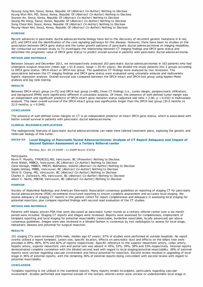
SSC04-03 Local Staging of Pancreatic Ductal Adenocarcinoma: Analysis of CT Report Adequacy and Impact ofSecond Opinion Assessment at a Tertiary Referral center
Monday, Nov. 28 10:50AM - 11:00AM Room: E353A
Hyoung Jung Kim, Seoul, Korea, Republic Of (Abstract Co-Author) Nothing to DiscloseKyung Won Kim, MD, Seoul, Korea, Republic Of (Abstract Co-Author) Nothing to DiscloseSoyeon An, Seoul, Korea, Republic Of (Abstract Co-Author) Nothing to DiscloseSeung-Mo Hong, Seoul, Korea, Republic Of (Abstract Co-Author) Nothing to DiscloseSong Cheol Kim, Seoul, Korea, Republic Of (Abstract Co-Author) Nothing to DiscloseMyung-Hwan Kim, Seoul, Korea, Republic Of (Abstract Co-Author) Nothing to Disclose
PURPOSE
Recent advances in pancreatic ductal adenocarcinoma biology have led to the discovery of recurrent genetic mutations in K-ras,p53, and DPC4 and the identification of the core signaling pathways for this disease. However, there have been no studies on theassociation between DPC4 gene status and the tumor growth patterns of pancreatic ductal adenocarcinoma on imaging modalities.We conducted our present study to To investigate the relationship between CT imaging findings and DPC4 gene status anddetermine the prognostic value of DPC4 gene status to predict overall survival in patients with pancreatic ductal adenocarcinoma.
METHOD AND MATERIALS
Between January and December 2011, we retrospectively analyzed 163 pancreatic ductal adenocarcinomas in 163 patients who hadundergone surgical resection (mean age = 61.8 years; range = 35–81 years). We divided the study patients into 2 groups accordingto DPC4 gene status: DPC4-intact or DPC4-lost group. The qualitative CT findings were analyzed by two reviewers. Theassociations between the CT imaging findings and DPC4 gene status were evaluated using univariate anlaysis and multivariatelogistic regression analysis. Overall survival was compared between the DPC4-intact and DPC4-lost group using Kaplan-Meieranalysis and log-rank testing.
RESULTS
Between DPC4-intact group (n=75) and DPC4-lost group (n=88), three CT findings (i.e., tumor margin, peripancreatic infiltration,and background IPMN) were significantly different in univariate analysis. Of these, the presence of well-defined tumor margin wasan independent and significant predictor of DPC4-intact gene mutation status (adjusted odd ratio = 2.06; p = 0.032) in multivariateanalysis. The mean overall survival of the DPC4-intact group was significantly longer than the DPC4-lost group (30.0 months vs22.0 months; p = 0.049).
CONCLUSION
The presence of well-defined tumor margins on CT is an independent predictor of intact DPC4 gene status, which is associated withbetter overall survival in patients with pancreatic ductal adenocarcinoma.
CLINICAL RELEVANCE/APPLICATION
The radiogenomic features of pancreatic ductal adenocarcinoma can make more tailored treatment plans, exploring the genetic andmolecular biology of this tumor.
ParticipantsKevin P. Murphy, FFR(RCSI),MD, Vancouver, BC (Presenter) Nothing to DiscloseAnne Walsh, MBBCh, Vancouver, BC (Abstract Co-Author) Nothing to DiscloseCarol Donagh, MBBCh, MRCPI, Ballinasloe, Ireland (Abstract Co-Author) Nothing to DiscloseSajida Ishtiaq, MBBS, Vancouver, BC (Abstract Co-Author) Nothing to DiscloseSilvia D. Chang, MD, Vancouver, BC (Abstract Co-Author) Nothing to DiscloseCharles V. Zwirewich, MD, Vancouver, BC (Abstract Co-Author) Nothing to DiscloseAlison C. Harris, MBChB, Vancouver, BC (Abstract Co-Author) Nothing to Disclose
PURPOSE
Society of Abdominal Radiology and American Pancreatic Association consensus guidelines on reporting of staging CT for pancreaticductal adenocarcinoma (PDA) recommend structured reporting to ensure complete assessment and accurate local staging. Weassess adequacy of staging CT reports in this patient cohort for report completeness and adequacy in assessing local staging forpotential resection, plus compare reported findings with second read evaluation of the CT studies.
METHOD AND MATERIALS
Patients with biopsy proven PDA that were discussed at pancreatic tumor rounds at a tertiary referral center over a six-monthperiod were included. Staging CT reports and images were reviewed. Reports were assessed for completeness, employment oftemplate reporting and local staging for potential resectability (resectable, borderline resectable, locally advanced) per aboveconsensus guidelines. Images were also reviewed in a blinded fashion in consensus by two radiologists to assess for local stage,metastatic disease and potential for surgical resection.
RESULTS
101 staging CTs were reviewed (55% male, median age 67 years). 67% of studies were performed at outside hospitals. No reportovertly utilized a report template. Lesion size, specific location, effects on pancreatic duct and effects on the biliary tree wereprovided in 89%, 96%, 87% and 82% of reports respectively. Specific reference to the superior mesenteric artery, celiac artery,hepatic artery, superior mesenteric vein and portal vein was absent in 45%, 53%, 59%, 38% and 53% respectively. Internal reportsdemonstrated complete correlation with the blinded second read with regard to local staging/potential resectability. 28% of externalreports were unclear regarding vascular involvement and hence potential for resection. Second review resulted in upgrading of localstage in 36% of external reports, with the remaining 36% of external reports being concordant with second review with regard topotential resectability.
CONCLUSION
Template reporting is not utilized in the examined reports. Many reports remain incomplete, particularly regarding vascularinvolvement. Studies performed and reported outside of the tertiary referral center were unclear or underestimate local stage in

SSC04-04 Additive Value of MRI with Diffusion-Weighted Imaging to MDCT for the Characterization of FocalLiver Lesions in Patients with Potentially Resectable Pancreatic Cancer
Monday, Nov. 28 11:00AM - 11:10AM Room: E353A
SSC04-05 Evaluating Qualitative Differences Between Liver Metastasis and Microabscess in Bile Duct andPancreas Carcinoma With Bile Duct Dilation in EOB-MRI
Monday, Nov. 28 11:10AM - 11:20AM Room: E353A
almost two thirds of cases.
CLINICAL RELEVANCE/APPLICATION
Accurate CT reporting of pancreatic ductal adenocarcinoma is essential in selecting patients that are potentially resectable.
ParticipantsSunkyung Jeon, MD, Seoul, Korea, Republic Of (Presenter) Nothing to DiscloseJeong Min Lee, MD, Seoul, Korea, Republic Of (Abstract Co-Author) Grant, Guerbet SA; Support, Siemens AG; Grant, Bayer AG;Grant, General Electric Company; Grant, STARmed Co, Ltd; Grant, RF Medical Co, Ltd; Grant, Toshiba Corporation; Grant, SamsungMedical HealthcareIjin Joo, MD, Seoul, Korea, Republic Of (Abstract Co-Author) Nothing to DiscloseSu Joa Ahn, Seoul , Korea, Republic Of (Abstract Co-Author) Nothing to DiscloseMyoung Seok Lee, MD, Seoul, Korea, Republic Of (Abstract Co-Author) Nothing to DiscloseJoon Koo Han, MD, Seoul, Korea, Republic Of (Abstract Co-Author) Nothing to Disclose
PURPOSE
To investigate the added value of magnetic resonance imaging(MRI) with diffusion weighted imaging(DWI) to preoperative stagingMDCT for the characterization of focal liver lesion(FLL) in the staging work-up of patients with potentially resectable pancreaticductal adenocarcinoma(PDAC)
METHOD AND MATERIALS
This retrospective study was approved by institutional review board; informed consent requirement was waived. We included 215patients with pathologically proven PDACs who underwent pancreas protocol MDCT and MRI with DWI, with histologic or clinicalreference standard confirmation of FLLs. Local resectability of PDACs were evaluated on MDCT (resectable, borderline resectable,unresectable) and characteristics of FLLs were scored as benign(including no FLL), indeterminate, or metastases on the MDCT setand combined MDCT and MRI with DWI set in separate sessions. Per-patient detection rate of hepatic metastasis unsuspected byMDCT and the additional diagnostic yield of MRI with DWI for FLLs were assessed.
RESULTS
Among 159 patients who were assessed to have a potentially resectable PDAC on MDCT(resectable, n=83; borderline resectable,n=76), 14 patients(8.81%, 14/159) were confirmed to have hepatic metastases and the other 145 as having benign-liver. RegardingFLLs, patients were classified as benign, indeterminate, or metastases in 128, 20, and 11 on MDCT and 142, 1, and 16 on thecombined set. In MDCT-benign group(n=128), there were three hepatic metastases assessed as combined set-metastases (2.3%,3/128). In MDCT-indeterminate(n=20), 10%(2/20) were scored as metastases and 90%(18/20) as benign on the combined set,which were finally confirmed. Among MDCT-metastasis(n=11), one case(1/11, 9.1%) was combined set-benign and confirmed asbenign. The combined set yielded significantly less indeterminate FLLs(1 vs. 20, p=<.001) and showed significantly greatersensitivity for the diagnosis of hepatic metastases(100%, 14/14 vs. 64.3%, 9/14; P=.008) than the MDCT set.
CONCLUSION
In potentially resectable PDAC patients, addition of MRI with DWI to MDCT would provide a more confident diagnosis for FLLs andincrease sensitivity for hepatic metastases than MDCT alone.
CLINICAL RELEVANCE/APPLICATION
The addition of MRI with DWI to MDCT would be useful in the characterization of focal liver lesions as either benign or metastases,especially for MDCT-indeterminate cases, thereby helping to determine operation candidates among patients with PDACs
ParticipantsTakuro Horikoshi, MD, Chiba, Japan (Presenter) Nothing to DiscloseYu Kawashima, Chiba City, Japan (Abstract Co-Author) Nothing to DiscloseAkiyo Ishige, Chiba City, Japan (Abstract Co-Author) Nothing to DiscloseHiroki Mukai, Chiba, Japan (Abstract Co-Author) Nothing to DiscloseHazuki Takishima, MD, Chiba, Japan (Abstract Co-Author) Nothing to DiscloseHiroaki Shimizu, Chiba, Japan (Abstract Co-Author) Nothing to DiscloseTakashi Uno, Chiba, Japan (Abstract Co-Author) Nothing to Disclose
PURPOSE
Pancreas head carcinoma or bile duct carcinoma tend to complicate bile duct dilatation, cholangitis and liver microabscess, and it isoften difficult to differentiate microabscess and metastasis. We evaluated the qualitative differences between metastasis andmicroabscess with bile duct dilatation due to bile duct carcinoma and pancreatic carcinoma in diffusion-weighted imaging (DWI),apparent diffusion coefficient (ADC) map and hepatobiliary phase (HBP) of Gd-EOB-DTPA magnetic resonance imaging (MRI).
METHOD AND MATERIALS
We retrospectively analyzed patients with bile duct dilatation due to carcinoma who had liver lesions which were difficult todifferentiate microabscess or metastasis in preoperative dynamic CT and Gd-EOB-DTPA MRI. 31 patients were included (21 males,10 females, mean age 66.7 years, range 46-87). Each nodule was diagnosed as metastasis or microabscess by pathologicaldiagnosis or clinical follow-up for 1 year. Two radiologists retrospectively reviewed in consensus DWI, arterial phase and HBP ofMRI. The signal intensity of the nodule on DWI, and ADC map, existence of A-P shunt around lesion, and wedge-shaped low signalintensity around lesion in HBP were evaluated. Pearson's chi-square test was performed in this study.
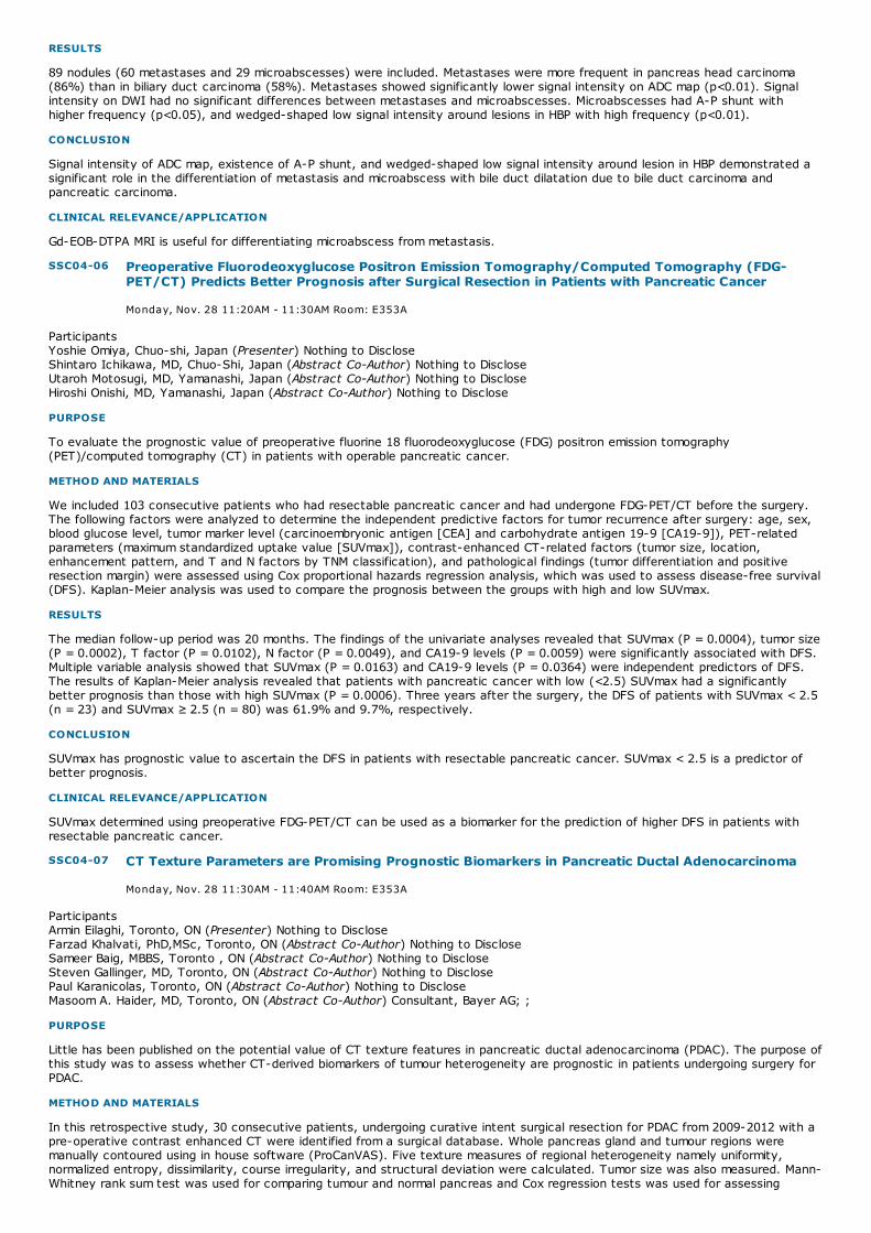
SSC04-06 Preoperative Fluorodeoxyglucose Positron Emission Tomography/Computed Tomography (FDG-PET/CT) Predicts Better Prognosis after Surgical Resection in Patients with Pancreatic Cancer
Monday, Nov. 28 11:20AM - 11:30AM Room: E353A
SSC04-07 CT Texture Parameters are Promising Prognostic Biomarkers in Pancreatic Ductal Adenocarcinoma
Monday, Nov. 28 11:30AM - 11:40AM Room: E353A
RESULTS
89 nodules (60 metastases and 29 microabscesses) were included. Metastases were more frequent in pancreas head carcinoma(86%) than in biliary duct carcinoma (58%). Metastases showed significantly lower signal intensity on ADC map (p<0.01). Signalintensity on DWI had no significant differences between metastases and microabscesses. Microabscesses had A-P shunt withhigher frequency (p<0.05), and wedged-shaped low signal intensity around lesions in HBP with high frequency (p<0.01).
CONCLUSION
Signal intensity of ADC map, existence of A-P shunt, and wedged-shaped low signal intensity around lesion in HBP demonstrated asignificant role in the differentiation of metastasis and microabscess with bile duct dilatation due to bile duct carcinoma andpancreatic carcinoma.
CLINICAL RELEVANCE/APPLICATION
Gd-EOB-DTPA MRI is useful for differentiating microabscess from metastasis.
ParticipantsYoshie Omiya, Chuo-shi, Japan (Presenter) Nothing to DiscloseShintaro Ichikawa, MD, Chuo-Shi, Japan (Abstract Co-Author) Nothing to DiscloseUtaroh Motosugi, MD, Yamanashi, Japan (Abstract Co-Author) Nothing to DiscloseHiroshi Onishi, MD, Yamanashi, Japan (Abstract Co-Author) Nothing to Disclose
PURPOSE
To evaluate the prognostic value of preoperative fluorine 18 fluorodeoxyglucose (FDG) positron emission tomography(PET)/computed tomography (CT) in patients with operable pancreatic cancer.
METHOD AND MATERIALS
We included 103 consecutive patients who had resectable pancreatic cancer and had undergone FDG-PET/CT before the surgery.The following factors were analyzed to determine the independent predictive factors for tumor recurrence after surgery: age, sex,blood glucose level, tumor marker level (carcinoembryonic antigen [CEA] and carbohydrate antigen 19-9 [CA19-9]), PET-relatedparameters (maximum standardized uptake value [SUVmax]), contrast-enhanced CT-related factors (tumor size, location,enhancement pattern, and T and N factors by TNM classification), and pathological findings (tumor differentiation and positiveresection margin) were assessed using Cox proportional hazards regression analysis, which was used to assess disease-free survival(DFS). Kaplan-Meier analysis was used to compare the prognosis between the groups with high and low SUVmax.
RESULTS
The median follow-up period was 20 months. The findings of the univariate analyses revealed that SUVmax (P = 0.0004), tumor size(P = 0.0002), T factor (P = 0.0102), N factor (P = 0.0049), and CA19-9 levels (P = 0.0059) were significantly associated with DFS.Multiple variable analysis showed that SUVmax (P = 0.0163) and CA19-9 levels (P = 0.0364) were independent predictors of DFS.The results of Kaplan-Meier analysis revealed that patients with pancreatic cancer with low (<2.5) SUVmax had a significantlybetter prognosis than those with high SUVmax (P = 0.0006). Three years after the surgery, the DFS of patients with SUVmax < 2.5(n = 23) and SUVmax ≥ 2.5 (n = 80) was 61.9% and 9.7%, respectively.
CONCLUSION
SUVmax has prognostic value to ascertain the DFS in patients with resectable pancreatic cancer. SUVmax < 2.5 is a predictor ofbetter prognosis.
CLINICAL RELEVANCE/APPLICATION
SUVmax determined using preoperative FDG-PET/CT can be used as a biomarker for the prediction of higher DFS in patients withresectable pancreatic cancer.
ParticipantsArmin Eilaghi, Toronto, ON (Presenter) Nothing to DiscloseFarzad Khalvati, PhD,MSc, Toronto, ON (Abstract Co-Author) Nothing to DiscloseSameer Baig, MBBS, Toronto , ON (Abstract Co-Author) Nothing to DiscloseSteven Gallinger, MD, Toronto, ON (Abstract Co-Author) Nothing to DisclosePaul Karanicolas, Toronto, ON (Abstract Co-Author) Nothing to DiscloseMasoom A. Haider, MD, Toronto, ON (Abstract Co-Author) Consultant, Bayer AG; ;
PURPOSE
Little has been published on the potential value of CT texture features in pancreatic ductal adenocarcinoma (PDAC). The purpose ofthis study was to assess whether CT-derived biomarkers of tumour heterogeneity are prognostic in patients undergoing surgery forPDAC.
METHOD AND MATERIALS
In this retrospective study, 30 consecutive patients, undergoing curative intent surgical resection for PDAC from 2009-2012 with apre-operative contrast enhanced CT were identified from a surgical database. Whole pancreas gland and tumour regions weremanually contoured using in house software (ProCanVAS). Five texture measures of regional heterogeneity namely uniformity,normalized entropy, dissimilarity, course irregularity, and structural deviation were calculated. Tumor size was also measured. Mann-Whitney rank sum test was used for comparing tumour and normal pancreas and Cox regression tests was used for assessing
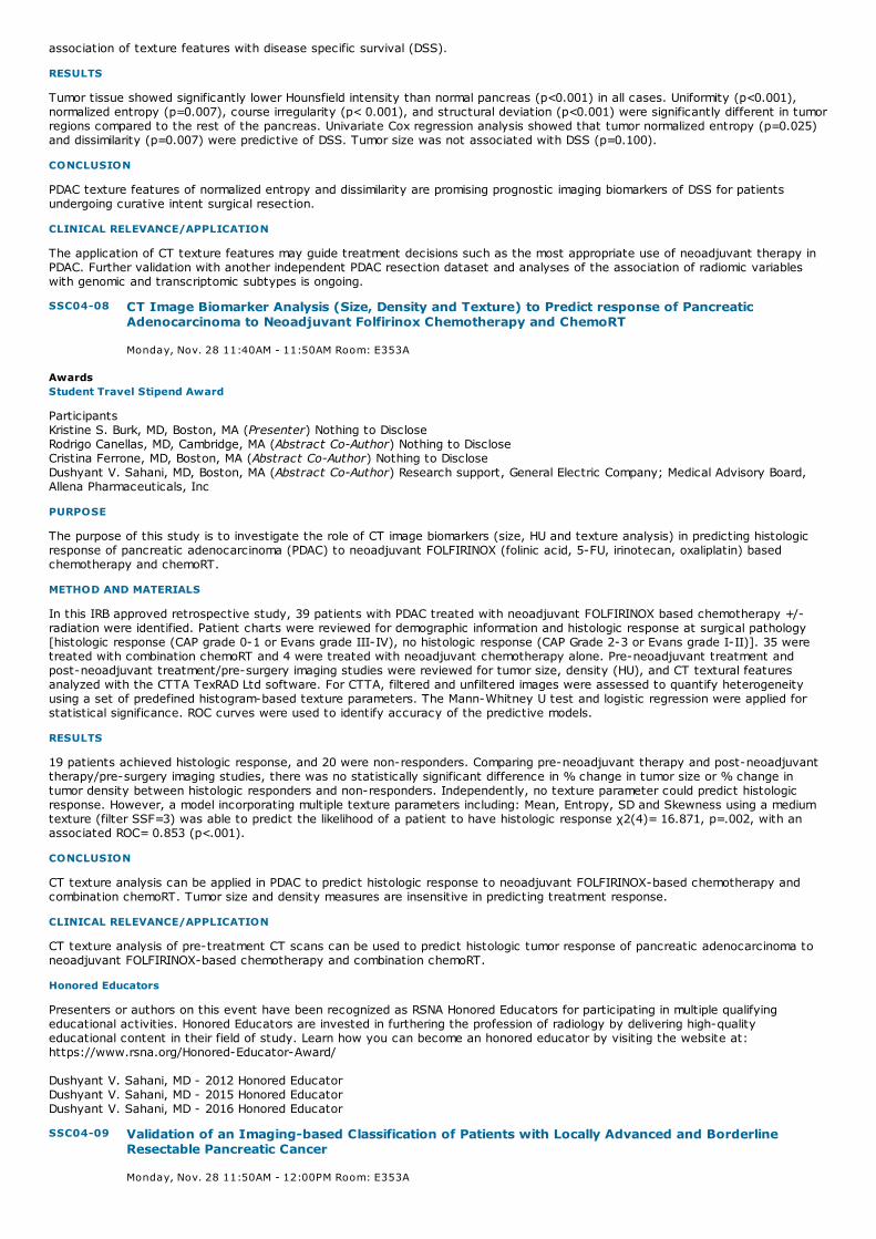
SSC04-08 CT Image Biomarker Analysis (Size, Density and Texture) to Predict response of PancreaticAdenocarcinoma to Neoadjuvant Folfirinox Chemotherapy and ChemoRT
Monday, Nov. 28 11:40AM - 11:50AM Room: E353A
SSC04-09 Validation of an Imaging-based Classification of Patients with Locally Advanced and BorderlineResectable Pancreatic Cancer
Monday, Nov. 28 11:50AM - 12:00PM Room: E353A
association of texture features with disease specific survival (DSS).
RESULTS
Tumor tissue showed significantly lower Hounsfield intensity than normal pancreas (p<0.001) in all cases. Uniformity (p<0.001),normalized entropy (p=0.007), course irregularity (p< 0.001), and structural deviation (p<0.001) were significantly different in tumorregions compared to the rest of the pancreas. Univariate Cox regression analysis showed that tumor normalized entropy (p=0.025)and dissimilarity (p=0.007) were predictive of DSS. Tumor size was not associated with DSS (p=0.100).
CONCLUSION
PDAC texture features of normalized entropy and dissimilarity are promising prognostic imaging biomarkers of DSS for patientsundergoing curative intent surgical resection.
CLINICAL RELEVANCE/APPLICATION
The application of CT texture features may guide treatment decisions such as the most appropriate use of neoadjuvant therapy inPDAC. Further validation with another independent PDAC resection dataset and analyses of the association of radiomic variableswith genomic and transcriptomic subtypes is ongoing.
AwardsStudent Travel Stipend Award
ParticipantsKristine S. Burk, MD, Boston, MA (Presenter) Nothing to DiscloseRodrigo Canellas, MD, Cambridge, MA (Abstract Co-Author) Nothing to DiscloseCristina Ferrone, MD, Boston, MA (Abstract Co-Author) Nothing to DiscloseDushyant V. Sahani, MD, Boston, MA (Abstract Co-Author) Research support, General Electric Company; Medical Advisory Board,Allena Pharmaceuticals, Inc
PURPOSE
The purpose of this study is to investigate the role of CT image biomarkers (size, HU and texture analysis) in predicting histologicresponse of pancreatic adenocarcinoma (PDAC) to neoadjuvant FOLFIRINOX (folinic acid, 5-FU, irinotecan, oxaliplatin) basedchemotherapy and chemoRT.
METHOD AND MATERIALS
In this IRB approved retrospective study, 39 patients with PDAC treated with neoadjuvant FOLFIRINOX based chemotherapy +/-radiation were identified. Patient charts were reviewed for demographic information and histologic response at surgical pathology[histologic response (CAP grade 0-1 or Evans grade III-IV), no histologic response (CAP Grade 2-3 or Evans grade I-II)]. 35 weretreated with combination chemoRT and 4 were treated with neoadjuvant chemotherapy alone. Pre-neoadjuvant treatment andpost-neoadjuvant treatment/pre-surgery imaging studies were reviewed for tumor size, density (HU), and CT textural featuresanalyzed with the CTTA TexRAD Ltd software. For CTTA, filtered and unfiltered images were assessed to quantify heterogeneityusing a set of predefined histogram-based texture parameters. The Mann-Whitney U test and logistic regression were applied forstatistical significance. ROC curves were used to identify accuracy of the predictive models.
RESULTS
19 patients achieved histologic response, and 20 were non-responders. Comparing pre-neoadjuvant therapy and post-neoadjuvanttherapy/pre-surgery imaging studies, there was no statistically significant difference in % change in tumor size or % change intumor density between histologic responders and non-responders. Independently, no texture parameter could predict histologicresponse. However, a model incorporating multiple texture parameters including: Mean, Entropy, SD and Skewness using a mediumtexture (filter SSF=3) was able to predict the likelihood of a patient to have histologic response χ2(4)= 16.871, p=.002, with anassociated ROC= 0.853 (p<.001).
CONCLUSION
CT texture analysis can be applied in PDAC to predict histologic response to neoadjuvant FOLFIRINOX-based chemotherapy andcombination chemoRT. Tumor size and density measures are insensitive in predicting treatment response.
CLINICAL RELEVANCE/APPLICATION
CT texture analysis of pre-treatment CT scans can be used to predict histologic tumor response of pancreatic adenocarcinoma toneoadjuvant FOLFIRINOX-based chemotherapy and combination chemoRT.
Honored Educators
Presenters or authors on this event have been recognized as RSNA Honored Educators for participating in multiple qualifyingeducational activities. Honored Educators are invested in furthering the profession of radiology by delivering high-qualityeducational content in their field of study. Learn how you can become an honored educator by visiting the website at:https://www.rsna.org/Honored-Educator-Award/
Dushyant V. Sahani, MD - 2012 Honored EducatorDushyant V. Sahani, MD - 2015 Honored EducatorDushyant V. Sahani, MD - 2016 Honored Educator
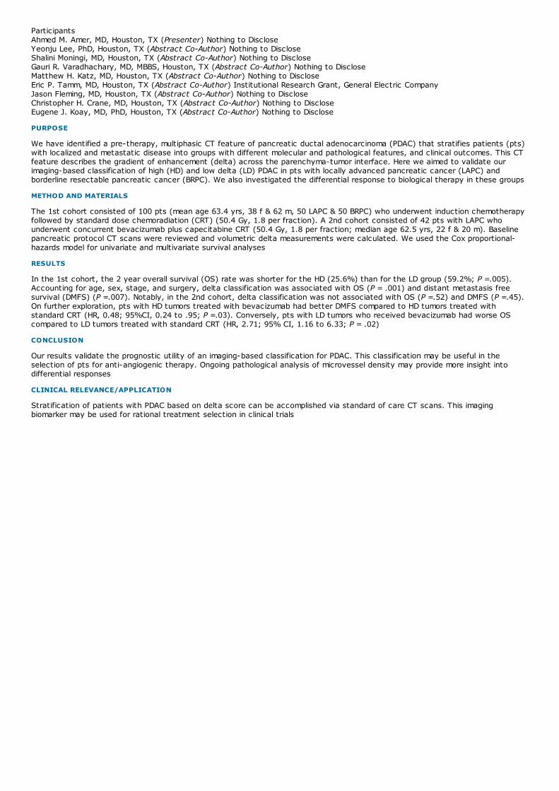
ParticipantsAhmed M. Amer, MD, Houston, TX (Presenter) Nothing to DiscloseYeonju Lee, PhD, Houston, TX (Abstract Co-Author) Nothing to DiscloseShalini Moningi, MD, Houston, TX (Abstract Co-Author) Nothing to DiscloseGauri R. Varadhachary, MD, MBBS, Houston, TX (Abstract Co-Author) Nothing to DiscloseMatthew H. Katz, MD, Houston, TX (Abstract Co-Author) Nothing to DiscloseEric P. Tamm, MD, Houston, TX (Abstract Co-Author) Institutional Research Grant, General Electric CompanyJason Fleming, MD, Houston, TX (Abstract Co-Author) Nothing to DiscloseChristopher H. Crane, MD, Houston, TX (Abstract Co-Author) Nothing to DiscloseEugene J. Koay, MD, PhD, Houston, TX (Abstract Co-Author) Nothing to Disclose
PURPOSE
We have identified a pre-therapy, multiphasic CT feature of pancreatic ductal adenocarcinoma (PDAC) that stratifies patients (pts)with localized and metastatic disease into groups with different molecular and pathological features, and clinical outcomes. This CTfeature describes the gradient of enhancement (delta) across the parenchyma-tumor interface. Here we aimed to validate ourimaging-based classification of high (HD) and low delta (LD) PDAC in pts with locally advanced pancreatic cancer (LAPC) andborderline resectable pancreatic cancer (BRPC). We also investigated the differential response to biological therapy in these groups
METHOD AND MATERIALS
The 1st cohort consisted of 100 pts (mean age 63.4 yrs, 38 f & 62 m, 50 LAPC & 50 BRPC) who underwent induction chemotherapyfollowed by standard dose chemoradiation (CRT) (50.4 Gy, 1.8 per fraction). A 2nd cohort consisted of 42 pts with LAPC whounderwent concurrent bevacizumab plus capecitabine CRT (50.4 Gy, 1.8 per fraction; median age 62.5 yrs, 22 f & 20 m). Baselinepancreatic protocol CT scans were reviewed and volumetric delta measurements were calculated. We used the Cox proportional-hazards model for univariate and multivariate survival analyses
RESULTS
In the 1st cohort, the 2 year overall survival (OS) rate was shorter for the HD (25.6%) than for the LD group (59.2%; P =.005).Accounting for age, sex, stage, and surgery, delta classification was associated with OS (P = .001) and distant metastasis freesurvival (DMFS) (P =.007). Notably, in the 2nd cohort, delta classification was not associated with OS (P =.52) and DMFS (P =.45).On further exploration, pts with HD tumors treated with bevacizumab had better DMFS compared to HD tumors treated withstandard CRT (HR, 0.48; 95%CI, 0.24 to .95; P =.03). Conversely, pts with LD tumors who received bevacizumab had worse OScompared to LD tumors treated with standard CRT (HR, 2.71; 95% CI, 1.16 to 6.33; P = .02)
CONCLUSION
Our results validate the prognostic utility of an imaging-based classification for PDAC. This classification may be useful in theselection of pts for anti-angiogenic therapy. Ongoing pathological analysis of microvessel density may provide more insight intodifferential responses
CLINICAL RELEVANCE/APPLICATION
Stratification of patients with PDAC based on delta score can be accomplished via standard of care CT scans. This imagingbiomarker may be used for rational treatment selection in clinical trials
SSC05-01 Dual Contrast Liver Imaging with Photon Counting CT: A New Approach for K-edge Imaging
Monday, Nov. 28 10:30AM - 10:40AM Room: E451A
SSC05-02 The Separation of Simultaneously Administered Intravascular and Oral X-ray Contrast Agents UsingSpectral CT: Pre-clinical Examples of Pseudo-cloaking with High-Z Materials
Monday, Nov. 28 10:40AM - 10:50AM Room: E451A
SSC05
Gastrointestinal (Dual-Energy CT)
Monday, Nov. 28 10:30AM - 12:00PM Room: E451A
CT GI
AMA PRA Category 1 Credits ™: 1.50ARRT Category A+ Credits: 1.50
ParticipantsBenjamin M. Yeh, MD, San Francisco, CA (Moderator) Research Grant, General Electric Company; Author with royalties, OxfordUniversity Press; Shareholder, Nextrast, Inc; Avinash R. Kambadakone, MD, Boston, MA (Moderator) Nothing to DiscloseLaura R. Carucci, MD, Midlothian, VA (Moderator) Nothing to Disclose
Sub-Events
ParticipantsDaniela Muenzel, MD, Munich, Germany (Presenter) Nothing to DiscloseHeiner Daerr, DIPLPHYS, Hambrg, Germany (Abstract Co-Author) Employee, Koninklijke Philips NVRoland Proksa, Hamburg, Germany (Abstract Co-Author) Employee, Koninklijke Philips NVAlexander A. Fingerle, MD, Munchen, Germany (Abstract Co-Author) Nothing to DisclosePhilippe C. Douek, MD, PhD, Lyon, France (Abstract Co-Author) Nothing to DiscloseFranz Pfeiffer, Munich, Germany (Abstract Co-Author) Nothing to DiscloseErnst J. Rummeny, MD, Munich, Germany (Abstract Co-Author) Nothing to DisclosePeter B. Noel, PhD, Munich, Germany (Abstract Co-Author) Nothing to Disclose
PURPOSE
To determine the feasibility of spectral photon-counting computed tomography (SPCCT) with K-edge depending materialdecomposition for dual contrast enhanced liver imaging.
METHOD AND MATERIALS
SPCCT was simulated with two different contrast agents (CA) at the time point where CA1 is in portal-venous contrast and CA2 isin arterial contrast phase. Material decomposition provided iodine and gadolinium maps which are calculated from the spectralinformation of SPCCT. Characteristic liver lesions (hemangioma, hepatocellular carcinoma, cyst, metastasis) have been inserted intothe simulation.
RESULTS
With SPCCT and an optimized contrast injection protocol, it becomes feasible to provide contrast-enhanced images with arterialdistribution of CA2 (gadolinium) and portal-venous phase of CA1 (iodine) in a single CT scan. The four inserted liver lesions wereclearly visible, and the characteristic patterns of contrast enhancement was seen in arterial and portal-venous images.
CONCLUSION
Our results reveal the possibilities to improve liver diagnostics by employing the combination of dual contrast protocols with SPCCTimaging. In addition to the potential dose reduction motion artifacts in-between acquisitions are eliminated.
CLINICAL RELEVANCE/APPLICATION
Dual contrast PCCT allows for simultaneous visualization of arterial and portal venous contrast enhancement, with reduced radiationdose, in a single acquisition.
ParticipantsTodd C. Soesbe, PhD, Dallas, TX (Presenter) Nothing to DiscloseMatthew A. Lewis, PhD, Dallas, TX (Abstract Co-Author) Research collaboration, CMR Naviscan CorporationKhaled A. Nasr, PhD, Dallas, TX (Abstract Co-Author) Nothing to DiscloseRobert E. Lenkinski, PhD, Dallas, TX (Abstract Co-Author) Research Grant, Koninklijke Philips NV; Research Consultant, AspectImaging;
PURPOSE
To show that the Spectral CT property of pseudo-cloaking (i.e., the masking or hiding of certain high-Z elements) can be used todifferentiate iodine-based intravascular (IV) contrast agents from tungsten-based oral contrast agents when administeredsimultaneously. Unlike iodine and barium, the contrast-enhanced bowel wall and bowel lumen can be imaged independently usingiodine and tungsten.
METHOD AND MATERIALS
Four female Fischer rats (n = 4) averaging 150 g mass were fasted for 24 hours. The rats were then given 4 mL of oral contrastbased on either barium (12 mg Ba/mL, FDA approved BaSO4) or tungsten (20 mg W/mL). The tungsten contrast was from atungsten carbide (WC) nanopowder colloidal suspension in methylcellulose. 30 minutes after oral contrast administration the rats
SSC05-03 Which Dual Energy CT Virtual Monochromatic keV Reconstruction is best to Simulate Typical kVpSettings at Standard CT?
Monday, Nov. 28 10:50AM - 11:00AM Room: E451A
SSC05-04 Determining the Origin of Periampullary Carcinoma Using Spectral Curves in Dual-energy Spectral CTImaging
Monday, Nov. 28 11:00AM - 11:10AM Room: E451A
were given a 0.3 mL dose of Isovue-370 (iopamidol) via tail vein injection, euthanized with CO2 within 2 minutes after injection,then immediately imaged on a detection-based Spectral CT scanner (IQon, Philips Healthcare). The axial scans used 120 kVp with a0.2 mm in-plane resolution. The spectrally derived images or SDIs (e.g., virtual non-contrast and iodine map) were analyzed usingthe thin-client Spectral Diagnostic Suite (SpDS, Philips Healthcare).
RESULTS
Both the IV (iodine) and oral (barium or tungsten) contrast agents appear in the conventional images as highly attenuatingmaterials. The IV contrast is most noticeable in the kidneys (nephrographic/late phase) and the oral contrast is most noticeable inthe stomach and small intestine. In the virtual non-contrast (VNC) images both the iodine and the barium are removed, but thetungsten is not affected. Conversely, in the iodine maps both the iodine and barium appear while the tungsten disappears (i.e., ispseudo-cloaked). We obtained similar results with phantoms on the Siemens dual-source scanners (SOMATOM Flash and Force).
CONCLUSION
Certain high-Z elements appear pseudo-cloaked in Spectral CT iodine maps, meaning they have negative pixel values and can bedifferentiated from iodine. This K-edge based phenomena includes ytterbium (Z=70) through platinum (Z=78) and provides a richpalette for the potential development of future Spectral CT contrast agents.
CLINICAL RELEVANCE/APPLICATION
Unlike barium, tungsten-based oral contrast agents can be differentiated from iodine, and provide complete segmentation betweenthe bowel wall and lumen (useful for bowel ischemia and Crohn’s disease).
ParticipantsYuxin Sun, BS,MSc, San Francisco, CA (Presenter) Nothing to DiscloseJack Lambert, PhD, San Francisco, CA (Abstract Co-Author) Nothing to DiscloseZhen J. Wang, MD, Hillsborough, CA (Abstract Co-Author) Stockholder, Nextrast, IncMichael A. Ohliger, MD, PhD, San Francisco, CA (Abstract Co-Author) Nothing to DiscloseBenjamin M. Yeh, MD, San Francisco, CA (Abstract Co-Author) Research Grant, General Electric Company; Author with royalties,Oxford University Press; Shareholder, Nextrast, Inc;
PURPOSE
To determine which virtual monochromatic (VMC) keV reconstruction of a rapid kV switching dual energy CT (rsDECT) scan givesthe closest CT numbers (HU) to standard kVp settings.
METHOD AND MATERIALS
We euthanized 10 rabbits 5 minutes after intravenous injection of 600 mg iodine/kg intravenous iohexol, then rapidly serially imagedthem on a rsDECT scanner with dual energy technique and again at single energy with 80, 100, 120, and 140 kVp tube potentials.Axial 1.25 mm images were then viewed on an Advantage Windows workstation to place 10 regions of interest (ROIs) on each ofthe following tissues: fat, muscle, vertebral body bone, liver, and aorta. ROIs were copied to all image sets to record the CTnumber (HU) at each kVp (2000 ROIs), and for each kVp we then recorded the VMC keV that gave the closest matching HU value.After determining the overall closest matching VMC keV for each kVp setting, ROIs were then obtained from each tissue at eachkVp and the corresponding "best" VMC keV.
RESULTS
For fat, muscle, bone, liver, and aorta, the keV that provided the closest HU values to 80 kVp images were 56, 55, 56, 54 and 56,respectively; for 100 kVp, the keV were 62, 60, 62, 60, and 61; for 120 kVp the keV were 67, 65, 67, 65, and 65; and for 140 kVpthe keV were 70, 70, 72, 69, and 70, respectively. Overall, the closest matches for 80, 100, 120, and 140 kVp CT numbers were55, 61, 66, and 70 keV, respectively. At these closest keVs, CT number standard deviations of 2.7, 2.5, 1.7, 18, and 11 HU for fat,muscle, bone, liver, and aorta, respectively, were seen compared to the corresponding kVp HU value.
CONCLUSION
For rsDECT, VMC reconstructions at 55, 61, 66, and 70 keV most closely approximate 80, 100, 120, or 140 kVp single energy CTimages, respectively, across a range of tissues in an intravenous contrast-enhanced body CT scan. Nevertheless, standarddeviations of up to 2.7 and 18 HU difference may be seen for soft tissues and bone, respectively, between a given kVp setting andits corresponding keV reconstruction.
CLINICAL RELEVANCE/APPLICATION
Our data provides the necessary bridge to allow rsDECT to provide quantitative comparisons between rsDECT and prior single kVpCT scans, and may allow rsDECT access to previously established conventional CT thresholds for the diagnosis various diseaseentities.
ParticipantsChenglong Ren, Shanxi, China (Presenter) Nothing to DiscloseMa Guangming, MMed, Xianyang City, China (Abstract Co-Author) Nothing to DiscloseRongqiang Zhang, Xianyang, China (Abstract Co-Author) Nothing to DiscloseXirong Zhang, Xianyang, China (Abstract Co-Author) Nothing to DiscloseQi Yang, Xianyang, China (Abstract Co-Author) Nothing to DiscloseFabao Gao, MD, PhD, Chengdu, China (Abstract Co-Author) Nothing to Disclose
SSC05-05 The Study of Quantitation of Hepatic Iron Deposition with Dual-energy CT on a Rabbit Model
Monday, Nov. 28 11:10AM - 11:20AM Room: E451A
SSC05-06 Can Non-enhanced Dual-energy Spectral CT Scans Be Used to Differentiate Small Liver Cysts fromSmall Metastatic Tumors?
Monday, Nov. 28 11:20AM - 11:30AM Room: E451A
PURPOSE
To investigate the value of using the spectral curve in dual-energy Spectral CT imaging for differentiating periampullary cancersoriginated from distal cholangiocarcinoma (DCA), duodenal adenocarcinoma (DA) and pancreatic adenocarcinoma (PA).
METHOD AND MATERIALS
In total, 55 patients with obstructive jaundice were imaged in the portal venous phase using dual-energy Spectral CT imaging modewith fast kVp switching. These patients were confirmed by surgical operation to have periampullary cancers that were originatedfrom distal cholangiocarcinoma (DCA, n=14), duodenal adenocarcinomas (DA, n=17) and pancreatic adenocarcinomas (PA, n=24).Virtual monochromatic images with energies from 40keV to 140keV were generated and analyzed on an Advanced Workstation 4.6.A circular region of interest (ROI) was place at the center of the lesion on the portal phase images to measure the CT value,avoiding the dilated common bile duct. To ensure the consistency, all measurements were performed 3 times in consecutive slicesto calculate the average values. The ROI measurements were propagated to all energy levels to generate the spectral HU curve:CT number as function of energy. The spectral HU curve slopes were calculated using the CT values from 40 to 80 keV: slope =[CTnumber (40keV) - CTnumber (80kev)]/40. The slopes for lesions of different origins were compared using the one-way ANOVAtest (p<0.05 was considered to have significant difference).
RESULTS
The slopes of the spectral curves were 3.54±0.89 for distal cholangiocarcinomas, 1.52±0.62 for duodenal adenocarcinomas and1.04±0.67 for pancreatic adenocarcinomas in portal phase. There were significant differences in the slope of the spectral HU curvesof periampullary lesions between any two groups (p<0.05): DCA vs DA (P < 0.001 ) ; DCA vs PA (P < 0.001 ) ; DA vs PA (P = 0.04 ).
CONCLUSION
The slopes of the spectral HU curve in Spectral CT imaging are different for periampullary carcinoma originated from DCA, DA or PA.Quantitative analysis of the spectral HU curve in Spectral CT in the portal venous phase may be used to determine the origins ofperiampullary carcinoma.
CLINICAL RELEVANCE/APPLICATION
For the obstructive jaundice patients, spectral CT imaging was a promising method to differentiate the origin of periampullarycarcinoma.
ParticipantsTao Li, nanning, China (Presenter) Nothing to Disclose
PURPOSE
To explore the feasibility and accuracy of liver iron quantification with virtual iron concentration(VIC)imaging based on dual energyCT(DECT)
METHOD AND MATERIALS
Ninety-six rabbits were randomly divided into liver iron overload experiment group,validation group,iron overload and fatty liverexperiment group,validation group. All rabbits underwent intramuscular injection of iron dextran 15 mg / kg from 1 to 16 week;thelast two groups were fed with high fat diet additionally. DECT and MR were used for all rabbits. Liver iron concentration (LIC),liverfat concentration (LFC) , HE and Prussian blue staining were examed.
RESULTS
CT numbers of 80kVp, 120kVp, 140kVp,VIC imaging and R2* in liver iron overload group positively linearly correlated with LIC(r=0.900,0.837,0.719,0.895 and 0.935,respectively); linear regression equation for LIC calculated from VIC was y=0.31x-0.48(F=129.7,P<0.01). CT numbers of VIC in the validation group were substituted into the equation to predicte LIC. Meandifference between predicted LIC and actual LIC was 0.21 mg/g. CT numbers of 80kVp, 120kVp, 140kVp,VIC imaging and R2* in theiron overload and fatty liver group positively linearly correlated with LIC(r = -0.376,-0.531,-0.618,0.924 and 0.942,respectively);linear regression equation for LIC calculated from VIC was y=0.30x+0.23(F=265.4,P<0.001). Mean difference between predictedLIC and actual LIC was 0.19 mg/g. The difference of slope and intercept of two equations has no statistical significance(F=0.020,P>0.05;F=0.203, P>0.05).Therefore,the data of two experiment groups were merged for ROC analysis. At a LIC threshold of 3.2mg/g, the corresponding optimal threshold of VIC was 13.7 HU, and diagnostic sensitivity, specificity and AUC were 80.43%,100%and 0.961. At a LIC threshold of 7.0 mg/g, the optimal threshold of VIC was 22.0 HU, and diagnostic sensitivity, specificity and AUCwere 95.65%,90.24% and 0.968.
CONCLUSION
Virtual iron concentration imaging on DECT shows potential ability to accurately quantify liver iron accumulation in the iron overloadrabbit model,without being affected by fatty liver.
CLINICAL RELEVANCE/APPLICATION
DECT is using for non-invasive quantitation of hepatic iron deposition
ParticipantsMa Guangming, MMed, Xianyang City, China (Presenter) Nothing to DiscloseHaifeng Duan, Xianyang City, China (Abstract Co-Author) Nothing to DiscloseChenglong Ren, Shanxi, China (Abstract Co-Author) Nothing to Disclose
SSC05-07 Clinical Value of Spectral CT Imaging in Preoparative Evaluation on Histo-Differentiation of RectalAdenocarcinoma
Monday, Nov. 28 11:30AM - 11:40AM Room: E451A
SSC05-08 Prototype Detection-based Spectral CT Scanner Derived Virtual Non-Contrast Attenuation ValuesCompare Favorably to Attenuation Values on Unenhanced Images
Dong Han, MA, Xianyang, China (Abstract Co-Author) Nothing to DiscloseJing Chen, Xianyang City, China (Abstract Co-Author) Nothing to DiscloseZhanli Ren, Xianyang, China (Abstract Co-Author) Nothing to DiscloseYongjun Jia, MMed, Xianyang City, China (Abstract Co-Author) Nothing to DiscloseTian Xin, MMed, Xianyang City, China (Abstract Co-Author) Nothing to Disclose
PURPOSE
To assess the clinical value of the non-enhanced dual-energy spectral CT scans in differentiating small hepatic cysts from smallmetastatic tumors.
METHOD AND MATERIALS
43 patients with small liver lesions (27 cases with liver cysts; 16 cases with metastases) who underwent the non-enhancedspectral CT scans were included. In total, there were 104 lesions (≤1.5cm), including 71 small liver cysts and 33 small livermetastases, were identified. The gemstone spectral imaging (GSI) Viewer software on the AW4.6 workstation was used to measurethe CT number on the 40 keV virtual monochromatic images and the water concentration on the water (fat) material decompositionimages. The curve slope for the spectral curve and the effective-Z values of lesions were also calculated. The above parametersfor the two types of lesions were analyzed using independent sample t test, and ROC curve analysis was used to evaluate thediagnostic efficiency.
RESULTS
The CT values at 40 keV, water concentration, curve slope and Effective-Z for liver cyst were -14.77±18.24HU,897.51±118.68g/L,-1.14±0.47 and 7.24±0.19, respectively; while the corresponding values for small metastases were15.39±24.87HU, 1101.53±129.11g/L, -0.33±0.46 and7.43±0.18, respectively. The differences for these parameters between thetwo lesion types were statistically significant (all p<0.05). The sensitivity and specificity for differentiating small liver cyst fromsmall metastasis with the CT number of images at 40keV was 75.8% and 76.1.1%, respectively. These values were improved to84.8% and 95.8%, respectively using water concentration in the lesions.
CONCLUSION
The parameters obtained in the non-enhanced dual-energy spectral CT scans demonstrated appreciable clinical values fordifferentiating small liver cysts from small metastases, with the water concentration on the water and fat-based materialdecomposition images providing the highest diagnostic performance.
CLINICAL RELEVANCE/APPLICATION
The non-enhanced dual-energy spectral CT scans may be used to differentiating small liver cysts from small metastases with highdiagnostic performance.
ParticipantsChuan-bin Wang, Hefei, China (Presenter) Nothing to DiscloseFei Gao, Hefei, China (Abstract Co-Author) Nothing to DiscloseDong Jiangning, Hefei, China (Abstract Co-Author) Nothing to Disclose
PURPOSE
To discuss the value of the spectral CT parameters in the preoperative evaluation on histo-differentiation of rectaladenocarcinoma.
METHOD AND MATERIALS
Totally 90 patients with rectal adenocarcinoma confirmed by pathology underwent dual-phase enhanced spectral CTscan. Monochromatic images with energy levels from 40 to 140 keV was generated and the slope Kvalues were computed. AndIodine concentrations were derived from iodine-based material-decomposition CT images and normalized to the iodine concentrationin the aorta(NIC). According to the results of pathology, the 90 patients were divided into poorly, moderately and welldifferentiated adenocarcinoma groups. Using the ROC curve to evaluate the differentiation diagnosis efficiency of NIC and slope K inrectal adenocarcinoma respectively.
RESULTS
The cases in well differentiated adenocarcinoma, moderately differentiated adenocarcinoma and poorly differentiatedadenocarcinoma were 22, 50 and 18, respectively. The iodine concentration, NIC and slope K value were statistically differenceboth in the arterial and venous phase (P<0.05). According to the ROC curve, the diagnostic value of NIC was close to the slope K,in the arterial phase. The sensitivity and specificity of slope K were 77% and 79%, respectively, which were similar to that of NIC(76% and 74%, respectively). In the venous phase, slope K was associated with higher sensitivity(86%) compared to that ofNIC(77%), but similar specificity( 72% vs 70%).
CONCLUSION
The spectral CT can provide a new imaging method for evaluating the histo-differentiation of rectal adenocarcinoma inpreoperative.
CLINICAL RELEVANCE/APPLICATION
The spectral CT can provide a new imaging method for evaluating the histo-differentiation of rectal adenocarcinoma inpreoperative.
Monday, Nov. 28 11:40AM - 11:50AM Room: E451A
SSC05-09 Comparison of Single-source Split-filter Dual-energy Abdominal CT with Single-energy Abdominal CTusing Automatic Tube Voltage Modulation: Assessment of Image Quality and Radiation Dose
Monday, Nov. 28 11:50AM - 12:00PM Room: E451A
ParticipantsLakshmi Ananthakrishnan, MD, Dallas, TX (Presenter) Nothing to DiscloseRichard W. Ahn, MD, PhD, Dallas, TX (Abstract Co-Author) Co-founder, ViXa LLC; Stockholder, Vixa LLCPrabhakar Rajiah, MD, FRCR, Dallas, TX (Abstract Co-Author) Institutional Research Grant, Koninklijke Philips NV; Speaker, KoninklijkePhilips NVNegin Rassouli, MD, Cleveland, OH (Abstract Co-Author) Institutional Grant support, Koninklijke Philips NVYin Xi, Dallas, TX (Abstract Co-Author) Nothing to DiscloseSuhny Abbara, MD, Dallas, TX (Abstract Co-Author) Author, Reed Elsevier; Editor, Reed Elsevier; Institutional research agreement,Koninklijke Philips NV; Institutional research agreement, Siemens AGTodd C. Soesbe, PhD, Dallas, TX (Abstract Co-Author) Nothing to DiscloseMatthew A. Lewis, PhD, Dallas, TX (Abstract Co-Author) Research collaboration, CMR Naviscan CorporationJohn R. Leyendecker, MD, Dallas, TX (Abstract Co-Author) Nothing to DiscloseRobert E. Lenkinski, PhD, Dallas, TX (Abstract Co-Author) Research Grant, Koninklijke Philips NV; Research Consultant, AspectImaging;
PURPOSE
A prototype detection-based spectral CT scanner allows generation of virtual non-contrast (VNC) images from contrast enhancedacquisitions, but it is unknown if the resulting HU correspond to those on unenhanced images. We aim to correlate the HU resultingfrom detection-based spectral CT VNC to those from unenhanced images.
METHOD AND MATERIALS
This multi-institutional prospective study is IRB approved. 84 pairs of unenhanced and virtual non contrast (VNC) datasets werereconstructed in patients scanned on a prototype detection-based spectral CT scanner (IQon, Philips Healthcare). Regions ofinterest (ROI) were drawn on identical locations of unenhanced and VNC images in the liver, spleen, renal cortex, abdominal aorta,right psoas muscle, and subcutaneous fat, and HU values were recorded. Descriptive statistics for mean HU per ROI werecalculated for different positions and phases. Equivalence testing was performed to determine if unenhanced and VNC HU were“equivalent”, defined as <5/10/15 HU difference. Interobserver variability was determined.
RESULTS
1476 ROIs were evaluated. The mean attenuation difference between unenhanced and VNC was less than 15 HU in 92.7%, lessthan 10 HU in 75.2%, and less than 5 HU in 44.4% of all measurements. When using a threshold of 10 HU difference, equivalencetesting demonstrated the unenhanced and VNC HU values to be equivalent in most tissues except fat. When divided by organ,difference in unenhanced and VNC was less than 10 HU in 81.7% of liver, 85.4% of psoas, and 81.8% of renal cortexmeasurements. HU in subcutaneous fat was overestimated by approximately 10 HU on all phases.
CONCLUSION
VNC data derived from contrast enhanced acquisitions on a prototype detection-based spectral CT scanner provide HU valuessimilar to unenhanced scans in most tissues except fat. Further study is needed to determine if attenuation thresholds currentlyused clinically for common pathology should be adjusted for spectral derived VNC images, particularly when fat/lipid is likely to bepresent.
CLINICAL RELEVANCE/APPLICATION
Reconstructed VNC images on a prototype detection-based spectral CT scanner have the potential to allow retrospectivecharacterization of incidental findings.
Honored Educators
Presenters or authors on this event have been recognized as RSNA Honored Educators for participating in multiple qualifyingeducational activities. Honored Educators are invested in furthering the profession of radiology by delivering high-qualityeducational content in their field of study. Learn how you can become an honored educator by visiting the website at:https://www.rsna.org/Honored-Educator-Award/
Suhny Abbara, MD - 2014 Honored EducatorPrabhakar Rajiah, MD, FRCR - 2014 Honored Educator
ParticipantsAndre Euler, MD, Basel, Switzerland (Presenter) Nothing to DiscloseMarkus M. Obmann, Basel, Switzerland (Abstract Co-Author) Nothing to DiscloseZsolt Szucs-Farkas, MD, PhD, Berne, Switzerland (Abstract Co-Author) Nothing to DiscloseBram Stieltjes, MD, Basel, Switzerland (Abstract Co-Author) Nothing to DiscloseDavid J. Winkel, MD, Basel, Switzerland (Abstract Co-Author) Nothing to DiscloseCaroline Zaehringer, Basel, Switzerland (Abstract Co-Author) Nothing to DiscloseAnna L. Falkowski, Basel, Switzerland (Abstract Co-Author) Nothing to DiscloseSebastian T. Schindera, MD, Basel, Switzerland (Abstract Co-Author) Research Grant, Siemens AG; Research Grant, Ulrich GmbH &Co KG; Research Grant, Bayer AG; Speakers Bureau, Bayer AG
PURPOSE
To assess the radiation dose and image quality of single-source split-filter dual-energy abdominal CT compared with single-energyCT using automatic tube voltage modulation (ATVM).
METHOD AND MATERIALS
In a retrospective study, 200 portal-venous phase abdominal CT scans were reviewed which were acquired on a single-source CT

scanner (SOMATOM AS+, Siemens) either with single-energy mode with ATVM (CarekV, Siemens) and 130 ref. mAs or with dual-energy split-filter technology (TwinBeam, Siemens) at AuSn120 kVp and 420 ref. mAs. CT numbers (HU) were obtained in varyingorgans. Image noise and contrast-to-noise ratio (CNR) for parenchymal and vascular structures were assessed. Subjective imagequality was evaluated by three radiologists independently. Radiation dose was estimated by size-specific dose estimate (SSDE).Descriptive statistics and Mann-Whitney-U-test were used.
RESULTS
The two patient groups showed no significant difference in water equivalent diameter (29.2 cm vs. 29.0 cm, respectively; P=0.79).In the single-energy group, ATVM selected 100 kVp in 78 patients and 120 kVp in 22 patients. Image noise was 18% lower withdual-energy compared with single-energy (8.4 HU vs. 10.2 HU, respectively; P<.0001). Parenchymal CNR was significantly higherwith dual-energy (24.9 vs 22.0, respectively; P=.007), whereas vascular CNR was significantly lower with dual-energy comparedwith single-energy (3.5 vs 5.2, respectively; P<.0001). Subjective image noise was graded superior and image contrast inferior inthe dual-energy group (P<.0001). However, there was no significant difference in diagnostic confidence between the two groups(P=.78). SSDE was 11% lower using dual-energy technique (12.6 mGy vs. 11.2 mGy, respectively; P=.02).
CONCLUSION
Single-source dual-energy CT with split-filter technology enables abdominal dual-energy scans at significantly lower image noiseand radiation dose compared with ATVM without impairing subjective diagnostic confidence.
CLINICAL RELEVANCE/APPLICATION
Dual-energy CT with split-filter technology has the potential to add information such as material decomposition without additionalradiation penalty compared with single-energy CT with ATVM.
GI355-SD-MOA1
Usefulness of Double Frequency MR Elastography for Assessment of the Necroinflammation on LiverStiffness Measurements
Station #1
GI356-SD-MOA2
Amide Proton Transfer Imaging to Predict Tumor Response after Preoperative Chemotherapy inLocally Advanced Rectal Cancer
Station #2
GIS-MOA
Gastrointestinal Monday Poster Discussions
Monday, Nov. 28 12:15PM - 12:45PM Room: GI Community, Learning Center
GI
AMA PRA Category 1 Credit ™: .50
ParticipantsElena K. Korngold, MD, Portland, OR (Moderator) Nothing to Disclose
Sub-Events
ParticipantsMinori Onoda, PhD,RT, Kanazawa, Japan (Presenter) Nothing to DiscloseTomoko Hyodo, MD, Osaka-Sayama, Japan (Abstract Co-Author) Nothing to DiscloseTakamichi Murakami, MD, PhD, Osakasayama, Japan (Abstract Co-Author) Nothing to DiscloseMasakatsu Tsurusaki, MD, PhD, Osaka, Japan (Abstract Co-Author) Nothing to DiscloseTakahiro Sakamoto, Osakasayama, Japan (Abstract Co-Author) Nothing to DiscloseTosiaki Miyati, PhD, Kanazawa, Japan (Abstract Co-Author) Nothing to Disclose
PURPOSE
The measurement of liver stiffness with magnetic resonance elastography (MRE) is affected by the liver viscosity which can befound by testing different vibration frequency settings. We aimed to evaluate the utility of double vibration frequency MRE methodin estimation of liver fibrosis and necroinflammation.
METHOD AND MATERIALS
Thirty patients underwent MRE with a 1.5-T MR system and liver biopsy for liver investigation. We performed MRE with two differentdriver frequencies of 60 and 80 Hz to measure the liver stiffness. The frequencies of motion encoding gradient were set as thesame as driver frequencies (60, 80 Hz) to optimize MR pulse sequences for MRE. We defined the difference of liver stiffness valuesas ⊿G (G80Hz - G60Hz) to compare with pathological findings of liver fibrosis (stages F0-4) and necroinflammation (grades A0-3).Statistical analysis included Spearman’s rank correlation test, followed by ROC analysis and AUC calculation when significantcorrelation exists. A P value <.05 was considered statistical significant.
RESULTS
The mean liver stiffness values with both 60 Hz and 80 Hz increased as fibrosis stages and necroinflammation grades progressed.For all patients, the measurements of liver stiffness by using vibration frequency of 80 Hz showed higher values than those of 60Hz. ⊿G showed moderate positive correlation with liver necroinflammation grades (r=0.68, P<0.01). No significant correlation wasobserved between ⊿G and pathological fibrosis stages (r=0.30, P=0.11). In ROC analyses, the optimal cutoff values of ⊿G were0.36 for ≧A1 (AUC 0.80, sensitivity 79%, specificity 100%), 0.90 for ≧A2 (AUC 0.81, sensitivity 55%, specificity 100%), and 1.58for ≧A3 (AUC 0.96, sensitivity 100%, specificity 83 %).
CONCLUSION
⊿G increased as necroinflammation grades progressed, but not as fibrosis stages. ⊿G may be useful index for evaluating the livernecroinflammation on liver stiffness measurements by MRE.
CLINICAL RELEVANCE/APPLICATION
The double vibration frequency MRE method has a potential to evaluate the liver necroinflammation without biopsy, and may beuseful to avoid the overestimation of liver fibrosis using MRE.
ParticipantsAkihiro Nishie, MD, Fukuoka, Japan (Presenter) Nothing to DiscloseYoshiki Asayama, MD, Fukuoka, Japan (Abstract Co-Author) Nothing to DiscloseKousei Ishigami, MD, Iowa City, IA (Abstract Co-Author) Nothing to DiscloseYasuhiro Ushijima, MD, Fukuoka, Japan (Abstract Co-Author) Nothing to DiscloseYukihisa Takayama, MD, Fukuoka, Japan (Abstract Co-Author) Research Grant, FUJIFILM Holdings CorporationHiroshi Honda, MD, Fukuoka, Japan (Abstract Co-Author) Nothing to DiscloseDaisuke Okamoto, MD, Fukuoka City, Japan (Abstract Co-Author) Nothing to DiscloseNobuhiro Fujita, MD, PhD, Fukuoka, Japan (Abstract Co-Author) Nothing to DiscloseOsamu Togao, MD, PhD, Fukuoka, Japan (Abstract Co-Author) Nothing to DiscloseKoji Sagiyama, MD, Fukuoka, Japan (Abstract Co-Author) Nothing to DiscloseDaisuke Tsurumaru, MD, Fukuoka, Japan (Abstract Co-Author) Nothing to DiscloseYuichiro Kubo, MD, Fukuoka, Japan (Abstract Co-Author) Nothing to DiscloseEiji Oki, Fukuoka, Japan (Abstract Co-Author) Nothing to DiscloseTatsuya Manabe, Fukuoka, Japan (Abstract Co-Author) Nothing to DiscloseJochen Keupp, PhD, Hamburg, Germany (Abstract Co-Author) Employee, Koninklijke Philips NV
GI357-SD-MOA3
Comparative Analysis of Imaging Feature and Pathology between Scirrhous Hepatocellular Carcinomaand Intrahepatic Mass-forming Cholangiocarcinoma
Station #3
GI358-SD-MOA4
Comparison Study on the Accuracy of Detecting Colorectal Cancer Lymph Nodes Metastasis withArterial Phase Dual-energy CT Spectral Curve and the Morphological Findings
Station #4
PURPOSE
To elucidate if amide proton transfer (APT) imaging can predict tumor response after preoperative chemotherapy in locallyadvanced rectal cancer
METHOD AND MATERIALS
A total of 17 patients with locally advanced rectal cancer who underwent MR examination including APT imaging and preoperativechemotherapy (at least two courses) were enrolled. APT imaging was scanned with single-shot 2D TSE-DRIVE on a 3T clinicalscanner using a 32-channel coil and 2-channel parallel transmission. APT signal was defined as MTR asymmetry (MTRasym) at theoffset of 3.5 ppm: {Ssat(−3.5 ppm)−Ssat(+3.5 ppm)}/S0(control). APT map was generated and a region-of-interest was carefullyplaced in each rectal cancer to measure APT signal (%). Each tumor resected was histologically evaluated for the degree ofdegeneration and necrosis and was divided into the five Grades (0, none; 1a, less than 1/3; 1b, 1/3 to 2/3; 2, more than 2/3; 3,all). The mean APT signal was compared between Grade 0/1a (Low response group) and Grade 1b/2/3 (High response group) usinga Student’s t-test. Diagnostic performance of APT signal, as well as apparent diffusion coefficient (ADC) obtained in diffusion-weighted imaging, for predicting tumor response was evaluated using receiver operating characteristics curve.
RESULTS
The mean APT signal of Low response group (n=12; 3.05+/-1.61%) was significantly higher than that of High response group (n=5;1.14+/-1.13%)(p=0.029). There was no significant difference in ADC between Low and High response groups. The area under curvefor predicting tumor response using APT signal was 0.867. When 2.75% or less was an indicator of high tumor response, sensitivity,specificity and accuracy of APT signal were 100%, 75% and 82.4%, respectively.
CONCLUSION
APT imaging can predict tumor response after preoperative chemotherapy in locally advanced rectal cancer. For predicting tumorresponse APT signal is superior to ADC.
CLINICAL RELEVANCE/APPLICATION
APT imaging contributes to the selection of a therapeutic strategy in locally advanced rectal cancer.
ParticipantsFeng Chen, Beijing, China (Presenter) Nothing to DiscloseDawei Zhao, MD, Beijing, China (Abstract Co-Author) Nothing to DiscloseHongjun Li, Beijing, China (Abstract Co-Author) Nothing to DiscloseJing Zhao, Beijing, China (Abstract Co-Author) Nothing to DiscloseShuo Wen, Beijing, China (Abstract Co-Author) Nothing to Disclose
PURPOSE
To investigate image findings and clinical applications of intrahepatic mass-forming cholangiocarcinoma(IMCC) and scirrhoushepatocellular carcinomas(SHCC).
METHOD AND MATERIALS
The CT and MRI findings of forty-eight patients with SHCC and ninety-five patients with IMCC proved by pathologic examinationwere analyzed. The group feature and difference were compared. Analyzed by Chi square test and Student’s t test.
RESULTS
The age of SHCC patients was younger than the patients with IMCC (t = -2.735,P = 0.007). There was no significant difference oftumor diameter between two groups (t = 1.328,P = 0.186) and SHCC was more common in chronic hepatitis patients (χ2 =10.810,P = 0.001). The tumors were frequently observed in the edge of liver with surface retraction (χ2 = 0.522,P = 0.470) andthere was no difference between groups. The enhanced CT and MRI imaging were divided into two types, typical enhancement andatypical enhancement which was observed normally. The probability of typical enhancement between IMCC and SHCC did not showsignificant difference (χ2 = 0.028,P = 0.876). Part of the atypical enhancement was partly similar between them when atypicalenhancement of IMCC was revealed more complicated and characteristic. Histopathologically, the dynamic contrast-enhancedimaging features were verified with the distribution, proportion of tumor cells and fibrous stroma, as well as with blood suppling anddegree of tumor necrosis. The incidence of portal vein invasion (χ2 = 0.358,P = 0.549) and lymph node metastasis (χ2 = 0.000,P= 0.982) was showed no difference, however bile duct dilatation in IMCC was revealed more frequently than SHCC (χ2 = 5.011,P =0.025).
CONCLUSION
Both of the SHCC and IMCC were primary malignant hepatic tumors with abundant fibrosis. The clinical and imaging findings of themwere similar. Full recognition of its imaging signs and combination consideration with clinical manifestations are of great significancefor its diagnosis and differentiation and provide a basis for rational treatment plan.
CLINICAL RELEVANCE/APPLICATION
Compared to common hepatocellular carcinoma, SHCC has the similar imaging features with IMCC. Both of them are often resistantto chemoembolization and hepatic surgery or microwave coagulation maybe the better choice. But IMCC has poorer prognosis.
ParticipantsXuejun Yang, kunming, China (Abstract Co-Author) Nothing to Disclose
GI359-SD-MOA5
Imaging Evaluation of Common Duct Size: Comparison between CT and US Measurements
Station #5
GI360-SD-MOA6
Texture based Classification and Correlation of Non-alcoholic Fatty Liver Disease Activity Score (NAS)using Gabor Wavelet Filters
Station #6
Wei Zhao, Kunming, China (Abstract Co-Author) Nothing to DiscloseJing Lei, Kunming, China (Abstract Co-Author) Nothing to DiscloseHan Dan, PhD, MD, Kunming, China (Abstract Co-Author) Nothing to DiscloseYaying Yang, Kunming, China (Presenter) Nothing to Disclose
PURPOSE
To explore the accuracy of arterial phase dual-energy CT spectral curve and the morphological findings in detecting the metastaticlymph nodes in colorectal cancer .
METHOD AND MATERIALS
22 patients with pathologically confirmed colorectal cancer had a total of 40 enlarged lymph nodes in the pelvic regionunderwent dual energy CT scan, We measured the size of the lymph nodes on CT images, and the CT value in non-enhanced andarterial phase CT scan; We also computed short-long diameter ratio and enhancement value of lymph node (lymph node CT value inarterial phase - CT value in plain scan). We observed the change trend of the spectrum curve and compared the diagnosticaccuracy in detecting colorectal cancer lymph nodes metastasis.
RESULTS
In the 40 lymph nodes, 17 lymph nodes were metastatic, while reactive hyperplasia was seen in 23 lymph nodes. Short-longdiameter ratio and the enhancement CT value in the diagnosis of lymph node metastasis had weak consistency with thepathological results (Kappa value=0.100 and 0.016, repsectively). The sensitivity of 64.7%, 35.3%, the specificity of 64.7%,68.2%, the area under the ROC curve were 0.434, 0.501 for short-long diameter ratio and the enhancement CT value in thediagnosis of lymph node metastasis, respectively. Dual-energy CT spectral curve had strong consistency with the pathologicalresults (Kappa value=0.899) and higher sensitivity of 94.1%, the specificity of 95.5%, and area under the ROC curve (0.949)compared with morphological findings.
CONCLUSION
The spectrum curve of dual-energy CT had higher sensitivity and specificity than the conventional morphology findings in diagnosisof colorectal cancer lymph nodes metastasis.
CLINICAL RELEVANCE/APPLICATION
The arterial phase dual-energy CT spectral curve had higher accuracy than the morphological findings in detecting the metastaticlymph nodes in colorectal cancer .
ParticipantsTatum A. Mcarthur, MD, Aurora, CO (Presenter) Nothing to DiscloseDesmin Milner, MD, Birmingham, AL (Abstract Co-Author) Nothing to DiscloseArgha Chatterjee, MBBS,MD, Chicago, IL (Abstract Co-Author) Nothing to DiscloseBradford Jackson, Birmingham, AL (Abstract Co-Author) Nothing to DiscloseMark E. Lockhart, MD, Birmingham, AL (Abstract Co-Author) Author, Oxford University Press
PURPOSE
To compare the common duct (CD) diameter by CT and ultrasound (US) in patients undergoing both studies within 48 hours of eachother.
METHOD AND MATERIALS
This IRB-approved, HIPAA-compliant retrospective study compared the CD diameter measured by both CT and US performed withina 48-hour period in 342 patients (ages 24 - 88) from 1/1/2004 - 4/1/2015. 132 patients were excluded secondary to complex biliarysurgery, acute cholecystectomy, liver transplant, CD stent, hepatobiliary malignancy, interval papillotomy, irretrievable images,and/or nonvisualization of the CD. In the remaining 210 patients, the CD was measured at the porta hepatis on US and CT inrandomized order by a blinded board-certified radiologist. CT readings were performed 6 months after US readings to limit recallbias. Presence of CD wall thickening was also recorded. 79% (n=166) of the CT exams were contrast-enhanced. Distributionalcharacteristics are presented as mean and standard deviation (SD). Bland-Altman analysis was used to estimate the bias and 95%limits of agreement between modalities. Paired T-tests were used to assess the statistical significance of the difference betweenmodalities.
RESULTS
The study population consisted of 113 women and 97 men with a mean age of 53.8 (+/-15.3) years. Mean CD diameter by CTmeasures 1.01±2.2mm more than US [p<0.0001]. The 95% confidence limit of agreement between the two modalities suggests CTmeasurements were 3.39 mm less to 5.42 mm more than that of US.
CONCLUSION
In our study, CT overestimated the CD diameter compared to US, and we observed a fixed bias of 1.01 mm.
CLINICAL RELEVANCE/APPLICATION
US is considered the reference standard to evaluate CD size. In our study, CT does overestimate the CD diameter when comparedto US, but not enough to typically change management, and US is not necessary to confirm duct size.
Participants
GI361-SD-MOA7
Quantitative Prediction of Treatment Effect of Transarterial Chemoembolization using Drug-elutingBead for Hepatocellular Carcinoma with use of Preoperative Dual-phase Contrast-enhanced CT
Station #7
GI362-SD- Diagnostic Value of Preoperative Abdominal CT Examination in Assessment of Quantitative Body
Rohit Badida, MS,BEng, Tempe, AZ (Abstract Co-Author) Nothing to DiscloseChristine M. Zwart, PhD, Scottsdale, AZ (Abstract Co-Author) Nothing to DiscloseAlvin C. Silva, MD, Scottsdale, AZ (Presenter) Nothing to Disclose
PURPOSE
Our main objective was to analyze texture features extracted from CT images of NAFLD patients and correlate with NAS score.Current non-invasive methods have limitations and cannot replace/assist liver biopsy for classifying Non-Alcoholic SteatoHepatitis(NASH) from fatty liver not diagnostic of NASH (steatosis). NAS scores of 0-3 occurred in cases largely considered not diagnosticof NASH, whereas scores of 5-8 were largely considered diagnostic of NASH (PMID:15915461).
METHOD AND MATERIALS
37 histologically diagnosed patients with NAFLD and their respective NAS scores were considered for the study (20 patients withNAS 5-8 and 17 with NAS 0-3). For extracting and analyzing textures, venous phase slices of contrast enhanced CT images withthe largest cross-sectional areas were selected followed by manual segmentation of region of interest (ROI). A MATLAB-based toolwas developed and utilized for extracting ROI, specifically avoiding larger blood vessels. This was followed by applying Gaborwavelet filters for feature extraction. Support Vector Machine was used to classify the extracted Gabor features and receiveroperating characteristics curve (AUROC) was used to calculate the performance of the classifier along with k-fold cross validation.
RESULTS
An AUC (Area under curve) of 0.88 was obtained for separating NASH versus fatty liver without NASH. A 5-fold cross validation on37 images (single ROI per patient) yielded a misclassification rate of 0.25 indicating excellent performance of the classifier.
CONCLUSION
Currently, there is no robustly validated CT technique for discriminating the spectrum of NAFLD. However, our results indicate thatGabor filters have the ability to classify fatty liver texture patterns for replacing/assisting NAS score. This also demonstrates thattexture features can be correlated to NAS score interpretation of NASH disease for a score of 5-8 and fatty liver without NASH fora score of 0-3.
CLINICAL RELEVANCE/APPLICATION
A noninvasive application applied to routine CT images that can differentiate steatosis from NASH in lieu or as a complement tobiopsy can have meaningful impact on the screening & management of this increasingly prevalent disease.
ParticipantsKeisuke Todoroki, Matsumoto, Japan (Presenter) Nothing to DiscloseAkira Yamada, MD, Matsumoto, Japan (Abstract Co-Author) Nothing to DiscloseTakanori Aonuma, Nagano, Japan (Abstract Co-Author) Nothing to DiscloseFumihito Ichinohe, Matsumoto, Japan (Abstract Co-Author) Nothing to DiscloseKazuki Oyama, matsumoto-shi, Japan (Abstract Co-Author) Nothing to DiscloseMana Nakamura, Matsumoto, Japan (Abstract Co-Author) Nothing to DiscloseYasunari Fujinaga, MD, Matsumoto, Japan (Abstract Co-Author) Nothing to DiscloseMasumi Kadoya, MD, Matsumoto, Japan (Abstract Co-Author) Nothing to Disclose
PURPOSE
To clarify the possibility predicting treatment effect of transarterial chemoembolization using drug-eluting bead (DEB-TACE) forhepatocellular carcinoma (HCC) with use of quantitative perfusion parameters obtained from preoperative dual-phase contrast-enhanced CT (DPCE-CT).
METHOD AND MATERIALS
Consecutive 16 patients with radiologically diagnosed HCC by DPCE-CT who underwent DEB-TACE were included in thisretrospective study. Arterial and portal venous input functions were estimated from CT values of 7 vessels (left atrium, abdominalaorta, right renal vein, superior mesenteric vein, inferior vena cava, splenic vein, and portal vein) using newly proposed dynamicphase index (DPI) technique. Perfusion parameters of the liver and HCC such as arterial and portal venous inflow velocity constant[K1a and K1p], arterial flow fraction [AF = K1a/(K1a + K1p)] were determined by compartment model analysis. Treatment effectwas evaluated within 8 weeks after DEB-TACE using contrast-enhanced CT or MRI. Predictive performance to classify completenecrosis group and the others using these perfusion parameters and Naïve Bayes classifier was statistically analyzed.
RESULTS
In univariate analysis, significant differences between complete necrosis group and the others were seen in AF (P = 0.032) and K1p(P = 0.036) of the liver, difference of K1p (P = 0.045) and AF (P = 0.046) of HCC against the liver. The area under the receiveroperating characteristic curve (AUROC) and its 95% confidence interval (95%CI) to classify complete necrosis group and the othersusing variable number of perfusion parameters and NBA were 0.850 (0.831, 0.868) using 1 parameter (AF of the liver), 0.931 (0.919,0.943) using 2 features (K1a of the liver and difference of AF of HCC against the liver), respectively. No significant increase inAUROC was observed even if more than 2 parameters were used.
CONCLUSION
Treatment effect of DEB-TACE for HCC can be predicted with use of quantitative perfusion parameters obtained from preoperativeDPCE-CT using DPI technique and compartment model analysis.
CLINICAL RELEVANCE/APPLICATION
Quantitative perfusion analysis of DPCE-CT using DPI technique will help determine the management of patients with HCC.
MOA8 Composition Analysis to Predict and Stratify Risk of Severe Pancreatic Fistula afterPancreatoduodenectomy
Station #8
GI108-ED-MOA9
Cross-Sectional Imaging in Celiac Disease: Uncomplicated, Refractory, Enteropathy-AssociatedLymphoma and Adenocarcinoma
Station #9
GI216-ED-MOA10
Pseudocirrhosis: Pathogenesis and Imaging Features
ParticipantsCammillo R. Talei Franzesi, Milan, Italy (Presenter) Nothing to DiscloseDavide Ippolito, MD, Monza, Italy (Abstract Co-Author) Nothing to DiscloseLuca Riva, MD, Missaglia, Italy (Abstract Co-Author) Nothing to DiscloseDavide Fior, MD, Monza, Italy (Abstract Co-Author) Nothing to DiscloseMarta Sandini, MD, Monza, Italy (Abstract Co-Author) Nothing to DiscloseLuca V. Gianotti, MD, Monza, Italy (Abstract Co-Author) Nothing to DiscloseSandro Sironi, MD, Monza, Italy (Abstract Co-Author) Nothing to Disclose
PURPOSE
To assess whether measures of abdominal fat distribution, visceral density, and antropometric parameters obtained from computedtomography (CT) may predict postoperative pancreatic fistula (POPF) occurrence in patient who undergo to pancreatodudenectomysurgery.
METHOD AND MATERIALS
We analyzed 117 patients who underwent pancreatoduodenectomy (PD) and had a preoperative CT scan staging. CT images wereobtained either with 16- or 256- MDCT (Brilliance iCT; Philips), applying a multiphase acquisition protocol (unenhanced, arterial,portal venous and equilibrium phase). CT reconstruction of unenhanced upper abdomen were used to obtain measures of total fatvolume (TFV), visceral fat volume (VFV), thickness of retro-renal fat, diameter of pancreatic duct and the density ratio of spleenand pancreas. The predictive ability of each parameter was investigated by receiver-operating characteristic (ROC) curvesmethodology and assessing optimal cutoff thresholds.
RESULTS
The overall rate of clinically relevant (grades B and C of ISGPF classification) POPF was 20.5% (24/117 patients). Areas under ROC-curves showed that none of the parameters was per se significantly predictive. Patient with grade A or no POPF had lower medianvalue of each CT parameters analyzed than the patients with grades B-C (i.e. VFV median value of 1800 cm3 vs 2702 cm3; TFV4100 cm3 vs 5432 cm3; Wirsung diameter 4 mm vs 2 mm). Moreover the multivariate analysis revealed that a VFV>2334 cm3,TFV>4408 cm3, pancreas/spleen density ratio <0.707, and pancreatic duct diameter <5 mm were predictive of POPF. The risk ofPOPF progressively increased with the number of the CT paramters involved and age.
CONCLUSION
The risk of occurrence of clinically relevant POPF may be predicted with relative accuracy by combining specific informationobtained from preoperative CT such as TFV and VFV, pancreas/spleen density, pancreatic duct diameter, in combination withpatient age. Even if it has been already suggested that obesity might be a handy predictor of complications after PD, thedistribution of fat is more important than obesity and probably the amount and specific organ deposition of visceral fat plays acentral role in this process.
CLINICAL RELEVANCE/APPLICATION
The organ fat distribution analysis, obtained at preoperative abdominal CT scan examination, may offer important information on therisk of post-surgical pancreatic fistula occurrence.
ParticipantsBadr Al-Bawardy, MD, Rochester, MN (Presenter) Nothing to DiscloseJohn M. Barlow, MD, Rochester, MN (Abstract Co-Author) Nothing to DiscloseRogerio Vasconcelos, MD, Rochester, MN (Abstract Co-Author) Nothing to DiscloseSarasa T. Kim, Rochester, MN (Abstract Co-Author) Nothing to DiscloseDavid Bruining, MD, Rochester, MN (Abstract Co-Author) Research Grant, Given Imaging Ltd Consultant, Bracco Group Stephanie Hansel, MD, Rochester, MN (Abstract Co-Author) Research support, Given Imaging Ltd Advisory Board, Medtronic, IncShannon P. Sheedy, MD, Rochester, MN (Abstract Co-Author) Nothing to DiscloseJeff L. Fidler, MD, Rochester, MN (Abstract Co-Author) Nothing to DiscloseJoel G. Fletcher, MD, Rochester, MN (Abstract Co-Author) Grant, Siemens AG; ;
TEACHING POINTS
1.Celiac disease (CD) affects 1% of the US population, and diagnosis is often delayed.2.CT/MR enterography (CT/MRE) of CD showdistinctive intestinal and extra-intestinal features that facilitate diagnosis.3.CT/MRE are used to detect complications such as typeI/type II refractory celiac disease (RCD), ulcerative jejunitis, enteropathy-associated lymphoma (EATL) and small boweladenocarcinoma (SBA).
TABLE OF CONTENTS/OUTLINE
Diagnosis: Non-specific sx: GI (diarrhea, bloating, weight loss) and extraintestinal (neuropsychiatric sx, arthritis, Fe deficiency,metabolic bone dz, infertility) Implications/limitations of TTG IgA and permissive genes (HLA DQ2/DQ8) Endoscopy - fold flattening,fissuring, scalloping and mosaic appearance. Histology: partial-total villous atrophy, intraepithelial lymphocytosis, and crypthyperplasia.CT/MRE findings (examples): Intestinal - decreased number of duodenal and jejunal folds, duodenal edema, increasednumber of ileal folds, excessive fluid, intussusception Extraintestinal - Hyposplenism, mesenteric vascular engorgement, and lymphnode enlargement.CD complications on CTE/MRE: Ulcerative jejunitis; small bowel dilation and increased small bowel thickness inRCD Refractory CD: EATL > SBA, but rare EATL patterns – aneurysmal, infiltrative, annular, gastricCase study quiz
Station #10
ParticipantsChristopher Czaplicki, Phoenix, AZ (Abstract Co-Author) Nothing to DiscloseChristine O. Menias, MD, Scottsdale, AZ (Presenter) Nothing to DiscloseVenkata S. Katabathina, MD, San Antonio, TX (Abstract Co-Author) Nothing to DiscloseAnil K. Dasyam, MD, Pittsburgh, PA (Abstract Co-Author) Book contract, Reed ElsevierAkram M. Shaaban, MBBCh, Salt Lake City, UT (Abstract Co-Author) Nothing to DiscloseAntonio Luna, MD, Jaen, Spain (Abstract Co-Author) Nothing to DiscloseKhaled M. Elsayes, MD, Ann Arbor, MI (Abstract Co-Author) Nothing to DiscloseNina Karlin, MD, Phoenix, AZ (Abstract Co-Author) Nothing to DiscloseAbimbola Adike, MD, Phoenix, AZ (Abstract Co-Author) Nothing to DiscloseElizabeth Carey, MD, Phoenix, AZ (Abstract Co-Author) Nothing to Disclose
TEACHING POINTS
1) Discuss the pathophysiology of Pseudocirrhosis- Due to the regression and response to chemotherapy of hepatic metastasis and as the progression with fibrosis surrounding the infiltrating hepatic tumorsA) Response of hepatic tumors to chemotherapy resultingin scarring and capsular retractionB) Fibrosis surrounding infiltrating hepatic metastatic massesC) Nodular regenerative hyperplasiain response to ischemia from chemotherapy-induced hepatic injuryD) Sinusoidal obstruction with venous obstruction as a result ofchemotherapy 2) Review the imaging Features of Pseudocirrhosis on CT and MR
TABLE OF CONTENTS/OUTLINE
Pseudocirrhosis is an imaging diagnosis used to describe the development of diffuse hepatic nodularity in the setting of hepaticmetastases successfully treated with chemotherapy. While it has been described in multiple types of cancer, it is most common inpatients with breast cancer. It is important to recognize pseudocirrhosis early as it can be associated with complications of portalhypertension, rapid progression to liver failure and increased morbidity in patients with metastatic cancer. This review will discussthe imaging features of this important but under recognized entity on CT and MR.
Honored Educators
Presenters or authors on this event have been recognized as RSNA Honored Educators for participating in multiple qualifyingeducational activities. Honored Educators are invested in furthering the profession of radiology by delivering high-qualityeducational content in their field of study. Learn how you can become an honored educator by visiting the website at:https://www.rsna.org/Honored-Educator-Award/
Christine O. Menias, MD - 2013 Honored EducatorChristine O. Menias, MD - 2014 Honored EducatorChristine O. Menias, MD - 2015 Honored EducatorChristine O. Menias, MD - 2016 Honored EducatorVenkata S. Katabathina, MD - 2012 Honored EducatorKhaled M. Elsayes, MD - 2014 Honored EducatorAkram M. Shaaban, MBBCh - 2015 Honored EducatorAkram M. Shaaban, MBBCh - 2016 Honored Educator
Transitioning to DR, Clinical and Financial Benefits Beyond Preventing Reimbursement Penalties: Supported byFujifilm (Invite only)
Monday, Nov. 28 12:30PM - 1:30PM Room: S403B
Participants
PARTICIPANTS
Jerry Thomas, MS, FAAPM, DABR, CHP, DABSNM Wichita, KSWilliam Tobin BS, Tyler, TX
PROGRAM INFORMATION
This course does not offer CME credit.
GI363-SD-MOB1
The Assessment of Radiation Dose and Image Quality by using Different Scan Parameter SelectionMethods with for Fast Kilo Voltage Switching Dual-energy CT (ssDECT) with Different ScanParameter Selection Methods
Station #1
GI364-SD-MOB2
The Diagnostic Performance of Shear-wave Elastography in Patients with Autoimmune Liver Disease
Station #2
GIS-MOB
Gastrointestinal Monday Poster Discussions
Monday, Nov. 28 12:45PM - 1:15PM Room: GI Community, Learning Center
GI
AMA PRA Category 1 Credit ™: .50
FDA Discussions may include off-label uses.
ParticipantsElena K. Korngold, MD, Portland, OR (Moderator) Nothing to Disclose
Sub-Events
Participantstsuyoshi kagimoto, hiroshima, Japan (Presenter) Nothing to DiscloseChisato Yoritaka, RT, Hiroshima, Japan (Abstract Co-Author) Nothing to DiscloseSatoshi Inada, hiroshima, Japan (Abstract Co-Author) Nothing to DiscloseMasashi Takahashi, RT, hiroshima, Japan (Abstract Co-Author) Nothing to DiscloseXiao Zhu Lin, MD, Shanghai, China (Abstract Co-Author) Nothing to Disclose
PURPOSE
The comparison of scan dose and image quality between DECT and 120kV helical scan mode (SECT) is important for DECT clinicalapplication. The purpose of this study iwas to assess radiation dose and image quality by using different scan parameter selectionmethods with for the fast kV switching dual-energy CT (DECT) with different scan parameter selection methods.
METHOD AND MATERIALS
50th abdomen dynamic scans were performed on 64detector DECT scanner (Revolution GSI, GE) using combination of 120kV helicaland fast kV (80/140kV) switching DECT techniques with in different scanning phases using different scan parameters:Plain scanwith 120kV and noise index (NI) of 12@5mm slice thickness,Arterial phase:GSI Assist by using automatically DECT scan parameterselection method (NI of 12@5mm), Portal vein phase:manual parameter selection using Max mA of mA table of SECT scan (NI of12@5mm).The CTDI (mGy),DLP (mGy-cm),ED (mSv) and Noise (SD) of three different scans compared between SECT and twoparameter selection methods of DECT.
RESULTS
The averages of CTDI with different scan mode and DECT parameter selection method were 12.2±4.8mGy at in Plain phase (120kV),12.8±4.2mGy at in Arterial phase (GSI assist) , 14.5±4.8mGy at in Portal vein phase (Max mA GSI). GSI assist produced similar CTDIas the 120kV scan, while the Max mA GSI had statistically higher CTDI than the 120kV scan there was a significant differencebetween 120kV and two GSI parameter selector methods. The averages of DLP were 374.0±164.2mGy-cm at 120kV,391.8±149.1mGy-cm at with GSI assist and, 443.6±169.0mGy-cm at with Max mA GSI, there was a no significant differencebetween 120kV and two GSI parameter selector methods. The averages of ED were 5.6±2.5mSv at 120kV, 5.9±2.2mSv at GSIassist and, 6.7±2.5mSv at Max mA GSI. The averages of Noise (SD) were 18.6±3.9HU at 120kV, 14.5±4.2HU at GSI assist and14.2±4.6HU at with Max mAs GSI there was a significant difference between 120kV and GSI assist and, between 120kV and Max mAGSI.
CONCLUSION
From this study, we can conclude that the automatically DECT scan parameter selection method (GSI Assist) has similar scan doseand image quality with as the 120kV helical and manual parameter selection methodscans.
CLINICAL RELEVANCE/APPLICATION
The automatically DECT scan parameter selection method (GSI assist) has similar scan DOSE and image quality with helical scan(SECT) and is very useful for clinicial application and clinical research of DECT.
ParticipantsChangtian Li, Beijing, China (Presenter) Nothing to DiscloseManish Dhyani, MBBS, Boston, MA (Abstract Co-Author) Nothing to DiscloseStephan D. Pratt, Boston, MA (Abstract Co-Author) Nothing to DiscloseAtul K. Bhan, MD, Boston, MA (Abstract Co-Author) Nothing to DiscloseAnthony E. Samir, MD, Boston, MA (Abstract Co-Author) Consultant, Pfizer Inc Consultant, General Electric Company Consultant,PAREXEL International Corporation Research Grant, Koninklijke Philips NV Research Grant, Siemens AG Research Grant, ToshibaCorporation Research Grant, General Electric Company Research Grant, Samsung Electronics Co, Ltd Research Grant, AnalogicCorporation Research support, SuperSonic Imagine Research support, Hitachi, Ltd
PURPOSE
(1) To assess shear wave elastography (SWE) performance for liver fibrosis staging in patients with autoimmune liver disease (ALD)(2) to validate a previously established Young’s modulus cut-off value in this new cohort.
GI365-SD-MOB3
Prediction of the Gastrointestinal Stromal Tumor Response to Second-line Sunitinib Therapy withApparent Diffusion Coefficient
Station #3
GI366-SD-MOB4
Can the Detection of Peritoneal Metastatic Disease be Improved using Virtual MonenergeticReconstructions of Contrast-enhanced Dual-energy CT Scans?
Station #4
METHOD AND MATERIALS
Patients with known or suspected ALD scheduled for ultrasound guided non-focal liver biopsy were included. SWE imaging wasperformed with the Aixplorer US system quipped with a convex broadband probe. Eight elastograms were obtained intercostally inthe right upper liver and the median value of the best 8 measurements was calculated. A single blinded pathologist staged liverfibrosis and calculated steatosis and necroinflammation scores using the METAVIR system. The Spearman correlation test wasperformed to assess correlation between estimated tissue Young’s modulus (eYM) and fibrosis, steatosis, necroinflammatory score,PBC stage and clinical data. The diagnostic performance of SWE for differentiating higher grades of fibrosis (F≥2) from lowergrades(F0, F1) was evaluated using ROC curve analysis.
RESULTS
51 subjects were enrolled. Fibrosis stage, necroinflammation score and PBC stage had moderate correlations with eYM, butsteatosis did not correlate with eYM. For all patients, AST, ALT and total bilirubin levels were positively correlated with eYM whileserum albumin was negatively correlated with eYM; for patients with PBC, AST and ALT had positive correlations with eYM;for patients with AIH, AST, ALT, Alk Phos and total bilirubin had positive correlations with eYM. The AUROC for differentiating F≥2from F0 and F1 was 0.781. Our previously established cut-off value of 7.29 kPa for a heterogenous population of subjects withdiffuse liver disease had sensitivity and specificity of 88.9% and 39.4% in this cohort. A cut-off value of 9.15kPa provided asensitivity of 83.3% and specificity of 72.7%,which also provided a sensitivity of 84.6% and specificity of 75% in the follow-upsubgroup and a sensitivity of 80% and specificity of 69.2% in the diagnostic subgroup.
CONCLUSION
Shear wave elastography is a novel, supplementary adjunct to liver biopsy in the evaluation of histological staging in patients withALD. A higher cutoff value for diagnosis of advanced fibrosis may yield superior diagnostic performance in these patients.
CLINICAL RELEVANCE/APPLICATION
Shear wave elastography is a supplementary adjunct to liver biopsy in the evaluation of histological staging in patients with ALD.
ParticipantsZheng Zhong, Chicago, IL (Presenter) Nothing to DiscloseLei Tang, MD, Beijing, China (Abstract Co-Author) Nothing to DiscloseMuge Karaman, PhD, Chicago, IL (Abstract Co-Author) Nothing to DiscloseYi Sui, PhD, Chicago, IL (Abstract Co-Author) Nothing to DiscloseYingshi Sun, Beijing, China (Abstract Co-Author) Nothing to DiscloseXiaohong J. Zhou, PhD, Chicago, IL (Abstract Co-Author) Nothing to Disclose
PURPOSE
To demonstrate the use of apparent diffusion coefficient (ADC) for early prediction of gastrointestinal stromal tumor (GIST)response to second-line sunitinib treatment.
METHOD AND MATERIALS
With the approval of the institutional review board, a total of 43 patients were enrolled in the study. All the patients underwentsunitinib treatment after imatinib resistance. MRI scans with multi-b-value diffusion-weighted imaging (DWI) were performed prior totreatment (baseline) and two weeks (for prediction of response) after initiating sunitinib treatment. Conventional MRI images attwelve weeks were used to determine the good (GoodR) and poor (PoorR) responders according to the established criteria. Theregions of interest (ROIs) were drawn on the b=1000 sec/mm² images in the solid tumor regions. Up to two ROIs (the largest ones)were selected from each patient, resulting in 36 GoodR and 27 PoorR ROIs. The ADC values were then calculated for each ROI byusing the mono-exponential diffusion model. The changes in the ADC values (ΔD) and the longest diameter of the lesion (ΔLD)before and after two weeks of treatment were compared between the two groups. A receiver operating characteristic (ROC)analysis was also performed to evaluate the response to sunitinib treatment.
RESULTS
Significant differences were detected in the change of ADC values (ΔD) (62.2% vs. 11.7%, p<0.001) between the two groups. ΔDprovided a significantly higher predictive power than ΔLD, producing higher AUC in the ROC analysis (80% vs. 69%).Thisperformance was further improved (AUC value of 82%) when ΔD was combined with ΔLD.
CONCLUSION
The change in ADC values two weeks into the treatment provides a better prediction of GIST response to sunitinib therapy thanthe change of tumor size.
CLINICAL RELEVANCE/APPLICATION
The change in ADC values after two weeks of therapy can be a useful marker to predict early response of GIST to sunitinibtreatment.
ParticipantsKathryn Darras, MD, Vancouver, BC (Presenter) Nothing to DiscloseSheldon J. Clark, MD, Vancouver, BC (Abstract Co-Author) Nothing to DiscloseSarah A. Barrett, MBBCh, Vancouver, BC (Abstract Co-Author) Nothing to DiscloseTriona M. Walshe, FFR(RCSI), Vancouver, BC (Abstract Co-Author) Nothing to Disclose
GI367-SD-MOB5
Hepatocellular Carcinoma treated with 90Yttrium Radioembolization: Value of Tumor StiffnessMeasured with MR Elastography for Assessment of Treatment Response
Station #5
Heejun Kang, MD, Vancouver, BC (Abstract Co-Author) Nothing to DiscloseLuck J. Louis, MD, Vancouver, BC (Abstract Co-Author) Nothing to DiscloseSilvia D. Chang, MD, Vancouver, BC (Abstract Co-Author) Nothing to DiscloseAlison C. Harris, MBChB, Vancouver, BC (Abstract Co-Author) Nothing to DiscloseMohammed F. Mohammed, MBBS, Vancouver, BC (Abstract Co-Author) Nothing to DiscloseSavvas Nicolaou, MD, Vancouver, BC (Abstract Co-Author) Institutional research agreement, Siemens AGPatrick D. McLaughlin, FFRRCSI, Vancouver, BC (Abstract Co-Author) Speaker, Siemens AG
PURPOSE
To evaluate the role of virtual monochromatic imaging (VMI) in the detection of peritoneal metastatic disease in contrast-enhancedcomputed tomography (CT) of the abdomen and pelvis and to compare this technique to conventional polychromatic imaging (PCI).
METHOD AND MATERIALS
Institutional review board approval was obtained, with no informed consent required, for this retrospective analysis. 43 consecutivepatients with confirmed peritoneal disease were scanned using a standard protocol on a 128-section dual source, dual energy CTsystem (100/140 keV). Scans were retrospectively reconstructed at VMI energy levels from 40 – 110 keV in 1 keV increments andwere analyzed both quantitatively and qualitatively. CNR values for peritoneal metastatic deposits were recorded using region ofinterest (ROI) analysis at each energy level for all VMI datasets. Subjective analysis was performed by two independent, blindedfellowship-trained readers with greater than 15 years combined experience. Qualitative parameters included diagnosticacceptability, subjective noise, spatial resolution, contrast resolution.
RESULTS
The contrast-to-noise ratios (CNRs) for peritoneal metastatic deposits at the different VMI energy levels were compared using aone-way ANOVA with Tukey Post Test and the optimal CNR was observed at 40 keV (p < 0.0001). Qualitative parameters werecompared using a Paired T Test. Subjective noise and spatial resolution was significantly better on PCI but contrast resolution wassignificantly better on VMI at 40 keV (p < 0.0001). Readers reported increased confidence on VMI at 40 keV compared to PCI.
CONCLUSION
VMI reconstruction of contrast enhanced dual energy CT scans of the abdomen and pelvis at 40 keV maximizes the conspicuity ofmetastatic peritoneal deposits when compared with PCI.
CLINICAL RELEVANCE/APPLICATION
Improved detection of peritoneal metastases will enable surgeons and oncologists to make more informed decisions regardingoperative intervention and chemotherapeutic regimens.
ParticipantsSonja Gordic, MD, New York, NY (Presenter) Nothing to DiscloseJad M. Bou Ayache, MD, New York, NY (Abstract Co-Author) Nothing to DiscloseMathilde Wagner, MD, PhD, Paris, France (Abstract Co-Author) Consultant Olea MedicalPaul Kennedy, MSc,PhD, New York, NY (Abstract Co-Author) Nothing to DiscloseCecilia Besa, MD, New York, NY (Abstract Co-Author) Nothing to DiscloseRichard L. Ehman, MD, Rochester, MN (Abstract Co-Author) CEO, Resoundant, Inc; Stockholder, Resoundant, Inc; Research Grant,Resoundant, IncEdward Kim, MD, New York, NY (Abstract Co-Author) Consultant, Koninklijke Philips NV Advisory Board, Onyx Pharmaceuticals, IncAdvisory Board, Nordion, IncBachir Taouli, MD, New York, NY (Abstract Co-Author) Consultant, MEDIAN Technologies ; Grant, Guerbet SA
PURPOSE
To correlate tumor stiffness (TS) measured with MR elastography (MRE) to degree of tumor enhancement and necrosis on contrast-enhanced T1-weighted imaging (CE-T1WI) in hepatocellular carcinomas (HCC) treated with 90Yttrium radioembolization (RE).
METHOD AND MATERIALS
This retrospective study was IRB-approved and the requirement for informed consent was waived. 22 patients (M/F 14/8, mean age64 y) with HCC who underwent RE and 11 patients (M/F 8/3, mean age 63 y) with newly diagnosed untreated HCC were included.Liver MRI included 2D GRE MRE sequence. Mean TS values were obtained by placing regions of interest (ROIs) over HCCs onstiffness maps. Visual assessment of tumor necrosis and calculation of enhancement ratio by placing ROIs over tumors on CE-T1WIincluding subtracted images was performed. Data were analyzed using an independent-samples t-test and Pearson correlation.
RESULTS
33 HCCs (mean size 3.1 cm) were evaluated. Mean TS in treated HCCs was significantly lower compared to untreated tumors(3.8±1.9 vs. 6.9±3.4 kPa, p < 0.05). TS showed a significant negative correlation with percentage of tumor necrosis (r -0.72, p <0.001) and a positive correlation with the enhancement ratio (r 0.64, p < 0.001).
CONCLUSION
TS measured with MRE may potentially be used as a marker of HCC response to RE. These results need to be verified in aprospective study.
CLINICAL RELEVANCE/APPLICATION
In patients with HCC, tumor stiffness measured with MRE may potentially be used as a marker of tumor necrosis after RE.
Honored Educators
Presenters or authors on this event have been recognized as RSNA Honored Educators for participating in multiple qualifying
GI368-SD-MOB6
Differentiation of Neoplastic Polyp of Gallbladder from Cholesterol Polyp with High ResolutionGallbladder Ultrasound
Station #6
GI369-SD-MOB7
Reliability and Intra-observer Reproducibility of Liver Surface Nodularity Software for theAssessment of Cirrhosis with CT and MRI
Station #7
Presenters or authors on this event have been recognized as RSNA Honored Educators for participating in multiple qualifyingeducational activities. Honored Educators are invested in furthering the profession of radiology by delivering high-qualityeducational content in their field of study. Learn how you can become an honored educator by visiting the website at:https://www.rsna.org/Honored-Educator-Award/
Richard L. Ehman, MD - 2016 Honored Educator
ParticipantsSong-Ee Baek, MD, Seoul, Korea, Republic Of (Presenter) Nothing to DiscloseYong Eun Chung, MD, PhD, Seoul, Korea, Republic Of (Abstract Co-Author) Nothing to DiscloseSungwon Kim, MD, Seoul, Korea, Republic Of (Abstract Co-Author) Nothing to DiscloseHo Kyoung Hwang, Seoul, Korea, Republic Of (Abstract Co-Author) Nothing to DiscloseChang Moo Kang, Seoul, Korea, Republic Of (Abstract Co-Author) Nothing to DiscloseMoon Jae Chung, Seoul, Korea, Republic Of (Abstract Co-Author) Nothing to DiscloseMyeong-Jin Kim, MD, PhD, Seoul, Korea, Republic Of (Abstract Co-Author) Research Grant, Bayer AG
PURPOSE
To evaluate the diagnostic performance of high resolution ultrasound (HRUS) of gallbladder (GB) for differentiation of neoplasticpolyps from cholesterol polyps
METHOD AND MATERIALS
Between Nov 2012 and Feb 2016, pathologically confirmed GB polyps (n=108) with HRUS were included (86 cholesterol polyps and22 neoplastic polyps including 10 adenomas and 12 carcinomas). HRUS is performed with a convex probe (1-6MHz) at first andfollowed with a linear probe (2-8 MHz). Two radiologists retrospectively determined image findings: multiplicity, size, layerdisruption, shape (sessile, polypoid), surface contour (lobulated, smooth), echogenicity of GB wall, echotexture (homogenous,heterogeneous), presence of echogenic foci, hypoechoic internal echo and Doppler flow. To find imaging features suggestingneoplastic polyps, logistic regression and receiver operating characteristic curve analysis were used.
RESULTS
Mean size of polyp was 12.1±7.4 mm and there was a significant difference in size between benign (9.8±4.2 mm) and neoplasticpolyps (21.0±10.3 mm, P<0.001). On univariate logistic regression, single, large size polyp, layer disruption, sessile shape, presenceof echogenic internal echo and presence of Doppler flow were significantly associated with neoplastic polyp (P<0.05, respectively).On multivariate logistic regression, only singularity (P=0.014) and size (P=0.005) were significantly associated with neoplastic polyp.On ROC analysis neoplastic polyp could be diagnosed with sensitivity 81.8%, specificity 84.9%, accuracy 84.3%, PPV 58.1% andNPV 94.8%, using cutoff value of 13.4mm. In subgroup analysis which excluded single polyps with more than 13.4mm (n=27,including 4 neoplastic polyps), presence of hypoechoic internal echo showed statistically significance higher in neoplastic polyps atmultivariate analysis (p=0.045, sensitivity 75%, specificity 82.6%, accuracy 81.5%, PPV 42.9% and NPV 95%).
CONCLUSION
Single, large polyps (>13.4mm) have higher malignancy potential. In the small polyps, presence of hypoechoic internal echo is thefeature suggesting possibility of malignancy.
CLINICAL RELEVANCE/APPLICATION
Even at the small GB polyp, if you find sessile shape, layer disruption and presence of hypoechoic internal echo in the polyp, youneed to consider cholecystectomy.
AwardsStudent Travel Stipend Award
ParticipantsGrace C. Lo, MD, New York, NY (Presenter) Nothing to DiscloseCecilia Besa, MD, New York, NY (Abstract Co-Author) Nothing to DiscloseMartin Kang, New York, NY (Abstract Co-Author) Nothing to DiscloseMathilde Wagner, MD, PhD, Paris, France (Abstract Co-Author) Consultant Olea MedicalAshley Stueck, MD, New York, NY (Abstract Co-Author) Nothing to DiscloseAndrew D. Smith, MD, PhD, Jackson, MS (Abstract Co-Author) Research Grant, Pfizer Inc; President, Radiostics LLC; President, LiverNodularity LLC; President, Color Enhanced Detection LLC; President, eMASS LLC; Pending patent, Liver Nodularity LLC; Pendingpatent, Color Enhanced Detection LLC; Pending patent, eMASS LLC; Bachir Taouli, MD, New York, NY (Abstract Co-Author) Consultant, MEDIAN Technologies ; Grant, Guerbet SA
PURPOSE
To assess the repeatability and reproducibility of liver surface nodularity (LSN) measurement using custom software with CT vs. MRIand to evaluate the accuracy of LSN score in diagnosing liver cirrhosis.
METHOD AND MATERIALS
This IRB-approved retrospective study included adult patients with liver MRI and CT within 6 months of biopsy. Custom softwarewas used to measure LSN on noncontrast CT (NCT), contrast-enhanced CT (CECT), T2-weighted HASTE images (T2 HASTE), andpost-contrast T1-weighted delayed hepatobiliary phase images (T1w HBP). Measurements were made along the anterior left lobe on2D axial images on NCT, CECT, T2 HASTE, and HBP and along the right lobe on 2D coronal images on HBP. Ten unique slices were
GI370-SD-MOB8
Role of Magnetic Resonance Imaging (MRI) in Fistula-in-ano-A MRI-Operation Correlation in 229Consecutive Patients: Can We Decide the Indications of getting MRI in Fistula-In-Ano Patients?
Station #8
GI269-ED-MOB10
Imaging Appearance and Surgical Management of Anovaginal and Rectovaginal Fistulas
Station #10
measured for each series and averaged to generate a LSN score. Technical failure was defined as inability to measure LSN on 10slices. Intra- and inter-observer agreement were assessed using intraclass correlation coefficient (ICC). For intra-observerassessment, one reader repeated measurements two weeks after the first session. A second independent reader was used toassess inter-observer agreement. Accuracy of LSN score for detection of cirrhosis was assessed using receiver operatingcharacteristic analysis.
RESULTS
A total of 27 patients (M/F 20/7, mean age 57y) with 30 MRIs, 25 NCTs, 27 CECTs, and pathology were included. 37% (10/27) hadcirrhosis on histopathology. Technical failure occurred only on NCT (4%, 1/25) and T2 HASTE (30%, 9/30). Intra-observeragreement was excellent for NCT, CECT, axial T1w HBP, and coronal T1w HBP (ICC=0.982, 0.946, 0.929, 0.914) but moderate forT2W HASTE (ICC=0.451). Inter-observer agreement was good for NCT and axial and coronal T1w HBP (ICC=0.669, 0.73, 0.683),moderate for CECT (ICC=0.517), poor for T2 HASTE (ICC=0.361). On NCT and coronal T1w HBP, there was a significant differencebetween F0-3 versus F4 (2.9 vs. 3.82, 3.73 vs 4.56; p<0.05) with AUCs of 0.758 (NCT) and 0.804 (T1 HBP) for identifying cirrhosis.
CONCLUSION
Liver surface nodularity measurement is reliable with NCT and post-contrast T1w HBP on MRI, with very good intra-observer andgood inter-observer reproducibility, and may be helpful in evaluating for liver cirrhosis.
CLINICAL RELEVANCE/APPLICATION
Liver surface nodularity quantified with software is a potential non-invasive marker of cirrhosis.
ParticipantsPankaj Garg, MBBS,MS, Mohali, India (Presenter) Nothing to DisclosePratiksha Singh, fortworth, TX (Abstract Co-Author) Nothing to DiscloseProf Mohinder Kumar Garg, Khanpur Kalan, India (Abstract Co-Author) Nothing to Disclose
PURPOSE
To corroborate MRI findings with operative findings in fistula-in-ano patients and quantify the information added by MRI.
METHOD AND MATERIALS
All consecutive fistula-in-ano patients operated between July 2013- May 2015 were prospectively enrolled. Preoperative MRI wasdone in every patient. The details of tracts, internal opening and the presence of ‘complexing features’- additional tract or internalopening, horseshoe tract, associated abscess and supralevator extension -were correlated in MRI and operative notes.
RESULTS
229 patients (424 tracts) were included. Mean age- 49.0 ± 11.3 years. M/F- 198/31. James hospital classification: Type I-58,II-20,III-49,IV-86,V-16. The sensitivity and specificity of MRI in diagnosing fistula tracts was 98.8% and 99.7% respectively and inidentifying internal opening was 97.7% and 98.6% respectively. MRI added significant information in 107 (46.7%) patients whichwas presence of additional tracts in 71 (66.3%), horseshoe tract in 63 (58.8%), supralevator extension in 16 (14.9 %),unsuspected abscess in 11 (10.3%) and multiple internal openings in one patient (1%)[categorized as ‘complexing features’]. Theproportion of simple/complex fistula (based on history and clinical examination alone) was 32.8/67.2% which changed to 21.4/78.6%after the MRI scan. MRI added significant information about unsuspecting ‘complexing features’ which were missed on history andclinical examination in more than one-third (26/75-34.6%) of simple fistulae and more than half (81/154 -52.5%) of already knowncomplex fistulae.
CONCLUSION
MRI is highly sensitive and specific. It adds significant information in about one-third of simple and half of complex fistula-in-ano. Itshould perhaps be done in all fistula-in-ano cases
CLINICAL RELEVANCE/APPLICATION
This is the largest study correlating MRI findings and operation in 229 patients of fistula-in-ano. MRI added significant information(to the information already obtained on history and clinical examination) about unsuspecting ‘complexing features’ (additional tracts,horseshoe tracts, abscess and supralevator extension) in 46.7% patients [34.6%-simple and 52.5%-complex fistula].
ParticipantsWendaline M. VanBuren, MD, Rochester, MN (Presenter) Nothing to DiscloseShannon P. Sheedy, MD, Rochester, MN (Abstract Co-Author) Nothing to DiscloseAmy L. Lightner, Rochester, IA (Abstract Co-Author) Nothing to DiscloseSarasa T. Kim, Rochester, MN (Abstract Co-Author) Nothing to DiscloseMadeline Miller, Rochester, MN (Abstract Co-Author) Nothing to DiscloseJoel G. Fletcher, MD, Rochester, MN (Abstract Co-Author) Grant, Siemens AG; ; Christine O. Menias, MD, Scottsdale, AZ (Abstract Co-Author) Nothing to Disclose
TEACHING POINTS
Anovaginal and rectovaginal fistulas (AVF/RVF’s) represent a challenging subset of pelvic fistulas for radiologists and surgeons, andhave a wide range of imaging appearances on CT, MR, and fluoroscopy. AVF/RVF may arise from obstetrical trauma, Crohn’sdisease, malignancy, pelvic radiation/iatrogenic, infection/diverticulitis. The surgical management of AVR/RVF’s will change basedon size, anatomic classification, etiology, and other factors.

TABLE OF CONTENTS/OUTLINE
Background of RV/AV fistulas Clinical presentation & non-imaging assessment Normal anatomic appearance Classification: RVF vs.AVF, low/middle/highETIOLOGY-based imaging of AVF/RVF’s (protocol, findings) Obstetric Inflammatory: Crohn’s disease,diverticulitis Malignancy Radiation IPAA IatrogenicCritical features across etiologies Relationship to dentate line and location withinvagina Sphincter involvement Previous sphincter damage or child birth Proctitis and condition of surrounding tissues Associatedabscess or undrained fluid collectionIllustrative review of surgical techniques and when/why they are chosen Surgical managementprior to definitive repair Fistulotomy Mucosal advancement flap +/- sphincteroplasty Transvaginal repair Tissue transfer procedures(gracilis/sartorius flaps, fad pad) Proctectomy, diversion
Honored Educators
Presenters or authors on this event have been recognized as RSNA Honored Educators for participating in multiple qualifyingeducational activities. Honored Educators are invested in furthering the profession of radiology by delivering high-qualityeducational content in their field of study. Learn how you can become an honored educator by visiting the website at:https://www.rsna.org/Honored-Educator-Award/
Christine O. Menias, MD - 2013 Honored EducatorChristine O. Menias, MD - 2014 Honored EducatorChristine O. Menias, MD - 2015 Honored EducatorChristine O. Menias, MD - 2016 Honored Educator
SPSP21A Bienvenida/Welcome
SPSP21B La Imagen Medica Personalizada y Precisa/Precise and Personalized Medical Imaging
SPSP21C Esclerosis Múltiple : Seguimiento Cuantitativo/Multiple Sclerosis: Quantitative Follow Up
SPSP21
Imagen Cuantitativa (Biomarcadores) En La Práctica Clínica: Sesión del Colegio Interamericano de Radiología(CIR) en Espanol/Quantitative Imaging and Biomarkers in Clinical Practice: Session of the InteramericanCollege of Radiology (CIR) in Spanish
Monday, Nov. 28 1:30PM - 4:30PM Room: E451A
BR CA GI MK NR BQ MR
AMA PRA Category 1 Credits ™: 3.00ARRT Category A+ Credits: 3.50
FDA Discussions may include off-label uses.
ParticipantsPablo R. Ros, MD, PhD, Cleveland, OH (Moderator) Nothing to DiscloseJose L. Criales, MD, Mexico City, Mexico (Moderator) Nothing to DiscloseMiguel E. Stoopen, MD, Mexico City, Mexico (Moderator) Nothing to Disclose
Sub-Events
ParticipantsPablo R. Ros, MD, PhD, Cleveland, OH (Presenter) Nothing to DiscloseMiguel E. Stoopen, MD, Mexico City, Mexico, ([email protected] ) (Presenter) Nothing to Disclose
LEARNING OBJECTIVES
Coordinator
ABSTRACT
Coordinator no abstract
URL
Coordinator
ParticipantsLuis Marti-Bonmati, MD, PhD, Godella, Spain, ([email protected]) (Presenter) Nothing to Disclose
LEARNING OBJECTIVES
1) To understand what imaging biomarkers are and how they can improve diagnosis and treatment follow-up. 2) To describe thedifferent types of biomarkers. 3) To analyze the process of biomarkers development, including validation, qualification andstandardization.
ABSTRACT
Imaging seems ideally suited to flourish as a quantitative science. Quantitative imaging biomarkers extract and measure objectivebiological characteristics from any type of medical images, being resolved in space, through parametric images, and in time, asresponse maps. As medical imaging does not destroy the evaluated samples, test-retest evaluations are feasible, allowing therepetition of experiments and measurements as frequently as desired. Each voxel in a computer derived image represents both thelocation and the value of a specific calculated parameter (morphological, biological, response) obtained by the application ofmathematical or simulation models to the source images. These synthetic parametric maps represent the new paradigm in clinicalradiology and should be considered as virtual biopsies, showing different morphological and biopathological abnormalities. Biomarkerscan be classified as prognostic, if accuracy of patient diagnosis or prognosis is improved; predictive, if the most beneficialtreatment can be defined; response, when the beneficial outcomes can be shown after treatment; and monitoring, to detectrelapse or toxicity.
URL
ParticipantsLeonardo Vedolin, MD, PhD, Sao Paulo, Brazil, ([email protected]) (Presenter) Nothing to Disclose
LEARNING OBJECTIVES
1) To describe basic background about quantitative MRI techniques applied to multiple sclerosis. 2) To discuss how quantitativeMRI techniques contribute to monitoring of MS progression.
ABSTRACT
Multiple sclerosis (MS) is a chronic demyelinating and neurodegenerative disease that affects the central nervous system (CNS).Brain and spine MRI are most important paraclinical tool for the diagnosis of MS as conventional MRI techniques, such as T2/FLAIRweighted and gadolinium-enhanced T1-weighted sequences are highly sensitive for detecting focal active white matter lesions.However, these techniques are not specific enough to detect diffuse injuries in both grey and white matter. Pathological andimaging data indicated that lesion pattern and timely detection of tissue damage could help identify patients with an increased risk
SPSP21D Preguntas/Q and A
SPSP21E Resonancia Magnética en las Cardiopatias/Non-invasive Evaluation of Cardiac Disease by MRI
SPSP21F Enfermedad Hepática por Depósito (Esteatosis, Fibrosis, Cirrosis y Hemocromatosis)/Liver StorageDisease (Steatosis, Fibrosis, Cirrhosis, and Hemochromatosis)
SPSP21G Preguntas/Q and A
SPSP21H Presentación del CIR/CIR Update
SPSP21I Cáncer de Próstata: Marcadores en Diagnóstico y Seguimiento/Prostate Cancer: Biomarkers inDiagnosis and Follow Up
SPSP21J Osteoartrosis: Evaluación Cuantitativa del Cartílago Articular/Osteoarthritis: Cartilage QuantitativeEvaluation
of developing severe disability and cognitive impairment. In this context, advanced quantitative MR tools have been used to accessbrain and spinal cord lesions in MS. Proton magnetic resonance spectroscopy (MRS) has been used in patients with CIS to identifytissue damage apart from the visible T2 lesions. Diffusion tensor imaging and magnetization transfer imaging have also revealeddifferences in normal-appearing brain tissue between patients with CIS and controls. Additionally, double inversion recovery (DIR)sequence, quantitative susceptibility mapping and phase sensitive inversion recovery (PSIR) are promising technique to monitorcortical damage and disease progression in patients with MS. The purpose of this lecture are (1) to describe basic backgroundregarding quantitative MRI techniques applied to multiple sclerosis and (2) to discuss how quantitative MRI techniques contribute tomonitoring of MS progression.
URL
Participants
ParticipantsAloha Meave, MD, Mexico City, Mexico (Presenter) Nothing to Disclose
ParticipantsManuela Franca, MD, Porto, Portugal, ([email protected] ) (Presenter) Nothing to Disclose
LEARNING OBJECTIVES
1) Identify the most common imaging features related to different liver storage diseases. 2) Understand that fat, iron and fibrosiscommonly co-exist in different diffuse liver diseases. 3) Apply the best MR imaging techniques to assess and to quantify liversteatosis and iron overload, and to stage liver fibrosis/cirrhosis. 4) Discuss the clinical relevance of MR imaging biomarkers indifferent clinical scenarios of liver diseases, emphasizing the role of MR biomarkers on follow up of patients and treatmentmonitoring, taking hemochromatosis as a clinical example.
ABSTRACT
Different amounts of fat, iron deposits and fibrosis can be found in different diffuse liver diseases. Because liver biopsy has severallimitations, MR imaging biomarkers have been developed for fat and iron quantification, and to stage liver fibrosis.Quantification ofproton density fat fraction (PDFF) can be accurately performed with multi-echo chemical shift encoded (MECSE) gradient echo MRsequences, which must be corrected for T1 relaxation, T2* decay effect, noise and fat spectral complexity. Quantification of liveriron content is needed to detect and stage iron overload, and also to monitor iron-reducing treatments. Iron MR quantification maybe performed with R2/R2* relaxometry techniques. Also, MECSE-MR sequences allow to simultaneously quantifying PDFF and R2* ofliver parenchyma. MR elastography can detect and stage significant or advanced fibrosis and cirrhosis, with high accuracy.All ofthese MR measurements are increasingly being used as non-invasive biomarkers of hepatic steatosis, siderosis and fibrosis.
URL
Participants
ParticipantsMiguel A. Pinochet, MD, Santiago, Chile (Presenter) Nothing to DiscloseDante R. Casale Menier, MD, Ciudad Juarez, Mexico (Presenter) Nothing to Disclose
ParticipantsIvan Pedrosa, MD, Dallas, TX (Presenter) Nothing to Disclose
ParticipantsNicolas Zilleruelo, MD, Santiago, Chile, ([email protected] ) (Presenter) Nothing to Disclose
LEARNING OBJECTIVES
1) Assess the potential of technological innovations and advances to enhance clinical practice and problem-solving. 2) Identify thedifferent quantitative techniques in the study of articular cartilage. 3) Practical applications of these quantitative techniques anddiscuss their clinical relevance.
SPSP21K Preguntas/Q and A
SPSP21L Evaluación de la Respuesta Precoz a la Terapia Neoadyuvante en el Cáncer de Mama conBiomarcadores de Imagen/Early Response Evaluation to Neoadjuvant Chemotherapy in BreastCancer with Imaging Biomarkers
SPSP21M Respuesta Oncológica: Imagen Híbrida/Oncologic Response: Hybrid Imaging
SPSP21N Preguntas/Q and A
SPSP21O Clausura/Closing
Participants
ParticipantsJulia Camps Herrero, DIPLPHYS, Alzira, Spain, ([email protected]) (Presenter) Nothing to Disclose
LEARNING OBJECTIVES
1) To know the diagnostic accuracy of Diffusion MRI in the evaluation of early response to Neoadjuvant Chemotherapy (NAC). 2) Tolearn the proof of principle and proof of mechanism of Diffusion Tensor MRI (DTI) as an Imaging Biomarker. 3) To learn about theresults of early response evaluation to NAC with DTI.
ABSTRACT
Dynamic contrast-enhanced (DCE) Breast MRI is the standard imaging modality in the response evaluation to neoadjuvantchemotherapy (NAC). Diagnostic accuracy of DCE-MRI in response evaluation to NAC is limited to around 70% in published meta-analysis with very few studies dealing with early response evaluation and DCE-MRI.Diffusion MRI has been show to be a solidimaging biomarker in the evaluation of response to neoadjuvant chemotherapy (NAC) and a recent meta-analysis (Wu, BreastCancer Res Treat, 2012) showed that it adds sensitivity to the high specificity provided by DCE-MRI. Pickles et al showed in 2006that diffusion changes precede size reduction in neoadjuvant treatment of breast cancer (Magnetic Resonance Imaging,2006).Diffusion Tensor imaging (DTI) is a three-dimensional technique, one must apply diffusion gradients along at least 6 non co-linear, non coplanar directions in order to provide enough information. The mammary ducts are anisotropic structures which neednon-scalar or multiple ADC measurements in order to characterize the orientation-dependent water mobility in this tissues. Thesemultiple ADC measurements are provided by DTI. We show our preliminary results in more than 30 patients treated with NAC inwhich we performed an early evaluation after the first two cycles of treatment with DTI, proving that the prediction of response toNAC is earlier and more accurate than the response evaluation with DCE-MRI.
URL
ParticipantsAndres Kohan, MD, Capital Federal, Argentina, ([email protected] ) (Presenter) Nothing to Disclose
LEARNING OBJECTIVES
1) List current hybrid imaging methods for assessing tumor response. 2) Identify which method is best to be used in specific clinicalscenarios. 3) Assess oncologic response through hybrid imaging.
ABSTRACT
URL
Participants
ParticipantsJose L. Criales, MD, Mexico City, Mexico (Presenter) Nothing to DiscloseJorge A. Soto, MD, Boston, MA (Presenter) Royalties, Reed ElsevierPablo R. Ros, MD, PhD, Cleveland, OH (Presenter) Nothing to Disclose
Honored Educators
Presenters or authors on this event have been recognized as RSNA Honored Educators for participating in multiple qualifyingeducational activities. Honored Educators are invested in furthering the profession of radiology by delivering high-qualityeducational content in their field of study. Learn how you can become an honored educator by visiting the website at:https://www.rsna.org/Honored-Educator-Award/
Jorge A. Soto, MD - 2013 Honored EducatorJorge A. Soto, MD - 2014 Honored EducatorJorge A. Soto, MD - 2015 Honored Educator
VSIO21-01 HCC: Medical Oncologist's Perspective
Monday, Nov. 28 1:30PM - 1:50PM Room: S406B
VSIO21-02 RFA Plus Lyso Thermosensitive Liposomal Doxorubicin Improves Survival Using Metric of RFADuration per Tumor Volume: Retrospective Analysis of Prospective Randomized Controlled Trial
Monday, Nov. 28 1:50PM - 2:00PM Room: S406B
VSIO21
Interventional Oncology Series: Hepatocellular Carcinoma and Cholangiocarcinoma
Monday, Nov. 28 1:30PM - 6:00PM Room: S406B
GI IR
AMA PRA Category 1 Credits ™: 4.50ARRT Category A+ Credits: 5.00
FDA Discussions may include off-label uses.
ParticipantsRiccardo A. Lencioni, MD, Pisa, Italy, ([email protected] ) (Moderator) Research Consultant, BTG International Ltd;Research Consultant, Guerbet SA; Research Consultant, Bayer AG
LEARNING OBJECTIVES
1) To understand the spectrum of interventional oncology treatments currently available for liver cancer patients. 2) To discuss theresults of recent clinical studies investigating interventional oncology treatments for liver cancer. 3) To describe the role ofinterventional oncology in the multidisciplinary management of patients with liver cancer.
ABSTRACT
Sub-Events
ParticipantsGhassan K. Abou-Alfa, MD, New York, NY, ([email protected] ) (Presenter) Research Grant, Abbott Laboratories; ResearchGrant, Amgen Inc; Research Grant, AstraZeneca PLC; Research Grant, Bayer AG; Research Grant, Eli Lilly and Company; ResearchGrant, Exelixis, Inc; Research Grant, F. Hoffmann-La Roche Ltd; Research Grant, Immunomedics, Inc; Research Grant, IncyteCorporation; Research Grant, Momenta Pharmaceuticals; Research Grant, Myriad Genetics, Inc; Research Grant, Novartis AG;Research Grant, OncoMed Pharmaceuticals, Inc; Research Grant, Polaris Group; Research Grant, Vicus Therapeutics, LLC;Consultant, Aduro BioTech, Inc; Consultant, Astellas Group; Consultant, Onxeo SA; Consultant, Boston Scientific Corporation;Consultant, Boston Therapeutics, Inc; Consultant, Bristol-Myers Squibb Company; Consultant, CASI Pharmaceuticals Inc;Consultant, Celgene Corporation; Consultant, Cipla Ltd; Consultant, Eli Lilly and Company; Consultant, Gilead Sciences, Inc;Consultant, IntegraGen SA; Consultant, AstraZeneca PLC; Consultant, Merrimack Pharmaceuticals, Inc; Consultant, MomentaPharmaceuticals; Consultant, Novartis AG; Consultant, Onxeo SA; Consultant, AbbVie Inc; Consultant, sanofi-aventis Group;Consultant, Silenseed Ltd; Consultant, SillaJen, Inc; Consultant, Vicus Therapeutics, LLC
LEARNING OBJECTIVES
1) Recognize the two disease state, the cancer itself and the generally associated cirrhosis of hepatocellular carcinoma (HCC). 2)Recognize the current standards of care used for advanced and metastatic HCC. 3) Learn about the current clinical trials combininglocal plus systemic therapy for locally advanced and metastatic HCC.
ABSTRACT
As the understanding of the science of advanced hepatocellular carcinoma (HCC) deepens, and with the therapeutic advances weare living, the perspectives of the different disciplines onto the management of HCC seem to converge. An understanding of thetwo diseases in one that inflict patients with HCC is critical. Recognizing the cirrhosis is a must to help guide therapy and assessoutcome. The advent of sorafenib has been followed by a whole armamentarium of clinical trials that so far has not yielded anypositive outcome that moves the needle of improved survival further to the right. This is at least the case until the time of writingthis abstract on the first of March 2016. While specialists wait to see the impact if any of immunotherapy in HCC, another novelapproach continues to be underway: Combining local and systemic therapy. While has yielded so far discouraging results with thecombination of TACE sorafenib, the study of this approach is not over yet, as investigators are visiting this approach with noveltherapeutics including checkpoint inhibitors and extending it to other stages of disease, especially the metastatic setting. It won’tbe long time before interventional radiologists and medical oncology may need to sit with the patient and deliver therapy at thesame time.
ParticipantsHaydar Celik, PhD, Bethesda, MD (Presenter) Nothing to DisclosePaul Wakim, Bethesda, MD (Abstract Co-Author) Nothing to DiscloseJohn W. Karanian, PhD, Laurel, MD (Abstract Co-Author) Nothing to DiscloseWilliam F. Pritchard Jr, MD, PhD, Bethesda, MD (Abstract Co-Author) Nothing to DiscloseMeryll Castro, Bethesda, MD (Abstract Co-Author) Nothing to DiscloseBradford J. Wood, MD, Bethesda, MD (Abstract Co-Author) Researcher, Koninklijke Philips NV; Researcher, Celsion Corporation;Researcher, BTG International Ltd; Researcher, W. L. Gore & Associates, Inc ; Researcher, Cook Group Incorporated; Patentagreement, VitalDyne, Inc; Intellectual property, Koninklijke Philips NV; Intellectual property, BTG International Ltd; ; ; ; Shelby Leonard, Bethesda, MD (Abstract Co-Author) Nothing to DiscloseWon Y. Tak, Daegu, Korea, Republic Of (Abstract Co-Author) Nothing to DiscloseNicholas Borys, Lawrenceville, NJ (Abstract Co-Author) Sr. Vice President, Celsion Corporation; Officer, Celsion CorporationRiccardo A. Lencioni, MD, Pisa, Italy (Abstract Co-Author) Research Consultant, BTG International Ltd; Research Consultant,Guerbet SA; Research Consultant, Bayer AG
VSIO21-03 HCC: Interventional Oncologist's Perspective
Monday, Nov. 28 2:00PM - 2:20PM Room: S406B
VSIO21-04 Sorafenib-loaded Theranostic Microspheres for the Transarterial Chemoembolization of a Liver
Monday, Nov. 28 2:20PM - 2:30PM Room: S406B
PURPOSE
Lyso-thermosensitive liposomal doxorubicin (LTLD) releases cytotoxic doxorubicin locally in the region of mild hyperthermia (40–42°C). The purpose of this study is to evaluate a novel treatment parameter, burn time per tumor volume (min/ml), for correlationwith clinical outcomes of RFA with LTLD.
METHOD AND MATERIALS
HEAT study is a double-blind, randomized controlled phase III trial of RFA-only vs RFA+LTLD. Patients with 3-7 cm diameterhepatocellular carcinoma (HCC) were included in the HEAT study, but only single lesions were used for this analysis (RFA only n=210vs. RFA+LTLD n=227). The effect of burn time per tumor (pre-treatment) volume on progression free survival (PFS) and overallsurvival (OS) was analyzed using multiple covariate Cox proportional hazard model.
RESULTS
Average burn time per volume for RFA+LTLD patients was 22.7% less than RFA-only patients. Furthermore, Cox multiple covariateanalysis was utilized to test the interaction of two different parameters: treatment groups (RFA-only vs. RFA+LTLD) and burn timeper tumor volume. OS was found to be significant (p=0.038, Hazard Ratio=0.85): increase in the burn time per tumor volumeimproves survival in the RFA+LTLD patients compared to RFA only patients. On the other hand, a similar result was not observed forPFS (p=0.389, HR=1.059). Each group (RFA only and RFA+LTLD) was also individually analyzed for effects of burn time per tumorvolume. For RFA+LTLD patients, one unit increase in RFA duration per tumor volume improved OS of RFA+LTLD patients by 19.6%(p=0.017, Hazard Ratio=0.836, CI=0.722-0.968, n=227). Conversely, burn time per tumor volume did not significantly affect RFA-only patients (p=0.57, Hazard Ratio=0.99, n=210).Kaplan-Meier analysis showed even more dramatic differences in a subgroup ofpatients (147/437) with the burn times per tumor volume larger than 2.5 min/ml (Fig).
CONCLUSION
LTLD may improve overall survival as RFA duration per unit tumor volume increases. This is a post hoc study, therefore should beconfirmed with prospective studies. Defining optimal device use to maximize drug deposition may require correlating the drugpharmacokinetics with duration of RFA.
CLINICAL RELEVANCE/APPLICATION
Longer burn times may optimize LTLD drug effects. Burn time per tumor volume may be a better measure for RFA+LTLD assessment.
ParticipantsRiccardo A. Lencioni, MD, Pisa, Italy, ([email protected] ) (Presenter) Research Consultant, BTG International Ltd;Research Consultant, Guerbet SA; Research Consultant, Bayer AG
LEARNING OBJECTIVES
1) To understand the spectrum of interventional oncology treatments currently available for liver cancer patients. 2) To discuss theresults of recent clinical studies investigating interventional oncology treatments for liver cancer. 3) To describe the role ofinterventional oncology in the multidisciplinary management of patients with liver cancer.
ABSTRACT
ParticipantsHyo-Cheol Kim, MD, Seoul, Korea, Republic Of (Presenter) Nothing to DiscloseKyu Ri Son, MD, Seoul, Korea, Republic Of (Abstract Co-Author) Nothing to DiscloseJin Woo Choi, MD, Seoul, Korea, Republic Of (Abstract Co-Author) Nothing to DiscloseHyun-Jong Cho, Chuncheon, Korea, Republic Of (Abstract Co-Author) Nothing to Disclose
PURPOSE
To develop poly(lactic-co-glycolic acid) (PLGA) microspheres that can co-encapsulated sorafenib and triiodobenzoic acid (TIBA) forsimultaneous transarterial embolization (TAE) and local delivery of sorafenib.
METHOD AND MATERIALS
Sorafenib and TIBA-loaded PLGA microspheres (SOF/TIBA/PLGA) were fabricated using a modified oil-in-water method. Afterfabrication, the microspheres were characterized for size, biodegradability, drug release profiles, CT imaging properties in vitro.After these initial in vitro characterization, a rat hepatoma model was employed to enable in vivo studies investigatingpharmacokinetics, tumor responses after TAE, and CT-monitoring capability.
RESULTS
The mean diameter of developed MSs was 28.5 ± 2.1 µm and the average of sorafenib encapsulation efficiency was 58.20% in thisstudy. The mean contents of sorafenib and iodine in MSs were 5.11% and 23.15%, respectively. In vitro drug release studydemonstrated that sorafenib release on day 1, 4, and 28 were 35.07 ± 0.59%, 46.64 ± 1.32%, and 67.41 ± 5.00%, respectively.According to the in vivo studies, PLGA MSs were detectable on post-procedural CT images. Both tumor concentration of sorafeniband tumor-to-liver drug concentration ratio were significantly higher in the SOF/TIBA/PLGA group compared with the sorafenib oraladministration group (p < .05). Tumor growth rate, addressed one week after the procedure, was significantly lower in theSOF/TIBA/PLGA group compared with the blank PLGA MSs group and control group (no treatment) (p < .05).
CONCLUSION
SOF/TIBA/PLGA was promising materials for TAE of liver tumors, as providing sufficient anticancer effect, angiogenesis inhibition,and imageability.
VSIO21-05 TACE in 2016: Techniques, Results, Practice Patterns
Monday, Nov. 28 2:30PM - 2:50PM Room: S406B
VSIO21-06 Spectral Photon-counting CT: Spatial Differentiation of Static Contrast versus Radiopaque Image-ableDrug Eluting Microspheres
Monday, Nov. 28 2:50PM - 3:00PM Room: S406B
VSIO21-07 Y90 Radioembolization: Current Indications and Protocols
Monday, Nov. 28 3:00PM - 3:20PM Room: S406B
CLINICAL RELEVANCE/APPLICATION
Sorafenib-loaded PLGA microsphere can be used for chemoembolization of HCC patients.
ParticipantsJean-Francois H. Geschwind, MD, Westport, CT, ([email protected] ) (Presenter) Consultant, BTG International Ltd;Consultant, Bayer AG; Consultant, Guerbet SA; Consultant, Sterigenics International LLC; Consultant, Koninklijke Philips NV;Consultant, Jennerex Biotherapeutics, Inc; Grant, BTG International Ltd; Grant, Bayer AG; Grant, Koninklijke Philips NV; Grant,Sterigenics International LLC; Grant, Threshold Pharmaceuticals, Inc; Grant, Guerbet SA; Founder and CEO, PreScience Labs, LLC
LEARNING OBJECTIVES
1. Understand the indications for TACE for HCC2. Know the side effects, toxicities, and expected results of TACE for HCC patients3.Understand the potential for combining TACE with systemic therapies
ABSTRACT
ParticipantsAmir Pourmorteza, PhD, Bethesda, MD (Presenter) Researcher, Siemens AGAyele Negussie, PhD, Bethesda, MD (Abstract Co-Author) Nothing to DiscloseRolf Symons, MD, Washington, DC (Abstract Co-Author) Nothing to DiscloseWilliam F. Pritchard Jr, MD, PhD, Bethesda, MD (Abstract Co-Author) Nothing to DiscloseElliot B. Levy, MD, Bethesda, MD (Abstract Co-Author) Nothing to DiscloseBradford J. Wood, MD, Bethesda, MD (Abstract Co-Author) Researcher, Koninklijke Philips NV; Researcher, Celsion Corporation;Researcher, BTG International Ltd; Researcher, W. L. Gore & Associates, Inc ; Researcher, Cook Group Incorporated; Patentagreement, VitalDyne, Inc; Intellectual property, Koninklijke Philips NV; Intellectual property, BTG International Ltd; ; ; ; David A. Bluemke, MD, PhD, Bethesda, MD (Abstract Co-Author) Research support, Siemens AG
PURPOSE
Spectral CT makes it possible to differentiate two or more high atomic number contrast agents (CA) or drug delivery vectors thatotherwise cannot be separately resolved using single energy CT or fluoroscopy. Here we demonstrate the feasibility of using asingle spectral photon-counting CT (PCCT) scan to image and localize both custom and FDA-cleared microspheres mixed in one oftwo FDA-cleared CA’s to assess the spatial heterogeneity of microspheres vs CA, which would be desirable after transarterialembolization (TAE).
METHOD AND MATERIALS
Two combinations of microspheres and vascular CAs were studied: I+Gd (LC Bead LUMI iodinated microspheres and gadolinium-based CA (Magnevist, Bayer)) and Bi+I (custom fabricated bismuth-engineered microspheres and iodinated CA (Isovue 300,Bracco)). Vascular phantoms were fabricated with beads inside 4-mm plastic tubes (resembling small vessels) with vascular CAdiluted to approximate clinical concentrations. A prototype whole-body PCCT scanner (Siemens Healthcare) was used. Using testtubes with calibrated dilutions of CAs, we searched for energy thresholds that maximized the separation between the CA inside thebead and the vascular CA, while minimizing image noise. The lower threshold was set at 22 keV and the higher threshold wasswept around the k-edge energies of Gd and Bi (50, 90 keV). Images were reconstructed with color maps correlating to thesedifferent PCCT detections.
RESULTS
The high energy threshold was incremented by 3keV steps at 80 and 140 kVp tube voltage settings. The following threshold andtube voltage settings provided optimized material separation and image noise tradeoff (I+Gd: 52 keV at 80 kVp and Bi+I: 75 keV at140 kVp). Linear material decomposition of the PCCT images showed clear differentiation between beads and vascular CA.
CONCLUSION
The results show the feasibility of tuning the spectra thresholds of a PCCT scanner in order to differentiate radiopaquemicrospheres from adjacent static columns of contrast following embolization. Such differentiation may optimally inform endpoints inembolization or locations of tumor at risk for under-dosing or under-treatment. Future work will include in vivo experiments in a largeanimal model.
CLINICAL RELEVANCE/APPLICATION
Photon-counting CT may be used to characterize the outcome of TAE by localization and differentiation of microspheres andvascular columns of contrast between microspheres in a single scan.
ParticipantsRiad Salem, MD, MBA, Chicago, IL (Presenter) Research Consultant, BTG International Ltd Research Grant, BTG International Ltd
VSIO21-08 Prospective Trial using Internal Pair-production Positron-emission Tomography (PET) afterRadioembolization to Determine the Effects of Yttrium-90 (90Y) Dose on Liver Toxicity
Monday, Nov. 28 3:20PM - 3:30PM Room: S406B
VSIO21-09 Panel Discussion: Management of Intermediate-Advanced HCC
Monday, Nov. 28 3:30PM - 3:45PM Room: S406B
VSIO21-10 Identification of Novel Angiogenesis Biomarkers Showing Transient and Sustained Changes inCirculating Levels after Hepatic Arterial Embolization for Hepatocellular Carcinoma
Monday, Nov. 28 3:45PM - 3:55PM Room: S406B
ParticipantsKeith T. Chan, MD, MS, Seattle, WA (Presenter) Spouse, Employee, Health Advocacy Strategies, LLCAdam M. Alessio, PhD, Seattle, WA (Abstract Co-Author) Research Grant, General Electric CompanySandeep Vaidya, MD, Seattle, WA (Abstract Co-Author) Nothing to DiscloseGuy E. Johnson, MD, Seattle, WA (Abstract Co-Author) Nothing to DiscloseWayne L. Monsky, MD, PhD, Sacramento, CA (Abstract Co-Author) Research Consultant, NexGen Medical Systems, IncSharon W. Kwan, MD, San Francisco, CA (Abstract Co-Author) Nothing to DiscloseAnn E. Wilson, MS, Seattle, WA (Abstract Co-Author) Nothing to DiscloseDavid H. Lewis, MD, Seattle, WA (Abstract Co-Author) Research funded, Eli Lilly and CompanySiddharth A. Padia, MD, Seattle, WA (Abstract Co-Author) Consultant, BTG International Ltd; Research Grant, Koninklijke Philips NV
PURPOSE
90Y internal pair-production PET can quantify the radiation dose delivered to non-tumoral hepatic parenchyma afterradioembolization. This study prospectively correlates radiation dose in normal liver to treatment-related toxicity.
METHOD AND MATERIALS
A single-arm prospective trial was performed at a single institution under IRB approval. 35 patients with unresectable intrahepaticmalignancies were enrolled. Inclusion criteria were: ECOG performance status 0–2, Child-Pugh A or B, and first 90Y radioembolizationtreatment. Time-of-flight PET imaging without additional tracer administration was performed the same day after 90Y treatment.Volumetric analysis of the non-tumor liver parenchyma was performed using a proprietary software and 90Y dose calculated.Patients were evaluated at 30 and 90 days post-treatment. Hypoalbuminemia, hyperbilirubinemia, elevation of AST and ALT, andleukopenia were assessed using multivariate models as indices of liver toxicity, with CTCAE Grade ≤1 change in laboratory valuescompared to Grade ≥2.
RESULTS
34 patients (median age 60, range 42–79) were included. 79% had HCC, 3% had cholangiocarcinoma, and 18% had livermetastases. One patient was excluded for poor image registration. 27 patients had underlying cirrhosis (81% HBV/HCV, 30%alcohol-related). 29 and 5 patients were Child-Pugh A and B, respectively. Most treatments (76%) were performed via lobar arteryinfusions with average delivered activity of 3.1GBq. No liver failure, abscess, biloma, or death occurred within 90 days. 15 patientshad Grade ≤1 toxicity. 12 patients had Grade 2, 7 had Grade 3, and none had Grade 4 or higher toxicity. The median dose deliveredto the normal liver in the treated lobe was 49Gy [range 5–133]. Patients with a Grade ≥2 change in albumin, bilirubin, and AST hada significantly higher parenchymal dose than those with Grade ≤1 (62±34Gy vs 39±21Gy, p=0.01). Multivariate models of eachtoxicity index showed that parenchymal dose is the most common factor associated with Grade ≥2 toxicity.
CONCLUSION
90Y delivered dose to normal liver parenchyma can be measured by internal pair-production PET after radioembolization and predictspost-treatment liver toxicity.
CLINICAL RELEVANCE/APPLICATION
90Y-PET tumor dosimetry permits immediate assessment of the dose received by normal liver after radioembolization and maypredict treatment-related liver toxicity.
Participants
AwardsStudent Travel Stipend Award
ParticipantsJames S. Ronald, MD, PhD, Durham, NC (Presenter) Nothing to DiscloseGemini L. Janas, RT, Durham, NC (Abstract Co-Author) Nothing to DiscloseWilla Chen, Durham, NC (Abstract Co-Author) Nothing to DisclosePaul V. Suhocki, MD, Durham, NC (Abstract Co-Author) Nothing to DiscloseWaleska M. Pabon-Ramos, MD, Durham, NC (Abstract Co-Author) Nothing to DiscloseDavid R. Sopko, MD, Durham, NC (Abstract Co-Author) Nothing to DiscloseAndrew Nixon, PhD, Durham, NC (Abstract Co-Author) Nothing to DiscloseCharles Y. Kim, MD, Durham, NC (Abstract Co-Author) Consultant, Halyard Health, Inc; Consultant, Cryolife, Inc; Consultant, MeritMedical Systems, Inc
PURPOSE
Prior studies of angiogenesis biomarker response after hepatic embolotherapy for hepatocellular carcinoma (HCC) in humans havebeen restricted to chemoembolization and radioembolization, which may confound biomarker response to ischemia. Furthermore,while many studies have focused on a limited number of biomarkers, numerous molecular pathways are implicated in angiogenesis.The purpose of this study was to characterize serum biomarker responses related to ischemia after bland transarterial embolization(TAE) of HCC using a robust and modern panel of circulating angiogenesis and associated biomarkers.
VSIO21-11 Characterizing Alterations in BCAA Metabolism in HCC in Vitro under TAE-like Ischemia Using Carbon-13 NMRS
Monday, Nov. 28 3:55PM - 4:05PM Room: S406B
VSIO21-12 Radiofrequency Hyperthermia-Enhanced Direct Intratumoral Chemotherapy of HepatocellularCarcinoma via an Interventional Molecular Imaging-Guided Approach
Monday, Nov. 28 4:05PM - 4:15PM Room: S406B
METHOD AND MATERIALS
This prospective study enrolled 25 patients with untreated HCC (LI-RADS 5 or biopsy proven) undergoing TAE as the sole method ofoncologic treatment. A panel of 19 angiogenesis biomarkers were measured immediately prior to treatment and at 1 day, 2 weeks,and 4 weeks after treatment using multiplex enzyme-linked immunosorbent assays. Paired Wilcoxon rank sum tests were used toidentify changes in biomarker levels compared with pre-TAE levels. Multiple testing corrections were performed.
RESULTS
Ten of 19 biomarkers showed statistically significant transient changes on the day following TAE, 10 biomarkers showed significantelevations at 2 weeks, and 2 biomarkers remained elevated as late as 4 weeks following TAE. IL-6 expression was initially increasedbut quickly returned to baseline levels. PDGF-AA, and PDGF-BB were transiently downregulated on the day following TAE. TGFß-2was initially downregulated followed later by an increase in TGFß-1. TSP-2 also showed delayed upregulation at 2 weeks.Interestingly VEGF-D was upregulated for at least 2 weeks following TAE whereas VEGF-A showed only transient change. TIMP-1,ICAM-1, PlGF, and VEGFR-2 and 3 showed early and sustained upregulation persisting at least 2 weeks following TAE whereasVEGFR-1 showed little change. OPN and VCAM-1 showed the most sustained upregulation, extending at least 4 weeks followingTAE.
CONCLUSION
Using a current panel of angiogenesis biomarkers, multiple biomarkers demonstrated significant changes after TAE, including severalnovel factors demonstrating sustained upregulation.
CLINICAL RELEVANCE/APPLICATION
Identification of specific pro-angiogenesis pathways after TAE may guide therapeutic targets for blocking the angiogenic response,which has been implicated in residual/recurrent tumorigenesis.
AwardsStudent Travel Stipend Award
ParticipantsMike Sheng, MD, Philadelphia, PA (Presenter) Nothing to DiscloseSantiago Pulido, Philadelphia, PA (Abstract Co-Author) Nothing to DiscloseMichael Noji, Philadelphia, PA (Abstract Co-Author) Nothing to DiscloseGregory J. Nadolski II, MD, Philadelphia, PA (Abstract Co-Author) Nothing to DiscloseStephen J. Hunt, MD,PhD, Philadelphia, PA (Abstract Co-Author) Nothing to DiscloseTerence P. Gade, MD, PhD, New York, NY (Abstract Co-Author) Research Grant, Guerbet SA
PURPOSE
Transarterial embolization (TAE) is the gold standard treatment for unresectable hepatocellular carcinoma (HCC). Under TAEischemic environments, HCC cells undergo metabolic reprogramming that enables survival by facilitating reduction oxidationchemistry (redox) homeostasis. Specifically, we have focused on the redox regulation of protein function through the role ofbranched chain aminotransferase (BCAT1 and BCAT2), enzymes that are epigenetically re-expressed in HCC and play a central rolein the metabolism of branched-chain amino acids (BCAAs), especially leucine and its α-keto acid (α-ketoisocaproic acid [KIC])precursor. Utilizing carbon-13 NMR spectroscopy, we characterize alterations of BCAA metabolism in HCC cells surviving severe,TAE-like ischemia.
METHOD AND MATERIALS
13C-NMR spectroscopy of [1-13C] KIC and [1-13C] leucine metabolism was performed on previously established diethylnitrosamine(DEN)-induced rat HCC cell lines to assess alterations in BCAA metabolism in vitro under TAE-like ischemia compared to standardconditions. Western blot and qPCR was applied to elucidate changes in BCAT1/BCAT2 protein and RNA under stressed conditions.
RESULTS
HCC cells incubated with [1-13C] KIC under ischemia demonstrated metabolism of [1-13C] KIC to [1-13C] CO2 and nearly absentmetabolism to [1-13C] leucine, in keeping with reductive stress induced inactivation of BCAT and activation of branched chainketoacid dehydrogenase (BCKDH). These findings were corroborated by Western blot analysis which showed decreasedBCAT1/BCAT2 and increased BCKDH protein expression under ischemic conditions.
CONCLUSION
HCC cells in TAE-like ischemic conditions undergo inactivation of BCAT and increased expression of BCKDH, leading to increasedBCAA catabolism.
CLINICAL RELEVANCE/APPLICATION
Characterization of alterations in protein metabolism in HCC cells under TAE-like ischemic conditions could provide an effectivebiomarker of surviving cancer cells.
ParticipantsJun Gao, MD, PhD, Seattle, WA (Presenter) Nothing to DiscloseFeng Zhang, MD, Seattle, WA (Abstract Co-Author) Nothing to DiscloseYin Jin, MD, Seattle, WA (Abstract Co-Author) Nothing to Disclose
VSIO21-13 Making Sense of Alabtion Technologies for Liver Cancer
Monday, Nov. 28 4:15PM - 4:35PM Room: S406B
VSIO21-14 Transcatheter Arterial Chemoembolization (TACE) followed by Immediate Radiofrequency Ablation(RFA) versus TACE alone in Treatment of Solitary Huge Hepatocellular Carcinoma (HCC>=10cm)
Monday, Nov. 28 4:35PM - 4:45PM Room: S406B
Yaoping Shi, MD, Seattle, WA (Abstract Co-Author) Nothing to DiscloseJianfeng Wang, MD, Seattle, WA (Abstract Co-Author) Nothing to DiscloseXiaoming Yang, MD, PhD, Seattle, WA (Abstract Co-Author) Nothing to Disclose
PURPOSE
Recurrence often occurs in the margin of radiofrequency (RF) ablated hepatocellular carcinoma (HCC), due to residual tumor cells inthe peritumoral zone. One potential strategy to overcome this clinical problem is combining RF ablation with adjuvant treatments. Inthis study, we investigated the opportunity of using RFH to enhance interventional molecular imaging-guided, direct intratumoralchemotherapy of HCC.
METHOD AND MATERIALS
For both in-vitro confirmation and in-vivo validation, different groups of Luciferase/mCherry-labeled human HCC cells (Lu/mC-HepG2) and mice with subcutaneous Lu/mC-HepG2 xenografts were treated by: (i) combination therapy with liposomal doxorubicinplus RFH at 42 ; (ii) liposomal doxorubicin alone; (iii) RFH alone; and (iv) saline. For in-vitro confirmation, MTS assay, confocalmicroscopy and flow cytometry were used to compare cell viabilities and apoptosis among different treatment groups. For in vivovalidation, liposomal doxorubicin was directly injected into the tumor under ultrasound imaging guidance, followed by intratumoralRFH for 30 min. Changes on bioluminescent signals and sizes of tumors were followed up by quantitative molecular optical andultrasound imaging overtime, which were correlated with subsequent histology confirmation.
RESULTS
Of in vitro experiments, MTS assay demonstrated the lowest cell proliferation in combination therapy compared with other threetreatments (25.0±5.6% vs 49.7±5.2% vs 94.2±3.9% vs 100%, p<0.001). Flow cytometry showed the highest apoptotic index incombination therapy compared to other treatments (37.9±3.2% vs 32.2±1.7% vs 2.9±1.7% vs 1.8±0.7%, p<0.001). Of in vivoexperiments, optical imaging demonstrated a significant decrease of bioluminescence intensities in combination therapy comparedwith other treatments (0.53±0.10 vs 1.4±0.5 vs 2.8±0.8 vs 3.0±0.3, p<0.05). Ultrasound images further showed the smallest tumorvolume for combination therapy compared to other treatments (0.7±0.1 vs 1.8±0.4 vs 3.0±0.8 vs 3.3±0.3, p<0.05). Both imagingfindings were confirmed by histologic correlation.
CONCLUSION
We have successfully validated the feasibility of using RFH to enhance direct intratumoral liposomal doxorubicin therapy of HCC.
CLINICAL RELEVANCE/APPLICATION
This concept may open new avenues for effective management of HCCs by combining RF technology with interventional molecularimaging-guided direct intratumoral chemotherapy.
ParticipantsGovindarajan Narayanan, MD, Miami, FL (Presenter) Consultant, BTG International Ltd; Consultant, AngioDynamics, Inc; Consultant,Medtronic plc; Consultant, Guerbet SA
ParticipantsZhijun Wang Sr, MD, PhD, Beijing, China (Presenter) Nothing to Disclose
PURPOSE
To compare the long-term clinical benefit of conventional TACE followed by immediate CBCT(C-arm Cone Bean CT) guided RFA andTACE alone in treatment of solitary huge HCC (≥10cm).
METHOD AND MATERIALS
This is a retrospective study involving 75 patients with unresectable solitary huge HCC (10.0 cm in diameter or larger) admitted toChinese PLA General Hospital (Beijing, China) between January 2010 and March 2013. The median follow-up time was 33 months(range, 6–72 months). Of these patients, 44 patients received TACE alone and 31 patients received TACE immediately followed byCBCT guided RFA. For TACE with synchronous RFA group, all of patents received one to three cycles of TACE before combinedtherapy. The tumor response rate, treatment sessions were compared between the two modalities and overall survival (OS) werehierarchically analyzed using log–rank tests.
RESULTS
All patients successfully underwent TACE alone or TACE followed by immediate RFA with no serious complications. The mediansurvival time was 19 months (range, 4–52 months) for TACE alone and 38 months (range, 6–70 months) for TACE with synchronousRFA group. Comparing to TACE alone, the combined therapeutic modality showed higher rates of complete response (87.1%) withfewer sessions (P<0.001). The 1-year, 2-year, and 3-year OS rates we were 66.7%, 31.8%, and 18.2% in TACE alone group,respectively. In contrast, for TACE with synchronous RFA group, the 1-year, 2-year, and 3-year OS rates were 90.3%, 64.5%, and48.4%, respectively. This difference was statistically significant between the two groups (P<0.001).
CONCLUSION
TACE combined with synchronous RFA had advantages in prolonging OS, higher tumor response rate and decreasing therapeuticsessions in patients with unresectable solitary huge HCC when compared to TACE alone. The promising results suggest that thefurther prospective studies are required to confirm the findings of this study.
CLINICAL RELEVANCE/APPLICATION
The combined therapeutic modalities have the clinical benefit to improve the long-term outcome in treatment of solitary huge HCC.
VSIO21-15 Interventional Oncology Treatment of Cholangiocarcinoma
Monday, Nov. 28 4:45PM - 5:05PM Room: S406B
VSIO21-16 Treatment of Recurrent, Unresectable Intrahepatic Cholangiocarcinoma using Multi-Agent DSM TACE(Triple-TACE)
Monday, Nov. 28 5:05PM - 5:15PM Room: S406B
VSIO21-17 Clinical Value of Multimodality 3D Imaging in Interventional Oncology
Monday, Nov. 28 5:15PM - 5:35PM Room: S406B
VSIO21-18 Use of Enhancing Tumor Burden on MRI for Response Assessment and Prediction of the Survivalafter 90Yttrium Radioembolization in Hepatocellular Carcinoma
The combined therapeutic modalities have the clinical benefit to improve the long-term outcome in treatment of solitary huge HCC.
ParticipantsWilliam S. Rilling, MD, Milwaukee, WI (Presenter) Research support, B. Braun Melsungen AG; Research support, Sirtex Medical Ltd;Research support, Siemens AG; Consultant, B. Braun Melsungen AG; Consultant, Cook Group Incorporated ; Consultant, TerumoCorporation; Advisory Board, Terumo Corporation
LEARNING OBJECTIVES
ParticipantsFabian Goerg, Aachen, Germany (Presenter) Nothing to DisclosePhilipp Bruners, MD, Aachen, Germany (Abstract Co-Author) Nothing to DiscloseChristiane K. Kuhl, MD, Bonn, Germany (Abstract Co-Author) Nothing to Disclose
PURPOSE
The aim of the study was to evaluate the efficacy of transarterial multi-agent chemoembolization used in combination withdegradable starch microspheres in patients with recurrent, unresectable cholangiocarcinoma (ICC)
METHOD AND MATERIALS
Single-center study on 18 patients (mean age 63y) with unresectable ICC, of whom 7 had undergone hemihepatectomy ortrisectorectomy, and another 4 had undergone segmentectomy, and who were secondary-progressive under systemicchemotherapy, were treated by multi-agent DSM-TACE according to a standardized protocol. A total 45 sessions of TACE wereperformed in 4 week intervals with a mean of 2.5 sessions per patient. The cytotoxic agents were Cisplatin, Mitomycin C andDoxorubicin (“Triple-TACE”). Degradable starch microspheres with a mean diameter of 50 µm were mixed with mitomycin anddoxorubicine. After thorough angiography of the celiac trunc and hepatic vessels, a microcatheter was placed in the respectivehepatic artery. Cisplatin was injected via perfusor set at 1 ml/min. The mitomycin/doxorubicine/DSM mixture was hand injectedunder permanent fluoroscopic control of antegrade flow. Local tumor response was evaluated by MRI and CT. In patients with bilio-enteric anastomses, aggressive preventive antibiotic treatment was applied.
RESULTS
Local tumor response according to RECIST 1.1 was as follows: One patient was lost to follow-up; another patient with whole-liverICC underwent liver transplantation. For the remaining 16 patients for whom follow-up is available, complete response was observedin 2 patients (12.5%), one of whom has remained without intrahepatic disease for over 28 months; partial response in 8 patients(50%), stable disease in 6 patients (43.8%). Beyond RECIST, based on multiparametric hepatic MRI, the reduction of hepatic vitaltumor load was categorized as follows: None of the patients had progressive intrahepatic disease; 2/16 (12.5%) had unchangedtumor load; 8/16 (50%) had a substantial reduction of vital tumor load; 4/16 (25%) had an almost complete reduction of tumorload, and 2/16 (12.5%) complete reduction with no vital tumor left.
CONCLUSION
Response assessment based on Recist 1.1 and multiparametric MRI showed excellent local tumor control in patients with ICC afterTriple-TACE with degradable starch microspheres.
CLINICAL RELEVANCE/APPLICATION
Triple-TACE with degradable starch microspheres is effective for treatment of otherwise therapy-refractory intrahepatic ICC.
ParticipantsJulius Chapiro, MD, New Haven, CT, ([email protected] ) (Presenter) Research Grant, Koninklijke Philips NV
LEARNING OBJECTIVES
1) Understand the challenges in assessing tumor response after interventional oncology procedures. 2) Become acquainted with themore recently introduced solutions for this problem such as computer-assisted tumor segmentation and image analysis. 3) Learnabout the previously available tumor response criteria as well as the more recently introduced three-dimensional software-assistedapproaches such as the quantitative European Association for the Study of the Liver (qEASL) criteria.
ABSTRACT
Assessing the tumor response of liver cancer lesions after intraarterial therapies is of major clinical interest. Over the last twodecades, tumor response criteria have come a long way from purely size-based, anatomic methods such as the ResponseEvaluation Criteria in Solid Tumors towards more functional, enhancement- and diffusion-based parameters with a strong emphasison MRI as the ultimate imaging modality. However, the relatively low reproducibility of those one- and two-dimenstional techniques(modified Response Evaluation Criteria in Solid Tumors and the European Association for the Study of the Liver criteria) provided therationale for the development of new, three-dimensional (3D) quantitative assessment techniques. This talk will summarize andcompare the existing methodologies used for 3D quantitative tumor analysis and provide an overview of the published clinicalevidence for the benefits of 3D quantitative tumor response assessment techniques.
Monday, Nov. 28 5:35PM - 5:45PM Room: S406B
VSIO21-19 Tumor Board
Monday, Nov. 28 5:45PM - 6:00PM Room: S406B
ParticipantsDuc Do Minh, BSc, Berlin, Germany (Presenter) Nothing to DiscloseJulius Chapiro, MD, New Haven, CT (Abstract Co-Author) Research Grant, Koninklijke Philips NVMing De Lin, PhD, Cambridge, MA (Abstract Co-Author) Employee, Koninklijke Philips NVMansur Ghani, BS, New Haven, CT (Abstract Co-Author) Nothing to DiscloseQiang Huang, Hangzhou, China (Abstract Co-Author) Nothing to DiscloseCuihong Liu, Jinan, China (Abstract Co-Author) Nothing to DiscloseBruno R. Tegel, Berlin, Germany (Abstract Co-Author) Nothing to DiscloseDavid Wainstejn, Berlin, Germany (Abstract Co-Author) Nothing to DiscloseBernhard Gebauer, MD, Berlin, Germany (Abstract Co-Author) Research Consultant, C. R. Bard, Inc ; Research Consultant, SirtexMedical Ltd; Research Grant, C. R. Bard, Inc; Research Consultant, PAREXEL International Corporation; Travel support,AngioDynamics , IncJean-Francois H. Geschwind, MD, Westport, CT (Abstract Co-Author) Consultant, BTG International Ltd; Consultant, Bayer AG;Consultant, Guerbet SA; Consultant, Sterigenics International LLC; Consultant, Koninklijke Philips NV; Consultant, JennerexBiotherapeutics, Inc; Grant, BTG International Ltd; Grant, Bayer AG; Grant, Koninklijke Philips NV; Grant, Sterigenics InternationalLLC; Grant, Threshold Pharmaceuticals, Inc; Grant, Guerbet SA; Founder and CEO, PreScience Labs, LLC
PURPOSE
Assessing tumor response after 90Yttrium radioembolization (Y90) has proven difficult when using traditional guidelines for imageassessment (i.e. RECIST, mRECIST, WHO and EASL). Thus, novel methodologies for an early assessment of treatment response inpatients with relatively short overall survival are needed. This study investigates the concept of enhancing tumor burden [ETB] anduses a whole-liver quantification of tumor enhancement as an early imaging biomarker for tumor response in patients withhepatocellular carcinoma (HCC) after the initial Y90.
METHOD AND MATERIALS
This retrospective single-center study included 35 HCC patients who underwent Y90 between 2000 and 2015. A semi-automated 3Dquantification of the ETB as seen on baseline and follow-up contrast enhanced MR imaging was performed (qEASL prototype, PhilipsHealthcare). Several cutoffs were tested to define response (≥25%, 30%, 50% or 65% volumetric decrease in ETB). The pairedstudent t-test was used to compare pre-and post-therapy ETB. Survival analysis included Kaplan-Meier curves with the log-ranktest and Cox-proportional hazards modeling (uni- and multivariate). The predictive value of the various response cutoffs wasevaluated via the Akaike information criterion (AIC).
RESULTS
Mean patient age was 62.0 years, 73.3% of patients were males. Only minimal decrease of mean ETB was measured after Y90(156.4 cm3 to 150.1 cm3; p=0.78). Regarding the cutoff values of ≥25%, 30% and 50%, ETB response was associated with longersurvival (HR:0.26; 0.22; 0.09, retrospectively, for p 50%, ETB response with the 50% cutoff remained significant in the multi-variate analysis (HR: 0.09, 95%CI: 0.02-0.41, p<0.01).
CONCLUSION
Volumetric changes in the ETB can be used as an imaging biomarker for tumor response and early survival prediction in patients withHCC after the initial lobar.
CLINICAL RELEVANCE/APPLICATION
Early response assessment after Y90 can be achieve using the ETB concept applied to contrast-enhanced MR imaging as early as4-8 weeks after the initial therapy session, thus allowing for early therapeutic decisions. The implications of these results warrantfurther analysis in larger patient cohorts.
Participants
MSRO23
BOOST: Gastrointestinal-Case-based Review (An Interactive Session)
Monday, Nov. 28 3:00PM - 4:15PM Room: S103AB
GI RO
AMA PRA Category 1 Credits ™: 1.25ARRT Category A+ Credits: 1.50
ParticipantsMukesh G. Harisinghani, MD, Boston, MA (Presenter) Nothing to DiscloseTheodore S. Hong, MD, Boston, MA (Presenter) Nothing to DiscloseLawrence Blaszkowsky, MD, Boston, MA (Presenter) Spouse, Stockholder, Pfizer IncCristina Ferrone, MD, Boston, MA (Presenter) Nothing to Disclose
LEARNING OBJECTIVES
1) Achieve a basic understanding of the anatomy pertinent to the pancreatico-biliary region and imaging appearance ofpancreaticobiliary tumors. 2)Understand strengths and limitations of imaging techniques, including MRI, PET-CT and CT, as they areused in delineating primary tumor and staging involved regional nodes. 3) Identify reasons for local recurrence and recognize theimaging appearances of these recurrences. 4) Improve radiation therapy delivery through understanding the contouringrecommendations for the gross tumor volume (GTV) and clinical target volumes (CTV) for anorectal tumors, both in the locallyadvanced and postoperative setting.
ABSTRACT
In this course cross sectional imaging will be used to contour normal pancreatico-biliary anatomy as well as tumors involving thisanatomical region. Also patterns of spread of pathological lymph nodes will be shown, and corss sectional imaging will be used tocontour the regional nodal lesions. Cases will bepresented and the participants will be stimulated to do the contouring themselves, and will have feed-back on their results
SSE07-01 Imaging in Patients with Crohn's Disease: Trends in Abdominal CT/MRI Utilization and RadiationDose Considerations Over a 10-year Period
Monday, Nov. 28 3:00PM - 3:10PM Room: E353A
SSE07-02 Utilization Trends in Abdominal Imaging from 2002-2014
Monday, Nov. 28 3:10PM - 3:20PM Room: E353A
SSE07
Gastrointestinal (Multi-modality)
Monday, Nov. 28 3:00PM - 4:00PM Room: E353A
GI CT MR
AMA PRA Category 1 Credit ™: 1.00ARRT Category A+ Credit: 1.00
ParticipantsMichael A. Blake, MBBCh, Boston, MA (Moderator) Editor with royalties, Springer Science+Business Media Deutschland GmbHAtif Zaheer, MD, Baltimore, MD (Moderator) Nothing to Disclose
Sub-Events
ParticipantsHamed Kordbacheh, MD, Boston, MA (Abstract Co-Author) Nothing to DiscloseVinit Baliyan, MBBS, MD, Boston, MA (Presenter) Nothing to DiscloseJessica Serrao, Boston, MA (Abstract Co-Author) Nothing to DiscloseMichael S. Gee, MD, PhD, Jamaica Plain, MA (Abstract Co-Author) Nothing to DiscloseDushyant V. Sahani, MD, Boston, MA (Abstract Co-Author) Research support, General Electric Company; Medical Advisory Board,Allena Pharmaceuticals, IncAvinash R. Kambadakone, MD, Boston, MA (Abstract Co-Author) Nothing to Disclose
PURPOSE
To study the trends in utilization of CT and MRI for diagnosis and follow up of patients with suspected Crohn’s disease and toevaluate changes in CT radiation dose over a 10-year period.
METHOD AND MATERIALS
In this IRB approved single institution retrospective study we included patients who underwent CT and MRI scans for diagnosis andfollow up evaluation of Crohn’s disease between 2005 and 2014. The medical records and imaging data were reviewed to documentpatient demographics, scan indication, imaging findings and CT radiation dose. The patient cohort was categorized into 3-groups -<20 years, 20-50 years and >50 years. Trends in CT/MR utilization was assessed by comparing the volume of imaging studiesperformed each year. The changes in CT radiation dose over the study period were estimated and compared.
RESULTS
A total of 2578 CT scans and 1586 MR scans were performed in 2652 patients (mean age: 34.8±4.9 yrs, age range: 2-93 yrs) forinitial diagnosis or follow-up of Crohn’s disease between 2005 and 2014. The annual combined CT/MR utilization demonstrated a 3-fold rise over the past decade (2005: n=267, 2014: n=677). Increased imaging utilization over this period was predominantly due toa substantial growth (30-fold increase) in the number of MR scans (2005: n=13, 2014: n=385, p<0.001), while CT scan volume didnot show significant difference (2005: n=254, 2014: n=292, 15% increase, p=0.84). Increased utilization of MRI was seen across allage groups (p<0.001). Over this same period, there was a 54% reduction in mean CT radiation dose (2005: CTDI –17.26±12.4 mGy-cm, DLP- 885±378 mGy and 2014: CTDI-7.53±4.49 mGy-cm, DLP-403±226 mGy, p<0.001).
CONCLUSION
A three-fold rise was seen for cross-sectional imaging utilization in patients with Crohn’s disease between 2005 and 2014 with asubstantial contribution to this growth from increase in MR scan volume. The trend of increasing imaging utilization paralleled atrend in reduction of CT radiation dose.
CLINICAL RELEVANCE/APPLICATION
Significant reduction in CT radiation dose exposure and increasing utilization of MRI has enabled enhanced role of imaging indiagnosis and management of patients with Crohn’s disease.
Honored Educators
Presenters or authors on this event have been recognized as RSNA Honored Educators for participating in multiple qualifyingeducational activities. Honored Educators are invested in furthering the profession of radiology by delivering high-qualityeducational content in their field of study. Learn how you can become an honored educator by visiting the website at:https://www.rsna.org/Honored-Educator-Award/
Dushyant V. Sahani, MD - 2012 Honored EducatorDushyant V. Sahani, MD - 2015 Honored EducatorDushyant V. Sahani, MD - 2016 Honored Educator
AwardsStudent Travel Stipend Award
ParticipantsSarah I. Kamel, MD, Philadelphia, PA (Presenter) Nothing to Disclose
SSE07-03 Defining a "Healthy" Body Composition Profile using MRI: Normative Data Extracted from the UKBiobank Imaging Cohort
Monday, Nov. 28 3:20PM - 3:30PM Room: E353A
David C. Levin, MD, Philadelphia, PA (Abstract Co-Author) Consultant, HealthHelp, LLC; Board of Directors, Outpatient ImagingAffiliates, LLCLaurence Parker, PhD, Philadelphia, PA (Abstract Co-Author) Nothing to DiscloseVijay M. Rao, MD, Philadelphia, PA (Abstract Co-Author) Nothing to Disclose
PURPOSE
To analyze recent trends in the utilization of the various abdominal imaging modalities among the Medicare population.
METHOD AND MATERIALS
The Medicare part B databases for 2002 through 2014 were reviewed. All CPT codes pertaining to noninvasive imaging of theAbdomen were selected and grouped into six categories: CT, MR, Nuclear scans, Ultrasound (US), X-ray and Fluoroscopy. Yearlyutilization rate per 1000 Medicare beneficiaries was determined. Medicare physician specialty codes were used to determine howmany studies were performed by radiologists versus self-referring nonradiologist physicians. Trends over the 12 year period werestudied.
RESULTS
Nonradiologists consistently performed 10-12% of all abdominal imaging throughout the study, mostly US. The following data referto utilization rates per 1,000 Medicare beneficiaries among radiologists: CT increased from 177 in 2002 to peak at 272 in 2009(+54%), then dropped to 267 in 2010. A large decline to 143 occurred in 2011 due to code bundling of CT of the abdomen andpelvis. Over the next 3 years a small increase to 149 occurred (+4%). US increased from 97 in 2002 to peak at 111 in 2010(+14%), then declined slightly to 105 by 2014. MR increased steadily from 6 in 2002 to 12 in 2014 (+100%). Nuclear scans droppedfrom 12 in 2002 to 10 in 2014 (-17%). Plain x-ray dropped steadily from 123 in 2002 to 84 in 2014 (-32%). Fluoroscopy alsodropped steadily from 41 in 2002 to 22 in 2014 (-46%). Overall totals increased from 456 in 2002 to peak at 531 in 2009 (+16%),declined to 526 the next year, and then dropped sharply to 398 in 2011 due to code bundling. A further decline to 383 occurred by2014.
CONCLUSION
Abdominal imaging is now driven largely by CT and US. Code bundling in 2011 had a large impact on CT utilization rates. From 2011-2014, CT grew slightly while US dropped slightly. MR has a relatively small role but is increasing slowly. Nuclear also has a small rolebut is declining. Both plain x-ray and fluoroscopy declined substantially over the study period. Radiologists have stronglypredominated in abdominal imaging and there is no evidence that this is changing.
CLINICAL RELEVANCE/APPLICATION
N/A
ParticipantsTheresa Tuthill, PhD, Cambridge, MA (Presenter) Employee, Pfizer, IncJoan Sopczynski, Collegeville, PA (Abstract Co-Author) Employee, Pfizer IncYili Chen, Collegeville, PA (Abstract Co-Author) Employee, Pfizer IncMelissa Miller, Cambridge, MA (Abstract Co-Author) Employee, Pfizer IncAlexandra Dumitriu, Cambridge, MA (Abstract Co-Author) Employee, Pfizer IncJennifer Linge, Linkoping, Sweden (Abstract Co-Author) Employee, AMRA ABOlof Dahlqvist Leinhard, PhD, Linkoping, Sweden (Abstract Co-Author) Stockholder, AMRA AB; Employee, AMRA AB
PURPOSE
Understanding the relationship of fat distribution with metabolic disease can aid in risk assessment. To standardize MRI-derivedbody composition measurements with normative values, criteria for the definition of “healthy” must first be derived.
METHOD AND MATERIALS
Body composition data on a cohort of 3000 subjects (1452 males, 1548 females) ranging from 45 to 78 years was acquired from theUK Biobank imaging study. Images from a 1.5T MR-scanner were from a 6 minute 2-point Dixon imaging protocol covering neck toknee and a 10-point Dixon single axial slice protocol positioned within the liver. Using AMRA® Profiler research (AMRA AB, Sweden,the following parameters were extracted: Visceral adipose tissue volume (VAT), Total abdominal adipose tissue volume (ATAT),Total lean thigh muscle volume and liver proton density fat fraction (PDFF). Two sets of criteria were defined to identify healthysubjects. The first definition (relaxed) excluded subjects with liver disease or diabetes type 2, high alcohol consumption, and BMI >25. The second definition (strict) excluded subjects with ICD10-codes captured in the definition of previously-defined PheWASclinical phenotypes deemed incompatible with healthy individual. For each measurement, the median, 25%-percentile and 75%-percentile were calculated. The observed differences were tested using Mann–Whitney U test.
RESULTS
The relaxed definition included 928 subjects, and the strict had 748.The abdominal fat parameters, VAT and ATAT, in the strictgroup were 2.8 (1.7-3.9) (median, 25%-75%percentile) and 8.5 (5.9-11.7), respectively, and were significantly less than the otherdefinition. The mean liver fat, PDFF, was 1.9 with a narrow range, (1.4-3.3). Using a cutoff of the 95th percentile, a liver fatfraction cutoff for healthy subjects was 5%, with males having a slightly larger cutoff than females.
CONCLUSION
A strict definition of healthy controls, regardless of BMI but taking disease history into account, results in lower VAT, ATAT, andliver fat compared to more relaxed definitions. These normative body compositions values can be used in the context of riskassessment for various diseases.
CLINICAL RELEVANCE/APPLICATION
Ranges for ”normal” values of body composition parameters determined through imaging will aid the the risk assessment of diseased
SSE07-04 Clinically Acceptable, Optimized Dose Reduction in CT Imaging of Necrotizing Pancreatitis
Monday, Nov. 28 3:30PM - 3:40PM Room: E353A
SSE07-05 Diagnostic Performance of MRI for Pregnant Patients with Clinically Suspected Appendicitis
Monday, Nov. 28 3:40PM - 3:50PM Room: E353A
subjects.
ParticipantsTracy A. Jaffe, MD, Durham, NC (Abstract Co-Author) Nothing to DiscloseXiaoyu Tian, durham, NC (Abstract Co-Author) Nothing to DiscloseMustafa R. Bashir, MD, Cary, NC (Presenter) Research support, Siemens AG; Research support, Guerbet SA; Research support,General Electric Company; Imaging Core Lab, NGM Biopharmaceuticals; Imaging Core Lab, TaiwanJ PharmaDaniele Marin, MD, Cary, NC (Abstract Co-Author) Research support, Siemens AGBhavik N. Patel, MD,MBA, Durham, NC (Abstract Co-Author) Nothing to DiscloseLisa M. Ho, MD, Durham, NC (Abstract Co-Author) Nothing to DiscloseEhsan Samei, PhD, Durham, NC (Abstract Co-Author) Research Grant, General Electric Company; Research Grant, Siemens AG
PURPOSE
To determine potential radiation dose reduction of contrast enhanced CT (CECT) for imaging of patients with necrotizingpancreatitis (NP) using a novel noise addition tool.
METHOD AND MATERIALS
Through a HIPAA-compliant, IRB approval retrospective search of electronic records, 73 patients were identified who had undergoneat least 1 abdominopelvic CECT for NP within a 2 year period using a routine abdomen pelvis protocol. Demographic data, number ofCECTs associated with NP episode, dose length product (DLP), and effective dose (ED) were recorded. A subset (60) consecutiveCECT scans were selected as reference routine radiation dose (100%) datasets. A noise addition software algorithm was used tosimulate 4 additional datasets of increased noise by 10 HU increments. 3 radiologists independently reviewed 300 datasets andrated confidence (1-100, with a score of 75 noted as threshold of perceptual acceptability) for identifying (i) clinically relevantanatomic structures, (ii) complications of NP, and (iii) overall diagnostic acceptability. Observer data were analyzed to identifynoise and dose levels associated with acceptability threshold and levels where observer scores were statistically indistinguishablefrom full dose results.
RESULTS
43 men and 30 women (average age 54 years) underwent an average of 6 CECT scans per episode of NP with average DLP and EDvalues of 966 mGy-cm and 16.4 mSv, respectively. Observers’ perception of image quality tasks decreased progressively with linearincrease in noise (p<0.05 for all tasks) with an average slope of -0.4 HU-noise/score. Both acceptability and statistical analysisindicated that noise can be increased from an average of 10 HU to 25 HU, corresponding to an average of 86% reduction in dosewith observer perception unchanged within 95% confidence interval (p<0.05).
CONCLUSION
Imaging NP frequently involves multiple CT scans. Higher image noise levels may be tolerated during interpretation of CECTexaminations in patients with NP, allowing significant reduction in patient radiation exposure. It is possible to add increased noise inCT images corresponding to 86% reduction in radiation dose while maintaining clinically acceptable image quality.
CLINICAL RELEVANCE/APPLICATION
Our study suggests the feasibility of significant reduction in radiation exposure during CECT of NP, which may significantly decreasethe cumulative radiation burden in this patient population.
ParticipantsDae Jung Kim, MD, Seongnam-si, Korea, Republic Of (Presenter) Nothing to Disclose
PURPOSE
To evaluate the accuracy of MRI (appendix protocol) in the diagnosis of acute appendicitis in pregnant patients with clinicallysuspected appendicitis, the ability of additional DWI and the incidence of appendicitis mimicking conditions.
METHOD AND MATERIALS
One hundred twenty-five pregnant patients with clinically suspected appendicitis, who underwent 1.5T MRI for diagnosing/excludingacute appendicitis, were enrolled on this study between May 2011 and January 2016. During this period, two radiologists with morethan 10 years of experience in MRI interpretation prospectively predicted appendicitis on MR, during daily interpretation. Weretrospectively reviewed clinical records and radiological results were correlated with surgical pathology and clinical outcomes. Wecalculated sensitivity, specificity and accuracy of MRI for diagnosing of acute appendicitis. We performed additional DWI betweenAugust 2014 and January 2016 and we calculated sensitivity, specificity and accuracy of MRI with/without DWI. We reviewedincidence of appendicitis mimicking conditions, which was commented on MRI with appendicitis negative result, and correlated withclinical outcome.
RESULTS
Twenty nine patients underwent surgery and twenty five cases were pathologically diagnosed as acute appendicitis. Thesensitivity, specificity and accuracy of MRI (n=125) for acute appendicitis were 100%, 95% and 96%, respectively. The sensitivity,specificity and accuracy of MRI without DWI (n=72); with DWI (n=53) were 100%, 94.7% and 95.8%; 100%, 95% and 96%. Theincidence of commented appendicitis mimicking conditions on MRI was 20.8% (20/96) and eleven cases (ureteral stone-4, uterusrupture-1, small bowel obstruction-1, enteritis-2, lymphoma-1, ovarian hyperstimulation syndrome-1, organizing hematoma-1) wereclinically diagnosed.
CONCLUSION
MRI has high accuracy for diagnosis of acute appendicitis in pregnant patients with clinically suspected appendicitis and additional
SSE07-06 Infected versus Sterile Abdominal Fluid Collections in Postoperative CT - A Scoring System Based onClinical and Imaging Findings
Monday, Nov. 28 3:50PM - 4:00PM Room: E353A
MRI has high accuracy for diagnosis of acute appendicitis in pregnant patients with clinically suspected appendicitis and additionalDWI improves accuracy.
CLINICAL RELEVANCE/APPLICATION
MRI with DWI may be acceptable for use as a first line diagnostic test for patients with clinically suspected appendicitis and helpfulfor looking pathologic processes outside the appendix.
ParticipantsChristoph Radosa, 01307 Dresden, Germany (Presenter) Nothing to DiscloseJulia Radosa, Dresden, Germany (Abstract Co-Author) Nothing to DiscloseDanilo Seppelt, Dresden, Germany (Abstract Co-Author) Nothing to DiscloseVerena Plodeck, MD, Dresden, Germany (Abstract Co-Author) Nothing to DiscloseRalf-Thorsten Hoffmann, MD, Dresden, Germany (Abstract Co-Author) Nothing to DiscloseMichael Laniado, MD, Dresden, Germany (Abstract Co-Author) Reviewer, Johnson & JohnsonAndreas M. Volk, MD, Dresden, Germany (Abstract Co-Author) Nothing to Disclose
PURPOSE
To implement a scoring system for discrimination between infected and sterile postoperative abdominal fluid collections in CT and tocompare it with a published scoring system.
METHOD AND MATERIALS
In a retrospective study (May-November 2015), all patients with portal-venous abdominal CT within 24 hours before CT-guidedaspiration/drainage of abdominal fluid collections were included. Two radiologists independently analyzed fluid collections forHounsfield units (HU), entrapped gas (yes/no) and wall enhancement (yes/no). C-reactive protein (CRP) ≤ 24h before interventionwas obtained from electronic patient files. Imaging data and CRP were correlated with microbiology of fluid collections. The sameinclusion criteria were applied to a second patient cohort (December 2015 - March 2016) to retrospectively compare our scoringsystem with a published scoring system which uses HU, entrapped gas, CRP and diabetes (Gnannt R et al. Invest Radiol 2015; 50:17-23).
RESULTS
From May to November 2015, 50 patients were included. On binary logistic regression analysis, each of the four parameters wasassociated with the presence of infected fluid collections. A scoring system consisting of nominal categorization of the fourvariables was developed based on scores from 0 to 11 (CRP
CONCLUSION
The proposed scoring system provides good discrimination between infected and sterile postoperative abdominal fluid collections inCT. It shows slightly better results compared to a recently published scoring system by Gnannt et al.
CLINICAL RELEVANCE/APPLICATION
The new scoring system is a helpful tool to discriminate between infected and sterile fluid collections in postoperative abdominalCT. It might help to prevent unnecessary interventions.
SSE08-01 Risk Stratification of Gallbladder Polyp Greater than 10 mm Using High-Resolution Ultrasonography(HRUS) and Texture Analysis
Monday, Nov. 28 3:00PM - 3:10PM Room: E353C
SSE08-02 Varying 18F-FDG Uptake and Glucose Metabolism in Mass Forming Type of IntrahepaticCholangiocarcinoma
Monday, Nov. 28 3:10PM - 3:20PM Room: E353C
SSE08
Gastrointestinal (Gall Bladder and Biliary Imaging)
Monday, Nov. 28 3:00PM - 4:00PM Room: E353C
GI MR US
AMA PRA Category 1 Credit ™: 1.00ARRT Category A+ Credit: 1.00
ParticipantsJeong Min Lee, MD, Seoul, Korea, Republic Of (Moderator) Grant, Guerbet SA; Support, Siemens AG; Grant, Bayer AG; Grant,General Electric Company; Grant, STARmed Co, Ltd; Grant, RF Medical Co, Ltd; Grant, Toshiba Corporation; Grant, Samsung MedicalHealthcareDavid J. Lomas, MD, Cambridge, United Kingdom (Moderator) Nothing to Disclose
Sub-Events
ParticipantsTae Won Choi, Seoul, Korea, Republic Of (Presenter) Nothing to DiscloseJung Hoon Kim, MD, Seoul, Korea, Republic Of (Abstract Co-Author) Nothing to DiscloseSang Joon Park, PhD, Seoul, Korea, Republic Of (Abstract Co-Author) Nothing to DiscloseSu Joa Ahn, Seoul , Korea, Republic Of (Abstract Co-Author) Nothing to DiscloseIjin Joo, MD, Seoul, Korea, Republic Of (Abstract Co-Author) Nothing to DiscloseJoon Koo Han, MD, Seoul, Korea, Republic Of (Abstract Co-Author) Nothing to Disclose
PURPOSE
To assess the important features for differentiating neoplastic from non-neoplastic polyp, and GB carcinoma from adenoma in polypgreater than 10mm using high-resolution ultrasonography (HRUS) findings and texture analysis.
METHOD AND MATERIALS
We included 136 patients with GB polyp (>10 mm) who underwent both pre-operative HRUS and cholecystectomy (non-neoplasticpolyp=58, adenoma=32, and carcinoma=46). Two radiologists retrospectively accessed HRUS images and we analyzed relationshipbetween image findings and differential diagnosis of GB polyps. Computerized texture analysis was quantified and textureparameters were compared between GB polyps. Multivariate logistic regression analysis was performed to identify significantpredictors for neoplastic polyps and GB carcinomas.
RESULTS
Statistically common findings for neoplastic polyp included single, larger size, sessile shape, lobular surface, vascular core,heterogenous echo, hypoechoic polyp, and non-hyperechoic foci (P<0.05). Single (OR, 3.680-3.856, P<0.05) and larger size (OR,1.450-1.477, P<0.001) were independently associated with neoplastic polyp. Using polyp >14mm as a cutoff level, diagnosticaccuracy to differentiate neoplastic polyp was 80.9-81.6%. To differentiate GB carcinoma from adenoma, sessile shape (OR, 9.485-41.257, P≤0.001) and larger size (OR, 1.267-1.303, P≤0.001) in HRUS, and higher skewness (OR, 6.382, P=0.006) and lower greylevel co-occurrence matrices (GLCM) contrast (OR, 0.963, P=0.006) in texture parameters were significant predictors for GBcarcinoma. Using polyp >21mm as a cutoff level, diagnostic accuracy to differentiate GB carcinoma was 73.1-79.5%. Among foursignificant predictors for GB carcinoma, if polyp showed at least one texture feature (skewness >0.24 or GLCM contrast <24.7) andone HRUS finding (polyp>21mm or sessile shape), diagnostic accuracy to differentiate carcinoma was increased to 89.7-91.0%.
CONCLUSION
In GB polyp >10mm, single lesion and polyp >14mm were useful for prediction of neoplastic polyps. In neoplastic polyps, sessileshape, polyp >21mm, higher skewness, and lower GLCM contrast were useful for prediction of GB carcinoma.
CLINICAL RELEVANCE/APPLICATION
Combined use of HRUS findings and computerized texture analysis would improve the accuracy of sonographic differentiationbetween GB adenomas and GB carcinomas.
ParticipantsKazuto Kozaka, MD, Kanazawa, Japan (Presenter) Nothing to DiscloseToshifumi Gabata, MD, PhD, Kanazawa, Japan (Abstract Co-Author) Nothing to DiscloseHiroyuki Takamura, Kanazawa city, Japan (Abstract Co-Author) Nothing to DiscloseYasunori Sato, Kanazawa city, Japan (Abstract Co-Author) Nothing to DiscloseSatoshi Kobayashi, MD, Kanazawa, Japan (Abstract Co-Author) Nothing to DiscloseHidehiro Tajima, Kanazawa city, Japan (Abstract Co-Author) Nothing to DiscloseSeigo Kinuya, Kanazawa city, Japan (Abstract Co-Author) Nothing to DiscloseKenichi Harada, Kanazawa City, Japan (Abstract Co-Author) Nothing to DiscloseTetsuo Ohta, Kanazawa city, Japan (Abstract Co-Author) Nothing to DiscloseOsamu Matsui, MD, Kanazawa, Japan (Abstract Co-Author) Nothing to Disclose
SSE08-03 Intrahepatic Mass Forming Type Cholangiocarcinoma: Subtype Differentiation between Small DuctType and Large Duct Type Using MDCT
Monday, Nov. 28 3:20PM - 3:30PM Room: E353C
SSE08-04 Added Value of Gadoxetic Acid Enhanced T1 Weighted Magnetic Resonance Cholangiography inDiagnosing Biliary Complications of Liver Transplant
Monday, Nov. 28 3:30PM - 3:40PM Room: E353C
PURPOSE
Recently, intrahepatic cholangiocellular carcinoma (ICC) can be divided by the origin of biliary tree. Bile ductular carcinoma (BDC) isconsidered to arise from small bile ducts such as septal, interlobular bile ducts and ductal carcinoma (DC) is considered to arisefrom large bile ducts. The usefulness of 18F-FDG PET/CT in ICC is well-known but some ICCs doesn’t show uptake of 18F-FDG. Theaim of this study was to assess the 18F-FDG accumulation and glucose metabolism in mass forming type of intrahepaticcholangiocarcinoma (M-ICC) with sub-classification into BDC and DC.
METHOD AND MATERIALS
Surgically resected M-ICC (n=15, all adenocarcinoma) was enrolled. M-ICC could be divided into BDC (n=7,tumor size 23±6.7mm)and DC (n=8, tumor size 47±26mm) by the pathologic appearance. The SUV-max value in 18F-FDG PET/CT was calculated andGlut-1, 2, Hexokinase (HK) Ⅱ and glucose-6-phosphatase (G6P) expression were evaluated by 5 point score using the number ofpositive cells and staining density.
RESULTS
SUV-max value in BDC was significantly lower than that in DC (3.2±0.8 vs 7.6±3.2, p<0.01). The score of Glut-1 and HKⅡ in DCwere significantly higher than those of BDC. The expressions of Glut 2 and G6P were variable and not significant in BDC and DC.
CONCLUSION
18F-FDG accumulation, Glut-1, and HKⅡ expression in DC was higher than those in BDC and thus ICC could be divided into BDC andDC by 18F-FDG PET/CT findings.
CLINICAL RELEVANCE/APPLICATION
Glucose metabolism is different between DC and BDC and 18F-FDG PET/CT is recommended not only for making disease staging butfor distinguish them.
ParticipantsJu G. Nam, MD, Seoul, Korea, Republic Of (Presenter) Nothing to DiscloseJeong Min Lee, MD, Seoul, Korea, Republic Of (Abstract Co-Author) Grant, Guerbet SA; Support, Siemens AG; Grant, Bayer AG;Grant, General Electric Company; Grant, STARmed Co, Ltd; Grant, RF Medical Co, Ltd; Grant, Toshiba Corporation; Grant, SamsungMedical HealthcareIjin Joo, MD, Seoul, Korea, Republic Of (Abstract Co-Author) Nothing to DiscloseSu Joa Ahn, Seoul , Korea, Republic Of (Abstract Co-Author) Nothing to DiscloseJinyoung Park, MD, Busan, Korea, Republic Of (Abstract Co-Author) Nothing to DiscloseJoon Koo Han, MD, Seoul, Korea, Republic Of (Abstract Co-Author) Nothing to Disclose
PURPOSE
To identify useful MDCT imaging features for the differential diagnosis between small duct (SD) and large duct (LD) types ofintrahepatic mass forming type cholangiocarcinoma (IMCC), which are regarded to have different stem cell origins and prognoses.
METHOD AND MATERIALS
This retrospective study was approved by our institutional review board and the requirement for informed consent was waived.Eighty-two patients with surgically confirmed IMCCs (60 SD type; 22 LD type) and available preoperative four-phase MDCTs wereincluded. Two independent radiologists assessed the dynamic enhancement patterns and morphologic characteristics of IMCCs aswell as the presence or absence of ancillary features. All discrepancies were resolved by a third reviewer. Univariate andmultivariate logistic regression analyses were performed to identify relevant MDCT features in the differentiation between SD and LDtype IMCCs.
RESULTS
On the arterial phase, peripheral hyper-enhancement and diffuse or heterogeneous enhancement were significantly frequent in SDtype IMCCs (56.7%, 34/60 and 17.1%, 12/60, respectively) while absence of arterial hyper-enhancement was frequent in the LDtype (63.6%, 14/22) (P=0.003). Frequencies of the presence of centripetal enhancement or washout were not significantlydifferent between the types (all Ps>0.05). Regarding morphologic and ancillary features, presence of bile duct encasement; andlymph node enlargement were significantly associated with the LD type (SD vs. LD type: 41.7%, 25/60 vs. 81.8%, 18/22; 23.3%14/60 vs 14/22 63.6%, P=0.013 and 0.007, respectively). Lesion size did not show a significant difference (5.73 cm ± 3.34 vs. 4.79cm ± 2.26, P=0.227). On multivariate analysis, presence of arterial hyper-enhancement, absence of bile duct encasement,lobulated contour, and absence of lymph node enlargement were significant MDCT findings suggestive of SD type rather than LDtype IMCCs (all Ps<0.05).
CONCLUSION
MDCT imaging features including the presence of arterial hyper-enhancement and absence of bile duct encasement can helpdifferentiate SD type from LD type IMCCs.
CLINICAL RELEVANCE/APPLICATION
As SD type and LD type IMCCs are known to have different prognoses, preoperative differentiation of subtype using MDCT would beimportant for patient management and prediction of outcomes.
Participants
SSE08-05 Non-specific Common Bile Duct Dilation on Ultrasound: Yield of Subsequent MRCP and Role in PatientManagement
Monday, Nov. 28 3:40PM - 3:50PM Room: E353C
Sonja Kinner, MD, Madison, WI (Presenter) Nothing to DiscloseTilman B. Schubert, MD, Madison, WI (Abstract Co-Author) Nothing to DiscloseAdnan Said, Madison, WI (Abstract Co-Author) Nothing to DiscloseJoshua Mezrich, Madison, WI (Abstract Co-Author) Nothing to DiscloseScott B. Reeder, MD, PhD, Madison, WI (Abstract Co-Author) Institutional research support, General Electric Company Institutionalresearch support, Bracco Group
PURPOSE
Biliary complications after liver transplantation (LT) are common. We compared T2 weighted (T2w) and gadoxetic acid enhanced T1weighted (T1w) magnetic resonance cholangiography (MRC) to evaluate the additional value of contrast-enhanced MRC to depictanastomosis stenosis (AST), non-anastomotic strictures (NAS), and biliary casts.
METHOD AND MATERIALS
Retrospective analysis of the local transplant database identified 60 patients with high suspicion of biliary complications after LTwho underwent T2w MRCP and gadoxetic enhanced T1w MRC followed by endoscopic retrograde or percutaneous transhepaticcholangiography. Two readers independently reviewed the MRC datasets and rated the image quality (IQ) as well as likelihood forAST, NAS and biliary casts on a 5-point Likert scale. Sensitivity and specificity values were calculated as well as receiver operatingcharacteristics (ROC) curves created and inter-reader variability assessed. The added value of gadoxetic acid enhanced T1w MRCwas questioned for each patient (yes/no, why?).
RESULTS
On average IQ was high for all sequences without any statistically significant differences (2.83-2.88). In 39 patients ERCP/PTCdetected at least one stricture or cast. Sensitivity/specificity values for AST were 0.69-0.82 for both readers using T2w MRCP,which increased to 0.79-1 using all sequences. For NAS and biliary casts, the addition of gadoxetic enhanced MRC increasedsensitivities from 0.57/0.74 to 0.71/0.93 and 0.6/0.68 to 0.76-0.89, respectively, for the two readers. Patients with bilioentericanastomoses showed a higher increase in sensitivity values compared to choledocho-choledochostomies. Kappa values weresubstantial (0.45-0.62). Added value of gadoxetic acid enhanced MRC was found in 75%/83.3% of the cases (enhanced diagnosticcertainty, correct diagnosis possible).
CONCLUSION
The addition of gadoxetic acid enhanced T1w MRC to T2w MRCP increased the sensitivity and specificity and diagnostic confidenceto evaluate biliary complications in patients after LT with suspected biliary complications. Gadoxetic acid enhanced T1w MRC is avaluable tool to evaluate for post-transplant biliary complications.
CLINICAL RELEVANCE/APPLICATION
The addition of gadoxetic acid enhanced T1w MRC to T2w MRCP aids in the detection of biliary complications in patients after LT.
AwardsStudent Travel Stipend Award
ParticipantsKristy Lee, MD, Boston , MA (Presenter) Nothing to DiscloseKatherine M. Troy, MD, Brookline, MA (Abstract Co-Author) Nothing to DiscloseJesse L. Wei, MD, Boston, MA (Abstract Co-Author) Nothing to DiscloseKaren S. Lee, MD, Boston, MA (Abstract Co-Author) Nothing to DiscloseAlexander Brook, PhD, Boston, MA (Abstract Co-Author) Nothing to DiscloseMaryellen R. Sun, MD, Boston, MA (Abstract Co-Author) Nothing to DiscloseKoenraad J. Mortele, MD, Boston, MA (Abstract Co-Author) Nothing to Disclose
PURPOSE
To assess the yield of MRCP recommended for nonspecific dilation of the CBD on ultrasound (US); to compare MRCP diagnosis withpathology and/or clinical follow up; to identify other imaging or clinical markers that could help triage patients for furthermanagement
METHOD AND MATERIALS
A retrospective review was performed of all patients in 2014 with an abdominal US in which MRCP was recommended for evaluationof new CBD dilation without a sonographically evident cause. Patients with pre-existing CBD stents were excluded. Accuracy,sensitivity, specificity, PPV and NPV of MRCP was calculated using pathology and/or ≥1 year clinical outcomes as referencestandard. T-tests comparing lab values, CBD diameter, and the presence of pancreatic duct dilation at imaging between those withpancreaticobiliary pathology and those without were determined to establish if there were any secondary parameters that couldpredict pathology on MRCP.
RESULTS
131 cases were relevant to the study. 63/131 (48%) cases of CBD dilation without an identifiable cause at US underwent MRCP. MRCP revealed a specific pancreaticobiliary (PB) cause for obstruction in 67% of cases (56% benign and 11% malignant), a non-PBcause in 17% of cases, normal in 13% of cases and 3% of cases were indeterminate. Accuracy of MRCP for detection of pathologywas 90% (sensitivity 92%, specificity 85%, PPV 92%; NPV 85%). 68/131 patients did not undergo MRCP. Of these, 30/68underwent ERCP. A specific PB cause was identified in 66% of cases (57% benign and 13% malignant). A non-PB cause wasidentified in 7% of cases and 27% of cases were normal. 38/131 did not undergo MRCP or ERCP. Only alkaline phosphate was foundto be significantly greater in those with a PB pathology compared to those without.
CONCLUSION
Of all patients with a new dilated CBD without any identifiable cause on US, approximately 50% underwent MRCP as per the
SSE08-06 Feasibility of 3D MRCP with Compressed Sensing at 3T: Comparison with Navigator-Triggered 3DMRCP
Monday, Nov. 28 3:50PM - 4:00PM Room: E353C
Of all patients with a new dilated CBD without any identifiable cause on US, approximately 50% underwent MRCP as per therecommendation. Of those, 67% of patients will have a specific PB cause for the dilation. The incidence of PB malignancy wasfound in 10% of all cases. Biochemical or additional US features are not reliably consistent at predicting which patients are morelikely to have disease.
CLINICAL RELEVANCE/APPLICATION
MRCP is an accurate and non-invasive means of further characterizing PB pathology and has high yield in patients with a newdilated CBD without any identifiable cause on US .
ParticipantsNieun Seo, MD, Seoul, Korea, Republic Of (Presenter) Nothing to DiscloseMi-Suk Park, MD, Seoul, Korea, Republic Of (Abstract Co-Author) Nothing to DiscloseJin-Young Choi, Seoul, Korea, Republic Of (Abstract Co-Author) Nothing to DiscloseHonsoul Kim, MD, PhD, Seoul, Korea, Republic Of (Abstract Co-Author) Nothing to DiscloseHye Jin Kim, Seoul, Korea, Republic Of (Abstract Co-Author) Nothing to DiscloseMinsu Lee, MD, Seoul, Korea, Republic Of (Abstract Co-Author) Nothing to DiscloseHeejin Bae, Seoul, Korea, Republic Of (Abstract Co-Author) Nothing to DiscloseMyeong-Jin Kim, MD, PhD, Seoul, Korea, Republic Of (Abstract Co-Author) Research Grant, Bayer AG
PURPOSE
To assess the feasibility of 3-dimensional (3D) MR cholangiopancreatography (MRCP) with compressed sensing (CS) in clinical usecompared with standard 3D navigator-triggered MRCP without CS
METHOD AND MATERIALS
From January 2016 to March 2016, 30 patients who required MRCP for suspected pancreaticobiliary disease were prospectivelyenrolled in this study. All patients underwent 3D navigator-triggered MRCP with and without CS. Acquisition times of bothsequences were recorded. Quantitative comparison including relative duct-to-periductal contrast ratio (RC) at three biliarysegments and acquisition times between two sequences were performed. Qualitative evaluation regarding the visualization of sevensegments of the pancreaticobiliary tree and the degree of artifacts was performed by six radiologists of three different experiencesubgroups with blinding. The data were analyzed using the paired t-test and the Wilcoxon paired signed-rank test. Interobserveragreement of qualitative evaluation was calculated using the weighted κ statistics.
RESULTS
The mean acquisition time of MRCP with CS (131.87 ± 33.60 sec) was significantly shorter than that of MRCP without CS (253.63 ±56.08 sec)(P<0.001). On quantitative evaluation, RC at two segments using MRCP with CS was slightly lower than that using MRCPwithout CS (P=0.007 and P=0.002), and RC of one segment was similar between MRCP with and without CS (P=0.816). Onqualitative analysis, the visualization of seven pancreaticobiliary segments and the degree of artifacts were not significant differentbetween MRCP with and without CS (P>0.08) in all three experience groups. The interobserver agreement was moderate to goodranging from 0.475 to 0.632 according to the segment.
CONCLUSION
MRCP with CS application can provide comparable image quality with standard navigator-triggered MRCP without CS in about halfthe acquisition time. Therefore, CS is feasible for 3D navigator triggered MRCP in a clinical setting.
CLINICAL RELEVANCE/APPLICATION
CS is a recently introduced undersampling method, and clinical applications of CS have been increasing. CS is a promising time-saving, and feasible technique for 3D navigator-triggered MRCP.
SSE09-01 Ultrasound-Guided Therapeutic Modulation of Hepatocellular Carcinoma Using ComplementarymicroRNAs
Monday, Nov. 28 3:00PM - 3:10PM Room: E350
SSE09-02 Detection of Residual Hepatocellular Carcinoma after Locoregional Treatment by MRI: A ComparativeStudy between Extracellular Gadolinium and Gadoxetic Acid
Monday, Nov. 28 3:10PM - 3:20PM Room: E350
SSE09
Gastrointestinal (Loco-regional Therapy Imaging)
Monday, Nov. 28 3:00PM - 4:00PM Room: E350
GI CT MR
AMA PRA Category 1 Credit ™: 1.00ARRT Category A+ Credit: 1.00
FDA Discussions may include off-label uses.
ParticipantsDebra A. Gervais, MD, Boston, MA (Moderator) Nothing to Disclose
Sub-Events
ParticipantsSayan Mullick Chowdhury, MSc,PhD, Belmont, CA (Presenter) Nothing to DiscloseTzu Yin Wang, PhD, Palo Alto, CA (Abstract Co-Author) Nothing to DiscloseSunitha Bachawal, PhD, Stanford, CA (Abstract Co-Author) Nothing to DiscloseLotfi Abou Elkacem, PhD, Palo Alto, CA (Abstract Co-Author) Nothing to DiscloseRammohan Devulapally, Palo Alto, CA (Abstract Co-Author) Nothing to DiscloseRamasamy Paulmurugan, PhD, Stanford, CA (Abstract Co-Author) Nothing to DiscloseJuergen K. Willmann, MD, Stanford, CA (Abstract Co-Author) Research Consultant, Bracco Group; Research Grant, Siemens AG;Research Grant, Bracco Group; Research Grant, Koninklijke Philips NV; Research Grant, General Electric Company; Advisory Board,Lantheus Medical Imaging, Inc; Advisory Board, Bracco Group
PURPOSE
Treatment options for hepatocellular carcinoma (HCC) are limited and new strategies to successfully treat this deadly disease arecritically needed. The aim of this study was to evaluate treatment effects of two miRNAs of complementary function (microRNA-122and antimicroRNA-21) encapsulated in biodegradable poly (lactic-co-glycolic acid) nanoparticles (PLGA-NP), co-administered by anultrasound (US) guided and microbubble (MB)-enhanced localized delivery approach in doxorubicin resistant and non-resistant HCC.
METHOD AND MATERIALS
An US-guided drug delivery apparatus optimized for PLGA-NP delivery in cancers was used for the experiments. Lipid-shelled MBwere used. For US-guided drug delivery, 1.8 MHz US pulses were used, generated by an array transducer connected to a researchplatform (V1, Verasonics, Redmond, WA). Proliferation and invasiveness of human HCC cells were assessed in vitro. Confocalmicroscopy and qRT-PCR were used to quantitate intracellular miRNA-loaded PLGA-NP delivery. Modulation of miRNA downstreamtargets, multidrug resistance proteins and extent of apoptosis were assessed in vivo in treated human HCC xenografts in mice.
RESULTS
Compared to single miRNA therapy, co-treatment of complementary miRNAs resulted in significantly (P<0.05) stronger therapeuticeffects with higher resensitization to doxorubicin in HCC cells. US-guided delivery significantly (P=0.0001) increased (>5-fold) miRNAdelivery in human HCC xenografts compared to controls (no US treatment) and electronic microscopy confirmed the internalizationof PLGA-NP into tumor cells. In both resistant and non-resistant HCC, western blot analysis established down-regulation of anti-apoptotic proteins after complementary miRNA treatment while ex vivo assays confirmed that the delivered miRNAs couldsignificantly improve sensitivity of HCC to doxorubicin.
CONCLUSION
Ultrasound-guided delivery of complementary miRNAs is highly efficient in treating doxorubicin-resistant and non-resistant HCC andcan be further developed to aid better treatment of patients with HCC.
CLINICAL RELEVANCE/APPLICATION
Therapeutic complementary miRNA modulation under ultrasound guidance has the potential to fulfill the current therapeutic void forpatients with drug resistant advanced HCC and/or poor hepatic reserve.
ParticipantsJordi Rimola, MD, Barcelona, Spain (Presenter) Consultant, Robarts Clinical TrialsMatthew S. Davenport, MD, Cincinnati, OH (Abstract Co-Author) Royalties, Reed Elsevier; Royalties, Wolters Kluwer nvPeter S. Liu, MD, Cleveland, OH (Abstract Co-Author) Nothing to DiscloseTheodore Brown, MD, Ann Arbor, MI (Abstract Co-Author) Nothing to DiscloseBarbara McKenna, MD, Ann Arbor, MI (Abstract Co-Author) Nothing to DiscloseHero K. Hussain, MD, Ann Arbor, MI (Abstract Co-Author) Nothing to Disclose
PURPOSE
To compare the diagnostic accuracy of extracellular gadolinium-based contrast-enhanced MRI (Gd-MRI) and gadoxetic acid-enhanced MRI (EOB-MRI) for the assessment of hepatocellular carcinoma (HCC) response to locoregional therapy (LRT) using
SSE09-03 A Cost-effectiveness Analysis of the Diagnostic Imaging Strategies in Decision Making of CurativeTreatment for Early Stage Hepatocellular Carcinoma: Dynamic Contrast Enhanced CT vs. AddingGadoxetic Acid-enhanced MRI
Monday, Nov. 28 3:20PM - 3:30PM Room: E350
SSE09-04 Validation of Clinical Scoring Systems ART and ABCR after Transarterial Chemoembolization ofHepatocellular Carcinoma
Monday, Nov. 28 3:30PM - 3:40PM Room: E350
explant correlation as the reference standard.
METHOD AND MATERIALS
IRB approval was obtained for this retrospective cohort study. Forty-nine subjects with cirrhosis and HCC treated with LRT whounderwent liver MRI using either Gd-MRI (n=26) or EOB-MRI (n=23) within 90 days of liver transplantation were included. Fourradiologists (two independent, two in consensus) reviewed the MR images blinded to histology to determine the size andpercentage of viable residual HCC compared to the pre-treatment tumor volume using a per-lesion explant reference standard.Sensitivities, specificities, and areas under the receiver operating characteristic (ROC) curves for the detection of any viable tumorwere calculated. Agreement (percent viable HCC on MRI vs. histology) was assessed with intra-class correlation coefficients.
RESULTS
Gd-MRI had greater agreement with histology (ICC: 0.98 [0.95-0.99] vs. 0.80 [0.63-0.90]) and greater sensitivity for viable HCC(76% [13/17 50-93%] vs. 58% [7/12; 28-85%]) than EOB-MRI; specificities were similar (84% [16/19; 60-97%] vs. 85% [23/27;66-96%]). Areas under ROC curves for detecting residual viable tumor were 0.80 (0.64-0.92) for Gd-MRI and 0.72 (0.55-0.85) forEOB-MRI. Gd-MRI had greater inter-rater agreement than EOB-MRI for determining the size (mm) of residual viable HCC (ICC: 0.96[0.92-0.98] vs. 0.85 [0.72-0.92]).
CONCLUSION
Gd-MRI may be more accurate and repeatable than EOB-MRI for the assessment of viable HCC following LRT.
CLINICAL RELEVANCE/APPLICATION
Imaging assessment of hepatocellular carcinoma response to loco-regional therapy is critical for patient management.
ParticipantsChong Hyun Suh, MD, Seoul, Korea, Republic Of (Presenter) Nothing to DiscloseKyung Won Kim, MD, Seoul, Korea, Republic Of (Abstract Co-Author) Nothing to DiscloseSeong Ho Park, MD, Seoul, Korea, Republic Of (Abstract Co-Author) Research Grant, DONGKOOK Pharmaceutical Co, Ltd
PURPOSE
We evaluated a cost-effectiveness of two different diagnostic imaging strategies in decision making of curative treatment for earlystage hepatocellular carcinoma (HCC).
METHOD AND MATERIALS
We developed a decision analytic model used as its starting point cohort of patients aged 55 years with HCC within the Milancriteria detected on dynamic contrast-enhanced CT and Child-Pugh A cirrhosis. The model compared two main diagnostic imagingstrategies on the initial workup, (1) conventional CT strategy using dynamic contrast-enhanced CT only, (2) EOB-MRI strategyusing additional the gadoxetic acid-enhanced MRI. A Markov cohort model was developed to simulate a cohort of patients afterundergoing curative and/or adjuvant treatment, and followed up over their remaining life expectancy. We analyzed the mean life-years gain, quality-adjusted life-years (QALYs), costs per person, and incremental cost-effectiveness ratios (ICERs). To evaluatethe robustness of the results of the model, we performed one-way, two-way, and probabilistic sensitivity analyses.
RESULTS
The mean life-years gain of the conventional CT strategy was 7.22 years and the EOB-MRI strategy was 7.79 years. The QALY ofthe conventional CT strategy was 5.08 and the EOB-MRI strategy was 5.52. The total expected costs were $99,770 for theconventional CT strategy, and $105,025 for the EOB-MRI strategy in the U.S.A. In the cost-effectiveness analysis, the ICER of theEOB-MRI strategy was $11,957 compared to the conventional CT strategy, which was lower than cost-effectiveness threshold of%50,000/QALY. One-way, two-way, and probabilistic sensitivity analyses showed unchanged results over an acceptable range.
CONCLUSION
The EOB-MRI strategy is the cost-effective strategy for detecting additional HCC in patients with early stage HCC compared to theconventional CT strategy. This cost effectiveness of EOB-MRI should be considered in the management of patients with early stageHCC during staging process.
CLINICAL RELEVANCE/APPLICATION
The gadoxetic acid-enhanced MRI should be considered in the management of patients with early stage HCC during stagingprocess.
ParticipantsRoman Kloeckner, MD, Mainz, Germany (Presenter) Nothing to DiscloseMichael B. Pitton, MD, Mainz, Germany (Abstract Co-Author) Nothing to DiscloseChristoph Dueber, MD, Mainz, Germany (Abstract Co-Author) Nothing to DiscloseIrene Schmidtmann, Mainz, Germany (Abstract Co-Author) Nothing to DisclosePeter R. Galle, Mainz, Germany (Abstract Co-Author) Nothing to DiscloseSandra Koch, Mainz, Germany (Abstract Co-Author) Nothing to DiscloseMarcus A. Worns, Mainz, Germany (Abstract Co-Author) Nothing to DiscloseArndt Weinmann, Mainz, Germany (Abstract Co-Author) Nothing to Disclose
SSE09-05 The Role of Perfusion-CT as an Early Predictor of Survival in Patients with Advanced HepatocellularCarcinoma Treated with Sorafenib
Monday, Nov. 28 3:40PM - 3:50PM Room: E350
SSE09-06 Ablation Therapy for Partially Responded Multi-Nodular HCCs after TACE Improves Achievement ofComplete Response and Clinical Outcome
Monday, Nov. 28 3:50PM - 4:00PM Room: E350
PURPOSE
Transarterial Chemoembolization (TACE) is the standard of care for intermediate stage hepatocellular carcinoma (HCC). It remainschallenging to decide whether to repeat treatment. Hence, we performed external validations of two scoring systems recentlydeveloped to evaluate TACE: the ART (assessment for retreatment with TACE) and the ABCR (alpha fetoprotein [AFP], Barcelonaclinic liver cancer [BCLC] stage, Child-Pugh score, and treatment response) and compared their prognostic value.
METHOD AND MATERIALS
From 2000 to 2015, 871 patients with HCC underwent TACE at our tertiary referral hospital. We acquired baseline data on BCLC-stages and AFP levels, and data measured before the second TACE on Child-Pugh scores, aspartate aminotransferase levels, andradiologic tumor responses. Overall survival for the different score groups (ART: 2 groups, ABCR: 2 and 3 groups) was calculated.Scores were validated and compared with Harrell s C-index, the integrated Brier score (IBS), and prediction error curves.
RESULTS
176 patients were included. Low and high ART scores predicted median survivals of 20.8 and 15.3 months, respectively. Low andhigh ABCR scores (2 groups) predicted median survivals of 20.2 and 9.8 months, respectively. Low, intermediate, and high ABCRscores (3 groups) predicted median survivals of 24.5, 15.8, and 5.8 months, respectively. For the ART score Harrell s C-index was0.572. For the ABCR score Harrell s C-index yielded 0.543 (2 groups) and 0.608 (3 groups). IBS were 0.135, 0.136, and 0.128 forART and ABCR, respectively. For both scores, an increase in Child Pugh ≥2 points and a radiological response independentlypredicted survival.
CONCLUSION
Both ART and ABCR scores provided some indication of which patients had a dismal prognosis, and were unlikely to benefit fromrepeated TACE. The ABCR score with 3 groups had slightly greater predictive value compared to the ART score and the ABCR scorewith only 2 groups.
CLINICAL RELEVANCE/APPLICATION
Neither score was sufficient to support clear-cut clinical decisions. Further effort is necessary to determine criteria for validpredictions.
AwardsStudent Travel Stipend Award
ParticipantsGiulia Querques, MD, Monza, Italy (Presenter) Nothing to DiscloseDavide Ippolito, MD, Monza, Italy (Abstract Co-Author) Nothing to DiscloseCammillo R. Talei Franzesi, Milan, Italy (Abstract Co-Author) Nothing to DisclosePietro A. Bonaffini, MD, Monza, Italy (Abstract Co-Author) Nothing to DiscloseSandro Sironi, MD, Monza, Italy (Abstract Co-Author) Nothing to Disclose
PURPOSE
To assess the role of quantitative perfusion-CT (pCT) as a diagnostic tool for the early prediction of survival in patients withadvanced hepatocellular carcinoma (HCC) treated with Sorafenib.
METHOD AND MATERIALS
Between 2012 and 2015, 22 cirrhotic patients (n.19 M, n.3 F) with biopsy proven multifocal HCC underwent MDCT and pCTexaminations before and every 3 months after Sorafenib administration. pCT technique is based on the acquisition of 16 dynamicslices/scan per 40 scans, performed on a 256-slice MDCT scanner, after iv bolus injection of 50 ml of iodinated contrast agent (350mgI/ml) at a flow rate of 5 ml/s. According to mRECIST, patients were stratified in complete (CR) or partial response (PR), stable(SD) or progressive disease (PD). The following pCT parameters were calculated: hepatic perfusion (HP, ml/s/100g), arterialperfusion (AP, ml/s), and percentage variation (Δ) of HP and AP, before and 3 months after treatment. Kaplan-Meier analysesestimated the time to survival in the overall population and after stratifying patients into mRECIST.
RESULTS
A significant reduction (p<0.01) in mean ΔHP of 79.1% vs 51.7% vs 20.8% was observed in CR vs PR vs SD, respectively.Conversely, ΔHP increased of 22.2% among PD. Similarly, a significant reduction (p<0.003) in mean ΔAP of 82.8% vs 56.8% vs18.6% was observed in CR vs PR vs SD, respectively, while ΔAP increased of 16.0% among PD. Median follow-up was 14 monthswith an overall survival rate of 31.0% at 18 months follow-up. When patients were stratified into mRECIST, the overall survival ratewas 50% (CR) vs 30% (PR) vs 28% (SD) vs 20% (PD).
CONCLUSION
ΔHP and ΔAP significantly differ among mRECIST categories, showing good correlation with overall survival. These parameters mightrepresent prognostic indicators of response to anti-angiogenic therapy, thus permitting the selection of patients who will benefitfrom treatment.
CLINICAL RELEVANCE/APPLICATION
pCT represents a feasible non-invasive technique for the quantitative assessment of hemodynamic changes related to Sorafenib inpatients with advanced HCC, allowing for early prediction of HCC outcome.
ParticipantsDong Ho Lee, MD, Seoul, Korea, Republic Of (Presenter) Nothing to DiscloseJeong Min Lee, MD, Seoul, Korea, Republic Of (Abstract Co-Author) Grant, Guerbet SA; Support, Siemens AG; Grant, Bayer AG;Grant, General Electric Company; Grant, STARmed Co, Ltd; Grant, RF Medical Co, Ltd; Grant, Toshiba Corporation; Grant, SamsungMedical Healthcare
PURPOSE
Ablation therapy can be used for residual hepatocellular carcinomas (HCCs) after transarterial chemoembolization (TACE). Weretrospectively evaluated the role of ablation therapy for patients with multinodular HCCs initially treated by TACE.
METHOD AND MATERIALS
A total of 168 patients who underwent TACE for 2 to 5 HCCs≤5cm were included. After initial TACE, ‘on-demand’ re-treatment wasdone using repeated TACE or ablation therapy. Initial, best response was evaluated using modified Response Evaluation Criteria inSolid tumors. According to achievement and time of complete response (CR), patients were classified into three groups: initial CR,subsequent CR and persistent non-CR. Overall survival (OS) was estimated using Kaplan-Meier method. Cox-regression analysis wasperformed to find out significant predicting factors.
RESULTS
Achieving CR as best response was significant predicting factor better OS (hazard ratio=6.09 [3.09-12.0], P<0.001). The 5-year OSwas 17.0% in 32 patients of persistent non-CR group and significantly poorer than that in 79 patients of initial CR group (50.9%)(P=0.001) or in 57 patients of subsequent CR group (56.3%) (P<0.001). The CR rate of ablation therapy (77.8%) as second sessiontreatment was significantly higher than that of repeated TACE (41.1%) (P=0.004). OS of ablation group was also significantlybetter than that of repeated TACE group (P=0.003), due to higher CR rate.
CONCLUSION
Achieving CR as best response was a significant predictor for better OS in patients with 2 to 5 nodular HCCs≤5cm initially treatedby TACE, and ablation therapy for residual tumor after initial TACE can improve achievement of CR and OS.
CLINICAL RELEVANCE/APPLICATION
Achieving CR as best response was a significant predictor for better OS in patients with 2 to 5 nodular HCCs≤5cm initially treatedby TACE, and ablation therapy for residual tumor after initial TACE can improve achievement of CR and OS.
SSE24-01 Feasibility of Vascular Matching for Pancreatic Stereotactic Body Radiotherapy
Monday, Nov. 28 3:00PM - 3:10PM Room: S104A
SSE24-02 Duodenum Planning Organ at Risk Volume: Estimation from Respiratory Phase Guided RadiotherapyPlanning CT Scan
Monday, Nov. 28 3:10PM - 3:20PM Room: S104A
SSE24
Radiation Oncology (Gastrointestinal)
Monday, Nov. 28 3:00PM - 4:00PM Room: S104A
GI RO OI
AMA PRA Category 1 Credit ™: 1.00ARRT Category A+ Credit: 1.00
ParticipantsTarita O. Thomas, MD, PhD, Chicago, IL (Moderator) Nothing to DiscloseRichard Tuli, MD, PhD, Los Angeles, CA (Moderator) Nothing to Disclose
Sub-Events
AwardsStudent Travel Stipend Award
ParticipantsSubha Perni, New York, NY (Presenter) Nothing to DiscloseChristine Chin, New York, NY (Abstract Co-Author) Nothing to DisclosePing Yan, New York, NY (Abstract Co-Author) Nothing to DiscloseTheodore Yanagihara, MD, PhD, New York, NY (Abstract Co-Author) Nothing to DiscloseDavid Horowitz, MD, New York, NY (Abstract Co-Author) Consultant, Champions Oncology
PURPOSE
Implanted fiducial markers (IFM) and biliary stents are established as targets for image-guidance during stereotactic body RT(SBRT) in treatment of pancreatic adenocarcinoma (PDAC). IFM are well tolerated, but may delay treatment. IFM/stent migrationmay also cause insufficient target coverage and normal tissue toxicity. This study evaluated vascular matching (VM), IFM and stentmatching for image-guidance in delivery of SBRT for PDAC.
METHOD AND MATERIALS
Between April 2011- November 2015, 39 patients received SBRT for PDAC at our institution. Cone beam CT (CBCT) was performedfor setup verification. 221 CBCT images were analyzed for setup shifts based on either IFM, stent or celiac and SMA vasculaturecontoured using Mosaiq, version 2.60 (IMPAC Medical Systems, Inc. Sunnyvale, CA). Kaplan-Meier calculations, analysis ofvariance, likelihood ratio, and Wilcoxon tests were used to evaluate local control and survival and compare groups.
RESULTS
15 patients were treated with IFM, 9 with stents, and 15 with VM. Waiting times from consult to treatment in the IFM, stent, andVM groups were 24 days (range 2-91 days), 14 days (range 9-56 days), and 19 days (range 5-39 days), respectively. The averagemagnitude for superior/inferior shifts was 0.45 ± 0.42 mm (IFM), 0.48 ± 0.46 mm (stent), and 0.37 ± 0.33 mm (VM) (p = 0.24),lateral shifts 0.42 ± 0.99 mm, 0.29 ± 0.35 mm, and 0.26 ± 0.22 (p = 0.26), and anterior/posterior shifts were 0.33 ± 0.40 mm, 0.34± 0.28 mm, and 0.37 ± 0.28 mm (p = 0.70). Vector magnitudes were 0.85 ± 1.04 mm, 0.74 ± 0.53 mm, and 0.67 ± 0.36 mm,respectively (p = 0.24). There were no complications from IFM or stent placement or significant differences in GI toxicities (p =0.25), but there was one Grade 3 toxicity in the stent group. There were no significant differences in tumor resectability (p =0.70), margin status (p = 0.43), local control (p = 0.31), or overall survival (p = 0.88).
CONCLUSION
There was no difference in positional shifts, toxicity, or outcomes for patients planned with implanted fiducial markers, stents, orvascular matching. VM may be preferable over more invasive methods of target localization.
CLINICAL RELEVANCE/APPLICATION
Vascular matching is a feasible, less invasive image-guidance technique that allows for reproducible and convenient SBRT deliveryto PDAC and shorter treatment delays.
ParticipantsTrinanjan Basu, Gurgaon, India (Presenter) Nothing to Disclose
ABSTRACT
Purpose/Objective(s): In the era of stereotactic body radiotherapy (SBRT) radiation induced changes in duodenum (D) are aconcern. The difficulties in delineation led to the publication of RTOG upper abdominal normal structure contouring guidelines. Wehave published contouring guidelines for D (Kataria T, Gupta D, Basu T et.al.Br J Radiol 2016; 89: 20150661). The current studyassesses the impact of respiration (expiration, inspiration and free breathing) on D with quantification of planning organ at riskvolume (PRV) from respiratory phase guided radiotherapy planning CT scan.(RPRTP).Materials/Methods: Ten cases of liver tumors(primary hepatocellular-eight(8) liver metastasis-two (2) were selected. RPRTP with 1 mm slice thickness were obtained in endexpiration (E), end inspiration (I) and free breathing (FB) accounting for three image sets and imported in contouring workstation(Focal Sim) with E as primary. D as a whole structure and also different parts (1st –D1,2nd- D2,3rd and 4th together as D3) in E, Iand FB phases of respiration were contoured creating twelve contour sets per patient. Motion variation for each structure was
SSE24-03 Repeat Stereotactic Body Radiation Therapy for Liver Tumors
Monday, Nov. 28 3:20PM - 3:30PM Room: S104A
SSE24-04 The Dosimetric Impact of Inter-fractional Organ-at-Risk Movement during Liver Stereotactic BodyRadiation Therapy
Monday, Nov. 28 3:30PM - 3:40PM Room: S104A
calculated by the difference in all three (XYZ) co-ordinates. Mean variations in position of D, D1, D2 and D3 with respect to E, Iand FB phases were noted. The difference between E/I, E/FB and I/FB for D, D1, D2 and D3 were analyzed. Final data had 36 setsof values for mean and standard deviation per patient.Results: Mean variations (cm) of D motion between E and I in XYZ co-ordinates were: 0.38(±0.53), 0.61(± 0.56), 0.53(± 0.72); between E and FB: 0.47(± 0.53), 0.49 (± 0.52), 0.49(± 0.74); between Iand FB 0.35(± 0.49), 0.62(± 0.39), 0.61(± 0.81).The next step was the motion calculation for different parts of D in XYZ co-ordinates. For D1: between E and I 0.31(± 0.25), 0.65(± 0.71), 0.44(± 0.38), between E and FB: 0.31(± 0.17), 1.0(± 1.35),0.66(± 0.84); between I and FB 0.22(± 0.15), 1.05(± 1.39), 0.66(± 0.88). For D2: between E and I; 1.18(± 1.26), 2.4(± 2.65),0.55(± 0.76); between E and FB 1.01(± 1.07), 2.28(± 2.29), 0.45(± 0.6), between I and FB: 0.29(±0.22), 0.46(± 0.44), 0.18(±0.16). Similarly for D3 between E and I; 0.77 (± 1.01), 1.5(± 2.13), 0.52(±0.65), between E and FB: 0.48(± 0.41), 1.48(±2.76),0.2(± 0.16) and between I and FB: 0.9 (± 1.11), 2.4(±2.99), 0.62(± 0.83).Conclusion: D moves maximally in cranio-caudal (CC)direction and minimally in lateral direction in different phases of respiration. Relatively fixed D1 moves maximally in anterio-posterior(AP) direction (range: 0.1-2.3 cm), while mobile parts D2 and D3 in CC directions (range: 0.5-4 cm). We propose a PRV forduodenum 3mm radial and 5 mm CC with respiratory phase guidance to cover the range of motion. Differential margin for D1-D3 withvalidated delineation guideline should be evaluated in a larger cohort.
AwardsStudent Travel Stipend Award
ParticipantsJames O. Galle, MD, Indianapolis, IN (Presenter) Nothing to DiscloseDavid Long, MD, Indianapolis, IN (Abstract Co-Author) Nothing to DiscloseMark Tann, MD, Indianapolis, IN (Abstract Co-Author) Nothing to DiscloseSusannah Ellsworth, Indianapolis, IN (Abstract Co-Author) Nothing to DiscloseJohn A. Cox, MD, Carmel, IN (Abstract Co-Author) Nothing to Disclose
PURPOSE
Stereotactic body radiation therapy (SBRT) for liver tumors has high rates of local control (LC) and acceptable toxicity. Somepatients develop recurrent hepatic disease and additional SBRT can be considered; however, outcomes after repeat SBRT are notwell described.
METHOD AND MATERIALS
383 patients treated with liver SBRT at a single institution from 2006-2016 were reviewed; 16 patients underwent multiple SBRTcourses. 7 patients were re-treated for hepatocellular carcinoma (HCC), 1 for cholangiocarcinoma, and 8 for metastases (LM). 2patients with HCC were excluded; 1 for incomplete radiation plans and 1 who had routine liver transplant after 1 fraction of repeatSBRT without toxicity. 2 patients received a 3rd course of SBRT.
RESULTS
Median dose for patients with primary liver tumors (PLT) was 48 Gray (Gy) / 3 fractions for the 1st SBRT and 40 Gy / 5 fractionsfor 2nd SBRT, compared to 54 Gy / 3 fractions and 50 Gy / 5 fractions for LM for the 1st and 2nd SBRT, respectively. Medianfollow up was 18.2 months in living patients. Crude LC for the 1st and 2nd treatment was 78.6% and 85.7%, respectively. For thewhole cohort, mean progression free survival (PFS) and overall survival (OS) from the 2nd SBRT were 11.9 and 28.1 months,respectively. PFS was significantly shorter in patients with LM compared to PLTs with median values of 4.3 vs 18.4 months,respectively (p=0.01), but there was no difference in OS between the two groups (median 20.7 vs. 26.6 months, p=0.18). Changein liver volume between the 1st and 2nd SBRT courses was predictive of PFS and OS (p=0.05 and p=0.02, respectively). Median OSin patients with liver volume loss between SBRT courses was 13.1 vs 42.5 months in patients without volume loss (p=0.01, HR 5.17[0.83-32.37]). 2nd SBRT was well tolerated, but severe liver decompensation was seen in both patients receiving a 3rd SBRTcourse.
CONCLUSION
A 2nd course of liver SBRT is safe and associated with high LC; however, PFS differs between patients with PLT and LM. Patientswith liver volume loss appear to have worse outcomes. Significant toxicity occurred in both patients undergoing a 3rd SBRT. Weaknesses of this study include its retrospective nature and low patient numbers.
CLINICAL RELEVANCE/APPLICATION
Although a 2nd course of liver SBRT appears safe, caution should be used when considering re-treating patients with liver volumeloss; more than 2 SBRT courses were not well tolerated.
ParticipantsRyan Schmid, BS, Milwaukee, WI (Presenter) Nothing to DiscloseAn Tai, Milwaukee, WI (Abstract Co-Author) Nothing to DiscloseKhalid Ramahi, Milwaukee, WI (Abstract Co-Author) Nothing to DiscloseTaylor Giordano, Milwaukee, WI (Abstract Co-Author) Nothing to DiscloseSlade Klawikowski, PhD, Milwaukee, WI (Abstract Co-Author) Nothing to DiscloseX. A. Li, PhD, Milwaukee, WI (Abstract Co-Author) Nothing to DiscloseJared R. Robbins, MD, Milwaukee, WI (Abstract Co-Author) Nothing to Disclose
PURPOSE
The use of stereotactic body radiation therapy (SBRT) for treating liver malignancies is increasing. The impact of inter-fractionalvariation of organs-at-risk (OAR) during SBRT is not well studied. We examine the dose variations due to inter-fractional organmovement in patients treated with liver SBRT.
SSE24-05 Quantitative Evaluation of Fat-to-Water Ratio (FWR) Change in the Liver using Three-Point DixonMagnetic Resonance Imaging (MRI) after Stereotactic Body Radiation Therapy (SBRT) forHepatocellular Carcinoma (HCC)
Monday, Nov. 28 3:40PM - 3:50PM Room: S104A
SSE24-06 Radiotherapy Technique Can Be Important on Survival in Patients with Gastric Cancer Treated withPostoperative ChemoRadiotherapy
Monday, Nov. 28 3:50PM - 4:00PM Room: S104A
METHOD AND MATERIALS
Ten subjects treated with liver SBRT were analyzed. Most patients were treated with five fraction regiments with gated treatmentdelivery. Daily image-guidance with diagnostic quality CT-on-rails imaging was performed prior to each fraction. OARs weredelineated on daily CTs including the liver, heart, right kidney, esophagus, stomach, duodenum and large bowel. Contouring andplanning was performed using Monaco planning software (Stockholm, Sweden). Dose distribution on each daily CT was generatedby templating the original plan to the daily CT using the daily shifts to replicate the daily treatment isocenter. Daily doses to all7 OARs were recorded including the maximum dose to the 5cc, 3cc, 1cc, 0.3cc and 0.1cc and other clinically relevant metrics.
RESULTS
Although the doses to the OARs varied daily, only one organ in one patient on one day exceeded a clinically relevant threshold for arate of error <1/300. For all OARs the dose to the liver was most consistent between fractions. For the liver dose parameters, thecomposite average percent change ranged from -5.92% to 1.2% with standard deviations of 0.11 to 0.74. Doses to the otherOARs varied more between fractions depending on the proximity of the OAR to the target volume and organ motion. There weresome large variations between the planned and delivered doses with up to two-fold differences for some OARs, but these did notexceed clinically meaningful levels.
CONCLUSION
With our current standard using CT-on-rails and respiratory gating, inter-fractional variations in liver doses were fairly consistent,while more variation was observed for other OARs. While inter-fractional variations of daily dose could be large, it was rarelyclinically relevant. Dose accumulation measurement may help further evaluate the clinical significance of these changes, butcurrently no extra planning parameters seem necessary in most patients and scenarios.
CLINICAL RELEVANCE/APPLICATION
There can be significant inter-fractional variation in radiation doses to OARs during liver SBRT, but in this study the variation did notlead to clinically significant risks to patients.
ParticipantsFaraz Amzajerdian, Charlottsville, VA (Abstract Co-Author) Nothing to DiscloseYuenan Wang, Charlottesville, VA (Presenter) Nothing to Disclose
PURPOSE
To quantitatively and noninvasively evaluate fat-to-water ratio (FWR) in the liver using the three-point Dixon MRI method in orderto compare the FWR variation after stereotactic body radiation therapy (SBRT) for patients with hepatocellular carcinoma (HCC).
METHOD AND MATERIALS
Five patients with hepatocellular carcinoma (HCC) treated with SBRT were retrospectively selected for this IRB-exempt quantitativeMRI study. The patients underwent SBRT, also known as stereotactic ablative radiotherapy, with prescribed dose of 20-50 Gy in 3-5 fractions on TrueBeam or Trilogy linear accelerators (Varian Medical Systems). At least two weeks before and two weeks afterSBRT, the T1 axial interleaved in-phase and out-of-phase MRI pulse sequence was acquired on 1.5 T Siemens MAGNETOM Avantoscanner within breathhold using the following parameters: resolution= 256x144, flip angle = 65 dgr, TR=206 ms, in/out of phaseTE=2.45 / 3.03 ms. Using the three-point Dixon method, where the in-phase signal intensity SI_in = Water + Fat and the out ofphase SI_out = Water - Fat, we obtained the fat-only image and water-only image to calculate pixel-by-pixel FWR in the pre-SBRTand post-SBRT liver using MATLAB (Fig 1). ROI of the liver was contoured and mean FWRs for the pre and post SBRT MRI werecompared.
RESULTS
Among the five patients, all of them had decreased FWR in the liver after SBRT, which was -12%, -19%, -43%, -61% and -44%,respectively. Their average FWR values in the liver for the pre vs. post SBRT were 0.017 vs. 0.015, 0.021 vs. 0.017, 0.075 vs.0.043, 0.038 vs. 0.015, and 0.026 vs. 0.014, respectively. Further study is needed to determine whether this decrease of FWR isdue to patient weight loss or liver necrosis / fibrosis after SBRT. Also more patient data is needed for future investigate.
CONCLUSION
Monotonically decreased fat-to-water ratio (FWR) in five patients was observed after SBRT. More retrospective MRI data will beincluded for liver functional study in the future; with more detailed segmentation, we will evaluate FWR change for tumor andnormal liver damage after SBRT. This method has clinical potential of quantitatively and noninvasively evaluating liver tissuechange after radiation therapy.
CLINICAL RELEVANCE/APPLICATION
Using three-point Dixon MRI, fat-to-water ratio (FWR) has been observed decreased monotonically in five patients afterstereotactic liver radiation therapy.
ParticipantsBeyza Sirin Ozdemir, antalya, Turkey (Presenter) Nothing to Disclose
ABSTRACT
Purpose/Objective(s): To investigate the clinical and pathological features and evaluate the survival rate of patients with gastric
Purpose/Objective(s): To investigate the clinical and pathological features and evaluate the survival rate of patients with gastriccarcinoma receiving postoperative chemoradiotherapy.Materials/Methods: Two hundred and four patients who have hadpostoperative chemoradiotherapy for the diagnosis of gastric cancer in our clinic between 1999 and 2014 have been evaluatedretrospectively. Clinical prognostic factors affecting survival were studied.Results: One hundred and twenty nine (63%) of thepatients were male, 75 (37%) were female and median age was 57 years (range 28-81). According to the stage distribution; 4(%2) patients were on stage I, 61 (30%) patients were on stage II and 139 (68%) patients were on stage III. Applied surgerytype: subtotal gastrectomy on 128 (62,7%) patients and total gastrectomy on 76 (37,3%) patients. Histopathologically, 73% ofthe patients were adenocarcinoma, 24% were signet-ring cell and 3% were other histopathological diagnosed. Tumor differentiationwas evaluated in 197 patients and 11,2% of them were well-differentiated, 33% were moderately differentiated and 55,8% werepoorly differentiated. Surgical margin status was positive or close in 33 (16,7%) out of 204 patients. Lymphatic dissection type wasD1 on 159 (78%) patients, D2 on 28 (14%) patients; however, it was unknown in 17 (8%) patients. The median number of thedissected lymph node was 16 (range 0-90), which was 10 or less in 63 (31%) patients, more than 10 in 141 (69%) patients. 166(81,4%) of the patients had lymph node metastasis. 92% of the patients received 5-fluorouracil (5-FU) -based chemotherapyduring radiotherapy (RT). Doses of RT ranged from 40 to 54 Gy with a median dose of 46 Gy in 1.8-2 Gy fractions. RT techniquewas two-dimensional conventional on 98 (48%) patients and three-dimensional conformal RT on 106 (52%) patients. Duringchemoradiotherapy, it was monitored that 14,3% of the patients had hematologic, 5% of the patients had gastrointestinal grade 3and more toxicity. The median follow-up was 29 months (range 3-147). The overall survival rates for 2, 5 and 10 years were 52%,37% and 32% respectively. Stage, lymphatic dissection type (D1 or D2), presence of lymph node metastasis, dissected lymphnode number (10 or less) and RT technique (two-dimensional conventional or three-dimensional conformal) have been found assignificant prognostic factors in terms of overall survival. The 2, 5 and 10- year progression-free survival rates were 59%, 51%and 46%, respectively. Stage and presence of nodal metastasis are significant prognostic factors of progression-freesurvival. Conclusion: Postoperative chemoradiotherapy should be considered for all patients with high risk of recurrence aftergastrectomy. Beside well-known prognostic factors such as stage, lymph node metastasis, lymphatic dissection type; RT techniquewas an important prognostic factor in our study. These results suggest that there is a long-term survival benefit for patientstreated with three-dimensional conformal radiotherapy.
SPDL21
RSNA Diagnosis Live™: Chest and Abdomen
Monday, Nov. 28 4:30PM - 6:00PM Room: E451B
CH GI
AMA PRA Category 1 Credits ™: 1.50ARRT Category A+ Credits: 1.50
ParticipantsPaul J. Chang, MD, Chicago, IL (Presenter) Co-founder, Stentor/Koninklijke Philips NV; Researcher, Koninklijke Philips NV; MedicalAdvisory Board, lifeIMAGE Inc; Advisory Board, Bayer AGNeety Panu, MD, FRCPC, Thunder Bay, ON (Presenter) Nothing to DiscloseGregory L. Katzman, MD, Chicago, IL (Presenter) Nothing to Disclose
LEARNING OBJECTIVES
1) The participant will be introduced to a series of radiology case studies via an interactive team game approach designed toencourage “active” consumption of educational content. 2) The participant will be able to use their mobile wireless device (tablet,phone, laptop) to electronically respond to various imaging case challenges; participants will be able to monitor their individual andteam performance in real time. 3) The attendee will receive a personalized self-assessment report via email that will review thecase material presented during the session, along with individual and team performance. This interactive session will use RSNADiagnosis Live™. Please bring your charged mobile wireless device (phone, tablet or laptop) to participate.
ABSTRACT
URL

MSRO29
BOOST: Gastrointestinal-eContouring
Monday, Nov. 28 4:45PM - 6:00PM Room: S104B
GI RO
AMA PRA Category 1 Credits ™: 1.25ARRT Category A+ Credits: 1.50
ParticipantsTheodore S. Hong, MD, Boston, MA (Presenter) Nothing to Disclose

ED005-TU
Gastrointestinal Tuesday Case of the Day
Tuesday, Nov. 29 7:00AM - 11:59PM Room: Case of Day, Learning Center
GI
AMA PRA Category 1 Credit ™: .50
ParticipantsChristine M. Peterson, MD, Hummelstown, PA (Presenter) Nothing to DisclosePerry J. Pickhardt, MD, Madison, WI (Abstract Co-Author) Co-founder, VirtuoCTC, LLC; Stockholder, Cellectar Biosciences, Inc;Stockholder, SHINE Medical Technologies, Inc; Research Grant, Koninklijke Philips NVConstantine A. Raptis, MD, Saint Louis, MO (Abstract Co-Author) Nothing to DiscloseChristine O. Menias, MD, Scottsdale, AZ (Abstract Co-Author) Nothing to DiscloseRex A. Parker III, MD, Los Angeles, CA (Abstract Co-Author) Nothing to DiscloseElizabeth B. Maddox, Madison, WI (Abstract Co-Author) Nothing to Disclose
TEACHING POINTS
1) Each GI case of the day will be taken from disorders of the luminal GI tract as well as the liver, spleen, pancreas, and biliarysystem. The findings may be uncommon manifestations of common diseases or common manifestations of uncommon diseases.
Honored Educators
Presenters or authors on this event have been recognized as RSNA Honored Educators for participating in multiple qualifyingeducational activities. Honored Educators are invested in furthering the profession of radiology by delivering high-qualityeducational content in their field of study. Learn how you can become an honored educator by visiting the website at:https://www.rsna.org/Honored-Educator-Award/
Perry J. Pickhardt, MD - 2014 Honored EducatorChristine O. Menias, MD - 2013 Honored EducatorChristine O. Menias, MD - 2014 Honored EducatorChristine O. Menias, MD - 2015 Honored EducatorChristine O. Menias, MD - 2016 Honored Educator
SPDL30
RSNA Diagnosis Live™: Imaging in the Cobra Kai Dojo
Tuesday, Nov. 29 7:15AM - 8:15AM Room: E451B
CA GI HN MK NR
AMA PRA Category 1 Credit ™: 1.00ARRT Category A+ Credit: 1.00
ParticipantsAdam E. Flanders, MD, Penn Valley, PA, ([email protected]) (Presenter) Nothing to DiscloseSandeep P. Deshmukh, MD, Philadelphia, PA, ([email protected]) (Presenter) Nothing to DiscloseChristopher G. Roth, MD, Philadelphia, PA (Presenter) Author, Reed Elsevier
LEARNING OBJECTIVES
1) The participant will be introduced to a series of radiology case studies via an interactive team game approach designed toencourage “active” consumption of educational content. 2) The participant will be able to use their mobile wireless device (tablet,phone, laptop) to electronically respond to various imaging case challenges; participants will be able to monitor their individual andteam performance in real time. 3) The attendee will receive a personalized self-assessment report via email that will review thecase material presented during the session, along with individual and team performance. This interactive session will use RSNADiagnosis Live™. Please bring your charged mobile wireless device (phone, tablet or laptop) to participate.
RC309-01 Hypervascular Liver Lesions in Non-cirrhotic Patients
Tuesday, Nov. 29 8:30AM - 8:50AM Room: E350
RC309-02 Distinguishing Benign from Malignant Hyperenhancing Hepatic Nodules in Post-Fontan Patients
Tuesday, Nov. 29 8:50AM - 9:00AM Room: E350
RC309
Gastrointestinal Series: Liver Imaging
Tuesday, Nov. 29 8:30AM - 12:00PM Room: E350
GI
AMA PRA Category 1 Credits ™: 3.25ARRT Category A+ Credits: 3.50
ParticipantsMustafa R. Bashir, MD, Cary, NC (Moderator) Research support, Siemens AG; Research support, Guerbet SA; Research support,General Electric Company; Imaging Core Lab, NGM Biopharmaceuticals; Imaging Core Lab, TaiwanJ PharmaJay P. Heiken, MD, Saint Louis, MO (Moderator) Patent agreement, Guerbet SA; Patent agreement, Bayer AGFrank H. Miller, MD, Chicago, IL (Moderator) Research Grant, Siemens AG
Sub-Events
ParticipantsDushyant V. Sahani, MD, Boston, MA (Presenter) Research support, General Electric Company; Medical Advisory Board, AllenaPharmaceuticals, Inc
Honored Educators
Presenters or authors on this event have been recognized as RSNA Honored Educators for participating in multiple qualifyingeducational activities. Honored Educators are invested in furthering the profession of radiology by delivering high-qualityeducational content in their field of study. Learn how you can become an honored educator by visiting the website at:https://www.rsna.org/Honored-Educator-Award/
Dushyant V. Sahani, MD - 2012 Honored EducatorDushyant V. Sahani, MD - 2015 Honored EducatorDushyant V. Sahani, MD - 2016 Honored Educator
ParticipantsMichael L. Wells, MD, Rochester, MN (Presenter) Nothing to DiscloseDavid M. Hough, MD, Rochester, MN (Abstract Co-Author) Nothing to DiscloseJeff L. Fidler, MD, Rochester, MN (Abstract Co-Author) Nothing to DiscloseJoseph Poterucha, Rochester, MN (Abstract Co-Author) Nothing to DisclosePatrick S. Kamath, MD, Rochester, MN (Abstract Co-Author) Nothing to DiscloseSudhakar K. Venkatesh, MD, FRCR, Rochester, MN (Abstract Co-Author) Nothing to Disclose
PURPOSE
Describe the imaging characteristics and pathological etiology of hyperenhancing hepatic nodules arising in post-Fontan patients(pts).
METHOD AND MATERIALS
Post-Fontan pts with hyperenhancing hepatic nodules found at dedicated abdominal multiphasic CT and/or MRI were identified byretrospective review of medical records. Imaging findings were reviewed by consensus agreement of four staff radiologists usingLIRADs criteria. Nodules with characteristic imaging findings of focal nodular hyperplasia (FNH) were defined as typical, and noduleswith findings associated with hepatocellular carcinoma (HCC) (LIRADS 4 or 5) were defined as atypical. Alpha fetoprotein levels(AFP) and central venous pressures (CVP) were recorded. Eight atypical nodules had histological confirmation.
RESULTS
32 pts, 17 with cirrhosis, had average age of 26.5 years (range 10-41 years) at diagnosis of a liver nodule. CVP was elevated(>8mmHg) in 22/23 (96%) pts. AFP was elevated (>20ng/mL) in 4/27 (15%) pts. 291 hyperenhancing nodules (257 typical, 34atypical) were detected. 13/32 (41%) pts had one or more atypical nodule. Pts with atypical nodules had significantly higher CVP(19.2 vs 14.2mmHg, P=0.016) and number of nodules (13 vs 4, P=0.01). Atypical nodules showed washout in 33 (portal phase in 7,delayed phase in 33), pseudocapsule in 2, threshold growth in 3, tumor in vein in 1, and ancillary features favoring malignancy in 6(mosaic architecture in 5). 20 atypical nodules showed washout without additional concerning features. Pathology confirmed HCC in5 atypical nodules in 4 pts and FNH in 3 atypical nodules in 2 pts and 4 typical nodules in 2 pts. 2 atypical nodules were present ina pt with clinical diagnosis of HCC. Atypical nodules were significantly more likely to be HCC than FNH (either biopsy-proven orstable >24 months without biopsy) when showing portal venous phase washout (P<.001), threshold growth (P=0.006), mosaicarchitecture (P<0.001) or when AFP was elevated (P<0.001).
CONCLUSION
Benign masses in Fontan pts may demonstrate washout and be mistaken as HCC by imaging criteria. Portal phase washout,threshold growth, mosaic architecture and elevated AFP were associated with HCC in this population.
CLINICAL RELEVANCE/APPLICATION
Post-Fontan patients with chronic passive hepatic congestion commonly develop benign arterial hyperenhancing masses within theliver which may show washout and be mistaken for HCC.
RC309-03 Dealing with Liver Incidentalomas
Tuesday, Nov. 29 9:00AM - 9:20AM Room: E350
RC309-04 Colorectal Cancer Liver Metastases: Enhancement Pattern on Hepatobiliary Phase of Gadoxetic Acid-enhanced MRI and Its Prognostic Value
Tuesday, Nov. 29 9:20AM - 9:30AM Room: E350
RC309-05 The Difficult Hepatocellular Carcinoma
Tuesday, Nov. 29 9:30AM - 9:50AM Room: E350
ParticipantsJay P. Heiken, MD, Saint Louis, MO, ([email protected] ) (Presenter) Patent agreement, Guerbet SA; Patent agreement, Bayer AG
LEARNING OBJECTIVES
1) Identify the imaging characteristics of common incidentally discovered liver lesions. 2) Recommend the most appropriate test tocharacterize an indeterminate incidentally discovered liver lesion. 3) Recognize the differences in risk of an incidentally discoveredliver lesion being malignant based on history of extrahepatic malignancy or cirrhosis.
ParticipantsSeunghyun Park, Seoul, Korea, Republic Of (Presenter) Nothing to DiscloseJin-Young Choi, Seoul, Korea, Republic Of (Abstract Co-Author) Nothing to DiscloseHonsoul Kim, MD, PhD, Seoul, Korea, Republic Of (Abstract Co-Author) Nothing to DiscloseMyeong-Jin Kim, MD, PhD, Seoul, Korea, Republic Of (Abstract Co-Author) Research Grant, Bayer AGEun Kyung Kim, Seoul, Korea, Republic Of (Abstract Co-Author) Nothing to Disclose
PURPOSE
To investigate factors associated with hepatobiliary phase (HBP) enhancement of gadoxetic acid-enhanced MRI and whetherquantitative and qualitative analysis of HBP images could be used to predict prognosis in patients with liver metastases (LM) fromcolorectal cancer.
METHOD AND MATERIALS
Ninety-eight patients who underwent surgical resection for LM were retrospectively analyzed. We evaluated HBP enhancementpattern of LM, qualitatively and classified into mixed and clearly hypointense groups. For quantitative measurement, 4 parameterswere calculated by the following equations. (1) Relative intensity ratio on HBP image (RIRpost) = SI of the nodule/SI of the liverparenchyma on HBP image. (2) Relative intensity ratio on precontrast image (RIRpre). (3) Relative enhancement ratio (RER) =RIRpost/RIRpre. (4) Contrast to noise ratio (CNR). To investigate factors associated with HBP enhancement, the amount of tumorcomponents (fibrosis, necrosis, tumor cellularity) was scored on a semi-quantitative 4 point scale. Immunohistochemistry wasperformed to evaluate the presence of organic anionic transporting polypeptide 1B3 (OATP1B3). Univariate and multivariateanalyses were done to determine significant factors for visual enhancement in both subgroups based on preoperativechemotherapy. Overall survival and disease-free survival after tumor removal were analyzed using Kaplan-Meier method anddifferences in survival curve were analyzed using the log-rank test.
RESULTS
Of the total 98 nodules, mixed and clearly hypointense group was 67 (68%), and 31 nodules (32%), respectively. In surgicallyresected 55 nodules without preoperative chemotherapy, a multivariate analysis revealed that OATP1B3 expression was the onlysignificant factor regarding the HBP enhancement (P= 0.049). In this subgroup, nodules with OATP1B3 expression showedsignificantly higher RIRpost and RER than nodules without OATP1B3 expression (P= 0.0285, 0.0014, respectively). Mixedhypointense group showed worse survival rate and disease free survival rate with statistical significance (P= 0.0169, 0.0466,respectively).
CONCLUSION
The enhancement pattern of CRLM could be associated with OATP1B3 expression in non-chemotherapy group. The quantitative andqualitative analyses of enhancement patterns on HBP may predict prognosis in patients with CRLM.
CLINICAL RELEVANCE/APPLICATION
HBP may predict prognosis in patients with CRLM and is recommended before making treatmen plan.
ParticipantsCher Heng Tan, MBBS, FRCR, Singapore, Singapore, ([email protected] ) (Presenter) Nothing to Disclose
LEARNING OBJECTIVES
1) Relate the classic imaging features of hepatocellular carcinoma (HCC) to its histological characteristics.2) Explain the limitationsof the criteria set forth by current clinical guidelines for the imaging diagnosis of HCC.3) Assess the potential of diffusion weightedMRI and hepatocyte specific contrast MRI agents for improving detection of HCC.
ABSTRACT
Hepatocellular carcinoma (HCC) is widely known for its association with chronic hepatitis and liver cirrhosis. The classic imagingfeatures of HCC can be explained by its unique histological characteristics. This enables patients to proceed to definitive treatmentwithout lesion biopsy, provided that specific criteria for imaging diagnosis are met. However, due the heterogeneous nature of HCC,non-classical imaging findings are frequently encountered. Diffusion weighted MRI and hepatocyte specific contrast MRI agents mayplay a bigger role in imaging diagnosis in such instances.
LEARNING OBJECTIVES
1) Relate the classic imaging features of hepatocellular carcinoma (HCC) to its histological characteristics. 2) Explain the limitations
RC309-06 Added Value of Peripheral Dark Rim in the Hepatobiliary Phase of Gadoxetic Acid-enhanced MRI inDetecting Capsule and Diagnosing Hepatocellular Carcinoma
Tuesday, Nov. 29 9:50AM - 10:00AM Room: E350
RC309-07 Long-term Observation of Hypovascular Hypointense Nodules (HHNs) on Gadoxetic Acid-enhancedMRI
Tuesday, Nov. 29 10:00AM - 10:10AM Room: E350
of the criteria set forth by current clinical guidelines for the imaging diagnosis of HCC. 3) Assess the potential of diffusion weightedMRI and hepatocyte specific contrast MRI agents for improving detection of HCC.
ABSTRACT
Hepatocellular carcinoma (HCC) is widely known for its association with chronic hepatitis and liver cirrhosis. The classic imagingfeatures of HCC can be explained by its unique histological characteristics. This enables patients to proceed to definitive treatmentwithout lesion biopsy, provided that specific criteria for imaging diagnosis are met. However, due the heterogeneous nature of HCC,non-classical imaging findings are frequently encountered. Diffusion weighted MRI and hepatocyte specific contrast MRI agents mayplay a bigger role in imaging diagnosis in such instances.
ParticipantsChae Jung Park, MD, Seoul, Korea, Republic Of (Presenter) Nothing to DiscloseChansik An, MD, Seoul, Korea, Republic Of (Abstract Co-Author) Nothing to DiscloseSungwon Kim, MD, Seoul, Korea, Republic Of (Abstract Co-Author) Nothing to DiscloseSumi Park, MD, Seoul, Korea, Republic Of (Abstract Co-Author) Nothing to DiscloseSong-Ee Baek, MD, Seoul, Korea, Republic Of (Abstract Co-Author) Nothing to DiscloseYeun Yoon Kim, MD, Seoul, Korea, Republic Of (Abstract Co-Author) Nothing to DiscloseMyeong-Jin Kim, MD, PhD, Seoul, Korea, Republic Of (Abstract Co-Author) Research Grant, Bayer AG
PURPOSE
To examine the added value of peripheral smooth dark rim in the hepatobiliary phase (HBP) of gadoxetic acid-enhanced magneticresonace imaging (MRI) in detecting tumor capsule and in diagnosing hepatocellular carcinoma (HCC).
METHOD AND MATERIALS
Between Jan 2008 and Dec 2011, 349 consecutive patients at risk for HCC underwent hepatic resection after gadoxetic acid-enhanced MRI at a single tertiary hospital. A total of 379 pathologically confirmed hepatic lesions from these patients were our finalstudy subjects: 330 HCCs, 35 non-HCC malignancies, and 13 benign lesions. For each hepatic lesion, two abdominal radiologistsdetermined in consensus the presence or absence of arterial enhancement, washout, conventional capsule appearance (defined asperipheral rim of smooth hyper-enhancement in the portal or delayed phase), and HBP capsule appearance (defined as peripheralsmooth dark rim in the HBP), as well as Liver Imaging Reporting and Database system (LI-RADS) category (LR-5 and LR-5V wereconsidered positive for HCC). Diagnostic accuracy was compared using generalized estimating equation method.
RESULTS
Using the results of pathologic examination as the reference standard, 249 (75.5%) of 330 HCCs and 8 (16.7%) of 48 non-HCClesions had fibrous capsules. Diagnostic accuracy for the presence of fibrous capsule was significantly higher when the presence ofeither conventional or HBP capsule appearance was considered positive than when only conventional capsule appearance wasconsidered (54% vs. 70.9%, P<0.001). Diagnostic accuracy of LI-RADS for HCC was also significantly higher when HBP capsuleappearance was included for the definition of capsule appearance in addition to conventional capsule appearance (61.4% vs.79.4%, P<0.001)
CONCLUSION
Peripheral smooth dark rim seen in the HBP should be included for the definition of capsule appearance on gadoxetic acid-enhancedMRI.
CLINICAL RELEVANCE/APPLICATION
Capsule appearance can be masked when hepatobiliary contrast agents are used. This may be overcome by using the smooth darkrim in hepatobiliary phase as a complement to conventional capsule appearance.
ParticipantsTatsuya Shimizu, MD, Yamanashi, Japan (Presenter) Nothing to DiscloseUtaroh Motosugi, MD, Yamanashi, Japan (Abstract Co-Author) Nothing to DiscloseKatsuhiro Sano, MD, PhD, Chuo, Japan (Abstract Co-Author) Nothing to DiscloseShintaro Ichikawa, MD, Chuo-Shi, Japan (Abstract Co-Author) Nothing to DiscloseHiroshi Onishi, MD, Yamanashi, Japan (Abstract Co-Author) Nothing to Disclose
PURPOSE
Hepatocellular carcinomas (HCCs) commonly develop via multistep carcinogenesis, which first appear as hypovascular HCCs alsoknown as early HCCs. Early HCCs show hypointensity on gadoxetic acid-enhanced hepatobiliary phase images. In this study, we callthese nodules “hypovascular hypointense nodule (HHN)”. We investigated outcomes of HHNs with long-term observation, andconducted a retrospective study to perform risk analysis of development of hypervascular HCC from HHNs (hypervascularization).
METHOD AND MATERIALS
We reviewed the radiological records about the patients who underwent gadoxetic acid-enhanced MRI for the purpose of screeningfor HCC at our hospital from February 2008 to February 2011. We included all patients who had HHN and follow-up MRI (222 HHNsin 92 patients). Endpoint of this study was the development of hypervascular HCC in the liver. Risk factors of hypervascular HCCwere analyzed by univariate and multivariate Cox proportional hazard model with the following factors: age, sex, tumor size, signalintensity on T2-weighted images and fat-saturated T1-weighted images, fat-containing appearance on opposed-phase T1-weighted images, growth rate in size. The incidence rate of hypervascular HCC was assessed using Kaplan-Meier curves.
RC309-08 Assessing the Risk of Developing Hepatocellular Carcinoma by Gadoxetic Acid-enhanced MRI and MRElastography: A Prospective Study
Tuesday, Nov. 29 10:10AM - 10:20AM Room: E350
RC309-09 Diffuse Liver Disease: Fat and Iron
Tuesday, Nov. 29 10:40AM - 11:00AM Room: E350
RC309-10 Hepatic Proton Density Fat Fraction Measurements:Same-Day Agreement between Readers andAcross Scanner Manufacturers
Tuesday, Nov. 29 11:00AM - 11:10AM Room: E350
RESULTS
Median of observation period was 775 days (184-2858). During the follow-up, 123 HHNs became hypervasular HCCs. Incidence rateof hypervascularization at 3 years and 5 years were 41.21% and 68.61%, respectively. Multivariate analysis revealed that, fat-saturated T1-weighted images (1.85 [1.09 – 3.08]), lager tumor size (>10mm, 1.56 [1.01 – 2.41]) were the independent riskfactors of hypervascularization.
CONCLUSION
Cumulative rate of hypervascularization from HHNs were ever-increasing over 5 years. Size of >10 mm were one of the independentrisk of becoming hypervascular HCCs.
CLINICAL RELEVANCE/APPLICATION
Cirrhotic patients with large hypovascular hypointense nodules on gadoxetic acid-enhanced MRI should be intensely followed up,especially when the size is large (≥10mm).
ParticipantsTatsuya Shimizu, MD, Yamanashi, Japan (Presenter) Nothing to DiscloseUtaroh Motosugi, MD, Yamanashi, Japan (Abstract Co-Author) Nothing to DiscloseShintaro Ichikawa, MD, Chuo-Shi, Japan (Abstract Co-Author) Nothing to DiscloseHiroshi Onishi, MD, Yamanashi, Japan (Abstract Co-Author) Nothing to Disclose
PURPOSE
Hypovascular hypointense nodules (HHNs) in the liver, as depicted in gadoxetic acid-enhanced hepatobiliary phase (HBP) images,and higher liver stiffness in MR elastography are risk factors for hepatocellular carcinoma (HCC) development in patients withchronic liver disease. In this study, we conducted a prospective study to investigate whether those MR features are risk factors forHCC.
METHOD AND MATERIALS
From March 2012 to May 2014, 110 chronic liver disease patients without a history of HCC were prospectively enrolled. All patientsunderwent MRI for screening HCC. The patients were classified on the basis of presence of HHN in the liver on gadoxetic acid-enhanced HBP images (present [non-clean liver], n=34; absent [clean liver], n=76), and stiffness value by MR elastography (softliver, <4.0kPa, n=53; and stiff liver, ≥4.0kPa, n=45). Risk factors for incidence of hypervascular HCC were analyzed by univariateand multivariate Cox regression analyses of the following factors: age, gender, liver cirrhosis, fatty liver, alcohol abuse, diabetesmillitus, non-clearn liver, stiff liver. Then, we calculated the incidence rates of hypervasculer HCC using Kaplan–Meier curves. Thelog-rank tests were used to analyze significant differences.
RESULTS
During the follow-up period (median, 25.0 months), 16 patients developed HCC. Patients with non-clean liver showed higherincidence of HCC compared to those with clean liver (3 year HCC incidence rates, 52.0% and 6.4%; p<0.05). However, nosignificant difference in incidence of HCC was observed between patients with soft and stiff livers. Cox-proportional model analysisrevealed that non-clean liver was an independent risk of HCC development, with a relative risk of 18.75 (95%CI, 4.83–128.63,p<.0001).
CONCLUSION
Patients showing HHNs on gadoxetic acid-enhanced HBP images are at high risk for developing HCC.
CLINICAL RELEVANCE/APPLICATION
Hypovascular hypointense nodule (HHN) on gadoxetic acid-enhanced MRI is a strong indicator of subsequent development ofhypervascular HCCs in patients with chronic liver disease.
ParticipantsAliya Qayyum, MBBS, Houston, TX (Presenter) Nothing to Disclose
ParticipantsSuraj Serai, PhD, Cincinnati, OH (Presenter) Nothing to DiscloseJonathan R. Dillman, MD, Cincinnati, OH (Abstract Co-Author) Research Grant, Siemens AG; Research Grant, Guerbet SA; Travelsupport, Koninklijke Philips NVAndrew T. Trout, MD, Cincinnati, OH (Abstract Co-Author) Advisory Board, Koninklijke Philips NV; Travel support, Koninklijke PhilipsNV ; Author, Reed Elsevier; Research Grant, Siemens AG
PURPOSE
Given the accuracy of MRI based proton density fat-fraction (PDFF) sequences in determination of hepatic steatosis. Thesesequences may ultimately replace biopsy for the diagnosis & monitoring of patients with steatosis and may serve to monitorresponse to treatment. This is particularly important for long-term follow-up as patients may be imaged on different scanners
RC309-11 Metabolic Effects Resulting from Changes of Liver Volume, Intrahepatic Fat and Body Weight in theCourse of a Lifestyle Interventional Study
Tuesday, Nov. 29 11:10AM - 11:20AM Room: E350
and possibly at different field strengths. The purpose of this study was to examine the agreement of hepatic MRI basedPDFF measurements between readers, scanner manufacturers and field strengths.
METHOD AND MATERIALS
Following informed consent, 24 adult volunteers were scanned on one 1.5T (Philips) & two different 3T (GE 750W & Philips) MRscanners on the same day in order to estimate hepatic PDFF. A single breath-hold Dixon-based acquisition was performed (mDIXONQuant [Philips], IDEAL IQ [GE]). One large region of interest (ROI), inclusive of as much liver parenchyma as possible in the rightlobe while avoiding large vessels, was placed by 5 readers on scanner generated parametric maps to measure PDFF. Two-wayICCs were used to assess inter-reader agreement and agreement across 3 scanner platforms.
RESULTS
There was excellent inter-reader agreement for hepatic PDFF measurements obtained using mDIXON Quant on the Philips 1.5Tscanner (ICC = 0.995, 95% CI: 0.991–0.998), mDIXON Quant on the Philips 3T scanner (ICC = 0.992, 95% CI: 0.986-0.996), andIDEAL IQ on the GE 3T scanner (ICC = 0.966, 95% CI: 0.939-0.984). Individual reader (n=5) ICCs for hepatic PDFF measurementsacross all three scanner manufacturer/field strength combinations also showed excellent inter-scanner agreement, ranging from0.917 to 0.942.
CONCLUSION
Our study shows that hepatic PDFF estimation using mDIXON Quant and IDEAL IQ is highly reproducible across MRI scannerplatforms (including different manufacturers & field strengths), with only minimal average bias. Additionally, PDFF measurementmade by multiple readers show excellent inter-reader agreement.
CLINICAL RELEVANCE/APPLICATION
PDFF is a robust and reliable noninvasive biomarker of hepatic steatosis that will likely be increasingly important in the initialassessment and follow-up of fatty liver disease. Furthermore, it is also likely that this technique will play an important role in theevaluation of pharmacologic therapies that are currently under development for the treatment of fatty liver disease, serving as anoninvasive, radiologic surrogate end-point.
Honored Educators
Presenters or authors on this event have been recognized as RSNA Honored Educators for participating in multiple qualifyingeducational activities. Honored Educators are invested in furthering the profession of radiology by delivering high-qualityeducational content in their field of study. Learn how you can become an honored educator by visiting the website at:https://www.rsna.org/Honored-Educator-Award/
Jonathan R. Dillman, MD - 2016 Honored Educator
ParticipantsMalte N. Bongers, MD, Tuebingen, Germany (Presenter) Nothing to DiscloseNorbert Stefan, Tubingen, Germany (Abstract Co-Author) Nothing to DiscloseHans-Ulrich Haering, Tuebingen, Germany (Abstract Co-Author) Nothing to DiscloseAndreas Fritsche, Tuebingen, Germany (Abstract Co-Author) Nothing to DiscloseKonstantin Nikolaou, MD, Tuebingen, Germany (Abstract Co-Author) Speakers Bureau, Siemens AG; Speakers Bureau, Bracco Group;Speakers Bureau, Bayer AGFritz Schick, MD, PhD, Tuebingen, Germany (Abstract Co-Author) Nothing to DiscloseJuergen Machann, MD, Tuebingen, Germany (Abstract Co-Author) Nothing to Disclose
PURPOSE
To investigate associations between changes of liver volume, intrahepatic lipids (IHL) and body weight during lifestyle interventionand to delineate effects on metabolic markers of liver function and systemic low grade-inflammation.
METHOD AND MATERIALS
Participants were retrospectively selected from prospective cohort studies, characterizing the risk of developing a type 2 diabetesmellitus. All participants followed a caloric restriction diet for 6-9 months. 66 females and 45 males, mean age 48 years (22–71years) with an average body mass index (BMI) of 31kg/m² (20-47kg/m²) were enrolled. The liver volume was determined usingthree-dimensional MRI (figure 1, A)and IHL were quantified by volume-selective 1H-MRS (figure 1, B). Liver enzymes and highsensitive C-reactive protein (hsCRP) as surrogate marker for systemic low grade inflammation were determined. Gender stratifiedcorrelation analyses were performed.
RESULTS
ΔLV was significantly correlated with ΔIHL in females and males (females: r=0.64, p<.0001; males: r=0.51, p<.005). ΔLV wassignificantly associated with ΔGGT in both genders (females: r=0.36, p=0.005; male: r=0.5, p=0.001), but not associated with ΔBW.In females, a significant correlation between ΔIHL and ΔGGT (r=0.45, p<.005) respectively ΔALT (r=0.28, p=0.02) was present.Males showed a significant correlation between ΔIHL and ΔALT (r=0.49, p<.005) and ΔAST (r=0.40, p=0.008). In females, but not inmales a significant association between ΔIHL and ΔCRP could be identified (females: r=0.45, p<.005).
CONCLUSION
The change of liver volume during lifestyle intervention is independent of change in body weight and is primarily determined by thechange of IHL. Changes of liver volume seems to be directly connected to changes of GGT in both genders. Reduction of IHL seemsto have gender-specific effects on liver enzymes, mainly reducing GGT in females and ALT in males as well as having a positiveeffect in reducing the systemic low grade inflammation only in females. These results of a lifestyle intervention show thereversibility of augmented liver volume in steatosis and the resulting gender-specific benefits on metabolism.
RC309-12 MR Elastography and Liver Fibrosis
Tuesday, Nov. 29 11:20AM - 11:40AM Room: E350
RC309-13 Test-Retest Repeatability of Magnetic Resonance Elastography of Liver-A Meta-Analysis
Tuesday, Nov. 29 11:40AM - 11:50AM Room: E350
CLINICAL RELEVANCE/APPLICATION
Knowing the details about hepatic steatosis and the effects on liver volume, liver function and systemic low-grade inflammation isof highest importance to determine effective lifestyle interventions.
ParticipantsFrank H. Miller, MD, Chicago, IL, ([email protected]) (Presenter) Research Grant, Siemens AG
LEARNING OBJECTIVES
1) Understand the role of MR elastography in the diagnosis of hepatic fibrosis and cirrhosis. 2) Understand the advantages andlimitations of MR elastography. 3) Recognize uncommon causes of elevated stiffness.
ABSTRACT
MR imaging plays in important role in the diagnosis of cirrhosis and hepatic. Invasive biopsy is currently the standard approach todiagnosis and stage liver fibrosis and inflammation. Biopsies however are invasive, prone to sampling error and poor patientacceptance. Magnetic resonance elastography can measure fibrosis-associated changes in liver stiffness. MR elastography is rapidand allows differentiation between the different stages of fibrosis and can be easily added to conventional liver MR examinations.Studies in the literature using MR elastography to assess hepatic fibrosis will be discussed. Comparisons with conventional imagingfeatures of cirrhosis will be described. The role of MR elastography will also be discussed in diagnosing conditions such asnonalcoholic steatohepatitis in patients with nonalcoholic fatty liver disease. Uncommon causes of elevated hepatic stiffnessvalues on MR elastography will be discussed. In addition, challenges faced with MR elastography including iron overload will bediscussed.
Honored Educators
Presenters or authors on this event have been recognized as RSNA Honored Educators for participating in multiple qualifyingeducational activities. Honored Educators are invested in furthering the profession of radiology by delivering high-qualityeducational content in their field of study. Learn how you can become an honored educator by visiting the website at:https://www.rsna.org/Honored-Educator-Award/
Frank H. Miller, MD - 2012 Honored EducatorFrank H. Miller, MD - 2014 Honored Educator
ParticipantsSuraj Serai, PhD, Cincinnati, OH (Presenter) Nothing to DiscloseNancy A. Obuchowski, PhD, Cleveland, OH (Abstract Co-Author) Research Consultant, Siemens AG; Research Consultant, QTUltrasound Labs; Research Consultant, Elucid Bioimaging IncSudhakar K. Venkatesh, MD, FRCR, Rochester, MN (Abstract Co-Author) Nothing to DiscloseClaude B. Sirlin, MD, San Diego, CA (Abstract Co-Author) Research Grant, General Electric Company; Research Grant, Siemens AG;Research Grant, Guerbet SA; ; Patricia E. Cole, MD, PhD, East Hanover, NJ (Abstract Co-Author) Nothing to DiscloseEdward Ashton, Pittsford, NY (Abstract Co-Author) Officer, VirtualScopics, IncMark Palmeri, MD, PhD, Durham, NC (Abstract Co-Author) Nothing to DiscloseFrank H. Miller, MD, Chicago, IL (Abstract Co-Author) Research Grant, Siemens AGRichard L. Ehman, MD, Rochester, MN (Abstract Co-Author) CEO, Resoundant, Inc; Stockholder, Resoundant, Inc; Research Grant,Resoundant, Inc
PURPOSE
Magnetic resonance elastography (MRE) is a non-invasive tool for staging liver fibrosis and is suitable for repeated use, to assessdisease progression & treatment response. Multiple studies of the test-retest repeatability of MRE have been published. Thepurpose of this work was to conduct a critical meta-analysis to generate a summary estimate of the repeatability coefficient fromthese multiple studies. This work was conducted to develop a longitudinal MRE “claim” by the RSNA QIBA MRE workgroup.
METHOD AND MATERIALS
A systematic search of PubMed, EMBASE, SCORPUS, Cochrane, Web of Science, CINAHL and Google scholar databases wasperformed for publications on MRE during the ten year period 2006-2015 at two major institution libraries. Studies performed onhumans & published in English or translations available were included. The following data were extracted: (1) Author, journal andyear of publication; (2) within-subject Coefficient of variation (wCV); (3) number of subjects; (4) number of readers; and (5) noteson method used to calculate the wCV. Two reviewers independently determined the percent repeatability coefficient (%RC) andeffective sample size from each article. The wCV were recalculated & validated as reported. A forest plot was constructed of the%RC estimates from the various studies. A 95% percentile bootstrap confidence interval (CI) was constructed for the summary%RC.
RESULTS
Library 1 search found 309 articles and library 2 search found 350 articles. Duplicate articles were removed and a list of 450 articleswas collected in a single end note library. The identified studies were screened independently and then verified reciprocally by twoobservers. Twelve studies comprising of 299 patients met the inclusion criteria and were included for analysis. The studies %RCranged from 10 to 37% (Figure 1). The estimated summary RC was 22.2%, with 95% CI of [16.4 – 27.8].
CONCLUSION
An accurate assessment of the degree of hepatic fibrosis has therapeutic & prognostic implications. The meta-analysis resultsprovide the basis for the following draft longitudinal QIBA MRE claim: a measured change in hepatic stiffness of 22% or greater
RC309-14 Comparing the Accuracy of Magnetic Resonance Elastography with that of Liver Biopsy Samples forDiagnosing Liver Fibrosis
Tuesday, Nov. 29 11:50AM - 12:00PM Room: E350
indicates that a true change in stiffness has occurred with 95% confidence.
CLINICAL RELEVANCE/APPLICATION
Our estimated Meta-analysis summary RC for MRE was 22.2% with 95% CI of [16.4–27.8]. Assuming no change in MRE hardware& software, a measured change exceeding the RC can be considered a true change over time.
Honored Educators
Presenters or authors on this event have been recognized as RSNA Honored Educators for participating in multiple qualifyingeducational activities. Honored Educators are invested in furthering the profession of radiology by delivering high-qualityeducational content in their field of study. Learn how you can become an honored educator by visiting the website at:https://www.rsna.org/Honored-Educator-Award/
Frank H. Miller, MD - 2012 Honored EducatorFrank H. Miller, MD - 2014 Honored EducatorRichard L. Ehman, MD - 2016 Honored Educator
ParticipantsHiroyuki Morisaka, MD, Kofu, Japan (Presenter) Nothing to DiscloseUtaroh Motosugi, MD, Yamanashi, Japan (Abstract Co-Author) Nothing to DiscloseShintaro Ichikawa, MD, Chuo-Shi, Japan (Abstract Co-Author) Nothing to DiscloseTadao Nakazawa, MD, PhD, Chuo, Japan (Abstract Co-Author) Nothing to DiscloseTetsuo Kondo, MD, PhD, Chuo, Japan (Abstract Co-Author) Nothing to DiscloseMasanori Matsuda, MD, Yamanashi, Japan (Abstract Co-Author) Nothing to DiscloseKatsuhiro Sano, MD, PhD, Chuo, Japan (Abstract Co-Author) Nothing to DiscloseTomoaki Ichikawa, Hidaka-shi, Japan (Abstract Co-Author) Nothing to Disclose
PURPOSE
Liver biopsy for the staging of liver fibrosis has some clinical concerns: sampling errors, inter-observer variability, and rare butserious complications. Sampling error can happen in the assessment of biopsy specimen, because liver fibrosis is heterogeneous.Therefore, large volume of non-tumorous liver tissue as obtained by surgical resection is necessary for correct staging of liverfibrosis. In this study, we aimed to compare the diagnostic accuracy of magnetic resonance elastography (MRE) and liver biopsyfor liver fibrosis staging by using surgically resected liver specimens as a standard of reference.
METHOD AND MATERIALS
This retrospective study included 170 patients with normal liver and chronic liver disease who underwent preoperative MRE on a 1.5or 3-Tesla clinical MR scanner and subsequent surgical liver resection. Fifty patients, 10 patients each for 5 fibrosis stages (F0-F4)corresponding to the METAVIR scoring system, were used to calculate optimal threshold liver stiffness values (kPa) and mean andstandard deviation of liver stiffness in each 5 liver fibrosis stage. Liver biopsy samples were directly obtained from surgicallyresected liver tissues in the other 120 patients by using an 18-gauge biopsy needle. Liver fibrosis was graded by a pathologist byusing Masson trichrome stain. Liver stiffness of the 120 patients was then measured using MRE, and these results were used tomake MRE-based fibrosis staging by using the threshold and Bayesian prediction methods. Diagnostic accuracy was determined asthe proportion of correct staging of liver fibrosis with use of whole tissue specimens as reference standards. The equivalence inaccuracy between liver biopsy samples and the threshold and Bayesian prediction MRE methods within a given range of ± 20% wastested using a modified McNemar’s test.
RESULTS
Diagnostic accuracy of liver fibrosis stages were 51.6% [62/120] for liver biopsy samples, 50.8% [61/120] for MRE with thresholdmethod, and 56.6% [68/120] for MRE with the Bayesian prediction method. The diagnostic accuracies of both MRE-based methodswere not inferior to that of liver biopsy (p<0.009), indicating that diagnostic accuracies of liver biopsy samples and MRE-basedmethods were statistically similar.
CONCLUSION
MRE provides equal diagnostic accuracy as liver biopsy for staging liver fibrosis.
CLINICAL RELEVANCE/APPLICATION
Liver MRE can be used as an alternative to liver biopsy in clinical practice.
RC331
Tumor Ablation beyond the Liver: Practical Techniques for Success
Tuesday, Nov. 29 8:30AM - 10:00AM Room: S104A
GI IR
AMA PRA Category 1 Credits ™: 1.50ARRT Category A+ Credits: 1.50
ParticipantsDebra A. Gervais, MD, Boston, MA (Presenter) Nothing to DiscloseTerrance T. Healey, MD, Providence, RI (Presenter) Nothing to DiscloseAnil N. Kurup, MD, Rochester, MN, ([email protected]) (Presenter) Research Grant, Galil Medical Ltd; Royalties, UpToDate, IncMuneeb Ahmed, MD, Wellesley, MA, ([email protected] ) (Presenter) Nothing to Disclose
LEARNING OBJECTIVES
1) Describe indications for tumor ablation in extrahepatic sites. 2) Describe approaches and techniques to help prevent and manageorgan specific complications. 3) Review results of tumor ablation in the lung, kidney, and bone.
ABSTRACT
Pulmonary malignancies, and specifically lung cancer, are a leading cause of death worldwide. Utilization of best current therapiesresults in an overall five-year relative survival rate for all stages combined to be only 15%, necessitating the use of alternativetherapies. Image-guided ablation of lung malignancies is a revolutionary concept whose clinical applications are just beginning to bedeveloped. It has some advantages over traditional radiotherapy and chemotherapy. Its safety profile is similar to percutaneousimage guided lung biopsy. Almost all image-guided ablative procedures can be performed in an outpatient setting, mostly withconscious sedation. Multiple applications can be performed without any additional risks. Contraindications are few and includeuncontrollable bleeding diathesis and recent use of anticoagulants.Image-guided ablation of lung malignancies is performed with twobasic rationales. In the first group it is used with an intention of achieving definitive therapy. These are patients who are notcandidates for surgery because of co-morbid medical contraindications to surgery, like poor cardiopulmonary reserve or patientsrefusing to undergo operation. This cohort could potentially derive significant benefit form a minimally invasive alternative therapy.In the second group it is used as a palliative measure as follows: (a) to achieve tumor reduction before chemotherapy (b) topalliate local symptoms related to aggressive tumor growth, such as chest pain, chest wall pain or dyspnea (c) hematogenouspainful bony metastatic disease (d) tumor recurrence in patients who are not suitable for repeat radiation therapy or surgeryImage-guided ablation is expanding treatment options for the local control of non-small cell lung cancer and metastatic disease.
Honored Educators
Presenters or authors on this event have been recognized as RSNA Honored Educators for participating in multiple qualifyingeducational activities. Honored Educators are invested in furthering the profession of radiology by delivering high-qualityeducational content in their field of study. Learn how you can become an honored educator by visiting the website at:https://www.rsna.org/Honored-Educator-Award/
Debra A. Gervais, MD - 2012 Honored Educator
RC352
Liver Elastography (Hands-on)
Tuesday, Nov. 29 8:30AM - 10:00AM Room: E264
GI
AMA PRA Category 1 Credits ™: 1.50ARRT Category A+ Credits: 1.50
FDA Discussions may include off-label uses.
ParticipantsRichard G. Barr, MD, PhD, Youngstown, OH (Presenter) Consultant, Siemens AG; Consultant, Koninklijke Philips NV; Research Grant,Siemens AG; Research Grant, SuperSonic Imagine; Speakers Bureau, Koninklijke Philips NV; Research Grant, Bracco Group; SpeakersBureau, Siemens AG; Consultant, Toshiba Corporation; Research Grant, Esaote SpA; Research Grant, B and K Ultrasound; ResearchGrant, Hitachi Aloka UltrasoundGiovanna Ferraioli, MD, Pavia, Italy, ([email protected]) (Presenter) Speaker, Koninklijke Philips NV; Speaker, Hitachi Ltd;Speaker, Toshiba CorporationCarlo Filice, MD, Pavia, Italy (Presenter) Speaker, Koninklijke Philips NV; Speaker, Hitachi, Ltd ; Research Grant, Bracco Group;Research Grant, Hitachi, Ltd; Research Grant, Toshiba Corporation; Research Grant, Esaote SpAVito Cantisani, MD, Rome, Italy, ([email protected]) (Presenter) Speaker, Toshiba Corporation; Speaker, Bracco Group;Speaker, Samsung Electronics Co, Ltd; Fabrizio Calliada, MD, Pavia, Italy (Presenter) Research Grant, Toshiba Corporation; Speakers Bureau Member, Hitachi, Ltd;Speakers Bureau Member, Shenzhen Mindray Bio-Medical Electronics Co, LtdAnn E. Podrasky, MD, Coral Gables, FL (Presenter) Nothing to DiscloseMichelle L. Robbin, MD, Birmingham, AL (Presenter) Consultant, Koninklijke Philips NV; Nitin G. Chaubal, MD, MBBS, Mumbai, India, ([email protected] ) (Presenter) Nothing to DiscloseHisham A. Tchelepi, MD, Los Angeles, CA (Presenter) Research Grant, General Electric Company; Research Grant, Roper Industries,IncNorihisa Yada, Osaka-Sayama, Japan (Presenter) Nothing to DisclosePaul S. Sidhu, MRCP, FRCR, London, United Kingdom, ([email protected] ) (Presenter) Speaker, Koninklijke Philips NV; Speaker,Bracco Group; Speaker, Hitachi, Ltd; Speaker, Siemens AGJuergen K. Willmann, MD, Stanford, CA, ([email protected] ) (Presenter) Research Consultant, Bracco Group; Research Grant,Siemens AG; Research Grant, Bracco Group; Research Grant, Koninklijke Philips NV; Research Grant, General Electric Company;Advisory Board, Lantheus Medical Imaging, Inc; Advisory Board, Bracco GroupLaura Maiocchi, MD, Pavia, Italy (Presenter) Nothing to Disclose
LEARNING OBJECTIVES
1) Improve basic knowledge and skills relevant to clinical practice in Liver elastography of the participants. 2) Teach how topractice liver elastography. 3) Show live how to do a proper examination, providing tips and triks and updating current knowledgeon different techniques. 4) Practical hands-on and slide presentation with key messages will be used.
ABSTRACT
SSG04-01 Fine Focal Spot CT Improves Image Quality in Abdominal CT Imaging
Tuesday, Nov. 29 10:30AM - 10:40AM Room: E352
SSG04-02 Effect of Different Reconstruction Algorithms of MDCT Examinations for Quantitative ImagingFeatures: Comparison with Liver Parenchyma, Focal Liver Lesion and Renal Cyst
Tuesday, Nov. 29 10:40AM - 10:50AM Room: E352
SSG04
Gastrointestinal (CT Technique and Contrast)
Tuesday, Nov. 29 10:30AM - 12:00PM Room: E352
GI BQ CT SQ
AMA PRA Category 1 Credits ™: 1.50ARRT Category A+ Credits: 1.50
ParticipantsVahid Yaghmai, MD, Chicago, IL (Moderator) Nothing to DiscloseNaveen Kulkarni, MD, Milwaukee, WI (Moderator) Nothing to DiscloseChristine O. Menias, MD, Scottsdale, AZ (Moderator) Nothing to Disclose
Sub-Events
ParticipantsYin P. Goh, MBBS, Clayton, Australia (Presenter) Nothing to DiscloseSidney M. Levy, MBBS, BMedSc, Melbourne, Australia (Abstract Co-Author) Nothing to DiscloseKenneth K. Lau, MBBS, FRANZCR, Melbourne, Australia (Abstract Co-Author) Nothing to DiscloseKeat Y. Low, MBBS, FRANZCR, Melbourne, Australia (Abstract Co-Author) Nothing to Disclose
PURPOSE
CT tubes usually have two focal-spot sizes, with the finer focal spot providing higher spatial resolution. The aim of thisretrospective study is to compare the image quality of the abdominal viscera between fine focal spot size (FFSS) and standardfocal spot size (SFSS).
METHOD AND MATERIALS
All contrast-enhanced CT abdomen and pelvis (CTAP) of all adult patients between June and September 2014 were included. Twoblinded radiologists assessed the margin clarity of the abdominal viscera and the detected lesions using a 5-point grading scale.Cohen’s kappa test was used to examine the inter-observer reliability amongst the two reviewers for organ margin clarity. Mann-Whitney U test was used to assess the statistical differences of the margin clarity of the abdominal viscera and the detectedlesions between the two groups.
CONCLUSION
FFSS improves the image quality in abdominal CT imaging in terms of better organ and lesion margin clarity.
ParticipantsSu Joa Ahn, Seoul , Korea, Republic Of (Presenter) Nothing to DiscloseJung Hoon Kim, MD, Seoul, Korea, Republic Of (Abstract Co-Author) Nothing to DiscloseSang Joon Park, PhD, Seoul, Korea, Republic Of (Abstract Co-Author) Nothing to DiscloseJoon Koo Han, MD, Seoul, Korea, Republic Of (Abstract Co-Author) Nothing to Disclose
PURPOSE
To determine whether different reconstruction algorithms affect quantitative features of CT imaging in liver parenchyma, focal liverlesions and renal cysts.
METHOD AND MATERIALS
We included 300 adult patients (192 men, 108 women; mean age, 58 years) who underwent MDCT with one CT scanner. Among300 patient, normal liver parenchyma without chronic liver disease nor malignant disease (n=200), well circumscribed focal liverlesion (n=100; 81 metastases, 9 hepatocellular carcinoma, 6 hemangioma, 4 hepatic abscess), and renal cysts larger than 1cm(n=34) were included. All CT images were reconstructed with filtered back projection (FBP), adaptive statistical iterativereconstruction (IRT), and iterative model reconstruction (IMR) algorithms. Computerized texture analysis was performed byextracting 16 quantitative imaging features including histographic parameter (mean attenuation, standard deviation, skewness,kurtosis, entropy, homogeneity), volumetric parameter (volume, effective diameter, surface area) , and morphologic features(sphericity, discrete compactness, GLCM moments, GLCM ASM, GLCM IDM, GLCM Contrast, GCLM entropy) using semi-automaticsegmentation of target lesions.
RESULTS
Different reconstruction algorithms had a significant effect on quantitative imaging features. IMR had more significant effect onthan IRT. IRT had a significant effect on five, eight, and three of the features for liver parenchyma, focal liver lesion, and renalcysts (P <.005), whereas IMR had a significant effect on seven, 11, and five of the features for respectively (P <.005). Focal liverlesion had more significant effect on different reconstruction algorithms (eight on IRT, 11 on IMR) than liver parenchyma (five onIRT, seven on IMR) or renal cysts (three on IRT, five on IMR). Although quantitative imaging features were significantly affected bydifferent reconstruction algorithms, the volumetric features did not effect on reconstruction algorithm (p>.03).
CONCLUSION
Different reconstruction algorithms affect quantitative features of CT imaging. In focal liver lesion, reconstruction algorithms show
SSG04-03 Sub-Second High-Pitch Abdominopelvic CT Angiography With Ultra-Low Dose Contrast Media (<30mL) at 80kV: A Feasibility Study
Tuesday, Nov. 29 10:50AM - 11:00AM Room: E352
SSG04-04 Low-kV CT Can Reduce the Frequency of Acute Adverse of Intravenous Iodine Contrast Medium
Tuesday, Nov. 29 11:00AM - 11:10AM Room: E352
Different reconstruction algorithms affect quantitative features of CT imaging. In focal liver lesion, reconstruction algorithms showmore significant effect on the quantitative features than liver parenchyma or renal cysts.
CLINICAL RELEVANCE/APPLICATION
Because of different reconstruction algorithms affect quantitative features of CT imaging, imaging quantification using uniformreconstruction algorithms would be increased their reliability level.
AwardsStudent Travel Stipend Award
ParticipantsFaezeh Sodagari, MD, Chicago, IL (Presenter) Grant, Siemens AGAlan L. Goodwin, RT, Chicago, IL (Abstract Co-Author) Nothing to DiscloseJeremy D. Collins, MD, Chicago, IL (Abstract Co-Author) Nothing to DiscloseVahid Yaghmai, MD, Chicago, IL (Abstract Co-Author) Nothing to Disclose
PURPOSE
To evaluate homogeneity of vascular enhancement and image quality in sub-second high-pitch abdominopelvic CT angiography(CTA) with ultra-low dose contrast media at 80 kV.
METHOD AND MATERIALS
In this HIPAA-compliant IRB-approved prospective study, twelve patients underwent high-pitch sub-second abdominopelvic CTA at80kV with ultra-low dose (<30 mL) of non-ionic iodinated contrast media (iopamidol 370) in a large academic institution. All scanswere performed using third generation dual source CT scanner. The homogeneity of the intravascular contrast attenuation atsuprarenal aorta, infrarenal aorta, and right common iliac artery was assessed. Image noise, contrast-to-noise ratio, and signal-to-noise ratio were measured to assess the objective image quality. Subjective image quality was evaluated on a 5-point scale (1 = unacceptable; 5 = excellent). Volume CT dose index (CTDIvol) was extracted from dose reports to assess radiation dose. Repeated-measures analysis of variances was used for data analysis. Significance was set at 0.05.
RESULTS
Six men and 6 women with the mean ± standard deviation (SD) age of 64.2 ± 13 years and the mean weight of 75.8 ± 16.2 kg wereincluded in this study. The patients received 15 to 28 mL of contrast with a mean of 20 ± 3.4 mL. The mean CTDIvol was 3.8 ± 1.1mGy. Mean CNR and SNR were 9.2 ± 4.9 and 9.9 ± 4.1, respectively. Mean image noise was 24.4 ± 5.0 Hounsfield units (HU). Allimages had diagnostic image quality with the median subjective image quality score of 4 (Good). The contrast attenuation washomogeneous across suprarenal aorta, infrarenal aorta, and right common iliac artery levels with the mean attenuation of 228.6 ±74.3, 249.3 ± 59.7, and 249.2 ± 56.1 HU, respectively (P = 0.16).
CONCLUSION
Abdominopelvic CTA with ultra-low dose (<30 mL) contrast at 80kV is technically and clinically feasible with good diagnostic imagequality and homogenous attenuation across vascular levels.
CLINICAL RELEVANCE/APPLICATION
This study shows the potential for reducing the contrast dose to as low as 15 mL for high-pitch abdominopelvic CT angiography at80kV. This may have clinical implications in abdominopelvic CT angiography in patients with renal impairment.
Honored Educators
Presenters or authors on this event have been recognized as RSNA Honored Educators for participating in multiple qualifyingeducational activities. Honored Educators are invested in furthering the profession of radiology by delivering high-qualityeducational content in their field of study. Learn how you can become an honored educator by visiting the website at:https://www.rsna.org/Honored-Educator-Award/
Vahid Yaghmai, MD - 2012 Honored EducatorVahid Yaghmai, MD - 2015 Honored Educator
ParticipantsTomoki Maebayashi, Kobe, Japan (Abstract Co-Author) Nothing to DiscloseSatoru Takahashi, MD, Suita, Japan (Presenter) Nothing to DiscloseTatsuya Nishii, MD, PhD, Kobe, Japan (Abstract Co-Author) Nothing to DiscloseAtsushi K. Kono, MD, PhD, Kobe, Japan (Abstract Co-Author) Nothing to DiscloseNoriyuki Negi, RT, Kobe, Japan (Abstract Co-Author) Nothing to DiscloseKiyosumi Kagawa, Kobe, Japan (Abstract Co-Author) Nothing to DiscloseErina Suehiro, RT, Kobe, Japan (Abstract Co-Author) Nothing to DiscloseWakiko Tani, RT, Kobe, Japan (Abstract Co-Author) Nothing to DiscloseToshinori Sekitani, MS, Kobe, Japan (Abstract Co-Author) Nothing to DiscloseHideaki Kawamitsu, MD, Kobe, Japan (Abstract Co-Author) Nothing to DiscloseKazuro Sugimura, MD, PhD, Kobe, Japan (Abstract Co-Author) Research Grant, Toshiba Corporation Research Grant, KoninklijkePhilips NV Research Grant, Bayer AG Research Grant, Eisai Co, Ltd Research Grant, DAIICHI SANKYO Group
PURPOSE
Acute adverse reactions of CM are either allergy-like reactions or physiologic reactions. Although dose and concentration of CM
SSG04-05 Prospective Evaluation of Reduced Dose Computed Tomography for the Detection of Low-contrastLiver Lesions: Direct Comparison with Concurrent Standard Dose Imaging
Tuesday, Nov. 29 11:10AM - 11:20AM Room: E352
unlikely affect allergic-like contrast reactions, physiologic reactions (chemotoxic or osmotoxic reactions) are often dose andconcentration dependent. Because recently introduced low-kV CT has enabled the reduction of contrast dosage in daily clinicalpractices, the incidence of acute adverse events would decrease in low-kV post-contrast body CT (CECT).The purpose of thisretrospective study was to compare the incidence of acute adverse events in low-kV CT with reduced CM and conventional CTwith full-dose CM.
METHOD AND MATERIALS
Routine CECT in 3rd generation dual-source CT has been acquired at 70 kV with 60% dose of 270 mgI/Kg CM (70 kV protocol),while in remaining scanners it has been acquired at 120 kV with a standard dose of 450 mgI/Kg CM (120 kV protocol) in ourinstitution. Because injection duration and scan delay time are fixed for routine CECT, iodine deliver rate at 70 kV protocol is also60% of 120 kV protocol. Between January 20, 2015 and February 26, 2016, contrast reaction reports and medical records ofpatients were reviewed.
RESULTS
1,317 patients underwent with 70 kV protocol, while 4,234 with 120 kV. Mean body weight in 70 kV protocol was smaller than 120kV protocol (p<.001). Mean injection dose and rate of CM for 70 kV protocol (50.6±9.4 mL, 1.1±0.2 mL/sec) were significantlysmaller than those for 120 kV protocol (82.1±15.2 mL, 1.8±0.3 mL/sec), respectively (p<.001).Among all 5,551 patients, allergic-type reactions occurred in 64 (1.15%) of patients, while physiologic reaction in 28 (0.50%): 86 (93%) reactions were mild and 6(7%) were moderate. Seven (0.52%) patients showed allergic-type reactions in 70 kV protocol, while 57 (1.35%) in 120 kV protocol(p<.05; χ2). Three (0.22%) patients demonstrated physiologic reaction in 70 kV protocol, while 25 (0.59%) in 120 kV protocol(p=.16; χ2).
CONCLUSION
Low kV CT with reduced CM injection may decrease the incidence of acute adverse events, especially allergic-like reaction.
CLINICAL RELEVANCE/APPLICATION
Although further larger prospective study is essential, reduced CM dosage with low-kV CT may reduce the chance of adverseevents.
AwardsStudent Travel Stipend Award
ParticipantsB. Dustin Pooler, MD, Madison, WI (Presenter) Nothing to DiscloseMeghan G. Lubner, MD, Madison, WI (Abstract Co-Author) Grant, Koninklijke Philips NV; Grant, Johnson & Johnson; David H. Kim, MD, Middleton, WI (Abstract Co-Author) Consultant, Viatronix, Inc; Co-founder, VirtuoCTC, LLC; Medical AdvisoryBoard, Digital ArtForms, Inc; Stockholder, Cellectar Biosciences, IncOliver T. Chen, MD, Frederick, MD (Abstract Co-Author) Nothing to DiscloseKe Li, PhD, Madison, WI (Abstract Co-Author) Nothing to DiscloseGuang-Hong Chen, PhD, Madison, WI (Abstract Co-Author) Research funded, General Electric Company Research funded, SiemensAGPerry J. Pickhardt, MD, Madison, WI (Abstract Co-Author) Co-founder, VirtuoCTC, LLC; Stockholder, Cellectar Biosciences, Inc;Stockholder, SHINE Medical Technologies, Inc; Research Grant, Koninklijke Philips NV
PURPOSE
To prospectively compare diagnostic performance of reduced-dose (RD) contrast-enhanced CT (CECT) with standard-dose (SD)CECT for detection of low-contrast liver lesions.
METHOD AND MATERIALS
70 adults with non-liver primary malignancies underwent abdominal SD-CECT immediately followed by RD-CECT, aggressivelytargeted at 60-70% dose reduction. SD series were reconstructed using FBP. RD series were reconstructed with FBP, ASIR, andMBIR. Three readers—blinded to clinical history and comparison studies—reviewed all series, identifying liver lesions ≥4 mm. Non-blinded review by two experienced abdominal radiologists—assessing SD against available clinical and radiologic information—established the reference standard.
RESULTS
RD-CECT mean effective dose was 2.01±1.36 mSv (median, 1.71), a 64.1±8.8% reduction. Pooled per-patient performance datawere (sensitivity/specificity/PPV/NPV/accuracy) 0.91/0.78/0.60/0.96/0.81 for SD-FBP, compared with RD-FBP0.79/0.75/0.54/0.91/0.76; RD-ASIR 0.84/0.75/0.56/0.93/0.78; and RD-MBIR 0.84/0.68/0.49/0.92/0.72. ROC AUC values were0.896/0.834/0.858/0.854 for SD-FBP/RD-FBP/RD-ASIR/RD-MBIR, respectively. RD-FBP (P=0.005) and RD-MBIR (P=0.047) AUC weresignificantly lower than SD-FBP; RD-ASIR was not (P=0.084). Reader confidence was lower for all RD series (P<0.001) comparedwith SD-FBP, especially when calling patients entirely negative.
CONCLUSION
Aggressive CT dose reduction resulted in inferior diagnostic performance and reader confidence for detection of low-contrast liverlesions compared to SD. Relative to RD-ASIR, RD-FBP showed decreased sensitivity and RD-MBIR showed decreased specificity.
CLINICAL RELEVANCE/APPLICATION
Aggressive CT dose reduction may result in mischaracterization of low-contrast liver lesions at contrast-enhanced CT, includingboth false positive and false negative casese.
Honored Educators
Presenters or authors on this event have been recognized as RSNA Honored Educators for participating in multiple qualifying

SSG04-06 Contrast Agent Dose Reduction in Combination with Low-Tube Voltage and Adaptive StatisticalIterative Reconstruction Algorithm in CT Enterography: Effect on Image Quality and Radiation Dose
Tuesday, Nov. 29 11:20AM - 11:30AM Room: E352
SSG04-07 Reduction of Patient Radiation Dose with a New Organ based Dose Modulation Technique for ChestAbdomen Pelvis CT
Tuesday, Nov. 29 11:30AM - 11:40AM Room: E352
Presenters or authors on this event have been recognized as RSNA Honored Educators for participating in multiple qualifyingeducational activities. Honored Educators are invested in furthering the profession of radiology by delivering high-qualityeducational content in their field of study. Learn how you can become an honored educator by visiting the website at:https://www.rsna.org/Honored-Educator-Award/
Meghan G. Lubner, MD - 2014 Honored EducatorMeghan G. Lubner, MD - 2015 Honored EducatorPerry J. Pickhardt, MD - 2014 Honored Educator
ParticipantsCui Feng I, MD, Wuhan, China (Presenter) Nothing to DiscloseZhen Li, MD, PhD, Wuhan, China (Abstract Co-Author) Nothing to Disclose
PURPOSE
The aim of the study was to investigate the image quality and radiation exposure of contrast enhanced CT enterography with low-tube-voltage, low-concentration contrast agent combined with adaptive statistical iterative reconstruction (ASIR).
METHOD AND MATERIALS
From October 2015 to February 2016,137 patients (65 female,72 male,mean age 54±14years) underwent contrast enhanced CTenterography using a 64-slice MDCT scanner (Discovery CT750 HD,GE Healthcare,USA).All the cases were randomly assigned intotwo groups.Group A (n=79) were examined on CT with low-tube-voltage according to BMI (BMI<23kg/m2, 80-kVp; BMI≥23kg/m2,100-kVp),and low-concentration contrast agent (270mgI/mL).The raw data were reconstructed with standardfiltered back projection (FBP) and 50%ASIR respectively.Group B (n=58) underwent conventional CT at 120-kVp,350 mgI/mLcontrast agent with FBP.The CTDIvol, DLP were recorded,effective dose(ED) and total contrast medium dosage were calculatedand compared. The CT value,SNR,CNR of the bowel wall, gastrointestinal lesions,mesenteric vessel were assessed and comparedstatistically.The subjective image quality was assessed by two radiologist using five-point Likertscale(1=poor;2=acceptable;3=moderate;4=good;5=excellent).
RESULTS
There were 63 gastrointestinal cancers,21 gastrointestinal inflammation proved pathologically by surgery or endoscopy.Comparedwith group B,CTDIvol,ED,DLP and total iodine contrast medium dosage in group A were decreased by 25.2%,25.7%,25.7%,26.07%respectively.The average qualitative image quality score of FBP image of group A(4.02±0.39),50% ASIR image of groupA(4.02±0.37) were lower than FBP image of group B (4.21±0.55),the difference was not statistically significant(P>0.05).SD of50%ASIR image of group A was significantly lower than FBP image of group A (13.18+2.687 vs.15.69±3.60,p<0.001).CT value,CNRand SNR were significantly higher in SMA,SMV,gastrointestinal lesions in 50 % ASIR image of group A (P<0.05).
CONCLUSION
Compared with conventional protocol,CT enterography performed at low tube voltage and low-concentration contrastagent combined with 50% ASIR produced diagnostically acceptable image quality with a mean ED of 6.34 mSv and a totaliodine dose reduction of 26.07%.
CLINICAL RELEVANCE/APPLICATION
Contrast agent dose reduction ,low tube voltage with ASIR is feasible in contrast enhanced CT enterography,reduce the radiationdose and the risk of contrast induced nephrology (CIN), without impairment of image quality.
ParticipantsMarie Fillon, Lyon, France (Presenter) Nothing to DisclosePhilippe Coulon, PhD, Suresnes, France (Abstract Co-Author) Employee, Koninklijke Philips NVSalim Si-Mohamed, Bron, France (Abstract Co-Author) Nothing to DiscloseAurelie Vuillod, Lyon, France (Abstract Co-Author) Nothing to DisclosePaul Klahr, PhD, Cleveland, OH (Abstract Co-Author) Researcher, Koninklijke Philips NVLoic Boussel, MD, Lyon, France (Abstract Co-Author) Nothing to Disclose
PURPOSE
In a single scan for chest, abdomen and pelvis (CAP) CT, using standard Automatic Exposure Control (AEC), the requirement forcontrast resolution in the liver is setting the target image quality for the whole scan, and, as a result, the radiation dose. Thepurpose of this study was to evaluate dose reduction and image quality resulting from a new organ based dose modulation systemallowing setting different image quality in the liver than in the chest and the pelvis.
METHOD AND MATERIALS
Our retrospective study included 37 patients who had CAP CT scans for oncology follow up in less than a year time difference, on 2scanners, one using standard AEC (Standard AEC) and one using new Liver DoseRight Index organ based dose modulation system(Liver DRI). For each acquisition, average Water Equivalent Diameter (WED), CT Dose Index (CTDI) and Size-Specific DoseEstimates (SSDE) were calculated for the total scan and for lung, breast, liver and pelvis area. Signal to Noise Ratio (SNR) wasmeasured in the muscle at each anatomic level and diagnostic confidence was evaluated by 2 radiologists. Quantitative andqualitative variables were compared using respectively paired t-test and Wilcoxon signed rank tests with Bonferroni correction.
RESULTS
While no significant WED difference was observed between the two scans (all p-values > 0.05), patient radiation dose was
SSG04-08 Liver Iron Quantification by Virtual Iron Concentration Imaging on DECT: Calibration on a RabbitModel
Tuesday, Nov. 29 11:40AM - 11:50AM Room: E352
SSG04-09 Photon-Counting-Detector CT for the Evaluation of Non-contrast Enhanced Abdominal Imaging inPatients
Tuesday, Nov. 29 11:50AM - 12:00PM Room: E352
While no significant WED difference was observed between the two scans (all p-values > 0.05), patient radiation dose wassignificantly reduced with Liver DRI compared to Standard AEC (all p-values <0.01) in the total scan, lung, breast and pelvis area,with a CTDI reduction of respectively 26.9%, 22.6%, 24.0% and 30.6% and a SSDE reduction of 23.2%, 20.5%, 22.6% and 28.7%.There was no significant dose reduction (p>0.08) in the liver area. SNR reduction was only significant (p<0.02) in the pelvis(4.2±1.2 vs 5.2±1.7). There was no significant difference (p>0.05) in diagnostic confidence between the 2 types of scan, in any ofthe anatomic regions with a good inter-observers correlation (kappa=0.72).
CONCLUSION
Liver DoseRight Index organ based dose modulation technique allows significant dose reduction compared to standard AEC whilepreserving diagnostic image quality in all CAP body areas.
CLINICAL RELEVANCE/APPLICATION
Liver DoseRight Index organ based dose modulation technique allows an optimization of dose and image quality in the different bodyareas individually and thus decreases the total radiation exposure in Chest Abdomen Pelvis CT.
ParticipantsTao Li, nanning, China (Presenter) Nothing to Disclose
PURPOSE
To evaluate the feasibility and accuracy of dual-energy CT(DECT) using an iron-specific, three-material decomposition algorithm forquantification of the liver iron concentration(LIC).
METHOD AND MATERIALS
Twenty-nine rabbits were divided into the experimental group(n=18) and validation group(n=9),underwent iron dextran loading byintramuscular injection (100 mg/kg/week) from 1 to 18 weeks. Two controls were studied as well. DECT was performed at 80 kVpand 140 kVp. Virtual iron concentration (VIC) imaging were reconstructed and post-processed by an iron-specific algorithm.CTvalues of VIC,80 kVp,140 kVp and ΔHU (attenuation difference between 80 kVp and 140 kVp) were measured. Ex vivo hepaticpathology was obtained for all rabbits. Postmortem assessments of LIC were conducted in an atomic absorption spectrophotometer.DECT measures were fitted against LIC using linear regression for the 18 rabbits in the experimental group. The remaining 9 rabbitsin the validation group were used to test the accuracy of the derived model.
RESULTS
Hepatic pathology confirmed that liver iron overload rose with administered amount over time. CT values of VIC,80 kVp,140 kVp,ΔHUwere linearly correlated to LIC with excellent correlation coefficient (r = 0.906-0.988, P=0.000) .The correlation coefficient of VICand LIC were highest(r =0.988, P=0.000). By regression, the linear equations were determined as: y=0.35x-1.43(y: LIC, X:VIC). Inthe 9 test rabbits, the predicted LIC using the equations agreed well with the results obtained using spectrophotometer.
CONCLUSION
Quantification of liver iron overload is feasible and accurate by virtual iron concentration imaging on DECT.
CLINICAL RELEVANCE/APPLICATION
Quantification of liver iron overload with virtual iron concentration imaging on DECT is feasible and accurate.
AwardsTrainee Research Prize - Fellow
ParticipantsRoy Marcus, MD, Rochester, MN (Presenter) Institutional research agreement, Siemens AG; Research support, Siemens AGJoel G. Fletcher, MD, Rochester, MN (Abstract Co-Author) Grant, Siemens AG; ; Shannon P. Sheedy, MD, Rochester, MN (Abstract Co-Author) Nothing to DiscloseJeff L. Fidler, MD, Rochester, MN (Abstract Co-Author) Nothing to DiscloseZhoubo Li, Rochester, MN (Abstract Co-Author) Nothing to DiscloseZhicong Yu, Rochester, MN (Abstract Co-Author) Nothing to DiscloseFelicity Enders, Rochester, MN (Abstract Co-Author) Nothing to DiscloseAhmed Halaweish, PhD, Rochester, MN (Abstract Co-Author) Employee, Siemens AGCynthia H. McCollough, PhD, Rochester, MN (Abstract Co-Author) Research Grant, Siemens AG
PURPOSE
To assess in patients the overall clinical image quality of routine unenhanced abdominal CT on a preclinical photon-counting-detector CT (PCCT) compared to the conventional CT using energy integrating detectors.
METHOD AND MATERIALS
Thirty patients received a routine unenhanced exam of the abdomen on a commercial 2nd generation dual-source CT (SOMATOMFlash, Siemens Healthcare) in dual energy mode (DECT). DECT scans were acquired using 80/140Sn kV or 100/140Sn kV, dependenton patient size. PCCT scans were acquired at 140 kV in macro-mode with energy thresholds set at 25 and 75 keV. CTDIvol valueswere matched between exams. 5 mm slices at 2.5 mm interval with reconstructed for both scanners using filtered back projectionand a D30 kernel. Low threshold PCCT images (25–140 keV) and mixed DECT (mDECT) images were evaluated by 3 abdominalradiologists in a blinded side-by-side comparison. After evaluating solid and hollow abdominal organs, readers independently

selected one of the datasets as their preference according to a 2-point scale (1=preferred but no effect on diagnostic confidence;2=definitely preferred and resulted in improved diagnostic confidence); ties were not allowed. Overall preference was decided by amajority rules criterion. Patient size was recorded as diameter at the level of the right renal vein.
RESULTS
There was no overall preference regarding the subjective image quality for routine unenhanced abdominal CT (16 cases preferredfor PCCT vs. 14 cases for mDECT). On a per reader basis, the preference showed a nonsignificant trend towards PCCT (p= 0.28).Subjective image preference was not patient size dependent (p=0.81).Both modalities performed equally in displaying smallstructures and parenchymal lesions, such as para aortal lymph nodes or parenchymal masses. However PCCT was beneficial inbetter demarcating small low attenuation structures such as biliary and pancreatic ducts, and reducing the osseous bloomingeffect.
CONCLUSION
No difference was found in image quality between PCCT images and conventional DECT mixed images for non-enhanced abdominalCT. PCCT appears promising for displaying small low attenuation structures. Patient’s size did not influence reader preference.
CLINICAL RELEVANCE/APPLICATION
PCCT has previously been shown to offer improved contrast-to-noise ratio for iodine imaging. This study demonstrated clinicalbenefit for unenhanced imaging as well.
GI371-SD-TUA1
When Size Matters: Achieving Better Interreader Agreement with a Modified LIRADS Algorithm in theDiagnosis of Hepatocellular Carcinoma
Station #1
GI372-SD-TUA2
Image Quality and Radiation Dose Comparison between Fixed and Individualized Spectral CT ScanProtocols in Abdomen
Station #2
GIS-TUA
Gastrointestinal Tuesday Poster Discussions
Tuesday, Nov. 29 12:15PM - 12:45PM Room: GI Community, Learning Center
GI
AMA PRA Category 1 Credit ™: .50
FDA Discussions may include off-label uses.
ParticipantsRajan T. Gupta, MD, Durham, NC (Moderator) Consultant, Bayer AG; Speakers Bureau, Bayer AG; Consultant, Invivo Corporation
Sub-Events
ParticipantsAnton S. Becker, Zurich, Switzerland (Presenter) Nothing to DiscloseBorna Barth, Zurich, Switzerland (Abstract Co-Author) Nothing to DiscloseOlivio Donati, MD, Zurich, Switzerland (Abstract Co-Author) Nothing to DiscloseErika J. Ulbrich, MD, Zurich, Switzerland (Abstract Co-Author) Nothing to DiscloseChristoph A. Karlo, MD, New York, NY (Abstract Co-Author) Nothing to DiscloseCaecilia S. Reiner, MD, Durham, NC (Abstract Co-Author) Nothing to DiscloseMichael A. Fischer, MD, Stockholm, Sweden (Abstract Co-Author) Nothing to Disclose
PURPOSE
LiRads is a standardized reporting system for liver lesions. Although providing excellent diagnostic performance, interreaderagreement is only moderate at best. We propose a simplified LiRads decision tree for indeterminate or likely malignant lesions toimprove interreader agreement while maintaining diagnostic performance for HCC.
METHOD AND MATERIALS
MRI scans of 104 liver lesions were retrospectively selected and stratified for LiRads grades (LR1-5). Histopathology and imagingfollow up served as standard of reference. Previous exams were provided to readers if available. Four independent andblinded radiologists categorized all lesions as benign (LR1-2) or potentially malignant (LR3-5), determined the primary LiRads basedimaging features lesion size, arterial enhancement, wash-out, capsule-like enhancement and threshold growth for LR3-5 lesions andtimed their readouts. LR3-5 lesions were classified according to LiRads v.2014-C and to a modified LiRads (mLiRads) algorithm.Diagnostic performance and Interreader agreement were determined for LiRads and mLiRads using receiver operating characteristics(ROC) and Fleiss’ and Cohen’s Kappa analysis respectively.
RESULTS
Diagnostic performance for LiRads and mLiRads was equal with an area under the receiver-operator curve of 0.91. Feature-wiseinterreader agreement was better for lesions ≤ 2 cm for arterial enhancement (0.61 ± 0.13 vs. 0.37 ± 0.18), washout (0.48 ± 0.13vs. 0.33 ± 0.18), and threshold growth (0.44 ± 0.13 vs. 0.13 ± 0.18). Interreader agreement was higher using mLiRads as comparedto current LiRads showing an improved overall (κ = 0.53±0.04 vs. 0.45±0.04), and pair-wise agreement between most readers (κrange 0.42-0.67 vs. 0.35-0.60) at a reduced evaluation time (median 44 vs. 62 sec/lesion, p<0.0001).
CONCLUSION
Giving lesion size and washout-behaviour more weight in a simplified, stepwise LiRads algorithm for LR3-5 lesions results in higherinterobserver reliability and faster classification while maintaining diagnostic accuracy.
CLINICAL RELEVANCE/APPLICATION
Evaluation of HCC-suspect liver lesions using LiRads may be simplified for higher interreader agreement at preserved diagnosticperformance.
ParticipantsTian Xin, MMed, Xianyang City, China (Presenter) Nothing to DiscloseXiaoxia Chen, MMed, Xianyang City, China (Abstract Co-Author) Nothing to DiscloseTian Qian, MMed, Xianyang City, China (Abstract Co-Author) Nothing to DiscloseLei Yuxin, MMed, Xianyang City, China (Abstract Co-Author) Nothing to DiscloseMa Chunling, MMed, Xianyang City, China (Abstract Co-Author) Nothing to Disclose
PURPOSE
To evaluate the feasibility of optimizing spectral CT scans by comparing the image quality and radiation dose between the fixed andindividualized scan protocol in abdominal CT.
METHOD AND MATERIALS
56 patients for contrast-enhanced abdominal CT were randomized into 2 groups: group A (n=28, BMI: 22.51±2.56kg/m2) using
GI373-SD-TUA3
Can Extra-Mural Vascular Invasion on MRI Rectum Predict Development of Liver Metastases in RectalCancer?
Station #3
GI374-SD-TUA4
Diagnostic Accuracy of R2* as a Noninvasive Biomarker for Hepatic Iron Concentration
Station #4
spectral CT with a fixed protocol (GSI-27 with 550mA and 0.8s); group B (n=28, BMI: 25.67±4.12 kg/m2) using spectral CT with anindividualized protocol. The mA value for the individualized protocol was determined based on the average of the min and max mAfrom the mA table for getting image noise index of 10 from the conventional 120kVp scan for the patient. CT dose index (CTDI)and effective dose was recorded. Images were reconstructed at 70keV with 5mm thickness and 50%ASIR. Image standarddeviations on the liver parenchyma, fat and erector spinae were measured and statistically compared.
RESULTS
Both scan groups produced clinically acceptable images for diagnosis. BMI in Group B with an individualized protocol was statisticallyhigher than that in group A. The CTDI and effective dose were (16.87mGy, 6.33mSv) for spectral CT with the fixed scan protocoland (13.53±1.35mGy, 5.07±0.51mSv) for the individualized scan protocol. The individualized protocol reduced the radiation dose by20% with statistically significant difference (p<0.01). The SD values (in HU) were (4.80±1.09, 4.87±0.62 and 4.36±0.90) for liverparenchyma, fat and erector spinae, respectively in the fixed scan protocol group and (5.15±0.47, 4.93±0.75 and 4.91±0.62) in theindividualized scan protocol group. There was no difference for image noise in the liver parenchyma and fat between the two groups(p>0.05). But the image noise of erector spinae in the fixed protocol group was statistically better (p<0.05).
CONCLUSION
The use of individualized scan protocols in spectral CT provides 20% dose reduction with similar image quality compared with thefixed scan protocol.
CLINICAL RELEVANCE/APPLICATION
Individualized scan protocols in spectral CT can reduce radiation dose while maintaining image quality.
AwardsStudent Travel Stipend Award
ParticipantsBarry Hutchinson, MBBCh, MRCS, Galway, Ireland (Presenter) Nothing to DiscloseJeeban-Paul Das, MBBS, MRCPI, Galway, Ireland (Abstract Co-Author) Nothing to DiscloseDeclan G. Sheppard, MD, Galway, Ireland (Abstract Co-Author) Nothing to Disclose
PURPOSE
The MRI features of extramural vascular invasion (EMVI) in rectal cancer have been well-described and are an independentpredictor of poor prognosis and synchronous hepatic metastases. The correlation between EMVI and development ofhepatic/distant metastases in patients who were M0 at initial staging has not been well-described. The purpose of this study wasto review the MRI findings of a large cohort of patients with rectal cancer for EMVI to look for an association between those whodo and don't go on to develop hepatic/distant metastases.
METHOD AND MATERIALS
The pre-operative MRI rectum studies of 150 patients with histologically-proven adenocarcinoma of rectum, <15 cm from theanus were reviewed. Studies were assessed for the presence of EMVI, as evidenced by the presence of: Increased vessel caliber,increased signal within the lumen, mural thickening/irregularity or visible extension of tumor into an adjacent vessel. The T staging,overall tumor length, enlarged lymph nodes and involvement of the circumferential resection margin (CRM) were also recorded. Allstaging studies were reviewed for the presence of hepatic and distal solid organ metastases. Measure of correlation was assessedusing a Chi-square test.
RESULTS
16 patients were excluded as they had no follow-up staging. Mean age was 66 +/- 11 years and median tumor length was 50 mm.The radiological T staging was T1 in 8 patients, T2 in 43, T3 in 73 and T4 in 10 patients. 44/134 (32.8%) demonstrated MRIfeatures of EMVI. Enlarged or abnormal lymph nodes were reported in 109 (81.3%) and involvement of the CRM in 26/134 (19.4%).20 patients (14.9%) were diagnosed with synchronous metastases and a further 23 patients (17.2%) developed hepatic/distantmetastases on follow-up CT staging (median follow-up time of 26 months). EMVI was demonstrated in 9/11 (45%) withsynchronous metastases, 12/23 (52.2%) of those who subsequently developed metastases and 21/43 (48.8%) of all patients withmetastases (p= 0.007, 0.08, 0.01). Involvement of the CRM was found to be significant for presence of synchronous metastases(p=0.04). Age, tumor length and MRI nodal status showed no correlation with metastases.
CONCLUSION
The presence of EMVI was found to be the only significant independent predictor of interval development of metastatic disease inpatients with rectal cancer in this study.
CLINICAL RELEVANCE/APPLICATION
EMVI detected on MRI in rectal cancer can be a predictor of metastases.
ParticipantsAdrija Mamidipalli, MBBS, San Diego, CA (Presenter) Nothing to DiscloseJonathan C. Hooker, BS, San Diego, CA (Abstract Co-Author) Nothing to DiscloseGavin Hamilton, PhD, San Diego, CA (Abstract Co-Author) Nothing to DiscloseTanya Wolfson, MS, San Diego, CA (Abstract Co-Author) Nothing to DiscloseCurtis N. Wiens, PhD, Madison, WI (Abstract Co-Author) Nothing to DiscloseNathan Artz, Madison, WI (Abstract Co-Author) Nothing to Disclose
GI375-SD-TUA5
The Imaging of Idiopathic Hepatic Sinusoidal Dilatation
Station #5
Alan B. McMillan, PhD, Madison, WI (Abstract Co-Author) Nothing to DiscloseSantiago Horgan, MD, San Diego, CA (Abstract Co-Author) Nothing to DiscloseMichael S. Middleton, MD, PhD, San Diego, CA (Abstract Co-Author) Consultant, Allergan plc Institutional research contract, BayerAG Institutional research contract, sanofi-aventis Group Institutional research contract, Isis Pharmaceuticals, Inc Institutionalresearch contract, Johnson & Johnson Institutional research contract, Synageva BioPharma Corporation Institutional researchcontract, Takeda Pharmaceutical Company Limited Stockholder, General Electric Company Stockholder, Pfizer Inc Institutionalresearch contract, Pfizer IncScott B. Reeder, MD, PhD, Madison, WI (Abstract Co-Author) Institutional research support, General Electric Company Institutionalresearch support, Bracco GroupClaude B. Sirlin, MD, San Diego, CA (Abstract Co-Author) Research Grant, General Electric Company; Research Grant, Siemens AG;Research Grant, Guerbet SA; ;
PURPOSE
The purpose of this study was to estimate the accuracy and identify the diagnostic cutoff of R2* to diagnose hepatic iron overloadin adults with nonalcoholic fatty liver disease (NAFLD), using contemporaneous percutaneous liver biopsy as the reference standard
METHOD AND MATERIALS
In this prospective, cross-sectional study, fifty-three adults with NAFLD, without suspicion for hepatic iron overload, andundergoing standard-of-care percutaneous liver biopsy had 3T MR exams within three days of biopsy. Each exam included low-flip-angle, multi-echo magnitude (M)- and complex (C)-based MRI with spectral modeling of fat to generate hepatic R2* parametricmaps. For each method (M- and C-MRI), regions of interest (ROIs) were placed on the R2* maps in each of the nine Couinaud liversegments, and average whole-liver R2* values were calculated. Demographics (BMI, age, sex) were collected. Biopsy was thereference standard for the presence and degree of iron overload, scored using a standard hepatic iron grading system. Receiveroperating characteristics (ROC) analyses were performed and diagnostic parameters at the Youden-index-based cutoffs computedfor each method
RESULTS
Nine subjects had biopsy-proven iron overload with iron grades of 1 or 2; R2* values ranged from 44 to 112 s-1 (mean 76 s-1) forM-MRI and 39 to 129 s-1 (mean 75 sec-1) for C-MRI. Forty-four subjects had biopsy-proven absence of iron overload; R2* rangedfrom 27 to 96 s-1 (mean 49 s-1) for M-MRI and 25 to 119 s-1 (mean 44 s-1) for C-MRI. Areas under ROC curves were 0.82 and0.85 for M-MRI and C-MRI, respectively. Youden-based cutoffs and their corresponding sensitivities/specificities were 59.5 s-1 and67%/80% for M-MRI, and 57 s-1 and 78%/84% for C-MRI
CONCLUSION
R2* estimated by M- and C-MRI provides reasonable accuracy to diagnose unsuspected iron overload in NAFLD. At 3T, R2* valuesgreater than 43 s-1 for M-MRI, and greater than 38 s-1 for C-MRI suggest the diagnosis. These cutoffs are lower than reportedpreviously using non-histology reference standards
CLINICAL RELEVANCE/APPLICATION
Our findings support that R2* estimated at 3T by M-MRI and C-MRI can diagnose unsuspected iron overload in adults with NAFLD;C-MRI had a slightly higher accuracy than M-MRI
ParticipantsKorosh Khalili, MD, Toronto, ON (Presenter) Nothing to DiscloseHyun-Jung Jang, MD, Toronto, ON (Abstract Co-Author) Nothing to DiscloseSandra Fischer, MD, Toronto, ON (Abstract Co-Author) Nothing to DiscloseTae Kyoung Kim, MD, PhD, Toronto, ON (Abstract Co-Author) Nothing to Disclose
PURPOSE
Idiopathic hepatic sinusoidal dilatation (IHSD) as a clinical condition has not been systematically described. The aim is to documentfor the first time imaging features of IHSD.
METHOD AND MATERIALS
Pathology and imaging report databases were searched over a 10 year period for features of sinusoidal dilatation (SD). All clinicalcharts and imaging were reviewed to exclude patients with cardiopulmonary, vascular, inflammatory, infectious and drug-inducedcauses of SD. Only patients with histology, at least one multiphasic CT/MRI and ≥3 months imaging follow-up with persistence offindings were included for the imaging review done in consensus by two trained hepatobiliary radiologists with 15-18 years ofexperience.
RESULTS
The incidence of IHSD was 11/108 (10.1%) among patients with histological SD. 11 patients (10 female, mean age 39 (29-49))fulfilled inclusion criteria with median 56 (3-169) months follow-up imaging. All patients were and remained asymptomatic , 7/11had prior use of oral contraception, and 6/11 had mild (<2x) elevation of serum ALP, transaminases, and/ or bilirubin. Imaging: 10/11 patients had ≥1 liver MRI (8 with hepatobiliary contrast). Persistently distended IVC (11/11), dilated hepatic veins (11) andmosaic enhancement in the hepatic periphery (8/11) were noted. The following hepatocyte type nodules (T1 iso/hyper) lesionswere seen: Indeterminate hypovascular nodules (hypo in arterial (AP) and venous phases (VP)) were noted in 6/11 patients(largest mean size 24 mm (7-40mm)) with variable appearance on the T1, T2, diffusion, delayed phase (DP). Indeterminate arterialhypervascular nodules were noted in 6/11 (largest nodule mean size 15 mm (8-25)) with iso on T1 and variable appearance in T2,diffusion, VP&DP. Typical FNH (iso/hyper on hepatobiliary MRI) were noted in 3/11 patients.
CONCLUSION
IHSD is an uncommon condition often found incidentally in patients who remain asymptomatic on long term follow-up. Persistentprominent hepatic veins/IVC, hepatic congestion and multiple hypo/hypervascular peripheral hepatic nodules are the imaging
GI376-SD-TUA6
Non-invasive Assessment of Fibrosis in Liver Transplant Recipients with Recurrent HCV Infection:Usefulness of Shearwave Elastography
Station #6
GI377-SD-TUA7
Quantification of Dual-Energy CT Iodine Uptake in Hepatic Metastases Using a Novel VolumetricSegmentation
Station #7
hallmarks of this condition.
CLINICAL RELEVANCE/APPLICATION
The constellation of typical dramatic imaging findings, previously undescribed, make prospective imaging diagnosis of idiopathichepatic sinusoidal dilatation syndrome possible and prevents erroneous management.
ParticipantsEdward G. Grant, MD, Los Angeles, CA (Presenter) Research Grant, General Electric Company ; Medical Advisory Board, NuanceCommunications, IncHisham A. Tchelepi, MD, Los Angeles, CA (Abstract Co-Author) Research Grant, General Electric Company; Research Grant, RoperIndustries, IncJeremy Paluch, BS, Culver City, CA (Abstract Co-Author) Nothing to DiscloseShefali Chopra, los angeles, CA (Abstract Co-Author) Nothing to DiscloseSteven Cen, PhD, Los Angeles, CA (Abstract Co-Author) Nothing to DiscloseJeffrey Kahn, Los Angeles, CA (Abstract Co-Author) Nothing to DiscloseBhushan Desai, MBBS, MS, Los Angeles, CA (Abstract Co-Author) Nothing to DiscloseHarshawn Malhi, MD, Los Angeles, CA (Abstract Co-Author) Nothing to DiscloseMario Franco, Los Angeles, CA (Abstract Co-Author) Nothing to Disclose
PURPOSE
Liver biopsy has been the gold standard technique to assess fibrosis due to recurrent HCV after liver transplantation. Our objectiveis to determine whether tissue stiffness information provided by shear wave elastography (SWE) can serve as a potential objectiveand quantitative tool for assessment of degree of fibrosis.
METHOD AND MATERIALS
As a part of this IRB-approved study, 34 consecutive HCV-positive liver transplant subjects underwent sequential SWE at the timeof their liver biopsies. A total of 45 SWE scans were included. Spearman correlation was used to examine the correlation betweenSWE values versus METAVIR Fibrosis Score. ROC curve was used to develop prediction model when predicting the biopsy decisionbased on METAVIR Score (biopsy No: 0 and 1; biopsy Yes: 2-4). The diagnostic cut off point was selected using Youden's index. Sensitivity, specificity, false positive and false negative value were calculated based on the selected cut point. The ANOVA test forglobal difference, followed by contract test for the difference from the reference group (category 0) with Bonferroni correction formultiple testing was used to assess non-linear difference in SWE values across METAVIR Scores.
RESULTS
SWE values were statistically significantly correlated with METAVIR Scores (r=0.56, p<0.01). AUC for SWE values in predicting thebiopsy decision was 0.85 (95% CI: 0.72, 0.98). Threshold SWE value of 1.68 m/s was selected. The sensitivity, specificity, falsepositive and false negative value associated with this cut point was 100%, 74.1%, 53.5%, 0%. ANOVA showed global difference inSWE values across METAVIR Scores (p=0.03). The contract test showed SWE values were borderline significantly higher incategory 2 of METAVIR Score, with an adjusted alpha level of 0.017.
CONCLUSION
SWE shows a good correlation of liver stiffness with histological METAVIR scores. Clinical decisions can be based on SWE resultsrather than biopsy. Study is also evaluating utility of performing sequential SWE as a predictive biomarker for fibrosis progressionwith the aim of avoiding liver biopsy in patients with improved or stable SWE values during follow-up.
CLINICAL RELEVANCE/APPLICATION
Successful validation of SWE as non-invasive, reproducible and quantitative alternative to biospy will increase diagnostic accuracyin staging fibrosis, establish guidelines for fibrosis assessment in liver transplant recipients and optimize criteria for decision making.
ParticipantsKatharina Stella Winter, Munich, Germany (Presenter) Nothing to DiscloseJulian Holch, Munich, Germany (Abstract Co-Author) Nothing to DiscloseMarkus Haindl, BS, Muenchen, Germany (Abstract Co-Author) Nothing to DiscloseMartin U. Sedlmair, MS, Forchheim, Germany (Abstract Co-Author) Employee, Siemens AGMelvin D'Anastasi, MD, New York, NY (Abstract Co-Author) Nothing to DiscloseFlorian Schwarz, MD, Munich, Germany (Abstract Co-Author) Nothing to DiscloseThorsten R. Johnson, MD, Munich, Germany (Abstract Co-Author) Nothing to DiscloseChristoph G. Trumm, MD, Munich, Germany (Abstract Co-Author) Speaker, Siemens AGMaximilian F. Reiser, MD, Munich, Germany (Abstract Co-Author) Nothing to DiscloseWieland H. Sommer, MD, Munich, Germany (Abstract Co-Author) Founder, QMedify GmbH
PURPOSE
With its unique capability to quantify absolute iodine uptake, dual-energy CT has the potential to reveal considerable diagnosticinformation about vascularity and viability of hepatic metastases – however, there currently is no consensus on how to measureabsolute iodine uptake within metastatic tissue. Our aim was to compare interobserver agreement of a novel tool for 3D-segmentation to quantify iodine uptake in hepatic colorectal metastases with conventional 2D-measurements.
METHOD AND MATERIALS
Seventy-five consecutive patients with a total of 222 hepatic metastases from colorectal cancer who had undergone Dual-Energy
GI378-SD-TUA8
Validation of Clinical Scoring System SNACOR (Tumor Size and Number, Baseline Alpha-fetoprotein,Child-Pugh and Objective Radiological Response) after TACE of HCC
Station #8
GI119-ED-TUA9
An Approach to the Diagnosis of Focal Liver Masses with CEUS
Station #9
Seventy-five consecutive patients with a total of 222 hepatic metastases from colorectal cancer who had undergone Dual-EnergyCT of the abdomen (140kVp plus Tin-Filter at 155mAs and 100kVp at 200mAs) were included. For semiautomatic segmentation andVOI-based quantification of specific iodine uptake (mgl/cm³) of all hepatic metastases, a novel software prototype was used bytwo independent observers. At an interval of four weeks, both observers also used manual 2D-ROI-measurements on 3.0mm slicesto quantify specific iodine uptake on a representative slice for each metastasis. To account for the clustering of metastases withinpatients, linear regression with random effects was used for calculating correlations. Intra-class-correlation coefficient wascompared between the two methods.
RESULTS
The two methods showed high correlation in iodine uptake of metastases (r=0.85, p<0.01). However, 3D VOI-based quantificationof iodine uptake yielded systematically higher values than 2D ROI-based assessment (1.2±0.48 mgI/cm³ vs. 1.05±0.51 mgI/cm³,p<0.05). Interobserver agreement for the 3D-VOI-based approach was significantly higher than for 2D ROI-based measurements(r=0.99 vs. r=0.88, p<0.01).
CONCLUSION
For the assessment of specific iodine uptake in hepatic metastases, 3D VOI-based shows a significantly higher interobserveragreement than the conventional 2D-approach. Assuming that the 3D VOI-based quantification is more accurate, our resultsindicate that conventional 2D-approaches underestimate iodine uptake.
CLINICAL RELEVANCE/APPLICATION
In patients with colorectal liver metastases, the novel 3D VOI-based method produces more reliable results and might thereforebecome a valuable tool for an improved therapy response evaluation.
ParticipantsRoman Kloeckner, MD, Mainz, Germany (Presenter) Nothing to DiscloseAline Maehringer-Kunz, MD, Mainz, Germany (Abstract Co-Author) Nothing to DiscloseMichael B. Pitton, MD, Mainz, Germany (Abstract Co-Author) Nothing to DiscloseChristoph Dueber, MD, Mainz, Germany (Abstract Co-Author) Nothing to DiscloseIrene Schmidtmann, Mainz, Germany (Abstract Co-Author) Nothing to DiscloseArndt Weinmann, Mainz, Germany (Abstract Co-Author) Nothing to Disclose
PURPOSE
Transarterial Chemoembolization (TACE) is the standard of care for intermediate stage hepatocellular carcinoma (HCC). Deciding towhat extent treatment should be repeated remains challenging. Therefore, we performed an external validation of the recentlypublished SNACOR risk prediction model.
METHOD AND MATERIALS
933 patients with HCC underwent TACE at our institution from 01/2000 to 09/2015. All variables needed to calculate the SNACOR(at baseline: tumor Size and Number, baseline Alpha-fetoprotein, Child-Pugh and after the first TACE Objective radiologicalResponse) were determined. The score could range from 0 - 10 points. Overall survival was calculated for a low (0-2 points),intermediate (3-6 points), and high risk (7-10 points) group. Furthermore, Harrell s C-index and the integrated Brier score (IBS)were calculated. Cox regression was performed in order to identify independent predictors of survival.
RESULTS
256 patients were finally included. The final evaluation date was the 31st of December 2015, by then 216 patients had beendeceased and 40 were censored. Low, intermediate, and high SNACOR scores predicted median survivals of 24.9, 17.3, and 8.1months, respectively. Harrell s C-index was 0.562 and IBS was 0.130. Baseline Child-Pugh and Alpha-fetoprotein as well asobjective radiological response after the first TACE were independent predictors of survival (all p<0.05). Tumor size and tumornumber were no additional independent predictors for survival.
CONCLUSION
The SNACOR risk prediction model is able, after the first TACE, to identify patients with a dismal prognosis which are unlikely tobenefit from further TACE. Nonetheless, Harrell s C-index showed only a moderate performance.
CLINICAL RELEVANCE/APPLICATION
This risk prediction model tends to overly simplify clinical decision-making as this is usually based on a greater number of variables.Accordingly, this risk prediction model can only serve as one of several components in forming the decision whether the treatmentshould be repeated. Further research is needed to more reliably identify patients who may benefit from further TACE sessions.
ParticipantsDavid P. Burrowes, MD, Calgary, AB (Presenter) Nothing to DiscloseAlexandra Medellin-Kowalewski, MD, Calgary, AB (Abstract Co-Author) Nothing to DiscloseAlison C. Harris, MBChB, Vancouver, BC (Abstract Co-Author) Nothing to DiscloseLaurent Milot, MD, MSc, Toronto, ON (Abstract Co-Author) Nothing to DiscloseStephanie R. Wilson, MD, Calgary, AB (Abstract Co-Author) Equipment support, Siemens AG; Equipment support, Koninklijke PhilipsNV
TEACHING POINTS
Diagnosis of focal liver masses with CEUS has similar algorithms to those for CT and MR based on enhancement in the arterial,

portal, and delayed phases of contrast enhancement.Differentiation of benign and malignant disease is initially achieved byassessing enhancement in the portal venous phase.Malignant tumors are characterized by washout, a reduction of theenhancement of a region of interest to less than the adjacent liver parenchyma, following initial enhancement. Washout raises thepossibility of malignant disease. Timing and intensity of washout differentiate malignant disease as hepatocellular ornonhepatocellular in origin. Sustained enhancement in the portal venous and delayed phase is supportive of benignancy.Benigntumors, hemangioma, focal nodular hyperplasia, and adenoma have specific and suggestive arterial phase vascular morphology, wellshown with the high temporal resolution of dynamic real-time CEUS.Malignant tumors have more variable arterial phaseenhancement on CEUS, making it less helpful for specific diagnoses..
TABLE OF CONTENTS/OUTLINE
An Approach to diagnosis with CEUSWashout – its significance.Washout – variations of intensity and timingSpecific APenhancement morphology of benign liver tumorsMalignant enhancement characteristics on CEUS
RCC33A Effective Use of Breast Computer-Aided Diagnosis in Clinical Practice
RCC33B Effective Use of Lung Computer-Aided Diagnosis in Clinical Practice
RCC33C Effective Use of Colon Computer-Aided Diagnosis in Clinical Practice
RCC33
Computer-Aided Diagnosis: Effective Use of Computer-Aided Diagnose in Clinical Practice
Tuesday, Nov. 29 12:30PM - 2:00PM Room: S501ABC
BR CH GI IN
AMA PRA Category 1 Credits ™: 1.50ARRT Category A+ Credits: 1.50
FDA Discussions may include off-label uses.
ParticipantsHiroyuki Yoshida, PhD, Boston, MA (Moderator) Patent holder, Hologic, Inc; Patent holder, MEDIAN Technologies;
Sub-Events
ParticipantsRobert M. Nishikawa, PhD, Pittsburgh, PA, ([email protected] ) (Presenter) Royalties, Hologic, Inc; Research Consultant,iCAD, Inc;
LEARNING OBJECTIVES
1) Learn current state of computer-aided detection in screening mammography. 2) Learn new possible ways to implementcomputer-aided detection clinically. 3) Learn how computer-aided detection is used clinically affects its effectiveness.
ABSTRACT
ParticipantsMatthew T. Freedman, MD, MBA, Baltimore, MD (Presenter) Institutional research contract, Riverain Technologies, LLC
LEARNING OBJECTIVES
1) Be aware that CADe approaches are available to help them detect lung nodules and lung cancer on chest radiographs and lungCTs, the detection of change, the detection of tubes, lines and catheters on chest radiographs, and to measure the extent ofCOPD, Computer approaches can also suppress the visibility of ribs on chest radiographs and pulmonary blood vessels on CTs. 2)They will be informed that some CADe devices in clinical tests decrease the time for the detection of lung nodules and thelocalization of tubes, lines and catheters. 3) They will better understand how to select CADe software for their own clinicalpractices.
ParticipantsStuart A. Taylor, MBBS, London, United Kingdom, ([email protected]) (Presenter) Research Consultant, Robarts Clinical Trials,Inc
LEARNING OBJECTIVES
1) To understand the rational for CAD in CT colonography. 2) To appreciate the diagnostic accuracy of CAD in detecting colonicneoplasia according to lesion morphology. 3) To learn about the various CAD reading paradigms used in CT colonography, and thepros and cons of each. 4) To understand how colon CAD can be integrated into clinical practice.
GI379-SD-TUB1
Colorectal Carcinoma: Ex Vivo Evaluation with 3-T High-Spatial-Resolution Quantitative T2 Mapping--Correlation with Histopathologic Findings
Station #1
GI380-SD-TUB2
Dual-energy CT to Evaluate the Efficacy of Neoadjuvant Chemotherapy in Patients with Stage III/IVGastric Cancer
Station #2
GIS-TUB
Gastrointestinal Tuesday Poster Discussions
Tuesday, Nov. 29 12:45PM - 1:15PM Room: GI Community, Learning Center
GI
AMA PRA Category 1 Credit ™: .50
FDA Discussions may include off-label uses.
ParticipantsRajan T. Gupta, MD, Durham, NC (Moderator) Consultant, Bayer AG; Speakers Bureau, Bayer AG; Consultant, Invivo Corporation
Sub-Events
ParticipantsIchiro Yamada, MD, Tokyo, Japan (Presenter) Nothing to DiscloseNorio Yoshino, DDS, Bunkyo-ku, Japan (Abstract Co-Author) Nothing to DiscloseKeigo Hikishima, PhD, MS, Kawasaki, Japan (Abstract Co-Author) Nothing to DiscloseNaoyuki Miyasaka, MD, Tokyo, Japan (Abstract Co-Author) Nothing to DiscloseShinichi Yamauchi, MD, Bunkyo-ku, Japan (Abstract Co-Author) Nothing to DiscloseHiroyuki Uetake, MD, Bunkyo-ku, Japan (Abstract Co-Author) Nothing to DiscloseMasamichi Yasuno, MD, Bunkyo-ku, Japan (Abstract Co-Author) Nothing to DiscloseYukihisa Saida, MD, Bunkyo-Ku, Japan (Abstract Co-Author) Nothing to DiscloseUkihide Tateishi, MD, PhD, Tokyo, Japan (Abstract Co-Author) Nothing to DiscloseDaisuke Kobayashi, MD, Tokyo, Japan (Abstract Co-Author) Nothing to DiscloseYoshinobu Eishi, MD, Tokyo, Japan (Abstract Co-Author) Nothing to Disclose
PURPOSE
To establish the feasibility of high-spatial-resolution (HSR) quantitative T2 mapping at 3-T MR imaging as a method of diagnosingthe depth of mural invasion by colorectal carcinomas.
METHOD AND MATERIALS
Twenty colorectal specimens each containing a carcinoma were imaged with a 3-T MR system equipped with a 4-channel phased-array surface coil. HSR quantitative T2 maps were acquired by using a spin-echo sequence: repetition time/echo time (TR/TE),7650/22.6-361.6 ms (16 echoes); field of view (FOV), 87 x 43.5 mm; section thickness, 2 mm; matrix, 448 x 224; and averages,one. HSR T2-weighted images were also acquired by using a fast spin-echo sequence: TR/TE, 5000/91 ms; FOV, 87 x 43.5 mm;section thickness, 2 mm; matrix, 384 x 192; averages, 10; and turbo factor, 12. Differences between the T2 values (ms) of thetumor tissue and both the colorectal wall layers and fibrosis were measured, and the MR images and the histopathologic findingswere then compared.
RESULTS
HSR quantitative T2 maps and T2-weighted images acquired at 3 T clearly depicted the normal colorectal wall in all 20 specimens(100%) as consisting of eight layers, each of which had T2 values that differed from its adjacent layers (P < 0.001): epithelium(84.1 ± 7.4 ms), lamina propria mucosae (111.8 ± 5.2 ms), muscularis mucosae (85.5 ± 10.4 ms), submucosa (165.9 ± 20.9 ms),inner circular muscle (95.2 ± 4.7 ms), intermuscular connective tissue (121.2 ± 10.6 ms), outer longitudinal muscle (95.1 ± 5.9 ms),and subserosa-serosa or adventitia (166.4 ± 16.4 ms). HSR quantitative T2 maps made it possible to clearly differentiate betweenthe tumor tissue (104.2 ± 6.4 ms) and fibrosis (73.6 ± 9.4 ms) (P < 0.001). In 19 of the 20 colorectal carcinomas (95%), HSRquantitative T2 maps made it possible to determine the same depth of tumor invasion of the colorectal wall as determined by thehistopathologic examination.
CONCLUSION
3-T HSR quantitative T2 mapping enables clear depiction of the layers of the colorectal wall and clear differentiation of tumor tissuefrom fibrosis, and it provides excellent diagnostic accuracy of mural invasion by colorectal carcinomas.
CLINICAL RELEVANCE/APPLICATION
By using 3-T HSR quantitative T2 mapping, we may have an effective tool to noninvasively diagnose the depth of mural invasion bycolorectal carcinomas.
ParticipantsXiaoyuan Gao, MD, Shanghai, China (Abstract Co-Author) Nothing to DiscloseHuan Zhang, Shanghai, China (Presenter) Nothing to DiscloseZilai Pan, MD, Shanghai, China (Abstract Co-Author) Nothing to DiscloseLianjun Du, Shanghai, China (Abstract Co-Author) Nothing to DiscloseJing Yan, Shanghai, China (Abstract Co-Author) Nothing to DiscloseFuhua Yan, MS, Shanghai, China (Abstract Co-Author) Nothing to Disclose
GI381-SD-TUB3
Protocol Optimization of Magnetic Resonance Colonography for Polyp Detection Using Pig ColonicPhantom: Influence of Magnetic Field Strength, Colonic Distension Technique, and MRI Sequence
Station #3
GI382-SD-TUB4
Experience with Second and Third Generation Iterative Reconstruction (IR) Techniques forImproving Routine Abdomino-pelvic CT in Morbidly Obese Patients
Station #4
PURPOSE
The study was to prospectively evaluate the different parameters of dual-energy CT in the assessment of efficacy duringneoadjuvant chemotherapy for stage III/IV gastric cancer.
METHOD AND MATERIALS
Thirty patients with stage III/IV gastric cancer were examined with DECT one week before, and three cycles after the start ofneoadjuvant chemotherapy. Image data were processed with the prototype software syngo.IPIPE. The percentage decrease oftumor volume (%△V), mean iodine-uptake (%△MIU-p, %△MIU-d), total iodine-uptake (%△TIU-p, %△TIU-d) in portal-phase anddelayed-phase, tumor density in portal-phase (%△HU, Choi criteria), maximum transverse diameter (%△D, RECIST) and tumor area(%△S, WHO criteria) were calculated. After surgery, the correlation between %△V, %△MIU-p, %△MIU-d, %△TIU-p, %△TIU-d,%△HU, %△D, %△S and histopathologic grades of regression (Becker score) were determined. The best cutoff value to distinguishresponder and nonresponder were calculated by ROC analysis. The association between responder, nonresponder and progression-free survival (PFS) time was assessed.
RESULTS
%△V, %△MIU-p and %△TIU-p were significant correlation with the pathological reaction(P=0.007, P=0.005, P=0.007,respectively). The correlation of the pathological regression with WHO, RECIST, and Choi criteria were no statisticallysignificant(P=0.878, 0.100, 0.105, respectively). %△V cutoff value of -43.34% and %△MIU-p cutoff value of -21.51%were acquired and the differences of the PFS time between the responder and non-responder groups were not statisticallysignificant (P=0.303, 0.512, respectively). Using -66.13% as the %△TIU-p cutoff level, the differences of the PFS time betweenthe responders and non-responders were statistically significant (P=0.046).
CONCLUSION
The percentage decrease of volume and iodine-uptake in DECT can be used to assess the response of neoadjuvantchemotherapy in patients with stage III/IV gastric cancer. %△TIU-p is hopeful to be a valuable predictive parameter of PFS timefor the patients with gastric cancer after neoadjuvant chemotherapy and surgery.
CLINICAL RELEVANCE/APPLICATION
(dealing with gastric cancer)"the volume and iodine-uptake changes in DECT are hopeful to replace WHO, RECIST and Choi criteriato evaluate the effect of neoadjuvant chemotherapy for gastric cancer."
ParticipantsEun-Suk Cho, Seoul, Korea, Republic Of (Presenter) Nothing to DiscloseJeong Min Choi, Seoul, Korea, Republic Of (Abstract Co-Author) Nothing to DiscloseJunyoung Kim, Seoul, Korea, Republic Of (Abstract Co-Author) Nothing to DiscloseYun Jung Choi, Seoul, Korea, Republic Of (Abstract Co-Author) Nothing to Disclose
PURPOSE
There is currently no consensus for the optimal techniques of MR colonography (MRC), in respect of magnetic field strength, colonicdistension technique and MRI sequences. The aim of this study was to compare the diagnostic performance for polyp detection andimage quality of different techniques of MRC using pig colon phantoms and to evaluate the influence of magnetic field strength (1.5T or 3.0 T), colonic distension technique (bright- or dark-lumen), and MRI sequences.
METHOD AND MATERIALS
Six pig colon segments (60–92 cm) with 56 artificial colon polyps (0.4–1.6 cm in diameter, fat and sessile in shape) were placed inplastic container containing soybean oil. The colon was distended using room air for dark-lumen MRC and with tap water or agadolinium-chelate based enema fluid for bright-lumen MRC. Each colon phantom was scanned on both 1.5 T and 3.0 T scannersusing the following sequences: two-dimensional (2D) fast imaging with steady-state precession (True-FISP), T2-weighted fat-suppressed (FS) 2D single-shot fast spin echo (SSFSE), and/or T1-weighted FS three-dimensional gradient-echo (3D GRE)sequences. We tried to acquire the highest spatial resolution within a 20-s acquisition time. Two radiologists evaluated thepresence of polyps based on a 4-point scale and analyzed image quality with respect to artifacts, colonic wall conspicuity, polypconspicuity, and polyp contrast using a 5-point scale.
RESULTS
For polyp detection sensitivity and image quality, MRC obtained at 1.5 T was better than that obtained at 3.0 T, and a bright-lumen technique was superior to a dark-lumen technique. Bright-lumen MRC at 1.5 T was most sensitive for polyp detection (p <0.001) and gave the highest image quality (p < 0.05) regardless of polyp size and shape. SSFSE and 3D GRE sequences at bright-lumen MRC at 1.5 T had highest sensitivity for polyp detection (83.9% and 83.0%, respectively) and highest image quality.
CONCLUSION
The most effective sequences of MRC for polyp detection were SSFSE- or 3D GRE-based bright-lumen MRC obtained with a 1.5 Tscanner. These sequences had the highest polyp detection rate and the best image quality.
CLINICAL RELEVANCE/APPLICATION
Single-shot fast spin echo or 3D gradient echo-based bright-lumen MR colonography obtained at 1.5T scanner has higher sensitivityfor polyp detection and superior image quality, compared to dark-lumen MRC or MRC obtained at 3.0T scanner.
GI383-SD-TUB5
Acoustic Radiation Force Impulse - Virtual Touch tissue Quantification ( ARFI - VTQ ) imaging inAcute and Chronic Pancreatitis
Station #5
ParticipantsKhalid W. Shaqdan, MD, Boston, MA (Presenter) Nothing to DiscloseManuel Patino, MD, Boston, MA (Abstract Co-Author) Nothing to DiscloseRodrigo Canellas, MD, Cambridge, MA (Abstract Co-Author) Nothing to DiscloseAvinash R. Kambadakone, MD, Boston, MA (Abstract Co-Author) Nothing to DiscloseDebra A. Gervais, MD, Boston, MA (Abstract Co-Author) Nothing to DiscloseDushyant V. Sahani, MD, Boston, MA (Abstract Co-Author) Research support, General Electric Company; Medical Advisory Board,Allena Pharmaceuticals, Inc
PURPOSE
To investigate the diagnostic performance of abdomen CT images reconstructed using filtered back projection (FBP) and 2nd (ASiR,SAFIRE) and 3rd generation (ADMIRE) iterative reconstruction (IR) algorithms in morbidly obese patients (BMI 40-55).
METHOD AND MATERIALS
A total of 193 portal phase abdomen-pelvis CT exams (5mm thickness) performed between Feb 2015 to Feb 2016 in 186 morbidlyobese patients (mean age=51, mean BMI = 45.6) were reviewed. 76/188 exams were reconstructed using FBP (120 kVp, ATCM 74-550 mA), and 117 exams were processed using IR (ASiR = 32, SAFIRE = 37, ADMIRE = 48) using 100-140 kVp and ATCM. Patientswere stratified into three BMI groups (<45, 45-49.9, 50-55) and images were reviewed for image quality (IQ), image noise (IMN),and artifacts. Objective noise and attenuation were also determined. Complex cases were identified as: presence of truncationartifacts, presence of metal artifacts, and arms positioned on abdomen. Size-specific-dose-estimates (SSDE) were compared andstatistically analyzed.
RESULTS
Diagnostic interpretation was rendered in all 193 exams with higher mean IQ score for IR (FBP = 3.4 and IR = 4.1). The mean imagenoise and CNR was comparable between ADMIRE (IMN = 15.2, CNR = 1.8) and SAFIRE (IMN = 15 and CNR = 2). Trends for allparameters were similar in patients across weight and BMI sub-categories. 94 exams (11 which had >1 complex issue) werecategorized as complex due to truncation artifacts in 65 (FBP=31, IR=34), metal artifacts in 26 (FBP = 6, IR = 20), and in 3 examsarms were positioned by waist (IR = 3). Patients with BMI 45-55 were frequently scanned using IR (FBP exams = 32 and IR exams =60). To correct for patient distribution based on BMI, dose comparisons were made for exams in <45 BMI group (FBP = 44, IR=57),and results showed lower dose for IR (IR = 13.8 mGy, FBP = 15.4).
CONCLUSION
In morbidly obese patients, despite technical challenges (abdomen girth, metal artifacts, proper positioning), diagnostic qualityimages could be generated with minimal artifacts and image noise with 2nd and 3rd generation IR when compared to FBP.
CLINICAL RELEVANCE/APPLICATION
As obesity rates continue to rise in the United States, radiologists and technologists face challenges imaging larger patients. ForCT, achieving diagnostic imaging studies with minimum noise and artifacts is challenging. This abstract aims to show benefits of IRcompared to FBP when imaging morbidly obese patients.
Honored Educators
Presenters or authors on this event have been recognized as RSNA Honored Educators for participating in multiple qualifyingeducational activities. Honored Educators are invested in furthering the profession of radiology by delivering high-qualityeducational content in their field of study. Learn how you can become an honored educator by visiting the website at:https://www.rsna.org/Honored-Educator-Award/
Debra A. Gervais, MD - 2012 Honored EducatorDushyant V. Sahani, MD - 2012 Honored EducatorDushyant V. Sahani, MD - 2015 Honored EducatorDushyant V. Sahani, MD - 2016 Honored Educator
AwardsStudent Travel Stipend Award
ParticipantsPushkaran Muralitharan, MBBS,DMRD, Chennai, India (Presenter) Nothing to DiscloseSudhakar Kattoju, DMRD, MD, Chennai, India (Abstract Co-Author) Nothing to Disclose
PURPOSE
To determine whether pancreatic parenchyma in acute and chronic pancreatitis exhibit significantly different ARFI-VTQ values ascompared to normal pancreatic parenchyma.
METHOD AND MATERIALS
A prospective observational study was conducted in our institution. Consecutive patients of acute and chronic pancreatitis wereincluded. For standardisation of reference values, controls were also included. Imaging was done with a 4C1 curved arraytransducer of Siemens Acuson S2000 ultrasound system having virtual touch tissue quantification software. Three ARFI-VTQ valueswere obtained from head & neck, body and tail of pancreas. Mean ARFI-VTQ of these three regions was calculated and from thesethe mean for the entire pancreas was obtained. In patients for whom the entire pancreas could not be visualised due to technicalfactors, at least three ARFI-VTQ values were obtained from a particular segment (head & neck, body or tail) and this mean alone orcombined with the mean of less than three ARFI-VTQ values from other segments was used to compute the mean for the entirepancreas.
RESULTS
GI384-SD-TUB6
Non-invasive Prediction of Portal Hypertension with MR Elastography and DCE-MRI of the Liver andSpleen
Station #6
GI385-SD-TUB7
Combined Hepatocellular Carcinoma-cholangiocarcinoma: Clinicopathologic Features and GadoxeticAcid-enhanced MRI Findings
Station #7
Mean ARFI-VTQ values in acute pancreatitis patients (1.37±0.44 m/s) and chronic pancreatitis patients (2.23±0.35 m/s) werehigher than controls (0.93±0.13 m/s) with a statistically significant difference.Using mean ARFI-VTQ cut off values of 1.01 m/s &1.39 m/s, acute and chronic pancreatitis patients could be differentiated from controls by 80% & 100% sensitivity, 73% & 93%specificity, 75% & 93% positive predictive value, 79% & 100% negative predictive value respectively.There was no significantdifference in mean ARFI-VTQ values between acute necrotising pancreatitis (1.36±0.53 m/s) and interstitial oedematouspancreatitis (1.37±0.39 m/s).No significant difference in mean ARFI- VTQ values was seen between chronic pancreatitis patientswith calcification (2.22±0.34 m/s) and chronic pancreatitis patients without calcification (2.24±0.37 m/s).
CONCLUSION
ARFI-VTQ imaging can distinguish acute and chronic pancreatitis from normal parenchyma. ARFI-VTQ cannot differentiate acutenecrotising and interstitial oedematous pancreatitis. ARFI-VTQ cannot differentiate chronic pancreatitis patients with and withoutcalcification.
CLINICAL RELEVANCE/APPLICATION
ARFI-VTQ may detect chronic pancreatitis before calcification and significant atrophy have occurred.
ParticipantsMathilde Wagner, MD, PhD, Paris, France (Presenter) Consultant Olea MedicalStefanie Hectors, PhD, New York, NY (Abstract Co-Author) Nothing to DiscloseOctavia Bane, PhD, New York, NY (Abstract Co-Author) Nothing to DiscloseThomas Schiano, NEW YORK, NY (Abstract Co-Author) Nothing to DiscloseAaron M. Fischman, MD, Harrison, NY (Abstract Co-Author) Consultan, Surefire Medical, Inc Consultant, Terumo CorporationSpeakers Bureau, Koninklijke Philips NVBachir Taouli, MD, New York, NY (Abstract Co-Author) Consultant, MEDIAN Technologies ; Grant, Guerbet SA
PURPOSE
To assess the diagnostic performance of MR elastography (MRE) and dynamic contrast-enhanced MRI (DCE-MRI) of liver and spleenfor non-invasive prediction of portal pressures.
METHOD AND MATERIALS
This ongoing prospective study included 26 patients (M/F 11/15, mean age 50y) with chronic liver disease who underwent hepaticvenous pressure gradient (HVPG) measurement. MRI examination (1.5T/3.0T) was performed within 3 months of HVPG and included2D-GRE-MRE of liver and spleen (n=25) and abdominal DCE-MRI using 3D-FLASH sequence (n=20). Liver (LS) and spleen (SS)stiffness were determined from stiffness maps. DCE-MRI data was analyzed using model-free parameters and pharmacokineticmodeling (dual-input single compartment model for liver and Tofts model for spleen). MRI parameters were correlated with HVPG.ROC and sensitivity/specificity analysis for prediction of HVPG ≥5 mm Hg (portal hypertension) and ≥10mmHg (significant portalhypertension) were performed for individual and combinations of parameters.
RESULTS
Mean HVPG was 8.0±7.5 mmHg. There were significant positive correlations between HVPG and liver time-to-peak (TTP; r=0.712,P<0.001), liver mean transit time (MTT; r=0.514, P=0.020) and LS (r=0.478, P=0.016), while liver upslope was negatively correlatedwith HVPG (r=-0.548, P=0.01). ROC analysis provided significant AUCs for HVPG ≥5mmHg (LS 0.786/SS 0.752) and for HVPG≥10mmHg (LS 0.833, SS 0.771, liver MTT 0.786, liver TTP 0.857, liver upslope 0.869, spleen TTP 0.845, spleen upslope 0.786).Sensitivity/specificity of LS for detection of HVPG≥10mmHg were 67% and 92%, while LS and spleen TTP combined yielded thehighest sensitivity/specificity (100%/92%).
CONCLUSION
These preliminary results suggest that liver and spleen perfusion and stiffness metrics can be combined into a multiparametricanalysis to maximize diagnostic performance for the non-invasive prediction of clinically significant portal hypertension.
CLINICAL RELEVANCE/APPLICATION
MRE and DCE-MRI parameters can be used as potential biomarkers of portal hypertension in patients with liver disease.
ParticipantsSo Hyun Park, MD, Incheon, Korea, Republic Of (Presenter) Nothing to DiscloseSeung Soo Lee, MD, Seoul, Korea, Republic Of (Abstract Co-Author) Nothing to DiscloseCheol Mog Hwang, MD, Daejeon, Korea, Republic Of (Abstract Co-Author) Nothing to DiscloseEun Sil Yu, MD, Seoul, Korea, Republic Of (Abstract Co-Author) Nothing to DiscloseJae Ho Byun, MD, Seoul, Korea, Republic Of (Abstract Co-Author) Nothing to DiscloseYong Moon Shin, Seoul, Korea, Republic Of (Abstract Co-Author) Nothing to DiscloseMoon-Gyu Lee, MD, Seoul, Korea, Republic Of (Abstract Co-Author) Nothing to Disclose
PURPOSE
To assess clinicopathologic features and imaging findings of combined hepatocellular carcinoma-cholangiocarcinoma (cHCC-CC) ongadoxetic acid-enhanced MRI according to the latest WHO classification and determine survival outcomes of patients in relation toarterial enhancement of tumors after curative surgery.
METHOD AND MATERIALS
GI232-ED-TUB8
Advanced Use of Specific Contrast Agents for Abdominal Imaging: How to Transform CommonContrast Agents into a Magic Bullet
Station #8
GI024-EB-TUB
Enterocutaneous Fistula: Can Interventional Radiologists Fix Big Leaks?
Hardcopy Backboard
Our institutional review board approved this study and waived the requirement for informed consent. 82 consecutive patients (64men, 18 women: mean age, 54.0 years; range, 30-81 years) with surgically-proven cHCC-CC who underwent gadoxetic acid-enhanced liver MRI were included in this retrospective study. We assessed clinical characteristics and pathologic findings accordingto the 2010 WHO classification. We analyzed imaging findings about arterial enhancement, hepatobiliary phase findings andmorphologic features in gadoxetic acid-enhanced MR imaging and compared these features with pathologic types. We also studied60 patients with cHCC-CC of surgical outcome in correlation with arterial enhancement after curative surgery.
RESULTS
Of 82 patients liver cirrhosis or chronic hepatitis was presence in 73 (89.0%) patients. 82 cHCC-CCs were classified into theclassical type (n = 53) or stem cell type (n = 29) including typical (n=14), intermediate (n=7), cholangiolocellular (n=3), andunclassified (n=5) subtypes. In the arterial phase, global enhancement was the most common enhancement pattern seen in 48(58.8%) lesions, followed by rim enhancement (n = 21, 25.6%), peripheral enhancement (n = 11, 13.4%), and isointensity (n = 2,2.4%). In the HBP, all tumors showed hypointensity or target pattern. Target appearance in the HBP was seen in 30 (36.6%)lesions. When MR imaging findings were compared between classical type and stem cell type tumors, none of morphologic andenhancement characteristics showed statistically significant difference (p ≥ 0.33). Arterial enhancement of cHCC-CC was anindependent factors associated with overall survival (p =0.033).
CONCLUSION
cHCC-CC had various imaging findings on gadoxetic acid-enhanced MR and histologic diversity. Imaging findings seems to besignificant and important to predict tumor composition and prognosis of cHCC-CC. Arterial enhancement cHCC-CC was anindependent factor of overall survival.
CLINICAL RELEVANCE/APPLICATION
cHCC-CC shares clinical characteristics with HCC. Arterial enhancement of cHCC-CC is an independent factor associated withoverall survival.
ParticipantsShota Tatsumoto, Kashihara, Japan (Presenter) Nothing to DiscloseNagaaki Marugami, Kashihara, Japan (Abstract Co-Author) Nothing to DiscloseShigeo Ichihashi, MD, Kashihara, Japan (Abstract Co-Author) Nothing to DiscloseTakahiro Itoh, MD, Kashihara, Japan (Abstract Co-Author) Nothing to DiscloseToshihiro Tanaka, MD, Kashihara, Japan (Abstract Co-Author) Nothing to DiscloseKimihiko Kichikawa, MD, Kashihara, Japan (Abstract Co-Author) Nothing to DiscloseHideyuki Nishiofuku, Kashihara, Japan (Abstract Co-Author) Nothing to DiscloseTakeshi Sato, Kashihara, Japan (Abstract Co-Author) Nothing to Disclose
TEACHING POINTS
To learn the advanced usage of various specific contrast agents for abdominal imaging that might help in high-level imaginginterpretations and treatment. To evaluate the clinical impact of the results achieved from appropriate and inventive contrasttechniques.
TABLE OF CONTENTS/OUTLINE
1) IntroductionAlthough radiologists use various contrast agents in daily practice, maximizing the potential of these agents is achallenge. Here, we present the advanced usage of various contrast agents beyond manual.2) Case presentationSPIO (detection ofendoleaks after endovascular aneurysm repair for renal failure and detection of extra-hepatic tumor), Gd-EOB-DTPA (evaluation ofbile flow), negative oral contrast agents for MRCP (imaging interpretation pitfalls), CO2 (assessment of drug distribution using intra-arterial chemoinfusion for renal failure), iodized oil (maximize anticancer effect of transarterial chemoembolization and treatment forlymphatic leakage), US contrast agent (evaluation of patients with renal failure), and barium (diagnosis of histological types ofgastric cancer and treatment of colonic diverticular hemorrhage).3) ConclusionRadiologists can utilize the advantages of variousspecific contrast agents, while using an appropriate technique that may help resolve complex clinical situations and in the directpatient management.
ParticipantsNicole A. Keefe, MD, Charlottesville, VA (Presenter) Nothing to DiscloseZiv J. Haskal, MD, Baltimore, MD (Abstract Co-Author) Advisory Board, W. L. Gore & Associates Research Consultant, W. L. Gore &Associates Advisory Board, IC Sciences Corporation Royalties, Cook Group Incorporated Research Support, C. R. Bard, Inc ResearchConsultant, C. R. Bard, Inc Advisory Board, NovaShunt AG Advisory Board, Endoshape, Inc
TEACHING POINTS
Enterocutaneous fistulae (ECF) can be a chronic, debilitating, high cost condition with a significant impact on quality of life. Whilesurgical and medical approaches may prove successful, these approaches carry operative morbidity, a protracted time course andnotable failure rates. Interventional radiologic management comprises mapping, abscess control, and, increasingly, closuretechniques. Methods include glues, sealants, collagen, and plugs for both low and high output ECF. Case series have demonstratedsuccess rates ranging from 71-100%. Newer materials, including smooth intestinal submucosa (SIS), may allow treatment of larger,high output, complicated proximal fistulae. This poster reviews indications, fistulography, and an algorithmic approach to catheter-based interventions.
TABLE OF CONTENTS/OUTLINE
1. Pathophysiology

2. Diagnostic Imaging3. Conventional Treatment4. Indications/Contraindications5. Sample cases6. Outcomes
MSCC33
Case-based Review of Nuclear Medicine: PET/CT Workshop-Cancers of the Abdomen and Pelvis (InConjunction with SNMMI) (An Interactive Session)
Tuesday, Nov. 29 1:30PM - 3:00PM Room: S406A
GI GU CT NM OI
AMA PRA Category 1 Credits ™: 1.50ARRT Category A+ Credits: 1.50
ParticipantsJanis P. O'Malley, MD, Birmingham, AL (Moderator) Nothing to DiscloseSamuel E. Almodovar-Reteguis, MD, Homewood, AL (Presenter) Nothing to Disclose
LEARNING OBJECTIVES
1) Recognize the role of PET/CT in the evaluation of abdominopelvic malignancies. 2) Describe how differences in tumor biologyimpact tumor assessment with PET. 3) Provide imaging examples specific to different abdominopelvic malignancies.
RC413-01 Pediatric MR Urography Applications
Tuesday, Nov. 29 3:00PM - 3:15PM Room: S102AB
RC413-02 Non-Invasive MR Imaging Diagnosis of Pediatric Kidney Transplant Rejection
Tuesday, Nov. 29 3:15PM - 3:25PM Room: S102AB
RC413
Pediatric Series: Abdomen
Tuesday, Nov. 29 3:00PM - 6:00PM Room: S102AB
GI MR PD
AMA PRA Category 1 Credits ™: 2.75ARRT Category A+ Credits: 3.00
FDA Discussions may include off-label uses.
ParticipantsYoung Hun Choi, MD, Seoul, Korea, Republic Of (Moderator) Nothing to DiscloseGeetika Khanna, MD, MS, Iowa City, IA (Moderator) Nothing to DiscloseEthan A. Smith, MD, Saline, MI (Moderator) Nothing to DiscloseShunsuke Nosaka, MD, Tokyo, Japan (Moderator) Nothing to DiscloseJonathan R. Dillman, MD, Cincinnati, OH (Moderator) Research Grant, Siemens AG; Research Grant, Guerbet SA; Travel support,Koninklijke Philips NV
Sub-Events
ParticipantsJonathan R. Dillman, MD, Cincinnati, OH, ([email protected] ) (Presenter) Research Grant, Siemens AG; Research Grant,Guerbet SA; Travel support, Koninklijke Philips NV
LEARNING OBJECTIVES
1) To understand state-of-the-art pediatric MR urography techniques. 2)To become familiar with common MRU applications inchildren and adolescents. 3) To comprehend how MRU can provide added value to the evaluation of the pediatric kidney and urinarytract.
ABSTRACT
LEARNING OBJECTIVES
1) Learn current state-of-the art techniques for the performance of MR urography in children. 2) Learn the various typical clinicalapplications of MR urography in the pediatric population.
ABSTRACT
MRI of the kidneys and urinary tract, or MR urography (MRU), is a very useful imaging tool in a subset of the pediatric populationwhen standard imaging evaluation (e.g., ultrasound, voiding cystourethrography, renal scintigraphy) is inadequate or unrevealing.This single imaging test can be used to thoroughly assess renal and urinary tract anatomy, differential renal function, and urinarytract drainage. This educational course will present state-of-the art MRU techniques that can be used in children and adolescents.Additionally, common as well as uncommon clinical applications will be discussed using a case-based approach.
Honored Educators
Presenters or authors on this event have been recognized as RSNA Honored Educators for participating in multiple qualifyingeducational activities. Honored Educators are invested in furthering the profession of radiology by delivering high-qualityeducational content in their field of study. Learn how you can become an honored educator by visiting the website at:https://www.rsna.org/Honored-Educator-Award/
Jonathan R. Dillman, MD - 2016 Honored Educator
AwardsTrainee Research Prize - Fellow
ParticipantsMaryam Aghighi, MD, Stanford, CA (Presenter) Nothing to DiscloseLaura J. Pisani, Stanford, CA (Abstract Co-Author) Nothing to DiscloseAnne Muehe, MD, Stanford, CA (Abstract Co-Author) Nothing to DiscloseSamantha Holdsworth, PhD, Palo Alto, CA (Abstract Co-Author) Nothing to DiscloseNeeraja Kambham, Stanford, CA (Abstract Co-Author) Nothing to DiscloseWaldo Concepcion, Stanford, CA (Abstract Co-Author) Nothing to DisclosePaul Grimm, Stanford, CA (Abstract Co-Author) Nothing to DiscloseHeike E. Daldrup-Link, MD, Palo Alto, CA (Abstract Co-Author) Nothing to Disclose
PURPOSE
In children with kidney transplants, rejection is the major cause of allograft failure. There is currently no diagnostic tool capable ofdetecting rejection in vivo. The purpose of this study was to develop a non-invasive imaging test for detection of allograft rejectionin pediatric patients.
METHOD AND MATERIALS
RC413-03 Quantitative 3D Ultrasound Imaging in Pediatric Hydronephrosis
Tuesday, Nov. 29 3:25PM - 3:35PM Room: S102AB
RC413-04 Not Reported Limitations of Contrast-enhanced Voiding Urosonography Due to PrematureDestruction of Second-generation Ultrasound Contrast Agents
Tuesday, Nov. 29 3:35PM - 3:45PM Room: S102AB
The approach relies on administration of the ultrasmall superparamagnetic iron oxide nanoparticle ferumoxytol, with long lastingblood half-life which is phagocytosed by macrophages causing significant signal effects on T2*-weighted MR images. In an IRB-approved single center prospective clinical trial, patients ranging between 10 to 26 years with acute transplant rejection (n = 4),non-rejecting allografts (n = 15) and normal native kidneys (n = 9) underwent multi-echo T2* fast spoiled gradient-echo (FSPGR)MR imaging at 1-14 days post intravenous injection (p.i.) of 5mg Fe/kg ferumoxytol. T2* relaxation times of renal allografts werecorrelated with the presence or absence of rejection using standard pathology criteria and macrophage semi-quantitative scoringby immunohistology.
RESULTS
Allografts with acute rejection showed prolonged T2* relaxation times compared to normal allografts at 1-24 p.i. (normal: T2* =8.01 ms, rejection: T2* = 37.88 ms, p < 0.001) and, to a lesser extend, at 1-14 days p.i. (normal: 20.97 ms, rejection: 38.92 ms, p= 0.005). Histological analyses revealed edema and compressed microvessels of allografts undergoing rejection leading to decreasedferumoxytol enhancement. Allografts with or without acute rejection did not show significant differences in macrophage content onhistopathology (p = 0.44).
CONCLUSION
A longer T2* on ferumoxytol-enhanced MR images was seen in patients with acute rejection compared with normal transplants. Thismay be attributable to reduced allograft perfusion and increased edema in rejected transplants as seen on histopathology. Whilefurther work is needed to decouple the confounding effects on MR imaging, this study suggests that a change in baseline MRimaging is a promising non-invasive tool for identifying allografts at high risk of rejection.
CLINICAL RELEVANCE/APPLICATION
A non-invasive diagnostic test enabling allograft rejection to be visualized and monitored directly and longitudinally in vivo couldreduce anesthesia, invasive biopsies, and associated complications and costs. Further studies are ongoing to integrate thistechnique into clinical care.
ParticipantsJuan Cerrolaza, PhD, Washington, DC (Abstract Co-Author) Nothing to DiscloseHansel J. Otero, MD, Silver Spring, MD (Abstract Co-Author) Nothing to DiscloseElijah Biggs, BS, Washington, DC (Abstract Co-Author) Nothing to DiscloseAmanda M. George, RT, Springfield, VA (Abstract Co-Author) Nothing to DisclosePeter H. Yao, BS, Washington, DC, DC (Abstract Co-Author) Nothing to DiscloseRoberto Ardon, Suresnes Cedex, France (Abstract Co-Author) Employee, Koninklijke Philips NVJames Jago, Bothell, WA (Abstract Co-Author) Employee, Koninklijke Philips NVCraig A. Peters, MD, Washington, DC (Abstract Co-Author) Nothing to DiscloseMarius G. Linguraru, DPhil, MS, Washington, DC (Presenter) Nothing to Disclose
PURPOSE
After proof of concept in 2DUS, we create the first 3DUS-based computer-aided diagnostic (CAD) tool for the assessment ofpediatric hydronephrosis (HN), aiming to identify 3D ultrasound (US)-based thresholds for hydronephrotic kidneys where diureticnuclear renography (DR) could be safely avoided.
METHOD AND MATERIALS
The retrospective dataset (IRB approved) consists of 20 patients (mean age 10.93 months; range 0-84 months) of variable severity(grade 1 to 4 according to the Society for Fetal Urology HN scale) with concurrent renal 2D, 3DUS and DR (MAG-3) imaging. Meanwashout half time (T1/2) was: 16 min. (range 2 to >100 min.). For each volume, segmentation of renal parenchyma (RP) andcollecting system (CS) was obtained using a minimally-interactive segmentation tool. A set of 90 3D morphological parameters(including descriptors of the RP and CS size and shape) were automatically extracted using quantitative imaging (QI) analysistechniques. Machine learning theory was used to identify critical cases whose T1/2 was higher than clinically relevant thresholds(20 and 30 min). A best-fit model was derived for each threshold using optimal morphological parameters to categorize the kidneysand receiver operating characteristic curve analysis was performed. For comparison, a similar study was performed using 2DUS-based analysis.
RESULTS
Operating at the optimal point with 100% sensitivity, the accuracy, specificity and area under the curve for the 3DUS-basedsystem were 100%, 100% and 100% for T1/2=20min, and 93%, 67% and 93% for T1/2=30min. This new 3DCAD tool significantlyoutperformed (p<0.05 using McNemar’s statistical test) the original 2D system, whose performance on the same data was 65%,57%, and 86% for T1/2=20min, and 65%, 53% and 93% for T1/2=30min, respectively.
CONCLUSION
QI analysis of renal 3DUS provides accurate and reliable assessment of pediatric HN, identifying T1/2 thresholds non-invasively. Thisnew 3DUS-based technology shows a higher diagnostic power than a previous 2DUS-based version.
CLINICAL RELEVANCE/APPLICATION
QI analysis of the 3D anatomy of the kidney has the potential to provide robust assessment of HN, avoiding unnecessary MAG-3 DRand its associated radiation and clinical cost.
Participants
RC413-05 Feed-and-Sleep: Performing a Functional MR Urography (fMRU) in Infants Without Sedation
Tuesday, Nov. 29 3:45PM - 3:55PM Room: S102AB
RC413-06 Adnexal Torsion Revisit: Focused on the Initial CT Findings in Pediatric Patients
Tuesday, Nov. 29 3:55PM - 4:05PM Room: S102AB
Dominik Swieton, MD, PhD, Gdansk, Poland (Presenter) Nothing to DiscloseMaciej Piskunowicz, MD, PhD, Gdansk, Poland (Abstract Co-Author) Nothing to DiscloseDorota Rybczynska, Gdansk, Poland (Abstract Co-Author) Nothing to DiscloseArkadiusz Szarmach, Gdansk, Poland (Abstract Co-Author) Nothing to DiscloseEdyta Szurowska, MD, PhD, Gdansk, Poland (Abstract Co-Author) Nothing to DiscloseMenno Pruijm, Lausanne, Switzerland (Abstract Co-Author) Nothing to Disclose
PURPOSE
Evaluation of contrast-enhanced voiding urosonography (ce-VUS) limitations due to premature destruction of second-generationultrasound contrast agents (UCA).
METHOD AND MATERIALS
161 children were enrolled in the study (71 female and 90 male), the mean age of 3.5years (range 4 weeks - 16.0 years) whounderwent ce-VUS examination between 2011 and 2015.
RESULTS
In 13 children (8%) who underwent ce-VUS we observed the premature destruction of the microbubbles in the urinary bladder,significant for examination efficiency. In all these cases the voiding phase of ce-VUS examination was impossible to be set correctlybecause of destroyed UCA microbubbles. The phenomenon occurred exclusively in anxious, crying infants and children withrestricted voiding.
CONCLUSION
The premature destruction of ultrasound contrast agent is a major factor limiting the efficiency of ce-VUS since applicable voidingphase cannot be reached. Acknowledgments:The study was financed from the means of the National Science Centre granted onthe basis of the decision No DEC-2012/05/B/NZ5/01554.
CLINICAL RELEVANCE/APPLICATION
The premature destruction of ultrasound contrast agent during ce-VUS is a significant limitation since it prevents applicable ce-VUSvoiding phase evaluation and establishing the vesico-ureteric reflux (VUR) diagnosis. Therefore, the usefulness of ce-VUS withsecond generation UCA needs to be reevaluated, and special attention must be taken for anxious, constantly crying infants andchildren with possible voiding problems.
ParticipantsSebastian Werner, Stuttgart, Germany (Presenter) Nothing to DiscloseIlias Tsiflikas, MD, Tuebingen, Germany (Abstract Co-Author) Nothing to DiscloseMatthias Teufel, Tuebingen, Germany (Abstract Co-Author) Nothing to DiscloseFlorian Obermayr, Tuebingen, Germany (Abstract Co-Author) Nothing to DiscloseKonstantin Nikolaou, MD, Tuebingen, Germany (Abstract Co-Author) Speakers Bureau, Siemens AG; Speakers Bureau, Bracco Group;Speakers Bureau, Bayer AGJuergen F. Schaefer, MD, Tuebingen, Germany (Abstract Co-Author) Nothing to Disclose
PURPOSE
Functional MR urography (fMRU) has established as a comprehensive imaging tool in evaluating malformations of kidneys and urinarytract. However, the utilization of fMRU is limited because of the need for sedation or even general anesthesia in infants, since thereare great concerns regarding a possible injury of the developing brain by anesthesia. Therefore, we evaluated the feasibility of fMRUin feed-and-sleep technique.
METHOD AND MATERIALS
We performed feed-and-sleep fMRU in 25 infants (11 boys, median age 104 days, range 37-277 days) with a duplex kidney.Examinations were performed using an optimized MRI-protocol including T2-weighted Half-Fourier-acquisition single-shot turbo spin-echo (HASTE) sequences in transversal, coronal and sagittal plane for morphological imaging and a 15-min dynamic post-contrastscan (50 coronal 3-D-fat-saturated T1-weighted gradient recalled echo (GRE) volumetric interpolated breath-hold examination(VIBE) series. The image quality of examinations was analyzed by two raters in consensus regarding moving artifacts ofmorphological sequences (1=no; 2= moderate, 3=severe) and visualization of anatomical details of the kidneys and urinary tract(1=high; 2=reduced; 3= no). Further, we analyzed the evaluability of the dynamic urographic sequences with a free availablesoftware.
RESULTS
In 22 infants fMRU was performed successfully including evaluation of dynamic urography. Only in 3 infants abortion of examinationwas necessary due to a severe motion. In 8 infants there were no artifacts observed in transversal and sagittal planes. In theremaining patients, moderate artifacts were noted. Anatomical details of kidneys were visualized with high details in 20/23 infants,whereas only in 3 patients there was no visualization of ureters and ureter ostia possible.
CONCLUSION
We could demonstrate that the feed-and-sleep technique is feasible for fMRU in infants.
CLINICAL RELEVANCE/APPLICATION
Functional MR urography is an important diagnostic tool in CAKUT. Fast sequences offer the possibility of performing thisexamination in the feed-and-sleep technique.
RC413-07 MR Elastography in Children
Tuesday, Nov. 29 4:05PM - 4:20PM Room: S102AB
RC413-08 Pediatric Hepatobiliary Imaging
Tuesday, Nov. 29 4:30PM - 4:45PM Room: S102AB
ParticipantsHyun-Hae Cho, MD, Seoul, Korea, Republic Of (Presenter) Nothing to DiscloseSun Wha Lee, MD, Seoul, Korea, Republic Of (Abstract Co-Author) Nothing to DiscloseSomi Lee, Taegu, Korea, Republic Of (Abstract Co-Author) Nothing to DiscloseSun Kyoung You, MD, Daejeon, Korea, Republic Of (Abstract Co-Author) Nothing to Disclose
PURPOSE
To evaluate the CT features of adnexal torsion in the pediatric patients and compared those features between two groups,whether hemorrhagic infarction exist or not.
METHOD AND MATERIALS
Total 31 consecutive pediatric patients (mean age, 12.1 years) were included. The presence of an abnormally located ovary, non-enhancement, hemorrhage, the string of pearls sign, protrusion, eccentric wall thickening, volume of the ipsi- and contralateralovaries including the volume ratio, and the presence of preexisting lesions were evaluated. The presence of uterine deviation,fallopian tube thickening or dilatation, engorged vessels and vascular knots were evaluated for the CT features of the fallopian tubeand uterus. The presence of ascites or hemoperitoneum and peritoneal fat infiltration were recorded for the peritoneal changes.
RESULTS
Among the parameters related with ovary itself, non-enhancement of the involved ovary, hemorrhage, string of pearls sign,protrusion of the torsed mass was noted significantly more frequent in hemorrhagic infarction group than non hemorrhagic infarctiongroup (All p < 0.05). Mean volume of the ipsilateral ovary or contralateral ovary didn’t show significant difference, but mean ratio ofthe volume of ovaries noted significantly larger in hemorrhagic infarction group (36.6, p =0.032). The presence of uterine deviationtoward the torsed side, fallopian tube thickening was noted significantly more frequent in hemorrhagic infarction group than nonhemorrhagic infarction group (All p < 0.05).
CONCLUSION
Since the primary signs were not well visualized on the CT scan of pediatric patients, secondary signs including non-enhancementof the twisted ovary with hemorrhage, string of pearls sign, protrusion of the torsed mass, increased volume ratio, uterinedeviation, fallopian tube thickening could be helpful enough for diagnosis the adnexal torsion in pediatric patients.
CLINICAL RELEVANCE/APPLICATION
Although CT is usually the initial diagnostic tool through which it may be possible to detect adnexal torsion, to our knowledge, onlya few reports concerning the CT features of adnexal torsion have been published (and fewer still have been published on pediatricpatients). And often misdiagnosis was made and misinterpretation of imaging results in adnexal torsion is commonplace. Therefore,this study might be helpful for improving the imaging diagnosis of adnexal torsion in pediatric patients.
ParticipantsAndrew T. Trout, MD, Cincinnati, OH, ([email protected] ) (Presenter) Advisory Board, Koninklijke Philips NV; Travel support,Koninklijke Philips NV ; Author, Reed Elsevier; Research Grant, Siemens AG
LEARNING OBJECTIVES
1) Explain the basics of MR elastography technique. 2) Describe the current evidence for use of MR elastography in children andyoung adults. 3) Identfy potential oppotunities to apply MR elastography in clinical practice.
ABSTRACT
This activity will review the basics of performing and interpreting MR elastography (MRE) examinations in pediatric and young adultpatients, in the context of disease processes relevant to these populations. Physics of MRE technique will be briefly reviewed andcurrent evidence supporting use of MRE both in adult and pediatric and young adult patients will be discussed. Advanced andresearch MRE techniques will also be briefly reviewed in regard to potential advantages in the pediatric and young adult population.
ParticipantsShunsuke Nosaka, MD, Tokyo, Japan, ([email protected] ) (Presenter) Nothing to Disclose
LEARNING OBJECTIVES
1) Identify the imaging spectrum of hepatobiliary pathology in the pediatric population. 2) Discuss differential diagnosis based onimaging findings.
ABSTRACT
Diagnostic imaging of pediatric hepatobiliary system is usually started with ultrasound (US). US provides fine details of hepatobiliarysystem when precisely performed. Based on US findings, further imaging study such as hepatobiliary scintigraphy, MRCP, and/or CTwill be indicated. An invasive procedure such as percutaneous transhepatic cholangiography is reserved for therapeutic purpose.This educational course will cover a variety of congenital as well as acquired diseases affecting the pediatric hepatobiliary system.In addition, imaging characteristics of common diseases with atypical presentations will be covered.
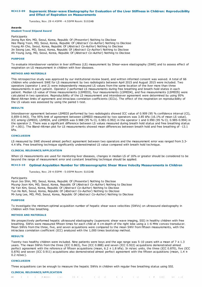
RC413-09 Supersonic Shear-wave Elastography for Evaluation of the Liver Stiffness in Children: Reproducibilityand Effect of Repiration on Measurements
Tuesday, Nov. 29 4:45PM - 4:55PM Room: S102AB
RC413-10 Optimal Acquisition Number for Ultrasonographic Shear Wave Velocity Measurements in Children
Tuesday, Nov. 29 4:55PM - 5:05PM Room: S102AB
AwardsStudent Travel Stipend Award
ParticipantsJeong Rye Kim, MD, Seoul, Korea, Republic Of (Presenter) Nothing to DiscloseHee Mang Yoon, MD, Seoul, Korea, Republic Of (Abstract Co-Author) Nothing to DiscloseYoung Ah Cho, Seoul, Korea, Republic Of (Abstract Co-Author) Nothing to DiscloseJin Seong Lee, MD, Seoul, Korea, Republic Of (Abstract Co-Author) Nothing to DiscloseAh Young Jung, MD, Seoul, Korea, Republic Of (Abstract Co-Author) Nothing to Disclose
PURPOSE
To evaluate interobserver variation in liver stiffness (LS) measurement by Shear-wave elastography (SWE) and to assess effect ofrespiration on LS measurement in children with liver diseases.
METHOD AND MATERIALS
This retrospective study was approved by our institutional review board, and written informed consent was waived. A total of 66children who underwent SWE for LS measurement by two radiologists between April 2015 and August 2015 were included. Twooperators (operator 1 and 2) were independently measured LS values from the same location of the liver more than threemeasurements in each patient. Operator 2 performed LS measurements during free breathing and breath-hold states in eachpatient. Median LS value of three measurements (LSMED3), four measurements (LSMED4), and five measurements (LSMED5) werecalculated in two operators. Reproducibility of the LS measurement and interobsever agreement were determined by using 95%Bland-Altman limits of agreement and intraclass correlation coefficients (ICCs). The effect of the respiration on reproducibility ofthe LS values was assessed by using the paired t-test.
RESULTS
Interobserver agreement between LSMED3 performed by two radiologists showed ICC value of 0.909 (95 % confidence interval (CI),0.859-0.943). The 95% limit of agreement between LSMED3 measured by two operators was 3.85 kPa (16.1% of mean LS value).ICC among LSMED3, LSMED4, and LSMED5 was 0.988 (95 % CI, 0.981-0.992) in the operator 1 and 0.990 (95 % CI, 0.985-0.994) inthe operator 2. There was a significant difference between LS values measured during breath hold status and free breathing status(P <.001). The Bland–Altman plot for LS measurements showed mean differences between breath hold and free breathing of -13.1kPa.
CONCLUSION
LS measured by SWE showed almost perfect agreement between two operators and the measurement error was ranged from 3.3-4.4 kPa. Free breathing technique significantly underestimated LS value compared with breath hold technique.
CLINICAL RELEVANCE/APPLICATION
When LS measurements are used for monitoring liver diseases, changes in median LS of 16% or greater should be considered to bebeyond the range of measurement error and constant breathing technique should be applied.
ParticipantsHyun Joo Shin, MD, Seoul, Korea, Republic Of (Presenter) Nothing to DiscloseMyung-Joon Kim, MD, Seoul, Korea, Republic Of (Abstract Co-Author) Nothing to DiscloseHa Yan Kim, Seoul, Korea, Republic Of (Abstract Co-Author) Nothing to DiscloseYun Ho Roh, Seoul, Korea, Republic Of (Abstract Co-Author) Nothing to DiscloseMi-Jung Lee, MD, PhD, Seoul, Korea, Republic Of (Abstract Co-Author) Nothing to Disclose
PURPOSE
To investigate the minimum optimal acquisition number of hepatic shear wave velocities (SWVs) on ultrasound elastography inchildren with free breathing.
METHOD AND MATERIALS
We prospectively performed hepatic ultrasound elastography (supersonic shear wave imaging, SSI) in healthy children with freebreathing. SWVs were measured fifteen times for each child at 4 cm depth of the right lobe using a 1-6 MHz convex transducer.Mean SWVs from the three, five, and seven acquisitions were compared to the mean SWV from fifteen measurements, with theintraclass correlation coefficient (ICC) analyzed with the 1,000 times bootstrap method.
RESULTS
Twenty-two healthy children were included. Nine patients were boys and the age range was 5-10 years with a mean of 7 ± 1.3years. The mean SWVs from the three (ICC 0.863), five (ICC 0.888) and seven (ICC 0.922) acquisitions demonstrated almostperfect agreement with the reference of fifteen acquisitions (mean, 6.2 ± 1.8 kPa). In m/sec units, the three (ICC 0.870), five (ICC0.876) and seven (ICC 0.911) acquisitions also demonstrated almost perfect agreement with the fifteen acquisitions (mean, 1.4 ±0.2 m/sec).
CONCLUSION
Three acquisitions can be enough to measure the hepatic SWVs in children with regular free breathing status using SSI.
CLINICAL RELEVANCE/APPLICATION
The reduced number of SWV acquisitions could shorten examination times and make examinations easier for children without the
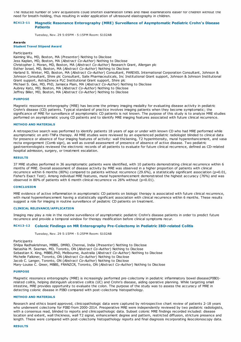
RC413-11 Magnetic Resonance Enterography (MRE) Surveillance of Asymptomatic Pediatric Crohn's DiseasePatients
Tuesday, Nov. 29 5:05PM - 5:15PM Room: S102AB
RC413-12 Colonic Findings on MR Enterography Pre-Colectomy in Pediatric IBD-related Colitis
Tuesday, Nov. 29 5:15PM - 5:25PM Room: S102AB
The reduced number of SWV acquisitions could shorten examination times and make examinations easier for children without theneed for breath-holding, thus resulting in wider application of ultrasound elastography in children.
AwardsStudent Travel Stipend Award
ParticipantsKaiming Wu, MD, Boston, MA (Presenter) Nothing to DiscloseJess Kaplan, MD, Boston, MA (Abstract Co-Author) Nothing to DiscloseChristopher J. Moran, MD, Boston, MA (Abstract Co-Author) Research Grant, Allergan plcEsther Israel, MD, Boston, MA (Abstract Co-Author) Nothing to DiscloseHarland S. Winter, MD, Boston, MA (Abstract Co-Author) Consultant, PAREXEL International Corporation Consultant, Johnson &Johnson Consultant, Shire plc Consultant, Salix Pharmaceuticals, Inc Institutional Grant support, Johnson & Johnson InstitutionalGrant support, AstraZeneca PLC Institutional Grant support, Shire plc Michael S. Gee, MD, PhD, Jamaica Plain, MA (Abstract Co-Author) Nothing to DiscloseAubrey Katz, MD, Boston, MA (Abstract Co-Author) Nothing to DiscloseJeffrey Biller, MD, Boston, MA (Abstract Co-Author) Nothing to Disclose
PURPOSE
Magnetic resonance enterography (MRE) has become the primary imaging modality for evaluating disease activity in pediatricCrohn’s disease (CD) patients. Typical standard of practice involves imaging patients when they become symptomatic; thesignificance of MRE for surveillance of asymptomatic CD patients is not known. The purpose of this study is to analyze MRE studiesperformed on asymptomatic young CD patients and to identify MRE imaging features associated with future clinical recurrence.
METHOD AND MATERIALS
A retrospective search was performed to identify patients 18 years of age or under with known CD who had MRE performed whileasymptomatic on anti-TNFa therapy. All MRE studies were reviewed by an experienced pediatric radiologist blinded to clinical datafor presence or absence of four imaging features of activity: wall thickening, T2 hyperintensity, mural hyperenhancement, and vasarecta engorgement (Comb sign), as well as overall assessment of presence of absence of active disease. Two pediatricgastroenterologists reviewed the electronic records of all patients to evaluate for future clinical recurrence, defined as CD-relatedhospital admission, surgery, or treatment escalation.
RESULTS
37 MRE studies performed in 36 asymptomatic patients were identified, with 10 patients demonstrating clinical recurrence within 6months of MRE. Overall assessment of disease activity by MRE was observed in a higher proportion of patients with clinicalrecurrence within 6 months (80%) compared to patients without recurrence (29.6%), a statistically significant association (p=0.01,Fisher’s Exact Test). Among individual MRE features, mural hyperenhancement demonstrated the highest accuracy (76%) and wasobserved in 80% of patients with 6 month clinical recurrence vs 26% without (p<0.01).
CONCLUSION
MRE evidence of active inflammation in asymptomatic CD patients on biologic therapy is associated with future clinical recurrence,with mural hyperenhancement having a statistically significant association with clinical recurrence within 6 months. These resultssuggest a role for imaging in routine surveillance of pediatric CD patients on treatment.
CLINICAL RELEVANCE/APPLICATION
Imaging may play a role in the routine surveillance of asymptomatic pediatric Crohn's disease patients in order to predict futurerecurrence and provide a temporal window for therapy modification before clinical symptoms recur.
ParticipantsShilpa Radhakrishnan, MBBS, DMRD, Chennai, India (Presenter) Nothing to DiscloseNatashia M. Seeman, MD, Toronto, ON (Abstract Co-Author) Nothing to DiscloseSebastian K. King, MBBS,PhD, Melbourne, Australia (Abstract Co-Author) Nothing to DiscloseMichelle Falkiner, Toronto, ON (Abstract Co-Author) Nothing to DiscloseJacob C. Langer, Toronto, ON (Abstract Co-Author) Nothing to DiscloseMary-Louise C. Greer, MBBS, FRANZCR, Toronto, ON (Abstract Co-Author) Nothing to Disclose
PURPOSE
Magnetic resonance enterography (MRE) is increasingly performed pre-colectomy in pediatric inflammatory bowel disease(PIBD)-related colitis, helping distinguish ulcerative colitis (UC) and Crohn's disease, aiding operative planning. While targeting smallintestine, MRE provides opportunity to evaluate the colon. The purpose of the study was to assess the accuracy of MRE indetecting colonic disease in PIBD compared with post-colectomy histopathology.
METHOD AND MATERIALS
Research and ethics board approved, clinicopathologic data were captured by retrospective chart review of patients 2-18 yearswho underwent colectomy for PIBD from 2000-2014. Preoperative MRE were independently reviewed by two pediatric radiologists,with a consensus read, blinded to reports and clinicopathologic data. Subset colonic MRE findings recorded included: diseaselocation and extent, wall thickness, wall T2 signal, enhancement degree and pattern, restricted diffusion, stricture presence andlength. These were compared with post-colectomy histopathology reports and final diagnosis incorporating ileocolonoscopy data.
RESULTS
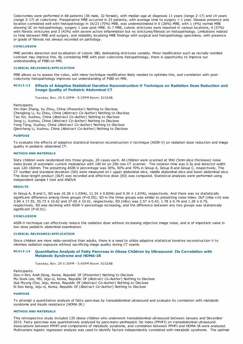
RC413-13 Effects of Adaptive Statistical Iterative Reconstruction-V Technique on Radiation Dose Reduction andImage Quality of Pediatric Abdominal CT
Tuesday, Nov. 29 5:25PM - 5:35PM Room: S102AB
RC413-14 Quantitative Analysis of Fatty Pancreas in Obese Children by Ultrasound: Its Correlation withMetabolic Syndrome and HOMA-IR
Tuesday, Nov. 29 5:35PM - 5:45PM Room: S102AB
Colectomies were performed in 68 patients (36 male, 32 female), with median age at diagnosis 11 years (range 2-17) and 14 years(range 3-17) at colectomy. Preoperative MRE occurred in 23 patients, with average time to surgery < 1 year. Disease presence andlocation correlated well with histopathology in 16/23 (70%) MRE, was underestimated in 6 (26%) MRE, with 1 (4%) normal MREshowing UC on histopathology, surgery 1 year post-MRE. In 7 MRE, colonic strictures were reported in various locations, 4 (57%)with fibrotic strictures and 3 (43%) with severe active inflammation but no strictures/fibrosis on histopathology. Limitations relatedto time between MRE and surgery, and reliability localizing MRE findings with surgical and histopathology specimens, with presenceor grade of fibrosis not always recorded on pathology.
CONCLUSION
MRE permits detection and localization of colonic IBD, delineating strictures variably. Minor modification such as rectally-instilledcontrast may improve this. By correlating MRE with post-colectomy histopathology, there is opportunity to improve ourunderstanding of PIBD on MRI.
CLINICAL RELEVANCE/APPLICATION
MRE allows us to assess the colon, with minor technique modification likely needed to optimize this, and correlation with post-colectomy histopathology improves our understanding of PIBD on MRI.
ParticipantsXin-Xian Zhang, Xu Zhou, China (Presenter) Nothing to DiscloseChenglong Li, Xu Zhou, China (Abstract Co-Author) Nothing to DiscloseTao Xin, Xuzhou, China (Abstract Co-Author) Nothing to DiscloseJiong Li, Xuzhou, China (Abstract Co-Author) Nothing to DiscloseYong Tang, Xuzhou, China (Abstract Co-Author) Nothing to DiscloseQiancheng Li, Xuzhou, China (Abstract Co-Author) Nothing to Disclose
PURPOSE
To evaluate the effects of adaptive statistical iterative reconstruction V technique (ASIR-V) on radiation dose reduction and imagequality in pediatric abdominal CT.
METHOD AND MATERIALS
Sixty children were randomized into three groups, 20 cases each. All children were scanned at 9HU (5mm slice thickness) noiseindex levels of automatic current modulation with 100 kV on 256-row CT scanner. The rotation time was 0.3s and detector widthwas 120-160mm. The presetting ASIR-V percentage was 30%, 50% and 70% in Group A, Group B and Group C, respectively. TheCT number and standard deviation (SD) were measured on t upper abdominal slice, middle abdominal slice and lower abdominal slice.The dose-length product (DLP) was recorded and effective dose (ED) was computed. Statistical analyses were performed usingindependent sample t test and ANOVA
RESULTS
In Group A, B and C, SD was 10.28 ± 1.03HU, 11.34 ± 0.82HU and 9.34 ± 1.67HU, respectively. And there was no statisticallysignificant difference among three groups (P>0.05); SD in the three groups was similar to presetting noise index. DLP (mGy·cm) was3.69 ± 17.55, 55.73 ± 10.62 and 37.65 ± 10.01, respectively; ED (mSv) was 2.57 ± 0.43, 1.78 ± 0.76 and 1.28 ± 0.75,respectively. ED was declining with ASIR-V percentage increasing, and the difference between any two groups was statisticallysignificant (P<0.01).
CONCLUSION
ASIR-V technique can effectively reduce the radiation dose without increasing objective image noise, and is of important value inlow-dose pediatric abdominal examination.
CLINICAL RELEVANCE/APPLICATION
Since children are more radio-sensitive than adults, there is a need to utilize adaptive statistical iterative reconstruction V tominimize radiation exposure without sacrificing image quality during CT exams.
ParticipantsDoo-ri Kim, Arail-Dong, Korea, Republic Of (Presenter) Nothing to DiscloseMu Sook Lee, MD, Jeju-si, Korea, Republic Of (Abstract Co-Author) Nothing to DiscloseGuk Myung Choi, Jeju, Korea, Republic Of (Abstract Co-Author) Nothing to DiscloseKi Soo Kang, Jeju-si, Korea, Republic Of (Abstract Co-Author) Nothing to Disclose
PURPOSE
To attempt a quantitative analysis of fatty pancreas by transabdominal ultrasound and evaluate its correlation with metabolicsyndrome and insulin resistance (HOMA IR.)
METHOD AND MATERIALS
This retrospective study included 135 obese children who underwent transabdominal ultrasound between January and December2015. Fatty pancreas was quantitatively analyzed by pancreato-perihepatic fat index (PPHFI) on transabdominal ultrasound.Associations between PPHFI and components of metabolic syndrome, and correlation between PPHFI and HOMA-IR were analyzed.Multivariate logistic regression analysis was used to identify factors independently correlated with metabolic syndrome. The optimal
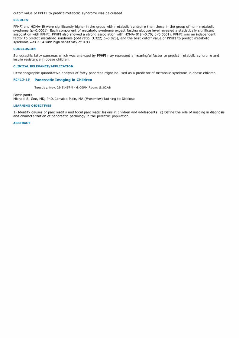
RC413-15 Pancreatic Imaging in Children
Tuesday, Nov. 29 5:45PM - 6:00PM Room: S102AB
cutoff value of PPHFI to predict metabolic syndrome was calculated
RESULTS
PPHFI and HOMA-IR were significantly higher in the group with metabolic syndrome than those in the group of non- metabolicsyndrome (p<0.0001). Each component of metabolic syndrome except fasting glucose level revealed a statistically significantassociation with PPHFI. PPHFI also showed a strong association with HOMA-IR (r=0.70; p<0.0001). PPHFI was an independentfactor to predict metabolic syndrome (odd ratio, 3.322; p=0.023), and the best cutoff value of PPHFI to predict metabolicsyndrome was 2.34 with high sensitivity of 0.93
CONCLUSION
Sonographic fatty pancreas which was analyzed by PPHFI may represent a meaningful factor to predict metabolic syndrome andinsulin resistance in obese children.
CLINICAL RELEVANCE/APPLICATION
Ultrasonographic quantitative analysis of fatty pancreas might be used as a predictor of metabolic syndrome in obese children.
ParticipantsMichael S. Gee, MD, PhD, Jamaica Plain, MA (Presenter) Nothing to Disclose
LEARNING OBJECTIVES
1) Identify causes of pancreatitis and focal pancreatic lesions in children and adolescents. 2) Define the role of imaging in diagnosisand characterization of pancreatic pathology in the pediatric population.
ABSTRACT
SSJ07-01 Prospective Intraindividual Comparison of Non-Contrast MRI and US as a Surveillance Tool for HCCin Patients with Cirrhosis at High Risk of HCC
Tuesday, Nov. 29 3:00PM - 3:10PM Room: E350
SSJ07-02 Diagnostic Accuracy of Prospective Applications of Liver Imaging Reporting and Data System (LI-RADS) on Gadoxetic Acid-Enhanced MRI
Tuesday, Nov. 29 3:10PM - 3:20PM Room: E350
SSJ07
Gastrointestinal (Hepatocellular Carcinoma)
Tuesday, Nov. 29 3:00PM - 4:00PM Room: E350
GI CT MR OI US
AMA PRA Category 1 Credit ™: 1.00ARRT Category A+ Credit: 1.00
FDA Discussions may include off-label uses.
ParticipantsHero K. Hussain, MD, Ann Arbor, MI (Moderator) Nothing to DiscloseSteven S. Raman, MD, Santa Monica, CA (Moderator) Nothing to Disclose
Sub-Events
ParticipantsHye Young Jang, Seoul, Korea, Republic Of (Presenter) Nothing to DiscloseSo Yeon Kim, MD, Seoul, Korea, Republic Of (Abstract Co-Author) Nothing to DiscloseJae Ho Byun, MD, Seoul, Korea, Republic Of (Abstract Co-Author) Nothing to DiscloseSo Jung Lee, Seoul, Korea, Republic Of (Abstract Co-Author) Nothing to DiscloseHyung Jin Won, MD, Seoul, Korea, Republic Of (Abstract Co-Author) Nothing to DiscloseJihyun An, Seoul, Korea, Republic Of (Abstract Co-Author) Nothing to DiscloseYoung-Suk Lim, Seoul, Korea, Republic Of (Abstract Co-Author) Nothing to DiscloseSeong Ho Park, MD, Seoul, Korea, Republic Of (Abstract Co-Author) Research Grant, DONGKOOK Pharmaceutical Co, Ltd
PURPOSE
To prospectively compare the diagnostic yield of non-contrast MRI including diffusion-weighted imaging (DWI) and US as asurveillance tool for detecting HCCs in patients with cirrhosis at high risk of HCC.
METHOD AND MATERIALS
A prospective surveillance study included 407 consecutive cirrhosis patients with an estimated annual risk of HCC > 5% whounderwent one to three, biannual screening examinations with paired liver MRI and US between November 2011 and August 2014.The referral criteria for HCC on non-contrast MRI were defined as a nodule showing one of the following criteria: the presence ofintralesion fat, mild to moderate hyperintensity on T2-weighted imaging, or hyperintensity on DWI at b-500 s/mm2. On US, adiscrete focal lesion equal or more than 1cm in diameter or suspicious tumor thrombosis was referred for a further work-up for HCC.During image interpretation, radiologists were blinded to the results of the other imaging modality. The confirmation of HCC wasbased on the results of a histologic examination and/or typical CT images of HCC. Per-lesion sensitivity of HCC and per-examspecificity were compared between non-contrast MRI and US.
RESULTS
In 1100 screening rounds of paired MRI and US, 48 HCCs were diagnosed in 43 patients. Among the 48 HCCs, intralesional fat wasdetected in 4 HCCs (8.3%), 31 HCCs (64.6%) showed mild to moderate hyperintensity on T2-weighted imaging, and 37 HCC(77.1%) appeared hyperintensity on DWI. Among the 71 positive cases on US, 12 patients were confirmed to have HCC. Using thediagnostic criteria, per lesion sensitivity of non-contrast MRI was 81.3 % (39/48), which was significantly higher that of 25%(12/48) (P<0.001). Non-contrast MRI showed a significantly higher per-exam specificity that US [98.0% (1036/1057) vs. 94.4%(998/1057), P<0.001].
CONCLUSION
In this prospective intraindividual comparison study, non-contrast MRI including DWI outperformed US as a surveillance test for HCCin patients with cirrhosis at high risk of HCC.
CLINICAL RELEVANCE/APPLICATION
In patients with cirrhosis at high risk of HCC, the non-contrast MRI including DWI can be considered to an alternative surveillancetool to US.
ParticipantsYeun Yoon Kim, MD, Seoul, Korea, Republic Of (Presenter) Nothing to DiscloseChansik An, MD, Seoul, Korea, Republic Of (Abstract Co-Author) Nothing to DiscloseSungwon Kim, MD, Seoul, Korea, Republic Of (Abstract Co-Author) Nothing to DiscloseMyeong-Jin Kim, MD, PhD, Seoul, Korea, Republic Of (Abstract Co-Author) Research Grant, Bayer AG
PURPOSE
To evaluate the diagnostic accuracy of prospectively applied Liver Imaging Reporting and Data System (LI-RADS, v2014) forhepatocellular carcinoma (HCC) on gadoxetic acid-enhanced liver MRI.
SSJ07-03 Abdominal Ultrasound Compared to Cross-Sectional Imaging for Surveillance of HepatocellularCarcinoma in High Risk Patients: Results of 5-year Cohort Follow-up
Tuesday, Nov. 29 3:20PM - 3:30PM Room: E350
SSJ07-04 Effect of Threshold Growth on Liver Imaging Reporting and Data System Categorization
Tuesday, Nov. 29 3:30PM - 3:40PM Room: E350
METHOD AND MATERIALS
This study included 528 hepatic observations from 268 patients who met the following criteria: 1) at high risk for HCC, 2)underwent gadoxetic acid-enhanced liver MRI in 2014 at our institution, 3) not previously treated for HCC, and 4) whose radiologicreports were prospectively reported using LI-RADS. Final diagnoses were determined histologically for all non-HCC malignancies (twocholangiocarcinomas [CCs] and four combined HCC-CCs), 110 of 122 HCCs, seven regenerative or dysplastic nodules, and onehemangioma. Fifty benign lesions and 12 HCCs were clinically diagnosed with follow-up imaging. The sensitivities and specificities ofLI-RADS categories were calculated with 95% confidence intervals (CIs).
RESULTS
None of the nine (0%) LR-1, one of 22 (4.5%) LR-2, 14 of 35 (40.0%) LR-3, 19 of 27 (70.4%) LR-4, 73 of 75 (97.3%) LR-5, and allof the five (100%) LR-5V observations were HCCs. Two of 75 (2.7%) LR-5 observations were combined HCC-CCs. Of 13 LR-Mlesions, only three (23.1%) were non-HCC malignancies and the remainder were HCCs. Three of six (50%) non-HCC malignancieswere categorized as LR-4 or LR-5. When LR-5 and LR-5V were considered positive diagnosis for HCC, the sensitivity and specificitywere 63.9% (95% CI, 54.8−72.4) and 96.9% (95% CI, 89.2−99.6), respectively. When LR-4, LR-5, and LR-5V were consideredpositive, the sensitivity and specificity were 79.5% (95% CI, 71.3−86.3) and 84.4% (95% CI, 73.1−92.2), respectively.
CONCLUSION
In high-risk patients for HCC, LR-5 and LR-5V showed a high specificity for diagnosing HCC. However, a significant portion of non-HCC malignancies were categorized as LR-4 or LR-5 and a majority of LR-M lesions were HCCs, which suggests the necessity formodification of criteria for the LR-M category.
CLINICAL RELEVANCE/APPLICATION
LR-M category of LI-RADS v2014 needs improvement to better exclude non-HCC malignancies in high risk patients for HCC and todecrease the misdiagnosis rate of HCCs as non-HCC malignancies.
ParticipantsFrancois Willemssen, MD, Hoogstraten, Belgium (Presenter) Nothing to DiscloseLievay van Dam, MSc, MD, Rotterdam, Netherlands (Abstract Co-Author) Nothing to DiscloseRoy S. Dwarkasing, MD, PhD, Rotterdam, Netherlands (Abstract Co-Author) Nothing to Disclose
PURPOSE
Hepatocellular carcinoma (HCC) is the most common primary malignant tumor of the liver and is the third leading cancer-relatedcause of death. Major risk factors are identified including cirrhosis caused by viral hepatitis B and C infection, and hereditaryhemochromatosis. According to European guidelines patients at risk for HCC should be surveyed with abdominal ultrasound (US)every 6 months.
METHOD AND MATERIALS
In our tertiary referral institute, we selected all patients between October 2005-October 2010 from our HCC database. Inclusioncriteria were: pathological or histochemical confirmed HCC, abdominal ultrasound and contrast-enhanced computed tomography(CT) or magnetic resonance imaging (MRI) within three months. Number and size of the detected lesions were compared.
RESULTS
In 88 patients included, 83 HCC lesions were described at US evaluation compared to 181 lesions detected using CT or MRI. Lesionsfound on US had a mean diameter of 60mm (range 5-135mm, median 45mm), on cross-sectional imaging the mean diameter was56mm (range 5-160mm, median 45mm). In 30 of the 88 (34%) patients US was negative, while cross-sectional imaging detectedHCC lesions. In 18 of those 30 cases all lesions were smaller than 25mm (60%). Of all the patients (n=25) with lesions smaller than25mm, 18 ultrasound examinations were false negative (72%).
CONCLUSION
Surveillance for HCC in high-risk patients using US is inadequate. A significant number of HCC lesions are missed using USsurveillance, especially small lesions, resulting in a false negative examination in a third of the cases.
CLINICAL RELEVANCE/APPLICATION
This warrants the question if surveillance for HCC should be performed with advanced cross sectional imaging modalities.
ParticipantsVictoria Chernyak, MD, Bronx, NY (Presenter) Nothing to DiscloseMariya Kobi, MD, New York, NY (Abstract Co-Author) Nothing to DiscloseKate Fruitman, Bronx, NY (Abstract Co-Author) Nothing to DiscloseAlla M. Rozenblit, MD, Bronx, NY (Abstract Co-Author) Nothing to DiscloseClaude B. Sirlin, MD, San Diego, CA (Abstract Co-Author) Research Grant, General Electric Company; Research Grant, Siemens AG;Research Grant, Guerbet SA; ;
PURPOSE
Liver Imaging Reporting and Data System (LI-RADS, LR) uses major features (arterial phase hyperenhancement [APHE], “washout”[WO], “capsule”, diameter, threshold growth [TG]) to codify probability of an observation being hepatocellular carcinoma. Inclusionof TG in the LI-RADS algorithm was based on expert opinion rather than scientific evidence. The goal of this study was to assessthe effect of TG on LR categorization.
SSJ07-05 Risk Assessment for Hepatocellular Carcinoma by Using Magnetic Resonance Elastography duringFollow-up
Tuesday, Nov. 29 3:40PM - 3:50PM Room: E350
SSJ07-06 Evaluation of Texture Analysis Parameters for Response Prediction and Monitoring in Patients withHepatocellular Carcinoma Undergoing Transarterial Chemoembolization Using Biphasic Contrast-Enhanced CT Image Data: Correlation with Results of Liver Pe
Tuesday, Nov. 29 3:50PM - 4:00PM Room: E350
METHOD AND MATERIALS
All MR and CT reports created using a standardized LR v2014 template at one tertiary care center between 4/15–2/16 wereidentified. For each LR3, LR4, and LR5 reported observation, the presence of every LR major feature was recorded retrospectively.Two LR categories were then assigned: one using all LR-v2014 major features and one using a revised system that disregards TG(LR-revised). Categories assigned using LR-v2014 and LR-revised were compared. Some analyses were repeated excluding patientswithout prior exams (TG not applicable).
RESULTS
136 patients (85 [62%] male, mean age 62 [±10] years) with 297 observations (median diameter 13mm, IQR 9-20mm) wereincluded. Of 297 observations, 204 (69%) had APHE, 186 (63%) had WO, 49 (16%) had “capsule” and 40 (14%) had TG. Of 40observations with TG, 26 (65%) were new observations ≥10mm, 8 (20%) had diameter increase ≥50% in ≤6 months and 6 (15%)had diameter increase ≥100% in >6 months. LR-v2014 categories were LR3 in 131/297 (44%), LR4 in 87/297 (29%) and LR5 in79/297 (27%). LR-revised categories were LR3 in 147/297 (50%), LR4 in 78/297 (26%) and LR5 in 72/297 (24%). Assignedcategories were discrepant in 22/297 (7%) observations. 7/79 (9%) observations categorized LR5 with LR-v2014 wererecategorized LR4 with LR-revised. Of these, all 7 were 10-19 mm and had APHE; 5/7 (71%) were new observations and 2/7 (29%)had diameter increase ≥50% in ≤6 months. When excluding 70 observations without prior exams, 22/227 (10%) categories werediscrepant; 7/50 (14%) observations categorized LR5 with LR-v2014 were recategorized LR4 with LR-revised.
CONCLUSION
TG affects LI-RADS category in the minority of cases. Disregarding TG causes a small but meaningful proportion of LR5 observationsto be downgraded to LR4.
CLINICAL RELEVANCE/APPLICATION
Removing threshold growth as a major feature to simplify LI-RADS algorithm would not affect final LI-RADS category in most casesbut would cause downgrading of a meaningful proportion of LR5 observations.
ParticipantsShintaro Ichikawa, MD, Chuo-Shi, Japan (Presenter) Nothing to DiscloseUtaroh Motosugi, MD, Yamanashi, Japan (Abstract Co-Author) Nothing to DiscloseTetsuya Wakayama, PhD, Hino-shi, Japan (Abstract Co-Author) Employee, General Electric CompanyHiroshi Onishi, MD, Yamanashi, Japan (Abstract Co-Author) Nothing to Disclose
PURPOSE
To evaluate magnetic resonance elastography (MRE) as a means for predicting the development of hepatocellular carcinoma (HCC)in chronic liver disease.
METHOD AND MATERIALS
We reviewed data from 161 patients with chronic liver disease with the following inclusion criteria: had 2 MRE examinations betweendays 365–1424 with >12 month interval, no prior history of or development of HCC between the two exams, and availablelaboratory results. Liver stiffness was classified as low (<3 kPa), moderate (3–4.7 kPa), and high (>4.7 kPa). The classification forchange in stiffness between the two MREs is as follows: high on both or high on the first and moderate on the second (group A,n=61), low on both (group C, n=39), and other combinations (group B, n=61). We used Cox analyses and Kaplan-Meyer methods todetermine the risk of developing HCC.
RESULTS
Forty-seven patients (29.2%) developed HCC during the follow-up period (45.9% (28/61) of group A, 27.9% (17/61) of group B;5.1% (2/39) of group C). There was a significant difference in the disease-free survival rates between groups A (54.9%), B(73.9%), and C (87.6%) at 3-years (p=0.0001). The independent risk factors for development of HCC included: belonging to GroupA (hazard ratio [HR] versus group C=6.0, P=0.0028; versus group B=2.16, P=0.0268), age (HR = 1.04, P=0.0154), and alanineaminotransferase level (HR=1.02, P =0.0196).
CONCLUSION
Results from MRE can stratify the risk of developing HCC during the follow-up of patients with chronic liver disease.
CLINICAL RELEVANCE/APPLICATION
Patients with chronic liver disease with high liver stiffness (>4.7 kPa) on their first MR elastography are at high risk for developingan HCC, regardless of the results of their second MR elastography. Thus, they should have meticulous follow-up and be screenedfor HCC development.
ParticipantsChristopher Kloth, Tuebingen, Germany (Presenter) Nothing to DiscloseWolfgang M. Thaiss, MD, Tuebingen, Germany (Abstract Co-Author) Nothing to DiscloseRainer Kargel, Forchheim, Germany (Abstract Co-Author) Employee, Siemens AGRainer Grimmer, Forchheim, Germany (Abstract Co-Author) Employee, Siemens AGGerd Grozinger, MD, Tubingen, Germany (Abstract Co-Author) Nothing to DiscloseRoland Syha, Tuebingen, Germany (Abstract Co-Author) Nothing to Disclose
Dominik Ketelsen, MD, Tuebingen, Germany (Abstract Co-Author) Nothing to DiscloseKonstantin Nikolaou, MD, Tuebingen, Germany (Abstract Co-Author) Speakers Bureau, Siemens AG; Speakers Bureau, Bracco Group;Speakers Bureau, Bayer AGMarius Horger, MD, Tuebingen, Germany (Abstract Co-Author) Nothing to Disclose
PURPOSE
To analyze the value of different parameters derived from CT texture analysis (CTTA) image data in hepatocellular carcinoma (HCC)for prediction of response and response evaluation to transarterial chemoembolization (TACE).
METHOD AND MATERIALS
The study group consisted of 56 HCC in 28 patients (27 male; mean age 67.2±10.4) who underwent CECT examinations before andafter TACE therapy. The study was approved by the local ethic committee. Mean time between the two CT examinations was 39.93± 62.21 days. Standard of reference was perfusion-CT of the liver additionally to arterial and portal-venous post-contrast phases.Patients were assigned into subgroups: no response-NR (n=9), partial response-PR (n=34) and complete response-CR (n=13). CTTAparameters were: heterogeneity/intensity/average/deviation/skewness/contrast of NGTDM (Neighborhood Grey-Tone DifferenceMatrix). For each parameter mean, entropy and uniformity were calculated.Blood flow(BF), blood volume(BV), arterial liverperfusion(ALP), portal-venous perfusion(PVP) and hepatic perfusion index(HPI) were calculated in the pre- and post-TACE settingsby liver perfusion-CT.
RESULTS
Patients with CR showed higher tumor perfusion parameters before TACE and a significant decrease in BF/BV/ALP/HPI after TACE(p=0.002/0.002/0.002/0.003). Patients with PR showed similar results, but only in responding tumor parts. ROC analysis of CTTAparameters yielded predictive cut-off values for CR in the arterial CECT-phase (sensitivity/specificity) for mean intensity(88.9%/69.8%), mean average (88.9%/69.8%) and skewness (90.0%/58.1%) In the portal-venous CECT-phase similar foruniformity of heterogeneity, uniformity of skewness and mean contrast (92.3%/81.8%/92.3%/54.5%/92.3%/95.2%). Significantcorrelations were registered between changes in mean heterogeneity and BF (p=0.004, r=-0.815), BV (p=0.002, r=-0.851) and ALP(p=0.002, r=-0.851) in the arterial phase in CR and PR.
CONCLUSION
Significant correlations exist between CTTA parameters and those derived from perfusion-CT both in the pre- and post- TACEsetting with predictive value for TACE outcome.
CLINICAL RELEVANCE/APPLICATION
Prediction of response to local therapy (TACE) by means of CTTA can be implemented for choosing the best treatmentstrategy.Improved response monitoring by CTTA is beneficial for optimal patient management and could be a substitute for moresophisticated imaging techniques like perfusion-CT or MRI.
SSJ08-01 Characterization of Malignant versus Benign Focal Liver Lesions with Lumason-Enhanced UltrasoundImaging
Tuesday, Nov. 29 3:00PM - 3:10PM Room: E353A
SSJ08-02 Characterization of Focal Liver Masses: A Multicentre Study Comparing Contrast Enhanced Ultrasound(CEUS) to Grayscale/Doppler Ultrasound, Computed Tomography and Magnetic Resonance Imaging
Tuesday, Nov. 29 3:10PM - 3:20PM Room: E353A
SSJ08
Gastrointestinal (New Ultrasound Techniques)
Tuesday, Nov. 29 3:00PM - 4:00PM Room: E353A
GI US
AMA PRA Category 1 Credit ™: 1.00ARRT Category A+ Credit: 1.00
FDA Discussions may include off-label uses.
ParticipantsAnthony E. Samir, MD, Boston, MA (Moderator) Consultant, Pfizer Inc Consultant, General Electric Company Consultant, PAREXELInternational Corporation Research Grant, Koninklijke Philips NV Research Grant, Siemens AG Research Grant, Toshiba CorporationResearch Grant, General Electric Company Research Grant, Samsung Electronics Co, Ltd Research Grant, Analogic CorporationResearch support, SuperSonic Imagine Research support, Hitachi, LtdJessica B. Robbins, MD, Madison, WI (Moderator) Nothing to Disclose
Sub-Events
ParticipantsRichard G. Barr, MD, PhD, Youngstown, OH (Presenter) Consultant, Siemens AG; Consultant, Koninklijke Philips NV; Research Grant,Siemens AG; Research Grant, SuperSonic Imagine; Speakers Bureau, Koninklijke Philips NV; Research Grant, Bracco Group; SpeakersBureau, Siemens AG; Consultant, Toshiba Corporation; Research Grant, Esaote SpA; Research Grant, B and K Ultrasound; ResearchGrant, Hitachi Aloka UltrasoundEdward G. Grant, MD, Los Angeles, CA (Abstract Co-Author) Research Grant, General Electric Company ; Medical Advisory Board,Nuance Communications, IncDirk-Andre Clevert, MD, Muenchen, Germany (Abstract Co-Author) Speaker, Siemens AG; Speaker, Koninklijke Philips NV; Speaker,Bracco Group; Barry B. Goldberg, MD, Philadelphia, PA (Abstract Co-Author) Nothing to DiscloseMichelle L. Robbin, MD, Birmingham, AL (Abstract Co-Author) Consultant, Koninklijke Philips NV; Andrew De la Torre, MD, Newark, NJ (Abstract Co-Author) Nothing to DiscloseWui K. Chong, MD, Chapel Hill, NC (Abstract Co-Author) Advisory Board, Bracco Group; Deike H. Strobel, MD, Erlangen, Germany (Abstract Co-Author) Nothing to Disclose
PURPOSE
To compare the diagnostic performance of contrast-enhanced ultrasound (CEUS) with Lumason (sulfur hexafluoride lipid-type Amicrospheres) to that of unenhanced ultrasound (UEUS) for characterization of malignant versus benign focal liver lesions (FLLs).
METHOD AND MATERIALS
Two multicenter clinical trials of identical design were conducted in adult subjects with at least one FLL requiring work-up forcharacterization; subjects underwent UEUS followed by CEUS with 2.4 mL Lumason. UEUS and CEUS images were evaluated by on-site investigators and 3 off-site, blinded readers unaffiliated with the enrolment centers and blinded to any clinical data. Diagnosticperformance of UEUS and CEUS for FLL characterization was determined using tissue pathology/histology (when FLL biopsy could beperformed), or 6-month follow-up with contrast-enhanced CT or MRI as truth standard.
RESULTS
A total of 499 subjects were included in the 2 trials. Per truth standard, 256 lesions were benign (n=116 hemangioma, n=64 focalnodular hyperplasia, n=76 other) and 243 were malignant (n=131 hepatocellular carcinoma, n=78 metastases, n=34 other). For theoff-site reads, the pooled sensitivity, specificity, and accuracy were 43.6%, 34.0%, 38.7%, respectively for UEUS and 72.4%,80.5%, 76.6%, respectively for CEUS (p<.05). For on-site reads, sensitivity, specificity, and accuracy were 37.0%, 21.5%, 29.1%,respectively for UEUS and 89.3%, 84.0%, 86.6%, respectively for CEUS (p<.05). No serious adverse events related to Lumasonadministration were reported.
CONCLUSION
Overall, Lumason-enhanced ultrasound imaging provided an improvement from UEUS for characterization of FLLs.
CLINICAL RELEVANCE/APPLICATION
The results suggest that CEUS with 2.4 mL of Lumason is safe and may be useful to improve characterization of focal liver lesionswhen UEUS is inconclusive.
AwardsStudent Travel Stipend Award
ParticipantsDavid P. Burrowes, MD, Calgary, AB (Presenter) Nothing to DiscloseAlexandra Medellin-Kowalewski, MD, Calgary, AB (Abstract Co-Author) Nothing to DiscloseAlison C. Harris, MBChB, Vancouver, BC (Abstract Co-Author) Nothing to Disclose

SSJ08-03 Relationship between Liver Tissue Stiffness and Histopathological Findings Analyzed by Shear WaveEastography in Patients with Non-alcoholic Fatty Liver Disease
Tuesday, Nov. 29 3:20PM - 3:30PM Room: E353A
SSJ08-04 Noninvasive Evaluation of Liver Fibrosis using Two-dimensional Shear Wave Elastography: AComparison with Different Serum Fibrosis Indices in Chronic Hepatitis B
Tuesday, Nov. 29 3:30PM - 3:40PM Room: E353A
Laurent Milot, Lyon, France (Abstract Co-Author) Nothing to DiscloseStephanie R. Wilson, MD, Calgary, AB (Abstract Co-Author) Equipment support, Siemens AG; Equipment support, Koninklijke PhilipsNV
PURPOSE
In light of recent FDA approval of a microbubble for liver CEUS in the USA, we propose that CEUS is superior to grayscale ultrasoundand equivalent to institutional CT and MR for characterization of focal liver masses.
METHOD AND MATERIALS
This prospective study comprises 224 patients, from 5 geographically separate centres, presenting for evaluation of a focal livermass (12 adenoma, 8 cholangiocarcinoma, 55 FNH, 41 hemangioma, 71 HCC, 25 metastasis, 12 other). All had US/CEUS and CTand/or MR.CEUS was performed with Definity (Lantheus, Billerica MA) on standard US systems. They included a continuous cine ofwash-in to peak enhancement, and still portal venous and delayed images to 5 minutes. CT and MR were performed as standardcare with institutional protocols. Pre-contrast, arterial phase and portal venous phase images were included.Three separateelectronic blind read files; one for each of CEUS (including grayscale and Doppler images), CT and MR were created and read blindlyby four radiologists, two for CEUS and two for CT/MR utilizing a Microsoft Access based questionnaire regarding the arterial, portalvenous and delayed phase enhancement of each lesion. Readers also provided a preference for malignancy or benignancy, a finaldiagnosis as well as a confidence levelResults of the blind read were compared with the truth standard from pathology, long-termstability or expert consensus.
RESULTS
Our results show that CEUS is superior to grayscale/Doppler ultrasound and at least equivalent to CT and MR both in determiningthe malignancy of a lesion and in diagnostic accuracy, with an increase in confidence from a mean of 1.6 for grayscale/Doppler to3.9 on CEUS on a 5 point scale. Sensitivity, specificity and accuracy of CEUS are 95%, 82%, 67%, of grayscale/Doppler are 81%,56% 40%, of CT are 89%, 75%, 62% and of MR are 85%, 79%, 62% respectively.
CONCLUSION
CEUS is superior to grayscale ultrasound both for determination of malignancy and when assigning a final diagnosis for a focal livermass. Additionally, CEUS is equivalent, and in select cases performs better, in the characterization of focal liver lesions thaninstitutional CT and MRI.
CLINICAL RELEVANCE/APPLICATION
In the evaluation of a focal liver mass, CEUS is a necessary adjuvant to grayscale/Doppler ultrasound and is equivalent to CT andMR in its ability to predict malignancy and provide a diagnosis.
ParticipantsHIROHITO TAKEUCHI, Tokyo, Japan (Presenter) Nothing to DiscloseKatsutoshi Sugimoto, MD, PhD, Tokyo, Japan (Abstract Co-Author) Nothing to DiscloseYoshiyuki Kobayashi, Toyo, Japan (Abstract Co-Author) Nothing to DiscloseFuminori Moriyasu, MD, Kyoto, Japan (Abstract Co-Author) Nothing to DiscloseTakao Itoi, Tokyo, Japan (Abstract Co-Author) Nothing to Disclose
PURPOSE
Shear wave elastography (SWE) is validated in chronic hepatitis C and B; however, limited data are available in Non-Alcoholic FattyLiver Disease (NAFLD). This study is aimed to assess the accuracy and the efficacy of SWE for the detection of fibrosis in patientswith NAFLD and to evaluate the effect of other histologic parameters on SWE measurement.
METHOD AND MATERIALS
Written informed consent was obtained from all subjects, and the local ethics committee approved the study. Seventy-one patientswith histologically proven NAFLD (mean age, 50.8 years ± 15.7) were examined. All patients underwent SWE (AixplorerTM;SuperSonic Imagine) and FIB4 index (based on age, aspartate aminotransferase [AST] and alanine aminotransferase [ALT] levels,and platelet counts). SWE measurements were compared to the histologic features based on of NAFLD activity score and FIB4index.
RESULTS
The area under the ROC curve (AUC) for the diagnosis of hepatic fibrosis stages 3 or higher was 0.821 (optimal cutoff value, 13.1kPa; sensitivity, 62.5%; specificity, 57.4%) for SWE, as was 0.822 (optimal cutoff value, 1.41; sensitivity, 71.9%; specificity,53.9%) for FIB4 index, respectively. Median liver stiffness values measured using SWE showed a stepwise increase with increasinghepatic fibrosis stage (P<0.001), inflammation score (P=0.018), and ballooning score (P<0.001), and showed a stepwise decreasewith increase hepatic steatosis stage (P=0.046).
CONCLUSION
SWE is a promising imaging modality for assessing the presence or absence of advanced fibrosis in patients with NAFLD. The effectof steatosis on SWE measurements may be controversial.
CLINICAL RELEVANCE/APPLICATION
SWE is a rapid and noninvasive method of detecting fibrosis in patients with nonalcoholic fatty liver disease.
SSJ08-05 Initial Experience Using a Telerobotic Ultrasound System to Perform Adult Abdominal Examinations
Tuesday, Nov. 29 3:40PM - 3:50PM Room: E353A
ParticipantsHong Ding, Shanghai, China (Presenter) Nothing to DiscloseYuan Zhuang, Shanghai, China (Abstract Co-Author) Nothing to DiscloseHong Han, Shanghai, China (Abstract Co-Author) Nothing to DiscloseWenping Wang, MD, PhD, Shanghai, China (Abstract Co-Author) Nothing to Disclose
PURPOSE
To explore the value of 2D-SWE in assessing the extent of liver fibrosis with chronic hepatitis B (CHB) and to compare its diagnosticaccuracy with different fibrosis indices involving aminotransferase/platelet ratio index (APRI), fibrosis index based on the 4 factors(FIB-4), King's score and Forns index.
METHOD AND MATERIALS
A total of 384 subjects (304 with CHB and 80 controls) who underwent partial hepatectomy were enrolled and divided into fivegroups (S0~S4) according to Scheuer scoring system histologically. All patients were examined with 2D-SWE to obtain liverstiffness measurements (LSMs), which were compared with the histological findings and analyzed with factors including age, genderand several blood markers that might influence the value of LSMs.
RESULTS
The intraclass correlation coefficient of five LSMs with 2D-SWE was 0.948. The average LSMs were 5.74±1.10 kPa for fibrosis S0(n=10), 6.53±0.96 kPa for S1 (n=30), 7.94±0.89 kPa for S2 (n=50), 9.40±1.31 kPa for S3 (n=47), and 14.52±3.55 kPa for S4(n=167). The multiple comparisons of LSMs showed significant statistical difference between every two fibrotic groups expectbetween S0 and S1 groups. For the prediction of S≥2 and S=4, the optimal cut-off values of LSMs were 7.6 kPa and 10.4 kPa, witha sensitivity of 92.0% and 94.6% and a specificity of 90.0% and 94.9%, respectively. All these methods correlated with fibrosisstages statistically (p<0.05) and the correlation coefficients between pathological stages with LSMs, APRI, FIB-4, King's score andForns index were 0.875, 0.409, 0.397, 0.428 and 0.452, respectively. The AUROC in diagnosing fibrosis S≥2 were 0.970, 0.771,0.727, 0.787, and 0.765, respectively; and the AUROC were 0.986, 0.703, 0.712, 0.716, and 0.740 for diagnosing S=4,respectively. Liver fibrosis stages, inflammatory activity grades and the level of gamma glutamyltranspeptidas (GGT), albalanineaminotransferase (ALT), and aspartate aminotransferase (AST) significantly correlated with LSMs(β=0.618, 0.015, 0.079, -0.083and 0.146, respectively; p<0.05).
CONCLUSION
2D-SWE could be used to predict significant fibrosis (S≥2) and cirrhosis (S=4) in CHB patients with notable higher diagnosticaccuracy than serum fibrosis models. In terms of LSM associated with hepatic fibrosis, the confounding factors includinginflammatory activity, and the level of GGT, ALT, and AST that independently influence the value of LSMs with 2D-SWE.
CLINICAL RELEVANCE/APPLICATION
N/A
AwardsStudent Travel Stipend Award
ParticipantsScott J. Adams, Saskatoon, SK (Presenter) Nothing to DiscloseBrent E. Burbridge, MD, FRCPC, Saskatoon, SK (Abstract Co-Author) Nothing to DiscloseIvar Mendez, MD,PhD, Saskatoon, SK (Abstract Co-Author) Nothing to DisclosePaul S. Babyn, MD, Saskatoon, SK (Abstract Co-Author) Nothing to DiscloseLeanne Langford, Saskatoon, SK (Abstract Co-Author) Nothing to DiscloseVincent Vergara, MD, Saskatoon, SK (Abstract Co-Author) Nothing to DiscloseAndreea Badea, BSC, Saskatoon, SK (Abstract Co-Author) Nothing to DiscloseRhonda Bryce, MD, Saskatoon, SK (Abstract Co-Author) Nothing to DiscloseLuis Bustamante, Saskatoon, SK (Abstract Co-Author) Nothing to Disclose
PURPOSE
To assess the feasibility of performing adult abdominal examinations using a telerobotic ultrasound system in whichradiologists/sonographers can control fine movements of a transducer and all ultrasound settings from a remote location.
METHOD AND MATERIALS
Eighteen patients prospectively underwent a conventional sonography examination (using EPIQ 5, Philips or LOGIQ E9, GEHealthcare) followed by a telerobotic sonography examination (using the MELODY System, AdEchoTech; SonixTablet, BKUltrasound; and TE30 All-in-One HD Videoconferencing Endpoint, Huawei Technologies) according to a standardized abdominalimaging protocol. For telerobotic examinations, patients located at an imaging clinic were scanned remotely by a sonographer 2.75km away. Conventional examinations were read independently from telerobotic examinations. We assessed the ability of the systemto generate images of diagnostic quality and acceptability of the system to patients and sonographers.
RESULTS
92% of organs visualized on conventional examinations were sufficiently visualized on telerobotic examinations. Five pathologicalfindings were identified on both telerobotic and conventional examinations, three findings were identified using only conventionalsonography and two findings were identified using only telerobotic sonography. A paired sample t-test showed no significantdifference between the two modalities in measurements of the liver, spleen, and diameter of the proximal aorta; however,telerobotic assessments overestimated distal aorta and common bile duct diameters and underestimated kidney lengths (p-values<0.05). All patients strongly agreed or somewhat agreed that they would be willing to have another telerobotic examination.
CONCLUSION
SSJ08-06 Differentiation of Inflammatory from Fibrotic Bowel Strictures Among Patients with Crohn's Diseasethrough Analysis of Time-intensity Curves Obtained after Microbubble Contrast Agent Injection
Tuesday, Nov. 29 3:50PM - 4:00PM Room: E353A
A telerobotic ultrasound system is feasible for performing abdominal ultrasound examinations at a distant location with minimaltraining and set-up requirements and a moderate learning curve. Telerobotic sonography may open up the possibility of establishingremote ultrasound clinics for communities which lack skilled sonographers and radiologists, thereby improving access to care.
CLINICAL RELEVANCE/APPLICATION
A telerobotic ultrasound system is feasible for performing abdominal ultrasound examinations at a distant location with minimaltraining and set-up requirements and may improve access to care.
Honored Educators
Presenters or authors on this event have been recognized as RSNA Honored Educators for participating in multiple qualifyingeducational activities. Honored Educators are invested in furthering the profession of radiology by delivering high-qualityeducational content in their field of study. Learn how you can become an honored educator by visiting the website at:https://www.rsna.org/Honored-Educator-Award/
Paul S. Babyn, MD - 2012 Honored Educator
ParticipantsEmilio Quaia, MD, Trieste, Italy (Presenter) Nothing to DiscloseAntonio Giulio Gennari, MD, Trieste, Italy (Abstract Co-Author) Nothing to DiscloseMichele Pontello, Trieste, Italy (Abstract Co-Author) Nothing to DiscloseFederica Arban, Trieste, Italy (Abstract Co-Author) Nothing to DiscloseMaria A. Cova, MD, Trieste, Italy (Abstract Co-Author) Nothing to Disclose
PURPOSE
To assess whether the analysis of time-intensity curves obtained after sulphur hexafluoride-filled microbubble contrast agentinjection could differentiate inflammatory from fibrotic strictures among patients with Crohn’s disease (CD).
METHOD AND MATERIALS
Sixty-five consecutive patients (40 male and 25 female; mean age ± SD, 42.2 years ± 12.22) with a diagnosis of CD were included.Inclusion criteria were: (1) CD involving the terminal ileal loop as shown by endoscopy with serial deep mucosal biopsies or crosssectional imaging with a thickness of the terminal ileal loop >3mm; (2) availability of the histologic score based on mucosalulceration (grade 0–3), edema (grade 0–3), and quantity (grade 0–3) and depth (grade 0–4) of neutrophilic infiltration; (3)availability of mural fibrosis diagnosis when abnormal depositions of collagens were identified in the edges of mucosal ulceration; (4)at least 12 weeks of clinical follow-up in CD diagnosed no more than 2 years before the study. In each patient the terminal ilealloop was scanned by a convex-array probe (2–5 MHz) before and after sulfur hexafluoride-filled microbubble injection. The digitalcine-clips registered after microbubble injection during the first-pass dynamic enhancement was quantified in lineary units by adedicated software through manually-drawn regions of interest (ROIs) encompassing the anterior bowel wall. Time-intensity curvesfrom patients with inflammatory and fibrotic stenosis were compared. The peak enhancement, rise time, time to peak, area underthe time-intensity curve (AUC), AUC during wash-in (AUCWI), and AUC during wash-out (AUCWO) were compared between patientswith inflammatory or fibrotic strictures.
RESULTS
Inflammatory (n=40) vs fibrotic stenoses (n=25) differed in the AUC (234274 ± 293293 vs 89787 ± 53819; P = <0.05), AUCWI(53256 ± 53871 vs 30993 ± 19454; P = <0.05), AUCWO (150910 ± 177859 vs 61930 ± 36181; P = <0.05) and peak enhancement(11556 ± 11298 vs 6252 ± 5504; P<.05).
CONCLUSION
The quantitative analysis of small bowel wall contrast enhancement after microbubble contrast agent injection may differentiateinflammatory from fibrotic ileal stenosis in patients with CD.
CLINICAL RELEVANCE/APPLICATION
Contrast-enhanced ultrasound provides an early identification of those patients with a fibrotic ileal stricture who deserve surgicalresection from those patients with an inflammatory stricture who deserve pharmacologic treatment.
SSJ09-01 The Optimal Measurement Number of Shear Wave Elastography(SWE) for Different Liver FibrosisStages
Tuesday, Nov. 29 3:00PM - 3:10PM Room: E353C
SSJ09-02 Gd-EOB-DTPA-Enhanced MR Relaxometry for the Detection and Staging of Liver Fibrosis
Tuesday, Nov. 29 3:10PM - 3:20PM Room: E353C
SSJ09
Gastrointestinal (Diffuse Liver Disease and HCC)
Tuesday, Nov. 29 3:00PM - 4:00PM Room: E353C
CT GI MR
AMA PRA Category 1 Credit ™: 1.00ARRT Category A+ Credit: 1.00
FDA Discussions may include off-label uses.
ParticipantsShahid M. Hussain, MD,PhD, Omaha, NE (Moderator) Nothing to DiscloseMyeong-Jin Kim, MD, PhD, Seoul, Korea, Republic Of (Moderator) Research Grant, Bayer AG
Sub-Events
ParticipantsQian Li, MD, Boston, MA (Presenter) Nothing to DiscloseManish Dhyani, MBBS, Boston, MA (Abstract Co-Author) Nothing to DiscloseAtul K. Bhan, MD, Boston, MA (Abstract Co-Author) Nothing to DiscloseXi Zhang, PhD, Indianapolis, IN (Abstract Co-Author) Nothing to DiscloseAnthony E. Samir, MD, Boston, MA (Abstract Co-Author) Consultant, Pfizer Inc Consultant, General Electric Company Consultant,PAREXEL International Corporation Research Grant, Koninklijke Philips NV Research Grant, Siemens AG Research Grant, ToshibaCorporation Research Grant, General Electric Company Research Grant, Samsung Electronics Co, Ltd Research Grant, AnalogicCorporation Research support, SuperSonic Imagine Research support, Hitachi, Ltd
PURPOSE
To explore the optimal minimum number of SWE measurements in liver fibrosis with different stages.
METHOD AND MATERIALS
218 consecutive patients who underwent SWE before their scheduled liver biopsy (age 18–78 years; mean 48 years; men 96,women 122) were enrolled in this study between August 2013 and January 2015. Ten SWE values were obtained at the right upperlobe of the liver through an intercostal approach in suspended respiration. A single sub-special pathologist reviewed all biopsyspecimens as per the METAVIR criteria. Median values using a different number of measurements (2-9) were chosen from the tenmeasurements randomly for each subject. Receiver operating characteristic (ROC) curves were constructed for distinguishing ≥F2fibrosis for 10 measurements, and then sequentially, for measurement numbers below 10. The variability in intra-subject SWEmeasurements was calculated by comparing difference of individual measurements from the median value for each subject, and itsrelationship with measurement numbers was analyzed.
RESULTS
Study population fibrosis stages were F0=86, F1=80, F2=22, F3=22, F4=8. For all subjects, the areas under the ROC curve (AUROC)of the 10 measurements in differentiation of stage F2 or greater and stage F4 were 0.81 (95%CI: 0.69-0.94) and 0.847 (95%CI:0.72-0.96) respectively. The minimum measurement numbers in differentiation of corresponding fibrosis stages were 7 (AUC=0.79,95%CI: 0.67-0.916, P=0.17) and 6 (AUC=0.82, 95%CI: 0.71-0.94, P=0.53) respectively. As shown in Figure 1, in both groups (F0 or1 vs. ≥F2), the variability of measurements did not change much with the increase of the measurement number (r=0.53, P=0.14),especially between 6-10, but the variability is much higher in stage F2 or greater.
CONCLUSION
Our study indicates that, in the patients with variety of hepatic diseases, the optimal minimum numbers of SWE measurements were7 for differentiation of ≥F2 fibrosis and 6 for F4. There was no statistically significant variability in intra-subject SWE measurementsusing 6-10 measurement numbers, which implies the optimal measurement numbers obtained in this study are reliable.
CLINICAL RELEVANCE/APPLICATION
Standardization of number of measurements needed for SWE assessment of fibrosis stage is essential for SWE clinical workflow.More measurements will decrease variability on a per subject basis, however time, labor, cost-effectiveness and reliability are veryimportant factors.
AwardsTrainee Research Prize - Resident
ParticipantsMichael Haimerl, Regensburg, Germany (Presenter) Nothing to DiscloseKirsten Utpatel, Regensburg, Germany (Abstract Co-Author) Nothing to DiscloseClaudia Fellner, MD, PhD, Regensburg, Germany (Abstract Co-Author) Nothing to DiscloseMarcel D. Nickel, Erlangen, Germany (Abstract Co-Author) Employee, Siemens AGChristian R. Stroszczynski, MD, Regensburg, Germany (Abstract Co-Author) Nothing to DisclosePhilipp Wiggermann, Regensburg, Germany (Abstract Co-Author) Nothing to Disclose
SSJ09-03 Role of Shear Wave Sono-Elastography of Liver in Monitoring Patients of Non-Alcoholic Fatty LiverDisease (NAFLD): Comparison with Magnetic Resonance Imaging- A Prospective Study
Tuesday, Nov. 29 3:20PM - 3:30PM Room: E353C
SSJ09-04 Maximum Value Measured by Two-Dimensional Shear Wave Elastography Helps in DifferentiatingMalignancy From Benign Focal Liver Lesions
PURPOSE
To examine the diagnostic performance of gadoxetic-acid (Gd-EOB-DTPA)-enhanced T1 relaxometry in the staging of hepaticfibrosis using histopathologic examination as the reference standard.
METHOD AND MATERIALS
Sixty–five patients (41 men and 24 women; mean age, 55.9 +/- 14.8) with histologically proven liver fibrosis (LF) who underwentGd-EOB-DTPA–enhanced MR including T1 MR–relaxometry were analyzed. The fibrosis stage was assessed according to theMETAVIR score (fibrosis scores range: F0, no fibrosis to F4, cirrhosis). For the T1 relaxometry, a transverse 3D VIBE sequence (TR5.79 ms, TE 2.46 ms, α 1°, 7°, 14°) with inline T1 calculation was acquired prior to and 20 minutes after Gd-EOB-DTPAadministration following the acquisition of a B1 map for inline correction of the B1 inhomogeneities. The reduction rates of the T1relaxation time (rrT1) between the pre- and postcontrast images were calculated, and the optimal cutoff values for the fibrosisstages were determined with receiver operating characteristic (ROC) curve analyses.
RESULTS
The rrT1 decreased with the severity of liver fibrosis with the following mean values (%): stage F0, 69.4±2.6; stage F1, 59.1±3.7;stage F2, 55.3±4.6; stage F3, 42.2±0.10; and stage F4, 28.8±8.2. Regression analysis revealed a significant correlation of the rrT1with the stage of liver fibrosis (r=-0.906, p<0.001). T1 relaxometry performed well in the staging of liver fibrosis with AUCs of 1.0for the stages of F1 and greater, 0.92 for the stages of F2 and greater, 0.98 for the stages of F3 and greater, and 0.96 for stageF4.
CONCLUSION
Gd-EOB-DTPA–enhanced T1 relaxometry is a reliable tool for the staging of hepatic fibrosis. The results of the ROC analysesdemonstrated the high accuracy in the detection of the initial stages of hepatic fibrosis.
CLINICAL RELEVANCE/APPLICATION
Our study supports the conclusion that measuring T1 relaxation times via Gd-EOB-DTPA-enhanced MRI may be incorporated intothe clinical routine as a liver imaging screening test for the detection of silent disease and the non-invasive definition of the stageof liver fibrosis.
AwardsStudent Travel Stipend Award
ParticipantsNikhil Makhija, MBBS, New Delhi, India (Presenter) Nothing to DiscloseMadhusudhan Kumble Seetharama, MD, FRCR, New Delhi, India (Abstract Co-Author) Nothing to DiscloseDeepnarayan Srivastava, Delhi, India (Abstract Co-Author) Nothing to DiscloseRaju Sharma, MD, New Delhi, India (Abstract Co-Author) Nothing to DiscloseShivanand R. Gamanagatti, MBBS, MD, New Delhi, India (Abstract Co-Author) Nothing to DiscloseNaval K. Vikram, New Delhi, India (Abstract Co-Author) Nothing to Disclose
PURPOSE
To measure liver stiffness (LS) by Shear-wave sono-elastography (SWE) in patients of non-alcoholic fatty liver disease (NAFLD)before and after intervention and to compare it with hepatic fat fraction (FF) measured by Magnetic ResonanceSpectroscopy (MRS)
METHOD AND MATERIALS
41 adult treatment-naive patients (mean age: 39yrs, male:female–22:19 of NAFLD were included in this prospective study afterobtaining approval from Institutional Ethics Committee and informed patient consent. Known diabetics, alcohol users (>20g/day)and patients on chronic drug intake were excluded. After initial clinical (body mass index-BMI) and biochemical (liver function tests-LFT, lipid profile) evaluation, MRS and SWE were done for all patients and mean hepatic fat fraction (FF) and mean liver stiffness(LS) were obtained. Following dietary and lifestyle changes and oral Vitamin E for six months repeat clinico-biochemical tests, MRIand SWE were done. Based on compliance to intervention, patients were categorized into good compliance and poor compliancegroups. The FF and LS before and after treatment in both groups were compared.
RESULTS
30 patients had good and 11 had poor compliance to intervention. Baseline mean LS did not show significant correlation with meanMRS FF (r=0.19, p >0.05). After intervention, in both the groups, mean LS and mean MRS FF did not show significant correlation.Inthe group with good compliance, there was significant reduction in BMI which showed moderate correlation (r=0.45, p<0.05) withchange in MRS FF. LS also showed significant reduction (p<0.05).In the group with poor compliance, there was significant increasein BMI which showed good correlation (r=0.79, p<0.05) with increase in mean MRS. LS did not show significant change(p>0.05).The change in mean LS showed no correlation either with mean MRS FF (p> 0.05) or with BMI after treatment.
CONCLUSION
LS measured by SWE does not show correlation with MRS FF and is not useful in monitoring of NAFLD.
CLINICAL RELEVANCE/APPLICATION
Change in gray scale USG, BMI and biochemical parameters do not accurately represent change in liver fat content. Although MRI isuseful as a non-invasive modality to diagnose and quantify liver fat as well as its monitoring, its use in follow-up is expensive. SWE,however, cannot be used in monitoring changes in liver fat content after interventional regimens.
Tuesday, Nov. 29 3:30PM - 3:40PM Room: E353C
SSJ09-05 Lack of Standardized Terminology and Definitions for Major Imaging Features of HCC among ClinicalPractice Guidelines Released by Medical Societies
Tuesday, Nov. 29 3:40PM - 3:50PM Room: E353C
ParticipantsWenshuo Tian, Guangzhou, China (Presenter) Nothing to DiscloseXiaoyan Xie, Guangzhou, China (Abstract Co-Author) Nothing to DiscloseLu-Yao Zhou, MD, Guangzhou, China (Abstract Co-Author) Nothing to DiscloseManxia Lin, Guangzhou, China (Abstract Co-Author) Nothing to DiscloseFu-Shun Pan, Guangzhou, China (Abstract Co-Author) Nothing to Disclose
PURPOSE
To evaluate the diagnostic efficacy of two-dimensional shear wave elastography (2D SWE) in differentiating malignancy frombenign focal liver lesions (FLL).
METHOD AND MATERIALS
Institutional review board approval and written informed consent was obtained. The maximum, minimum, mean, and the standarddeviation of 2D SWE measurements, expressed in kPa (Emax, Emin, Emean, Esd), were underwent in 221 patients with 229 FLLs.Receiver operating characteristic (ROC) curve analysis was performed to evaluate the diagnostic performance of 2D SWE. Mann-Whitney U test was used to assess the inter-group differences.
RESULTS
Emax, Emin, Emean, Esd was significantly higher in 164 malignant lesions than that in 65 benign lesions (P<0.001). For identificationof malignant FLL, the area under ROC curves for Emax was 0.920 (Emin 0.710, Emean 0.879, Esd 0.915). Emax was 96.21±35.40 for19 intrahepatic cholangiocarcinomas (ICC) and 90.32±54.71 for 35 liver metastatic lesions, which was significantly higher than61.83±28.87 for 103 hepatocellular carcinomas (HCC), respectively (P<0.0001, P=0.0237). Emax was 38.72±18.65 for 15 focalnodular hyperplasias, which was significantly higher than 20.56±10.74 for 37 hemangiomas (P=0.0009). Emax of adjacent liverparenchyma of HCC and ICC were significantly higher than that of the other three lesion types (P<0.005).
CONCLUSION
Emax of FLL and adjacent liver parenchyma could help in differentiating malignancy from benign FLLs.
CLINICAL RELEVANCE/APPLICATION
Maximum value measured by two-dimensional shear wave elastography of focal liver lesion and adjacent liver parenchyma may beconsidered for differential malignant from benign lesions.
ParticipantsAdrija Mamidipalli, MBBS, San Diego, CA (Presenter) Nothing to DiscloseSaya Igarashi, San Diego, CA (Abstract Co-Author) Nothing to DiscloseRyan L. Brunsing, MD, San Diego, CA (Abstract Co-Author) Nothing to DiscloseClaude B. Sirlin, MD, San Diego, CA (Abstract Co-Author) Research Grant, General Electric Company; Research Grant, Siemens AG;Research Grant, Guerbet SA; ;
PURPOSE
Multiple medical societies have published clinical practice guidelines that include algorithms for non-invasive imaging-based diagnosisof HCC in patients with cirrhosis using multiphasic contrast-enhanced MRI/CT. The purpose of this structured review of theliterature was to extract and compare the terminology and definitions endorsed by these guidelines for the following major imagingfeatures of HCC: arterial phase hyperenhancement (APHE), “washout”(WO), “capsule” (C), and diameter (D). Liver ImagingReporting and Data System (LI-RADS) was not included in this review
METHOD AND MATERIALS
We identified 38 guidelines published since 1995, cited in PubMed, and containing imaging criteria for HCC. Each guideline wasreviewed by 2 independent readers. Terms and definitions describing APHE, WO, C, and D were recorded. If for a given feature noterm was used, “term not used” was recorded and the next feature assessed. If a term was used but no definition provided, allcitations supporting that term were similarly reviewed and all definitions relating to the term were recorded. If no definition wasprovided within either the guideline or citations, “no definition used” was recorded
RESULTS
There was >95% inter-reader agreement in identifying terms and definitions. Of 38 guidelines reviewed; 78%, 68%, 13%, and 42%contained terms while only 8%, 13%, 3%, and 3% contained definitions for APHE, WO, C, and D, respectively. Terms used for APHEand WO were variable with only 53% incorporating the language "arterial-phase enhancement" or "washout". There were 43 uniquecitations supporting undefined terms, yielding an additional 13 definitions each for APHE/WO and 4 each for C/D. While both APHEand WO were usually described using lesion signal/attenuation relative to the surrounding liver parenchyma, details on contrastphase and measurement technique were inconsistent. The few definitions for C and D used ambiguous language and cleardefinitions could not be extracted
CONCLUSION
Imaging guidelines for the non-invasive diagnosis of HCC use variable terms for key imaging features. Definitions usually are notprovided. When provided, definitions are inconsistent between guidelines and/or ambiguous
CLINICAL RELEVANCE/APPLICATION
Lack of standardized and defined terminology for imaging features used in the non-invasive diagnosis of HCC complicates theinterpretation and comparison of the HCC literature
SSJ09-06 Prediction of Microvascular Invasion in Hepatocellular Carcinoma: Preoperative Gd-EOB-DTPA-Dynamic Enhanced MRI and Histopathologic Correlation
Tuesday, Nov. 29 3:50PM - 4:00PM Room: E353C
AwardsStudent Travel Stipend Award
ParticipantsMengqi Huang, MD, Guang zhou, China (Presenter) Nothing to DiscloseZhi Dong, Guangzhou, China (Abstract Co-Author) Nothing to DiscloseKeguo Zheng, MD, Guangzhou, China (Abstract Co-Author) Nothing to DisclosePing Xu, BMedSc, GuangZhou, China (Abstract Co-Author) Nothing to DiscloseShiting Feng, MD, Guangzhou, China (Abstract Co-Author) Nothing to Disclose
PURPOSE
We aimed to prospectively investigate the imaging features on the preoperative Gd-EOB-DTPA-dynamic enhanced MRI correlatewith the present of microvascular invasion(MVI) in hepatocellular carcinoma(HCC).
METHOD AND MATERIALS
A total of 66 HCC lesions in 60 patients with preoperative Gd-EOB-DTPA-dynamic enhanced MRI were prospectively analyzed.Tumor size, signal homogeneity, tumor capsule, tumor margin, peritumor enhancement in mid-arterial phase, peritumor hypointensein hepatobiliary phase, signal intensity ratio on DWI and apparent diffusion coefficients (ADCs), T1 relaxation times and thereduction rate between pre- and post-contrast enhancement images were assessed. Correlation between these factors andpresence of MVI were determined and the prediction model was established.
RESULTS
Histopathologic findings revealed 17 of 66 HCC had MVI. The univariate analysis showed the tumor size(p=0.003),margin(p=0.013),peritumor enhancement(p=0.001)and hypointense in hepatobiliary phase(p=0.004) were associated with MVI.However,the multiple logistic regression indicated that the tumor size, margin and peritumor enhancement were predictors ofpresent of MVI(α=0.01). The R^2 of this prediction model was 0.353,and the sensitivity and specificity were 52.9% and93.0%,respectively.
CONCLUSION
Tumor size, margin and peritumor enhancement in the preoperative Gd-EOB-DTPA-dynamic enhanced MRI can predict MVI of HCCwith low sensitivity but high specificity.
CLINICAL RELEVANCE/APPLICATION
Preoperative Gd-EOB-DTPA- dynamic enhanced MRI can help to predict MVI of HCC, though are not sensitive but have highspecificity.
RC408A Abdomen (Solid Organs and Vessels)
RC408B Diaphragm
RC408C Bowel/Pelvis
RC408D Extremities
RC408
Trauma Imaging Pitfalls
Tuesday, Nov. 29 4:30PM - 6:00PM Room: N228
GI MK ER
AMA PRA Category 1 Credits ™: 1.50ARRT Category A+ Credits: 1.50
Participants
Sub-Events
ParticipantsFelipe Munera, MD, Miami, FL, ([email protected]) (Presenter) Nothing to Disclose
LEARNING OBJECTIVES
1. Identify common pitfalls in interpretation of abdominal trauma CT studies - focus on solid organs and vascular structures. 2.Optimize CT acquisition techniques to reduce likelihood of missing potentially significant injuries. 3. Develop a search pattern thatincludes organs and structures where important lesions are commonly missed. 4. Describe strategies to improve detection of easilymissed injuries
ParticipantsMichael N. Patlas, MD, FRCPC, Hamilton, ON, ([email protected] ) (Presenter) Nothing to Disclose
LEARNING OBJECTIVES
1) To describe direct and indirect signs of blunt and penetrating diaphragmatic injury. 2) To highlight factors affecting detection ofdiaphragmatic injury. 3) To discuss pitfalls in diagnosis of diaphragmatic injury.
ABSTRACT
The traumatic diaphragmatic injury is an uncommon entity. Blunt diaphragmatic injury is undiagnosed at initial presentation in 7-66%. Penetrating diaphragmatic injury can be occult in 7% of cases. Diaphragmatic injury does not resolve spontaneously and cancause disastrous complications. The misinterpretation in patients with diaphragmatic injury may be caused by suboptimal technique,failure to review portion of examination e.g. MPRs, or satisfaction of search error. Potential pitfalls in interpretation includecongenital diaphragmatic hernias and atraumatic defects simulating diaphragmatic injury.
ParticipantsStephan W. Anderson, MD, Boston, MA (Presenter) Nothing to Disclose
ParticipantsO. C. West, MD, Houston, TX (Presenter) Nothing to Disclose
LEARNING OBJECTIVES
1) Find upper extremity injuries that are difficult detect on screening radiographs. 2) Employ search patterns that may improvedetection of easily missed injuries.
ABSTRACT
Summary of upper extremity pitfalls: Posterior shoulder dislocation; Supracondylar fracture (anterior humeral line); Monteggiafracture-dislocation (radio-capitellar line); Proximal radius including vertical head fracture (external oblique view), impacted neckfracture, flipped radial head fracture-dislocation, Galeazzi fracture-dislocation – beware the lateral radiograph.Imaging jointsrequires 3 radiographic projections. The 3rd view varies: Axillary view of shoulder and External oblique of elbow. The wrist needs 4views: PA, lateral, external oblique and “Scaphoid” view (ulnar deviated PA view).Words to live by: watch for the least obvious ofmultiple injuries.
RC409A Pitfalls in Hepatobiliary Imaging
RC409B Pitfalls in Pancreas Imaging
RC409C Pitfalls in Bowel Imaging
RC409D Pitfalls in Post-op Imaging
RC409
Pitfalls in Abdominal Imaging
Tuesday, Nov. 29 4:30PM - 6:00PM Room: E451B
GI
AMA PRA Category 1 Credits ™: 1.50ARRT Category A+ Credits: 1.50
FDA Discussions may include off-label uses.
Participants
Sub-Events
ParticipantsAntonio Luna, MD, Jaen, Spain (Presenter) Nothing to Disclose
LEARNING OBJECTIVES
1) Identify the most common pitfalls in hepatobiliary imaging. 2) Differentiate them from pathological conditions using amultimodality approach with CT and MRI.
ABSTRACT
ParticipantsZhen J. Wang, MD, Hillsborough, CA (Presenter) Stockholder, Nextrast, Inc
LEARNING OBJECTIVES
1) Become familiar with the pitfalls in pancreas imaging techniques. 2) Learn the pitfalls in imaging evaluation of benign pancreaticdiseases. 3) Review the pitfalls in imaging evaluation of pancreatic malignancies.
ABSTRACT
ParticipantsBenjamin M. Yeh, MD, San Francisco, CA, ([email protected] ) (Presenter) Research Grant, General Electric Company; Author withroyalties, Oxford University Press; Shareholder, Nextrast, Inc;
LEARNING OBJECTIVES
1) Understand critical imaging features associated with the most common bowel emergencies. 2) Be able to tailor exams to suitspecific clinical imaging scenarios, depending on patient history and clinical question. 3) Be familiar with common artifacts that canobscure or mimic clinical disease in and adjacent to bowel. 4) Understand how physiology, contrast material, and imaging techniqueaffect cross-sectional imaging of bowel. 5) Provide cogent differential diagnoses based on CT and MR findings to direct patientcare.
ABSTRACT
Imaging of the bowel and interpretation of radiological bowel images is arguably the most technically challenging aspect ofabdominal imaging, with a high rate of missed diagnostic findings and misinterpretations. The bowel is highly changeable inappearance from scan to scan, and moment to moment. Bowel appearance depends not only on the disease state, but also lumencontents, timing of intravenous contrast material, and many technical image-related factors. Understanding of the mesenteric andbowel anatomy, normal and diseased bowel physiology and vascular flow, and the interaction of contrast agents, is critical to theaccurate interpretation of bowel disease. In this talk we will discuss common pitfalls of technique and imaging. In addition, we willexplore controversies regarding imaging technique which have become more pressing in recent years, including when to use positiveversus neutral oral contrast agents versus no oral contrast in different clinical scenarios.
ParticipantsKumaresan Sandrasegaran, MD, Carmel, IN, ([email protected]) (Presenter) Nothing to Disclose
LEARNING OBJECTIVES
1) To understand the postoperative anatomy after pancreatic and bowel surgery. 2) To differentiate between expected anatomyand postoperative complications. 3) To differentiate between expected post-op anatomy and tumor recurrence.
ABSTRACT
Honored Educators
Presenters or authors on this event have been recognized as RSNA Honored Educators for participating in multiple qualifyingeducational activities. Honored Educators are invested in furthering the profession of radiology by delivering high-quality

educational content in their field of study. Learn how you can become an honored educator by visiting the website at:https://www.rsna.org/Honored-Educator-Award/
Kumaresan Sandrasegaran, MD - 2013 Honored EducatorKumaresan Sandrasegaran, MD - 2014 Honored EducatorKumaresan Sandrasegaran, MD - 2016 Honored Educator
RC422A IGRT and Anatomical Adaptation
RC422B Functional Targeting, Clinical Goals, and Toxicity Risks
RC422
Imaging for Personalized Medicine: Abdomen
Tuesday, Nov. 29 4:30PM - 6:00PM Room: S102C
GI RO PH
AMA PRA Category 1 Credits ™: 1.50ARRT Category A+ Credits: 1.50
ParticipantsKristy K. Brock, PhD, Ann Arbor, MI (Moderator) License agreement, RaySearch Laboratories AB; Development agreement, VarianMedical Systems, Inc;
ABSTRACT
The use of imaging and other biomarkers to increase the efficacy of treatment and decrease the risk of toxicity increased in theabdomen. Functional imaging and serum-based biomarkers can enable a more detailed understanding of the tumor, itscharacteristics, and early indications of its response to therapy. In addition, they can also be utilized to assess an individualpatients risk for toxicity, enabling a personalize approach to radiotherapy. These advanced imaging techniques can be combinedwith anatomical information to generate high precision treatment plans which can be adapted over the course of treatment toaccount for identified uncertainties, changes, and deviations which may compromise the delivery of the intended treatment oridentify the ability to re-optimize treatment to improve the therapeutic ration. In this session, technical and clinical concepts will bedescribed to design and deliver personalized radiotherapy in the abdomen. Technical concepts will include incorporation of multi-modality imaging for treatment planning, image guidance at treatment, and functional and anatomical adaption. Clinical conceptswill include functional targeting, clinical goals, and toxicity risks.
Sub-Events
ParticipantsKristy K. Brock, PhD, Ann Arbor, MI (Presenter) License agreement, RaySearch Laboratories AB; Development agreement, VarianMedical Systems, Inc;
LEARNING OBJECTIVES
1) Describe the processes necessary for the safe and accurate integration of multi-modality imaging for treatment planning. 2)Understand the role of image guidance for abdominal radiotherapy. 3) Illustrate methods to perform functional and anatomicaladaptation in the abdomen.
ABSTRACT
The use of imaging and other biomarkers to increase the efficacy of treatment and decrease the risk of toxicity increased in theabdomen. Functional imaging and serum-based biomarkers can enable a more detailed understanding of the tumor, itscharacteristics, and early indications of its response to therapy. In addition, they can also be utilized to assess an individualpatients risk for toxicity, enabling a personalize approach to radiotherapy. These advanced imaging techniques can be combinedwith anatomical information to generate high precision treatment plans which can be adapted over the course of treatment toaccount for identified uncertainties, changes, and deviations which may compromise the delivery of the intended treatment oridentify the ability to re-optimize treatment to improve the therapeutic ration. In this session, technical and clinical concepts will bedescribed to design and deliver personalized radiotherapy in the abdomen. Technical concepts will include incorporation of multi-modality imaging for treatment planning, image guidance at treatment, and functional and anatomical adaption. Clinical conceptswill include functional targeting, clinical goals, and toxicity risks.
ParticipantsJoseph M. Herman, MD, MSc, Baltimore , MD (Presenter) Nothing to Disclose
LEARNING OBJECTIVES
1) Review methods to obtain, process and analyze tissue and serum based biomarkers for abdominal tumors. 2) Describe currentdose/fractionation regimens as well as normal tissue constraints utilized in treating abdominal tumors. 3) Explain potentialadvantages of assessing treatment response with MRI and quantitative PET/SPECT (PERCIST) imaging over CT based response(RECIST) in abdominal tumors.
ABSTRACT
In order to deliver personalized radiation therapy in abdominal tumors, it is important to understand the methods used to obtain,analyze, and interpret serum and tissue based biomarkers. Most research to date has focused on identifying specific biomarkersused to personalize systemic or targeted therapies. Radiation-specific biomarkers are emerging and may eventually be used todetermine whether radiation is indicated or identify specific radiation sensitizers for use in abdominal tumors. Radiation therapyplanning has historically used computed tomography (CT)-based imaging. Molecular imaging using hybrid positron emissiontomography (PET)/CT scanning or single-photon emission computed tomography (SPECT) imaging and functional magneticresonance imaging (MRI) has provided new insights into the precise identification of gross tumor volume (GTV) and clinical tumorvolume (CTV) and has provided response information during and after therapy. The effective use of PET/SPECT and MRI in clinicalpractice, however, requires an appreciation of the unique challenges inherent to these modalities. Fundamental physical issues oflimited spatial resolution relative to the biological process, partial volume effects, image misregistration, motion management, andedge delineation must be carefully considered and can differ by agent or the method applied. Integration of PET/SPECT andMRI imaging into multicenter clinical trials and clinical practice can be particularly challenging due to differences in imagingprotocols, machines, and anatomy. Imaging protocols that clearly outline scan and fusion parameters are crucial. Further,

interpretation of tumor response should be standardized, and scans should be obtained at consistent time intervals. In addition, it isimportant to consider novel tracers of tumor biology (e.g. hypoxia, proliferation, apoptosis) beyond the commonly usedradiotracers. In this session, we will discuss these applications and challenges as well as provide guidance on how to integratePET/SPECT/MRI into radiation treatment planning and assessing treatment response. Finally, we will evaluate common dose andfractionation regimens as well as established dose constraints used in treating abdominal tumors with conventional and stereotacticbody radiation therapy.
RC450
Targeted Treatment and Imaging of Liver Cancers: Basic to Advanced Techniques in Minimally-InvasiveTherapies and Imaging
Tuesday, Nov. 29 4:30PM - 6:00PM Room: S403B
GI OI IR
AMA PRA Category 1 Credits ™: 1.50ARRT Category A+ Credits: 1.50
FDA Discussions may include off-label uses.
ParticipantsJinha Park, MD, PhD, Duarte, CA (Presenter) Speakers Bureau, Bayer AG; Steven S. Raman, MD, Santa Monica, CA (Presenter) Nothing to DiscloseJohn J. Park, MD, PhD, Duarte, CA (Presenter) Proctor, Sirtex Medical Ltd; Advisory Board, Guerbet SA; Speakers Bureau, MedtronicplcMarcelo Guimaraes, Charleston, SC, ([email protected] ) (Presenter) Consultant, Cook Group Incorporated ; Consultant, BaylisMedical Company; Consultant, Terumo Corporation; Patent holder, Cook Group IncorporatedAndrew C. Price, MD, Gilbert, AZ, ([email protected]) (Presenter) Nothing to Disclose
LEARNING OBJECTIVES
1) Discuss the role of the interventional radiologist in the treatment and management of patients with primary and metastatic livercancer as part of the multidisciplinary team. 2) Learn best practice techniques in the treatment of liver cancers, with emphasis onboth locoregional and focal therapeutic approaches, and indications for treatment. 3) Explore various tips and tricks for eachtreatment modality and learn how to avoid complications through good patient selection, choosing the appropriate techniques, andknowing what common mistakes to avoid. 4) Learn about newer and developing techniques and devices, their potential roles andindications, and potential pitfalls. 5) Explore advanced imaging modalities in the detection of tumors and for monitoring treatmentresponse.
ABSTRACT
Primary and metastatic liver disease may benefit from combined techniques such as bland/chemoembolization and liver ablation. Thepresentation will provide the rationale for the association of techniques, patient selection, tips and tricks, equipment and suppliesnecessary, protective techniques and how to avoid complications. Also, it will be discussed the results and current literature tosupport the association of techniques.
ED005-WE
Gastrointestinal Wednesday Case of the Day
Wednesday, Nov. 30 6:00AM - 11:59PM Room: Case of Day, Learning Center
GI
AMA PRA Category 1 Credit ™: .50
ParticipantsChristine M. Peterson, MD, Hummelstown, PA (Presenter) Nothing to DisclosePerry J. Pickhardt, MD, Madison, WI (Abstract Co-Author) Co-founder, VirtuoCTC, LLC; Stockholder, Cellectar Biosciences, Inc;Stockholder, SHINE Medical Technologies, Inc; Research Grant, Koninklijke Philips NVConstantine A. Raptis, MD, Saint Louis, MO (Abstract Co-Author) Nothing to DiscloseChristine O. Menias, MD, Scottsdale, AZ (Abstract Co-Author) Nothing to DiscloseRex A. Parker III, MD, Los Angeles, CA (Abstract Co-Author) Nothing to DiscloseElizabeth B. Maddox, Madison, WI (Abstract Co-Author) Nothing to Disclose
TEACHING POINTS
1) Each GI case of the day will be taken from disorders of the luminal GI tract as well as the liver, spleen, pancreas, and biliarysystem. The findings may be uncommon manifestations of common diseases or common manifestations of uncommon diseases.
Honored Educators
Presenters or authors on this event have been recognized as RSNA Honored Educators for participating in multiple qualifyingeducational activities. Honored Educators are invested in furthering the profession of radiology by delivering high-qualityeducational content in their field of study. Learn how you can become an honored educator by visiting the website at:https://www.rsna.org/Honored-Educator-Award/
Perry J. Pickhardt, MD - 2014 Honored EducatorChristine O. Menias, MD - 2013 Honored EducatorChristine O. Menias, MD - 2014 Honored EducatorChristine O. Menias, MD - 2015 Honored EducatorChristine O. Menias, MD - 2016 Honored Educator
RC509A Post-op Assessment of Esophagus and Stomach
RC509B Imaging the Non-operative Esophagus and Stomach
RC509C Large Bowel
RC509D Defecography: Pearls and Pitfalls
RC509
GI Fluoroscopy: Don't Bury 'em Yet!
Wednesday, Nov. 30 8:30AM - 10:00AM Room: E450B
GI
AMA PRA Category 1 Credits ™: 1.50ARRT Category A+ Credits: 1.50
FDA Discussions may include off-label uses.
Participants
Sub-Events
ParticipantsCheri L. Canon, MD, Birmingham, AL, ([email protected] ) (Presenter) Author with royalties, The McGraw-Hill Companies
LEARNING OBJECTIVES
1) Protocol fluoroscopic examination for safe evaluation and diagnosis. 2) Identify normal postoperative anatomy of the esophagusand stomach. 3) Diagnose radiographic complications in these patients.
ABSTRACT
Fluoroscopic evaluation of postoperative patients continues to grow in volume and complexity. In many cases, the fluoroscopicapproach is superior to CT and requires the radiologist to have a fundamental understanding of the surgical anatomy. Thispresentation will review the approach and diagnosis of patients after undergoing surgery of the upper GI tract (esophagus andstomach).Safe fluoroscopic technique will be discussed. Normal postoperative anatomy and surgical complications will be reviewed.
ParticipantsLaura R. Carucci, MD, Midlothian, VA, ([email protected] ) (Presenter) Nothing to Disclose
LEARNING OBJECTIVES
1) Discuss the continued importance of fluoroscopic evaluation of the esophagus and stomach. 2) Describe examination techniquesfor the esophagus and stomach. 3) Review the radiologic diagnosis of pathologic conditions involving the esophagus and stomach.
ABSTRACT
Despite an overall trend towards a decreasing number of fluoroscopic procedures, the number of esophagography studies hasincreased in recent years. Fluoroscopy remains the primary modality for evaluation the esophagus. In addition, fluoroscopy providesa noninvasive means to evaluate for benign and malignant gastric pathology. Radiologists should be able to perform and interpretesophagography and upper GI studies. A spectrum of benign and malignant processes that may affect the esophagus and stomachwill be discussed.
ParticipantsChristine O. Menias, MD, Scottsdale, AZ, ([email protected] ) (Presenter) Nothing to Disclose
LEARNING OBJECTIVES
1) Review role of Contrast Enemas in case-based format. 2) Highlight common indications for Fluoroscopy. 3) Review cases in thepre-operative and post-operative colon. 4) Emphasize imaging features that should not be overlooked.
ABSTRACT
Honored Educators
Presenters or authors on this event have been recognized as RSNA Honored Educators for participating in multiple qualifyingeducational activities. Honored Educators are invested in furthering the profession of radiology by delivering high-qualityeducational content in their field of study. Learn how you can become an honored educator by visiting the website at:https://www.rsna.org/Honored-Educator-Award/
Christine O. Menias, MD - 2013 Honored EducatorChristine O. Menias, MD - 2014 Honored EducatorChristine O. Menias, MD - 2015 Honored EducatorChristine O. Menias, MD - 2016 Honored Educator
ParticipantsDean D. Maglinte, MD, Indianapolis, IN, ([email protected] ) (Presenter) Nothing to Disclose
LEARNING OBJECTIVES
1) To define the role of fluoroscopic defecography in the evaluation of anorectal dysfunction and pelvic organ prolapse. 2) Todemonstrate how it should be done to avoid technical and interpretive pitalls. 3) To illustrate how to report a clinically relevantfunctional pelvic floor examination.
ABSTRACT
Despite advances in other imaging methods, fluoroscopic defecography has remained a practical cost effective procedure in theevaluation of anorectal and pelvic floor dysfunction. Dynamic cystocolpodefecograpohy (DCP) has evolved from a method ofevaluating the anorectum for functional disorders to it's current status as a functional method of evaluating the global pelvic floorfor defecatory disorders and pelvic organ prolapse (POP). It provides the maximum stress to the pelvic floor resulting in levator anirelaxation accompanied by rectal emptying needed to diagnose defecatory disorders and organ specific quantificative information onPOP.
RC510A Imaging and Doppler of Portal Hypertension
RC510B Contrast Ultrasound of the Liver
RC510C Liver Elastography
RC510
Liver Doppler, Contrast and Elastography
Wednesday, Nov. 30 8:30AM - 10:00AM Room: E353C
GI US
AMA PRA Category 1 Credits ™: 1.50ARRT Category A+ Credits: 1.50
FDA Discussions may include off-label uses.
Participants
Sub-Events
ParticipantsMyron A. Pozniak, MD, Madison, WI, ([email protected] ) (Presenter) Stockholder, Cellectar Biosciences, Inc; Support,General Electric Company
LEARNING OBJECTIVES
1) Understand the normal anatomy, anatomic variants of the hepatic vasculature. 2) Identify the normal Doppler flow profiles of thehepatic vasculature. 3) Understand the hemodynamic principles of portal hypertension and how they impact the Doppler waveformsof the hepatic arteries, portal veins and hepatic veins. 4) Understand the role of ultrasound in the evaluation of variceal pathways.
ABSTRACT
ParticipantsHans-Peter Weskott, MD, Hannover, Germany, ([email protected] ) (Presenter) Speaker, General Electric Company;Speaker, Bracco Group
LEARNING OBJECTIVES
1) Indications when to use contrast enhanced ultrasound (CEUS) in focal liver diseases. 2) Kinetics of US contrast agents 3)Learning about the importance of the three contrast phases, how CEUS performs in detecting and characterizing focal liverlesions. 4) Learning about the potential value as well as the limitations of CEUS in liver disease. 5) Learning how CEUS performswhen compared to B-mode, color Doppler, CT and MRT imaging.
ABSTRACT
In patients with favorable scanning conditions CEUS is at least as sensitive as contrast enhanced CT (CECT) and in most casesMRI in detecting malignant liver lesions. Due to its high temporal resolution, even a hyper-enhancement of a few seconds can beseen, thus improving the characterization of focal liver lesions. A majority of malignant lesions can therefore be characterized asiso- or hyper-enhancing. During the arterial phase the tumor`s vessel architecture and direction of contrast filling helps incharacterizing a lesions character. Metastases and intrahepatic cholangiocarcinoma wash out at the end of the arterial or earlyportal venous phase, while HCC may wash out later. When using high spatial resolution contrast modes washed out lesions down to3mm in size can be detected. CEUS characterizes focal liver lesions with a much higher diagnostic confidence than conventional USand is comparable to CT and MRI. CEUS also improves intraoperative tumor detection and characterization. Using time intensityanalysis a change in contrast enhancement and kinetics helps in estimating tumor response to chemotherapy. CEUS is also used tomonitor local ablation therapy and is a useful imaging tool to detect early tumor recurrence.
ParticipantsPaul S. Sidhu, MRCP, FRCR, London, United Kingdom, ([email protected]) (Presenter) Speaker, Koninklijke Philips NV; Speaker,Bracco Group; Speaker, Hitachi, Ltd; Speaker, Siemens AG
LEARNING OBJECTIVES
1) Understand the concept of liver fibrosis grading and the implications for healthcare management. 2) Review the basis for theassessment of liver fibrosis using elastography, with emphasis on the different techniques. 3) Understand the differences in thetechniques and the variability in measurement assessment. 4) Achieve and overview of the need and position of this technique inclinical care.
ABSTRACT
Liver fibrosis and cirrhosis from many causes is an important cause of long term morbidity and mortlaity. Most cases are aconsequence of chronic viral disease (Hepatitis B and C) with alcoholic lever disease an important ethiological factor. The degree ofliver fibrosis, and the presence of established cirrhosis confer differnet mamangement stratergies, with imaging playing an importantrole in the non-invasive assessment of patents with chronic liver disease. Fibrosis grading traditionally performed using the Metaviror Ishak scoring system is essentially a hiistological grading system. Ultimately the possibility to avoid a liver biopsy is the aim, if anon-invasive technique can stage the grade of fibrosis, establishing correct patient management. Liver ultrasound elastography is adeveloping technique that offers this possibility, with varying methods of aassessment ranging form strain methods and shear wavemethods. These techniques will be explained, the status of the current standing of the techniques will be summarised, and the levelof technology offered by differnet machines will be reviewed. An overall summary of the current status and the implications forclinical practice will be discussed.
RC517A Elastography of the Liver: What the Clinician Wants to Know
RC517B Ultrasound Elastography: How and When?
RC517C MR Elastography: How and When?
RC517
Emerging Technology: Elastography - Opportunities and Challenges
Wednesday, Nov. 30 8:30AM - 10:00AM Room: S505AB
GI MR US
AMA PRA Category 1 Credits ™: 1.50ARRT Category A+ Credits: 1.50
ParticipantsJuergen K. Willmann, MD, Stanford, CA, ([email protected]) (Moderator) Research Consultant, Bracco Group; Research Grant,Siemens AG; Research Grant, Bracco Group; Research Grant, Koninklijke Philips NV; Research Grant, General Electric Company;Advisory Board, Lantheus Medical Imaging, Inc; Advisory Board, Bracco Group
LEARNING OBJECTIVES
1) To understand how elastography measurements are integrated into the management of patients with chronic liver disease. 2) Tolearn imaging techniques and protocols of ultrasound and MR elastography. 3) To compare US and MR elastography in assessingliver fibrosis. 4) To review emerging clinical indications of US and MR elastography. 5) To understand limitations of currentelastography techniques.
Sub-Events
ParticipantsMindie Nguyen, MD, Stanford, CA (Presenter) Consultant, Intercept Pharmaceuticals, Inc; Consultant, Johnson & Johnson;Consultant, Gilead Sciences, Inc; Consultant, Alynam Pharmaceuticals, Inc; Consultant, Dynavax Technologies Corporation;Research Grant, Johnson & Johnson; Research Grant, Gilead Sciences, Inc; Research Grant, Bristol-Myers Squibb Company
LEARNING OBJECTIVES
View learning objectives under the main course title.
ParticipantsJuergen K. Willmann, MD, Stanford, CA, ([email protected]) (Presenter) Research Consultant, Bracco Group; Research Grant,Siemens AG; Research Grant, Bracco Group; Research Grant, Koninklijke Philips NV; Research Grant, General Electric Company;Advisory Board, Lantheus Medical Imaging, Inc; Advisory Board, Bracco Group
LEARNING OBJECTIVES
1) Understand the clinical indications of ultrasound elastography (USE). 2) Learn about the various techniques and imagingprotocols of USE. 3) Review the diagnostic accuracy of USE in the assessment of elasticity in liver fibrosis and other clincalapplications in the body. 4) Compare USE with MR elastography. 5) Understand current limitations of USE.
ABSTRACT
Ultrasound elastography (USE) is a general term for various techniques available for objectively and quantitatively assessing tissuestiffness using ultrasonic techniques, creating noninvasive images of mechanical characteristics of tissues. Elastography is based onthe fact that the elasticity of a tissue is changed by pathological or physiological processes. For example, cancer or fibrosisassociated with various disease processes including chronic liver disease or chronic pancreatitis result in increased tissuestiffness.Recently, various USE techniques have been cleared by the FDA and all major ultrasound companies offer differentapproaches of measuring tissue stiffness on their ultrasound machines.The objective of this talk is to familiarize the audience withthe clinical indications, imaging techniques and protocols, interpretation, diagnostic accuracy, and limitations of the various USEtechnique for assessment of tissue stiffness, with special focus on assessment of fibrosis in chronic liver disease.
ParticipantsRichard L. Ehman, MD, Rochester, MN (Presenter) CEO, Resoundant, Inc; Stockholder, Resoundant, Inc; Research Grant,Resoundant, Inc
LEARNING OBJECTIVES
View learning objectives under the main course title.
Honored Educators
Presenters or authors on this event have been recognized as RSNA Honored Educators for participating in multiple qualifyingeducational activities. Honored Educators are invested in furthering the profession of radiology by delivering high-qualityeducational content in their field of study. Learn how you can become an honored educator by visiting the website at:https://www.rsna.org/Honored-Educator-Award/
Richard L. Ehman, MD - 2016 Honored Educator
MSES42A Hepatobiliary Agents: The Basics
MSES42B Sonography of Focal Liver Lesions
MSES42C Pancreatic Cancer: Detection and Staging
MSES42D Imaging Rectal Carcinoma
MSES42
Essentials of GI Imaging
Wednesday, Nov. 30 10:30AM - 12:00PM Room: S100AB
GI OI
AMA PRA Category 1 Credits ™: 1.50ARRT Category A+ Credits: 1.50
Participants
Sub-Events
ParticipantsGiuseppe Brancatelli, MD, Palermo, Italy, ([email protected] ) (Presenter) Speaker, Bayer AG
LEARNING OBJECTIVES
1) Describe the mechanism of action of hepatobiliary agents. 2) Understand the added value of hepatobiliary agents in thecharacterization of focal liver lesions. 3) Identify the most common pitfalls and limitations of hepatobiliary agents.
ABSTRACT
ParticipantsMitchell E. Tublin, MD, Pittsburgh, PA (Presenter) Nothing to Disclose
LEARNING OBJECTIVES
1) Understand historical role of ultrasound in identifying and characterizing hypoechoic and echogenic liver lesions. 2) Describefeatures characteristic of classic and typical "atypical" hemangiomas. 3) Utilize a cost effective imaging algorithm for managementof echogenic lesions identifiied at ultrasound based upon ultrasound features and clinical history. 4) Describe how ultrasoundcontrast (microbubble) enhancement patterns of differeing liver lesions increase study specificity.
ABSTRACT
ParticipantsNikolaos Kartalis, MD,PhD, Stockholm, Sweden, ([email protected]) (Presenter) Nothing to Disclose
LEARNING OBJECTIVES
1) To explain the role of CT and MRI in the evaluation of patients with pancreatic cancer. 2) To describe important technicalaspects of CT and MRI protocols in these patients. 3) To identify imaging features in regard to the stage and extent of pancreaticcancer, including features that indicate non-resectability.
ParticipantsRegina G. Beets-Tan, MD, PhD, Amsterdam, Netherlands (Presenter) Nothing to Disclose
LEARNING OBJECTIVES
1. To understand the relevant role of Imaging in rectal cancer. 2. To know the clinical management of rectal cancer and how MRfindings influence treatment choice 3. To know the accuracy and pitfalls in interpretation of staging and restaging MRI 4. To learnabout new treatment options and the role of imaging
ABSTRACT
SSK06-01 Hepatic Perfusion CT Yields Early Imaging Biomarkers for Treatment Response of Sorafenib onHepatocellular Carcinoma
SSK06-02 Early Treatment Response Evaluation after Yttrium-90 Radioembolization of Hepatocellular Carcinomawith Quantitative CT Analysis
Wednesday, Nov. 30 10:40AM - 10:50AM Room: E353A
SSK06
Gastrointestinal (Quantitative Imaging)
Wednesday, Nov. 30 10:30AM - 12:00PM Room: E353A
GI BQ CT MR
AMA PRA Category 1 Credits ™: 1.50ARRT Category A+ Credits: 1.50
FDA Discussions may include off-label uses.
ParticipantsDushyant V. Sahani, MD, Boston, MA (Moderator) Research support, General Electric Company; Medical Advisory Board, AllenaPharmaceuticals, IncAlvin C. Silva, MD, Scottsdale, AZ (Moderator) Nothing to Disclose
Sub-Events
ParticipantsYuko Nakamura, MD, Bethesda, MD (Presenter) Nothing to DiscloseToru Higaki, PhD, Hiroshima, Japan (Abstract Co-Author) Nothing to DiscloseWataru Fukumoto, Hiroshima, Japan (Abstract Co-Author) Nothing to DiscloseChikako Fujioka, RT, Hiroshima, Japan (Abstract Co-Author) Nothing to DiscloseMasao Kiguchi, RT, Hiroshima, Japan (Abstract Co-Author) Nothing to DiscloseKazuo Awai, MD, Hiroshima, Japan (Abstract Co-Author) Research Grant, Toshiba Corporation; Research Grant, Hitachi, Ltd;Research Grant, Bayer AG; Research Grant, Eisai Co, Ltd; Medical Advisor, General Electric Company; ; ; ; ; Akira Taniguchi, RT, Tokyo, Japan (Abstract Co-Author) Employee, Toshiba CorporationMakoto Iida, Hiroshima, Japan (Abstract Co-Author) Nothing to Disclose
PURPOSE
Sorafenib inhibits excessive angiogenesis of abnormal arteries seen in advanced hepatocellular carcinoma (HCC). Hepatic perfusionCT facilitates the quantitative evaluation of hepatic arterial- and portal venous perfusion. We investigated whether perfusion CTperformed before- and 1 week after sorafenib administration yields imaging biomarkers for the survival of HCC patients.
METHOD AND MATERIALS
We evaluated 51 HCC patients (11 women, 40 men, mean age 68.9 years, mean survival 250 days) who underwent perfusion CTbefore- and 1 week after sorafenib therapy. Serum alpha-fetoprotein (sAFP) and des-γ-carboxyprothrombin were recorded beforesorafenib administration. We measured arterial and portal perfusion in their hepatic tumor and liver parenchyma [(AP)(PP)tumor],[(AP)(PP)liver] before and after sorafenib administration. The perfusion ratio was calculated by dividing the post- by the pre-sorafenib value. The effect of each value on the overall survival rate was analyzed with the Cox proportional hazards model;statistically significant parameters were subjected to receiver operating characteristic (ROC) analysis based on 250-day survivalafter sorafenib administration to determine the overall survival rate with the Kaplan-Meier method.
RESULTS
By univariate Cox regression analysis, sAFP and pre-sorafenib APtumor were significantly associated with the overall survival rate(p=0.04 and 0.03, respectively). The APtumor ratio tended to be associated with the overall survival rate (p=0.06). The optimalcut-off value for sAFP, pre-sorafenib APtumor, and the APtumor ratio was 133ng/mL, 82.1 mL/min/100 mL, and 1.1 by ROC analysis,respectively. By the Kaplan-Meier method the overall survival rate was higher in patients with sAFP<=133 ng/mL, with pre-sorafenibAPtumor >82.1 mL/min/100 mL, and with APtumor ratio<=1.1 (log rank, p=0.01, 0.03, and 0.03, respectively).
CONCLUSION
Not only sAFP, but also pre-sorafenib APtumor, and the APtumor ratio were significantly associated with the overall survival rate,suggesting that early evaluation by hepatic perfusion CT yields imaging biomarkers for predicting overall survival in HCC patientstreated with sorafenib.
CLINICAL RELEVANCE/APPLICATION
On hepatic perfusion CT scans, pre-sorafenib APtumor and the APtumor ratio are promising imaging biomarkers for predicting theoverall survival rate in sorafenib-treated HCC patients.
ParticipantsSungwon Kim, MD, Seoul, Korea, Republic Of (Presenter) Nothing to DiscloseJin-Young Choi, Seoul, Korea, Republic Of (Abstract Co-Author) Nothing to DiscloseDo-Young Kim, Seoul, Korea, Republic Of (Abstract Co-Author) Nothing to DiscloseJong Yun Won, MD, Seoul, Korea, Republic Of (Abstract Co-Author) Nothing to DiscloseMyeong-Jin Kim, MD, PhD, Seoul, Korea, Republic Of (Abstract Co-Author) Research Grant, Bayer AG
PURPOSE
To evaluate quantitative computed tomography (CT) perfusion analysis for assessment of early treatment response after Yttrium-
SSK06-03 The Role of Volume Perfusion Computed Tomography (VPCT) for Evaluation of HCC Response toSorafenib
Wednesday, Nov. 30 10:50AM - 11:00AM Room: E353A
90 transarterial radioembolization (TARE) in patients with hepatocellular carcinoma (HCC).
METHOD AND MATERIALS
Dynamic contrast-enhanced CT was performed before and 4 weeks after transarterial radioembolization in 45 patients (35 men and10 women; mean age, 61 years) with HCC. Enhancement maps of the liver were made by subtracting precontrast images fromdelayed phase images before and after the TARE. The portion of the liver which enhances less than the mean enhancement plusone standard deviation of the normal liver parenchyma was segmented out. The difference between remaining over-threshold areasof the tumoral and peritumoral enhancement before and after TARE was divided by the initial tumor area (the increased area ofdelayed enhancement per tumor size; IDEPT). The difference between nonenhancing areas within tumors (INPT) before and afterTARE per tumor size was also computed. All processes were performed in a quantitative method by in-house programming writtenby Matlab. Kaplan-Meier curves were plotted to illustrate tumor recurrence rates.
RESULTS
Post-treated HCCs with IDEPT over 25% showed significantly longer recurrence-free periods than those with IDEPT less than 25%(P = .0184). The hazard ratio of the tumor recurrence was 8.42 (P = .045) for IDEPT <25% group versus IDEPT >25% group. HCCswith high INPT had a tendency to have longer recurrence-free periods but, did not reach statistically significant difference.
CONCLUSION
Quantitative analysis of the tumor and peritumoral delayed enhancement difference before and 4 weeks after TARE seems topredict tumor recurrence-free periods in patients with HCCs. In early follow up, the increased necrotic area may not predict thepotential for recurrence.
CLINICAL RELEVANCE/APPLICATION
Early treatment response evaluation after Yttrium-90 radioembolization can help physicians to determine whether the patients needan early additional treatment or not.
ParticipantsWolfgang M. Thaiss, MD, Tuebingen, Germany (Abstract Co-Author) Nothing to DiscloseSascha Kaufmann, Tuebingen, Germany (Presenter) Nothing to DiscloseChristopher Kloth, Tuebingen, Germany (Abstract Co-Author) Nothing to DiscloseSven Schneeweiss, MD, Tubingen, Germany (Abstract Co-Author) Nothing to DiscloseMichael Bitzer, Tuebingen, Germany (Abstract Co-Author) Nothing to DiscloseUlrich Lauer, Tubingen, Germany (Abstract Co-Author) Nothing to DiscloseKonstantin Nikolaou, MD, Tuebingen, Germany (Abstract Co-Author) Speakers Bureau, Siemens AG; Speakers Bureau, Bracco Group;Speakers Bureau, Bayer AGMarius Horger, MD, Tuebingen, Germany (Abstract Co-Author) Nothing to Disclose
PURPOSE
To investigate the impact of sorafenib on perfusion parameters of tumors and uninvolved liver parenchyma by volume perfusioncomputed tomography (VPCT) and to assess interim therapy response in patients with advanced Hepatocellular Carcinoma (HCC).
METHOD AND MATERIALS
VPCT was performed in 29 HCC patients (70.6y, SD 10) before initiation of sorafenib therapy and after therapy. Changes in tumorand liver parenchyma perfusion measurements were assessed between baseline (BL) and follow-up (FU, mean 3 months, range 2-4)and compared to mRECIST response criteria. Response to sorafenib was classified by FU as response (R, time to progression >6months) or non-response (nonR, time to progression <6 months).VPCT was performed for 40-sec using 80 kV, 100/120 mAs, 64x0.6mm collimation, 26 consecutive measurements, i.v. injection (50 ml iodinated contrast), flow rate (5 ml/s). Blood flow (BF), bloodvolume (BV), arterial liver perfusion (ALP), portal-venous perfusion (PVP) and the hepatic perfusion index (HPI) were registered bothin tumor and non-involved liver parenchyma. Permeability was additionally assessed for HCCs.Paired t-test was used for FU andunpaired t-test for group comparisons, p-values were adjusted for multiple testing.
RESULTS
One patient was lost in FU. 14 were R and 14 were nonR according to mRECIST. Time to progression was 114.1d (SD 40.2) in thenonR group and 295.4d (SD 108, p<0.0001) for the R group (one complete remission).Perfusion parameters did not vary betweengroups at BL. Significant differences existed for all perfusion parameters between nonR and R at FU (e.g. BF mean nonR: 44.6mL/100mL/min (SD 10.3), FU 48.7 (SD 17.8); BF mean R: 47.6 mL/100mL/min (SD 25.8), FU 29.4 (13.6); n.s. at BL, p=0.00013 atFU) and for R between BL and FU (p=0.0035 for BF). Permeability decreased in R (36.4 mL/min (SD 28), FU 26.6 (SD 12.6),p=0.011) and varied significantly to nonR (p=0.00013).No significant differences existed for normal liver parenchyma neither for timeor group factor.
CONCLUSION
Perfusion parameters were significantly different in HCC patients that responded (mRECIST) to sorafenib treatment both betweenbaseline and follow-up as well as compared to non-responders at follow-up.
CLINICAL RELEVANCE/APPLICATION
VPCT perfusion parameters accurately assess the impact of sorafenib therapy on HCCs and may predict progression after initialfollow-up after 3 months. Lager studies are needed for confirmation.
SSK06-04 CT Perfusion of Colorectal Cancer: Impact of 4D Volume Coverage on Quantitative Analysis
Wednesday, Nov. 30 11:00AM - 11:10AM Room: E353A
SSK06-05 Study of Intra-Patient Variability and Reproducibility of Quantitative Tumor Perfusion ParametersEvaluated with Dynamic Contrast-enhanced Ultrasonography
Wednesday, Nov. 30 11:10AM - 11:20AM Room: E353A
AwardsStudent Travel Stipend Award
ParticipantsHongliang Sun, MD, Beijing, China (Presenter) Nothing to DiscloseYanyan Xu, Beijing, China (Abstract Co-Author) Nothing to DiscloseWu Wang, MD, PhD, Beijing, China (Abstract Co-Author) Nothing to Discloseying hou, Beijing, China (Abstract Co-Author) Nothing to Disclose
PURPOSE
To assess the feasibility of CT perfusion imaging in adaptive 4-dimensional(4D)-volume mode in patients with colorectal cancer(CRC), and to compare quantitative measurements between 2-dimensional regions of interest (2D-ROI) and 3-dimensional volumesof interest (3D-VOI).
METHOD AND MATERIALS
Sixty patients with biopsy-proven CRCs were enrolled in this study. They underwent CT perfusion imaging in a 4D-volume mode with320-slice CT. Quantitative perfusion analysis (artery flow [AF], blood volume [BV], flow extraction product [KTrans]) wereperformed by using modified Patlak mode in 2D-ROI and 3D-VOI, respectively. The differences of perfusion quantitative parametersin 2D-ROIs and 3D-VOIs were analyzed by using paired samples t test or Wilcoxon signed ranks test. Intra- and inter-observeragreement for 2D-ROIs and 3D-VOIs analysis was evaluated using the intraclass correlation coefficient (ICC) and Bland-Altmananalysis.
RESULTS
No significant differences were found between the values of perfusion quantitative parameters based on 2D-ROIs and 3D-VOIsanalysis. Intra- and inter-observer reproducibility in 2D-ROIs and 3D-VOIs were relatively good to excellent, respectively. The intra-observer 95% limits of consistency of perfusion parameters(AF, BV, KTrans) were (-33.9~13.5)%、(-62.8~77)%、(-87.1~73.3)%with 2D-ROIs, respectively; (-33.5~11.8)%、(-44.7~50.6)%、(-64.5~48.5)% with 3D-VOIs, respectively; The inter-observer 95%limits of consistency of perfusion parameters(AF, BV, Ktrans) were (-49.2~13.9)%、(-57.6~48.4)%、(-71.9~72.3)% with 2D-ROIs,respectively; (-39.6~11.3)%、(-42.6~37.1)%、(-64.7~61.9)% with 3D-VOIs, respectively.
CONCLUSION
There were no statistically differences between 2D-ROI and 3D-VOI analysis of colorectal cancer CT perfusion parameters.However, the 3D-VOI analysis appropriately improves intra- and inter-observer consistency, reflecting perfusion parameters moreaccurately and repeatedly.
CLINICAL RELEVANCE/APPLICATION
Considerable colorectal cancer heterogeneity was found in both qualitative andquantitative analysis with the use of 4D volume perfusion techniquesfor both imaging and perfusion parameters analysis.
ParticipantsNathalie B. Lassau, MD, PhD, Villejuif, France (Presenter) Speaker, Toshiba Corporation; Speaker, Bracco GroupBenedicte Coiffier, Vilejuif, France (Abstract Co-Author) Nothing to DiscloseLaura Faivre, Villejuif, France (Abstract Co-Author) Nothing to DiscloseBernard Asselain, MD, Paris, France (Abstract Co-Author) Nothing to DiscloseStephanie Pitre-Champagnat, Villejuif, France (Abstract Co-Author) Nothing to DiscloseSerge Koscielny, Villejuif, France (Abstract Co-Author) Nothing to Disclose
PURPOSE
Dynamic Contrast Enhanced Ultrasonography (DCE-US) is a functional imaging technique enabling quantitative assessment of solidtumor perfusion in metastatic patients treated with antiangiogenic therapies. A multicentric study including 539 patients validatedthe area under the curve parameter (AUC) as correlated to freedom from progression and overall survival.The objective of thisprospective monocentric study was to evaluate the intra-patient variability and reproducibility of DCE-US criteria by comparing thetwo perfusion curves obtained for a same patient on the same metastasis during 2 visits performed the same day, before and afterlunch.
METHOD AND MATERIALS
For each patient, two DCE-US examinations were performed at baseline, day 15 and day 30, one before and one after lunch. A 3-minute perfusion curve was recorded just after injection of Sonovue to determine seven perfusion criteria. DCE-US examinationswere analyzed by pairs: pre and post prandial. Log transformed values were used to determine the variability of the two measures(coefficient of variation CV) and their reproducibility (Spearman correlation coefficient).
RESULTS
Sixty patients with cancer (23 colon, 36 kidney and 1 breast), treated by Axitinib (26), Sunitinib (27) and other anti-angiogenictreatments (7) were included. Thirty had hepatic and 30 extra-hepatic metastatic target lesions. A total of 312 DCE-US wereperformed. Data with a quality from 1 to 5 were available for 128 pairs of DCE-US examinations: 45 at baseline, 45 at day 15 and38 at day 30. Pre and post prandial values were not significantly different. Three parameters were associated with a correlationcoefficient above 0.8 (p<0.0001): AUC (r=0.88), area under the wash-out AUWO (r=0.88) and area under the wash-in AUWI(r=0.82). For AUC and AUWO, the range of individual variations (value after/value before lunch) was less than 2 log with parameter
SSK06-06 Reproducibility of mRECIST in Measurement and Response Assessment for Hepatocellular Carcinomawith Transarterial Chemoembolization: Intraobserver and Interobserver Reproducibility amongMultiple Readers
Wednesday, Nov. 30 11:20AM - 11:30AM Room: E353A
SSK06-07 Differentiation of Malignant Thrombus from Bland Thrombus of the Portal Vein in Patient withCirrhosis: Application of Intravoxel Incoherent Motion Diffusion-Weighted MR Imaging
Wednesday, Nov. 30 11:30AM - 11:40AM Room: E353A
values ranging over about 4 logs (CV=61% and 64% respectively). Variability was independent of the site of metastases.
CONCLUSION
This study showed that AUC and AUWO are the two most reproducible and reliable DCE-US parameters.
CLINICAL RELEVANCE/APPLICATION
AUC and AUWO are two reliable and reproducible DCE-US parameters, and relevant for the evaluation of the response to TKItreatments.
ParticipantsGa-Eun Park, MD, Seoul, Korea, Republic Of (Abstract Co-Author) Nothing to DiscloseGa Ram Kang, Seoul, Korea, Republic Of (Presenter) Nothing to DiscloseJoon Il Choi, MD, PhD, Seoul, Korea, Republic Of (Abstract Co-Author) Nothing to DiscloseMoon Hyung Choi, MD, Seoul, Korea, Republic Of (Abstract Co-Author) Nothing to DiscloseSung Eun Rha, MD, Seoul, Korea, Republic Of (Abstract Co-Author) Nothing to DiscloseSoon Nam Oh, MD, Seoul, Korea, Republic Of (Abstract Co-Author) Nothing to DiscloseMichael Y. Park, MD, Seoul, Korea, Republic Of (Abstract Co-Author) Nothing to DiscloseSeung Eun Jung, MD, Seoul, Korea, Republic Of (Abstract Co-Author) Nothing to DiscloseJae Young Byun, MD, Seoul, Korea, Republic Of (Abstract Co-Author) Nothing to Disclose
PURPOSE
The purpose of this retrospective study was to evaluate the reproducibility of Modified Response Evaluation Criteria in Solid Tumors(mRECIST) for hepatocellular carcinoma (HCC) in patients who underwent transarterial chemoemobolization (TACE) as a first-linetreatment.
METHOD AND MATERIALS
97 patients who underwent TACE (60 conventional TACE and 37 drug-eluting beads embolization) as a first-line treatment for HCCfrom January 2010 to December 2014 were enrolled. Four readers (two experts in mRECIST and two non-experts) with varyingdegree of experience underwent the education for mRECIST first, and then evaluated the pair of contrast enhanced dynamic CT ofthe liver scanned within one or two months before and after TACE. They independently measured the longest diameter of targetlesions (up to three hepatic lesions). and categorized the responses of target lesion by comparing sum of longest diameters (SLD)on pre- and post-TACE. They also categorized the response of non-target lesions, assessed the presence of new lesions anddecided overall responses according to mRECIST. Repeated image review and size measurement were performed two or more weekslater after first review by all four readers. To assess the intra- and interobserver reproducibility of SLD, Lin’s concordancecorrelation coefficients (ρc) were used. The kappa statistics (κ) were used to compare the agreement of response categorizationof target lesions and overall responses.
RESULTS
Results For concordance correlation coefficient of SLD, intraobserver reproducibilities were very good (ρc > 0.9) and interobserverreproducibilities were acceptable to very good (ρc = 0.8756 - 0.9756) in all four readers. In kappa statistics, intraobsereveragreements of response categorization were moderate to very good (κ = 0.564-0.656) for non-experts and very good (κ = 0.853 -0.989) for experts for both target lesions and overall responses. Most of interobserver agreements for response categorizationbetween any two readers were within good range (κ = 0.6-0.8).
CONCLUSION
mRECIST demonstrates acceptable intra- and interobserver reproducibilities for HCC patients treated with TACE. Intraobserverreproducibilities were better for experts in mRECIST than non-experts, even though satisfactory level in both cases.
CLINICAL RELEVANCE/APPLICATION
Response evaluation for HCC after TACE by mRECIST is robust and can be used in daily practice and multicenter studies involved bymultiple radiologists..
ParticipantsJeong Min Choi, Seoul, Korea, Republic Of (Presenter) Nothing to DiscloseEun-Suk Cho, Seoul, Korea, Republic Of (Abstract Co-Author) Nothing to DiscloseJunyoung Kim, Seoul, Korea, Republic Of (Abstract Co-Author) Nothing to DiscloseYun Jung Choi, Seoul, Korea, Republic Of (Abstract Co-Author) Nothing to Disclose
PURPOSE
Malignant thrombus of the portal vein (PV) shows internal blood flow. On the other hand, bland thrombus does not have intra-thrombus blood flow. Our purpose of this study was to demonstrate the presence of blood flow within the malignant thrombus usingintravoxel incoherent motion (IVIM) diffusion-weighted (DW) MR imaging and investigate the utility of IVIM in distinguishing blandthrombus from malignant thrombus of the PV in patients with cirrhosis or hepatocellular carcinoma.
METHOD AND MATERIALS
Forty three patients with PV thrombosis (malignant thrombus n = 29 and bland thrombus n = 14) examined with gadoxetic acid-
SSK06-08 Functional Imaging of Small Bowel: Quantitative Perfusion Analysis with Dynamic Contrast EnhancedMRI in Vascular Assessment of Chrohn's Localizations to Distinguish Fibrotic from Active Disease
Wednesday, Nov. 30 11:40AM - 11:50AM Room: E353A
SSK06-09 Texture Analysis on MRI of Low Rectal Cancer Improves Evaluation of Tumor Response and Outcomeafter Combined Chemoradiation Therapy
Wednesday, Nov. 30 11:50AM - 12:00PM Room: E353A
enhanced MR imaging including IVIM were enrolled. IVIM DW imaging was acquired with free-breathing axial single-shot echo-planartwo-dimensional imaging sequence and the following eight b values: 0, 25, 50, 75, 100, 200, 500 and 800 sec/mm2. Diffusioncoefficient (D), pseudo-diffusion coefficient (D*), and perfusion fraction (f) were calculated with bi-exponential model using Matlabsoftware and were compared between malignant and bland thrombi using unpaired t-test.
RESULTS
D* of malignant thrombus (mean = 67.51 x 10⁻³mm2/sec) was significantly higher than that of bland thrombus (9.31 x 10⁻³mm2/sec, p < 0.001). However, there was no significant difference in f between malignant (18.47 %) and bland thrombi (18.46%). D of bland thrombus (1.35 x 10⁻³mm2/sec) was significantly higher than that of malignant thrombus (1.03 x 10⁻³mm2/sec, p =0.014). However, the mean D difference between the two thrombi was relatively small.
CONCLUSION
Increased pseudo-diffusion coefficient (D*) of malignant thrombus suggests higher intra-thrombus micro-perfusion, which might bedue to blood flow by arterial neovascularization within the malignant thrombus. Therefore, IVIM DW imaging appears to be apromising method for the discrimination between bland and malignant PV thrombi.
CLINICAL RELEVANCE/APPLICATION
Malignant thrombus had higher pseudo-diffusion coefficient, which might be due to increased intra-thrombus blood flow byneovascularization. IVIM imaging can discriminate bland thrombus from malignant thrombus.
ParticipantsSophie Lombardi, Vimercate, Italy (Presenter) Nothing to DiscloseDavide Ippolito, MD, Monza, Italy (Abstract Co-Author) Nothing to DiscloseCammillo R. Talei Franzesi, Milan, Italy (Abstract Co-Author) Nothing to DisclosePietro A. Bonaffini, MD, Monza, Italy (Abstract Co-Author) Nothing to DiscloseChiara Trattenero, MD, Sirone, Italy (Abstract Co-Author) Nothing to DiscloseSandro Sironi, MD, Monza, Italy (Abstract Co-Author) Nothing to Disclose
PURPOSE
To differentiate Crohn's disease small bowel activity (fibrotic from acitve disease) by measurement of quantitative perfusionkinetics parameters obtained from dynamic contrast-enhanced magnetic resonance imaging.
METHOD AND MATERIALS
We analyzed 43 patients with known biopsy proven CD,who underwent MR-enterography, performed on 1.5T MRI system (Achieva,Philips), using a phased array sense body multi-coil, after oral administration of 1,5-2 of PEG solution. MRE protocol included T1weighted, sSShT2, sBTFE and gadolinium enhanced THRIVE sequences acquired on coronal and axial planes. Dedicated workstationwas used to generate colour permeability maps, and after the placing of Region of Interest (ROI) on bowel site involved by CDlocalization the following parameters were calculated and statistically analyzed: Relative Arterial, Venous and Late enhancement(RAE, RVE, RLE), Maximum Enhancement (ME) and Time To Peak (TTP).
RESULTS
Quantitative perfusion analysis showed a good correlation with local degree of Crohn’s inflammation activity, showing significantdifferences (p< 0.001) between inflamed and fibrotic bowel wall vascularity for all the evalueted parameters, obtaining higher valuesin active Crohn’s disease bowel site. Twenty-nine out of 43 patients showed active disease (reference standard disease activity onmorphologic sequences: wall bowel thickness, hyper-enhancement and layered enhancement)with following perfusion parameters: RAE (%) 116.1, RVE (%) 125.3, RLE (%)127.1, ME (%) 1054.7, TTP (sec) 157; the sameparameters calculated in patients with mural fibrosis were: REA 56.4, RVE 81.2, RLE 85.4, ME 809.6, TTP 203.4.
CONCLUSION
Evaluation of perfusion kinetics parameters of small bowel CD lesions, by dynamic contrast perfusion-MR analysis, represents afeasible and routinely appliable complementary diagnostic tool that enables quantitative evaluation of local inflammation activity inCD patients.
CLINICAL RELEVANCE/APPLICATION
MRI sequences combined with quantitative dynamic contrast enhanced analysis can add functional information, related to bowelvascularity, useful to differentiate active from chronic lesion in CD.
AwardsStudent Travel Stipend Award
ParticipantsVipul Bist, Montreal, QC (Abstract Co-Author) Nothing to DiscloseAnthony Dohan, MD, Montreal, QC (Presenter) Nothing to DiscloseStephanie Nougaret, MD, Montpellier, France (Abstract Co-Author) Nothing to DiscloseMarc Ychou, MD,PhD, Montpellier, France (Abstract Co-Author) Nothing to DisclosePhilippe Rouanet, Montpellier, France (Abstract Co-Author) Nothing to DiscloseChristophe Cassinotto, MD, Pessac, France (Abstract Co-Author) Nothing to DiscloseFrederic Bibeau, Montpellier, France (Abstract Co-Author) Nothing to Disclose
Caroline Reinhold, MD, MSc, Montreal, QC (Abstract Co-Author) Consultant, GlaxoSmithKline plcBenoit P. Gallix, MD, PhD, Montpellier, France (Abstract Co-Author) Nothing to Disclose
PURPOSE
To evaluate texture analysis (TA) on magnetic resonance images for predicting pathological tumor response grade (TRG) anddisease-free survival (DFS) in patients with locally advanced rectal tumors who underwent combined chemoradiation therapy (CRT)before mesorectal excision.
METHOD AND MATERIALS
Institutional review board approval was obtained for this observational retrospective study. Fifty-two patients were included. TAwas performed using TexRAD® software on single-slices T2-weighted-images before and after CRT. The tumor volume reductionratio, TA parameters and their changes were compared with the TRG and DFS. Receiver operating characteristic (ROC) curveanalyses were performed and the 3 strongest parameters were incorporated into a multivariate regression model to identifypredictive factors for response and their odds-ratios (OR). A score combining these 3 factors was built and optimal cutoff value forpredicting response was assessed with ROC curve analysis. DFS was estimated with the Kaplan-Meier statistic and comparedbetween groups with the log-rank test.
RESULTS
The strongest independent predictive factors for TRG were the decrease in volume (AUC=.84;.72-.96), the decrease in meanvalue(ssf=0) (AUC=.82;.69-.94) and the post-CRT entropy(ssf=4) (AUC=.80;.66-.93). In multivariate analysis, the predictivefactors of TRG=3 or 4 were a decrease in volume>70%(OR=9.5,P=.033), decrease in mean value(ssf=0)>15%(OR=7.2,P=.037) and apost-CRT entropy(ssf=4)<5.74(OR=21.62,P=.002). A score ranging from 0 to 6 was built. AUC of the score for predicting goodresponse was .92(.84-.99) with a sensitivity of 84% and a specificity of 80% for a cutoff value of 3. The mean follow-up ofsurvivors at the time of analysis was 76 months±48(SD). Patients with a score>3 had a higher DFS (P=.005).
CONCLUSION
A score combining changes in signal intensity, entropy and volumes offer the best performances to predict TRG and outcome inrectal cancer.
CLINICAL RELEVANCE/APPLICATION
MRI Texture analysis performed on rectal cancer before and after CRT allows prediction of tumor response and patients DFS.
Honored Educators
Presenters or authors on this event have been recognized as RSNA Honored Educators for participating in multiple qualifyingeducational activities. Honored Educators are invested in furthering the profession of radiology by delivering high-qualityeducational content in their field of study. Learn how you can become an honored educator by visiting the website at:https://www.rsna.org/Honored-Educator-Award/
Stephanie Nougaret, MD - 2013 Honored EducatorCaroline Reinhold, MD, MSc - 2013 Honored EducatorCaroline Reinhold, MD, MSc - 2014 Honored Educator
SSK07-01 Comparison Between MRI with MRCP and EUS for Differentiating Malignant from Benign IntraductalPapillary Mucinous Neoplasms and Mucinous Cystic Neoplasms of the Pancreas: Focusing on MuralNodule
SSK07-02 Mechanisms of Main Pancreatic Duct Dilatation in Pancreatic Neuroendocrine Tumors: RadiopathologicCorrelation
Wednesday, Nov. 30 10:40AM - 10:50AM Room: E353B
SSK07
Gastrointestinal (Pancreas)
Wednesday, Nov. 30 10:30AM - 12:00PM Room: E353B
CT GI MR
AMA PRA Category 1 Credits ™: 1.50ARRT Category A+ Credits: 1.50
ParticipantsKoenraad J. Mortele, MD, Boston, MA (Moderator) Nothing to DiscloseAliya Qayyum, MBBS, Houston, TX (Moderator) Nothing to Disclose
Sub-Events
ParticipantsJiyoung Hwang, Seoul, Korea, Republic Of (Presenter) Nothing to DiscloseYoung Kon Kim, MD, PhD, Seoul, Korea, Republic Of (Abstract Co-Author) Nothing to DiscloseJi Hye Min, Seoul, Korea, Republic Of (Abstract Co-Author) Nothing to DiscloseSeong Sook Hong, MD, Seoul, Korea, Republic Of (Abstract Co-Author) Nothing to DiscloseHyun-Joo Kim, MD, Seoul, Korea, Republic Of (Abstract Co-Author) Nothing to DiscloseYun Woo Chang, MD, PhD, Seoul, Korea, Republic Of (Abstract Co-Author) Nothing to Disclose
PURPOSE
The purpose of this study was to compare the diagnostic performance between contrast-enhanced MRI with MRCP and EUS fordifferentiating malignant from benign intraductal papillary mucinous neoplasms (IPMN) and mucinous cystic neoplasms (MCN) ofpancreas,especially focusing on mural nodule.
METHOD AND MATERIALS
This retrospective study included a total of 55 patients with surgically confirmed 48 IPMNs (12 malignant, 36 benign) and 7 MCNs (2malignant, 5 benign) who underwent both gadoxetic acid-enhanced MRI with MRCP and EUS.Two observers independentlyevaluated MR images, and another observer reviewed EUS images, and recorded their confidence for malignancy of the pancreaticIPMN and MCN. Diagnostic performance (ROC curve analysis), accuracy, sensitivity, specificity, positive predictive values (PPV),and negative predictive value (NPV) were evaluated. For feature of mural nodule, comparison between the malignant and benigngroups at each modality was also evaluated.
RESULTS
The area under ROC curve values of MRI for predicting malignancy of the pancreatic IPMN and MCN were significantly higher thanthat of EUS (0.676 and 0.652 for MRI vs 0.543 for EUS; p < 0.05). The diagnostic accuracies (72.7%), specificity (78.1% and80.5%), PPV (47.1% and 46.7%), and NPV (84.2% and 82.5%) of MRI in two observers were higher than those (56.4%, 58.5%,29.2% and 77.4%, respectively) of EUS. There was no difference between the sensitivity of MRI and EUS (57.1% and 50.0% forMRI vs 50.0% for EUS). In term of mural nodule, the ability to predict malignancy were better in MRI (42.9% in malignant group vs9.8% in benign group, p < 0.05) than in EUS (50.0% in malignant group vs 36.6% in benign group, p > 0.05).
CONCLUSION
Compared with EUS, contrast-enhanced MRI with MRCP shows better diagnostic performance with better specificity for predictionof malignancy of the pancreatic IPMN and MCN.
CLINICAL RELEVANCE/APPLICATION
Because MR imaging with MRCP showed better diagnostic accuracy and specificity for prediction of malignancy of the pancreaticIPMN and MCN compared with EUS, MRI is recommended as primary diagnostic test in surveillance of the pancreatic IPMN and MCN.Cautious interpretation is need during evaluation of the pancreatic IPMN and MCN with EUS, particularly in presence of muralnodule.
ParticipantsMaxime Ronot, MD, Clichy, France (Presenter) Nothing to DiscloseAlex Faccinetto, Clichy, France (Abstract Co-Author) Nothing to DiscloseJerome Cros, Clichy, France (Abstract Co-Author) Nothing to DiscloseAlain Sauvanet, MD, Clichy, France (Abstract Co-Author) Nothing to DiscloseMarie-Pierre Vullierme, MD, Clichy, France (Abstract Co-Author) Nothing to DiscloseValerie Vilgrain, MD, Clichy, France (Abstract Co-Author) Nothing to Disclose
PURPOSE
To elucidate the mechanisms of main pancreatic duct (MPD) dilatation observed in patients with pancreatic neuroendocrine tumors(PNETs) by performing radiopathologic correlations of resected tumors.
METHOD AND MATERIALS
SSK07-03 Subtype Differentiation of Pancreatic Neoplasm Using Biexponential Diffusion-Weighted ImagingModels
Wednesday, Nov. 30 10:50AM - 11:00AM Room: E353B
SSK07-04 Prediction of Pancreatic Neuroendocrine Tumor Grade on CT Images using A Software Based TextureAnalysis
Wednesday, Nov. 30 11:00AM - 11:10AM Room: E353B
From 2006 to 2016 all consecutive patients with surgically proven PNET and MPD dilatation seen on pre-operative contrast-enhanced MRI were included. MR images were independently reviewed by two radiologists. Readers noted tumor size, tumor size-MPD ratio, and tumor signal and enhancement (hypo, iso or hyper compared with adjacent pancreatic parenchyma). Histopathologicanalysis of the tumors included fibrosis quantification, microvessel density [MVD], and serotonin antibodies.
RESULTS
The study included 17 patients (12 men, mean 51yo. [36-79]). The primary site of PNETs was the body (53%), and head (47%). AllPNETs were non-hyperfunctioning. Pathological analysis identified two groups of PNETs: sclerosing (n=11), and non-sclerosing(n=6). Sclerosing tumors were smaller (mean 12 vs. 41m,; p=0.002), with smaller size-MPD ratio (median 1.4 [0.3–3.6] vs. 4.9 [3.7-7.0], p=0.002). Sclerosing tumors had significantly lower Ki-67 (mean 1.7±0.4% vs. 5.2±1.0%, p=0.008). All but one sclerosingtumors strongly expressed serotonin, while none of the non-sclerosing did. Sclerosing tumors harboured a more abundant fibrousstroma (60 vs. 39%, p=0.04) They had comparable MVD (mean 190±17 vs. 150±23 vessel/mm2, p=0.20). Most (83%) non-sclerosing PNETs were hypointense on T1-w images while most sclerosing PNETs were isointense (64%, p=0.131). Four (67%) non-sclerosing PNETs were hyperintense on T2-w images while only one sclerosing PNETs was (9%, p=0.039). The MPD size was notdifferent (median 8.5 [6–10] vs. 7mm [4.5-25], p=0.92).On pancreatic phase, 30% and 36% of sclerosing and non-sclerosingtumors were hypervascular (p=1.00). On portal venous phase images, these proportions rose to 50% and 73%,respectively (p=0.60).
CONCLUSION
A main pancreatic duct dilatation is observed either in large tumors due to tumor compression or in smaller low-proliferativesclerosing tumor, and is expained by local fibrotic deposition due to serotonin overexpression. Imaging helps differentiate bothsubgroups.
CLINICAL RELEVANCE/APPLICATION
In patients with PNETs, MPD dilatation is not always due to tumor invasion, but may be explained by marked focal peritumoralfibrosis due to particular tumor phenotype. This may have consequence on patient management.
ParticipantsZhen Li, MD, PhD, Wuhan, China (Presenter) Nothing to DiscloseYao Hu, Wuhan, China (Abstract Co-Author) Nothing to DiscloseLili Liang, Wuhan, China (Abstract Co-Author) Nothing to Disclose
PURPOSE
To evaluate the utility of biexponential Diffusion-weighted imaging models MRI for pancreatic neoplasm subtypes compared withfocal pancreatitis and normal pancreas tissues.
METHOD AND MATERIALS
The study cohort consisted of 203 patients who underwent a 3-T MRI scanner (GE Healthcare 750 discovery, USA) with diffusion-weighted imaging at 12 b values(0~1000 sec/mm2) and were found to have 77 pathologically-proven pancreatic neoplasm. Fifteenfocal pancreatitis (FP) cases and 21 normal pancreas (NP) were enrolled as control. An isotropic apparent diffusion coefficient(ADC) was calculated from DWI by using monoexponential model.Pseudo-ADC (ADCfast), true ADC (ADCslow), and perfusionfraction (f) were calculated from DWI by using biexponential model. All parameters were compared by one-way analysis ofvariance. Multiple comparisons between groups were accomplished using LSD test.
RESULTS
The 77 pancreatic neoplasm cases were including 38 pancreatic cancer (PC), 14 pancreatic mucinouscystadenoma (PMC), 14pancreatic serous cystadenoma(PSC) and 11 solid pseudopaillary neoplasm (SPN). Monoexponential model can be used todifferentiate PC VS PMC, PSC, SPN(P<0.001, P<0.001, P=0. 02, respectively); PMC VS SPN, FP, NP (all P<0.001); PSC VS SPN, FP,NP(all P<0.001) ; SPN VS NP(P=0. 01),but not able to differentiate PMC VS PSC,PC vs FP,(all P>0.05). Biexponential model can beused to differentiate PC vs SPN(ADCslow P=0.001, ADCfast P=0.027, respectively), PC vs PMC, PSC, (ADCslow P<0.001, P=0.001,respectively), PMC and FP(ADCslow P<0.001, ADCfast P=0. 004,respectively),PSC VS FP(ADCslow P<0.001, ADCfast P=0. 001respectively), PC vs FP(ADCfast P=0. 03), PMC VS SPN (ADCfast P=0.004), PSC vs SPN (ADCfast P=0.001), but it may be not ableto differentiate PMC VS PSC( P>0.05). There were no significant differences in f values between the pancreatic diseases(P>0.05)except between PC and NP(p=0.037).
CONCLUSION
Monoexponential diffusion-weighted imaging models combined biexponential models can provide more parameters in differentiation ofpancreatic diaeases, especially the biexponential models maybe better than monoexponential models.
CLINICAL RELEVANCE/APPLICATION
Compared with monoexponential DWI models, biexponential DWI models may be more helpful to differentiate pancreatic neoplasm,and it can provide more information for clinical.
AwardsStudent Travel Stipend Award
ParticipantsRodrigo Canellas, MD, Cambridge, MA (Presenter) Nothing to Disclose
SSK07-05 Sarcopenia Assessed by Computed Tomography is a Predictor of Pancreatic Fistula Occurrence afterPancreatoduodenectomy
Wednesday, Nov. 30 11:10AM - 11:20AM Room: E353B
Sreejita Bhowmik, MBBS, Boston, MA (Abstract Co-Author) Nothing to DiscloseRenata R. Almeida, boston, MA (Abstract Co-Author) Nothing to DiscloseKristine S. Burk, MD, Boston, MA (Abstract Co-Author) Nothing to DiscloseShaunagh McDermott, FFR(RCSI), Boston, MA (Abstract Co-Author) Nothing to DiscloseDushyant V. Sahani, MD, Boston, MA (Abstract Co-Author) Research support, General Electric Company; Medical Advisory Board,Allena Pharmaceuticals, Inc
PURPOSE
The purpose of our study is to investigate the role of computed tomography texture analysis (CTTA) in predicting the grade ofpancreatic neuroendocrine tumors (pNETs).
METHOD AND MATERIALS
In this IRB approved retrospective study, CECT scans in 59 patients with surgically verified pancreatic NETs were included foranalysis. Tumors were graded based on mitotic count and Ki-67 index (2010 WHO classification). Assessments of pNETs texturalfeatures were made using commercial CTTA software (TexRAD Ltd). CTTA generated filtered and unfiltered images were assessedto quantify heterogeneity using a set of predefined histogram-based texture parameters. Mann-Whitney U test and binary logisticregression was applied through all the filters to assess their effects on the likelihood of a lesion to be grade 2 or 3 (based on the2010 WHO classification). ROC curves were used to identify accuracy of the predictive models.
RESULTS
Out of the 59 scans, there were 31 lesions low grade and 28 intermediate/ high grade. Statistical significance difference betweenmean values in both groups was only found for the texture parameter Skewness, using no filtration (p=0.036). When no filter wasused (SSF=0), the model combining the 6 texture parameters (Mean, Mpp, Entropy, SD, Skewness and Kurtosis) achieved an AUCequal to 0.771 (p<.001); using fine filtration (SSF=2) the AUC for the same model was 0.745 (p=0.001); for SSF=3, AUC was 0.783(p<.001); for SSF=4, AUC was 0.854 (p<.001); for SSF=5, AUC was 0.794 (p<0.001); for SSF=6, AUC was 0.799 (p<.001). Themodel combining all parameters and filters had an AUC equal to 0.726 (p<.001). All the predictive models were statisticallysignificant (p<0.05).
CONCLUSION
CTTA parameters can enable predicting the grade of pancreatic NETs with high accuracy (85.4%).
CLINICAL RELEVANCE/APPLICATION
The management of small pancreatic neuroendocrine tumors is evolving. Since CTTA can help predicting the tumor grade, it canpotentially serve as image biomarker to guide the management of small or incidental pNET.
Honored Educators
Presenters or authors on this event have been recognized as RSNA Honored Educators for participating in multiple qualifyingeducational activities. Honored Educators are invested in furthering the profession of radiology by delivering high-qualityeducational content in their field of study. Learn how you can become an honored educator by visiting the website at:https://www.rsna.org/Honored-Educator-Award/
Dushyant V. Sahani, MD - 2012 Honored EducatorDushyant V. Sahani, MD - 2015 Honored EducatorDushyant V. Sahani, MD - 2016 Honored Educator
ParticipantsMaxime Ronot, MD, Clichy, France (Presenter) Nothing to DiscloseGiulio C. Vitali, Geneva, Switzerland (Abstract Co-Author) Nothing to DiscloseMichela Assalino, Geneva, Switzerland (Abstract Co-Author) Nothing to DisclosePhilippe Morel, Geneva, Switzerland (Abstract Co-Author) Nothing to DiscloseSylvain Terraz, MD, Geneva, Switzerland (Abstract Co-Author) Nothing to DiscloseAlex Andres, Geneva, Switzerland (Abstract Co-Author) Nothing to DiscloseThierry Berney, Geneva, Switzerland (Abstract Co-Author) Nothing to Disclose
PURPOSE
To determine the predictive value of sarcopenia assessed by computed tomography (CT) for the development of pancreaticanastomosis leak after pancreatoduodenectomy (PD)
METHOD AND MATERIALS
This study was IRB aproved. All patients undergoing PD at our institution since 2000 were included. The surface of skeletal muscleswas segmented on preoperative CT at the level of L3-L4 disc space. Patients were grouped according to the presence or not ofsarcopenia as defined by a skeletal muscle index <52.4 cm2/m2 for men and <38.9 cm2/m2 for women. Post-operative pancreaticfistulas (POPF) were scored according to the ISGPF definition. A Cox-Mantel model was used to identify factors associated withPOPF, and calculate a predictive score. The score was validated using the Hosmer-Lemeshow test and by determining an optimism-corrected area under ROC curve. Optimism was assessed by 1000 iterations.
RESULTS
A total of 183 patients met inclusion criteria (109 male [60%], mean age 66±11 yo), of whom 164 (90%) had PD for malignantdisease, and 154 (84%) were found to be sarcopenic. POPF occurred in 50 patients with sarcopenia, and 1 without (32% vs. 3.4%;p < 0.001). Grade B or C POPF occurred in 33 vs. 1 patients (21% vs. 3.4%; p = 0.020). There was no significant difference inhospital stay, occurrence of Clavien > 2 complications or perioperative mortality. Three factors were independently associated withPOPF: sarcopenia, absence of pancreas fibrosis, and peri-operative blood loss > 500 mL. The derived score was calculated as
SSK07-06 Incremental Role of MRI for Determination of Preoperative Resectability and Prediction of Survival inPatients with Pancreas Ductal Adenocarcinoma
Wednesday, Nov. 30 11:20AM - 11:30AM Room: E353B
SSK07-07 Focal Tumor-like Autoimmune Pancreatitis: The Added Diagnostic Value of Contrast-enhancedUltrasound
Wednesday, Nov. 30 11:30AM - 11:40AM Room: E353B
follow: 3 x sarcopenia + 3 x absence of fibrosis + 1 x blood loss. The score was well calibrated (Hosmer-Lemeshow test, p = 0.123).The optimism-corrected area under the ROC curve was 0.79. Five-year survival rate was significantly higher in non-sarcopenicpatients (58% vs. 35%; p = 0.040).
CONCLUSION
Sarcopenia is a strong predictor of the occurrence of pancreatic fistula after pancreatoduodenectomy. It is easily assessable onpreoperative CT. It is a potentially reversible condition, and every effort should be undertaken to correct it before surgery.
CLINICAL RELEVANCE/APPLICATION
Imaging may help identify sarcopenic patients and trigger the call for their reconditioning before pancreatoduodenectomy.
ParticipantsHye Jin Kim, MD, Seoul, Korea, Republic Of (Presenter) Nothing to DiscloseMi-Suk Park, MD, Seoul, Korea, Republic Of (Abstract Co-Author) Nothing to DiscloseYong Eun Chung, MD, PhD, Seoul, Korea, Republic Of (Abstract Co-Author) Nothing to DiscloseJin-Young Choi, Seoul, Korea, Republic Of (Abstract Co-Author) Nothing to DiscloseJoonseok Lim, MD, Seoul, Korea, Republic Of (Abstract Co-Author) Nothing to DiscloseChansik An, MD, Seoul, Korea, Republic Of (Abstract Co-Author) Nothing to DiscloseSong-Ee Baek, MD, Seoul, Korea, Republic Of (Abstract Co-Author) Nothing to DiscloseMyeong-Jin Kim, MD, PhD, Seoul, Korea, Republic Of (Abstract Co-Author) Research Grant, Bayer AGJin Young Lee, Seoul, Korea, Republic Of (Abstract Co-Author) Nothing to Disclose
PURPOSE
We investigated the additional effect of contrast enhanced MRI on overall survival and recurrence free survival of pancreas ductaladenocarcinoma (PDA) patients initially assessed by CT.
METHOD AND MATERIALS
This study was approved by the institutional review board, and the requirement to obtain informed consent was waived. Weperformed a retrospective study of data from 342 patients diagnosed with potentially operable PDA categorized with resectable,borderline resectable, and undetermined state due to liver lesions that are too small to characterize at CT from January 2005 toDecember 2012. Of these patients, 250 underwent additional evaluation with MRI (CT+MR group). The 92 patients who did notundergo MR imaging are referred to as the CT group. The characteristics of the two groups were compared using a chi-square testand a t test for categorical and continuous variables, respectively. Multivariable analyses were performed using a Cox-proportionalhazard model.
RESULTS
The CT and CT+MR groups were comparable in most baseline characteristics. Preoperative resectability was changed fromresectable or borderline resectable to unresectable state in 27 patients (12.1%) and from undetermined to unresectable state in 8patients (30.8%) who underwent MR evaluation after CT, respectively. On multivariate analyses, the patients with curative surgicaltreatment had a significantly lower overall mortality (HR, 0.37; 95% CI, 0.27-0.44) than those without surgical treatment. Theoverall survival rate of the CT+MR group with resectability change by MR imaging was significantly lower than that of the CT+MRgroup without resectability change and the CT group (p = .002), with 4-year survival rates of 8.7%, 21,7%, and 26.1%, andrespectively.
CONCLUSION
Among patients who underwent CT analysis of a potentially resectable PDA, additional evaluation by MR imaging led to the changein operability in 14% of patients and decreased overall survival rate.
CLINICAL RELEVANCE/APPLICATION
Our study showed that the additional MR imaging showed change of preoperative resectability in a significant proportion of patientsand decreased overall survival of patients who initially were assessed to have potentially operable PDA by CT.
ParticipantsWenping Wang, MD, PhD, Shanghai, China (Presenter) Nothing to DiscloseYi Dong, MD, PhD, Shanghai, China (Abstract Co-Author) Nothing to DiscloseFeng Mao, Shanghai, China (Abstract Co-Author) Nothing to DiscloseZhengbiao Ji, MD, shanghai, China (Abstract Co-Author) Nothing to DiscloseBei-Jian Huang, MD, Shanghai, China (Abstract Co-Author) Nothing to DiscloseJiaying Cao, Shanghai, China (Abstract Co-Author) Nothing to Disclose
PURPOSE
Focal tumor-like autoimmune pancreatitis (AIP) is less common than diffuse AIP and manifests as a focal mass. It is still a challengeto make differentiate diagnose from pancreatic ductal adenocarcinomas (PDA) before surgery. The purpose of current study is toanalyse contrast-enhanced ultrasound (CEUS) features of histologically proven focal tumor-like AIP, in comparison to PDA.
METHOD AND MATERIALS
The present study was approved by the institutional ethics committee and informed consent was waived. Twelve patients with
SSK07-08 Intravoxel Incoherent Motion MR Imaging for Assessment of Chronic Pancreatitis
Wednesday, Nov. 30 11:40AM - 11:50AM Room: E353B
SSK07-09 Estimating the Risk of Type 2 Diabetes with Non-Contrast Enhanced Abdominal CT: ModelDevelopment and Initial Validation
Wednesday, Nov. 30 11:50AM - 12:00PM Room: E353B
histologically proved focal tumor-like AIP lesions and 25 patients with histologically proved PDA lesions were retrospectivelyanalyzed. The CEUS features of pancreatic lesions were compared to the surrounding normal pancreatic parenchyma. Tworadiologists assessed the CEUS enhancement pattern in consensus.
RESULTS
All focal tumor-like AIP and PDA lesions were hypoechoic on BMUS. Most of AIP lesions (83.3%, 10/12) and PDA lesions (80%,20/25) were located on the head of pancreas. On CEUS, all AIP lesions were isoenhanced both in arterial phase and late phase. Incontrast, all PDA lesions showed hypoenhancment in the arterial phase as the most distinctive feature (P < 0.01).
CONCLUSION
CEUS of the pancreas has the potential to provide complementary information for improved differentiate diagnosis, particularly whenused for characterization of focal tumor-like AIP to avoid surgery. The differential diagnosis to the typically hypovascularisedpancreatic adenocarcinoma was always possible.
CLINICAL RELEVANCE/APPLICATION
CEUS can successfully visualize fine vessels in pancreatic lesions and may play a vital important role in the depiction and differentialdiagnosis of pancreatic tumors.
ParticipantsNobuhiro Fujita, MD, PhD, Fukuoka, Japan (Presenter) Nothing to DiscloseAkihiro Nishie, MD, Fukuoka, Japan (Abstract Co-Author) Nothing to DiscloseYoshiki Asayama, MD, Fukuoka, Japan (Abstract Co-Author) Nothing to DiscloseKousei Ishigami, MD, Iowa City, IA (Abstract Co-Author) Nothing to DiscloseYasuhiro Ushijima, MD, Fukuoka, Japan (Abstract Co-Author) Nothing to DiscloseHiroshi Honda, MD, Fukuoka, Japan (Abstract Co-Author) Nothing to DiscloseYukihisa Takayama, MD, Fukuoka, Japan (Abstract Co-Author) Research Grant, FUJIFILM Holdings CorporationDaisuke Okamoto, MD, Fukuoka City, Japan (Abstract Co-Author) Nothing to Disclose
PURPOSE
To investigate the usefulness of intravoxel incoherent motion (IVIM) MR imaging parameters in patients with chronic pancreatitis(CP) with a special focus on its early stage.
METHOD AND MATERIALS
A total of 35 patients with early CP (eCP, n=24) and CP (n=11) were assessed with IVIM imaging using 8 b-values (0–1000 s/mm2)at 3T. Patients were diagnosed as eCP or CP based on the diagnostic criteria proposed by the Japan Pancreas Society and theJapanese Society of Gastroenterology. eCP was diagnosed by the presence of clinical symptoms plus mild changes of endoscopicultrasonography (EUS) findings without characteristic imaging findings of CP such as stone in pancreatic ducts, multiple calcificationor irregular dilatation of main pancreatic duct. For a comparison, a control group of 49 patients without CP was also enrolled. Theperfusion fraction (f), true diffusion coefficient (D), pseudo-diffusion coefficient (D*) and apparent diffusion coefficient (ADC) ofpancreatic parenchyma were calculated by fitting the bi-exponential model. These measurements were compared between threegroups by Kruskal-Wallis test.
RESULTS
Compared with control group, patients with eCP and CP showed lower f (27.87 ± 9.02 in eCP, 22.04 ± 3.86 in CP and 32.53 ± 9.20in control group; p=0.039 and p<0.001, respectively) and higher D (1.03 ± 0.18 in eCP, 1.11 ± 0.28 in CP and 0.88 ± 0.19 in controlgroup; p=0.003 and p=0.004, respectively). Patients with CP showed lower D* than patients with eCP and control group (29.97 ±25.69 in CP, 61.95 ± 31.41 in eCP and 50.12 ± 25.43 in control group; p=0.005 and p=0.017, respectively). ADC was not differentbetween three groups.
CONCLUSION
Pancreas f decreased and D increased in eCP and CP compared to control group. D* decreased in CP compared to eCP and controlgroup. IVIM MR imaging may be useful to detection of eCP and in monitoring patients with CP.
CLINICAL RELEVANCE/APPLICATION
IVIM MR imaging, by combining three parameters, may contribute to diagnosis, treatment decision and monitoring in CP. It could berecommended as a non-invasive imaging modality for evaluating CP.
ParticipantsBing-Qing Dong, MD, Nanjing, China (Presenter) Nothing to DiscloseShenghong Ju, MD, PhD, Nanjing, China (Abstract Co-Author) Nothing to Disclose
PURPOSE
To establish a risk score model to predict the development of type 2 diabetes mellitus by using non-contrast enhanced abdominalcomputed tomography (CT) features.
METHOD AND MATERIALS
This retrospective study was approved by the local institutional review board, and informed consent was waived. Inpatients whounderwent non-contrast enhanced abdominal CT in a tertiary teaching hospital from July 2014 to April 2015 (training cohort,
n=2075) and from May 2015 to February 2016 (validation cohort, n=2229) were included. CT features including liver steatosis,pancreatic steatosis, subcutaneous and visceral adipose tissue area, abdominal perimeter, abdominal aortic calcification and CTattenuation value of psoas major muscle were measured. In training cohort, a risk score model was constructed by using CTfeatures which were independently associated with T2DM, identified by multiple logistic regression analysis. The model was furthervalidated using the receiver operating characteristic (ROC) analysis in the validation cohort.
RESULTS
In the training cohort, the risk score model was constructed on the basis of five independent risk factors, including liver steatosis(odds ratio [OR] = 1.697; 95% confidence interval [CI]: 1.399, 2.060), abdominal perimeter(OR = 1.034; 95% CI: 1.024, 1.044),abdominal aortic calcification (OR = 1.149; 95% CI: 1.081, 1.222), CT attenuation value of psoas major muscle(OR = 0.976; 95%CI: 0.966, 0.987) and subcutaneous adipose tissue area(OR = 0.266; 95% CI: 0.086, 0.824). The area under the receiver operatingcharacteristic curve (AUC) of the model was 0.678. When the model was applied to the validation cohort, the AUC was 0.683.
CONCLUSION
A risk score model, constructed on the basis of simple non-contrast enhanced abdominal CT, has the potential to estimate the riskof developing T2DM. However, future follow-up validation of this risk model is necessary.
CLINICAL RELEVANCE/APPLICATION
The risk score model, developed based on non-contrast enhanced abdominal CT, has the potential to predict the risk of developingT2DM for clinical purpose.
SSK08-01 Iodine Threshold for Discrimination of Papillary Renal Cell Carcinomas from Complex Cyst Using aRapid kV-switching Dual Energy CT
SSK08-02 A Preliminary Study of Spectral CT Imaging in Gastrointestinal Stromal Tumor Risk Classification
Wednesday, Nov. 30 10:40AM - 10:50AM Room: E450B
SSK08
Genitourinary (Abdominal DECT)
Wednesday, Nov. 30 10:30AM - 12:00PM Room: E450B
CT GI GU OI
AMA PRA Category 1 Credits ™: 1.50ARRT Category A+ Credits: 1.50
FDA Discussions may include off-label uses.
ParticipantsBenjamin M. Yeh, MD, San Francisco, CA (Moderator) Research Grant, General Electric Company; Author with royalties, OxfordUniversity Press; Shareholder, Nextrast, Inc; Avinash R. Kambadakone, MD, Boston, MA (Moderator) Nothing to Disclose
Sub-Events
ParticipantsDesmin Milner, MD, Birmingham, AL (Presenter) Nothing to DiscloseJessica G. Zarzour, MD, Birmingham, AL (Abstract Co-Author) Nothing to DiscloseRoberto C. Valentin, MD, Birmingham, AL (Abstract Co-Author) Nothing to DiscloseJanelle West, Birmingham, AL (Abstract Co-Author) Nothing to DiscloseBradford Jackson, Birmingham, AL (Abstract Co-Author) Nothing to DiscloseDesiree E. Morgan, MD, Birmingham, AL (Abstract Co-Author) Consultant, General Electric Company; Research support, GeneralElectric CompanySoroush Rais-Bahrami, MD, Birmingham, AL (Abstract Co-Author) Nothing to DiscloseJennifer Gordestsky, MD, Birmingham, AL (Abstract Co-Author) Nothing to Disclose
PURPOSE
Determine the iodine content threshold to discriminate papillary renal cell carcinomas (pRCC) from complex cysts (CCs) using rapidkV-switching dual energy CT (rsDECT).
METHOD AND MATERIALS
IRB approved retrospective study of consecutive patients with pathologic diagnosis of renal cell carcinoma that underwent rsDECTfrom 2011-2015 at a tertiary care center. Control group included patients with complex cysts on DECT. Iodine content (IC) andother rsDECT variables for each papillary renal cell carcinoma (pRCC, n=27) were recorded on rsDECT workstation for arterial (n=15)and nephrographic phase (n=12), then compared to IC for clear cell renal cell carcinomas (ccRCC, n=46) and complex cysts (CC,n=54). An optimal iodine content threshold was estimated using logistic regressions and Youden’s J based on maximum specificityand sensitivity. ROC curves were generated for each of the measurements.
RESULTS
Tumor IC threshold of 1.28 mg/cc was the optimal cutoff value to discriminate between pRCCs and CCs for nephrographic phase(sens 1.0, spec 0.96, PPV 0.92, and NPV 1.0; AUC 0.997, acc 0.97, p<0.0001). Statistically significant difference in mean IC forpRCC and CC was present for nephrographic phase (1.90 mg/cc and 0.82 mg/cc, respectively, p<0.0001). Tumor IC threshold of1.22 mg/cc was the optimal cutoff value to discriminate between pRCCs and CCs in the arterial phase (sens 0.67, spec 0.97, PPV0.91, NPV 0.85, AUC 0.76, acc 0.84, p=0.006). The optimal threshold to discriminate between cc RCCs and pRCCs was 1.85 mg/ccin the arterial phase (sens 0.87, spec 0.92, PPV 0.87, NPV 0.92, p<0001) and 2.71 mg/cc in the nephrographic phase (sens 1.0,spec 1.0, PPV 1.0, NPV 1.0, p<0.0001).
CONCLUSION
rsDECT accurately discriminates between papillary RCC and complex cysts in a post contrast acquisition. Iodine content thresholdhas higher sens, spec, PPV, NPV and accuracy when measured in the nephrographic phase. Our thresholds differ from previouslypublished work primarily observed with dual source DECT.
CLINICAL RELEVANCE/APPLICATION
A reliable iodine content threshold for renal lesions evaluated by rsDECT in a single post contrast acquisition has direct implicationsfor patient care as it can eliminate the need for additional studies and guide management for these patients.
ParticipantsXueling Zhang, Lanzhou, China (Presenter) Nothing to DiscloseJunlin Zhou, Lanzhou, China (Abstract Co-Author) Nothing to Disclose
PURPOSE
To evaluate the value of spectral CT imaging in gastrointestinal stromal tumor (GIST) risk classification.
METHOD AND MATERIALS
41 patients with GIST were retrospectively reviewed, of which 13 were high risk, 12 were medium , 11 were low and 5 were very
SSK08-03 Quantitative Spectral CT in Differentiation of Clear Cell Renal Cell Carcinoma and Fat Poor RenalAngiomyolipoma
Wednesday, Nov. 30 10:50AM - 11:00AM Room: E450B
SSK08-04 Photon-counting CT Assessment of Complex Renal Lesions: First Clinical Experience
Wednesday, Nov. 30 11:00AM - 11:10AM Room: E450B
low risk (low and very low risk were divided into low-risk group), all the patients underwent plain and triple phases contrastenhanced CT with spectral CT imaging mode. Quantative parameters including CT value of 70keV monochromatic images. theslope of spectral curve and normalized iodine concentration(NIC) were measured and calculated. Data were compared with one-wayANOVA and rank sum test when it is heterogeneity of variance.
RESULTS
1, the CT value of 70 keV monochromatic images was negatively correlated with GIST risk (high to low) in arterial phase (r =-0.173), positively correlated with GIST risk (r = 0.552) in delay phase and no correlation with the GIST risk in venous phase. 2, theslope of spectral curve had significant difference in delay phase(2.94±0.33,2.50±0.29,3.41±1.33, c2= 6.641,P= 0.036), nosignificant difference in arterial phase and venous phase. 3, the NIC values for different risk of GIST were significant different intriple phases(F=3.646,c2=6.046,F=57.233,P=0.035,P=0.049,P<0.001),and correlated with GIST risk (from high to low) (r = -0.564)in delay phase.
CONCLUSION
Spectral CT imaging has distinct image findings of gastrointestinal stromal tumor (GIST) risk classification and can be used as apreoperative reference.
CLINICAL RELEVANCE/APPLICATION
CT spectral imaging has distinct imaging finding of gastric stromal tumors of different risk classifications, and may help thedifferential diagnosis and the preoperative therapy selection.
ParticipantsXiaotong Yang, Shenyang, China (Abstract Co-Author) Nothing to DiscloseYi Liu, MD, PhD, Shenyang, China (Presenter) Nothing to Disclose
PURPOSE
To explore the value of CT spectral imaging in differentiation of clear cell renal carcinoma (ccRCC) and fat poor renalangiomyolipoma (RAML) in the cortex phase (CP) and nephpographic phase (NP).
METHOD AND MATERIALS
In this institutional review board-approved study, we retrospectively analysed the data of spectral CT in 38 cases of ccRCCs and 9cases of RAMLs. The monochromatic images and spectral curve were measured to get the iodine concentration (IC), normalizediodine concentration (NIC) and slope of spectral curve in the CP and NP. The independent sample t-test was used to compare theslope of spectral curve, ICs and NICs of the two kinds of lesions. The prediction parameters and diagnostic threshold werecompared by drawing receiver operating characteristic curve (ROC). P<0.05 was considered to indicate a significant difference.
RESULTS
The ICs and NICs of RAML were lower than those of ccRCC in the CP and NP (p<0.05), and the slope of spectral curve of ccRCCwas significant higher than RAML in the CP and NP (8.66±2.53 versus 4.87±2.50 in CP, 4.86±1.17 versus 3.32±1.30 in the NP,p<0.05). The areas under ROC curve of NIC in CP and NP was the largest: 0.901 and 0.893, respectively. ROC curves indicated thatNIC< 0.389 in CP and NIC< 0.694 in NP could differentiate ccRCC from fat poor RAML, Sensitivity: 73.5% and 91.2%,Specificity: 100% and 75%, respectively.
CONCLUSION
CT spectral imaging with multiple parameter quantitative analysis may help to increase the accuracy of differentiating ccRCC fromfat poor RAML.
CLINICAL RELEVANCE/APPLICATION
Quantitative spectral CT could be a valuable method to enhancement the differential diagnosis ability of ccRCC from fat poorRAML. It is important for the planning of therapeutic.
ParticipantsAshkan A. Malayeri, MD, Bethesda, MD (Presenter) Nothing to DiscloseRolf Symons, MD, Washington, DC (Abstract Co-Author) Nothing to DiscloseAmir Pourmorteza, PhD, Bethesda, MD (Abstract Co-Author) Researcher, Siemens AGMohammad Hadi Bagheri, MD, Bethesda, MD (Abstract Co-Author) Nothing to DiscloseRabindra Gautam, Bethesda, MD (Abstract Co-Author) Nothing to DiscloseW. Marston Linehan, MD, Bethesda, MD (Abstract Co-Author) Nothing to DiscloseDavid A. Bluemke, MD, PhD, Bethesda, MD (Abstract Co-Author) Research support, Siemens AG
PURPOSE
Renal cell carcinoma (RCC) is may be multifocal and identification of neoplasm often relies on determining changes in size of complexlesions. Accurate assessment of renal lesions in VHL patients is critical to determine lesion growth and possible indication forsurgical intervention. The aim of this study was to determine the performance of a photon-counting detector (PCD) CT comparedwith an energy-integrating detector (EID) CT in assessment of renal lesions in VHL patients.
METHOD AND MATERIALS
This study used a hybrid (dual-source) whole-body prototype CT system (Siemens Healthcare, Germany), which consists of an EID
SSK08-05 Iodine Concentration and Normalized Iodine Concentration in Diagnosis of the Inflammatory andMetastatic Lymph Nodes in Colorectal Cancer
Wednesday, Nov. 30 11:10AM - 11:20AM Room: E450B
SSK08-06 Choi Criteria and Iodine Quantification with Single Source Dual Energy CT (ssDECT) in the Evaluationof Treatment Response of Neuroendocrine Tumor Liver Metastases
Wednesday, Nov. 30 11:20AM - 11:30AM Room: E450B
and a PCD. Seven VHL patients (mean age ± standard deviation: 58.9±6.6 years) were prospectively enrolled between 11/2015 and01/2016. Radiation dose-matched three-phase spiral EID and PCD scans were acquired at vendor recommended guidelines (tubevoltage/current 120 kVp/20 mAs for average 75 kg patient). Images were reconstructed with sinogram-affirmed iterativereconstruction (SAFIRE strength 2), 2 mm slice thickness, and 1 mm increment. The maximum diameter of renal lesions wasassessed by an independent radiologist blinded to the detector. Correlation between EID and PCD renal lesion measurements wasassessed with the intra-class correlation coefficient (ICC) and Bland-Altman analysis.
RESULTS
In total, 200 renal lesions were measured (157 simple cysts, 31 complex cysts, and 12 solid masses). Reproducibility ofmeasurements between EID and PCD was excellent (ICC = 0.97, 95% confidence interval 0.96-0.98, P<0.001). Bland-Altmananalysis showed narrow limits of agreement without significant bias (mean difference: 0.4 mm, 95% limits of agreement: [-2.7,3.7mm]).
CONCLUSION
Photon-counting CT provides excellent reproducibility of renal lesion measurements in VHL patients compared with conventional EIDCT.
CLINICAL RELEVANCE/APPLICATION
Photon-counting CT can be used to accurately measure renal lesions in VHL patients. The spectral information of PCD can be usedfor additional virtual-non contrast images and iodine concentration maps.
ParticipantsXuejun Yang, kunming, China (Abstract Co-Author) Nothing to DiscloseWei Zhao, Kunming, China (Abstract Co-Author) Nothing to DiscloseYaying Yang, Kunming, China (Presenter) Nothing to DiscloseLin Xu, Shiyan, China (Abstract Co-Author) Nothing to Disclose
PURPOSE
To explore the value of iodine concentration and normalized iodine concentration in dual-energy CT in the differential diagnosis ofthe inflammatory and metastatic lymph nodes in colorectal cancer.
METHOD AND MATERIALS
35 patients had pathologically confirmed colorectal cancer underwent arterial and venous phase contrast-enhanced dual energy CTscan. Iodine concentration, normalized iodine concentration and enhanced CT value of the inflammatory and metastatic lymphnodes were measured in Liver VNC software And compared by using independent sample t-test. Sensitivity, specificity and receiveroperating characteristic curve (ROC) was performed for diagnosing metastatic lymph nodes in colorectal cancer.
RESULTS
35 patients had105 local lymph nodes, including 56 metastatic lymph nodes, 49 reactive hyperplasia lymph nodes. Contrastenhanced CT value of metastastic and of reactive hyperplasia lymph nodes in the arterial and venous phase were32.67±11.99 HU, 38.30±14.65 HU and 35.48±13.55 HU, 37.08±15.57 HU, respectively. The responding iodine concentration were1.58±0.81 mg/ml, 3.17±1.07 mg/ml and 1.85±0.90 mg/ml, 3.56±1.38 mg/ml; and normalized iodine concentration were0.12±0.06, 0.24±0.75 and 0.33±0.20, 0.62±0.24; respectively. The arterial and venous phase iodine content and normalized iodineconcentration and arterial phase enhanced CT value had statistical differences(p<0.000) in the two kinds of lymph nodes. Therewere no statistical differences (p>0.05) for the venous phase enhanced CT value. Normalized iodine concentration was the mostvaluable in the diagnosis of metastatic lymph nodes with sensitivity, specificity and AUCof 88.9%, 73.2%, 0.895 and 80.6%, 82.1%, 0.877.
CONCLUSION
Iodine concentration and normalized iodine concentration can be used in the differential diagnosis of the inflammatory andmetastatic lymph nodes in colorectal cancer. The diagnostic value of the normalized iodine concentration was superior to enhancedCT value and iodine concentration to differentiate inflammatory from metastatic lymph nodes.
CLINICAL RELEVANCE/APPLICATION
Iodine concentration and normalized iodine concentration can be used in the differential diagnosis of the inflammatory andmetastatic lymph nodes in colorectal cancer.
AwardsStudent Travel Stipend Award
ParticipantsAndrea Agostini, MD, Fermo, Italy (Presenter) Nothing to DiscloseAlessandra Borgheresi, New York, NY (Abstract Co-Author) Nothing to DiscloseUsman Mahmood, MS, New York, NY (Abstract Co-Author) Nothing to DiscloseAndrea Giovagnoni, MD, Ancona, Italy (Abstract Co-Author) Nothing to DiscloseLorenzo Mannelli, MD, PhD, New York, NY (Abstract Co-Author) Speaker, Bracco Group; Speaker, General Electric Company
PURPOSE
SSK08-07 Comparison of Single Source-dual Energy CT (SS-DECT) Imaging with 140 kVp Conventional MDCTin Hypervascular Liver Lesions in Cirrhosis
Wednesday, Nov. 30 11:30AM - 11:40AM Room: E450B
SSK08-08 Dual Energy CT Workflow: Multicenter Consensus on Standardization of GU Exams
Wednesday, Nov. 30 11:40AM - 11:50AM Room: E450B
To correlate the iodine quantification with single source dual energy CT (ssDECT) and Choi Criteria, for the evaluation of treatmentresponse in liver metastases of neuroendocrine tumors.
METHOD AND MATERIALS
The present study was approved by the local IRB. Patients treated for liver metastatic neuroendocrine tumors (NET) with at leasttwo ssDECT exams between March 2015 and March 2016 were included. ssDECT images were acquired in the arterial phase afteradministration of iodinated contrast material (Iohexol 300mg/mL) and bolus-tracking. Scanning parameters: 80/140 kVp fastswitching; fixed mA; pitch 0.984:1; kernel standard; matrix 512x512; slice thickness/spacing: 5/5mm. A radiologist segmentedhypervascular liver lesions >10mm using the GSI MD analysis on Advantage Workstation VolumeShare 7 (GE Healthcare) andmeasured maximum diameter, volume, and iodine concentration in the first and last ssDECT exams in two consecutive exams.Lesion density was measured on monochromatic 70 keV images. Iodine quantity (iodine concentration x volume), and differencesamong diameters, volumes, iodine concentrations, iodine quantities, and lesion densities were calculated for each lesion. Choiresponse was calculated for each lesion. Non parametric statistical tests were used and p<0.05 were considered significant.
RESULTS
We included 9 patients (M/F=2/1, mean age 62 years), for a total of 33 lesions. Patients were treated with systemic therapy (n=5)or combined systemic and locoregional therapy (n=4). 8 lesions had Choi partial response (PR), 13 lesions had stable disease (SD)while 12 lesions had progressive disease (PD). Choi response significantly stratified the absolute and percentage differences iniodine quantity for each lesion (Kruskal-Wallis p=0.003). We found significant correlation between percentage differences in iodinequantity maximum diameters (Spearman’s Rho 0.580, p=0.0004), while the Spearman’s Rho among percentage differences of iodinequantities and volumes was of 0.759 (p<0.0001).
CONCLUSION
Changes in iodine quantity of liver NET metastases correlate with Choi response, and changes in lesion diameters and volumes.
CLINICAL RELEVANCE/APPLICATION
Iodine quantification can be used as a marker of response in neuroendocrine tumor liver metastases
ParticipantsRitu M. Kakkar, MBBS, DMRD, Mumbai, India (Presenter) Nothing to DiscloseShrinivas B. Desai, MD, Mumbai, India (Abstract Co-Author) Nothing to DiscloseSameer S. Soneji, DMRD, Mumbai, India (Abstract Co-Author) Nothing to Disclose
PURPOSE
Compare Single source-dual energy CT (SS-DECT) imaging with 140 kVp conventional MDCT in hypervascular liver lesions in cirrhoticpatients .Determine whether there is improved detection of hypervascular liver lesions using SS-DECT
METHOD AND MATERIALS
30 cirrhotic patients (21:9 = M:F) with known or suspected liver lesions underwent SS-DECT during the late hepatic arterial phaseusing fast kV switching dual energy acquisition technology of GE. Conventional polychromatic images at 140 kVp andmonochromatic images at 70 keV were reconstructed in the late hepatic arterial phase from the single spectral CT acquisition andevaluated . Virtual Monochromatic Spectral (VMS) image sets were created at 70 keV .ROIs were placed on tumor and thebackground liver .AD (Relative Attenuation Difference, CNR (contrast-to-noise ratio), SNR (signal-to-noise ratio) ,image noise andquality were also graded on a 5 point system by two experienced radiologists Averaged values of the image noise and image qualitywere calculated and compared. Statistical analysis was carried out using Mann-Whitney Rank Sum Test and paired t-test whereverapplicable
RESULTS
Total of 45 lesions were detected on 70 keV image sets when 140 kVp image sets detected only 38 lesions. The mean attenuationdifference at 140 kVp and at 70 keV was 69.5±11 and 91.7±9.1 HU. The difference in mean signal-to-noise ratio at 140 kVp and at70 keV was 5.7±1.3 and 7.0±1.3 HU respectively. The difference in mean contrast-to-noise ratio at 140 kVp and at 70 keV was3.0±0.6 and 4.5±0.9 HU respectively. The difference in mean image quality at 140 kVp and at 70 keV was 3.4±0.7 HU rated as goodand 2.2±0.4 HU rated as poor respectively. The difference in mean image noise at 140 kVp and at 70 keV was 2.0±0.2 HU rated asminimal and 3.7±0.5 HU rated as moderate respectively. All the parameters used for evaluation were found to be statisticallysignificant (p < 0.01).
CONCLUSION
Single source-dual energy MDCT offers improved detection and diagnosis of hypervascular liver lesions by increasing the imagecontrast and conspicuity at an acceptable level of increase in image noise and decreased image quality.
CLINICAL RELEVANCE/APPLICATION
Higher detection rates of hypervascular liver lesions can be achieved at low keV images (70 keV) images in comparison to 140 kVpconventional MDCT
ParticipantsBhavik N. Patel, MD,MBA, Durham, NC (Presenter) Nothing to DiscloseLauren F. Alexander, MD, Atlanta, GA (Abstract Co-Author) Spouse, Stockholder, Abbott Laboratories; Spouse, Stockholder, AbbVie
SSK08-09 Implementation of a Volume-mode Dual Energy CT Protocol for Hepatocellular Carcinoma:Prospective 4-readers Study for Selection of the Most Valuable Post-processing Options andComparison with Subtraction
Wednesday, Nov. 30 11:50AM - 12:00PM Room: E450B
Inc; Spouse, Stockholder, General Electric CompanyBrian C. Allen, MD, Durham, NC (Abstract Co-Author) Nothing to DiscloseLincoln L. Berland, MD, Birmingham, AL (Abstract Co-Author) Consultant, Nuance Communications, Inc; Amir Borhani, MD, Pittsburgh, PA (Abstract Co-Author) Nothing to DiscloseAchille Mileto, MD, Durham, NC (Abstract Co-Author) Nothing to DiscloseCourtney A. Coursey Moreno, MD, Suwanee, GA (Abstract Co-Author) Nothing to DiscloseDesiree E. Morgan, MD, Birmingham, AL (Abstract Co-Author) Consultant, General Electric Company; Research support, GeneralElectric CompanyDushyant V. Sahani, MD, Boston, MA (Abstract Co-Author) Research support, General Electric Company; Medical Advisory Board,Allena Pharmaceuticals, IncWilliam P. Shuman, MD, Seattle, WA (Abstract Co-Author) Research Grant, General Electric CompanyEric P. Tamm, MD, Houston, TX (Abstract Co-Author) Institutional Research Grant, General Electric CompanyMitchell E. Tublin, MD, Pittsburgh, PA (Abstract Co-Author) Nothing to DiscloseBenjamin M. Yeh, MD, San Francisco, CA (Abstract Co-Author) Research Grant, General Electric Company; Author with royalties,Oxford University Press; Shareholder, Nextrast, Inc; Daniele Marin, MD, Cary, NC (Abstract Co-Author) Research support, Siemens AG
PURPOSE
To standardize workflow for dual energy computed tomography (DECT) involving renal cell carcinoma (RCC), genitourinary (CTU),and renal stone exams.
METHOD AND MATERIALS
9 institutions (1 ssDECT, 4 dsDECT, 4 both) with 32 participants (average # yrs. (range) in practice and DECT experience 12.3 (1-35) and 4.6 (1-14), respectively) filled out a single survey (n=9). A five-point agreement scale (0,1,2,3,4 – contra-, not, mildly,moderately, strongly indicated, respectively) and utilization scale (0 – not performing and shouldn’t; 1- performing but not clinicallyuseful; 2 – performing but not sure if clinically useful; 3 – not performing it but would like to; 4 – performing and clinically useful)were used. Consensus was considered with a score of 2.5 or higher. Survey results were discussed over three separate live webinarsessions.
RESULTS
5/9 (56%) institutions exclude large patients from DECT, 2 (40%) use weight, 2 (40%) use transverse dimension, and 1 (20%) usesboth. 7/9 (78%) use 2.5 mm slice thickness for interpretation in the axial plane. 7/9 (78%) use 50 keV for low and 70 keV formedium monochromatic reconstructed images. 5/6 (56%) use the same order when viewing DECT exams, while 4/9 (44%) use anexam dependent approach. DECT for RCC exams is indicated (AS 3.44; US 3.33) in the arterial phase (AS 3.33; US 2.78) but not inthe venous phase (AS 2.44; US 2.22). DECT for CTU is indicated (AS 3.33; US 3.22) in the nephrographic phase (AS 3.11; US2.89). Virtual unenhanced (VUE) reconstructed images cannot replace a true enhanced exam (AS 2.78; US 2.78). DECT for renalstone exams is indicated (AS 3.00) though most centers are not performing it (US 1.89).
CONCLUSION
There is strong agreement that DECT is indicated and should be performed during the arterial phase for RCC exams andnephrographic phase for CTU. There is a role for DECT for renal stone exams but most centers are not performing it. VUE cannotreplace true unenhanced exams.
CLINICAL RELEVANCE/APPLICATION
Despite the advances in the technology, there is not widespread adoption of DECT into practice. Workflow standardization isessential to guide implementation of DECT into practice.
Honored Educators
Presenters or authors on this event have been recognized as RSNA Honored Educators for participating in multiple qualifyingeducational activities. Honored Educators are invested in furthering the profession of radiology by delivering high-qualityeducational content in their field of study. Learn how you can become an honored educator by visiting the website at:https://www.rsna.org/Honored-Educator-Award/
Courtney A. Coursey Moreno, MD - 2016 Honored EducatorDushyant V. Sahani, MD - 2012 Honored EducatorDushyant V. Sahani, MD - 2015 Honored EducatorDushyant V. Sahani, MD - 2016 Honored Educator
ParticipantsLuis S. Guimaraes, MD, Toronto, ON (Presenter) Nothing to DiscloseSeng Thipphavong, MD, Toronto, ON (Abstract Co-Author) Nothing to DiscloseSarah A. Johnson, MD, Toronto, ON (Abstract Co-Author) Nothing to DiscloseChristin Farrell, Toronto, ON (Abstract Co-Author) Employee, Toshiba CorporationAzadeh Khalatbari, MD, MSc, Toronto, ON (Abstract Co-Author) Nothing to DisclosePatrik Rogalla, MD, Toronto, ON (Abstract Co-Author) Research Grant, Toshiba Corporation; Speakers Bureau, Bayer AG
PURPOSE
To determine the best post-processing options in the arterial and delayed phases for dual energy (DE) CT in patients withhepatocellular carcinoma (HCC), as well as the added value of subtraction (St).
METHOD AND MATERIALS
Arterial and delayed phases were performed in DE volume mode in patients with HCC. 120 kV equivalent (120 Eq), 100 kV equivalent
Arterial and delayed phases were performed in DE volume mode in patients with HCC. 120 kV equivalent (120 Eq), 100 kV equivalent(100 Eq), “Best CNR” (BCNR) and “Enhanced” (ENH) blending, monochromatic images (arterial phase: 55, 60, 65 keV; delayedphase: 70 keV), iodine maps (IM) and St images were reconstructed.Four abdominal radiologists blindly rated IQ (5=excellent) andranked the 4 preferred series for IQ and LC (1=preferred). Quantitative assessment [contrast-to-noise-ratio (CNR), lesion-to-livercontrast (LLC) and lesion-to-liver CNR (LLCNR)] was performed for nodules >1 cm.
RESULTS
This ongoing study included 27 patients with 95 hypervascular and 90 hypodense lesions. All readers was rated IQ as 4/5 in >91%.Readers ranked the series with unequal preference (p ≤ 0.05).For the arterial phase IQ, for all readers, BCNR ranked 1st or 2nd interms of percentage of ratings 1 or 2 for preference (59.3%, 48.2%, 62.7% and 70.4%, readers 1-4). For LC, IMs ranked 1st in55.6% for readers 1 and 4. St ranked 4th (7.4%) and 2nd (51.9%), respectively. Excluding IM and St, readers 1 and 3 preferred the80 kv or 55 keV images (together rated as best or 2nd in 88.9% and 96% respectively). Readers 2 and 4 preferred 100 Eq series(51.9% and 18.5%, respectively).For the delayed phase, series that were either the 1st or 2nd more frequently chosen as the bestor 2nd best were BCNR for IQ (by 3 readers - 92.6%, 48.2%, 85.2% and 40.7%, readers 1-4) and ENH for LC (also by 3 readers –66.7%, 25.9%, 51.9% and 48.2%, readers 1-4).Arterial phase series with the best LLC and LLCNR averages were 55 keV (LLC=88.3)and 100 Eq (LLCNR=5.2). For the delayed phase, 1st for LLCNR (2.2) was BCNR, 1st for LLC was 80 kV (31.5).
CONCLUSION
Despite the idiosyncrasy of radiologists’ preferences, BCNR provides the best overall image quality. For lesion conspicuity, the bestseries are IMs, 80 kv, 55 kev and 100 Eq images for the arterial phase and ENH for the delayed phase. Subtraction does not seemto add value.
CLINICAL RELEVANCE/APPLICATION
The identification of the most valuable post-processing DE options permits reduction of post-processing times, which may allow forthe feasibility of widespread implementation of HCC DE protocols.
GI386-SD-WEA1
Contrast Enhancement in Abdominal CT: Comparison of the Different CT Scanners from DifferentVendors; Phantom and Clinical Study
Station #1
GI387-SD-WEA2
Evaluation of the ACR's Recommendations for Management of Incidental Nodal Findings
Station #2
GIS-WEA
Gastrointestinal Wednesday Poster Discussions
Wednesday, Nov. 30 12:15PM - 12:45PM Room: GI Community, Learning Center
GI
AMA PRA Category 1 Credit ™: .50
ParticipantsManish Dhyani, MBBS, Boston, MA (Moderator) Nothing to Disclose
Sub-Events
ParticipantsNarumi Taguchi, Kumamoto, Japan (Presenter) Nothing to DiscloseSeitaro Oda, MD, Kumamoto, Japan (Abstract Co-Author) Nothing to DiscloseTakeshi Nakaura, MD, Kumamoto, Japan (Abstract Co-Author) Nothing to DiscloseDaisuke Utsunomiya, MD, Kumamoto, Japan (Abstract Co-Author) Nothing to DiscloseYoshinori Funama, PhD, Kumamoto, Japan (Abstract Co-Author) Nothing to DiscloseMasanori Imuta, MD, Kumamoto, Japan (Abstract Co-Author) Nothing to DiscloseHideaki Yuki, MD, Kumamoto, Japan (Abstract Co-Author) Nothing to DiscloseMasafumi Kidoh, Kumamoto, Japan (Abstract Co-Author) Nothing to DiscloseKenichiro Hirata, Kumamoto, Japan (Abstract Co-Author) Nothing to DiscloseMasahiro Hatemura, Kumamoto, Japan (Abstract Co-Author) Nothing to DiscloseYasuyuki Yamashita, MD, Kumamoto, Japan (Abstract Co-Author) Consultant, DAIICHI SANKYO Group
PURPOSE
As different CT scanners feature different x-ray spectra and photon energy even at the same kVp settings, contrast enhancementcan be different on different CT scanners. This have not been fully validated in clinical settings. We attempted to address thisissue in phantom and clinical studies.
METHOD AND MATERIALS
Two different CT scanners were used in this study: scanner A (Brilliance-64; Philips Healthcare) and scanner B (Aquilion ONE,Toshiba). In phantom study, we compared the contrast enhancement between the two scanners under 80-, 100- and 120-kVp. Then, we calculated the effective energies of the two CT scanners by using the half-value layer measurement. In clinical study,40 patients underwent abdominal scanning with scanner A and other 40 patients were scanned on scanner B at the same scanning-and contrast material injection protocol at 120-kVp. The contrast enhancement of their abdominal organs were assessedquantitatively and qualitatively.
RESULTS
In phantom study, the contrast enhancement for scanner B was 36.9%, 32.6% and 30.8% higher than scanner A at 80-, 100- and120-kVp, respectively. The effective energies was higher for scanner A than scanner B (eg, 62.4 keV vs. 54.4 keV for 120-kVpwith the large filter). In clinical study, quantitative analysis revealed that scanner B yielded significantly better contrastenhancement of the liver, kidney, portal vein, and inferior vena cava than scanner A. The mean visual scores in regard to contrastenhancement were significantly higher on images obtained by scanner B than scanner A.
CONCLUSION
There were significant differences in contrast enhancement of the abdominal organs between the two different CT scanners fromdifferent vendors even at the same scan- and contrast parameters.
CLINICAL RELEVANCE/APPLICATION
Contrast enhancement of the abdominal organs is slightly different between the two different CT scanners even at the same scanprotocols.
AwardsStudent Travel Stipend Award
ParticipantsPaul Smereka, MD, New York, NY (Presenter) Nothing to DiscloseAnkur Doshi, MD, New York, NY (Abstract Co-Author) Nothing to DiscloseJustin M. Ream, MD, Ann Arbor, MI (Abstract Co-Author) Nothing to DiscloseAndrew B. Rosenkrantz, MD, New York, NY (Abstract Co-Author) Royalties, Thieme Medical Publishers, Inc
PURPOSE
The ACR Incidental Findings Committee (ACR-IFC) advises that incidental abdominopelvic lymph nodes (LNs) with certainindeterminate imaging features undergo a 3 month follow-up exam and be deemed benign after 12 month stability. Our aim was toassess the utility of the ACR-IFC’s recommendations for identifying and following-up abnormal incidental abdominopelvic LNs.
GI388-SD-WEA3
Hepatopulmonary Shunting on Tc99m-MAA Liver Mapping: Correlation with Dynamic Cross SectionalImaging and Description of Different Shunting Patterns
Station #3
METHOD AND MATERIALS
We searched for abdominopelvic CT reports mentioning a LN in the Impression. Among 3,074 results from a 27 month period, 64 LNsmet ACR criteria as incidental (no malignancy or lymphoproliferative disorder) and with sufficient follow-up to be classified as benign(biopsy; decrease in size; ≥12 month stability) or malignant (biopsy; increased FDG activity and detection of a primary malignancy).Two radiologists independently assessed the LNs for suspicious features by the ACR-IFC criteria. Outcomes were assessed insummary fashion.
RESULTS
10% (6) of LNs were malignant [lymphoma (n=4), lung cancer (n=1), small bowel adenocarcinoma (n=1)]; 90% (54) benign. 2 ofthe 6 malignant LNs were stable at initial 3-6 month follow-up prior to eventual diagnosis. 22% (13), 8% (5), 13% (8), and 15% (9)were deemed benign based on decreased size at initial <3 month, 3-5 month, 6-11 month, or ≥12 month follow-up, respectively;23% (14) based on ≥12 month stability. Among 5 LNs stable at 3-5 month with additional follow-up, 2 were malignant (lymphoma onbiopsy), 2 decreased at 11 month and 23 month follow-up, and 1 was stable at 20 month follow-up. No ACR-IFC feature (roundwith indistinct hilum; necrosis; hypervascularity; cluster ≥3 LNs; cluster ≥2 LNs in ≥2 stations; size ≥1cm in retroperitoneum) wassignificantly different between benign and malignant LNs (p=0.15-1). Nearly all LNS (97-98%) had ≥1 suspicious feature for eachreader. Two features (necrosis and hypervascularity) were not identified in any LN; one feature (cluster ≥3 LNs) was present in88%-93% of LNs.
CONCLUSION
ACR-IFC imaging features did not differentiate benign and malignant LNs. Abnormal LNs rarely decreased at the advised 3 monthfollow-up exam. When stable at 3 months, LNs were occasionally malignant or decreased on continued follow-up.
CLINICAL RELEVANCE/APPLICATION
The management of incidental abdominopelvic LNs remains a challenge. Further optimization of strategies for both the definition andfollow-up of suspicious LNs is warranted.
AwardsStudent Travel Stipend Award
ParticipantsMohammed S. Bermo, MD, FRCR, Seattle , WA (Presenter) Nothing to DiscloseFatemeh Behnia, MD, Seattle, WA (Abstract Co-Author) Nothing to DiscloseClaudia Zacharias, MD, Seattle, WA (Abstract Co-Author) Nothing to DiscloseMalak Itani, MD, Seattle, WA (Abstract Co-Author) Nothing to DiscloseHubert J. Vesselle, MD, PhD, Seattle, WA (Abstract Co-Author) Consultant, MIM Software Inc
PURPOSE
Various types of intrahepatic vascular shunting are seen on dynamic CT/MRI, especially in patients with liver tumors. Intrahepaticshunts diverting hepatic arterial blood flow to systemic venous return could potentially increase radiation dose to the lungs afterintra-arterial Y90-microsphere treatment. To simulate this therapy, patients undergo an intra-arterial injection of Tc-99m MAA andgamma camera imaging (mapping), which is used to estimate the hepato-pulmonary shunt fraction. We compared findings on pre-therapy dynamic CT/MRI with the calculated shunt fraction from Tc99m-MAA mapping.
METHOD AND MATERIALS
We reviewed 523 Tc99m-MAA scans in 453 patients (301 males, 152 female) and their correlative cross sectional imaging. Caseswith corresponding well-timed dynamic CT/MRI studies performed within 3 months of Tc-99m MAA mapping, and who had notreceived embolization between the two scans, were enrolled in the comparison of shunt fraction as calculated on Tc99m-MAAstudy and vascular shunting as identified on dynamic CT/MRI.
RESULTS
342 studies met these inclusion criteria: CT/MRI showed visible shunting in 63 of the studies. In 271 studies, no shunt wasidentified by cross sectional imaging. In 8 cases, CT/MRI studies were inconclusive for the presence of vascular shunting. In thefirst group with visible shunting on dynamic CT/MRI, the mean shunt fraction was 12.9% +/- 10.36% (95% CI 10.29-15.15%). Inthe second group without visible shunting, the mean shunt fraction was 4.3% +/-3.17% (95% CI 3.93-4.68%). The difference wasstatistically significant (p value <0.001). Three CT/MRI patterns of hepatic arterial flow shunting that potentially lead to increasedhepatopulmonary shunting were identified: 1) Shunting to a hepatic vein. 2) To the portal vein with recanalized Paraumbilical vein.3) To the portal vein with a TIPS in place.
CONCLUSION
Mean hepatopulmonary shunt fraction, calculated on Tc99m-MAA liver mapping, is higher when vascular shunting is visible ondynamic CT/MRI. Differences are statistically significant. Three patterns of shunting associated with increased hepatopulmonaryshunt fractions are identified.
CLINICAL RELEVANCE/APPLICATION
Our results might help better understand the pathophysiology of hepatopulmonary shunting, and may help predict the patients withhigh hepatopulmonary shunting based on their dynamic non-invasive imaging.
GI389-SD-WEA4
Single-source Dual Energy Spectral CT Imaging for Evaluation of Pancreatic Ductal Adenocarcinoma -A Preliminary Study
Station #4
GI390-SD-WEA5
Disappearing or Residual Tiny (<= 5 mm) Colorectal Liver Metastases after Chemotherapy onGadoxetic Acid-enhanced Liver MRI:Is Local Treatment Required?
Station #5
GI391-SD- Virtual Enteroscopy: A Role for Small Bowel Imaging? A Preliminary and Feasibility Study
ParticipantsHuanhuan Xie, Shanghai, China (Presenter) Nothing to DiscloseXiao-Zhu Lin, Shanghai, China (Abstract Co-Author) Nothing to Disclose
PURPOSE
To explore the quantitative parameters and characteristics of pancreatic ductal adenocarcinoma in dual energy spectral CTimaging.
METHOD AND MATERIALS
From 2013 to 2014, 113 patients were scanned with Gemstone Spectral Imaging (GSI). The ROI files including the monochromaticCT values , the eff-Z, the iodine-water concentration and the corresponding normalized values of the lesion (normalized topancreatic parenchyma) and of pancreatic parenchyma in AP and PVP. The works were performed three times repeatedly. Paired T-test or Wilcoxon test were used for analyzing the differences between different phases and between PDAC and pancreaticparenchyma,and P <0.05 was considered statistically significant.
RESULTS
The eff-Z and iodine-water concentration of PDCA in two phases were 8.14 ± 0.36 and 8.45 ± 0.39, 9.38 ± 5.75 (100ug/cm3) and14.73 ± 7.00 (100ug/cm3), 1034 ± 12 (mg/cm3) and 1034 ± 14 (mg/cm3) respectively. The monochromatic CT values, the eff-Z,iodine (water) concentration and normalized monochromatic CT values (rang from 40 keV to 54 keV) of PDCA have significantdifferences between two phases (P<0.05), and no difference in the water (iodine) concentration. The monochromatic CT values inlate arterial phase were lower than in portal venous phase at each energy level. And normalized monochromatic CT values increasedwith the increase of energy level. The eff-Z, iodine (water) concentration and water (iodine) concentration of pancreaticparenchyma in two phases were 8.91±0.41 and 9.24±0.39, 23.67±7.89 (100ug/cm3) and 30.41±8.55 (100ug/cm3), 1042±14(mg/cm3) and 1041±15 (mg/cm3) respectively. The differences of eff-Z and iodine-water concentration between PDCA andpancreatic parenchyma were significant in both two phases.
CONCLUSION
The CT spectral HU curves, eff-Z and iodine concentration of PDCA were lower than those of pancreatic parenchyma in both APand PVP. The CT spectral HU curve,eff-Z, iodine (water) concentration of PDCA in AP were lower than those of in PVP. Thedifferences of CT spectral HU curves were more distinct at lower energy.
CLINICAL RELEVANCE/APPLICATION
The study suggests that combined with multi-parameter of spectral imaging can improve pancreatic ductal adenocarcinoma CTdensity resolution.
ParticipantsKyoung Doo Song, MD, Seoul, Korea, Republic Of (Presenter) Nothing to DiscloseSeung Soo Kim, MD, Seoul, Korea, Republic Of (Abstract Co-Author) Nothing to Disclose
PURPOSE
To evaluate the follow-up results of disappearing colorectal liver metastases (DLM) or residual tiny (≤ 5 mm) colorectal livermetastases (RTCLM) on gadoxetic acid-enhanced magnetic resonance imaging (MRI) in patients who had colorectal livermetastases (CLM) and received chemotherapy.
METHOD AND MATERIALS
Our institutional review board approved this retrospective study and informed consent was waived. Among patients who receivedchemotherapy for CLM and underwent gadoxetic acid-enhanced MRI between 2010 and 2012, 43 patients with 168 DLMs and 48RTCLMs were included. The cumulative in situ recurrence rate of DLM and progression rate of RTCLM and their predictive factorswere evaluated.
RESULTS
A total of 150 DLMs and 26 RTCLMs were followed up without additional treatment. The median follow-up period was 22.1 months(range, 2.4–73.8 months) for DLMs and 11.9 months (range, 2.4–60.9 months) for RTCLMs. At 1 and 2 years respectively, thecumulative in situ recurrence rates for DLM were 10.9% and 15.7% and the cumulative progression rates for RTCLM were 27.2%and 33.2%. The in situ recurrence rates at 1 and 2 years were both 4.9% for the DLM group that did not show reticularhypointensity of liver parenchyma on hepatobiliary phase.
CONCLUSION
DLM on gadoxetic acid-enhanced liver MRI indicates a high possibility of true complete response, especially in patients withoutchemotherapy-induced sinusoidal obstruction syndrome. Thirty seven percent of RTCLMs showed progression at 2 years.
CLINICAL RELEVANCE/APPLICATION
1. Follow-up without local treatment can be considered one of management options for disappearing liver metastasis on gadoxeticacid-enhanced liver MRI.2. Management of residual tiny colorectal liver metastases on gadoxetic acid-enhanced MRI should bedetermined with consideration of the patient’s performance status and the risk of local treatment.
WEA6Station #6
GI392-SD-WEA7
Evaluation Precision of Liver Surface Nodularity Measurements using a CT Imaging Phantom
Station #7
ParticipantsDavid Szapiro, MD, Kirkcaldy, United Kingdom (Presenter) Nothing to DiscloseAnmol Gangi, Kirkcaldy, United Kingdom (Abstract Co-Author) Nothing to DiscloseLuc Bidaut, Kirkcaldy, United Kingdom (Abstract Co-Author) Nothing to DiscloseThomas Hartley, Kirkcaldy, United Kingdom (Abstract Co-Author) Nothing to DiscloseHasnain Jafferbhoy, Kirkcaldy, United Kingdom (Abstract Co-Author) Nothing to DiscloseDean D. Maglinte, MD, Indianapolis, IN (Abstract Co-Author) Nothing to Disclose
PURPOSE
CT-based Virtual Enteroscopy (VE) uses warm CO2 as an intraluminal negative contrast agent eliciting bowel wall relaxationresponses via thermoreceptors in the gut. VE is a feasible technique for small bowel imaging whenever fluid-filled exams are contra-indicated or inconclusive. VE has been applied to subocclusive small bowel diseases, whether from known Crohn's disease or fromother causes. When no definite cause for the patient's symptoms was found and capsule endoscopy was discarded as analternative for diagnosis, VE was considered as an appropriate modality for optimising therapeutic management.
METHOD AND MATERIALS
Patients were prospectively selected after multidisciplinary discussion. Warm CO2 was delivered by a dedicated insufflator (VMX-1020A, Vimap Technologies) using a two-ends insufflation technique via a nasojejunal (NJ) tube and a rectal cannula. SimultaneousCO2 insufflation was then performed at 45 degrees Celsius. Automatic feedback from the insufflator indicated when optimaldistention was reached. CT acquisition with IV iodine contrast followed. Image analysis was performed by using virtual endoscopysoftware. Each exam was then compared to a previous fluid-filled agent exam whenever available.
RESULTS
Small bowel insufflation with warm CO2 was applied successfully to 12 consecutive patients from July 2015 to March 2016, while215 fluid-filled exams were performed during the same period. Compared to fluid-filled exams jejunal distention was significantlyimproved and more homogenous distention of the small bowel was obtained altogether. Patients with subocclusive disease havefound VE more comfortable than with fluid-filled exams. In total, additional findings not found on fluid-filled exams included non-described stenosis (n=7), fistula (n=1), jejunal diverticulum (n=1) and tumours (n=2). Questionnaires demonstrated satisfactorypatient experience despite NJ tube insertion.
CONCLUSION
While VE is more technically demanding for imaging patients with subocclusive symptoms, it is feasible and has shown promisingresults. All patients presenting with subocclusive pathologies showed better tolerance to warm CO2 insufflation than with orallyingested agents.
CLINICAL RELEVANCE/APPLICATION
Subocclusive disorders of the small bowel can be explored by warm CO2 when fluid-filled exams are non diagnostic, or contra-indications to more sophisticated or expensive exam like capsule endoscopy are present.
AwardsStudent Travel Stipend Award
ParticipantsTara Lewis, RN, Jackson, MS (Presenter) Nothing to DiscloseKevin A. Zand, MD, Jackson, MS (Abstract Co-Author) Nothing to DiscloseSeth Lirette, MS, Jackson, MS (Abstract Co-Author) Nothing to DiscloseJoshua Levy, Salem, NY (Abstract Co-Author) Stockholder, The Phantom Laboratory President, The Phantom LaboratoryStockholder, Image Owl, Inc Jason Bryan, MS, Cleveland, OH (Abstract Co-Author) Nothing to DiscloseAmit Vasanji, PhD,BEng, Cleveland, OH (Abstract Co-Author) Officer, ImageIQ, Inc Michael Griswold, PhD, Jackson, MS (Abstract Co-Author) Nothing to DiscloseAndrew D. Smith, MD, PhD, Jackson, MS (Abstract Co-Author) Research Grant, Pfizer Inc; President, Radiostics LLC; President, LiverNodularity LLC; President, Color Enhanced Detection LLC; President, eMASS LLC; Pending patent, Liver Nodularity LLC; Pendingpatent, Color Enhanced Detection LLC; Pending patent, eMASS LLC;
PURPOSE
To evaluate precision of liver surface nodularity measurements using a CT imaging phantom.
METHOD AND MATERIALS
An anthropomorphic CT imaging phantom was constructed with simulated normal/cirrhotic liver and visceral/subcutaneous fatcomponents. Simulated liver surface had regions with no surface nodules (SMOOTH) and with 2 mm radius half spheres (NODULAR).The phantom was scanned multiple times on a GE Lightspeed 16 CT scanner with adjustment of individual imaging parameters(section thickness, kVp, mA, field of view [FOV], and smoothing kernel) and on 22 different CT scanners from 4 manufacturers at12 imaging centers. Simulated LSN was measured using quantitative software. Repeatability, reproducibility, and inter-readeragreement were evaluated by repeatability coefficient (RC), reproducibility coefficient (RDC), and intra-class correlation (ICC),respectively.
RESULTS
LSN measurements of SMOOTH and NODULAR simulated liver surface (2.13 and 3.50, respectively) were significantly different(p<0.001) and matched expected values from previously assessed patient exams. Optimal LSN measurements were obtained forsection thickness between 1.0-5.0mm, kVp ≥200, mAs ≥200, and FOV ≤40cm. Smoothing kernel had little impact on LSN
GI393-SD-WEA8
CT in Occlusive and Non-Occlusive Ischemic Colitis
Station #8
GI130-ED-WEA9
Fifty Shades of Intraductal Papillary Neoplasm of the Bile Duct
Station #9
measurements. LSN measurements from CT images derived from a single scanner showed high repeatability (RC=0.321). LSNmeasurements had high inter-reader agreement (ICC=0.996; N=5 readers) with a RDC of 0.287. LSN measurements are highlyreproducible across multiple CT scanners (N=22) from 4 different manufacturers at 12 different imaging centers using routineimaging protocols (RDC=0.511).
CONCLUSION
Routine liver CT imaging parameters can be used to measure LSN with high precision, suggesting that the technology is adaptableto multi-institutional clinical studies.
CLINICAL RELEVANCE/APPLICATION
Routine liver CT imaging parameters can be used to measure LSN with high precision, suggesting that the technology is adaptableto multi-institutional clinical studies. Routine liver CT imaging parameters can be used to measure liver surface nodularity with highprecision, suggesting that the technology is highly reproducible across multiple different CT scanners from different manufacturersand different imaging centers and therefore adaptable to multi-institutional clinical studies.
ParticipantsNadia S. Gonzalez, MD, Dresden, Germany (Presenter) Nothing to DiscloseVerena Plodeck, MD, Dresden, Germany (Abstract Co-Author) Nothing to DiscloseJakob Dobroschke, Dresden, Germany (Abstract Co-Author) Nothing to DiscloseMichael Laniado, MD, Dresden, Germany (Abstract Co-Author) Reviewer, Johnson & Johnson
PURPOSE
To evaluate CT findings of Ischemic Colitis (IC) and to correlate findings with etiology (occlusive versus non-occlusive).
METHOD AND MATERIALS
We retrospectively evaluated 41 patients with IC confirmed by surgery and/or endoscopy. CT and clinical history were reviewed. CTsigns of IC (pericolic fat stranding/fluid, colonic wall thickening/thinning, abnormal wall enhancement, dilatation, pneumatosisintestinalis, portomesenteric gas, pneumoperitoneum and/or mesenteric vascular stenosis/occlusion) and its localization wereanalyzed and correlated with the presence of occlusive IC (OIC) and non-occlusive IC (NOIC).
RESULTS
There were 29 male and 12 female patients (mean age 70 years). NOIC was the cause of IC in 26 patients (low flow state, s.p.cardiac surgery, aortic wall hematoma and dissection) and OIC in the remaining 15 patients (s.p. infrarenal aortic surgery, cardio-embolic and thrombotic event, portal hypertension). All patients showed signs of pericolic fat stranding or fluid. Wall thinning wasfound in 24 patients (58%) and wall thickening in 14 (34%). Fourteen patients (34%) showed abnormal wall enhancement.Mesenteric vascular abnormalities were found in 31 patients (75.6%). Segmental involvement was more frequent (n=29, mostlyright colon) than subtotal involvement of the colon (n=12). A significant correlation was found between NOIC and the presence ofwall thinning and colonic dilatation (p=0.002 and p=0.001 respectively). Patients with OIC showed a significant correlation with wallthickening (p=0.002). The presence of intraabdominal gas significantly correlated with NOIC (p=0.005). Right colon involvement wasmore frequent with NOIC (66%). All patients had cardiovascular comorbidities. Overall mortality was 31% (n=13).
CONCLUSION
CT allows the detection of signs of IC. NOIC is the more frequent cause of IC compared to OIC. NOIC usually shows wall thinningwith dilatation, affecting more frequently the right colon. OIC frequently presents with wall thickening. Knowledge of the fullspectrum of potential CT signs and certain risk factors of IC is essential for diagnosis, allowing a prompt and effective treatment.
CLINICAL RELEVANCE/APPLICATION
Ischemic Colitis shows a wide spectrum of CT findings which are often subtle. Knowledge of the etiology (occlusive or non-oclusive)and certain CT signs is essential to make the diagnosis.
ParticipantsHyo Jung Park, Seoul, Korea, Republic Of (Abstract Co-Author) Nothing to DiscloseSo Yeon Kim, MD, Seoul, Korea, Republic Of (Presenter) Nothing to DiscloseHyoung Jung Kim, MD, Seoul, Korea, Republic Of (Abstract Co-Author) Nothing to DiscloseSeung Soo Lee, MD, Seoul, Korea, Republic Of (Abstract Co-Author) Nothing to DiscloseGil-Sun Hong, MD, Seoul, Korea, Republic Of (Abstract Co-Author) Nothing to DiscloseSeung-Mo Hong, Seoul, Korea, Republic Of (Abstract Co-Author) Nothing to DiscloseJae Ho Byun, MD, Seoul, Korea, Republic Of (Abstract Co-Author) Nothing to DiscloseMoon-Gyu Lee, MD, Seoul, Korea, Republic Of (Abstract Co-Author) Nothing to Disclose
TEACHING POINTS
1. To outline the current concept in intraductal papillary neoplasm of the bile duct2. To present various morphologic features ofintraductal papillary neoplasm of the bile duct and their mimickers3. To establish radiologic perspectives on multidisciplinarymanagement
TABLE OF CONTENTS/OUTLINE
1. Introduction of the current concept in intraductal papillary neoplasm of the bile duct (IPNB)(1) Previous names (2) New conceptaccording to the 2010 WHO classification(3) Similarities and differences of intraductal papillary neoplasms involving thepancreaticobiliary system2. Imaging features(1) Factors associated with morphologic features: The presence or absence of mucin

hyper secretion, the size and morphology of tumors, tumor location(2) Four morphologic features and their mimickers: Mass withproximal duct dilatation, mass with proximal and distal duct dilatation, disproportional duct dilatation with mass, cystic mass(3) NewMRI features: DWI findings, mucus thread sign3. Radiologic perspectives on multidisciplinary management(1) Disease extentevaluation(2) Diagnosis and plans for an optimal treatment
GI394-SD-WEB1
Assessment of Pancreatic Fibrosis by Using Magnetic Resonance Elastography in Obstructive ChronicPancreatitis
Station #1
GI395-SD-WEB2
High-intensity Focused Ultrasound (HIFU) for Palliative Treatment of Pancreatic Carcinoma
Station #2
GIS-WEB
Gastrointestinal Wednesday Poster Discussions
Wednesday, Nov. 30 12:45PM - 1:15PM Room: GI Community, Learning Center
GI
AMA PRA Category 1 Credit ™: .50
ParticipantsManish Dhyani, MBBS, Boston, MA (Moderator) Nothing to Disclose
Sub-Events
ParticipantsShi Yu, MS, Shenyang, China (Presenter) Nothing to DiscloseHe An, Shenyang, China (Abstract Co-Author) Nothing to DiscloseBing Yu, MD, Shenyang, China (Abstract Co-Author) Nothing to Disclose
PURPOSE
To determine the diagnostic performance of spin-echo echo-planar imaging (SE-EPI) MR elastography, in comparison to surgeon’spalpation, for evaluating pancreatic fibrosis in obstructive chronic pancreatitis(CP) caused by pancreatic or bile duct masses.
METHOD AND MATERIALS
This was a 20-month retrospective study involving 67 patient with pancreatic or bile duct tumors. MR elastography examinationsused a 32-slice SE-EPI pulse sequence at low freequency of 40Hz. All patients underwent pancreatectomy. The histopathologicdiagnosis was obtained to stage pancreatic fibrosis for the nontumoral parenchyma distal to the tumor.Pancreatic stiffness wasmeasured by MRE, in comparison with the subjective assessment by surgeon‘s palpation intraoperatively (evaluated as soft, mediumor hard) for staging pancreatic fibrosis by using the receiver operating characteristic curve analysis and the area under the curve(AUC).
RESULTS
In all patients, the median stiffness of the pancreas was 1.31 kPa (range:0.98-2.71 kPa). The stiffness of parenchyma showed apositive correlation with histopathologic fibrosis (spearman correlation test:r = 0.785, P< .001) . MRE showed significantly betterperformance than that of the surgeon’s palpation for both predicting pancreatic fibrosis ≥F1(AUC:0.90 vs 0.77), ≥F2(AUC:0.94 vs0.87) and ≥F3(AUC:0.97 vs 0.91), with all P values lower than 0.05. For detection of ≥F2 and ≥F3 pancreatic fibrosis stages, acutoff value of 1.49kPa(≥F2) and 1.68kPa(≥F3), yielded a sensitivity of 92.7% and 100%, and specificity of 91.2% and 95.7%,respectively.
CONCLUSION
SE-EPI MRE offers quantitative evaluation of shear stiffness in pancreas, which can be served as a promising tool for assessingpancreatic fibrosis in patients with obstructive chronic pancreatitis.
CLINICAL RELEVANCE/APPLICATION
MRE is a potential useful biomarker for assessing the progression of pancreatic fibrosis, which might be useful for early diagnosis ofCP, evaluation of desmoplastic reaction in pancreatic cancer,and prediction of postoperative pancreatic fistula.
ParticipantsHolger M. Strunk, MD, Bonn, Germany (Presenter) Nothing to DiscloseMilka Marinova, MD, Bonn, Germany (Abstract Co-Author) Nothing to DiscloseMaximilian Rauch, Bonn, Germany (Abstract Co-Author) Nothing to DiscloseMartin Mucke, Bonn, Germany (Abstract Co-Author) Nothing to DiscloseJana Henseler, Bonn, Germany (Abstract Co-Author) Nothing to DiscloseHenning Cuhls, Bonn, Germany (Abstract Co-Author) Nothing to DiscloseLukas Radbruch, Bonn, Germany (Abstract Co-Author) Nothing to DiscloseLian Zhang, MD, Chongqing, China (Abstract Co-Author) Nothing to DiscloseHans H. Schild, MD, Bonn, Germany (Abstract Co-Author) Nothing to Disclose
PURPOSE
In patients with locally advanced pancreatic adenocarcinoma prognosis is extremely poor. Their quality of life is often reduced dueto cancer-related pain. This prospective observational study aimed to evaluate ultrasound-guided high-intensity focused ultrasound(HIFU) in Caucasian patients with inoperable pancreatic cancer for feasibility, local tumor response, and changes in quality of lifeand symptoms.
METHOD AND MATERIALS
HIFU was performed in 14 patients with locally advanced inoperable pancreatic carcinoma and tumor-related pain symptoms, 6 withUICC stage III, 8 with UICC stage IV. 13 patients underwent simultaneous standard chemotherapy. Ablation was performed using JCHIFU system (Chongqing, China HAIFU Company) with an ultrasonic device for real-time imaging. Clinical and imaging follow-up (US,
GI396-SD-WEB3
Different Curative Effect of Preoperative Chemoradiotherapy in Subgroups with Different Proportionof Mucous Lake of Rectal Cancer
Station #3
GI397-SD-WEB4
Diffusion Weighted Magnetic Resonance Imaging of Pancreatic Ductal Adenocarcinoma: Correlationwith Metastatic Disease Potential
Station #4
CT, MRI) was performed before and up to 18 months after HIFU. Clinical assessment included evaluation of quality of life andsymptom changes using standardized questionnaires.
RESULTS
HIFU was successfully performed in all patients, despite biliary or duodenal stents (n=4) and encasement of epigastric vessels(14/14). Average treatment time and sonication time was 111 min and 1103 s, respectively, applied mean total energy was 386,768J. After HIFU ablation, contrast-enhanced imaging showed devascularisation of treated tumor regions with a significant averagevolume reduction of 63% after 3 months. Considerable pain relief was achieved in 12 patients after HIFU (complete or partial painreduction in 6 patients, respectively). Quality of life was improved revealing increased global health status and alleviated symptoms.HIFU treatment was well-tolerated, eight patients experienced transient abdominal pain directly after HIFU.
CONCLUSION
US-guided HIFU can be used successfully for local tumor control and pain relief in patients with locally advanced pancreatic cancer.
CLINICAL RELEVANCE/APPLICATION
• US-guided HIFU is feasible and safe for patients withunresectable pancreatic cancer.• HIFU can considerably reduce tumour volume and cancerrelatedpain.• Patients treated with HIFU experienced significant and lastingreduction of pain intensity.• HIFU has a crucial clinical benefit for patients with pancreaticcancer.
ParticipantsWuteng Cao, Guangzhou, China (Presenter) Nothing to DiscloseZhiyang Zhou, PhD, Guangzhou, China (Abstract Co-Author) Nothing to DiscloseFangqian Li, guangzhou, China (Abstract Co-Author) Nothing to DiscloseYanbang Lian, Zhengzhou, China (Abstract Co-Author) Nothing to DiscloseDan Liang, Guangzhou, China (Abstract Co-Author) Nothing to DiscloseXinhua Wang, Guangzhou, China (Abstract Co-Author) Nothing to DiscloseDechao Liu, Guangzhou, China (Abstract Co-Author) Nothing to Disclose
PURPOSE
There was a certain proportion of rectal carcinoma with different proportions of mucus lake. The purpose is to compare curativeeffects after CRT in subgroups with different proportion of mucous lake.
METHOD AND MATERIALS
323 patients with rectal cancer were underwent rectal MRI examinition including T2-weighted and T1-weighted FSE sequencesbefore CRT. High signal area of mucous lake in T2 imaging was manually draw up and the proportion of mucous lake was calculatedautomatically in GE AW4.5 post-processing workstation, according which the cases were divided into the following subgroups:conventional carcinoma without any mucous lake (Group AC); cases containing the proportion of 1-25% (Group1), 26-50%(Group2),51-75%(Group3), 76-100%(Group4) mucous lake compared with the whole tumor area. Cases of Group3 and Group4 were known asMC. Compare TN stage of rectal cancer using MRI before CRT with pathologic TN stage of the specimen after surgical removal. TheTN downstaging and degree of tumor regression after CRT were evaluated in different subgroups above.
RESULTS
The five subgroups with different proportions of mucous lake had statistical differences in T downstaging rate (P = 0.002). Thedownstage rate of Group3 was significantly lower than that of Groupl AC (6/26 vs 128/214, P < 0.001). The downstage rate ofGroup 4 was higher than that of group 3 (5/7 vs. 6/26). There were no significant statistical differences of N downstaging rate inthe five subgroups (84.6% vs. 79.5%, 76.9%, 66.7%, 76.9%, p = 0.081). The downstage degree of Group3 was significantly lowerthan that of the conventional AC (P < 0.001), and that of Group1 (P < 0.001). Compare tumor completely response (CR) and therewere overall statistical differences in subgroups (CR rate, respectively, 22.4% for AC, 19.1% for group1, 20.7% for group2, 0% forgroup3, 42.9% for group4. P = 0.017). The CR rate of Group 3 was obviously lower than other groups, but Group4 obviously higherthan that of other groups.
CONCLUSION
T downstaging rate after CRT in MC( with 51-75% mucous lake) was poor than that of conventional adenocarcinoma without anymucous lake and adenocarcinoma with a small quantity of mucous lake( < 50%). However, the curative effect index in MCcases(with mucous lake > 75%) was better than that of cases with 51-75% mucous lake after CRT.
CLINICAL RELEVANCE/APPLICATION
Cases with mucous lake of more than 75% proportion may be more sensitive to neoadjuvant therapy.
AwardsStudent Travel Stipend Award
Participants
GI398-SD-WEB5
Follow-up of Multicentric HCC according to the mRECIST Criteria: Role of 320-Row CT with Semi-automatic 3D Analysis Software for Evaluating the Response to Systemic Therapy
Station #5
GI399-SD-WEB6
Two-Dimensional US Elastography for Focal Lesions in Liver Phantoms: Influencing Factors forStiffness Measurement of Small Lesions
Station #6
Trevor Morrison, MD, Boston, MA (Presenter) Nothing to DiscloseAlejandro Garces-Descovich, Boston, MA (Abstract Co-Author) Nothing to DiscloseArthur Moser, Boston, MA (Abstract Co-Author) Nothing to DiscloseKoenraad J. Mortele, MD, Boston, MA (Abstract Co-Author) Nothing to Disclose
PURPOSE
To analyze the possible relationship between the apparent diffusion coefficient (ADC) of pancreatic ductal adenocarcinoma (PDAC),at the time of initial staging, and the presence or development of metastatic disease.
METHOD AND MATERIALS
IRB approval with waiver of informed consent was obtained for this HIPPA compliant retrospective study. A total of 75 consecutivepatients with histopathologically proven PDAC who underwent DWI-MRI between January 2012 and December 2014 were evaluated;24/75 patients were excluded because PDAC was not seen on MRI (n=14) or due to the patient being on neo-adjuvant therapyalready (n=10). Outcome data for the remaining 51 patients (28 male with average age 65.8, 23 female with average age 66.7) wasobtained from the online medical record (mean follow-up 720 days). The overall correlation between the ADC value and thepresence or development of metastatic disease was assessed using Student’s T-test.
RESULTS
Of 51 patients, 22 (43%) either had metastatic disease at the time of the MRI (n=12) or went on to develop metastatic disease(n=10). Metastasis were observed primarily in liver (n=19), but also in omentum (peritoneum) (n=1), lung and liver (n=1) and bone(n=1). Patients with metastatic disease had a significantly lower mean pre-treatment ADC value (1,271 mm2/s) than those who didnot (1,472 mm2/s) (p<0.01). No significant difference between ADC values of those who had metastatic disease at the time ofthe MRI (1,232 mm2/s) compared to those who developed metastasis at a later date (1,316 mm2/s) (p=0.2) was identified.
CONCLUSION
Pre-treatment ADC values of PDAC are significantly lower in patients who have or will develop metastatic disease.
CLINICAL RELEVANCE/APPLICATION
An MRI biomarker that determines which patients with PDAC have or go on to develop metastasis would have great impact onclinical management. While more investigation is needed, our results suggest that DWI-MRI can be of value in determining whichpatients with PDAC will develop metastatic disease.
ParticipantsMarco Moschetta, MD, Bari, Italy (Abstract Co-Author) Nothing to DiscloseMichele Telegrafo, MD, Bari, Italy (Presenter) Nothing to DiscloseAmato Antonio Stabile Ianora, Bari, Italy (Abstract Co-Author) Nothing to DiscloseGiuseppe Angelelli, Bari, Italy (Abstract Co-Author) Nothing to Disclose
PURPOSE
To evaluate the role of 320-row CT with 3D Analysis software in the follow-up of multicentric hepatocellular carcinoma (HCC)treated with systemic therapy according to the mRECIST criteria.
METHOD AND MATERIALS
In the period between December 2012 and June 2015 38 patients with multicentric HCC undergoing systemic therapy were assessedtwice a year by MDCT before and after injection of contrast medium with three-phasic technique. Two blinded radiologistsevaluated multi-planar images (MPR) images classifying the response to therapy in PR (partial response), PD (progressive disease),SD (stable disease) and CR (complete response) by using the mRECIST criteria. 30 days later, the same two blinded radiologistsevaluated the same target lesions applying the 3D semi-automatic analysis software. The differences between the two evaluatingsystems were assessed using the analysis of variance (ANOVA test). The inter-observer agreement for both the evaluating systemswas calculated using Cohen’s kappa statistics.
RESULTS
In 10/38 cases (26%) PR was found; in 6/38 (16%) PD; in 22/38 (58%) SD. The analysis of variance did not detect statisticallysignificant differences between the two systems of measurement (p>0.05). The inter-observer agreement (k) was 0.62 for themeasurements in MPR and 0.86 for the measurements performed by using the 3D Analysis Software, with a significantly higher valuefor the proposed semi-automatic software (p<0.05).
CONCLUSION
The semi-automatic 3D Analysis Software represents a reliable method for evaluating the HCC response to therapy according to themRECIST criteria and could be proposed in the clinical practice.
CLINICAL RELEVANCE/APPLICATION
The proposed semi-automatic 3D Analysis Software represents a fast, reliable and accurate technique for evaluating the HCCresponse to therapy according to the mRECIST criteria and could be proposed in the clinical practice.
ParticipantsJeong Ah Hwang, Seoul, Korea, Republic Of (Presenter) Nothing to Disclose
GI400-SD-WEB7
Agreement of Six- and Composite Three-echo Magnitude PDFF-Estimation MRI Sequences in a Multi-center Clinical Trial
Station #7
Woo Kyoung Jeong, MD, Seoul, Korea, Republic Of (Abstract Co-Author) Nothing to DiscloseKyoung Doo Song, MD, Seoul, Korea, Republic Of (Abstract Co-Author) Nothing to DiscloseTae Wook Kang, MD, Seoul, Korea, Republic Of (Abstract Co-Author) Nothing to DiscloseHyo Keun Lim, MD, Seoul, Korea, Republic Of (Abstract Co-Author) Nothing to DiscloseJi Eun Lee, MD, Seoul, Korea, Republic Of (Abstract Co-Author) Nothing to Disclose
PURPOSE
US elastography (USE) has been investigated for the purpose of estimating liver fibrosis, but 2-dimensional (2-D) USE can depict afocal lesion with different elasticity with a color map. The purpose of this study is to determine accuracy and influencing factors ofstiffness value of focal lesions in the phantoms using 2-D USE.
METHOD AND MATERIALS
Using two customized phantoms with different elasticity (4±1 kilopascal [kPa], mimicking normal liver; 15±2 kPa, mimicking livercirrhosis [LC]) which have 9 spherical hypoechoic inclusions with same elasticity (23±3 kPa), different size (20mm, 15mm and 10mmin a raw) and different depth (3cm, 5cm and 7cm). Two radiologist investigated stiffness of inclusion bodies and background foreach inclusions using in the region of interest (ROI). Mean stiffness and standard deviation (SD) in ROI were acquired, and theshape of inclusion was also assessed with a qualitative 5-graded scoring system about target visualization on color map. Aspossible influencing factors, the type of background phantom, depth of inclusions, size of inclusions, and observers wereconsidered. We compared by Kruskal-Wallis test, and performed multiple regression tests to detect significant influencing factorsabout 2-D USE.
RESULTS
Measured mean stiffness value was significantly higher in LC phantom (10.50 kPa in normal, 13.81 kPa in LC; p=0.013), inclusions in7cm of depth (10.94 kPa in 3cm, 11.20 kPa in 5cm and 15.59 kPa in 7cm; p=0.001). In multiple regression analysis in meanstiffness, there was significant difference of mean stiffness in type of phantom, depth and size of inclusions. Mean SD in ROI wasalso significantly larger in 7cm of depth (0.86 kPa in 3cm, 1.23 kPa in 5cm and 3.94 kPa in 7cm; p=0.001). In multiple regressionanalysis for SD in ROI, there were significant differences in type of phantom and depth of inclusions. Morphologic score wassignificantly different only in aspect of the size of inclusion bodies (p<0.001). Background stiffness was not different according todepth or observers (p=0.491 and 0.522, respectively).
CONCLUSION
2-D USE for focal lesion evaluation could be influenced by different background stiffness, deep position of the lesion, and small sizeof lesion.
CLINICAL RELEVANCE/APPLICATION
Background liver stiffness, depth and size of the target lesion should be considered for focal hepatic lesion evaluation using 2-DUSE.
ParticipantsMichael S. Middleton, MD, PhD, San Diego, CA (Presenter) Consultant, Allergan plc Institutional research contract, Bayer AGInstitutional research contract, sanofi-aventis Group Institutional research contract, Isis Pharmaceuticals, Inc Institutional researchcontract, Johnson & Johnson Institutional research contract, Synageva BioPharma Corporation Institutional research contract,Takeda Pharmaceutical Company Limited Stockholder, General Electric Company Stockholder, Pfizer Inc Institutional researchcontract, Pfizer IncMary Kosinski, PhD, Nashville, TN (Abstract Co-Author) Employee, NuSirt Biopharma, IncOmar Flores, PhD,MBA, Nashville, TN (Abstract Co-Author) Employee, NuSirt Biopharma, IncMichael Zemel, PhD, Nashville, TN (Abstract Co-Author) Employee, NuSirt Biopharma, IncClaude B. Sirlin, MD, San Diego, CA (Abstract Co-Author) Research Grant, General Electric Company; Research Grant, Siemens AG;Research Grant, Guerbet SA; ;
PURPOSE
Most MR scanners can acquire 6-echo and/or composite 3-echo magnitude MRI sequences to estimate proton density fat fraction(PDFF). PDFF estimated from the first 3 echos of a 6-echo sequence is nearly equivalent to that estimated from all 6-echos.However, to pool data in clinical trials, we need to show that PDFF derived from 6-echo sequences is essentially equivalent to thatderived from separately-acquired composite 3-echo sequences. Hence, the purpose of this study was to calculate the agreementof baseline 6- and composite 3-echo sequences using regression and Bland-Altman analysis in a multi-center clinical trial.
METHOD AND MATERIALS
Anonymized baseline MRIs were analyzed to estimate PDFF for 64 subjects from 7 sites in an ongoing multi-center clinical trial. Amulti-echo (ME) magnitude spoiled gradient-recalled-echo (6-echos) MRI sequence, and a pair of double-echo sequences wereacquired for each subject. A composite 3-echo dataset was created from the double-double-echo (DDE) sequences. Two additionalcomposite three-echo datasets (one from 1st, 2nd, an 4th echoes; one from first 3 echoes) were created directly from the MEsequence. Three co-localized regions of interest were placed in the right lobe of the liver for all sequences. PDFF was calculated foreach dataset using a custom MatLab algorithm. Regression and Bland-Altman analysis was performed to evaluate agreementbetween PDFF derived from the three composite DDE datasets, and the ME sequence.
RESULTS
ME sequence-derived PDFF ranged from 2.11 to 35.07%. PDFFs from separately- acquired ME and composite DDE sequencesshowed strong agreement (slope near 1, intercept near 0), but were slightly different (mean difference 0.46%; difference range -2.01 to 1.96%; p < 0.0001). Agreement was even stronger for the composite DDE sequences derived directly from the MEsequence, with best agreement for the composite DDE sequence derived from the first 3 echos of the ME sequence (meandifference 0.14%; difference range -0.86 to 0.69%).
GI165-ED-WEB8
Evaluation of Abdominal and Pelvic Manifestations of Graft versus Host Disease (GvHD) afterHematopoietic Stem Cell Transplantation
Station #8
GI174-ED-WEB9
Old Friends, New Foes: Hepatic Vascular Tumors on Gadoxetic Acid-enhanced MRI and Diffusion-weighted Images
Station #9
CONCLUSION
PDFFs derived from ME sequences showed strong agreement with those derived from separately-acquired and derived compositeDDE sequences, with small differences that are likely to be considered negligible in clinical trials.
CLINICAL RELEVANCE/APPLICATION
Our data support that PDFFs from 6-echo and separately-acquired composite 3-echo magnitude MRI sequences may be pooled,given their strong agreement in a multi-center clinical trial.
ParticipantsKinan Alhalabi, Scottsdale, AZ (Presenter) Nothing to DiscloseChristine O. Menias, MD, Scottsdale, AZ (Abstract Co-Author) Nothing to DiscloseMeghan G. Lubner, MD, Madison, WI (Abstract Co-Author) Grant, Koninklijke Philips NV; Grant, Johnson & Johnson; Venkata S. Katabathina, MD, San Antonio, TX (Abstract Co-Author) Nothing to DiscloseKhaled M. Elsayes, MD, Ann Arbor, MI (Abstract Co-Author) Nothing to DiscloseAmy K. Hara, MD, Scottsdale, AZ (Abstract Co-Author) Royalties, General Electric Company; Perry J. Pickhardt, MD, Madison, WI (Abstract Co-Author) Co-founder, VirtuoCTC, LLC; Stockholder, Cellectar Biosciences, Inc;Stockholder, SHINE Medical Technologies, Inc; Research Grant, Koninklijke Philips NV
TEACHING POINTS
1. Describe the pathology behind Graft-versus-Host-Disease.2. Review the most important radiologic findings of the disease in the abdomen and pelvis.3. Highlight the contribution of radiology in the management of the disease.
TABLE OF CONTENTS/OUTLINE
Graft versus Host disease is a frequent complication of bone marrow and liver transplantation. Grafted T cells proliferate in theimmunocompromised host and attack host cells with ‘’foreign’’ proteins, leading to severe organ dysfunction. T cells can attack anyorgan in the body, but the GI tract, skin, liver, eyes, mucous membranes are the main affected organs. GvHD can present within100 days after transplantation (acute phase), or after 100 days (chronic phase). It usually presents with a maculopapular rash,jaundice, diarrhea, and hepatosplenomegaly. Radiology plays a significant role in diagnosing GvHD, showing the extent of thedisease, and in guiding the management. Radiologic findings depend on the organs involved. When the GI tract is affected, thebowel is described as having a ‘’ribbon’’ appearance with fold thickening. Immunosuppressant drugs have reduced the incidence ofthe disease, although it does still frequently occur.
Honored Educators
Presenters or authors on this event have been recognized as RSNA Honored Educators for participating in multiple qualifyingeducational activities. Honored Educators are invested in furthering the profession of radiology by delivering high-qualityeducational content in their field of study. Learn how you can become an honored educator by visiting the website at:https://www.rsna.org/Honored-Educator-Award/
Meghan G. Lubner, MD - 2014 Honored EducatorMeghan G. Lubner, MD - 2015 Honored EducatorChristine O. Menias, MD - 2013 Honored EducatorChristine O. Menias, MD - 2014 Honored EducatorChristine O. Menias, MD - 2015 Honored EducatorChristine O. Menias, MD - 2016 Honored EducatorVenkata S. Katabathina, MD - 2012 Honored EducatorAmy K. Hara, MD - 2015 Honored EducatorKhaled M. Elsayes, MD - 2014 Honored EducatorPerry J. Pickhardt, MD - 2014 Honored Educator
ParticipantsBohyun Kim, MD, Suwon, Korea, Republic Of (Presenter) Nothing to DiscloseSo Yeon Kim, MD, Seoul, Korea, Republic Of (Abstract Co-Author) Nothing to DiscloseJei Hee Lee, MD, Suwon, Korea, Republic Of (Abstract Co-Author) Nothing to DiscloseKyoung Won Kim, Seoul, Korea, Republic Of (Abstract Co-Author) Nothing to DiscloseSeung Soo Lee, MD, Seoul, Korea, Republic Of (Abstract Co-Author) Nothing to DiscloseJin Roh, Seoul, Korea, Republic Of (Abstract Co-Author) Nothing to DiscloseMoon-Gyu Lee, MD, Seoul, Korea, Republic Of (Abstract Co-Author) Nothing to Disclose
TEACHING POINTS
To describe the causes why hepatic vascular tumors can look so different on gadoxetic acid-enhanced MRI compared toconventional ECF contrast agent-enhanced MRI. To outline the imaging spectrum of hepatic vascular tumors on gadoxetic acid-enhanced MRI and describe imaging pearls and pitfalls in comparison with features on MRI using ECF contrast agents To presentimaging pearls and pitfalls of common hepatic vascular tumors on diffusion-weighted images with an emphasis on the speed ofenhancement.
TABLE OF CONTENTS/OUTLINE
1. Introduction to vascular tumors of the liver
(1) Spectrum of benign, borderline, and malignant entities (2) Why can hepatic vascular tumors look so different on gadoxetic acid-enhanced MRI compared to conventional ECF contrastagent-enhanced MRI?2. Cavernous hemangioma(1) Pearls and pitfalls(2) Special considerations i. Speed of intratumoral enhancement: effects on DWI ii. Sclerosing hemangioma: a great mimicker 3. Epithelioid hemangioendothelioma(1) Pearls and pitfalls4. Primary hepatic angiosarcoma(1) Pearls and pitfalls5. Summary for key features in gadoxetic acid-enhanced MR and diffusion-weighted images
VSIO41-01 Thermal Biophysiology - The Basis for Improving Tumor Ablation
Wednesday, Nov. 30 1:30PM - 1:45PM Room: S405AB
VSIO41-02 Ischemia - The Primer Mover: Apoptosis, Hif-1a and VEGF Pathways
Wednesday, Nov. 30 1:45PM - 2:00PM Room: S405AB
VSIO41-03 Sensitivity of Hepatocellular Cell Lines to Aerobic and Glycolytic Metabolic Inhibitors under SimulatedHeat Shock and Hypoxia
Wednesday, Nov. 30 2:00PM - 2:10PM Room: S405AB
VSIO41
Interventional Oncology Series: Mechanisms Matter: Basic Science Every IO Should Know
Wednesday, Nov. 30 1:30PM - 6:00PM Room: S405AB
GI IR
AMA PRA Category 1 Credits ™: 4.25ARRT Category A+ Credits: 5.00
FDA Discussions may include off-label uses.
ParticipantsS. Nahum Goldberg, MD, Jerusalem, Israel, ([email protected]) (Moderator) Consultant, AngioDynamics, Inc; Researchsupport, Cosman Medical, Inc; Consultant, Cosman Medical, Inc;
LEARNING OBJECTIVES
1) Gain an appreciation of the basic scientific underpinnings of interventional oncology. 2) Understand how and why thesemechanistic studies can have an impact on both daily clinical practice and future therapeutic paradigms. 3) Characterize the mostimportant advances of tumor ablation over the last two decades. 4) Gain a better understanding of the cutting edge imagingtechniques that facilitate successful state of the art interventional oncologic practice.
ABSTRACT
The first half of the session has been organized into a thematic unit entitled: "Mechanisms Matter: Basic science every IO shouldknow" and will be dedicated to gaining an appreciation of the basic scientific underpinnings of interventional oncology andunderstand how and why such studies can have an impact on both daily clinical practice and future therapeutic paradigms. This willinclude an initial lecture outlining the many insights and lessons that can be directly applied from radiation therapy andhyperthermia, followed by lectures that center upon key mechanistic pathways that are being used to improve transcatheterembolization and tumor ablation. Two presentations will outline our current understanding of the potential systemic effects of post-procedure, cytokine-mediated inflammation - the negative effects leading to tumorigenesis and the potential beneficial immune(abscopic) effects of IO therapies. A highlight of the session will be a keynote address "20 years of thermal ablation: Progress,Challenges and Opportunities". Dr. Solomon, a noted thought leader in the field will not only characterize the most importantadvances of tumor ablation over the last two decades and place them in their proper historical and developmental context, but willalso identify key areas of research in device and technique development that hold the potential to propel the field forward in theupcoming decade. The second half of the session "Advancing IO with cutting-edge imaging techniques" will be dedicated to thecutting edge imaging modalities that facilitate successful state of the art IO practice. Leading authorities will provide an in depthlook at advances and adaptation of 5 of the main technologies as they relate to enhancing interventional oncology including:advanced ultrasound and fusion techniques; state-of-the-art angiographic imaging (including Cone beam CT and subtractionreconstruction); tailoring MR for IO; the the role of PET/CT; and molecular imaging.
Sub-Events
ParticipantsChristopher L. Brace, PhD, Madison, WI, ([email protected]) (Presenter) Shareholder, NeuWave Medical Inc Consultant, NeuWaveMedical Inc Shareholder, Symple Surgical Inc Consultant, Symple Surgical Inc
LEARNING OBJECTIVES
View learning objectives under main course title.
ParticipantsBradford J. Wood, MD, Bethesda, MD (Presenter) Researcher, Koninklijke Philips NV; Researcher, Celsion Corporation; Researcher,BTG International Ltd; Researcher, W. L. Gore & Associates, Inc ; Researcher, Cook Group Incorporated; Patent agreement,VitalDyne, Inc; Intellectual property, Koninklijke Philips NV; Intellectual property, BTG International Ltd; ; ; ;
LEARNING OBJECTIVES
View learning objectives under the main course title.
AwardsStudent Travel Stipend Award
ParticipantsAndrew J. Barrow, MD, Houston, TX (Presenter) Nothing to DiscloseAndrea Cortes, Houston, TX (Abstract Co-Author) Nothing to DiscloseRony Avritscher, MD, Houston, TX (Abstract Co-Author) Nothing to DiscloseDerek L. West, MD, Houston, TX (Abstract Co-Author) Nothing to Disclose
PURPOSE
VSIO41-04 Tailoring Nanodrugs for IO: Free Radicals, Heat Shock Proteins, and Beyond
Wednesday, Nov. 30 2:10PM - 2:25PM Room: S405AB
VSIO41-05 Thermal Ablation: Induction of Tumorigenesis Factors by in Vitro Hyperthermia
Wednesday, Nov. 30 2:25PM - 2:35PM Room: S405AB
The purpose of this study is to explore targeted killing of hepatocellular cancer cells surviving incomplete thermal ablation throughthe use of metabolic inhibitors. We investigate the sensitivity of hepatocellular cell lines to various metabolic inhibitors under thefollowing conditions: normal culture conditions, following sub-lethal heat exposure, and hypoxic conditions, in order to simulate theeffects of incomplete ablation.
METHOD AND MATERIALS
Human Hep3B and HepG2 and rat RH-7777 hepatocellular tumor lines were exposed to sub-lethal heat shock at 43 C or to normalincubation temperature of 37 C. Cells were then treated with serial dilutions of the glycolytic inhibitors: 3-bromopyruvate or FX-11,or modulators of oxidative phosphorylation: neutralized dichloroacetic acid (DCA), antimycin A, carbonyl cyanide-4-trifluoromethoxyphenylhydrazone (FCCP), or a combination of FX-11 and antimycin A. Hypoxia experiments were performed by pre-incubating cellsin a hypoxic environment, and then treating with serial dilutions of doxorubicin, FX-11, and Antimycin A. Cell viability was assessedusing resazurin indicator and plate reader.
RESULTS
All hepatocellular cell lines demonstrated cytotoxicity to all of the glycolytic and anaerobic inhibitors in a concentration dependentmanner. Heat shock experiments demonstrated that hepatocellular cell lines exposed to sub-lethal heat shock were surprisingly lesssensitive to 3-bromopyruvate than controls maintained at normal culture conditions. For the other inhibitors, no significantdifferences in sensitivity were observed between sub-lethal heat shock groups and control groups. Hypoxia experiments revealedthat all hepatocellular cell lines demonstrated increased sensitivity to doxorubicin in hypoxic conditions. In addition, this effect wasfurther potentiated by the glycolytic inhibitor FX-11, but not oxidative phosphorylation inhibitor (OXPHOS) antimycin A.
CONCLUSION
Hypoxia increases the sensitivity of hepatocellular cell lines to doxorubicin, an effect which is further potentiated by the glycolyticinhibitor FX-11, but not by OXPHOS inhibitor Antimycin A.
CLINICAL RELEVANCE/APPLICATION
Our results imply that oxidative phosphorylation is important in doxorubicin sensitivity, and that FX-11 is a useful candidate toincrease the efficacy of hypoxia inducing cancer treatments such as chemoembolization and anti-VEGF therapy
ParticipantsS. Nahum Goldberg, MD, Jerusalem, Israel, ([email protected]) (Presenter) Consultant, AngioDynamics, Inc; Researchsupport, Cosman Medical, Inc; Consultant, Cosman Medical, Inc;
LEARNING OBJECTIVES
View learning objectives under main course title.
ParticipantsAurelia Markezana, MSc, Jerusalem, Israel (Presenter) Nothing to DiscloseElina Zorde Khvalevsky, PhD, Jerusalem, Israel (Abstract Co-Author) Nothing to DiscloseMuneeb Ahmed, MD, Wellesley, MA (Abstract Co-Author) Nothing to DiscloseEithan Galun, MD, PhD, Jerusalem, Israel (Abstract Co-Author) Nothing to DiscloseSvetlana Gourovich, BSC, Jerusalem, Israel (Abstract Co-Author) Nothing to DiscloseS. Nahum Goldberg, MD, Jerusalem, Israel (Abstract Co-Author) Consultant, AngioDynamics, Inc; Research support, CosmanMedical, Inc; Consultant, Cosman Medical, Inc;
PURPOSE
To determine whether the moderate hyperthermic doses, routinely encountered in the periablational zone during radiofrequency andmicrowave ablation, induce pro-tumorigenic genetic and proteomic activation of tumor cells and hepatocytes.
METHOD AND MATERIALS
An in vitro assay was established to asses the effect of hyperthermia on tumorigenesis factor induction. R3230 rat mammary tumorcells (n=2.5x10^5) were heated in 250 µl RPMI+10%FCS medium to 45±1ºC or 43±1ºC for 0, 2, 5, & 10 min and then incubated at37ºC in a humidified incubator for 1.5, 3, 8, or 24 hr (n=3 trials each; total N=135). Primary mouse hepatocytes (n=4x10^5) werealso heated in 1ml DMEM/F12+5%FCS medium to 45±1ºC or 44±1ºC (N=48). mRNA expression profile of factors and cytokinesimplicated in RF-induced tumorigenesis including IL-6, TNFα,STAT3, hepatocyte growth factor (HGF) and vascular endothelialgrowth factor (VEGF) were evaluated by relative quantitative Real-Time PCR. Heat shock protein (HSP) 70 was used as control.Secretion of cytokine IL-6 at 3 and 8 hr following 45±1ºC heating for 5 min was assessed by ELISA, with intracellular transcriptionfactor STAT3 assessed by Western blot.
RESULTS
Dose and time dependent elevation of HSP70, IL-6, TNFα, and HGF gene expression were observed. For R3230, IL-6 and TNFαmRNA levels were maximum at 3hr incubation following 45ºCx10min heating (11.8±7.8 and 19.1±15.9 fold induction, respectively).HGF expression peaked at 8hr with a maximal induction of 24.1±17.8. HSP70 expression also peaked at 8 hr with a maximal inductionof 156.2± 17.8 fold over unheated controls. Likewise, IL-6 expression doubled at 44ºC for hepatocytes. For both cell types, STAT3doubled its expression at 3hr. VEGF expression showed little if any upregulation. At 43ºC HSP70 was the only gene with elevatedexpression. ELISA of IL-6 confirmed a significant time dependent increase of IL-6 secretion by 14±3 % over controls (p<0.05) at8hr. Likewise, Western blot of R3230 demonstrated an increase of 20 ±10% (p<0.05) STAT3 total protein at 8hr compared tounheated controls.
CONCLUSION
VSIO41-06 Non-thermal Percutaneous Ablation: Cryo, IRE and More
Wednesday, Nov. 30 2:35PM - 2:50PM Room: S405AB
VSIO41-07 Optimizing Irreversible Electroporation (IRE) Ablation with a Bipolar Electrode
Wednesday, Nov. 30 2:50PM - 3:00PM Room: S405AB
VSIO41-08 Understanding Post-Procedure Inflammation: AKT and C-Met Pathways
Wednesday, Nov. 30 3:00PM - 3:15PM Room: S405AB
VSIO41-09 Systemic Implications of IO Therapies: Increased Tumorigenesis?
Wednesday, Nov. 30 3:15PM - 3:30PM Room: S405AB
Both hepatocytes and some tumor cell populations react to heating by increasing transcription, secretion, and protein production offactors that are known to induce increased post-ablation tumorigenesis.
CLINICAL RELEVANCE/APPLICATION
Elucidation of the pathways that can potentially induce tumorigenesis post-ablation may allow us to uncover robust methods toeliminate these unwanted secondary effects.
ParticipantsStephen B. Solomon, MD, New York, NY (Presenter) Research Grant, General Electric Company
LEARNING OBJECTIVES
View learning objectives under main course title.
ParticipantsAyelet Wandel, MD, Holon, Israel (Presenter) Nothing to DiscloseEliel Ben-David, MD, Jerusalem, Israel (Abstract Co-Author) Nothing to DiscloseMohamed Faroja, MD, Jerusalem, Israel (Abstract Co-Author) Nothing to DiscloseIsaac Nissenbaum, BSc, Jerusalem, Israel (Abstract Co-Author) Nothing to DiscloseSvetlana Gourovich, BSC, Jerusalem, Israel (Abstract Co-Author) Nothing to DiscloseS. Nahum Goldberg, MD, Jerusalem, Israel (Abstract Co-Author) Consultant, AngioDynamics, Inc; Research support, CosmanMedical, Inc; Consultant, Cosman Medical, Inc;
PURPOSE
To prospectively optimize bipolar irreversible electroporation (IRE) by characterizing the effects of electrical parameters andcontrolling tissue electrical properties.
METHOD AND MATERIALS
Single electrode bipolar IRE was performed in 28 in-vivo pig livers (78 ablations). First, effects of voltage (2,700- 3,000V), numberof pulses, repeated cycles (1-6), and pulse width (70- 100μsec) were studied. Next, electrical conductivity was altered byinstillation of hypertonic and hypotonic fluids. Finally, effects of thermal stabilization were assessed using internal electrode cooling.Treatment effect was evaluated 2-3hr post-IRE. Dimensions were compared and subjected to statistical analysis.
RESULTS
Delivering 3,000 V at 70μsec for a single 90 pulse cycle yielded 3.8±0.4 x 2.0±0.3cm of ablation. Applying 6 cycles of energyincreased the ablation to 4.5±0.4 x 2.6±0.3cm (p<0.001). Further increasing pulse lengths to 100μsec (6 cycles) increased ablationto 5.0±0.4 x 2.9±0.3cm (p<0.001), but resulted in electric spikes and system crashes in 40-50% of cases. Increasing tissueelectrical conductivity via hypertonic solution instillation in surrounding tissues increased the frequency of generator crashes,whereas continuous instillation of distilled water eliminated this arcing phenomenon, but reduced ablation to 2.3±0.1cm. Controlledinstillation of distilled water when electrical arcing was suspected from audible popping produced ablations of 5.3±0.6 x 3.1±0.3cmwithout crashes. Finally, 3.1±0.1cm short-axis ablation was achieved without system crashes with internal electrode perfusion at37°C vs. 2.3±0.1cm with 4-10°C perfusion (p<0.001).
CONCLUSION
Bipolar IRE ablation zones can be increased with repetitive high voltage and greater pulse widths while performing maneuvers suchas judicious instillation of hypotonic fluids or internal electrode perfusion that minimize accompanied unwanted electrical arcing.
CLINICAL RELEVANCE/APPLICATION
The development of single insertion IRE devices that can achieve clinically meaningful ablation sizes will facilitate appropriateadoption of this platform for clinical practice.
ParticipantsDavid A. Woodrum, MD, PhD, Rochester, MN (Presenter) Nothing to Disclose
LEARNING OBJECTIVES
View learning objectives under main course title.
ParticipantsMuneeb Ahmed, MD, Wellesley, MA, ([email protected] ) (Presenter) Nothing to Disclose
LEARNING OBJECTIVES
View learning objectives under main course title.
VSIO41-10 Targeting STAT3 to Suppress the Systemic Pro-Oncogenic Effects of Hepatic RF Ablation
Wednesday, Nov. 30 3:30PM - 3:40PM Room: S405AB
VSIO41-11 Systemic Implications of IO Therapies: Beneficial Immune Effects?
Wednesday, Nov. 30 3:40PM - 3:55PM Room: S405AB
VSIO41-12 Panel Discussion: So What Does all This Mean?
Wednesday, Nov. 30 3:55PM - 4:05PM Room: S405AB
VSIO41-13 Trancatheter IO: 30 Years of Progress, Challenges and Opportunities
Wednesday, Nov. 30 4:05PM - 4:20PM Room: S405AB
ABSTRACT
ParticipantsGaurav Kumar, PhD, Boston, MA (Presenter) Nothing to DiscloseS. Nahum Goldberg, MD, Jerusalem, Israel (Abstract Co-Author) Consultant, AngioDynamics, Inc; Research support, CosmanMedical, Inc; Consultant, Cosman Medical, Inc; Eithan Galun, MD, PhD, Jerusalem, Israel (Abstract Co-Author) Nothing to DiscloseMuneeb Ahmed, MD, Wellesley, MA (Abstract Co-Author) Nothing to Disclose
PURPOSE
To identify key expressed genes in the periablational rim after hepatic radiofrequency ablation (RFA) and their role in driving thestimulation of distant tumor growth in small animal models.
METHOD AND MATERIALS
First, gene expression analysis (Illumina array) was performed after RFA of normal liver in C57BL mice. 12 mice were allocated toreceive RFA (70 Cx5min) or sham electrode placement (no RF). Periablational liver was harvested at 24, 72h, and 7d post RFA. Datawas analyzed for differentially expressed genes (>2 fold change) and their functional annotations. Next, STAT3 was identified as akey periablational mediator, and 16 F344 animals were allocated to hepatic RFA or sham treatment ± STAT3 inhibitor S3I-201 (4groups, n=4) for pSTAT3 immunohistochemistry (IHC) at 24h. Finally, 24 rats were implanted with subcutaneous R3230adenocarcinoma and allocated to RFA or sham ± S3I-201 arms (4 groups; n=6). Outcomes included tumor growth analysis, tumorproliferation (Ki67) and microvascular density(MVD) analysis.
RESULTS
At 24h post-RFA, 217 genes had altered expression (107 up-regulated/110 down-regulated). This decreased to 55 genes (27 up/28down) and 18 genes at 72h and 7d post-RF, respectively. At 24h, functional annotations identified 7 activated pathways of whichthe STAT3 gene occurred in 4 including those associated with various pro-oncogenic genes and pathways. Correlative IHC of theperiablational rim 24h after hepatic RFA confirmed elevated pSTAT3 activation, which was suppressed using S3I-201 (31.7±3.4 vs.3.8±1.7% cells/field; p<0.001). The addition of S3I-201 with hepatic RFA reduced systemic distant R3230 tumor growth includingcompared to the sham arm (at 7d, RFA/S3I-201: 11.8±0.5mm; RFA alone: 19.8±0.7mm; S3I-201 alone: p<0.001; sham: 15±0.7mm,p<0.001 for all comparisons). Distant tumor proliferation and MVD mirrored trends in tumor growth between arms.
CONCLUSION
STAT3 activation is observed after hepatic RFA in rodents and can be suppressed using adjuvant STAT3 inhibitor. This mayrepresent a clinically implementable solution for blocking unwanted ‘off-target’ effects of RFA generated by several upstream genessuch as IL-6.
CLINICAL RELEVANCE/APPLICATION
Local STAT3 inhibition after hepatic RFA may offer a clinically implementable solution for blocking systemic tumorigenic effects ofRFA generated by upstream cytokines/factors.
ParticipantsJoseph P. Erinjeri, MD, PhD, New York, NY (Presenter) Nothing to Disclose
LEARNING OBJECTIVES
View learning objectives under main course title.
Participants
LEARNING OBJECTIVES
View learning objectives under main course title.
ParticipantsJean-Francois H. Geschwind, MD, Westport, CT, ( [email protected] ) (Presenter) Consultant, BTG International Ltd;Consultant, Bayer AG; Consultant, Guerbet SA; Consultant, Sterigenics International LLC; Consultant, Koninklijke Philips NV;Consultant, Jennerex Biotherapeutics, Inc; Grant, BTG International Ltd; Grant, Bayer AG; Grant, Koninklijke Philips NV; Grant,Sterigenics International LLC; Grant, Threshold Pharmaceuticals, Inc; Grant, Guerbet SA; Founder and CEO, PreScience Labs, LLC
LEARNING OBJECTIVES
1. Learn the scientific rationale for intra-arterial therapy in Liver Cancer2. Understand the pharmacokinetic principles of TACE3.Recognize the impact of Cone Beam CT imaging on the efficacy of TACE
ABSTRACT
VSIO41-14 Advancing IO with Cutting-edge Imaging Techniques
VSIO41-15 Advanced Ultrasound and Fusion Techniques
Wednesday, Nov. 30 4:30PM - 4:45PM Room: S405AB
VSIO41-16 Response Assessment by Volumetric Iodine-uptake Measurement: Preliminary Experience in Patientswith Intermediate-advanced Hepatocellular Carcinoma Treated with Radioembolization
Wednesday, Nov. 30 4:45PM - 4:55PM Room: S405AB
VSIO41-17 State-of-the-Art Angiographic Imaging: Cone Beam CT and Beyond
Wednesday, Nov. 30 4:55PM - 5:10PM Room: S405AB
LEARNING OBJECTIVES
View learning objectives under main course title.
Participants
LEARNING OBJECTIVES
View learning objectives under main course title.
ParticipantsLuigi Solbiati, MD, Rozzano, Italy, ([email protected] ) (Presenter) Nothing to Disclose
LEARNING OBJECTIVES
LEARNING OBJECTIVES
View learning objectives under main course title.
ABSTRACT
ParticipantsGiulia Lorenzoni, Pisa, Italy (Presenter) Nothing to DiscloseAlessandro Grigolini, Pisa, Italy (Abstract Co-Author) Nothing to DiscloseIrene Bargellini, MD, Pisa, Italy (Abstract Co-Author) Nothing to DiscloseLaura Crocetti, MD, Pisa, Italy (Abstract Co-Author) Nothing to DiscloseRoberto Cioni, MD, Pisa, Italy (Abstract Co-Author) Nothing to DiscloseDavide Caramella, MD, Pisa, Italy (Abstract Co-Author) Nothing to Disclose
PURPOSE
To evaluate the volumetric iodine-uptake (VIU) changes by dual-energy CT (DECT) in assessing early response to selective internalradiotherapy (SIRT) in hepatocellular carcinoma (HCC) patients, compared with RECIST 1.1 and mRECIST.
METHOD AND MATERIALS
Nineteen patients treated with 24 sessions of SIRT were included in this retrospective study. At baseline and early (4-8 weeks)follow-up, target lesion response was assessed by RECIST 1.1, mRECIST, tumor volume (cm3), mean iodine content (μg/cm3) andVIU (mg). Cox regression and Kaplan-Meyer analysis were used to explore differences in overall survival between responders andnon-responders for each method.
RESULTS
There were 18 (75%) responders for tumor volume (≥10% decrease), 9 (37.5%) responders for iodine content (≥20% decrease)and 18 (75%) responders for VIU. Cumulative survival significantly (p<0.01) correlated with all these parameters, with the strongestcorrelation found with the iodine content. As opposite, there was no significant association between survival and RECIST 1.1(CR+PR: 16.7%, 4 patients) and mRECIST (CR+PR: 33.3%, 8 patients).
CONCLUSION
Early assessment of volume and iodine content changes after SIRT can differentiate responders from non-responders, overcomingthe limitations of RECIST 1.1 and mRECIST.
CLINICAL RELEVANCE/APPLICATION
Volumetric iodine-uptake changes can assess early response to radioembolization in hepatocellular carcinoma patients, correlatingwith cumulative survival.
ParticipantsMing De Lin, PhD, Cambridge, MA, ([email protected] ) (Presenter) Employee, Koninklijke Philips NV
LEARNING OBJECTIVES
-Discuss the role of cone-beam computed tomography (CBCT) for intraprocedural imaging during transcatheter arterialchemoembolization (TACE)-Explain the advantages of CBCT over standard 2D angiography in the detection of hepatocellularcarcinoma lesions and their feeding arteries-Describe how CBCT during TACE can be used to assess the technical endpoint ofembolization-Show how 3D quantification can be used in CBCT and how this fits into the treatment workflow along with MR andMDCT.
ABSTRACT
Cone-beam computed tomography (CBCT) is an imaging technique that provides 3D imaging intraprocedurally from a rotational scan
VSIO41-18 Validation of a CBCT Virtual Injection and Perfusion Planning Tool with Selective CBCT Angiographyand Radiopaque Bead Embolization
Wednesday, Nov. 30 5:10PM - 5:20PM Room: S405AB
VSIO41-19 Tailoring MR for IO
acquired with a C-arm equipped with a flat panel detector. Utilizing CBCT images during interventional procedures bridges the gapbetween the world of diagnostic imaging, where the image acquisition is typically performed separately from the procedure, andthat of interventional radiology, which traditionally has been 2-dimensional (fluoroscopy and angiography).In the scope oftranscatheter arterial chemoembolization (TACE), CBCT is capable of providing more information than standard two-dimensionalimaging alone in localizing and/or visualizing liver tumors (“seeing” the tumor) and targeting tumors though precisemicrocatheter placement in close proximity to the tumors (“reaching” the tumor). It can also be useful in evaluatingtreatment success at the time of procedure (“assessing” treatment success). We will also look into how 3D quantification can beused in CBCT and how this fits into the treatment workflow along with MR and MDCT.
LEARNING OBJECTIVES
1) Discuss the role of cone-beam computed tomography (CBCT) for intraprocedural imaging during transcatheter arterialchemoembolization (TACE). 2) Explain the advantages of CBCT over standard 2D angiography in the detection of hepatocellularcarcinoma lesions and their feeding arteries. 3) Describe how CBCT during TACE can be used to assess the technical endpoint ofembolization. 4) Show how 3D quantification (qEASL) can be used in CBCT and how this fits into the treatment workflow along withMR and MDCT.
ABSTRACT
Cone-beam computed tomography (CBCT) is an imaging technique that provides 3D imaging intraprocedurally from a rotational scanacquired with a C-arm equipped with a flat panel detector. Utilizing CBCT images during interventional procedures bridges the gapbetween the world of diagnostic imaging, where the image acquisition is typically performed separately from the procedure, andthat of interventional radiology, which traditionally has been 2-dimensional (fluoroscopy and angiography).In the scope oftranscatheter arterial chemoembolization (TACE), CBCT is capable of providing more information than standard two-dimensionalimaging alone in localizing and/or visualizing liver tumors (“seeing” the tumor) and targeting tumors though precisemicrocatheter placement in close proximity to the tumors (“reaching” the tumor). It can also be useful in evaluatingtreatment success at the time of procedure (“assessing” treatment success). We will also look into how 3D quantification (qEASL)can be used in CBCT and how this fits into the treatment workflow along with MR and MDCT.
ParticipantsWilliam F. Pritchard Jr, MD, PhD, Bethesda, MD (Presenter) Nothing to DiscloseMartijn Van Der Bom, MSC, Andover, MA (Abstract Co-Author) Employee, Koninklijke Philips NVWilliam van der Sterren, MSc, Best, Netherlands (Abstract Co-Author) Employee, Koninklijke Philips NVHaydar Celik, PhD, Bethesda, MD (Abstract Co-Author) Nothing to DiscloseAlessandro G. Radaelli, PHD, MS, Best, Netherlands (Abstract Co-Author) Employee, Koninklijke Philips NVBradford J. Wood, MD, Bethesda, MD (Abstract Co-Author) Researcher, Koninklijke Philips NV; Researcher, Celsion Corporation;Researcher, BTG International Ltd; Researcher, W. L. Gore & Associates, Inc ; Researcher, Cook Group Incorporated; Patentagreement, VitalDyne, Inc; Intellectual property, Koninklijke Philips NV; Intellectual property, BTG International Ltd; ; ; ; Juan A. Esparza, Laurel, MD (Abstract Co-Author) Nothing to DiscloseDavid L. Woods Jr, MS,BA, Bethesda, MD (Abstract Co-Author) Nothing to DiscloseIvane Bakhutashvili, Bethesda, MD (Abstract Co-Author) Nothing to DiscloseElliot B. Levy, MD, Bethesda, MD (Abstract Co-Author) Nothing to DiscloseVenkatesh P. Krishnasamy, MD, Delaware, OH (Abstract Co-Author) Nothing to DiscloseJohn W. Karanian, PhD, Laurel, MD (Abstract Co-Author) Nothing to Disclose
PURPOSE
To assess the accuracy of novel virtual injection and perfusion planning software based on arterial CBCT angiograms in predictingregions of subsequent selective actual perfusion and embolization in the liver
METHOD AND MATERIALS
Nonselective dual phase CBCT angiography of the liver (XperCT, Allura Xper FD20, Philips, Best, Netherlands) was performed in 3domestic swine. Virtual perfusion maps of 9 target hepatic arteries were extracted using novel software added on the framework ofa 3D vessel-tracking and fluoroscopy guidance platform for tumor embolization (Emboguide, Philips). Target arteries were selectivelycatheterized and dual phase CBCT performed without reflux for retrospective comparison to the virtual perfusion maps. The virtualcatheter site was selected based on actual catheter location. The software identified all downstream arterial branches supplied andgenerated a map of adjacent tissue perfused. One target artery in each animal was embolized to stasis using radiopaque beads (LCBead LUMI, Biocompatibles UK, Farnham, UK) followed by CBCT. Anonymized imaging data from a patient with hypervascular livertumors undergoing DEB/TACE were retrospectively analyzed with the same virtual perfusion software to assess its potential forprediction of subselective perfusion and embolization.
RESULTS
In all injections, the manually selected virtual injection and virtual perfusion maps corresponded to actual target vessels andcontrast-perfused tissue in swine. The imageable beads co-localized in the expected distribution of the virtual injection. Thesoftware successfully correlated virtual and actual perfusion in a patient undergoing DEB TACE.
CONCLUSION
Virtual injection and perfusion maps derived from CBCT using novel software corresponded to actual target vessels and embolizedtissue in a swine model.
CLINICAL RELEVANCE/APPLICATION
This virtual injection and perfusion software may provide a useful tool for procedural definition of partitions of the tumor in planningand predicting DEB/TACE delivery and facilitate sequential subselective embolization.
Wednesday, Nov. 30 5:20PM - 5:35PM Room: S405AB
VSIO41-20 Volumetric Changes in the Enhancing Tumor Burden Can Be Used to Evaluate Tumor Response andPredict Survival Early in Patients with Infiltrative/Multifocal Hepatocellular Carcinoma (HCC) after theFirst Transcatheter Arterial Chemoembolization (TACE)
Wednesday, Nov. 30 5:35PM - 5:45PM Room: S405AB
ParticipantsPhilippe L. Pereira, MD, Heilbronn, Germany (Presenter) Research Consultant, Terumo Corporation; Speaker, AngioDynamics, Inc;Speaker, Terumo Corporation; Advisory Board, Siemens AG; Advisory Board, Terumo Corporation; Board, Bayer AG; Advisory Board,Medtronic, Inc; Support, Bracco Group; Support, PharmaCept GmbH; Support, Terumo Corporation; Support, Siemens AG; Support,Novartis AG; Support, Cook Group Incorporated; Research Grant, Biocompatibles International plc; Research Grant, Siemens AG;Research Grant, Terumo Corporation; Research Grant, BTG International Ltd
LEARNING OBJECTIVES
View learning objectives under main course title.
ABSTRACT
MR imaging is now used for guidance of biopsy and for therapy with thermal ablation. During the last 15 years, field of interventionalMRI has switched from experimental studies to clinical application in patients presented with cancer. The major role of imageguidance for biopsy and thermal ablation is to ensure a safe and precise intervention with a percutaneous approach, i.e. biopsy ofviable tumor tissue or for thermal ablation a complete coagulative necrosis of targeted tumor tissue including a safety marginwithout injury of critical structures. Therefore, image guidance during the intervention should allow a 3D-planning and provide anexact targeting, a reliable monitoring in case of therapy, and a controlling after ablation therapy. A precise targeting for theplacement of the instrument is a precondition for a safe and effective intervention. New MR-scanners offer the unique possibility tocombine the advantages of MR imaging such as excellent soft-tissue contrast, multiplanar capabilities, ultra-fast imaging, andsensitivity to thermal effects, during the entire ablation procedure with a good access to the patients. The development of fastacquisition techniques with adequate temporal and spatial resolution necessary to perform thermal therapies as well as the advanceof different configured MR-Scanners e.g. open-bore contributed mostly to the development of interventional MR imaging over thelast years. Finally, more sophisticated features available in high-field MR-Scanners such as assessment of different physiologicalinformation with diffusion weighted imaging (DWI) and near on-line temperature monitoring, which represents an essential feature inMR imaging-guided percutaneous ablation for the control of heat distribution. Regarding the clear advantages by using MRimaging in interventional oncology, it is somewhat surprising that the use of MR guided for intervention remains after almost20 years of clinical evaluation still limited to some centers worldwide. Limited availability of open bore MR scanners is probably themain reason for its reduced use in interventional oncology. The feasibility of MR guided intervention specially thermal ablation hasbeen evaluated in several clinical studies, showing its effectivity, its safety and even its time-efficacy in skilled hands.
ParticipantsCuihong Liu, Jinan, China (Presenter) Nothing to DiscloseSusanne Smolka, New Haven, CT (Abstract Co-Author) Nothing to DiscloseDuc Do Minh, BSc, Berlin, Germany (Abstract Co-Author) Nothing to DiscloseJohn Treilhard, New Haven, CT (Abstract Co-Author) Nothing to DiscloseQiang Huang, Hangzhou, China (Abstract Co-Author) Nothing to DiscloseJulius Chapiro, MD, New Haven, CT (Abstract Co-Author) Research Grant, Koninklijke Philips NVMing De Lin, PhD, Cambridge, MA (Abstract Co-Author) Employee, Koninklijke Philips NVJean-Francois H. Geschwind, MD, Westport, CT (Abstract Co-Author) Consultant, BTG International Ltd; Consultant, Bayer AG;Consultant, Guerbet SA; Consultant, Sterigenics International LLC; Consultant, Koninklijke Philips NV; Consultant, JennerexBiotherapeutics, Inc; Grant, BTG International Ltd; Grant, Bayer AG; Grant, Koninklijke Philips NV; Grant, Sterigenics InternationalLLC; Grant, Threshold Pharmaceuticals, Inc; Grant, Guerbet SA; Founder and CEO, PreScience Labs, LLC
PURPOSE
Imaging based response assessment after transcatheter arterial chemoembolization (TACE) is important. A new 3D enhancementbased approach (quantitative European Association for the Study of the Liver [qEASL]) has been shown to be more accurate thantraditional methods (RECIST, mRECIST, WHO, EASL). Previous qEASL works were done on a lesion basis with ≤3 hepatocellularcarcinoma (HCC) tumors per patient and the lesions had well-defined tumor borders. In this work, we evaluated patients with eithermulti-focal or infiltrative (ambiguous tumor border) HCC on a whole liver basis. This study investigates whether the whole liverenhancement pattern (enhancing tumor burden [ETB]) can be used to measure treatment response and predict survival.
METHOD AND MATERIALS
Retrospective study of 53 patients with infiltrative or multifocal HCC treated by TACE from 2001-14. Semi-automated 3Dquantification software was used to segment the whole liver and calculate the ETB on multi-phasic contrast-enhanced MRI beforeand 1 month after TACE. The whole liver was segmented, then pre-contrast MR was registered to and subtracted from arterialphase MR. To calculate ETB, amount of voxels with greater enhancement than healthy liver (user indicated in 10x10x10 ROI) wasmeasured. ETB change before and after TACE was used to assess response. Survival analysis included Kaplan-Meier curves withlog-rank test and Cox regression. Cutoffs distinguishing responders from non-responders were 20,25,30,35,40,45 and 50% reductionin ETB. Akaike information criterion (AIC) was used to determine optimal cutoff to predict survival.
RESULTS
Mean age 59.9 years, 73.6% male, 30.2% presented with >50% tumor burden, 43.4% portal venous invasion, and none hadextrahepatic metastasis. Mean ETB decreased significantly after TACE: 997.9 to 629.4cm3 (p<0.01). There was significant survivaldifference between responders (2.1-85.1months) and non-responders (15.8-44months); all cutoffs p<0.01. Based on AIC, 45%response cutoff was best predictive model (HR:0.06, 95%CI: 0.13-0.267, p<0.01)
CONCLUSION
Volumetric ETB can be used to evaluate the tumor response and predict survival in patients with infiltrative, multifocal HCC afterfirst TACE.
CLINICAL RELEVANCE/APPLICATION
VSIO41-21 The Role of PET/CT
Wednesday, Nov. 30 5:45PM - 6:00PM Room: S405AB
Response assessment after TACE for infiltrative and multifocal HCC by whole liver volumetric enhancement quantification is feasibleand can predict survival.
ParticipantsPaul B. Shyn, MD, Boston, MA, ([email protected] ) (Presenter) Nothing to Disclose
LEARNING OBJECTIVES
ABSTRACT
LEARNING OBJECTIVES
1) Compare advantages of PET/CT with other imaging modalities in guiding interventional radiology procedures. 2) Describestrategies to improve lesion targeting during PET/CT interventional procedures. 3) Apply various PET/CT imaging techniques for theintraprocedural assessment of tumor ablation margins.
ABSTRACT
Positron Emission Tomography/Computed Tomography (PET/CT) enhances our capabilities in image-guided interventions in multipleways. PET/CT enables targeting of disease foci not visible using other imaging modalities, provides uninterrupted visibility of targetsdespite intraprocedural changes in surrounding tissues or thermal effects of ablation, and facilitates unique intraproceduralstrategies for assessing tumor ablation results. Many case examples will be shown that highlight rationales, strategies and emergingtechniques for successful PET/CT-guided interventions.
SPOR41A Imaging for Selection and Prioritization of LT Candidates with HCC: The Asian Perspective
SPOR41B Imaging for Selection and Prioritization of LT Candidates with HCC: The North American Perspective
SPOR41C Imaging of Liver Donors
SPOR41
RSNA-AOSR Joint Symposium: The Role of Imaging in Transplantation
Wednesday, Nov. 30 3:00PM - 4:30PM Room: E350
GI OI
AMA PRA Category 1 Credits ™: 1.50ARRT Category A+ Credit: 1.00
FDA Discussions may include off-label uses.
ParticipantsRichard L. Baron, MD, Chicago, IL (Director) Nothing to DiscloseByung Ihn Choi, MD, PhD, Seoul, Korea, Republic Of (Director) Nothing to Disclose
Sub-Events
ParticipantsUtaroh Motosugi, MD, Yamanashi, Japan (Presenter) Nothing to Disclose
LEARNING OBJECTIVES
1) To understand the basic idea of liver transplantation in Japan. 2) To recognize the standard approach for the treatment of HCCsbased on a guideline by Japanese Society of Hepatology. 3) To list the role of radiologists for selection of liver transplantcandidates with HCCs.
ABSTRACT
Japan is one of the major countries, which develop large number of HCCs in the world. Traditionally, liver transplantation takesplace in Japan much less frequently compared with western countries. Especially, the number of cadaveric transplant is enormouslysmall. Living donor liver transplantation is a treatment option only for the patients with Child-Pugh class C disease, if present, withHCCs within Milan criteria: expected prognosis is <1 year. Therefore, most HCCs are managed with local treatment includingpercutaneous ablation and surgical resection. The liver imaging with CT and MRI has important role for the selection of candidate ofliver transplantation. For this sake, dynamic CT is typically used because Japanese government recommends that to simplify theselection of the patients. Basic roles of pre-transplantation imaging in patients with HCC include staging of HCCs, evaluating extra-hepatic portal vein and other vessels for operators, screening diseases of other organs such as infection, intracranial hemorrhage,and malignancy, as well as steatosis of the donor’s liver. Indication of liver transplantation beyond Milan criteria has been discussedfor long time. That might be acceptable for living donor transplantation; if a donor’s strong will is confirmed and there is no hope forthe patients’ survival otherwise. Liver transplantation in Japan usually means living donor transplantation, which implies patient-tailored management is necessary rather than standardization.
URL
ParticipantsAn Tang, MD, Montreal, QC, ([email protected] ) (Presenter) Advisory Board, Imagia Cybernetics Inc
LEARNING OBJECTIVES
1) To review the diagnostic criteria of hepatocellular carcinoma (HCC) in North America. 2) To discuss the selection criteria of livertransplant candidates with HCC in USA and Canada. 3) To summarize the prioritization systems of liver transplant candidates withHCC.
ABSTRACT
Liver transplant candidates with HCC are competing for the same organs as those without HCC. Hence, it is important for thisscarce resource to be allocated fairly and justly to those who will benefit the most from it. Unlike many other cancers, HCC is oftendiagnosed noninvasively by imaging without biopsy confirmation. A misdiagnosis by the radiologist can lead to management errorswith potentially fatal implications not only for the transplant recipient but also for those left on the waiting list. In this lecture, wewill briefly discuss the geographic regions and distribution of liver transplant centers in USA and Canada. We will provide anoverview of the organizational structure of agencies coordinating tranplant selection, prioritization, and logistics. We will review thediagnostic criteria of HCC in North America according to Liver Imaging Reporting And Data System (LI-RADS), which is congruentwith the HCC diagnostic imaging components of the American Association for the Study of Liver Diseases (AASLD) and UnitedNetwork for Organ Sharing and Organ Procurement and Transplantation Network (UNOS-OPTN) systems. We will discuss imagingmodalities, contrast agents accepted, recommended minimum specifications for dynamic CT or MRI of the liver, and modalitiesaccepted for extrahepatic spread or metastatic disease. We will summarize the selection criteria, staging and prioritization systemsapplicable to liver transplant candidates with HCC.
URL
ParticipantsJeong Min Lee, MD, Seoul, Korea, Republic Of (Presenter) Grant, Guerbet SA; Support, Siemens AG; Grant, Bayer AG; Grant, GeneralElectric Company; Grant, STARmed Co, Ltd; Grant, RF Medical Co, Ltd; Grant, Toshiba Corporation; Grant, Samsung MedicalHealthcare
SPOR41D Imaging After Loco Regional Treatment as a Bridge to Transplant - Assessment of Response
LEARNING OBJECTIVES
1) To illustrate essential components of preoperative evaluation of liver donor candidates for improved candidate selection and areduction in transplant operative complication rates. 2) To review commonly used diagnostic imaging tests for assessment of thevascular and biliary anatomy, evaluation of the hepatic parenchyma and calculation of liver volumes. 3) To demonstrate commonsurgically relevant vascular and biliary anatomic variants.
ABSTRACT
Liver transplantation (LT) is widely accepted to be the only curative treatment modality for end stage liver disease today, and isalso commonly performed in patients with hepatocellular carcinoma (HCC) who do not have a sufficient hepatic reserve to undergoresection with curative intent. In order to improve donor candidate selection, and to reduce complication related with livertransplantation, it is essential to evaluate presence of normal variation of the hepatic vasculature and biliary system, and abnormalchanges of parenchyma such as hepatic fibrosis or steatosis and assessment of liver volume. Currently, many transplantationcenters, preoperative evaluation of liver donor candidates is performed by using of computed tomography (CT) with threedimensional (3D) postprocessing techniques and MR imaging with magnetic resonance cholangiopancreatography (MRCP). CTangiography is able to delineate the small segmental hepatic arteries and accessory hepatic veins very well, and is also used for CTvolumetry, and for screening of significant parenchymal abnormalities. Breath-hold T2 corrected MR spectroscopy as well as multi-echo chemical shift imaging can provide proton density fat fraction, which is well correlated with pathological degree of hepaticsteatosis. Biliary system evaluation commonly involves a traditional T2 weighted noncontrast MRCP, but T1 weighted contrast-enhanced MRCP utilizing hepatobiliary contrast agents can be used as a supplement examinations to improve diagnostic confidencefor biliary anatomy. Combined use of CT and MRI can effectively reduce the necessity of invasive tests such as liver biopsy, directangiography or intraoperative cholangiogram in donor candidates. Although surgically relevant vascular and biliary anatomic variantsare quite common, preoperative identification of surgically relevant vascular and biliary anatomic variants with imaging allowed mostdonor candidates with anatomical variants remain eligible for donation.
ParticipantsClaude B. Sirlin, MD, San Diego, CA (Presenter) Research Grant, General Electric Company; Research Grant, Siemens AG; ResearchGrant, Guerbet SA; ;
LEARNING OBJECTIVES
1) To review the radiologic stages that may qualify for priority points for liver transplant in North America. 2) To understand therationale for loco regional treatment as a bridge to transplant. 3) To review the loco regional treatments most frequently used as abridge to transplant. 4) To become familiar with the emerging LI-RADS criteria for treatment response assessment in pre-transplantpatients.
SSM08-01 Hepatocellular Adenoma MRI Phenotype Identification: 2016 Update
Wednesday, Nov. 30 3:00PM - 3:10PM Room: E353A
SSM08-02 The Value of Gadoxetic Acid-enhanced and Diffusion-weighted MR Imaging for PredictingCytokeratin19-Positive Hepatocellular Carcinoma
Wednesday, Nov. 30 3:10PM - 3:20PM Room: E353A
SSM08
Gastrointestinal (Radiogenomics)
Wednesday, Nov. 30 3:00PM - 4:00PM Room: E353A
GI CT MR
AMA PRA Category 1 Credit ™: 1.00ARRT Category A+ Credit: 1.00
ParticipantsAndrew D. Smith, MD, PhD, Jackson, MS (Moderator) Research Grant, Pfizer Inc; President, Radiostics LLC; President, LiverNodularity LLC; President, Color Enhanced Detection LLC; President, eMASS LLC; Pending patent, Liver Nodularity LLC; Pendingpatent, Color Enhanced Detection LLC; Pending patent, eMASS LLC; Srinivasa R. Prasad, MD, Houston, TX (Moderator) Nothing to Disclose
Sub-Events
ParticipantsSylvain Bise, Pessac, France (Abstract Co-Author) Nothing to DiscloseNora Frulio, Bordeaux, France (Abstract Co-Author) Nothing to DiscloseArnaud Hocquelet, Pessac, France (Presenter) Nothing to DiscloseJean-Frederic Blanc, Bordeaux, France (Abstract Co-Author) Nothing to DiscloseNicolas Alberti, MD, Bordeaux, France (Abstract Co-Author) Nothing to DiscloseCharles Balabaud, Bordeaux, France (Abstract Co-Author) Nothing to DiscloseHerve Trillaud, MD, Bordeaux, France (Abstract Co-Author) Nothing to DisclosePaulette Bioulac-Sage, Bordeaux, France (Abstract Co-Author) Nothing to Disclose
PURPOSE
Magnetic resonance imaging (MRI) might be the cornerstone of the management of hepatocellular adenomas (HCA). The objectivewas to perform correlation between MRI and pathological HCA subtypes, to improve MRI diagnosis towards a less invasive diagnosisstrategy.
METHOD AND MATERIALS
Pathological and radiological data of 116 HCAs were consecutively and retrospectively analyzed to investigate new MR features ofthe 4 HCA pathological subtypes. Complication risks according to both HCA subtypes and sizes were also analyzed.
RESULTS
38/43 (88.4%) HNF1α mutated (H-HCAs) were discriminated by (i) fatty component (homogenous or heterogeneous) and (ii)hypovascular pattern association on MRI, with sensitivity (Se) of 88.4% and specificity (Sp) of 97.2%. 51/58 (87.9%) inflammatoryHCAs (IHCAs) displayed MRI features of sinusoidal distension (SD) including 3 different patterns (global SD and atoll sign previouslydescribed, and a new "croissant sign" (portion of peripheral rim hyperintense on T2W and/or arterial phase with persistent delayedenhancement - 14/58, 24.1%), corresponding to Se of 87.9% and Sp of 100%. Central Hemosiderin deposits (13/58, 22.4%)appeared very specific (100%) of IHCAs in association to SD. β-catenin mutation were unidentifiable from IHCAs (14 b-IHCAs,24.1%). HCAs remaining non-classifiable by MRI were H-HCAs (4/116, 3.4%) and IHCAs (3/116, 2.5%) with tumor area remodeledby necrotic/hemorrhagic changes >50%, H-HCA without steatosis (1/116, 0.8%), IHCAs without SD (4/116, 3.4%), β-cateninmutated (6/116, 5.1%) and unclassified HCAs (9/116, 7.7%). Malignant transformation (6/116, 5.1%) and bleeding (24/116, 20.6%)cases were not found below HCA sizes of 5.2cm and 4.2cm respectively.
CONCLUSION
The contribution of new MRI criteria in addition to previously described classical criteria, allow identifying 88% of the 2 main HCAssubtypes (H-HCA, IHCA) and therefore decreasing considerably the place of biopsy in diagnosis strategy.
CLINICAL RELEVANCE/APPLICATION
The contribution of new MRI criteria in addition to Laumonier’s criteria allows to identify 88% of the 2 major HCAs subtypes indiscriminating (i) low to moderate steatotic H-HCAs with “hypovascular pattern”, and (ii) remodeled IHCAs bearing peripheral“croissant sign” +/- central hemosiderin deposits.
ParticipantsSeo-Youn Choi, MD, Bucheon, Korea, Republic Of (Presenter) Nothing to DiscloseSeong Hyun Kim, MD, Seoul, Korea, Republic Of (Abstract Co-Author) Nothing to DiscloseJi Eun Lee, MD, Seoul, Korea, Republic Of (Abstract Co-Author) Nothing to DiscloseJeong Ah Hwang, Seoul, Korea, Republic Of (Abstract Co-Author) Nothing to DiscloseHyun Jeong Park, Seoul, Korea, Republic Of (Abstract Co-Author) Nothing to DiscloseJi Hye Min, Seoul, Korea, Republic Of (Abstract Co-Author) Nothing to Disclose
PURPOSE
To determine the value of preoperative MR imaging for predicting cytokeratin (CK) 19-positive hepatocellular carcinoma (HCC) andto evaluate the prognosis after surgery of CK 19-positive HCC.
SSM08-03 Volumetric Textural Analysis of Colorectal Masses at CT Colonography: Differentiating Benign fromMalignant Pathology and Comparison with Human Readers
Wednesday, Nov. 30 3:20PM - 3:30PM Room: E353A
SSM08-04 Pathological T Stages of Rectal Cancer Can Be Accurately Discriminated via Radiomic Features From
METHOD AND MATERIALS
Two hundred and four patients with a CK19-positive HCC and 38 with a CK19-negative HCC who underwent surgery after gadoxeticacid-enhanced and diffusion-weighted MR imaging were retrospectively evaluated. Two radiologists evaluated the preoperative MRimaging findings in consensus. Significant MR imaging findings for differentiating two groups were identified on univariate andmultivariate analyses. Time-to-recurrence rate after surgery was also compared between two groups.
RESULTS
In univariate and multivariate analyses, lobulated or ill-defined margin (p = 0.024), rim enhancement on arterial phase (p < 0.001),lower tumor-to-liver signal intensity ratio on hepatobiliary phase images (≤0.5221, p = 0.01) and tumor-to-liver ADC ratio (≤0.820,p < 0.001) were independently significant factors for predicting CK19-positive HCC. When three of these four criteria are combined,63.2% (24/38) of CK19-positive HCCs were identified with specificity of 90.7% (185/204). When all four criteria are satisfied,specificity was 99.5%. The time-to-recurrence rate was significantly higher in patients with CK19-positive HCCs compared withthose with CK19-negative HCCs (23.9% vs. 3%, 36.1% vs. 10.0%, 36.1% vs. 20.1% and 45.2% vs. 29.8% in 6months, 1, 2 and3years, respectively) (p = 0.001).
CONCLUSION
Gadoxetic acid-enhanced and DW MR imaging may be helpful for predicting CK19-positive HCCs with higher time-to-recurrencerate.
CLINICAL RELEVANCE/APPLICATION
Lobulated or ill-defined margin, rim enhancement on arterial phase, lower tumor-to-liver signal intensity ratio on hepatobiliary phaseimages and lower tumor-to-liver ADC ratio were independently significant factors for predicting CK19-positive HCC, which is relatedto poor prognosis or early recurrence of HCC after surgical resection.
ParticipantsB. Dustin Pooler, MD, Madison, WI (Presenter) Nothing to DiscloseMeghan G. Lubner, MD, Madison, WI (Abstract Co-Author) Grant, Koninklijke Philips NV; Grant, Johnson & Johnson; Yifan Hu, MS, Stony Brook, NY (Abstract Co-Author) Nothing to DiscloseJake R. Theis, MD, Bloomington, MN (Abstract Co-Author) Nothing to DiscloseRichard Halberg, Madison, WI (Abstract Co-Author) Nothing to DiscloseZhengrong Liang, Stony Brook, NY (Abstract Co-Author) Nothing to DisclosePerry J. Pickhardt, MD, Madison, WI (Abstract Co-Author) Co-founder, VirtuoCTC, LLC; Stockholder, Cellectar Biosciences, Inc;Stockholder, SHINE Medical Technologies, Inc; Research Grant, Koninklijke Philips NV
PURPOSE
To apply higher-order volumetric texture analysis to colorectal masses at CT colonography (CTC) for differentiation of benign andmalignant lesions, and compare against human performance.
METHOD AND MATERIALS
A validated volumetric texture analysis method was applied to 63 pathologically-proven colorectal masses (mean size, 4.2 cm;range, 3-8 cm) at non-contrast CTC in 60 adults (mean age, 66.5 years; range, 45.9-91.6 years). 32 (51%) of the 63 masseswere invasive adenocarcinoma and 31 (49%) were benign advanced adenomas. Analyzed texture features included density,gradient, and curvature. Three readers with CTC experience (radiology resident, fellow, and staff) independently analyzed thelikelihood of malignancy using a 5-point scale (1=definitely benign, 2=probably benign, 3=indeterminate, 4=probably malignant,5=definitely malignant). Areas under the ROC curve (AUC) and point accuracy levels were compared.
RESULTS
The ROC AUC for differentiating benign and malignant lesions using the texture features from the original density and higher-orderderivative images of gradient and curvature reached 0.9363. AUC for the resident, fellow, and staff was 0.926, 0.869, and 0.960,respectively. Volumetric texture analysis was 83.6% sensitive and 87.5% specific at a threshold score of 0.5300. Resident/fellow/staff/pooled sensitivity and specificity using 3-5 score as positive were 93.5%/81.8/96.9%/90.7% and81.3%/74.2%/77.4%/77.6%, respectively. Pooled accuracy for the three readers was 85%.
CONCLUSION
Higher-order volumetric textural analysis demonstrated excellent performance for distinguishing benign from malignant colorectalmasses at CTC, comparable to experienced human performance.
CLINICAL RELEVANCE/APPLICATION
Higher-order volumetric textural analysis may be useful in characterization of colorectal masses at CT.
Honored Educators
Presenters or authors on this event have been recognized as RSNA Honored Educators for participating in multiple qualifyingeducational activities. Honored Educators are invested in furthering the profession of radiology by delivering high-qualityeducational content in their field of study. Learn how you can become an honored educator by visiting the website at:https://www.rsna.org/Honored-Educator-Award/
Perry J. Pickhardt, MD - 2014 Honored EducatorMeghan G. Lubner, MD - 2014 Honored EducatorMeghan G. Lubner, MD - 2015 Honored Educator
Post-Chemoradiation T2w MRI
Wednesday, Nov. 30 3:30PM - 3:40PM Room: E353A
SSM08-05 VEGFR2 Expression in Hepatocellular Carcinoma, Dysplastic and Regenerative Liver Nodules andCorrelation with Pre-Biopsy Dynamic Contrast Enhanced CT
Wednesday, Nov. 30 3:40PM - 3:50PM Room: E353A
ParticipantsJacob T. Antunes, Cleveland, OH (Presenter) Nothing to DiscloseJustin T. Brady, MD, Cleveland, OH (Abstract Co-Author) Nothing to DiscloseScott R. Steele, MD, Tacoma, WA (Abstract Co-Author) Nothing to DiscloseConor Delaney, Cleveland, OH (Abstract Co-Author) Nothing to DiscloseJoseph Willis, Cleveland, OH (Abstract Co-Author) Nothing to DiscloseJayakrishna Gollamudi, MD, Cincinnati, OH (Abstract Co-Author) Nothing to DiscloseRaj M. Paspulati, MD, Cleveland, OH (Abstract Co-Author) Nothing to DisclosePablo R. Ros, MD, PhD, Cleveland, OH (Abstract Co-Author) Nothing to DiscloseAnant Madabhushi, PhD, Piscataway, NJ (Abstract Co-Author) Nothing to DiscloseSatish Viswanath, Cleveland, OH (Abstract Co-Author) Nothing to Disclose
PURPOSE
Treatment response evaluation in rectal cancer typically involves tumor-node-metastasis (TNM) staging, which may be visuallyidentifiable on in vivo MRI but has high interobserver variability (expert agreement~53%). We present preliminary results of usingradiomics (computer-extracted features from radiographic images) to identify quantitative imaging signatures on post-chemoradiation T2w-weighted (T2w) MRI that differentiate between pathologic T stages, a marker of tumor response to treatment.
METHOD AND MATERIALS
38 rectal cancer patients with pathologic T stage from excised specimens (and N0 M0) had been pre-operatively imaged using a 3TT2w turbo-spin echo MRI post-chemoradiation. 78 first-order statistical, gray level, gradient, Haralick, and Gabor radiomic featureswere extracted from T2w volumes on a per voxel basis. Tumor extent was annotated on T2w images based on visual inspection byan expert radiologist. Our approach comprised of (1) computing statistics to describe feature values within each tumor annotation(median, variance, kurtosis, skewness, entropy), (2) ranking features using statistical analysis to identify significant differencesbetween different groups of patients, and (3) clustering of the top 3 radiomic features to determine whether they differentiatepathologic T stage groupings.
RESULTS
Our cohort comprised 4 T stage groups: T0 (n = 7), T1-2 (n =11), T3 (n = 11), and T4 (n = 9). Scatterplots of top features in 3Dspace demonstrate excellent clustering of T0 vs T1-2 patients (89% accuracy) and T3 vs T4 patients (80% accuracy). The T2wintensity does not capture any differences between T0 vs T1-2 tumors and T3 vs T4 tumors. Top-ranked radiomic featurescomprised macro-scale Gabor operators which capture oriented filter responses indicative of sub-visual micro-architecturaldifferences between different pathologic T stages.
CONCLUSION
Gabor radiomic features on T2w MRI appear to be predictive of pathologic T stage following neoadjuvant chemoradiation in patientswith rectal cancer. This could help more definitively assess treatment response in these patients in vivo.
CLINICAL RELEVANCE/APPLICATION
Tumor stage is used for treatment response assessment in patients with rectal cancer, and radiomic features from post-chemoradiation MRI may be able to accurately identify pathologic T stage. These may be useful in predicting patient response toneoadjuvant treatment in vivo to help assist pre-operative planning.
ParticipantsWolfgang M. Thaiss, MD, Tuebingen, Germany (Presenter) Nothing to DiscloseSascha Kaufmann, Tuebingen, Germany (Abstract Co-Author) Nothing to DiscloseChristopher Kloth, Tuebingen, Germany (Abstract Co-Author) Nothing to DiscloseKonstantin Nikolaou, MD, Tuebingen, Germany (Abstract Co-Author) Speakers Bureau, Siemens AG; Speakers Bureau, Bracco Group;Speakers Bureau, Bayer AGHans Bosmuller, Tubingen, Germany (Abstract Co-Author) Nothing to DiscloseMarius Horger, MD, Tuebingen, Germany (Abstract Co-Author) Nothing to Disclose
PURPOSE
To evaluate the distribution of Vascular Endothelial Growth Factor Receptor 2 (VEGFR2) in hepatocellular carcinoma (HCC),dysplastic (DN) and regenerative liver nodules (RLN) for possible group differentiation and to correlate the results with pre-histology, in vivo dynamic contrast enhanced CT (DCE-CT) data.
METHOD AND MATERIALS
Retrospective database search revealed 34 patients (63.6y, SD 8.9, 7 female) at our institution that underwent liver biopsy orsurgery due to suspected HCC or dysplastic nodules after DCE-CT was performed between 2009 and 2015. Patients received noprevious chemo- or interventional therapy. Immunohistochemistry for VEGFR2 was performed and the Immunoreactive Score (IRS)was used for staining quantification.DCE-CT was performed at 80 kVp with a 128-row CT scanner (Somatom Definition AS+),evaluation and ROI measurements were carried out using syngo Volume Perfusion CT Body (Siemens Healthcare). Blood flow, bloodvolume and hepatic perfusion index were assessed. One-way ANOVA with Tukey correction for group comparison and Pearson’s r forcorrelations were used.
RESULTS
Histology confirmed HCCs (n = 10), DN (n = 7) and RLN (n = 34). Mean IRS for VEGFR2 in HCCs was 9.1 (SD 3.0), 7.3 (SD 1.6) fordysplastic nodules and 5.2 (SD 2.8) for regenerative liver nodules (p=0.0004 for HCC vs. RLN; p=0.06 for HCC vs. DN, not
SSM08-06 CT Radiogenomic Characterization of ATRX and DAXX Alterations in Primary PancreaticNeuroendocrine Tumors
Wednesday, Nov. 30 3:50PM - 4:00PM Room: E353A
significant).Perfusion values varied significantly between all three groups for blood flow and hepatic perfusion index (p<0.001 andp<0.0001) and for blood volume in HCC vs. RLN (p<0.0001) and DN vs. RLN (p = 0.0019).Strong correlations between VEGFR2-IRSand perfusion parameters were observed for blood flow in HCCs (r = 0.88, p<0.01) and hepatic perfusion index in HCCs and DN (r =0.85, p<0.04; r = 0.9, p<0.01).
CONCLUSION
Immunostaining revealed different expression levels for VEGFR2 in HCCs, dysplastic liver nodules and regenerative liver nodules.Non-invasive perfusion assessment of HCC and its precursors is feasible using DCE-CT and perfusion markers blood flow, bloodvolume and hepatic perfusion index correlated well with VEGFR2-immunostaining.
CLINICAL RELEVANCE/APPLICATION
The strong correlations between DCE-CT perfusion parameters and the expression levels of VEGFR2 in immunohistochemistrypropose new horizons in diagnosis and anti-VEGFR therapy monitoring in HCC and its precursors.
AwardsTrainee Research Prize - Resident
ParticipantsJonathan McGovern, MD, Pittsburgh, PA (Presenter) Nothing to DiscloseAatur Singhi, MD, PhD, Pittsburgh, PA (Abstract Co-Author) Nothing to DiscloseAmir Borhani, MD, Pittsburgh, PA (Abstract Co-Author) Nothing to DiscloseAlessandro Furlan, MD, Pittsburgh, PA (Abstract Co-Author) Book contract, Reed Elsevier; Research Grant, General ElectricCompanyHerbert J. Zeh, MD, Johnstown, PA (Abstract Co-Author) Nothing to DiscloseKevin M. McGrath, MD, Pittsburgh, PA (Abstract Co-Author) Nothing to DiscloseNathan Bahary, MD,PhD, Pittsburgh, PA (Abstract Co-Author) Nothing to DiscloseAnil K. Dasyam, MD, Pittsburgh, PA (Abstract Co-Author) Book contract, Reed Elsevier
PURPOSE
To compare the computed tomography (CT) characteristics of primary pancreatic neuroendocrine tumors (PanNETs) in patients withand without loss of the death domain–associated protein (DAXX) or alpha thalassemia/mental retardation syndrome X-linked (ATRX)proteins. PanNETs with DAXX/ATRX alterations have previously been shown to be associated with poor prognosis.
METHOD AND MATERIALS
Institutional review board approval was obtained and the requirement for informed consent was waived for this retrospective study.In 186 subjects with pathology-proven PanNETs, contrast-enhanced multiphasic CTs were available in 127 and the remainingsubjects were excluded. Of these 127 subjects, 33 were DAXX/ATRX-negative and 94 were DAXX/ATRX-positive. Two blindedreviewers evaluated preoperative multiphasic CTs in consensus for the following variables: Invasion of adjacent organs and vessels,the presence of cystic or necrotic degeneration, hepatic, osseous, or lymph node metastases, tumor calcification, objective andsubjective tumor enhancement, lobulated/irregular or smooth contour, tumor size, and the presence of biliary or pancreatic ductaldilatation. Statistical analysis was performed using chi-squared tests or Student’s t-tests.
RESULTS
Subjects with DAXX/ATRX-negative PanNETs were significantly more likely to have tumors which demonstrated vascular invasion (p< 0.001), hepatic or osseous metastases (p < 0.001), and pancreatic ductal dilatation (p < 0.001). Additionally, PanNETsassociated with loss of DAXX/ATRX were larger in size (mean 51.5 mm, in comparison with 27.8 mm for wild-type tumors; p <0.001), more likely to be irregular in contour (p < 0.001), and more likely to contain calcifications (p < 0.007). There was nosignificant difference between groups in invasion of adjacent organs, the presence of cystic or necrotic degeneration, biliarydilatation, metastatic lymphadenopathy, or in subjective or objective tumor enhancement.
CONCLUSION
PanNETs which harbor alterations in the DAXX or ATRX proteins demonstrate radiographic evidence of aggressive behavior, includingvascular invasion, hepatic or osseous metastases, larger size, irregular contour, tumor calcification, and pancreatic ductaldilatation.
CLINICAL RELEVANCE/APPLICATION
Identification of imaging characteristics suggesting the presence of DAXX/ATRX alterations could guide pathologic diagnosis andhave implications for clinical prognosis and management.
SSM09-01 Dual-Energy CT of Gastrointestinal Stromal Tumors: Impact of Noise-Optimized VirtualMonochromatic Imaging on Image Quality and Diagnostic Accuracy
Wednesday, Nov. 30 3:00PM - 3:10PM Room: E353B
SSM09-02 Radiation Dose Consideration with CT Enterography Exams in Adult IBD Patients: A Report on 246Examinations
Wednesday, Nov. 30 3:10PM - 3:20PM Room: E353B
SSM09
Gastrointestinal (Small Bowel Imaging)
Wednesday, Nov. 30 3:00PM - 4:00PM Room: E353B
GI CT MR
AMA PRA Category 1 Credit ™: 1.00ARRT Category A+ Credit: 1.00
ParticipantsJoel G. Fletcher, MD, Rochester, MN (Moderator) Grant, Siemens AG; ; Seong Ho Park, MD, Seoul, Korea, Republic Of (Moderator) Research Grant, DONGKOOK Pharmaceutical Co, Ltd
Sub-Events
ParticipantsSimon S. Martin, MD, Frankfurt, Germany (Presenter) Nothing to DiscloseJulian L. Wichmann, MD, Charleston, SC (Abstract Co-Author) Nothing to DiscloseJan-Erik Scholtz, MD, Boston, MA (Abstract Co-Author) Nothing to DiscloseBoris Bodelle, MD, Frankfurt Am Main, Germany (Abstract Co-Author) Nothing to DiscloseVolkmar Jacobi, MD, Frankfurt, Germany (Abstract Co-Author) Nothing to DiscloseMoritz H. Albrecht, MD, Charleston, SC (Abstract Co-Author) Nothing to DiscloseThomas J. Vogl, MD, PhD, Frankfurt, Germany (Abstract Co-Author) Nothing to Disclose
PURPOSE
To assess the impact of a noise-optimized algorithm for reconstruction of virtual monoenergetic images (VMI+) regarding detectionand localization of abdominal gastrointestinal stroma tumors (GIST) on image quality and diagnostic accuracy in dual-energy CT(DECT).
METHOD AND MATERIALS
Arterial phase DECT images of 27 patients (15 men; 65.9±14.1 years) with GIST were reconstructed with standard linear blended(F_0.5), VMI+ and traditional monoenergetic (VMI) algorithm in 10-keV increments from 40 to 100 keV. Attenuation measurementswere performed in GIST lesions and metastases for objective signal-to-noise (SNR) and contrast-to-noise ratio (CNR) calculation.Five-point scales were used to evaluate contrast enhancement and overall image quality. Diagnostic accuracy for the detection ofGIST lesions and metastases was assessed in F_0.5, 70-keV VMI, 40-keV VMI+ and 60-keV VMI+ series and receiver operatingcharacteristics (ROC) curve analysis was performed.
RESULTS
VMI+ series at 40-keV showed highest quantitative image parameters (SNR, 11.0±4.6; CNR, 9.2±4.5). Subjective assessment ofvascular enhancement peaked in the 40-keV VMI and VMI+ series (both median, 5), whereas subjective overall image quality valueswere highest in 50-keV and 60-keV VMI+ series (both median, 5). Sensitivity and specificity for detection of GIST lesions andmetastases were 96% and 91% for 40-keV VMI+, 90% and 87% for 60-keV VMI+, 88% and 87% for 70-keV for VMI, and 90% and86% for F_0.5 reconstructions. Area under the curve (AUC) was significantly superior (P≤0.039) for 40-keV VMI+ (0.998) comparedto 60-keV VMI+ (0.949), 70-keV VMI (0.939) and F_0.5 series (0.961).
CONCLUSION
Diagnostic accuracy in the detection of GIST lesions and metastases can be significantly increased using 40-keV VMI+reconstructions compared with standard linearly blending and traditional VMI technique in arterial phase DECT.
CLINICAL RELEVANCE/APPLICATION
Noise-optimized virtual monoenergetic reconstruction (VMI+) can improve diagnostic accuracy for detection and localization of GISTlesions and metastases.
AwardsStudent Travel Stipend Award
ParticipantsKhalid W. Shaqdan, MD, Boston, MA (Presenter) Nothing to DiscloseRodrigo Canellas, MD, Cambridge, MA (Abstract Co-Author) Nothing to DiscloseManuel Patino, MD, Boston, MA (Abstract Co-Author) Nothing to DiscloseAvinash R. Kambadakone, MD, Boston, MA (Abstract Co-Author) Nothing to DiscloseOnofrio A. Catalano, MD, Napoli, Italy (Abstract Co-Author) Nothing to DiscloseDushyant V. Sahani, MD, Boston, MA (Abstract Co-Author) Research support, General Electric Company; Medical Advisory Board,Allena Pharmaceuticals, Inc
PURPOSE
This study evaluates filtered back projection (FBP) and iterative reconstruction (IR) algorithms on image quality and radiation dose
SSM09-03 Correlation of MRI Features and MRI Activity Scoring Systems to Hisopatholgy in The Terminal Ileumin Crohn's Disease
Wednesday, Nov. 30 3:20PM - 3:30PM Room: E353B
in adults undergoing CT enterography (CTE) exams.
METHOD AND MATERIALS
From Dec 2012 to Mar 2015, 246 portal venous phase CTE exams (5mm thickness) were performed in 240 patients (mean age =43.4, mean bodyweight = 73 kg, male-female ratio=117:123). 122 CTE scans were processed using FBP, and 124 examinations wereprocessed using IR (ASiR, n = 96 and SAFIRE, n = 28). 120 kVp (ATCM 75 - 450 mA) was applied in 218 exams and in 28 exams 100kVp (Quality ref mAs 180) was used .Two readers independently reviewed 30 randomly sampled CTE for image quality, noise, andartifacts using 5-point scale. Size-specific-dose-estimates (SSDE) were compared and statistically analyzed. Objective noise andattenuation were also determined. Examinations were retrieved using dose-monitoring software (eXposure) to obtain SSDE, and ACRdose index registry was used to obtain benchmark radiation doses.
RESULTS
Diagnostic interpretations were rendered in all 246 exams. The diagnostic image quality (IQ) scores were high (mean IQ = 4.25)with comparable scores for FBP and IR images (FBP = 4.4 and IR = 4.2). IR image noise had lower noise (FBP = 22 and IR = 12.3),and higher CNR than FBP (FBP=1.9 and IR = 2.4). The median SSDE value (9.6 mGy) was 30% lower than the benchmark reportedvalue in ACR dose index registry (13.9 mGy). The mean SSDE was lower for IR (FBP = 8.6 mGy and IR = 7.4 mGy) and higher dosereduction in patients weighing ≤90 kg (FBP = 9.7 mGy and IR = 8.7 mGy) over those weighing > 90kg (FBP = 12.3 and IR = 11.9).
CONCLUSION
CTE exams in adults had 30% lower dose than the benchmark and studies performed with IR produces diagnostically adequateimage quality when compared to FBP at a lower radiation dose especially in those with average to smaller body weight.
CLINICAL RELEVANCE/APPLICATION
CTE is often preferred exam in evaluation of IBD patients for both diagnosis and disease monitoring, therefore accumulation ofradiation is a concern since patients are often young and will require serial studies. With a combination of low-dose techniques andIR methods, one can markedly reduce radiation dose at CTE while maintaining diagnostic accuracy.
Honored Educators
Presenters or authors on this event have been recognized as RSNA Honored Educators for participating in multiple qualifyingeducational activities. Honored Educators are invested in furthering the profession of radiology by delivering high-qualityeducational content in their field of study. Learn how you can become an honored educator by visiting the website at:https://www.rsna.org/Honored-Educator-Award/
Dushyant V. Sahani, MD - 2012 Honored EducatorDushyant V. Sahani, MD - 2015 Honored EducatorDushyant V. Sahani, MD - 2016 Honored Educator
ParticipantsCarl A. Puylaert, MSc, Amsterdam, Netherlands (Abstract Co-Author) Nothing to DiscloseCharlotte J. Tutein Nolthenius, MD, Amsterdam, Netherlands (Abstract Co-Author) Nothing to DiscloseJeroen Tielbeek, MD, Amsterdam, Netherlands (Presenter) Nothing to DiscloseFrans M. Vos, PhD, Amsterdam, Netherlands (Abstract Co-Author) Nothing to DiscloseStuart A. Taylor, MBBS, London, United Kingdom (Abstract Co-Author) Research Consultant, Robarts Clinical Trials, IncJaap Stoker, MD, PhD, Amsterdam, Netherlands (Abstract Co-Author) Research Consultant, Robarts Clinical Trials
PURPOSE
To evaluate several magnetic resonance imaging (MRI) activity scoring systems for diagnosis and grading of Crohn’s disease (CD) inthe terminal ileum (TI) as compared to histopathology.
METHOD AND MATERIALS
Ethics approval and written informed consent were obtained prior to inclusion, Suspected/proven CD were prospectively includedfrom December 2011 until August 2014. Each patient underwent MR enterography (MRE) and ileocolonoscopy with standardized TIbiopsies within two weeks time. The TI at MRE was evaluated by two observers for individual disease features on MRI,based onwhich four MRI scoring systems (CDMI, London, MaRIA and Clermont) were calculated. MRI features and scoring systems werecorrelated to the histopathological eAIS score (0–6). Reproducibility and diagnostic accuracy were evaluated by the weightedkappa and intraclass coefficient (ICC).
RESULTS
A total of 114 patients (mean age 31 years; female 55%) were included. Enhancement, length, perimural T2 signal, apparentdiffusion coefficient (ADC) and relative contrast enhancement (RCE) showed good reproducibility (ICC: 0.6–0.8). Wall thickness,mural T2 signal, pattern and degree of enhancement, mural edema, length and ADC showed moderate correlation to eAIS (r=0.40-0.60), while other individual features showed no or poor correlation (r<0.40). All four scoring systems showed moderate correlationto eAIS and good reproducibility. CDMI showed the highest correlation to eAIS (r=0.52–0.57) and highest agreement (ICC=0.78),although the difference was not significant. Diagnostic accuracies for scoring systems ranged from 74–77% for observer 1 and from69–72% for observer 2. Reproducibility, correlation coefficients and diagnostic accuracy did not differ significantly between thescoring systems.
CONCLUSION
For all four scoring systems reproducibility, correlation to the histopathological reference standard and diagnostic accuracy werecomparable. Therefore, the choice for usage may be based on other aspects such as personal preference, experience and practicalconsiderations.
SSM09-04 Bowel Inflammatory Severity of Non-stricturing Crohn Disease on MR Enterography: Can DWI ModifyMaRIA Score System for Improved Quantitative Assessment?
Wednesday, Nov. 30 3:30PM - 3:40PM Room: E353B
SSM09-05 Value of MRI for Prediction of Histopathologic features of Ileal Crohn's Disease
Wednesday, Nov. 30 3:40PM - 3:50PM Room: E353B
CLINICAL RELEVANCE/APPLICATION
Four MRI scoring systems (CDMI, London, MaRIA and Clermont) are comparable for diagnosis and grading of CD activity and showsimilar reproducibility; preference can be based on other aspects.
ParticipantsJin Sil Kim, Seoul, Korea, Republic Of (Presenter) Nothing to DiscloseHye Young Jang, Seoul, Korea, Republic Of (Abstract Co-Author) Nothing to DiscloseSeong Ho Park, MD, Seoul, Korea, Republic Of (Abstract Co-Author) Research Grant, DONGKOOK Pharmaceutical Co, LtdKyunghwa Han, PhD, Seoul, Korea, Republic Of (Abstract Co-Author) Nothing to DiscloseAh Young Kim, MD, Seoul, Korea, Republic Of (Abstract Co-Author) Nothing to Disclose
PURPOSE
Magnetic Resonance Index of Activity (MaRIA) is a score system developed before use of DWI to measure bowel inflammatoryseverity of Crohn disease (CD) on MR enterography (MRE). Its validation results diverged and interpretation of “ulcer” in the scoresystem seems to cause inconsistency. This study is to determine whether DWI can modify and add to MaRIA for improvedquantitative assessment of bowel inflammation.
METHOD AND MATERIALS
Fifty consecutive adults suspected of CD underwent MRE and ileocolonoscopy within 1 week. MRE including DWI (b=900 s/mm²)was performed after oral administration of 1500 mL 2.5% sorbitol over a prolonged time (80 min) to achieve good fluid distention ofboth the small bowel and the right-sided colon. Endoscopy recorded inflammatory activity according to CD endoscopic index ofseverity (CDEIS) to serve as reference standard. Excluding final non-CD diagnoses, technical failure, and unobtainable referencestandard, 79 segments (39 terminal ilea and 40 right-sided colons) in 42 patients (M:F, 33:9; 27±6.2 years) were analyzed. MaRIAscore and modified (m) MaRIA score, in which the original "ulcer" (presence vs. absence) was replaced with DWI grades (0–2), wereobtained by three random independent readers. MaRIA and mMaRIA were compared regarding their correlation with CDEIS,accuracies for diagnosing active (CDEIS>=3) and severe (CDEIS>=12) inflammation, and interobserver agreement. Additionally, itwas determined whether DWI was an independent factor to predict CDEIS when adjusted for MaRIA.
RESULTS
Both MaRIA and mMaRIA correlated strongly with CDEIS without statistically significant difference between them: r=0.737 (95% CI,0.652–0.810) and 0.742 (0.651–0.816), respectively (p=.387). ROC areas were not significantly different between MaRIA andmMaRIA for diagnosing active inflammation, 0.908 vs. 0.904 (p=.814), and severe inflammation, 0.901 vs. 0.886 (p=.485).Intraclass correlation coefficient among the three readers was significantly higher for mMaRIA than for MaRIA, 0.841 vs. 0.697(p<.001). DWI grade was not independently correlated with CDEIS when adjusted for MaRIA (ß=3.22; p=.076).
CONCLUSION
mMaRIA (ulcer replaced with DWI grades) could significantly improve interobserver agreement; however, DWI did not independentlycontribute to predict CDEIS when adjusted for MaRIA.
CLINICAL RELEVANCE/APPLICATION
mMaRIA incorporating DWI enables to assess bowel inflammatory severity of CD more consistently across readers.
ParticipantsMathilde Wagner, MD, PhD, Paris, France (Presenter) Consultant Olea MedicalMakel Ko, New York, NY (Abstract Co-Author) Nothing to DiscloseManjil Chatterji, MD, Boston, MA (Abstract Co-Author) Nothing to DiscloseXiaofei Zhang, New York, NY (Abstract Co-Author) Nothing to DiscloseJoana Torres, New York, NY (Abstract Co-Author) Nothing to DiscloseNoam Harpaz, New York, NY (Abstract Co-Author) Nothing to DiscloseBachir Taouli, MD, New York, NY (Abstract Co-Author) Consultant, MEDIAN Technologies ; Grant, Guerbet SA
PURPOSE
To assess the value of magnetic resonance enterography (MRE) including diffusion-weighted imaging (DWI) for characterization ofhistopathologic tissue composition of small bowel Crohn’s disease (CD) strictures.
METHOD AND MATERIALS
During a one-year period, consecutive patients with ileal CD and ileal strictures who underwent small bowel resection and apreoperative contrast-enhanced MRE exam including DWI within one month before surgery were retrospectively included. Pattern,degree of enhancement, presence of ulcerations, fistulas, abscesses and stenotic lesions were assessed by 3 radiologists inconsensus. One of the radiologists performed a quantitative analysis in strictured small bowel segments: wall thickness (mm) on T2HASTE and T1 fat sat, enhancement ratios (ER) on contrast-enhanced images (ER=[(signal intensity (SI) post - SI pre)/SI pre]x100%), mean apparent diffusion coefficient (ADC) and computed the MaRIA score. A pathologist assessed the degree ofinflammation using a 3-point scale. Smooth muscle actin (SMA) and Sirius red (SR) immunostains were performed, quantified usingmorphometry and normalized by the length of the involved segment. The ratio of the two normalized staining (=SMA/SR) wascomputed.
RESULTS
35 patients with CD (M/F: 17/18, mean age 33y) were included. The mean delay between MRE and surgery was 10.6 days (1-30).
SSM09-06 MR Enterography for the Evaluation of Anastomosis Status In Crohn Disease: A Double-institutionalRetrospective Study
Wednesday, Nov. 30 3:50PM - 4:00PM Room: E353B
40 ileal segments were analyzed. Layered pattern at early post-contrast phase was more frequent with marked inflammation(P=0.015). Mean ADC was significantly lower in inflammation grade 2-3 compared to grade 1 (1.07±0.14 vs. 1.28±0.27 10-3 mm2/s,P=0.028). MaRIA score was significantly higher in grade 2-3 than in grade 1 (30±9 vs. 23±8, P=0.013). Weak but significantcorrelations were found between inflammation grade and ADC/MaRIA score (r -0.388/0.376, P<0.021). Maria score and wallthickness on T1 were correlated with SMA staining (r 0.514/0.378, P<0.016). The wall thickness on T2 was correlated with theratio (r 0.368, P=0.019) and was significantly higher in strictures with predominant muscle hypertrophy (P=0.037).
CONCLUSION
MRI parameters can predict tissue composition of stricturing ileal CD. These findings need to be verified in a prospective study.
CLINICAL RELEVANCE/APPLICATION
Quantitative MRI can potentially be used to predict tissue composition of ileal stricturing CD, and can potentially be used fortreatment response.
ParticipantsChiara Pozzessere, MD, Siena, Italy (Presenter) Nothing to DiscloseMaria A. Mazzei, MD, Siena, Italy (Abstract Co-Author) Nothing to DiscloseMourad Boudiaf, MD, Paris, France (Abstract Co-Author) Nothing to DiscloseAlfredo Cirigliano, siena, Italy (Abstract Co-Author) Nothing to DiscloseRaphael Dautry, MD, Paris, France (Abstract Co-Author) Nothing to DiscloseYoucef Guerrache, Paris, France (Abstract Co-Author) Nothing to DisclosePaul Bazeries, Angers, France (Abstract Co-Author) Nothing to DiscloseGiulia Fiorilli, Siena, Italy (Abstract Co-Author) Nothing to DiscloseCarla Vindigni, Siena, Italy (Abstract Co-Author) Nothing to DisclosePhilippe A. Soyer, MD, PhD, Paris, France (Abstract Co-Author) Consultant, Guerbet SA; Luca Volterrani, Siena, Italy (Abstract Co-Author) Nothing to Disclose
PURPOSE
Anastomosis strictures after ileocolic resection are common in Crohn disease (CD) and differentiating between inflammatoryrecurrence from fibrotic alteration is challenging for the clinical management. The aim of the study was to evaluate MagneticResonance Enterography (MRE) in characterizing the anastomosis status in patients with Crohn disease who had previouslyundergone ileocolic resection and, in particular, in differentiating between inflammatory recurrence from fibrotic alterations.
METHOD AND MATERIALS
Informed consent was obtained for all patients. From two different institutions, sixty-three consecutive MREs, of patients with CDwho had previously undergone ileocolic resection were retrospectively and independently analyzed by two readers. The results werecompared to the endoscopic findings. Interobserver agreement was calculated. Correlations between MRE findings and anastomoticstatus were evaluated at univariate analysis. A P value ≤0.05 was considered statistically significant.
RESULTS
Inter-observer agreement was good (k=0.8). At univariate analysis, wall thickening, restricted diffusion and wall enhancement werethe most discriminating variables between normal and abnormal anastomosis ( P<0.001). Greater wall thickening (>6mm), wallstratification, greater degree (moderate to severe) of restricted diffusion, greater degree (moderate to severe) of wallenhancement and layered enhancement pattern were the most discriminating variables between inflammatory recurrence andfibrotic strictures ( P<0.001).
CONCLUSION
In this double-institutional retrospective study, MRE could differentiate between normal and abnormal anastomosis and, in theabnormal cases, between inflammatory recurrence and fibrotic strictures.
CLINICAL RELEVANCE/APPLICATION
MRE is useful for anastomosis status evaluation in patients with CD who underwent ileocolic resection and, in particular, it candifferentiate between inflammatory recurrence and fibrotic alterations.
SSM23-01 Minimally Invasive Low Voltage Pulsed Electric Fields of the Fundus of the Stomach as a Weight-lossIntervention
Wednesday, Nov. 30 3:00PM - 3:10PM Room: E450B
SSM23-02 Partially-Covered Stent Placement versus Surgical Gastrojejunostomy for the Palliation of MalignantGastroduodenal Obstruction Secondary to Pancreatic Cancer
Wednesday, Nov. 30 3:10PM - 3:20PM Room: E450B
SSM23
Vascular Interventional (Gastrointestinal Interventions)
Wednesday, Nov. 30 3:00PM - 4:00PM Room: E450B
GI IR
AMA PRA Category 1 Credit ™: 1.00ARRT Category A+ Credit: 0
FDA Discussions may include off-label uses.
ParticipantsHyeon Yu, MD, Chapel Hill, NC (Moderator) Nothing to DiscloseNaganathan B. Mani, MD, Chesterfield, MO (Moderator) Nothing to Disclose
Sub-Events
ParticipantsRahmi Oklu, MD, PhD, Scottsdale, AZ (Presenter) Nothing to DiscloseHassan Albadawi, MD, Boston, MA (Abstract Co-Author) Nothing to Disclose
PURPOSE
The aim of this study was to examine whether low voltage electric fields can ablate enteroendocrine cells--the cells that makeghrelin as well as other hormones in the fundus of the stomach that could be important in the regulation of body weight.
METHOD AND MATERIALS
16 week old diet-induced obese (DIO) C57BL/6J mice and their aged matched control mice were treated with 60, 90, 120 and 150volts per mm delivered at 1 hertz for a total of 100 pulses with each pulse lasting 60-80 microseconds. In the treated group, a 10mm tweezertrode was used to deliver the pulsed electric fields to the glandular part of the stomach. Animals in both groups weresubsequently observed daily for weight and food-intake while on high fat diet. At 3 days and at 10 days, the stomach was removedand processed for histology, immunohistochemistry and Western blotting. Sequential five-micron sections were immunostained forghrelin, myeloperoxidase and smooth muscle actin. Images of these immunostained slides were then analyzed to estimate proportionof ghrelin positive cells and myeloperoxidase positive cells in the treated segments compared to the untreated segments.Additionally, estimated volume of smooth muscle layer in the stomach wall was measured in both treated and untreated segments.Western blots were performed to quantitate ghrelin levels.
RESULTS
Electric field treatment with 120 volts per mm produced the best results in the sections examined. The ablated zone in these micedemonstrated nearly complete absence of ghrelin positive cells when compared to the sham and non-treated segments of thestomach (P<0.05); this was consistent with ghrelin Western blotting (P<0.05). The ablated zone revealed histologic signs ofinflammation, however, there was preservation of the lamina propria and the longitudinal and circumferential smooth muscle celllayers remained intact. While on high-fat diet, the control groups gained 6±2.4gr, and the treated mice lost -2±1.1gr (P<0.05).
CONCLUSION
In this feasibility study, low voltage electric fields applied to the stomach are safe and can be used to selectively ablateenteroendocrine cells in the fundus of the stomach to cause weight loss.
CLINICAL RELEVANCE/APPLICATION
The obesity epidemic is one of the primary causes of morbidity and mortality. The need for a safe, cost-effective, and minimallyinvasive intervention is of paramount importance given the massive scope of this epidemic.
ParticipantsJiaywei Tsauo, Seoul, Korea, Republic Of (Abstract Co-Author) Nothing to DiscloseHo-Young Song, MD, Seoul, Korea, Republic Of (Abstract Co-Author) Nothing to DiscloseKun Yung Kim, MD, Jeonju-Si, Korea, Republic Of (Abstract Co-Author) Nothing to DiscloseJung-Hoon Park, PhD, Seoul, Korea, Republic Of (Abstract Co-Author) Nothing to DiscloseEun Jung Jun, PhD, Seoul, Korea, Republic Of (Presenter) Nothing to DiscloseMin Tae Kim, Seoul, Korea, Republic Of (Abstract Co-Author) Nothing to Disclose
PURPOSE
To compare the outcomes of partially-covered self-expandable metallic stent (SEMS) placement with surgical gastrojejunostomy(GJ) in patients with gastroduodenal obstruction caused by pancreatic cancer.
METHOD AND MATERIALS
The medical records of 107 patients with gastroduodenal obstruction caused by pancreatic cancer who underwent fluoroscopicpartially-covered SEMS placement (n = 75) or surgical GJ (n = 32) at our institution were reviewed.
SSM23-03 Radiation-Induced Esophageal Strictures Treated with Fluoroscopic Balloon Dilation: Long-termClinical Outcomes and Factors Influencing Recurrence in 62 Patients
Wednesday, Nov. 30 3:20PM - 3:30PM Room: E450B
SSM23-04 Fluoroscopic Removal of Retrievable Self-Expandable Metal Stents in Patients with MalignantEsophageal Strictures: Using Three Different Removal Techniques
Wednesday, Nov. 30 3:30PM - 3:40PM Room: E450B
RESULTS
The technical (100% vs. 100%; P > 0.999) and clinical (98.7% vs. 96.9%; P = 0.511) success rates were similar between the SEMSand GJ group. The mean GOOSS score was higher in the SEMS group at 1 week after treatment (2.3 ± 0.5 vs. 1.2 ± 0.4; P < 0.001)but was similar between the two groups at 1 month (2.7 ± 0.5 vs. 2.8 ± 0.5; P = 0.242). The median hospital stay was shorter inthe SEMS group than in the GJ group (7 days vs. 14 days; P < 0.001). The overall complication (22.7% vs. 28.1%; P = 0.547) andreintervention (21.3% vs. 25.0%; P = 0.677) rates were similar between the two groups. The median patency (99 days vs. 138days; P = 0.102) and survival (106 days vs. 140 days; P = 0.245) were also similar between the two groups.
CONCLUSION
The outcomes of partially-covered SEMS placement seem to be more favorable than surgical GJ in patients with gastroduodenalobstruction caused by pancreatic cancer.
CLINICAL RELEVANCE/APPLICATION
Partially-covered SEMS placement has similar technical and clinical success, complication, and reintervention rates compared withsurgical GJ Partially-covered SEMS placement is more advantageous than surgical GJ with regard to the time to restore oral intakeand length of hospital stay. Partially-covered SEMS placement has similar patency and survival compared with surgical GJ
ParticipantsKun Yung Kim, MD, Jeonju-Si, Korea, Republic Of (Presenter) Nothing to DiscloseHo-Young Song, MD, Seoul, Korea, Republic Of (Abstract Co-Author) Nothing to DiscloseJung-Hoon Park, PhD, Seoul, Korea, Republic Of (Abstract Co-Author) Nothing to DiscloseYoung Chul Cho, BS, Seoul, Korea, Republic Of (Abstract Co-Author) Nothing to DiscloseJiaywei Tsauo, Seoul, Korea, Republic Of (Abstract Co-Author) Nothing to DiscloseEun Jung Jun, PhD, Seoul, Korea, Republic Of (Abstract Co-Author) Nothing to DiscloseMin Tae Kim, Seoul, Korea, Republic Of (Abstract Co-Author) Nothing to Disclose
PURPOSE
To evaluate the long-term outcomes of balloon dilation in patients with radiation-induced esophageal stricture (RIES) and toidentify factors associated with its recurrence.
METHOD AND MATERIALS
A retrospective study was performed in a single, tertiary-referral, university hospital in 62 consecutive patients who had undergonefluoroscopic balloon dilation for RIES between December 1998 and December 2015. The outcomes were assessed with reference tothe following variables: success rates; complications; procedural details, recurrence, patency; and survival. Univariate andmultivariate Cox proportional hazards regression analyses was performed to identify independent predictors of recurrence.
RESULTS
120 balloon dilation sessions were performed in 62 patients (mean, 1.9 sessions per patient; range, 1 to 15 sessions). During thefollow-up period (mean, 38.1 months; range, 2.2 to 156 months), clinical success was achieved in 53 (85.5%) patients after single(n = 37) or multiple (n = 16) dilation sessions. Complications occurred in 26.7% of the dilation sessions with 2 cases requiringinvasive treatment. The primary patency rates at 1, 2, 3, and 5 years were 60%, 56%, 52%, and 52%, respectively. Secondarypatency rates at 1, 2, 3, and 5 years were 87%, 85%, 85%, and 80%, respectively. Multivariate logistic regression analysisidentified an interval from radiation therapy (RT) to stricture of ≥ 6 months (hazard ratio [HR], 0.205; P < 0.001), strictures locatedat the cervical esophagus (HR, 5.846; P < 0.001), and stricture length of ≥ 2 cm (HR, 2.923; P = 0.006) as significant predictors ofrecurrence.
CONCLUSION
Balloon dilation is safe and effective for treating RIES, with favorable long-term outcomes. An interval from RT to stricture of ≥ 6months, strictures located at the cervical esophagus, and stricture length of ≥ 2 cm appear to be associated with recurrence.
CLINICAL RELEVANCE/APPLICATION
Fluoroscopic balloon dilation is valuable as an initial therapeutic option and relatively simple, safe, and easy procedure for patientswith RIES.
ParticipantsMin Tae Kim, Seoul, Korea, Republic Of (Presenter) Nothing to DiscloseHo-Young Song, MD, Seoul, Korea, Republic Of (Abstract Co-Author) Nothing to DiscloseJung-Hoon Park, PhD, Seoul, Korea, Republic Of (Abstract Co-Author) Nothing to DisclosePyeong Hwa Kim, Seoul, Korea, Republic Of (Abstract Co-Author) Nothing to DiscloseKun Yung Kim, MD, Jeonju-Si, Korea, Republic Of (Abstract Co-Author) Nothing to DiscloseJiaywei Tsauo, Seoul, Korea, Republic Of (Abstract Co-Author) Nothing to DiscloseSung-Hwan Yoon, Seoul, Korea, Republic Of (Abstract Co-Author) Nothing to Disclose
PURPOSE
To evaluate clinical outcomes of fluoroscopic removal of retrievable self-expandable metal stents (SEMSs) for malignant esophagealstrictures, to compare clinical outcomes of three different removal techniques, and to identify predictive factors of successfulremoval by standard technique (primary technical success).
SSM23-05 Efficacy and Safety of A Newly Designed, Externally Full Covered Self-expandable Metallic Stent withAnti-migration Feature for the Treatment of Malignant and Benign Esophageal Strictures
Wednesday, Nov. 30 3:40PM - 3:50PM Room: E450B
SSM23-06 Fluoroscopic Balloon Dilation for the Treatment of Adhesive Small Bowel Obstruction: A 15-YearSingle Institution Experience
Wednesday, Nov. 30 3:50PM - 4:00PM Room: E450B
METHOD AND MATERIALS
137 stents were removed in 128 patients with malignant esophageal strictures. Primary, overall technical success, removal-relatedcomplications were evaluated. Logistic regression models were constructed to identify predictive factors of primary technicalsuccess.
RESULTS
Primary technical success rate was 78.8% (108/137). Complications occurred in six (4.4%) cases. Stent location in the upperesophagus (P=0.004), stricture length over 8 cm (P=0.030), and the proximal granulation tissue (P<0.001) were negative predictivefactors of primary technical success. If the proximal granulation tissue existed, eversion technique was more frequently used(P=0.002).
CONCLUSION
Fluoroscopic removal of retrievable SEMSs for malignant esophageal strictures using three different removal techniques appeared tobe safe and easy. Standard technique is safe and effective in majority of patients. The proximal granulation tissue, stent location inthe upper esophagus, and stricture length over 8 cm were negative predictive factors of primary technical success.
CLINICAL RELEVANCE/APPLICATION
Fluoroscopic removal of retrievable SEMSs using three different removal techniques is safe and easy in patients with malignantesophageal strictures.
ParticipantsKun Yung Kim, MD, Jeonju-Si, Korea, Republic Of (Presenter) Nothing to Disclose
PURPOSE
The purpose of this study was to investigate the efficacy and safety of the new self-expandable metallic stent (SEMS) for thetreatment of malignant and benign esophageal strictures.
METHOD AND MATERIALS
With approval from our institutional review board, the records of 81 patients who underwent the new externally full covered SEMSplacement in esophagus between October 2010 and June 2015 were retrospectively reviewed. The stent has a double step flangeat each end to prevent stent migration and has a externally covered membrane to prevent membrane separation. Data wereretrospectively reviewed for patient characteristics, technical and clinical success, stent dysfunction, survival, and complicationswere analyzed.
RESULTS
Technical and clinical success was achieved in all patients (100%). The dysphagia score improved from 3.3 ± 0.6 to 1.1 ± 0.7 aftertreatment (P < 0.001). In malignant strictures, stent dysfunction occurred in 14 patients (20.0%): migration in five (7.1%), tumorovergrowth in eight (11.4%), and food impaction in one (1.4%). In benign strictures, stents dysfunction occurred in 5 patients(45.5%): migration in four (36.4%), tissue granulation in 1 (9.1%). The major complication occurred in 6 patients (8.6%) withmalignant strictures and 1 patient (9.1%) with benign strictures, including aspiration pneumonia (n=4), tracheoesophageal fistula(n=2), intractable vomiting (n=1). There was no membrane separation in both groups. The median survival was 135 days inmalignant strictures, and was 670 days in benign strictures. Patients within benign group had a longer survival, but had higher stentmigration rate than those on malignant group (P < 0.05).
CONCLUSION
The new stent was safe and effective for the palliation of malignant esophageal strictures, and resulted in relatively low migrationand tumor overgrowth rates compared to those reported previously. Also, it could be a treatment option in refractory benignstricture, but stent migration rate was relatively high.
CLINICAL RELEVANCE/APPLICATION
Double step flange and externally covered membrane design in new stent could contribute solving problems what the current stenthas.
ParticipantsJiaywei Tsauo, Seoul, Korea, Republic Of (Presenter) Nothing to DiscloseHo-Young Song, MD, Seoul, Korea, Republic Of (Abstract Co-Author) Nothing to DiscloseKun Yung Kim, MD, Jeonju-Si, Korea, Republic Of (Abstract Co-Author) Nothing to DiscloseJung-Hoon Park, PhD, Seoul, Korea, Republic Of (Abstract Co-Author) Nothing to DiscloseEun Jung Jun, PhD, Seoul, Korea, Republic Of (Abstract Co-Author) Nothing to DiscloseMin Tae Kim, Seoul, Korea, Republic Of (Abstract Co-Author) Nothing to Disclose
PURPOSE
To evaluate the safety and effectiveness of fluoroscopic balloon dilation (FBD) for the treatment of adhesive small bowelobstruction (ASBO).
METHOD AND MATERIALS
Data of 44 patients (Sex, 26 males and 18 females; mean age, 53.7 ± 13.0 years) treated for ASBO with FBD at our institution
Data of 44 patients (Sex, 26 males and 18 females; mean age, 53.7 ± 13.0 years) treated for ASBO with FBD at our institutionbetween January 2000 and February 2016 were obtained from a prospectively maintained electronic database.
RESULTS
Technical success was achieved in 39 (88.6%) patients. Thirty-five (79.5%) patients had partial obstruction and nine (20.5%) hadcomplete obstruction. The mean length of the stricture was 3.0 ± 1.8 cm. The median procedure time was 32.0 minutes(interquartile range [IQR], 21-47 minutes). Technical failure due to inability to negotiate the guidewire through the strictureoccurred in five (13.6%) patients. Clinical success was achieved in 34 (87.2%) patients after one (n = 32) or two (n=2) sessions ofFBD. There were no major complications directly related to FSP. The median hospital stay was 11 days (IQR, 4-19 days). Themedian follow-up period was 1406 days (IQR, 594-2236 days). Recurrence occurred in nine (26.5%) patients in a median time of 47days (IQR, 20-212 days). Eight (23.5%) patients died of comorbid conditions in a median time of 346 days (IQR, 202-703 days).
CONCLUSION
FBD appears to be safe and effective for the treatment of ASBO. However, recurrence seems to be a major drawback of FBD inASBO treatment. Future prospective studies are warranted to determine the role of FBD in the management of ASBO.
CLINICAL RELEVANCE/APPLICATION
FBD appears to be safe and effective for the treatment of ASBO. Recurrence seems to be a major drawback of FBD in ASBOtreatment. Future prospective studies are warranted to determine the role of FBD in the management of ASBO.

ED005-TH
Gastrointestinal Thursday Case of the Day
Thursday, Dec. 1 7:00AM - 11:59PM Room: Case of Day, Learning Center
GI
AMA PRA Category 1 Credit ™: .50
ParticipantsChristine M. Peterson, MD, Hummelstown, PA (Presenter) Nothing to DisclosePerry J. Pickhardt, MD, Madison, WI (Abstract Co-Author) Co-founder, VirtuoCTC, LLC; Stockholder, Cellectar Biosciences, Inc;Stockholder, SHINE Medical Technologies, Inc; Research Grant, Koninklijke Philips NVConstantine A. Raptis, MD, Saint Louis, MO (Abstract Co-Author) Nothing to DiscloseChristine O. Menias, MD, Scottsdale, AZ (Abstract Co-Author) Nothing to DiscloseRex A. Parker III, MD, Los Angeles, CA (Abstract Co-Author) Nothing to DiscloseElizabeth B. Maddox, Madison, WI (Abstract Co-Author) Nothing to Disclose
TEACHING POINTS
1) Each GI case of the day will be taken from disorders of the luminal GI tract as well as the liver, spleen, pancreas, and biliarysystem. The findings may be uncommon manifestations of common diseases or common manifestations of uncommon diseases.
Honored Educators
Presenters or authors on this event have been recognized as RSNA Honored Educators for participating in multiple qualifyingeducational activities. Honored Educators are invested in furthering the profession of radiology by delivering high-qualityeducational content in their field of study. Learn how you can become an honored educator by visiting the website at:https://www.rsna.org/Honored-Educator-Award/
Perry J. Pickhardt, MD - 2014 Honored EducatorChristine O. Menias, MD - 2013 Honored EducatorChristine O. Menias, MD - 2014 Honored EducatorChristine O. Menias, MD - 2015 Honored EducatorChristine O. Menias, MD - 2016 Honored Educator
RC609A Pancreatic Cancer
RC609B Cystic Pancreatic Tumors
RC609C Acute Pancreatitis
RC609
Imaging Pancreatic Diseases
Thursday, Dec. 1 8:30AM - 10:00AM Room: N228
GI
AMA PRA Category 1 Credits ™: 1.50ARRT Category A+ Credits: 1.50
Participants
Sub-Events
ParticipantsEric P. Tamm, MD, Houston, TX, ( [email protected] ) (Presenter) Institutional Research Grant, General Electric Company
LEARNING OBJECTIVES
1) Understand the current status of staging pancreatic cancer, the impact of cross-sectional imaging on staging, and understandthe category of "borderline resectable pancreatic cancer." 2) Appreciate the impact of advances in vascular reconstruction surgeryon staging and surgical planning. 3) Have a basic understanding of neoadjuvant therapy, and its impact on staging.
ABSTRACT
Because of recent advances in surgical trechnique and preoperative therapy, it has become useful for clinicians to grouppancreatic cancer into categories useful for clinical trials and treatment management. Besides the clearly resectable, and clearlyunresectable tumors, there has emerged the category of "borderline" resectable pancreatic cancer. Classifying patients into thesethree categories is dependent on precise descriptions of the extent of tumor, particularly vascular involvement, as seen on crosssectional imaging. These descriptions also depend on the use of commonly understood terminology. Understanding and appreciatingnew surgical techniques, advances in preoperative therapy and how this has impacted margin positivity, and therefore why it isimportant to describe accurately and clearly tumor involvement and how best to do that will be the focus of this lecture.
ParticipantsTemel Tirkes, MD, Indianapolis, IN, ([email protected]) (Presenter) Nothing to Disclose
LEARNING OBJECTIVES
1) Classify cystic pancreatic tumors. 2) Describe the common and uncommon imaging patterns that allow differentiation of cysticpancreatic tumors. 3) Debate the role of imaging modalities in determining options for patient management.
ABSTRACT
MRI with MRCP has high sensitivity and moderate specificity for diagnosis of cystic neoplasms. Due to overlapping imaging features,specificity and inter-observer agreement is not very high. MRCP is useful for differentiating side-branch IPMNs from other pancreaticcysts such as MCNs which carries a higher malignant potential. Making this distinction can be very helpful in the management of thecystic neoplasms.
Honored Educators
Presenters or authors on this event have been recognized as RSNA Honored Educators for participating in multiple qualifyingeducational activities. Honored Educators are invested in furthering the profession of radiology by delivering high-qualityeducational content in their field of study. Learn how you can become an honored educator by visiting the website at:https://www.rsna.org/Honored-Educator-Award/
Temel Tirkes, MD - 2013 Honored EducatorTemel Tirkes, MD - 2014 Honored Educator
ParticipantsRiccardo Manfredi, MD, Verona, Italy, ([email protected] ) (Presenter) Nothing to Disclose
LEARNING OBJECTIVES
1) Understand the working party classification of acute pancreatitis. 2) Appreciate the difference between peripancreatic andpancreatic necrosis. 3) Have an understanding of how imaging findings affect endoscopic and surgical management of severe acutepancreatitis.
ABSTRACT
Acute pancreatitis is an inflammatory disease of the pancreas. The diagnosis is achieved by means of clinical symptoms andconfirmed by increased levels (X≥3) of amylase and lipase. Pancreatic injury is mild in 80% of patients, who recover withoutcomplications. The remaining patients have a severe disease with local and systemic complications. Gallstone migration, into thecommon bile duct, and alcohol abuse are the most frequent causes of pancreatitis in adults.The role of diagnostic imaging is toassess the severity of the disease, both in the early and the late phase, according to the revised criteria of Atlanta Classification,and According. Diagnostic imaging is also helpful in treatment planning: in mild forms supportive therapy is sufficient, whereas in
RC609D Autoimmune Pancreatitis
severe episodes management need to be decided by a multidisciplinary team including gastroenterologists, interventionalradiologists, intensivists, and surgeons.
ParticipantsJoel G. Fletcher, MD, Rochester, MN (Presenter) Grant, Siemens AG; ;
LEARNING OBJECTIVES
1) To review the diagnostic criteria for autoimmune pancreatitis. 2) To discuss the differences between Type 1 and Type 2autoimmune pancreatitis. 3) To understand the temporal changes and morphologic patterns of contrast enhancement inautoimmune pancreatitis. 4) To describe imaging features relating to the pancreatic and intrahepatic ducts, and periductalparenchyma, in autoimmune pancreatitis that may distinguish it from cancer, chronic pancreatitis, or PSC. 5) To describe non-diagnostic but other frequently seen imaging findings of autoimmune pancreatitis. 6) To illustrate imaging findings demonstratingresponse to treatment in autoimmune pancreatitis, as well as recurrence after initial remission.
RC629A LI-RADS Overview: Current Status and Future Directions
RC629B LI-RADS Imaging Features: What´s the Evidence?
RC629C LI-RADS and Hepatobiliary Agents
RC629D Interactive Cases
RC629
Hepatocellular Carcinoma in the Cirrhotic Liver and LI-RADS (An Interactive Session)
Thursday, Dec. 1 8:30AM - 10:00AM Room: S402AB
GI MR OI
AMA PRA Category 1 Credits ™: 1.50ARRT Category A+ Credits: 1.50
Participants
Sub-Events
ParticipantsCynthia S. Santillan, MD, San Diego, CA, ([email protected]) (Presenter) Consultant, Robarts Clinical Trials, Inc
LEARNING OBJECTIVES
1) To teach participants how to apply the Liver Imaging Reporting and Data System (LI-RADS) to their interpretation of imagingstudies for the evaluation of hepatocellular carcinoma in at-risk patients. 2) To inform radiologists about the various onlineresources available via the ACR LI-RADS website, including an atlas, lexicon, reporting templates, and flashcards. 3) To updateradiologists about future content in LI-RADS, including ultrasound and treatment response assessment guidelines.
ABSTRACT
ParticipantsAn Tang, MD, Montreal, QC, ([email protected]) (Presenter) Advisory Board, Imagia Cybernetics Inc
LEARNING OBJECTIVES
1) To review the major and ancillary CT and MRI features used in LI-RADS categorization for assessment of hepatocellularcarcinoma (HCC). 2) To highlight the scientific literature supporting the major imaging features and criteria. 3) To summarize theevidence supporting ancillary features.
ABSTRACT
The Liver Imaging Reporting and Data System (LI-RADS) relies on major and ancillary CT and MRI features to categorizeobservations for assessment of hepatocellular carcinoma (HCC). The major features include arterial phase enhancement, diameter,"washout" appearance, "capsule" appearance and threshold growth. In this course, we will discuss the scientific literaturesupporting the major imaging features. This will include estimates of diagnostic performance, and intra- and inter-reader agreement.LI-RADS also includes ancillary imaging features that modify the likelihood of HCC. We will provide a brief overview of the evidencesupporting these ancillary features. We will summarize the current literature on probability of HCC for each LI-RADS observationcategory.
ParticipantsKathryn J. Fowler, MD, Chesterfield, MO (Presenter) Nothing to Disclose
LEARNING OBJECTIVES
1) To provide an overview of LI-RADS content that refers to hepatobiliary contrast agents. 2) To review the ancillary features thatare described with hepatobiliary contrast agents. 3) To present case examples to illustrate the role of hepatobiliary contrast agentsin the diagnosis of hepatocellular carcinoma.
ABSTRACT
Hepatobiliary contrast agents are routinely used in practice for diagnosing and staging HCC. Despite the potential diagnosticbenefits, the role of hepatobiliary phase imaging has not been well defined in diagnostic algorithms. LI-RADS provides information onthe use of these agents, their role in diagnosis, and potential pitfalls. The aim of this presentation is to provide an overview ofhepatobiliary content included in the current version of LI-RADS.
ParticipantsThomas A. Hope, MD, San Francisco, CA, ([email protected]) (Presenter) Research Grant, Consultant, GE Healtcare
LEARNING OBJECTIVES
1) To understand how to apply LI-RADS major criteria for the diagnosis of HCC. 2) To review the role of LI-RADS M and LI-RADS 5Vin the LI-RADS algorithm 3) To reinforce the use of LI-RADS through a series of multiple case examples.
ABSTRACT
SSQ06-01 The Value of Magnetic Resonance Elastography in Differential Diagnosis Between Benign MalignantFocal Liver Tumor
Thursday, Dec. 1 10:30AM - 10:40AM Room: E350
SSQ06-02 Body Composition Profiling using MRI - Normative Data for Subjects with Diabetes Extracted fromthe UK Biobank Imaging Cohort
Thursday, Dec. 1 10:40AM - 10:50AM Room: E350
SSQ06
Gastrointestinal (MRI Techniques and Contrast Media)
Thursday, Dec. 1 10:30AM - 12:00PM Room: E350
GI MR SQ
AMA PRA Category 1 Credits ™: 1.50ARRT Category A+ Credits: 1.50
FDA Discussions may include off-label uses.
ParticipantsScott B. Reeder, MD, PhD, Madison, WI (Moderator) Institutional research support, General Electric Company Institutional researchsupport, Bracco GroupJason A. Pietryga, MD, Riverside, RI (Moderator) Nothing to Disclose
Sub-Events
ParticipantsKan Liu, MD, beijing, China (Presenter) Nothing to DiscloseHan Ouyang, MD, Bejing, China (Abstract Co-Author) Nothing to DiscloseChunwu Zhou, MD, Beijing, China (Abstract Co-Author) Nothing to DiscloseXinming Zhao, Beijing, China (Abstract Co-Author) Nothing to Disclose
PURPOSE
To determine magnetic resonance elastography for differentiating malignant and benign focal liver tumor.
METHOD AND MATERIALS
Imaging was performed on a 3.0-T clinical MR scanner equipped with a 32-element phased-array torso coil. Ninety patients weresubjected to conventional plain scan and dynamic contrast-enhanced imaging (DCE) as well as magnetic resonance elastography(MRE). The MR elastography were acquired by using 60-Hz mechanical waves and two-dimensional Spin echo echo planar sequence(2D-SE-EPI ). Elastogram was generated in an automated process consisting of an inversion algorithm. The stiffness value wasmeasured in the elastogram of 86 tumors after exclusion of cyst, histopathologically undetermined or examination failed cases.Mann-Whitney U test and receiver-operating characteristic curve were performed on the stiffness values for differentiation of41malignant tumors (24 hepatocellular carcinoma, 10 cholangiocarcinoma, 2 combined hepatocellular and cholangiocarcinoma,1sarcomatoid carcinoma, 4 metastatic lesions) and 45 benign tumors( 28 hemangiomas, 16 focal nodular hyperplasia, and 1angiomyolipoma) .
RESULTS
Malignant focal liver tumors had significantly greater mean shear stiffness than benign focal liver tumor (7.64 ±2.4kPa vs 3.8±1.6kPa), they have statistically significant difference(p<0.01).The area under the receiver-operating characteristic curve is0.933.
CONCLUSION
MR elastography can help differentiating malignant and benign focal liver tumors
CLINICAL RELEVANCE/APPLICATION
In addition to conventional MRI techniques,MR elastography can iprove the ability of diagnosis of benign and malignant focal livertumors.
ParticipantsOlof Dahlqvist Leinhard, PhD, Linkoping, Sweden (Presenter) Stockholder, AMRA AB; Employee, AMRA ABJennifer Linge, Linkoping, Sweden (Abstract Co-Author) Employee, AMRA ABJanne West, MSc, PhD, Linkoping, Sweden (Abstract Co-Author) Employee, AMRA AB; Stockholder, AMRA ABJimmy D. Bell, PhD, London, United Kingdom (Abstract Co-Author) Nothing to DiscloseMagnus Borga, PhD, Linkoping, Sweden (Abstract Co-Author) Stockholder, AMRA AB
PURPOSE
To describe the distribution of MRI derived body composition measurements in subjects with diabetes mellitus (DM) compared tosubjects without diabetes.
METHOD AND MATERIALS
3900 subjects (1864 males and 2036 females) from the UK Biobank imaging study were included in the study. The age range was 45to 78 years. Visceral adipose tissue volume normalized with height^2 (VATi), total abdominal adipose tissue volume normalized withheight^2 (ATATi), total lean thigh muscle volume normalized with body weight (muscle ratio) and liver proton density fat fraction(PDFF) were measured with a 6 minutes 2-point Dixon imaging protocol covering neck to knee and a 10-point Dixon single axial slice
SSQ06-03 Evaluation of Different Contrast Agent Application Protocols in Relation to Respiratory MotionArtifacts at Gadoxetate Disodium-Enhanced MRI
Thursday, Dec. 1 10:50AM - 11:00AM Room: E350
SSQ06-04 Usefulness of Oxygen Inhalation in Patients with a Prior Episode of Arterial Phase Motion Associatedwith Gadoxetate Disodium: Intraindividual Comparison of Sequential Studies
Thursday, Dec. 1 11:00AM - 11:10AM Room: E350
protocol positioned within the liver using a 1.5T MR-scanner (Siemens, Germany). The MR-images were analyzed using AMRA®Profiler research (AMRA, Sweden). 194 subjects with clinically diagnosed DM (DM group) were age and gender matched to subjectswithout DM (control group). For each variable and group, the median, 25%-percentile and 75%-percentile was calculated. Mann-Whitney U test was used to test the observed differences.
RESULTS
VATi in the DM group was 2.13 (1.43-2.62) l/m^2 (median, 25% - 75% percentile) compared to 1.32 (0.86 - 1.79) l/m^2 in thecontrol group. ATATi in the DM group was 4.94 (3.86-6.19) l/m^2 compared to 3.40 (2.56 - 4.70) l/m^2 in the control group.Muscle ratio in the DM group was 0.13 (0.11 - 0.14) l/kg compared to 0.14 (0.12 - 0.15) l/kg in the control group. Liver PDFF in theDM group was 7.23 (2.68 - 13.26) % compared to 2.49 (1.53 - 4.73) % in the control group. Mann-Whitney U test detectedsignificant differences between the DM group and the control group for all variables (p<10-5).
CONCLUSION
DM is strongly associated with high visceral fat, liver fat, and total abdominal fat, and low muscle ratio.
CLINICAL RELEVANCE/APPLICATION
Body composition profiling shows high potential to provide direct biomarkers to improve characterization and early diagnosis of DM.
ParticipantsKristina I. Ringe, MD, Hannover, Germany (Presenter) Nothing to DiscloseChristian Von Falck, MD, Hannover, Germany (Abstract Co-Author) Research Grant, Pro Medicus Limited Research Grant, Siemens AGHans-Juergen Raatschen, MD, Hannover, Germany (Abstract Co-Author) Nothing to DiscloseFrank K. Wacker, MD, Hannover, Germany (Abstract Co-Author) Research Grant, Siemens AG; Research Grant, Pro Medicus Limited;Research Grant, Delcath Systems, Inc; Jan Hinrichs, MD, Hannover, Germany (Abstract Co-Author) Nothing to Disclose
PURPOSE
To evaluate the influence of different contrast agent application parameters on the occurrence of respiratory motion artifacts aftergadoxetate disodium injection.
METHOD AND MATERIALS
200 patients (m=129, f=71; mean age 51y) who underwent a clinical routine liver MRI including gadoxetate disodium injection wereincluded in this retrospective IRB approved study. Contrast agent application protocols (n=4) differed with regards to injection rate(1 or 2ml/s), dose (0.1ml/kg or fixed dose of 10ml) and nasal oxygen application (no or yes). For quantitative analysis, tworadiologists in consensus performed arterial phase SNR measurements in the aorta and portal vein. Two different readers separatelyassessed the degree of motion artifacts (5-point scale) as well as arterial phase image quality (4-point scale). Quantitative andqualitative results were compared (Kruskal-Wallis Test, Dunn’s multiple comparison Test). The influence of different contrast agentapplication parameters on the occurrence of respiratory motion artifacts was assessed using univariate analysis (p<0.05 deemedsignificant).
RESULTS
Use of a lower contrast injection rate resulted in significantly higher SNRs in the aorta and portal vein (p<0.05). Severe motionartifacts were observed in 11.5% of examinations, with no significant difference between protocols. At univariate analysis, injectionrate, dose and oxygen application had no significant influence on the incidence or degree of severe respiratory artifacts (p>0.05).
CONCLUSION
Severe respiratory motion artifact at gadoxetate disodium-enhanced MRI is a known adverse effect. Technique specific factorsregarding the mode of contrast application do not seem to significantly reduce the incidence of this phenomenon.
CLINICAL RELEVANCE/APPLICATION
Contrast dose, injection rate and supporting transnasal application of oxygen do not seem to significantly reduce the occurrence ofsevere motion artifacts at gadoxetate disodium-enhanced MRI.
ParticipantsTomohiro Namimoto, MD, Kumamoto, Japan (Abstract Co-Author) Nothing to DiscloseKie Shimizu, kumamoto, Japan (Abstract Co-Author) Nothing to DiscloseMasataka Nakagawa, Kumamoto, Japan (Presenter) Nothing to DiscloseSeitaro Oda, MD, Kumamoto, Japan (Abstract Co-Author) Nothing to DiscloseTakeshi Nakaura, MD, Kumamoto, Japan (Abstract Co-Author) Nothing to DiscloseDaisuke Utsunomiya, MD, Kumamoto, Japan (Abstract Co-Author) Nothing to DiscloseYasuyuki Yamashita, MD, Kumamoto, Japan (Abstract Co-Author) Consultant, DAIICHI SANKYO Group
PURPOSE
To determine whether the use of oxygen inhalation provides adequate image quality in patients with a prior episode of transientsevere motion (TSM) , based on severe arterial phase motion, despite minimal motion in the other dynamic phases on liver magneticresonance (MR) images obtained with gadoxetate disodium.
METHOD AND MATERIALS
SSQ06-05 Intravenous Gadoxetate Disodium Administration Reduces Breath-Holding Capacity in the HepaticArterial Phase - A Multi-Center Randomized Placebo-Controlled Trial
Thursday, Dec. 1 11:10AM - 11:20AM Room: E350
SSQ06-06 Initial Clinical Experience using An Abbreviated Hepatobiliary Phase-Based MR Protocol for HCCScreening in Cirrhotic Patients
This prospective study was approved by the institutional review board. The requirement to obtain informed consent was waived.Fifty-one consecutive patients with TSM at the previous study with bolus injection of gadoxetate disodium were evaluated.Qualitative evaluation of TSM in this examination with oxygen inhalation (2L/min) was compared between patients who had TSM intheir previous examination. And qualitative evaluation of TSM in the examination before previous examination without oxygeninhalation was compared between patients who had TSM in their previous examination in 35 patients. Two radiologistsindependently evaluated for the contrast material rated motion on a scale of 1 (non-diagnostic images) to 4 (no motion) at thearterial phase of dynamic MR imaging. Motion scores were compared by using the Wilcoxon signed rank test. The kappa statisticwas determined to evaluate the agreement between two readers.
RESULTS
Mean motion scores in latest examinations with oxygen inhalation were significantly higher for patients who had TSM in theirprevious examination at arterial phase: Reviewer 1 (R1) 2.51 ± 0.70 vs 1.80 ± 0.57 (P < .0001); Reviewer2 (R2) 2.51 ± 0.70 vs 1.84± 0.67 (P < .0001), kappa=0.84, 0.74, respectively. Mean motion scores in the examination before previous examination withoutoxygen inhalation were slightly higher for patients who had TSM in their previous examination: R1 2.03 ± 0.79 vs 1.80 ± 0.57 (P =.085); R2 1.97 ± 0.79 vs 1.84 ± 0.67 (P =.391) , kappa=0.90, 0.74, respectively.
CONCLUSION
Use of oxygen inhalation improved image quality in patients with a prior episode of arterial phase TSM in liver MR imaging withgadoxetate disodium.
CLINICAL RELEVANCE/APPLICATION
Use of oxygen inhalation improves image quality in patients with a prior episode of arterial phase transient severe motion in liver MRimaging with gadoxetate disodium.
ParticipantsTaylor McClellan, BS, Durham, NC (Presenter) Nothing to DiscloseUtaroh Motosugi, MD, Yamanashi, Japan (Abstract Co-Author) Nothing to DiscloseMichael S. Middleton, MD, PhD, San Diego, CA (Abstract Co-Author) Consultant, Allergan plc Institutional research contract, BayerAG Institutional research contract, sanofi-aventis Group Institutional research contract, Isis Pharmaceuticals, Inc Institutionalresearch contract, Johnson & Johnson Institutional research contract, Synageva BioPharma Corporation Institutional researchcontract, Takeda Pharmaceutical Company Limited Stockholder, General Electric Company Stockholder, Pfizer Inc Institutionalresearch contract, Pfizer IncBrian C. Allen, MD, Durham, NC (Abstract Co-Author) Nothing to DiscloseTracy A. Jaffe, MD, Durham, NC (Abstract Co-Author) Nothing to DiscloseChad M. Miller, MD, Durham, NC (Abstract Co-Author) Nothing to DiscloseScott B. Reeder, MD, PhD, Madison, WI (Abstract Co-Author) Institutional research support, General Electric Company Institutionalresearch support, Bracco GroupClaude B. Sirlin, MD, San Diego, CA (Abstract Co-Author) Research Grant, General Electric Company; Research Grant, Siemens AG;Research Grant, Guerbet SA; ; Mustafa R. Bashir, MD, Cary, NC (Abstract Co-Author) Research support, Siemens AG; Research support, Guerbet SA; Researchsupport, General Electric Company; Imaging Core Lab, NGM Biopharmaceuticals; Imaging Core Lab, TaiwanJ Pharma
PURPOSE
To determine, in a multi-center, double-blinded placebo-controlled trial, whether hepatic arterial phase breath-holding duration isaffected by gadoxetate disodium administration.
METHOD AND MATERIALS
IRB approval was obtained for this prospective, multi-institutional HIPAA-compliant study; written informed consent was obtainedfrom all subjects.At three sites, a total of forty-four volunteers each underwent an MRI examination in which images were acquiredbefore and dynamically after bolus injection of gadoxetate disodium, normal saline, and gadoterate meglumine, administered inrandom order. The technologist and volunteer were blinded to the agent. Arterial phase breath-holding duration was timed aftereach injection, and volunteers reported subjective symptoms. Heart rate (HR) and oxygen saturation were monitored. Images wereindependently analyzed for motion artifacts by three radiologists. Arterial-phase breath-holding duration and motion artifacts aftereach agent were compared using the Mann-Whitney-U test and the McNemar test. Factors affecting the above outcomes wereassessed using a multi-step linear modeling procedure.
RESULTS
Arterial-phase breath-holds were shorter after gadoxetate disodium (mean 32±19 sec) than after saline (mean 40±17 sec, p<0.001)or gadoterate meglumine (43±21 sec, p<0.001). In 80% (35/44) of subjects, arterial phase breath-holds were shorter aftergadoxetate disodium than after both saline and gadoterate meglumine. 7% (3/44) of volunteers had severe arterial phase motionartifacts after gadoxetate disodium, 2% (1/44, p=0.62) after gadoterate meglumine, and none (0/44, p=0.25) after saline. HR andoxygen saturation changes were small and not significantly associated with contrast agent.
CONCLUSION
Hepatic arterial phase breath-holding duration is reduced following gadoxetate disodium administration in healthy volunteers.
CLINICAL RELEVANCE/APPLICATION
Hepatic arterial phase breath-holding duration is reduced following gadoxetate disodium administration in healthy volunteers. Despite prior controversies regarding whether hepatic arterial phase artifacts are due to motion or non-motion artifacts, this studyshows that arterial phase breath holding is impaired and causes motion artifacts.
Thursday, Dec. 1 11:20AM - 11:30AM Room: E350
SSQ06-07 Gadoxetic Acid-Enhanced High Temporal Resolution Hepatic Arterial Phase Imaging: Impact on theLI-RADS Category
Thursday, Dec. 1 11:30AM - 11:40AM Room: E350
AwardsStudent Travel Stipend Award
ParticipantsRyan L. Brunsing, MD, San Diego, CA (Presenter) Nothing to DiscloseAlexandra A. Schlein, BS, San Diego, CA (Abstract Co-Author) Nothing to DisclosePaul M. Murphy II, MD, PhD, San Diego, CA (Abstract Co-Author) Nothing to DiscloseYuko Kono, MD, PhD, San Diego, CA (Abstract Co-Author) Nothing to DiscloseRohit Loomba, MD, MSc, La Jolla, CA (Abstract Co-Author) Nothing to DiscloseRobert M. Marks, MD, San Diego, CA (Abstract Co-Author) Nothing to DiscloseBachir Taouli, MD, New York, NY (Abstract Co-Author) Consultant, MEDIAN Technologies ; Grant, Guerbet SAClaude B. Sirlin, MD, San Diego, CA (Abstract Co-Author) Research Grant, General Electric Company; Research Grant, Siemens AG;Research Grant, Guerbet SA; ; Alexander L. Kuo, MD, San Diego, CA (Abstract Co-Author) Nothing to Disclose
PURPOSE
Hepatocellular carcinoma (HCC) surveillance has been shown to improve survival in patients with cirrhosis. Multiphasic contrast-enhanced MRI has higher sensitivity than other surveillance modalities, however widespread adoption of MRI-based surveillance islimited by cost, acquisition time, and availability. A rapid acquisition MRI protocol could address two of these limitations as the costof MRI is driven partly by scan time. Previous work has demonstrated that a gadoxetate-enhanced, hepatobiliary-phase (HBP)abbreviated MRI (AMRI) protocol has higher sensitivity than reported values for ultrasound and CT. Here we present our institution’sinitial clinical experience after implementing AMRI for HCC surveillance.
METHOD AND MATERIALS
Between August 2014 and March 2016, 284 patients (91% with cirrhosis, 58% male, age 19-83) underwent AMRI including T2-weighted SSFSE, T1-weighted gadoxetate-enhanced HBP, and diffusion weighted images (DWI); requiring approximately 5.5minutes of nominal scan time and 15-20 minutes of actual time. Scans were acquired on 1.5T or 3T clinical scanners, read by theradiologists on the clinical service, and reported using standard templates. Positive AMRIs triggered multiphasic CT or MRI within 4weeks, which were interpreted and reported using LI-RADS v2014. Further management was determined by the patient’s physiciansas informed by these imaging results. Data was collected retrospectively with IRB approval and informed consent waiver;descriptive summaries were prepared.
RESULTS
Of 378 total AMRI scans 36 (9.5%) were positive, including 30 in cirrhotic patients. 23 of 30 underwent multiphasic imaging: 5/23(22%) had at least one LR-4 or -5 nodule on the initial multiphasic CT or MRI, 2/23 (9%) has at least one LR-4 or -5 nodule on thefollow-up multiphasic CT or MRI, and 8/23 (36%) had no observations (false positives). In all, 2.7% of all cirrhotic patients werefound to have a LI-RADS 4/5 nodule after positive AMRI. 20 AMRI exams (5.3%) were inadequate.
CONCLUSION
Our initial clinical experience shows the feasibility of applying AMRI-based surveillance clinically. Further research is needed todetermine the performance and cost-effectiveness of this approach.
CLINICAL RELEVANCE/APPLICATION
Development of a rapid acquisition lower-cost MRI would broaden the use of MRI-based HCC surveillance, potentially offering highersensitivity than current recommended surveillance methods.
ParticipantsSatoshi Goshima, MD, PhD, Gifu, Japan (Abstract Co-Author) Nothing to DiscloseYoshifumi Noda, MD,PhD, Gifu, Japan (Presenter) Nothing to DiscloseNobuyuki Kawai, MD, Gifu, Japan (Abstract Co-Author) Nothing to DiscloseHiroshi Kawada, MD, Gifu, Japan (Abstract Co-Author) Nothing to DiscloseKyongtae T. Bae, MD, PhD, Pittsburgh, PA (Abstract Co-Author) Patent agreement, Bayer AG; Patent agreement, Guerbet SA;Patent agreement, Nemoto Kyorindo Co, Ltd; Masayuki Matsuo, Gifu, Japan (Abstract Co-Author) Nothing to Disclose
PURPOSE
To evaluate the value of view-sharing multi-hepatic arterial phase (mHAP) imaging for the diagnosis of hypervascular hepatocellularcarcinoma (HCC).
METHOD AND MATERIALS
This prospective study was approved our institutional review board and written informed consent was obtained. From May 2014 toDecember 2015, forty-seven consecutive high-risk patients for HCC underwent gadoxetic acid-enhanced magnetic resonance (MR)image followed by transcatheter arterial chemoembolization (TACE). Hepatic arterial phase images were obtained 5 phases in a rowwith a shared central k-space of 12.5% within 22 seconds single breath hold, followed by portal venous, late (2 and 3 minutes),and hepatobiliary phase imaging. One-hundred-eight HCC nodules (size: 5–88 mm, mean size: 18.2 mm) confirmed on angiographicCT and lipiodol CT were evaluated for LI-RADS category based on single arterial phase and mHAP findings. Appearances of washout, capsule, corona enhancement, and image quality were also evaluated using 5-point scale.
RESULTS
Twenty-four HCCs (22.2 %) (size: 6–19 mm, mean size: 12.3 mm) were categorized as LR-3 based on the single arterial phase.
SSQ06-08 Quick and Robust Liver Iron Content Determination by MRI at 1.5 T Studied in 195 Patients
Thursday, Dec. 1 11:40AM - 11:50AM Room: E350
SSQ06-09 Quantitative Measurements for Gadoxetic Acid-Enhanced MRI for Preoperative Prediction ofPosthepatectomy Liver Failure in Patients with Hepatocellular Carcinoma: Comparison with FutureRemnant Liver Volume and Indocyanine Green Clearance Test
Thursday, Dec. 1 11:50AM - 12:00PM Room: E350
Capsule appearance (25.9 %) and washout (57.4 %) were most frequently observed in late phase (2 minutes) and portal venousphase, respectively. Corona enhancement was observed in 73.1 % of all HCCs. Among the 24 HCCs with LR-3, corona enhancementwas observed in 75 % nodules and contributed to upgrade category. No significant difference was found in the frequency of coronaenhancement between mHAP and angiographic CT (P = 0.17). Image quality was valued as good or excellent in all cases.
CONCLUSION
View-sharing mHAP was feasible without compromising image quality and contributed to improve diagnostic confidence forhypervascular HCC in gadoxetic acid-enhance MR imaging.
CLINICAL RELEVANCE/APPLICATION
Multi-phasic hepatic arterial phase imaging using view-sharing technique can frequently capture the corona enhancement andimprove diagnostic confidence for HCC in gadoxetic acid-enhanced MRI.
ParticipantsArthur P. Wunderlich, PhD, Ulm, Germany (Presenter) Nothing to DiscloseHolger Cario, Ulm, Germany (Abstract Co-Author) Nothing to DiscloseMario Teigeler, Ulm, Germany (Abstract Co-Author) Nothing to DiscloseMeinrad J. Beer, MD, Wuerzburg, Germany (Abstract Co-Author) Nothing to DiscloseStefan A. Schmidt, Ulm, Germany (Abstract Co-Author) Nothing to Disclose
PURPOSE
To validate a quick and robust method for LIC determination based on gradient echo (GRE) MRI in a large patient cohort.
METHOD AND MATERIALS
195 patients (86f, 109m, age range 2 … 80 y, mean 29.1 ± 19.9 y) suspected for liver iron overload were investigated by 1.5 T MRI(Siemens Avanto, Siemens Healthcare, Iselin, NY). LIC reference values were obtained by the MRI based Ferriscan® method.Additionally, transversal slices of the liver were acquired with GRE using whole-body resonator as receiver coil with flip angle (FA) of20° and 90°, TR 120ms and TE 4.8/19.1ms. Liver-to-muscle signal intensity ratio (SIR) and its uncertainty was calculated bymanually placing three ROIs in vessel-free parts of the liver and two in paraspinal muscles. From linear regression of SIR logarithm toreference LIC, a relation was derived to determine LIC based on gradient echo data. Results were tested for diagnostic accuracycompared to reference LIC with respect to the threshold of 4.5 mg/g (80 µmol/g) relevant for treatment management.
RESULTS
LIC determination from GRE data performed excellent. Concerning the 4.5 mg/g threshold, our analysis yielded 48 true negatives(results of both methods below the threshold) and 2 false negatives (only GRE results below the threshold) as well as 143 truepositives (above threshold) and 2 false positives. This means a sensitivity of 98.6% ± 1.9%, specifity of 96.0% ± 5.4% andpositive/negative predictive value of 98.6%/96%. Accuracy was 98%.
CONCLUSION
The simple SIR approach, eliminating the need for sophisticated mathematics, proved highly efficient using our data analysisapproach working with logarithm of intensity ratios. Demand for good signal homogeneity is a limitation, since body volume coil hasto be used as receiver. This reduces signal-to-noise ratio and prohibits use of parallel imaging. Despite this, LIC can be determinedwith high diagnostic accuracy from GRE data requiring only a few breathholds.
CLINICAL RELEVANCE/APPLICATION
GRE MRI based LIC determination using logarithm of liver-to-muscle SIR is quick and easy as well as precise and suitable for clinicalroutine.
ParticipantsDongkyun Kim, Seoul, Korea, Republic Of (Presenter) Nothing to DiscloseJoon-Il Choi, Seoul, Korea, Republic Of (Abstract Co-Author) Nothing to DiscloseMoon Hyung Choi, MD, Seoul, Korea, Republic Of (Abstract Co-Author) Nothing to DiscloseJae Young Byun, MD, Seoul, Korea, Republic Of (Abstract Co-Author) Nothing to DiscloseSung Eun Rha, MD, Seoul, Korea, Republic Of (Abstract Co-Author) Nothing to DiscloseSoon Nam Oh, MD, Seoul, Korea, Republic Of (Abstract Co-Author) Nothing to DiscloseSeung Eun Jung, MD, Seoul, Korea, Republic Of (Abstract Co-Author) Nothing to DiscloseMichael Y. Park, MD, Seoul, Korea, Republic Of (Abstract Co-Author) Nothing to Disclose
PURPOSE
To identify if gadoxetic acid-enhanced MR quantitative measurements are useful for predicting posthepatectomy liver failure (PHLF)in hepatocellular carcinoma (HCC) patients by comparison with future remnant liver volume (FRLV) and indocyanine green (ICG)clearance test.
METHOD AND MATERIALS
Preoperative gadoxetic acid-enhanced MR were retrospectively evaluated in 73 consecutive patients with anatomical liver resectionof HCC. As quantitative measurement of liver function, relative liver enhancement (RLE) and hepatocyte uptake index (HUI) weremeasured. RLE was calculated as relative increase of signal intensity of liver parenchyma between unenhanced and hepatobiliary
phase. HUI was calculated as following : VL x [(L20/S20)-1], (VL=liver volume, L20=mean signal intensity of liver, S20=of spleen onhepatobiliary phase). FRLV is expected volume of remnant liver after anatomical resection on preoperative CT. Plasmadisappearance rate (PDR) and retention rate at 15 minutes (R15) of ICG were measured. We investigated additional value ofmultiplication of FRLV by RLE or HUI. Univariate analysis by Mann-Whitney test and multivariate analysis by logistic regression wereperformed to identify independent predictors of PHLF. To compare predictive performances between them, AUCs from receiveroperating characteristic (ROC) were compared. PHLF was defined according to the criteria of International Study Group of LiverSurgery.
RESULTS
18 (24.6%) of 73 patients met the criteria of PHLF. Univariate analysis revealed that all 5 parameters, RLE, HUI, FRLV, ICG-PDR andR15, were significantly associated with PHLF. Multivariate analysis demonstrated that HUI, FRLV and ICG-R15 were independentpredictors (p=0.007, 0.042 and 0.022, respectively). AUCs of HUI, FRLV, ICG-R15 and HUI x FRLV were 0.916, 0.840, 0.749 and0.926. AUC of HUI or HUI x FRLV was significantly higher than that of ICG-R15 (p=0.039 and 0.023). However, AUC of HUI x FRLVwas not significantly higher than that of HUI (p=0.252).
CONCLUSION
HUI measured from gadoxetic acid-enhanced MRI is superior than ICG clearance test and can be used to predict PHLF in HCCpatients. Addition of FRLV to HUI does not further improve predictive performance for PHLF than HUI alone.
CLINICAL RELEVANCE/APPLICATION
1. Estimated liver function from gadoxetic acid-enhanced MR is superior than ICG clearance test in predicting PHLF.2. Addition of FRLV to HUI does not further improve predictive performance for PHLF.
SSQ07-01 Gastrointestinal Keynote Speaker: CT Colonography in 2016
Thursday, Dec. 1 10:30AM - 10:40AM Room: E353A
SSQ07-02 Bowel Preparation in CT Colonography:Is Diet Necessary? (DIETSAN ) A Randomized Trial
Thursday, Dec. 1 10:40AM - 10:50AM Room: E353A
SSQ07-03 CT Colonography in Patients with Colon-Containing Hernias
Thursday, Dec. 1 10:50AM - 11:00AM Room: E353A
SSQ07
Science Session with Keynote: Gastrointestinal (Colon Cancer Screening and Staging)
Thursday, Dec. 1 10:30AM - 12:00PM Room: E353A
GI CT OI
AMA PRA Category 1 Credits ™: 1.50ARRT Category A+ Credits: 1.50
ParticipantsJudy Yee, MD, San Francisco, CA (Moderator) Research Grant, EchoPixel, Inc
Sub-Events
ParticipantsJudy Yee, MD, San Francisco, CA (Presenter) Research Grant, EchoPixel, Inc
AwardsStudent Travel Stipend Award
ParticipantsDavide Bellini, MD, Latina, Italy (Presenter) Nothing to DiscloseDomenico De Santis, MD, Charleston, SC (Abstract Co-Author) Nothing to DiscloseDamiano Caruso, MD, Rome, Italy (Abstract Co-Author) Nothing to DiscloseMarco Rengo, MD, Rome, Italy (Abstract Co-Author) Nothing to DiscloseAndrea Laghi, MD, Rome, Italy (Abstract Co-Author) Speaker, Bracco Group Speaker, Bayer AG Speaker, General Electric CompanySpeaker, Koninklijke Philips NV
PURPOSE
To investigate whether diet restriction affects the quality of colon cleansing during reduced bowel preparation for CT colonography.
METHOD AND MATERIALS
This pragmatic single-centre randomized trial recruited asymptomatic and symptomatic patients aged 45 years or older and referredby clinicians for CT Colonography. Patients were randomly assigned in a 1:1 ratio, in block of ten, to receive a reduced bowelpreparation with diet restriction or the same reduced bowel preparation without diet restriction. All investigators were masked totreatment allocation. The primary endpoint, analyzed by intention to treat, was a composite of indices describing the quality oflarge bowel cleansing. The trial is registered with ClinicalTrial.gov, number NCT02371655.
RESULTS
100 patients were randomly allocated to treatments (50 with diet restriction; 50 without diet restriction) and were included inintention-to-treat analysis. Six patients withdrew consent, leaving for analysis 46 assigned to reduced bowel preparation with dietrestriction and 48 assigned to reduced bowel preparation without diet restriction. The mean residual stool (0.22 of three, 95%CI0.00-0.44) and fluid burden (0.39 of three, 95%CI 0.25-0.53) scores for patients underwent diet restriction were similar to those inpatients didn’t follow any diet restriction (0.25, 95%CI 0.03-0.47 [P = .82] and 0.49, 95%CI 0.30-0.67 [P = .38], respectively).Both bowel preparations resulted in optimal colon cleansing. Serious adverse events were absent.
CONCLUSION
A reduced bowel preparation in association with faecal tagging and without any diet restriction demonstrated optimal coloncleansing effectiveness for CT colonography.
CLINICAL RELEVANCE/APPLICATION
Diet restriction during bolowel preparation for CT colonography is not necessary.
ParticipantsMichael S. Furman, MD, Providence, RI (Presenter) Nothing to DiscloseKevin J. Chang, MD, Sharon, MA (Abstract Co-Author) Consultant, Kentucky Imaging Technologies LLC
PURPOSE
To evaluate the utility of CT Colonography (CTC) as a noninvasive screening tool in patients with a colon-containing hernia.
METHOD AND MATERIALS
This retrospective analysis was performed under IRB waiver and was HIPAA compliant. A full text search of radiology reports for twohospitals from 2006 to March 2016 was conducted using a data-mining system (Montage, Montage Healthcare Systems,Philadelphia, PA, USA). A keyword search term of “CT Colonography” and “Hernia” yielded an initial set of reports in which the studyindication, the type of hernia, and the contents of the hernia sac were recorded. The C-RADS score was recorded to evaluate
SSQ07-04 Effect of Computer-Aided Detection of Flat Lesions on the Performance of Readers with VariousExperience in CT Colonography
Thursday, Dec. 1 11:00AM - 11:10AM Room: E353A
SSQ07-05 Evaluation of a CT Colonography Self-Training Program with or without CAD in InexperiencedReaders
Thursday, Dec. 1 11:10AM - 11:20AM Room: E353A
diagnostic quality and determine the incompletion rate (i.e. C0 rate). A review of the reports and selected images on PACS was alsoconducted to determine the cause of exam incompletion.
RESULTS
2100 CTCs were performed during the time period with 433 reports mentioning a hernia. While most of the hernias were either hiatalor without bowel content, 79 patients had hernias containing bowel (3.8%) with 87% of these exams being performed afterincomplete colonoscopy (69/79). 51 of these patients had colon-containing hernias (2.4%). 38 cases consisted of herniated colonalone and 13 cases consisted of both herniated small and large bowel. Colon-containing hernia locations were 49% ventral (25/51),43% inguinal (22/51), and 4% hiatal (2/51), with 1 case each in umbilical, diaphragmatic, and parastomal locations. While the shortsegments of colon at the hernia neck remained nondistensible, adequate insufflation beyond the hernia sac was possible in 46 cases(90%). 6 cases were C0 with 4 directly related to poor distension beyond the hernia, 1 for poor prep, and 1 for poor distension ofthe rectosigmoid unrelated to the hernia. No complications occurred during any CTC in the setting of a bowel-containing hernia.
CONCLUSION
Colon containing hernias have long been considered a relative contraindication to CTC. However, these hernias are often the causeof incomplete colonoscopy leaving few alternatives to complete screening of the nonvisualized colon. This review shows a carefully-performed CTC may be performed safely in patients with colon-containing hernias with successful colonic distension beyond thehernia sac achieved in 90% of patients.
CLINICAL RELEVANCE/APPLICATION
CT Colonography is a safe and effective way to screen the colon in patients with colon-containing hernias.
ParticipantsYasuji Ryu, MD, Boston, MA (Presenter) Nothing to DiscloseJanne J. Nappi, PhD, Boston, MA (Abstract Co-Author) Royalties, Hologic, Inc.; Royalties, MEDIAN Technologies; Minh Phan, Boston, MA (Abstract Co-Author) Nothing to DiscloseKoichi Hayano, MD, Chiba, Japan (Abstract Co-Author) Nothing to DiscloseChinatsu Watari, MD, boston, MA (Abstract Co-Author) Nothing to DiscloseHiroyuki Yoshida, PhD, Boston, MA (Abstract Co-Author) Patent holder, Hologic, Inc; Patent holder, MEDIAN Technologies;
PURPOSE
To evaluate the effect of optimized computer-aided detection (CADe) on the performance of human readers in the detection ofnon-polypoid flat lesions in aymptomatic patients from a large multi-center CT colonography (CTC) clinical trial.
METHOD AND MATERIALS
A total of 200 cathartic CTC cases including examinations with colonoscopy-confirmed, morphologically flat lesions and normalexaminations were sampled from a European multi-center CTC trial for asymptomatic patients at increased risk of colorectal cancer.Iodine tagging without or with barium was used in 1/3 of the cases. Three experienced readers (radiologists, surgeon, and internist)and an inexperienced reader reviewed the 200 CTC cases and recorded all detected lesions using primary 3D interpretation and aCADe second-read paradigm, where the CADe that was developed at our institution had been trained with cases independent fromthis study. The per-patient sensitivities, specificity, and area under the ROC curve (AUC) for the detection of flat lesions werecompared between unassisted and CADe-assisted readers.
RESULTS
There were 34 patients (17%) with non-polypoid flat lesions: 17 patients had 18 flat lesions ≥10 mm and 17 had 27 flat lesions 6-9mm, for which standalone CADe yielded 94% per-patient sensitivities. For the flat lesions ≥6 mm, the average per-patientsensitivities, specificities, and AUCs of the four unassisted readers were 60%, 81%, and 0.71, respectively, whereas those ofCADe-assisted readers were 73% (p=.037), 79% (p=.34), and .77 (p=.0036), respectively. For 6-9 mm flat lesions, thecorresponding performances were 54%, 83%, and 69% for unassisted readers, whereas those of CADe-assisted readers were 73%(p=.058), 82% (p=.28), and .78 (p=.019), respectively. For the flat lesions ≥10 mm, the performances were 59%, 94%, and .77 forunassisted readers, whereas those of CADe-assisted readers were 64% (p=.093), 94% (p=.76), and .80 (p=.069), respectively.
CONCLUSION
The use of CADe optimized for the detection of flat lesions increased statistically significantly the detection performance asmeasured by the AUC for ≥6 mm and 6-9 mm flat lesions in asymptomatic patients.
CLINICAL RELEVANCE/APPLICATION
Colorectal flat lesions are often missed in CT colonography examinations; however, the use of computer-aided detection couldsubstantially improve detection sensitivity.
ParticipantsLapo Sali, MD, Florence, Italy (Presenter) Nothing to DiscloseSilvia Delsanto, PhD, Torino, Italy (Abstract Co-Author) Researcher, im3D SpADaniela Sacchetto, Turin, Italy (Abstract Co-Author) Researcher, im3D SpAAndrea Ferraris, MD, Turin, Italy (Abstract Co-Author) Nothing to DiscloseGiovanni Gandini, MD, Torino, Italy (Abstract Co-Author) Nothing to DiscloseFranco Iafrate Sr, MD, Rome, Italy (Abstract Co-Author) Nothing to DiscloseAndrea Laghi, MD, Rome, Italy (Abstract Co-Author) Speaker, Bracco Group Speaker, Bayer AG Speaker, General Electric Company
SSQ07-06 Forward Projected Model-Based Iterative Reconstruction Solution (FIRST) in Low-Dose CTColonography: Preoperative Assessment in Patients with Colorectal Cancer
Thursday, Dec. 1 11:20AM - 11:30AM Room: E353A
SSQ07-07 Reducing the Radiation Dose of CT Colonography Applying a Spectral Filtration Technique and
Speaker, Koninklijke Philips NVLia Morra, PhD, Torino, Italy (Abstract Co-Author) Researcher, im3D SpADaniele Regge, MD, Torino, Italy (Abstract Co-Author) Speakers Bureau, General Electric CompanyMario Mascalchi, MD, PhD, Florence, Italy (Abstract Co-Author) Nothing to Disclose
PURPOSE
To determine whether: 1) a CT colonography (CTC) self-training program may improve reading performance of inexperiencedreaders; 2) CAD use during self-training may influence learning.
METHOD AND MATERIALS
In 3 centers 20 trainees (17 radiology residents, 3 radiologists) with no experience in CTC interpretation were randomized (1:1) toperform a self-training program using specific software (im3D CADCOLON SelfTraining): 1) with CAD as second reader; 2) withoutCAD. Training dataset consisted of 150 CTCs verified at colonoscopy. After a brief introductory course, an assessment test of 37cases (21 positive, 24 lesions) with second reader CAD was administered to trainees. The same test was repeated after training.Per-patient sensitivity and specificity, and per-lesion sensitivity of the unassisted phase were calculated. Correlation betweenbaseline and final performance was studied with linear regression analysis. A generalized estimating equation model was applied toevaluate learning and the impact of CAD use during training.
RESULTS
Average per-patient baseline vs. post-training sensitivity and specificity were respectively 93%(390/420) vs. 94%(395/420), and76%(242/320) vs. 85%(271/320); per-lesion sensitivity was 74% (356/480) vs. 83%(396/480). Improvements in per-patientspecificity (p<0.0001) and per-lesion sensitivity (p=0.002) were statistically significant. A significant inverse correlation was foundbetween baseline performance and learning both for per-patient specificity (R=-0.77,p<0.0001) and sensitivity (R=-0.56,p=0.009),but not for per-lesion sensitivity (p=0.77). After correcting for baseline performance, a significant positive effect on specificity inthe final test was observed in the CAD arm compared with the no-CAD arm (no CAD vs. CAD OR:0.46, CI95 %: 0.26-0.81; p=.007).No effect on per-patient or per-lesion sensitivity was observed (p=0.56 and p=0.85).
CONCLUSION
A self-training program for CTC improves readers’ specificity and per-lesion sensitivity. CAD use during training may be beneficial forimprovements in specificity.
CLINICAL RELEVANCE/APPLICATION
CAD use during training may help to improve specificity. Further studies to generalize this finding are needed.
ParticipantsNarumi Taguchi, Kumamoto, Japan (Presenter) Nothing to DiscloseSeitaro Oda, MD, Kumamoto, Japan (Abstract Co-Author) Nothing to DiscloseMasanori Imuta, MD, Kumamoto, Japan (Abstract Co-Author) Nothing to DiscloseTakeshi Nakaura, MD, Kumamoto, Japan (Abstract Co-Author) Nothing to DiscloseDaisuke Utsunomiya, MD, Kumamoto, Japan (Abstract Co-Author) Nothing to DiscloseYoshinori Funama, PhD, Kumamoto, Japan (Abstract Co-Author) Nothing to DiscloseHideaki Yuki, MD, Kumamoto, Japan (Abstract Co-Author) Nothing to DiscloseMasafumi Kidoh, Kumamoto, Japan (Abstract Co-Author) Nothing to DiscloseKenichiro Hirata, Kumamoto, Japan (Abstract Co-Author) Nothing to DiscloseYasuyuki Yamashita, MD, Kumamoto, Japan (Abstract Co-Author) Consultant, DAIICHI SANKYO Group
PURPOSE
The purpose of this study was to assess the effect of forward projected model-based iterative reconstruction solution (FIRST) onthe preoperative assessment and planning of colorectal cancer in low-dose CT colonography (CTC).
METHOD AND MATERIALS
We studied 30 consecutive patients with colorectal cancer referred for surgical treatment. All underwent CTC under a standardradiation dose (SD) protocol in the supine position and a low-dose (LD, radiation dose reduction of approximately 85%) protocol inthe prone position. The SD protocol images were reconstructed with standard filtered back projection (FBP). The LD protocolimages were postprocessed using FBP and FIRST. Objective and subjective image quality parameters were compared among thethree different CT image sets. Preoperative evaluation e.g. site, length, and T- and N staging was also performed by twoexperienced, and compared to post-surgical findings.
RESULTS
FIRST yielded the lowest image noise among the three reconstructions, there was a significant difference among them. The meanvisual scores were significantly higher for SD-FBP and LD-FIRST than LD-FBP; those for SD-FBP and LD-FIRST were equivalent.More accurate preoperative information by CTC was obtained under SD-FBP and LD-FIRST than LD-FBP; those of SD-FBP and LD-FIRST were comparable.
CONCLUSION
FIRST can yield significantly improved image quality at low-dose CTC and provide equivalent preoperative information to a standardradiation dose protocol.
CLINICAL RELEVANCE/APPLICATION
FIRST can markedly reduce radiation dose while maintaining sufficient preoperative information.
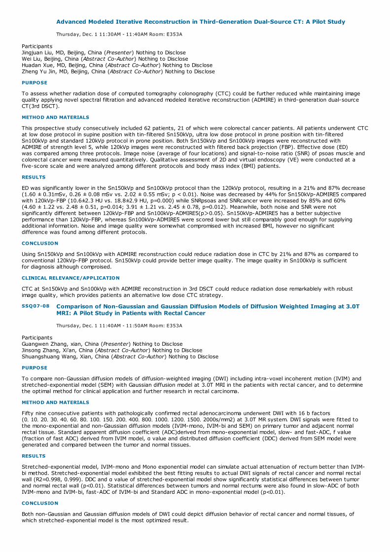
Advanced Modeled Iterative Reconstruction in Third-Generation Dual-Source CT: A Pilot Study
Thursday, Dec. 1 11:30AM - 11:40AM Room: E353A
SSQ07-08 Comparison of Non-Gaussian and Gaussian Diffusion Models of Diffusion Weighted Imaging at 3.0TMRI: A Pilot Study in Patients with Rectal Cancer
Thursday, Dec. 1 11:40AM - 11:50AM Room: E353A
ParticipantsJingjuan Liu, MD, Beijing, China (Presenter) Nothing to DiscloseWei Liu, Beijing, China (Abstract Co-Author) Nothing to DiscloseHuadan Xue, MD, Beijing, China (Abstract Co-Author) Nothing to DiscloseZheng Yu Jin, MD, Beijing, China (Abstract Co-Author) Nothing to Disclose
PURPOSE
To assess whether radiation dose of computed tomography colonography (CTC) could be further reduced while maintaining imagequality applying novel spectral filtration and advanced modeled iterative reconstruction (ADMIRE) in third-generation dual-sourceCT(3rd DSCT).
METHOD AND MATERIALS
This prospective study consecutively included 62 patients, 21 of which were colorectal cancer patients. All patients underwent CTCat low dose protocol in supine position with tin-filtered Sn150kVp, ultra low dose protocol in prone position with tin-filteredSn100kVp and standard 120kVp protocol in prone position. Both Sn150kVp and Sn100kVp images were reconstructed withADMIRE of strength level 5, while 120kVp images were reconstructed with filtered back projection (FBP). Effective dose (ED)was compared among three protocols. Image noise (average of four locations) and signal-to-noise ratio (SNR) of psoas muscle andcolorectal cancer were measured quantitatively. Qualitative assessment of 2D and virtual endoscopy (VE) were conducted at afive-score scale and were analyzed among different protocols and body mass index (BMI) patients.
RESULTS
ED was significantly lower in the Sn150kVp and Sn100kVp protocol than the 120kVp protocol, resulting in a 21% and 87% decrease(1.60 ± 0.31mSv, 0.26 ± 0.08 mSv vs. 2.02 ± 0.55 mSv; p < 0.01). Noise was decreased by 44% for Sn150kVp-ADMIRE5 comparedwith 120kVp-FBP (10.6±2.3 HU vs. 18.8±2.9 HU, p=0.000) while SNRpsoas and SNRcancer were increased by 85% and 60%(4.60 ± 1.22 vs. 2.48 ± 0.51, p=0.014; 3.91 ± 1.21 vs. 2.45 ± 0.78, p=0.012). Meanwhile, both noise and SNR were notsignificantly different between 120kVp-FBP and Sn100kVp-ADMIRE5(p>0.05). Sn150kVp-ADMIRE5 has a better subjectiveperformance than 120kVp-FBP, whereas Sn100kVp-ADMIRE5 were scored lower but still comparably good enough for supplyingadditional information. Noise and image quality were somewhat compromised with increased BMI, however no significantdifference was found among different protocols.
CONCLUSION
Using Sn150kVp and Sn100kVp with ADMIRE reconstruction could reduce radiation dose in CTC by 21% and 87% as compared toconventional 120kVp-FBP protocol. Sn150kVp could provide better image quality. The image quality in Sn100kVp is sufficientfor diagnosis although comproised.
CLINICAL RELEVANCE/APPLICATION
CTC at Sn150kVp and Sn100kVp with ADMIRE reconstruction in 3rd DSCT could reduce radiation dose remarkablely with robustimage quality, which provides patients an alternative low dose CTC strategy.
ParticipantsGuangwen Zhang, xian, China (Presenter) Nothing to DiscloseJinsong Zhang, Xi'an, China (Abstract Co-Author) Nothing to DiscloseShuangshuang Wang, Xian, China (Abstract Co-Author) Nothing to Disclose
PURPOSE
To compare non-Gaussian diffusion models of diffusion-weighted imaging (DWI) including intra-voxel incoherent motion (IVIM) andstretched-exponential model (SEM) with Gaussian diffusion model at 3.0T MRI in the patients with rectal cancer, and to determinethe optimal method for clinical application and further research in rectal carcinoma.
METHOD AND MATERIALS
Fifty nine consecutive patients with pathologically confirmed rectal adenocarcinoma underwent DWI with 16 b factors(0,10,20,30,40,60,80,100,150,200,400,800,1000,1200,1500,2000s/mm2) at 3.0T MR system. DWI signals were fitted tothe mono-exponential and non-Gaussian diffusion models (IVIM-mono, IVIM-bi and SEM) on primary tumor and adjacent normalrectal tissue. Standard apparent diffusion coefficient (ADC)derived from mono-exponential model, slow- and fast-ADC, f value(fraction of fast ADC) derived from IVIM model, α value and distributed diffusion coefficient (DDC) derived from SEM model weregenerated and compared between the tumor and normal tissues.
RESULTS
Stretched-exponential model, IVIM-mono and Mono exponential model can simulate actual attenuation of rectum better than IVIM-bi method. Stretched-exponential model exhibited the best fitting results to actual DWI signals of rectal cancer and normal rectalwall (R2=0.998, 0.999). DDC and α value of stretched-exponential model show significantly statistical differences between tumorand normal rectal wall (p<0.01). Statistical differences between tumors and normal rectums were also found in slow-ADC of bothIVIM-mono and IVIM-bi, fast-ADC of IVIM-bi and Standard ADC in mono-exponential model (p<0.01).
CONCLUSION
Both non-Gaussian and Gaussian diffusion models of DWI could depict diffusion behavior of rectal cancer and normal tissues, ofwhich stretched-exponential model is the most optimized result.
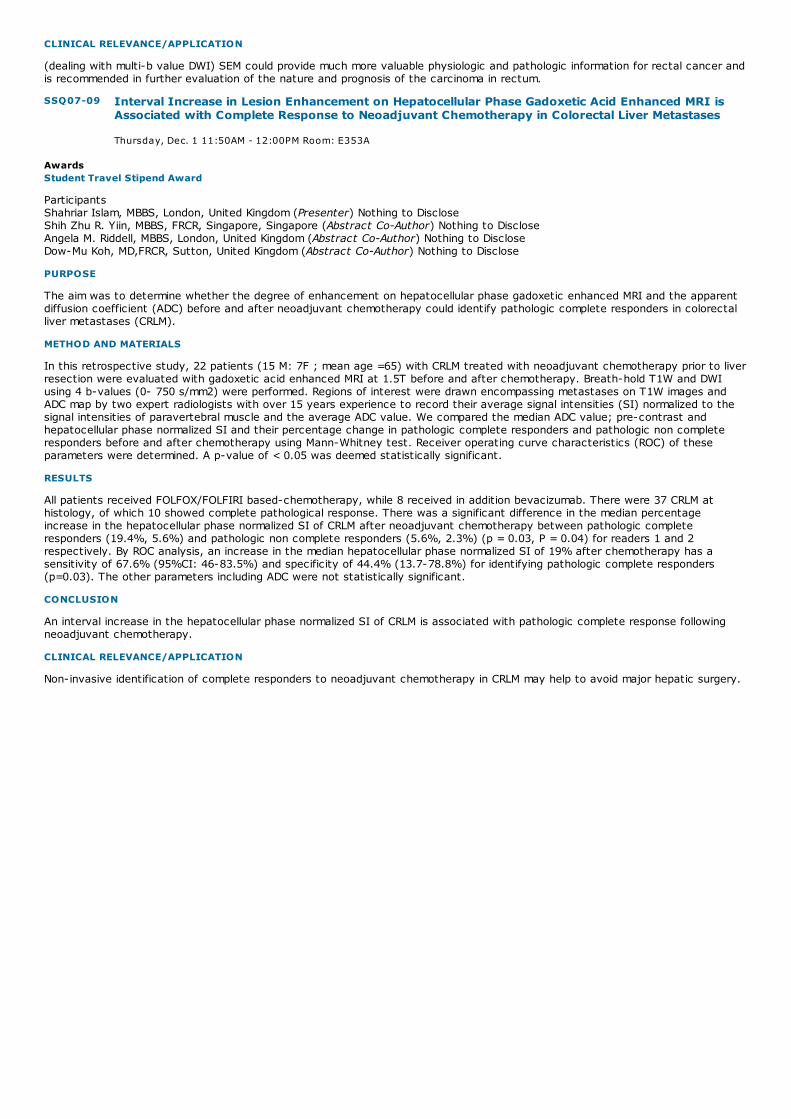
SSQ07-09 Interval Increase in Lesion Enhancement on Hepatocellular Phase Gadoxetic Acid Enhanced MRI isAssociated with Complete Response to Neoadjuvant Chemotherapy in Colorectal Liver Metastases
Thursday, Dec. 1 11:50AM - 12:00PM Room: E353A
CLINICAL RELEVANCE/APPLICATION
(dealing with multi-b value DWI) SEM could provide much more valuable physiologic and pathologic information for rectal cancer andis recommended in further evaluation of the nature and prognosis of the carcinoma in rectum.
AwardsStudent Travel Stipend Award
ParticipantsShahriar Islam, MBBS, London, United Kingdom (Presenter) Nothing to DiscloseShih Zhu R. Yiin, MBBS, FRCR, Singapore, Singapore (Abstract Co-Author) Nothing to DiscloseAngela M. Riddell, MBBS, London, United Kingdom (Abstract Co-Author) Nothing to DiscloseDow-Mu Koh, MD,FRCR, Sutton, United Kingdom (Abstract Co-Author) Nothing to Disclose
PURPOSE
The aim was to determine whether the degree of enhancement on hepatocellular phase gadoxetic enhanced MRI and the apparentdiffusion coefficient (ADC) before and after neoadjuvant chemotherapy could identify pathologic complete responders in colorectalliver metastases (CRLM).
METHOD AND MATERIALS
In this retrospective study, 22 patients (15 M: 7F ; mean age =65) with CRLM treated with neoadjuvant chemotherapy prior to liverresection were evaluated with gadoxetic acid enhanced MRI at 1.5T before and after chemotherapy. Breath-hold T1W and DWIusing 4 b-values (0- 750 s/mm2) were performed. Regions of interest were drawn encompassing metastases on T1W images andADC map by two expert radiologists with over 15 years experience to record their average signal intensities (SI) normalized to thesignal intensities of paravertebral muscle and the average ADC value. We compared the median ADC value; pre-contrast andhepatocellular phase normalized SI and their percentage change in pathologic complete responders and pathologic non completeresponders before and after chemotherapy using Mann-Whitney test. Receiver operating curve characteristics (ROC) of theseparameters were determined. A p-value of < 0.05 was deemed statistically significant.
RESULTS
All patients received FOLFOX/FOLFIRI based-chemotherapy, while 8 received in addition bevacizumab. There were 37 CRLM athistology, of which 10 showed complete pathological response. There was a significant difference in the median percentageincrease in the hepatocellular phase normalized SI of CRLM after neoadjuvant chemotherapy between pathologic completeresponders (19.4%, 5.6%) and pathologic non complete responders (5.6%, 2.3%) (p = 0.03, P = 0.04) for readers 1 and 2respectively. By ROC analysis, an increase in the median hepatocellular phase normalized SI of 19% after chemotherapy has asensitivity of 67.6% (95%CI: 46-83.5%) and specificity of 44.4% (13.7-78.8%) for identifying pathologic complete responders(p=0.03). The other parameters including ADC were not statistically significant.
CONCLUSION
An interval increase in the hepatocellular phase normalized SI of CRLM is associated with pathologic complete response followingneoadjuvant chemotherapy.
CLINICAL RELEVANCE/APPLICATION
Non-invasive identification of complete responders to neoadjuvant chemotherapy in CRLM may help to avoid major hepatic surgery.
SSQ08-01 Gastrointestinal Keynote Speaker: Update on the Role of Imaging in the Management of Esophagusand Gastric Cancer
Thursday, Dec. 1 10:30AM - 10:40AM Room: E353C
SSQ08-02 Barium Swallow: Is It Still Useful in Modern Radiology?
Thursday, Dec. 1 10:40AM - 10:50AM Room: E353C
SSQ08-03 Esophageal Carcinoma at 3.0-T: High-resolution Ex Vivo MR Imaging Compared withHistopathological Findings
Thursday, Dec. 1 10:50AM - 11:00AM Room: E353C
SSQ08
Science Session with Keynote: Gastrointestinal (Esophagus and Stomach)
Thursday, Dec. 1 10:30AM - 12:00PM Room: E353C
GI CT MR
AMA PRA Category 1 Credits ™: 1.50ARRT Category A+ Credits: 1.50
FDA Discussions may include off-label uses.
ParticipantsDouglas S. Katz, MD, Mineola, NY (Moderator) Nothing to DiscloseWilliam C. Small, MD, PhD, Atlanta, GA (Moderator) Nothing to Disclose
Sub-Events
ParticipantsDouglas S. Katz, MD, Mineola, NY (Presenter) Nothing to Disclose
Honored Educators
Presenters or authors on this event have been recognized as RSNA Honored Educators for participating in multiple qualifyingeducational activities. Honored Educators are invested in furthering the profession of radiology by delivering high-qualityeducational content in their field of study. Learn how you can become an honored educator by visiting the website at:https://www.rsna.org/Honored-Educator-Award/
Douglas S. Katz, MD - 2013 Honored EducatorDouglas S. Katz, MD - 2015 Honored Educator
ParticipantsCheng Xie, MBChB,BSC, Oxford, United Kingdom (Presenter) Nothing to DiscloseSophie Shilston, Oxford, United Kingdom (Abstract Co-Author) Nothing to DisclosePeter Cox, Oxford, United Kingdom (Abstract Co-Author) Nothing to DiscloseHorace F. D'Costa, Oxford, United Kingdom (Abstract Co-Author) Nothing to Disclose
PURPOSE
Elderly patients complaining of dysphagia but do not meet the criteria for direct endoscopy are often referred for barium swallowexamination (BSE) to investigate both structural and functional abnormalities. The purpose of this study was to evaluate thediagnostic accuracy of BSE in the detection of oesophageal malignancy in this group of patients and illustrate its complementaryrole in modern radiology.
METHOD AND MATERIALS
This is a retrospective cohort study of 200 consecutive patients (mean age 72 years) who had BSE for dysphagia without alarmingsymptoms of malignancy. Patients’ BSE results, subsequent investigations including endoscopy/biopsy, histology, CT and up to 3years of electronic case notes were reviewed.
RESULTS
BSE showed 22 suspicious malignant strictures, 50 normal findings, 128 benign causes of dysphagia in which 74% were due todysmotility. Subsequent endoscopy and histology confirmed 15/22 cases of oesophageal malignancy (8 adenocarcinomas; 7squamous cell carcinomas), 5 benign strictures (Barrett’s oesophagus and peptic disease), and 2 normal results. These findingsyielded 100% sensitivity, 96.2% specificity of BSE for the detection of oesophageal malignancy in this group of patients. Positiveand negative predictive values were 68.2% and 100%, respectively. In addition, a BSE case series was collected to illustrate itsrole in the diagnosis and exclusion of incidental oesophageal mass.
CONCLUSION
BSE not only plays an important role in the diagnosis of functional oesophageal disorders, but it also demonstrates high sensitivityand specificity in the detection of oesophageal malignancy in the elderly patient with non-specific dysphagia.
CLINICAL RELEVANCE/APPLICATION
For elderly patients complaining of dysphagic symptoms without alarming features of malignancy BSE can serve as an effective firstline investigation. This can help reduce the demand of endoscopy for every elderly patient without red flag symptoms and act analternative investigation for those who are unable to tolerate endoscopy. It is also an useful tool for investigating incidentaloesophageal mass found on CT.
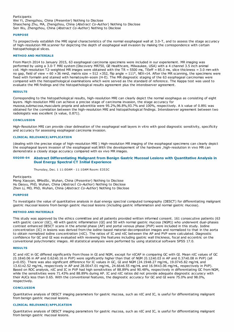
SSQ08-04 Abstract Differentiating Malignant from Benign Gastric Mucosal Lesions with Quantitative Analysis inDual Energy Spectral CT Initial Experience
Thursday, Dec. 1 11:00AM - 11:10AM Room: E353C
ParticipantsWei Yi, Zhengzhou, China (Presenter) Nothing to DiscloseShaocheng Zhu, MA, Zhengzhou, China (Abstract Co-Author) Nothing to DiscloseSen Wu, Zhengzhou, China (Abstract Co-Author) Nothing to Disclose
PURPOSE
To prospectively establish the MRI signal characteristics of the normal esophageal wall at 3.0-T, and to assess the stage accuracyof high-resolution MR scanner for depicting the depth of esophageal wall invasion by making the correspondence with certainhistopathological slices.
METHOD AND MATERIALS
From March 2014 to January 2015, 63 esophageal carcinoma specimens were included in our experiement. MR imaging wasperformed by using a 3.0-T MRI system (Discovery MR750, GE Healthcare, Milwaukee, USA) with a 4 channel 3.5 inch animalcoil. High-resolution T2-weighted MR images were obtained with the TR = 5500 ms, TEeff = 85.0 ms, slice thickness = 3.0 mm withno gap, field of view = 60 ×36 mm2, matrix size = 512 ×352, flip angle = 111°, NEX=14. After the MR scanning, the specimens werefixed with formalin and stained with hematoxylin-eosin (H-E). The MR diagnostic staging of the 63 esophageal carcinomas werecompared with the histopathological examinations which were served as the standard of reference. The Kappa test was used toevaluate the MR findings and the histopathological results agreement plus the interobserver agreement.
RESULTS
Corresponding to the histopathological results, high-resolution MRI can clearly depict the normal esophagus as consisting of eightlayers. High-resolution MRI can achieve a precise stage of carcinoma invasion, the stage accuracy formusosa,submucosa,muscularis propria and adventitia were 95.2%,96.8%,93.7% and 100%, respectively. A k value of 0.891 wasobtained for the correlation between the high-resolution MRI and histopathological findings. Interobserver agreement between tworadiologists was excellent (k value, 0.871).
CONCLUSION
High-Resolution MRI can provide clear delineation of the esophageal wall layers in vitro with good diagnostic sensitivity, specificityand accuracy for assessing esophageal carcinoma invasion.
CLINICAL RELEVANCE/APPLICATION
(dealing with the precise stage of high-resolution MRI ) High-resolution MR imaging of the esophageal specimens can clearly depictthe esophageal layers invasion of the esophageal wall.With the developement of the hardware ,high-resolution in vivo MR candemonstrate a closely stage accuracy compared with ex vivo MR images.
ParticipantsMeng Xiaoyan, BMedSc, Wuhan, China (Presenter) Nothing to DiscloseHu Daoyu, PhD, Wuhan, China (Abstract Co-Author) Nothing to DiscloseZhen Li, MD, PhD, Wuhan, China (Abstract Co-Author) Nothing to Disclose
PURPOSE
To investigate the value of quantitative analysis in dual energy spectral computed tomography (DESCT) for differentiating malignantgastric mucosal lesions from benign gastric mucosal lesions (including gastric inflammation and normal gastric mucosa).
METHOD AND MATERIALS
This study was approved by the ethics committee and all patients provided written informed consent. 161 consecutive patients (63with gastric cancer (GC), 48 with gastric inflammation (GI) and 50 with normal gastric mucosa (NGM)) who underwent dual-phasescontrast enhanced DESCT scans in the arterial phase (AP) and portal venous phase (PVP) were included in this study. Iodineconcentration (IC) in lesions was derived from the iodine-based material-decomposition images and normalized to that in the aortato obtain normalized iodine concentration (nIC). The ratios of IC and nIC between the AP and PVP were calculated. Diagnosticconfidence for GC and GI was evaluated with reviewing the features including gastric wall thickness, focal and eccentric on theconventional polychromatic images. All statistical analyses were performed by using statistical software SPSS 17.0.
RESULTS
IC and nIC in GC differed significantly from those in GI and NGM, except for nICAP in comparing GC with GI. Mean nIC values of GC(0.18±0.06 in AP and 0.62±0.16 in PVP) were significantly higher than that of NGM (0.12±0.03 in AP and 0.37±0.08 in PVP) (allp<0.05). There was also significant difference for IC values in GC, GI and NGM (24.19±8.27 mg/mL, 19.07±5.82 mg/mL and13.61±2.52 mg/mL, respectively in AP and 28.00±7.01 mg/mL, 24.66±6.55 mg/mL and 16.94±3.06 mg/mL, respectively in PVP).Based on ROC analysis, nIC and IC in PVP had high sensitivities of 88.89% and 90.48%, respectively in differentiating GC from NGM,while the sensitivities were 71.43% and 88.89% during AP. IC and nIC ratios did not provide adequate diagnostic accuracy withtheir AUCs less than 0.65. With the conventional features, the diagnostic accuracy for GC and GI were 75.0% and 98.0%,respectively.
CONCLUSION
Quantitative analysis of DESCT imaging parameters for gastric mucosa, such as nIC and IC, is useful for differentiating malignantfrom benign gastric mucosal lesions.
CLINICAL RELEVANCE/APPLICATION
Quantitative analysis of DESCT imaging parameters for gastric mucosa, such as nIC and IC, is useful for differentiating malignantfrom benign gastric mucosal lesions.
SSQ08-05 Extramural Venous Invasion Detected by MDCT as an Adverse Feature in Predicting SynchronousMetastases in T4 Gastric Cancer
Thursday, Dec. 1 11:10AM - 11:20AM Room: E353C
SSQ08-06 Preoperative TNM Stating of Initially Unresectable Gastric Cancer After Neoadjuvant Chemothereapy:Diagnostic Performance of MDCT Using New Staging Criteria
Thursday, Dec. 1 11:20AM - 11:30AM Room: E353C
SSQ08-07 A Prediction Model using Computed Tomographic Findings for Peritoneal Carcinomatosis in Patientswith Advanced Gastric Cancer
ParticipantsJin Cheng, MD, Beijing, China (Presenter) Nothing to DiscloseJing Wu, Beijing, China (Abstract Co-Author) Nothing to DiscloseYingjiang Ye, Beijing, China (Abstract Co-Author) Nothing to DiscloseYinli Zhang, Beijing, China (Abstract Co-Author) Nothing to DiscloseYi Wang, MD, Beijing, China (Abstract Co-Author) Nothing to Disclose
PURPOSE
To determine the association between synchronous metastatic disease in patients with T4 gastric cancer and EMVI detected oncontrast-enhanced multiple-row detector computed tomography (ceMDCT).
METHOD AND MATERIALS
152 patients with T4 gastric carcinoma were retrospectively reviewed and divided into EMVI-positive and EMVI-negative groupswhere EMVI, as detected on ceMDCT, was defined as a tubular or nodular soft tissue thickening extending from the tumor along thevessels of the mesentery. Synchronous metastases were detected by ceMDCT and/or confirmed by post-operative diagnosis.Logistic regression analyses were performed to analyze the predictive factors of synchronous metastases in gastric cancer.
RESULTS
Synchronous metastases were found in 47 of 152 (30.9%) patients with T4 gastric cancer. 31 of 77 (40.3%) patients in the EMVI-positive group had evidence of metastases compared to 16 (21.3%) of 75 patients in the EMVI-negative group (P=0.019).Synchronous metastases were significantly associated with EMVI with an odds ratio (OR) of 2.250 (95% CI, 1.072-4.724).
CONCLUSION
EMVI-positive tumors, as an adverse imaging feature, were significantly associated with synchronous metastases in patients withT4 gastric cancer.
CLINICAL RELEVANCE/APPLICATION
(Dealing with CT imaging) “Contrast-enhanced multiple-row detector CT can demonstrate the extramural venous invasion and isrecommended in the initial evaluation of gastric cancer before curative surgery.”
ParticipantsIjin Joo, MD, Seoul, Korea, Republic Of (Presenter) Nothing to DiscloseSe Hyung Kim, Seoul, Korea, Republic Of (Abstract Co-Author) Nothing to DiscloseCheong-Il Shin, MD, Seoul, Korea, Republic Of (Abstract Co-Author) Nothing to DiscloseSu Joa Ahn, Seoul , Korea, Republic Of (Abstract Co-Author) Nothing to DiscloseJoon Koo Han, MD, Seoul, Korea, Republic Of (Abstract Co-Author) Nothing to Disclose
PURPOSE
To evaluate the diagnostic performance of new MDCT criteria for TNM staging in gastric cancer patients after neoadjuvantchemotherapy.
METHOD AND MATERIALS
This retrospective study was approved by our institutional review board and the requirement for informed consent was waived. Weincluded thirty-seven patients with initially unresectable gastric cancers who underwent neoadjuvant chemotherapy followed bysurgery. MDCT images were reviewed to determine the TNM staging of gastric cancers using new criteria dedicated to assessmentafter chemotherapy. In the new criteria, non-enhancing perigastric infiltrations do not affect T-staging; non-enhancing lymphnodes are regarded as negative lymph nodes; and subtle remaining infiltrations after marked decrease in distant metastasis size areregarded as M0. Using pathologic stages or operation records as the reference standard, the diagnostic performance of MDCT usingthe new criteria was assessed.
RESULTS
According to histologic analysis, T-staging was confirmed in 34 patients including 5 ypT4b cases and N-staging was confirmed in 37patients including 17 ypN-positive cases. Using histology or operation records, 10 patients were confirmed as M1 and 26 patients asM0. Sensitivity, specificity, and accuracy of MDCT for the diagnosis of adjacent organ invasion (T4b); regional LN metastases (N-positive); and distant metastases (M1) were 80.0% (4/5), 96.6% (28/29), 94.1% (32/34); 70.6% (12/17), 70.0% (14/20), 70.3%(26/37); and 60.0% (6/10), 100% (26/26), 88.9% (32/36), respectively.
CONCLUSION
By applying new criteria for initially unresectable gastric cancers after neoadjuvant chemotherapy, MDCT demonstrated gooddiagnostic performances for the determination of adjacent organ invasion or distant metastases.
CLINICAL RELEVANCE/APPLICATION
In unresectable gastric cancer patients, MDCT may be useful in selecting resection candidates after neoadjuvant therapy as itshows high specificity for adjacent organ invasion and distant metastases.
Thursday, Dec. 1 11:30AM - 11:40AM Room: E353C
SSQ08-08 CT Perfusion Evaluation of Gastric Cancer: Correlation with Histologic Type
Thursday, Dec. 1 11:40AM - 11:50AM Room: E353C
ParticipantsMimi Kim, MD, Seoul, Korea, Republic Of (Abstract Co-Author) Nothing to DiscloseWoo Kyoung Jeong, MD, Seoul, Korea, Republic Of (Presenter) Nothing to DiscloseDongil Choi, Seoul, Korea, Republic Of (Abstract Co-Author) Nothing to DiscloseKyoung Doo Song, MD, Seoul, Korea, Republic Of (Abstract Co-Author) Nothing to DiscloseSoon Jin Lee, Seoul, Korea, Republic Of (Abstract Co-Author) Nothing to DiscloseWon Jae Lee, MD, Seoul, Korea, Republic Of (Abstract Co-Author) Research Grant, Samsung Electronics Co, Ltd
PURPOSE
The sensitivity of computed tomography (CT) for detection of peritoneal carcinomatosis (PC) of advanced gastric cancer (AGC)before surgery is relatively low. The purpose of our study is to develop a predictive model derived from the CT features of PC in thepatients with AGC.
METHOD AND MATERIALS
94 patients (M:F=54:40; mean age, 56.0 years; range, 24-83 years) with PC and randomly sampled 120 patients (M:F=82:39; meanage, 59.4 years; range, 31-83 years) among the 360 AGC patients without PC, who underwent preoperative CT and undergoingsurgery between January 2012 and December 2014 were included in the study. A radiologist retrospectively analyzed tumor size,presence of lymph node metastasis, and subtle findings of PC including peritoneal thickening without enhancement, stranding,plaque and small bowel wall thickening or irregularity on a five-point scale and presence of ascites. The definitive CT finding of PCincluding omental cake and rectal shelf was also evaluated. A prediction model named as PC score was constructed by CT featuresindependently associated with PC with use of multiple logistic regression analysis and diagnostic performance was compared withthat of the definitive findings of PC using ROC curve analysis.
RESULTS
Three imaging features of were independently associated with PC: Tumor size larger than 4.2cm (odds ratio [OR]=11.42 [4.02–32.45]), lymph node metastasis (OR=2.20 [1.19–4.06]) and plaque (OR=4.38 [2.86–6.70]). The PC score could be calculated byfollowing formula: PC score = 0.79×lymph node metastasis + 1.48×grades of peritoneal plaque + 2.44×tumor larger than 4.2cm.The PC score showed higher area under the receiver operating characteristic curve (AUC) of 0.915 in the prediction of the PC withhigher sensitivity(88.3%) than definitive finding of PC (AUC=0.694, sensitivity=38.3%)
CONCLUSION
Tumor size, lymph node metastasis and peritoneal plaque grading are independently associated with PC of AGC and the derivedprediction model shows higher AUC and sensitivity for prediction of PC than definitive finding.
CLINICAL RELEVANCE/APPLICATION
We developed a robust model with high sensitivity to predict PC with the use of preoperative CT findings, and this model may behelpful in customizing the treatment for AGC patients.
ParticipantsDong Ho Lee, MD, Seoul, Korea, Republic Of (Presenter) Nothing to DiscloseSe Hyung Kim, Seoul, Korea, Republic Of (Abstract Co-Author) Nothing to DiscloseJoon Koo Han, MD, Seoul, Korea, Republic Of (Abstract Co-Author) Nothing to Disclose
PURPOSE
To prospectively evaluate if the perfusion parameters of gastric cancer can provide information regarding histologic types of gastriccancers.
METHOD AND MATERIALS
Our institutional review board approved this study and informed consent was obtained from all patients. Between October 2015 andMarch 2016, 19 patients who underwent preoperative perfusion CT (PCT) and subsequent curative gastrectomy were enrolled. PCTdata were analyzed using dedicated software program (VPCT body, Siemens Medical System). We calculated blood flow, bloodvolume, mean transit time, time-to-peak, and permeability surface value of gastric cancers. Perfusion parameters were comparedaccording to histologic type (poorly cohesive carcinoma [PCC] versus non-PCC), tumor differentiation (well or moderatelydifferentiated versus poorly differentiated) and N-staging (positive versus negative lymph node metastasis) using Mann-Whitney Utest. Receiver operating characteristic analysis was used to determine the optimal cut-off value of significant parameters.
RESULTS
Permeability surface value of PCC was significantly higher than other histologic types (44.9 ml/100g/min in PCC versus 25.2ml/100g/min in non-PCC, P=0.005). The area under the curve for permeability surface value to predict PCC was 0.875. Sensitivity of88.9% (8/9) and specificity of 90.9% (10/11) were achieved with a cut-off value of 37.9 ml/100g/min. Other perfusion parameterswere not significantly different according to histologic type, tumor differentiation, and N-staging of gastric cancers.
CONCLUSION
Obtaining perfusion parameters from PCT was feasible in all patients with gastric cancers. A preoperative imaging diagnosis of PCCtype gastric cancers could be possible with a permeability surface value of PCC which was significantly higher than non-PCC.
CLINICAL RELEVANCE/APPLICATION
Exact preoperative differentiation between poorly cohesive carcinoma (PCC) and non-PCC by perfusion CT may be clinically usefulas CT can overcome the sampling error by endoscopic biopsy.
SSQ08-09 A Comparison Between 3.0T MRI and Histopathology for Preoperative T Staging of PotentiallyResectable Esophageal Cancer
Thursday, Dec. 1 11:50AM - 12:00PM Room: E353C
ParticipantsZhaoqi Wang, Zhengzhou, China (Presenter) Nothing to DiscloseHui Liu, Shanghai, China (Abstract Co-Author) Employee, Siemens AGHongkai Zhang, zhengzhou, China (Abstract Co-Author) Nothing to DiscloseJinrong Qu, Zhengzhou, China (Abstract Co-Author) Nothing to Disclose
PURPOSE
To explore the value of 3.0T MRI using multiple sequences (star VIBE, Blade and Diffusion-weighted imaging/DWI) in evaluating thepreoperative T staging for potentially resectable espophageal cancer (EC) with pathologic confirmation.
METHOD AND MATERIALS
Sixty-six patients with endoscopically proven EC and indeterminate T1/T2/T3/T4a staging by CT were examined on a 3T scanner.Two independent readers assigned a T staging on MRI according to the 7th edition of UICC-AJCC TNM Classification, and post-operative pathologic confirmation was considered the gold standard. Inter-reader agreement was also calculated.
RESULTS
The accuracy of MRI T staging was: 82% (54/66) for reader 1, 86% (57/66) for reader 2, Inter-reader agreement is excellent(kappa=0.951). For the EC with invading lamina propria, muscularis mucosae, or submucosa (T1 staging), the T staging of MRIachieved 82% (14/17) agreement for both reader 1 and 2; For invasion of muscularis propria (T2 staging), MRI agreement achieved90%(20/22)for reader 1 and 100%(22/22)for reader 2; while for the invasion of adventitia (T3 staging), MRI could achieveagreement in 86%(18/21) for two readers; then for the invasion of pleura, pericardium, diaphragm, or adjacent peritoneum (T4astaging), MRI could achieve agreement is 33%(2/6) and 50%(3/6) for reader 1 and 2, respectively.
CONCLUSION
3.0T MRI using multiple sequences (star VIBE、Blade and DWI) showed high accuracy for patients with potentially resectable EC in Tstaging. The T staging accuracy of lamina propria or submucosa, muscularis proria and adventitia is better than adjacent membranestructure. 3.0T MRI using multiple sequences could be used as a noninvasive imaging method for pre-operative T staging of EC.
CLINICAL RELEVANCE/APPLICATION
3.0T MRI using multiple sequences (star VIBE、Blade and DWI) could be used as a noninvasive imaging method for pre-operative Tstaging of EC.
GI401-SD-THA1
Texture Analysis in Estimating the Malignancy Grade of Hepatocellular Carcinoma using DynamicEnhanced-MRI:A Primary Study
Station #1
GI402-SD-THA2
Revisiting the Role of Ultrasound Surveillance for Hepatocellular Carcinoma (HCC) in in Chronic LiverDisease: Does it Work Well?
Station #2
GIS-THA
Gastrointestinal Thursday Poster Discussions
Thursday, Dec. 1 12:15PM - 12:45PM Room: GI Community, Learning Center
GI
AMA PRA Category 1 Credit ™: .50
FDA Discussions may include off-label uses.
ParticipantsBhavik N. Patel, MD,MBA, Durham, NC (Moderator) Nothing to Disclose
Sub-Events
ParticipantsHe Qing Wang, MSc, shanghai, China (Presenter) Nothing to DiscloseMengsu Zeng, MD, PhD, Shanghai, China (Abstract Co-Author) Nothing to DiscloseAilian Liu, MD, Dalian, China (Abstract Co-Author) Nothing to DiscloseShengxiang Rao, MD, Shanghai, China (Abstract Co-Author) Nothing to DiscloseYe Ju, Dalian, China (Abstract Co-Author) Nothing to DiscloseJiyong Wang, MSc, Jiading, China (Abstract Co-Author) Researcher, Shanghai United Imaging Healthcare Co, LtdXin Weng, shanghai, China (Abstract Co-Author) Nothing to Disclose
PURPOSE
The aim of the study was to access the efficiency of texture analysis (TA) in estimating the malignancy grade of hepatocellularcarcinoma (HCC) by MR images.
METHOD AND MATERIALS
A total of 96 histologically proved HCCs were retrospectively studied. The institutional research ethics board approvedthis study and waived informed consent. Categorizing the pathologic malignancy grade in term of Edmondson'spathologic classification criterion(ⅠⅠ~ⅣⅣgrade): grade I (3 cases), grade I-II (2 cases), grade II (45 cases), grade II-III(26 cases), grade III (20 cases). We randomly choose 64 HCCs as training sets (including 39 HCCs of grade I | I-II | IIand 25 HCCs of grade II-III | III) 32 HCCs as test sets (including 11 HCCs of grade I | I-II | II and 21 HCCs of grade II-III| III). Up to 253 features were extracted as descriptors for each region of interest (ROI). The MR images comprised ofpre-contrast (including T1-, T2- and diffusion weighted imaging) and post-dynamic contrast (including arterial phase,portal venous phase and delayed phase) series. For each series 253 features were extracted, so totally 1771 featureswere calculated on all 7 series. Then a stepwise feature selection strategy was employed to select out a subset offeatures from the total 1771 features. Performance was assessed by measuring the area under the receiver operatingcharacteristics curve (AUC).
RESULTS
A total of 12 features (see table 2) were selected in the training group. The accuracy and AUROC were 90.4% and 0.934in training sets, and 78% and 0.813 in test sets separately.
CONCLUSION
TA maybe a reliable non-invasive method with the potential for effective use in estimating the malignancy grade of HCCusing MRI. It can detect microscopic tissue changes in different malignancy grade of HCC that visual inspection cannotfind.
CLINICAL RELEVANCE/APPLICATION
Texture analysis can detect microscopic tissue changes in different malignancy grade of HCC that visual inspectioncannot find. TA maybe helpful to estimate the malignancy grade of HCC by MR images for the patients of advanced HCCthat cannot do operation. So we may make a correct judgment of prognosis for the patients of advanced HCC.
GI403-SD-THA3
Preclinical Spectral Detector CT Scanner Derived Iodine-Based Spectral Images reveal ImprovedImage Quality Metrics Compared to Conventional CT
Station #3
ParticipantsSu Joa Ahn, Seoul , Korea, Republic Of (Presenter) Nothing to DiscloseJung Hoon Kim, MD, Seoul, Korea, Republic Of (Abstract Co-Author) Nothing to DiscloseYoung Rock Jang, Seoul, Korea, Republic Of (Abstract Co-Author) Nothing to DiscloseJoon Koo Han, MD, Seoul, Korea, Republic Of (Abstract Co-Author) Nothing to Disclose
PURPOSE
We investigated the role of US surveillance for HCC in chronic liver disease and analyzed the factors which effect on US detectionof HCC
METHOD AND MATERIALS
From Jan.2013 to Dec. 2013, 16986 patients with chronic liver disease underwent surveillance US. Among them, 1901patients (1252 men, 649 women; average age, 60.04 years) who underwent both surveillance US and had subsequently CT within 6months included in this study. We retrospective evaluated baseline patient characteristics including patient factors, US findings,and laboratory assessment. We also assessed factor which effect on the development of HCC within 2 years.
RESULTS
158 patients (8.3%, 158/1901) with US surveillance were confirmed HCC. For 158 HCCs, US detected 81 HCC (51%). Thesensitivity, specificity, PPV, and NPV of surveillance US were 51.3%, 90.8%, 33.6%, and 95.4%, respectively. Tumor size(21.48±14.57 mm vs. 25.21±14.81 mm), echo texture, surface nodularity, and grade of fibrosis on US showed significant differentfactors for detection of HCC on surveillance US (p<0.005). In multivariate analysis, surface nodularity (OR, 0.314; 95% CI: 0.15,0.67; p= .003) on US was the only significant factor for detection of HCC on US. There was no significant difference of BarcelonaClinic Liver Cancer (BCLC) staging between detected and undetected HCC on surveillance US (p=0.121). At the time of HCCdiagnosis, 127 patients (80%, 127/158) were in BCLC very early stage or early stage.On follow-up for 1743patients without HCC,HCC developed in 110 patients within 2 years (mean time: 13 months). According to the multivariable analysis, numerousindeterminate nodules<1cm on US (OR, 2.15; 95% CI: 1.23, 2.94;p< .01), age (OR, 1.05; 95% CI: 1.03, 1.07;p< .01), ALT (OR,1.01; 95% CI: 1.00, 1.01;p= .02), and platelet count (OR, 0.99; 95% CI: 0.99, 1.0;p< .01) were significantly related to thedevelopment of HCC.
CONCLUSION
US surveillance for HCC plays important role for early detection of HCC in chronic liver disease. However, US findings including echotexture, surface nodularity, and grade of fibrosis effect the detection of HCC. Numerous indeterminate nodules<1cm on USincreased the risk of development of HCC within 2 years.
CLINICAL RELEVANCE/APPLICATION
Because of numerous indeterminate nodules<1cm on US increased the risk of development of HCC within 2 years, close surveillanceis need in this patients
ParticipantsSuhny Abbara, MD, Dallas, TX (Presenter) Author, Reed Elsevier; Editor, Reed Elsevier; Institutional research agreement, KoninklijkePhilips NV; Institutional research agreement, Siemens AGLakshmi Ananthakrishnan, MD, Dallas, TX (Abstract Co-Author) Nothing to DiscloseWilliam A. Moore, MD, Dallas, TX (Abstract Co-Author) Nothing to DiscloseLuna Fahoum, Andover, MA (Abstract Co-Author) Employee, Koninklijke Philips NVZimam Romman, Haifa, Israel (Abstract Co-Author) Employee, Koninklijke Philips NVJulia R. Fielding, MD, Dallas, TX (Abstract Co-Author) Nothing to DiscloseXinhui Duan, PhD, Dallas, TX (Abstract Co-Author) Nothing to Disclose
PURPOSE
Several types of Iodine-based spectral derived images (SDIs) can be reconstructed from a preclinical prototype Spectral DetectorCT (SDCT) scanner (IQon, Philips Healthcare) at no additional radiation dose. The potential incremental value over conventionalfiltered back projection (FBP) images alone has not been evaluated to date. We aim to determine image quality parameters andpotential incremental value of iodine-based SDIs obtained from a SDCT scanner.
METHOD AND MATERIALS
This prospective IRB approved study consists of 562 individual CT DICOM dataset reconstructed from 73 unique SBIs in 53 patients(some had multiple contrast phases). Reconstructions included conventional CT, and the following spectral derived images: Iodine-No-Water and quantitative Iodine-Density image datasets.Image quality and lesion detectability was evaluated on Likert scales(much worse, worse, similar, moderately better, much better) for 1) iodine enhancement 2) hypodense lesion/structure detection,3) hyper dense lesion/structure detection.Diagnostic reader confidence was evaluated on a 4 point scale (low, moderate, good,high). Comparative statistics were performed.
RESULTS
Iodine enhancement seen in Iodine-No-Water scored 'moderately better’ (3.9±0.9, n=31) and ‘similar’ to ‘moderately better’ onIodine-Density SDIs (3.6±1.0, n=35) compared to FBP.Detection of hyper dense structures on Iodine-No-Water and Iodine-Densityimages was ‘similar’ to ‘moderately better’ (3.5±1.1, n=31 and 2.9±1.5, n=34).Compared to conventional images, no added valuewas found in the detection of hypo dense structures (Iodine-No-Water 2.2±1.2, n=30, Iodine-Density 2.0±1.3, n=34).Diagnosticconfidence rating averages scored ‘good’ to ‘high’ for iodine enhancement on the Iodine-no-Water images (3.1±0.8) and 'moderate'to 'good' on the Iodine-Density images. Hyper dense structure detection was ‘moderate’ to ‘good' on the Iodine-No-Water images.
CONCLUSION
Iodine-based spectral derived images can be obtained from a novel detection based spectral CT scanner. This study shows that
GI404-SD-THA4
The Prognostic Value of Gadoxetic-acid Enhanced MRI in Prediction of Patients' Survival FollowingLiver Transplantation for Hepatocellular Carcinoma
Station #4
GI405-SD-THA5
Dynamic Enhancement Pattern of Insulinomas on Volume Perfusion CT
Station #5
Iodine-based spectral derived images can be obtained from a novel detection based spectral CT scanner. This study shows thatthese SDIs improved high attenuation structure detectability due to improved contrast enhancement compared to conventional FBPimages.
CLINICAL RELEVANCE/APPLICATION
Iodine-based spectral derived images from a novel detection based spectral CT scanner have favourable image qualityparameters and incremental value for lesion detectability compared to conventional CT images alone.
Honored Educators
Presenters or authors on this event have been recognized as RSNA Honored Educators for participating in multiple qualifyingeducational activities. Honored Educators are invested in furthering the profession of radiology by delivering high-qualityeducational content in their field of study. Learn how you can become an honored educator by visiting the website at:https://www.rsna.org/Honored-Educator-Award/
Suhny Abbara, MD - 2014 Honored Educator
ParticipantsAh Yeong Kim, MD, Daejeon, Korea, Republic Of (Abstract Co-Author) Nothing to DiscloseWoo Kyoung Jeong, MD, Seoul, Korea, Republic Of (Presenter) Nothing to DiscloseTae Wook Kang, MD, Seoul, Korea, Republic Of (Abstract Co-Author) Nothing to Disclose
PURPOSE
Given the limited availability of donor organs for liver transplantation (LT) and recent attempt to expand the Milan criteria, carefulpatient selection is mandatory to reduce tumor recurrence and maximize the effectiveness. We hypothesized that preoperativemagnetic resonance imaging (MRI) can provide prognostic power in prediction of tumor recurrence and survival rate in patient withLT for hepatocellular carcinoma (HCC).
METHOD AND MATERIALS
HCC patients (n=65) with preoperative gadoxetic-acid enhanced MRI underwent LT between Jan. 2009 to Jun. 2012. MRIparameters including signal intensity of the tumor, enhancement pattern and diffusivity were evaluated retrospectively by tworadiologists. Overall and disease-free survival rates were analyzed. Uni- and multivariate analyses with MRI parameters, serumalpha-fetoprotein (AFP), and pathologic findings were performed for prediction of overall survival and HCC recurrence. Andrelationship between selected variables in imaging and pathologic findings was evaluated.
RESULTS
The 3-year overall and recurrence-free survival rate were 76.7% and 70.4%, respectively. In multivariate analyses, only peritumoralhypointensity on hepatobiliary phase (HBP) (hazard ratio [HR], 8.77; p<0.001) on MRI and intrahepatic metastasis (HR, 2.89;p=0.03) on pathology were significant predictable factor for overall survival. And both the presence of satellite nodule (HR, 5.56;p=0.006) and peritumoral hypointensity on HBP (HR, 11.90; p<0.001) on MRI and histologic grade (HR, 4.98; p =0.006) and absenceof fibrous capsule (HR, 3.03; p=0.018) on pathology were significant risk of tumor recurrence. AFP was an independent prognosticfactor for both overall survival (HR, 1.26; p=0.025) and tumor recurrence (HR, 1.47; p<0.001). Among the significant predictivevariables on preoperative MRI, peritumoral hypointensity on HBP was correlated to the tumor grade on pathologic finding (p=0.015).And presence of satellite nodule on preoperative MRI was correlated to the intrahepatic metastasis on pathology (p<0.001).
CONCLUSION
Peritumoral hypointensity on HBP on preoperative MRI was significant predictable factor for overall survival and tumor recurrenceafter LT for HCC, and correlated to the tumor grade on pathologic finding.
CLINICAL RELEVANCE/APPLICATION
Prediction of tumor recurrence and survival after LT for HCC can be attempted using preoperative MRI, and this might be helpful inselection of best candidate for LT.
ParticipantsLiang Zhu, MD, Beijing, China (Presenter) Nothing to DiscloseHuadan Xue, MD, Beijing, China (Abstract Co-Author) Nothing to DiscloseWei Liu, Beijing, China (Abstract Co-Author) Nothing to DiscloseHao Sun, MD, Beijing, China (Abstract Co-Author) Nothing to Disclose
PURPOSE
To assess the enhancement pattern of insulinomas on volume perfusion CT (VPCT), and to select phases for optimal tumor-parenchyma contrast.
METHOD AND MATERIALS
From August 2014 to December 2015, consecutive patients who underwent VPCT of the pancreas (80kV) with clinicallysuspected insulinomas were identified. Patients who received surgery and had pathological diagnosis of insulinomas were included,and patients with known multiple endocrine syndrome were excluded. Two experienced radiologists retrospectively evaluated tumorenhancement patterns on VPCT and analyzed the time-attenuation curve of the tumor and pancreatic parenchyma in consensus.The tumors were identified on CT with reference to surgical reports. Tumor-parenchyma contrast at each time point was measuredand phases for optimal tumor-parenchyma contrast were selected.
GI406-SD-THA6
3D Dynamic Contrast-Enhanced Ultrasound of Liver Metastases from Gastrointestinal Tumors: First-in-Human Assessment of Feasibility and Reproducibility
Station #6
GI286-ED-THA7
Collateral Damage: Unintended Consequences of Oncologic Therapy in the Chest, Abdomen, andPelvis
Station #7
RESULTS
Sixty-three patients were included,with 63 tumors.Five tumors were isoattenuating (tumor-parenchyma contrast <20HU in all 25dynamic phases), 19 tumors had transient hyperenhancement (tumor-parenchyma contrast >20HU, duration < 10s), and 39 tumorshad persistent hyperenhancement. Optimal tumor-parenchyma contrast was observed 9 s after abdominal aorta arriving atthreshold of 200HU (AAT), with mean tumor-parenchyma attenuation difference of 77.6±57.2HU. At 9 s after AAT, 14 tumors wereisoattenuating, including 5 tumors with intrinsic low contrast and 9 tumors with missed transient hyperenhancement.Complementary phases could be 12 s after AAT (detects another 4 tumors), 4 s after AAT and 1s after AAT (both detect another 3tumors).
CONCLUSION
VPCT enables detection of insulinomas even if the hyperenhancement is transient. Optimal tumor-parenchyma contrast occurs 9 safter AAT. Tumors that are inconspicous on single- or biphasic contrast enhanced CT are mainly due to missed transienthyperenhancement.
CLINICAL RELEVANCE/APPLICATION
By analyzing dynamic enhancement of insulinomas, a single opitmal phase was selected, which yeilded the maximum tumor-parenchyma contrast and facilitates detection of insulinomas with low radiation dose. It was also found that transienthyperenhancement was the main reason that some insulinomas were inconspicuous on single- or biphasic enhanced CT.VPCT coulddetect insulinomas even if the hyperenhancement was transient.
ParticipantsAhmed El Kaffas, PhD, Palo Alto, CA (Presenter) Co-founder, OncousticsRosa Maria Silveira Sigrist, MD, Palo Alto, CA (Abstract Co-Author) Nothing to DiscloseGeorge Fisher, MD, PhD, Stanford, CA (Abstract Co-Author) Nothing to DiscloseSunitha Bachawal, PhD, Stanford, CA (Abstract Co-Author) Nothing to DiscloseJoy Liau, MD, PhD, Palo Alto, CA (Abstract Co-Author) Nothing to DiscloseAlexander Karanany, Palo Alto, CA (Abstract Co-Author) Nothing to DiscloseJarrett Rosenberg, PhD, Stanford, CA (Abstract Co-Author) Nothing to DiscloseHuaijun Wang, MD, PhD, Stanford, CA (Abstract Co-Author) Nothing to DiscloseDimitre Hristov, PhD, Stanford, CA (Abstract Co-Author) Research Grant, Koninklijke Philips NV Partner, SoniTrack Systems, IncJuergen K. Willmann, MD, Stanford, CA (Abstract Co-Author) Research Consultant, Bracco Group; Research Grant, Siemens AG;Research Grant, Bracco Group; Research Grant, Koninklijke Philips NV; Research Grant, General Electric Company; Advisory Board,Lantheus Medical Imaging, Inc; Advisory Board, Bracco Group
PURPOSE
Dynamic contrast enhanced ultrasound (DCE-US) is a low-cost tool proposed for identifying early responders to cancer therapy. Todate, sampling errors resulting from 2D imaging have restricted DCE-US when used to assess highly heterogeneous tumors. Thepurpose of this study was to perform a first-in-human clinical assessment of 3D DCE-US feasibility and reproducibility, and as a wayto overcome sampling errors.
METHOD AND MATERIALS
Patients with liver metastases from gastrointestinal cancers were imaged with a Philips EPIQ7 coupled to an X6-1 matrix transducer.A total of 16 scan sessions were carried out over 10 patients. Pairs of repeated bolus and disruption-replenishment images wereacquired within each scan session to determine reproducibility of parameters. Bolus consisted of intravenous injection of 0.2 mlDefinity microbubbles followed by saline. Disruption-replenishment was carried out by infusing 0.9 ml of Definity microbubbles in 35.1ml of saline over 8 min. Volumes-of-interest (VOI) and regions-or-interest (ROI) were segmented in each image to extract time-intensity curves (TICs). Parameters were quantified for the whole VOI and 4 sub-ROIs. Bolus parameters were: time-to-peak (TP),peak enhancement (PE), area-under-the-curve (AUC), mean-transit-time (MTT). Disruption-replenishment parameters were:relative blood volume (rBV), relative blood flow (rBF) and regional mean flow velocity (rMFV).
RESULTS
A large coefficient of variation (CV) was for ROIs from the same volume confirming potential sampling errors. The TP and MTT hadthe lowest CV while the rBF, rBV and rMFV parameters had the largest plane-to-plane variations with CVs up to 54%.Measurements made in 3D were consistently different than measurements made in 2D with an average percent difference of 60%.Reproducibility, evaluated by the concordance correlation coefficient (CCC) between repeated measurements, was good (0.80) toexcellent (0.95). The TP and MTT were the least reproducible with CCCs lesser than 0.80.
CONCLUSION
This first in human study confirms 2D DCE-US sampling errors and demonstrates that 3D DCE-US imaging with a matrix arraytransducer is feasible and reproducible in the clinic.
CLINICAL RELEVANCE/APPLICATION
DCE-US is inexpensive, exempt from radiation exposure and available at the bedside. Eliminating sampling errors through 3D imagingfurther potentiates its role as a clinical tool to identify early response to cancer therapy.
ParticipantsJulie C. Bulman, MD, Dallas, TX (Abstract Co-Author) Nothing to DiscloseGaurav Khatri, MD, Dallas, TX (Presenter) Nothing to Disclose
GI300-ED-THA8
HIV below the Belt: Infections, Tumors and Everything in Between
Station #8
GI183-ED-THA9
Abdominal Masses with Central Scar: No Organ Left Behind
Station #9
Lori M. Watumull, MD, Dallas, TX (Abstract Co-Author) Research Grant, Toshiba CorporationYull Arriaga, Dallas, TX (Abstract Co-Author) Nothing to DiscloseJohn R. Leyendecker, MD, Dallas, TX (Abstract Co-Author) Nothing to Disclose
TEACHING POINTS
Radiologists must differentiate effects of various oncologic therapies to help direct management and improve patient outcomes. GIcomplications from oncologic therapies include inflammation, pneumatosis, fistulae, perforation, graft-versus-host disease (GVHD).Unintended liver abnormalities from oncologic therapy include steatosis, hepatitis, fibrosis, vascular changes. Pancreaticobiliary, GU,vascular, and other systemic complications may manifest on imaging.
TABLE OF CONTENTS/OUTLINE
Chemotherapy- cytotoxic, molecular, immunotherapy GI - inflammation, pneumatosis, necrosis, perforation, fistula, bleed Liver -Steatosis, pseudocirrhosis, autoimmune hepatitis, Hepatic Veno-Occlusive Disease (HVOD) Pancreaticobiliary - Pancreatitis,cholangitis, strictures Genitourinary - Nephrotoxicity, cystitis Vascular – Arterial/venous thrombosis, atherosclerosis Other organs,pseudoprogression, radiation recall Local/regional Rx (ablation, trans-arterial chemoembolization, radiation therapy) GI - Gastritis,radiation enteritis Liver - Radiation injury, fibrosis Biliary - Cholangitis, stricture, biloma, gallbladder injury GU - renal, bladder, fistulaOther - infection, dehiscence Bone Marrow Transplant Liver - HVOD, portal hypertension GI - inflammation, pneumatosis, fistulaSystemic – PTLD, GVHD
ParticipantsCecil Patel, MD, Los Angeles, CA (Presenter) Nothing to DiscloseCecilia M. Jude, MD, Los Angeles, CA (Abstract Co-Author) Author, UpToDate, IncMaitraya K. Patel, MD, Sylmar, CA (Abstract Co-Author) Nothing to DiscloseJacqueline Tang, Sylmar, CA (Abstract Co-Author) Nothing to DiscloseMichael J. Nguyen, MD, Santa Barbara, CA (Abstract Co-Author) Nothing to DiscloseAnokh Pahwa, MD, Los Angeles, CA (Abstract Co-Author) Nothing to DiscloseShaden F. Mohammad, MD, Sylmar, CA (Abstract Co-Author) Nothing to Disclose
TEACHING POINTS
Because HIV can remain undiagnosed or untreated, abdominal manifestations of HIV-associated diseases may be a main presentingfeature. Familiarity with these diseases can lead to correct diagnosis and treatment.The purpose of this exhibit is to: Review thepathophysiology and clinical presentations of HIV-associated infections and neoplasms in the abdomen and pelvis. Illustrate thecharacteristic imaging features of these entities, including treatment related abnormalities. Integrate clinical andradiographic findings to guide appropriate management.
TABLE OF CONTENTS/OUTLINE
HIV-related infections and neoplasms can have a variety of manifestations and involve multiple organ systems. Treatment responsemay mimic worsening of an underlying disease. Opportunistic infections Bacterial Mycobacteria Clostridium difficile Typhlitis: gram-positive organisms Bartonella henselae Fungal Candida Histoplasma capsulatum Viral Cytomegalovirus Herpes simplex virus 3Protozoal infections: Cryptosporidium Pneumocystis jirovecii AIDS-related neoplasia Kaposi sarcoma Non-Hodgkin’s lymphoma HIV-related organ manifestations Treatment related Immune reconstitution inflammatory syndrome (IRIS)
ParticipantsAmina Saghir, MBBS, Shrewsbury, MA (Presenter) Nothing to DiscloseDaniel Kowal, MD, Sturbridge, MA (Abstract Co-Author) Nothing to Disclose
TEACHING POINTS
To present abdominal mass lesions associated with a central scar (CS). To emphasize that while CS is important to recognize, it isnot always pathognomonic for diagnosis, but is often useful as a supplemental diagnostic feature.
TABLE OF CONTENTS/OUTLINE
Numerous abdominal masses contain a CS which may guide a differential diagnosis. A clear understanding of imaging characteristicsof not only the scar, but of the mass itself is imperative to avoid pitfalls and mimics. Focal nodular hyperplasia, fibrolamellarhepatocellular carcinoma, giant hemangioma, and metastases are hepatic masses that may be associated with CS. MRI signalcharacteristics, enhancement patterns, hepatocyte-specific gadolinium-based contrast agents, and presence of calcification aid todifferentiate hepatic masses. Pancreatic serous cystadenoma is a benign lesion with a CS which may be calcified. Sclerosingangiomatoid nodular transformation is a rare splenic lesion often associated with a CS which is T2 hyperintense and hypoenhancingon MRI. Splenic pseudotumor is also a rare benign entity with a central stellate scar. CS is seen in 50% of cases of benign renaloncocytoma, but renal cell carcinoma may also exhibit a scar. CS in an abdominal mass is important to recognize, but its presenceor absence should not preclude the diagnosis of certain abdominal tumors.
GI407-SD-THB1
Diagnostic Performance of Surveillance US and Influencing Factors on US Visibility of HCCs: AProspective Study in Patients with Cirrhosis at High Risk of HCC
Station #1
GI408-SD-THB2
Diagnostic Accuracy of Colonic Lesions in Ultra-low Dose CT Colonography: Is Diagnosis in Sub-mSvPossible?
Station #2
GIS-THB
Gastrointestinal Thursday Poster Discussions
Thursday, Dec. 1 12:45PM - 1:15PM Room: GI Community, Learning Center
GI
AMA PRA Category 1 Credit ™: .50
FDA Discussions may include off-label uses.
ParticipantsBhavik N. Patel, MD,MBA, Durham, NC (Moderator) Nothing to Disclose
Sub-Events
ParticipantsSo Hyun Park, MD, Incheon, Korea, Republic Of (Presenter) Nothing to DiscloseHyung Jin Won, MD, Seoul, Korea, Republic Of (Abstract Co-Author) Nothing to DiscloseSo Yeon Kim, MD, Seoul, Korea, Republic Of (Abstract Co-Author) Nothing to DiscloseJae Ho Byun, MD, Seoul, Korea, Republic Of (Abstract Co-Author) Nothing to DiscloseSo Jung Lee, Seoul, Korea, Republic Of (Abstract Co-Author) Nothing to DiscloseSeong Ho Park, MD, Seoul, Korea, Republic Of (Abstract Co-Author) Research Grant, DONGKOOK Pharmaceutical Co, LtdJihyun An, Seoul, Korea, Republic Of (Abstract Co-Author) Nothing to DiscloseYoung-Suk Lim, Seoul, Korea, Republic Of (Abstract Co-Author) Nothing to Disclose
PURPOSE
To investigate diagnostic performance, the characteristics of nodules detected on surveillance US and influencing factors on USvisibility of HCCs in patients with cirrhosis at high risk of HCC.
METHOD AND MATERIALS
A prospective surveillance study included 407 consecutive patients with an estimated annual risk of HCC >5% who underwent oneto three, biannual surveillance US examinations combined with US elastography between November 2011 and August 2014. Thefindings of lesions detected on US were recorded in a predefined standardized way using a four-point scale indicating the likelihoodof HCC, i.e. suspicious, equivocal, probably benign or definitely benign/negative, corresponding to categories 4, 3, 2, and 1,respectively. The confirmation of HCC was based on the results of a histologic examination and/or typical CT images of HCC. Thedetection rate of HCC and the false-positive rate were calculated. The characteristics of category 4 nodules detected on US weredetermined using the simultaneous or follow-up gadoxetic acid-enhanced MRI and CT. We investigated clinical factors, including theChild-Pugh class and body mass index as well as imaging characteristics such as liver stiffness, US image quality, tumor location,and tumor size, all of which possibly influenced the US visibility of HCCs.
RESULTS
Among the 43 patients with HCCs found during 1100 US screening sessions, the detection rate of HCC was 27.9% (12/43) and thefalse positive rate was 5.6% (59/1057). Among the 71 category 4 lesions, 12 were HCC, 27 were not matched with any lesionsseen on MRI or CT, 20 were confirmed as cirrhosis-related nodules, four were exophytic hepatic parenchyma, four werehemangiomas, three were complicated cysts, and one was abnormal vasculature. Among the clinical and imaging characteristics, asubcapsular location was the only significant factor related to the failure to detect HCC on US (P=0.019). Among 20 HCCs insubcapsular locations, US detected only two HCCs.
CONCLUSION
In patients with cirrhosis at high risk of HCC, surveillance US did not show satisfactory diagnostic performance. The subcapsularlocation of HCC can be the cause of the suboptimal detection rate.
CLINICAL RELEVANCE/APPLICATION
US is a suboptimal surveillance tool for HCC in patients with cirrhosis at high risk of HCC. The subcapsular area of the liver is wherestrict scrutiny should be applied when US is performed in these patients.
ParticipantsTakaaki Yasuda, RT, Nagasaki, Japan (Presenter) Nothing to DiscloseTetsuro Honda, MD, Nagasaki, Japan (Abstract Co-Author) Nothing to DiscloseTakahiro Yasaka, Nagasaki, Japan (Abstract Co-Author) Nothing to DiscloseKenichi Utano, MD, Aizuwakamatu-shi, Japan (Abstract Co-Author) Nothing to DiscloseTakashi Kato, MD, Sapporo, Japan (Abstract Co-Author) Nothing to Disclose
PURPOSE
To evaluate the diagnostic accuracy of ultra-low dose CT Colonography (CTC).
METHOD AND MATERIALS
GI409-SD-THB3
Radiology-pathology Box: A Tool for MRI Guidance of Ex Vivo Histopathologic Analysis
Station #3
GI410-SD-THB4
Value of Advanced Virtual Monoenergetic Images with Third-generation Dual-Energy CT in DetectingEarly Gastric Cancer
Station #4
Two hundred patients, (98 male and 102 female, average age 67.5 years), who were scheduled for total colonoscopy (TCS),underwent ultra-low dose CTC. These patients were scanned in prone and supine positions by the 80-slices MDCT with thescanning conditions set to 120 kV, SD45-50, and an adaptive iterative reconstruction algorithm was applied . The targeted size oflesions, which were pathologically diagnosed as adenoma and adenocarcinoma, were 10 mm or larger. A radiologist (R) and agastroenterologist (G), who had extensive experience of reading CTC images and did not know the endoscopy results, performedthe reading independently. By using TCS as a gold standard, the Sensitivity, Specificity, PPV and NPV were calculated.
RESULTS
DLP (mGy/cm) was 21.31 (0.32 mSv) for supine and 21.27 (0.32 mSv) for prone (converted using an effective dose conversioncoefficient of 0.015).The CTC per-lesion sensitivity R 73% / G 71% and PPV R 79% / G 94% for lesions larger than 10 mm (n = 45). None of the flatlesions was identified in CTC, but the accuracy for elevated lesions excluding flat lesions was sensitivity R 87% / G 84% and PPV R85% / G 94%. For the per-patient, sensitivity, specificity, PPV and NPV were R 74% / G 74%, R 96% / G 99%, R 83% / G 94% andR 94% / G 94%, respectively.
CONCLUSION
Detecting elevated lesions in ultra-low dose CTC is possible with a good diagnostic accuracy; however, detecting flat lesions inultra-low dose CTC is not possible due to the poor accuracy. It is considered as a limitation.
CLINICAL RELEVANCE/APPLICATION
We have reduced the radiation dose as much as possible by utilizing scanning technology clinically available today. Accuracy fordetecting elevated lesions was good at sub-mSv CT scan, and the results of this study will provide an indicator for the mostsuitable ways to achieve dose reduction in the future.
ParticipantsTilman B. Schubert, MD, Madison, WI (Presenter) Nothing to DiscloseSonja Kinner, MD, Madison, WI (Abstract Co-Author) Nothing to DiscloseElisabetta A. Nocerino, Milano, Italy (Abstract Co-Author) Nothing to DiscloseShannon Hynes, MS, Madison, WI (Abstract Co-Author) Nothing to DiscloseChristopher L. Brace, PhD, Madison, WI (Abstract Co-Author) Shareholder, NeuWave Medical Inc Consultant, NeuWave Medical IncShareholder, Symple Surgical Inc Consultant, Symple Surgical IncTimothy Colgan, PhD, Madison, WI (Abstract Co-Author) Nothing to DiscloseEmily Winslow, MD, Madison, WI (Abstract Co-Author) Nothing to DiscloseScott B. Reeder, MD, PhD, Madison, WI (Abstract Co-Author) Institutional research support, General Electric Company Institutionalresearch support, Bracco Group
PURPOSE
Ex vivo pathologic analysis and lesion identification in resected specimen is demanding due to tissue deformation and alteredorientation. Small lesion in particular can therefore be missed during tissue sectioning potentially impacting the staging ofmalignancies. As delineation of resection margins and tumor burden is of utmost importance, we aimed to develop an MRIcompatible localization device and test its feasibility for localizing small lesions in explanted liver tissue.
METHOD AND MATERIALS
An MR-compatible localization device was entirely constructed from Plexiglas consisting of two stationary and one removable MR-visible grid, creating a 3D matrix (Figure). Laser-edged grid lines and labels filled with silicone gel for MR visibility in threedimensions. Alginate is used to fix and stabilize tissue during imaging and cutting. Slicing of the specimen within the box is enabledin a guided fashion through prefabricated grooves corresponding to the grid lines of the device (y-direction). To test the device,five specimen of swine liver with a random number of lesions created by thermoablation (lesion size: 5-15 mm) were imaged on a 3TMR scanner. The MR protocol consisted of a high-resolution T1 SPGRE-sequence (0.5x0.5x0.4mm3). Two readers independentlyevaluated lesion coordinates and size, which was then correlated with results from cutting.
RESULTS
Imaging with the device was feasible and all images were of high diagnostic quality. All 38 lesions were detected in the expectedlocalization with the exception of one lesion incorrectly localized by one of the two readers. Inter-reader agreement of lesionlocalization was excellent (0.92). Size (5-12mm) was correct within +/-2mm, in 34/35 lesions.
CONCLUSION
The MR-visible device proved feasible for detection and localization of liver lesions, and has potential to play an important role inlesion identification and in radiologic-pathologic evaluation. Assuming that a comparably high sensitivity can be achieved in clinicaltrials, this device may enable image-guided pathologic analysis . In addition, this tool might give further insight into radiologic-pathologic correlation of lesions if in vivo-, ex vivo MR data and histopathology is available.
CLINICAL RELEVANCE/APPLICATION
This tool enables ex vivo lesion detection and localization in resected specimen. This might lead to focused histopathologic analysiswhich has the potential to impact tumor staging and grading.
ParticipantsWei Liu, Beijing, China (Presenter) Nothing to Disclose
GI412-SD-THB6
Lymph Node Metastasis in Patients with Gastric Cancer: A Multi-modality, Morphologic and FunctionalImaging Study
Station #6
Jingjuan Liu, MD, Beijing, China (Abstract Co-Author) Nothing to DiscloseHuadan Xue, MD, Beijing, China (Abstract Co-Author) Nothing to DiscloseZheng Yu Jin, MD, Beijing, China (Abstract Co-Author) Nothing to Disclose
PURPOSE
To evaluate the impact of advanced monoenergetic dual-energy computed tomography (DECT) datasets on detection rate andcontrast-to-noise ratio (CNR) of early gastric cancer (EGC) on the third generation DECT.
METHOD AND MATERIALS
20 patients suspected of EGC were prospectively enrolled in this study. Arterial phase (AP) and portal phase (PP) imaging wereacquired in dual-energy mode (80/Sn150KV) using third-generation DECT (SOMATOM Force, Siemens Healthcare, Germany).Monoenergetic images (MEIs) (range 40-100keV) were calculated from the 80 and Sn150KV image data using monoenergeticsoftware (Dual energy Mono+, syngo via, Siemens). Differences in detection rates of EGC and CNR numbers were comparedbetween different MEIs from 40 to 100 keV and conventionally reconstructed polyenergetic images (PEIs) at 120 kVp.Histopathologic analysis of resected gastric specimens was used as reference standard. Radiation metrics were compared withthose obtained in the second-general DECT (Definition Flash, Siemens Healthcare, Germany) from published reports.
RESULTS
MEIs at 40keV and 50keV showed the highest detection rates (60.0% and 60.0% respectively) for EGC, which showed nosignificant difference with PEIs(35%)(P=0.205). For EGC lesions of less than 2cm, an increase in the detection rates was observedfrom MEIs at 40keV and 50keV(53.8%,7/13 and 53.8%,7/13) as compared to PEIs(23.1%,3/13), though no statistical significancewas found (P=0.226). MEIs at 40keV and 50keV revealed statistical higher CNR-AP and CNR-PP compared to PEIs (all P<0.05). TheCTDIvol of the AP and PP were 4.28 ± 0.92 mGy and 4.67 ± 0.95 mGy respectively, which was about 35% of the radiation dosewhen using routine second-generation DECT (100/Sn140KV) from literatures.
CONCLUSION
Virtual 40keV and 50keV monoenergetic images calculated using the Mono+ algorithm significantly increase the CNR of EGC, whichwas helpful to enhance the EGC detection rate. Third-generation DECT could potentially allow for radiation dose reduction in EGCdetection as compared to the second-general DECT.
CLINICAL RELEVANCE/APPLICATION
The third-generation DECT with Mono+ technique could improve the detection rate of early gastric cancer and result in substantialradiation dose reduction for patients.
ParticipantsJing Ren, MD, Chicago, IL (Presenter) Nothing to DiscloseYi Huan, MD, PhD, Xi'an, China (Abstract Co-Author) Nothing to DiscloseFaezeh Sodagari, MD, Chicago, IL (Abstract Co-Author) Grant, Siemens AGAtilla Arslanoglu, MD, Chicago, IL (Abstract Co-Author) Grant, Siemens AGVahid Yaghmai, MD, Chicago, IL (Abstract Co-Author) Nothing to Disclose
PURPOSE
To investigate the value of computed tomography (CT), magnetic resonance imaging (MRI) and diffusion weighted imaging (DWI)for diagnosing lymph nodes metastasis before treatment in gastric cancer.
METHOD AND MATERIALS
Eighty-two patients with proven gastric cancer underwent CT, morphological MRI (T2WI) and DWI examinations. Two radiologistsindependently assessed these images for the presence of lymph nodes involvement. Pathologic findings were considered as “goldstandard”. Independent samples t-test was used for the comparisons of short diameters and ADC values between the positivelymph nodes and the negative lymph nodes. Diagnostic accuracy of these three imaging modalities was evaluated by area under thereceiver operating characteristics (ROC) curve (AUC).
RESULTS
The ADC value of the positive lymph nodes was (1.15 ± 0.01) × 10-3 mm2/s, which was significantly lower than that of thenegative lymph nodes (1.48 ± 0.01) × 10-3 mm2/s (t = 18.70, P < 0.0001). The short diameter of the positive lymph nodes (1.54 ±0.38 cm) was significantly greater than that of the negative lymph nodes (0.95 ± 0.12 mm) (t = 19.03, P < 0.001). The AUC for allimaging modalities combined (0.893) was significantly larger than that for each imaging modality alone (P < 0.05), and the AUC ofDWI (0.797) was significantly larger than (P < 0.05) that of morphological MRI (0.733). There was no statistically significantdifference between the AUCs of CT and morphological MRI (P = 0.462).
CONCLUSION
CT, MRI and DWI combined present significantly higher accuracy than each imaging modality alone in the detection of lymph nodesinvolvement.
CLINICAL RELEVANCE/APPLICATION
CT, MRI and DWI can be used as an imaging biomarker for lymph nodes involvement assessment in gastric cancer.
Honored Educators
Presenters or authors on this event have been recognized as RSNA Honored Educators for participating in multiple qualifyingeducational activities. Honored Educators are invested in furthering the profession of radiology by delivering high-qualityeducational content in their field of study. Learn how you can become an honored educator by visiting the website at:
GI278-ED-THB7
Computed Tomography (CT) Imaging of Abdominal Hernias Following Mesh Repairs
Station #7
GI298-ED-THB8
Hepatic Lesions that Mimic Metastasis on Radiological Imaging during Chemotherapy forGastrointestinal Malignancy: Recent Updates
Station #8
GI113-ED-THB9
The Ever-Changing Landscape of Medical Devices Found in the Abdomen and Pelvis: What theRadiologist Can Tell the Clinician
Station #9
https://www.rsna.org/Honored-Educator-Award/
Vahid Yaghmai, MD - 2012 Honored EducatorVahid Yaghmai, MD - 2015 Honored Educator
ParticipantsEugene Bivins Jr, BS, Washington, D.C,, DC (Presenter) Nothing to DiscloseKovosh Dastan, MSc, Ashburn, VA (Abstract Co-Author) Nothing to DiscloseSarah Mohamedaly, MPH, Washington, D.C., DC (Abstract Co-Author) Nothing to DiscloseAdrian Godoy Vazquez, BS, Washington, DC (Abstract Co-Author) Nothing to DiscloseAndre J. Duerinckx, MD, PhD, Washington, DC (Abstract Co-Author) Nothing to DiscloseBonnie C. Davis, MD, Washington, DC (Abstract Co-Author) Nothing to DiscloseDahn Truong, BS, Washington, DC (Abstract Co-Author) Nothing to DiscloseBelen Tesfaye, MD, Washington, DC (Abstract Co-Author) Nothing to DiscloseDaniel Tran, MD, Washington, DC (Abstract Co-Author) Nothing to Disclose
TEACHING POINTS
At the end of this presentation, participants will be able to: Identify normal findings on CT images after abdominal hernia repairsUnderstand the importance of CT imaging in the identification of mesh displacement, mesh migration, abscess formation, pseudo-abscess formaion, and other complications Better detect and manage complications following hernia repairs in the early post-meshrepair period
TABLE OF CONTENTS/OUTLINE
Review the use of mesh repairs following abdominal hernias and the normal post-op imaging appearance Describe the types ofcomplications that may ensue as a result of hernia repairs and review CT findings of such complications State the relevance ofmonitoring patients in the early post-mesh repair period and early detection of complications
ParticipantsYeo Eun Han, Seoul, Korea, Republic Of (Presenter) Nothing to DiscloseBeom Jin Park, MD, Seoul, Korea, Republic Of (Abstract Co-Author) Nothing to DiscloseMin-Ju Kim, MD, Seoul, Korea, Republic Of (Abstract Co-Author) Nothing to DiscloseDeuk Jae Sung, MD, Seoul, Korea, Republic Of (Abstract Co-Author) Nothing to DiscloseNa Yeon Han, Seoul, Korea, Republic Of (Abstract Co-Author) Nothing to DiscloseKi Choon Sim, Seoul, Korea, Republic Of (Abstract Co-Author) Nothing to DiscloseSung Bum Cho, Seoul, Korea, Republic Of (Abstract Co-Author) Nothing to Disclose
TEACHING POINTS
For multidisciplinary teams involved in cancer care, the precise imaging validation of a newly detected hepatic nodule has becomecrucial to achieve optimal treatment during chemotherapy for malignancies. Hepatic incidentaloma arising from chemotherapy–induced parenchymal injury, tumor-associated eosinophilic abscess, and fungal infection should be considered a mimicker ofmetastasis in patients with gastrointestinal malignancy. We have reviewed widely used causative chemo-agents, the updatedconcept of chemotherapy–induced hepatopathies, and other hepatic pseudometastasis focusing on their pathological andradiological findings for accurate diagnosis.
TABLE OF CONTENTS/OUTLINE
A. The concept of imitator of hepatic metastasis during chemotherapyB. Chemotherapy-induced focal hepatopathyChemotherapy for gastrointestinal malignancy Chemotherapy-induced focal sinusoidal injury Pathophysiology / Clinicalissues/ Imaging Features Chemotherapy-induced focal steatosis/steatohepatitis Pathophysiology / Clinical issues/ ImagingfeaturesC. Tumor-associated eosinophilic abscess in the liver Pathophysiology / Clinical issues/ Imaging featuresD.Compromised immunity-associated fungal abscess in the liver Pathophysiology / Clinical issues/ Imaging featuresE. TheTake-home message
ParticipantsEverett Gu, MD, Ann Arbor, MI (Presenter) Nothing to DiscloseAshish P. Wasnik, MD, Ann Arbor, MI (Abstract Co-Author) Nothing to DiscloseRavi K. Kaza, MD, Ann Arbor, MI (Abstract Co-Author) Nothing to DiscloseMahmoud M. Al-Hawary, MD, Ann Arbor, MI (Abstract Co-Author) Nothing to Disclose
TEACHING POINTS
While viewing this exhibit, participants will: Review common medical devices and gain exposure to newer or more uncommon medicaldevices found in the abdomen and pelvis. Appreciate the physical appearance of a spectrum of medical devices as portrayed inphotographs, and be comfortable recognizing these devices on plain radiographs and CT. Understand how to properly evaluatevarious medical devices, including appropriate positioning and potential complications.
TABLE OF CONTENTS/OUTLINE
Introduction explaining the importance of identifying and evaluating medical devices Overview of various medical devices found in

Introduction explaining the importance of identifying and evaluating medical devices Overview of various medical devices found inthe abdomen and pelvis, grouped into five major categories: Gastrointestinal (e.g. Sengstaken-Blakemore tubes, esophageal stents,over-the-scope clips) Hepatobiliary (e.g. cholecystostomy tubes, biliary stents, cystogastrostomy stents) Genitourinary (e.g.vaginal pessaries, tubal ligation clips, artificial urinary sphincters) Vascular (e.g. endografts, endovascular cooling catheters) Bodywall (e.g. spinal cord stimulators, baclofen pumps) Dedicated slides for each medical device, which will focus on the purpose of thedevice, appropriate placement, and potential complications, supplemented with photographs and radiology images Summaryemphasizing the role of radiology in evaluating medical devices
MSCA51A Pancreatic Imaging
MSCA51B Hepatobiliary Disorders
MSCA51C Congenital Abdominal Pathology that Can be Seen in Adults
MSCA51
Case-based Review of the Abdomen (An Interactive Session)
Thursday, Dec. 1 1:30PM - 3:00PM Room: S406A
GI
AMA PRA Category 1 Credits ™: 1.50ARRT Category A+ Credits: 1.50
ParticipantsJulie H. Song, MD, Sharon, MA (Director) Nothing to Disclose
Sub-Events
ParticipantsJay P. Heiken, MD, Saint Louis, MO, ([email protected] ) (Presenter) Patent agreement, Guerbet SA; Patent agreement, BayerAG
LEARNING OBJECTIVES
1) Identify the differential imaging features of solid and cystic pancreatic neoplasms. 2) Make appropriate recommendations forcharacterization, follow-up and/or treatment of an incidentally discovered pancreatic mass. 3) Recognize and appropriatelycharacterize pancreatitis and its complications.
ParticipantsJeong Min Lee, MD, Seoul, Korea, Republic Of (Presenter) Grant, Guerbet SA; Support, Siemens AG; Grant, Bayer AG; Grant, GeneralElectric Company; Grant, STARmed Co, Ltd; Grant, RF Medical Co, Ltd; Grant, Toshiba Corporation; Grant, Samsung MedicalHealthcare
LEARNING OBJECTIVES
1) Demonstrate typical and atypical imaging appearances of common hepatobiliary diseases. 2) Illustrate key imaging findings thataid in differential diagnosis. 3) Learn to avoid pitfalls and misdiagnoses of hepatobiliary lesions.
ParticipantsSudha A. Anupindi, MD, Philadelphia, PA, ([email protected] ) (Presenter) Nothing to Disclose
LEARNING OBJECTIVES
1) Describe the essential imaging techniques used to diagnose important congenital abdominal pathology seen into adulthood. 2)Identify the imaging features of these entities. 3) Apply this knowledge into their clinical practice.
ABSTRACT
VSIO51-01 Setting the Stage: NCCN/ESMO Guidelines for mCRC
Thursday, Dec. 1 1:30PM - 1:45PM Room: S405AB
VSIO51-02 Role of PVE in the Surgical Management of Colorectal Liver Metastases
Thursday, Dec. 1 1:45PM - 2:00PM Room: S405AB
VSIO51-03 Advances in the Surgical Toolbox for Colorectal Liver Metastases
Thursday, Dec. 1 2:00PM - 2:15PM Room: S405AB
VSIO51-04 SBRT for Isolated Hepatic Metastases
Thursday, Dec. 1 2:15PM - 2:30PM Room: S405AB
VSIO51-05 Radiation Map Fusion Guided Combination of External Radiation with Thermal Ablation for up to 5Liver Tumors and Up to 10cm in Diameter
Thursday, Dec. 1 2:30PM - 2:40PM Room: S405AB
VSIO51
Interventional Oncology Series: Management of Hepatic Metastases from Colorectal Cancer, NeuroendocrineTumors and more
Thursday, Dec. 1 1:30PM - 6:00PM Room: S405AB
GI IR
AMA PRA Category 1 Credits ™: 4.50ARRT Category A+ Credit: 0
FDA Discussions may include off-label uses.
ParticipantsSarah B. White, MD,MS, Philadelphia, PA, ([email protected] ) (Moderator) Research support, Guerbet SA; Research support,Siemens AG; Consultant, Guerbet SA; Consultant, IO Rad
LEARNING OBJECTIVES
The overall objective of this session is to get a better understanding of multimodality, multidisciplinary treatment hepatic dominantmetastatic disease. We will accomplish this by:1.) Defining the alphabet soup of the cancer treatments2.) Highlighting the generalalgorithm for the treatment of mCRC and mNETs3.) Briefly discuss the evidence basis behind medical, surgical, radiation and liverdirected therapies4.) Exploring the clinical outcomes and adverse events with the different treatment modalities5.) Demonstrate thenecessity of multidisciplinary tumor boards for the treatment of patients with dominant hepatic metastases
Sub-Events
ParticipantsEmily Bergsland, MD, San Francisco, CA (Presenter) Institutional research support, Lexicon Pharmaceuticals, Inc; Institutionalresearch support, Merck & Co, Inc; Institutional research support, Novartis AG; Consultant, Ipsen SA; Consultant, LexiconPharmaceuticals, Inc;
LEARNING OBJECTIVES
1) Review the general approach to the treatment of metastatic colorectal cancer (mCRC). 2) Summarize the current systemictreatment options for mCRC. 3) Identify the clinical and molecular subgroups of patients with mCRC which have implications in termsof choice of therapy. 4) Examine the differences between published guidelines for the care of patients with mCRC.
ABSTRACT
ParticipantsDavid C. Madoff, MD, New York, NY (Presenter) Advisory Board, RenovoRx
ParticipantsT. Clark Gamblin, MD, MS, Milwaukee, WI (Presenter) Nothing to Disclose
ParticipantsMary U. Feng, MD, San Francisco, CA (Presenter) Nothing to Disclose
ParticipantsHayet Amalou, MD, Bethesda, MD (Presenter) Nothing to DiscloseDeborah Citrin, MD, Bethesda, MD (Abstract Co-Author) Nothing to DiscloseSheng Xu, PhD, Bethesda, MD (Abstract Co-Author) Nothing to DiscloseHolly Ning, Bethesda, MD (Abstract Co-Author) Nothing to DiscloseRobert W. Miller, PhD, Bethesda, MD (Abstract Co-Author) Nothing to DiscloseBradford J. Wood, MD, Bethesda, MD (Abstract Co-Author) Researcher, Koninklijke Philips NV; Researcher, Celsion Corporation;Researcher, BTG International Ltd; Researcher, W. L. Gore & Associates, Inc ; Researcher, Cook Group Incorporated; Patentagreement, VitalDyne, Inc; Intellectual property, Koninklijke Philips NV; Intellectual property, BTG International Ltd; ; ; ; Victoria L. Anderson, MS, Bethesda, MD (Abstract Co-Author) Nothing to Disclose
PURPOSE
External beam radiation therapy (XRT) and thermal ablation (RFA or microwave ablation) have complementary risk profiles and
VSIO51-06 New Angles on Ablating Colorectal Liver Metastases
Thursday, Dec. 1 2:40PM - 2:55PM Room: S405AB
VSIO51-07 Prospective MonoIstitutional Study on Safety and Efficacy of dRug-eluting Microspheres Loaded withIrinotecan in Patients with Colorectal Liver mEtastases (Miracle III Trial)
Thursday, Dec. 1 2:55PM - 3:05PM Room: S405AB
synergistic mechanisms. XRT works better with blood flow, but ablation is impaired by perfusion due to convection. XRT is safer inthe porta hepatis, but ablation with hydrodissection is safer adjacent to bowel. XRT is typically not given for liver lesions largerthan 6 cm due to integral radiation dose toxicities, and ablation is less effective for > 3cm tumors.
METHOD AND MATERIALS
Inclusion criteria included up to 5 liver tumors, each up to 10 cm in diameter. Patients were treated with 5 total fractions (50 Gytotal) of XRT over 2 weeks, and RFA or microwave ablation was between fractions 3 and 4. A navigation system for ablation wasdeveloped fusing radiation dose map, multiple imaging modalities, and probe locations. Following multi-modality liver registration, thesoftware populated a planned ablation treatment volume by overlapping the expected ablation zone of each probe to define tumorcoverage. 3 patients were enrolled.
RESULTS
Fusion software guidance combines XRT with ablation for synergistic treatment of large and/or multiple liver tumors. Thiscombination allows respecting the limitations, while taking advantage of the strengths, of each modality. Multi-modality registrationoverlays CT images, radiation dose map, planned ablation probe insertion paths, and expected overlapping ablation treatmentvolumes from multiple probes. Two patients received spatially synergistic treatment, using XRT and ablation to achieve an overalllarger lesion coverage compared to what each modality might achieve alone. One patient received temporally synergistic treatment(RFA and radiation to same volume), which resulted in an ablation volume of 59 cm3 after 12 minutes of RFA, (nearly 3 timesgreater than the expected volume of 20.5 cm3 seen in porcine studies). On most recent follow-up, one of three patients (1 of 5tumors) had local recurrence and one hepatocellular carcinoma patient remains disease-free over two years after treatment for a10 cm tumor.
CONCLUSION
Fusion navigation guided by radiation dose map input may facilitate combination of complementary therapies of XRT and thermalablation, Both spatial and temporal synergy are feasible but speculative..
CLINICAL RELEVANCE/APPLICATION
Complementary combination therapy with XRT + RFA allows treatment of larger liver lesions in otherwise risky locations.
ParticipantsPaul B. Shyn, MD, Boston, MA, ([email protected] ) (Presenter) Nothing to Disclose
LEARNING OBJECTIVES
ABSTRACT
LEARNING OBJECTIVES
1) Assess the indications for and efficacy of image-guided ablation of liver metastases in patients with colorectal cancer. 2)Examine the spectrum of ancillary techniques and ablation technologies that expand eligibility for liver tumor ablation. 3) Assess thecapabilities of current liver ablation strategies through case examples.
ABSTRACT
ParticipantsGiovanni Mauri, MD, Milan, Italy (Presenter) Consultant, Esaote SpAGianluca M. Varano, Rome, Italy (Abstract Co-Author) Nothing to DiscloseRiccardo Foa, Milan, Italy (Abstract Co-Author) Nothing to DiscloseMaria Zampino, Milan, Japan (Abstract Co-Author) Nothing to DisclosePaola Ravenda, Milan, Italy (Abstract Co-Author) Nothing to DisclosePaolo Della Vigna, MD, Milan, Italy (Abstract Co-Author) Nothing to DiscloseLorenzo Monfardini, Brescia, Italy (Abstract Co-Author) Nothing to DiscloseGuido Bonomo, Milan, Italy (Abstract Co-Author) Nothing to DiscloseFranco Orsi, MD, Rome, Italy (Abstract Co-Author) Nothing to Disclose
PURPOSE
Primary endpoints of the study were the evaluation of the safety of the drug-eluting microspheres loaded with Irinotecan and theliver control rate at three months in patients with colorectal liver metastases. Secondary endpoints were the liver control rate at6 and 12 months, time to progression and overall survival.
METHOD AND MATERIALS
18 patients with colorectal liver or liver-dominant disease (11 M, 7 F, mean age 62 years, range 47-79) were prospectively enrolledbetween November 2013 and February 2015. Main characteristics: all patients received > 2 prior lines of systemic therapy(containing Fluoropyrimidines/Oxaliplatin and Irinotecan), 13 were treated with liver surgery or ablation, 14 had extra-hepaticspread at the time of first embolization, 11 had liver bilobar involvement disease with a mean of 9 (range 1-30) hepaticmetastases and with mean diameter of 47.6 mm (range 12-161 mm). The study procedure was performed by using 40mmmicroparticles loaded with 100 mg of Irinotecan and recycled every 4 weeks, at least twice per each lobe and repeated up todisease progression. Adverse events (AEs) were evaluated according to the NIH-CTC V 4.0 criteria. Response to treatment wasevaluated according to RECIST 1.1criteria. Estimated median for time to local tumor progression was calculated using Kaplan-Meyeranalysis
VSIO51-08 Embolotherapy for CRLM
Thursday, Dec. 1 3:05PM - 3:20PM Room: S405AB
VSIO51-09 Detectability of Liver Metastases by Dual Phase Cone-Beam Computed Tomography during Liver-directed Liver Interventions: Comparison with Conventional Contrast-enhanced ComputedTomography
Thursday, Dec. 1 3:20PM - 3:30PM Room: S405AB
VSIO51-10 Y-90 for CRLM
Thursday, Dec. 1 3:30PM - 3:45PM Room: S405AB
RESULTS
Overall, 69 treatments were performed (mean of 3.8/patient, range 2-9);G1/G2 AEs occurred in 42/69 (60.8) procedures while G3,G4 or G5 complications did not occurred.Liver tumor control was achieved in 14/18 (77.8%) patients at 3 months and in 10/18(55.6%) patients at 6 months. Median time to liver progression was 7 months.
CONCLUSION
Drug-eluting microspheres loaded with Irinotecan are safe and effective in treatment of patients affected by colorectal cancer withpredominant hepatic involvement unresponsive to standard therapies.
CLINICAL RELEVANCE/APPLICATION
drug-eluting microspheres loaded with Irinotecan represent a safe and effective option for treating patients with colorectal livermetastases that could be tested in a larger series and early clinical setting.
ParticipantsMichael C. Soulen, MD, Philadelphia, PA, ([email protected]) (Presenter) Royalties, Cambridge University Press;Consultant, Guerbet SA; Research support, Guerbet SA; Consultant, BTG International Ltd; Research support, BTG InternationalLtd; Consultant, Merit Medical Systems, Inc; Speaker, Sirtex Medical Ltd; Consultant, Terumo Corporation; Consultant, Bayer AG
LEARNING OBJECTIVES
1) Learn to select appropriate patients with metastatic colorectal cancer for liver directed therapies. 2) Understand the data forconventional and drug-eluting chemoembolization for treatment of mCRC.
ABSTRACT
ParticipantsGeert Maleux, MD, PhD, Leuven, Belgium (Presenter) Nothing to DiscloseMaria-Louisa Izamis, Best, Netherlands (Abstract Co-Author) Nothing to DiscloseCedric Werbrouck, Leuven, Belgium (Abstract Co-Author) Nothing to DiscloseAlessandro G. Radaelli, PHD, MS, Best, Netherlands (Abstract Co-Author) Employee, Koninklijke Philips NVVincent R. Vandecaveye, MD, Leuven, Belgium (Abstract Co-Author) Nothing to Disclose
PURPOSE
To compare the diagnostic performance of intra-arterial dual phase cone-beam computed tomography (DP-CBCT) and contrast-enhanced computed tomography (CE-CT) in accurately characterizing tumor burden in patients with metastatic liver cancer.
METHOD AND MATERIALS
This retrospective study included patients with liver metastases, referred for catheter-directed liver intervention. Demographic andradiological data were gathered, including type of tumor, number, maximum size, type and degree of contrast enhancement of livermetastases.
RESULTS
29 patients with colorectal (n=10), breast (n=9) and neuroendocrine (n=10) liver metastases were included. DP-CBCT and CE-CTwere in agreement on number and distribution of liver metastases in 18 out of 29 patients (62%). In 9 out of 11 patients DP-CBCTand CE-CT identified a mean of 7.2 and 10 metastases, respectively (P=0.025). In 2 out of 11 patients DP-CBCT identified less(n=1) or more (n=1) metastases than CE-CT. Metastases were larger in diameter on DP-CBCT than on CE-CT regardless of origin:colorectal: 57 +/- 9.5 mm vs 43 +/- 8.3 mm, P=0.02; breast: 57 +/- 10 mm vs 43 +/- 8.5 mm, P=0.03 and neuroendocrine: 56 +/-6.3 mm vs 51 +/- 5.8 mm, P= 0.01. In colorectal metastases, rim enhancement appeared in 100% of cases on DP-CBCT, but wasvariable on CE-CT. In breast metastases, DP-CBCT displayed thick, hyper-dense rims while CE-CT did not, or had rims of variablethickness and density. Neuroendocrine tumors had variable rim enhancement within the same patient and differed between DP-CBCT and CE-CT in 40% of patients.
CONCLUSION
DP-CBCT appears to identify the vast majority of liver metastases, demonstrates a larger diameter and a peripherally enhancingtumoral rim compared to CE-CT; in more than 30% of patients, DP-CBCT identified more tumors than CE-CT. DP-CBCT providesadditional information to CE-CT that may impact treatment decisions and dosimetry for catheter-directed liver interventions.
CLINICAL RELEVANCE/APPLICATION
DP-CBCT provides additional information, including number, diameter and pattern of contrast enhancement of liver metastases, toCE-CT which may impact treatment decisions for catheter-directed liver interventions.
ParticipantsRobert J. Lewandowski, MD, Chicago, IL, ([email protected] ) (Presenter) Advisory Board, BTG International Ltd;Advisory Board, Boston Scientific Corporation; Consultant, Cook Group Incorporated; Consultant, ABK Medical Inc
VSIO51-11 Differences between Hepatic Distribution of Tc99m MAA and Y-90 Microspheres Could Complicate 3DImage Based Dosimetry
Thursday, Dec. 1 3:45PM - 3:55PM Room: S405AB
VSIO51-12 Question and Answer
Thursday, Dec. 1 3:55PM - 4:10PM Room: S405AB
LEARNING OBJECTIVES
1) Learn the evidence for radioembolization in patients with mCRC (salvage setting). 2) Learn the evidence for radioembolization inpatients with mCRC (combined with systemic therapy). 3) Discuss optimal timing of radioembolization in patients with mCRC.
ParticipantsJulien S. Wonderlick, MD, Burlington, VT (Presenter) Nothing to DiscloseChristopher A. Ford, MD, Burlington, VT (Abstract Co-Author) Nothing to DiscloseSean Reynolds, MD, Burlington, VT (Abstract Co-Author) Nothing to DiscloseJoseph T. Shields, MD, Burlington, VT (Abstract Co-Author) Nothing to DiscloseAnant D. Bhave, MD, Richmond, VT (Abstract Co-Author) Nothing to DiscloseMarleen Moore, MS, Burlington, VT (Abstract Co-Author) Nothing to DiscloseJanusz K. Kikut, MD, Burlington, VT (Abstract Co-Author) Nothing to Disclose
PURPOSE
Selective internal radiation therapy is radioembolization of malignant tumors in the liver with microspheres carrying Y90. 2 products(SIR-Spheres and TheraSpheres) are approved for use in the USA. Prior to injection of either product, treatment planningscintigraphy is performed by selective arterial injection of Tc99m MAA. MAA distribution is taken into account for dose calculationswith a goal of delivering a minimum of 100 Gy to the tumor. There is extensive literature on predictive dosimetry based on the pre-treatment scan. However, such calculations are only valid if the MAA scan is highly predictive of the distribution of SIRT particlespost-embolization. A semi-quantitative scale was used to compare the pre-treatment MAA scan to the post-treatmentBremsstrahlung scan in 30 SIRT cases to determine how well the MAA-planned target volume approximated the Treated LiverVolume on Bremsstrahlung SPECT.
METHOD AND MATERIALS
Consecutive 17 SIRsphere (1 HCC,12 colon, 4 carcinoid) and 13 TheraSphere treatments (12 HCC, 1 neuroendocrine) were reviewedfrom 2012 to present. Comparison of MAA distribution in the liver vs.Y90 TheraSphere and Y90 SIRSphere was done based on thefollowing score:0: TLV-Y90 matches PTV-MAA and are isogenous1: TLV-Y90 matches PTV-MAA with varying pattern of internal heterogeneity2: TLV-Y90 smaller than PTV-MAA3: TLV-Y90 larger than PTV-MAA
RESULTS
MAA and Y90 microsphere distribution matched in 7 (53%) Theraspheres and 2 (12.5%) SIRspheres treatments. In 6 (47%)Theraspheres and 10 (62.5%) SIRspheres treatments the treated volume matched MAA but the implanted Y90 microspheresdemonstrated a different pattern of distribution within the volume. In 4 (25%) SIRspheres treatments the treated volume wasdifferent from the pretreatment MAA scan.
CONCLUSION
Differences in particle number and injection technique are likely culprits for hepatic distribution of implanted Y90 Theraspheres andSIRspheres inconsistently matching the pretreatment MAA scan. This study supports recommendations for post-treatment imagingwith both products and calls into question routine use of MAA Quantitative SPECT scans for voxel by voxel based dosimetry,particularly for SIRspheres.
CLINICAL RELEVANCE/APPLICATION
Significant disagreement between pretreatment MAA and post-treatment Bremsstrahlung scan can lead to less than optimal Y-90dose for radioembolization.
ParticipantsWilliam S. Rilling, MD, Milwaukee, WI (Presenter) Research support, B. Braun Melsungen AG; Research support, Sirtex Medical Ltd;Research support, Siemens AG; Consultant, B. Braun Melsungen AG; Consultant, Cook Group Incorporated ; Consultant, TerumoCorporation; Advisory Board, Terumo CorporationSarah B. White, MD,MS, Philadelphia, PA, ([email protected]) (Presenter) Research support, Guerbet SA; Research support,Siemens AG; Consultant, Guerbet SA; Consultant, IO RadEmily Bergsland, MD, San Francisco, CA (Presenter) Institutional research support, Lexicon Pharmaceuticals, Inc; Institutionalresearch support, Merck & Co, Inc; Institutional research support, Novartis AG; Consultant, Ipsen SA; Consultant, LexiconPharmaceuticals, Inc; David C. Madoff, MD, New York, NY (Presenter) Advisory Board, RenovoRxT. Clark Gamblin, MD, MS, Milwaukee, WI (Presenter) Nothing to DiscloseMary U. Feng, MD, San Francisco, CA (Presenter) Nothing to DisclosePaul B. Shyn, MD, Boston, MA (Presenter) Nothing to DiscloseMichael C. Soulen, MD, Philadelphia, PA, ([email protected]) (Presenter) Royalties, Cambridge University Press;Consultant, Guerbet SA; Research support, Guerbet SA; Consultant, BTG International Ltd; Research support, BTG InternationalLtd; Consultant, Merit Medical Systems, Inc; Speaker, Sirtex Medical Ltd; Consultant, Terumo Corporation; Consultant, Bayer AGRobert J. Lewandowski, MD, Chicago, IL, ([email protected]) (Presenter) Advisory Board, BTG International Ltd;Advisory Board, Boston Scientific Corporation; Consultant, Cook Group Incorporated; Consultant, ABK Medical Inc
LEARNING OBJECTIVES
1) Discuss real-word cases as part of a multi-discipline team. 2) Understand importance of a multi-discipline approach to cancercare.
VSIO51-13 Setting the Stage mNET
Thursday, Dec. 1 4:10PM - 4:25PM Room: S405AB
VSIO51-14 Aggressive Surgical Management in mNET
Thursday, Dec. 1 4:25PM - 4:40PM Room: S405AB
VSIO51-15 Intra-arterial Therapies of GEP-NET
Thursday, Dec. 1 4:40PM - 4:55PM Room: S405AB
VSIO51-16 Comparing cTACE, DEB-TACE and 90Yttrium Radioembolization as Treatment Options for Patientswith Neuroendocrine Tumor Liver Metastases
Thursday, Dec. 1 4:55PM - 5:05PM Room: S405AB
ParticipantsEmily Bergsland, MD, San Francisco, CA (Presenter) Institutional research support, Lexicon Pharmaceuticals, Inc; Institutionalresearch support, Merck & Co, Inc; Institutional research support, Novartis AG; Consultant, Ipsen SA; Consultant, LexiconPharmaceuticals, Inc;
LEARNING OBJECTIVES
1) Review the epidemiology and classification of gastroenteropancreatic neuroendocrine tumors (GEPNETS). 2) Discuss the role ofsomatostatin analogs for the treatment of GEPNETS. 3) Summarize the current systemic treatment options for metastatic GEPNETs.4) Examine commonly applied treatment algorithms/guidelines for advanced GEPNETS.
ABSTRACT
ParticipantsT. Clark Gamblin, MD, MS, Milwaukee, WI (Presenter) Nothing to Disclose
ParticipantsWilliam S. Rilling, MD, Milwaukee, WI (Presenter) Research support, B. Braun Melsungen AG; Research support, Sirtex Medical Ltd;Research support, Siemens AG; Consultant, B. Braun Melsungen AG; Consultant, Cook Group Incorporated ; Consultant, TerumoCorporation; Advisory Board, Terumo Corporation
ParticipantsDuc Do Minh, BSc, Berlin, Germany (Presenter) Nothing to DiscloseJulius Chapiro, MD, New Haven, CT (Abstract Co-Author) Research Grant, Koninklijke Philips NVBoris Gorodetski, MD, Berlin, Germany (Abstract Co-Author) Nothing to DiscloseSusanne Smolka, New Haven, CT (Abstract Co-Author) Nothing to DiscloseQiang Huang, Hangzhou, China (Abstract Co-Author) Nothing to DiscloseCuihong Liu, Jinan, China (Abstract Co-Author) Nothing to DiscloseDavid Wainstejn, Berlin, Germany (Abstract Co-Author) Nothing to DiscloseMing De Lin, PhD, Cambridge, MA (Abstract Co-Author) Employee, Koninklijke Philips NVLynn J. Savic, Baltimore, MD (Abstract Co-Author) Nothing to DiscloseTodd Schlachter, MD, Farmington, CT (Abstract Co-Author) Nothing to DiscloseBernhard Gebauer, MD, Berlin, Germany (Abstract Co-Author) Research Consultant, C. R. Bard, Inc ; Research Consultant, SirtexMedical Ltd; Research Grant, C. R. Bard, Inc; Research Consultant, PAREXEL International Corporation; Travel support,AngioDynamics , IncJean-Francois H. Geschwind, MD, Westport, CT (Abstract Co-Author) Consultant, BTG International Ltd; Consultant, Bayer AG;Consultant, Guerbet SA; Consultant, Sterigenics International LLC; Consultant, Koninklijke Philips NV; Consultant, JennerexBiotherapeutics, Inc; Grant, BTG International Ltd; Grant, Bayer AG; Grant, Koninklijke Philips NV; Grant, Sterigenics InternationalLLC; Grant, Threshold Pharmaceuticals, Inc; Grant, Guerbet SA; Founder and CEO, PreScience Labs, LLC
PURPOSE
To compare efficacy, survival outcomes and identify prognostic factors of conventional transarterial chemoembolization (cTACE),drug-eluting beads TACE (DEB-TACE), and 90Yttrium-radioembolization (Y90) for the treatment of gastro-entero-pancreatic (GEP)neuroendocrine tumor liver metastases (NELMs).
METHOD AND MATERIALS
This retrospective single center-study included 192 patients (58.6 years mean age, 56% men) with NELM of known GEP origintreated with cTACE (N=122), DEB-TACE (N=26), or Y90 (N=44) between 2000 and 2014. Follow-up imaging studies were comparedwith baseline imaging to determine the radiologic response. Median overall survival (OS) and hepatic progression-free survival(HPFS) were evaluated. Propensity score analysis (PSA) was performed to minimize selection bias. Survival analysis was calculatedusing the PS-adjusted Kaplan-Meier method with the PS-adjusted log-rank test and the uni- and multivariate (MVA) Coxproportional hazards model.
RESULTS
Median OS and HPFS were 28.8 and 18.1 months for entire study group, 34.0 and 20.1 months after cTACE, 23.6 and 12.4 monthsafter Y90, 21.7 and 13.3 months after DEB-TACE treatment, respectively. In PSA, cTACE demonstrated significant prolongedmedian OS compared to Y90 (p=0.035) but not compared to DEB-TACE (p=0.198). Five-year survival time after first intra-arterialtreatment (IAT) for cTACE, DEB-TACE and Y90 were 28.2%, 10.3% and 18.5%, respectively. No significant differences in HPFSwere seen among evaluated IAT. MVA of the entire study cohort identified extra-hepatic metastasis (HR, 1.63, p<0.01) and tumorburden >50% (HR, 1.93, p<0.01) as predictive for reduced OS whereas 3≥ embolization sessions were related to prolonged OS (HR,0.59, p<0.05). Among all IAT options the response rates (RR) and the incidence of adverse effects were not significantly different.In MVA, DEB-TACE corresponded with improved RR compared to cTACE (OR, 1.2, p=0.04).
CONCLUSION
Due to significantly improved OS cTACE revealed to be the preferable IAT option compared to Y90. DEB-TACE vs. cTACE and DEB-
VSIO51-17 Theranostic Approaches to the Management of Neuroendocrine Tumors
Thursday, Dec. 1 5:05PM - 5:20PM Room: S405AB
VSIO51-18 Impact of Experience on Oncologic and Quality Outcomes of Hepatic Artery Embolization forMetastatic Neuroendocrine Tumors
Thursday, Dec. 1 5:20PM - 5:30PM Room: S405AB
VSIO51-19 Emerging Data for Embolotherapy of 'other' Metastases
Thursday, Dec. 1 5:30PM - 5:45PM Room: S405AB
VSIO51-20 Question and Answer
Thursday, Dec. 1 5:45PM - 6:00PM Room: S405AB
Due to significantly improved OS cTACE revealed to be the preferable IAT option compared to Y90. DEB-TACE vs. cTACE and DEB-TACE vs. Y90 appeared equally effective according to OS. Prospective studies are warranted to determined the optimal IAT optionfor patients with unresectable NELM.
CLINICAL RELEVANCE/APPLICATION
NELM decreases OS dramatically. IAT appealed as an indispensable mainstay in palliative treatment of unresectable NELM toincrease OS. Thus, choice of IAT with greatest improved OS still needs to be investigated.
ParticipantsChaitanya Divgi, MD, New York, NY (Presenter) Nothing to Disclose
ParticipantsAdrian J. Gonzalez, MD, New York, NY (Presenter) Nothing to DiscloseHooman Yarmohammadi, MD, New York, NY (Abstract Co-Author) Nothing to DiscloseFranz E. Boas, MD,PhD, New York, NY (Abstract Co-Author) Co-founder, ClariPACSGeorge I. Getrajdman, MD, New York, NY (Abstract Co-Author) Medical Advisory Board, CareFusion CorporationEtay Ziv, MD, PhD, New York, NY (Abstract Co-Author) Nothing to DiscloseJoseph P. Erinjeri, MD, PhD, New York, NY (Abstract Co-Author) Nothing to DiscloseStephen B. Solomon, MD, New York, NY (Abstract Co-Author) Research Grant, General Electric CompanyMajid Maybody, MD, New York, NY (Abstract Co-Author) Nothing to Disclose
PURPOSE
The aim of this study was to evaluate the oncologic and quality outcomes of hepatic artery embolization (HAE) based on the yearsof experience (YOE) of the operator in patients with liver metastasis from neuroendocrine tumors (NET)
METHOD AND MATERIALS
We collected data on patients with metastatic NET to the liver that underwent HAE from 01/01/2012 to 01/31/2015. Only patientswhose treatments were performed by a single operator were included.Demographics, tumor characteristics, % of liver involvement,presence of extrahepatic disease, non-locoregional treatment as well as technical aspects of embolization and complications werecaptured.YOE was measured in years after completion of IR Fellowship and date of first HAE for each patient. Patients wereassigned to one of five groups depending on the years of operator’s experience : G1) <5 YOE, G2) 5-10 YOE, G3) 10-15 YOE, G4)15 to 20 YOE and G5) > 20 YOE.Means were compared using Students T. X2 was used for categorical variables. Kaplan Meiercurves and Mantel-Cox statistics were used for TLTP and OS.
RESULTS
91 patients were included. G1, 14 patients; G2, 17 patients; G3, 27 patients; G4, 3 patients; G5, 30 patients.There were nosignificant differences between groups in terms of demographics, tumor grade, primary site, % of liver involvement, presence ofextrahepatic disease and non-locoregional treatments. On technical aspects of embolization G5 used more vials (mean 3.7 vs.G1=2.9, G2=2.1, G3=2.2 and G4= 2.1) of embolization material than the rest (p=0.002). There were no differences on major postHAE complications.OS (days) grouped by YOE was 1786(95%CI: 1139 - 2432) for G1 , 1097(95%CI: 677 - 1516) for G2 ,1893(95%CI: 1534 - 2252) for G3 , 813 (95%CI: 670 - 955) for G4 and 1585 (95%CI: 1160 - 2010) for G5; p = 0.310.TLP(days)grouped by YOE was 372 (95%CI: 238 - 506) for G1, 625(95%CI: 411 - 839) for G2 , 557(95%CI: 354 - 760) for G3 , 440(95%CI:269 - 968) for G4 and 605 (95%CI: 474 - 736 ) for G5; p = 0.226.
CONCLUSION
Based on analysis of this population composed of patients with metastatic NET efficiency of treatment, as measured by OSand TTLP was similar for all experience groups indicating no learning curve effect. Additionally, complication rates were similarbetween different experience groups.
CLINICAL RELEVANCE/APPLICATION
HAE can be performed safely and with similar oncologic and quality outcomes by all experience groups
ParticipantsRajesh P. Shah, MD, Stanford, CA (Presenter) Research support, Merit Medical Systems, Inc
LEARNING OBJECTIVES
1.) Describe the different intra-arterial treatments options available for metastatic disease to the liver. 2) Develop an understandingof which intra-arterial treatment options are best for various different non-colorectal/neuroendocrine metastases to the liver basedon available evidence.
ParticipantsWilliam S. Rilling, MD, Milwaukee, WI (Presenter) Research support, B. Braun Melsungen AG; Research support, Sirtex Medical Ltd;

Research support, Siemens AG; Consultant, B. Braun Melsungen AG; Consultant, Cook Group Incorporated ; Consultant, TerumoCorporation; Advisory Board, Terumo CorporationSarah B. White, MD,MS, Philadelphia, PA, ([email protected]) (Presenter) Research support, Guerbet SA; Research support,Siemens AG; Consultant, Guerbet SA; Consultant, IO RadEmily Bergsland, MD, San Francisco, CA (Presenter) Institutional research support, Lexicon Pharmaceuticals, Inc; Institutionalresearch support, Merck & Co, Inc; Institutional research support, Novartis AG; Consultant, Ipsen SA; Consultant, LexiconPharmaceuticals, Inc; T. Clark Gamblin, MD, MS, Milwaukee, WI (Presenter) Nothing to DiscloseChaitanya Divgi, MD, New York, NY (Presenter) Nothing to DiscloseRajesh P. Shah, MD, Stanford, CA (Presenter) Research support, Merit Medical Systems, Inc
MSCA52A Imaging of Abdominal Trauma
MSCA52B Imaging of the Acute Abdomen
MSCA52C Abdominal Pain in Pregnancy
MSCA52
Case-based Review of Abdomen (An Interactive Session)
Thursday, Dec. 1 3:30PM - 5:00PM Room: S406A
GI ER
AMA PRA Category 1 Credits ™: 1.50ARRT Category A+ Credits: 1.50
ParticipantsJulie H. Song, MD, Sharon, MA (Director) Nothing to Disclose
Sub-Events
ParticipantsMichael N. Patlas, MD, FRCPC, Hamilton, ON, ([email protected] ) (Presenter) Nothing to Disclose
LEARNING OBJECTIVES
1) Discuss common mistakes in interpretation of cases of blunt and penetrating abdominal trauma. 2) Analyze factors leading toerrors. 3) Discuss advantages of intraluminal contrast, delayed phase of imaging and multiplanar reconstructions for detection oftraumatic injuries.
ABSTRACT
This case-based presentation will focus on uncommon abdominal blunt and penetrating traumatic injuries including bowel,pancreatic, biliary, adrenal, ureteric and vascular injuries. Misses and misinterpretations in the diagnosis of traumatic injuries onMDCT will be illustrated, based on lessons learned from Morbidity & Mortality Rounds. Optimised imaging protocols will be reviewed.Multimodality imaging evaluation of complications related to missed abdominal injuries will be discussed.
ParticipantsDouglas S. Katz, MD, Mineola, NY, ([email protected] ) (Presenter) Nothing to Disclose
LEARNING OBJECTIVES
1) Demonstrate and review a series of challenging cases of CT of the acute abdomen and pelvis, in a case-based review session. 2)Review the differential diagnosis and potential pitfalls in the diagnosis of these entities. 3) Briefly review the furtherworkup/management of these entities.
ABSTRACT
This presentation, in conjunction with several other speakers demonstrating case-based examples of various aspects ofabdominal/pelvic imaging, will review a series of challenging CT cases of the acute abdomen and pelvis, with an emphasis ondifferential diagnosis and potential pitfalls. The literature of these entities - clinical and imaging - will be briefly reviewed, and theoptimal management/further workup of these entities will be briefly discussed.
Honored Educators
Presenters or authors on this event have been recognized as RSNA Honored Educators for participating in multiple qualifyingeducational activities. Honored Educators are invested in furthering the profession of radiology by delivering high-qualityeducational content in their field of study. Learn how you can become an honored educator by visiting the website at:https://www.rsna.org/Honored-Educator-Award/
Douglas S. Katz, MD - 2013 Honored EducatorDouglas S. Katz, MD - 2015 Honored Educator
ParticipantsAna P. Lourenco, MD, Providence, RI, ([email protected] ) (Presenter) Nothing to Disclose
LEARNING OBJECTIVES
1) Describe a differential diagnosis for abdominal pain in pregnancy, as well as the best initial imaging study depending upon theleading differential diagnosis. 2) Recognize diagnostic findings across multiple modalities, including US, CT and MRI. 3) Explain someof the imaging pitfalls associated with each modality.
ABSTRACT
In this session, we will review the varied differential of abdominal pain in pregnancy, both gynecologic and non-gynecologic. Caseswill be used to illustrate the imaging findings across multiple modalities, highlighting the importance of making the diagnosis at thefirst opportunity whenever possible.
RC709A Radiation Dose Reduction in CT
RC709B Intravenous Contrast Issues
RC709C Dual Energy CT
RC709D Advances in Oncologic Imaging
RC709
Advances in Abdominal CT
Thursday, Dec. 1 4:30PM - 6:00PM Room: N227B
GI CT
AMA PRA Category 1 Credits ™: 1.50ARRT Category A+ Credits: 1.50
Participants
Sub-Events
ParticipantsAmy K. Hara, MD, Scottsdale, AZ (Presenter) Royalties, General Electric Company;
LEARNING OBJECTIVES
1) Compare advantages and disadvanatages of various techniques to reduce radiation dose for abdominal CT. 2) Describe howiterative reconstruction techniques work and how they can improve image quality of low dose exams. 3) Develop a strategy toimplement low dose techniques in clinical practice.
Honored Educators
Presenters or authors on this event have been recognized as RSNA Honored Educators for participating in multiple qualifyingeducational activities. Honored Educators are invested in furthering the profession of radiology by delivering high-qualityeducational content in their field of study. Learn how you can become an honored educator by visiting the website at:https://www.rsna.org/Honored-Educator-Award/
Amy K. Hara, MD - 2015 Honored Educator
ParticipantsRamit Lamba, MD, Sacramento, CA, ([email protected] ) (Presenter) Nothing to Disclose
LEARNING OBJECTIVES
1) Understand the current status with respect to iodinated contrast medium induced acute kidney injury.2) Understand keyconcepts with respect to management of moderate and severe acute reactions to iodinated contrast medium.3) Understand keyconcepts with respect to reducing the volume of injected contrast for CT exams.
ABSTRACT
Honored Educators
Presenters or authors on this event have been recognized as RSNA Honored Educators for participating in multiple qualifyingeducational activities. Honored Educators are invested in furthering the profession of radiology by delivering high-qualityeducational content in their field of study. Learn how you can become an honored educator by visiting the website at:https://www.rsna.org/Honored-Educator-Award/
Ramit Lamba, MD - 2014 Honored Educator
ParticipantsAlvin C. Silva, MD, Scottsdale, AZ (Presenter) Nothing to Disclose
LEARNING OBJECTIVES
1) Discuss the basic principles and different approaches for Dual-Energy CT. 2) Review common Dual-Energy CT post-processingdisplays. 3) Describe strategies for implementing Dual-Energy CT in clinical practice.
ABSTRACT
ParticipantsMeghan G. Lubner, MD, Madison, WI (Presenter) Grant, Koninklijke Philips NV; Grant, Johnson & Johnson;
LEARNING OBJECTIVES
1) Briefly define established size-related oncologic response criteria used in CT. 2) Discuss application of volumetric assessment oftumor burden at diagnosis and in assessing response to therapy. 3) Briefly describe selected examples of response assessmentcriteria looking at other tumor imaging characteristics such as tumor attenuation or enhancement in addition to size. 4) Examine CTtumor texture analysis as an additional tool to evaluate tumor heterogeneity at baseline and during therapy.
ABSTRACT

Honored Educators
Presenters or authors on this event have been recognized as RSNA Honored Educators for participating in multiple qualifyingeducational activities. Honored Educators are invested in furthering the profession of radiology by delivering high-qualityeducational content in their field of study. Learn how you can become an honored educator by visiting the website at:https://www.rsna.org/Honored-Educator-Award/
Meghan G. Lubner, MD - 2014 Honored EducatorMeghan G. Lubner, MD - 2015 Honored Educator
RC712A Aortic Branch Dissections
RC712B Symptomatic Aneurysms
RC712C Mesenteric Ischemia
RC712
Acute Abdominal Vascular Diseases (An Interactive Session)
Thursday, Dec. 1 4:30PM - 6:00PM Room: N229
GI VA ER
AMA PRA Category 1 Credits ™: 1.50ARRT Category A+ Credits: 1.50
ParticipantsDominik Fleischmann, MD, Palo Alto, CA, ([email protected]) (Moderator) Research support, Siemens AG;
Sub-Events
ParticipantsDominik Fleischmann, MD, Palo Alto, CA (Presenter) Research support, Siemens AG;
LEARNING OBJECTIVES
1) Review the epidemiology of aortic side-branch dissections, which can occur as a complication of aortic dissection, or as isolatedspontaneous dissections of the visceral or renal arteries. 2) Explain the pathophysiology of side branch malperfusion syndromes inaortic dissection. 3) Present the spectrum of imaging findings in spontaneous aortic branch dissections, including the differentialdiagnosis (vasculitis, connective tissue diseases, fibromuscular dysplasia, segmental arterial mediolysis).
ABSTRACT
Dissections of aortic side branches is a common complication of Type A and Type B acute aortic dissection which substantiallyincreases mortality. It is important to understand the pathophysiology and the two principle mechanisms of side branch malferfusionin aortic dissection: flow obstruction can be due to (A) local abnormalities, such as occlusive dissection flaps, blind ending falselumen with true lumen occlusion ('windsock'), or frank thrombosis. Side-branch malperfusion may also occur due to (B) limitedinflow: The classic situation is complete true lumen collapse in the upstream aorta, resulting in underperfusion of all downstreambranches supplied by the true lumen. Wile local obstructions are most commonly treated by stent placement into the diseased sidebranch, inflow-lesions typically require surgical or endovascular repair of the upstream aorta.Spontaneous dissections of the celiac, mesenteric, or renal arteries are relatively rare events, and typically present with acuteabdominal or flank pain. Dissections of side branch arteries can lead to ischemic complications or to frank rupture with intra- orretroperitoneal hemorrhage. Patients presenting with mesenteric or renal artery dissection require a thorough workup to identifygenetic disorders (notably Ehlers Danlos IV), inflammatory conditions (vasculitis), and other entities such as fibromuscular dysplasiaand segmental arterial mediolysis (SAM). Imaging findings range from non-obstructive lesions such as intramural hematoma, double-barrel lumen, to partial or complete obstruction ('windsock'). Complications include rupture or ischemia. Spontaneous dissectionsmay heal, or evolve into aortic branch aneurysms.
ParticipantsPhillip M. Young, MD, Rochester, MN, ([email protected]) (Presenter) Nothing to Disclose
ABSTRACT
Symptomatic aneurysms cover the spectrum of arterial aneurysms presenting with a) localized symptoms secondary to aneurysmexpansion and possible rupture b) regional symptoms secondary to dissection and embolism and c) systemic cardiovasculardysfunction related to hypotension and organ dysfunction. Common clinical scenarios include aneurysm rupture – most commonlyabdominal aortic, popliteal and abdominal visceral aneurysms as well as thoracoabdominal aortic dissection. Symptomatic aneurysmsmay also occur in patients with known arterial pathology including connective tissue disorders such as Marfan’s and Ehlers-Danlossyndrome and Takayasu aortitis/arteritis. Patients with suspected rupture of abdominal aortic or ileofemoropopliteal arteryaneurysms may initially be evaluated by sonography. However, in all circumstances, CT angiography due to its robustimplementation and high-resolution imaging of the vasculature and regional anatomy that allows for planning of endovascular andsurgical intervention is the preferred technique. CT Angiographic protocols appropriate to the suspected anatomic location of theaneurysm that provide an adequate roadmap for endovascular or surgical intervention are employed. Extended coverage isparticularly important in patients with suspected thoracoabdominal aortic dissection or aneurysms associated with peripheralembolism. Cardiac gating should be utilized in any patient with a suspected type A aortic dissection or rupture of an ascendingaortic aneurysm. Aortic, cardiac and coronary artery imaging are integral to the evaluation and management of these patients. Aparticular subset of the “symptomatic aneurysm” is post-trauma aortic disruption, usually thoracic in which diagnosis of traumaticaneurysm is critical and the aneurysm is associated with additional sites of soft tissue and skeletal trauma. Guidelines forendovascular or surgical intervention or non invasive management with serial CT Angiographic imaging will be discussed.
ParticipantsIain D. Kirkpatrick, MD, Winnipeg, MB, ([email protected] ) (Presenter) Nothing to Disclose
LEARNING OBJECTIVES
1) Discuss the various categories of mesenteric ischemia (arterial occlusive, embolic, venous thrombotic, and nonocclusive), andthe pathophysiologic basis behind the imaging findings in each case. 2) Understand the basis behind modern CT protocols formesenteric ischemia, particularly the biphasic examination with CT mesenteric angiography. 3) Demonstrate techniques to rapidlyanalyze a mesenteric CT angiographic dataset. 4) Review the CT signs of mesenteric ischemia and their sensitivity and specificity.5) Evaluate the current literature on mesenteric ischemia and discuss optimal diagnostic criteria.
RC712D Gastrointestinal Bleeding
ABSTRACT
Acute mesenteric schema (AMI) is a life-threatening condition said to affect up to 1% of patients presenting with an acuteabdomen, and it carries a mortality rate ranging between 59-93% in the published literature. Time to diagnosis and surgicaltreatment are the only factors which have been shown to improve mortality, and evidence shows that the clear test of choice forAMI is now biphasic CT. Water is preferably administered as a negative contrast agent, followed by CT mesenteric angiography andthen a portal venous phase exam. Diagnostic accuracy is significantly improved by analysis of the CT angiogram for arterialstenoses or occlusions, evidence of emboli, or angiographic criteria of nonocclusive ischemia. It is the use of CT angiography inaddition to routine portal phase imaging which has pushed the sensitivity and specificity of the test to >90% in recent publishedarticles. Other nonangiographic CT findings that are relatively specific for AMI in the appropriate clinical setting include pneumatosisintestinalis, portal or mesenteric venous gas or thrombosis, and decreased bowel wall enhancement. Bowel wall thickening,mesenteric stranding, ascites, and mucosal hyperenhancement are more nonspecific findings which may also be seen. Nonocclusiveschema may be the most difficult form to diagnose, and findings of shock abdomen can aid in identification. Knowledge of thepatient's clinical history is critical not only for the selection of an appropriate study protocol but also for interpretation of theimaging findings in context.
ParticipantsJorge A. Soto, MD, Boston, MA (Presenter) Royalties, Reed Elsevier
LEARNING OBJECTIVES
1) To review the appropriate implementation of CT angiography in the evaluation of patients presenting with acute lower intestinalbleeding. 2) To describe the technical details that are necessary for acquiring good quality CT angiography examinations. 3)Illustrate the characteristic CT angiographic findings of active or recent bleeding with specific examples of multiple etiologies.
ABSTRACT
Acute gastrointestinal bleeding is a serious condiition that may threaten a patient’s life depending on the severity and duration ofthe event. Precise identification of the location, source and cause of bleeding are the primary objectivse of the diagnosticevaluation. Implementation of colonoscopy in the emergency setting poses multiple challenges, especially the inability toadequately cleanse the colon and poor visualization owing to the presence of intraluminal blood clots. Scintigraphy with technetium99m–labeled red blood cells is highly sensitive but also has some limitations, such as the inability to precisely localize the source ofbleeding and determine its cause. Properly performed and interpreted CT angiography examinations offer logistical and diagnosticadvantages in the detection of active hemorrhage. A three-phase examination (non-contrast, arterial and portal venous) istypically performed. Potential technical and interpretation pitfalls should be considered and will be explained. The informationderived from CT angiography helps direct therapy and select the most appropriate hemostatic intervention (when necessary):endoscopic, angiographic, or surgical. Precise anatomic localization of the bleeding point also allows a targeted endovascularembolization. The high diagnostic performance of CT angiography makes this test a good alternative for the initial emergentevaluation of patients with acute lower intestinal bleeding.
Honored Educators
Presenters or authors on this event have been recognized as RSNA Honored Educators for participating in multiple qualifyingeducational activities. Honored Educators are invested in furthering the profession of radiology by delivering high-qualityeducational content in their field of study. Learn how you can become an honored educator by visiting the website at:https://www.rsna.org/Honored-Educator-Award/
Jorge A. Soto, MD - 2013 Honored EducatorJorge A. Soto, MD - 2014 Honored EducatorJorge A. Soto, MD - 2015 Honored Educator
RC714A Islet Cell Transplant
RC714B Liver: Vascular
RC714C Liver: Nonvascular
RC714D Renal
RC714
Transplant Interventions
Thursday, Dec. 1 4:30PM - 6:00PM Room: S105AB
GI IR
AMA PRA Category 1 Credits ™: 1.50ARRT Category A+ Credit: 0
ParticipantsBrian S. Funaki, MD, Riverside, IL (Moderator) Data Safety Monitoring Board, Novate Medical LtdRon C. Gaba, MD, Chicago, IL (Moderator) Research Grant, Guerbet SA; Research Grant, NeuWave Medical, Inc
LEARNING OBJECTIVES
1) To familiarize interventional radiologists with patient selection for interventions related to liver, kidney, and pancreastransplantation. 2) To review procedural technique for transplant-related interventions, including tips, tricks, and pitfalls. 3) Todescribe clinical outcomes of interventional therapies in the setting of organ transplantation.
ABSTRACT
Not applicable.
Sub-Events
ParticipantsRon C. Gaba, MD, Chicago, IL (Presenter) Research Grant, Guerbet SA; Research Grant, NeuWave Medical, Inc
LEARNING OBJECTIVES
View learning objectives under the main course title.
ABSTRACT
Not applicable.
ParticipantsBulent Arslan, MD, Chicago, IL (Presenter) Advisory Board, Nordion, Inc Advisory Board, Angiotech Pharmaceuticals, Inc SpeakersBureau, Nordion, Inc Speakers Bureau, W. L. Gore & Associates, Inc Consultant, Bayer AG
LEARNING OBJECTIVES
View learning objectives under the main course title.
ParticipantsNicholas Fidelman, MD, San Francisco, CA (Presenter) Research Grant, BTG International Ltd
LEARNING OBJECTIVES
1) To provide an overview of current approaches to diagnosis and treatment of common non-vascular complications following livertransplantation including 1) Biliary stricture; 2) Bile duct leak; 3) Biloma.
ABSTRACT
ParticipantsRyan P. Lokken, MD, Chicago, IL (Presenter) Nothing to Disclose
LEARNING OBJECTIVES
View learning objectives under the main course title.
RC718A Pulmonary Complications
RC718B GI Complications
RC718C Musculoskeletal Complications
RC718
Imaging Cancer Treatment Complications: New Challenges (An Interactive Session)
Thursday, Dec. 1 4:30PM - 6:00PM Room: S103AB
CH GI MK OI
AMA PRA Category 1 Credits ™: 1.50ARRT Category A+ Credits: 1.50
FDA Discussions may include off-label uses.
Participants
Sub-Events
ParticipantsMichelle S. Ginsberg, MD, New York, NY, ([email protected] ) (Presenter) Nothing to Disclose
LEARNING OBJECTIVES
1) To recognize complications in the postoperative thoracic patient in both immediate and late periods. In the immediate period thiswill include lobar collapse, hemorrhage, pulmonary edema, pneumonia, as well as rarer complications such as bronchopleural fistula,chylothorax and lung torsion. In the later period it is important to follow these patients and to recognize and distinguishingrecurrent tumor from treatment changes and new primary tumors.
ABSTRACT
ParticipantsNina Tunariu, MD, Sutton, United Kingdom, ([email protected] ) (Presenter) Nothing to Disclose
LEARNING OBJECTIVES
1) Be familiar with classification of novel targeted and standard anticancer drugs. 2) Able to identify GI toxicity imagingappearances by understanding the mechanisms of action of the chemotherapeutic agents. 3) Be aware that toxicities can beasymptomatic and that radiologists are instrumental in identifying and reporting early manifestations of toxicities. 4) Describe theimaging appearance of GI complications of anti-cancer therapy. 5) Differentiate between post-therapeutic changes and diseaseprogression.
ABSTRACT
ParticipantsHassan Douis, MRCP, FRCR, Birmingham, United Kingdom (Presenter) Spouse, Grant, Eisai Co, Ltd; Spouse, Grant, Pharma Mar SA
LEARNING OBJECTIVES
1) To describe common chemotherapy-induced, radiation-therapy induced and surgical complications of the musculoskeletalsystem2) To recognize early and late musculoskeletal complications of oncological treatment3) To describe the imaging features ofcommon musculoskeletal complications of oncological treatment
ABSTRACT
RC729A Systematic Approach to Pancreatic Cancer
RC729B Pancreatic Cyst: A Multidisciplinary Approach to Diagnosis and Management
RC729C The Inflamed Pancreas: Pearls and Perils
RC729
Pancreatic Imaging Update: Spotlight on MRI (An Interactive Session)
Thursday, Dec. 1 4:30PM - 6:00PM Room: N230B
GI MR
AMA PRA Category 1 Credits ™: 1.50ARRT Category A+ Credits: 1.50
Participants
LEARNING OBJECTIVES
Please bring your charged mobile wireless device (phone, tablet or laptop) to participate.
Sub-Events
ParticipantsElizabeth M. Hecht, MD, New York, NY, ([email protected]) (Presenter) Nothing to Disclose
LEARNING OBJECTIVES
1) Discuss a systematic approach to diagnosing and staging pancreatic cancer and discuss template reporting for preoperativeplanning. 2) Discuss potential mimics and pitfalls related to diagnosis and staging of solid pancreatic neoplasms
ABSTRACT
Treatment of pancreas cancer requires a multidisciplinary approach. Imaging interpretation and reports play a critical role inmanaging patients with pancreatic pathology. Accurate staging of pancreatic neoplasms is paramount to determining managementand imaging plays a central role in stratifying patients for treatment. The goal of surgery is to achieve resection margins free oftumor to maximize survival benefit. Unnecessary surgery and accompanying morbidity need be minimized in patients with no addedsurvival benefit from resection. Structured reporting and standardized terminology enhances communication with the clinic teamand imparts key elements into a diagnostic report that will help determine appropriate management.
ParticipantsIhab R. Kamel, MD, PhD, Baltimore, MD (Presenter) Nothing to Disclose
LEARNING OBJECTIVES
1) Describe the imaging features of pancreatic cysts and the impact of multidisciplinary approach to diagnosis and management.
ABSTRACT
Honored Educators
Presenters or authors on this event have been recognized as RSNA Honored Educators for participating in multiple qualifyingeducational activities. Honored Educators are invested in furthering the profession of radiology by delivering high-qualityeducational content in their field of study. Learn how you can become an honored educator by visiting the website at:https://www.rsna.org/Honored-Educator-Award/
Ihab R. Kamel, MD, PhD - 2015 Honored Educator
ParticipantsKoenraad J. Mortele, MD, Boston, MA (Presenter) Nothing to Disclose
LEARNING OBJECTIVES
1) To review the imaging features of a vast array of inflammatory conditions that may involve the pancreas.
RC809A The CT Indeterminate Lesion in the Non-Cirrhotic Liver: Extracellular or Hepatobiliary Contrast-Enhanced MRI
RC809B Is MRI Needed to Further Evaluate a CT Indeterminate Renal Mass?
RC809C Perianal Fistulae: What Does the Surgeon Want to Know?
RC809D How Do I Perform and Interpret MRI of Pelvic Floor Weakness?
RC809E Is MRI the Next Step after US to Evaluate Non-Obstetric Pelvic Pain in Pregnancy?
RC809
Six Common Difficult Problems in GI and GU MRI: The Expert's Approach
Friday, Dec. 2 8:30AM - 10:00AM Room: E353B
GI GU CT MR
AMA PRA Category 1 Credits ™: 1.50ARRT Category A+ Credits: 1.50
FDA Discussions may include off-label uses.
ParticipantsHero K. Hussain, MD, Ann Arbor, MI (Moderator) Nothing to Disclose
Sub-Events
ParticipantsReena C. Jha, MD, Washington, DC, ([email protected] ) (Presenter) Nothing to Disclose
LEARNING OBJECTIVES
1) Compare the advantages and limitations of extracellular and hepatocellular contrast agents for indeterminate liver lesion. 2)Apply the most appropriate radiological guidance based on clinical context.
ABSTRACT
ParticipantsMaryellen R. Sun, MD, Boston, MA, ([email protected]) (Presenter) Nothing to Disclose
LEARNING OBJECTIVES
1) Review the heterogeneity of renal masses and importance of renal mass diagnosis to treatment planning and prognosis. 2) Discuss MRI technique and interpretation in characterization of renal masses. 3) Learn scenarios in which MRI evaluation of renalmasses can further enhance diagnosis and clinical management of previously indeterminate lesions.
ABSTRACT
ParticipantsMahmoud M. Al-Hawary, MD, Ann Arbor, MI (Presenter) Nothing to Disclose
LEARNING OBJECTIVES
1) Identify normal pelvic muscles, pelvic spaces and anal sphincter complex on MRI examination. 2) Differentiate and classify typesof perianal fistulas and associated complications. 3) Develop a system of reporting the findings to clinical providers.
ABSTRACT
ParticipantsVictoria Chernyak, MD, Bronx, NY, ([email protected] ) (Presenter) Nothing to Disclose
LEARNING OBJECTIVES
1) Familiarize themselves with MR protocols for assessment of pelvic floor dysfunction. 2) Learn techniques for improving patientcooperation for dynamic images. 3) Identify normal anatomy of anterior, middle and posterior compartments. 4) Apply referencelines and angles used in assessment of pelvic floor dysfunction. 5) Identify and grade the severity of pelvic floor relaxation. 6)Identify and grade the severity of pelvic organ prolapse.
ABSTRACT
ParticipantsKeyanoosh Hosseinzadeh, MD, Winston Salem, NC, ([email protected]) (Presenter) Nothing to Disclose
LEARNING OBJECTIVES
1) Discuss non-obstetric etiologies of pelvic pain in pregnancy. 2) Highlight indeterminate or inconclusive sonographic appearance ofpathologies. 3) Discuss technique and added value of MRI for certain conditions.
ABSTRACT
RC809F How Do I Perform a Diagnostic MRI in a Non-cooperative Patient?
ParticipantsMustafa R. Bashir, MD, Cary, NC, ([email protected]) (Presenter) Research support, Siemens AG; Research support,Guerbet SA; Research support, General Electric Company; Imaging Core Lab, NGM Biopharmaceuticals; Imaging Core Lab, TaiwanJPharma
LEARNING OBJECTIVES
1) Describe patient and technical factors that may contribute to suboptimal or nondiagnostic body MRI examinations. 2) Assessmethods for reducing the impact of the above factors.
ABSTRACT
SPHT61A How to Screen and Diagnose HCC: American, Asian, and European Guidelines; Why are They Differentand What are the Consequences?
SPHT61B Advanced HCC, Diagnosis, Present and Future Treatment
SPHT61C Why We Need LI-RADS and Future Directions
SPHT61D Unusual Appearances of HCC and Lesions that Simulate HCC: How to Put These in Clinical Perspective
SPHT61
Novel Concepts in Hepatobiliary Tumor Imaging Symposium: Current Concerns (In Conjunction with Societyof Abdominal Radiology (SAR), Japanese Society of Abdominal Radiology (JSAR), Korean Society ofAbdominal Radiology (KSAR), and French Society of Abdominal Radiology (SIAD))
Friday, Dec. 2 8:30AM - 9:30AM Room: E253BCD
GI OI
AMA PRA Category 1 Credit ™: 1.00ARRT Category A+ Credit: 1.00
ParticipantsJudy Yee, MD, San Francisco, CA, ([email protected] ) (Moderator) Research Grant, EchoPixel, IncWon Jae Lee, MD, Seoul, Korea, Republic Of (Director) Research Grant, Samsung Electronics Co, LtdTakamichi Murakami, MD, PhD, Osakasayama, Japan (Director) Nothing to DiscloseJudy Yee, MD, San Francisco, CA, ([email protected] ) (Director) Research Grant, EchoPixel, IncMarc Zins, MD, Paris, France, ([email protected]) (Director) Nothing to Disclose
LEARNING OBJECTIVES
ABSTRACT
URL
Sub-Events
ParticipantsAlain Luciani, MD,PhD, Creteil, France, ([email protected] ) (Presenter) Nothing to Disclose
LEARNING OBJECTIVES
1) To describe and compare current international guidelines for the non-invasive diagnosis of HCC. 2) To understand how to applycurrent international guidelines in clinical practice for the diagnosis of HCC. 3) To highlight the role of a multimodal approach for theoptimisation of HCC diagnosis
ABSTRACT
The Amercian Association for the Study of Liver Disease (AASLD), the European Association for the Study of the Liver (EASL) theAsian Pacific Association for the Study of the Liver (APASL) and the Japanese Society of Hepatology (JSH) all providerecommendations for the use of non invasive imaging for the diagnosis of HCC. The lecture will review differences between thesecurrent guidelines, addressing specific issues such as the potential use of CEUS and Hepatobiliary specific contrast agents, theimportance of size and / or vascular profile for establishing an HCC diagnosis.
URL
ParticipantsCarmen Ayuso, MD,PhD, Barcelona, Spain (Presenter) Nothing to Disclose
ParticipantsClaude B. Sirlin, MD, San Diego, CA (Presenter) Research Grant, General Electric Company; Research Grant, Siemens AG; ResearchGrant, Guerbet SA; ;
LEARNING OBJECTIVES
1) Understand the need for standardized terminology, interpretation, and reporting for clinical care. 2) Understand the need forstandardized terminology, interpretation, and reporting for research. 3) Become familiar with LI-RADS terminology, interpretation,and reporting.
ParticipantsRichard L. Baron, MD, Chicago, IL, ([email protected] ) (Presenter) Nothing to Disclose
LEARNING OBJECTIVES
1) Appropriately utilize the noninvasive imaging based diagnosis criteria for hepatocellular carcinoma in patients with chronic liverdisease. 2) Differentiate imaging appearances of unusual benign vascular liver lesions and atypical hepatocellular carcinoma inpatients with cirrhosis.
ABSTRACT
Current treatment of patients with chronic liver disease rely on noninvasive, imaging based diagnosis of hepatocellular carcinoma

(HCC). These criteria are meant to be very specific, but are known to have low sensitivity due to the broad spectrum of imagingappearances of HCC. This lecture will review unusual appearances of HCC in patients with chronic liver disease that can bedetected at imaging and confirmed with biopsy to allow timely effective treatment. Atypical presentations of common benignprocesses that can simulate HCC will be presented to aid radiologists in avoiding misdiagnosis of HCC, essential in current era ofnoninvasive imaging diagnosis directing patient care.
URL
SPHT62A Gadoxetic Acid (Gd-EOB-DTPA) Enhanced MR Imaging of Hepatocellular Carcinoma: Molecular andGenetic Background
SPHT62B Hepatic Dynamic CT with Iterative Reconstruction
SPHT62
Novel Concepts in Hepatobiliary Tumor Imaging Symposium: State-of-the-Art Imaging Techniques (InConjunction with Society of Abdominal Radiology (SAR), Japanese Society of Abdominal Radiology (JSAR),Korean Society of Abdominal Radiology (KSAR), and French Society of Abdominal Radiology (SIAD))
Friday, Dec. 2 9:45AM - 10:45AM Room: E253BCD
GI OI US
AMA PRA Category 1 Credit ™: 1.00ARRT Category A+ Credit: 1.00
FDA Discussions may include off-label uses.
ParticipantsTakamichi Murakami, MD, PhD, Osakasayama, Japan (Moderator) Nothing to DiscloseWon Jae Lee, MD, Seoul, Korea, Republic Of (Director) Research Grant, Samsung Electronics Co, LtdJudy Yee, MD, San Francisco, CA, ([email protected]) (Director) Research Grant, EchoPixel, IncMarc Zins, MD, Paris, France (Director) Nothing to DiscloseTakamichi Murakami, MD, PhD, Osakasayama, Japan (Director) Nothing to Disclose
Sub-Events
ParticipantsAzusa Kitao, MD, Kanazawa, Japan (Presenter) Nothing to Disclose
LEARNING OBJECTIVES
1. Estimate the molecular background (haptocyte membrane transporter expression) of enhancement mechanism on gadoxetic acidenhanced MR imaging in hepatocellular carcinoma (HCC).2. Describe the reason of usefulness of gadoxetic acid enhanced MRimaging in early diagnosis of HCC. 3. Identify molecular subtype of HCC by gadoxetic acid enhanced MR imaging and apply topersonalized medicine.
ABSTRACT
In hepatocellular carcinoma (HCC), organic anion transporting polypeptide 1B3 (OATP1B3) is the main uptake transporter ofgadoxetic acid. The hepatobiliary phase of gadoxetic acid enhanced MR imaging is a molecular imaging that sensitively reflectsexpression of OATP1B3. From the stage of high-grade dysplastic nodule to overt HCC, the enhancement ratio on hepatobiliaryphase decreases in accordance with the decline of OATP1B3 expression. The decrease of enhancement ratio on hepatobiliary phaseis the most useful finding for early diagnosis of HCC among existing imaging modality. In other word, decrease of OATP1B3 is asensitive molecular marker reflecting early stage of hepatocarcinogenesis. In addition, most of overt HCC show low contrastenhancement on hepatobiliary phase with decreased expression of OATP1B3, whereas 10% of them show high enhancement due toatypically increased expression of OATP1B3. There highly enhanced HCC may be classified into a less aggressive subtype of HCCwith some particular molecular backgrounds such as beta-catenin or hepatocyte nuclear factor 4a activation. It can be applied tothe future personalized medicine. In conclusion, to understand the molecular background of gadoxetic acid enhanced MR imaging isimportant to appropriate diagnosis and treatment for HCC.
URL
ParticipantsTakeshi Nakaura, MD, Kumamoto, Japan, ([email protected] ) (Presenter) Nothing to Disclose
LEARNING OBJECTIVES
1) Review the technical explanation and the non-linear nature of MBIR as compared with FBP reconstruction. 2) Address the pointof optimal MBIR setting for hepatic dynamic CT. 3) Display hepatic dynamic CT images reconstructed with FBP, IR and MBIR.
ABSTRACT
Image noise is a serious problem in hepatic dynamic CT because of the requirements for low-contrast detectability. Additionallylow kilo-voltage (kVp) scan is recently used for low contrast and low radiation dose CT; however, increased image noise is aproblem. It is difficult to achieve the high-resolution and high-quality CT images without increase in radiation dose because imagenoise of filtered back projection (FBP) technique is proportional to radiation dose. Iterative reconstruction (IR) techniques have re-emerged in CT reconstruction to increase image quality and decrease radiation dose. Recent introduced model based IR (MBIR)dramatically reduces image noise, offers accurate CT attenuation, and enables improvement in low-contrast detectability especiallyat thin slice images. CT manufacturers take unique approaches to offer the high image quality, and also provide complicatedsettings and the nonlinear nature for these iterative reconstruction techniques. At a glance, the principles of these reconstructiontechniques seem very complicated; however, the required knowledge for clinicians about the recent advantages is not complicated.We aim to demonstrate the recent advances in CT reconstruction techniques, and the required knowledge to optimize thereconstruction setting for the hepatic dynamic CT.
URL
SPHT62C Quantitative Assessment of Tissue Perfusion in HCC and Liver Cirrhosis
SPHT62D Contrast US for HCC
ParticipantsJeong Min Lee, MD, Seoul, Korea, Republic Of (Presenter) Grant, Guerbet SA; Support, Siemens AG; Grant, Bayer AG; Grant, GeneralElectric Company; Grant, STARmed Co, Ltd; Grant, RF Medical Co, Ltd; Grant, Toshiba Corporation; Grant, Samsung MedicalHealthcare
LEARNING OBJECTIVES
1) To review various imaging based approaches to evaluate perfusion characteristics of the liver and hepatocellular carcinomas. 2)To address strengths and weaknesses of imaging modalities for evaluation of liver perfusion. 3) To illustrate the potential role ofperfusion imaging in management of liver cirrhosis and HCC.
ABSTRACT
Recent technological development of magnetic resonance imaging (MRI) and multidetector CT(MDCT) with wide detector or dualsource technology improved temporal resolution significantly, which resulted in more practical use of perfusion imaging techniquesfor evaluation of hepatocellular carcinoma (HCC) and liver cirrhosis. Currently, intravoxel incoherent motion (IVIM) diffusion-weighted imaging (DWI), dynamic contrast-enhanced MRI (DCE-MRI), dual energy CT and perfusion CT are common imaging basedtools to evaluate perfusion properties of liver tumors. Moreover, IVIM-DWI and DCE-MRI can be performed during the same sessionof liver MRI, thus providing a comprehensive "one-step" morphological and functional evaluation of HCC and background liverparenchyma. These functional imaging techniques may predict prognosis or clinical outcome before treatment, and also, may playan important role for post-treatment assessment, especially after Sorafenib treatment or interventional treatments. The aim of thisshorty presentation is to describe possible applications of new perfusion imaging techniques in management of patients with HCCs,with a particular emphasis on prognostic stratification and post-treatment monitoring.
ParticipantsYasunori Minami, MD, PhD, Osaka-sayama, Japan (Presenter) Nothing to Disclose
LEARNING OBJECTIVES
1) Understand properties of Sonazoid contrast-enhanced ultrasonography (CEUS) and realize the differences of other contrastagents. 2) Learn the typical enhancement patterns of HCC during the vascular phase and defects during the post vascular phaseon Sonazoid CEUS. 3) Understand Sonazoid CEUS improve the diagnosis of HCC using defect reinjection technique.
ABSTRACT
Two breakthroughs in the field of US technology, harmonic imaging and the development of second-generation contrast agents,have demonstrated the potential to dramatically broaden the scope of US diagnosis of hepatic lesions. Second-generationmicrobubbles provide stable nonlinear oscillation in a low power acoustic field because of the hard shell of these bubbles, producinggreat detail in the harmonic signals in real-time. Perfluorocarbon microbubbles (Sonazoid®) is the only contrast agent that can betaken up by Kupffer cells in the liver. Contrast-enhanced US (CEUS) using Sonazoid ® can provide detailed data, not only of theperfusion features during the vascular phase, but also of Kupffer imaging during the post vascular phase. The typical SonazoidCEUS findings of typical hepatocellular carcinoma (HCC) show diffuse enhancement during the vascular phase and defects duringthe post vascular phase. However, Sonazoid CEUS findings of HCC can vary according to the degree of differentiation.Hepatocarcinogenesis is the step-wise development from a low-grade dysplastic nodule (DN), high-grade DN, high-grade DN withmalignant foci, and well-differentiated HCC, to classical HCC. The intranodular blood supply changes in accordance with thisprogression. Moreover, an additional injection of Sonazoid ® is used to determine the presence or absence of arterial blood flow inthe defective area (defect reinjection test). A fast wash-in of arterial flow in the Kupffer defect area can confirm the diagnosis ofHCC. Worldwide, US imaging plays an important role not only in screening, evaluating, staging and monitoring disease, but also insurveillance following tumor ablation. Advances in US imaging techniques increase our ability to detect and characterize focal liverlesions.
URL
SST04-01 Personalized 3D-printed Transparent Liver Model Using the Hepatobiliary Phase MR Imaging:Usefulness in the Lesion-by-lesion Imaging-pathologic Matching of Focal Liver Lesions - PreliminaryResults
Friday, Dec. 2 10:30AM - 10:40AM Room: E353B
SST04-02 Automated Computed Tomography Volumetry to Predict Hemihepatectomy Specimens' Volumes inPatients with Primary or Secondary Hepatic Malignancy: Correlation with Intraoperative Volumes
Friday, Dec. 2 10:40AM - 10:50AM Room: E353B
SST04
Gastrointestinal (Oncology Imaging and Response)
Friday, Dec. 2 10:30AM - 12:00PM Room: E353B
GI BQ CT OI
AMA PRA Category 1 Credits ™: 1.50ARRT Category A+ Credits: 1.50
ParticipantsElizabeth M. Hecht, MD, New York, NY (Moderator) Nothing to DiscloseJanio Szklaruk, MD, PhD, Bala Cynwyd, PA (Moderator) Nothing to Disclose
Sub-Events
ParticipantsIjin Joo, MD, Seoul, Korea, Republic Of (Presenter) Nothing to DiscloseJung Hoon Kim, MD, Seoul, Korea, Republic Of (Abstract Co-Author) Nothing to DiscloseSang Joon Park, PhD, Seoul, Korea, Republic Of (Abstract Co-Author) Nothing to DiscloseKyung Bun Lee, MD, Seoul, Korea, Republic Of (Abstract Co-Author) Nothing to DiscloseNam-Joon Yi, Seoul, Korea, Republic Of (Abstract Co-Author) Nothing to Disclose
PURPOSE
To investigate the usefulness of personalized three-dimensional (3D)-printed transparent liver model using MR imaging for focal liverlesions (FLLs) in the lesion-by-lesion imaging-pathologic matching.
METHOD AND MATERIALS
This preliminary prospective study was approved by our institutional review board and written informed consents were obtained.Eleven patients with multiple FLLs who underwent gadoxetic acid-enhanced MRI for the preoperative workup of colorectal livermetastases (CRLM) or hepatocellular carcinoma (HCC) were included. A total of 51 indeterminate or malignant FLLs (13.5±14.0 mm;range, 2~80 mm) were detected on MRI. After the digital segmentation of hepatobiliary phase MR images, a transparent 3D-printedliver model with colored anatomical structures and FLLs at a 50% scale (except for too small FLLs at 100 or 150% scales) wasproduced. During gross sectioning of formalin-fixed liver specimen, a lesion-by-lesion matching between FLLs on liver model and ongross specimen was performed, and if needed, additional cross-section of the specimen was made.
RESULTS
Imaging-pathologic matching was successfully performed in 98.0% (50/51) of MRI-detected FLLs. On gross pathology analysis,80.4% (41/51) of FLLs (14.8±15.1 mm) including histologically confirmed 36 CRLM, 4 HCC, and 1 no viable tumor were found oninitial assessment; and 17.6% (9/51) of FLLs (7.3±3.3 mm) including 6 CRLM, 2 HCC, and 1 focal nodular hyperplasia wereadditionally found by second review of the specimen with an aid of 3D-printed transparent liver model with or without additionalcross section (5.9%, 3/51 and 11.8%, 6/51, respectively). One FLL (2.0%, 1/51) of 2 mm was not found even after additional crosssection of the specimen.
CONCLUSION
3D-printed liver model with FLLs enhances the lesion detection in the resected specimen and imaging-pathologic matching whichmay be helpful for making an accurate assessment of tumor burden as well as obtaining a reliable reference for imaging-detectedFLLs.
CLINICAL RELEVANCE/APPLICATION
1. 3D-printed liver model with focal liver lesions (FLLs) might be helpful in the pathologic tumor staging by avoiding missed diagnosisof small FLLs.2. A lesion-by-lesion imaging-pathologic matching using 3D-printed liver model may provide a reliable reference of standard for eachFLL which is essential in the evaluation of diagnostic performance of imaging studies.
ParticipantsPhilipp Mayer, Heidelberg, Germany (Presenter) Nothing to DiscloseMartin Groezinger, Heidelberg, Germany (Abstract Co-Author) Nothing to DisclosePeter Schemmer, Heidelberg, Germany (Abstract Co-Author) Nothing to DiscloseHans-Ulrich Kauczor, MD, Heidelberg, Germany (Abstract Co-Author) Research Grant, Siemens AG Research Grant, Bayer AGSpeakers Bureau, Boehringer Ingelheim GmbH Speakers Bureau, Siemens AG Speakers Bureau, Novartis AG Speakers Bureau,GlaxoSmithKline plc Speakers Bureau, Almirall SAMiriam Klauss, MD, Heidelberg, Germany (Abstract Co-Author) Nothing to Disclose
PURPOSE
One of the major causes of perioperative mortality of patients undergoing major hepatic resections is post-hepatectomy liver failure(PHLF). For surgical planning and preoperative appraisal of the risk of PHLF it is important to accurately predict the resectate
SST04-03 Differentiation of Intrahepatic Mass-forming Cholangiocarcinoma from Liver Abscess by Dual SourceDual-energy Spectral CT Quantitative Parameters
Friday, Dec. 2 10:50AM - 11:00AM Room: E353B
SST04-04 Micro-vascular Invasion in Small Hepatocellular Carcinomas: Is it Preoperative Detectable usingTexture Analysis on Dynamic Enhanced MR Images
Friday, Dec. 2 11:00AM - 11:10AM Room: E353B
volume and the volume of the future liver remnant.
METHOD AND MATERIALS
The objective of our study is to prospectively evaluate the accuracy of hemihepatectomy resectate volumes determined bycomputed tomography volumetry (CTV) using an automated Liver Analysis Module (S) (syngo.CT, Siemens, Erlangen Germany) whencompared with intraoperatively measured volumes and weights as gold standard in patients undergoing hemihepatectomy. BetweenJanuary 2014 and November 2015, 24 patients (13 women, 11 men) scheduled for hemihepatectomy due to histologically provenprimary or secondary hepatic malignancies were included in our study. Conversion factors between CT volumes one the one sideand intraoperative volumes and weights on the other side were calculated using the method of least squares. Absolute and relativedisagreements between CT volumes (with and without conversion factors) and intraoperative volumes were determined.
RESULTS
A conversion factor of c = 0.9057 most precisely predicted intraoperative volumes of exsanguinated hemihepatectomy specimensfrom CT volumes in all patients with mean absolute and relative disagreements between CT volumes and intraoperative volumes of56.80 ml and 6.25 %. The use of operation-specific conversion factors yielded even better results.
CONCLUSION
CTV performed with S accurately predicts intraoperative volumes of hemihepatectomy specimens when applying conversion factors.
CLINICAL RELEVANCE/APPLICATION
Precise preoperative estimation of the resectate volume and the volume of the future liver remnant can help to minimize the risk ofPHLF in patients undergoing major hepatic resections.
ParticipantsJi Eun Kim, MD, Jinju, Korea, Republic Of (Presenter) Nothing to DiscloseKyungsoo Bae, MD, Changwon, Korea, Republic Of (Abstract Co-Author) Nothing to DiscloseJae Min Cho, MD, Jinju, Korea, Republic Of (Abstract Co-Author) Nothing to DiscloseHocheol Choi, Jinju, Korea, Republic Of (Abstract Co-Author) Nothing to DiscloseDae Seob Choi, BA, Jinju, Korea, Republic Of (Abstract Co-Author) Nothing to DiscloseHyunok Kim, Jinju, Korea, Republic Of (Abstract Co-Author) Nothing to Disclose
PURPOSE
To investigate the use of dual source dual-energy spectral CT quantitative parameters compared with the use of conventional CTimaging features for differentiating intrahepatic mass-forming cholangiocarcinoma (IMCC) from liver abscess (LA).
METHOD AND MATERIALS
In this institutional review board-approved, retrospective study, 64 patients with IMCC and 52 patients with LA who were imaged inthe portal venous phase (PVP) using dual energy-mode were included. Regions of interest were placed within each lesion tomeasure the mean CT value and its standard deviation (SD), normalized iodine concentration (NIC), and slope (k) of the spectralcurve on virtual monochromatic spectral (VMS) images. Two observers qualitatively evaluated lesion types on the basis ofconventional CT features. Independent samples t-test was used to compare quantitative parameters between IMCC sand LA.Parameters were fitted to logistic regression models. Sensitivity and specificity analyses were performed by using receiver operatingcharacteristic curves and were compared with data from the qualitative analysis.
RESULTS
The CT value on VMS images at 50-130 keV (20 keV-interval), NIC, and k value were significantly higher in IMCCs than in LAs (P <.0001). The best single parameter for differentiating IMCC from LA was CT value at 90 keV, with sensitivity, specificity, accuracy,positive predictive value, and negative predictive value of 89.1%, 86.5%, 87.9%, 89.1%, and 86.5%, respectively. The bestcombinations of parameters were CT value at 70-130 keV and NIC, with values of 87.5-90.6%, 75.0-78.9%, 83.6%, 81.7-83.6%,and 83.7-86.7%, respectively. Compared with qualitative analysis, the CT value at 90 keV showed higher accuracy (87.9% vs74.1%, P = .0113) and sensitivity (89.1% vs 71.9%, P = .0433) and similar specificity (86.5% vs 76.9%, P > .05), and thecombined CT value at 110-130 keV and NIC showed higher sensitivity (90.6% vs 71.9%, P = .0118) and similar specificity (75.0%vs 76.9%, P > .05) and accuracy (83.6% vs 74.1%, P > .05).
CONCLUSION
Quantitative analysis of dual source dual-energy spectral CT quantitative parameters showed higher sensitivity than qualitativeanalysis of conventional CT imaging features for differentiating IMCC from LA.
CLINICAL RELEVANCE/APPLICATION
The results of this study suggest that quantitative parameters of dual-sorce dual energy spectral CT imaging can becomplementary to conventional CT imaging for differentiating IMCC from LA.
ParticipantsXin Zhou, PhD, Shanghai, China (Abstract Co-Author) Nothing to DisclosePengju Xu, Shanghai, China (Abstract Co-Author) Nothing to DiscloseQiang Li, PhD, Shanghai, China (Presenter) Patent agreement, General Electric Company; Patent agreement, Hologic, Inc; Patentagreement, Riverain Technologies, LLC; Patent agreement, MEDIAN Technologies; Patent agreement, Mitsubishi Corporation ;
SST04-05 Early Evaluation of Sunitinib for the Treatment of Advanced Gastroenteropancreatic NeuroendocrineNeoplasms via CT Imaging: RECIST or Choi Criteria?
Friday, Dec. 2 11:10AM - 11:20AM Room: E353B
SST04-06 One-step Spectral and Perfusion CT Scan: Correlation between Spectral and Perfusion CT in RabbitVX2 Liver Tumors
Friday, Dec. 2 11:20AM - 11:30AM Room: E353B
Research support, Shanghai United Imaging Healthcare Co, Ltd
PURPOSE
Currently Micro-vascular invasion (MVI) of hepatocellular carcinomas (HCC) is only available by histology of explanted tissue. Weproposed a novel texture-analysis approach for preoperative MVI detection on dynamic enhanced MR images.
METHOD AND MATERIALS
A total of 69 patients with small hepatocellular carcinomas (2 cm or less in diameter) were retrospectively analyzed. Each MR scanwas obtained with 1.5-T MR scanners (Magnetom Aera; Siemens Healthcare, Germany) and consisted of four phases: the plainscan, the arterial phase, the portal venous phase and the equilibrium phase. The slice thickness is 3 mm and in-plane resolution is1.19 mm × 1.19 mm. Radiologists manually drew the contours of the tumors on one slice where tumor was perceptional clear. The69 lesions included 25 with MVI and 44 without MVI. We analyzed the texture on two directions along and perpendicular to thetumor growth direction. Thus polar gradients on radial and circular directions were calculated using Sobel filter. The original point ofpolar coordination was set to be the centroid of the manually identified tumor. For each pixel outside tumor, a distance to tumorwas calculated. The texture analysis was applied to pixels within 20mm of tumors. Various statistical moments such as mean,variance, skewness, and kurtosis were determined on polar gradients for MVI/non-MVI classification using linear classifier.
RESULTS
The separability of MVI and non-MVI cases is more evident on circular direction than on radial direction. Statistic moments of higherorders (skewness and kurtosis) are more separable than lower orders (mean and variance). ROC (receiver operating characteristiccurve) analysis showed that the AUC (Area Under Curve) of circular gradients analysis using skewness was 99.2%. At one operatingpoint, the sensitivity was 100% and the specificity was 97.7%.
CONCLUSION
The skewness of circular gradients in tumor’s surrounding regions appears to be useful for preoperative MVI detection in small HCC.More cases will be used for validation in the coming months.
CLINICAL RELEVANCE/APPLICATION
MVI is a powerful predictor of early recurrence and poor overall survival for surgical treatment of HCC. Preoperative MVI detection isof key importance for decision support and treatment planning.
ParticipantsYanji Luo, Guangzhou, China (Presenter) Nothing to DiscloseShiting Feng, MD, Guangzhou, China (Abstract Co-Author) Nothing to DiscloseZi-Ping Li, Guangzhou, China (Abstract Co-Author) Nothing to DiscloseJie Chen, Guangzhou, China (Abstract Co-Author) Nothing to Disclose
PURPOSE
The aim of this study was to assess and compare the Response Evaluation Criteria in Solid Tumors (RECIST) and the Choi criteriafor evaluating the early response of advanced gastroenteropancreatic neuroendocrine neoplasms (GEP-NENs) treated with sunitinib.
METHOD AND MATERIALS
Eighteen patients with pathologically proven advanced GEP-NENs treated with sunitinib were enrolled in the study. Pre- and post-treatment CT scans (plain, biphasic enhanced CT scan) were performed on all patients. Changes in the target tumor size anddensity from pre-treatment to 1.4-3.1 months after treatment were measured and recorded for each patient. Tumor responseswere identified using RECIST and the Choi criteria. The time to tumor progression (TTP) for each patient was measured andcompared between groups using the Kaplan-Meier method. The expression of vascular endothelial growth factor receptor 2 (VEGFR2) was detected in 10 GEP-NEN tissues via immunohistochemical(IHC) staining.
RESULTS
Among the 18 patients, 4 (22%) exhibited a partial response (PR), 9 (50%) exhibited stable disease (SD), and 5 (28%) experiencedprogressive disease (PD), according to RECIST. However, based on the Choi criteria, 8 (44%) patients exhibited a PR, 4 (22%)exhibited SD, and 6 (33%) experienced PD. According to RECIST, the TTP of the PR group was significantly longer than that of thePD group (P=0.007) but not the SD group (P=0.131). According to the Choi criteria, the TTP of the PR group was significantlylonger than that of the SD (P=0.026) and PD groups (P<0.001). Among the 10 patients showing IHC staining for VEGFR 2, seven(70%) tissues showed positive results, among which 3 (42.9%) and 4 (57.1%) exhibited PR, according to RECIST and the Choicriteria at early evaluation, respectively.
CONCLUSION
The Choi criteria appear to be more sensitive and more precise than RECIST for assessing the early response of advanced GEP-NENs treated with sunitinib.
CLINICAL RELEVANCE/APPLICATION
Choi criteria might be considered as an alternative to RECIST to evaluate the effects of sunitinib in patients with advanced GEP-NENs.
Awards
SST04-07 Diagnostic Value of Low Dose CT-protocol with 4th Generation Iterative Reconstruction Algorithm inAssessment of Mesorectal Fascia Invasion in Rectal Cancer: Comparison with Magnetic Resonance
Friday, Dec. 2 11:30AM - 11:40AM Room: E353B
Student Travel Stipend Award
ParticipantsPeijie Lv, MMed, Zhengzhou, China (Presenter) Nothing to DiscloseXiaopeng Yan, Zhengzhou, China (Abstract Co-Author) Nothing to DiscloseYaru Chai, Zhengzhou, China (Abstract Co-Author) Nothing to DiscloseJianbo Gao, MD, Zhengzhou, China (Abstract Co-Author) Nothing to Disclose
PURPOSE
To develop a spectral contrast–enhanced computed tomographic (CT) protocol from perfusion CT data obtained with the samescan based on the time-attenuation curves and to evaluate the relationship between iodine concentration from spectral CT andperfusion CT parameters in rabbit VX2 liver tumors.
METHOD AND MATERIALS
The institutional animal care and use committee approved this study. In 23 VX2 liver tumor–bearing rabbits, a spectral CT protocolwas reconstructed to evaluate the maximum contrast of liver tumors by using time-attenuation curves from the perfusion CT datain the same scan. In another 20 rabbits with VX2 liver tumors, an intraindividual comparison of spectral CT and perfusion CT datawas performed. Iodine concentrations (ICs) and normalized ICs (NICs) to the aorta from spectral CT were compared with perfusionCT parameters. Correlation between ICs (or NICs) and perfusion CT parameters and between ICs (or NICs) and microvessel density(MVD) were analyzed by using the Pearson or Spearman rank test.
RESULTS
The optimal timing for tumor perfusion quantification in spectral CT was 15 seconds±2 in the arterial phase (AP) and 49 seconds±1in the portal phase (PP), with the tumor CT values of 173-341HU in AP and 122-236 HU in PP at the optimal tumor contrast-to-noise ratio energy levels. In AP, moderate correlations were found between ICs and perfusion CT parameters (time to peak, positiveenhancement integral and blood volume, r=-0.445, 0.445 and 0.447 respectively, All P values<0.05) and between NICs and time topeak (r=-0.585, P= 0.001) while substantial correlations were found between ICs (or NICs) and mean slope of increase (r=0.648and 0.671 respectively, both P values <0.001).There were no significant differences between ICs (or NICs) and perfusion CTparameters in PP. The tumor ICs and NICs in AP had correlations with MVD (r=0.581 and 0.623 respectively, both P values <0.05).The volume CT dose index and dose-length product of spectral CT were lower compared to perfusion CT .
CONCLUSION
Iodine concentrations from spectral CT protocol derived from perfusion CT data based on the time-attenuation curves in AP werewell correlated with perfusion CT parameters and MVD in rabbit VX2 liver tumors.
CLINICAL RELEVANCE/APPLICATION
Spectral CT can quantify the arterial tumor perfusion at a substantially lower radiation dose, and have good correlations withperfusion CT and histopathological finding.
ParticipantsSilvia Girolama Drago, Monza, Italy (Presenter) Nothing to DiscloseDavide Ippolito, MD, Monza, Italy (Abstract Co-Author) Nothing to DiscloseCammillo R. Talei Franzesi, Milan, Italy (Abstract Co-Author) Nothing to DiscloseAlessandra S. Casiraghi, Casatenovo, Italy (Abstract Co-Author) Nothing to DiscloseSandro Sironi, MD, Monza, Italy (Abstract Co-Author) Nothing to Disclose
PURPOSE
To determine the diagnostic efficacy in terms of radiation dose and image quality of low dose CT protocol combined with iterativereconstruction algorithm (iDose4) in the assessment of mesorectal fascia (MRF) invasion in rectal cancer patients in comparisonwith standard dose CT, using MRI as reference standard.
METHOD AND MATERIALS
Ninety-one patients with biopsy proven primary rectal adenocarcinoma underwent CT whole-body staging: 42 of them underwentlow-dose CT while 49 underwent standard CT protocol. Low dose contrast-enhanced MDCT scans were performed on a 256 (ICT,Philips) scanner with tube voltage 120 KV, automated mAs modulation and slice thickness 2 mm, using iDose4 iterativereconstruction algorithm. The control group of 49 patients underwent standard dose (120 KV, 200-300mAs) contrast-enhancedMDCT examination on the same scanner. All patients were also evaluated with standard lower abdomen MR study, performed on 1.5T magnet, including standard multiplanar sequences, considered as reference standard. Diagnostic accuracy in terms of MRFassessment was determined on CT images in portal venous phase for both CT protocol and compared with MRI imaging finding. Doselength product (DLP) calculated for both groups was compared and statistically analyzed.
RESULTS
Low-dose protocol combined with iDose4 showed high diagnostic quality in assessment of MRF involvement, also in comparison withstandard MR studies. Total DLP was significantly (p<0.0081) lower for low dose protocol studies (2453.4 mGy*cm), as compared tostandard dose examinations (3194.3 mGy*cm), allowing an overall dose reduction of 23 %. Image noise was slightly higher in lowdose images but the difference was not statistically significant.
CONCLUSION
Low-dose CT protocol combined with iDose4 reconstruction algorithm offers high quality images with significant reduction ofradiation dose, being a useful tool in the evaluation of MRF involvement in rectal cancer patients.
CLINICAL RELEVANCE/APPLICATION
MDCT with low dose protocol, due to radiation dose reduction and high image quality, should be considered a reliable tool in rectal
SST04-08 Quantitative Functional Evaluation of Advanced Hepatocellular Carcinoma: Correlation between Dual-energy Iodine Maps and Perfusion CT Parameters
Friday, Dec. 2 11:40AM - 11:50AM Room: E353B
SST04-09 Value of Pelvis CT during Follow-up of Patients with Pancreatic Adenocarcinoma
Friday, Dec. 2 11:50AM - 12:00PM Room: E353B
MDCT with low dose protocol, due to radiation dose reduction and high image quality, should be considered a reliable tool in rectalcancer staging, especially in patient with MRI contraindications.
AwardsStudent Travel Stipend Award
ParticipantsSebastien Mule, MBBS, Reims, France (Abstract Co-Author) Nothing to DiscloseFrederic Pigneur, MD, Creteil, France (Abstract Co-Author) Nothing to DiscloseRonan Quelever, BUC, France (Abstract Co-Author) Employee, General Electric CompanyLaurence Baranes, MD, Creteil, France (Abstract Co-Author) Nothing to DisclosePhilippe Richard, Velizy Cedex, France (Abstract Co-Author) Employee, General Electric CompanyAlain Rahmouni, MD, Nogent Sur Marne, France (Abstract Co-Author) Nothing to DiscloseValerie Vilgrain, MD, Clichy, France (Abstract Co-Author) Nothing to DiscloseAlain Luciani, MD,PhD, Creteil, France (Presenter) Nothing to Disclose
PURPOSE
To determine whether dual-energy CT (DECT) iodine concentrations correlate with perfusion CT parameters in patients withadvanced hepatocellular carcinomas (HCC).
METHOD AND MATERIALS
Sixteen patients with advanced HCC who underwent dynamic perfusion CT and multiphase DECT using a single source DECT (GEHD750) were retrospectively included. Iodine maps and blood flow (BF), blood volume (BV), hepatic arterial BF (artBF), and hepaticportal BF (portBF) maps were calculated. Circular regions of interest were manually drawn in HCC and tumor-free liver parenchymaand copied to all perfusion and iodine maps. Correlation between arterial and portal iodine maps and perfusion maps was calculated(Pearson correlation coefficient). An adjusted P value (Holm's method) < 0.05 was considered significant.
RESULTS
Mean HCC and liver-free parenchyma iodine concentrations were 22.7 mg/ml and 17.1 mg/ml at arterial phase (p=0.02), and 18.7mg/ml and 24.5 mg/ml at portal phase (p=0.07), respectively. A significant correlation was found between BF and arterial iodineconcentration (r=0.73, p<0.0001), BF and portal iodine concentration (r=0.72, p<0.0001), artBF and arterial iodine concentration(r=0.74, p<0.0001), BV and arterial iodine concentration (r=0.56, p=0.0027), BV and portal iodine concentration (r=0.53,p=0.0094).
CONCLUSION
DECT derived iodine maps correlate with perfusion parameters and may therefore allow accurate tumor functional evaluation inpatients with advanced HCC.
CLINICAL RELEVANCE/APPLICATION
DECT derived iodine concentration significantly correlate with perfusion parameters and may therefore allow accurate longitudinaltumor functional evaluation in patients with advanced HCC.
AwardsStudent Travel Stipend Award
ParticipantsJason J. Bailey, MD, Ann Arbor, MI (Presenter) Nothing to DiscloseJames H. Ellis, MD, Ann Arbor, MI (Abstract Co-Author) Consultant, General Electric Company Matthew S. Davenport, MD, Cincinnati, OH (Abstract Co-Author) Royalties, Reed Elsevier; Royalties, Wolters Kluwer nvRichard H. Cohan, MD, Ann Arbor, MI (Abstract Co-Author) Nothing to DiscloseBin Nan, PhD, Ann Arbor, MI (Abstract Co-Author) Nothing to DiscloseAishwarya Parameswaran, MS, Ann Arbor, MI (Abstract Co-Author) Nothing to DiscloseLin Hsu, MD, Ann Arbor , MI (Abstract Co-Author) Nothing to DiscloseVaibhav Sahai, MBBS, Ann Arbor, MI (Abstract Co-Author) Nothing to DiscloseIsaac R. Francis, MD, Ann Arbor, MI (Abstract Co-Author) Nothing to Disclose
PURPOSE
To determine the frequency in which the pelvis component of an abdominopelvic CT provides information that would influenceclinical management in patients with previously resected or locally advanced pancreatic ductal adenocarcinoma (PDA).
METHOD AND MATERIALS
This institutional review-board approved retrospective study with waived informed consent included 247 subjects with histologicallyproven PDA, including 153 post-pancreaticoduodenectomy and 94 with locally advanced unresectable disease who had imaginginterpreted at our cancer center between January 2005 and December 2013. Imaging reports were obtained from our institution’sRadiology Information System, reports were searched for the words “whipple” and “unresectable” to create two cohorts, and theradiology report associated with the first examination intended for oncologic treatment guidance was chosen for review. Separateabdomen and pelvis CT findings were reviewed and graded for their likelihood of representing metastatic disease. Probability offinding isolated metastatic disease on pelvic CT was determined using 95% binomial proportion confidence intervals.
RESULTS

No subjects who had undergone pancreaticoduodenectomy had an isolated pelvic metastasis on follow-up imaging (0%; 95% CI 0-2.38, p = 0.0004); 33 had metastatic disease in the abdomen, and 120 had no or equivocal evidence of abdominopelvic metastaticdisease. One subject with locally advanced unresectable PDA had a possible isolated pelvic metastasis on follow-up imaging (1.1%;95% CI 0.03-5.79, p = 0.048); 20 had metastatic disease in the abdomen, and 73 had no or equivocal evidence of abdominopelvicmetastatic disease.
CONCLUSION
Isolated pelvic metastatic disease rarely occurs in patients with PDA who have had prior pancreaticoduodenectomy or have alocally advanced unresectable primary tumor, suggesting that routine pelvic CT follow-up imaging of these patients is unnecessary.
CLINICAL RELEVANCE/APPLICATION
Routine pelvic CT may not be necessary in follow-up of patients with pancreatic adenocarcinoma who have had priorpancreaticoduodenectomy or have a locally advanced unresectable primary tumor.
SPHT63A Abbreviated MRI for HCC Screening and Surveillance: An Accurate Alternative to US
SPHT63B Imaging-based Management Guidelines for HCC
SPHT63C Hypovascular and Hypointense Nodule in Gadoxetic Acid-Enhanced Liver MRI: Outcome andManagement
SPHT63
Novel Concepts in Hepatobiliary Tumor Imaging Symposium: Management Issues (In Conjunction withSociety of Abdominal Radiology (SAR), Japanese Society of Abdominal Radiology (JSAR), Korean Society ofAbdominal Radiology (KSAR), and French Society of Abdominal Radiology (SIAD))
Friday, Dec. 2 11:00AM - 12:00PM Room: E253BCD
GI MR OI
AMA PRA Category 1 Credit ™: 1.00ARRT Category A+ Credit: 1.00
FDA Discussions may include off-label uses.
ParticipantsMarc Zins, MD, Paris, France, ([email protected] ) (Moderator) Nothing to DiscloseWon Jae Lee, MD, Seoul, Korea, Republic Of (Director) Research Grant, Samsung Electronics Co, LtdTakamichi Murakami, MD, PhD, Osakasayama, Japan (Director) Nothing to DiscloseJudy Yee, MD, San Francisco, CA, ([email protected]) (Director) Research Grant, EchoPixel, IncMarc Zins, MD, Paris, France, ([email protected] ) (Director) Nothing to Disclose
LEARNING OBJECTIVES
ABSTRACT
URL
Sub-Events
ParticipantsBachir Taouli, MD, New York, NY, ([email protected] ) (Presenter) Consultant, MEDIAN Technologies ; Grant, GuerbetSA
LEARNING OBJECTIVES
1) Review scientific evidence behind HCC screening and surveillance. 2)Review current AASLD/EASL guidelines for HCCscreening/surveillance. 3) Introduce the concept of abbreviated MRI (AMRI) for HCC screening and surveillance. 4) Review earlyresults and future directions of AMRI for HCC screening.
ABSTRACT
URL
ParticipantsCynthia S. Santillan, MD, San Diego, CA, ([email protected]) (Presenter) Consultant, Robarts Clinical Trials, Inc
LEARNING OBJECTIVES
1) Review imaging guidelines for diagnosis of hepatocellular carcinoma, including AASLD, LI-RADS, and OPTN. 2) Discuss differencesin intended users and patient population for these guidelines. 3) Emphasize how management options influence the desiredspecificity of imaging diagnosis for HCC.
ABSTRACT
URL
ParticipantsUtaroh Motosugi, MD, Yamanashi, Japan (Presenter) Nothing to Disclose
LEARNING OBJECTIVES
1) Recognize a pathological category called early HCCs or hypovascular HCCs. 2) Understand the concept of hypovascularhypointense nodule in gadoxetic acid-enhanced MRI.
ABSTRACT
In a typical case, the imaging-based diagnosis of HCC is simple. However, we often encounter atypical cases: hypo vascular HCCsor early HCCs. Gadoxetic acid has advantage in hepatobiliary phase imaging, which helps distinguish HCC from pre-malignant lesion.“Hypovascular hypointense nodule in gadoxetic acid-enhanced MRI” is a new concept observed in cirrhotic patients, which issupposed to be an early HCC and will develop hypervascular (typical) HCC subsequently. According to the published articles,hypovascular hypointense nodule at high risk of hypervascularization shows large size, high signal intensity on T2WI/DWI, fatdeposition in the lesion, and size increase on follow-up examination. If we treat these lesions, the cirrhotic patients can getextended HCC-free period. The hypovascular hypointense nodule is also an indicator of the carcinogenesis of the background liver.We can define imaging-based de novo development of HCC, by assessing previous MR examination with hepatobiliary phase images,
SPHT63D New Concepts in Monitoring Tumor Treatment
i.g. HCC develops in a place where no hypovascular hypointense nodule was observed in the previous MRI. Interestingly, de novo-type HCC also develops more frequently in the liver with hypovascular hypointense nodule (non-clean liver) than in the liver withoutthem (clean liver).
URL
ParticipantsYves M. Menu, MD, Paris, France, ([email protected] ) (Presenter) Nothing to Disclose
LEARNING OBJECTIVES
1) To learn the recent development of new treatments of HCC, including systemic chemotherapy and intervention. 2) Tounderstand the rationale for an relevant follow-up of treatment efficacy. 3) To become familiar with the different standards forimaging follow-up.
ABSTRACT
Modern tretament of HCC include OLT, surgery, intervention, either percutaneouis or endovascular, and systemic chemotherapy.The role of imaging is essential, whatever is the treatment goal. Imaging looks for recurrence after curative tretament, tumourresponse after palliation. CT still is the work horse of evaluuation, due to its capability to evaluation tumour vascularization andwhole body survey. However, MRI is increasingly associated in order to improve sensitivity for the detection of smalltumours. Concerning systemic chemotherapy, it appear that RECIST is unreliable, and should be replaced with mRECIST, due to thespecific modification in tumour viability that are indced by targeted agents. To a certain extent, this standard also applies to thefollow-up of chemoembolization. Concerning percutaneous ablation, knowledge of short and long-term post-procedural appearanceis important in order to detect early recurrence.Overall, imaging is the cornerstone of treatment evaluation of HCC
URL
SPHT64A Genotype-Phenotype Correlations and Management of Hepatic Adenoma
SPHT64B Imaging of FNH: Role of CEUS and MRI
SPHT64C Imaging Spectrum of Benign and Malignant Vascular Tumors: How to Avoid Misdiagnosis
SPHT64
Novel Concepts in Hepatobiliary Tumor Imaging Symposium: Benign Liver Tumors (In Conjunction withSociety of Abdominal Radiology (SAR), Japanese Society of Abdominal Radiology (JSAR), Korean Society ofAbdominal Radiology (KSAR), and French Society of Abdominal Radiology (SIAD))
Friday, Dec. 2 1:00PM - 2:00PM Room: E253BCD
GI MR OI
AMA PRA Category 1 Credit ™: 1.00ARRT Category A+ Credit: .50
ParticipantsJean-Pierre Tasu, MD, PhD, Poitiers, France (Moderator) Nothing to DiscloseWon Jae Lee, MD, Seoul, Korea, Republic Of (Director) Research Grant, Samsung Electronics Co, LtdTakamichi Murakami, MD, PhD, Osakasayama, Japan (Director) Nothing to DiscloseJudy Yee, MD, San Francisco, CA (Director) Research Grant, EchoPixel, IncMarc Zins, MD, Paris, France (Director) Nothing to Disclose
Sub-Events
ParticipantsValerie Vilgrain, MD, Clichy, France (Presenter) Nothing to Disclose
LEARNING OBJECTIVES
1) Describe the different Genotypes of Hepatic Adenoma. 2) Correlate the MR imaging findings with the different genotypes. 3)Detail the most important factors involved in patient management of Hepatic Adenoma.
ABSTRACT
Hepatocellular adenomas (HCA) are a variety of solid hepatocellular lesions mostly found in the absence of underlying chronic liverdisease in young patients especially women. HCA is no longer to be considered as a unique lesion but as a recollection of differententities sharing common points but most of all separated by different genotype-phenotype aspects. The best imaging technique forsubtyping HCAs is magnetic resonance imaging, as specific combinations of imaging features have been associated with thedifferent subtypes.The two most common complications of HCA are hemorrhage and malignant transformation. Large HCAs(exceeding 5 cm) are more exposed to these two complications than small HCAs. Malignant transformation is much more common inmen than in women. Last, the different HCA subtypes expose to different risks of complication. Therefore patient managementranges from conservation treatment to surgical resection.
URL
ParticipantsOlivier Lucidarme, MD, Paris, France (Presenter) Consultant, Bracco Group Consultant, F. Hoffmann-La Roche Ltd Consultant,Boehringer Ingelheim GmbH
ParticipantsWon Jae Lee, MD, Seoul, Korea, Republic Of, ([email protected] ) (Presenter) Research Grant, Samsung Electronics Co, Ltd
LEARNING OBJECTIVES
1) To described the imaging findings of benign and malignant vascular tumors of the liver including peliosis hepatis, hemangioma,epithelioid hemangioendothelioma and angiosarcoma, as well as the differential points among them. 2) To describe the differentialpoints between hepatic hemangioma and non-vascular lesions or tumors of the liver mimicking a hemangioma using various imagingmodalities.
ABSTRACT
Vascular tumors arising primarily from the liver encompass both benign and malignant tumors including peliosis hepatis, hemangioma,epithelioid hemangioendothelioma and angiosarcoma in the order from benignancy to malignancy. Imaging findings of hepatichemangioma are well established, especially in terms of the enhancement pattern, though there are a wide range of its imagingspectrum. Imaging findings of other vascular tumors of the liver (e.g., peliosis hepatis, epithelioid hemangioendothelioma andangiosarcoma) are infrequently reported, revealing that some findings are characteristic and others are overlapped. Moreover, somehepatic lesions or tumors of non-vascular origin (e.g., such as cyst, hepatocellular carcinoma, cholangiocarcinoma, metastasis, etc)resemble hepatic hemangioma in some imaging modalities, necessitating further imaging evaluation. This lecture will firstly describethe imaging findings of above-mentioned vascular hepatic tumors, as well as the differential points among them so that we canavoid misdiagnosis. Secondly, non-vascular hepatic lesions or tumors mimicking a hemangioma in certain imaging modalities will bedifferentiated from hemangioma using other imaging modalities so that we can avoid misdiagnosis.
URL
SPHT65A Prognostic Implications of Imaging Features in Hepatobiliary Carcinoma
SPHT65B Intrahepatic Cholangiocarcinoma and Combined HCC/CCC in Cirrhotic Patient: How to Diagnose?
SPHT65C Intraductal Papillary Neoplasm of the Bile Duct: Radiologic and Pathologic Correlation
SPHT65
Novel Concepts in Hepatobiliary Tumor Imaging Symposium: Bile Duct Tumors (In Conjunction with Societyof Abdominal Radiology (SAR), Japanese Society of Abdominal Radiology (JSAR), Korean Society ofAbdominal Radiology (KSAR), and French Society of Abdominal Radiology (SIAD))
Friday, Dec. 2 2:00PM - 3:00PM Room: E253BCD
GI OI
AMA PRA Category 1 Credit ™: 1.00ARRT Category A+ Credit: 1.00
ParticipantsByung Ihn Choi, MD, PhD, Seoul, Korea, Republic Of (Moderator) Nothing to DiscloseTakamichi Murakami, MD, PhD, Osakasayama, Japan (Director) Nothing to DiscloseJudy Yee, MD, San Francisco, CA, ([email protected]) (Director) Research Grant, EchoPixel, IncMarc Zins, MD, Paris, France (Director) Nothing to DiscloseWon Jae Lee, MD, Seoul, Korea, Republic Of (Director) Research Grant, Samsung Electronics Co, Ltd
Sub-Events
ParticipantsMyeong-Jin Kim, MD, PhD, Seoul, Korea, Republic Of, ([email protected] ) (Presenter) Research Grant, Bayer AG
LEARNING OBJECTIVES
1) Identify imaging features for the diagnosis hepatobiliary carcinoma. 2) Illustrate the imaging features related with the prognosisof hepatobiliary carcinoma. 3) Define the independent imaging parameters to predict the outcome after the treatment.
ABSTRACT
URL
ParticipantsAkira Yamada, MD, Matsumoto, Japan, ([email protected] ) (Presenter) Nothing to Disclose
LEARNING OBJECTIVES
1) Identify the imaging features of intrahepatic cholangiocarcinoma (ICC) and combined hepatocelluar carcinoma (HCC) andcholangiocellular crcinoma (CCC) in CT and MR imaging. 2) Differentiate ICC or mixed HCC/CCC from the other hepatic lesions in CTand MR imaging.
ABSTRACT
URL
ParticipantsJae Hoon Lim, Goyang-si, Korea, Republic Of (Presenter) Nothing to Disclose
LEARNING OBJECTIVES
1) To understand the pathologic spectrum of intrductal papillary neoplasm of the bile duct(IPNB). 2) To become familiar withdistinguising image appearances of IPNB based on the pathologic types. 3) To recognize and differentiate IPNB from the morecommon cholangiocarcinomas and to emphasize the role of radiiology in therapeutic decision making.
Related Documents