GASTROINTESTINAL DYSFUNCTION IN MICE WITH A TARGETED MUTATION IN THE GENE ENCODING VASOACTIVE INTESTINAL POLYPEPTIDE: A Model for the Study of Intestinal Ileus and Hirschsprung’s Disease V Lelievre 1,4 , G Favrais 1 , C Abad 4 , H Adle-Biassette 1,2 , Y Lu, PM Germano, G Cheung- Lau 4 , JR Pisegna 3 , P Gressens 1 , G Lawson 5 , and JA Waschek 4,6 1 IFR Paris 7 and INSERM 676, Hopital Robert Debre Paris, France - AP HP, Hopital Bichat-Claude Bernard 2 Service d’Anatomie Pathologie, Paris France 3 Division of Gastroenterology and Hepatology VA Greater Los Angeles Healthcare System Los Angeles, CA 4 Department of Psychiatry, David Geffen School of Medicine, University of California at Los Angeles, USA 5 Deparment of Laboratory Animal Medicine, David Geffen School of Medicine, University of California at Los Angeles, USA Abstract In 1970, Drs. Said and Mutt isolated a novel peptide from porcine intestinal extracts with powerful vasoactive properties, and named it vasoactive intestinal peptide (VIP). Since then, the biological actions of VIP in the gut as well as its signal transduction pathways have been extensively studied. A variety of in vitro and in vivo studies have indicated that VIP, expressed in intrinsic non-adrenergic non-cholinergic (NANC) neurons, is a potent regulator of gastrointestinal (GI) motility, water absorption and ion flux, mucus secretion and immune homeostasis. These VIP actions are believed to be mediated mainly by interactions with highly expressed VPAC 1 receptors and the production of nitric oxide (NO). Furthermore, VIP has been implicated in numerous physiopathological conditions affecting the human gut, including pancreatic endocrine tumors secreting VIP (VIPomas), insulin-dependent diabetes, Hirschsprung’s disease, and inflammatory bowel syndromes such as Crohn’s disease and ulcerative colitis. To further understand the physiological roles of VIP on the GI tract, we have begun to analyze the anatomical and physiological phenotype of C57BL/6 mice lacking the VIP gene. Herein, we demonstrate that the overall intestinal morphology and light microscopic structure is significantly altered in VIP −/− mice. Macroscopically there is an overall increase in weight, and decrease in length of the bowel compared to wild type (WT) controls. Microscopically, the phenotype was characterized by thickening of smooth muscle layers, increased villi length, and higher abundance of goblet cells. Alcian blue staining indicated that the latter cells were deficient in mucus secretion in VIP −/− mice. The differences became more pronounced from the duodenum to the distal jejunum or ileum of the small bowel but, became much less apparent or absent in the colon with the exception of mucus secretion defects. Further examination of the small intestine revealed larger axonal trunks and unusual unstained patches in myenteric plexus. Physiologically, the VIP −/− mice showed an impairment in intestinal transit. Moreover, unlike WT 6 To whom correspondence should be addressed: James A Waschek. Publisher's Disclaimer: This is a PDF file of an unedited manuscript that has been accepted for publication. As a service to our customers we are providing this early version of the manuscript. The manuscript will undergo copyediting, typesetting, and review of the resulting proof before it is published in its final citable form. Please note that during the production process errors may be discovered which could affect the content, and all legal disclaimers that apply to the journal pertain. NIH Public Access Author Manuscript Peptides. Author manuscript; available in PMC 2008 September 1. Published in final edited form as: Peptides. 2007 September ; 28(9): 1688–1699. NIH-PA Author Manuscript NIH-PA Author Manuscript NIH-PA Author Manuscript

Welcome message from author
This document is posted to help you gain knowledge. Please leave a comment to let me know what you think about it! Share it to your friends and learn new things together.
Transcript
GASTROINTESTINAL DYSFUNCTION IN MICE WITH A TARGETEDMUTATION IN THE GENE ENCODING VASOACTIVE INTESTINALPOLYPEPTIDE: A Model for the Study of Intestinal Ileus andHirschsprung’s Disease
V Lelievre1,4, G Favrais1, C Abad4, H Adle-Biassette1,2, Y Lu, PM Germano, G Cheung-Lau4, JR Pisegna3, P Gressens1, G Lawson5, and JA Waschek4,6
1 IFR Paris 7 and INSERM 676, Hopital Robert Debre Paris, France - AP HP, Hopital Bichat-Claude Bernard
2 Service d’Anatomie Pathologie, Paris France
3 Division of Gastroenterology and Hepatology VA Greater Los Angeles Healthcare System Los Angeles, CA
4 Department of Psychiatry, David Geffen School of Medicine, University of California at Los Angeles, USA
5 Deparment of Laboratory Animal Medicine, David Geffen School of Medicine, University of California atLos Angeles, USA
AbstractIn 1970, Drs. Said and Mutt isolated a novel peptide from porcine intestinal extracts with powerfulvasoactive properties, and named it vasoactive intestinal peptide (VIP). Since then, the biologicalactions of VIP in the gut as well as its signal transduction pathways have been extensively studied.A variety of in vitro and in vivo studies have indicated that VIP, expressed in intrinsic non-adrenergicnon-cholinergic (NANC) neurons, is a potent regulator of gastrointestinal (GI) motility, waterabsorption and ion flux, mucus secretion and immune homeostasis. These VIP actions are believedto be mediated mainly by interactions with highly expressed VPAC1 receptors and the productionof nitric oxide (NO). Furthermore, VIP has been implicated in numerous physiopathologicalconditions affecting the human gut, including pancreatic endocrine tumors secreting VIP (VIPomas),insulin-dependent diabetes, Hirschsprung’s disease, and inflammatory bowel syndromes such asCrohn’s disease and ulcerative colitis. To further understand the physiological roles of VIP on theGI tract, we have begun to analyze the anatomical and physiological phenotype of C57BL/6 micelacking the VIP gene. Herein, we demonstrate that the overall intestinal morphology and lightmicroscopic structure is significantly altered in VIP−/− mice. Macroscopically there is an overallincrease in weight, and decrease in length of the bowel compared to wild type (WT) controls.Microscopically, the phenotype was characterized by thickening of smooth muscle layers, increasedvilli length, and higher abundance of goblet cells. Alcian blue staining indicated that the latter cellswere deficient in mucus secretion in VIP−/− mice. The differences became more pronounced fromthe duodenum to the distal jejunum or ileum of the small bowel but, became much less apparent orabsent in the colon with the exception of mucus secretion defects. Further examination of the smallintestine revealed larger axonal trunks and unusual unstained patches in myenteric plexus.Physiologically, the VIP−/− mice showed an impairment in intestinal transit. Moreover, unlike WT
6To whom correspondence should be addressed: James A Waschek.Publisher's Disclaimer: This is a PDF file of an unedited manuscript that has been accepted for publication. As a service to our customerswe are providing this early version of the manuscript. The manuscript will undergo copyediting, typesetting, and review of the resultingproof before it is published in its final citable form. Please note that during the production process errors may be discovered which couldaffect the content, and all legal disclaimers that apply to the journal pertain.
NIH Public AccessAuthor ManuscriptPeptides. Author manuscript; available in PMC 2008 September 1.
Published in final edited form as:Peptides. 2007 September ; 28(9): 1688–1699.
NIH
-PA Author Manuscript
NIH
-PA Author Manuscript
NIH
-PA Author Manuscript
C57BL/6 mice, a significant percentage of VIP−/− mice died in the first postnatal year with overtstenosis of the gut.
INTRODUCTIONSince its original characterization by Drs. Victor Mutt and Sami Said in the early 1970’s, theactions of vasoactive intestinal polypeptide (VIP) on the gastrointestinal (GI) tract have beenextensively studied [30]. VIP displays a 28-amino acid sequence highly conserved betweenspecies, and belongs to the secretin/glucagon/pituitary adenylyl cyclase-activating peptide(PACAP) family. Among these members, VIP and PACAP are the most closely relatedpeptides in terms of structure, function and signaling.
VIP has been shown to interact with two differentially expressed, high-affinity, polyvalentVIP/PACAP receptors (VPAC1 and VPAC2), and a PACAP-preferring receptor calledPAC1. Its interaction with these receptors triggers a cascade of intracellular events that differbetween the cellular and receptor subtypes. However, in peripheral organs including the GItract, VIP interacts most commonly with VPAC1, but also VPAC2 receptors [31]. VPACreceptors were initially pharmacologically characterized in membrane extracts from intestinalcells [4] and in colonic adenocarcinoma cell lines [14–15]. VIP was also shown to interact withnatriuretic receptors [24] and a recently cloned VIP-specific receptor [41] in guinea pig taeniaecoli smooth muscle. In addition, immune cells that are intrinsically present, and that infiltratethe tissue in inflammatory conditions, express both VPAC receptor subtypes. PACAP actionand PAC1 expression appear rather limited in this system, but may play an importantphysiological role in the digestion-related stomach and pancreas [20].
Among the multiple signaling pathways that VIP activates in the GI tract, the cAMP/PKApathway is the canonical cascade associated with its intestinal secretagogue functions.However, VIP cAMP-dependent, PKA-independent pathway activation of chloride channelsalso plays a key role in intestinal absorption. VIP also regulates GI smooth muscle function.These actions may be primarily mediated by VPAC1-initiated phospholipase activities (forreview, [18]), including PLC, PLD and PLA. Furthermore, numerous studies have highlightedthe functional interaction between VIP and nitric oxide (NO) signaling in GI relaxation, throughboth cAMP and cGMP signaling cascades (for review, [35]).
VIP is highly expressed in certain specialized enteric neurons. In particular, it has beencalculated that VIP-positive neurons represent 45% of the submucosal and 20% of themyenteric neurons in rodent intestine, and 40% of the submucosal innervations of the rat smallbowel (for review, [8]). VIPergic innervation along the GI tract plays an important role in themediation of non-adrenergic, non-cholinergic (NANC) inhibition of smooth musclecontraction, facilitating the relaxation phase of peristalsis. In the GI tract, VIP also acts as aprosecretory neurotransmitter. Since 1971, with the initial demonstration by Barbezat andGrossman that intravenous administration of VIP resulted in fluid accumulation in small bowelof dogs [2], its effects on mucus secretion and electrolyte transport have been extensivelystudied in primary cell cultures isolated from stomach [11] and colonic cell lines [7,28].Pathophysiological consequences of this key function in intestine are highlighted by thepresence of severe electrolytic diarrhea (Verner and Morrison’s syndrome) in patients sufferingfrom VIPomas (for review, [13]).
Actions of VIP on physiological and pathological conditions of the GI tract have beenextensively studied using various animal models. Among the wide range of GI pathologies inwhich VIP may be involved, it is notable that VIP receptors are commonly overexpressed indigestive tumors (including pancreatic tumors and colonic adenocarcinoma). VIP was shownto regulate colonic tumor cell proliferation in vitro [17] and radiolabeled VIP analogs have
Lelievre et al. Page 2
Peptides. Author manuscript; available in PMC 2008 September 1.
NIH
-PA Author Manuscript
NIH
-PA Author Manuscript
NIH
-PA Author Manuscript

proven useful as agents to image tumors in vivo [27]. Furthermore, changes in VIP levels havebeen associated with inflammatory syndromes. That VIP may exert primarily anti-inflammatory actions was illustrated by its beneficial effects in experimental models ofinflammatory bowel disease including Crohn’s disease [1] and ulcerative colitis [23]. Finally,VIP may also play a role in peristaltic defects as observed in Hirschsprung’s Disease, small-bowel obstruction and paralytic ileus [6]. However, direct evidence linking VIP to the etiologyof any GI disorder in humans is lacking.
The development of VIP-deficient mice has provided a unique opportunity to assess the normalbiological functions of the peptide, and to assess the potential contribution of its loss to GIdisorders in humans. Herein, we report selected baseline anatomical and physiologicalabnormalities in VIP deficient mice relevant to intestinal disorders such as chronic ileus andHirschsprung’s Disease. Our major findings revealed that the morphology of both epitheliumand smooth muscles are affected by loss of VIP, with an overall reduction in secretion of mucusfrom goblet cells and a major increase in thickness of the muscular layers. These modificationswere found to be associated with an intestinal transit defect assessed by invasive methods usingnon-absorbable materials.
METHODSAnimals
VIP deficient mice backcrossed for at least six generations in a C57BL/6 background housedand fed ad libitum in a specific pathogen free animal facility were used in the study [5]. . Ascontrols, WT (VIP+/+) age-matched mice from the same genetic background were analyzed.All experiments were approved by the Institutional Animal Care Committees.
Anatomical analyses of gut overall structureAll mice examined in this study were sacrificed by carbon dioxide inhalation. Animals werethen weighed and autopsied with systematic analysis of organs weights and potentialpathologies. The intestine from the stomach to the rectum was rinsed three times with PBS,prior to fixation with 10% formalin. Cross-sections of the duodenum, proximal, mid and distaljejunum, ileum, proximal, mid and distal colon were collected based on anatomicalcharacteristics. All sections were embedded in paraffin and 5μm sections were cut and stainedwith hematoxylin and eosin (H&E) (Pathology Inc., Torrance, CA). Sections were observedon a Zeiss microscope and pictures were taken with a 5M pixels digital camera (evolutionMP,Mediacybernetics). Morphometric analyses performed using Q-capture Pro software.
Histological and immunohistological studiesTo further analyze differences observed in gut structures in WT vs. VIP−/− animals, periodicacid-schiff (PAS) and Alcian blue stainings were performed using standard procedures on7μm sections to visualize mucous-secreting cells in both colon and duodenum. In addition,immunostainings to reveal the presence of Schwann cells and axons were performed usingS100β, raised in rabbit against a purified bovine S100β (Swant), and pan-neurofilament (NF),raised against a highly purified bovine 150 KD NF peptide (AB1981, Chemicon), respectively.Sections were deparaffinized, rehydrated, and incubated with blocking solution (PBSsupplemented with 4% triton X100 and 10% goat serum) for 45 min, prior to primary antibodies(1/1,000 and 1/1,600, respectively). Slides were then rinsed twice with PBS and incubated withHRP-conjugated anti-rabbit IgG goat serum for 45 min according to the original protocol (ABCVectastain kit) for S100β or with a goat, anti-rabbit IgG conjugate labeled with Fluoprobes488 dye (FT-FN291, Interchim) for NF, at the working dilution of 1/400. After washes withPBS, sections were directly mounted after serial dehydration in Pertex™ solution for NFlabeling or incubated with H202 for 45 s for HPRT staining. Pictures were taken at x40
Lelievre et al. Page 3
Peptides. Author manuscript; available in PMC 2008 September 1.
NIH
-PA Author Manuscript
NIH
-PA Author Manuscript
NIH
-PA Author Manuscript
magnification either on Zeiss light microscope equipped with a 5M pixels digital camera(evolutionMP, Mediacybernetics) or on a Leica fluorescent microscope using an Apogee™
LCD digital camera.
Gastrointestinal motilityTwo different methods using non-absorbable boluses were used:
Charcoal gum protocol—Two-month-old female mice (7 wild-type, 9 VIP−/−) werepreviously fasted for 24h (water ad lib.). Gavage on lightly-anaesthetized animals wasperformed with 200μl of a freshly prepared mixture of gum tragacanth and charcoal (4%/7.5%,respectively) in water. Mice were euthanized 30 min after recovery from anesthesia. GI tractswere dissected and carefully placed on a flat surface. The bolus was easily visualized throughthe intestinal wall by the black taint of charcoal. Distance from stomach to the most downstreamtrace of charcoal was measured.
Fluorescein-labeled dextran protocol—Transit through the stomach and intestine wasmeasured in VIP−/− mice and WT controls by evaluating the location of fluorescein-labeleddextran (MW 70 kDa) in the GI tract. After fasting overnight, mice were lightly anesthetizedwith isoflurane and then gavaged with 150μl of fluorescein-labeled dextran dissolved indistilled water (5 mg/ml). Sixty minutes later, the animals were sacrificed with an overdose ofsodium pentobarbital. The entire GI tract from stomach to distal colon was excised and dividedinto stomach, 10 segments of small intestine with equal length, cecum, proximal colon anddistal colon. Dissection of the gut and division into segments were performed carefully withminimal pulling to avoid moving of the gut contents. The content of each segment was collectedinto a small tube with 1 ml of distilled water. The samples were vortexed and then clarified bycentrifugation. The fluorescence of the cleared supernatant from each bowel segment was readat excitation wavelength of 494 nm and emission wavelength of 521 nm. A mean histogramof the fluorescence was then plotted for analysis of transit. The transit of fluorescein-labeleddextran along the GI tract was summarized by calculating the geometric center (GC) using thefollowing formula: GC = (percentage of total fluorescent signal per segment × segmentnumber)/100.
RESULTSVIP−/− animals show gross anatomical abnormalities in the gastrointestinal tract
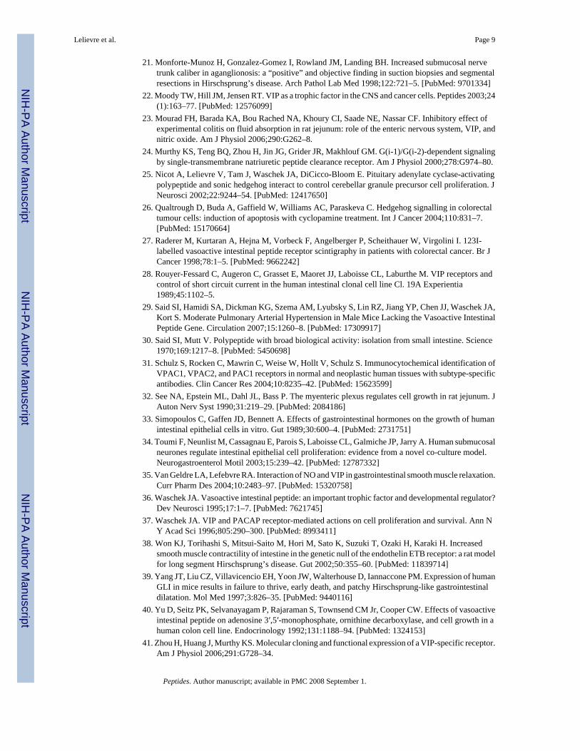
Autopsy performed on healthy adult animals revealed obvious anatomical differences in theintestinal tract of knock-out (KO) vs. WT mice. Investigators blind to the genotype could easilydistinguish a WT from KO mouse due to rubbery appearance of the gut in VIP−/− mice. Inaddition, as visualized in Figure 1, an overall reduction in intestinal length was observed inVIP-deficient mice. This difference was noticeable in heterozygous mice, but was morepronounced in KO animals. In addition, we observed a very interesting anatomical abnormalityin small bowel (black arrows) associated with degenerated appendices (arrowhead). The weightand density of the small intestinal was found to be greater, but the length reduced in VIP−/−
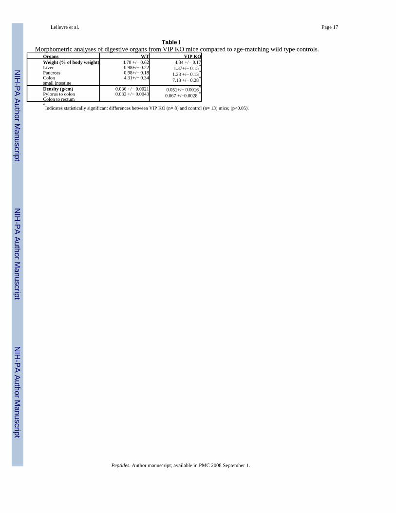
mice (Tables I and II), providing a possible explanation for the ‘rubbery’ phenotype
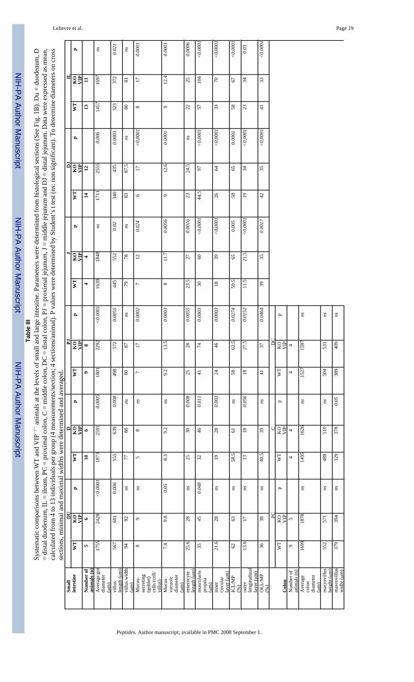
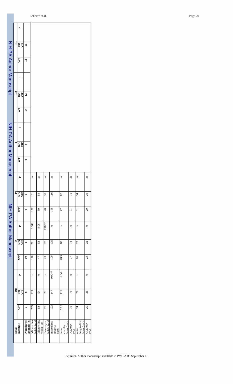
Differential changes in small bowel and colon light microscopic structureDetailed morphometric analyses were on performed on histological sections of different partsof the gut, including duodenum, jejunum and ileum for the small bowel and the three sections(ascending, transverse and descending) of the colon. Results are shown in Table III (see theschematic small bowel section in Fig. 1B for structure identification and measurements).Overall, an increase of the cross sectional diameter of the gut sections was observed throughoutthe small intestine. However, this phenomenon tended to decrease in the small bowel from
Lelievre et al. Page 4
Peptides. Author manuscript; available in PMC 2008 September 1.
NIH
-PA Author Manuscript
NIH
-PA Author Manuscript
NIH
-PA Author Manuscript

140% of the control in duodenum, 128% in proximal jejunum, and 120% in distal jejunum toa non-significant 110% in ileum. The increases in diameter were associated with an increasein the length of villi. These tended to completely fill the lumen of the duodenum, as easilyobserved in Figure 2A (vs. 2B). At the cellular level, an increase in enterocytic height in VIP-deficient mice was also observed (Table III).
In addition, a significant increase in thickness of the muscularis propria was observed (Figure2D & F and Table III). This increase in thickness primarily affected the inner (circular) layer.For example, the thickness of the inner layer was almost double in distal jejunum in VIP−/−
animals when compared to controls, whereas the thickness of the outer (longitudinal) layer wasincreased by only 20%. This striking observation is obvious from the pictures presented inFigure 2A–F, but to some extent differentially affected the various parts of the small intestine,with an increased severity in the jejunum, when compared to the duodenum or large intestine(Figures 2E vs. 2F and 3C vs. 3D).
We also investigated the colon of VIP−/− mice for anatomical differences. Unlike that observedin the small intestine, VIP-deficient mice did not appear significantly different to WT micewith respect to villus architecture or smooth muscle thickness (Figure 3, left vs. right panels).Only small differences were observed (Table III), however, these were not consistently seenat all levels of the colon.
Evidence of dysfunctions in goblet cell mucus secretion in VIP−/− animalsBecause VIP has long been considered to be a prosecretory neuropeptide, we also focused onthe histology of goblet cells in both small and large bowels. In small intestine, we observed asignificant increase of the number of mucus-secreting cells in jejunum and ileum in VIP KOmice (Table III). This increase in cell number was associated with a robust increase in theaverage diameter of the mucus vacuoles of the cells (Table III). The latter is easily observableon HE-stained sections, as shown on figures 2G vs. 2H from proximal jejunum. Thesedifferences were further confirmed by selectively labeling mucus using PAS (data not shown)and Alcian blue staining (Figure 4A–D). Alcian blue staining demonstrated mucusaccumulation inside the goblet cells in VIP KO mice, correlated with a lack of mucus depositswithin the lumen of the duodenum (Figure 4C vs. 4D). The number of goblet cells could notbe accurately assessed in colon using H&E (Figures 3C vs. 3D) and specific PAS stainings(data not shown), because of their extremely high abundance.
Effects of VIP on submucosal and myenteric neural innervationsVIP is expressed within neurons of the myenteric plexus, located between the outer longitudinaland inner circular muscle layers of the muscularis propria, and in neurons in the submucosalplexus, located between the submucosa and lamina propria. To determine if loss of VIP affectedtheir anatomical structure, we examined the light microscopic appearance of these plexuses inboth small and large intestine. These studies revealed unusual large H&E-negative negativepatches within the myenteric and submucosal plexus (Figures 5A vs. 5B) in VIP KO but notWT mice. These patches did not react with the Schwann cell S100β antibody (Pictures C vs.D). In an attempt to further characterize these unstained patches we incubated duodenal sectionswith fluorescent antibody directed against pan-NF to test whether these patches correspondedto enlarge nerve trunks. This procedure failed to highlight these particular structures, butrevealed instead the presence of thicker and brighter axons on sections from VIP-deficientanimals (Figures 5E vs. F), suggesting the presence of hypertrophied axons. H&E (Figure 5,G vs. H) and S100 immunostaining (Figure 5, I vs. J) of colonic sections failed to reveal similardifferences in large bowel.
Lelievre et al. Page 5
Peptides. Author manuscript; available in PMC 2008 September 1.
NIH
-PA Author Manuscript
NIH
-PA Author Manuscript
NIH
-PA Author Manuscript

Impaired gastrointestinal transit and incidence of early death in VIP-deficient animalsMotility in the upper part of the GI tract was examined in fasted WT and VIP-deficient miceusing two different methodologies. In the first of these, charcoal stained gum tragacanth wasused as a bolus. This experiment revealed that VIP−/− animals displayed a significant reductionin motility. Indeed, when animals were sacrificed 30 minutes after feeding, we observed anoverall 36% decrease (p=0.0002) in bolus distance in VIP-deficient mice when compared tocontrols (Figure 6A). In the second experiment, we used a fluorescent dextran methodologyin which the concentration of fluorescent intensity was determined along the length of theintestine. Based on our previous experience with this model, we sacrificed the mice one hourafter the beginning of the experiment. As shown in Figure 6B, there was a significant reductionin intestinal transit time in the VIP−/− mice compared to WT mice. Peristalsis and fixed timeanalysis revealed significant differences in motility pattern indicating that the lack of VIP andassociated microscopic abnormalities were reflected in a functional change in motility.
Day to day monitoring of our VIP−/− animal colony in our specific pathogen-free facilityrevealed that approximately 10–15% of VIP-deficient mice experienced sudden death beforethe age of twelve months, whereas less than 2% of the wild-type mice in the colony expiredduring the same period. Origins of these early deaths were difficult to elucidate because of theshort time window allowing the autopsy of the carcasses to be performed prior to autolysis.Nonetheless, an abrupt blockade in movement of intestinal contents was observed in most ofthese mice, as revealed by a gross swelling of the intestine at all levels above an apparentstricture. In a few cases, we were able to euthanize before they had succumbed. H&E stainedsections did not reveal physical block or inflammatory infiltrates, or any obvious pathologyexcept for typical GI stenosis (data not shown). These findings suggested that the early deathin these mice was due to stenosis of the gut.
DISCUSSIONVIP was first characterized as a peptide expressed in the intestine and the lung, but was laterfound to be widely expressed in the central and peripheral nervous systems. A wide variety ofexperimental approaches indicate that this peptide acts as a neurotransmitter or neuromodulatorin the brain and nearly all peripheral systems, including cardiovascular, reproductive, andimmune systems. We report here the first description of the GI phenotype of mice deficient inthis peptide. While some of the anatomical findings were unexpected, functional studiesrevealed that GI motility was impaired in VIP−/− mice, a finding fully in line with studiesshowing that this peptide acts in the gut as a smooth muscle relaxant. Moreover, the ratio ofgoblet cell-retained vs. secreted mucus was higher in VIP KO mice, suggesting a pro-secretoryrole for VIP in goblet cells, another finding in accordance to considerable work of others.
The most obvious anatomical alteration in VIP KO mice was that the intestine was heavier butshorter compared to WT controls. The higher density was reflected in an increased luminaldiameter at several levels of the GI tract, especially in the duodenum and jejunum portions ofthe intestine. Part of the increased diameter could be accounted for by a thickening of both theouter longitudinal and inner circular layers of the muscularis propria. Some peculiar findingswere that VIP-deficient mice exhibited increases in the mean enterocyte length (basal toserosal) and villus length.
A possible explanation for the decreased length and increased thickness of the bowel is thatthe intestine might have been in a relatively enhanced state of tonic contraction. However, thismechanism cannot account for the substantial increase in overall mass of the small intestine(>60%, see Table I). This increased mass could be triggered in some way in response to theimpaired GI function in VIP KO mice. Another possibility is that some of the changes are dueto a developmental loss of VIP. In this regard, VIP has been shown to exhibit growth factor-
Lelievre et al. Page 6
Peptides. Author manuscript; available in PMC 2008 September 1.
NIH
-PA Author Manuscript
NIH
-PA Author Manuscript
NIH
-PA Author Manuscript
like action in vitro on many different types of primary cells and tumor cell lines (reviewed in[22,36–37]. With respect to the intestine, VIP has been shown to regulate the proliferation ofintestinal epithelial cells [32–34] and cell lines [10,17,40]. This peptide might also regulateintestinal smooth muscle proliferation, because VIP was show to inhibit the proliferation ofvascular smooth muscle [19]. In this regard, we recently showed that VIP−/− mice exhibitpulmonary hypertension with thickening of the smooth muscle layers in the pulmonary arteryand smaller branches [29].
The possibility that VIP acts a growth factor in the gut is interesting in light of recent findingsthat hedgehog proteins regulate GI development [9]. Hedgehog actions are antagonized bycAMP-dependent protein kinase A (PKA). The VIP-related peptide PACAP was shown toinhibit sonic hedgehog-induced proliferation of cultured cerebellar granule precursors [25].The growth factor-like actions of VIP and PACAP raise the possibility that these peptides havesignificance in intestinal tumor pathogenesis. In this regard, VIP and PACAP have been shownto modulate proliferation of adenocarcinoma cell lines [17] and VIP levels are enhanced inserum samples from patients suffering from colonic carcinoma [12]. The interaction with thehedgehog pathway might also be relevant in these tumors because the hedgehog pathway hasbeen found to be overactive in human intestinal carcinomas [3] and associated with tumor cellsurvival [26]. In these respects, VIP-deficient mice may be a useful model to study the role ofthe endogenous peptide in the pathogenesis and/or progression of intestinal tumors.
An appealing extrapolation of the present results is to link the observed phenotypes with majorhuman digestive disorders. Among the characteristic GI features observed in VIP−/− animalswas small bowel dilatation and muscular hypertrophy associated with an apparent reductionof peristalsis. Thus, mice lacking VIP-ergic innervation might represent a model for humanpathologies characterized by peristaltic defects, for example Hirschsprung’s disease, orchildren with constipation associated with clinical symptoms of intestinal pseudo-obstruction.The potential clinical relevance of this comparison is reinforced by the observation of lethalduodenal stenosis in VIP−/− animals and by analogies in clinical features previously observedin a rat model for long segment Hirschsprung’s disease [38]. Furthermore, while a reductionin VIP innervation has been reported in these pathologies (for review, [16]), it is still quiteunclear whether reduction in VIPergic innervation is indeed causative or resultant in the diseaseprogression. In addition, the unusual spotted plexuses observed in VIP−/− animals may in factcorrespond to the increase in nerve trunks in the muscularis propria previously reported frombiopsies of patients suffering from Hirschsprung’s disease [21]. Moreover, as speculatedabove, interactions between VIP and hedgehog pathway might also play a role the generationof Hirschsprung’s-like phenotype in mice, since overexpression of human gli1 gene intransgenic mice triggered patches of GI dilatation [39].
In conclusion, the present data represent the first direct evidence that VIP has an endogenouseffect on digestive function and GI tract integrity. Additional work is necessary to fully describethe overall digestive phenotype observed in VIP null mice and understand the relationshipbetween lack of VIP and GI disorders. These studies provide an insight into the pathogenesisof intestinal motility abnormalities in humans and provide an experimental model to studyintestinal ileus, pseudoobstruction (Oglievie’s Syndrome) and Hirschsprung’s disease.
Acknowledgements
G. Favrais was recipient of fellowships from the French Society for Pediatrics and the Evian’s Foundation. J. Waschekreceived a pilot award from the Center of Ulcer Research and Education (CURE), and was funded by HD0657 andHD34475. JR Pisegna was funded by the Department of Veterans Affairs. PM Germano was funded by an NIDDKNIH Re-entry grant program.
Lelievre et al. Page 7
Peptides. Author manuscript; available in PMC 2008 September 1.
NIH
-PA Author Manuscript
NIH
-PA Author Manuscript
NIH
-PA Author Manuscript

References1. Abad C, Martinez C, Juarranz MG, Arranz A, Leceta J, Delgado M, Gomariz RP. Therapeutic effects
of vasoactive intestinal peptide in the trinitrobenzene sulfonic acid mice model of Crohn’s disease.Gastroenterology 2003;124:961–71. [PubMed: 12671893]
2. Barbezat GO, Grossman MI. Intestinal secretion: stimulation by peptides. Science 1971;174:422–4.[PubMed: 5111998]
3. Berman DM, Karhadkar SS, Maitra A, Montes De Oca R, Gerstenblith MR, Briggs K, Parker AR,Shimada Y, Eshleman JR, Watkins DN, Beachy PA. Widespread requirement for Hedgehog ligandstimulation in growth of digestive tract tumours. Nature 2003;425:846–51. [PubMed: 14520411]
4. Binder HJ, Lemp GF, Gardner JD. Receptors for vasoactive intestinal peptide and secretin on smallintestinal epithelial cells. Am J Physiol 1980;238:G190–6. [PubMed: 6245588]
5. Colwell CS, Michel S, Itri J, Rodriguez W, Tam J, Lelievre V, Hu Z, Liu X, Waschek JA. Disruptedcircadian rhythms in VIP- and PHI-deficient mice. Am J Physiol Regul Integr Comp Physiol2003;285:R939–49. [PubMed: 12855416]
6. Cullen JJ, Caropreso DK, Hemann LL, Hinkhouse M, Conklin JL, Ephgrave KS. Pathophysiology ofadynamic ileus. Dig Dis Sci 1997;42:731–7. [PubMed: 9125641]
7. Dharmsathaphorn K, McRoberts JA, Mandel KG, Tisdale LD, Masui H. A human colonic tumor cellline that maintains vectorial electrolyte transport. Am J Physiol 1984;246:G204–8. [PubMed:6141741]
8. Fujimiya M, Inui A. Peptidergic regulation of gastrointestinal motility in rodents. Peptides2000;21:1565–82. [PubMed: 11068106]
9. Fukuda K, Yasugi S. Versatile roles for sonic hedgehog in gut development. J Gastroenterol2002;37:239–46. [PubMed: 11993506]
10. Gamet L, Murat JC, Remaury A, Remesy C, Valet P, Paris H, Denis-Pouxviel C. Vasoactive intestinalpeptide and forskolin regulate proliferation of the HT29 human colon adenocarcinoma cell line. JCell Physiol 1992;150:501–9. [PubMed: 1371513]
11. Hakanson R, Chen D, Lindstrom E, Bernsand M, Norlen P. Control of secretion from rat stomachECL cells in situ and in primary culture. Scand J Clin Lab Invest Suppl 2001;234:53–60. [PubMed:11713981]
12. Hejna M, Hamilton G, Brodowicz T, Haberl I, Fiebiger WC, Scheithauer W, Virgolini I, Kostler WJ,Oberhuber G, Raderer M. Serum levels of vasoactive intestinal peptide (VIP) in patients withadenocarcinomas of the gastrointestinal tract. Anticancer Res 2001;21:1183–7. [PubMed: 11396161]
13. Krejs GJ. VIPoma syndrome. Am J Med 1987;82:37–48. [PubMed: 3035922]14. Laburthe M, Breant B, Rouyer-Fessard C. Molecular identification of receptors for vasoactive
intestinal peptide in rat intestinal epithelium by covalent cross-linking. Evidence for two classes ofbinding sites with different structural and functional properties. Eur J Biochem 1984;139:181–7.[PubMed: 6321173]
15. Laburthe M, Rousset M, Chevalier G, Boissard C, Dupont C, Zweibaum A, Rosselin G. Vasoactiveintestinal peptide control of cyclic adenosine 3′:5′-monophosphate levels in seven human colorectaladenocarcinoma cell lines in culture. Cancer Res 1980;40:2529–33. [PubMed: 6248206]
16. Larsson LT. Hirschsprung’s disease--immunohistochemical findings. Histol Histopathol1994;9:615–29. [PubMed: 7981507]
17. Lelievre V, Meunier AC, Caigneaux E, Falcon J, Muller JM. Differential expression and function ofPACAP and VIP receptors in four human colonic adenocarcinoma cell lines. Cell Signal 1998;10:13–26. [PubMed: 9502113]
18. Makhlouf GM, Murthy KS. Signal transduction in gastrointestinal smooth muscle. Cell Signal1997;9:269–76. [PubMed: 9218127]
19. Maruno K, Absood A, Said SI. VIP inhibits basal and histamine-stimulated proliferation of humanairway smooth muscle cells. Am J Physiol 1995;268:L1047–51. [PubMed: 7541947]
20. Miampamba M, Germano PM, Arli S, Wong HH, Scott D, Tache Y, Pisegna JR. Expression ofpituitary adenylate cyclase-activating polypeptide and PACAP type 1 receptor in the rat gastric andcolonic myenteric neurons. Regul Pept 2002;105:145–54. [PubMed: 11959368]
Lelievre et al. Page 8
Peptides. Author manuscript; available in PMC 2008 September 1.
NIH
-PA Author Manuscript
NIH
-PA Author Manuscript
NIH
-PA Author Manuscript
21. Monforte-Munoz H, Gonzalez-Gomez I, Rowland JM, Landing BH. Increased submucosal nervetrunk caliber in aganglionosis: a “positive” and objective finding in suction biopsies and segmentalresections in Hirschsprung’s disease. Arch Pathol Lab Med 1998;122:721–5. [PubMed: 9701334]
22. Moody TW, Hill JM, Jensen RT. VIP as a trophic factor in the CNS and cancer cells. Peptides 2003;24(1):163–77. [PubMed: 12576099]
23. Mourad FH, Barada KA, Bou Rached NA, Khoury CI, Saade NE, Nassar CF. Inhibitory effect ofexperimental colitis on fluid absorption in rat jejunum: role of the enteric nervous system, VIP, andnitric oxide. Am J Physiol 2006;290:G262–8.
24. Murthy KS, Teng BQ, Zhou H, Jin JG, Grider JR, Makhlouf GM. G(i-1)/G(i-2)-dependent signalingby single-transmembrane natriuretic peptide clearance receptor. Am J Physiol 2000;278:G974–80.
25. Nicot A, Lelievre V, Tam J, Waschek JA, DiCicco-Bloom E. Pituitary adenylate cyclase-activatingpolypeptide and sonic hedgehog interact to control cerebellar granule precursor cell proliferation. JNeurosci 2002;22:9244–54. [PubMed: 12417650]
26. Qualtrough D, Buda A, Gaffield W, Williams AC, Paraskeva C. Hedgehog signalling in colorectaltumour cells: induction of apoptosis with cyclopamine treatment. Int J Cancer 2004;110:831–7.[PubMed: 15170664]
27. Raderer M, Kurtaran A, Hejna M, Vorbeck F, Angelberger P, Scheithauer W, Virgolini I. 123I-labelled vasoactive intestinal peptide receptor scintigraphy in patients with colorectal cancer. Br JCancer 1998;78:1–5. [PubMed: 9662242]
28. Rouyer-Fessard C, Augeron C, Grasset E, Maoret JJ, Laboisse CL, Laburthe M. VIP receptors andcontrol of short circuit current in the human intestinal clonal cell line Cl. 19A Experientia1989;45:1102–5.
29. Said SI, Hamidi SA, Dickman KG, Szema AM, Lyubsky S, Lin RZ, Jiang YP, Chen JJ, Waschek JA,Kort S. Moderate Pulmonary Arterial Hypertension in Male Mice Lacking the Vasoactive IntestinalPeptide Gene. Circulation 2007;15:1260–8. [PubMed: 17309917]
30. Said SI, Mutt V. Polypeptide with broad biological activity: isolation from small intestine. Science1970;169:1217–8. [PubMed: 5450698]
31. Schulz S, Rocken C, Mawrin C, Weise W, Hollt V, Schulz S. Immunocytochemical identification ofVPAC1, VPAC2, and PAC1 receptors in normal and neoplastic human tissues with subtype-specificantibodies. Clin Cancer Res 2004;10:8235–42. [PubMed: 15623599]
32. See NA, Epstein ML, Dahl JL, Bass P. The myenteric plexus regulates cell growth in rat jejunum. JAuton Nerv Syst 1990;31:219–29. [PubMed: 2084186]
33. Simopoulos C, Gaffen JD, Bennett A. Effects of gastrointestinal hormones on the growth of humanintestinal epithelial cells in vitro. Gut 1989;30:600–4. [PubMed: 2731751]
34. Toumi F, Neunlist M, Cassagnau E, Parois S, Laboisse CL, Galmiche JP, Jarry A. Human submucosalneurones regulate intestinal epithelial cell proliferation: evidence from a novel co-culture model.Neurogastroenterol Motil 2003;15:239–42. [PubMed: 12787332]
35. Van Geldre LA, Lefebvre RA. Interaction of NO and VIP in gastrointestinal smooth muscle relaxation.Curr Pharm Des 2004;10:2483–97. [PubMed: 15320758]
36. Waschek JA. Vasoactive intestinal peptide: an important trophic factor and developmental regulator?Dev Neurosci 1995;17:1–7. [PubMed: 7621745]
37. Waschek JA. VIP and PACAP receptor-mediated actions on cell proliferation and survival. Ann NY Acad Sci 1996;805:290–300. [PubMed: 8993411]
38. Won KJ, Torihashi S, Mitsui-Saito M, Hori M, Sato K, Suzuki T, Ozaki H, Karaki H. Increasedsmooth muscle contractility of intestine in the genetic null of the endothelin ETB receptor: a rat modelfor long segment Hirschsprung’s disease. Gut 2002;50:355–60. [PubMed: 11839714]
39. Yang JT, Liu CZ, Villavicencio EH, Yoon JW, Walterhouse D, Iannaccone PM. Expression of humanGLI in mice results in failure to thrive, early death, and patchy Hirschsprung-like gastrointestinaldilatation. Mol Med 1997;3:826–35. [PubMed: 9440116]
40. Yu D, Seitz PK, Selvanayagam P, Rajaraman S, Townsend CM Jr, Cooper CW. Effects of vasoactiveintestinal peptide on adenosine 3′,5′-monophosphate, ornithine decarboxylase, and cell growth in ahuman colon cell line. Endocrinology 1992;131:1188–94. [PubMed: 1324153]
41. Zhou H, Huang J, Murthy KS. Molecular cloning and functional expression of a VIP-specific receptor.Am J Physiol 2006;291:G728–34.
Lelievre et al. Page 9
Peptides. Author manuscript; available in PMC 2008 September 1.
NIH
-PA Author Manuscript
NIH
-PA Author Manuscript
NIH
-PA Author Manuscript
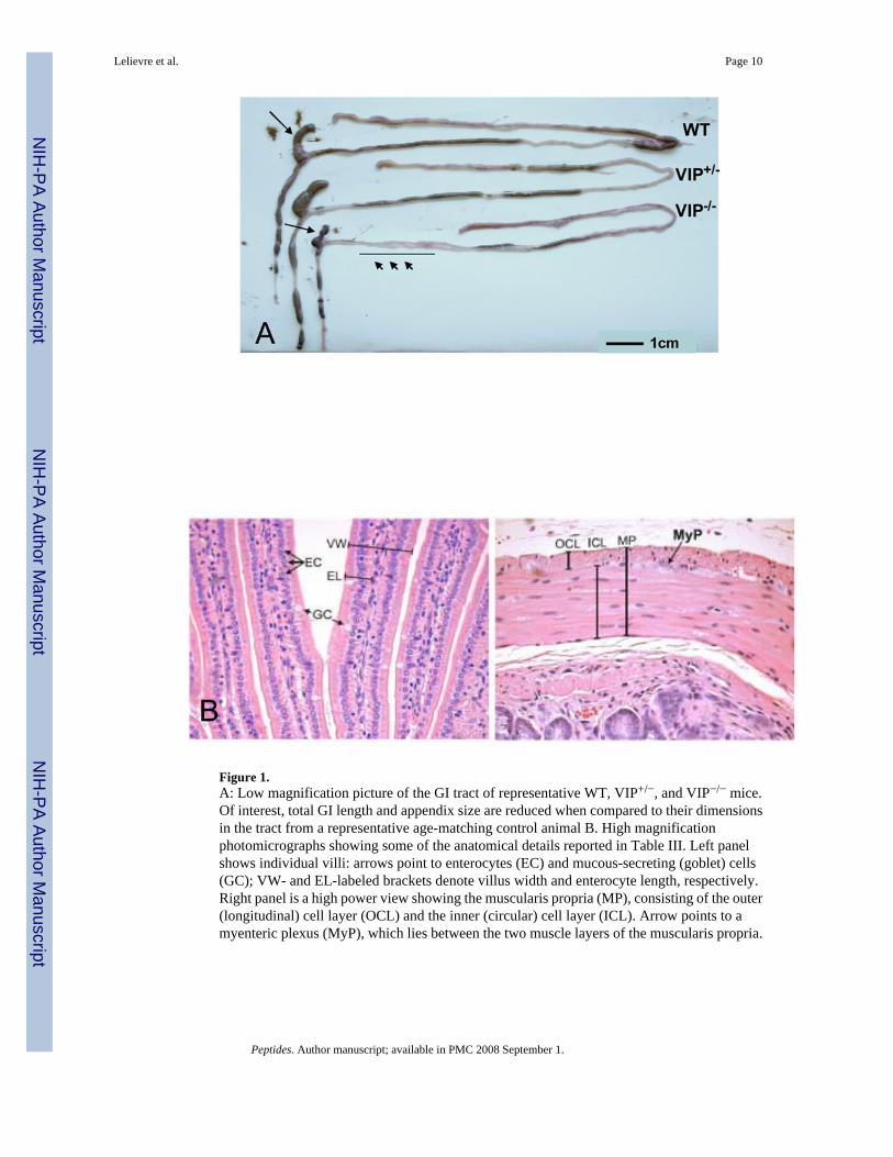
Figure 1.A: Low magnification picture of the GI tract of representative WT, VIP+/−, and VIP−/− mice.Of interest, total GI length and appendix size are reduced when compared to their dimensionsin the tract from a representative age-matching control animal B. High magnificationphotomicrographs showing some of the anatomical details reported in Table III. Left panelshows individual villi: arrows point to enterocytes (EC) and mucous-secreting (goblet) cells(GC); VW- and EL-labeled brackets denote villus width and enterocyte length, respectively.Right panel is a high power view showing the muscularis propria (MP), consisting of the outer(longitudinal) cell layer (OCL) and the inner (circular) cell layer (ICL). Arrow points to amyenteric plexus (MyP), which lies between the two muscle layers of the muscularis propria.
Lelievre et al. Page 10
Peptides. Author manuscript; available in PMC 2008 September 1.
NIH
-PA Author Manuscript
NIH
-PA Author Manuscript
NIH
-PA Author Manuscript

Figure 2.Microscopic structure of the proximal jejunum in WT and VIP−/− mice. Small bowel sectionswere stained using standard H&E staining. Pictures at low (x2.5, x10, upper panel) and highermagnification (x20, x40; mid and lower panels, respectively) revealed an overall increase inthickness of muscularis mucosa in VIP KO (B, D and F) vs. controls (A, C and E), as well ashigher numbers of mucus-forming cells (G vs. E).
Lelievre et al. Page 11
Peptides. Author manuscript; available in PMC 2008 September 1.
NIH
-PA Author Manuscript
NIH
-PA Author Manuscript
NIH
-PA Author Manuscript
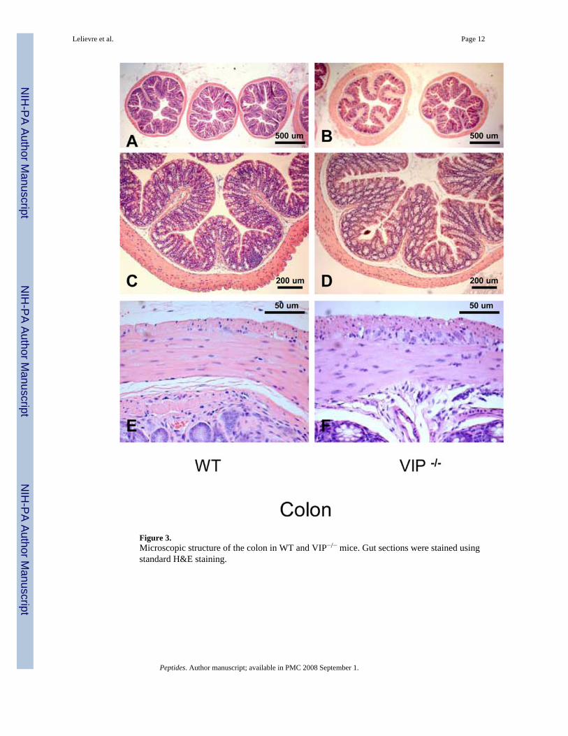
Figure 3.Microscopic structure of the colon in WT and VIP−/− mice. Gut sections were stained usingstandard H&E staining.
Lelievre et al. Page 12
Peptides. Author manuscript; available in PMC 2008 September 1.
NIH
-PA Author Manuscript
NIH
-PA Author Manuscript
NIH
-PA Author Manuscript
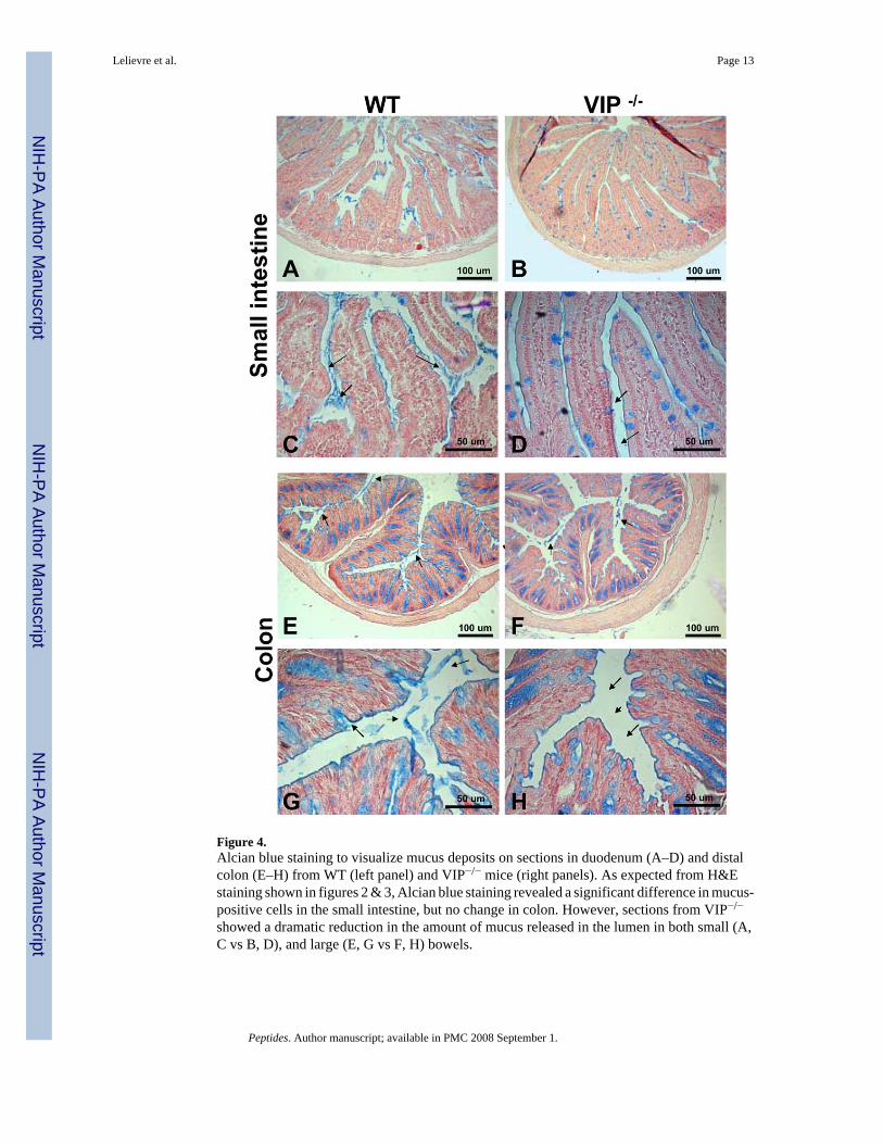
Figure 4.Alcian blue staining to visualize mucus deposits on sections in duodenum (A–D) and distalcolon (E–H) from WT (left panel) and VIP−/− mice (right panels). As expected from H&Estaining shown in figures 2 & 3, Alcian blue staining revealed a significant difference in mucus-positive cells in the small intestine, but no change in colon. However, sections from VIP−/−
showed a dramatic reduction in the amount of mucus released in the lumen in both small (A,C vs B, D), and large (E, G vs F, H) bowels.
Lelievre et al. Page 13
Peptides. Author manuscript; available in PMC 2008 September 1.
NIH
-PA Author Manuscript
NIH
-PA Author Manuscript
NIH
-PA Author Manuscript

Figure 5.Microscopic structure of myenteric plexus in duodenum and distal colon of WT and VIP−/−
mice. H&E (A, B, G and H) and S100β (C–D & I–J) and pan-neurofilaments (NF) (E, F)stainings are shown in WT and VIP KO mice (left and right panels, respectively). Structuraldifferences were observed in duodenum of VIP deficient mice when compared to WT controls.Plexuses from KO mice showed enlarged unstained patches (A vs B) that were non-immunoreactive with S100β or NF antibodies used to specifically reveal Schwann cells andaxons (C vs. D & E vs. F, respectively). However, immunofluorescence emitted by the pan-NF antibody coupled with green fluorescent dye-labeled conjugates (E vs. F) revealed largerarea signals within the external plexus of the KO mice than WT controls, suggesting the
Lelievre et al. Page 14
Peptides. Author manuscript; available in PMC 2008 September 1.
NIH
-PA Author Manuscript
NIH
-PA Author Manuscript
NIH
-PA Author Manuscript
presence of bigger axons within the myenteric plexus of VIP-deficient mice. Conversely, noobvious differences in plexus structures of the large bowel (G–J) were using these techniques(pan-NF not shown).
Lelievre et al. Page 15
Peptides. Author manuscript; available in PMC 2008 September 1.
NIH
-PA Author Manuscript
NIH
-PA Author Manuscript
NIH
-PA Author Manuscript
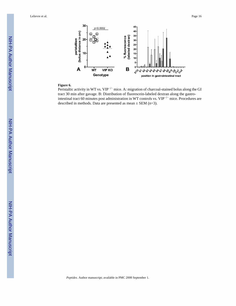
Figure 6.Peristaltic activity in WT vs. VIP−/− mice. A: migration of charcoal-stained bolus along the GItract 30 min after gavage. B: Distribution of fluorescein-labeled dextran along the gastro-intestinal tract 60 minutes post administration in WT controls vs. VIP−/− mice. Procedures aredescribed in methods. Data are presented as mean ± SEM (n=3).
Lelievre et al. Page 16
Peptides. Author manuscript; available in PMC 2008 September 1.
NIH
-PA Author Manuscript
NIH
-PA Author Manuscript
NIH
-PA Author Manuscript
NIH
-PA Author Manuscript
NIH
-PA Author Manuscript
NIH
-PA Author Manuscript
Lelievre et al. Page 17
Table IMorphometric analyses of digestive organs from VIP KO mice compared to age-matching wild type controls.
Organs WT VIP KOWeight (% of body weight)LiverPancreasColonsmall intestine
4.70 +/− 0.620.98+/− 0.220.98+/− 0.184.31+/− 0.34
4.34 +/− 0.171.37+/− 0.15*
1.23 +/− 0.13*
7.13 +/− 0.28*
Density (g/cm)Pylorus to colonColon to rectum
0.036 +/− 0.00210.032 +/− 0.0043
0.051+/− 0.0016*
0.067 +/−0.0028 *
*Indicates statistically significant differences between VIP KO (n= 8) and control (n= 13) mice; (p<0.05).
Peptides. Author manuscript; available in PMC 2008 September 1.

NIH
-PA Author Manuscript
NIH
-PA Author Manuscript
NIH
-PA Author Manuscript
Lelievre et al. Page 18
Table IIMorphometric analyses of intestinal structures in WT and VIP−/− mice. * Indicates statistically significant differencesbetween VIP KO (n= 6) and control mice (n= 5)
Intestinal structures Measurements (cm +/− SD) Differences(% of WT control)WT VIP−/−
LengthPylorus to rectum
57.00+/− 0.71 50.25+/−2.72 88.16*
Pylorus to cecum 47.13 +/− 1.03 42.12+/−3.11 89.39Cecum to rectum 9.88 +/− 0.63 8.00+/−0.72 81.01*Cecum 2.63 +/− 0.25 3.25+/−0.52 123.81*p<0.05,
**p<0.01
***p<0.005
Peptides. Author manuscript; available in PMC 2008 September 1.
NIH
-PA Author Manuscript
NIH
-PA Author Manuscript
NIH
-PA Author Manuscript
Lelievre et al. Page 19Ta
ble
IIISy
stem
atic
com
paris
ons b
etw
een
WT
and
VIP−/−
anim
als a
t the
leve
ls o
f sm
all a
nd la
rge
inte
stin
e. P
aram
eter
s wer
e de
term
ined
from
his
tolo
gica
l sec
tions
(See
Fig
. 1B
). D
u =
duod
enum
, D=
dist
al d
uode
num
, IL
= ile
um, P
C =
pro
xim
al c
olon
, C =
mid
dle
colo
n, D
C =
dis
tal c
olon
, PJ =
pro
xim
al je
junu
m, J
= m
iddl
e je
junu
m a
nd D
J = d
ista
l jej
unum
. Dat
a w
ere
expr
esse
d as
mea
n,ca
lcul
ated
from
4 to
13
indi
vidu
als p
er g
roup
(4 m
easu
rem
ents
/sec
tion;
4 se
ctio
ns/a
nim
al).
P va
lues
wer
e de
term
ined
by
Stud
ent’s
t te
st (n
s: n
on si
gnifi
cant
). To
det
erm
ine
diam
eter
s on
cros
sse
ctio
ns, m
inim
al a
nd m
axim
al w
idth
s wer
e de
term
ined
and
ave
rage
d.Sm
all
inte
stin
eD
UD
PJJ
DJ
ILW
TK
OV
IPp
WT
KO
VIP
pW
TK
OV
IPp
WT
KO
VIP
pW
TK
OV
IPp
WT
KO
VIP
p
Num
ber
ofan
imal
s (n)
56
106
98
44
1412
1311
Ave
rage
gut
diam
eter
(μm
)
1755
2428
<0.
0001
1873
2591
0.00
0518
0122
92<
0.00
0116
3918
48ns
1711
2555
0.00
614
5716
97ns
villu
sle
ngth
(μm
)56
768
10.
006
555
639
0.00
849
857
20.
0055
445
552
0.02
340
435
0.00
0332
337
20.
021
villu
s wid
th(μ
m)
9492
ns77
88ns
8087
ns79
78ns
8387
.5ns
8081
ns
Muc
us-
secr
etin
g(g
oble
t)ce
lls (c
ell/
villu
s)
89
ns5
8ns
717
0.00
027
120.
024
617
<0,
0001
817
0.00
01
Muc
usva
cuol
edi
amet
er(μ
m)
7.8
9.8
0.05
8.3
9.2
ns9.
213
.50.
0003
811
.70.
0056
912
.60.
0001
912
.40.
0001
ente
rocy
tele
ngth
(μm
)25
.628
ns25
300.
008
2528
0.00
5523
.527
0.00
1623
24.5
ns22
250.
0006
mus
cula
rispr
opria
(μm
)
3545
0.04
832
460.
011
4174
0.00
0330
60<
0,00
0144
.597
<0,
0001
5710
4<
0.00
01
inne
rci
rcul
arla
yer (μm
)
21.6
28ns
1928
0.00
324
460.
0003
1839
<0,
0001
2664
<0,
0001
3370
<0.
0001
ICL/
MP
(%)
6263
ns58
.561
ns58
62.5
0.02
7459
.565
0.00
558
650.
0001
5867
<0.
0001
oute
rlo
ngitu
dina
lla
yer (μm
)
13.6
17ns
1318
0.05
618
27.5
0.01
5211
.521
.5<
0,00
0119
34<
0,00
0123
340.
03
OLL
/MP
(%)
3638
ns40
.539
ns41
370.
0464
3935
0.00
1742
35<
0,00
0141
33<
0.00
01
Col
on
PCC
DC
WT
KO
VIP
pW
TK
OV
IPp
WT
KO
VIP
p
Num
ber o
fan
imal
s (n)
95
44
44
Ave
rage
colo
ndi
amet
er(μ
m)
1699
1878
ns14
9516
26ns
1527
1597
ns
mac
rovi
llus
heig
ht (μ
m)
552
571
ns48
851
0ns
504
531
ns
mav
rovi
llus
wid
ht (μ
m)
379
394
ns32
937
80.
0538
940
9ns
Peptides. Author manuscript; available in PMC 2008 September 1.
NIH
-PA Author Manuscript
NIH
-PA Author Manuscript
NIH
-PA Author Manuscript
Lelievre et al. Page 20Sm
all
inte
stin
eD
UD
PJJ
DJ
ILW
TK
OV
IPp
WT
KO
VIP
pW
TK
OV
IPp
WT
KO
VIP
pW
TK
OV
IPp
WT
KO
VIP
p
Num
ber
ofan
imal
s (n)
56
106
98
44
1412
1311
Mic
rovi
llus
heig
ht (μ
m)
209
219
ns17
821
10.
003
177
191
ns
Mic
rovi
llus
wid
th (μ
m)
5454
ns47
540.
0550
54ns
Ente
rocy
tehe
ight
(μm
)27
29ns
2328
0.00
3728
30ns
mus
cula
rispr
opria
(μm
)
123
147
0.00
4710
010
5ns
108
116
ns
inne
rci
rcul
arla
yer (μm
)
97.5
115
0.04
78.5
82ns
7782
ns
ICL/
MP
(%)
7978
ns77
78ns
7171
ns
oute
rlo
ngitu
dina
lla
yer (μm
)
2427
ns16
22ns
3134
ns
OLL
/MP
(%)
2021
ns23
22ns
2929
ns
Peptides. Author manuscript; available in PMC 2008 September 1.
Related Documents