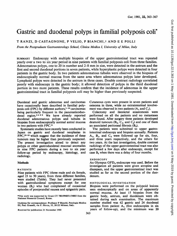
Gut, 1981, 22, 363-367 Gastric and duodenal polyps in familial polyposis coli T RANZI, D CASTAGNONE, P VELIO, P BIANCHI,t AND E E POLLI From the Postgraduate Gastroenterology School, Clinica Medica I, University of Milan, Italy SUMMARY Endoscopy with multiple biopsies of the upper gastrointestinal tract was repeated yearly over a two to six year period in nine patients with familial polyposis coli from three families. Adenomatous polyps, one to 20 in number and 2-8 mm in size, were detected in the antrum and the first and second duodenal portions in seven patients, while hyperplastic polyps were detected in four patients in the gastric body. In two patients adenomatous tubules were observed in the biopsies of endoscopically normal mucosa from the same area where adenomatous polyps later developed. Lymphoid polyps were detected in the antrum in three cases. Double contrast radiology correlated poorly with endoscopy in the gastric body; it allowed detection of polyps in the third duodenal portion in two more patients. These results confirm that the incidence of adenomas in the upper gastrointestinal tract in familial polyposis coli may be higher than previously suspected. Duodenal and gastric adenomas and carcinomas have occasionally been described in familial poly- posis coli (FPC) by different authors,1-'0 carcinomas being particularly frequent in the pancreaticoduo- denal region.2 4-6 9 We have already reported duodenal adenomatous polyps and tubules in biopsies from endoscopically normal antral mucosa in two patients with FPC.1" Systematic studies have recently been conducted in Japan on gastric and duodenal neoplasias in FPC, 2-'8 which suggest that the incidence of these tumours may be higher than previously suspected. The present investigation aimed to detect any polyps or other gastroduodenal mucosal anomalies in nine FPC patients during a two to six year follow-up period by endoscopy, histology, and radiology. Methods PATIENTS Nine patients with FPC (three male and six female, aged 23 to 50 years), from three different families, were studied (Table). They had never presented with gastroduodenal symptoms except for one woman (B,) who had complained of occasional episodes of postprandial nausea and epigastric pain. *This work was supported by Grant CT 7801996051154803 from the National Research Council, Rome. fAddress for correspondence: Professor P Bianchi, Patologia Medica III, Pad. Granelli, via F. Sforza, 35, 20122 Milano, Italy. Received for publication 21 November 1980 Cutaneous cysts were present in seven patients and osteoma in three, while no extraintestinal involve- ment was observed in two patients (A, and C,). Colectomy and ileorectal anastomosis were performed on all the patients and no metastases were found. After surgery three patients developed desmoid tumours (B2, C2, and C3) and one of them an associated peritoneal fibrosis (B2). The patients were submitted to upper gastro- intestinal endoscopy and biopsies annually. Patients A,, B,, and C2 were followed up for six, four, and three years respectively, and the others for two years. At the last investigation, double contrast radiology of the upper gastrointestinal tract was also performed a few days after endoscopy, except for case B3 when there was a delay of four months. ENDOSCOPY An Olympus GFD2 endoscope was used. Before the investigation all patients were given atropine and diazepam, and the upper gastrointestinal tract was studied as far as the second portion of the duo- denum. HISTOLOGICAL INVESTIGATION Biopsies were performed on the polypoid lesions seen endoscopically and on areas of apparently normal mucosa. At least 15 biopsies from the gastric body, antrum, and duodenum were ob- tained during each examination. The maximum number studied was 62 gastric and 24 duodenal samples from patient A, (five endoscopies in six years of follow-up), and the minimum was 20 363 on 9 May 2019 by guest. Protected by copyright. http://gut.bmj.com/ Gut: first published as 10.1136/gut.22.5.363 on 1 May 1981. Downloaded from

Welcome message from author
This document is posted to help you gain knowledge. Please leave a comment to let me know what you think about it! Share it to your friends and learn new things together.
Transcript
Gut, 1981, 22, 363-367
Gastric and duodenal polyps in familial polyposis coli
T RANZI, D CASTAGNONE, P VELIO, P BIANCHI,t AND E E POLLI
From the Postgraduate Gastroenterology School, Clinica Medica I, University of Milan, Italy
SUMMARY Endoscopy with multiple biopsies of the upper gastrointestinal tract was repeatedyearly over a two to six year period in nine patients with familial polyposis coli from three families.Adenomatous polyps, one to 20 in number and 2-8 mm in size, were detected in the antrum and thefirst and second duodenal portions in seven patients, while hyperplastic polyps were detected in fourpatients in the gastric body. In two patients adenomatous tubules were observed in the biopsies ofendoscopically normal mucosa from the same area where adenomatous polyps later developed.Lymphoid polyps were detected in the antrum in three cases. Double contrast radiology correlatedpoorly with endoscopy in the gastric body; it allowed detection of polyps in the third duodenalportion in two more patients. These results confirm that the incidence of adenomas in the uppergastrointestinal tract in familial polyposis coli may be higher than previously suspected.
Duodenal and gastric adenomas and carcinomashave occasionally been described in familial poly-posis coli (FPC) by different authors,1-'0 carcinomasbeing particularly frequent in the pancreaticoduo-denal region.2 4-6 9 We have already reportedduodenal adenomatous polyps and tubules inbiopsies from endoscopically normal antral mucosain two patients with FPC.1"
Systematic studies have recently been conducted inJapan on gastric and duodenal neoplasias inFPC,2-'8 which suggest that the incidence of thesetumours may be higher than previously suspected.The present investigation aimed to detect anypolyps or other gastroduodenal mucosal anomaliesin nine FPC patients during a two to six yearfollow-up period by endoscopy, histology, andradiology.
Methods
PATIENTSNine patients with FPC (three male and six female,aged 23 to 50 years), from three different families,were studied (Table). They had never presentedwith gastroduodenal symptoms except for onewoman (B,) who had complained of occasionalepisodes of postprandial nausea and epigastric pain.
*This work was supported by Grant CT 7801996051154803 from theNational Research Council, Rome.
fAddress for correspondence: Professor P Bianchi, Patologia MedicaIII, Pad. Granelli, via F. Sforza, 35, 20122 Milano, Italy.
Received for publication 21 November 1980
Cutaneous cysts were present in seven patients andosteoma in three, while no extraintestinal involve-ment was observed in two patients (A, and C,).Colectomy and ileorectal anastomosis were
performed on all the patients and no metastaseswere found. After surgery three patients developeddesmoid tumours (B2, C2, and C3) and one of theman associated peritoneal fibrosis (B2).The patients were submitted to upper gastro-
intestinal endoscopy and biopsies annually. PatientsA,, B,, and C2 were followed up for six, four,and three years respectively, and the others fortwo years. At the last investigation, double contrastradiology of the upper gastrointestinal tract was alsoperformed a few days after endoscopy, except forcase B3 when there was a delay of four months.
ENDOSCOPYAn Olympus GFD2 endoscope was used. Before theinvestigation all patients were given atropine anddiazepam, and the upper gastrointestinal tract wasstudied as far as the second portion of the duo-denum.
HISTOLOGICAL INVESTIGATIONBiopsies were performed on the polypoid lesionsseen endoscopically and on areas of apparentlynormal mucosa. At least 15 biopsies from thegastric body, antrum, and duodenum were ob-tained during each examination. The maximumnumber studied was 62 gastric and 24 duodenalsamples from patient A, (five endoscopies in sixyears of follow-up), and the minimum was 20
363
on 9 May 2019 by guest. P
rotected by copyright.http://gut.bm
j.com/
Gut: first published as 10.1136/gut.22.5.363 on 1 M
ay 1981. Dow
nloaded from
Ranzi, Castagnone, Velio, Bianchi, and Polli
Table Number ofpolyps detected in upper gastrointestinal tract by double contrast radiology (X-ray) or by endvscopy(Endo.) and histological features (Hist.) of biopsies from polyps or apparently normal mucosa in nine FPC patientsfrom three families (A, B, C)
Patients Body Antrum Duodenum
X-ray Endo. Hist. X-ray Endo. Hist. X-ray Endo. Hist.
A, 20 5 HP D D AP, LP 10 15 APA, 30* 0 n 0 1* n 10t 0B1 0 5* HP D D AP, LP 0 0 nB, 0 0 n 4 0 n 4 2 APB, 0 1, n 0 3 AP, LP 4 6 APC1 15 10 HP 0 0 n 6 8 APC, 30* 5 n 3 5 AP, HP 6 5 APC, 30* 1* csg 0 0 n 2 3 APC, 30* 1 HP 0 0 n lot 0
n: normal. D: diffuse 'sago-grain' nodularity. AP: adenomatous polyps. HP: hyperplastic polyps. LP: lymphoid polyps. csg: chronic suLperficialgastritis. *Equivocal finding. tThird portion.
gastric and 11 duodenal samples from patient C4who was followed up for the least period of time(two years four months).
RADIOLOGICAL EXAMINATIONThe technique used, similar to that of Laufer,16consists of hypotonisation with N-methyl hyoscinebromide or glucagon followed by oral administrationof Gastrovison effervescent powder and a bariumsuspension, 250% (wt/vol). Various projections weretaken, and particular attention was paid to obtaininga good double contrast of the duodenum.
Results
The total number of polyps detected in the stomachand duodenum by endoscopy and radiology, as wellas the histological results of the biopsies performed,are reported in the Table.
BODYIn four patients, one to 10 polyps of diameter<3 mm were detected by endoscopy and identifiedas protrusions from the surrounding mucosa of thesame colour. Histological examination of theselesions showed that they were of the hyperplastictype in three patients, while in the fourth completelynormal mucosa was found. In three patients, lesswell-defined protrusions were found on the foldsof the great curve which were difficult to interpret:in one patient biopsies of one of the lesions showed ahyperplastic polyp, in another superficial gastritiswas present, and no lesions were found in the third.Definite evidence of the presence of polyps wasprovided by radiology in only two patients in whomsuch lesions had also been demonstrated by endo-scopy and histology. Equivocal radiological evidenceof polyps was found in four other patients: biopsies
from two of them were abnormal, a hyperplasticpolyp was found in the third, and chronic super-ficial gastritis in the fourth.
ANTRUMEndoscopy revealed small and rare polyps ofdiameter <3mm, lighter in colour than the sur-rounding mucosa, in four patients, as well as a singleprepyloric polyp of 8 mm diameter in patient B1.
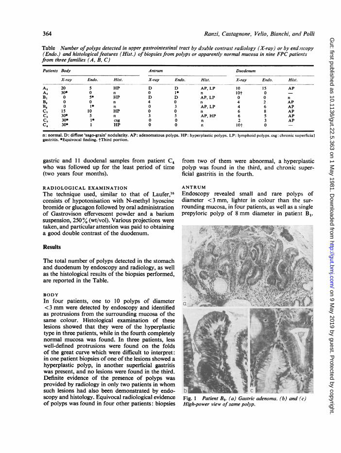
Fig. 1 Patient B3. (a) Gastric adenoma. (b) and (c)High-power view of same polyp.
364
on 9 May 2019 by guest. P
rotected by copyright.http://gut.bm
j.com/
Gut: first published as 10.1136/gut.22.5.363 on 1 M
ay 1981. Dow
nloaded from
Gastr-ic and duodenal polyps in familial polyposis coli
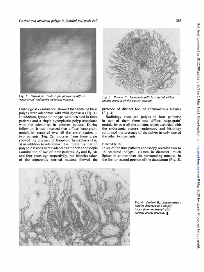
Fig. 2 Patient A1. Endoscopic picture of diffuse'sago-gr-ain' nodularity of antral mucosa.
Histological examination showed that some of thesepolyps were adenomas with mild dysplasia (Fig. 1).In addition, lymphoid polyps were detected in threepatients and a single hyperplastic polyp associatedwith the adenomas in another patient. Duringfollow-up, it was observed that diffuse 'sago-grain'nodularity appeared over all the antral region intwo patients (Fig. 2); biopsies from these areasshowed the presence of lymphoid hyperplasia (Fig.3) in addition to adenomas. It is interesting that nopolypoid lesions were evidenced at the first endoscopicexamination of two of these patients, A1 and B1, sixand four years ago respectively, but biopsies takenof the apparently normal mucosa showed the
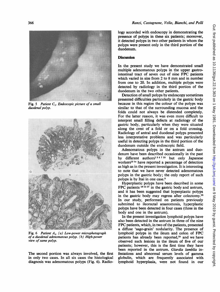
Fig. 3 Patient B, Lymphoidfollicle situated withinlamina propria of the gastric antrum.
presence of distinct foci of adenomatous tubules(Fig. 4).
Radiology visualised polyps in four patients;in two of them there was diffuse 'sago-grain'nodularity over all the antrum, which accorded withthe endoscopic picture; endoscopy and histologyconfirmed the presence of the polyps in only one ofthe other two patients.
DUODENUMIn six of the nine patients endoscopy revealed two to15 scattered polyps, <3 mm in diameter, muchlighter in colour than the surrounding mucosa, inthe first or second portion of the duodenum (Fig. 5).
Fig. 4 Patient B1. Adenomatous1 N !< .tubules detected in a biopsy
taken from endoscopicallynormal antral mucosa.
365
on 9 May 2019 by guest. P
rotected by copyright.http://gut.bm
j.com/
Gut: first published as 10.1136/gut.22.5.363 on 1 M
ay 1981. Dow
nloaded from
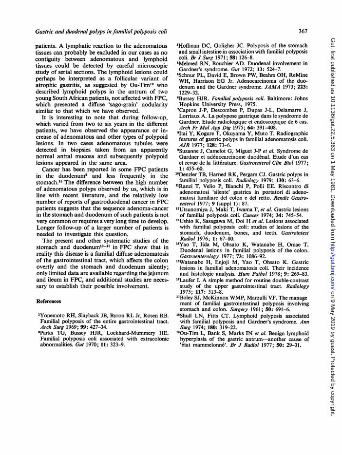
Ranzi, Castagnone, Velio, Bianchi, and Polli~~~~~~~~~~~~~~~~~~~~~~~~~~~~~~~~~~~~~~~~~~~~~~ ..::.:Fig. 5 Patient Cl. Endoscopic picture ofa smallduodenal polyp.
a
bFig. 6 Patient A,. (a) Low-power microphotographofa duodenal adenomatous polyp. (b) High-powerview ofsame polyp.
The second portion was always involved, the firstin only two cases. In all six cases the histologicaldiagnosis was adenomatous polyps (Fig. 6). Radio-
logy accorded with endoscopy in demonstrating thepresence of polyps in these six patients; moreover,it detected polyps in two other patients in whom thepolyps were present only in the third portion of theduodenum.
Discussion
In the present study we have demonstrated smallmultiple adenomatous polyps in the upper gastro-intestinal tract of seven out of nine FPC patientswhich varied in size from 2 to 8 mm and in numberfrom one to 20. In addition, multiple polyps weredetected by radiology in the third portion of theduodenum in the two other patients.
Detection of small polyps by endoscopy sometimespresented difficulties particularly in the gastric bodybecause in this region the colour of the polyps wassimilar to that of the surrounding mucosa and thefolds could not always be distended completely.For the latter reason, it was even more difficult tointerpret small filling defects at radiology of thegastric body, particularly when they were situatedalong the crest of a fold or on a fold crossing.Radiology of antral and duodenal polyps presentedless interpretative problems and was particularlyuseful in detecting polyps in the third portion of theduodenum outside the endoscopic field.Adenomatous polyps in the antrum and duo-
denum have been described occasionally in the pastby different authorsl 3 4 710 but only Japaneseworkers'2 1 have reported a percentage of detectionas high as in the present investigation. It is interestingto note that we have never detected adenomatouspolyps in the gastric body; the only report of suchpolyps is by Itai in one case.8
Hyperplastic polyps have been described in someFPC patients 101217 in the gastric body and antrum,and it has been suggested that hyperplastic polypsin the gastric body may regress after colectomy.'sIn our study, performed on patients previouslysubmitted to ileorectal anastomosis, hyperplasticpolyps have been detected in four cases (three in thebody and one in the antrum).
In the present investigation lymphoid polyps havealso been detected in the antrum in three of the nineFPC patients, which, in two of the patients, presenteda diffuse 'sago-grain' nodularity. The presence oflymphoid polyps in the ileum and colon of FPCpatients has already been reported,18 and we haveobserved such lesions in the ileum of five of ourpatients; however, this is the first time they havebeen reported in the antrum. Giardia lamblia in-festations and abnormal serum levels of gammaglobulin, which are frequently associated withlymphoid hyperplasia, were not found in our
366
on 9 May 2019 by guest. P
rotected by copyright.http://gut.bm
j.com/
Gut: first published as 10.1136/gut.22.5.363 on 1 M
ay 1981. Dow
nloaded from
Gastric and duodenal polyps in familial polyposis coli 367
patients. A lymphatic reaction to the adenomatoustissues can probably be excluded in our cases as nocontiguity between adenomatous and lymphoidtissues could be detected by careful microscopicstudy of serial sections. The lymphoid lesions couldperhaps be interpreted as a follicular variant ofatrophic gastritis, as suggested by Ou-Tim19 whodescribed lymphoid polyps in the antrum of twoyoung South African patients, not affected with FPC,which presented a diffuse 'sago-grain' nodularitysimilar to that which we have observed.
It is interesting to note that during follow-up,which varied from two to six years in the differentpatients, we have observed the appearance or in-crease of adenomatous and other types of polypoidlesions. In two cases adenomatous tubules weredetected in biopsies taken from an apparentlynormal antral mucosa and subsequently polypoidlesions appeared in the same area.
Cancer has been reported in some FPC patientsin the duodenumg and less frequently in thestomach."' The difference between the high numberof adenomatous polyps observed by us, which is inline with recent literature, and the relatively lownumber of reports of gastroduodenal cancer in FPCpatients suggests that the sequence adenoma-cancerin the stomach and duodenum of such patients is notvery common or requires a very long time to develop.Longer follow-up of a larger number of patients isneeded to investigate this question.The present and other systematic studies of the
stomach and duodenum'-15 in FPC show that inreality this disease is a familial diffuse adenomatosisof the gastrointestinal tract, which affects the colonovertly and the stomach and duodenum silently;only limited data are available regarding the jejunumand ileum in FPC, and additional studies are neces-sary to establish their possible involvement.
References
'Yonemoto RH, Slayback JB, Byron RL Jr, Rosen RB.Familial polyposis of the entire gastrointestinal tract.Arch Surg 1969; 99: 427-34.2Parks TG, Bussey HJR, Lockhard-Mummery HE.Familial polyposis coli associated with extracolonicabnormalities. Gut 1970; 11: 323-9.
3Hoffman DC, Goligher JC. Polyposis of the stomachand small intestine in association with familial polyposiscoli. Br J Surg 1971; 58: 126-8.4Melmed RN, Bouchier AD. Duodenal involvement inGardner's syndrome. Gut 1972; 13: 524-7.5Schnur PL, David E, Brown PW, Beahrs OH, ReMineWH, Harrison EG Jr. Adenocarcinoma of the duo-denum and the Gardner syndrome. JAMA 1973; 223:1229-32."Bussey HJR. Familial polyposis coli. Baltimore: JohnsHopkins University Press, 1975.7Capron J-P, Descombes P, Dupas J-L, Delamarre J,Lorriaux A. La polypose gastrique dans le syndrome deGardner. Etude radiologique et endoscopique de 6 cas.Arch Fr Mal App Dig 1975; 64: 391-408.8Itai Y, Kogure T, Okuyama Y, Muto T. Radiographicfeatures of gastric polyps in familial adenomatosis coli.AJR 1977; 128: 73-6.9Suzanne J, Camelot G, Miguet J-P et al. Syndrome deGardner et adenocarcinome duodenal. Etude d'un caset revue de la litt6rature. Gastroenterol Clin Biol 1977;1: 455-60.
10Denzler TB, Harned RK, Pergam CJ. Gastric polyps infamilial polyposis coli. Radiology 1979; 130: 63-6.
1Ranzi T, Velio P, Bianchi P, Polli EE. Riscontro diadenomatosi 'silente' gastrica in portatori di adeno-matosi familiare del colon e del retto. Rendic Gastro-enterol 1977; 9 (suppl 1): 87.
12Utsunomiya J, Maki T, Iwama T, et al. Gastric lesionsof familial polyposis coli. Cancer 1974; 34: 745-54.
13Ushio K, Sasagawa M, Doi H et al. Lesions associatedwith familial polyposis coli: studies of lesions of thestomach, duodenum, bones, and teeth. GastrointestRadiol 1976; 1: 67-80.
14Yao T, lida M, Ohsato K, Watanabe H, Omae T.Duodenal lesions in familial polyposis of the colon.Gastroenterology 1977; 73: 1086-92.
15Watanabe H, Enjoji M, Yao T, Ohsato K. Gastriclesions in familial adenomatosis coli. Their incidenceand histologic analysis. Hum Pathol 1978; 9: 269-83.
'6Laufer I. A simple method for routine double-contraststudy of the upper gastrointestinal tract. Radiology1975; 117: 513-8.
17Boley SJ, McKinnon WMP, Marzulli VF. The management of familial gastrointestinal polyposis involvingstomach and colon. Surgery 1961; 50: 691-6.
18Shull LN, Fitts CT. Lymphoid polyposis associatedwith familial polyposis and Gardner's syndrome. AnnSurg 1974; 180: 319-22.
'90u-Tim L, Bank S, Marks IN et al. Benign lymphoidhyperplasia of the gastric antrum-another cause of'etat mammelonne'. Br J Radiol 1977; 50: 29-31.
on 9 May 2019 by guest. P
rotected by copyright.http://gut.bm
j.com/
Gut: first published as 10.1136/gut.22.5.363 on 1 M
ay 1981. Dow
nloaded from
Related Documents