Clostridia that produce invasive infections Histotoxic Group Clostridia that produce invasive infections • Variety of species of clostridium are associated with invasive infection in humans, i.e. – Clostridium perfringens – C. novyi – C. septicum – C. histolyticum – C. tertium – C. bifermentans – C. sporogenes 1

Welcome message from author
This document is posted to help you gain knowledge. Please leave a comment to let me know what you think about it! Share it to your friends and learn new things together.
Transcript
Clostridia that produce invasive infections
Histotoxic GroupClostridia that produce invasive infections• Variety of species of clostridium are associated with invasive infection in humans, i.e. – Clostridium perfringens – C. novyi – C. septicum – C. histolyticum – C. tertium – C. bifermentans – C. sporogenes
1
• Clostridium perfringens is the most common cause of clostridial gas gangrene (80-90% of cases).
• Other clostridia species responsible for the condition include
• Clostridium novyi (40%), • Clostridium septicum (20%), • Clostridium histolyticum (10%), • Clostridium bifermentans (10%), and • Clostridium fallax (5%).
• They are not highly pathogenic when introduced into healthy tissues; but in the presence of tissue injury, they cause rapidly progressive devastating infection with accumulation of gas & the extensive destruction of muscle and connective tissue.
• Pathogenesis is due to the production of toxins with necrotising, hemolytic or other destructive properties.
• Gangrene: The death of body tissue due to the loss of blood supply to that tissue, sometimes permitting bacteria to invade it and accelerate its decay.
• Types of gangrene: • Dry gangrene• Wet gangrene • Gas gangrene
Specific Gangrenes• Noma is a gangrene of the face. • Necrotizing fasciitis affects the deeper layers of the skin.
• Fournier gangrene usually affects the male genitals
6
CLOSTRIDIUM PERFRINGENS
Clostridium perfringens • Clostridia are large gram-positive, anaerobic, spore-forming bacilli commonly found throughout nature.
• They are non-motile, capsulated.• The spores are located terminally or subterminally
• The commonest of several members of the genus clostridium associated with gas gangrene.
• Although found in the colon of 25-35% of healthy people, under certain conditions it can produce serious, life treating infections.
7
Clostridium perfringens
• There are 5 types of C. perfringens (A - E) classified according to the various toxins they produce.
• Their presence in water indicates fecal pollution.
• C. perfringens type A & C produce enterotoxin which are responsible for food poisoning.
Morphology: – Box car-like gram-positive large bacilli, – The only non-motile spp in the genus, – spores are oval, sub-terminal & non- projecting– Capsules are formed in tissues.
8
Clostridium perfringens
Cultural characters:– Anaerobes, colonies on blood agar may show zones of complete haemolysis.
Biochemical activities: – They ferment glucose, lactose maltose & sucrose with much gas production (various sugars in thioglycolate). The organism causes rapid fermentation of lactose in litmus milk & the gas produced splits the clot ‘Stormy clot reaction’.
9
Clostridium perfringens
• Nagler's reaction: a test for the identification of alpha toxin of C. perfringens. The addition of antitoxin to cultures on egg yolk agar prevents visible opacity, due to lecithinase action which is normally observed around colonies.
• C. perfringens esp. type A produce opalescence in egg yolk media (5% egg yolk agar) due to production of lecithinase (α toxin) which causes a visible precipitate around the colonies.
• Such opalescence can be inhibited by C. perfringens antitoxin, which can be placed on half of the plate at the inoculation.
Other test: • negative motility test, • nitrate reduction, • proteolysis
Clostridium perfringens
Toxins or virulence factors• C. perfringens produce a variety of toxins & enzymes that result in spreading of infection.
1. Alpha toxin: a lecithinase that acts on lecithin which is a component of the cell membrane.
2. Theta toxin: has a haemolytic & necrotizing effect.
3. DNase, 4. Hyaluronidase 5. Collagenase.6. Enterotoxin produced by strains A & C which cause food poisoning following ingestion of warmed meat dish
11
Clostridium perfringens
• The enterotoxin is a protein that appears identical with a component of the spore coat.
12
Gas gangreneClinical disease: soft tissue infections• Portal of entry: trauma or intestinal tract• There are 3 types of clostridial wound infection:
1. Cellulitis: gas formation in the soft tissue
2. Fasciitis or supputarive myocytis: accumulation of gas in the muscle planes
3. Myonecrosis or gas gangrene – a life threatening disease
Clostridium perfringens
Gangrene
15
GAS GANGRENE
Pathogenesis: • The infection occurs when wounds are contaminated with soil containing the organism or its spores
• The condition occurs in deep lacerated, devitalized wounds as in car accidents or war wounds.
• Necrotising toxin, collagenase & hyaluronidase favour necrosis and spread of infection.
• The presence of foreign bodies, mixed infection with aerobic pyogenic bacteria and decreased blood supply lowers the O2 tension & favours germination of spores
GAS GANGRENE
• Vegetative cells multiply & ferment sugars producing large amounts of gas which distends the tissues and interferes with blood supply leading to tissue death.
• Proteolytic clostridia digest dead tissues leading to change in colour & foul odour of the wound.
• The condition is accompanied by generalized toxemia
Risk factors• Patients most at risk for this usually have underlying blood vessel disease (atherosclerosis or hardening of the arteries), diabetes, or colon cancer
GAS GANGRENE
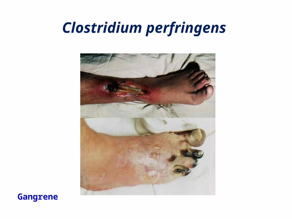
Symptoms of gas gangrene • Onset: onset of gas gangrene is sudden and dramatic. Inflammation begins at the site of infection as a pale-to-brownish-red and extremely painful tissue swelling.
• Gas gangrene causes very painful swelling. • The skin turns pale to brownish-red. If you press on the swollen area with your fingers, you may feel gas as a crackly sensation / air under the skin (subcutaneous emphysema)
• Blisters filled with brown-red fluid; vesicle formation
• Drainage from the tissues, foul-smelling brown-red or bloody fluid (serosanguineous discharge)
18
Clostridium perfringens
• Increased heart rate (tachycardia)• Moderate to high fever, sweating• Moderate to severe pain around a skin injury
• Pale skin color, later - dusky and dark red or purple
• Yellow color to the skin (jaundice)• Progressive swelling around a skin injury
Clostridium perfringens
Food poisoning due to C. perfringens • C. perfringens is a common cause but mild type of food
poisoning. • Its spores are heat resistant, surviving normal cooking.• More commonly found in foods that have been prepared in
bulk; thrives at the bottom of a stockpot or in the centre of a meat pie.
• Symptoms occur 6-18 hrs after consumption of the contaminated food
• They include abdominal pain, profuse diarrhoea, nausea, but rarely vomiting
• Two serotypes produce the enterotoxin which cause gastroenteritis: – type A is the more common agent of food poisoning; – type C is responsible for a more serious but rare condition known as enteritis necroticans.
• Toxin is released in the gut causing diarrhoea after 6-18 hrs which lasts for 1-2 days.
19
20
Clostridium perfringens
Diagnosis: • Diagnosis must be primarily on clinical grounds.
• Bacteriological confirmation is important.• Wound exudate & swabs from deeper areas are taken.
• Direct smear are stained by gram, the presence of large gram-positive rods is suggestive.
• Cultures made on blood agar, Robertson’s cooked meat media or thioglycolate broth incubated anaerobically, then subculture from fluid media on blood agar.
Clostridium perfringens
Colonies are identified by:-1. Morphology2. Biochemical activities:- sugar fermentation & stormy clot formation in litmus milk by Nagler's reaction (a test for the identification of alpha toxin of C. perfringens to detect the lecithinase activity)
3. Animal inoculation to show toxigenicity & its neutralization by specific antitoxin.
21
22
Clostridium perfringens
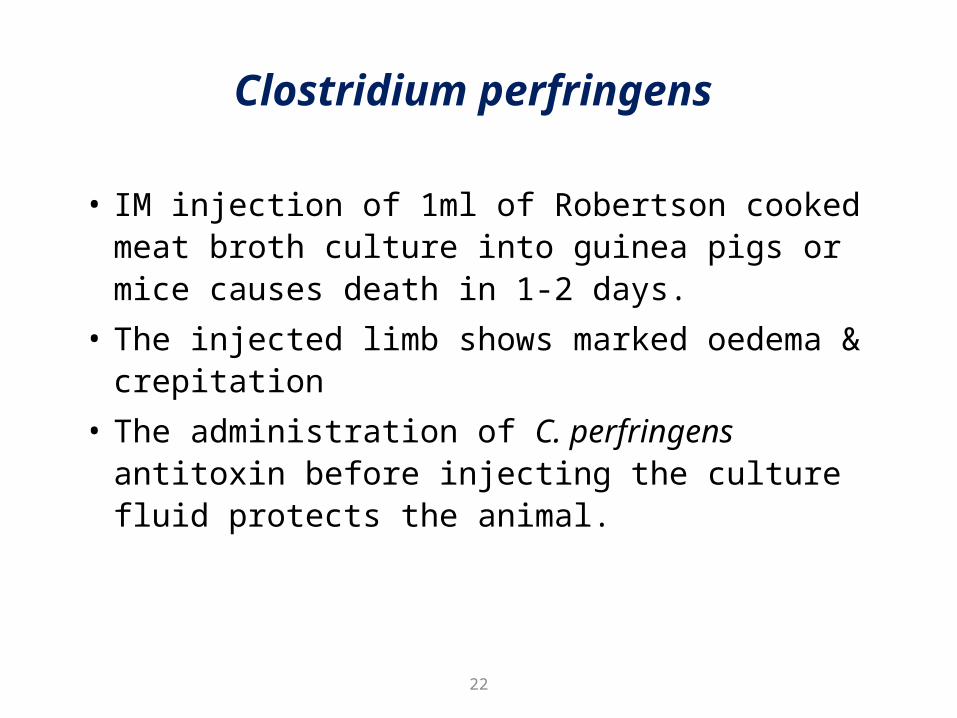
• IM injection of 1ml of Robertson cooked meat broth culture into guinea pigs or mice causes death in 1-2 days.
• The injected limb shows marked oedema & crepitation
• The administration of C. perfringens antitoxin before injecting the culture fluid protects the animal.
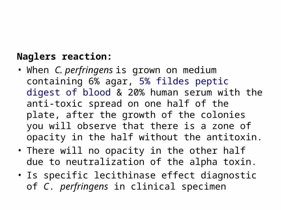
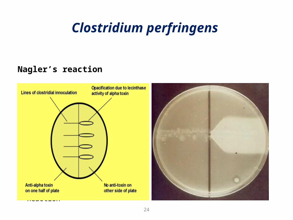
Naglers reaction: • When C. perfringens is grown on medium containing 6% agar, 5% fildes peptic digest of blood & 20% human serum with the anti-toxic spread on one half of the plate, after the growth of the colonies you will observe that there is a zone of opacity in the half without the antitoxin.
• There will no opacity in the other half due to neutralization of the alpha toxin.
• Is specific lecithinase effect diagnostic of C. perfringens in clinical specimen
Clostridium perfringens
Nagler’s reaction
• Procedure of Nagler Reaction Positive Nagler Reaction
24
Clostridium perfringens
Treatment• No specific treatment for toxin –Toxoid not available
• Antibiotics: penicillin.• Gas gangrene: surgical removal (amputation) of an arm or leg may be needed to control the spread of infection
• Hyperbaric oxygen
25
Prevention:• Prevention depends upon adequate cleaning of contaminated wounds, surgical removal of foreign bodies & excision of all devitalized tissues.
• Antitoxic sera for passive prophylaxis is unreliable.

Related Documents