Accepted Manuscript Garbage in, Garbage out: A Critical Evaluation of Strategies Used for Validation of Immunohistochemical Biomarkers Gillian O’Hurley, Evelina Sjöstedt, Arman Rahman, Bo Li, Caroline Kampf, Fredrik Pontén, William M. Gallagher, Cecilia Lindskog PII: S1574-7891(14)00056-8 DOI: 10.1016/j.molonc.2014.03.008 Reference: MOLONC 486 To appear in: Molecular Oncology Received Date: 24 February 2014 Accepted Date: 10 March 2014 Please cite this article as: O’Hurley, G., Sjöstedt, E., Rahman, A., Li, B., Kampf, C., Pontén, F., Gallagher, W.M., Lindskog, C., Garbage in, Garbage out: A Critical Evaluation of Strategies Used for Validation of Immunohistochemical Biomarkers, Molecular Oncology (2014), doi: 10.1016/ j.molonc.2014.03.008. This is a PDF file of an unedited manuscript that has been accepted for publication. As a service to our customers we are providing this early version of the manuscript. The manuscript will undergo copyediting, typesetting, and review of the resulting proof before it is published in its final form. Please note that during the production process errors may be discovered which could affect the content, and all legal disclaimers that apply to the journal pertain.

Welcome message from author
This document is posted to help you gain knowledge. Please leave a comment to let me know what you think about it! Share it to your friends and learn new things together.
Transcript
Accepted Manuscript
Garbage in, Garbage out: A Critical Evaluation of Strategies Used for Validation ofImmunohistochemical Biomarkers
Gillian O’Hurley, Evelina Sjöstedt, Arman Rahman, Bo Li, Caroline Kampf, FredrikPontén, William M. Gallagher, Cecilia Lindskog
PII: S1574-7891(14)00056-8
DOI: 10.1016/j.molonc.2014.03.008
Reference: MOLONC 486
To appear in: Molecular Oncology
Received Date: 24 February 2014
Accepted Date: 10 March 2014
Please cite this article as: O’Hurley, G., Sjöstedt, E., Rahman, A., Li, B., Kampf, C., Pontén, F.,Gallagher, W.M., Lindskog, C., Garbage in, Garbage out: A Critical Evaluation of Strategies Usedfor Validation of Immunohistochemical Biomarkers, Molecular Oncology (2014), doi: 10.1016/j.molonc.2014.03.008.
This is a PDF file of an unedited manuscript that has been accepted for publication. As a service toour customers we are providing this early version of the manuscript. The manuscript will undergocopyediting, typesetting, and review of the resulting proof before it is published in its final form. Pleasenote that during the production process errors may be discovered which could affect the content, and alllegal disclaimers that apply to the journal pertain.

MANUSCRIP
T
ACCEPTED
ACCEPTED MANUSCRIPT
Garbage in, Garbage out: A Critical Evaluation of Strategies
Used for Validation of Immunohistochemical Biomarkers
Gillian O’Hurleya,b,c, Evelina Sjöstedtb, Arman Rahmanc, Bo Lia, Caroline Kampf b, Fredrik
Ponténb*, William M. Gallaghera,c*, Cecilia Lindskogb.
aUCD School of Biomolecular and Biomedical Science, UCD Conway Institute, University College Dublin,
Belfield, Dublin 4, Ireland
bDepartment of Immunology, Genetics and Pathology, Science for Life Laboratory, Uppsala University,
Uppsala, Sweden
cOncoMark Ltd, NovaUCD, Belfield Innovation Park, Belfield, Dublin 4, Ireland.
*Corresponding authors:
Fredrik Pontén Department of Immunology, Genetics and Pathology Science for Life Laboratory Uppsala University 751 85 Uppsala Sweden Tel: +46 18 611 3846 Email: [email protected]
William M. Gallagher UCD School of Biomolecular and Biomedical Science UCD Conway Institute University College Dublin Belfield Dublin 4 Ireland Tel: +353 1 7166743 Email: [email protected]
MANUSCRIP
T
ACCEPTED
ACCEPTED MANUSCRIPT
Abstract
The use of immunohistochemistry (IHC) in clinical cohorts is of paramount importance in
determining the utility of a biomarker in clinical practice. A major bottleneck in translating a
biomarker from bench-to-bedside is the lack of well characterized, specific antibodies
suitable for IHC. Despite the widespread use of IHC as a biomarker validation tool, no
universally accepted standardization guidelines have been developed to determine the
applicability of particular antibodies for IHC prior to its use. In this review, we discuss the
technical challenges faced by the use of immunohistochemical biomarkers and rigorously
explore classical and emerging antibody validation technologies. Based on our review of
these technologies, we provide strict criteria for the pragmatic validation of antibodies for use
in immunohistochemical assays.
Keywords
Immunohistochemistry, biomarker discovery, antibody reliability, antibody validation,
workflow

MANUSCRIP
T
ACCEPTED
ACCEPTED MANUSCRIPT
1. Introduction
The classical method of immunohistochemistry (IHC) allows for visualization of specific
antigens in tissues or cells based on antibody-antigen recognition, using brightfield or
fluorescence microscopy. The history of IHC goes back to the early 1940s, when Coons and
colleagues detected antigens in frozen tissue sections by developing an immunofluorescence
technique (Coons et al., 1941). Introduction of a method based on peroxidase-labelled
antibodies opened the door to development of more advanced approaches (Mason et al.,
1969, Nakane, 1968), enabling IHC to be used on routinely processed tissue sections, such as
formalin-fixed paraffin-embedded (FFPE) tissues. However, it took until the early 1990s for
the method to become generally accepted in diagnostic pathology (Leong, 1992, Taylor,
1994).
IHC is today a widely used method that can be rapidly performed in most laboratories. The
procedure is short, simple and cost-effective. Indeed, IHC has emerged as an important tool
to detect cellular markers defining specific phenotypes relative to disease status and biology.
Moreover, IHC is utilized for basic and clinical research, from small projects to high-
throughput strategies, to evaluate potential biomarkers in clinical patient cohorts. However,
the lack of standardized guidelines for determining the specificity and functionality of
antibodies renders the translation of promising biomarkers to the clinic difficult. Herein, we
discuss the various limitations and technical challenges that need to be addressed when using
IHC for biomarker development and clinical validation.
2. Review of clinically used IHC markers approved by FDA
A biomarker is defined as a molecule that is objectively measured and evaluated as an
indicator of normal biological process, pathogenic process, or pharmacological responses to
therapeutic intervention (Biomarkers-Definitions-Working-Group, 2001). Although great
efforts have been made in the last decade to discover novel cancer biomarkers for use in
clinical practice, a striking number of these efforts fail to make it into the clinic (Fuzery et al.,
2013). One of the causes of this failure of translation could be the limited knowledge that
scientists working in biomarker discovery have in analytical, diagnostic and regulatory
requirements for clinical assays (Fuzery et al., 2013). Over the last few decades a number of
key FDA approved cancer biomarkers have been introduced into the clinic for differential
diagnosis of specific tumours, leading to improvement of cancer detection and staging,
MANUSCRIP
T
ACCEPTED
ACCEPTED MANUSCRIPT
identification of tumour subclasses, prediction of outcome after treatment, and selection of
patients for different treatment options. However, of these approved biomarkers, only five are
individual IHC-based biomarkers (Fuzery et al., 2013) (Table 1). The earliest FDA approved
biomarkers for IHC application were assays to detect the estrogen receptor (ER),
progesterone receptor (PR) and HER-2/neu (c-erbB-2). The presence of these biomarkers in
breast cancer tissue serves as a diagnostic, prognostic and predictive method to assist
pathologists in identifying breast cancer subtypes and determine whether patients are suitable
candidates to receive certain targeted therapies such as Tamoxifen (ER positive patients) or
Trastuzumab (Her-2 positive patients). The IHC biomarker c-kit (CD117), which is used in
the clinic to detect gastrointestinal stromal tumours (GISTs) (Debiec-Rychter et al., 2004),
and p63, which is used to detect the presence of basal cells indicative of normal prostate
glands (Shah et al., 2002, Weinstein et al., 2002), are the latest FDA approved single marker
IHC-based assays which were approved almost a decade ago in 2004 and 2005, respectively.
Since then no other individual biomarker developed for detection in an IHC assays has been
FDA approved. However, despite lack of FDA approval, there are many IHC markers utilized
in some clinics to assist pathologists in diagnosis and decision making. Such examples
include the use of E-Cadherin and/or p120 staining to assist diagnosis of invasive lobular
breast carcinoma (Rakha et al., 2010), various antibody panels for diagnosis and sub-
classification of malignant lymphomas, as well as the use of the proliferating nuclear marker,
Ki67.
An ideal biomarker demonstrating clinical sensitivity and specificity of 100% is almost never
achieved in practice due the fact that increasing one of the parameters is only achieved at the
expense of the other. As a result, panel biomarker assays are becoming more relevant. Two
emerging IHC panel-based assays are Mammostrat by Clarient InsightDx and IHC4 by
Genoptix Medical Laboratory. Mammostrat is an IHC-based panel assay that can estimate
risk of recurrence in hormone receptor-positive, early stage breast cancer patients which is
independent of proliferation and grade. This assay quantifies p53, HTF9C, CEACAM5,
NDRG1 and SLC7A5 by a defined mathematical algorithm resulting in a risk index (Bartlett
et al., 2012, Bartlett et al., 2010). Similarly, IHC4 is another emerging assay which estimates
recurrence risk for early stage breast cancer patients by quantifying IHC measurement of ER,
PR, HER2 and Ki-67 using Aqua® technology (Cuzick et al., 2011).

MANUSCRIP
T
ACCEPTED
ACCEPTED MANUSCRIPT
IHC-based biomarker assays represent an attractive approach for biomarker detection in the
clinic as the IHC technique is routinely carried out in clinical laboratories, there is a fast turn-
around time from assay to results and it is cost-effective. However, the paucity of FDA-
approved biomarkers for IHC-based assays emphasizes the importance and urgent
requirement of standardized guidelines and workflows for IHC assay development which
should be implemented at an early stage of biomarker discovery. This will ensure robust
analytical and clinical performance and ultimately lead to a better chance of an IHC-based
biomarker assay achieving FDA approval.
3. Review of factors influencing the IHC process
The standard brightfield IHC technique is comprised of three components; slide preparation,
IHC procedure and interpretation. Antibodies used in the clinic have undergone thorough
testing and every step of the protocol has been well established, including both positive and
negative controls. Factors which may affect the outcome of IHC include tissue handling,
epitope retrieval, storage and handling of tissue sections, choice of antibody, detection
method and interpretation procedure. To yield the expected staining pattern when establishing
a new antibody, all factors which may influence the standardization and reproducibility of the
process need to be carefully considered. These factors are summarized in Figure 1 and will be
described more in detail below.
3.1. Tissue handling immediately after surgery, fixation and processing
‘Ischemia time’ refers to the time from when a tissue or organ is cut off from O2 supply
through removal of a specimen from the body in surgery, to fixation of the specimen.
Ischemia results in degradation of protein, RNA and DNA, as well as activation of tissue
enzymes and autolysis (Kumar, 2005) and can therefore be a major factor influencing IHC
results. Recently, Pekmezci et al. demonstrated that longer cold ischemia time affects the
detection of ER and PR by IHC in breast cancer (Pekmezci et al., 2012). Although the
American Society of Clinical Oncology and College of American Pathologists (ASCO/CAP)
has developed guidelines for handling of tissues for ER, PR and HER-2 detection in breast
cancer patients, such guidelines are not available for other surgical specimens (Comanescu et
al., 2012, Hammond et al., 2010). Fixation is another critical step in the IHC process to
preserve tissue morphology and retain antigenicity of the target molecules. Two types of
fixatives are commonly used in histopathology; (1) non-coagulating fixatives (formaldehyde,
MANUSCRIP
T
ACCEPTED
ACCEPTED MANUSCRIPT
glutaraldehyde, osmium teroxide, potassium dichromate and acetic acid) and (2) coagulating
fixatives (alcohol, zinc salts, mercuric chloride, chromium trioxide and picric acid). The most
common fixative used in histopathology is 10% neutral-buffered formalin. This is composed
of 4% paraformaldehyde solution which is buffered to a neutral pH. Formalin cross-links
peptides by formation of hydroxymethyl groups on reactive amino acid side chains, providing
excellent preservation of tissue architecture; however, formalin fixation can mask epitopes
and result in decreased antigenicity. Several factors influence the formalin fixation method,
such as temperature, time, penetration rate, specimen dimension, volume ratio, pH of the
buffer and osmolality, but unfortunately, there is a lack of available guidelines to establish a
standard practise across pathology laboratories.
3.2. Appropriate storage and handling of tissue sections
Another factor that may influence the IHC outcome is storage of prepared tissue sections
(Wester et al., 2000, Williams et al., 1997). It has been suggested that storing tissue sections
longer than two months leads to loss of p53 antigen reactivity (Prioleau and Schnitt, 1995).
The mechanisms underlying the loss of antigenicity in FFPE tissue is unclear. It has been
hypothesised that oxidation may be the key contributor of antigenicity loss (Blind et al.,
2008, Sauter and Mirlacher, 2002). Due to this and the fact that degradation of protein is
temperature dependent, a large variety of storage conditions for cut sections have been
advocated such as cold storage, paraffin coating or vacuum sealed desiccators. However,
recently it has been suggested by Xie et al, 2011 that the presence of water both
endogenously and exogenously plays a central role in loss of antigenicity. Therefore, slide
storage conditions that are protected from oxidization by vacuum storage or paraffin coating
are not completely protecting slides from loss of antigenicity if residual water from
inadequate tissue processing is present on the tissue (Xie et al., 2011). Thus, the optimal
storage of unstained sections is yet to be defined, making freshly cut sections or sections
stored for less than two months most ideal. For long-term storage, vacuum containers or
storage in colder conditions (+4/-18 degrees) is often recommended.
3.3. Appropriate and efficient epitope retrieval
Another major step that should be considered carefully when performing IHC is antigen
retrieval (AR). The two methods of antigen retrieval are (1) heat-induced epitope retrieval
(HIER) (e.g. citrate pH 6.0, Tris-EDTA pH 9.0 and EDTA pH 8.0) and (2) proteolytic
enzyme-induced epitope retrieval (PIER) (e.g. proteinase K, trypsin, pepsin, pronase). Of the

MANUSCRIP
T
ACCEPTED
ACCEPTED MANUSCRIPT
two methods, HIER is most commonly used. The technique was first described by Shi and
colleagues (Shi et al., 1991) and has been improved by a number of investigators (Cattoretti
et al., 1993, Greenwell et al., 1991, Greenwell et al., 1993) for its routine use in laboratories
throughout the world. However, the mechanisms of AR are not fully understood. It is
speculated that both HIER and PIER serve to break the methylene bridges created during
fixation, exposing the antigenic sites in order to allow the antibodies to bind (D'Amico et al.,
2009, Fowler et al., 2011, Kakimoto et al., 2008, Leong and Leong, 2007, Suurmeijer and
Boon, 1993).
There are several different AR variables that can affect IHC staining results such as heating,
the choice of AR solution, its pH and molarity, and the effect of metal ions (D'Amico et al.,
2009, Emoto et al., 2005). An appropriately controlled AR method can restore antigenicity in
formalin fixed paraffin embedded (FFPE) tissue to resemble the antigenicity of frozen tissue
and can facilitate IHC standardization, despite variations in tissue fixation and subsequent
handling (von Boguslawsky, 1994) (Shi et al., 2007, Taylor, 2006). However, the appropriate
AR protocol is dependent on both the antibody and the target protein, and needs to be
optimized for every antibody.
3.4. Appropriate choice of antibody (monoclonal vs polyclonal)
The three cardinal points that must be considered when buying commercial primary
antibodies for IHC are as follows: (1) use reliable, recommended companies, (2) obtain
complete information about the antibody to ensure it is applicable or recommended for IHC
and, (3) characterize the specificity of the antibody. A significant number of commercial
antibodies are not thoroughly analysed for off-target binding, e.g. using protein arrays
(Chang, 1983, Nilsson et al., 2005). In addition, several companies do not provide the
sequence of the antigen the antibody was raised against (Saper, 2009) and, therefore,
antibody validation is a mandatory step before proceeding with IHC.
The choice of using either monoclonal or polyclonal primary antibodies for IHC further
complicates the issue of epitope specificity and determining which antibody would be more
suitable for IHC (Bordeaux et al., 2003). Polyclonal antibodies are collection of antibodies
targeted against multiple epitopes of a particular antigen. Generally, when an animal is
injected with a specific antigen, the immune system elicits a primary immune response by
producing multiple B cell clones against the antigen. After subsequent immunization with the
MANUSCRIP
T
ACCEPTED
ACCEPTED MANUSCRIPT
same antigen, these B cells differentiate into plasma cells producing and secreting antibodies
found in the serum. The serum containing polyclonal antibodies can be affinity purified using
the antigen as a ligand, which eliminates 99% of antibodies recognizing other targets than the
antigen. This procedure results in antibodies with higher specificity than conventional
polyclonal antibodies, still retaining the ability to recognize different epitopes on the same
antigen (Lindskog et al., 2005). A monoclonal antibody is generated by selection of one
single B cell from spleen or bone marrow of the immunized animal and fusing this cell with
immortal myeloma cells to produce hybridoma cells (Kohler and Milstein, 1975). As such,
the culture supernatant contains only one type of antibody specific for a single epitope of the
immunizing peptide. The advantages and disadvantages of using polyclonal and monoclonal
antibodies for IHC are summarized in Table 2.
A useful tool to search for appropriate antibodies suitable for IHC is the portal,
Antibodypedia (http://www.antibodypedia.com). Here, antibodies are listed with reference to
antibody companies and associated validation data (Bjorling and Uhlen, 2008).
3.5. Use of a sensitive and robust detection system
The outcome of an IHC assay depends on the use of sensitive protein detection system in
order to visualize the antigen-antibody reaction. The most popular methods of detection are
enzyme and fluorophore-mediated detection systems. With chromogenic substrates, an
enzyme label is reacted with the substrate to yield a strong colour product visualized by
brightfield imaging. Alkaline phosphatase (AP) and horseradish peroxidase (HRP) are the
two most extensively used enzymes, both with available chromogenic, fluorogenic and
chemiluminescent substrates.
Detection systems in IHC can be divided into two broad categories, namely direct or indirect.
In the direct detection method, the primary antibody is labelled with enzymes or
fluorochromes, enabling direct detection of the antigen on the tissue section without the
requirement of a secondary antibody. This method of detection is simpler and less time
consuming; however, it has the disadvantage of lower sensitivity compared with indirect
methods. The indirect detection method involves the use of unlabelled primary antibodies and
labelled secondary/tertiary antibodies, which are specific for the bound primary antibody.
Although this method is time consuming and complicated by multiple steps, indirect
detection method is more sensitive in detecting tissue antigens. Some commonly used
MANUSCRIP
T
ACCEPTED
ACCEPTED MANUSCRIPT
indirect detections mechanisms are as follows; the avidin-biotin complex (ABC) method, the
labelled streptavidin biotin (LSAB) method, the phosphatase-anti-phosphatase (PAP) and the
polymer-based detection system.
There are several other immunohistochemical detection methods such as tyramide
amplification, cycled tyramide amplification, fluorescyl-tyramide amplification and rolling
circle amplification, but these are not heavily used to date in routine IHC.
3.6. Detection of phosphorylation using IHC
Post-translational modifications are important biological events that control the behaviour of
a protein. Phosphorylation is a post-translational process regulating protein activity by the
addition and removal of a phosphate group. Tissue phosphoproteomic studies show promise
for the discovery of key phosphorylated proteins and signalling pathways in many diseases
(Bodo and Hsi, 2011). The detection and quantification of phosphorylation has been well
established using techniques such as Western blotting on cell lysates but it represents a new
era in diagnostic pathology. Many phospho-specific antibodies have been generated for
immunohistochemical application; however, the detection step remains challenging due to the
labile nature of phosphorylated proteins, reflecting dynamic processes. In addition, tissues
become oxygen deficient shortly after being isolated from the blood supply and subsequently
undergo rapid protein dephosphorylation (Blow, 2007). Therefore, if the tissues are not fixed
within 60 minutes post-surgical removal from the living body, the majority of phospho-
epitopes are lost (Baker et al., 2005, Jones et al., 2008). Due to this, most phosphorylation
studies have not been reproduced. Other variations between studies leading to these
discrepant results can include sample procurement, processing, scoring/quantification and
subjectively selected cut-offs (Bodo and Hsi, 2011). Therefore, rigorous standardization of
laboratory procedures for tissue preservation and for the overall IHC technique as well as
quantification is required for success in quantifying phosphorylation by IHC in tissue. Post-
translational modifications such as phosphorylation can also be studied with Proximity
Ligation Assay (PLA), described in Section 4.3. However, many of the same issues discussed
here will also apply to PLA.
3.7. Use of manual immunohistochemistry versus automated immunohistochemistry
platforms

MANUSCRIP
T
ACCEPTED
ACCEPTED MANUSCRIPT
A major milestone in the standardization, reliability and reproducibility of IHC is the
invention of automated IHC platforms. Many critical steps in the manual IHC method are
operator-dependent and essential to the quality of the final IHC result and its reproducibility
(Shi and Taylor, 2011). These include the critical antigen retrieval step, reagent preparation,
application of reagents, appropriate washing steps and multiple incubation times. The use of
automated IHC not only allows for larger volumes of slides to be stained simultaneously
under standardized conditions, but also provides assistance to operators through additional
processing monitoring errors such as alarms for inappropriate temperatures, insufficient
volumes of reagent, expired reagents and even the selection of an incorrect reagent via the
use of barcode scanning (Fetsch and Abati, 1999, Moreau et al., 1998, Prichard et al., 2011).
Many automated IHC machines, particularly those used in a clinical setting, are what is
termed as “closed systems” which means the instrument is closed to introducing variations.
Although this is an important advantage for standardization of IHC staining, it can be a
drawback for research as the flexibility of choosing reagents, retrieval methods and introduce
subtle variation to the technique is lost. This has led to the development of “open” automated
systems, offering similar flexibility as manual staining (Prichard et al., 2011). However, as
HIER is not performed on an “open” platform, some of the same limitations of manual IHC
discussed previously apply to this type of automated IHC. Clearly, there are advantages and
disadvantages to the manual staining method and the “open” and “closed” automated systems
so the choice of method should be influenced by the laboratory’s purpose. (Prichard et al.,
2011). However, for large-scale IHC efforts where planning and standardized IHC protocols
are necessary (Uhlen et al., 2005, Warford et al., 2004) it can be anticipated that automated
IHC may lead to reduction in error rate as each step of the staining procedure is recorded
(Howat et al., 2014). Together with the tissue microarray (TMA) technology (Battifora, 1986,
Kononen et al., 1998), where a large number of tissues from different organs or individuals
are assembled on a single slide, high-throughput IHC minimizes reproducibility issues.
3.8. Interpretation via manual and automated approaches
Manual assessment of IHC staining remains the traditional method for most diagnostic and
predictive decisions in pathology. However, manual interpretation of IHC data can be time
intensive, laborious and an inherently subjective and semi-quantitative process (Fiore et al.,
2012). Observer variability can exist in three forms; intra-observer variability, inter-observer
variability and inter-laboratory variability (Conway et al., 2008). The latter is usually
attributed to issues regarding tissue fixation and processing, antibodies used and detection

MANUSCRIP
T
ACCEPTED
ACCEPTED MANUSCRIPT
systems. Intra-observer variability, referring to the lack of consistent assessment by the
observer, occurs less frequently than inter-observer variability due to the fact that
pathologists adhere to their own internal standards (Kay et al., 1994). Inter-observer
variability is the greatest problem associated with human-based assessment of IHC staining,
influenced by factors such as misplaced orientation on a TMA slide, eye fatigue, complexity
of data management following differential categorical scoring, quality of microscope,
illumination of microscope and individual human vision limitations (Conway et al., 2008).
Utilizing image analysis systems on virtual microscopy slides or whole slide images has been
proposed as solving the problem of standardized quantification of IHC data, due to its
capability of producing continuous datasets eliminating categorical and biased assessment.
High-throughput image analysis methods can also reduce workloads and outperform human
manual scoring in terms of reproducibility and precision, as they are not affected by fatigue
or subjectivity. Enormous advances in image analysis systems on tissue sections have been
achieved over the years (Taylor and Levenson, 2006). However, despite these advances,
image analysis is far from ready to replace the expert pathologist, as it is still very much a
semi-automated approach as most algorithms require specific input and training by a
pathologist in order to produce accurate output. In addition, image analysis approaches are
highly influenced by a number of factors that can affect the quality of their performance. For
example, the quality of sections/TMAs hugely affects the resulting data obtained from image
analysis. This is due to the inability of most of the current automated image analysis systems
to identify irregularities on a section that the human eye can ignore, such as artefacts, edge
effect staining, folding of tissue and thickness of tissue section, which may produce a false
score. Moreover, image analysis often fails to distinguish tumour from benign tissue.
Nevertheless, it is widely accepted that the continuous development in computer-aided image
analysis technologies will lead to quantitative systems that will compliment and support the
pathologist/human expert to produce a less subjective and accurate IHC assessment.
3.9. Multiplexing: Brightfield vs. darkfield
When measuring protein expression levels in tissue, a decision must be made as to whether
assessment should be performed by IHC using brightfield imaging or immunofluorescence
(IF) using fluorescent imaging, where both techniques offer advantages over the other.
Brightfield imaging utilizes visible white light to illuminate the tissue, and protein expression
MANUSCRIP
T
ACCEPTED
ACCEPTED MANUSCRIPT
is classically observed and graded based on the intensity of 3,3’-diaminobenzidine (DAB),
generating a brown staining (Gustashaw et al., 2010). Counterstaining with haematoxylin
keeps morphological detail of the surrounding tissue intact and allows visualisation and
analysis of localized protein. The IF technique visualizes protein expression in tissue against
a dark background, using an antibody with a chemically attached fluorochrome, such as
fluorescein isothiocyanate (FITC) or tetramethyl rhodamine isothiocyanate (TRITC) (Jordan
et al., 2002). The antigen-antibody complex can be visualized using a fluorescent imaging
instrument such as a microscope or scanner.
IHC using brightfield imaging is one of the pillars of modern pathology and a fundamental
research tool in both pathology and translational research (Robertson et al., 2008), due to the
many advantages associated with the technique. It can be performed routinely on FFPE
tissue, which permits a pathologist or researcher to work with a familiar, conventional
microscope (Jordan et al., 2002). In addition, it can detect antigens expressed at relatively low
levels due to chromogenic enhancement steps, the equipment cost is low, and only minimal
laboratory space is required. Most importantly in a clinical setting, the chromogens are very
stable and long-term slide storage is possible for many years. However, as a research tool,
there are some major limitations associated with the technique. Firstly, the resolution of
antigen localization is limited due to the chromogenic substrate precipitate, as well as the
thickness of the sections imaged in the light microscope. Secondly, saturation of chromogenic
systems occurs easily, which restricts quantitative analyses (Robertson et al., 2008). Above
all, IHC using brightfield microscopy has a narrow dynamic range limiting its capability of
multiplexing, and as cross reactivity is common, three antibodies/chromogens at a time is a
maximum. Therefore, sequential or multi-step staining is crucial to ensure cross reactivity
does not occur with enzymes used or with primary/secondary antibodies raised in the same
species. In addition, choosing colour combinations that are distinguishable by eye from each
other and from the counterstain can be challenging, particularly when looking at co-localized
proteins. The concentration of precipitate may also inhibit further reaction, making it difficult
to visualize rare targets and highly abundant targets on the same slide (Christensen and
Winthers, 2009). Moreover, quantitation of multi-staining using brightfield microscopy is
even more limited, as most brightfield image analysis tools are primarily designed to quantify
single chromogens. However, the use of spectral imaging technologies allows unmixing of
stains and individual quantification of each chromogen.
MANUSCRIP
T
ACCEPTED
ACCEPTED MANUSCRIPT
In contrast to brightfield IHC, IF has a better capability of multiple labelling, as IF is of
higher resolution due to the fluorophores being directly conjugated to the antibody
(Robertson et al., 2008). Although choosing dyes with distinguishable spectral properties is
still an issue, fluorescent imaging has a much broader dynamic range compared with
brightfield imaging (Christensen and Winthers, 2009). On the other hand, IF-based detection
presents certain difficulties in respect to interpretation of tissue morphology, as well as the
cost of reagents and equipment. Moreover, a fluorescent signal can be quenched when the
fluorophores are in close proximity, and as fluorophores are not as stable as chromogens,
photobleaching of stored slides is an issue. The most restraining aspect of IF is inherent
autofluorescence of FFPE material, making high quality immunofluorescence imaging
capricious (Robertson et al., 2008) and limiting the use of clinical material. Examples of
consecutive sections stained with both brightfield and darkfield are displayed in Figure 2,
illustrating some of the advantages and disadvantages with both methods.
The use of multispectral imaging has overcome many of the issues regarding
autofluorescence on FFPE tissue (Mansfield et al., 2005, Robertson et al., 2008). However,
many reports using IF labelling of FFPE sections (Bataille et al., 2006, Bossard et al., 2006,
Ferri et al., 1997, Hoover et al., 1998, Mason et al., 2000, Niki et al., 2004, Nurnberger et al.,
2006, Papaxoinis et al., 2007, Scott et al., 2004, Suetterlin et al., 2004) have not been widely
acknowledged by the scientific community (Robertson et al., 2008) rendering IHC by
brightfield microscopy a more accepted assay for clinical use in quantifying protein
expression. However, continuous research and development of new methods in the area of IF
and image analysis, such as the new technique MxIF (Gerdes et al., 2013), will bridge the gap
between classical IHC of FFPE material and the acceptance of IF analysis of human FFPE
tissues.
One potentially might also consider application of both brightfield and fluorescent imaging,
e.g. use of H&E staining/brightfield imaging for localisation of tumour regions and use of
fluorescence-based imaging for quantitation of consecutive tissue sections.
4. Review of currently used validation methods for antibodies for IHC
Commercial production of antibodies is well established; however, there are no universally
accepted guidelines or standardized methods for determining the validity of these reagents
MANUSCRIP
T
ACCEPTED
ACCEPTED MANUSCRIPT
(Bordeaux et al., 2003). The production and validation of specific antibodies is a challenging,
costly and time consuming process. Perhaps as a result, the quality control by the antibody
vendors is not always what it should be (Couchman, 2009). Moreover, the information
supplied in academic publications where the antibodies are used is often insufficient.
Therefore, it is imperative that investigators take requisite steps to assure themselves that the
specificity of each antibody is as advertised. Here we explore both classical and emerging
technologies for antibody validation.
4.1. Which staining pattern is expected?
The signal intensity is generally related to the antibody concentration (Dabbs, 2006). In order
to get an optimal dilution of an antibody, rendering the greatest contrast between desired
(specific) positivity and unwanted (non-specific) background, it is necessary to know which
staining pattern to expect. Hence, the first crucial step in antibody validation is to understand
the nature of the target protein. For well-known or partly characterized proteins, information
regarding the expected staining pattern can be obtained from available databases such as
Uniprot (www.uniprot.org), the Human Protein Atlas (www.proteinatlas.org), or by searches
in published literature. Bioinformatic prediction algorithms for expected subcellular
localisation, including presence of signal peptides or transmembrane regions, is gathered in
online sources such as MDM (Fagerberg et al., 2010), SPOCTOPUS (Viklund et al., 2008)
and Phobius (Kall et al., 2004). Furthermore, information on post-translational modifications
or splice variants is important in order to predict detection of multiple bands in Western
blotting. Such information can be retrieved from e.g. OMIM (www.ncbi.nlm.nih.gov/omim)
or Genecards (www.genecards.org). A large fraction of the human proteins are essentially
uncharacterized and experimental data is needed for validation of the generated staining
pattern in IHC.
4.2. Western blotting
The standard antibody validation method is Western blotting, whereby antibody specificity is
confirmed by the presence of a single band corresponding to the predicted molecular weight
of the target protein. However, as many proteins have a similar molecular weight, a band of
the correct size is not full evidence for targeting the intended protein. Moreover, the kinetics
of antibody-antigen binding is context dependent and validation needs to be performed in an
application-specific manner. Therefore, even if an antibody yields a band of correct predicted
size in Western blotting, it does not necessarily imply that the antibody is functional in IHC

MANUSCRIP
T
ACCEPTED
ACCEPTED MANUSCRIPT
assays on FFPE tissue. This is mainly due to the fact that immunogenic epitopes are exposed
differently in SDS-PAGE compared to formalin fixation. Proteins are denatured during the
Western blotting process so post-translational modifications on the native protein may not be
represented, while epitope masking (Hawkes et al., 1982) can occur with formalin fixation.
Furthermore, as Western blot is dependent on the relative concentration of both the target and
other proteins in the sample, even antibodies validated as highly specific may generate cross-
reactivity to off-target proteins in the sample. This may be overcome by using cell lysates
overexpressing the full-length target protein, as the probability of correct protein detection is
higher when a protein is present at sufficiently high level (Algenas et al., 2014).
4.3. Paired antibodies and proximity ligation assay
Paired antibodies are defined as antibodies raised against different, non-overlapping epitopes
on the same target protein. A similar IHC staining pattern yielded by two separate antibodies
towards the same target protein on consecutive sections suggests a higher level of reliability,
especially of importance for proteins lacking previous characterization (Uhlen et al., 2010). A
dissimilar staining pattern does not however necessarily imply that both antibodies are
unspecific, as one of them still could show the correct pattern. In addition, dissimilar
antibodies could potentially mean that the antibodies are directed towards different isoforms
of the same target protein, and other methods are necessary to decide if the antibody is
specific. Even a similar staining patterns obtained by a set of paired antibodies can be
difficult to interpret, and do not conclude if the two antibodies display the same unspecific
background. The latter can be further elucidated using in situ proximity ligation assay (PLA).
The PLA technique is highly sensitive method determining protein interactions and analysing
post-translational modifications (Blokzijl et al., 2010, Lizardi et al., 1998, Soderberg et al.,
2006). It is based on the principle that two or more oligonucleotide-conjugated antibodies
need to bind in close proximity in order to detect a signal, and can be utilized directly in
frozen or FFPE tissue sections (Soderberg et al., 2008, Zieba et al., 2010). The binding is
visualized by labelling the oligonucleotides with fluorophores or HRP. As two separate
binding events are required to produce a signal, PLA also serves as a useful and reliable tool
for antibody validation, using antibodies directed towards different epitopes on the same
target protein. The signal generated by PLA can be quantified, and as each event produces a
single "dot", the outcome can be measured more easily compared to IHC staining intensity,
facilitating automated image analysis.
MANUSCRIP
T
ACCEPTED
ACCEPTED MANUSCRIPT
4.4. Comparison with RNA sequencing data
The central dogma suggests a direct relationship between mRNA expression and protein
levels in a population of cells at steady state. Lately, development of RNA sequencing (RNA-
Seq) has provided sensitive and reproducible expression analyses which can be easily applied
for large scale exploration (Brawand et al., 2011, Wang et al., 2009). Comparison with
transcription data may be a valuable antibody validation tool, whereby the quantitative
measurement of the transcript abundance can be used to support the validation of protein
expression. Several comprehensive RNA expression datasets are available online, e.g. at the
Human Protein Atlas (www.proteinatlas.org) (Fagerberg et al., 2013), the RNA-Seq atlas
(www.medicalgenomics.org) (Krupp et al., 2012) and the BioGPS portal (www.biogps.org)
(Wu et al., 2009). However, expression and abundance data is more noisy and complex than
the underlying genomic sequence information, and protein levels are influenced by
translational and post-translational mechanisms. Some proteins are secreted or transported to
other sites, and may not be observed in the organ where mRNA is expressed. This is the case
for e.g. liver, where a large set of genes displaying high liver-specific mRNA expression are
negative for the corresponding proteins in liver, while positive in plasma (Kampf et al.,
submitted manuscript). Hence, some proteins may be present at levels not readily predicted
by mRNA levels (Ghaemmaghami et al., 2003, Schwanhausser et al., 2011). On the contrary,
a high correlation between mRNA and protein levels has still been shown in a number of
studies (Greenbaum et al., 2002, Lu et al., 2007). The molecular pathways determining the
expression patterns need to be further elucidated, in order to answer the fundamental question
to what extent mRNA and protein expression correlate.
4.5. In situ hybridization
The RNA-Seq technique may provide quantitative measurements of transcript levels;
however, the comparison to IHC data is quite crude. The sequence mRNA pool from a tissue
sample reflects all the different cell types present in the sample, and the RNA-Seq lacks the
precise localization and high cellular resolution provided by IHC. For morphological
information on spatial distribution, in situ hybridization (ISH) uses RNA probes labelled with
e.g. biotin that can be visualized in FFPE tissues (Carson et al., 2002, Gall and Pardue, 1969,
Jin and Lloyd, 1997). One example of a large-scale initiative using ISH spatial data is the
Allen Brain Atlas (Lein et al., 2007), extensively used in the field of neuroscience. ISH
renders a staining that can be compared with that of IHC and may thus serve as an antibody

MANUSCRIP
T
ACCEPTED
ACCEPTED MANUSCRIPT
validation technique, e.g. identifying false positive results (Kiflemariam et al., 2012).
However, as for several other methods, blocking of endogenous peroxidase and biotin could
be a limiting factor (Qian and Lloyd, 2003), and in addition, ISH lacks the sensitivity to
distinguish between sequences of high homology.
4.6. Mass spectrometry
Mass spectrometry provides the standard for detecting and quantifying a targeted set of
proteins in a sample. The method uses the principle of ionizing peptides derived by
proteolysis, and measuring the signal intensity of fragment ions over time, which indicates
the abundance of the peptide in the sample (Anderson and Hunter, 2006, Towbin et al.,
1979). As mass spectrometry yields a quantitative measurement of the target protein, it may
be an important complement in validating the expression pattern rendered by an antibody, i.e.
in analysing unexpected bands yielded by Western blotting. However, mass spectrometry
lacks the spatial resolution that can be provided by IHC, and has problems of sensitivity. It
has been shown that the signal response of different peptides from the same protein can vary
as much as 100-fold in intensity (Picotti et al., 2007). Mass spectrometry also has a bias
towards highly expressed proteins, as a low detection limit results in a reduced signal-to-
noise-ratio (Hack, 2004, Lange et al., 2008).
4.7. Appropriate positive and negative cell/tissue controls
Another approach to ensure antibody specificity is to perform IHC on positive and negative
FFPE control cell lines known to express or not express the target protein, and to perform
Western blotting on their subsequent lysates. This also a useful tool to ensure your antibody
is applicable to use on FFPE material prior to its use on valuable FFPE tissue. However, cell
lines in which targets have appropriate levels of expression or lack of expression can be
limited. In these instances, alternative approaches of cell manipulation can be performed to
create positive and negative control cells. Overexpression models can be created and used as
positive controls by introducing viral constructs that contain the gene/protein of interest into a
cell line via lentiviral or retroviral transduction or plasmid-based transfection (Seth, 2005).
Similarly, negative control cell lines can be derived by RNA interference (RNAi), whereby
expression of a target gene can be knocked down with high specificity (Rao et al., 2009).
Alternatively, the use of the recently developed approach of clustered regularly interspaced
short palindromic repeats (CRISPR) (Cho et al., 2013, Mali et al., 2013) could be used to
generate a negative control. Unlike RNAi knockdown where transfection efficiency rarely
MANUSCRIP
T
ACCEPTED
ACCEPTED MANUSCRIPT
reaches 100%, the CRISPR approach allows for complete knock-out which is ideal for
insurance of antibody specificity. In addition, the use of tissue where a knockout of the gene
has been engineered can be used to argue specificity of the primary antibody. It must also be
noted that there is an increasing provision of commercial recombinant cell lines on the market
with either ectopic overexpression of specific proteins (e.g. from Origene Technologies Inc.)
or knockouts in cell lines (e.g. Horizon Discovery Ltd.).
4.8. Other commercially available controls
Many other techniques available through antibody suppliers can be carried out on tissue to
test for antibody specificity. Isotope controls can be used to control for cross-reactivity. This
method ensures that the staining observed is not a result of immunoglobulins binding non-
specifically to Fc receptors present on the cell surface. However, the method does not prove
that the antibody is binding to the target antigen.
Synthetic peptides towards which the commercial antibodies were generated can be used in
competitive assays, where antiserum is incubated with the synthetic peptide prior to staining.
If the staining component of the antiserum is raised against that antigen, the antibodies should
adsorb to the peptide and little or no staining should be observed (Saper, 2005). However,
although this is an acceptable assay for validation of polyclonal antibodies, the technique
cannot be used for monoclonal antibodies as they will always be adsorbed by their antigen,
even if they are staining something entirely different in the tissue (Saper, 2005). Furthermore,
even as a polyclonal antibody validation tool, it does not rule out that other tissue proteins
cross-react with the synthetic peptide.
5. Ideal work-flow
There does not exist an unflawed antibody validation method for IHC, and each method has
its own advantages and disadvantages. In this section, we describe and discuss two alternative
recommended work-flows to follow in order to ensure an antibody is of highest quality prior
to use in IHC. One is intended for IHC in high-throughput strategies, such as the Human
Protein Atlas project (Figure 3), and one is suitable for IHC in mainstream biomarker
development applications, particularly those intending clinical application (Figure 4).
MANUSCRIP
T
ACCEPTED
ACCEPTED MANUSCRIPT
Both approaches firstly involve the identification and selection of an appropriate antibody,
and searches of literature and databases in order to fully understand the target protein and
identify positive and negative controls. In the case of well characterized differentially
expressed genes, IHC staining on cell lines or tissues known to express or not express the
target protein is a relatively inexpensive, fast and easily assessed method. Ideally, the
validation should be complemented by Western blotting of the corresponding cell or tissue
lysates. Previous experiments suggest, however, that a large fraction of all proteins are
expressed in a house-keeping manner (Fagerberg et al., 2013, Ponten et al., 2009). For such
ubiquitously expressed proteins, this validation strategy has limitations as to lack of negative
controls, as almost any antibody could render a ubiquitous staining pattern in IHC depending
on the antibody concentration used. In addition, many proteins are largely un-characterized
and a more thorough investigation needs to be performed in order to ensure the antibody
binds to its intended target.
The recommended antibody validation techniques to consider next largely rely on the cost
and time that can be spent for thorough validation, and the laboratory’s access to tissues and
certain equipment. Moreover, it needs to be taken into consideration that the desired level of
accuracy and specificity versus sensitivity may differ depending on the aim of the study. A
biomarker intended to be used for labelling of beta cells in pancreas may only require absent
staining in other cells of islet of Langerhans and abdominal organs adjacent to pancreas,
while unspecific antibody binding in other tissues does not interfere with the result (Lindskog
et al., 2012). In contrast, a potential diagnostic marker with the aim to accurately determine
the origin of a metastasis tumour needs a higher level of specificity, in order to set the correct
diagnosis (Gremel et al., 2014). The strategies also differ between validation of antibodies in
high-throughput projects and antibodies intended to be used in biomarker assays.
One example of a high-throughput IHC initiative is the Human Protein Atlas project, which
systematically explores the human proteome using in-house generated affinity purified
polyclonal antibodies on TMAs (Kampf et al., 2012, Uhlen et al., 2005). The TMAs contain
samples from 44 different normal tissues, the 20 most common cancer types, 46 cell lines and
six samples of primary cells. The current publically available version of the online atlas
(www.proteinatlas.org) covers 16,621 human genes, represented by data from 21,984
antibodies, and thus serves as a valuable resource in biomarker discovery (Asplund et al.,
2012, Ponten et al., 2011). The Human Protein Atlas utilizes paired antibodies and
MANUSCRIP
T
ACCEPTED
ACCEPTED MANUSCRIPT
comparison with mRNA data, which in conjunction with IHC staining on test TMAs and
Western blotting suggest a high level of antibody reliability. In challenging cases where the
obtained results are contradictive or indecisive, thorough investigation with other methods
such as PLA, knockdown models, in situ hybridization or mass spectrometry could add
potential value in determining an antibody’s specificity. A flow-chart recommended for such
high-throughput projects is displayed in Figure 3.
In oncology drug research and development, where researchers seek to introduce drugs
targeted to molecular pathways and reduce development timelines, there is an increasing
demand for specific and sensitive cancer tissue-based IHC biomarkers (Smith and Womack,
2014). The two most critical elements of a successful IHC assay are reliable antibodies and
tissue sample integrity, and a failure to validate these elements sufficiently will lead to
conflicting, irreproducible results (Smith and Womack, 2014). Therefore, we propose a strict
but appropriate IHC workflow that should be adhered to for research and development of
potential biomarkers (Figure 4). In this workflow, inclusion of definite positive and negative
FFPE controls is imperative in every IHC run where antibody specificity can be verified as
well as controlling for additional run variations. These controls may be in the form of either
cell or tissue controls. Moreover, the use of automated systems is recommended to limit
errors due to technical and laboratory variability.
6. Discussion and conclusion
IHC is an invaluable validation tool in biomarker discovery. However, considering the
excessive number of existing studies proposing novel IHC biomarkers, markers validated in
several clinical cohorts are extremely few, stressing the need to raise quality standards for
clinical biomarker studies. Even if results can be reproduced, the transition towards a
routinely used marker is complex. For a new factor to become of potential value in the clinic,
it has to add an important value compared with other already used factors. Moreover, it also
has to be taken into account in which patient material the factor was analysed and if it fits
with the population where it potentially will be used. To be able to perform and reproduce a
multitude of studies for the same marker, a specific antibody and standardized antibody
validation workflow is crucial. We agree with the proposal recently made by Howat and
colleagues (Howat et al., 2014), suggesting that the antibody conditions should be published
on an open access site following publication in order to keep the knowledge already gained
MANUSCRIP
T
ACCEPTED
ACCEPTED MANUSCRIPT
by research groups. This would aid in protocol optimization, minimize waste of valuable
patient material and improve the quality of publications.
In this review, we described and discussed methods available for the validation of antibodies
prior to usage in IHC, as well as numerous factors in the IHC procedure that can potentially
influence the end result. In addition, we provide strict criteria that should be adhered for the
pragmatic validation of antibodies for use in both high-throughput, systematic investigations
and mainstream biomarker discovery-oriented immunohistochemical assays.
Acknowledgements
The work was financially supported by the Marie Curie Industry-Academia Partnerships and
Pathways program, FAST-PATH (www.fastpathproject.com), as well as the FP7
Collaborative Projects, APO-DECIDE (www.apodecide.eu) and RATHER
(www.ratherproject.com). Support is also acknowledged from the Irish Cancer Society
Collaborative Cancer Research Centre, BREAST-PREDICT (www.breastpredict.com), as
well as the Wallenberg Research Foundation (KAW).
MANUSCRIP
T
ACCEPTED
ACCEPTED MANUSCRIPT
References
Algenas, C., C. Agaton, L. Fagerberg, A. Asplund, L. Bjorling, E. Bjorling, C. Kampf, E.
Lundberg, P. Nilsson, A. Persson, K. Wester, F. Ponten, H. Wernerus, M. Uhlen, J. Ottosson
Takanen and S. Hober,2014. "Antibody performance in western blot applications is context-
dependent." Biotechnol J.
Anderson, L. and C. L. Hunter,2006. "Quantitative mass spectrometric multiple
reaction monitoring assays for major plasma proteins." Mol Cell Proteomics 5(4): 573-588.
Asplund, A., P. H. Edqvist, J. M. Schwenk and F. Ponten,2012. "Antibodies for
profiling the human proteome-The Human Protein Atlas as a resource for cancer research."
Proteomics 12(13): 2067-2077.
Baker, A. F., T. Dragovich, N. T. Ihle, R. Williams, C. Fenoglio-Preiser and G.
Powis,2005. "Stability of phosphoprotein as a biological marker of tumor signaling." Clin
Cancer Res 11(12): 4338-4340.
Bartlett, J. M., K. J. Bloom, T. Piper, T. J. Lawton, C. J. van de Velde, D. T. Ross, B. Z.
Ring, R. S. Seitz, R. A. Beck, A. Hasenburg, D. Kieback, H. Putter, C. Markopoulos, L. Dirix, C.
Seynaeve and D. Rea,2012. "Mammostrat as an immunohistochemical multigene assay for
prediction of early relapse risk in the tamoxifen versus exemestane adjuvant multicenter
trial pathology study." Journal of clinical oncology : official journal of the American Society
of Clinical Oncology 30(36): 4477-4484.
Bartlett, J. M., J. Thomas, D. T. Ross, R. S. Seitz, B. Z. Ring, R. A. Beck, H. C. Pedersen,
A. Munro, I. H. Kunkler, F. M. Campbell, W. Jack, G. R. Kerr, L. Johnstone, D. A. Cameron and
U. Chetty,2010. "Mammostrat as a tool to stratify breast cancer patients at risk of
recurrence during endocrine therapy." Breast cancer research : BCR 12(4): R47.
Bataille, F., S. Troppmann, F. Klebl, G. Rogler, B. Stoelcker, F. Hofstadter, A. K.
Bosserhoff and P. Rummele,2006. "Multiparameter immunofluorescence on paraffin-
embedded tissue sections." Applied immunohistochemistry & molecular morphology :
AIMM / official publication of the Society for Applied Immunohistochemistry 14(2): 225-228.
Battifora, H.,1986. "The multitumor (sausage) tissue block: novel method for
immunohistochemical antibody testing." Lab Invest 55(2): 244-248.
Biomarkers-Definitions-Working-Group,2001. "Biomarkers and surrogate endpoints:
preferred definitions and conceptual framework." Clinical pharmacology and therapeutics
69(3): 89-95.
Bjorling, E. and M. Uhlen,2008. "Antibodypedia, a portal for sharing antibody and
antigen validation data." Mol Cell Proteomics 7(10): 2028-2037.
Blind, C., A. Koepenik, M. Pacyna-Gengelbach, G. Fernahl, N. Deutschmann, M.
Dietel, V. Krenn and I. Petersen,2008. "Antigenicity testing by immunohistochemistry after
tissue oxidation." Journal of clinical pathology 61(1): 79-83.
Blokzijl, A., M. Friedman, F. Ponten and U. Landegren,2010. "Profiling protein
expression and interactions: proximity ligation as a tool for personalized medicine." J Intern
Med 268(3): 232-245.
Blow, N.,2007. "Tissue preparation: Tissue issues." Nature 448(7156): 959-963.
Bodo, J. and E. D. Hsi,2011. "Phosphoproteins and the dawn of functional
phenotyping." Pathobiology : journal of immunopathology, molecular and cellular biology
78(2): 115-121.
Bordeaux, J., A. Welsh, S. Agarwal, E. Killiam, M. Baquero, J. Hanna, V. Anagnostou
and D. Rimm,2003. "Antibody validation." Biotechniques 48(3): 197-209.

MANUSCRIP
T
ACCEPTED
ACCEPTED MANUSCRIPT
Bossard, C., A. Jarry, C. Colombeix, K. Bach-Ngohou, A. Moreau, D. Loussouarn, J. F.
Mosnier and C. L. Laboisse,2006. "Phosphohistone H3 labelling for histoprognostic grading
of breast adenocarcinomas and computer-assisted determination of mitotic index." Journal
of clinical pathology 59(7): 706-710.
Brawand, D., M. Soumillon, A. Necsulea, P. Julien, G. Csardi, P. Harrigan, M. Weier, A.
Liechti, A. Aximu-Petri, M. Kircher, F. W. Albert, U. Zeller, P. Khaitovich, F. Grutzner, S.
Bergmann, R. Nielsen, S. Paabo and H. Kaessmann,2011. "The evolution of gene expression
levels in mammalian organs." Nature 478(7369): 343-348.
Carson, J. P., C. Thaller and G. Eichele,2002. "A transcriptome atlas of the mouse
brain at cellular resolution." Curr Opin Neurobiol 12(5): 562-565.
Cattoretti, G., S. Pileri, C. Parravicini, M. H. Becker, S. Poggi, C. Bifulco, G. Key, L.
D'Amato, E. Sabattini, E. Feudale and et al.,1993. "Antigen unmasking on formalin-fixed,
paraffin-embedded tissue sections." The Journal of pathology 171(2): 83-98.
Chang, T. W.,1983. "Binding of cells to matrixes of distinct antibodies coated on solid
surface." J Immunol Methods 65(1-2): 217-223.
Cho, S. W., S. Kim, J. M. Kim and J. S. Kim,2013. "Targeted genome engineering in
human cells with the Cas9 RNA-guided endonuclease." Nature biotechnology 31(3): 230-
232.
Christensen, N. K. and L. Winthers,2009. Multi-Staining Immunohistochemistry. IHC Staining
Methods: 103-108.
Comanescu, M., D. Arsene, C. Ardeleanu and G. Bussolati,2012. "The mandate for a
proper preservation in histopathological tissues." Romanian journal of morphology and
embryology = Revue roumaine de morphologie et embryologie 53(2): 233-242.
Conway, C., L. Dobson, A. O'Grady, E. Kay, S. Costello and D. O'Shea,2008. "Virtual
microscopy as an enabler of automated/quantitative assessment of protein expression in
TMAs." Histochemistry and cell biology 130(3): 447-463.
Coons, A. H., H. J. Creech and R. N. Jones,1941. "Immunological properties of an
antibody containing a fluorescent group." Proc Soc Exp Biol Med 47: 200-202.
Couchman, J. R.,2009. "Commercial antibodies: the good, bad, and really ugly." The
journal of histochemistry and cytochemistry : official journal of the Histochemistry Society
57(1): 7-8.
Cuzick, J., M. Dowsett, S. Pineda, C. Wale, J. Salter, E. Quinn, L. Zabaglo, E. Mallon, A.
R. Green, I. O. Ellis, A. Howell, A. U. Buzdar and J. F. Forbes,2011. "Prognostic value of a
combined estrogen receptor, progesterone receptor, Ki-67, and human epidermal growth
factor receptor 2 immunohistochemical score and comparison with the Genomic Health
recurrence score in early breast cancer." Journal of clinical oncology : official journal of the
American Society of Clinical Oncology 29(32): 4273-4278.
D'Amico, F., E. Skarmoutsou and F. Stivala,2009. "State of the art in antigen retrieval
for immunohistochemistry." Journal of immunological methods 341(1-2): 1-18.
Dabbs, D. J. (2006). Diagnostic Immunohistochemistry. Pittsburgh, Elsevier.
Debiec-Rychter, M., B. Wasag, M. Stul, I. De Wever, A. Van Oosterom, A. Hagemeijer
and R. Sciot,2004. "Gastrointestinal stromal tumours (GISTs) negative for KIT (CD117
antigen) immunoreactivity." The Journal of pathology 202(4): 430-438.
Emoto, K., S. Yamashita and Y. Okada,2005. "Mechanisms of heat-induced antigen
retrieval: does pH or ionic strength of the solution play a role for refolding antigens?" The
MANUSCRIP
T
ACCEPTED
ACCEPTED MANUSCRIPT
journal of histochemistry and cytochemistry : official journal of the Histochemistry Society
53(11): 1311-1321.
Fagerberg, L., B. M. Hallstrom, P. Oksvold, C. Kampf, D. Djureinovic, J. Odeberg, M.
Habuka, S. Tahmasebpoor, A. Danielsson, K. Edlund, A. Asplund, E. Sjostedt, E. Lundberg, C.
Al-Khalili Szigyarto, M. Skogs, J. Ottosson Takanen, H. Berling, H. Tegel, J. Mulder, P. Nilsson,
J. M. Schwenk, C. Lindskog, F. Danielsson, A. Mardinoglu, A. Sivertsson, K. von Felitzen, M.
Forsberg, M. Zwahlen, I. Olsson, S. Navani, M. Huss, J. Nielsen, F. Ponten and M. Uhlen,2013.
"Analysis of the human tissue-specific expression by genome-wide integration of
transcriptomics and antibody-based proteomics." Mol Cell Proteomics.
Fagerberg, L., K. Jonasson, G. von Heijne, M. Uhlen and L. Berglund,2010. "Prediction
of the human membrane proteome." Proteomics 10(6): 1141-1149.
Ferri, G. L., R. M. Gaudio, I. F. Castello, P. Berger and G. Giro,1997. "Quadruple
immunofluorescence: a direct visualization method." The journal of histochemistry and
cytochemistry : official journal of the Histochemistry Society 45(2): 155-158.
Fetsch, P. A. and A. Abati,1999. "Overview of the clinical immunohistochemistry
laboratory: regulations and troubleshooting guidelines." Methods Mol Biol 115: 405-414.
Fiore, C., D. Bailey, N. Conlon, X. Wu, N. Martin, M. Fiorentino, S. Finn, K. Fall, S. O.
Andersson, O. Andren, M. Loda and R. Flavin,2012. "Utility of multispectral imaging in
automated quantitative scoring of immunohistochemistry." Journal of clinical pathology
65(6): 496-502.
Fowler, C. B., D. L. Evers, T. J. O'Leary and J. T. Mason,2011. "Antigen retrieval causes
protein unfolding: evidence for a linear epitope model of recovered immunoreactivity." The
journal of histochemistry and cytochemistry : official journal of the Histochemistry Society
59(4): 366-381.
Fuzery, A. K., J. Levin, M. M. Chan and D. W. Chan,2013. "Translation of proteomic
biomarkers into FDA approved cancer diagnostics: issues and challenges." Clinical
proteomics 10(1): 13.
Gall, J. G. and M. L. Pardue,1969. "Formation and detection of RNA-DNA hybrid
molecules in cytological preparations." Proc Natl Acad Sci U S A 63(2): 378-383.
Gerdes, M. J., C. J. Sevinsky, A. Sood, S. Adak, M. O. Bello, A. Bordwell, A. Can, A.
Corwin, S. Dinn, R. J. Filkins, D. Hollman, V. Kamath, S. Kaanumalle, K. Kenny, M. Larsen, M.
Lazare, Q. Li, C. Lowes, C. C. McCulloch, E. McDonough, M. C. Montalto, Z. Pang, J. Rittscher,
A. Santamaria-Pang, B. D. Sarachan, M. L. Seel, A. Seppo, K. Shaikh, Y. Sui, J. Zhang and F.
Ginty,2013. "Highly multiplexed single-cell analysis of formalin-fixed, paraffin-embedded
cancer tissue." Proceedings of the National Academy of Sciences of the United States of
America 110(29): 11982-11987.
Ghaemmaghami, S., W. K. Huh, K. Bower, R. W. Howson, A. Belle, N. Dephoure, E. K.
O'Shea and J. S. Weissman,2003. "Global analysis of protein expression in yeast." Nature
425(6959): 737-741.
Gore, A. C.,2013. "Editorial: antibody validation requirements for articles published
in endocrinology." Endocrinology 154(2): 579-580.
Greenbaum, D., R. Jansen and M. Gerstein,2002. "Analysis of mRNA expression and
protein abundance data: an approach for the comparison of the enrichment of features in
the cellular population of proteins and transcripts." Bioinformatics 18(4): 585-596.
Greenwell, A., J. F. Foley and R. R. Maronpot,1991. "An enhancement method for
immunohistochemical staining of proliferating cell nuclear antigen in archival rodent
tissues." Cancer letters 59(3): 251-256.
MANUSCRIP
T
ACCEPTED
ACCEPTED MANUSCRIPT
Greenwell, A., J. F. Foley and R. R. Maronpot,1993. "Detecting proliferating cell
nuclear antigen in archival rodent tissues." Environmental health perspectives 101 Suppl 5:
207-209.
Gremel, G., J. Bergman, D. Djureinovic, P. H. Edqvist, V. Maindad, B. M. Bharambe,
W. A. Khan, S. Navani, J. Elebro, K. Jirstrom, D. Hellberg, M. Uhlen, P. Micke and F.
Ponten,2014. "A systematic analysis of commonly used antibodies in cancer diagnostics."
Histopathology 64(2): 293-305.
Gustashaw, K. M., P. Najmabadi and S. Potts,2010. "Measuring Protein Expression in
Tissue: The complemenatary roles of brightfield and fluoescence in whole slide scanning."
LABMEDICINE 41(3): 135-142.
Hack, C. J.,2004. "Integrated transcriptome and proteome data: the challenges
ahead." Brief Funct Genomic Proteomic 3(3): 212-219.
Hammond, M. E., D. F. Hayes, M. Dowsett, D. C. Allred, K. L. Hagerty, S. Badve, P. L.
Fitzgibbons, G. Francis, N. S. Goldstein, M. Hayes, D. G. Hicks, S. Lester, R. Love, P. B. Mangu,
L. McShane, K. Miller, C. K. Osborne, S. Paik, J. Perlmutter, A. Rhodes, H. Sasano, J. N.
Schwartz, F. C. Sweep, S. Taube, E. E. Torlakovic, P. Valenstein, G. Viale, D. Visscher, T.
Wheeler, R. B. Williams, J. L. Wittliff and A. C. Wolff,2010. "American Society of Clinical
Oncology/College Of American Pathologists guideline recommendations for
immunohistochemical testing of estrogen and progesterone receptors in breast cancer." J
Clin Oncol 28(16): 2784-2795.
Hawkes, R., E. Niday and J. Gordon,1982. "A dot-immunobinding assay for
monoclonal and other antibodies." Anal Biochem 119(1): 142-147.
Hoover, K. B., S. Y. Liao and P. J. Bryant,1998. "Loss of the tight junction MAGUK ZO-1
in breast cancer: relationship to glandular differentiation and loss of heterozygosity." The
American journal of pathology 153(6): 1767-1773.
Howat, W. J., A. Lewis, P. Jones, C. Kampf, F. Ponten, C. M. van der Loos, N. Gray, C.
Womack and A. Warford,2014. "Antibody validation of immunohistochemistry for
biomarker discovery: Recommendations of a consortium of academic and pharmaceutical
based histopathology researchers." Methods.
Jin, L. and R. V. Lloyd,1997. "In situ hybridization: methods and applications." J Clin
Lab Anal 11(1): 2-9.
Jones, R. J., T. Boyce, M. Fennell, V. Jacobs, F. Pinto, E. Duffield, G. Clack, T. Green, J.
Kelly and J. Robertson,2008. "The impact of delay in cryo-fixation on biomarkers of Src
tyrosine kinase activity in human breast and bladder cancers." Cancer Chemother
Pharmacol 61(1): 23-32.
Jordan, R. C., T. E. Daniels, J. S. Greenspan and J. A. Regezi,2002. "Advanced
diagnostic methods in oral and maxillofacial pathology. Part II: immunohistochemical and
immunofluorescent methods." Oral surgery, oral medicine, oral pathology, oral radiology,
and endodontics 93(1): 56-74.
Kakimoto, K., S. Takekoshi, K. Miyajima and R. Y. Osamura,2008. "Hypothesis for the
mechanism for heat-induced antigen retrieval occurring on fresh frozen sections without
formalin-fixation in immunohistochemistry." Journal of molecular histology 39(4): 389-399.
Kall, L., A. Krogh and E. L. Sonnhammer,2004. "A combined transmembrane topology
and signal peptide prediction method." J Mol Biol 338(5): 1027-1036.
Kampf, C., I. Olsson, U. Ryberg, E. Sjostedt and F. Ponten,2012. "Production of tissue
microarrays, immunohistochemistry staining and digitalization within the human protein
atlas." J Vis Exp(63).

MANUSCRIP
T
ACCEPTED
ACCEPTED MANUSCRIPT
Kay, E. W., C. J. Walsh, M. Cassidy, B. Curran and M. Leader,1994. "C-erbB-2
immunostaining: problems with interpretation." Journal of clinical pathology 47(9): 816-822.
Kiflemariam, S., S. Andersson, A. Asplund, F. Ponten and T. Sjoblom,2012. "Scalable
in situ hybridization on tissue arrays for validation of novel cancer and tissue-specific
biomarkers." PLoS One 7(3): e32927.
Kohler, G. and C. Milstein,1975. "Continuous cultures of fused cells secreting
antibody of predefined specificity." Nature 256(5517): 495-497.
Kononen, J., L. Bubendorf, A. Kallioniemi, M. Barlund, P. Schraml, S. Leighton, J.
Torhorst, M. J. Mihatsch, G. Sauter and O. P. Kallioniemi,1998. "Tissue microarrays for high-
throughput molecular profiling of tumor specimens." Nat Med 4(7): 844-847.
Krupp, M., J. U. Marquardt, U. Sahin, P. R. Galle, J. Castle and A. Teufel,2012. "RNA-
Seq Atlas--a reference database for gene expression profiling in normal tissue by next-
generation sequencing." Bioinformatics 28(8): 1184-1185.
Kumar, V. A., A.K. Fausto, N. (2005). Robbins and Cotran pathologic basis of disease, Elsevier
Inc.
Lange, V., P. Picotti, B. Domon and R. Aebersold,2008. "Selected reaction monitoring
for quantitative proteomics: a tutorial." Mol Syst Biol 4: 222.
Lein, E. S., M. J. Hawrylycz, N. Ao, M. Ayres, A. Bensinger, A. Bernard, A. F. Boe, M. S.
Boguski, K. S. Brockway, E. J. Byrnes, L. Chen, L. Chen, T. M. Chen, M. C. Chin, J. Chong, B. E.
Crook, A. Czaplinska, C. N. Dang, S. Datta, N. R. Dee, A. L. Desaki, T. Desta, E. Diep, T. A.
Dolbeare, M. J. Donelan, H. W. Dong, J. G. Dougherty, B. J. Duncan, A. J. Ebbert, G. Eichele, L.
K. Estin, C. Faber, B. A. Facer, R. Fields, S. R. Fischer, T. P. Fliss, C. Frensley, S. N. Gates, K. J.
Glattfelder, K. R. Halverson, M. R. Hart, J. G. Hohmann, M. P. Howell, D. P. Jeung, R. A.
Johnson, P. T. Karr, R. Kawal, J. M. Kidney, R. H. Knapik, C. L. Kuan, J. H. Lake, A. R. Laramee,
K. D. Larsen, C. Lau, T. A. Lemon, A. J. Liang, Y. Liu, L. T. Luong, J. Michaels, J. J. Morgan, R. J.
Morgan, M. T. Mortrud, N. F. Mosqueda, L. L. Ng, R. Ng, G. J. Orta, C. C. Overly, T. H. Pak, S.
E. Parry, S. D. Pathak, O. C. Pearson, R. B. Puchalski, Z. L. Riley, H. R. Rockett, S. A. Rowland,
J. J. Royall, M. J. Ruiz, N. R. Sarno, K. Schaffnit, N. V. Shapovalova, T. Sivisay, C. R.
Slaughterbeck, S. C. Smith, K. A. Smith, B. I. Smith, A. J. Sodt, N. N. Stewart, K. R. Stumpf, S.
M. Sunkin, M. Sutram, A. Tam, C. D. Teemer, C. Thaller, C. L. Thompson, L. R. Varnam, A.
Visel, R. M. Whitlock, P. E. Wohnoutka, C. K. Wolkey, V. Y. Wong, M. Wood, M. B. Yaylaoglu,
R. C. Young, B. L. Youngstrom, X. F. Yuan, B. Zhang, T. A. Zwingman and A. R. Jones,2007.
"Genome-wide atlas of gene expression in the adult mouse brain." Nature 445(7124): 168-
176.
Leong, A. S.,1992. "Diagnostic immunohistochemistry--problems and solutions."
Pathology 24(1): 1-4.
Leong, T. Y. and A. S. Leong,2007. "How does antigen retrieval work?" Advances in
anatomic pathology 14(2): 129-131.
Lindskog, C., O. Korsgren, F. Ponten, J. W. Eriksson, L. Johansson and A.
Danielsson,2012. "Novel pancreatic beta cell-specific proteins: antibody-based proteomics
for identification of new biomarker candidates." J Proteomics 75(9): 2611-2620.
Lindskog, M., J. Rockberg, M. Uhlen and F. Sterky,2005. "Selection of protein
epitopes for antibody production." Biotechniques 38(5): 723-727.
Lizardi, P. M., X. Huang, Z. Zhu, P. Bray-Ward, D. C. Thomas and D. C. Ward,1998.
"Mutation detection and single-molecule counting using isothermal rolling-circle
amplification." Nat Genet 19(3): 225-232.
MANUSCRIP
T
ACCEPTED
ACCEPTED MANUSCRIPT
Lu, P., C. Vogel, R. Wang, X. Yao and E. M. Marcotte,2007. "Absolute protein
expression profiling estimates the relative contributions of transcriptional and translational
regulation." Nat Biotechnol 25(1): 117-124.
Mali, P., L. Yang, K. M. Esvelt, J. Aach, M. Guell, J. E. DiCarlo, J. E. Norville and G. M.
Church,2013. "RNA-guided human genome engineering via Cas9." Science 339(6121): 823-
826.
Mansfield, J. R., K. W. Gossage, C. C. Hoyt and R. M. Levenson,2005.
"Autofluorescence removal, multiplexing, and automated analysis methods for in-vivo
fluorescence imaging." Journal of biomedical optics 10(4): 41207.
Mason, D. Y., K. Micklem and M. Jones,2000. "Double immunofluorescence labelling
of routinely processed paraffin sections." The Journal of pathology 191(4): 452-461.
Mason, T. E., R. F. Phifer, S. S. Spicer, R. A. Swallow and R. B. Dreskin,1969. "An
immunoglobulin-enzyme bridge method for localizing tissue antigens." J Histochem
Cytochem 17(9): 563-569.
Moreau, A., T. Le Neel, M. Joubert, A. Truchaud and C. Laboisse,1998. "Approach to
automation in immunohistochemistry." Clin Chim Acta 278(2): 177-184.
Nakane, P. K.,1968. "Simultaneous localization of multiple tissue antigens using the
peroxidase-labeled antibody method: a study on pituitary glands of the rat." J Histochem
Cytochem 16(9): 557-560.
Niki, H., S. Hosokawa, K. Nagaike and T. Tagawa,2004. "A new immunofluorostaining
method using red fluorescence of PerCP on formalin-fixed paraffin-embedded tissues."
Journal of immunological methods 293(1-2): 143-151.
Nilsson, P., L. Paavilainen, K. Larsson, J. Odling, M. Sundberg, A. C. Andersson, C.
Kampf, A. Persson, C. Al-Khalili Szigyarto, J. Ottosson, E. Bjorling, S. Hober, H. Wernerus, K.
Wester, F. Ponten and M. Uhlen,2005. "Towards a human proteome atlas: high-throughput
generation of mono-specific antibodies for tissue profiling." Proteomics 5(17): 4327-4337.
Nurnberger, J., R. Kavapurackal, S. J. Zhang, A. Opazo Saez, G. Heusch, T. Philipp, F.
Pietruck and A. Kribben,2006. "Differential tissue distribution of the Invs gene product
inversin." Cell and tissue research 323(1): 147-155.
Papaxoinis, K., E. Patsouris, C. Kittas and P. Nicolopoulou-Stamati,2007. "Insulinlike
growth factor I receptor and estrogen receptor beta expressions are inversely correlated in
colorectal neoplasms and affected by the insulin resistance syndrome." Human pathology
38(7): 1037-1046.
Pekmezci, M., A. Szpaderska, C. Osipo and C. Ersahin,2012. "The Effect of Cold
Ischemia Time and/or Formalin Fixation on Estrogen Receptor, Progesterone Receptor, and
Human Epidermal Growth Factor Receptor-2 Results in Breast Carcinoma." Pathology
research international 2012: 947041.
Picotti, P., R. Aebersold and B. Domon,2007. "The implications of proteolytic
background for shotgun proteomics." Mol Cell Proteomics 6(9): 1589-1598.
Ponten, F., M. Gry, L. Fagerberg, E. Lundberg, A. Asplund, L. Berglund, P. Oksvold, E.
Bjorling, S. Hober, C. Kampf, S. Navani, P. Nilsson, J. Ottosson, A. Persson, H. Wernerus, K.
Wester and M. Uhlen,2009. "A global view of protein expression in human cells, tissues, and
organs." Mol Syst Biol 5: 337.
Ponten, F., J. M. Schwenk, A. Asplund and P. H. Edqvist,2011. "The Human Protein
Atlas as a proteomic resource for biomarker discovery." J Intern Med 270(5): 428-446.
Prichard, J., A. Bitting and J. Meyers (2011). Handbook of Practical Immunohistochemistry,
Springer New York.
MANUSCRIP
T
ACCEPTED
ACCEPTED MANUSCRIPT
Prioleau, J. and S. J. Schnitt,1995. "p53 antigen loss in stored paraffin slides." N Engl J
Med 332(22): 1521-1522.
Qian, X. and R. V. Lloyd,2003. "Recent developments in signal amplification methods
for in situ hybridization." Diagn Mol Pathol 12(1): 1-13.
Rakha, E. A., A. Patel, D. G. Powe, A. Benhasouna, A. R. Green, M. B. Lambros, J. S.
Reis-Filho and I. O. Ellis,2010. "Clinical and biological significance of E-cadherin protein
expression in invasive lobular carcinoma of the breast." The American journal of surgical
pathology 34(10): 1472-1479.
Rao, D. D., J. S. Vorhies, N. Senzer and J. Nemunaitis,2009. "siRNA vs. shRNA:
similarities and differences." Advanced drug delivery reviews 61(9): 746-759.
Robertson, D., K. Savage, J. S. Reis-Filho and C. M. Isacke,2008. "Multiple
immunofluorescence labelling of formalin-fixed paraffin-embedded (FFPE) tissue." BMC cell
biology 9: 13.
Saper, C. B.,2005. "An open letter to our readers on the use of antibodies." The
Journal of comparative neurology 493(4): 477-478.
Saper, C. B.,2009. "A guide to the perplexed on the specificity of antibodies." J
Histochem Cytochem 57(1): 1-5.
Sauter, G. and M. Mirlacher,2002. "Tissue microarrays for predictive molecular
pathology." Journal of clinical pathology 55(8): 575-576.
Schwanhausser, B., D. Busse, N. Li, G. Dittmar, J. Schuchhardt, J. Wolf, W. Chen and
M. Selbach,2011. "Global quantification of mammalian gene expression control." Nature
473(7347): 337-342.
Scott, I. S., T. M. Heath, L. S. Morris, S. M. Rushbrook, K. Bird, S. L. Vowler, M. J.
Arends and N. Coleman,2004. "A novel immunohistochemical method for estimating cell
cycle phase distribution in ovarian serous neoplasms: implications for the histopathological
assessment of paraffin-embedded specimens." British journal of cancer 90(8): 1583-1590.
Seth, P.,2005. "Vector-mediated cancer gene therapy: an overview." Cancer biology
& therapy 4(5): 512-517.
Shah, R. B., M. Zhou, M. LeBlanc, M. Snyder and M. A. Rubin,2002. "Comparison of
the basal cell-specific markers, 34betaE12 and p63, in the diagnosis of prostate cancer." Am
J Surg Pathol 26(9): 1161-1168.
Shi, S. R., M. E. Key and K. L. Kalra,1991. "Antigen retrieval in formalin-fixed, paraffin-
embedded tissues: an enhancement method for immunohistochemical staining based on
microwave oven heating of tissue sections." The journal of histochemistry and
cytochemistry : official journal of the Histochemistry Society 39(6): 741-748.
Shi, S. R., C. Liu and C. R. Taylor,2007. "Standardization of immunohistochemistry for
formalin-fixed, paraffin-embedded tissue sections based on the antigen-retrieval technique:
from experiments to hypothesis." The journal of histochemistry and cytochemistry : official
journal of the Histochemistry Society 55(2): 105-109.
Shi, S. R. and C. R. Taylor (2011). Antigen Retrieval Immunohistochemistry Based Research
and Diagnostics, John Wiley & Sons.
Smith, N. R. and C. Womack,2014. "A matrix approach to guide IHC-based tissue
biomarker development in oncology drug discovery." J Pathol 232(2): 190-198.
Soderberg, O., M. Gullberg, M. Jarvius, K. Ridderstrale, K. J. Leuchowius, J. Jarvius, K.
Wester, P. Hydbring, F. Bahram, L. G. Larsson and U. Landegren,2006. "Direct observation of
individual endogenous protein complexes in situ by proximity ligation." Nat Methods 3(12):
995-1000.
MANUSCRIP
T
ACCEPTED
ACCEPTED MANUSCRIPT
Soderberg, O., K. J. Leuchowius, M. Gullberg, M. Jarvius, I. Weibrecht, L. G. Larsson
and U. Landegren,2008. "Characterizing proteins and their interactions in cells and tissues
using the in situ proximity ligation assay." Methods 45(3): 227-232.
Suetterlin, R., W. Baschong and R. H. Laeng,2004. "Immunofluorescence and
confocal laser scanning microscopy of chronic myeloproliferative disorders on archival
formaldehyde-fixed bone marrow." The journal of histochemistry and cytochemistry :
official journal of the Histochemistry Society 52(3): 347-354.
Suurmeijer, A. J. and M. E. Boon,1993. "Notes on the application of microwaves for
antigen retrieval in paraffin and plastic tissue sections." European journal of morphology
31(1-2): 144-150.
Taylor, C. R.,1994. "An exaltation of experts: concerted efforts in the standardization
of immunohistochemistry." Hum Pathol 25(1): 2-11.
Taylor, C. R.,2006. "Standardization in immunohistochemistry: the role of antigen
retrieval in molecular morphology." Biotechnic & histochemistry : official publication of the
Biological Stain Commission 81(1): 3-12.
Taylor, C. R. and R. M. Levenson,2006. "Quantification of immunohistochemistry--
issues concerning methods, utility and semiquantitative assessment II." Histopathology
49(4): 411-424.
Towbin, H., T. Staehelin and J. Gordon,1979. "Electrophoretic transfer of proteins
from polyacrylamide gels to nitrocellulose sheets: procedure and some applications." Proc
Natl Acad Sci U S A 76(9): 4350-4354.
Uhlen, M., E. Bjorling, C. Agaton, C. A. Szigyarto, B. Amini, E. Andersen, A. C.
Andersson, P. Angelidou, A. Asplund, C. Asplund, L. Berglund, K. Bergstrom, H. Brumer, D.
Cerjan, M. Ekstrom, A. Elobeid, C. Eriksson, L. Fagerberg, R. Falk, J. Fall, M. Forsberg, M. G.
Bjorklund, K. Gumbel, A. Halimi, I. Hallin, C. Hamsten, M. Hansson, M. Hedhammar, G.
Hercules, C. Kampf, K. Larsson, M. Lindskog, W. Lodewyckx, J. Lund, J. Lundeberg, K.
Magnusson, E. Malm, P. Nilsson, J. Odling, P. Oksvold, I. Olsson, E. Oster, J. Ottosson, L.
Paavilainen, A. Persson, R. Rimini, J. Rockberg, M. Runeson, A. Sivertsson, A. Skollermo, J.
Steen, M. Stenvall, F. Sterky, S. Stromberg, M. Sundberg, H. Tegel, S. Tourle, E. Wahlund, A.
Walden, J. Wan, H. Wernerus, J. Westberg, K. Wester, U. Wrethagen, L. L. Xu, S. Hober and
F. Ponten,2005. "A human protein atlas for normal and cancer tissues based on antibody
proteomics." Mol Cell Proteomics 4(12): 1920-1932.
Uhlen, M., P. Oksvold, L. Fagerberg, E. Lundberg, K. Jonasson, M. Forsberg, M.
Zwahlen, C. Kampf, K. Wester, S. Hober, H. Wernerus, L. Bjorling and F. Ponten,2010.
"Towards a knowledge-based Human Protein Atlas." Nat Biotechnol 28(12): 1248-1250.
Wang, Z., M. Gerstein and M. Snyder,2009. "RNA-Seq: a revolutionary tool for
transcriptomics." Nat Rev Genet 10(1): 57-63.
Warford, A., W. Howat and J. McCafferty,2004. "Expression profiling by high-
throughput immunohistochemistry." J Immunol Methods 290(1-2): 81-92.
Weinstein, M. H., S. Signoretti and M. Loda,2002. "Diagnostic utility of
immunohistochemical staining for p63, a sensitive marker of prostatic basal cells." Mod
Pathol 15(12): 1302-1308.
Wester, K., E. Wahlund, C. Sundstrom, P. Ranefall, E. Bengtsson, P. J. Russell, K. T.
Ow, P. U. Malmstrom and C. Busch,2000. "Paraffin section storage and
immunohistochemistry. Effects of time, temperature, fixation, and retrieval protocol with
emphasis on p53 protein and MIB1 antigen." Appl Immunohistochem Mol Morphol 8(1): 61-
70.

MANUSCRIP
T
ACCEPTED
ACCEPTED MANUSCRIPT
Viklund, H., A. Bernsel, M. Skwark and A. Elofsson,2008. "SPOCTOPUS: a combined
predictor of signal peptides and membrane protein topology." Bioinformatics 24(24): 2928-
2929.
Williams, J. H., B. L. Mepham and D. H. Wright,1997. "Tissue preparation for
immunocytochemistry." J Clin Pathol 50(5): 422-428.
von Boguslawsky, K.,1994. "Immunohistochemical detection of progesterone
receptors in paraffin sections. A novel method using microwave oven pretreatment." APMIS
: acta pathologica, microbiologica, et immunologica Scandinavica 102(9): 641-646.
Wu, C., C. Orozco, J. Boyer, M. Leglise, J. Goodale, S. Batalov, C. L. Hodge, J. Haase, J.
Janes, J. W. Huss, 3rd and A. I. Su,2009. "BioGPS: an extensible and customizable portal for
querying and organizing gene annotation resources." Genome Biol 10(11): R130.
Xie, R., J. Y. Chung, K. Ylaya, R. L. Williams, N. Guerrero, N. Nakatsuka, C. Badie and S.
M. Hewitt,2011. "Factors influencing the degradation of archival formalin-fixed paraffin-
embedded tissue sections." The journal of histochemistry and cytochemistry : official journal
of the Histochemistry Society 59(4): 356-365.
Zieba, A., C. Wahlby, F. Hjelm, L. Jordan, J. Berg, U. Landegren and K. Pardali,2010.
"Bright-field microscopy visualization of proteins and protein complexes by in situ proximity
ligation with peroxidase detection." Clin Chem 56(1): 99-110.

MANUSCRIP
T
ACCEPTED
ACCEPTED MANUSCRIPT
Tables
Table 1. IHC biomarker assays for FFPE tissues
FDA approved single IHC biomarkers
Biomarker Cancer type Year of approval or clearance Clinical use
p63 protein Prostate 2005 Nuclear basal cell marker for differential diagnosis
c-Kit (CD117) Gastrointestinal stromal tumours
2004 Diagnosis
Estrogen receptor (ER) Breast 1999 Prognosis, response to therapy
Progesterone receptor (PR) Breast 1999 Prognosis, response to therapy
HER-2/neu Breast 1998 Prognosis, response to therapy
Emerging panel-based IHC biomarker assays
Biomarker assay Cancer type Company Clinical use Mammostrat® (p53, HTF9C, CEACAM5, NDRG1 and SLC7A5 IHC combined with a defined mathematical algorithm)
Breast Clarient InsightDx®
Recurrence risk index for hormone-receptor positive, early stage breast cancer, independent of proliferation and grade
IHC4 (AQUA® Technology combined with ER/PR, HER2 and Ki-67 IHC).
Breast Genoptix® Medical Laboratory
Recurrence risk assessment
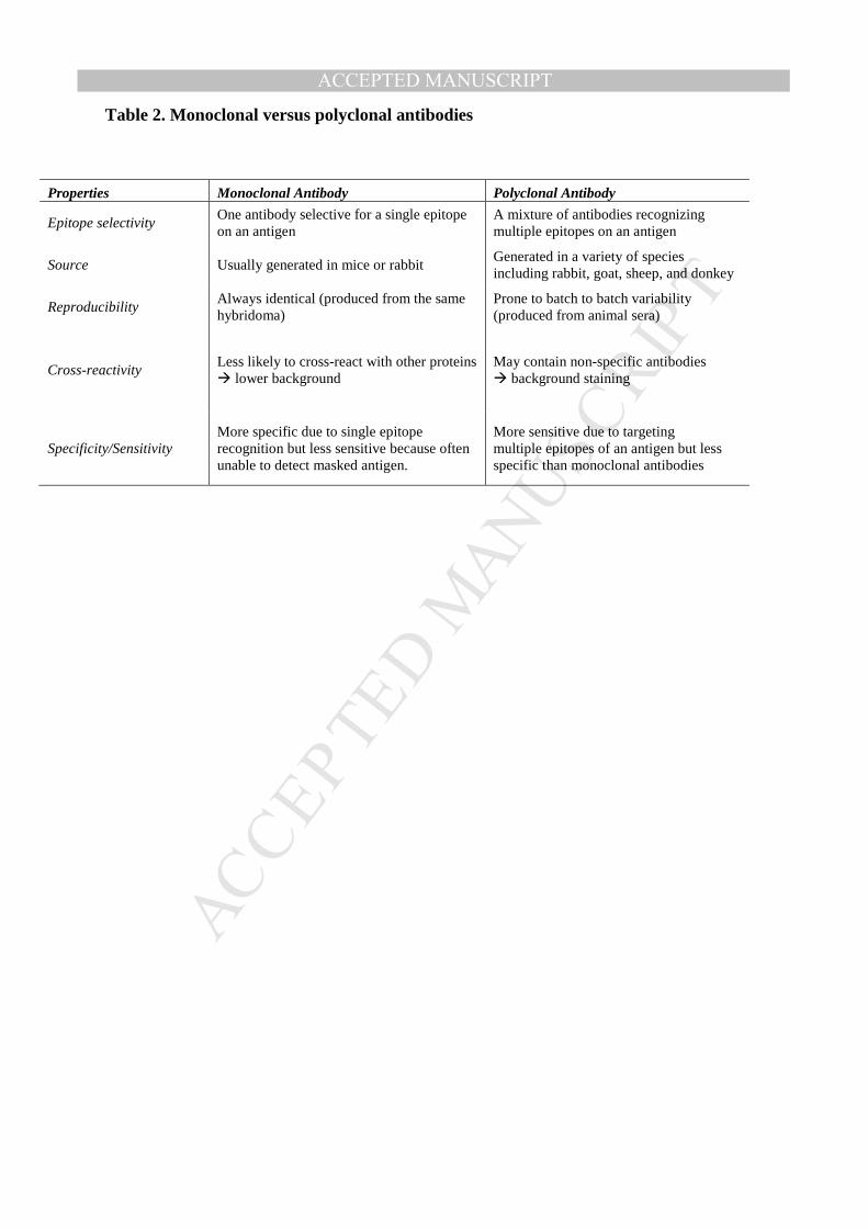
MANUSCRIP
T
ACCEPTED
ACCEPTED MANUSCRIPT
Table 2. Monoclonal versus polyclonal antibodies
Properties Monoclonal Antibody Polyclonal Antibody
Epitope selectivity One antibody selective for a single epitope on an antigen
A mixture of antibodies recognizing multiple epitopes on an antigen
Source Usually generated in mice or rabbit Generated in a variety of species including rabbit, goat, sheep, and donkey
Reproducibility Always identical (produced from the same hybridoma)
Prone to batch to batch variability (produced from animal sera)
Cross-reactivity Less likely to cross-react with other proteins � lower background
May contain non-specific antibodies � background staining
Specificity/Sensitivity More specific due to single epitope recognition but less sensitive because often unable to detect masked antigen.
More sensitive due to targeting multiple epitopes of an antigen but less specific than monoclonal antibodies
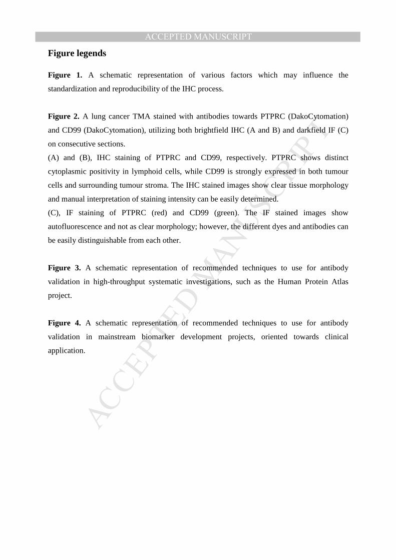
MANUSCRIP
T
ACCEPTED
ACCEPTED MANUSCRIPT
Figure legends
Figure 1. A schematic representation of various factors which may influence the
standardization and reproducibility of the IHC process.
Figure 2. A lung cancer TMA stained with antibodies towards PTPRC (DakoCytomation)
and CD99 (DakoCytomation), utilizing both brightfield IHC (A and B) and darkfield IF (C)
on consecutive sections.
(A) and (B), IHC staining of PTPRC and CD99, respectively. PTPRC shows distinct
cytoplasmic positivity in lymphoid cells, while CD99 is strongly expressed in both tumour
cells and surrounding tumour stroma. The IHC stained images show clear tissue morphology
and manual interpretation of staining intensity can be easily determined.
(C), IF staining of PTPRC (red) and CD99 (green). The IF stained images show
autofluorescence and not as clear morphology; however, the different dyes and antibodies can
be easily distinguishable from each other.
Figure 3. A schematic representation of recommended techniques to use for antibody
validation in high-throughput systematic investigations, such as the Human Protein Atlas
project.
Figure 4. A schematic representation of recommended techniques to use for antibody
validation in mainstream biomarker development projects, oriented towards clinical
application.

MANUSCRIP
T
ACCEPTED
ACCEPTED MANUSCRIPT
MANUSCRIP
T
ACCEPTED
ACCEPTED MANUSCRIPT

MANUSCRIP
T
ACCEPTED
ACCEPTED MANUSCRIPT
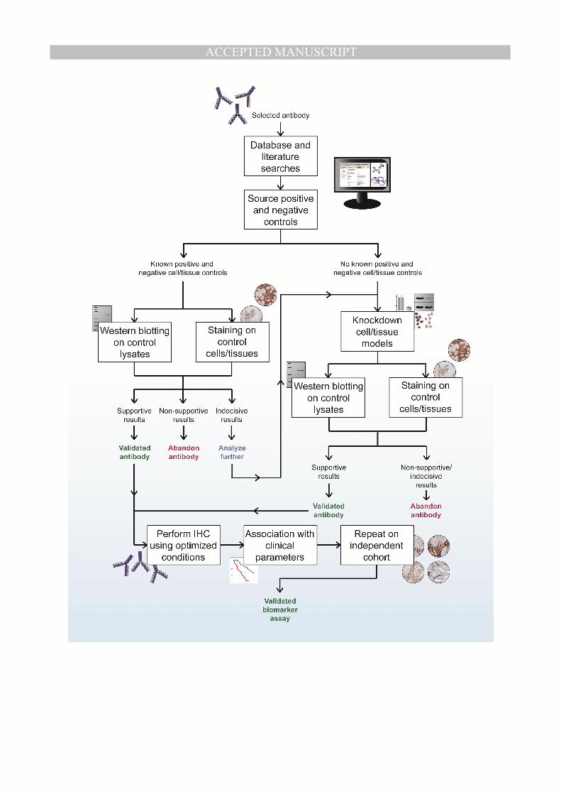
MANUSCRIP
T
ACCEPTED
ACCEPTED MANUSCRIPT

MANUSCRIP
T
ACCEPTED
ACCEPTED MANUSCRIPT
Highlights
• Immunohistochemistry is an important tool for biomarker development
• Few potential immunohistochemistry biomarkers reach the clinical utility
• We describe the various factors influencing the IHC process
• We discuss classical and emerging antibody validation technologies
• We provide recommended work-flows for proper antibody validation
Related Documents