1 2 3 Gap2 Promotes the Formation of a Stable Protein 4 Complex Required for Mature Fap1 Biogenesis 5 6 7 8 9 10 11 12 13 14 Haley Echlin 1,2 , Fan Zhu 1,2 , Yirong Li 1,3 , Zhixiang Peng 1,4 , Teresa Ruiz 5 , 15 Gregory J. Bedwell 2 , Peter E. Prevelige Jr 2 and Hui Wu 1,2* 16 17 18 19 20 21 22 23 Departments of Pediatric Dentistry 1 and Microbiology 2 , Schools of Dentistry and Medicine, 24 University of Alabama at Birmingham, Birmingham, AL 35294; Department of Laboratory 25 Medicine 3 , Tongji Medical School, Huazhong University of Science and Technology, Wuhan, 26 Hubei, China; Department of Endodontics 4 , Guanghua School of Stomatology, Sun Yat-sen 27 University, Guangzhou, Guangdong, China; Department of Molecular Physiology and 28 Biophysics, 5 University of Vermont, Burlington, VT0540 29 30 31 32 33 Key words: Streptococcus parasanguinis, Gap1, Gap2, Gap3, Fap1, protein stability, and ClpP 34 35 36 37 *For Correspondence. Department of Pediatric Dentistry, School of Dentistry, University of 38 Alabama at Birmingham, Birmingham, AL 35294, USA. Tel: 205-996-2392. Fax: 205-975-6251. 39 Email Address: [email protected] 40 41 42 43 Copyright © 2013, American Society for Microbiology. All Rights Reserved. J. Bacteriol. doi:10.1128/JB.02255-12 JB Accepts, published online ahead of print on 8 March 2013

Welcome message from author
This document is posted to help you gain knowledge. Please leave a comment to let me know what you think about it! Share it to your friends and learn new things together.
Transcript
1
2
3
Gap2 Promotes the Formation of a Stable Protein 4
Complex Required for Mature Fap1 Biogenesis 5
6
7
8
9
10
11
12
13
14 Haley Echlin1,2, Fan Zhu1,2, Yirong Li1,3, Zhixiang Peng1,4, Teresa Ruiz5, 15
Gregory J. Bedwell2 , Peter E. Prevelige Jr2 and Hui Wu1,2* 16 17
18 19 20 21 22 23
Departments of Pediatric Dentistry1 and Microbiology2, Schools of Dentistry and Medicine, 24 University of Alabama at Birmingham, Birmingham, AL 35294; Department of Laboratory 25
Medicine3, Tongji Medical School, Huazhong University of Science and Technology, Wuhan, 26 Hubei, China; Department of Endodontics4, Guanghua School of Stomatology, Sun Yat-sen 27
University, Guangzhou, Guangdong, China; Department of Molecular Physiology and 28 Biophysics,5University of Vermont, Burlington, VT0540 29
30 31 32
33 Key words: Streptococcus parasanguinis, Gap1, Gap2, Gap3, Fap1, protein stability, and ClpP 34 35 36 37 *For Correspondence. Department of Pediatric Dentistry, School of Dentistry, University of 38 Alabama at Birmingham, Birmingham, AL 35294, USA. Tel: 205-996-2392. Fax: 205-975-6251. 39 Email Address: [email protected] 40 41 42 43
Copyright © 2013, American Society for Microbiology. All Rights Reserved.J. Bacteriol. doi:10.1128/JB.02255-12 JB Accepts, published online ahead of print on 8 March 2013

ABSTRACT 44
Serine-rich repeat glycoproteins (SRRPs) are important bacterial adhesins conserved in 45
streptococci and staphylococci. Fap1, a SRRP identified in Streptococcus parasanguinis, is the 46
major constituent of bacterial fimbriae and is required for adhesion and biofilm formation. An 47
eleven gene cluster is required for Fap1 glycosylation and secretion; however, the exact 48
mechanism of Fap1 biogenesis remains a mystery. Two glycosylation-associated proteins within 49
this cluster-Gap1 and Gap3- function together in Fap1 biogenesis. Here we report the role of the 50
third glycosylation-associated protein, Gap2. A gap2 mutant exhibited the same phenotype as 51
the gap1 and gap3 mutants in terms of Fap1 biogenesis, fimbrial assembly, and bacterial 52
adhesion- suggesting that the three proteins interact. Indeed, all three proteins interacted with 53
each other independently and together to form a stable protein complex. Mechanistically, Gap2 54
protected Gap3 from degradation by ClpP protease and Gap2 required the presence of Gap1 55
for expression at wild-type level. Gap2 augmented Gap1’s function of stabilizing Gap3; this 56
function was conserved in Gap homologs from Streptococcus agalactiae. Our studies 57
demonstrate that the three Gap proteins work in concert in Fap1 biogenesis and reveal a new 58
function of Gap2. This insight will help us elucidate the molecular mechanism of SRRP 59
biogenesis in this bacterium and in pathogenic species. 60
61
INTRODUCTION 62
Two of the most prevalent infectious diseases of humans are dental caries and 63
inflammatory periodontal disease. Oral streptococci comprise a large proportion of oral bacterial 64
species in dental plaque and are one of the first colonizers of the tooth surface (1-3). As such, oral 65
streptococci will encounter not only host oral epithelial cells, but also other microbial cells, of 66
which there are over 500 species in the oral cavity, including the major periodontal pathogens- 67
which often cannot colonize unless a layer of initial colonizers, such as oral streptococci, has 68
developed first (4-8). Like other oral streptococci, S. parasanguinis has several colonization and 69

adhesion factors; one of its adhesion factors is long peritrichous fimbriae (9). S. parasanguinis 70
fimbriae are made of Fap1 (fimbriae-associated protein 1), a 200 kDa cell wall anchored serine-71
rich repeat glycoprotein (SRRP) (10). Fap1 is required for fimbrial formation, bacterial adhesion 72
(1, 11), and biofilm formation (10, 12). Since the discovery of Fap1 (13, 14), Fap1-like SRRPs 73
have been identified in many streptococci, staphylococci, and other gram-positive bacteria and 74
have been implicated in bacterial interactions with hosts, adhesion, biofilm formation, and 75
pathogenesis (10, 11, 15-20). They include GspB and Hsa of S. gordonii (21, 22), SraP of S. 76
sanguinis (23), PsrP of S. pneumonia (18), Srr-1 and Srr-2 of S. agalactiae (16, 17), SrpA of S. 77
cristatus (24), SraP of S. aureus (10, 19), and FimS of S. salivarius (25). 78
The exact mechanism of SRRP biogenesis is not well understood. The chromosomal 79
region dedicated to SRRP glycosylation and secretion is quite large and highly conserved. For 80
Fap1, the cluster is separated into two regions: a core region that is conserved in every genome 81
(secY2, gap1-3, secA2, and gtf1-2) and a variable region that includes several putative 82
glycosyltransferases (gly, nss, galT1, and galT2) (10). gtf1 and gtf2 and genes from the gly-gtf3-83
galT1-galT2 locus mediate Fap1 glycosylation (13, 26-30); Fap1 is glycosylated in the cytoplasm 84
with several monosaccharides, including glucose, N-acetyl glucosamine, N-acetyl galactosamine, 85
and rhamnose (11, 29). The secY2-gap1-gap2-gap3-secA2 locus is responsible for secretion of 86
Fap1 (28, 29, 31). SecA2 and SecY2 have homology to their counterparts in the canonical Sec 87
pathway and are required for the export of mature Fap1 to the cell wall surface (28, 29). There is 88
no known homology for the remainder of the locus- gap1-gap2-gap3- outside of the SRRP family. 89
We have shown previously that both gap1 and gap3 mutants produce a similar immature Fap1 90
and that the interaction between Gap1 and Gap3 is required for Fap1 biogenesis, indicating that 91
Gap1 and Gap3 are involved in mature Fap1 biogenesis (32-34). However, to date, the function of 92
Gap2 is unknown. 93

In this study, we determined the role of Gap2 and found it is involved in Fap1 biogenesis 94
by stabilizing Gap3 through interactions with Gap1 and Gap3; this study reveals an activity of 95
Gap2 and its homolog that was previously unknown. 96
97
MATERIALS AND METHODS 98
Bacterial Strains, Plasmids, Primers, and DNA Manipulation 99
Bacterial strains and plasmids used in this study are listed in Table 1. Escherichia coli and S. 100
parasanguinis strains were cultured as previously described (15). S. parasanguinis cell 101
concentrations were determined by absorbance at 470 nm. Antibiotics were used at the 102
following concentrations: 10 μg/ml erythromycin,125 μg/ml kanamycin, and 250 μg/ml 103
spectinomycin in Todd Hewitt (TH) broth or agar plates for S. parasanguinis; 300 μg/ml 104
erythromycin, 50 μg/ml kanamycin, 50 μg/ml ampicillin, and 50 μg/ml spectinomycin in Luria-105
Bertani (LB) broth or agar plates for E. coli. Standard recombinant DNA techniques were used 106
for DNA preparation and analyses (35). Plasmid DNA preparations were isolated with QIAprep 107
Miniprep Kit (Qiagen). Primers used in this study are listed in Table 2. PCR was carried out with 108
Taq DNA polymerase (Promega) or KOD DNA polymerase (Novagen). PCR products were 109
purified with QIAquick PCR Purification Kit (Qiagen). DNA digestion, ligation, and transformation 110
were performed using standard methods. Competence cells for S. parasanguinis electroporation 111
were prepared as described previously (36). 112
113
Western Blot Analysis 114
All S. parasanguinis strains were grown to OD470=0.5-0.6 and centrifuged; cell pellets were 115
subjected to amidase treatment to lyse the cells (28). Cell lysates were boiled in sample buffer 116
(0.0625 M Tris, pH6.8, 2% SDS, 10% glycerol, 0.01% bromophenol blue) for 10 min before 117
loading into 10% SDS-PAGE gels and subjected to western blotting analysis. Two monoclonal 118
antibodies were used to detect Fap1- mAb E42, which is specific to the peptide backbone of 119

Fap1, and mAb F51, which is specific to the mature Fap1 (11); mAbF51 only recognizes the 200 120
kDa mature Fap1, whereas mAb E42 recognizes both the 200 kDa mature Fap1 and the 470 121
kDa Fap1 precursor. Rabbit polyclonal antibodies against Gap1, Gap2, and Gap3 were custom 122
produced using recombinant Gap1, Gap2, Gap3, or Gap1/2/3 complex as an antigen. 123
Monoclonal antibody against Hsv (Novagen) was used to detect tagged proteins. Polyclonal 124
antibody against FimA was used to standardize protein loading of S. parasanguinis proteins. 125
126
Construction of the Insertional gap2 Mutant and gap2/clpP Double Mutant 127
A gap2 mutant was constructed by allelic replacement of gap2 with a kanamycin resistant 128
cassette, aphA-3 (aminoglycoside phosphotransferase). A fragment containing the gap2 gene 129
and its flanking regions was amplified from S. parasanguinis chromosomal DNA using 130
Gap2+Flank-F/ Gap2+Flank-R. The PCR fragment was ligated into pGEM-T easy (Promega). A 131
850 bp region of gap2 was deleted by inverse PCR using Gap2-StuI-F/ Gap2-StuI-R. The 132
inverse PCR product was digested with StuI and ligated with a promoterless aphA-3 kanamycin 133
resistant cassette from pALH124 (37) to generate pGEM::∆gap2-aphA3. Finally, the gap2 134
insertion mutant was constructed by transformation of FW213 with pGEM::∆gap2-aphA3, 135
followed by selection of kanamycin resistant colonies. The in-frame insertion was further 136
examined by DNA sequencing analyses. A Western blot analysis probed with antiserum against 137
SecA2, a protein encoded by a gene downstream of gap2, was performed to confirm that the 138
mutation was non-polar (data not shown). The fap1 (1), secY2 (28), gap1 (34), gap3 (32), and 139
clpP (38) mutants were constructed in a similar method. For the gap2/clpP double mutant, a 140
spectinomycin resistant cassette (Spec) was inserted into clpP in the gap2 mutant. The 141
pGEM::∆clpP-aphA-3 construct (38)was digested with HindIII to remove the kanamycin resistant 142
cassette and then ligated in-frame with the spectinomycin resistance cassette amplified from 143
pCG1 (39) to construct pGEM::∆clpP-spec. The gap2/clpP double mutant was constructed by 144
transformation of the gap2 mutant with pGEM::∆clpP-spec, followed by selection of kanamycin 145

and spectinomycin resistant colonies. The in-frame insertion was further examined by DNA 146
sequencing analyses. 147
148
Complementation of the gap1, gap2, and gap3 Mutants 149
The full-length gap1, gap2, and gap3 genes were amplified from FW213 genomic DNA by PCR 150
using primers Gap1-SalI-F/ Gap1-KpnI-R, Gap2-SalI-F/ Gap2-KpnI-R, and Gap3-SalI-F/ Gap3-151
KpnI-R, respectively (Table 2). The purified gap1, gap2, and gap3 PCR products were digested 152
with SalI and KpnI and then cloned into E. coli-Streptococcus shuttle vector pVPT-gfp (40) to 153
generate corresponding complementation plasmids pVPT-gap1-gfp, pVPT-gap2-gfp, and pVPT-154
gap3 (no gfp). The plasmid and its control vector pVPT-gfp were then transformed into the 155
gap1, gap2, and gap3 mutants via electroporation. The transformants were selected on TH agar 156
plates containing kanamycin and erythromycin. 157
158
Modification of an E. coli-Streptococcus Shuttle Vector pIB184 159
A second E. coli-Streptococcus shuttle vector, pIB184 (41), was used in this study for better 160
expression and genetic manipulation. To enhance the utility of this vector, pIB184 was modified 161
by cloning in gfp and hsv-his tags within the multiple cloning site. The full-length gfp and hsv-his 162
were amplified from pVPT-gfp and pET27b (Novagen) using primers GFP-XmaI-F/ GFP-SacI-R 163
and HsvHis-Xmal-F/ HsvHis-SacI-R, respectively. The purified gfp and hsv-his PCR products 164
were digested with XmaI and SacI and then ligated with the vector pIBI84 to create pIB184-gfp 165
and pIB184-hsv-his. 166
167
Construction of Overexpression Strains in S. parasanguinis FW213 168
The full-length gap3, gap2-gap3, and gap1-gap2-gap3 were amplified from FW213 genomic 169
DNA by PCR using primers Gap3-BamHI-F/ Gap3-XmaI-R, Gap2-BamHI-F/ Gap3-XmaI-R, and 170
Gap1-BamHI-F/ Gap3-XmaI-R, respectively (Table 2). The purified gap3, gap2-gap3, and gap1-171

gap2-gap3 PCR products were digested with BamHI and XmaI and then cloned into E. coli-172
Streptococcus shuttle vector pIB184-gfp to generate pIB184-gap3-gfp, pIB184-gap2-gap3-gfp, 173
and pIB184-gap1-gap2-gap3-gfp, where Gap3 is tagged with GFP in all vectors. The plasmids 174
were then transformed into the wild-type and gap1 and gap2 mutants via electroporation. 175
pIB184-gap2-hsv-his and pIB184-gap1-gap2-hsv-his were created in the same fashion using 176
pIB184-hsv-his and primer pairs Gap2-BamHI-F/ Gap2-XmaI-R and Gap1-BamHI-F/ Gap2-177
XmaI-R, respectively. The Gap homologs, Asp1-2-3, from S. agalactiae wild-type J48 were used 178
to check for conservation of function. pIB184-asp3-gfp, pIB184-asp2-gap3-gfp, pIB184-asp1-179
asp2-asp3-gfp, pIB184-asp2-hsv-his, and pIB184-asp1-asp2-hsv-his were created in the same 180
manner as above, using primers Asp3-BamHI-F/ Asp3-XmaI-R, Asp2-BamHI-F/ Asp3-XmaI-R, 181
Asp1-BamHI-F/ Asp3-XmaI-R, Asp2-BamHI-F/ Asp2-XmaI-R, and Asp1-BamHI-F/ Asp2-XmaI-182
R, respectively (Table 2). The resulting plasmids were then transformed into FW213 and gap1 183
and gap2 mutants via electroporation. The transformants were selected on TH agar plates 184
containing erythromycin (wild-type) or kanamycin and erythromycin (mutants). 185
186
Bacterial Adhesion Assay 187
Saliva-coated hydroxyapatite (SHA) was used as an in vitro tooth model to test the binding 188
abilities of S. parasanguinis and the relevant derivatives as described previously (42). Briefly, 189
[3H]-thymidine-labeled bacteria of OD470=1.0 in adhesion buffer (67 mM phosphate buffer, pH 190
6.0) were sonicated for 15 s at 85W using an ultrasonic cuphorn system (Heat Systems-191
Ultrasonics). 1 ml of sonicated bacteria (in triplicate) were added to 7 ml scintillation vials 192
containing SHA and incubated for 1 h at 37°C with gentle shaking. The supernatant fluids were 193
removed and the beads were washed 3 times with adhesion buffer. The amounts of unbound 194
bacteria in the supernatant fluids and bacteria bound to SHA were determined in a Beckman 195
Coulter LS6500 Scintillation Counter (Beckman-Coulter) (1). Differences in SHA adhesion were 196
analyzed via 2-tailed Student’s t-test for two samples with equal variances. 197

198
Transmission Electron Microscopy 199
S. parasanguinis cell cultures (5 ml) grown to OD470 = 0.4 were harvested by centrifugation. Cell 200
pellets were washed twice with ice-cold PBS and resuspended in 100 µl PBS. 5 µl of the 201
bacterial suspension was diluted in PBS and was applied to 400 mesh copper grids coated with 202
a thin carbon film. The grids were first washed by several drops of PBS buffer. The samples 203
were stained with a few drops of 2% phosphotungstic acid, pH 7.0 (PTA) over the grid surfaces. 204
The excess liquid was wicked off and the grids were fast air dried. The grids were observed on 205
a Tecnai 12 Philips electron microscope (FEI, Holland) equipped with a LaB6 cathode operated 206
in point mode (Kimball) and a 2048 CCD camera (TVIPS, Germany). The microscope was run 207
to obtain images that show Thon rings beyond 0.9 nm resolution in vitreous ice preparations 208
(43). Images were recorded at an accelerating voltage of 100kV and nominal magnifications in 209
the range of 40,000-70,000X under low dose conditions on either film (S0-163 Kodak) or the 210
CCD camera. Images were converted to SPIDER format (44) and high-pass filter to remove the 211
background. 212
213
in vitro GST Pull-down Assays 214
The GST pull-down protocol was developed to determine protein-protein interactions in 215
solutions (45). Gap1- and Gap3-pGADT7 were constructed as described (34). Gap2-pGADT7 216
was constructed by PCR amplification of gap2 using primers Gap2-EcoRI-F/ Gap2-BamHI-R 217
(Table 2) from FW213 chromosomal DNA, digestion with EcoRI and BamHI, and ligation into 218
pGADT7. GST-Gap1, GST-Gap2, and GST-Gap3 fusion proteins were created by cloning of 219
EcoRI and XhoI digested fragments from Gap1-, Gap2- and Gap3-pGADT7 into pGEX-5X-2, 220
respectively. The GST fusion proteins were expressed and purified using glutathione Sepharose 221
4B beads. The same amounts of GST or GST fusion proteins (5 µg) immobilized on beads and 222
estimated by SDS-PAGE analysis were re-suspended in NETN washing buffer (20 mM Tris-223

HCl, pH 7.0, 150 mM NaCl, 1 mM EDTA, 0.5% NP40) and mixed with 5 µl of in vitro translated 224
c-Myc -Gap1, -Gap2, and -Gap3 fusion protein products (34). The mixtures were reconstituted 225
in a final volume of 200 µl with NETN binding buffer and incubated at 4°C overnight on a rotary 226
shaker. The beads were washed three times with 600 µl of NETN washing buffer. The proteins 227
bound to the beads were eluted by boiling in SDS loading buffer and subjected to Western 228
blotting analyses using anti-c-Myc antibody (Invitrogen). The interaction between Gap1 and 229
Gap3 was confirmed previously (34) and was used here as a control. 230
231
Analytical Ultracentrifugation 232
Sample Preparation 233
A fusion plasmid was constructed to express His-SUMO-tagged Gap1-2-3 by the same method 234
used in the construction of His-SUMO- tagged Gap1-3 (38). Briefly, full-length gap1-gap2-gap3 235
was amplified from genomic DNA of S. parasanguinis FW213 using Gap1-NotI-1F/Gap3-XhoI-236
R, digested by NotI and XhoI, and ligated into pET-SUMO to construct the His-SUMO-Gap1-2-3 237
fusion protein. The constructed plasmid was verified by DNA sequence analysis and then 238
transformed into E. coli BL21 (DE3). Gap1-3 and Gap1-2-3 were expressed and purified as 239
described previously (26). Peak fractions from gel filtration were collected and used for 240
ultracentrifugation. Concentrations of the proteins were determined by measuring sample 241
absorbance at 280 nm using a Beckman DU-640 Spectrophotometer (International MI-SS, Inc. 242
Corona CA). The sample proteins were diluted to desired concentrations with buffer G (26). 243
Sedimentation Equilibrium 244
Sedimentation equilibrium (SE) experiments were performed at 20°C using six-channel 245
centerpieces in a Beckman Optima XL-A with absorption optics. Three concentrations (0.2 246
mg/ml, 0.4 mg/ml, and 0.9 mg/ml) were analyzed at two rotor speeds- 17,000 rpm and 20,000 247
rpm- with detection by absorbance at 280 nm. All data sets from different protein concentrations 248
and rotor speeds were fit to a single global model (global fits) to determine the stoichiometry 249

and equilibrium constants. Model fittings of the SE data were performed by software 250
HETEROANALYSIS (Biotechnology/Bioservices Center, University of Connecticut, Storrs, CT). 251
252
RESULTS 253
Gap2 mutant exhibits same phenotype as Gap1 and Gap3 mutants 254
Gap1 and Gap3 have been shown to be involved in Fap1 biogenesis (32, 34). However, there 255
have been no reports on the function of the third glycosylation-associated protein, Gap2. In this 256
study, we generated a Gap2 deficient mutant and examined its phenotype. Fap1 production in 257
the Gap2 deficient strain was similar to that in the strains deficient in Gap1, Gap3, and SecY2, 258
where mature Fap1 (Fig. 1, Lane 1), recognized by F51, was undetectable and a larger band-259
corresponding to an immature Fap1 (Lanes 3-6) was observed when probed by E42, a peptide 260
specific antibody. The wild-type phenotype was restored upon complementation (Lanes 7-9); 261
the empty vectors could not restore the wild-type phenotype (Lanes 10-12). This result 262
demonstrates that Gap2, like Gap1 and Gap3, is required for the production of mature Fap1 (32, 263
34). 264
Since Fap1 is required for assembly of S. parasanguinis fimbriae (1), the cell surface 265
structure of S. parasanguinis variants was examined using transmission electron microscopy. In 266
the gap2 mutant (Fig. 2C), fimbriae were no longer detected as they are in the wild-type FW213 267
strain (Fig. 2A). However, the Gap2 deficiency had no effect on a smaller fibril (indicated by the 268
white arrows in Fig. 2B-D), which has been identified previously as BapA1 (46). This fimbriae 269
phenotype is comparable to that of the gap1 (Fig. 2B) and gap3 (Fig. 2D) mutants. Furthermore, 270
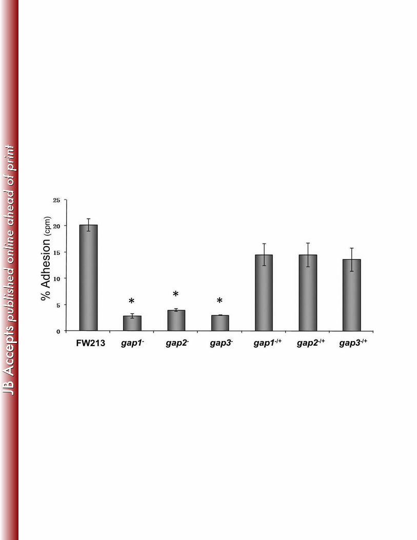
the Gap2 deficiency decreased bacterial adherence to SHA (Fig. 3). This phenotype was similar 271
to that observed in strains deficient in Gap1 or Gap3. For all three strains, complementation 272
nearly restored adhesion levels to that of the wild-type (Fig. 3). These results indicate that Gap2 273
functions in concert with Gap1 and Gap3 in Fap1 biogenesis, with a subsequent effect on 274
fimbriae biogenesis and adhesion level. 275

276
Gap1, Gap2, and Gap3 interact with each other to form a complex 277
Because not only do Gap1, Gap2, and Gap3 deficient strains share a similar phenotype (Fig. 1-278
3), but also the interaction between Gap1 and Gap3 is required for biogenesis of Fap1 (34, 38), 279
it is likely that Gap2 interacts with Gap1 and Gap3 as well. To determine this, we coexpressed 280
all three proteins in E. coli, with Gap1 tagged with GST, and performed GST-pull down assays. 281
Gap2 and Gap3 were invariably pulled down with GST-Gap1 (Fig. 4A). GST itself did not pull 282
down Gap2 and Gap3 (data not shown). To address whether Gap2 could interact with Gap1 283
and Gap3 independently, we expressed each protein tagged with GST individually and 284
incubated them with in vitro translated c-Myc fusion proteins. Upon GST pull-down assays, 285
GST-tagged Gap2 pulled down Gap1 and Gap3, and Gap2 was pulled down by GST-tagged 286
Gap1 and Gap3 (Fig. 4B). This result indicates that Gap2 can interact with both Gap1 and Gap3 287
directly. The interaction between Gap1 and Gap3 was used as positive assay controls. In 288
negative controls, Gap1, Gap2, nor Gap3 interacted with GST alone, indicating that the 289
interaction between Gap2 and Gap1, and Gap3 was specific. 290
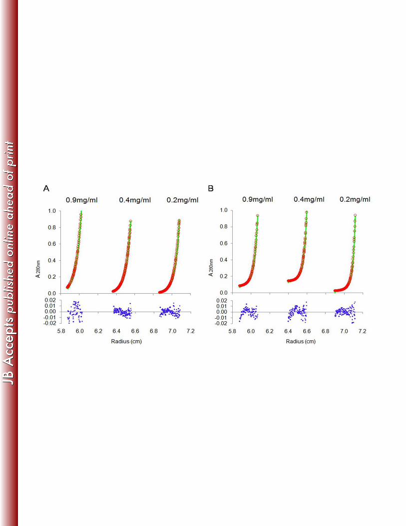
Analytical ultracentrifugation sedimentation equilibrium experiments were performed to 291
further characterize the interaction among Gap1, Gap2, and Gap3. Sedimentation equilibrium 292
(SE) data show that the Gap1/3 complex fits a single species model well (Fig. 5A), suggesting 293
that the binding between Gap1 and Gap3 was tight. The binding of Gap2 to the already formed 294
Gap1/3 complex fits a heterodimer model (“A+B<>AB,” where A represents Gap1/3 and B 295
represents Gap2; Kd of 4.4E-07 M) (Fig. 5B), suggesting that Gap2 binds to Gap1/3 to form a 296
Gap1/2/3 complex in a reversible manner. The experimental data fit the models regardless of 297
the concentration (0.2 mg/ml, 0.4 mg/ml, and 0.9 mg/ml; Fig. 5) or speed [17,000 rpm (Fig. 5) 298
and 20,000 rpm (data not shown)] used. 299
300
Gap2 is increased with Gap1 overexpression 301

Previously, we have shown that Gap1 is required for the stability of Gap3 (38). In this study, we 302
demonstrated that Gap2 interacted with both Gap1 and Gap3. In order to determine how Gap2 303
affects or is affected by Gap1, protein levels of Gap1 and Gap2 were determined in wild-type 304
and gap mutant variants (Fig. 6A). A gap2 mutant had no effect on the amount of Gap1 (Lane 305
3). On the other hand, in the absence of Gap1 (Lane 2), Gap2 was decreased compared to the 306
wild-type (Lane 1). The gap1 complemented strain restored the wild-type phenotype (Lane 5); 307
expression of Gap1-GFP was observed as a band slightly above 75 kDa when probing with the 308
Gap1 antibody. The negative vector had no effect on the decreased amount of Gap2 (Lane 8). 309
This result suggests that Gap1 expression increases the amount of Gap2. To confirm this, we 310
compared expression of Gap2 in a strain that overexpressed Gap2 alone to a strain that 311
overexpressed both Gap1 and Gap2 (Fig. 6B). Gap2 expression was greatly increased when 312
both Gap1 and Gap2 (Lanes 2, 4, and 6) were overexpressed compared to overexpression of 313
Gap2 alone (Lanes 1, 3, and 5); expression of Gap2-HH was observed as a band about 65 kDa 314
when probing with the Gap2 antibody. This phenotype was observed in wild-type strain, as well 315
as gap1 and gap2 mutants. RT-PCR analysis of gap2 transcription demonstrates no difference 316
between wild-type and the gap1 mutant, indicating that the effect of Gap1 on Gap2 occurs on 317
the post-transcriptional level (Figure S1A). Together, these data demonstrate that the amount of 318
Gap2 is modulated by Gap1. 319
320
Gap2 expression results in increased Gap3 321
To determine the association between Gap2 and Gap3, we examined the effect of Gap2 322
deficiency on Gap3 (Fig. 6A). In the absence of Gap2 (Lane 3), Gap3 was decreased compared 323
to wild-type. Further, in the gap2 complement (Lane 6), the amount of Gap3 was restored to 324
wild-type level; expression of Gap2-GFP was observed as a stronger band compared to a non-325
specific band present at 75 kDa when probing with the Gap2 antibody. The negative vector had 326
no effect on the decreased the amount of Gap3 (Lane 9). However, the gap3 mutant had no 327

effect on the amount of Gap2 (Lane 4). This result suggested that Gap2 expression increases 328
the amount of Gap3. To confirm this, we overexpressed Gap2 and determined its impact on 329
Gap3 (Fig. 7A). Overexpression of Gap2 in the wild-type strain (Lane 4) indeed increased Gap3. 330
In the gap2 mutant (Lane 6) background, overexpression of Gap2 did not quite restore the 331
amount of Gap3 to the wild-type level. However, this could be due to the reduced amount of 332
Gap2 in the mutant strain compared to the wild-type strain. RT-PCR analysis of gap3 333
transcription demonstrates no difference between wild-type and the gap2 mutant, indicating that 334
the effect of Gap2 on Gap3 occurs on the post-transcriptional level (Figure S1A). These data 335
demonstrate that Gap2 modulates Gap3 amount. 336
337
Gap2 modulates Gap3 amount independently of Gap1 338
Gap2 deficiency resulted in a diminished amount of Gap3 and overexpression of Gap2 led to a 339
greater Gap3 amount. However, from these data, we cannot determine whether Gap2 functions 340
independently of Gap1; in the absence of Gap1, native Gap3 was no longer detected, even 341
when Gap2 was overexpressed (Fig. 7A, Lanes 2 and 5). To determine if Gap2 can affect Gap3 342
independently of Gap1, strains were created that overexpressed Gap3 alone, Gap2 and Gap3, 343
or Gap1, Gap2, and Gap3 in wild-type and in gap1 and gap2 mutants (Fig. 7B); expression of 344
Gap3-GFP was observed as a band slightly below 50 kDa when probing with the Gap3 345
antibody. Again, when Gap2 was overexpressed, both native and overexpressed Gap3 was 346
increased (Lane 2), compared to the strain overexpressing Gap3 alone (Lane 1). Moreover, 347
Gap3 was increased even further when both Gap1 and Gap2 were overexpressed along with 348
Gap3 (Lane 3). This phenomenon was not limited to the wild-type as it also occurred in the gap1 349
(Lanes 4-6) and gap2 (Lanes 7-9) mutant strains, albeit the overall levels were lower compared 350
to the wild-type. RT-PCR analysis of gap3 transcription demonstrates no difference between 351
overexpressing strains, indicating that the effect of Gap1 and Gap2 on Gap3 occurs on the post-352
transcriptional level (Fig. S1B). These data demonstrate that increasing Gap2 expression can 353

increase the amount of overexpressed Gap3 in the absence of Gap1, suggesting that Gap2 354
augments Gap1’s function in stabilizing Gap3. 355
356
Gap homologs from S. agalactiae displayed same conserved functions as Gap proteins 357
Gap1, Gap2, and Gap3 are highly conserved in SRRP-containing Gram-positive bacteria. We 358
have previously shown that the Gap1 homolog from S. agalactiae stabilizes the Gap3 homolog, 359
much like Gap1 acts as a chaperone for Gap3 (38). To determine if the relationship between 360
Gap2 and Gap1, and Gap3 is conserved, we expressed Gap homolog from S. agalactiae (Asp1, 361
Asp2, and Asp3) in S. parasanguinis (Fig. 8A). In S. parasanguinis wild-type, Asp2 was 362
detected when both Asp1 and Asp2 were expressed (Lane 2), but was undetectable when 363
expressed alone (Lane 1). This result suggests that the amount of Asp2 is increased in the 364
presence of Asp1, much like the Gap proteins in S. parasanguinis (Fig. 6B). This phenomenon 365
was also observed in the absence of Gap1 (Fig. 8A, Lanes 3 and 4), further demonstrating that 366
Asp1 can increase the Asp2 amount. To determine if the function of Gap2 is conserved, we 367
expressed Gap homologs (Asp1, Asp2, and Asp3) from S. agalactiae in S. parasanguinis 368
strains lacking Gap2 (gap2 mutant). In these strains, Asp3 was expressed by itself, with Asp2, 369
or with Asp1 and Asp2 (Fig. 8B). When Asp2 was expressed along with Asp3 (Lane 2), the 370
amount of Asp3 increased compared to Asp3 expressed alone (Lane1); when Asp1 was 371
expressed with Asp2 and Asp3 (Lane 3), the amount of Asp3 was even greater. Because this 372
trend is similar to the one observed in the S. parasanguinis homologs (Fig. 7B), this result 373
indicates that Asp2 can function in a similar manner as Gap2. Together, these data suggest that 374
the relationship among the Gap proteins is conserved. 375
376
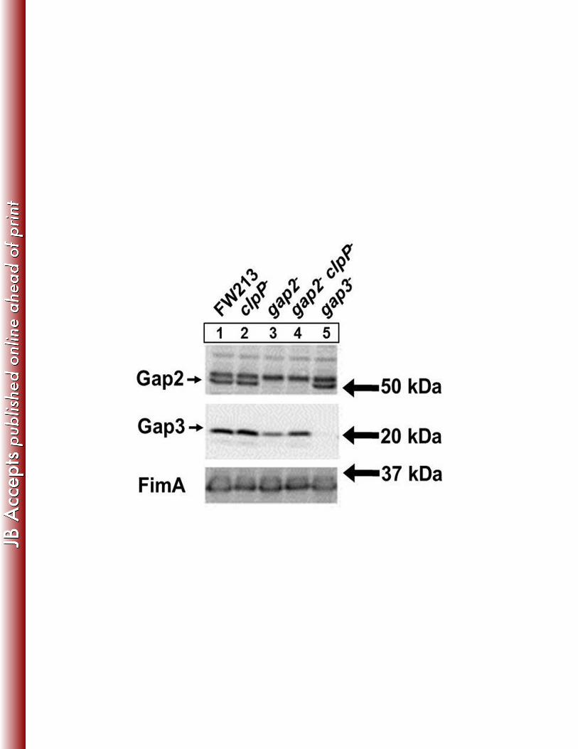
Gap2 prevents Gap3 degradation by ClpP protease 377
Proteases are often involved in the degradation of misfolded proteins. Previously, the protease 378
ClpP was shown to be responsible for the degradation of Gap3 in the absence of Gap1, a 379

specific chaperone of Gap3 (38). Here, we wanted to determine if Gap2 protected Gap3 in a 380
similar fashion. We constructed a clpP mutant and a gap2/clpP double mutant to examine the 381
ability of Gap2 to shield Gap3 from degradation by ClpP (Fig. 9). No difference in Gap3 was 382
observed between wild-type (Lane 1) and the clpP mutant (Lane 2). In the absence of both ClpP 383
and Gap2 (Lane 4), the amount of Gap3 was increased compared to the gap2 single mutant 384
(Lane 3), nearly restoring it to wild-type level. This result suggests that Gap2, similarly to Gap1, 385
protects Gap3 from degradation by ClpP. 386
387
DISCUSSION 388
Biogenesis of SRRPs is mediated by glycosylation and accessory secretory loci, which 389
are highly conserved in many streptococci and staphylococci (10). In S. parasanguinis, an 390
eleven gene cluster including glycosyltransferase genes and genes involved in protein secretion 391
have been identified for Fap1 biosynthesis. Accessory secretion components- containing SecA2 392
and SecY2, and glycosylation associated proteins, Gap1, Gap2, and Gap3 (10, 28, 29)- are 393
implicated in Fap1 secretion and maturation. The exact role of Gap1, Gap2, and Gap3 in Fap1 394
biogenesis remains unknown. We have shown previously that Gap1 and Gap3 are required for 395
production of mature Fap1, formation of fimbriae, and adhesion to SHA (32, 34). In this study, 396
we have determined the function of Gap2. Similar to Gap1 and Gap3, Gap2 was necessary for 397
mature Fap1 biogenesis, with direct effects on fimbriae production and adhesion to an in vitro 398
tooth surface model (Fig. 1-3). Because all three of the gap mutants shared a similar phenotype, 399
it is likely they interact and work in concert to complete Fap1 biogenesis. Indeed, we show here 400
that Gap1, Gap2, and Gap3 interact to form a complex (Fig. 4). The formation of a protein 401
complex by Gap homologs has been demonstrated in S. gordonii as well (47); however, the 402
details of the interactions were not characterized. Through ultracentrifugation, we determined 403
that Gap2 could interact with an already formed Gap1/3 complex in a reversible manner. While 404

Gap1 and Gap3 bind tightly to each other, Gap2 has a lower binding affinity toward the Gap1/3 405
complex, suggesting Gap2 may have regulatory activity toward the Gap1/3 complex (Fig. 5). 406
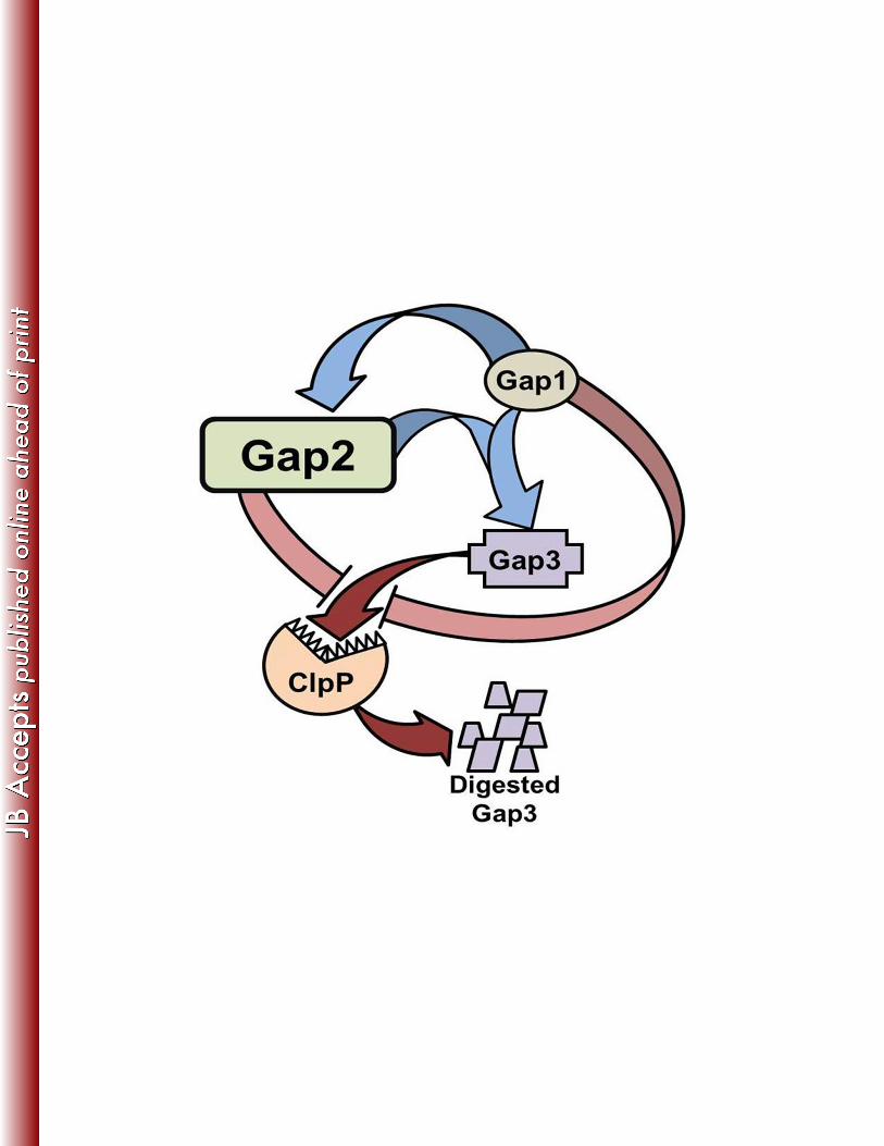
Based on the data obtained from the current study (summarized in Fig. 10), we can 407
expand our previous model of Fap1 biogenesis. In this model, Gap1 binds to Gap3 (38) (Fig. 408
5A). This is then followed by binding of Gap2, which can further stabilize Gap3 and is, itself, 409
stabilized by Gap1 (Fig. 5B, 6, and 7). Such binding and stabilization was also observed for Gap 410
homologs from S. agalactiae (Fig. 8), suggesting that this new function of Gap2 is conserved 411
among SRRP-containing Gram-positive bacteria. Further, the current study indicates that Gap2, 412
protects Gap3 from degradation by ClpP (Fig. 9). Similarly, we have previously shown that the 413
protease ClpP is responsible for the degradation of Gap3 in the absence of Gap1, which acted 414
as a specific chaperone of Gap3 (38). As to how ClpP gains access to the Gap3 protein 415
remains to be determined. 416
Since Gap2 works in concert with Gap1 to stabilize Gap3- the putative key scaffolding 417
protein required for the formation of the Fap1 biosynthetic protein complex- we believe the 418
function of Gap2 is to ensure Gap3 activity, which promotes Fap1 biogenesis. A similar 419
proposition has been made in S. gordonii, in which Asp2 interacts with the Asp1, Asp3, and 420
SecA2 complex for optimal export of GspB (47). Gap2 can interact with the Gap1/3 complex, 421
which then interacts with SecA2 and SecY2 to aid in Fap1 secretion (31). However, the precise 422
biochemical function of this Gap complex in the conversion of an immature form of Fap1 to the 423
mature form remains to be elucidated. Recent work in S. gordonii indicates that Asp2 is required 424
for export of GspB as well as the conversion to the final glycoform of GspB, where mutants of 425
Asp2 resulted in altered GspB glycoforms that had increased GlcNAc content (48). Our previous 426
study also suggested that the Gap1 deficiency altered glycosyl composition of Fap1 (34). 427
Although these data provide insights into the function of the accessory secretion 428
components, the question regarding details of biochemical activity of the complex still remains 429
unanswered. It is possible that by binding to the Gap1/3 complex, Gap2 is brought within an 430

appropriate distance to monitor glycosylation status of Fap1 to ensure export of a correctly 431
folded Fap1- possibly suggesting a role for Gap2 as a glycoside hydrolase, an important activity 432
in quality control of glycoproteins in eukaryotes (49, 50). This activity is often associated with 433
removal of sugar residues and typically function through the Ser-Asp-His catalytic triads 434
identified in the Gap2 homolog (48). Indeed, analysis of the Gap2 sequence with the Phyre fold 435
predication program predicted Gap2 is a hydrolase (51). In S. gordonii, Asp2 alone does not 436
exhibit detectable enzymatic activity against a panel of hydrolase substrates- suggesting that 437
the catalytic activity requires additional cofactors (48). Alternatively, Gap2 may also bind to 438
Fap1, bringing Gap3 within proximity of Fap1, therefore modulating Fap1 maturation. Indeed, in 439
S. gordonii, Asp2, along with Asp3, is capable of binding the unglycosylated serine-rich repeat 440
domains of GspB, and these interactions are required for optimal GspB export (52). Along the 441
lines of this alternative, Gap2 may possess some sort of regulatory function, which may then 442
become a means of controlling Fap1 fimbrial assembly and fine tune bacterial adhesion levels. 443
In this study, we identify the necessity of Gap2 for mature Fap1 biogenesis, fimbriae 444
production, and adhesion to the in vitro tooth surface model and demonstrate that Gap2 forms a 445
complex with Gap1/3 and is required for full amount of Gap3. However, whether and how Gap2 446
acts as a regulatory protein for Fap1 biogenesis remains to be determined. 447
448
ACKNOWLEDGMENTS 449
This work was supported by NIH grants R01 DE017954, T32 DE017607, and F31 DE022995 450
from the National Institutes of Dental and Craniofacial Research, and National Natural Science 451
Foundation of China 30970060. 452
453
454
455
456

REFERENCES 457
1. Wu H, Mintz KP, Ladha M, Fives-Taylor PM. 1998. Isolation and characterization of Fap1, a 458
fimbriae-associated adhesin of Streptococcus parasanguis FW213. Mol Microbiol 28:487-500. 459
2. Palmer RJ, Jr., Gordon SM, Cisar JO, Kolenbrander PE. 2003. Coaggregation-mediated 460
interactions of streptococci and actinomyces detected in initial human dental plaque. J Bacteriol 461
185:3400-9. 462
3. Morris EJ, McBride BC. 1984. Adherence of Streptococcus sanguis to saliva-coated 463
hydroxyapatite: evidence for two binding sites. Infect Immun 43:656-63. 464
4. Jenkinson HF, Lamont RJ. 1997. Streptococcal adhesion and colonization. Crit Rev Oral Biol Med 465
8:175-200. 466
5. Whittaker CJ, Klier CM, Kolenbrander PE. 1996. Mechanisms of adhesion by oral bacteria. Annu 467
Rev Microbiol 50:513-52. 468
6. Lamont RJ, Hersey SG, Rosan B. 1992. Characterization of the adherence of Porphyromonas 469
gingivalis to oral streptococci. Oral Microbiol Immunol 7:193-7. 470
7. Yao ES, Lamont RJ, Leu SP, Weinberg A. 1996. Interbacterial binding among strains of 471
pathogenic and commensal oral bacterial species. Oral Microbiol Immunol 11:35-41. 472
8. Slots J, Gibbons RJ. 1978. Attachment of Bacteroides melaninogenicus subsp. asaccharolyticus 473
to oral surfaces and its possible role in colonization of the mouth and of periodontal pockets. 474
Infect Immun 19:254-64. 475
9. Fives-Taylor PM, Thompson DW. 1985. Surface properties of Streptococcus sanguis FW213 476
mutants nonadherent to saliva-coated hydroxyapatite. Infect Immun 47:752-9. 477
10. Zhou M, Wu H. 2009. Glycosylation and biogenesis of a family of serine-rich bacterial adhesins. 478
Microbiology 155:317-27. 479
11. Stephenson AE, Wu H, Novak J, Tomana M, Mintz K, Fives-Taylor P. 2002. The Fap1 fimbrial 480
adhesin is a glycoprotein: antibodies specific for the glycan moiety block the adhesion of 481
Streptococcus parasanguis in an in vitro tooth model. Mol Microbiol 43:147-57. 482
12. Froeliger EH, Fives-Taylor P. 2001. Streptococcus parasanguis fimbria-associated adhesin fap1 is 483
required for biofilm formation. Infect Immun 69:2512-9. 484
13. Wu H, Zeng M, Fives-Taylor P. 2007. The glycan moieties and the N-terminal polypeptide 485
backbone of a fimbria-associated adhesin, Fap1, play distinct roles in the biofilm development of 486
Streptococcus parasanguinis. Infect Immun 75:2181-8. 487
14. Wu H, Fives-Taylor PM. 1999. Identification of dipeptide repeats and a cell wall sorting signal in 488
the fimbriae-associated adhesin, Fap1, of Streptococcus parasanguis. Mol Microbiol 34:1070-81. 489
15. Zhou M, Peng Z, Fives-Taylor P, Wu H. 2008. A conserved C-terminal 13-amino-acid motif of 490
Gap1 is required for Gap1 function and necessary for the biogenesis of a serine-rich glycoprotein 491
of Streptococcus parasanguinis. Infect Immun 76:5624-31. 492
16. Samen U, Eikmanns BJ, Reinscheid DJ, Borges F. 2007. The surface protein Srr-1 of 493
Streptococcus agalactiae binds human keratin 4 and promotes adherence to epithelial HEp-2 494
cells. Infect Immun 75:5405-14. 495
17. Seifert KN, Adderson EE, Whiting AA, Bohnsack JF, Crowley PJ, Brady LJ. 2006. A unique serine-496
rich repeat protein (Srr-2) and novel surface antigen (epsilon) associated with a virulent lineage 497
of serotype III Streptococcus agalactiae. Microbiology 152:1029-40. 498
18. Shivshankar P, Sanchez C, Rose LF, Orihuela CJ. 2009. The Streptococcus pneumoniae adhesin 499
PsrP binds to Keratin 10 on lung cells. Mol Microbiol 73:663-79. 500
19. Siboo IR, Chambers HF, Sullam PM. 2005. Role of SraP, a Serine-Rich Surface Protein of 501
Staphylococcus aureus, in binding to human platelets. Infect Immun 73:2273-80. 502
20. Takamatsu D, Bensing BA, Cheng H, Jarvis GA, Siboo IR, Lopez JA, Griffiss JM, Sullam PM. 2005. 503
Binding of the Streptococcus gordonii surface glycoproteins GspB and Hsa to specific 504
carbohydrate structures on platelet membrane glycoprotein Ibalpha. Mol Microbiol 58:380-92. 505
21. Takamatsu D, Bensing BA, Sullam PM. 2004. Four proteins encoded in the gspB-secY2A2 506
operon of Streptococcus gordonii mediate the intracellular glycosylation of the platelet-binding 507
protein GspB. J Bacteriol 186:7100-11. 508
22. Bensing BA, Sullam PM. 2002. An accessory sec locus of Streptococcus gordonii is required for 509
export of the surface protein GspB and for normal levels of binding to human platelets. Mol 510
Microbiol 44:1081-94. 511
23. Plummer C, Wu H, Kerrigan SW, Meade G, Cox D, Ian Douglas CW. 2005. A serine-rich 512
glycoprotein of Streptococcus sanguis mediates adhesion to platelets via GPIb. Br J Haematol 513
129:101-9. 514
24. Handley PS, Correia FF, Russell K, Rosan B, DiRienzo JM. 2005. Association of a novel high 515
molecular weight, serine-rich protein (SrpA) with fibril-mediated adhesion of the oral biofilm 516
bacterium Streptococcus cristatus. Oral Microbiol Immunol 20:131-40. 517
25. Levesque C, Vadeboncoeur C, Chandad F, Frenette M. 2001. Streptococcus salivarius fimbriae 518
are composed of a glycoprotein containing a repeated motif assembled into a filamentous 519
nondissociable structure. J Bacteriol 183:2724-32. 520
26. Zhu F, Erlandsen H, Ding L, Li J, Huang Y, Zhou M, Liang X, Ma J, Wu H. 2011. Structural and 521
functional analysis of a new subfamily of glycosyltransferases required for glycosylation of 522
serine-rich streptococcal adhesins. J Biol Chem 286:27048-57. 523
27. Zhou M, Zhu F, Dong S, Pritchard DG, Wu H. 2010. A novel glucosyltransferase is required for 524
glycosylation of a serine-rich adhesin and biofilm formation by Streptococcus parasanguinis. J 525
Biol Chem 285:12140-8. 526

28. Wu H, Bu S, Newell P, Chen Q, Fives-Taylor P. 2007. Two gene determinants are differentially 527
involved in the biogenesis of Fap1 precursors in Streptococcus parasanguis. J Bacteriol 528
189:1390-8. 529
29. Chen Q, Wu H, Fives-Taylor PM. 2004. Investigating the role of secA2 in secretion and 530
glycosylation of a fimbrial adhesin in Streptococcus parasanguis FW213. Mol Microbiol 53:843-531
56. 532
30. Bu S, Li Y, Zhou M, Azadin P, Zeng M, Fives-Taylor P, Wu H. 2008. Interaction between two 533
putative glycosyltransferases is required for glycosylation of a serine-rich streptococcal adhesin. 534
J Bacteriol 190:1256-66. 535
31. Zhou M, Zhang H, Zhu F, Wu H. 2011. Canonical SecA Associates with an Accessory Secretory 536
Protein Complex Involved in Biogenesis of a Streptococcal Serine-Rich Repeat Glycoprotein. J 537
Bacteriol 193:6560-6. 538
32. Peng Z, Wu H, Ruiz T, Chen Q, Zhou M, Sun B, Fives-Taylor P. 2008. Role of gap3 in Fap1 539
glycosylation, stability, in vitro adhesion, and fimbrial and biofilm formation of Streptococcus 540
parasanguinis. Oral Microbiol Immunol 23:70-8. 541
33. Peng Z, Fives-Taylor P, Ruiz T, Zhou M, Sun B, Chen Q, Wu H. 2008. Identification of critical 542
residues in Gap3 of Streptococcus parasanguinis involved in Fap1 glycosylation, fimbrial 543
formation and in vitro adhesion. BMC Microbiol 8:52. 544
34. Li Y, Chen Y, Huang X, Zhou M, Wu R, Dong S, Pritchard DG, Fives-Taylor P, Wu H. 2008. A 545
conserved domain of previously unknown function in Gap1 mediates protein-protein interaction 546
and is required for biogenesis of a serine-rich streptococcal adhesin. Mol Microbiol 70:1094-104. 547
35. Sambrook J, E. F. Fritsch, and T. Maniatis. 1989. Molecular cloning: a laboratory manual, 2nd 548
ed, Cold Spring Harbor Laboratory, Cold Spring Harbor, NY. 549

36. Fenno JC, Shaikh A, Fives-Taylor P. 1993. Characterization of allelic replacement in 550
Streptococcus parasanguis: transformation and homologous recombination in a 551
'nontransformable' streptococcus. Gene 130:81-90. 552
37. Kremer BH, van der Kraan M, Crowley PJ, Hamilton IR, Brady LJ, Bleiweis AS. 2001. 553
Characterization of the sat operon in Streptococcus mutans: evidence for a role of Ffh in acid 554
tolerance. J Bacteriol 183:2543-52. 555
38. Zhou M, Zhu F, Li Y, Zhang H, Wu H. 2012. Gap1 functions as a molecular chaperone to stabilize 556
its interactive partner Gap3 during biogenesis of serine-rich repeat bacterial adhesin. Mol 557
Microbiol 83:866-78. 558
39. Chen YY, Shieh HR, Lin CT, Liang SY. Properties and construction of plasmid pFW213, a shuttle 559
vector with the oral Streptococcus origin of replication. Appl Environ Microbiol 77:3967-74. 560
40. Zhou M, Fives-Taylor P, Wu H. 2008. The utility of affinity-tags for detection of a streptococcal 561
protein from a variety of streptococcal species. J Microbiol Methods 72:249-56. 562
41. Biswas I, Jha JK, Fromm N. 2008. Shuttle expression plasmids for genetic studies in 563
Streptococcus mutans. Microbiology 154:2275-82. 564
42. Fachon-Kalweit S, Elder BL, Fives-Taylor P. 1985. Antibodies that bind to fimbriae block 565
adhesion of Streptococcus sanguis to saliva-coated hydroxyapatite. Infect Immun 48:617-24. 566
43. Ruiz T, Lenox C, Radermacher M, Mintz KP. 2006. Novel surface structures are associated with 567
the adhesion of Actinobacillus actinomycetemcomitans to collagen. Infect Immun 74:6163-70. 568
44. Frank J, Radermacher M, Penczek P, Zhu J, Li Y, Ladjadj M, Leith A. 1996. SPIDER and WEB: 569
processing and visualization of images in 3D electron microscopy and related fields. J Struct Biol 570
116:190-9. 571

45. Kaelin WG, Jr., Pallas DC, DeCaprio JA, Kaye FJ, Livingston DM. 1991. Identification of cellular 572
proteins that can interact specifically with the T/E1A-binding region of the retinoblastoma gene 573
product. Cell 64:521-32. 574
46. Liang X, Chen YY, Ruiz T, Wu H. New cell surface protein involved in biofilm formation by 575
Streptococcus parasanguinis. Infect Immun 79:3239-48. 576
47. Seepersaud R, Bensing BA, Yen YT, Sullam PM. 2010. Asp3 mediates multiple protein-protein 577
interactions within the accessory Sec system of Streptococcus gordonii. Mol Microbiol 78:490-578
505. 579
48. Seepersaud R, Bensing BA, Yen YT, Sullam PM. The accessory Sec protein Asp2 modulates 580
GlcNAc deposition on the serine rich repeat glycoprotein GspB. J Bacteriol. 581
49. Hebert DN, Foellmer B, Helenius A. 1995. Glucose trimming and reglucosylation determine 582
glycoprotein association with calnexin in the endoplasmic reticulum. Cell 81:425-33. 583
50. Hurtley SM, Helenius A. 1989. Protein oligomerization in the endoplasmic reticulum. Annu Rev 584
Cell Biol 5:277-307. 585
51. Bennett-Lovsey RM, Herbert AD, Sternberg MJ, Kelley LA. 2008. Exploring the extremes of 586
sequence/structure space with ensemble fold recognition in the program Phyre. Proteins 587
70:611-25. 588
52. Yen YT, Seepersaud R, Bensing BA, Sullam PM. Asp2 and Asp3 interact directly with GspB, the 589
export substrate of the Streptococcus gordonii accessory Sec System. J Bacteriol 193:3165-74. 590
591
592
593
594

TABLES 595
596 Table 1. Bacterial strains and plasmids used in this study 597
598
Strain, plasmid
Relevant characteristics Reference, source
Strains
S. parasanguinis FW213 Wild-type (9) fap1-- fap1 insertion mutant, KanR (1) secY2-- secY2 insertion mutant, KanR (28) gap1-- gap1 insertion mutant, KanR (34) gap2-- gap2 insertion mutant, KanR This study gap3-- gap3 insertion mutant, KanR (32) gap1--/pVPT gap1-- containing pVPT-gfp, vector control strain, ErmR KanR (38) gap2--/pVPT gap2-- containing pVPT-gfp, vector control strain, ErmR KanR This study gap3--/pVPT gap3-- containing pVPT-gfp, vector control strain, ErmR KanR This study gap1--/pVPT-gap1 gap1-- containing pVPT-gap1-gfp plasmid, ErmR KanR (38) gap2--/pVPT-gap2 gap2-- containing pVPT-gap2-gfp plasmid, ErmR KanR This study gap3--/pVPT-gap3 gap3-- containing pVPT-gap3-gfp plasmid, ErmR KanR This study FW213/pIB184-gap3 FW213 containing pIB184-gap3-gfp plasmid, ErmR This studygap1--/pIB184-gap3 gap1-- containing pIB184-gap3-gfp plasmid, ErmR KanR This studygap2--/pIB184-gap3 gap2-- containing pIB184-gap3-gfp plasmid, ErmR KanR This studyFW213/pIB184-gap2-3 FW213 containing pIB184-gap2-gap3-gfp plasmid, ErmR This studygap1--/pIB184-gap2-3 gap1-- containing pIB184-gap2-gap3-gfp plasmid, ErmR KanR This studygap2--/pIB184-gap2-3 gap2-- containing pIB184-gap2-gap3-gfp plasmid, ErmR KanR This studyFW213/pIB184-gap1-2-3 FW213 containing pIB184-gap1-gap2-gap3-gfp plasmid, ErmR This studygap1--/pIB184-gap1-2-3 gap1-- containing pIB184-gap1-gap2-gap3-gfp plasmid, ErmR KanR This studygap2--/pIB184-gap1-2-3 gap2-- containing pIB184-gap1-gap2-gap3-gfp plasmid, ErmR KanR This studyFW213/pIB184-gap2 FW213 containing pIB184-gap2-hsv-his plasmid, ErmR This studygap1--/pIB184-gap2 gap1-- containing pIB184-gap2-hsv-his plasmid, ErmR KanR This studygap2--/pIB184-gap2 gap2-- containing pIB184-gap2-hsv-his plasmid, ErmR KanR This studyFW213/pIB184-gap1-2 FW213 containing pIB184-gap1-gap2-hsv-his plasmid, ErmR This studygap1--/pIB184-gap1-2 gap1-- containing pIB184-gap1-gap2-hsv-his plasmid, ErmR KanR This studygap2--/pIB184-gap1-2 gap2-- containing pIB184-gap1-gap2-hsv-his plasmid, ErmR KanR This studygap2--/pIB184-asp3 gap2-- containing pIB184-asp3-gfp plasmid, ErmR KanR This studygap2--/pIB184-asp2-3 gap2-- containing pIB184-asp2-asp3-gfp plasmid, ErmR KanR This studygap2--/pIB184-asp1-2-3 gap2-- containing pIB184-asp1-asp2-asp3-gfp plasmid, ErmR KanR This studyFW213/pIB184-asp2 FW213 containing pIB184-asp2-hsv-his plasmid, ErmR This studygap1--/pIB184-asp2 gap1-- containing pIB184-asp2-hsv-his plasmid, ErmR KanR This studyFW213/pIB184-asp1-2 FW213 containing pIB184-asp1-asp2-hsv-his plasmid, ErmR This studygap1--/pIB184-asp1-2 gap1-- containing pIB184-asp1-asp2-hsv-his plasmid, ErmR KanR This study S. agalactiae J48 Wild-type (17) E. coli Top10 Host strain for cloning Invitrogen BL21 Host strain for protein expression Invitrogen Plasmids pVPT-gfp E. coli and S. parasanguinis shuttle vector. ErmR (40) pVPT-Gap1-gfp gap1 from FW213 cloned into pVPT-gfp. ErmR This study pVPT-Gap2-gfp gap2 from FW213 cloned into pVPT-gfp. ErmR This study pVPT-Gap3-gfp gap3 from FW213 cloned into pVPT-gfp. ErmR This study pIB184 E. coli and S. parasanguinis shuttle vector, ErmR (41) pIB184-gfp E. coli and S. parasanguinis shuttle vector with gfp tag, ErmR This study pIB184-hsv-his E. coli and S. parasanguinis shuttle vector with hsv-his tag, ErmR This study

pIB184-Gap3-gfp gap3 from FW213 cloned into pIB184-gfp. ErmR This study pIB184-Gap2-3-gfp gap2 and gap3 from FW213 cloned into pIB184- gfp. ErmR This study pIB184-Gap1-2-3-gfp gap1, gap2 , and gap3 from FW213 cloned into pIB184 gfp. ErmR This study pIB184-Gap2-hsv-his gap2 from FW213 cloned into pIB184-hsv-his. ErmR This study pIB184-Gap1-2-hsv-his gap1 and gap2 from FW213 cloned into pIB184-hsv-his. ErmR This study pIB184-Asp3-gfp asp3 from J48 cloned into pIB184-gfp. ErmR This study pIB184-Asp2-3-gfp asp2 and asp3 from J48 cloned into pIB184- gfp. ErmR This study pIB184-Asp1-2-3-gfp asp1, asp2 , and asp3 from J48 cloned into pIB184-gfp. ErmR This study pIB184-Asp2-hsv-his asp2 from J48 cloned into pIB184-hsv-his. ErmR This study pIB184-Asp1-2-hsv-his asp1 and asp2 from J48 cloned into pIB184-hsv-his. ErmR This study pGEX-GST-Gap1 pGEX-GST vector containing gap1 gene from FW213. AmpR (34) pGEX-GST-Gap2 pGEX-GST vector containing gap2 gene from FW213. AmpR (34) pGEX-GST-Gap3 pGEX-GST vector containing gap3 gene from FW213. AmpR This study pET-His-SUMO-Gap1-3 pET-His-SUMO vector containing gap1 and gap3 genes. KanR (38) pET-His-SUMO-Gap1-2-3 pET-His-SUMO vector containing gap1, gap2 , and gap3 genes. KanR This study pGEM::∆gap2-aphA3 pGEM vector containing gap2 with aphA-3 insertion. KanR This study pGEM::∆clpP-aphA-3 pGEM vector containing clpP with aphA-3 insertion. KanR (38) pGEM::∆clpP-spec pGEM vector containing clpP with spec insertion. SpecR This study
599
Table 2. Primers used in this study 600 601
Primers
Sequences
Gap1-SalI-F
ATACGCGTCGACATGTTTTATTTTGTACCTTC
Gap1-KpnI-R CGGGGTACCTTTCTTTTTTAGCATACCTTTCC Gap2-SalI-F ATACGCGTCGACATGAAGATTTTACAATTGGC Gap2-KpnI-R CGCGGTACCTCTTCCAAACTGATCTTCTAG Gap3-SalI-F ACTCGCGTCGACATGACTAAACAGTTAATTTCTG Gap3-KpnI-R CGCGGTACCAATATATTCTATTAAATTTTTCACC
Gap2+Flank-F ATACGCGTCGACATGAAG ATTTTACAAATTGGCCG Gap2+Flank-R CGGGGTACCTCTTCCAAACTGATCTTC TAG Gap2-StuI-F GCAGAGGCCTACAAGTGCTGATATGCTACTG Gap2-StuI-R GCAGAGGCCTCTTTGCTCCGTATTGACTAC Spec-HindIII-F CGGCCGCAAGCTTGTGAGGAGGATATATTTGAA Spec-HindIII-R CGGGCGCCGCAAGCTTTTATAATTTTTTTAATCTG Gap1-BamHI-F CCGGCGCCGGATCCGGATGTTTTATTTTGTACCTTCTTGGGap2-BamHI-F GAGCGGATCCGGATGAAGATTTTACAAATTGGCCG
Gap2-XmaI-R CCGCTGCCCGGGTCTTCCAAACTGATCTTCTA Gap3-BamHI-F GCGGCCTCGCGGATCCGAATGACTAAACAGTTAATTTCTG Gap3-XmaI-R GGCTCGCCGCGGTCCCGGGAATATATTCTATTAAATTTTTCACCAAATC GFP-XmaI-F GACGCCCGGGATGAGTAAAGGAGAAGAACTTTTCACTG GFP-SacI-R GCCGCGAGCTCCTATTTGTATAGTTCATCCATGCC HsvHis-XmaI-F ATATAACCCGGGAGCCAGCCAGAACTCGC HsvHis-SacI-R TATTGAGCTCTCAGTGGTGGTGGTGGTGGTGC Asp1-BamHI-F GGCGCGCGGATCCGGATGTTTTATTTTATTCCTTCGTGG Asp2-BamHI-F CGCCGCCGGCGGATCCGGATGGAAAAATTAAAAATTTTGCAG Asp2-XmaI-R GATCCCCGGGACCACTAAACACTCTCCCAAAAT Asp3-BamHI-F GCCGATCGGATCCGGATGATTTTGGGAGAGTGTTTAG Asp3-XmaI-R GCGGCCGGATGCCCGGGCGATTTTTTATCCTTAGAAAATGCTATCAACG Gap2-EcoRI-F GACGAATTCATGAAGATTTTACAATTGGC Gap2-BamHI-R TGTGGATCCTCTTCCAAACTGATCTTCTAG Gap1-NotI-1F AAGGAAAAAAGCGGCCGCATGTTTTATTTTGTACCTTCTTGGGap3-XhoI-R ACCGCTCGAGTTAAATATATTCTATTAAATTTTTC
602

FIGURE LEGENDS 603
604
Figure 1. Biogenesis of mature Fap1 requires Gap2, as well as Gap1 and Gap3. Western blot 605
analysis of Fap1 present in S. parasanguinis cell lysates. Strains used include FW213 wild-type, 606
insertional mutants of fap1, secY2, gap1, gap2, and gap3, and complemented strains of the 607
gap1, gap2, and gap3 mutants with the full gene in pVPT or with the empty vector. Antibodies 608
used include F51 (specific to mature Fap1), E42 (specific to the polypeptide Fap1), and FimA 609
(loading control). 610
611
Figure 2. Gap2 is necessary for production of wild-type fimbriae. Transmission electron 612
micrographs of S. parasanguinis bacteria wild-type strain and mutants: (A) FW213, (B) gap1 613
mutant, (C) gap2 mutant, (D) gap3 mutant. Black arrows point to the long fimbriae. White arrows 614
point to the short fibrils. Scale bar=100nm. 615
616
Figure 3. Gap2, like Gap1 and Gap3, is required for S. parasanguinis adhesion to SHA. in vitro 617
adhesion of S. parasanguinis FW213 and its derivatives to saliva-coated hydroxyapatite (SHA). 618
The data were obtained from two independent experiments in three replicates and are 619
presented as means ± standard deviation. gap1-, gap2,- and gap3- are the insertional mutants of 620
gap genes; gap1-/+, gap2-/+, and gap3-/+ are the complemented mutant strains. (*) indicates that 621
the level of adhesion was significantly lower than that observed for FW213 (P < 0.003). 622
623
Figure 4. Gap2 interacts with Gap1 and Gap3 individually and together to form a complex. in 624
vitro GST pull-down assays to detect interaction among Gap1, Gap2 and Gap3. (A) SDS-PAGE 625
analysis of E. coli cell lysates expressing GST-Gap1, Gap2, and Gap3. Gap2 and Gap3 are 626
invariably pulled down by GST-Gap1. (B) Western blot analysis of GST pull-down assay 627
between Gap1, Gap2, and Gap3. Antibody against c-myc was used. 628

629
Figure 5. Gap2 binds to the Gap1/3 complex in a reversible manner. Sedimentation equilibrium 630
analysis of protein complex Gap1/3 (A) and Gap1/2/3 (B). Three concentrations (0.2 mg/ml, 0.4 631
mg/ml, and 0.9 mg/ml) were analyzed at 17,000 rpm with detection by absorbance at 280 nm. 632
(A) Sedimentation equilibrium data from Gap1/3 were well fit to a single species model. 633
RMSD=0.00736. (B) Sedimentation equilibrium data from Gap1/2/3 were well fit to a 634
heterodimer model which consists of monomers Gap2 and Gap1/3, with a Kd of 4.4E-07 M. 635
RMSD=0.00643. The green curves are the calculated sample based on the model fitting; the 636
red circles are the experimental data points of Gap1/3 or Gap1/2/3 concentration distribution 637
along the radius; the blue dots are the residuals, which represent the difference between the 638
sample and the model values. All residuals were randomly distributed. 639
Figure 6. Gap2 amount is increased with overexpression of Gap1. Western blot analysis of 640
Gap1, Gap2, and Gap3 in S. parasanguinis cell lysates. (A) Strains used include FW213 wild-641
type, insertional mutants of gap1, gap2, and gap3, and complemented strains of the gap1, 642
gap2, and gap3 mutants with the full gene in pVPT or with the empty vector. In the gap1 and 643
gap2 complement strains, Gap1 and Gap2 are tagged with GFP. (B) Strains used include 644
FW213 wild-type, gap1 mutant, and gap2 mutant overexpressing Gap2 alone or Gap1 and 645
Gap2 in the pIB184-hsv-his vector, where Gap2 is tagged with Hsv-His (abbreviated as HH) in 646
all strains. Polyclonal antibodies against Gap1, Gap2, and Gap3 and FimA (loading control) 647
were used. 648
649
Figure 7. Overexpression of Gap2 increases Gap3 amount; addition of Gap1 to overexpressed 650
Gap2 results in an even greater amount of Gap3. Western blot analysis of Gap1, Gap2, and 651
Gap3 in S. parasanguinis cell lysates. (A) Strains used include FW213 wild-type, gap1 mutant, 652
and gap2 mutant and FW213, gap1mutant, and gap2 mutant overexpressing Gap2 in the 653

pIB184-hsv-his vector (tagged protein is abbreviated with HH). (B) Strains used include FW213 654
wild-type, gap1 mutant, and gap2 mutant overexpressing Gap3 alone, Gap2 and Gap3, or 655
Gap1, Gap2, and Gap3 in the pIB184-gfp vector, where Gap3 is tagged with GFP in all strains. 656
Polyclonal antibodies against Gap1, Gap2, Gap3, and FimA (loading control) were used. 657
658
Figure 8. Gap homologs from S. agalactiae display same conserved functions as Gap proteins. 659
Western blot analysis of Gap1, Gap2, and the Gap homologs in S. parasanguinis cell lysates. 660
(A) To check conservation of function, the Gap homologs from S. agalactiae J48- Asp1-2- were 661
transformed into S. parasanguinis wild-type and gap1 mutant. Strains included wild-type and 662
gap1 mutant overexpressing Asp2 alone or Asp1 and Asp2 in the pIB184-hsv-his vector, where 663
Asp2 is tagged with Hsv-His (abbreviated as HH) in all strains. (B) Gap homologs- Asp1-2-3- 664
were transformed into S. parasanguinis gap2 mutant. Strains included gap2 mutant 665
overexpressing Asp3 alone, Asp2 and Asp3, or Asp1, Asp2, and Asp3 in the pIB184-gfp vector, 666
where Asp3 is tagged with GFP in all strains. Polyclonal antibodies against Gap1, Gap2, and 667
Gap3 were used. Monoclonal antibodies against Hsv (A) and GFP (B) were used to detect Asp2 668
and Asp3, respectively. Antibody against FimA was used as a loading control. 669
670
Figure 9. ClpP deficiency in the gap2 mutant restores the amount of Gap3 nearly to wild-type 671
level. Western blot analysis of Gap2 and Gap3 in S. parasanguinis cell lysates. Strains used 672
include FW213 wild-type, clpP mutant, gap2 mutant, gap2/clpP double mutant, and gap3 673
mutant. Polyclonal antibodies against Gap2, Gap3, and FimA (loading control) were used. 674
675
Figure 10. Model representation of Gap interactions. Gap2 is stabilized by Gap1 and augments 676
Gap1’s ability to stabilize Gap3 (indicated by blue arrows). Gap2 inhibits (pink arrows) Gap3 677
degradation by ClpP (red arrows), similar to Gap1. 678


Related Documents