REVIEW Folia Microbiol. 48 (3), 291–318 (2003) http://www.biomed.cas.cz/mbu/folia/ Gamma Herpesviruses: Pathogenesis of Infection and Cell Signaling J. RAJČÁNI a , M. KÚDELOVÁ b a Institute of Microbiology and Immunology, Jessenius Medical Faculty, Martin, Slovakia e-mail [email protected] b Institute of Virology, Slovak Academy of Sciences, Bratislava, Slovakia Received 26 August 2002 Revised version 17 March 2003 ABSTRACT. Altered cell signaling is the molecular basis for cell proliferation occurring in association with several gamma herpesvirus infections. Three gamma herpesviruses, namely EBV/HHV-4, KSHV/HHV-8 and the MHV-68 (and/or MHV-72) and their unusual cell-pirated gene products are discussed in this respect. The EBV, KSHV as well as the MHV DNA may persist lifelong in an episomal form in the host carrier cells (mainly in lymphocytes but also in macrophages, in non-hornifying squamous epithelium and/or in blood vessel endothelial cells). Under conditions of extremely limited transcription, the EBV-infected cells express EBNA1 (EB nuclear antigen 1), the KSHV infected cells express LANA1 (latent nuclear antigen 1), while the MHV DNA carrier cells express the latency-associated protein M2. With the full set of latency-associ- ated proteins expressed, EBV carrier cells synthesize additional EBNAs and at least one LMP (latent mem- brane protein 1). The latent KSHV carrier cells, in addition to LANA1, may express a viral cyclin, a viral Fas-DD-like ICE inhibitor protein (vFLIP) and a virus-specific transformation protein called kaposin (K12). In MHV latency with a wide expression of latency-associated proteins, the carrier cells express a LANA analogue (ORF73), the M3 protein, the K3/IE (immediate early) proteins and M11/bcl-2 homologue pro- teins. During the period of limited gene expression, the latency-associated proteins serve mainly for the maintenance of the latent episomal DNA (a typical example is EBNA1). In contrast, during latency with a broader spectrum gene expression, the virus-encoded products activate transcription of otherwise silenced cellular genes, which leads to the synthesis of enzymes capable of promoting not only viral but also cellular DNA replication. Thus, the latency-associated proteins block apoptosis and drive host cells towards division and immortalization. Proliferation of hemopoetic cells, which had become gamma herpesvirus DNA car- riers, can be initiated and strongly enhanced in the presence of inflammatory cytokines and by virus-encoded analogues of interleukins, chemokines and IFN regulator proteins. At early stages of tumor formation, many proliferating hemopoetic and/or endothelium cells, which had became transcriptionally active under the influence of chemokines and cytokines, may not yet be infected. In contrast, at later stages of oncogenesis, the virus-encoded proteins, inducing false signaling and activating the proliferation pathways, bring the pre- viously infected cells into full transformation burst. Abbreviations AIDS acquired immunodeficiency virus AM adherent mononuclear (cells) Apo1 apoptosis receptor 1 (Fas receptor) ART activator of replication and transcription BL Burkitt’s lymphoma BRLF BamHI R leftward fragment (EBV DNA) BZLF BamHI Z leftward fragment (EBV DNA) CD cluster of differentiation (leukocyte marker) cdk cyclin-dependent kinases c-jun cellular ju-nana (japanese expression for 17 sarcoma virus) CMV cytomegalovirus CNS central nervous system CREB (cAMP-response element)-binding protein CTAR C-terminal activator regions DD death domain DED death effector domain DS dyad symmetry (EBV DNA region) EBER Epstein–Barr encoded nonpolyadenylated RNA EBNA Epstein–Barr nuclear antigen EBV Epstein–Barr virus FADD Fas receptor-associated death domain Fas FS-7 associated cell surface (protein) FGARAT N-formylglycinamide ribotide aminotransferase FLICE FADD-like interleukin converting enzyme inhibitor protein (vFLIP) FR family of repeats GAS gamma-activated sequence GPCR G-protein coupled receptor HAX-1 HS-1 associated protein X-1 HHV human herpesvirus HIV human immunodeficiency virus HL Hodgkin’s lymphoma HLA human leukocyte antigen HS-1 hemopoetic specific protein 1 HSV-1 herpes simplex virus 1 HVS herpesvirus saimiri ICE interleukin-1β converting enzyme IE immediate early proteins IFN interferon IKK inactivator kinases IL interleukin IM infectious mononucleosis

Welcome message from author
This document is posted to help you gain knowledge. Please leave a comment to let me know what you think about it! Share it to your friends and learn new things together.
Transcript

REVIEW
Folia Microbiol. 48 (3), 291–318 (2003) http://www.biomed.cas.cz/mbu/folia/
Gamma Herpesviruses: Pathogenesis of Infection and Cell Signaling J. RAJČÁNIa, M. KÚDELOVÁb aInstitute of Microbiology and Immunology, Jessenius Medical Faculty, Martin, Slovakia e-mail [email protected] bInstitute of Virology, Slovak Academy of Sciences, Bratislava, Slovakia
Received 26 August 2002 Revised version 17 March 2003
ABSTRACT. Altered cell signaling is the molecular basis for cell proliferation occurring in association with several gamma herpesvirus infections. Three gamma herpesviruses, namely EBV/HHV-4, KSHV/HHV-8 and the MHV-68 (and/or MHV-72) and their unusual cell-pirated gene products are discussed in this respect. The EBV, KSHV as well as the MHV DNA may persist lifelong in an episomal form in the host carrier cells (mainly in lymphocytes but also in macrophages, in non-hornifying squamous epithelium and/or in blood vessel endothelial cells). Under conditions of extremely limited transcription, the EBV-infected cells express EBNA1 (EB nuclear antigen 1), the KSHV infected cells express LANA1 (latent nuclear antigen 1), while the MHV DNA carrier cells express the latency-associated protein M2. With the full set of latency-associ-ated proteins expressed, EBV carrier cells synthesize additional EBNAs and at least one LMP (latent mem-brane protein 1). The latent KSHV carrier cells, in addition to LANA1, may express a viral cyclin, a viral Fas-DD-like ICE inhibitor protein (vFLIP) and a virus-specific transformation protein called kaposin (K12). In MHV latency with a wide expression of latency-associated proteins, the carrier cells express a LANA analogue (ORF73), the M3 protein, the K3/IE (immediate early) proteins and M11/bcl-2 homologue pro-teins. During the period of limited gene expression, the latency-associated proteins serve mainly for the maintenance of the latent episomal DNA (a typical example is EBNA1). In contrast, during latency with a broader spectrum gene expression, the virus-encoded products activate transcription of otherwise silenced cellular genes, which leads to the synthesis of enzymes capable of promoting not only viral but also cellular DNA replication. Thus, the latency-associated proteins block apoptosis and drive host cells towards division and immortalization. Proliferation of hemopoetic cells, which had become gamma herpesvirus DNA car-riers, can be initiated and strongly enhanced in the presence of inflammatory cytokines and by virus-encoded analogues of interleukins, chemokines and IFN regulator proteins. At early stages of tumor formation, many proliferating hemopoetic and/or endothelium cells, which had became transcriptionally active under the influence of chemokines and cytokines, may not yet be infected. In contrast, at later stages of oncogenesis, the virus-encoded proteins, inducing false signaling and activating the proliferation pathways, bring the pre-viously infected cells into full transformation burst.
Abbreviations AIDS acquired immunodeficiency virus AM adherent mononuclear (cells) Apo1 apoptosis receptor 1 (Fas receptor) ART activator of replication and transcription BL Burkitt’s lymphoma BRLF BamHI R leftward fragment (EBV DNA) BZLF BamHI Z leftward fragment (EBV DNA) CD cluster of differentiation (leukocyte marker) cdk cyclin-dependent kinases c-jun cellular ju-nana (japanese expression
for 17 sarcoma virus) CMV cytomegalovirus CNS central nervous system CREB (cAMP-response element)-binding protein CTAR C-terminal activator regions DD death domain DED death effector domain DS dyad symmetry (EBV DNA region) EBER Epstein–Barr encoded nonpolyadenylated RNA EBNA Epstein–Barr nuclear antigen EBV Epstein–Barr virus FADD Fas receptor-associated death domain
Fas FS-7 associated cell surface (protein) FGARAT N-formylglycinamide ribotide
aminotransferase FLICE FADD-like interleukin converting enzyme
inhibitor protein (vFLIP) FR family of repeats GAS gamma-activated sequence GPCR G-protein coupled receptor HAX-1 HS-1 associated protein X-1 HHV human herpesvirus HIV human immunodeficiency virus HL Hodgkin’s lymphoma HLA human leukocyte antigen HS-1 hemopoetic specific protein 1 HSV-1 herpes simplex virus 1 HVS herpesvirus saimiri ICE interleukin-1β converting enzyme IE immediate early proteins IFN interferon IKK inactivator kinases IL interleukin IM infectious mononucleosis

292 J. RAJČÁNI and M. KÚDELOVÁ Vol. 48
IRF IFN regulating factor IS immunosuppression ITAM immunoreceptor tyrosine-based activator motif K-bZIP KSHV analogue of the EBV-specified Zta KIP/CIP kinase inhibitor protein/cyclin inhibitor protein KS Kaposi’s sarcoma KSHV Kaposi’s sarcoma (associated) herpesvirus LANA latent nuclear antigen LCL lymphoblastoid cell lines LMP latent membrane protein LPD lymphoproliferative disorders LTP large tegument protein MAPK mitogen activated protein kinase MapK/MKK mitogen activated kinase/kinase cascade MCD multicentric Castleman disease MCP monocyte chemoattractant proteins MHV murine herpesvirus NFAT nuclear factor activator of T cells NF-κB nuclear factor κB NIK NF-κB inducing kinase NPC nasopharyngeal carcinoma OBP ori-binding protein PAN polyadenylated nuclear RNA species
PEL primary effusion lymphoma Rb retinoblastoma (proteins) RR ribonucleotide reductase (genes) RS Reed–Sternberg (cells) Rta R transactivator protein (R fragment encoded) Sos son of sevenless (protein) STAT signaling transduction and transcription TK thymidine kinase TNF tumor necrosis factor TNFR TNF receptor TPA 4β,9α,12β,13α,20-pentahydroxytiglia-
1,6-dien-3-one 12β-myristate 13α-acetate (‘12-O-tetradecanoylphorbol 13-acetate’)
TRADD TNFR-associated death domain TRAF TNFR-associated factor vCyclin viral cyclin VEGF vascular endothelial growth factor vFLIP viral FLICE (caspase 1) inhibitor protein vGPCR viral G-protein coupled receptor vMIP viral macrophage inflammatory protein VZV varicella zoster virus Zta Z (‘Zebra’) transactivator protein
(Z fragment encoded)
CONTENTS 1 Biological properties of gamma herpesviruses 292 2 Genome structure of gamma herpesviruses 293 3 EBV pathogenesis: molecular aspects 296 4 Kaposi’s sarcoma herpesvirus (KSHV-8/HHV-8): a recently emerged human pathogen 300 5 Murine herpesvirus (MHV): animal model for human gamma herpesvirus 301 6 Gamma herpesvirus genes promoting DNA replication, cell division and transformation 304 6.1 Nuclear gamma herpesvirus proteins and latency maintenance 304 6.2 Latent membrane proteins and cell signaling 306 6.3 Gamma herpesvirus-encoded cyclins 309 6.4 Gamma herpesvirus-encoded virokines and chemokines 309 7 Functional significance of the gamma herpesvirus-specific genes 311 8 Gamma herpesvirus genomics: cell gene pirating and evolution 311 References 312
1 BIOLOGICAL PROPERTIES OF GAMMA HERPESVIRUSES
Gamma herpesviruses comprise a herpesvirus subfamily, whose members, by tradition, have been regarded as lymphotropic. Their typical human representative, Epstein–Barr virus (EBV/HHV-4), however, replicates better in epithelial cells than in B cells. While EBV belongs to the Lymphocryptovirus genus (gamma-1 herpesvirus), the majority of gamma herpesvirus subfamily members belong to the Rhadinovirus genus (gamma-2 herpesviruses). Gamma herpesviruses persist lifelong in host lymphocytes. Their circulari-zed plasmid-like DNA duplicates together with the host cell DNA. A special mechanism assures the distri-bution of viral DNA molecules into dividing host progeny cells. Due to the close relationship between viral and cell DNA replications, non-lytic infection of lymphocytes may cause lymphoproliferative disorders (Table I) in man as well as in mammals.
In contrast to EBV, which causes infectious mononucleosis at primoinfection, primary infection with rhadinoviruses (such as KSHV/HHV-8) is clinically silent. The latent KSHV reactivates in immuno-suppressed subjects (often accompanying HIV infection or organ post-transplantation treatment).
Some other gamma herpesviruses (such as EBV and possibly MHV) promote the development of malignant disease also in natural (non-IS) hosts. The nonhuman primate and ungulate gamma herpesviruses are usually not recognized as pathogens for their natural hosts; on the other hand, they cause lymphoproli-ferative disease in heterologous hosts that are not too distantly related. As shown later, during latency, the gamma herpesviruses express non-structural proteins, which disregulate the host cell growth cycle. Several of their gene products (viral cyclins, LMP1, EBNA2, latent proteins), immortalize latent gamma-herpesvi- rus carrier cells in culture. When expressed in transfected cells, the latter become immortalized as well. In addition, gamma herpesviruses produce proteins interfering with cell signaling. These are either analogues

2003 GAMMA HERPESVIRUSES — review 293
of cellular signal transduction pathway proteins or anti-apoptotic proteins (vFLIP, bcl-2 product, GPCR). Finally, viral IL analogues (vIL-6, vIL-10) contribute to the proliferation of carrier lymphocytes. The aim of this paper is to describe the mechanisms of action of these unusual viral proteins, which participate in host cell transformation. Some latency-associated proteins help to maintain latency in dividing cells (a feature not found in alpha herpesviruses, which reside latent in nondividing cells). Others (vCyclins) activate the cel-lular cdk, which phosphorylate and inactivate the retinoblastoma protein(s) to drive cells into the next phase of the division cycle. Additional latent proteins constitutively produce false growth signals and activate their transmission along the intracellular signaling pathways. Our interest will be focused on the two human gamma herpesviruses (EBV and KSHV) and on the MHV. The latter, at least in laboratory mice, seems to mimick the cell transforming effects of KSHV and/or EBV, and therefore might be useful for studying the pathogenesis of Kaposi’s sarcoma and/or the development of EBV-related lymphomas and sarcomas.
Table I. Overview of gamma herpesviruses causing lymphoproliferative disordersa
Virusb Abbreviation Natural host Reference Notec
G e n u s L y m p h o c r y p t o v i r u s
Epstein–Barr virus Human herpesvirus 4
EBV/HHV-4 man Baer et al. 1984 DNA fully sequenced
Herpesvirus papio HVP baboon Falk et al. 1976
G e n u s R h a d i n o v i r u s
Kaposi’s sarcoma herpesvirusd
Human herpesvirus 8
KSHV/HHV-8 man Russo et al. 1996 DNA fully sequenced; tumors under immune deficiency conditions
Herpesvirus saimiri HVS squirrel monkey Albrecht et al. 1992 DNA fully sequenced; tumors in owl and marmoset monkeys
Herpesvirus ateles HVA spider monkey Albrecht 2000 DNA fully sequenced; tumors in marmosets
Murine herpesvirus MHV-68 bank vole Virgin et al. 1997 DNA fully sequenced; Rhesus monkey rhadinovirus RRV Macaccus
monkey Desrosiers et al. 1997
Herpesvirus sylvilagus cottontail rabbit Medveczky et al. 1989 Alcephaline herpesvirus 1 AHV-1 wilderbeest Ensser et al. 1997 DNA fully sequenced;
tumors in cattle and wild ruminants
Bovine herpesvirus 4 BHV-4 cattle Bublot et al. 1992 association with lymphoproliferation not confirmed
Equine herpesvirus EHV-2 horse Telford et al. 1995 DNA fully sequenced; association with lymphoproliferation not confirmed
aTumors developing in man under natural conditions are listed in Table V. bItalics – viruses which are discussed in this paper. cOnly tumors developing in heterologous hosts are listed here. dOriginally referred to as Kaposi’s sarcoma associated herpesvirus.
2 GENOME STRUCTURE OF GAMMA HERPESVIRUSES
It has become clear from the analysis of the genomes of at least eight gamma herpesviruses sequen-ced so far that they carry genes common for many (probably all) representatives of the alpha- and/or beta herpesvirus subfamilies. The family-common herpesvirus genes (Table II lists the gamma herpesvirus genes which are homologous also to HSV-1 genes) can be found among all the three kinetic classes (nonstructural immediate early genes, early genes as well as late structural genes). The total length of homologous sequen-ces comprises at least 25 % of the total gamma herpesvirus DNA sequence. The family-common genes loca-

294 J. RAJČÁNI and M. KÚDELOVÁ Vol. 48
ted within the conserved regions of the gamma herpesvirus DNAs are arranged in blocks labeled either I–IV (according to Nicholas) or I–VII (van Regenmortel et al. 2000) (Fig. 1). As expected, the sequences of these individual genes, when compared to their homologous counterparts, are not fully identical; they usually reveal co-linear stretches, which may be longer among viral DNAs coming from the same subfamily than among the DNAs coming from members of different subfamilies (Nicholas 2000; Damania et al. 2000). For example, the family-common gB gene sequence variations within the gamma herpesvirus subfamily confir-med its further subdivision into two groups termed γ-1 and γ-2. The comparison of the DNA polymerase genes, on the other hand, has led to reclassification of the position of HHV-6 and HHV-7, now classified as beta herpesviruses. Both these relatively novel human herpesviruses were originally isolated from T lym-phocytes, where they may reside in a latent form. Therefore, they were initially assumed to belong to the γ-subfamily. Recently, the genome comparisons showed that they belong to beta herpesviruses where they form the separate Roseolovirus genus (Gompels et al. 1995).
Table II. Conservative herpesvirus genes common for all (alpha, beta, gamma) herpesvirus subfamilies and encoding the general herpesvirus proteins
Gamma HV gene block EBV ORF HHV-8
ORF MHV-
68 ORF Protein/functiona HSV-1 ORF/gene blockc
I BALF2b 6 6 ssDNA-binding protein UL29/II I BALF4 8 8 glycoprotein B UL27/II I BALF5 9 9 DNA polymerase UL30/II II BXLF2 22 22 glycoprotein H UL22/IV II BcLF1 25 25 major capsid protein UL19/V III BDRF1 29b 29b terminase, DNA packaging UL18/V III BGRF1 29a 29a DNA packaging UL6/VI III BGLF4 36 36 phosphotransferase UL13/VI III BGLF5 37 37 alkaline exonuclease UL12/VI III BBRF3 39 39 glycoprotein M UL10/VI III BBLF2 40 40 helicase UL5/VI III BBLF3 41 41 helicase component UL8/VI III BKRF3 46 46 uracil DNA glycosylase UL2/VII III BKRF2 47 47 glycoprotein L UL1/VII IV BLLF3 54 54 dUTPase UL50/III IV BSLF1 56 56 primase UL52/III IV BMLF1 57 57 posttranscriptional regulator UL54/III IV BMRF1 59 59 processivity factor UL42/I IV BARF1 60 60 ribonucleotide reductase UL40/I IV BORF2 61 61 ribonucleotide reductase UL39/I IV BPLF1 64 64 large tegument protein UL36/I
aBold – common proteins typical for each gene block. bBamHI A leftward fragment 2 (see EBV genome convention; Farrel 1992). cSee Fig. 1.
As mentioned, the official classification of herpesviruses (van Regenmortel et al. 2000) has recog-
nized 7 conserved gene blocks in which the family-common herpesvirus genes are arranged. Their order in the HSV-1 DNA is VII/VI/V/IV/II/I/III. All the conserved genes are located within the UL segment of HSV DNA (Fig. 1D), starting with the UL1 (gL) as first and ending with the UL54 (IE63/ICP27) as the last. The legend to Fig. 1D (and Table II) lists the marker genes representing individual blocks, namely, the gL and uracil DNA glycosylase genes (gL/UDG) for block VII, the gM and part of genes coding for the helic-ase/primase complex (gM, h/p) for block VI, the major capsid protein gene (MCP) for V, the gH gene for block IV, the DNA polymerase and ssDNA binding protein genes (pol/ssBP) for block II, the large tegument protein and ribonucleotide reductase genes (LTP/RR) for block I, and finally the rest of the primase/helicase helper protein genes (h/p) for block III – (bold type in the Table II). The individual blocks of homologous genes in the EBV genome are ordered from block II, IV, V, VI to block VII, followed by III and I and located between the terminal and the multiple internal repeats (Fig. 1C). The order of individual blocks in the EBV DNA is reversed (as compared to HHV-1/HSV-1 DNA) and, in addition, the position of blocks III and I is transposed (Figs 1A, B).
Out of the herpesvirus family-common genes, at least four encode envelope glycoproteins while another three encode capsid and/or tegument structural components (all belong to the kinetic class-γ pro-

2003 GAMMA HERPESVIRUSES — review 295
teins). Another 11 proteins are either enzymes and/or proteins directly participating in DNA replication or involved in the synthesis of inevitable nucleotides (non-structural kinetic class-β proteins). One protein, the posttranscriptional viral mRNA regulator UL54 protein homologue is class-α IE (immediate early) protein (Phelan and Clements 1998).
In addition to the genes which are common for herpesviruses of all 3 subfamilies, several genes
may be found among the members of at least two subfamilies. As an example, the thymidine kinase was found in alpha and gamma herpesviruses (protein II/21 in rhadinoviruses, BXLF1 in EBV and UL23 in HSV); such genes are called α/γ. Alternatively, the ori-binding protein encoded by the HSV-1 UL9 gene can be found in human CMV, being an example of the α/β subfamily-common genes. The gene coding for GPCR, probably an IL-8 receptor homologue, is present in many gamma herpesviruses as well as beta herpesviruses
Fig.
1.
Arr
ange
men
t of t
he fa
mily
-com
mon
her
pesv
irus g
enes
in th
e he
rpes
sim
plex
viru
s 1 (H
HV
-1) a
nd E
pste
in–B
arr v
irus (
HH
V-4
) gen
omes
. A: F
our c
onse
rvat
ive
bloc
ks o
f ga
mm
a he
rpes
viru
s ge
nes
(acc
ordi
ng to
Nic
hols
200
0) a
rran
ged
in a
rev
erse
d or
der
(com
pare
with
Tab
le I
I, fir
st c
olum
n));
the
posi
tion
of th
e γ-
spec
ific
FGA
RA
T ge
ne is
als
o sh
own
(see
Tab
le I
II);
B: s
even
con
serv
ativ
e ge
ne b
lock
s ac
cord
ing
to th
e in
tern
atio
nal c
lass
ifica
tion
(van
Reg
enm
orte
l et a
l. 20
00);
C: t
he
cons
erva
tive
bloc
ks a
s in
B in
a re
vers
ed o
rder
(fro
m th
e ri
ght t
o th
e le
ft), a
n op
posi
te o
rder
is li
sted
in T
able
II (f
irst
col
umn)
; D: c
ompa
rison
of s
even
con
serv
ativ
e ga
mm
a he
rpes
viru
s ge
ne b
lock
s w
ith th
eir
posi
tion
in th
e H
HV
-1 g
enom
e; n
ote
that
the
bloc
k co
mpl
ex I
II/I
in th
e EB
V D
NA
has
bee
n tra
nspo
sed
as r
elat
ed to
the
posi
tion
of th
e bl
ock
com
plex
VII/
VI/V
/IV/II
.

296 J. RAJČÁNI and M. KÚDELOVÁ Vol. 48
(in human CMV its equivalent is UL78, while in HHV-6/HHV-7 the corresponding equivalents are UL51 and UL85, respectively). Thus, GPCR is an example of β/γ genes. The gene for Ox-2 (the N-CAM homolo-gue) can be found in KSHV (K14 gene) and in RRV (R15 gene) as well as in human CMV. The latter gene is an example of a “cross-subfamily” gene, which is neither confined to a single subfamily, nor is common for each member of a single subfamily (Nicholas 2000).
Last but not least, a large group of genes comprises the gamma-specific ones, which fall into 2 cate-gories: the common gamma-specific genes (present in each gamma herpesvirus) and the single gamma her-pesvirus-specific genes, some of which might be present in several (but not in each) gamma herpesviruses. Among the general (widely shared) gamma-specific genes (Table III) one can find the gene coding for anti-apoptosis protein bcl-2, the gene coding for the cell cycle regulator vCyclin, and the nuclear latency genes (such as EBNA1 and LANA1) which are involved in the replication of the plasmid-like viral DNA when latent in cells which undergo occasional division. The group of virus-specific genes, which may have analo-gues present in several (but not in each) gamma herpesviruses, are, for example, genes coding for latent membrane proteins (LMP1, LMP2a, Tio/two in one) involved in pathological signaling inducing immorta-lization as well as genes coding for viral IL and/or chemokine analogues involved either in immune evasion or in stimulation of host cell proliferation (Table IV).
Table III. Genes common for the gamma herpesviruses and encoding γ1 and/or γ2 specific proteins
Protein EBV ORF KSHV ORF MHV-68 ORF HVS ORF Gene/function
FGARAT BNRF1 75 75a,b,c 3 N-formylglycinamide-ribotide aminotransferase CCP K4 4 4 complement control protein Bcl-2 BALF1 16 M11 16 B cell leukemia-2 (anti-apoptotic) protein
analogue, Bax-inhibitor vFLIP 71 71 FLICEa inhibitory protein vCyclin BKRF1 72 72 72 an analogue of cellular cyclin D EBNA1 EBNA1 73 (LANA) 73 73 EB (or latent) nuclear antigen G-PCR BLR1 74 74 74 G-protein coupled receptor (IL-8
receptor homologue) Rta BRLF1 50 50 50 R transactivator, IE protein Zta BZLF1 K8 Z transactivator, K-bZIP (leucine zipper)
aFLICE – FADD like ICE (IL-1β converting enzyme), FADD – Fas-associated death domain.
3 EBV PATHOGENESIS: MOLECULAR ASPECTS
The Epstein–Barr virus was discovered in lymphoid cells derived from Burkitt’s lymphomas. It was soon recognized that Burkitt’s lymphoma (BL) cell lines fall into several categories: cells which express structural viral proteins (capsid formation, lytic virus replication), those which express several immediate early and nonstructural antigens (latency III) but contain no virus particles, and cells which express a single latency associated nuclear antigen (EBNA1) only (Kerr et al. 1992). The latter stage of latency described in nonproducer BL cells (such as Raji) has been termed latency I (Magrath 1990; Magrath et al.1992). The latency I state is associated with silencing of the lytic Cp/Wp EBNA1 promoters and activation of a down-stream positioned latency-associated promoter termed FQp. Therefore, EBNA1 (see below) is the only EBV-encoded protein found in latency I nonproducer BL cell lines. In vivo, latency I occurs in memory B lym-phocytes of subjects who recovered from infectious mononucleosis and then carry the silenced circularized plasmid-like EBV DNA (Sample et al. 1991). Another form of latency, termed latency II, is characterized by expression of the LMP1. This protein is transcribed from a distinct promoter, termed LMP1p. In vitro, laten-cy II occurs following EBV infection of T lymphoma cells expressing the CD21 receptor and in human epithelial cell lines expressing this receptor (Li et al. 1992). Experimental infection of primary B cells gene-rates latency III (Fig. 2). The latter latency pattern involves the expression of a full set of EBNAs (EBNA1, 2, 3A, 3b, 3C) and LMP proteins (LMP1, LMP2a) and of the EBNA leader protein (Gregory et al. 1988). This type of latency is most frequent in B cells in vivo. It generates immortalized (permanently dividing) lymphoblastoid cell lines, which may occasionally undergo lytic virus replication (Pope et al. 1968). For this

2003 GAMMA HERPESVIRUSES — review 297
reason, reactivation of infectious EBV production may occur in throat washings of most asymptomatic EBV carriers (Yao et al. 1985). However, in acute IM, productive replication of EBV occurs mainly in nasopha-ryngeal epithelium cells causing saliva to become infectious (Gerber et al. 1972). Latently infected B cells Table IV. Unique gamma herpesvirus genes encoding specialized gamma herpesvirus proteinsa
Protein EBV ORF
KSHV ORF
MHV-68 ORF
HVS ORF Gene/function
LMP1/ STP/Tio BNLF1 K1 1 latent membrane protein 1/saimiri transformation (associated) protein/two in one (protein)
Serpin BARF1 M1 chemokine receptor? M2 latency protein DHFR 2 2 dihydrofolate reductase vIL-6 K2 IL-6 analogue IE1B K3 K3 immediate early (protein)
BHV-4 analogue/HLA class I inhibitor
Chemokine K4 (MIP1) macrophage inflammatory protein 1 K5 immediate early (protein) BHV-4 analogue Chemokine K6(MIP3) macrophage inflammatory protein 3 IE/SAg 14 superantigen (IE protein) CD59 M6 15 cluster of differentiation marker (analogue) BCRF1 IL-10 homologue v-IRFs K9, K10,
K11 IFN-regulating factors
Kaposin K12 transformation protein, latency associated Ox-2 K14 N-CAM homologue Tip/LMP2 LMP2 K15 tyrosine kinase interacting protein/latent
membrane protein 2
aSome of these proteins are analogues of cellular proteins and/or chemokines
also contain an abundant population of Epstein–Barr nonpolyadenylated RNAs (EBERs) accumulated mainly in the nucleus and complexed with the cellular La protein. Their function is obscure, but they may be invol-ved in blocking the activity of the IFN-induced kinase, which inhibits translation by binding the initiation factor eIF2 (Glickman et al. 1988).
The initial attachment of EBV to susceptible cells is mediated by the major envelope glycoprotein, gp350/220, which interacts with the CD21/CR2 complement receptor (Nemerow et al. 1985). Post-attache-ment events involve the gL/gH/gp42 glycoprotein complex formation (Speck et al. 2000). Clearly, gH requi-res gL in order to mediate membrane fusion and penetration. This complex, lacking gp42, is efficient for EBV entry into epithelial cells occurring in the absence of the CD21 receptor (Wang et al. 1998; Moles-worth et al. 2000). Thus, adsorption to epithelial cells is mediated by a distinct attachment mechanism. The virion gp42 is needed for B cell penetration, in which latency III (a still nonlytic stage of the genome) is frequently established. The latter glycoprotein interacts with the HLA class II molecule, especially with HLA-DP, -DR or -DQ isotypes, explaining why persons carrying the above mentiones HLA isotype preferen-tially develop IM (Haan et al. 2000). As already mentioned, during primoinfection, B cells develop the third type of latency (latency III) and not lytic replication, because they are less permissive for EBV replication than the nasopharyngeal epithelium cells. The Cp/Wp directed transcription of EBNA mRNA and the LMP1 mRNA are regularly detected in the B cells coming from patients at the onset of IM (Tierney et al. 1994). Such tonsils also contain many B cells expressing EBERs, as well as EBNA2 and LMP1 antigens (Rickinson and Kieff 1996). The atypical mononuclear cells, occurring in large numbers in blood during acute IM, are predominantly lymphoblasts of the CD8 subset, which proliferate due to the expression of the above mentioned EBV-specific antigens as well as due to the expression of nonviral (cellular) proteins

298 J. RAJČÁNI and M. KÚDELOVÁ Vol. 48
(receptors) activated in B cells during EBV infection (for example, CD23, CD21, CD39, CD40, CD44 and others). Because CD8 T cells are essential for recovery from IM, in X chromosome-linked immunodefici-ency, which is related to impaired T cell receptor differentiation, a fatal form of IM develops called X-linked lymphoproliferative syndrome. Alternatively, T-cell deficient AIDS patients having EBV infection develop oral hairy leukoplakia within their mouth cavity, characterized by thickening of squamous epithelium cells. This process based on impaired squamous epithelium differentiation program due to EBV infection.
Fig. 2. Molecular pathogenesis of EBV infection (modified according to Kieff 1996). During acute stages post-infection, the virus replicates in the pharyngeal epithelial cells and nonproductively infects B cells (these then express EBERs, EBNAs, LMPs). The CD8 T cells react to the appearance of viral antigens in B cells. T cells proliferate, forming atypical blastic elements in the peripheral blood. During latency, the virus persists in B cells, where various forms of latency may develop (latency I – expres-sion of EBNA1 only, latency II – EBNA1 and LMP1 expression; latency III – expression of a full set of latency-associated proteins). The latter form of latency may result in virus reactivation or lympho-proliferation occurs due to constant expression of LMP1 and other viral proteins exerting cell growth signaling. Additional intercurrent infections (malaria) in combination with EBV latency cause chromo-somal translocations and c-myc activation (for details see text).
The key step towards lytic replication of EBV resides in the transcription of two transactivator pro-
teins, termed BZLF1 and BRLF1 (Speck 1997; Zalani et al. 1996; Ragoczy et al. 1999). Both transactivator proteins are needed for the expression of structural EBV proteins as well as for replication of viral DNA from the lytic OriL initiation site (Feederle et al. 2000). The Zta (zipper transactivator encoded by the BZLF1 gene) is a basic leucine-zipper protein that binds to specific motifs in the promoter sequences of many early and late EBV genes. Similarly as the HSV ICP4 transactivator protein, it has an activation domain which acts in accord with cellular transcription factors such as TBP (TATA-binding protein) and TfIIID (Flint et al. 2000). The availability (expression) of Zta is regulated by several cellular transcription activator proteins, namely through at least two (or three) signaling cascades. In vitro (in non-producer BL cells), Zta expression can be activated by phorbol esters such as TPA. The cross-linking of B-cell surface IgM recep-tors due to antigen interactions was recognized as natural activator signal (it activates the tyrosine kinase-phospholipase/C-calcineurin pathway or acts via LMP1 expression). These stimuli recruit the cellular enhan-cer transcription proteins SP1, Atf1 and Mef2d, which positively regulate the initiation of Zta transcription from the EBV genome when latent in B cells. In contrast to reactivation of latency III, in latency II the LMP2a expression blocks B-cell receptor mediated antigen stimulated signaling (see below).
Several malignancies were found related to EBV infection (Table V). In a smaller portion of pati-ents with X-linked lymphoproliferation, the anaplastic growth of B cells becomes dominant resulting in uncontrolled immunoblastic lymphoma. Such lymphomas may develop also in EBV-infected AIDS patients (Pedersen et al. 1991). With the introduction of a profound immunosuppressive therapy directed against the T-cell-mediated immune response, post-transplant lymphoproliferative disorders (LPD) of varying degree and severity were recognized following organ transplantation; their association with EBV has been sugges-

2003 GAMMA HERPESVIRUSES — review 299
ted and in part confirmed (Klein and Purtillo 1981). However, the majority of non-Hodgkin lymphomas, which develop in non-immunocompromised patients, do not show EBNA and/or EBER positivity; this fact raises doubt on EBV etiology of LPDs and/or non-Hokgin lymphomas in other than immunocompromised subjects (Craighead 2000a).
Table V. EBV-associated lymphoproliferative diseasesa
Tumor Subtype EBV positivity, % Antigen expression Latency type
Burkitt’s lymphoma endemic sporadic 85–100
EBNA1 I
Nasopharyngeal carcinoma
100 EBNA1, LMP1/2 II
Hodgkin’s disease mc/ldb <90 EBNA1, LMP1/2 II T-cell lymphoma 100 EBNA1, LMP1/2 II Immunoblastic (B cell) lymphoma
transplantation or AIDS associated
100 EBNA1, 2, 3A, 3B, 3C, LMP1/2 III
aAccording to Rickinson and Kieff (1996). bMixed cellularity or lymphocyte depleted forms.
A lymphoproliferative condition most frequently associated with EBV is BL, which is endemic
mostly in Africa and in South-East Asia. The disease apparently develops due to local circumstances such as malaria infection (Burkitt 1962). Rarely, a sporadic lymphoma unrelated to malaria can be recognized in developed countries (O’Connor et al. 1960). Both the sporadic and the endemic BL revealed a similar histo-logical appearance (large lymphoid cells a occasional “starry sky” macrophages) and common typical histo-chemical markers (CD10 and the CD77BL glycolipid moiety). The BL patients display reciprocal translo-cations of the c-myc gene locus between chromosomes 8 and 14 (the heavy chain immunoglobulin locus) or 8 and 22 (the light chain immunoglobulin locus) (Bernheim et al. 1981; Dalla-Favera et al. 1982). The translocations in question liberate the c-myc locus from its usual transcription control (Nishikura et al. 1983). As mentioned above, the cell lines established from BL tumors frequently show latency III pheno-type, rarely latency I phenotype. Stable group I latency cells show heavy methylation of the resident EBV DNA, especially in the Cp/Wp promoter region (Emberg et al. 1989; Jansson et al. 1992). Switching from the Qp promoter transcription to the Cp/Wp promoter driven transcription is the first step and also a pre-requisite for activation of the latent genome, which then proceeds into activated B cells with the help of Zta and Rta proteins. The expression of these transactivation proteins is regulated by numerous mechanisms that are related to signal transduction cascades, which activate EBV replication in B cells. The Zta transcripts are spliced from the primary EBNA transcripts. The availability of the Zta mRNA is cell-regulated by means of at least 2 enhancer sites (promoter site ZI binds the cellular leucine-zipper proteins Mef2d and SP1, while the ZII promoter site binds the cellular Atf transcription factor). An excess of Zta downregulates its own transcription via binding to the ZIII promoter site (Chi and Carey 1993). The latency III BL cells express many B cell markers such as CD23 and CD21 (Kieff 1996). How exactly the EBV contributes to pathogene-sis of BL is still poorly understood, since neither LMP1 nor EBNA2 are continuously expressed in noncultu-red BL cells found in the lymphoid tumor (i.e. noncultured BL cells show latency I). On the other hand, when EBV infects the nonpermissive B cells in vitro, these become latency III EBV carriers, with new anti-gens induced and immortalized. Subsequently they may undergo recombination events and chromosome translocations. Thus latency III may occur at an early stage of any lymphoproliferative process. The stable retention of EBV DNA and consecutive EBNA1 expression in BL cells argues for some role of this nuclear protein in maintaining and segregation of the viral episomal genome and in B cell growth achieved by bind-ing to oriP (the latency promoter) and to appropriate viral and cellular gene promoters (Rickinson and Kieff 1996; Leight and Sugden 2000).
The link between nasopharyngeal carcinoma (NPC) and EBV infection was suggested by seroepi-demiological studies. NPC cells are EBV DNA positive and express EBERs, which are markers of latent EBV infection of both the epithelium and lymphoid cells (Gillighan et al. 1990). Furthermore, NPC cells express EBNA1 translated from the long transcripts initiated at FQp and possess the BamHI QUK splice structure, while the Cp/Wp promoter remains downregulated (Smith and Griffin 1992). In contrast to BL cells, LMP1 and LMP2a transcripts are present in NPC cells (Niedobitek et al. 1992). Nevertheless, the contribution of EBV to NPC development is still not fully elucidated. The tumor is clearly confined to the

300 J. RAJČÁNI and M. KÚDELOVÁ Vol. 48
South-Eastern Asian population and to particular HLA haplotypes such as HLA-A11 and B13 (Lu et al. 1990). Cytogenetic studies showed deletions on the short arm of chromosome 3 at sites postulated for tumor suppressor (anti-onc) genes other than p53 (Choi et al. 1993).
Epidemiological studies also raised the possibility that EBV is associated with Hodgkin’s lympho-ma (HL) and that IM increases the risk of HL development (Rosdal et al. 1974). Histological criteria for HL are the disrupted architecture of affected lymph node and the presence of Reed–Sternberg (RS) cells. The HL tumor tissue consists mainly of either lymphocytes (lymphocyte predominant form) or fibroblasts (nodu-lar sclerosing form) or reveals a mixed cellularity. A lymphocyte depleted form has also been described. The RS cells express lymphocyte activation markers (CD30, CD70 and the IL-2 receptor) but rarely the B- or T-cell specific markers. EBERs and LMP1 antigens are regularly present, strongly suggesting latency II pattern (Herbst et al. 1991; Pallesen et al. 1991). However, the association between EBV and HL has been chal-lenged, because there is no correlation between LMP1 and bcl-2 protein expression in RS cells (Armstrong et al. 1992).
4 KAPOSI’S SARCOMA HERPESVIRUS (KSHV/HHV-8): A RECENTLY EMERGED HUMAN PATHOGEN
About 130 years ago, the Hungarian dermatologist Kaposi described a case of rare idiopathic pig-mented skin sarcoma in an elderly Jewish male of Mediterranean origin. During the last two decades Kapo-si’s sarcoma (KS) became very frequent in AIDS patients (Friedman-Kien 1981). The disease may also occur in allotransplant recipients who underwent IS therapy (Akhtar et al. 1984). The DNA of the new gam-ma herpesvirus, termed KS associated (KSHV), has been identified in nondifferentiated spindle cells within skin lesions (Chang et al. 1994) and, before the onset of the disease, in mononuclear peripheral blood cells of HIV-positive KSHV carriers (Whitby et al. 1995). The virus spreads by sexual contact mainly among homosexual HIV-positive men, but in Central African countries where KSHV is endemic, also among HIV-negative subjects, where the virus can be acquired in childhood due to close family contacts (Schultz 2000). Rarely, HIV-negative Jewish males acquire the classical disease in the region of the Mediterranean Basin. Early stages of the disease show formation of aberrant vessels (lymphatic–venous shunts) lined by disconti-nuous endothelial cells. The sarcomatous lesions consist of fibroblastoid spindle cells (or KS cells), believed to represent proliferating elements derived from endothelium cells (Craighead 2000b). In early vascular lesions a small proportion of KS cells and endothelium cells show positivity for KSHV DNA, while in advan-ced nodular (sarcomatous) lesions the majority of spindle cells contain this DNA (Staskus et al. 1997). Further-more, an EBNA1 protein analogue, called LANA (latent nuclear antigen), encoded by KSHV (Table III), was found to be expressed in the KS spindle cells (Dupin et al. 1999). In addition to KS, cells bearing the episomal KSHV DNA were found in other lymphoproliferative conditions, namely in angiofollicular lymph node hyperplasia, termed Castleman disease (CDe) and in body cavity-based non-Hodgkin lymphoma, the so-called primary effusion lymphoma (PEL). Castleman originally described a relatively benign lymphade-nopathy, located in the mediastinum, in which the lymph node structure resembled to that of thymus (Castleman 1956). Histologically, the aberrant B cells (CD5+, KiB3–) along with plasmacytoid monocytes and light chain producing plasma cells represent a hallmark of CDe (Palestro et al. 1999). A key event in the pathogenesis of CDe is overproduction of B-cell growth factor(s), such as IL-6, leading to B-cell prolifera-tion and plasma cell differentiation. A systemic multicentric variant at this condition (multicentric Castle-man disease, MCDe), in which the Hassel’s body-like central follicular structure is surrounded by abundant proliferating plasmablasts of B-cell origin, has been associated with HIV-1 infection and AIDS (Frizzera et al. 1983; Dupin et al. 2000). KSHV DNA as well as LANA was detected in the plasmablasts surrounding the hyalinized germinal centers containing rare capillaries (Dupin et al. 1999; Sharp and Boshop 2000). The lym-phomatous effusions in PEL occur in pleural and peritoneal cavities but in the absence of a tumor mass. They are usually comprised by large pleomorphic cells expressing LANA1 (ORF73) and several other KSHV-encoded proteins (listed in Tables II–IV) such as the viral IL-6 (vIL-6/K2), the viral DNA poly-merase processivity factor (ORF59), the capsid proteins encoded by ORF26 and ORF65 and the vIRFs (Ka-tano et al. 2000).
The proliferation of KS cells and of the plasmablasts in PEL is maintained by a series of gene products such as the vCyclin, the viral interleukins, the KSHV K1-encoded LMP1 analogue and others (see later). In HIV-positive patients, the KS cells usually do not express enhanced cellular cyclin D activity. However, vCyclin (ORF72) expression is higher especially in the early lesions, supporting the “hit and run” hypothesis (Kennedy et al. 1999). The vCyclin/ORF72 and the ORF71 (encoding the anti-apoptotic protein vFLIP) are transcribed into a common bicistronic transcript. While the bicistronic transcript is translated to

2003 GAMMA HERPESVIRUSES — review 301
vCyclin by the usual cap-dependent ribosomal machinery, the vFLIP protein is translated with the help of a ribosomal entry site, located upstream of the vFLIP gene within the vCyclin coding sequence (Bieleski and Talbot 2001; Grundhoff and Ganem 2001). The translation product vFLIP is a FLICE (Fas-associated death domain-like IL-1β converting enzyme) inhibitor and has been proposed to block apoptosis mediated by the Fas-receptor or the TNFRα ligand (Table III). In MCD, plasmablasts proliferate due to an overexpression of vIL-6, which is produced within the altered lymph nodes. Namely, vIL-6 induces a growth factor, termed vascular endothelial growth factor (VEGF), which is present in MCD lymph nodes, but not in normal ones (Nishi and Maruyama 2000). The details of cytokine-mediated paracrine growth will be discussed later.
As described above, both EBV and KSHV DNAs reside in B lymphocytes possibly in episomal (circularized) form expressing at least a single latency associated protein (EBNA1 or LANA1) or several latency-related proteins. To initiate the cascade transcription of nonstructural (early) and late structural virus genes, immediate early transactivator protein(s) must be produced that initiate lytic virus replication. The EBV IE transcription is regulated by several cellular transcription cofactors which modify the transcription and splicing of EBNA1 mRNA in order to prepare the messenger RNA for transactivator proteins Zta and Rta (encoded by BZLF1 and BRLF1 genes); both transactivator proteins are required for full expression of EBV proteins during lytic replication (Feederle et al. 2000). The KSHV gene K8 codes for leucine zipper protein K-bZIP (Table III) which is an analogue of EBV-specified Zta (Polson et al. 2001). The nearby located ORF50 of KSHV DNA encodes the Rta analogue called activator of replication and transcription (ART) (Wang et al. 2001). The latter protein seems to play an important role in the transition from latency to productive infection, since its C-terminal domain (amino acids 527–634) binds to a 16 bp consensus sequen-ce within the IE K8 gene promoter (K-bZIP protein expression) and within the promoter of an additional IE gene, the ORF57. The latter encodes the HSV-1 IE63/ICP27 protein analogue, which regulates the nucleo-cytoplasmic transport of viral mRNA (Bello et al. 2000). The ART protein also binds to the promoter, which directs the transcription of an abundant non-coding polyadenylated nuclear (PAN) RNA species analogous to EBER (Song et al. 2001). The functional K-bZIP protein is expressed in KSHV virion producing cell lines derived from PEL lesions. It has a DNA-binding leucine-zipper domain in combination with a basic domain (bZip); it can be phosphorylated by cellular cyclin dependent kinases (cdks) indicating some link between the host B cell proliferation and productive KSHV replication. In IE transcription, once activated, the expression of delayed early (non-structural) and late (structural) KSHV genes follows in a cascade resembling more HSV-1 lytic replication than the highly sophisticated EBV lytic reactivation. Once the lytic origin of KSVH DNA replication is provided in cis, the minimal late promoter regions are sufficient for transcription and expression of late genes (Chang and Ganem 2000). The requirement of viral DNA syn-thesis for efficient γ-gene transcription can be explained by altering the inhibited state of the originally latent genome during viral DNA copying (absence of any methylation in critical promoter regions, removal of negative inhibitory proteins, etc.).
5 MURINE HERPESVIRUS (MHV): ANIMAL MODEL FOR HUMAN GAMMA HERPESVIRUS
Blaškovič et al. (1980) reported the isolation of a new herpesvirus from free living rodents Apode-mus flavicollis and Clethrionomys glareolus. The novel MHV, in contrast to the mouse cytomegalovirus, replicated in cell cultures of various species such as chick, rabbit, hamster, mink, swine, monkey as well as in cells of human origin (Svobodová et al. 1982). When newborn mice were inoculated with MHV-68 by oral or intranasal (i.n.) routes, the virus spread quickly to lungs (necrotising pneumonia), liver, spleen, kid-neys, heart muscle, striated muscles and spinal ganglia (Blaškovič et al. 1984). In juvenile and adult mice, which were given various doses of MHV-68/MHV-72, hematogenous dissemination from lungs to heart muscle, spleen, liver, thymus, kidneys and mammary glands has been demonstrated by immunofluorescence (Rajčáni et al. 1985), by plaque assay and Southern blot analysis (Rašlová et al. 2001). Electron microscopy confirmed the replication of MHV in capillary endothelium cells of damaged alveolar septa. In survivors, especially in infected adults, persistent infection of spleen, lungs and kidneys has developed, but also tri-geminal ganglia became involved. Additional studies (Rajčáni et al. 1986) confirmed the absence of neural spread of MHV-68 to Gasserian ganglia. On the other hand, explantation increased the rate of virus recovery from neural as well as non-neural tissues indicating the presence of non-productive latency without lytic virus replication. Summing up, the latency established in outbred mice seemed more dynamic than it is the case in HSV-1/2 latency, so that the term persistent infection (implicating continuous production of small amounts of infectious virus at least in lungs or spleen) was used in the above mentioned pilot studies.

302 J. RAJČÁNI and M. KÚDELOVÁ Vol. 48
MHV-68 has received worldwide attention when a Cambridge group showed that this virus is gene-tically related to EBV and to HVS (Efstathiou et al. 1990). They found that at least 9 MHV-68 DNA genes had a relatively high homology with EBV genes coding for proteins which contained 49–87 amino acids long stretches completely homologous with the corresponding EBV genes. Only the ribonucleotide reduct-ase large subunit gene was found to be homologous also to the corresponding VZV and HSV-1 genes (cf. Table II) while the DNA polymerase gene was more related to EBV than to HSV-1. Therefore, the authors suggested that the new virus should be classified as a member of the gamma herpesvirus subfamily. These findings confirmed the statement that MHV was a new murine herpesvirus distinct from mouse CMV. However, it also became clear that the virus in question does not belong to the alpha herpesvirus subfamily as suggested in some pilot studies (Svobodová et al. 1982). Due to these results, and due to some new bio-logical properties, which became more obvious in Balb/c than in conventional mice (spleen atrophy in acute disease as well as splenomegaly in healthy survivors at late intervals post-infection), Sunil-Chandra et al. (1992a,b) postulated that the virus is lymphotropic and infects predominantly B cells. Nevertheless, the above authors confirmed the crucial role of lungs in the pathogenesis of acute and chronic MHV disease stating that both the lungs and spleen are major sites of latency. In contrast to the dynamic latency observed in conventional MHV-infected mice, Balb/c mice developed a more typical non-productive form of latency including spleen, where viral DNA was present in the absence of acute virus replication. When µMT transgenic mice (which do not produce heavy µ chains and therefore do not possess mature B cells) were used to establish latency, the MHV DNA persisted in lungs, namely in the pulmonary epithelium cells (Stewart et al. 1998). In such mice, the IgM deficient but CD21 positive B cells may also be used for esta-blishment of latency but, more frequently, macrophages and NK cells represent the main cell population, in which the viral DNA survives. This has been already shown by Mistríková et al. (1994), who claimed that adherent peritoneal and/or peripheral blood mononuclear cells (mainly macrophages) participate in virus dis-semination during acute infection and may represent an important reservoir of latent virus in healthy adults which did not undergo acute disease. The population of peritoneal and bone marrow cells most frequently harboring the latent MHV DNA was identified as T-cell depleted adherent mononuclear cells (AMC) enri-ched by means of the anti-macrophage F4/80 monoclonal antibody (Weck et al. 1996, 1999). In addition, acute virus reproduction was not needed for establishment of MHV latency, a scenario already known with HSV. Last but not least, it was demonstrated that clearance of MHV producing B cells from blood can be achieved by cytotoxic T cells since microglobulin β-2 deficient mice showed elevated titers of the virus in the blood and spleen and developed a long lasting viremia in comparison with MHC I non-deficient mice. Furthermore, mice depleted of CD8 T cells failed to resolve the pulmonary disease and died. In contrast, the µ-chain deficient mice showed lower virus titers in the spleen confirming the importance of mature (IgM plus) B cells for acute (lytic) virus replication at early intervals post-inoculation. Summing up, mature B cells and macrophages replicate MHV at early intervals, while CD8 T cells participate in the clearance of infected B cells. Macrophages and possibly NK cells (in addition to lung epithelium) are the main site of MHV latency in lungs, spleen and bone marrow. In consequence of the trigeminal ganglion involvement after corneal inoculation (Rajčáni et al. 1986), the neurotropism of MHV was further investigated. In adult mice deficient in IFN-α/β receptor gene the virus replication appeared to be prominent at a peripheral inocu-lation site (similarly as in newborn and juvenile conventional mice) followed by perivascular CNS involve-ment (Terry et al. 2000). As expected, there was no spread of the virus along fila olphactoria after i.n. inoculation.
An important feature during either acute or chronic infections with strains MHV-68 (Usherwood et al. 1994) and MHV-72 (Mistríková and Mrmusová 1998; Rašlová et al. 2000) is the lymphoproliferative changes of T and B cells found in lungs, spleen and peripheral blood of relatively resistant adult Balb/c mice in association with limited virus replication. In splenomegaly, cytofluorimetric analysis revealed increased numbers of B lymphocytes and CD4 as well as CD8 T cells. After i.n. inoculation, a substantial population of infected B cells appears in mediastinal lymph nodes, where reactive CD8 T cells proliferate as first. Then, within several months, their numbers fall back to onset levels (Stevenson and Doherty 1998). A continued proliferation of CD8 as well as CD4 T cells (the later ones produce IFN-γ) could be demonstrated in the spleen of affected mice. Recently, increased amounts of both T cells and of blastic CD19 B cells were found in the blood of Balb/c mice at early and late intervals p.i. with MHV-72 (Mrmusová et al. 2002). Lympho-proliferative infiltrates developing in the spleen and lung consisted of a mixed population of B and T cells (Sunil-Chandra et al. 1994). The lymphoblastic elements present in lymphomas expressed the pan T CD45 marker (Mistríková et al. 1999). The frequency of lymphoproliferative disorders in MHV-infected mice varied between 9–13 %; their rate increased after IS either with cyclosporin or FK506 (Sunil-Chandra et al. 1994; Mistríková et al. 1999, 2000). Due to the high age of some chronically infected mice (the animals were kept under observation for up to 2 years), tumors other than non-Hodgkin lymphomas were also found

2003 GAMMA HERPESVIRUSES — review 303
(spinocellular carcinomas of skin, anaplastic carcinomas, non-differentiated hemoblastomas and sarcomas). MHV was recovered by explantation (as a sign of positive latency) from nearly all lymphomas and sarcomas and from a proportion of anaplastic epiteloid cell tumors (unpublished results). Summing up, it seems that the pathogenesis of MHV infection in Balb/c mice shows similarity to EBV induced IM (atypical lympho-cyte proliferation in the blood, spleen and lymph nodes). Furthermore, the lymphoproliferative changes in the spleen of latent MHV carriers histologically resembled either EBV-related non-Hodgkin lymphoma or KSHV-related multicentric lymph node hyperplasia (multicentric Castleman disease).
Recently, considerable efforts were made to identify the latency associated genes from those tran-scribed during individual phases (immediate early, early and late) of virus growth cycle (Fig. 3). The transcrip-tion of MHV-68 mRNAs during latency has been compared in peritoneal macrophages and splenocytes (a population of macrophages and CD19 B cells) harvested from IgM deficient mice (Virgin et al. 1999). In the absence of lytic gene transcription (no IE transcripts such as K3/IE1, ORF50/Rta and M8; no E tran-scripts such as ORF6/ssDBP and ORF9/DNApol, no ORF8/gB, no ORF25/MCP, and no M7/gp structural gene transcription) at least 4–9 latency candidate genes were transcribed in a differential manner. In peritoneal macrophages mainly M2 (latency associated protein) and M11 (bcl-2 analogue) genes, the ORF73/LANA1 analogue (in comparison to KSHV) and the ORF74/GPCR gene were transcribed. In cells derived from the spleen, besides the M2 gene, transcription of M3 and M9 genes was prominent. It is noteworthy, that these latter genes were not found to be transcribed in peritoneal macrophages. Thus, while the latently infected peritoneal cells did not express the M3 and M9 genes, the latent spleen cells expressed not only the M2 but also the M3 and M9 genes.
Fig. 3. The MHV-68 genome (modified according to the in vivo data of Virgin et al. 1999 and according to the in vitro data of Roch-ford et al. 2001 and Ahn et al. 2002). Four conservative gene blocks as shown in Table II in comparison with seven conservative gene blocks (see Fig. 1); the latency gene candidates according to Virgin et al. (1999): M1 – serpin; M2 – latency associated protein (expressed in macrophages and spleen B cells); M3 – (expressed in spleen cells); M4 – complement control protein (CCP); M5 – IE superantigen analogue (HVS gene homologue); M6 – ?; M9 – capsid protein (expressed since 5 h p.i., not a true latency protein; cf. Ahn et al. 2002); ORF72 – vCyclin (homologue); M11 – bcl-2 homologue, herpesvirus specific (cf. Table III); ORF73 – LANA (latency nuclear antigen); ORF74 – GPCR (G-protein coupled receptor; IL-8 receptor homologue); IE/E non-structural gene expressed since 3–5 h p.i. (Ahn et al. 2002): ORF73 – immediate early protein; early nonstructural genes expressed since 3–5 h p.i. (Ahn et al. 2002): M3 – chemokine binding protein; ORF6 – ssDNA binding protein; ORF9 – DNA polymerase; K3 – IE1 protein, BHV-4 – gene homologue; ORF37 – alkaline exonuclease; ORF50 – Rta homologue (early transcriptional transactivator); ORF52 – unknown function, highly conserved among herpesviruses; ORF57 – IE transcript regulator (analogue of ICP27 from HSV-1); ORF59 – DNA polymerase processivity factor, an HSV-1/UL42 analogue; ORF60/61 – both RR subunits, HSV-1 UL40/39 analogues; cf. Table II); further early non-structural genes expressed since 8 h p.i.: ORF37 – alkaline exonuclease; structural genes expressed since 8 h p.i.: ORF19 – tegument protein; ORF20 – fusion protein; ORF25 – major capsid protein; ORF38 – early membrane protein; M7 – an MHV-specific glycoprotein, gp150; ORF68 – glycoprotein (?) ORF33 – tegument protein, unclear function; ORF53 – (? ); the immediate early (IE), the early (E), the early late (EL) and late (L) genes were listed according to Rochford et al. (2001). IE genes: K3, ORF50, M8 – recently classified as ORF57 and ORF73; E genes: M3, M11, ORF74; L genes: M2, ORF8 (gB) and M9.
When using the cDNA array technique to study the early stages of lytic MHV replication in vitro
(Ahn et al. 2002) the ORF73 (LANA analogue in comparison with KSHV) was found to be expressed with an α-gene kinetics from 5 h p.i. The early non-structural genes expressed at 5 h p.i. at the onset of lytic virus replication are the putative transactivator protein ORF50/Rta (not transcribed in latent cells) and the several genes involved in initiation and continuation of viral DNA replication (Fig. 3). The first structural gene was the M9 (capsid protein) expressed in overwhelming amounts from 5 or 8 h p.i., respectively. Thus, the ORF73/LANA1, the ORF74/GPCR and even the M11/bcl-2 latency candidate genes cannot be regarded as

304 J. RAJČÁNI and M. KÚDELOVÁ Vol. 48
truly expressed throughout the latent infection defined as nonproductive MHV DNA persistence with highly limited gene expression.
Rochford et al. (2001) quantified the expression from selected lytic (K3, ORF50/Rta, M8, ORF9/DNApo1 and ORF8/gB) and the above candidate latency genes by multiprobe RNAase protection assay based on RT-PCR generated riboprobe templates. Following lytic infection of mouse 3T3 cells, the MHV genes could be classified according to their expression as immediate early (K3, ORF50/Rta), M8, M9 and ORF73/LANA), early (ORF72/vCyclin), early/late (M3, M11/bcl-2 and ORF74/vGPCR) and late (M2, M9/capsid and ORF8/gB). In vivo, viral gene expression could be detected in the lung as early as 1 d p.i. All latent transcript candidates were expressed in lungs (days 3–6) during the acute (virus-productive) phase of infection. From them, only two (M3 and M9) were detected in the spleen (on days 10–16) and three (M3, M9 and M11) were found in mediastinal lymph nodes (MLN) (on days 2–16) at both latency sites. Interes-tingly, the kinetics of expression was strikingly different between lungs, spleen and MLN. No expression of candidate latency transcripts ORF73, ORF74 and M2 was found in the spleen or in the MLN. Moreover, two lytic transcripts K3 and M8 were found to be also expressed. Levels of K3 transcripts become almost equi-valent to those of M3 and M9 both in the spleen and MLN. Thus, the authors suggested the existence of further latency associated candidates indicating the establishment of latency-like DNA persistence in the absence of real nonproductive replication – K3 and M8. The function of the M8 protein is still unknown but may be similarly important as M9 for immune evasion and the establishment of latency. These results give rise to some uncertainty as to which genes are really latency-associated in the strict meaning of this term, and point to the possibility that different forms of MHV latency exist (in parallel to EBV). In the case of most strictly defined latency probably only M2 is expressed. It is possible, however, that additional genes may be expressed still in the absence of virus replication. Under such conditions the expressed genes may help to maintain the non-productive DNA persistence. These findings, furthermore, reflect the findings described in conventional mice, in which virus replication was seen more frequently than in Balb/c mice due to spon-taneous reactivation.
The K3 gene product, similarly as the bovine herpesvirus 4 IE1 protein homologue, downregulates the HLA class I glycoprotein expression on the surface of cells (Stevenson et al. 2001), i.e. it has a function comparable with that of HSV1/2 IE protein ICP47. M3 is a broad-spectrum chemokine-binding secreted protein transcribed during acute infection and at early stages of latency (van Berkel et al. 1999). M9 is a structural (capsid) protein extensively expressed from the earliest stages of virus replication which is thought to show essentially a late expression kinetics. The M11, a bcl-2 homologue, inhibits TNF-α induced apoptosis (Roy et al. 2000). It is an early/late gene continuously expressed in lungs and spleen of mice with persistent MHV infection. The expression of M9 and M11 in spleen and lungs points at a dynamic form of latency, while macrophages show a static form of latency. Possibly, various forms of MHV latency can spontaneously reactivate less or more frequently, depending on the pattern of limited expression of IE lytic genes such as K3, M9, M11. More recently, the M2 gene was suggested to represent a typical latency-associated protein, having been detected in nonproductively infected splenocytes (Husain et al. 1999), in nonproductively infected peritoneal exudate cells (Virgin et al. 1999) as well as in B cells coming from MHV-related lymphomas (Husain et al. 1999). After i.n. infection, the M2 mutant virus exhibited decreased establishment of latency and inefficient reactivation from latency. Noteworthy, the M2 mutant virus had no significant effect on the establishment of latency in macrophages (Jacoby et al. 2002).
6 GAMMA HERPESVIRUS GENES PROMOTING DNA REPLICATION, CELL DIVISION AND TRANSFORMATION
6.1 Nuclear gamma herpesvirus proteins and latency maintenance
EBNA1 and the ORF73/LANA are gamma herpesvirus proteins expressed during latency in cells carrying the circularized (episomal) virus genome. Latency, as defined here, means the presence of gamma herpesvirus DNA in the absence of proteins needed for virus reproduction. The production of EBNA1 (la-tency I) does not serve for the initiation of virus replication cycle, but rather to maintain latent episomal DNA as an episomal plasmid, which must be distributed into the daughter cells during division. The protein may also function as a key transcriptional regulator of the latent gene expression (Leight and Sugden 2000). The viral DNA component required for duplication and for maintenance of the EBV genome is the latent origin of replication, oriP, which acts in cis (Yates et al. 1985). The oriP sequence is composed of a family of repeats (FR) and a region of dyad symmetry (DS), separated by an approximately 1-kbp non-specific sequence (Rawlings et al. 1985). The FR sequence contains 20 copies of a 30-bp element, each of which is equipped

2003 GAMMA HERPESVIRUSES — review 305
with a 16-bp palindrome showing EBNA1 binding properties (Ambinder et al. 1990). The dyad symmetry sequence region consists of two 65-bp palindromes in an opposite orientation, at which the EBV DNA plasmid synthesis starts and, finally, close to which the DNA synthesis terminates (Gahn et al. 1989). In addition, the dyad symmetry region contains four EBNA1 binding sites which compete for binding with the kinesin-like DNA binding (kid) cellular protein linking the EBV plasmid to the spindle apparatus (Tokai et al. 1996).
EBNA1 has at least three important domains (Ambinder et al. 1991). A signal peptide (at amino acids 379–386) was identified as being responsible for the nuclear localization, which binds this protein to an import receptor (importin) at the nuclear pore (Kim et al. 1997). The N-terminal peptide domain con-taining a GGA motif inhibits the ubiquitin/proteasome mediated degradation of EBNA1 to prevent it from being presented as a foreign viral immunogenic peptide by MHC I glycoproteins. Thus, EBNA1 is not recognized by the receptors of cytotoxic (CD8) T cells. The third domain located at the C-terminus (amino acids 451–608) allows a site-specific DNA binding and dimerization of the EBNA1 molecule. Besides binding to oriP, this site has the ability to form dimers bound to DNA and thereby to link or loop out the intervening DNA sequence. EBNA1 is also able to bind RNA in vitro, including the EBER molecules. For better understanding of EBNA1 function, its DNA binding property should be discussed separately for DNA copying, for maintenance of the EBV plasmid and for segregation of the newly copied viral DNAs into daughter cells during host cell division. The DS region of the EBV plasmid is needed for replication of the EBV genome; this occurs even with viral DNA lacking the DS sequence from oriP (Mackey et al. 1999). Since EBNA1 does not possess any helicase activity (in contrast to the ori-binding protein/UL9 of HSV1/2), it must associate with a cellular DNA replication protein, such as replication protein A (RPA). The exact mechanism of EBV plasmid replication is not known (Zhang et al. 1994). In contrast to the dyad symmetry region of oriP, the FR region provides the maintenance plus segregation functions. According to a hypo-thesis, the EBNA1 plasmid not only associates with oriP by means of the C-terminal dimerization domain at the DS region, but in addition, the chromosome attachment protein(s) interact with the linking regions of the EBNA1 molecule, which flank the GGA motif-rich N-terminal domain. Most probably, the cellular EBP2 associates with LR assuring the “homing” of the episome at the metaphase chromosome(s). Then the EBV plasmid can be duplicated during the S phase of cell division. When the DNA replication becomes com-pleted, the linkage between the dyad symmetry region and kid protein is exploited for parting of the dupli-cated episomal DNA. In addition to the above mentioned roles of EBNA1, it should be noted that any origin of DNA replication (oriP not excluding) may contain sequences enhancing and/or silencing the DNA syn-thesis. The binding of EBNA1 to oriP indeed enhances transcription from the BamHI Q promoter. It seems that this viral nuclear protein positively upregulates its own expression. In summary, the latency I pattern does not promote immortalization of carrier cells by itself, since transition from latency I to latency II requi-res additional genome transcription from an independent promoter. Burkitt’s lymphoma develops from the latency I status due to chromosome translocations. In EBV latency II (Fig. 2), when the membrane signaling protein LMP1 becomes expressed in addition to EBNA1, the former acts as a potent NF-κB activator (see below); this is the case in NPC, in a proportion of non-Hodgkin T-cell lymphomas and Hodgkin lymphomas (cf. Table V).
The KSHV ORF73/LANA1 protein is expressed in KS spindle cells carrying the latent genome (Kedes et al. 1997). LANA1 is a highly acidic protein equipped with a leucine-zipper domain and with a cytoplasmic C-terminus with overall basic charge composed of 190 amino acids. It was found to inhibit efficiently the p53 protein so that it eliminates its activity at promoting p21 transcription (removal of cyc-lin/ckd inhibition) and diminishes the apoptotic response generated by p53 (Friborg et al. 1999). Unlike EBV, which codes for a cell transforming LMP1 expressed independently in DNA virus carrier cells, the transcription of KSHV ORF73 (LANA1) is closely associated with the expression of another two cell growth promoting genes, namely ORF72 (codes for vCyclin) and ORF71 (codes for the anti-apoptotic vFLIP). The transcripts governed by the same promoter are either a tricistronic mRNA (unspliced as well as spliced), or a bicistronic transcript presumed to represent ORF72 mRNA (Dittmer et al. 1998). The cluster of latent genes in the KSHV genome predicts some functional link between LANA and vCyclin expression in KS spindle cells carrying the latent KSHV episome. No single ORF71/vFLIP gene transcripts have so far been identified, but it is possible that the vFLIP anti-apoptotic protein is translated from the same bicistronic message with the help of an internal ribosomal entry site. These findings strongly suggest that both LANA1 and vCyclin are expressed in KS and PEL tissues. Interestingly, deregulated cellular cyclin D is increased and activated in many human tumors (Motokura et al. 1991) and it can be assumed that vCyclin would exert a similar effect (see later). The whole mechanism how KSHV induces proliferation of endothelium cells into nondifferentiated KS spindle cells is not quite clear, i.e. the genesis of tumor formation seems very complex. An abundant 700-bp transcript, T0.7 (transcribed from the K12 gene), coding for the protein called kaposin

306 J. RAJČÁNI and M. KÚDELOVÁ Vol. 48
was found in KS spindle cells (Staskus et al. 1997). When the K12 was transfected into Rat-3 cells, sarcoma formation was observed (Muralidhar et al. 1998). The transformed cells caused tumors in nu/nu mice. The exact mechanism of kaposin action has not been elucidated but its strongly hydrophobic terminus suggests a possible analogy with the papillomavirus E5 protein action. The latter polypeptide interacts with the sub-unit of cellular vacuolar ATPase hindering the acidification of vacuolar interior and degradation of cell sur-face receptors such as the epidermal and/or the platelet derived growth factor receptors (EGFR, PDGFR). It is possible that kaposin prolongs the presence of growth factor receptors, making cells more sensitive to growth factor signaling (Cohen et al. 1993). Furthermore, KSHV encodes additional proteins linked to proli-feration, such as the viral G-protein coupled receptor (Cesarman et al. 1996; Arvanitakis et al. 1997; Bais et al. 1998), and at least two above mentioned apoptosis antagonists: the viral FLICE- (FADD-like IL-1β con-verting enzyme) inhibitor protein (vFLIP), which is a proteinase attacking caspase 1, and a bcl-2 like protein (ORF16). Finally, it encodes vIL-6 and several IFN regulating factor homologues (vIRFs). All these proteins may contribute to cell transformation in accord with the K1 and K15 gene products (LMP analogues), which will be discussed next (Table VI).
Table VI. Gamma herpesvirus gene expression during latency
Gene expression Virus
latency highly restricted moderately restricted Genes expresseda
I yes II yes
EBV
III yes
EBNA1 EBNA1, EBNA2, EBNA3a, 3b, 3c; LMP1 and LMP2
KSHV Ib yes ORF73/LANA IIb yes ORF73/LANA, ORF74/v-FLIP,
ORF72/vCyclin; K1/LMP1; K15/LMP2 and K12 (kaposin)
MHV-68 Ib yes M2 IIb yes M2, M3, K3/IE1; M11/bcl-2,
ORF73/LANA
aBold – genes whose products may contribute to cell transformation. bProposed by authors.
6.2 Latent membrane proteins and cell signaling
EBV was the first human gamma herpesvirus in which a transforming protein, interfering with cell signaling, had been characterized (Baichwal et al. 1988). The protein in question, called latent membrane protein 1, has 6 transmembrane domains and a cytoplasmic domain of 199 amino acids. The LMP1 molecu-les form oligomers by a process which itself leads to activation of cellular signaling protein pathways. To activate transcription in non-infected B cells (by cofactors such as NF-κB or c-jun), helper proteins associate with the C-terminal activator regions (CTARs) of receptor proteins such as the CD40 B-cell receptor and the TNF receptor (TNFR). Like CD40, the LMP1 cytoplasmic domain (C-terminus associated receptor 1, CTAR1) interacts with the TNFR-associated factor (TRAF2), an adapter protein capable of activating NF-κB, i.e. its transcription activator complex p50/p65 (Fig. 4). This complex is then translocated to the nucleus to initiate the transcription of several surface B cell marker proteins (CD23, VLA) and to induce transcription of enzymes needed for B cell proliferation. The p50/p65 mediated activation of cellular transcription is itself a complex process, mediated by a phosphorylation cascade (for review see Chabot-Fletcher 2000). The target of this cascade (represented by several IKKs, inactivator kinases) is an inactivator protein I-κB, which binds to the p50/p65 complex in order to retain the inactive transcription activator (inac-tive NF-κB) within the cytoplasm of nonstimulated B cells (Bauerle and Baltimore 1988). However, in stimulated cells, the phosphorylated I-κB is released from the p50/p65 complex and degraded by ubiquitin-ation. Before this happens, another protein, the NF-κB inducing kinase (NIK), must be activated. NIK repre-sents a pivotal molecule, which becomes activated upon stimulation through the cytoplasmic domain of vari-ous surface receptors, such as TNFR-associated death domain (TRADD). TRADD can activate (via TRAF) also another transcription pathway, the effectors of which are the c-jun/AP1 transcription factors (Song et al. 1997). The AP1 (activator protein 1) pathway itself is activated by additional series of kinases such as, e.g.,

2003 GAMMA HERPESVIRUSES — review 307
mitogen activated protein kinase (MAPK). Summing up, in non-infected cells, when the CD40 receptor and/or TNFR (both surface molecules) interact with appropriate ligands, they activate a series of phospho-
Fig. 4. The latent membrane protein (LMP1) dimer (according to Flint et al. 2000) and interact-ions of its CTAR (C-terminal associated receptor) domains with the cellular signaling pathway activators TRAF, NIK and IKK; TRAF – TNF receptor associated factor, TRADD – TNF receptor associated death domain, NIK – NF-κB inducing kinase, IKK – inactivator kinase (kinase), I-κB – inactivator κB, NF-κB – nuclear factor κB (a Rel family protein), AP-1 (activator protein 1) inclu-des members such as c-jun characterized by a basic region-leucin zipper DNA-binding domain; M(AP)K(K)-mitogen (activator protein) kinase (kinase); JNK – c-jun N-terminal kinase; MapK can be phosphorylated by activated Ras/GTP (cf. Fig. 5).
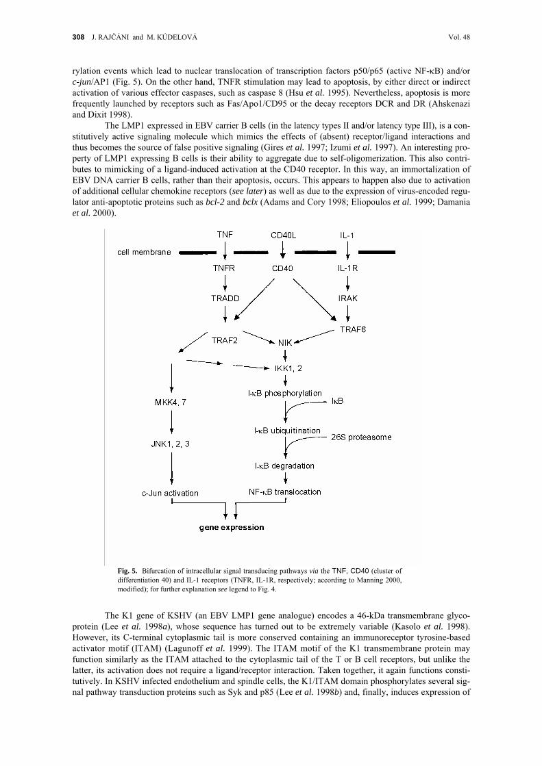
308 J. RAJČÁNI and M. KÚDELOVÁ Vol. 48
rylation events which lead to nuclear translocation of transcription factors p50/p65 (active NF-κB) and/or c-jun/AP1 (Fig. 5). On the other hand, TNFR stimulation may lead to apoptosis, by either direct or indirect activation of various effector caspases, such as caspase 8 (Hsu et al. 1995). Nevertheless, apoptosis is more frequently launched by receptors such as Fas/Apo1/CD95 or the decay receptors DCR and DR (Ahskenazi and Dixit 1998).
The LMP1 expressed in EBV carrier B cells (in the latency types II and/or latency type III), is a con-stitutively active signaling molecule which mimics the effects of (absent) receptor/ligand interactions and thus becomes the source of false positive signaling (Gires et al. 1997; Izumi et al. 1997). An interesting pro-perty of LMP1 expressing B cells is their ability to aggregate due to self-oligomerization. This also contri- butes to mimicking of a ligand-induced activation at the CD40 receptor. In this way, an immortalization of EBV DNA carrier B cells, rather than their apoptosis, occurs. This appears to happen also due to activation of additional cellular chemokine receptors (see later) as well as due to the expression of virus-encoded regu-lator anti-apoptotic proteins such as bcl-2 and bclx (Adams and Cory 1998; Eliopoulos et al. 1999; Damania et al. 2000).
Fig. 5. Bifurcation of intracellular signal transducing pathways via the TNF, CD40 (cluster of differentiation 40) and IL-1 receptors (TNFR, IL-1R, respectively; according to Manning 2000, modified); for further explanation see legend to Fig. 4.
The K1 gene of KSHV (an EBV LMP1 gene analogue) encodes a 46-kDa transmembrane glyco-
protein (Lee et al. 1998a), whose sequence has turned out to be extremely variable (Kasolo et al. 1998). However, its C-terminal cytoplasmic tail is more conserved containing an immunoreceptor tyrosine-based activator motif (ITAM) (Lagunoff et al. 1999). The ITAM motif of the K1 transmembrane protein may function similarly as the ITAM attached to the cytoplasmic tail of the T or B cell receptors, but unlike the latter, its activation does not require a ligand/receptor interaction. Taken together, it again functions consti-tutively. In KSHV infected endothelium and spindle cells, the K1/ITAM domain phosphorylates several sig-nal pathway transduction proteins such as Syk and p85 (Lee et al. 1998b) and, finally, induces expression of

2003 GAMMA HERPESVIRUSES — review 309
a transactivator protein termed nuclear factor of T cells (NFAT). The rightmost end of the KSHV genome situated opposite to the K1 gene encodes another signaling transmembrane protein called K15, which is an EBV LMP2a analogue (Choi et al. 1999). The cytoplasmic tail of K15 (as well as of LMP2a) contains SH2 and/or SH3 binding sites (src homology domains) capable of combining with proteins exerting tyrosine kinase activity, such as Lyn, Fyn, Syk and Csk kinases and other members of the src-tyrosine kinase family (Burkhardt et al. 1992). These kinases participate in proliferative signal transduction in response to various growth factors (ligands) interacting with receptors on T cells, endothelial cells, B cells, platelets, bone mar-row cells, brain glial cells and fibroblasts. Unlike noninfected cells, in which tyrosine kinase activation is cAMP dependent and requires ligand–receptor interactions, in KSHV or EBV infected cells the K15 or LMP2a cytoplasmic tails are constitutively tyrosine phosphorylated by the above cellular enzymes. In EBV latency III with LMP2a expression, the B-cell receptor-associated signal transduction can be downregulated, so that the latency carrier cells would not become activated upon antigen stimulation. A similar effect has been described in KSHV infected cells (Choi et al. 1999). K15 is expressed especially in PEL and Castle-man disease. This protein has the ability to associate with HAX-1, the HS-1 associated protein X-1. HAX-1 shows anti-apoptotic activity, but K15 protein does not influence apoptosis. Because HAX-1 also affects contacting, which promotes actin polymerization via phosphorylation, it can be assumed that K15 may influ-ence endothelial cell motility (Sharp et al. 2002).
6.3 Gamma herpesvirus-encoded cyclins
vCyclins expressed during infection of cells by cyclin encoding viruses may mimic cellular cyclins and functionally substitute them even if they are down-regulated by p53 and the cyclin inhibitors KIP/CIP (kinase or cdk inhibitor proteins) (Mittnacht and Boshoff 2000). In non-infected cells, the cyclins are cofac-tors which complex with cyclin-dependent kinases (cdk). To activate transcription, cdk phosphorylates the retinoblastoma (Rb) protein. After this, the phosphorylated Rb protein is released from the promoter region of silenced genes, especially those participating in cellular DNA replication. The Rb anti-onc protein blocks transcription of enzymes involved in cellular DNA synthesis and repair. In association with these different functions, there are several cyclin/cdk complexes regulating either the transition from G1 to S phase (dupli-cation of cellular DNA) or the transition from G2 to M phase (DNA control and repair). The cellular cyclins are controlled via inhibitors such as the p21 protein, which belongs to the KIP/CIP family. The expression and synthesis of the latter is regulated by the p53 anti-onc protein. When activated, p53 upregulates the transcription of KIP, which in turn inhibits the cyclin/cdk complexes.
KSHV encodes a vCyclin resembling cellular type D cyclins (Chang et al. 1996), while MHV-68 encodes a type A/E vCyclin (van Dyck et al. 1999). KSHV similarly as the HVS-encoded vCyclin binds and activates the cellular cdk6, which is usually activated by cellular type D cyclin molecules (Swanton et al. 1997). The resulting phosphokinase activity phosphorylates the Rb protein, which in turn becomes inactiva-ted allowing the expression of DNA polymerase and other related cellular enzymes to proceed. In addition, KSHV vCyclin modulates the p27/KIP levels inducing p27 degradation (Ellis et al. 1999). The latter effect can never be achieved by cellular type D cyclins, but is known to occur in the presence of type A/E cyclins (Mann et al. 1999). As mentioned above, the KSHV vCyclin ORF72 is flanked by ORF71 encoding the viral FLICE inhibitor protein (vFLIP) – which is in fact a proteinase/caspase-1 inhibitor – and by ORF73 enco-ding the LANA1 protein. Common translation of vFLIP and vCyclin arises from a bicistronic transcript with an internal ribosomal entry site indicating that their synthesis is highly linked. Thus, expression of the anti-apoptotic protein vFLIP correlates with the expression of vCyclin when cells are being driven to the synthe-tic phase and when cellular as well as viral DNA synthesis is promoted.
6.4 Gamma herpesvirus-encoded virokines and chemokines
Chemokines are a superfamily of small structurally related cytokines which selectively promote adhesion, chemotaxis and activation of leukocyte populations (Baggiolini et al. 1997; Nelson and Krensky 1998). All biological effects of chemokines are mediated by a large group of receptors, whose transmembra-ne domain crosses the cell membrane seven times (seven-span, serpentine) and whose cytoplasmic domain generates GTP as a second messenger (GPCR). A typical feature of chemokines is the presence of four con-served cystein residues (i.e. the four-cystein motifs). The most important are the C-C subgroup peptides (with no intervening amino acids) and the CXC subgroup peptides (with a single intervening amino acids). KSHV encodes at least three chemokines termed viral macrophage inflammatory proteins (vMIP1/K4, vMIP2/ORF4.1 and vMIP3/K6). The vMIP1 acts as an antagonist of the C-C receptor 8 (Dairaghi et al. 1999), vMIP2 binds to C-C receptor 3 to inhibit the chemotaxis of monocytes (Boshoff et al. 1997), while vMIP3 interacts with C-C receptor 4 (Stine et al. 2000). In addition, these virus-encoded chemokines can

310 J. RAJČÁNI and M. KÚDELOVÁ Vol. 48
bind to some C-X-C receptors (Kledal et al. 1997). All of them are chemoattractant receptors for TH2 lym-phocytes.
The ORF74 of MHV-68 encodes a CXCR2 chemokine receptor homologue (Virgin et al. 1997) (termed oncogenic G protein-coupled receptor) (Wakeling et al. 2001) and a M3 gene-encoded soluble che-mokine-binding protein that could potentially interfere with chemokine–receptor interactions. Recently, M3 protein has been shown capable of binding a broad variety of chemokines neutralizing cellular responses to chemokines in vitro (Parry et al. 2000). In vivo it probably attenuates the immune response to virus infection and assists the mechanisms regulating leukocyte recruitment that could lead to enhanced viral replication (Sarawar et al. 2002).
In addition to the secreted chemokines, EBV, KSHV as well as MHV code for an IL-8 receptor homo-logue. The corresponding gene is BLR1 and/or ORF74, respectively (Table III). The viral GPCR after inter-action with IL-8 activates the secretion of VEGF. The viral GPCR could be induced in vitro in PEL derived lymphoma cell line (Arvinatakis et al. 1997). The VEGF production in KS tissues is limited to a few KS cells which underwent expression of multiple latency genes (Table VI) and of additional IE and E proteins including trans-activating ones (ORF50 and K8). Secretion of VEGF may promote proliferation of conven-tional endothelium cells and their transition to the transformed KS endothelium cell phenotype. This occurs due to the presence of additional stimulation factors (cytokines) such as vIL-6 (Molden et al. 1997). In CD and PEL, vIL-6 appears to be responsible for proliferation of many uninfected B cells that make up these tumors. In turn, vIL-6 enhances VEGF secretion (Ensoli et al. 2000). Furthermore, the basic fibroblast growth factor (bFGF) produced by the spindle cells within the KS lesions induces vascular proliferation and pro-motes angiogenesis (Samaniego et al. 1995). Both growth factors act in accord with HIV-1 transactivator protein tat. Inflammatory cytokines (IFN-γ and monocyte chemoattractant proteins, MCPs) contribute to the accumulation of circulating B cells carrying the latent KSHV DNA and stimulate lytic virus activation. As already mentioned, in general, following receptor–lingand interaction any GPCR molecule (either cellular or virus-encoded) , undergoes dimer formation which activates the tyrosine kinase activity of its cytoplasmic tail. An adapter protein such as Grb2, containing SH2 and SH3 domains, interacts with a receptor (Sos) protein. The Grb2/Sos complex exchanges GTP with Ras so that Ras/GTP is formed. The latter binds to a cytoplasmic serine-threonine kinase such as Raf in order to induce the phosphorylation signal, which is further transferred via the MapK/MKK (mitogen activated kinase/kinase) cascade. Finally, c-jun/AP1 or other transcription factors are translocated into the nucleus in order to activate transcription.
It seems that vIL-6 is the most important virokine produced by KSHV gene K2 (Neipel et al. 1997; Nikolas et al. 1997) and secreted from KSHV-infected lymphocytes, fibroblasts, monocytes and/or endo-thelium cells. The IL-6 receptor signal transducing moiety gp130 can be activated by vIL-6 directly, i.e. without the need of any coreceptor to phosphorylate and translocate the corresponding transcription factors STAT1, 3 or 5 (Molden et al. 1997). In addition, the above mentioned Ras/MKK signaling pathway may become activated (Moore and Chang 2002). It is likely that these activated signaling pathways may induce anti-apoptotic proteins as well. Neither vIL-6 nor vMIPs are expressed during restricted latency in KSHV DNA carrier cells (Table VI) so that they cannot act at initial stages of KS development (Canon et al. 1999). This is in contrast with the large amounts of this virokine produced by early infiltrating cells. Further-more, HIV-encoded tat is released from HIV-infected T cells, and is also an efficient transcription activator of KSHV transcription (Ensoli et al. 1993). The synergistic effect of HIV-tat and bFGF (basic fibroblast growth factor) has been already mentioned. Summing up, early stages of KS are characterized by a few spindle shaped cells expressing KSHV latency genes and many proliferating non-infected cells reacting to the presence of cellular and viral inflammatory chemokines (Cannon et al. 1999).
The IRF (IFN regulating factor) family of transcription factors is involved in regulating gene expression in response to IFN signaling. KSHV possesses a mechanism allowing it to prevent the induction of cellular IFN genes that are important components of natural (non-specific) immune response. The virus induced IRF molecules have an N-terminal DNA binding motif and a C-terminal repressor/transactivator region. Though the first characterized vIRF1/K9 (Table IV) does not directly bind to DNA, it inhibits the IFN-α-gene promoter expression, down-regulates p21 expression and prevents IFN-α/β-induced growth arrest (Gao et al. 1997). The vIFN-2 encodes 163 amino acids protein and shares the N-terminal region with IRF1 which binds to the NF-κB binding site (Burysek et al. 1999). The vIFN-3 interferes with the transcription of IFN-α in infected cells (Lubyová and Pitha 2000). IFN regulators are involved also in the IFN-γ response inhibition, which is of special interest, since this class two IFN is a cytokine rather than a classical virus inhibitor (Vilcek and Sen 1996). In non-infected cells, interaction of IFN-γ with corres-ponding receptor causes Janus-kinase (JAK) phosphorylation which activates the signaling transduction and transcription (STAT) protein homodimer formation, an alternative pathway similar to that activated upon IL stimulation. The activated STAT-1 homodimers migrate to the nucleus, where they act as transcription

2003 GAMMA HERPESVIRUSES — review 311
cofactors by recruiting the γ-activated sequence (GAS) element and its co-adapters such as HAT (the histone acetyltransferase) and CBP/p300 (the CREB-binding protein) necessary for initiation of transcription at the IFN-responsive promoters (Goodman and Smolik 2000). The vIRFs inhibit this signaling mechanism by binding to STAT co-adapter proteins and interfering with their transcription promoting function.
Because IFNs up regulate the cdk promoter, the expression of which is regulated by p21 (KIP), vIRFs interfere also with the function leading to cell arrest. The p53- and p21-induced cell cycle arrest can be lifted via different KSHV-encoded products: the vIRFs, LANA1/K1 and LANA2/K15 proteins. In this way, KS cells expressing early (nonstructural) KSHV products are shifted from apoptosis to a state of upregulation of cell division promoting regulatory proteins. Apoptosis, in addition, is prevented by the virus-encoded bcl-2 analogue (ORF16) and by the early expression of ORF71/K13/vFLIP. Especially the latter is an efficient anti-apoptotic protein, since it acts as an inhibitor of caspase 8 activation. The vFLIP has a simi-lar death effector domain (DED) as the Fas receptor-associated death domain (FADD) adapter protein, which is an efficient transducer of the apoptosis activation signaling to inactive caspases. Since the DED of caspase 8 associates with vFLIP as well as with the death domain of FADD, in fact the viral FLICE (i.e. the virus-encoded FADD-like ICE) inhibitor protein efficiently interferes with the induction of apoptosis. vFLIP can also inhibit the TNFR-mediated cellular apoptosis induced signaling. Finally, the viral bcl-2 analogues (encoded by many gamma herpesviruses; Table III) can prevent apoptosis either due to vCyclin/cdk6 over-expression or by BAX binding (Ojala et al. 1999). In KS sarcoma, however, vFLICE expression may be more frequent than the bcl-2 analogue expression.
7 FUNCTIONAL SIGNIFICANCE OF THE GAMMA HERPESVIRUS-SPECIFIC GENES
The steps of KSHV oncogenesis and transformation by gamma herpesviruses discussed here include an initial non-specific phase of cell proliferation mediated by inflammatory cykines and chemokines which is followed by an activation of latency with an expression of a wide spectrum of latency-associated viral genes (Table VI). The expression of genes such as LMP1 (transition of EBV latency to latency II and/or III) or the latency-associated KSHV genes (such as ORFs71–73 and the K1, K12 and K15 genes) activates host cell DNA transcription. This can result in driving host cells towards division. Latency II/ latency III in EBV carrier B cells, for example, causes expression of CD markers and cytokine receptors, a state which renders them sensitive to the action of corresponding cytokines and growth factors. Further-more, heterologous infections such as HIV and the presence of their transactivation proteins (HIV-tat) or other stimuli activate the expression of EBV or KSHV-encoded transactivator proteins (Zta/Rta), which turn on the transcription of additional virus-encoded regulatory proteins, viral virokines, viral chemokines and growth receptors (Table VII). Expression of structural proteins and virion formation may be important for virus spread, but it is not relevant for oncogenesis. The block of lytic virus replication might be even of advantage for cell transformation and tumor formation, since the above effects are launched by nonstructural viral proteins.
8 GAMMA HERPESVIRUS GENOMICS: CELL GENE PIRATING AND EVOLUTION
The majority of herpesvirus genes contain no introns and their mRNAs need no splicing. This indi-cates that herpesviruses might have acquired the cellular genes through mRNA intermediates. Conversion from mRNA into DNA could occur with the help of reverse transcriptase supplied by a co-infecting retro-virus. The idea of possibility of direct virus-to-virus gene transposition is supported by identification of gene inserts of virus origin such as the adeno associated virus type 2 rep gene sequence in the HHV-6 genome (Thomson et al. 1991). In addition to the acquisition of sequences from heterologous viruses and/or of cel-lular gene sequences, there is also some evidence of gene loss and/or of reduplication of gene sequences in herpesvirus DNAs. Gamma herpesvirus genes such as FGARAT, GCPR, IRF or MIP might be present in multiple copies. The presence of transcripts transcribed by the RNA polymerase III, from tandem repeat sequences encoding the EBERs, tRNAs or mRNAs, respectively, points at the fact that these are non-coding repetitive elements. Whether such sequences are just compensations for sequence deletions or whether they are of functional significance should be further investigated. As an evidence of gene loss can be understood the absence of ori-binding protein gene in the gamma herpesvirus DNA (Inoue et al. 1994). Similarly, the presence of GCPR IL-8 receptor gene in KSHV but not in MHV-68 DNA indicates its loss from certain gamma herpesvirus genomes (Nicholas 2000). Coincidence of genetic divergence at particular genomic locations suggests that these loci allow more readily the loss or acquisition of foreign sequences from an

312 J. RAJČÁNI and M. KÚDELOVÁ Vol. 48
hypothetical ancient progenitor virus genome. It is possible that more variations occur at the DNA termini due to nicks and cuts during concatemer cleavage and packaging events. The divergent loci flank both DNA
Table VII. A survey of gamma herpesvirus genes involved in cell transformation: from latency to oncogenesisa
KSHV EBV MHV Responsible protein
Cell proliferation due to cytokine and virokine action
vIL-6 viral IL-6; growth factor with a wide spectrum of target cells
M1, serpin immune regulator receptor (?) ORF4/CCP ORF4/CCP complement control protein K4/MIP1 macrophage inflammatory proteins K4.1/MIP2 K6/MIP3 K9/IRF IFN regulatory proteins interfering
with the JAK/STAT signaling pathway K10/IRF K10.l/IRF K11.1/IRF BCFR1 viral IL-10 homologue, inhibitor
of inflammatory cytokines ORF74/vGPCR ORF74/vGPCR viral G-protein coupled receptor (activates
Ras/c-jun/AP-1 signaling)
Anti-apoptosis proteins
ORF16 BALF1 M11 functional bcl-2 homologue ORF71/vFLIP Fas-DD-like (ICE) inhibitor protein;
interferes with death domain signaling
Proteins directly affecting intracellular signaling pathways
ORF72/vCyclin ORF72/vCyclin vCyclin D analogue; cdk6 activator K1 BNLF1/LMP1 latent membrane protein 1; the EBV transformation
protein activates NF-κB; K1 interacts with ITAM adapter protein
K12 kaposin, an E5 transformation protein analogue K15 LMP2 SH2/SH3 binding and tyrosine kinase activation ORF73/LANA1 ORF73/LANA latent nuclear antigen, interacts with
Rb protein, activates E2F K10.1/LANA2 BYRF1/EBNA2 inhibits p53-dependent transcription
aCf. with Tables III, IV and VI.
termini and are located between the conserved gene blocks I to II, and III to IV as depicted in the MHV-68 genome (Fig. 3). Both terminal regions of MHV-68 as well as of KSHV DNA contain the latency associated and acquired virus specific or subgroup-specific genes confirming the notion that these diverging regions undergo more frequent recombination events. In contrast to the pirated genes, the core structural and the essential non-structural proteins are encoded by conservative genes (Table II) organized in homologous blocks (Fig. 1). Acquisition of new genes could confer on the virus new and specific properties altering its biological behavior on the basis of natural selection. Whatever the mechanism, the phenomenon itself pro-vides a powerful tool for rapid adaptation of herpesviruses giving the family members considerable diver-sity.
The authors thank the Grant Agency of the Slovak Academy of Sciences (VEGA) for support (projects 02/1082/01 and 02/2010/02).
REFERENCES
ADAMS J.M., CORY S.: The bcl-2 protein family: arbiters of cell survival. Science 281, 1322–1325 (1998). AHN J.W., POWELL K.L., KELLAM P., ALBER D.G.: Gamma herpesvirus lytic gene expression as characterized by DNA array. J.Virol.
76, 6244–6256 (2002).

2003 GAMMA HERPESVIRUSES — review 313
AKHTAR M., BUNUAN H., ALI M., GODWIN J.: Kaposi’s sarcoma in renal transplant patients: ultrastructural and immunoperoxidase study of four cases. Cancer 53, 258–266 (1984).
ALBRECHT J.C., NICHOLAS J., BILLER D., CAMERON K.R., BIESINGER B., NEWMAN C., WITTMANN S., CRAXTON M.A., COLEMAN H., FLECKENSTEIN B., HONESS R.W.: Primary structure of the herpesvirus saimiri genome. J.Virol. 66, 5047–5058 (1992).
ALBRECHT J.C.: Primary structure of the herpesvirus ateles genome. J.Virol. 74, 1033–1037 (2000). AMBINDER R.F., SHAH W.A., RAWLINGS D.R., HAYWARD G.S., HAYWARD S.D.: Definition of the sequence requirements for binding of
the EBNA1 protein to its palindromic target sites in Epstein–Barr virus DNA. Cell 58, 527–535 (1990). AMBINDER R.F., MULLEN M.A., CHANG Y.N., HAYWARD G.S., HAYWARD S.D.: Functional domains of Epstein–Barr virus nuclear
antigen EBNA1. J.Virol. 65, 1466–1478 (1991). ARMSTRONG A.A., GALLAGHER A., KRAJEWSKI A.S.: The expression of the EBV latent membrane protein (LMP1) is independent of
CD23 and bcl-2 in Reed–Sternberg cells. Histopathology 21, 72–73 (1992). ARVANITAKIS L., MESRI E.A., NADOR R.G., SAID J.W., ASCH A.S., KNWOLES D.M., CESARMAN E.: Establishment and characterization
of a primary effusion (body cavity based) lymphoma cell line (BC-3) harboring Kaposi’s sarcoma-associated herpesvirus (KSHV/HHV-8) in the absence of Epstein–Barr virus. Blood 88, 2648–2654 (1996).
ARVANITAKIS L.A., GERAS-RAAKA E., VARMA A., GERSHENGORN M.C., CESARMAN E.: Human herpesvirus KSHV encodes a consti-tutively active G-protein-coupled receptor linked to cell proliferation. Nature 385, 347–349 (1997).
ASKENAZI A., DIXIT V.M.: Death receptors: signaling and modulation. Science 281, 1305–1308 (1998). BAER R., BANKIER A.T., BIGGIN M.D., DEININGER P.L., FARRELL P.J., GIBSON T.J., HATFULL G., HUDSON G.S., SATCHWELL S.C.,
SEGUIN C., TUFNELL P.S., BARRELL B.G.: DNA sequence and expression of the B95-8 Epstein–Barr virus genome. Nature 310, 207–211 (1984).
BAGGIOLINI M., DEWALD B., MOSER B.: Human chemokines: an update. Ann.Rev.Immunol. 15, 675–705 (1997). BAICHWAL V.R., SUGDEN B.: Transformation of Balb/c 3T3 cells by the BNLF-1 gene of Epstein–Barr virus. Oncogene 2, 461–467
(1988). BAIS C., SANTOMASSO B., COSO O., ARVANITAKIS L., RAAKA E.G., GUTKIND J.S., ASCH A.S., CESARMAN E., GERHENGORN M.C.,
MESRI E.A.: G-Protein-coupled receptor of Kaposi’s sarcoma-associated herpesvirus is a viral oncogene and angiogenesis activator. Nature 391, 86–89 (1998).
BAUERLE P., BALTIMORE D.: I-κB: a specific inhibitor of the NF-κB transcription factor. Science 242, 540–546 (1988). BELLO L.J., DAVISON A.J., GLENN M.A., WHITEHOUSE A., RETHMEIER N., SCHULTZ T.F., CLEMENTS J.B.: The human herpesvirus 8
ORF57 gene and its properties. J.Gen.Virol. 80, 3207–3215 (2000). VAN BERKEL V., PREITER K., VIRGIN H.W. IV, SPECK S.H.: Identification and initial characterization of the murine gamma herpesvirus
68 gene M3, encoding an abundantly secreted protein. J.Virol. 73, 4524–4529 (1999). BERNHEIM A., BERGER R., LENOIR G.: Cytogenetic studies on African Burkitt’s lymphoma cell lines; t(8:14), t(2:8) and t(8:22)
translocations. Cancer Genet.Cytogenet. 3, 307–315 (1981). BIELESKI L., TALBOT S.: Kaposi’s sarcoma-associated herpesvirus vCyclin open reading frame contains an internal ribosomal entry site.
J.Virol. 75, 1864–1869 (2001). BLAŠKOVIČ D., STANČEKOVÁ M., SVOBODOVÁ J., MISTRÍKOVÁ J.: Isolation of five strains of herpesviruses from two species of free
living small rodents. Acta Virol. 24, 468–473 (1980). BLAŠKOVIČ D., STANEKOVÁ D., RAJČÁNI J.: Experimental pathogenesis of murine herpesvirus in newborn mice. Acta Virol. 28, 225–
231 (1984). BOSHOFF C., ENDO Y., COLLINS P.D., TAKEUCHI Y., REEVES J.D., SCHWEICKART V.L., SIANI M.A., SASAKI T., WILLIAMS T.J.,
GRAY P.W., MOORE P.S., CHANG Y., WEISS R.A.: Angiogenic and HIV inhibitory functions of KSHV-encoded chemokines. Science 278, 290–294 (1997).
BUBLOT M., LOMONTE P., LEQUARRE A.S., ALBRECHT J.C., NICHOLAS J., FLECKENSTEIN B., PASTORET P.P., THIRY E.: Genetic rela-tionships between bovine herpesvirus 4 and the gamma herpesviruses Epstein–Barr virus and herpesvirus saimiri. Virology 190, 654–665 (1992).
BURKHARDT A.L., BOLEM J.B., KIEFF E., LONGNECKER R.: An Epstein–Barr virus transformation-associated membrane protein inter-acts with Src family tyrosine kinases. J.Virol. 66, 5161–5167 (1992).
BURKITT D.A.: A children’s cancer dependent upon climatic factors. Nature 194, 232–234 (1962). BURYSEK L., YEOW W.S., LUBYOVA B., KELLUM M., SCHAFER S.L., HUANG Y.Q., PITHA P.M.: Functional analysis of human herpes-
virus 8-encoded viral interferon regulatory factor I and its association with cellular interferon regulatory factors and p300. J.Virol. 73, 7334–7342 (1999).
CANNON J.S., NICHOLAS J., ORENSTEIN J.M., MANN R.B., MURRAY P.G., BROWNING P.J., DI GUISEPPE J.A., CESARMAN E., HAYWARD G.S., AMBINDER R.F.: Heterogeneity of viral IL-6 expression in HHV-8 associated disease. J.Infect.Dis. 180, 824–828 (1999).
CASTLEMAN B., IVERSON L., MENEDEY V.P.: Localized mediastinal lymph-node hyperplasia resembling thymoma. Cancer 9, 822–830 (1956).
CESARMAN E., NADOR R.G., BAI F., BOHENSKY R.A., RUSSO J.J., MOORE P.S., CHANG Y., KNOWLES D.M.: Kaposi’s sarcoma-asso-ciated herpesvirus contains G protein-coupled receptor and cyclin D homologues which are expressed in Kaposi’s sarcoma and malignant lymphoma. J.Virol. 70, 8218–8223 (1996).
CHABOT-FLETCHER M.: Cellular signaling to NF-kB: role in inflammation and therapeutic promise, pp. 23–38 in G.L. Letts, D.W. Mor-gan (Eds): Inflammatory Process: Molecular Mechanisms and Therapeutic Opportunities. Birkhauser, Basel–Boston–Berlin 2000.
CHANG Y., MOORE P.S., TALBOT S.J.: Cyclin encoded by KS herpesvirus. Nature 382, 410–411 (1996). CHANG J., GANEM D.: On the control of late gene expression in Kaposi’s sarcoma-associated herpesvirus (human herpesvirus 8). J.Gen.
Virol. 81, 2039–2047 (2000). CHI T., CAREY M.: The ZEBRA activation domain: modular organization and mechanism of action. Mol.Cell.Biol. 13, 7045–7055
(1993). CHOI P.H.K., SVEN M.W.M., HAUNG D.P., LO K.-W., LEE J.C.K.: Nasopharyngal carcinoma: genetic changes, Epstein–Barr virus
infection or both. A clinical study of 36 patients. Cancer 72, 2873–2878 (1993).

314 J. RAJČÁNI and M. KÚDELOVÁ Vol. 48
CHOI J.K., LEE B.S., SHIM S.N., LI M., JUNG J.U.: Identification of the novel K15 gene at the rightmost end of the Kaposi’s sarcoma associated herpesvirus genome. J.Virol. 74, 436–446 (1999).
COHEN B.D., GOLDSTEIN D.J., RUTLEGE L., WASS W.C., LOWY D.R., SCHLEGEL R., SCHILLER J.T.: Transformation specific interaction of the bovine papillomavirus E5 oncoprotein with the platelet derived growth factor receptor domain and the epidermal growth factor receptor cytoplasmic domain. J.Virol. 67, 5303–5311 (1993).
CRAIGHEAD J.E.: Epstein–Barr virus (EBV), pp. 117–146 in Pathology and Pathogenesis of Human Viral Disease. Academic Press, London–San Diego 2000a.
CRAIGHEAD J.E.: Kaposi’s sarcoma associated herpesvirus (KSHV, HHV-8), pp. 171–188 in Pathology and Pathogenesis of Human Viral Disease. Academic Press, London–San Diego 2000b.
DAIRAGHI D.J., FAN R.A., MCMASTER B.E., HANLEY M.R., SCHALL T.J.: HHV-8 encoded vMIP-1 selectively engages chemokine receptor CCR8. Agonist and antagonist profiles of viral chemokines. J.Biol.Chem. 274, 21569–21574 (1999).
DALLA-FAVERA R., BREGNI M., ERIKSON J., PATTERSON D., GALLO R.W., CROCE C.M.: Human c-myc gene is located in the region of chromosome 8 that is translocated in Burkitt lymphoma cells. Proc.Nat.Acad.Sci.USA 81, 7632–7636 (1982).
DAMANIA B., CHOI JOONG-KOOK, JUNG J.U.: Signaling activities of gamma herpesvirus membrane proteins. J.Virol. 74, 1593–1601 (2000).
DESROSIERS R.C., SASSEVILLE V.G., CZAJAK S.C., ZHANG X., MANSFIELD K.G., KAUR A., JOHNSON R.P., LACKNER A.A., JUNG J.U.: A herpesvirus of rhesus monkeys related to the human Kaposi’s sarcoma-associated herpesvirus. J.Virol. 71, 9764–9769 (1997).
DITTMER D., LAGUNOFF M., RENNE R., STASKUS K., HAASE A., GANEM D.: A cluster of latently expressed genes in Kaposi’s sarcoma-associated herpesvirus. J.Virol. 72, 8309–8315 (1998).
DUPIN N., FISHER C., KELLAM P., ARIAD S., TULLIEZ M., FRANCK N., VAN MARCK E., SALMON D., GORIN I., ESCANDE J.P., WEISS R.A., ALITALO K., BOSHOFF C.: Distribution of HHV-8 positive cells in Kaposi’s sarcoma, multicentric Castleman’s disease and primary effusion lymphoma. Proc.Nat.Acad.Sci.USA 96, 4546–4551 (1999).
DUPIN N., DISS T., KELLAM P., TULLIEZ M., DU M.Q., WEISS R.A., ISAACSON P.G., BOSHOFF C.: HHV-8 is associated with a plasma-blastic variant of Castleman disease that is linked to HHV-8 positive plasmablastic lymphoma. Blood 95, 1406–1412 (2000).
VAN DYK L.F., HESS J.L., KATZ J.D., JAKOBY M., SPECK S.H., VIRGIN H.W. IV: The murine gamma herpesvirus 68 vCyclin is an oncogene that promotes cell cycle progression in primary lymphocytes. J.Virol. 73, 5119–5122 (1999).
EFSTATHIOU S., HO M., HALL S., STYLES C.J., SCOTT S.D., GOMPELS U.A.: Murine herpesvirus 68 is genetically related to the gamma herpesviruses Epstein–Barr virus and herpesvirus saimiri. J.Gen.Virol. 71, 1365–1372 (1990).
ELIOPOULOS A.G., BLAKE S.M., FLOETMANN J.E., ROWE M., YOUNG L.S.: Epstein–Barr virus encoded latent membrane protein 1 acti-vates the JNK pathway through extreme C-terminus via a mechanism involving TRADD and TRAF2. J.Virol. 73, 1023–1035 (1999).
ELLIS M., CHEW Y.P., FALLIS L.: Degradation of p27KIP cdk inhibitor triggered by Kaposi’s sarcoma virus cyclin–cdk6 complex. EMBO J. 18, 644–653 (1999).
ENSOLI B., BUONAGURO L., BARRILARI G., FIORELLI V., GENDELMAN R., MORGAN R.A., WINGFIELD P., GALLO R.C.: Release, uptake and effects of extracellular HIV-Tat protein in induction of Kaposi’s sarcoma. J.Virol. 67, 277–287 (1993).
ENSOLI B., STURZL M., MONINI P.: Cytokine-mediated growth promotion of Kaposi’s sarcoma and primary effusion lymphoma. Cancer Biol. 10, 367–381 (2000).
ENSSER A., PFLANZ R., FLECKENSTEIN B.: Primary structure of the alcephaline herpesvirus 1 genome. J.Virol. 71, 6517–6525 (1997). ERNBERG I., FALK K., MINAROVITS J.: The role of methylation in the phenotype dependent modulation of Epstein–Barr nuclear antigen
2 and latent membrane protein genes in cells latently infected with Epstein–Barr virus. J.Gen.Virol. 70, 2989–3002 (1989). FALK L., DEINHARDT F., NONOYAMA M., WOLFE L.G., BERGHOLZ C.: Properties of a baboon lymphotropic herpesvirus related to
Epstein–Barr virus. Internat.J.Cancer 18, 798–807 (1976). FARRELL P.J.: Epstein–Barr virus, pp. 120–133 in S.J. O’Brien (Ed.): Genetic Maps. Cold Spring Harbor Press, New York 1992. FEEDERLE R., KOST M., BAUMANN M., JANZ A., HAMMERSCHMIDT W., DELECLUSE H.-J.: The Epstein–Barr virus lytic program is con-
trolled by the cooperative functions of two transactivators. EMBO J. 19, 3080–3089 (2000). FLINT S.J., ENQUIST L.W., KRUG R.M., RACANIELLO V.R., SKALKA A.M.: The transcriptional cascades of DNA viruses, pp. 261–276 in
Principles of Virology. Molecular Biology, Pathogenesis and Control. ASM Press, Washington (DC) 2000. FRIBORG J., KONG W., HOTTIGER M.O., NABEL G.J.: p53 inhibition by the LANA protein of KSHV protects against cell death. Nature
402, 889–894 (1999). FRIEDMAN-KIEN A.: Disseminated Kaposi’s sarcoma syndrome in young homosexual men. J.Am.Acad.Dermatol. 5, 468–471 (1981). FRIZZERA G., MASSARELLI G., BANKS P.M., ROSAI J.: A systemic lymphoproliferative disorder with morphologic features of Castleman
disease. Am.J.Surg.Pathol. 7, 211–231 (1983). GAHN T.A., SCHILDKRAUT C.L.: The Epstein–Barr virus origin of plasmid replication, oriP, contains both the initiation and termination
sites of DNA replication. Cell 58, 527–535 (1989). GAO S.J., BOSHOFF C., JAYACHANDRA S., WEISS R.A., CHANG Y., MOORE P.S.: KSHV ORF K9 (vIRF) is an oncogene that inhibits the
interferon signaling pathway. Oncogene 15, 1979–1986 (1997). GERBER P., NONOYAMA M., LUCAS S., PERLIN E., GOLDSTEIN L.I.: Oral excretion of Epstein–Barr virus by healthy subjects and pati-
ents with infectious mononucleosis. Lancet 2, 988–989 (1972). GILLIGAN K., SATO H., RAJADURAI P., BUSSON P., YOUNG L.S., RICKINSON A.L., TURSZ T., RAAB-TRAUB N.: Novel transcription from
the Epstein–Barr virus terminal EcoRI fragment, DIJhet, in nasopharyngeal carcinoma. J.Virol. 64, 4948–4956 (1990). GIRES O., ZIMBER-STROBL U., GONNELLA R., UEFFING M., MARSCHALL G., ZRIDLER R., PICH D., HAMMERSCHMIDT W.: Latent
membrane protein 1 of Epstein–Barr virus mimics a constitutively active receptor molecule. EMBO J. 16, 6131–6140 (1997).
GLICKMAN J., HOWE G., STEITZ J.: Structural analyses of EBER1 and EBER2 ribonucleoprotein particles present in EBV infected cells. J.Virol. 62, 902–911 (1988).
GOMPELS U.A., NICHOLAS J., LAWRENCE G.: The DNA sequence of human herpesvirus 6: structure, coding content and genome evolu-tion. Virology 209, 29–51 (1995).
GOODMAN R.H., SMOLIK S.: CPB/p300 in cell growth, transformation and development. Genes Dev. 14, 1553–1577 (2000).

2003 GAMMA HERPESVIRUSES — review 315
GREGORY C.D., EDWARDS C.F., MILNER A., WIELS J., LIPINSKI M., ROWE M., TURSY T., RICKINSON A.B.: Isolation of a normal B cell subset with a Burkitt-like phenotype and transformation in vitro with Epstein–Barr virus. Internat.J.Cancer 42, 213–220 (1988).
GRUNDHOFF A., GANEM D.: Mechanisms governing expression of the vFLIP gene of Kaposi’s sarcoma-associated herpesvirus. J.Virol. 75, 1857–1863 (2001).
HAAN K.M., KWOK W.W., LONGNECKER R., SPECK P.: Epstein–Barr virus entry utilizing HLA-DP DR or DQ cofactors. J.Virol. 74, 2451–2454 (2000).
HENLE W., DIEHL V., KOHN G., ZUR HAUSEN H., HENLE G.: Herpes type virus and chromosome marker in normal leukocytes after growth with irradiated Burkitt cells. Science 157, 1064–1065 (1967).
HERBST H., STEIN H., NIEDOBITEK G.: Epstein–Barr virus and CD30 – malignant lymphomas. Crit.Rev.Oncogen. 4, 191–239 (1991). HSU H., XION J., GOEDDEL D.V.: The TNF receptor 1-associated protein TRADD signals cell death and νF-κB activation. Cell 81, 495–
504 (1995). HUSAIN S.M., USHERWOOD E.J., DYSON H., COLECLOUGH C., COPPOLA D.L., WOODLAND M.A., BLACKMAN J.P., STEWART J.P.,
SAMPLE J.T.: Murine gamma herpesvirus M2 gene is latency associated and its protein is a target for CD8 T lymphocytes. Proc.Nat.Acad.Sci.USA 96, 7508–7513 (1999).
INOUE N., DAMBOUGH T.R., RAPP J.C.: Alpha herpesvirus origin-binding protein homologue encoded by human herpesvirus 6B, a beta herpesvirus, binds to nucleotide sequences that are similar to ori regions of alpha herpesviruses. J.Virol. 68, 4126–4136 (1994).
IZUMI K.M., KIEFF E.D.: The Epstein–Barr virus oncogenic product latent membrane protein 1 engages the tumor necrosis factor recep-tor-associated death domain protein to mediate B lymphocyte growth transformation and activate NF-κB. Proc.Nat.Acad. Sci.USA 94, 12592–12597 (1997).
JACOBY M.A., VIRGIN H.W., SPECK S.H.: Disruption of the M2 gene of murine gamma herpesvirus 68 alters splenic latency following intranasal, but not intraperitoneal, inoculation. J.Virol. 76, 1790–1801 (2002).
JANSSON A., MASUCCI M., RYMO L.: Methylation of discrete sites within the enhancer region regulates the activity of the Epstein–Barr virus BamHI W promoter region in Burkitt lymphoma lines. J.Virol. 66, 62–69 (1992).
KASOLO F.C., MONZE M., OBEL N., ANDERSON R.A., FRENCH C., GOMPELS U.A.: Sequence analyses of human herpesvirus 8 strains from both African human immunodeficiency virus-negative and -positive childhood endemic Kaposi’s sarcoma show a close relationship with strains identified in febrile children and variation in the K1 glycoprotein. J.Gen.Virol. 79, 3055–3065 (1998).
KATANO H., SATO Y., KURATA T., MORI S.H., SATA T.: Expression and localization of human herpesvirus 8-encoded proteins in pri-mary effusion lymphoma, Kaposi’s sarcoma and multicentric Castleman disease. Virology 269, 335–344 (2000).
KEDES D.H., LAGUNOFF M., RENNE R., GANEM D.: Identification of the gene encoding the major latency-associated nuclear antigen of the Kaposi’s sarcoma-associated herpesvirus. J.Clin.Invest. 100, 2602–2610 (1997).
KENNEDY M.M., BIDDOLPH S., LUCAS S.B., HOWELS D.D., PICTON S., MCGEE J.O.D., SILVA I., UHLMAN V., LUTTICH K., LEARY J.J.: Cyclin D1 expression and HHV8 in Kaposi’s sarcoma. J.Clin.Pathol. 52, 569–573 (1999).
KERR B.M., LEAR A.L., ROWE M., CROOM-CARTER D., YOUNG L.S., ROOKES S.M., GALLIMORE P.H., RICKINSON A.B.: Three tran-scriptionally distinct forms of Epstein–Barr virus latency in somatic cell hybrids: cell phenotype dependence of promoter virus usage. Virology 187, 189–201 (1992).
KIEFF E.: Epstein–Barr virus and its replication, pp. 2343–2396 in B.N. Fields, D.M. Knipe, P.M. Howley (Eds): Field’s Virology, 3rd ed. Lippincott–Raven Publishers, Philadelphia–New York–London–Hong Kong–Tokyo 1996.
KIM A.L., MAHER M., HAYMAN J.B.: An imperfect correlation between DNA replication activity of Epstein–Barr virus nuclear antigen and binding to the nuclear import receptor, Rch/importin alpha. Virology 239, 340–351 (1997).
KLEDAL T.N., ROSENKILDE M.M., COULIN F., SIMMONS G., JOHNSEN A.H., ALOUANI S., POWER C.A., LUTTICHAU H.R., GERSTOFT J., CLAPHAM P.R., CLARK-LEWIS I., WELLS T.N., SCHWARTZ T.W.: A broad spectrum chemokine antagonist encoded by Ka-posi’s sarcoma associated herpesvirus. Science 277, 1656–1659 (1997).
KLEIN G., PURTILO D.: Summary: symposium on Epstein–Barr virus induced lymphoproliferative diseases in immunodeficient patients. Cancer Res. 41, 4302–4304 (1981).
KRYSAN P.J., HAASE S.B., CALOS M.B.: Isolation of human sequences that replicate autonomously in human cells. Mol.Cell.Biol. 9, 1026–1033 (1989).
LAGUNOFF M., GANEM D.: The structure and coding organization of the genomic termini of Kaposi’s sarcoma-associated herpesvirus. Virology 236, 147–154 (1997).
LAGUNOFF M.R., MAJETT R., WEISS A., GANEM D.: Deregulated signal transduction by the K1 gene product of Kaposi’s sarcoma-associated herpesvirus. Proc.Nat.Acad.Sci.USA 96, 5704–5709 (1999).
LEE H., GUN J., LI M., CHOI J.K., DEMARIA M., ROSENYWEIG M., JUNG J.U.: Identification of an immunoreceptor tyrosine-based acti-vation motif of K1 transforming protein of Kaposi’s sarcoma-associated herpesvirus. Mol.Cell.Biol. 18, 5219–5228 (1998a).
LEE H., VEAZEY R., WILLIAMS K., LI M., GUO J., NEIPEL F., FLECKENSTEINM B., LACKNER A., DESROSIERS R.C., JUNG J.U.: Deregulation of cell growth by the K1 gene of Kaposi’s sarcoma-associated herpesvirus. Nature Med. 4, 435–440 (1998b).
LEIGHT E.R., SUGDEN B.: EBNA1: a protein pivotal to latent infection by Epstein–Barr virus. Rev.Med.Virol. 10, 83–100 (2000). LI Q.X., YOUNG L.S., NIEDOBITEK G., DAWSON C.W., BIRKENBACH M., WANG F., RICKINSON A.B.: Epstein–Barr virus infection and
replication in human epithelial cell system. Nature 356, 347–350 (1992). LU S.-J., DAY N.E., GEGOS L.: Linkage of nasopharyngeal carcinoma subseptibility locus to the HLA region. Nature 346, 470–471
(1990). LUBYOVA B., PITHA P.M.: Characterization of a novel human herpesvirus 8-encoded protein, vIRF3, that shows homology to viral and
cellular interferon regulatory factors. J.Virol. 74, 8194–8201 (2000). MACKEY D., SUGDEN B.: The linking regions of EBNA1 are essential for its support of replication and transcription. Mol.Cell.Biol. 19,
3349–3359 (1999). MAGRATH I.: The pathogenesis of Burkitt’s lymphoma. Adv.Cancer Res. 55, 133–269 (1990). MAGRATH I., JAIN V., BHATIA K.: Epstein–Barr virus and Burkitt’s lymphoma. Semin.Cancer Biol. 3, 285–295 (1992).

316 J. RAJČÁNI and M. KÚDELOVÁ Vol. 48
MANN D.J., CHILD E.S., SWANTON C., LAMAN H., JONES N.: Modulation of p27/KIP levels by the cyclin encoded by Kaposi’s sarcoma-associated herpesvirus. EMBO J. 18, 654–663 (1999).
MANNING A.M.: Small molecule regulators of AP-1 and NF-κB, pp. 39–52 in G.L. Letts, D.W. Morgan (Eds): Inflammatory Process: Molecular Mechanisms and Therapeutic Opportunities. Birkhauser, Basel–Boston–Berlin 2000.
MASOOD R., CAI J., ZHENG T., SMITH D.L., NAIDU Y., GILL P.C.: Vascular endothelial growth factor – vascular permeability factor is an autocrine growth factor for AIDS-Kaposi’s sarcoma. Proc.Nat.Acad.Sci.USA 94, 979–984 (1997).
MEDVECZKY M.M., GECK P., CLARKE C., BYRNES J., SULLIVAN J.L., MEDVECZKY P.G.: Arrangement of repetitive sequences in the genome of herpesvirus sylvilagus. J.Virol. 63, 1010–1014 (1989).
MIDDLETON T., SUGDEN B.: Retention of plasmid DNA in mammalian cells is enhanced by binding of the Epsten-Barr virus replication protein EBNA1. J.Virol. 68, 4067–4071 (1994).
MISTRÍKOVÁ J., REMEŇOVÁ A., LEŠŠO J., STANČEKOVÁ M.: Replication and persistence of murine herpesvirus 72 in lymphatic system and peritoneal blood mononuclear cells of Balb/c mice. Acta Virol. 38, 151–156 (1994).
MISTRÍKOVÁ J., MRMUSOVÁ M.: Detection of abnormal lymphocytes in the blood of Balb/c mice infected with murine herpesvirus strain 72. Acta Virol. 42, 79–82 (1998).
MISTRÍKOVÁ J., MRMUSOVÁ M., ĎURMANOVÁ V., RAJČÁNI J.: Increased neoplasm development due to immunosuppressive treatment with FK506 in Balb/c mice persistently infected with mouse herpesvirus. Viral Immunol. 12, 237–247 (1999).
MISTRÍKOVÁ J., RAŠLOVÁ H., MRMUSOVÁ M., KÚDELOVÁ M.: A murine gamma herpesvirus – review. Acta Virol. 44, 211–226 (2000). MITTNACHT S., BOSHOFF C.: Viral cyclins. Rev.Med.Virol. 10, 175–184 (2000). MOLDEN J., CHANG Y., YOU Y., MOORE P.S., GOLDSMITH M.A.: A Kaposi’s sarcoma-associated herpesvirus-encoded cytokine
homologue (vIL-6) activates signaling through the shared gp130 receptor subunit. J.Biol.Chem. 272, 19625–19631 (1997). MOLESWORTH S.J., LAKE C.M., BORZA C.M., TURK S.M., HUTT-FLETCHER L.M.: Epstein–Barr virus gH is essential for penetration of
B cells, but also plays a role in attachment of virus to epithelial cells. J.Virol. 74, 6324–6332 (2000). MOORE P.S., CHANG Y.: Molecular virology of Kaposi’s sarcoma-associated herpesvirus. Phil.Trans.Roy.Soc.London B 356, 499–516
(2001). MOTOKURA T., BLOOM T., KIM H.G., JUPPNER H., RUDERMAN J.V., KRONENBERG H.M., ARNOLD A.: A novel cyclin encoded by a bcl-
1 linked candidate oncogene. Nature 350, 512–525 (1991). MRMUSOVÁ M., HORVÁTHOVÁ M., KLOBUŠICKÁ M., MISTRÍKOVÁ J.: Immunotyping of leukocytes in peripheral blood of Balb/c mice
infected with mouse herpesvirus isolate 72. Acta Virol. 46, 19–24 (2002). MURALIDHAR S., PUMPERY A.M., HASSANI M., SADAIE M.R., AZUMI N., KISHISHITA M., BRADY J.N., DONIGER J., MEDVECZKY P.,
ROSENTHAL L.J.: Identification of kaposin open reading frame K12 as a human herpesvirus 8 (Kaposi’s sarcoma-associated herpesvirus) transforming gene. J.Virol. 72, 4980–4988 (1998).
NEIPEL F., ALBRECHT J.C., ENSSER A., HUANG Y.Q., LI J.J., FRIEDMAN K.A., FLECKENSTEIN B.: Human herpesvirus 8 encodes a homologue of macrophage inhibitory protein-1 and interleukin-6. J.Virol. 71, 839–842 (1997).
NELSON P.J., KRENSKI A.M.: Chemokines, lymphocytes and viruses: what goes around comes around. Curr.Opin.Immunol. 10, 265–270 (1998).
NEMEROW G.R., WOLFERT R., MCNAUGHTON M.E., COOPER N.R.: Identification and characterization of the Epstein–Barr virus receptor on human B lymphocytes and its relationship to the C3d complement receptor (CR2). J.Virol. 55, 347–351 (1985).
NICHOLAS J.: Evolationary aspects of oncogenic herpesviruses. J.Clin.Pathol.Mol.Pathol. 53, 222–237 (2000). NICHOLAS J., RUVOLO V.R., BURNS W.H., SANDFORD G., WAN X., GIUFO D., HENDRICKSON S.B., GUO H.G., HAYWARD G.S., REITZ
M.S.: Kaposi’s sarcoma-associated human herpesvirus-8 encodes homologues of macrophage inflammatory protein 1 and interleukin-6. Nature Med. 3, 287–292 (1997).
NIEDOBITEK G., YOUNG L.S., SAM C.K., BROOKS L., PRASAD U., RICKINSON A.B.: Expression of Epstein–Barr virus genes and of lym-phocyte activation molecules in undifferentiated nasopharyngeal carcinomas. Am.J.Pathol. 140, 879–887 (1992).
NISHI J., MARUYAMA I.: Increased expression of vascular endothelial growth factor (VEGF) in Castleman disease: proposed patho-mechanism for vascular proliferation in the affected lymph node. Leuk.Lymphoma 38, 387–394 (2000).
NISHIKURA K., AR-RUSHIDI A., ERIKSSON J., WATT R., ROVERA G., CROCE C.M.: Differential expression of the normal amd of the translocated human c-myc oncogenes in B cells. Proc.Nat.Acad.Sci.USA 80, 291–296 (1983).
O’CONNOR G., DAVIES N.: Malignant tumors in African children with special reference to malignant lymphomas. J.Pediatr. 56, 526–535 (1960).
OJALA P.M., TIANINEN M., SALVEN P., VEIKKOLA T., CASTANOS-VELEZ E., SARID R., BIBERFELD P., MAKELA T.P.: Kaposi’s sarcoma-associaed herpesvirus encoded vCyclin dependent kinase 6. Cancer Res. 59, 4984–4989 (1999).
PALESTRO G., TURRINI F., PAGANO M., CHIUSA L.: Castleman disease. Adv.Clin.Path. 3, 11–22 (1999). PALLESEN G., HAMILTON-DUTOIT S.J., ROWE M., YOUNG L.S.: Expression of Epstein–Barr virus latent gene products in tumor cells of
Hodgkin’s disease. Lancet 337, 329–332 (1991). PARRY C.M., SIMAS J.P., SMITH C.A., STEWART A.C., MINSON C.A., EFSTATHIOU S., ALCAMI A.: A broad spectrum secreted chemo-
kine binding protein encoded by a herpesvirus. J.Exp.Med. 191, 573–578 (2000). PEDERSON C., GERSTOFT J., LUNDGREN J.D.: HIV-associated lymphoma: histopathology and association with Epstein–Barr virus geno-
me related to clinical, immunological and prognostic features. Eur.J.Cancer 27, 1416–1423 (1991). PHELAN A., CLEMENTS J.B.: Posttranscriptional regulation in herpes simplex virus. Semin.Virol. 8, 309–318 (1998). POLSON A.G., HUANG L., LIKAC D.M., BLETHROW J.D., MORGAN D.O., BURLINGAME A.L., GANEM D.: Kaposi’s sarcoma associated
herpesvirus K-bZIP protein is phosphorylated by cyclin-dependent kinases. J.Virol. 75, 3175–3184 (2001). POPE J.H., HORNE M.K., SCOTT W.: Transformation of fetal human leukocytes in vitro by filtrates of human leukemic cell line contain-
ing herpes-like virus. Internat.J.Cancer 3, 857–866 (1968). RAGOCZY T., MILLER G.: Role of Epstein–Barr virus Rta protein in activation of distinct classes of viral lytic cycle genes. J.Virol. 73,
9858–9866 (1999). RAJČÁNI J., BLAŠKOVIČ D., SVOBODOVÁ J., ČIAMPOR F., HUČKOVÁ D., STANEKOVÁ D.: Pathogenesis of acute and persistent murine
herpesvirus infection in mice. Acta Virol. 29, 51–60 (1985). RAJČÁNI J., BUSTAMANTE DE CONTRERAS L.R., SVOBODOVÁ J.: Corneal inoculation of murine herpesvirus in mice: the absence of
neural spread. Acta Virol. 31, 25–30 (1986).

2003 GAMMA HERPESVIRUSES — review 317
RAŠLOVÁ H., MISTRÍKOVÁ J., KÚDELOVÁ M., MISHAL Z., SARASIN A., BLANGY D., BEREBBI M.: Immunophenotypic study of atypical lymphocytes generated in peripheral blood and spleen of nude mice after MHV-72 infection. Viral Immunol. 13, 313–327 (2000).
RAŠLOVÁ H., BEREBBI M., RAJČÁNI J., SARASIN A., MATIS J., KÚDELOVÁ M.: Susceptibility of mouse mammary glands to murine gamma herpesvirus 72 (MHV-72) infection: evidence of MHV-72 transmission via breast milk. Microb.Pathogen. 31, 47–58 (2001).
RAWLINGS D.R., MILMAN G., HAYWARD S.D., HAYWARD G.S.: Sequence-specific DNA binding of the Epstein–Barr virus nuclear antigen (EBNA1) to clustered sites in the plasmid maintenance region. Cell 42, 859–868 (1985).
VAN REGENMORTEL M.H.V., FAUQUET C.M., BISHOP D.H.L.: Herpesvirus family, pp. 220–226 in Virus Taxonomy: Classification and Nomenclature of Viruses. 7th ICTV Report. Academic Press, San Diego 2000.
RICKINSON A.B., KIEFF E.: Epstein–Barr virus, pp. 2397–2446 in B.N. Fields, D.M. Knipe, P.M. Howley (Eds): Fields’ Virology, 3rd ed. Lippincott–Raven Publishers, Philadelphia–New York–London–Hong Kong–Tokyo 1996.
ROCHFORD R., LUTZKE M.L., ALFINITO R.S., CLAVO A., CARDIN R.D.: Kinetics of murine gamma herpesvirus 68 gene expression fol-lowing infection of murine cells in culture and in mice. J.Virol. 75, 4955–4963 (2001).
ROSDAHL L., LARSEN S.O., CLEMMENSEN J.: Hodgkin’s disease in patients with previous infectious mononucleosis; 30 years expe-rience. Brit.Med.J. 2, 253–256 (1974).
ROY D.J., EBRAHIMI B.C., DUTIA B.M., NASH A.A., STEWART J.P.: Murine gamma herpesvirus M11 gene product inhibits apoptosis and is expressed during virus persistence. Arch.Virol. 145, 2411–2420 (2000).
RUSSO J.J., BOHENZKY R.A., CHIEN M.C., CHEN J., YAN M., MADDALENA D., PARRY J.P., PERUZZI D., EDELMAN I.S., CHANG Y., MOORE P.S.: Nucleotide sequence of the Kaposi’s sarcoma-associated herpesvirus (HHV8). Proc.Nat.Acad.Sci.USA 93, 14862–14867 (1996).
SAMANIEGO F., MARKHAM P., GALLO R.C., ENSOLI B.: Inflammatory cytokines induce AIDS-Kaposi’s sarcoma like lesion formation in nude mice. J.Immunol. 154, 3582–3592 (1995).
SAMPLE J., BROOKS L., SAMPLE C.: Restricted Epstein–Barr virus protein expression in Burkitt lymphoma is due to a different Epstein–Barr nuclear antigen I transcriptional site. Proc.Nat.Acad.Sci.USA 88, 6343–6347 (1991).
SARAWAR S.R., LEE B.J., ANDERSON M., TENG Y.C., ZUBERI R., VON GEJSEN S.: Chemokine induction and leukocyte trafficking to the lungs during murine gamma herpesvirus 68 (MHV-68) infection. Virology 293, 54–62 (2002).
SCHULTZ T.F.: Kaposi’s sarcoma-associated herpesvirus (human herpesvirus 8): epidemiology and pathogenesis. Antimicrob.Chemo-ther. 45 (Suppl.), 15–27 (2000).
SEN P., BALTIMORE D.: Multiple nuclear factors interact with the immunoglobin enhancer sequences. Cell 46, 705–716 (1986). SHARP T.V., BOSHOFF C.: Kaposi’s sarcoma associated herpesvirus: from cell biology to pathogenesis. Life 49, 97–104 (2000). SHARP T.V., WANG H.W., KUOMI A., HOLLYMAN D., ENDO Y., YE H., DU M.Q., BOSHOFF C.: K15 protein of Kaposi’s sarcoma-
associated herpesvirus is latently expressed and binds to HAX-1, a protein with antiapoptotic function. J.Virol. 76, 802–806 (2002).
SMITH P.R., GRIFFIN B.E.: Transcription of the Epstein–Barr virus gene EBNA1 from different promoters in nasopharyngeal carcinoma and B lymphoblastoid cells. J.Virol. 66, 706–714 (1992).
SONG H.Y., REGNIER C.H., KIRSCHNING C.J., GOEDDEL D.V., ROTHE M.: Tumor necrosis factor (TNF) mediated cascades: bifurcation of nuclear factor κB and c-jun N-terminal kinase (JNK/SAPK) pathways at the TNF receptor associated factor 2. Proc.Nat. Acad.Sci.USA 94, 9792–9796 (1997).
SONG M.J., BROWN H.J., WU T.-T., SUN R.: Transcription activation of polyadenylated nuclear RNA by Rta in human herpes-virus-8/Kaposi’s sarcoma-associated herpesvirus. J.Virol. 75, 3129–3140 (2001).
SPECK P., HAAN K.M., LONGNECKER R.: Epstein–Barr virus entry into cells. Virology 277, 1–5 (2000). SPECK S.H., CHATILA T., FLEMINGTON E.: Reactivation of Epstein–Barr virus: regulation and function of the BZLF1 gene. Trends
Microbiol. 5, 399–405 (1997). STASKUS K.A., ZHONG W., GEBHARD K., HERNDIER B., WANG H., RENNE R., BENEKE J., PUDNEY J., ANDERSON D.J., GANEM D.,
HAASE A.T.: Kaposi’s sarcoma-associated herpesvirus gene expression in endothelial (spindle) tumor cells. J.Virol. 71, 715–19 (1997).
STEVENSON P.G., DOHERTY P.C.: Kinetic analysis of the specific host response to a murine gamma herpesvirus. J.Virol. 72, 943–949 (1998).
STEVENSON P.G., EFSTATHIOU S., DOHERTY P.C., LEHNER P.J.: Inhibition of MHC class I-restricted by gamma 2 herpesviruses. Proc. Nat.Acad.Sci.USA 97, 8455–8460 (2000).
STEWART J.P., ESUEWOOD E.J., ROSS A., DYSON H., NASH T.: Lung epithelial cells are a major site of murine gamma herpesvirus persistence. J.Exp.Med. 187, 1941–1951 (1998).
STINE J.T., WOOD C., HILL M., EPP A., RAPORT C.J., SCHWEICKART V.L., ENDO Y., SASAKI T., SIMMONS G., BOSHOFF C., CLAP- HAM P., CHANG Y, MOORE P., GRAY P.W., CHANTRY D.: KSHV encoded CC chemokine vMIPIII is a CCR4 agonist, stimulates angiogenesis and selectively chemoattract TH2 cells. Blood 95, 1151–1157 (2000).
SUNIL-CHANDRA N.P., EFSTATHIOU S., ARNO J., NASH A.A.: Virological and pathological features of mice with murine gamma herpes-virus. J.Gen.Virol. 73, 2347–2356 (1992a).
SUNIL-CHANDRA N.P., EFSTATHIOU S., ARNO J., NASH A.A.: Murine gamma herpesvirus establishes a latent infection in mouse B lym-phocytes in vivo. J.Gen.Virol. 73, 3275–3279 (1992b).
SUNIL-CHANDRA N.P., FAZAKERLEY A.J., NASH A.A.: Lymphoproliferative disease in mice infected with murine gamma herpesvirus 68. Am.J.Pathol. 145, 818–826 (1994).
SVOBODOVÁ J., BLAŠKOVIČ D., MISTRÍKOVÁ J.: Growth characteristics of herpesviruses isolated from free living small rodents. Acta Virol. 26, 256–263 (1982).
SWANTON C., MANN D.J., FLECKENSTEIN B., NEIPEL F., PETERS G., JONES N.: Herpes viral cyclin/Cdk6 complexes evade inhibition by CDK inhibitor proteins. Nature 390, 187–187 (1997).
TANIGUCHI M., LAMPHIER M.S., TANAKA N.: IRF-1: the transcription factor linking the interferon response and oncogenesis. Biochim. Biophys.Acta 1333, M9–M17 (1997).

318 J. RAJČÁNI and M. KÚDELOVÁ Vol. 48
TELFORD E.A.R., WATSON M.S., AIRD H.C., PERRY J., DAVISON A.J.: The DNA sequence of equine herpesvirus 2. J.Mol.Biol. 249, 520–528 (1995).
TERRY L.A., STEWART J.P., NASH A.A., FAZAKERLY J.K.: Murine gamma herpesvirus-68 infection of and persistence in the central nervous system. J.Gen.Virol. 81, 2535–2543 (2000).
THOME M., SCHNEIDER P., HOFMANN K., FICKENSCHER H., MEINL E., NEIPEL F., MATTMANN C., BURNS K., BODMER J.L., SCHRO- TER M., SCAFFIDI C., KRAMMER P.H., PETER M.E., TSCHOPP J.: Viral FLICE-inhibitory proteins (vFLIPs) prevent apoptosis induced by death receptors. Nature 386, 517–521 (1997).
THOMSON B.J., EFSTATHIOU S., HONESS R.W.: Acquisition of the human adeno-associated virus type 2 rep gene by human herpesvirus 6. Nature 351, 78–80 (1991).
TIERNEY R.J., STEVEN N., YOUNG L.S., RICKINSON A.B.: Epstein–Barr virus latency in blood mononuclear cells: analysis of viral gene transcription during primary infection and in the carrier state. J.Virol. 68, 7374–7385 (1994).
TOKAI N., FUJIMOTO N.A., TOYOSHIMA Y.: Kid, a novel kinesin-like DNA binding protein, is localized to chromosomes and the mitotic spindle. EMBO J. 15, 457–467 (1996).
USHERWOOD E.J., ROSS A.J., ALLEN D.J., NASH A.A.: Murine gamma herpevirus-induced splenomegaly: a critical role for CD4 T cells. J.Gen.Virol. 77, 627–630 (1996).
VILCEK J., SEN G.C.: Interferons and other cytokins, pp. 375–399 in B.N. Fields, D.M. Knipe, P.M. Howley (Eds): Fields’ Virology, 3rd ed. Lippincott–Raven Publishers, Philadelphia 1996.
VIRGIN H.W. IV, LATREILLE P., WAMSLEY P., HALLSWORTH K., WECK K.E., DAL CANTO A.J., SPECK H.S.: Complete sequence and genomic analysis of murine gamma herpesvirus 68. J.Virol. 71, 5894–5904 (1997).
VIRGIN H.W. IV, PRESTI R.M., LI X.Y., LIU C., SPECK S.H.: Three distinct regions of the murine gamma herpesvirus 68 genome are transcriptionally active in latently infected mice. J.Virol. 73, 2321–2332 (1999).
WAKELING M.N., ROY D.J., NASH A.A., STEWART J.P.: Characterization of the murine gamma herpesvirus ORF74 product: a novel oncogenic G protein-coupled receptor. J.Gen.Virol. 82, 1187–1197 (2001).
WANG X., KENYON W.J., LI Q., MULLBERG J., HUTT-FLETCHER L.M.: Epstein–Barr virus uses different complexes of glycoproteins gH and gL to infect B lymphocytes and epithelial cells. J.Virol. 72, 5552–5558 (1998).
WANG S., LUI S., WU M., GENG Y., WOOD C.: Kaposi’s sarcoma-associated herpesvirus – human herpesvirus-8 ORF50 gene product contains a potent C-terminal activation domain which activates gene expression via a specific target sequence. Arch.Virol. 146, 1415–1426 (2001).
WECK K.E., BARKON M.L., YOO L.I., SPECK H., VIRGIN H.W. IV: Mature B cells are required for acute splenic infection, but not for establishment of latency by murine gamma herpesvirus. J.Virol. 70, 6775–6780 (1996).
WECK K.E., KIM S.S., VIRGIN H.W. IV, SPECK S.: Macrophages are the major reservoir of latent murine gamma herpesvirus 68 in peri-toneal cells. J.Virol. 73, 3273–3283 (1999).
WHITBY D., HOWARD M., TENANT-FLOWERS M., BRINK N., COPAS A., BOSHOFF C., HATZIOANNOU T., SUGGETT F., ALDAM D., DENTON A., MILLER R., WELLER I., WEISS R., TEDDER R., SCHULTZ T.: Detection of Kaposi’s sarcoma-associated herpesvirus in peripheral blood of HIV-infected individuals as progression to Kaposi’s sarcoma. Lancet 346, 799–802 (1995).
YAO Q.Y., RICKINSON A.B., EPSTEIN M.A.: A re-examination of the Epstein–Barr virus carrier state in healthy seropositive individuals. Internat.J.Cancer 35, 35–42 (1985).
YATES J.L., WARREN N., SUGDEN B.A.: Stable replication of plasmids derived from Epstein–Barr virus in various mammalian cells. Nature 313, 812–815 (1985).
ZALANI S., HOLLEY-GUTHRIE E., KENNEY S.: Epstein–Barr viral latency is disrupted by the immediate early BRLF1 protein through a cell-specific mechanism. Proc.Nat.Acad.Sci.USA 93, 9194–9199 (1996).
ZHANG S., NONOYAMA M.: The cellular proteins that bind specifically to the Epstein–Barr virus belong to a family of plasmid DNA replication origin. Proc.Nat.Acad.Sci.USA 91, 2843–2847 (1994).
Related Documents

![Contribution of TLR signaling to the pathogenesis of ... · signaling pathways: the classical pathway (through TIRAP and MyD88) and the alternative pathway (via TRIF and TRAM)[18].](https://static.cupdf.com/doc/110x72/607f1f7f0609627187448e24/contribution-of-tlr-signaling-to-the-pathogenesis-of-signaling-pathways-the.jpg)