DOI: 10.1002/chem.201102544 Galvanic Replacement Reactions of Active-Metal Nanoparticles Kai-Yang Niu, [a] Sergei A. Kulinich, [b] Jing Yang, [a] Aaron L. Zhu, [b] and Xi-Wen Du* [a] Introduction Nanoscale galvanic replacement (NGR) reactions, systemat- ically studied by Xia)s group among others since 2002, [1] have been proved to be a convenient technique to synthe- size diverse metal nanostructures with a certain composition and spatial pattern. [2] Unlike other syntheses of bimetallic nanocrystals based on wet-chemistry routes, which rely on the reaction of molecular precursors, the NGR, in essence, is a sort of cementation plating with very effective self-tem- plating features in electrochemical terms, which distin- guishes it from electroless deposition. In the design and fab- rication of fine noble-metal nanostructures through this ap- proach, numerous nanoboxes, frames, and cages with tuna- ble pore sizes have been controllably produced. [1, 3] Owing to their unique spatial configuration, excellent biocompatibil- ity, and tunable optical properties, some of them have al- ready been used for drug delivery, contrast-enhanced optical imaging, photothermal therapy, and catalysis. [2, 4] More recently, several groups have investigated more active-metal seeds (such as Co, Fe, Ni, and NiCo), thereby producing composite nanostructures through NGR reac- tions. [5] However, the number of such studies is very limited, and the capabilities of the NGR reactions as a preparation technique are still far from being fully exploited. To date, nonactive-metal nanostructures (such as Ag, Cu) and noble- metal ions (i.e., AuCl 4 , Ag + ) have been commonly used as sacrificial seeds and reactants, respectively. As a result, the products are mainly limited to noble-metal materials. [3, 6] Thus, there is a strong motivation to expand the NGR ap- proach to make it a more universal strategy that allows a much wider variety of products. To achieve this goal, our idea was to employ nanoparticles (NPs) of a superactive metal, for example, Mg, as sacrificial seeds for NGR-based syntheses. Such seeds presumably have several advantages. First, in comparison with Ag nano- seeds, the use of Mg nanoseeds is expected to expand prod- uct materials to a big family of all metals with lower activity than the seed material. Second, due to the sensitivity of Mg nanoseeds to the environment, the reaction modes and thus the final nanostructures can be controlled by changing the reaction medium. As shown below, we were able to design and fabricate Mg@MgO-Au, MgO hollow NPs loaded with Au and Cu NPs (anticipated for drug/gene delivery and cat- alysis), Ag colloids with unique optical properties, SnO 2 NPs with photocatalytic properties, as well as spongy Au nano- spheres that are potentially attractive for bioapplications Abstract: We present a systemic inves- tigation of a galvanic replacement tech- nique in which active-metal nanoparti- cles are used as sacrificial seeds. We found that different nanostructures can be controllably synthesized by varying the type of more noble-metal ions and liquid medium. Specifically, nano-het- ACHTUNGTRENNUNGeroACHTUNGTRENNUNGstructures of noble metal (Ag, Au) or Cu nanocrystals on active-metal (Mg, Zn) cores were obtained by the reaction of active-metal nanoparticles with more noble-metal ions in ethanol; Ag nanocrystal arrays were produced by the reaction of active-metal nano- particles with Ag + ions in water; spongy Au nanospheres were generat- ed by the reaction of active-metal nanoparticles with AuCl 4 ions in water; and SnO 2 nanoparticles were prepared when Sn 2 + were used as the oxidant ions. The key factors determin- ing the product morphology are shown to be the reactivity of the liquid medium and the nature of the oxidant– reductant couple, whereas Mg and Zn nanoparticles played similar roles in achieving various nanostructures. When microsized Mg and Zn particles were used as seeds in similar reactions, the products were mainly noble-metal dendrites. The new approach proposed in this study expands the capability of the conventional nanoscale galvanic re- placement method and provides new avenues to various structures, which are expected to have many potential applications in catalysis, optoelectron- ics, and biomedicine. Keywords: galvanic replacement reactions · gold · nanoparticles · nanostructures · silver [a] K.-Y. Niu, Dr. J. Yang, Prof.Dr. X.-W. Du Tianjin Key Laboratory of Composite and Functional Materials School of Materials Science and Engineering Tianjin University Tianjin 300072 (P.R. China) Fax: (+ 86) 22-2740-5694 E-mail : [email protected] [b] Dr. S. A. Kulinich, Dr. A. L. Zhu Department of Chemistry University of British Columbia 2036 Main Mall Vancouver, BC, V6T 1Z1 (Canada) Supporting information for this article is available on the WWW under http://dx.doi.org/10.1002/chem.201102544. # 2012 Wiley-VCH Verlag GmbH&Co. KGaA, Weinheim Chem. Eur. J. 2012, 18, 4234 – 4241 4234

Welcome message from author
This document is posted to help you gain knowledge. Please leave a comment to let me know what you think about it! Share it to your friends and learn new things together.
Transcript
DOI: 10.1002/chem.201102544
Galvanic Replacement Reactions of Active-Metal Nanoparticles
Kai-Yang Niu,[a] Sergei A. Kulinich,[b] Jing Yang,[a] Aaron L. Zhu,[b] and Xi-Wen Du*[a]
Introduction
Nanoscale galvanic replacement (NGR) reactions, systemat-ically studied by Xia�s group among others since 2002,[1]
have been proved to be a convenient technique to synthe-size diverse metal nanostructures with a certain compositionand spatial pattern.[2] Unlike other syntheses of bimetallicnanocrystals based on wet-chemistry routes, which rely onthe reaction of molecular precursors, the NGR, in essence,is a sort of cementation plating with very effective self-tem-plating features in electrochemical terms, which distin-guishes it from electroless deposition. In the design and fab-rication of fine noble-metal nanostructures through this ap-proach, numerous nanoboxes, frames, and cages with tuna-ble pore sizes have been controllably produced.[1,3] Owing totheir unique spatial configuration, excellent biocompatibil-
ity, and tunable optical properties, some of them have al-ready been used for drug delivery, contrast-enhanced opticalimaging, photothermal therapy, and catalysis.[2,4]
More recently, several groups have investigated moreactive-metal seeds (such as Co, Fe, Ni, and NiCo), therebyproducing composite nanostructures through NGR reac-tions.[5] However, the number of such studies is very limited,and the capabilities of the NGR reactions as a preparationtechnique are still far from being fully exploited. To date,nonactive-metal nanostructures (such as Ag, Cu) and noble-metal ions (i.e., AuCl4
�, Ag+) have been commonly used assacrificial seeds and reactants, respectively. As a result, theproducts are mainly limited to noble-metal materials.[3,6]
Thus, there is a strong motivation to expand the NGR ap-proach to make it a more universal strategy that allowsa much wider variety of products.
To achieve this goal, our idea was to employ nanoparticles(NPs) of a superactive metal, for example, Mg, as sacrificialseeds for NGR-based syntheses. Such seeds presumablyhave several advantages. First, in comparison with Ag nano-seeds, the use of Mg nanoseeds is expected to expand prod-uct materials to a big family of all metals with lower activitythan the seed material. Second, due to the sensitivity of Mgnanoseeds to the environment, the reaction modes and thusthe final nanostructures can be controlled by changing thereaction medium. As shown below, we were able to designand fabricate Mg@MgO-Au, MgO hollow NPs loaded withAu and Cu NPs (anticipated for drug/gene delivery and cat-alysis), Ag colloids with unique optical properties, SnO2 NPswith photocatalytic properties, as well as spongy Au nano-spheres that are potentially attractive for bioapplications
Abstract: We present a systemic inves-tigation of a galvanic replacement tech-nique in which active-metal nanoparti-cles are used as sacrificial seeds. Wefound that different nanostructures canbe controllably synthesized by varyingthe type of more noble-metal ions andliquid medium. Specifically, nano-het-ACHTUNGTRENNUNGero ACHTUNGTRENNUNGstructures of noble metal (Ag, Au)or Cu nanocrystals on active-metal(Mg, Zn) cores were obtained by thereaction of active-metal nanoparticleswith more noble-metal ions in ethanol;Ag nanocrystal arrays were producedby the reaction of active-metal nano-
particles with Ag+ ions in water;spongy Au nanospheres were generat-ed by the reaction of active-metalnanoparticles with AuCl4
� ions inwater; and SnO2 nanoparticles wereprepared when Sn2+ were used as theoxidant ions. The key factors determin-ing the product morphology are shownto be the reactivity of the liquidmedium and the nature of the oxidant–
reductant couple, whereas Mg and Znnanoparticles played similar roles inachieving various nanostructures.When microsized Mg and Zn particleswere used as seeds in similar reactions,the products were mainly noble-metaldendrites. The new approach proposedin this study expands the capability ofthe conventional nanoscale galvanic re-placement method and provides newavenues to various structures, whichare expected to have many potentialapplications in catalysis, optoelectron-ics, and biomedicine.
Keywords: galvanic replacementreactions · gold · nanoparticles ·nanostructures · silver
[a] K.-Y. Niu, Dr. J. Yang, Prof. Dr. X.-W. DuTianjin Key Laboratory of Composite and Functional MaterialsSchool of Materials Science and EngineeringTianjin UniversityTianjin 300072 (P.R. China)Fax: (+86) 22-2740-5694E-mail : [email protected]
[b] Dr. S. A. Kulinich, Dr. A. L. ZhuDepartment of ChemistryUniversity of British Columbia2036 Main MallVancouver, BC, V6T 1Z1 (Canada)
Supporting information for this article is available on the WWWunder http://dx.doi.org/10.1002/chem.201102544.
� 2012 Wiley-VCH Verlag GmbH & Co. KGaA, Weinheim Chem. Eur. J. 2012, 18, 4234 – 42414234
and catalysis. Third, homogeneous nanoseeds with a finesize are beneficial for the formation of stable nanostructureswith uniform morphology and structure, and the size of thefinal products could be well determined by the size of theMg nanoseeds, which would be appropriate for applicationsin photocatalysis, drug/gene delivery, cell imaging, and soforth.[2,7]
Because of their superactivity, the synthesis of active-metal NPs through conventional wet-chemistry routes isknown to be difficult. In previous work, we reported thatlaser ablation of a metal target can generate pure metal NPsin a chemically inert environment.[8] Herein, we use superac-tive Mg and Zn NPs prepared by laser ablation as reduc-tants, and select Ag+ , AuCl4
�, Sn2+ , and Cu2+ solutions asoxidants for the NGR reaction. We show that both the reac-tants and liquid media play important roles in the finalproduct structure. For instance, Mg–Au and Mg–Ag nano-heterostructures were formed in solutions in ethanol, where-as Ag nanocrystal arrays and spongy Au nanospheres weregenerated in solutions in water after Mg nanoseeds hadbeen completely dissolved. Our results indicate that theNGR reaction exploiting superactive-metal NPs as a sacrifi-cial material can open new avenues toward numerous novelnanostructures with unique morphologies.
In this paper, we first characterize the products of theNGR reaction of Mg NPs with two different solutions, afterwhich we discuss the growth mechanisms for the diverse ob-served products. Then, we expand the approach to other sys-tems with Cu2+ and Sn2+ as oxidants or Zn NPs as a reduc-tant. Next, we demonstrate the difference between theNGR synthesis and a similar approach in which microsizedactive-metal powders are used as sacrificial seeds. In addi-tion, we also present optical and photocatalytic propertiesdemonstrated by certain new products.
Results and Discussion
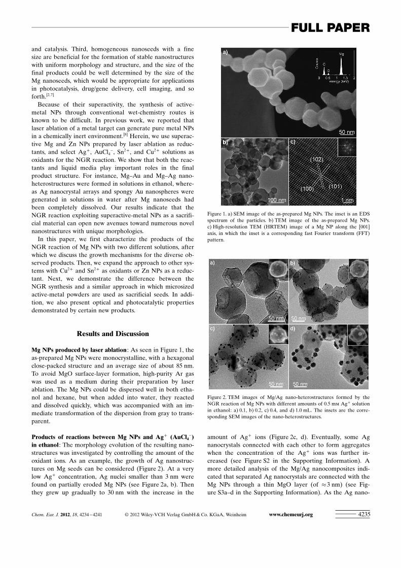
Mg NPs produced by laser ablation : As seen in Figure 1, theas-prepared Mg NPs were monocrystalline, with a hexagonalclose-packed structure and an average size of about 85 nm.To avoid MgO surface-layer formation, high-purity Ar gaswas used as a medium during their preparation by laserablation. The Mg NPs could be dispersed well in both etha-nol and hexane, but when added into water, they reactedand dissolved quickly, which was accompanied with an im-mediate transformation of the dispersion from gray to trans-parent.
Products of reactions between Mg NPs and Ag+ ACHTUNGTRENNUNG(AuCl4�)
in ethanol : The morphology evolution of the resulting nano-structures was investigated by controlling the amount of theoxidant ions. As an example, the growth of Ag nanostruc-tures on Mg seeds can be considered (Figure 2). At a verylow Ag+ concentration, Ag nuclei smaller than 3 nm werefound on partially eroded Mg NPs (see Figure 2a, b). Thenthey grew up gradually to 30 nm with the increase in the
amount of Ag+ ions (Figure 2c, d). Eventually, some Agnanocrystals connected with each other to form aggregateswhen the concentration of the Ag+ ions was further in-creased (see Figure S2 in the Supporting Information). Amore detailed analysis of the Mg/Ag nanocomposites indi-cated that separated Ag nanocrystals are connected with theMg NPs through a thin MgO layer (of �3 nm) (see Fig-ure S3a–d in the Supporting Information). As the Ag nano-
Figure 1. a) SEM image of the as-prepared Mg NPs. The inset is an EDSspectrum of the particles. b) TEM image of the as-prepared Mg NPs.c) High-resolution TEM (HRTEM) image of a Mg NP along the [001]axis, in which the inset is a corresponding fast Fourier transform (FFT)pattern.
Figure 2. TEM images of Mg/Ag nano-heterostructures formed by theNGR reaction of Mg NPs with different amounts of 0.5 mm Ag+ solutionin ethanol: a) 0.1, b) 0.2, c) 0.4, and d) 1.0 mL. The insets are the corre-sponding SEM images of the nano-heterostructures.
Chem. Eur. J. 2012, 18, 4234 – 4241 � 2012 Wiley-VCH Verlag GmbH & Co. KGaA, Weinheim www.chemeurj.org 4235
FULL PAPER
crystals grew bigger, their structure transformed from singlecrystals to multitwinned (see Figure S3d–f in the SupportingInformation).
We followed the spectral evolution of the Mg/Ag nano-composites throughout the growing process. As the amountof reacting Ag+ ions increased, the corresponding signalfrom Mg NPs (around 310 nm, Figure 3) gradually de-creased, and the localized surface plasmon resonance(LSPR) peak of Ag nanocrystals progressively emerged andredshifted from 390 to 430 nm as they grew bigger(Figure 3).
Similar results were found for the Mg/AuCl4� system.
Gold nanocrystals were found on Mg NPs and grew biggerwith the increase in AuCl4
� concentration (see Figures S4,S5, and S6 in the Supporting Information). The structure ofthe Mg/Au nanocomposites was found to be similar to thatof the Mg/Ag products (see Figure S6 in the Supporting In-formation ). Similarly to the Mg/Ag system, the LSPRpeaks of Au gradually increased and redshifted due to thesize effect (see Figure S7 in the Supporting Information).[9]
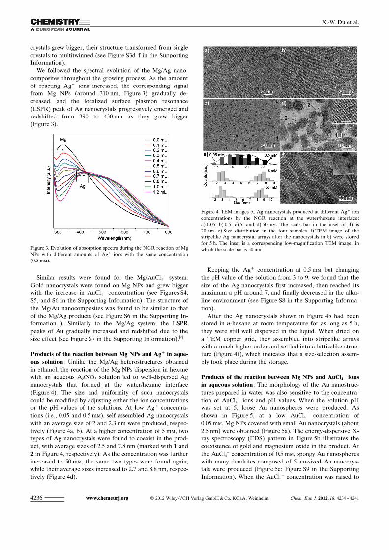
Products of the reaction between Mg NPs and Ag+ in aque-ous solution : Unlike the Mg/Ag heterostructures obtainedin ethanol, the reaction of the Mg NPs dispersion in hexanewith an aqueous AgNO3 solution led to well-dispersed Agnanocrystals that formed at the water/hexane interface(Figure 4). The size and uniformity of such nanocrystalscould be modified by adjusting either the ion concentrationsor the pH values of the solutions. At low Ag+ concentra-tions (i.e., 0.05 and 0.5 mm), self-assembled Ag nanocrystalswith an average size of 2 and 2.3 nm were produced, respec-tively (Figure 4a, b). At a higher concentration of 5 mm, twotypes of Ag nanocrystals were found to coexist in the prod-uct, with average sizes of 2.5 and 7.8 nm (marked with 1 and2 in Figure 4, respectively). As the concentration was furtherincreased to 50 mm, the same two types were found again,while their average sizes increased to 2.7 and 8.8 nm, respec-tively (Figure 4d).
Keeping the Ag+ concentration at 0.5 mm but changingthe pH value of the solution from 3 to 9, we found that thesize of the Ag nanocrystals first increased, then reached itsmaximum a pH around 7, and finally decreased in the alka-line environment (see Figure S8 in the Supporting Informa-tion).
After the Ag nanocrystals shown in Figure 4b had beenstored in n-hexane at room temperature for as long as 5 h,they were still well dispersed in the liquid. When dried ona TEM copper grid, they assembled into stripelike arrayswith a much higher order and settled into a latticelike struc-ture (Figure 4f), which indicates that a size-selection assem-bly took place during the storage.
Products of the reaction between Mg NPs and AuCl4� ions
in aqueous solution : The morphology of the Au nanostruc-tures prepared in water was also sensitive to the concentra-tion of AuCl4
� ions and pH values. When the solution pHwas set at 5, loose Au nanospheres were produced. Asshown in Figure 5, at a low AuCl4
� concentration of0.05 mm, Mg NPs covered with small Au nanocrystals (about2.5 nm) were obtained (Figure 5a). The energy-dispersive X-ray spectroscopy (EDS) pattern in Figure 5b illustrates thecoexistence of gold and magnesium oxide in the product. Atthe AuCl4
� concentration of 0.5 mm, spongy Au nanosphereswith many dendrites composed of 5 nm-sized Au nanocrys-tals were produced (Figure 5c; Figure S9 in the SupportingInformation). When the AuCl4
� concentration was raised to
Figure 3. Evolution of absorption spectra during the NGR reaction of MgNPs with different amounts of Ag+ ions with the same concentration(0.5 mm).
Figure 4. TEM images of Ag nanocrystals produced at different Ag+ ionconcentrations by the NGR reaction at the water/hexane interface:a) 0.05, b) 0.5, c) 5, and d) 50 mm. The scale bar in the inset of d) is20 nm. e) Size distribution in the four samples. f) TEM image of thestripelike Ag nanocrystal arrays after the nanocrystals in b) were storedfor 5 h. The inset is a corresponding low-magnification TEM image, inwhich the scale bar is 50 nm.
www.chemeurj.org � 2012 Wiley-VCH Verlag GmbH & Co. KGaA, Weinheim Chem. Eur. J. 2012, 18, 4234 – 42414236
X.-W. Du et al.
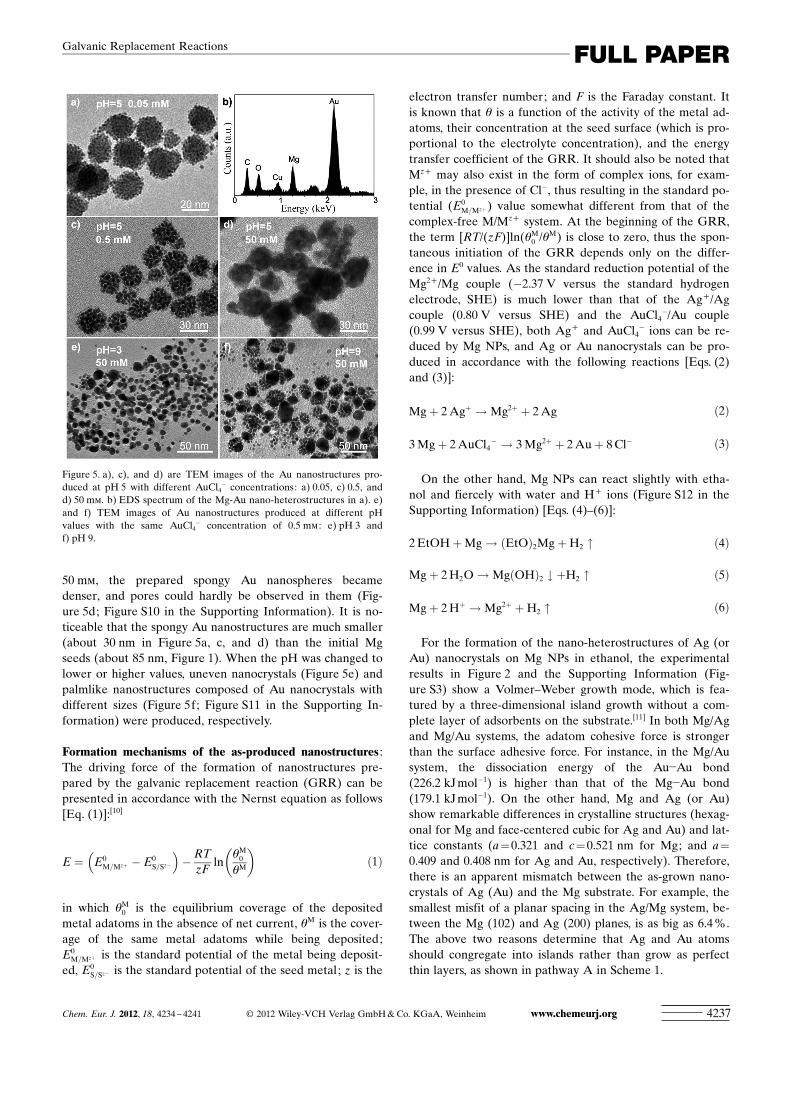
50 mm, the prepared spongy Au nanospheres becamedenser, and pores could hardly be observed in them (Fig-ure 5d; Figure S10 in the Supporting Information). It is no-ticeable that the spongy Au nanostructures are much smaller(about 30 nm in Figure 5a, c, and d) than the initial Mgseeds (about 85 nm, Figure 1). When the pH was changed tolower or higher values, uneven nanocrystals (Figure 5e) andpalmlike nanostructures composed of Au nanocrystals withdifferent sizes (Figure 5f; Figure S11 in the Supporting In-formation) were produced, respectively.
Formation mechanisms of the as-produced nanostructures :The driving force of the formation of nanostructures pre-pared by the galvanic replacement reaction (GRR) can bepresented in accordance with the Nernst equation as follows[Eq. (1)]:[10]
E ¼ E0M=Mzþ � E0
S=Sz�
� �� RT
zFln
qM0
qM
� �ð1Þ
in which qM0 is the equilibrium coverage of the deposited
metal adatoms in the absence of net current, qM is the cover-age of the same metal adatoms while being deposited;E0
M=Mzþ is the standard potential of the metal being deposit-ed, E0
S=Sz� is the standard potential of the seed metal; z is the
electron transfer number; and F is the Faraday constant. Itis known that q is a function of the activity of the metal ad-ACHTUNGTRENNUNGatoms, their concentration at the seed surface (which is pro-portional to the electrolyte concentration), and the energytransfer coefficient of the GRR. It should also be noted thatMz+ may also exist in the form of complex ions, for exam-ple, in the presence of Cl�, thus resulting in the standard po-tential (E0
M=Mzþ) value somewhat different from that of thecomplex-free M/Mz+ system. At the beginning of the GRR,the term [RT/(zF)]ln(qM
0 /qM) is close to zero, thus the spon-taneous initiation of the GRR depends only on the differ-ence in E0 values. As the standard reduction potential of theMg2+/Mg couple (�2.37 V versus the standard hydrogenelectrode, SHE) is much lower than that of the Ag+/Agcouple (0.80 V versus SHE) and the AuCl4
�/Au couple(0.99 V versus SHE), both Ag+ and AuCl4
� ions can be re-duced by Mg NPs, and Ag or Au nanocrystals can be pro-duced in accordance with the following reactions [Eqs. (2)and (3)]:
Mgþ 2 Agþ !Mg2þ þ 2 Ag ð2Þ
3Mgþ 2 AuCl4� ! 3Mg2þ þ 2 Auþ 8 Cl� ð3Þ
On the other hand, Mg NPs can react slightly with etha-nol and fiercely with water and H+ ions (Figure S12 in theSupporting Information) [Eqs. (4)–(6)]:
2 EtOHþMg! ðEtOÞ2MgþH2 " ð4Þ
Mgþ 2 H2O!MgðOHÞ2 # þH2 " ð5Þ
Mgþ 2 Hþ !Mg2þ þH2 " ð6Þ
For the formation of the nano-heterostructures of Ag (orAu) nanocrystals on Mg NPs in ethanol, the experimentalresults in Figure 2 and the Supporting Information (Fig-ure S3) show a Volmer–Weber growth mode, which is fea-tured by a three-dimensional island growth without a com-plete layer of adsorbents on the substrate.[11] In both Mg/Agand Mg/Au systems, the adatom cohesive force is strongerthan the surface adhesive force. For instance, in the Mg/Ausystem, the dissociation energy of the Au�Au bond(226.2 kJ mol�1) is higher than that of the Mg�Au bond(179.1 kJ mol�1). On the other hand, Mg and Ag (or Au)show remarkable differences in crystalline structures (hexag-onal for Mg and face-centered cubic for Ag and Au) and lat-tice constants (a=0.321 and c= 0.521 nm for Mg; and a=
0.409 and 0.408 nm for Ag and Au, respectively). Therefore,there is an apparent mismatch between the as-grown nano-crystals of Ag (Au) and the Mg substrate. For example, thesmallest misfit of a planar spacing in the Ag/Mg system, be-tween the Mg (102) and Ag (200) planes, is as big as 6.4 %.The above two reasons determine that Ag and Au atomsshould congregate into islands rather than grow as perfectthin layers, as shown in pathway A in Scheme 1.
Figure 5. a), c), and d) are TEM images of the Au nanostructures pro-duced at pH 5 with different AuCl4
� concentrations: a) 0.05, c) 0.5, andd) 50 mm. b) EDS spectrum of the Mg-Au nano-heterostructures in a). e)and f) TEM images of Au nanostructures produced at different pHvalues with the same AuCl4
� concentration of 0.5 mm : e) pH 3 andf) pH 9.
Chem. Eur. J. 2012, 18, 4234 – 4241 � 2012 Wiley-VCH Verlag GmbH & Co. KGaA, Weinheim www.chemeurj.org 4237
FULL PAPERGalvanic Replacement Reactions
On the other hand, once the noble-metal nuclei form onthe surface of Mg NPs, they could grow by reducing noble-metal ions with electrons released from the Mg core. Sincedissolved oxygen was maximally eliminated by bubbling thesolution with argon gas, surface oxidation of Mg NPs duringthe NGR reactions could be effectively suppressed. Howev-er, during storage, the surface of Mg NPs could be oxidizedinto a thin MgO layer (Figure S3d in the Supporting Infor-mation), which gave rise to the final structure of noble-metal nanoparticles loaded on Mg/MgO core–shell nano-structures. When such Mg@MgO-Au nanostructures wereheated at reflux in ethanol at 50 8C for 1 h, the Mg@MgOcore–shells were fully transformed into MgO hollow nano-particles through the Kirkendall voiding process,[8] while stillkeeping the Au nanoparticles on their surface (Figure S13 inthe Supporting Information).
As for the formation of the self-assembled Ag nanocrys-tals in an aqueous solution, it is likely a result of the compe-tition between the growth of Ag on the surface of Mg NPs,on Ag nuclei, and the erosion of Mg NPs. Ag nanocrystalscould grow on the surface of Mg NPs and/or Ag nucleithrough the NGR reaction, as they did in ethanol. At thesame time, the surface of the Mg NPs was badly etched bysolvents following Equations (5) and (6). Thus, the as-grownAg nanocrystals lost their support and then fell into theliquid medium, as shown in pathway B, Scheme 1.
At low ion concentrations, the Ag nanocrystals grew moreslowly and finally stopped as the erosion of their seed pro-ceeded relatively fast. Hence, ultrafine and homogeneousAg nanocrystals were obtained, which subsequently movedto the water/hexane interface (to lower their surface energy)and self-assembled into arrays owing to their homogeneoussize, as shown in pathway B in Scheme 1. Under high ionconcentrations, the growth rate of particles was clearly en-
hanced, whereas the erosion rate was expected not tochange much. Therefore, some Ag nanocrystals grew biggerbefore separating from their Mg substrate. As a result, Agnanocrystals in the product were not uniform in size anymore (Figure 4c, d). When the pH was changed to be moreacidic or alkaline, the erosion rate increased drastically ac-cording to Equation (6). Therefore, Ag nanocrystals couldbe separated from the sacrificial Mg seeds much faster, thusleading to the formation of smaller Ag nanocrystals (seeFigure S8 in the Supporting Information).
As for the NGR reaction in the Mg/AuCl4� system in
water, we attribute the formation of spongy Au nanostruc-tures to a stronger bonding of Au nanocrystals with Mgseeds. As the Mg particles eroded and shrank in size, the Aunanocrystals kept growing, being well attached to their sur-face (see Figure 5a). This led to an increase in the densityand size of the Au nanocrystals on the Mg core surface. Fi-nally, the Mg cores were completely etched out, and the Aunanocrystals joined with each other to form a spongy struc-ture, as shown in pathway C, Scheme 1. Due to the weightloss of the Mg core caused by water erosion, the obtainedspongy Au nanostructures are about three times smallerthan the initial Mg seeds.
Similar to the case of Ag nanocrystals produced in theMg/Ag+ system in water, the final morphology of the Aunanostructures could be modified by adjusting the AuCl4
�
ion concentration and the pH of the aqueous solution.When the ion concentration was elevated, the growth rateincreased, hence more Mg atoms were consumed by theNGR reaction to reduce Au nanocrystals, while the erosionrate was mainly unchanged. This led to denser (less porous)Au nanospheres (Figure 5d). However, when the erosionrate of the Mg core was enhanced by changing the pH, theAu nanocrystals could not bind tightly with the Mg core,giving rise to separate Au nanocrystals (Figure 5e, f).
Products in other NGR reaction systems : We tested theabove-proposed mechanisms in other systems with substitut-ed oxidant ions or sacrificial metal seeds.
First, we chose Cu2+ ions as an oxidant to react with MgNPs and found that Mg@MgO-Cu heterostructures wereproduced in ethanol (Figure 6a; Figure S14a–d in the Sup-porting Information). After having been left at reflux in thesame liquid, they turned into MgO hollow nanoparticlesloaded with Cu nanocrystals (Figure 6b; Figure S14e in theSupporting Information). Spectrum evolution during thereflux process demonstrated the disappearance of an Mg-re-lated signal (410 nm) and appearances of MgO (270 nm[8b])and Cu LSPR (�550 nm[12]) signals (Figure 6c). When theprocess was modified and an aqueous Cu2+ solution wasadded to the ethanol-dispersed Mg NPs, stripelike arrays ofCu nanocrystals were obtained, as seen in Figure 6d–f andthe Supporting Information (Figure S14f).
Second, we used Sn2+ ions as an oxidant to react with Mgseeds. Since Sn is also an active metal, SnO2 NPs were di-rectly produced when Mg NPs reacted with Sn2+ ions inwater. The TEM image in Figure 7a illustrates that the size
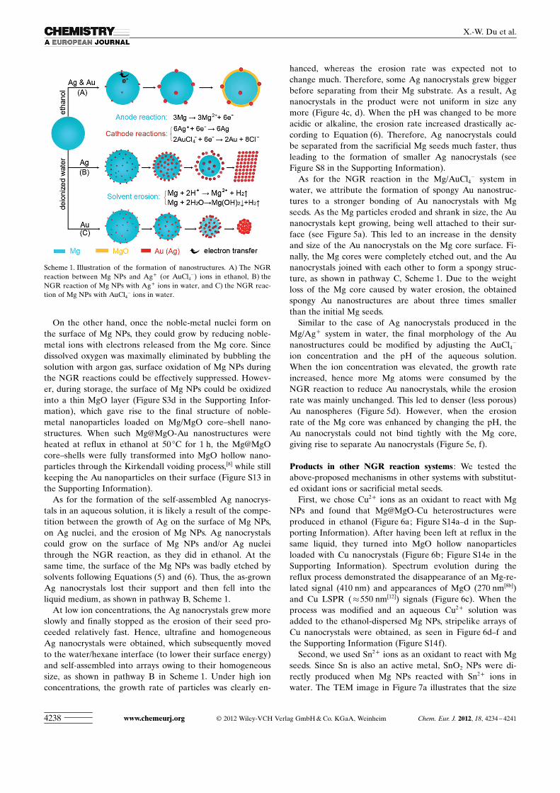
Scheme 1. Illustration of the formation of nanostructures. A) The NGRreaction between Mg NPs and Ag+ (or AuCl4
�) ions in ethanol, B) theNGR reaction of Mg NPs with Ag+ ions in water, and C) the NGR reac-tion of Mg NPs with AuCl4
� ions in water.
www.chemeurj.org � 2012 Wiley-VCH Verlag GmbH & Co. KGaA, Weinheim Chem. Eur. J. 2012, 18, 4234 – 42414238
X.-W. Du et al.
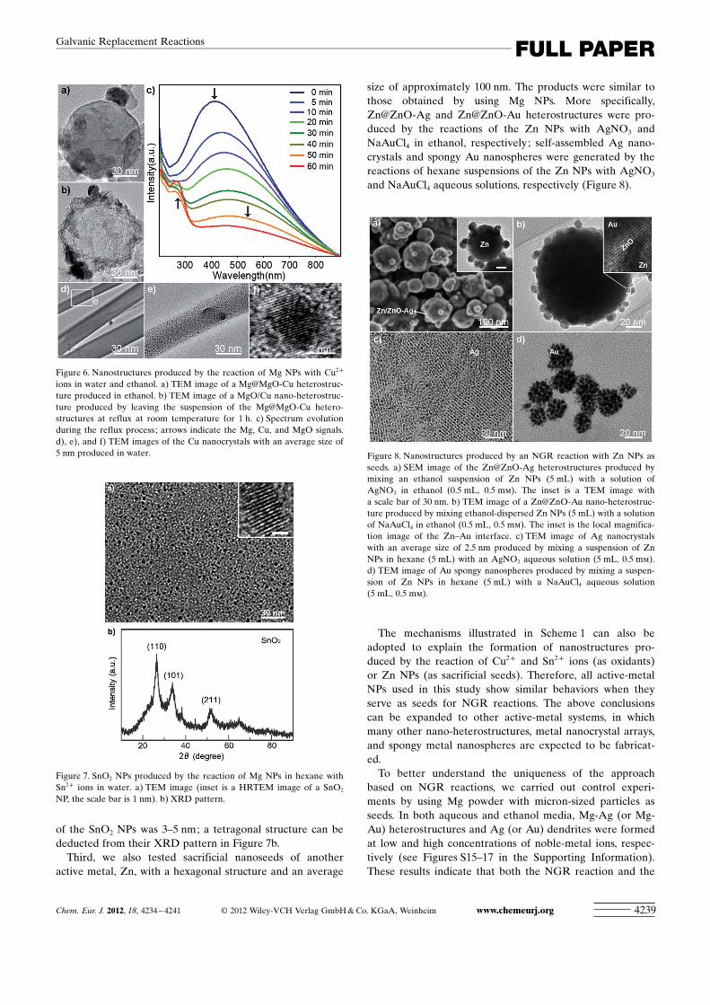
of the SnO2 NPs was 3–5 nm; a tetragonal structure can bededucted from their XRD pattern in Figure 7b.
Third, we also tested sacrificial nanoseeds of anotheractive metal, Zn, with a hexagonal structure and an average
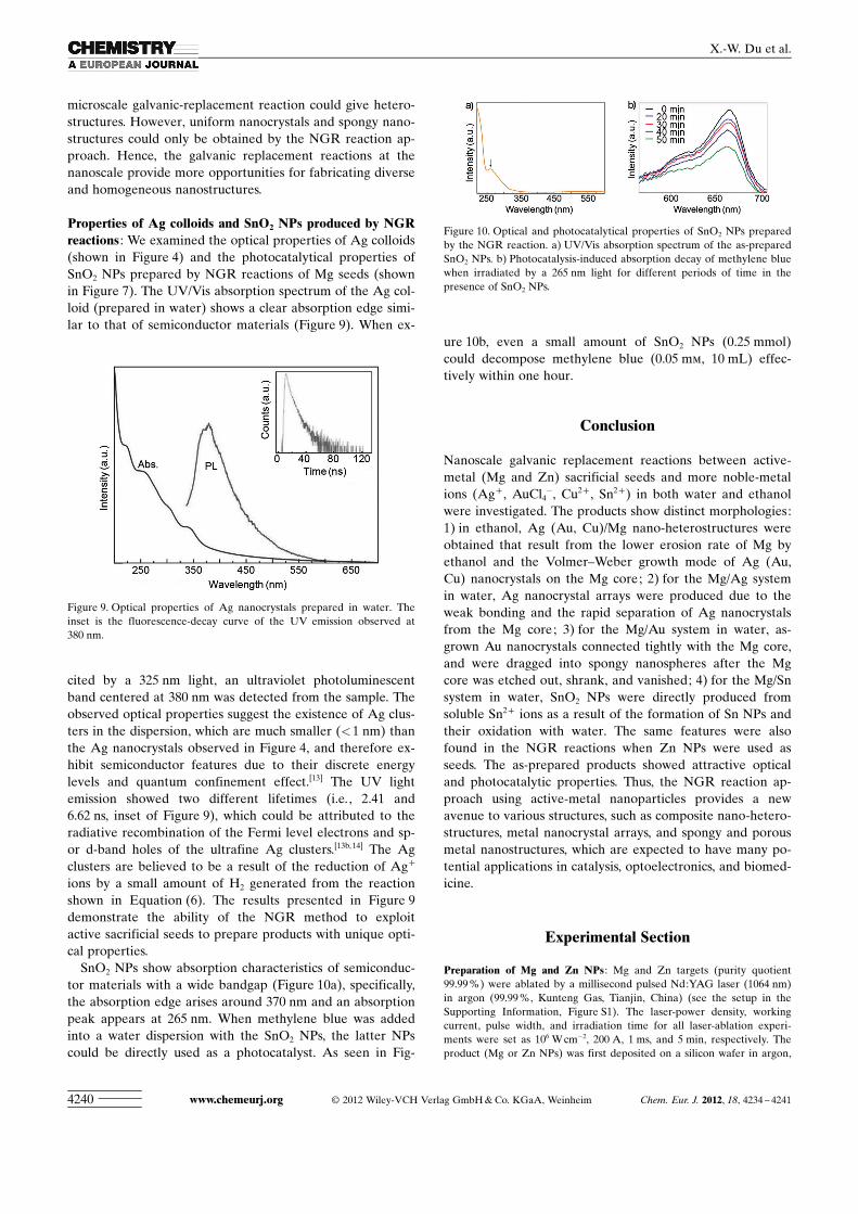
size of approximately 100 nm. The products were similar tothose obtained by using Mg NPs. More specifically,Zn@ZnO-Ag and Zn@ZnO-Au heterostructures were pro-duced by the reactions of the Zn NPs with AgNO3 andNaAuCl4 in ethanol, respectively; self-assembled Ag nano-crystals and spongy Au nanospheres were generated by thereactions of hexane suspensions of the Zn NPs with AgNO3
and NaAuCl4 aqueous solutions, respectively (Figure 8).
The mechanisms illustrated in Scheme 1 can also beadopted to explain the formation of nanostructures pro-duced by the reaction of Cu2+ and Sn2+ ions (as oxidants)or Zn NPs (as sacrificial seeds). Therefore, all active-metalNPs used in this study show similar behaviors when theyserve as seeds for NGR reactions. The above conclusionscan be expanded to other active-metal systems, in whichmany other nano-heterostructures, metal nanocrystal arrays,and spongy metal nanospheres are expected to be fabricat-ed.
To better understand the uniqueness of the approachbased on NGR reactions, we carried out control experi-ments by using Mg powder with micron-sized particles asseeds. In both aqueous and ethanol media, Mg-Ag (or Mg-Au) heterostructures and Ag (or Au) dendrites were formedat low and high concentrations of noble-metal ions, respec-tively (see Figures S15–17 in the Supporting Information).These results indicate that both the NGR reaction and the
Figure 6. Nanostructures produced by the reaction of Mg NPs with Cu2+
ions in water and ethanol. a) TEM image of a Mg@MgO-Cu heterostruc-ture produced in ethanol. b) TEM image of a MgO/Cu nano-heterostruc-ture produced by leaving the suspension of the Mg@MgO-Cu hetero-structures at reflux at room temperature for 1 h. c) Spectrum evolutionduring the reflux process; arrows indicate the Mg, Cu, and MgO signals.d), e), and f) TEM images of the Cu nanocrystals with an average size of5 nm produced in water.
Figure 7. SnO2 NPs produced by the reaction of Mg NPs in hexane withSn2+ ions in water. a) TEM image (inset is a HRTEM image of a SnO2
NP, the scale bar is 1 nm). b) XRD pattern.
Figure 8. Nanostructures produced by an NGR reaction with Zn NPs asseeds. a) SEM image of the Zn@ZnO-Ag heterostructures produced bymixing an ethanol suspension of Zn NPs (5 mL) with a solution ofAgNO3 in ethanol (0.5 mL, 0.5 mm). The inset is a TEM image witha scale bar of 30 nm. b) TEM image of a Zn@ZnO-Au nano-heterostruc-ture produced by mixing ethanol-dispersed Zn NPs (5 mL) with a solutionof NaAuCl4 in ethanol (0.5 mL, 0.5 mm). The inset is the local magnifica-tion image of the Zn–Au interface. c) TEM image of Ag nanocrystalswith an average size of 2.5 nm produced by mixing a suspension of ZnNPs in hexane (5 mL) with an AgNO3 aqueous solution (5 mL, 0.5 mm).d) TEM image of Au spongy nanospheres produced by mixing a suspen-sion of Zn NPs in hexane (5 mL) with a NaAuCl4 aqueous solution(5 mL, 0.5 mm).
Chem. Eur. J. 2012, 18, 4234 – 4241 � 2012 Wiley-VCH Verlag GmbH & Co. KGaA, Weinheim www.chemeurj.org 4239
FULL PAPERGalvanic Replacement Reactions
microscale galvanic-replacement reaction could give hetero-structures. However, uniform nanocrystals and spongy nano-structures could only be obtained by the NGR reaction ap-proach. Hence, the galvanic replacement reactions at thenanoscale provide more opportunities for fabricating diverseand homogeneous nanostructures.
Properties of Ag colloids and SnO2 NPs produced by NGRreactions : We examined the optical properties of Ag colloids(shown in Figure 4) and the photocatalytical properties ofSnO2 NPs prepared by NGR reactions of Mg seeds (shownin Figure 7). The UV/Vis absorption spectrum of the Ag col-loid (prepared in water) shows a clear absorption edge simi-lar to that of semiconductor materials (Figure 9). When ex-
cited by a 325 nm light, an ultraviolet photoluminescentband centered at 380 nm was detected from the sample. Theobserved optical properties suggest the existence of Ag clus-ters in the dispersion, which are much smaller (<1 nm) thanthe Ag nanocrystals observed in Figure 4, and therefore ex-hibit semiconductor features due to their discrete energylevels and quantum confinement effect.[13] The UV lightemission showed two different lifetimes (i.e., 2.41 and6.62 ns, inset of Figure 9), which could be attributed to theradiative recombination of the Fermi level electrons and sp-or d-band holes of the ultrafine Ag clusters.[13b, 14] The Agclusters are believed to be a result of the reduction of Ag+
ions by a small amount of H2 generated from the reactionshown in Equation (6). The results presented in Figure 9demonstrate the ability of the NGR method to exploitactive sacrificial seeds to prepare products with unique opti-cal properties.
SnO2 NPs show absorption characteristics of semiconduc-tor materials with a wide bandgap (Figure 10a), specifically,the absorption edge arises around 370 nm and an absorptionpeak appears at 265 nm. When methylene blue was addedinto a water dispersion with the SnO2 NPs, the latter NPscould be directly used as a photocatalyst. As seen in Fig-
ure 10b, even a small amount of SnO2 NPs (0.25 mmol)could decompose methylene blue (0.05 mm, 10 mL) effec-tively within one hour.
Conclusion
Nanoscale galvanic replacement reactions between active-metal (Mg and Zn) sacrificial seeds and more noble-metalions (Ag+ , AuCl4
�, Cu2+ , Sn2+) in both water and ethanolwere investigated. The products show distinct morphologies:1) in ethanol, Ag (Au, Cu)/Mg nano-heterostructures wereobtained that result from the lower erosion rate of Mg byethanol and the Volmer–Weber growth mode of Ag (Au,Cu) nanocrystals on the Mg core; 2) for the Mg/Ag systemin water, Ag nanocrystal arrays were produced due to theweak bonding and the rapid separation of Ag nanocrystalsfrom the Mg core; 3) for the Mg/Au system in water, as-grown Au nanocrystals connected tightly with the Mg core,and were dragged into spongy nanospheres after the Mgcore was etched out, shrank, and vanished; 4) for the Mg/Snsystem in water, SnO2 NPs were directly produced fromsoluble Sn2+ ions as a result of the formation of Sn NPs andtheir oxidation with water. The same features were alsofound in the NGR reactions when Zn NPs were used asseeds. The as-prepared products showed attractive opticaland photocatalytic properties. Thus, the NGR reaction ap-proach using active-metal nanoparticles provides a newavenue to various structures, such as composite nano-hetero-structures, metal nanocrystal arrays, and spongy and porousmetal nanostructures, which are expected to have many po-tential applications in catalysis, optoelectronics, and biomed-icine.
Experimental Section
Preparation of Mg and Zn NPs : Mg and Zn targets (purity quotient99.99 %) were ablated by a millisecond pulsed Nd:YAG laser (1064 nm)in argon (99.99 %, Kunteng Gas, Tianjin, China) (see the setup in theSupporting Information, Figure S1). The laser-power density, workingcurrent, pulse width, and irradiation time for all laser-ablation experi-ments were set as 106 W cm�2, 200 A, 1 ms, and 5 min, respectively. Theproduct (Mg or Zn NPs) was first deposited on a silicon wafer in argon,
Figure 9. Optical properties of Ag nanocrystals prepared in water. Theinset is the fluorescence-decay curve of the UV emission observed at380 nm.
Figure 10. Optical and photocatalytical properties of SnO2 NPs preparedby the NGR reaction. a) UV/Vis absorption spectrum of the as-preparedSnO2 NPs. b) Photocatalysis-induced absorption decay of methylene bluewhen irradiated by a 265 nm light for different periods of time in thepresence of SnO2 NPs.
www.chemeurj.org � 2012 Wiley-VCH Verlag GmbH & Co. KGaA, Weinheim Chem. Eur. J. 2012, 18, 4234 – 42414240
X.-W. Du et al.
and then dispersed in pure ethanol or hexane for further experiments.The concentration of metal NPs in the dispersions was adjusted to 25 mm
by measuring the weight increase of the silicon wafer and selecting thevolume of solvent.
NGR reactions of Mg and Zn NPs with AgNO3, NaAuCl4, andCu(OOCCH3)2 solutions : For the reactions in ethanol, a 5 mL aliquot ofthe above-mentioned ethanol dispersion of Mg (or Zn) NPs was removedand stirred by a magnetic stirrer for 5 min, after which different amountsof solutions of Cu(OOCCH3)2 (0.5 mm), AgNO3 (0.5 mm), or NaAuCl4
(0.05 mm or 0.5 mm) in ethanol were added dropwise. After stirring for5 min at approximately 30 8C, samples of the dispersions were removedfor the measurement of absorption spectra, and the products were putonto TEM copper grids for TEM and SEM observations. For the reac-tions in deionized water, aliquots (5 mL) of the dispersion of the Mg (orZn) NPs in hexane were removed and stirred by using a magnetic stirrerfor 5 min, after which 0.5 mm AgNO3 (or 0.5 mm NaAuCl4) aqueous solu-tions (5 mL) with different concentrations (i.e., 0.05, 0.5, 5, 50 mm) or0.5 mm Cu(OOCCH3)2 were added dropwise. The mixtures were stirredfor another 5 min at approximately 30 8C.
In a typical procedure of the replacement reactions between Mg micro-powder and Ag+ (or AuCl4
�), Mg powder (30 mg, 99.99 %, Keruisi, Tian-jin, China) with a particle size of about one micron was dispersed in etha-nol (or deionized water; 20 mL in either case) under magnetic stirring,and then solutions of AgNO3 (0.5 or 5 mm) or NaAuCl4 (0.5 or 5 mm) inethanol (or water; 10 mL in either case) were added dropwise into thesuspensions. After reacting for 30 min, the products were centrifuged,washed with ethanol, and then dried for TEM and SEM observations.
NGR reactions of Mg NPs with SnCl2 in water : An aliquot (10 mL) ofMg NP dispersion in hexane was stirred magnetically for 5 min, afterwhich 0.5 mm SnCl2 aqueous solution (10 mL) was added dropwise andstirred for another 5 min at approximately 30 8C. Then methylene blue(5 mmol) was added into the resultant mixture, which was irradiated bya 265 nm light for different periods of time. The liquid was removed di-rectly for UV/Vis absorption measurements after photoirradiation.
Measurements and analysis : The morphology and structure were deter-mined by a Hitachi S-4800 scanning electron microscope and FEI Tech-nai G2 F20 transmission electron microscope equipped with a field-mis-sion gun and an EDS unit. Absorption spectra were recorded in a HitachiU-3010 UV/Vis spectrometer.
Acknowledgements
This work was supported by the Natural Science Foundation of China(nos. 51171127, 50972102, 50902103), and Natural Science Foundation ofTianjin City (nos. 09JCZDJC22600 and 08JCYBJC02900).
[1] Y. G. Sun, B. T. Mayers, Y. N. Xia, Nano Lett. 2002, 2, 481 – 485.[2] a) S. E. Skrabalak, J. Chen, L. Au, X. Lu, X. Li; Y. N. Xia, Adv.
Mater. 2007, 19, 3177 –3184; Y. N. Xia, Adv. Mater. 2007, 19, 3177 –3184; b) X. Lu, J. Chen, S. E. Skrabalak, Y. Xia, Proc. Inst. Mech.Eng., Part N: J. Nanoeng. Nanosyst. 2008, 221, 1 –16; c) Y. D. Yin,C. Erdonmez, S. Aloni, A. P. Alivisatos, J. Am. Chem. Soc. 2006,
128, 12671 – 12673; d) D. Seo, H. Song, J. Am. Chem. Soc. 2009, 131,18210 – 18211.
[3] a) L. Au, X. M. Lu, Y. N. Xia, Adv. Mater. 2008, 20, 2517 –2522;b) S. E. Skrabalak, L. Au, X. D. Li, Y. Xia, Nat. Protoc. 2007, 2,2182 – 2190; c) J. Y. Chen, J. M. McLellan, A. Siekkinen, Y. J. Xiong,Z. Y. Li, Y. N. Xia, J. Am. Chem. Soc. 2006, 128, 14776 –14777;d) Y. G. Sun, Y. N. Xia, J. Am. Chem. Soc. 2004, 126, 3892 –3901.
[4] a) J. Zeng, Q. Zhang, J. Y. Chen, Y. N. Xia, Nano Lett. 2010, 10, 30 –35; b) J. Y. Chen, C. Glaus, R. Laforest, Q. Zhang, M. X. Yang, M.Gidding, M. J. Welch, Y. N. Xia, Small 2010, 6, 811 –817; c) J. Chen,F. Saeki, B. J. Wiley, H. Cang, M. J. Cobb, Z. Y. Li, L. Au, H. Zhang,M. B. Kimmey, X. D. Li; Y. Xia, Nano Lett. 2005, 5, 473 – 477; Y.Xia, Nano Lett. 2005, 5, 473 – 477; d) Y. G. Sun, B. Mayers, Y. N. Xia,Adv. Mater. 2003, 15, 641 –646; e) S. E. Skrabalak, J. Y. Chen, Y. G.Sun, X. M. Lu, L. Au, C. M. Cobley, Y. N. Xia, Acc. Chem. Res.2008, 41, 1587 –1595.
[5] a) Y. Lu, C. Shi, M. J. Hu, Y. J. Xu, L. Yu, L. P. Wen, Y. Zhao, W. PXu, S. H. Yu, Adv. Funct. Mater. 2010, 20, 3701 –3706; b) Y. Lu, Y.Zhao, L. Yu, L. Dong, C. Shi, M. J. Hu, Y. J. Xu, L. P. Wen, S. H.Yu, Adv. Mater. 2010, 22, 1407 –1411; c) M. Mohl, A. Kumar,A. L. M. Reddy, A. Kukovecz, Konya, Z. I. Kiricsi, R. Vajtai, P. M.Ajayan, J. Phys. Chem. C 2010, 114, 389 –393; d) I. Najdovski, A. P.O�Mullane, S. K. Bhargava, Electrochem. Commun. 2010, 12, 1535 –1538.
[6] a) Q. B. Zhang, J. P. Xie, J. Y. Lee, J. X. Zhang, C. Boothroyd, Small2008, 4, 1067 – 1071; b) L. Au, Y. C. Chen, F. Zhou, P. H. C. Camar-go, B. Lim, Z. Y. Li, D. S. Ginger, Y. N. Xia, Nano Res. 2008, 1, 441 –449.
[7] a) B. Yoon, H. Hakkinen, U. Landman, A. S. Worz, J. M. Antonietti,S. Abbet, K. Judai, U. Heiz, Science 2005, 307, 403 – 407; b) C. R.Henry, Prog. Surf. Sci. 2005, 80, 92–116; c) H. B. Zeng, W. P. Cai,P. S. Liu, H. J. Zhou, C. Klingshirn, H. Kalt, ACS Nano 2008, 2,1661 – 1670; d) H. B. Zeng, P. S. Liu, W. P. Cai, S. K. Yang, X. X. Xu,J. Phys. Chem. C 2008, 112, 19620 – 19624.
[8] a) K. Y. Niu, J. Yang, S. A. Kulinich, J. Sun, H. Li, X. W. Du, J. Am.Chem. Soc. 2010, 132, 9814 – 9819; b) K. Y. Niu, J. Yang, J. Sun,X. W. Du, Nanotechnology 2010, 21, 295 604.
[9] a) S. Link, M. A. EI-Sayed, J. Phys. Chem. B 1999, 103, 4212 –4217;b) P. K. Jain, K. S. Lee, I. H. EI-Sayed, M. A. EI-Sayed, J. Phys.Chem. B 2006, 110, 7238 – 7248.
[10] C. H. Hamann, A. Hamnett, W. Vielstich, Electrochemistry, chap-ter 4, Wiley-VCH, Weinheim, 1998.
[11] a) F. R. Fan, D. Y. Liu, Y. F. Wu, S. Duan, Z. X. Xie, Z. Y. Jiang,Z. Q. Tian, J. Am. Chem. Soc. 2008, 130, 6949 – 6951; b) K. Oura,V. G. Lifshits, A. A. Saranin, A. V. Zotov, M. Katayama, Surface Sci-ence: An Introduction, Springer, Berlin, Heidelberg, 2003.
[12] N. A. Dhas, C. P. Raj, A. Gedanken, Chem. Mater. 1998, 10, 1446 –1452.
[13] a) L. Maretti, P. S. Billone, Y. J. C. Liu, Scaiano, J. Am. Chem. Soc.2009, 131, 13972 –13980; b) W. T. Wu, T. Zhou, S. Q. Zhou, Chem.Mater. 2009, 21, 2851 –2861.
[14] a) O. A. Yeshchenko, I. M. Dmitruk, A. A. Alexeenko, M. Yu. Lo-sytskyy, A. V. Kotko, A. O. Pinchuk, Phys. Rev. B 2009, 79, 235438;b) I. D�ez, R. H. A. Ras, Nanoscale 2011, 3, 1963 –1970.
Received: August 16, 2011Published online: February 28, 2012
Chem. Eur. J. 2012, 18, 4234 – 4241 � 2012 Wiley-VCH Verlag GmbH & Co. KGaA, Weinheim www.chemeurj.org 4241
FULL PAPERGalvanic Replacement Reactions
Related Documents


![[PPT]Electrochemistry - Berkeley City College · Web viewElectrochemistry 18.1 Balancing Oxidation–Reduction Reactions 18.2 Galvanic Cells 18.3 Standard Reduction Potentials 18.4](https://static.cupdf.com/doc/110x72/5ac5bcc87f8b9a12608dc1dd/pptelectrochemistry-berkeley-city-viewelectrochemistry-181-balancing-oxidationreduction.jpg)