HAL Id: pasteur-01721100 https://hal-pasteur.archives-ouvertes.fr/pasteur-01721100 Submitted on 1 Mar 2018 HAL is a multi-disciplinary open access archive for the deposit and dissemination of sci- entific research documents, whether they are pub- lished or not. The documents may come from teaching and research institutions in France or abroad, or from public or private research centers. L’archive ouverte pluridisciplinaire HAL, est destinée au dépôt et à la diffusion de documents scientifiques de niveau recherche, publiés ou non, émanant des établissements d’enseignement et de recherche français ou étrangers, des laboratoires publics ou privés. Distributed under a Creative Commons Attribution| 4.0 International License Galactosaminogalactan, a New Immunosuppressive Polysaccharide of Aspergillus fumigatus Thierry Fontaine, Aurélie Delangle, Catherine Simenel, Bernadette Coddeville, Sandra J. van Vliet, Yvette van Kooyk, Silvia Bozza, Silvia Moretti, Flavio Schwarz, Coline Trichot, et al. To cite this version: Thierry Fontaine, Aurélie Delangle, Catherine Simenel, Bernadette Coddeville, Sandra J. van Vliet, et al.. Galactosaminogalactan, a New Immunosuppressive Polysaccharide of Aspergillus fumigatus. PLoS Pathogens, Public Library of Science, 2011, 7 (11), pp.e1002372. 10.1371/journal.ppat.1002372. pasteur-01721100

Welcome message from author
This document is posted to help you gain knowledge. Please leave a comment to let me know what you think about it! Share it to your friends and learn new things together.
Transcript

HAL Id: pasteur-01721100https://hal-pasteur.archives-ouvertes.fr/pasteur-01721100
Submitted on 1 Mar 2018
HAL is a multi-disciplinary open accessarchive for the deposit and dissemination of sci-entific research documents, whether they are pub-lished or not. The documents may come fromteaching and research institutions in France orabroad, or from public or private research centers.
L’archive ouverte pluridisciplinaire HAL, estdestinée au dépôt et à la diffusion de documentsscientifiques de niveau recherche, publiés ou non,émanant des établissements d’enseignement et derecherche français ou étrangers, des laboratoirespublics ou privés.
Distributed under a Creative Commons Attribution| 4.0 International License
Galactosaminogalactan, a New ImmunosuppressivePolysaccharide of Aspergillus fumigatus
Thierry Fontaine, Aurélie Delangle, Catherine Simenel, BernadetteCoddeville, Sandra J. van Vliet, Yvette van Kooyk, Silvia Bozza, Silvia
Moretti, Flavio Schwarz, Coline Trichot, et al.
To cite this version:Thierry Fontaine, Aurélie Delangle, Catherine Simenel, Bernadette Coddeville, Sandra J. van Vliet,et al.. Galactosaminogalactan, a New Immunosuppressive Polysaccharide of Aspergillus fumigatus.PLoS Pathogens, Public Library of Science, 2011, 7 (11), pp.e1002372. �10.1371/journal.ppat.1002372�.�pasteur-01721100�

Galactosaminogalactan, a New ImmunosuppressivePolysaccharide of Aspergillus fumigatusThierry Fontaine1.*, Aurelie Delangle1.¤, Catherine Simenel2, Bernadette Coddeville3, Sandra J. van
Vliet4, Yvette van Kooyk4, Silvia Bozza5, Silvia Moretti5, Flavio Schwarz6, Coline Trichot7, Markus Aebi6,
Muriel Delepierre2, Carole Elbim7, Luigina Romani5, Jean-Paul Latge1
1 Unite des Aspergillus, Institut Pasteur, Paris, France, 2 Unite de Resonance Magnetique Nucleaire des Biomolecules, CNRS URA 2185, Institut Pasteur, Paris, France,
3 Laboratoire de Glycobiologie Structurale et Fonctionnelle, UMR 8576 CNRS, Universite des sciences et Technologies de Lille Flandres-Artois, Villeneuve d’Ascq, France,
4 Department of Molecular Cell Biology and Immunology, VU University Medical Center, Amsterdam, The Netherlands, 5 Department of Experimental Medicine and
Biochemical Sciences, University of Perugia, Perugia, Italy, 6 Institute of Microbiology, ETH Honggerberg, Zurich, Switzerland, 7 Universite Pierre et Marie Curie – Paris 6,
UMR-S 945 Immunite et Infection, Faculte de Medecine Pitie Salpetriere, Paris, France
Abstract
A new polysaccharide secreted by the human opportunistic fungal pathogen Aspergillus fumigatus has been characterized.Carbohydrate analysis using specific chemical degradations, mass spectrometry, 1H and 13C nuclear magnetic resonanceshowed that this polysaccharide is a linear heterogeneous galactosaminogalactan composed of a1-4 linked galactose anda1-4 linked N-acetylgalactosamine residues where both monosacharides are randomly distributed and where thepercentage of galactose per chain varied from 15 to 60%. This polysaccharide is antigenic and is recognized by a majority ofthe human population irrespectively of the occurrence of an Aspergillus infection. GalNAc oligosaccharides are an essentialepitope of the galactosaminogalactan that explains the universal antibody reaction due to cross reactivity with otherantigenic molecules containing GalNAc stretches such as the N-glycans of Campylobacter jejuni. The galactosaminogalactanhas no protective effect during Aspergillus infections. Most importantly, the polysaccharide promotes fungal development inimmunocompetent mice due to its immunosuppressive activity associated with disminished neutrophil infiltrates.
Citation: Fontaine T, Delangle A, Simenel C, Coddeville B, van Vliet SJ, et al. (2011) Galactosaminogalactan, a New Immunosuppressive Polysaccharide ofAspergillus fumigatus. PLoS Pathog 7(11): e1002372. doi:10.1371/journal.ppat.1002372
Editor: Marta Feldmesser, Albert Einstein College of Medicine, United States of America
Received June 7, 2011; Accepted September 27, 2011; Published November 10, 2011
Copyright: � 2011 Fontaine et al. This is an open-access article distributed under the terms of the Creative Commons Attribution License, which permitsunrestricted use, distribution, and reproduction in any medium, provided the original author and source are credited.
Funding: This work was partly supported by a grant from Agence Nationale de la Recherche (ANR-06-EMPB-011-01) and by European grants Allfun, Fungwall,ANRpathogenomics. The funders had no role in study design, data collection and analysis, decision to publish, or preparation of the manuscript.
Competing Interests: The authors have declared that no competing interests exist.
* E-mail: [email protected]
¤ Current address: GlaxoSmithKline, Genval, Belgium
. These authors contributed equally to this work.
Introduction
Aspergillus fumigatus is an opportunistic human fungal pathogen
that causes a wide range of diseases including allergic reactions
and local or systemic infections such as invasive pulmonary
aspergillosis (IA) that has emerged in recent years as a leading
cause of infection-related mortality among immunocompromised
patients [1,2]. The innate immune system provides the first line of
defense against A. fumigatus with macrophages and neutrophils that
sense, phagocytose and kill conidia and hyphae through the
production of anti-microbial agents. Later, antigen presenting cells
initiate an adaptative response activating various populations of T-
helper cells that impact differently on the evolution of the disease
[3,4]. Because of its external localisation, and specific composition,
the cell wall represents a specific target for recognition and specific
interaction with the host immune cells. The cell wall of A. fumigatus
is mainly composed of branched b1-3glucans, a1-3glucans, chitin,
b1-3/1-4 glucan and galactomannan [5]. These constitutive
polysaccharides have been shown to induce specific immune
responses from the host. For example in murine models
of aspergillosis, a1-3glucan and b1-3glucan chains induce a
protective response through the activation of Th1 and Th17 or
Treg responses [4] whereas galactomannan favours the disease
through the activation of the Th2/Th17 response. In other
medically important fungi, capsular and cell wall polysaccharides
and especially mannan and b-glucans also induce an immune
response that either favours or inhibits fungal infection [6,7,8,9].
During growth in vitro in aerial conditions or in vivo in the lung
tissues, the mycelium of A. fumigatus is covered by a polysaccharide-
rich extracellular matrix (ECM) that because of its outer position,
plays a major role in the interaction with the host immune cells
[10,11]. The ECM contains a1-3glucan and galactomannan that
are two of the major cell wall polysaccharides, recognised by T
cells. A third galactosamine-rich polysaccharide has been now
identified in the ECM. Although the presence of such cell wall
associated polysaccharide was noticed 20 years ago [12,13], its
structural analysis has not been investigated to date. The present
report shows that this polysaccharide is a linear heterogenous
chain constituted by a1-4 linked galactose and a1-4 linked N-
acetylgalactosamine residues. Most interestingly, the analysis of the
immune response towards this polysaccharide shows that it is
immunosuppressive and favors A. fumigatus infection.
PLoS Pathogens | www.plospathogens.org 1 November 2011 | Volume 7 | Issue 11 | e1002372

Results
A galactosaminogalactan is secreted by the mycelium ofA. fumigatus
The culture filtrate of A. fumigatus was precipitated by 70%
ethanol. In our experimental conditions, an amount of 80 mg of
ethanol precipitate was recovered per g of mycelial dry weight.
The incubation of the ethanol precipitate of the culture filtrate of
A. fumigatus for 1 h in a 150 mM NaCl aqueous solution resulted in
the solubilisation of glycoproteins and galactomannan. The NaCl-
insoluble material represented 43+/28% of the ethanol pre-
cipitate. The remaining insoluble material was separated in two
fractions based on their solubility in 8 M urea. The urea-soluble
material (SGG, urea soluble galactosaminogalactan) accounted for
30+/2 4% of the total ethanol precipitate whereas the urea-
insoluble material (PGG, urea insoluble galactosaminogalactan)
represented 13+/2 6% of the total ethanol precipitate. Gas liquid
chromatography (GC) analysis of both fractions showed that they
were exclusively composed of galactosamine and galactose with
ratios of 60/40 and 15/85 in SGG and PGG respectively. Ni-
trous deamination of native polysaccharide did not solubilise the
polysaccharide and did not produce anhydrotalose showing that
all galactosamine residues were N-acetylated (not shown). GC
analysis showed that the galactosaminogalactan was absent in
resting conidia but was present in the cell wall of mycelium from
both solid and liquid cultures and in different media (not shown).
Immunofluorescence with specific anti-GG mAb confirmed that
GG was not present on the surface of resting conidia. In contrast, a
positive detection was seen in the cell wall as soon as the coni-
dium germinates (Fig. 1). This result indicated that part of the
galactosaminogalactan was not secreted and remained strongly
associated with the cell wall. The amount of cell wall bound
galactosaminogalactan was equivalent to the amount recovered in
the culture medium (data not shown).
Structural analysis of GGGC analysis of permethylated GG revealed only two methyl
ethers: 2,3,6-tri-O-methyl-galactitol and 3,6-di-O-methyl-N-acet-
ylgalactosaminitol (Fig. S1), indicating the substitution in position
4 of both monosaccharides. The absence of methylether from non-
reducing end sugar or disubstituted monosaccharide indicated that
the galactosaminogalactan was an unbranched linear polysaccha-
ride. The apparent Mr estimated by gel filtration chromatography
after the carboxymethylation of the GG fraction was in agreement
with methylation data. The galactosaminogalactan was eluted as a
polydisperse homogenous polymer between 10 and 1000 kDa with
a median size of 100 kDa (Fig. S2). The 1D 1H and 2D 1H, 13C
nuclear magnetic resonance (NMR) spectra of carboxymethylated
GG fraction exhibited two main signals in the sugar anomeric
region at 5.003/103.07 and 5.287/99.07 ppm compatible with a-
anomers (Fig. S3). NMR data showed downfield shifts for the
carbone-4 of both sugar residues, indicating their 4-O substitution
and their pyranose configuration, which were in agreement with
the methylation data.
In order to elucidate the repartition of each monosaccharide on
the main polysaccharidic chain, two specific chemical degrada-
tions of both galactosaminogalactan fractions (PGG and SGG)
Author Summary
Aspergillus fumigatus is an opportunistic human fungalpathogen that causes a wide range of diseases includingallergic reactions and local or systemic infections such asinvasive pulmonary aspergillosis that has emerged in therecent years as a leading cause of infection relatedmortality among immunocompromised patients. Polysac-charides from the fungal cell wall play essential biologicalfunctions in the fungal cell biology and in host-pathogeninteractions. Indeed, it has been shown that polysaccha-rides can modulate the human immune response; some ofthem (b-glucan and a-glucans) having a protective effectagainst Aspergillus infection. We report here the purifica-tion and chemical characterization of a new antigenicpolysaccharide (galactosaminogalactan) produced by A.fumigatus. This polymer is secreted during infection. Inmurine models of aspergillosis, this galactosaminogalactanis not protective but it is immunosuppressive and favors A.fumigatus infection. Particularly it induces the apoptoticdeath of neutrophils that are the phagocytes playing anessential role in the killing of fungal pathogens.
Figure 1. Detection of the galactosaminogalactan by immunofluorescence on resting, germinated conidia and on mycelium. Thespecificity of the cell wall labelling was confirmed by the full inhibition of the labelling with the anti-GG MAb recognition seen after the MAb wasincubated with GalNAc oligosaccharides obtained by HCl hydrolysis galactosaminogalactan (last panel on the right).doi:10.1371/journal.ppat.1002372.g001
Galactosaminogalactan of Aspergillus fumigatus
PLoS Pathogens | www.plospathogens.org 2 November 2011 | Volume 7 | Issue 11 | e1002372

were undertaken: periodate oxidation that degraded 4-O-substi-
tuted galactose residues and N-de-acetylation/nitrous deamination
that degraded hexosamine residues. Solubilised fractions were
separated by gel filtration on HW40S column and chemically
analysed by methylation, GC-Mass spectrometry (GC-MS),
Matrix-assisted laser desorption-Time of flight (MALDI-TOF)
and NMR.
The periodate oxidation followed by mild acid hydrolysis
solubilised 90% of the SGG. The insoluble product was composed
of only N-acetylgalactosamine (GalNAc) residues. Three solubi-
lised fractions were separated by gel filtration on HW40S column
(Fig. 2A). GC-MS analyses showed that fraction III corresponded
to threitol, resulting from the periodate degradation of 4-O-
subsituted galactose residues (not shown). GC-MS analyses of
permethylated fraction II showed the presence of compound with
a pseudomolecular ion mass [M + H]+ of m/z = 424 and [M +
NH4]+ of m/z = 441 corresponding to the permethylated
GalNAc-threitol (Fig. S4). NMR data confirmed this analysis
and showed that the fraction II contained a compound with an a-
GalNAc1-2-threitol arrangement (Table S1). MALDI-TOF anal-
ysis of compounds of fraction I indicated a mixture of GalNAc
oligosaccharides linked to one threitol residue (Fig. 2B).
Nitrous deamination solubilised 95% of the SGG. Here, only a
polygalactan that accounted for 5% of the total polysaccharide was
not solubilised. The soluble material was separated on a HW40S
gel permeation column. In addition to the anhydrotalose (resulting
from the degradation of the galactosamine), a wide peak (fraction
I) was eluted from the column (Fig.3). The MALDI-TOF analysis
of fraction I revealed the presence of several pseudomolecular ion
masses with a regular increase of m/z = 162 and a shift of 18,
corresponding to hexose oligosaccharide linked to a non-reduced
anhydrotalose in its aldehyde and hydrated forms, respectively
Figure 2. Analysis of periodate-oxidized galactosaminogalactan of A. fumigatus. A, Gel permeation chromatography pattern of solubilisedproducts on a HW40S column eluted with a 0.25% acetic acid solution. The three carbohydrate containing fractions (I-III) were identified by therefractometry index (RI). B, Composition of the oligosaccharides of fraction I purified on the HW40S gel filtration; composition was based on MALDI-TOF mass spectra (mass m/z = [M+Na]+); Th: threitol (from Galactose degradation); GalNAc: N-acetylgalactosamine.doi:10.1371/journal.ppat.1002372.g002
Galactosaminogalactan of Aspergillus fumigatus
PLoS Pathogens | www.plospathogens.org 3 November 2011 | Volume 7 | Issue 11 | e1002372

[14] (Fig. 3). This result showed that the fraction I was composed
of a mixture of galactooligosaccharides of dp 2 to 11 with an
anhydrotalose at the reducing end. This result was confirmed by
the NMR analysis that indicated the presence of the linkage -4-
aGal1-4AHT in this fraction (Table S2).
Carbohydrate structure analyses showed that the galactosami-
nogalactan from A. fumigatus is a linear heterogeneous polymer
of a1-4galactosyl and a1-4N-acetylgalactosaminyl residues. Both
SGG and PGG were analyzed and showed similar structures
(Table 1). The major differences between these two fractions relied
on the degree of polymerization of the galactooligosaccharides and
the presence of a higher amount of GalNAc in PGG. The
insoluble material after periodate treatment accounted for 25% of
the initial material of the PGG indicating that the homogenous
linear polyN-acetylgalactosamine was 2 to 3 times higher in PGG.
In addition, in contrast to SGG where galactose oligosaccharides
of 2 to 10 residues were joined by one GalNAc residue, in PGG
GalNAc or polyGalNAc oligosaccharides were mainly joined by a
single galactose residue (Fig. S5). These data showed that the
galactosaminogalactan of A. fumigatus did not contain a repeat
unit and displayed a high heterogeneity in the sequences of
oligosaccharides composed of Gal and GalNAc and that this
Figure 3. Analysis of de-N-deacetylated and nitrous deaminated galactosaminogalactan of A. fumigatus. A, Gel permeationchromatography pattern of solubilised products on a HW40S column eluted with a 0.25% acetic acid solution. The carbohydrate containing fractionswere detected by the colorimetric phenol-sulphuric acid method. B, Composition of the oligosaccharides of fraction I purified on the HW40S gelfiltration. The composition was based on MALDI-TOF mass spectra (mass m/z = [M+Na]+); AHT: 2,5-anhydrotalose (from GalNAc degradation); Gal:Galactose.doi:10.1371/journal.ppat.1002372.g003
Galactosaminogalactan of Aspergillus fumigatus
PLoS Pathogens | www.plospathogens.org 4 November 2011 | Volume 7 | Issue 11 | e1002372

heterogeneity impacted on the physicochemical properties of the
polysaccharide.
Humans carry antibodies directed against GGThe antigenicity of the GG was tested first with sera from a
blood bank. Surprisingly, antibodies directed against GG were
present in most human sera tested: in our experimental conditions,
40% of the 131 tested sera gave by direct ELISA an OD reading
.1 at a 1:500 dilution (Fig. S6). The isotype responsible was
mainly IgG2 and full inhibition of the antigen-antibody reaction
was obtained with SGG, confirming the specificity of the antibody
reaction. Infection with Aspergillus was not associated with an
increase in the serum titers against GG. In a similar ELISA format
with sera from aspergillosis patients, only 40% of aspergilloma
patient gave an OD value higher than 1 by direct ELISA, whereas
all these aspergilloma patients had high titers against the
galactomannan that is a marker polysaccharide antigen of A.
fumigatus. Similarly, only 30% of patient with invasive aspergillosis
reacted positively with the galactosaminogalactan (not shown).
The lack of correlation between aspergillosis and the occurrence of
high serum titers against GG in healthy patients suggested that the
antibody reaction against GG was due to a cross-reactivity with a-
GalNAc-containing molecules, since GalNAc has been recognised to
be an immunologically reactive hexosamine present in several human
or microbial antigens. Among all the molecules tested, the Tn-antigen
(a-GalNAc-serine/threonine) or the serotype A marker (a-GalNAc1-
3[b-Fuc1-6]b-Gal1-) did not cross react with GG (data not shown).
The lack of cross reactivity with human molecules that contained a
single GalNAc molecule at their non-reducing end suggested that the
presence of several GalNAc molecules was required to form the
immunogenic epitope. Accordingly, a high cross reactivity was found
between the GG of A. fumigatus and the N-glycan of cell surface gly-
coproteins of Campylobacter jejuni (AcraA) that is an a1-4 linked GalNAc
rich structurea-GalNAc1-4 a-GalNAc1-4 [b-Glc1-3]a-GalNAc1-4 a-
GalNAc1-4 a-GalNAc1-3 b-Bac1-Asn(where Bac is 2,4-diacetamido-
2,3,6-trideoxy-D-glucose; Asn, asparagine and Glc, glucose, [15]).
A significant positive correlation was calculated for the OD
values obtained with the GG and AcraA molecules in 131 blood
bank sera. The Spearman’s rho correlation coefficient had a value
of 0.71 (p,0.0001) (Fig. S6). In addition, ELISA showed that a
specific rabbit polyclonal antiserum directed against the N-glycan
of surface proteins of C. jejuni reacted positively with the GG of A.
fumigatus (not shown). ELISA-inhibition assays were performed on
a group of 30 sera with OD .1 for both AcraA and GG antigens.
AcraA positive sera with OD.1 were always highly inhibited with
5 mg/ml of SGG (not shown), suggesting that the epitope recognised
by these AcraA (and GG) positive sera was a linear a1-4GalNAc
oligosaccharide. This result was confirmed by ELISA inhibition
studies using the fraction containing exclusively the a1-4GalNAc
oligosaccharide obtained after periodate oxidation or acid hydro-
lysis of SGG (Fig. S7). However, in 20% of serum samples,
oligoGalNAc did not completely inhibit the GG recognition,
indicating that, in human sera, some IgG could be specifically
directed against Gal-GalNAc or Gal-Gal sequences (Fig. S7).
GG is immunosuppressive and favors aspergillosisMice were treated with antigen and CpG (oligonucleotide
containing umethylated CpG motifs) as adjuvant to assess the
putative protective effect of this antigen against pulmonary
aspergillosis in a murine model of vaccine-induced resistance [4].
Figure 4 shows that, in contrast to the protection afforded by
conidia, SGG failed to confer resistance to infection and even
favored fungal growth (Fig. 4A). No reduced inflammatory
pathology was seen in the lung where actual fungal growth was
observed in GpG+SGG-treated mice (Fig. 4B) and the cytokine
pattern showed that SGG inhibited Ifnc/Il10 and activated Il4 gene
expression in the TLN, thus suggesting inhibition of protective
Th1/Treg cells and promotion of Th2 responses (Fig. 4C).
Most interestingly, when the immunomodulatory activity of
SGG was assessed in intact mice with primary infection, SGG
promoted the infection, as seen by the increased lung CFUs and
inflammatory pathology in SGG-treated mice as compared to
controls (Fig.5A and B). Figure 5C shows that SGG induced
inflammatory cytokine gene expression, such as Tnfa and Il6.
Moreover, SGG induced the expression of Il17a genes but
suppressed Ifnc and Il10 expression. These data were in agreement
with the expression of the relative Th cell specific transcription
factors in the TLN (data not shown). Of interest, SGG appeared to
reduce neutrophil infiltrates in the lung during infection, as also
seen by the reduced Mpo expression (Fig. 5C). These data suggest
that SGG inhibit host defence against A. fumigatus.
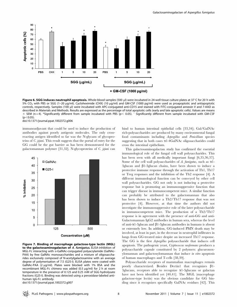
GG induces neutrophil apoptosisBloodstream neutrophils have a short half-life and prolongation
of their lifespan is critical for efficient pathogen destruction. As
SGG-treated mice exhibited reduced neutrophil infiltrates in the
lung during infection as compared to controls, we investigated the
effect of SGG on neutrophil apoptosis. Neutrophils cultured at
37uC died rapidly by apoptosis, about 60% of cells being annexin
V+ after 20 h. As previously reported [16], apoptosis was
accelerated by cycloheximide and delayed by GM-CSF. The
percentage of apoptotic cells in whole-blood samples incubated
with SGG (10–20 mg/ml) was significantly higher than in the PBS
control. In addition, SGG significantly inhibited GM-CSF-
induced PMN survival (Fig. 6).
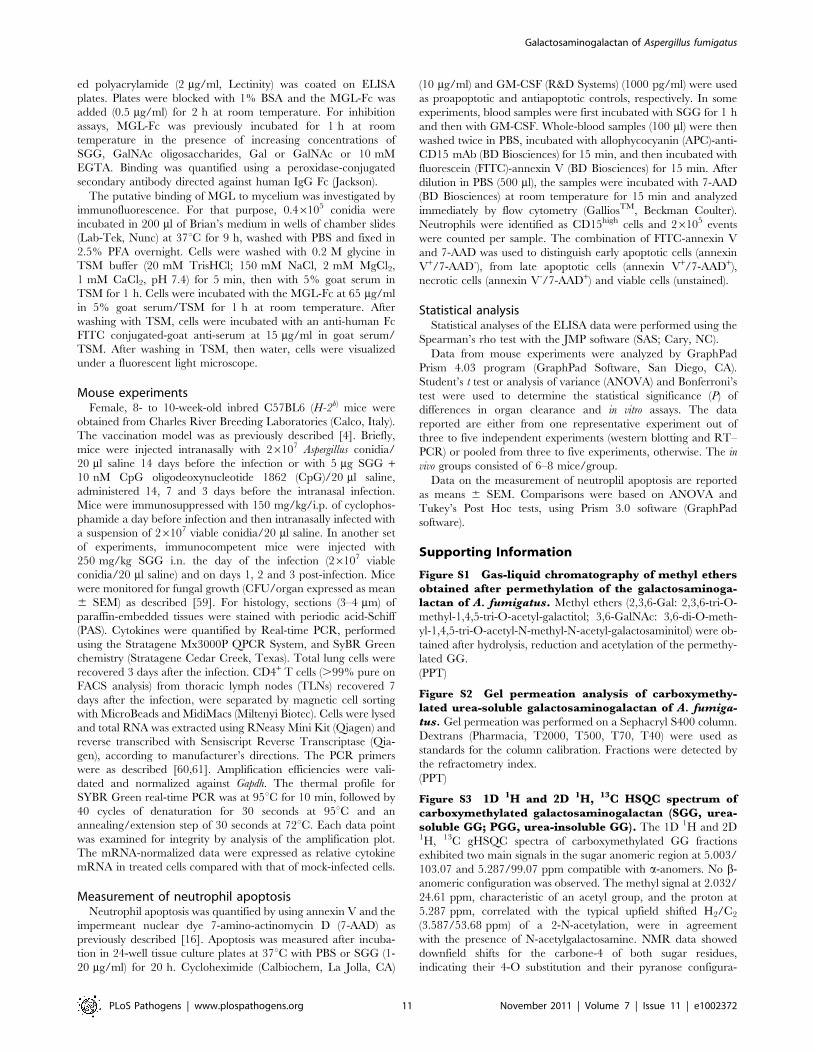
Macrophage galactose-type lectin (MGL) and GGSince the C-type lectin MGL has been shown to be specific for
GalNAc residues, the binding of GG to MGL was investigated.
Using SGG and PGG as ligands, ELISA experiment showed
a lack of specific interaction of the GG of A. fumigatus and
recombinant MGL-Fc. Similarly, immunofluorescence experiments
showed that MGL-Fc did not bind to the cell wall of germinated
conidia expressing GG on their surface (data not shown). ELISA
inhibition using GalNAc coupled to polyacrylamide (GalNAc-PAA)
as the ligand showed that GG did not inhibit the binding of
GalNAc-PAA to MGL. In contrast, GalNAc oligosaccharides
obtained by HCl hydrolysis (Fig. S8) inhibited the interaction with
MGL (Fig. 7). Since MGL recognized terminal GalNAc residues
and since the average degree of polymerisation of the oligosaccha-
Table 1. Percentage of oligosaccharide sequences found inthe galactosaminogalactan of A. fumigatus.
SGG PGG
Ratio Galactose/GalNAc 60/40 15/85
Oligosaccharide sequences Relative percentage1
aGalNAc1-[4aGalNAc1-]n n.10 10 25
aGal1-[4aGal1-]n n.10 5 2
aGal1-[4aGalNAc1-]2-10-aGal1- 10 40
aGalNAc1-[4aGal1-]2-10aGal1- 30 10
aGal1-[4aGalNAc1-4aGal1]n-4aGalNAc1- 45 23
1Percentage was estimated from the monosaccharide composition and analysisof chemically degraded products.
doi:10.1371/journal.ppat.1002372.t001
Galactosaminogalactan of Aspergillus fumigatus
PLoS Pathogens | www.plospathogens.org 5 November 2011 | Volume 7 | Issue 11 | e1002372

ride fraction used was 7.5, the relative inhibition was similar for
GalNAc and the GalNAc oligosaccharide pool when expressed in
molar concentration. In contrast to GalNAc monomers that inhibit
100% of the binding at 1 mg/ml in our experimental conditions, no
full inhibition was obtained with the oligoGalNAc fraction because
at concentrations higher than 500 mg/ml, the GalNAc oligosac-
charides precipitated. The lack of binding of MGL to the whole GG
was due to the presence of one terminal GalNAc per 700 GalNAc
residues in average in the linear 100 kDa GG polysaccharide. Only
oligoGalNAc resulting from the degradation of GG can be
recognised efficiently by MGL.
Discussion
Here, we describe the purification and the chemical character-
ization of a new galactosaminogalactan secreted by the myce-
lium of A. fumigatus. Cell wall and extracellular polysaccharides
containing galactosamine residues have been also identified in
other filamentous fungi, such as Neurospora, Rhizopus, Helminthos-
porium, Penicillium and Aspergillus species [17,18]. However, the
structure of these polysaccharides has been poorly characterized
with linkages that can be either a1-4 and/or a1-3 linkages with
part of the GalNAc molecules being N-deacetylated [19,20,21,22].
Figure 4. Vaccine potential of the urea soluble galactosaminogalactan (SGG) of A. fumigatus against invasive pulmonaryaspergillosis. A, C57BL/6 mice were injected with 26107 Aspergillus conidia 14 days or with CpG (10 nM) or CpG and SGG (5 mg) (CpG+SGG) 14, 7and 3 days before the intranasal infection with 2 6 107 live resting conidia. Naıve are uninfected mice and – are infected, untreated mice. Fungalgrowth is expressed as CFUs per lung and statistical significance is indicated by a p value ,0.001. B, Bronchoalveolar cells were obtained by lunglavage and lung histology (PAS-staining) was done 3 days after infection. Note that SGG failed to ameliorate inflammatory pathology and evenfavoured fungal growth (insert) in the absence of neutrophil recruitment. C, Cytokines were determined by RT-PCR in lung homogenates 3 days afterthe infection. Results pooled from 2 experiments (6 animals/group). Photographs were taken using a high-resolution Microscopy Color CameraAxioCam. Bars indicate SEM and statistical significance is indicated by p values.doi:10.1371/journal.ppat.1002372.g004
Galactosaminogalactan of Aspergillus fumigatus
PLoS Pathogens | www.plospathogens.org 6 November 2011 | Volume 7 | Issue 11 | e1002372

The A. fumigatus galactosaminogalactan is exclusively composed of
a1-4linked galactose and a1-4linked N-acetylgalactosamine resi-
dues. In our growth conditions, the GG was totally N-acetylated. It
was, however, shown that this linear polysaccharide is extremely
heterogeneous, with strands of galactose and N-acetylgalactosa-
mine of variable length that impact on the polysaccharide
solubility and putatively on biological properties. This heteroge-
neity is unique to the galactosaminogalactan because the other
constitutive cell wall polysaccharides of A. fumigatus are homopol-
ymers (chitin, glucans) or have well defined repeating unit, such
as A. fumigatus galactomannan [12,23]. The main motif is Gal-
GalNAc, but the variable Gal/GalNAc ratio inside each polymer
chain suggests random synthesis of the polymer, as in some
plant polysacharides [24]. The synthesis of polygalactose and
polyN-acetylgalactosamine oligosaccharides, as well as the synthe-
sis of repetitive Gal-GalNAc unit is totally unknown. The galactose
of GG is present in a pyranose form, whereas the galactose of the
galactomannan, which is a major antigen of A. fumigatus, is in a
galactofuranose form. A. fumigatus has the ability to synthesise the
two isoforms of galactose, like many bacterial, parasite and fungal
microorganisms [25,26,27,28,29]. This was indeed shown in a
UDP-Gal epimerase mutant, in which galactofuranose synthesis
was abolished, but some galactose was still present in the cell wall,
corresponding to the GG [30] (data not shown.).
It was very surprising to see that a majority of the sera from the
blood bank had high titers of IgG against GG, with GalNAc
residues being the main determinant for the antigenicity. This
result suggested that this polysaccharide could be a very potent
Figure 5. Impact of SGG on primary aspergillosis in intact mice. A, C57BL/6 mice were first injected with SGG at day 3, 2 and 1 before conidialinhalation and infected on day 0 with 26107 Aspergillus conidia. Naıve are uninfected mice, – are infected, untreated mice and SGG are mice that havereceived SGG (250 mg/kg i.n. the day of the infection and on days 1, 2 and 3 post-infection). Fungal growth is expressed as CFUs per lung andstatistical significance is indicated by a p value ,0.001. Results pooled from 2 experiments (6 animals/group) with one example shown in panel A. B,Lung histology (PAS-staining) of mice treated as indicated, 3 days after infection showing signs of inflammatory pathology in the immunocompetentmice treated with SGG. C, Cytokines were determined by RT-PCR in lung homogenates 3 days after the infection. Note that SGG inducedinflammatory cytokine gene expression, such as Tnfa, Il6, Il17a and Il4 genes, but suppressed Ifnc and Il10 expression; the low level of Mpo geneexpression is in agreement with the low counts of neutrophils in the lung of infected mice. Bars indicate SEM and statistical significance is indicatedby p values.doi:10.1371/journal.ppat.1002372.g005
Galactosaminogalactan of Aspergillus fumigatus
PLoS Pathogens | www.plospathogens.org 7 November 2011 | Volume 7 | Issue 11 | e1002372
immunoadjuvant that could be used to induce the production of
antibodies against poorly antigenic molecules. The only cross-
reacting antigen identified so far was the N-glycans of glycopro-
teins of C. jejuni. This result suggests that the portal of entry for the
GG could be the gut barrier as has been demonstrated for the
galactomannan polymer [31,32]. N-glycoproteins of C. jejuni can
bind to human intestinal epithelial cells [33,34]; Gal/GalNAc
rich-polysaccharides are produced by many environmental fungal
food contaminants including Aspergillus and Penicillium species
suggesting that in both cases a1-4GalNAc oligosaccharides could
cross the intestinal epithelium.
This galactosaminogalactan study has confirmed the essential
immunological role of the fungal cell wall polysaccharides. This
has been seen with all medically important fungi [6,35,36,37].
Some of the cell wall polysaccharides of A. fumigatus, such as a1-
3glucan and b1-3glucan chains, have been shown to induce a
protective immune response through the activation of Th1, Th17
or Treg responses and the inhibition of the Th2 response [4]. A
different immunological function can be conveyed by other cell
wall polysaccharides. GG not only is not inducing a protective
response but is promoting an immunosuppressive function that
can trigger disease in immunocompetent mice. A similar function
can probably be attributed to the galactomannan that also
has been shown to induce a Th2/Th17 response that was not
protective [4]. However, at that time the authors did not
investigate the immunosuppressive role of the later polysaccharide
in immunocompetent mice. The production of a Th2/Th17
response is in agreement with the presence of anti-GG and anti-
Galactomannan IgG2 antibodies in human sera, whereas the level
of anti-a1-3glucan and b1-3glucan antibodies in humans is absent
or extremely low. In addition, GG-induced PMN death may be
involved, at least in part, in the decrease in neutrophil infiltrates in
lungs from GG-treated mice despite an increased Th17 response.
The GG is the first Aspergillus polysaccharide that induces cell
apoptosis. The pathogenic yeast, Cryptococcus neoformans produces a
polysaccharide capsule constituted by 2 polymers: glucuronox-
ylomannan and galactoxylomannan that induce in vitro apoptosis
of human macrophages and T-cells [38,39].
Polysaccharide receptors of mammalian macrophages remain
poorly characterized. Besides Dectin1 that recognizes b1-
3glucans, receptors able to recognize a1-3glucans or galactan
have not been identified yet [40,41]. The MGL (macrophage
galactose-type lectin) was the obvious candidate for GG bin-
ding since it recognizes specifically GalNAc residues [42]. This
Figure 6. SGG induces neutrophil apoptosis. Whole-blood samples (500 ml) were incubated in 24-well tissue culture plates at 37uC for 20 h with5% CO2 with PBS or SGG (1–20 mg/ml). Cycloheximide (CHX) (10 mg/ml) and GM-CSF (1000 pg/ml) were used as proapoptotic and antiapoptoticcontrols, respectively. Samples (100 ml) were incubated with APC-conjugated anti-CD15 and stained with FITC-conjugated annexin V and 7-AAD asdescribed in Materials and Methods. Results are expressed as the percentage of total apoptotic cells (early and late apoptotic cells). Values are means6 SEM (n = 4). *Significantly different from sample incubated with PBS (p, 0.05). u Significantly different from sample incubated with GM-CSF(p,0.05).doi:10.1371/journal.ppat.1002372.g006
Figure 7. Binding of macrophage galactose-type lectin (MGL)to the galactosaminogalactan of A. fumigatus. ELISA-inhibition ofMGL-Fc interacting with a-GalNAc-conjugated polyacrylamide (GalNAc-PAA) by free GalNAc monosaccharides and a mixture of oligosaccha-rides exclusively composed of N-acetylgalactosamine with an averagedegree of polymerisation of 7.5 (G25-I). ELISA plates were coated withGalNAc-PAA (2 mg/ml). Plates were blocked with 1% BSA and therecombinant MGL-Fc chimera was added (0.5 mg/ml) for 2 h at roomtemperature in the presence of 0.125 and 0.25 mM of SGG hydrolysatefractions (G25-I). Binding was detected using a peroxidase-labeled anti-human IgG-Fc antibody.doi:10.1371/journal.ppat.1002372.g007
Galactosaminogalactan of Aspergillus fumigatus
PLoS Pathogens | www.plospathogens.org 8 November 2011 | Volume 7 | Issue 11 | e1002372

receptor is located at the cell surface of immature dentritic
cells and has been shown to be involved in the recognition of
pathogens through GalNAc residues and in the retention of
immature DCs in peripheral tissue and lymphoid organs
[42,43,44]. The MGL is able to bind to N-glycoproteins of C.
jejuni through a1-4 linked GalNAc residues [45] and the binding
of these N-glycans to MGL influences the function of human
dentritic cells. However, no specific binding of GG to human
MGL-Fc was seen, suggesting that cell surface MGL was not
involved in the recognition of A. fumigatus GG. However, the
intracellular hydrolysis of GG, as shown for some bacterial
polysaccharides [46], may release oligosaccharides that can bind
to MGL that has been seen in endocytic compartments. Such
intracellular recognition of GG oligosaccharides could then
induce the pro-inflammatory response. This hypothesis is
currently being investigated.
Materials and Methods
Strain, media and galactosaminogalactan productionThe A. fumigatus, strain CBS 144–89 was grown in a 15l fer-
menter in modified Brian medium (2% asparagine, 5% glucose,
2.4 g/l NH4NO3, 10 g/l KH2PO4, 2 g/l MgSO4-7H2O, 26 mg/l
ZnSO4-7H2O, 2.6 mg/l CuSO4-5H2O, 1.3 mg/l Co(NO3)2-
6H2O, 65 mg/l CaCl2, pH 5.4) for 72 h at 25uC. The mycelium
was removed by filtration under vacuum and the supernatant was
precipitated with 2.5 vol. of ethanol overnight at 4uC. The pellet
was collected by centrifugation (3000g, 10 min). The pellet was
washed twice with 2.5 l of 150 mM NaCl and then extracted with
8 M urea (2 h twice at room temperature under shaking). Urea-
supernatants (SGG) were pooled and extensively dialyzed against
water and freeze-dried. Urea-insoluble pellet (PGG) was washed
with water and freeze-dried.
Monosaccharide analysisTotal hexoses were measured by the phenol-H2SO4 method
using galactose as a standard [47]. Total hexosamines were
determined with p-(dimethylamino)-benzaldehyde reagent after
4 h of 8N HCl hydrolysis at 100uC using galactosamine as a
standard [48]. Monosaccharides were identified by GC as their
alditol acetates after total acid hydrolysis (trifluoroacetic acid
(TFA) 4N or HCl 4N, 100uC, 4 h) [49]. Threitol resulting from
the periodate oxidation of galactose was identified by GC-MS and
NMR. In absence of reference spectrum, anhydrotalose resulting
from the nitrous deamination of galactosamine was identified by
GC-MS by comparison with the mass spectrum of anhydroman-
nitol and by NMR.
MethylationPrior to the methylation procedure, polysaccharides were
peracetylated as previously described [50]. Dried sample (2 mg)
was methylated by the DMSO/lithium methyl sulfinyl carbanion/
ICH3 procedure [50]. After hydrolysis of the permethylated
sample (4 N TFA 100uC, 4 h), borodeuteride-reduction and
peracetylation, methyl ethers were identified by GC-MS. Oligo-
saccharides were permethylated by the DMSO/NaOH/ICH3
procedure [51].
Periodate oxidationPolysaccharide fractions (30 mg) were resuspended in 4 ml of
10 mM HCl at 50uC for 24 h and then oxidized with 100 mM
sodium m-periodate at 4uC in darkness during 7 days. Excess
reagent was destroyed by adding 0.5 ml of ethylene glycol. The
solution was dialysed against water and freeze-dried. The material
was reduced overnight by 10 mg/ml NaBH4 at room tempera-
ture. After neutralisation to destroy the excess of borohydride,
reduced oxidized polysaccharide was dialysed against water and
freeze-dried. A mild acid hydrolysis was performed by 1.5 ml
of 50 mM TFA at 100uC for 1 h. The solubilised fraction was
fractionated on a HW40S column (TosoHaas, 9061.4 cm)
equilibrated in 0.25% acetic acid at the flow rate of 0.4 ml/min.
Eluted sample were detected by refractometry. The insoluble
fraction was washed twice in water.
Nitrous acid deaminationPolysaccharide fractions (30 mg) were resuspended in 4 ml of
10 mM HCl at 50uC for 24 h and then de-N-acetylated with 40%
NaOH (final concentration) at 100uC for 4 h. After neutralisation
by addition of acetic acid, samples were dialysed and freeze-dried.
Dried samples were resuspended in 600 ml of NaOAc 0.5 M pH 4.
The deamination was started by addition of 300 ml of 1 M NaNO3
and performed at 50uC during 3 h with the addition of 300 ml
of 1 M NaNO2 each hour. Soluble degraded products were
fractionated by gel filtration chromatography through a HW40S
column, as described above. Neutral sugars were detected by the
phenol-H2SO4 method [47].
Partial acid hydrolysis10 mg of polysaccharide were treated with 1 ml of 0.1 M
HCl for 3 h at 100uC. After neutralisation with 1% Na2CO2,
solubilised materials were purified by gel filtration through a
Sephadex G25 column (GE Heathcare, 9061.4 cm) and eluted
with 0.25% acetic acid at a flow rate of 9 ml/h.
Molecular weight analysis by gel filtrationchromatography
Due to its insolubility, carboxymethylation of GG was necessary
to estimate its molecular size by gel filtration. The polysaccharide
(0.2 g) was carboxymethylated by addition of 20 ml of 1.6 M
NaOH and 0.3 g of monochloroacetic acid. The mixture was
heated at 75uC and stirred magnetically for 8 h. After neutralisa-
tion, the solution was dialysed against water and freeze-dried. The
carboxymethylated polysaccharide was soluble in 0.5% acetic
acid and 10 mg were deposited onto a Sephacryl S400 column
(Pharmacia, 9061.4 cm) at the flow rate of 10 ml/h. Dextrans
(Pharmacia, T2000, T500, T70, T40) were used as standards for
the column calibration.
GC and GC-MSGC was performed on a Perichrom PR2100 instrument
with a flame ionisation detector using a capillary column
(30 m60.32 mm id) filled with a DB-1 (SGE) under the following
conditions: gas vector and pressure, helium 0.7 bar; temperature
program 120 to 180uC at 2uC/min and 180 to 240uC at 4uC/
min. GC-MS was performed on an EI/CI mass spectrometer
detector (model 5975C, Agilent technologies, Massy France)
coupled to a chromatograph (model 7890A), using a HP-5MS
capillary column (30 m60.25 mm id, Agilent technologies)
under the following conditions: gas vector: helium at 1.2 ml/
min; temperature program: 100 to 240uC at 8uC/min and
240uC for 10 min. Ammoniac gas was used for the chemical
ionisation.
Matrix-assisted desorption ionisation/Time of flight(MALDI-TOF) mass spectrometry
MALDI-TOF mass spectra were acquired on a Voyager Elite
DE-STR mass spectrometer (Perspective Biosystems, Framing-
Galactosaminogalactan of Aspergillus fumigatus
PLoS Pathogens | www.plospathogens.org 9 November 2011 | Volume 7 | Issue 11 | e1002372

ham, MA, USA) equipped with a pulsed nitrogen laser (337 nm)
and a gridless delayed extraction ion source. The spectrometer was
operated in positive reflectron mode by delayed extraction with an
accelerating voltage of 20 kV and a pulse delay time of 200 ns and
a grid voltage of 66%. Samples were prepared by mixing directly
on the target 0.5 ml of oligosaccharide solution in water (10–
50 pmol) with 0.5 ml of 2,5-dihydroxybenzoic acid matrix solution
(10 mg/ml in CH3OH/H2O, 50:50, V/V). The samples were
dried for about 5 min at room temperature. Between 50 and 100
scans were averaged for every spectrum.
NMR SpectroscopyNMR spectra of the polysaccharides were acquired at 318
and/or 343 K on a Varian Inova 500 spectrometer equipped
with a triple resonance 1H{13C/15N} PFG (pulsed field gradient)
probe whereas spectra of either nitrous deamination or periodate
oxidation products were acquired at 298 K on Varian Inova 500
and 600 spectrometers equipped with a triple resonance1H{13C/15N} PFG and a cryogenically-cooled triple resonance1H{13C/15N} PFG probe respectively (Agilent technologies,
Massy France). Polysaccharidic samples solubilized in acetic acid
0.05%V/V in H2O by warming for one hour at 100uC were
freeze dried and redissolved in DCl 0.06 M in D2O (DCl $
99.0% 2H atoms and D2O $99.9% 2H atoms, Euriso-top,
Saint-Aubin, France). After a second freeze-drying, they were
redissolved in D2O and transferred in a 5 mm NMR tube
(Wilmad 535-PP, Interchim, Montlucon, France). The final
concentration was about 5 mg/mL. Samples were dissolved in
D2O and transferred in a 5 mm NMR tube (Shigemi BMS-
005 V, Shigemi Inc., Alison Park, United States). 1H chemical
shift were referenced to external DSS (2,2-methyl-2-silapentane-
5-sulfonate sodium salt hydrate, its methyl resonance was set to
0 ppm). 13C chemical shifts were then calculated from 1H
chemical shift and gamma ratio relative to DSS. 13C/1H gamma
ratio of 0.251449530 was used [52].
The following strategy was used for assignment of nuclei. First,
the non-exchangeable proton resonances of intra glycosidic
residue spin systems were assigned using two-dimensional COSY
(correlation spectroscopy), relayed COSY (up to two relays)
and TOCSY (Total correlation spectroscopy; with mixing time
ranging from 30 to 120 ms) experiments [53]. Secondly, 1H-13C
edited gHSQC (Gradient selected heteronuclear single-quantum
correlation) and gHSQC-TOCSY (mixing time up to 80 ms)
experiments allowed the 13C chemical shifts assignment from
previously identified 1H resonances [54]. Then, 1H,1H coupling
constants for the oligosaccharides were extracted from 1D and/
or 2D spectra (1H resolution of 0.1 Hz and 0.6 Hz respectively)
and the anomeric configuration was established from the
knowledge of 3J1,2 value. Finally, the interglycosidic linkages
determination was achieved with 1H-1H NOESY (Nuclear
overhauser effect spectroscopy) experiments for the polysaccha-
rides (mixing time of 15 and 50 ms) and with 1H-1H ROESY
experiments (mixing time of 250 ms) [55] and/or 1H-13C
gHMBC (Gradient selected heteronuclear multiple bond corre-
lation) experiment (long range delay of 60 ms) [54] for the
oligosaccharides.
Ethics statementPatient samples were collected according to French Ethical
rules. Written informed consent and approval by institutional
review Board at the Pitie-Salpetriere Hospital, at the Etablisse-
ment francais du sang and at Saint-Louis Hospital were obtained.
Mouse experiments were performed according to the Italian
Approved Animal Welfare Assurance A–3143–01. Legislative
decree 157/2008-B regarding the animal licence was obtained by
the Italian Ministry of Health lasting for three years (2008–2011).
Infections were performed under avertin anesthesia and all efforts
were made to minimize suffering.
Analysis of anti-galactosaminogalactan antibodiesSerum samples from 131 healthy subjects (from Groupe francais
du sang and Hopital Saint-Louis, Paris), 25 invasive aspergillosis
patients (Hopital Saint-Louis; kind gift of A. Sulhaian) and 5
aspergilloma patients (CHU Toulouse; kind gift of P. Recco)
were used through this study. Blood group was determined by
the Etablissement francais du sang. The presence of antibo-
dies directed against the A. fumigatus galactosaminogalactan was
assessed by a direct enzyme-linked immunosorbent assay method
(ELISA). Purified A. fumigatus galactosaminogalactan and AcraA, a
recombinant N-glycoprotein from Campylobacter jejuni expressed in
E. coli [56,57] were used as antigens. Wells of microdilution plates
(F-form, Greiner, Frickenhausen, Germany) were coated with
100 ml of a suspension of 1 mg/ml galactosaminogalactan (PGG)
or 5 mg/ml AcraA diluted in 50 mM Na2CO3 pH 9 and incu-
bated overnight at room temperature. Binding of antibodies to the
ELISA-plate was estimated with patient sera diluted 1:500 and
peroxidase-conjugated anti-human immunoglobulin G, as previ-
ously described [12]. Cross reactivity between GG and the Tn
antigen (a-GalNAc-Serine) was analysed by ELISA with a
monoclonal antibody against the Tn antigen (kind gift from Dr
R. Lo-Man, Institut Pasteur).
Production of anti-galactosaminogalactan monoclonalantibody
Mice (Balb-C) were immunized subcutaneously with a crude
cell wall preparation of A. fumigatus mycelium. Monoclonal
antibodies have been produced by F. Nato and P. Beguin
(Plateforme technique de proteines recombinantes et anticorps
monoclonaux, Institut Pasteur) as previously described [58].
Screening of positive hybridoma was followed by ELISA using
the HCl-treated PGG as specific antigen. These mAbs did not
react with other Aspergillus polysaccharides, such as galacto-
mannan, b1-3glucan, a1-3glucan. ELISA-inhibition experiments
showed that the recognition of mAb-galactosaminogalactan was
fully inhibited by oligoGalNAc obtained after partial HCl
hydrolysis and gel filtration chromatography on G25 sephadex
column as described above (Fig. S8)
ImmunofluorescenceResting conidia and conidia germinated for 8 h in a 2%
glucose/1% peptone liquid medium were fixed with 2.5% p-
formaldehyde (PFA) overnight at 4uC. After fixation, cells were
washed with 0.2 M glycine in PBS for 5 min, then with 5%
goat serum in PBS for 1 h. Cells were incubated with the anti-
galactosaminogalactan monoclonal antibody at 20 mg Ig/ml in 5%
goat serum/PBS for 1 h at room temperature. After washing with
PBS-BSA 1%, cells were incubated with a goat FITC-conjugated
Ab directed against mouse IgG(H+L) diluted 1:100 in goat serum/
PBS. After washing in PBS, cells were visualized with an inverted
fluorescence light microscope. Specificity of labelling was assessed
by preincubation of MAb with 50 mg/ml of G25-I fraction (Fig.
S8).
Binding to MGL assayBinding assay to the macrophage galactose lectin (MGL) was
done by ELISA-inhibition using a recombinant MGL-Fc chimeric
protein as previously described [42]. Briefly, a-GalNAc-conjugat-
Galactosaminogalactan of Aspergillus fumigatus
PLoS Pathogens | www.plospathogens.org 10 November 2011 | Volume 7 | Issue 11 | e1002372
ed polyacrylamide (2 mg/ml, Lectinity) was coated on ELISA
plates. Plates were blocked with 1% BSA and the MGL-Fc was
added (0.5 mg/ml) for 2 h at room temperature. For inhibition
assays, MGL-Fc was previously incubated for 1 h at room
temperature in the presence of increasing concentrations of
SGG, GalNAc oligosaccharides, Gal or GalNAc or 10 mM
EGTA. Binding was quantified using a peroxidase-conjugated
secondary antibody directed against human IgG Fc (Jackson).
The putative binding of MGL to mycelium was investigated by
immunofluorescence. For that purpose, 0.46105 conidia were
incubated in 200 ml of Brian’s medium in wells of chamber slides
(Lab-Tek, Nunc) at 37uC for 9 h, washed with PBS and fixed in
2.5% PFA overnight. Cells were washed with 0.2 M glycine in
TSM buffer (20 mM TrisHCl; 150 mM NaCl, 2 mM MgCl2,
1 mM CaCl2, pH 7.4) for 5 min, then with 5% goat serum in
TSM for 1 h. Cells were incubated with the MGL-Fc at 65 mg/ml
in 5% goat serum/TSM for 1 h at room temperature. After
washing with TSM, cells were incubated with an anti-human Fc
FITC conjugated-goat anti-serum at 15 mg/ml in goat serum/
TSM. After washing in TSM, then water, cells were visualized
under a fluorescent light microscope.
Mouse experimentsFemale, 8- to 10-week-old inbred C57BL6 (H-2b) mice were
obtained from Charles River Breeding Laboratories (Calco, Italy).
The vaccination model was as previously described [4]. Briefly,
mice were injected intranasally with 26107 Aspergillus conidia/
20 ml saline 14 days before the infection or with 5 mg SGG +10 nM CpG oligodeoxynucleotide 1862 (CpG)/20 ml saline,
administered 14, 7 and 3 days before the intranasal infection.
Mice were immunosuppressed with 150 mg/kg/i.p. of cyclophos-
phamide a day before infection and then intranasally infected with
a suspension of 26107 viable conidia/20 ml saline. In another set
of experiments, immunocompetent mice were injected with
250 mg/kg SGG i.n. the day of the infection (26107 viable
conidia/20 ml saline) and on days 1, 2 and 3 post-infection. Mice
were monitored for fungal growth (CFU/organ expressed as mean
6 SEM) as described [59]. For histology, sections (3–4 mm) of
paraffin-embedded tissues were stained with periodic acid-Schiff
(PAS). Cytokines were quantified by Real-time PCR, performed
using the Stratagene Mx3000P QPCR System, and SyBR Green
chemistry (Stratagene Cedar Creek, Texas). Total lung cells were
recovered 3 days after the infection. CD4+ T cells (.99% pure on
FACS analysis) from thoracic lymph nodes (TLNs) recovered 7
days after the infection, were separated by magnetic cell sorting
with MicroBeads and MidiMacs (Miltenyi Biotec). Cells were lysed
and total RNA was extracted using RNeasy Mini Kit (Qiagen) and
reverse transcribed with Sensiscript Reverse Transcriptase (Qia-
gen), according to manufacturer’s directions. The PCR primers
were as described [60,61]. Amplification efficiencies were vali-
dated and normalized against Gapdh. The thermal profile for
SYBR Green real-time PCR was at 95uC for 10 min, followed by
40 cycles of denaturation for 30 seconds at 95uC and an
annealing/extension step of 30 seconds at 72uC. Each data point
was examined for integrity by analysis of the amplification plot.
The mRNA-normalized data were expressed as relative cytokine
mRNA in treated cells compared with that of mock-infected cells.
Measurement of neutrophil apoptosisNeutrophil apoptosis was quantified by using annexin V and the
impermeant nuclear dye 7-amino-actinomycin D (7-AAD) as
previously described [16]. Apoptosis was measured after incuba-
tion in 24-well tissue culture plates at 37uC with PBS or SGG (1-
20 mg/ml) for 20 h. Cycloheximide (Calbiochem, La Jolla, CA)
(10 mg/ml) and GM-CSF (R&D Systems) (1000 pg/ml) were used
as proapoptotic and antiapoptotic controls, respectively. In some
experiments, blood samples were first incubated with SGG for 1 h
and then with GM-CSF. Whole-blood samples (100 ml) were then
washed twice in PBS, incubated with allophycocyanin (APC)-anti-
CD15 mAb (BD Biosciences) for 15 min, and then incubated with
fluorescein (FITC)-annexin V (BD Biosciences) for 15 min. After
dilution in PBS (500 ml), the samples were incubated with 7-AAD
(BD Biosciences) at room temperature for 15 min and analyzed
immediately by flow cytometry (GalliosTM, Beckman Coulter).
Neutrophils were identified as CD15high cells and 26105 events
were counted per sample. The combination of FITC-annexin V
and 7-AAD was used to distinguish early apoptotic cells (annexin
V+/7-AAD-), from late apoptotic cells (annexin V+/7-AAD+),
necrotic cells (annexin V-/7-AAD+) and viable cells (unstained).
Statistical analysisStatistical analyses of the ELISA data were performed using the
Spearman’s rho test with the JMP software (SAS; Cary, NC).
Data from mouse experiments were analyzed by GraphPad
Prism 4.03 program (GraphPad Software, San Diego, CA).
Student’s t test or analysis of variance (ANOVA) and Bonferroni’s
test were used to determine the statistical significance (P) of
differences in organ clearance and in vitro assays. The data
reported are either from one representative experiment out of
three to five independent experiments (western blotting and RT–
PCR) or pooled from three to five experiments, otherwise. The in
vivo groups consisted of 6–8 mice/group.
Data on the measurement of neutroplil apoptosis are reported
as means 6 SEM. Comparisons were based on ANOVA and
Tukey’s Post Hoc tests, using Prism 3.0 software (GraphPad
software).
Supporting Information
Figure S1 Gas-liquid chromatography of methyl ethersobtained after permethylation of the galactosaminoga-lactan of A. fumigatus. Methyl ethers (2,3,6-Gal: 2,3,6-tri-O-
methyl-1,4,5-tri-O-acetyl-galactitol; 3,6-GalNAc: 3,6-di-O-meth-
yl-1,4,5-tri-O-acetyl-N-methyl-N-acetyl-galactosaminitol) were ob-
tained after hydrolysis, reduction and acetylation of the permethy-
lated GG.
(PPT)
Figure S2 Gel permeation analysis of carboxymethy-lated urea-soluble galactosaminogalactan of A. fumiga-tus. Gel permeation was performed on a Sephacryl S400 column.
Dextrans (Pharmacia, T2000, T500, T70, T40) were used as
standards for the column calibration. Fractions were detected by
the refractometry index.
(PPT)
Figure S3 1D 1H and 2D 1H, 13C HSQC spectrum ofcarboxymethylated galactosaminogalactan (SGG, urea-soluble GG; PGG, urea-insoluble GG). The 1D 1H and 2D1H, 13C gHSQC spectra of carboxymethylated GG fractions
exhibited two main signals in the sugar anomeric region at 5.003/
103.07 and 5.287/99.07 ppm compatible with a-anomers. No b-
anomeric configuration was observed. The methyl signal at 2.032/
24.61 ppm, characteristic of an acetyl group, and the proton at
5.287 ppm, correlated with the typical upfield shifted H2/C2
(3.587/53.68 ppm) of a 2-N-acetylation, were in agreement
with the presence of N-acetylgalactosamine. NMR data showed
downfield shifts for the carbone-4 of both sugar residues,
indicating their 4-O substitution and their pyranose configura-
Galactosaminogalactan of Aspergillus fumigatus
PLoS Pathogens | www.plospathogens.org 11 November 2011 | Volume 7 | Issue 11 | e1002372

tion that was in agreement with the NOESY experiments and
methylation data.
(PPT)
Figure S4 GC-MS analysis of permethylated N-acetyl-galactosaminyl-threitol from the fraction II obtainedafter periodate oxidation of GG. TIC, total ion chromato-
gram of permethylated fraction II. CI, chemical ion spectra using
NH4 as collision gas of the main peak eluted at 21 min. EI,
electonic impact spectra of the peak eluted at 21 min. Ion mass m/
z were identified according to Fournet et al., [62]. Ion J1 = 207;
A1 = 260, A2 = 228, ion [M-NH-MeCOMe] = 350, F1 = 142;
H1 = 129; H2 = 87.
(PPT)
Figure S5 Gel filtration analysis of degraded SGG andPGG fractions of A. fumigatus. Gel permeation chromatog-
raphy was performed on a HW40S column eluted with a 0.25%
acetic acid solution. A. Analysis of solubilised oligosaccharides
obtained after periodate-oxidation of GG. The three carbohydrate
containing fractions (I-III) are identified by the refractometry
index (RI). B Analysis of solubilised oligosaccharides obtained after
nitrous deamination of the GG. Carbohydrates were detected with
the phenol-sulfuric method (OD reading at 492 nm). (SGG, urea-
soluble GG; PGG, urea-insoluble GG)
(PPT)
Figure S6 Spearman’s representation of the correlationbetween reactivity of sera from a blood bank against thegalactosaminogalactan (GG) of A. fumigatus and a N-glycosylated recombinant protein of Campylobacterjejuni (AcraA). ELISA ODs obtained with 131 sera against
GG (y axis) and AcraA (x axis) showing the fit between these two
populations using the JMP software. Bivariate density ellipse with
P = 0.95 is shown. Spearman’s rho value r = 0.71 (p,0.0001).
(PPT)
Figure S7 Examples of ELISA inhibition by a1-4GalNAcoligosaccharides of serum reactivity towards PGG of A.fumigatus or AcraA of C. jejuni. Wells were coated with
PGG or AcraA and the serum was incubated with increasing
concentration of the GalNAc oligosaccharides obtained by partial
HCl hydrolysis of GG. The antibody reactivity to both antigens
was inhibited by the linear GalNAc oligosaccharides, indicating
that the two antigens share the same epitope in 80% of serum
samples. In 20% of sera, the GG recognition was not fully
inhibited by the GalNAc oligosaccharides.
(PPT)
Figure S8 MALDI-TOF mass spectra of the oligosac-charide fraction obtained by partial HCl hydrolysis.Partial hydrolysis of SGG was performed by 0.1 M HCl at 100uCfor 3 h. Solubilised material (G25-I) was purified by gel filtration
on a G25 Sephadex column. (mass m/z = [M+Na]+) GN: N-
acetylgalactosamine. G: galactose. The fraction of the hydrolysate
excluded from a G25-Sephadex column contained a mixture of
oligosaccharides with an average of 7.5 GalNAc per molecule and
confirmed the presence of oligoGalNAc in the GG polysaccharide
chain. This mild acid hydrolysis was an alternative method to
periodate oxidation to prepare quickly and in a single step GalNAc
oligosaccharides.
(PPT)
Table S1 1H and 13C NMR chemical shifts (ppm) andcoupling constants (JH,H,JC,H Hz) for the SGG polysac-charidic fraction II obtained after periodate oxidation(Fig. 2).
(DOC)
Table S2 1H and 13C NMR chemical shifts (ppm) andcoupling constants (JH,H ,JC,H Hz) for the SGG poly-saccharidic fraction I obtained after nitrous deamina-tion (Fig. 3).
(DOC)
Author Contributions
Conceived and designed the experiments: TF YvK MD CE LR JPL.
Performed the experiments: TF AD CS BC SJvV SB SM CT FS. Analyzed
the data: TF AD CS SJvV SB CT MD CE LR JPL. Contributed reagents/
materials/analysis tools: TF CS BC SJvV Yvk SB SM FS MA MD CE LR
JPL. Wrote the paper: TF CS SJvV Yvk MD CE LR JPL.
References
1. Morgan J, Wannemuehler KA, Marr KA, Hadley S, Kontoyiannis DP, et al.
(2005) Incidence of invasive aspergillosis following hematopoietic stem cell and
solid organ transplantation: interim results of a prospective multicenter
surveillance program. Med Mycol 43 Suppl 1: S49–58.
2. Singh N, Paterson DL (2005) Aspergillus infections in transplant recipients. Clin
Microbiol Rev 18: 44–69.
3. Brakhage AA, Bruns S, Thywissen A, Zipfel PF, Behnsen J (2010) Interaction of
phagocytes with filamentous fungi. Curr Opin Microbiol 13: 409–415.
4. Bozza S, Clavaud C, Giovannini G, Fontaine T, Beauvais A, et al. (2009) Immune
sensing of Aspergillus fumigatus proteins, glycolipids, and polysaccharides and the
impact on Th immunity and vaccination. J Immunol 183: 2407–2414.
5. Mouyna I, Fontaine T (2009) Cell wall of Aspergillus fumigatus: a dynamic
structure Latge J-P, Steinbach WJ, eds. Washington, D.C: :American Society for
Microbiology. pp 169–183.
6. Netea MG, Brown GD, Kullberg BJ, Gow NA (2008) An integrated model of
the recognition of Candida albicans by the innate immune system. Nat Rev
Microbiol 6: 67–78.
7. Romani L, Puccetti P (2008) Immune regulation and tolerance to fungi in the
lungs and skin. Chem Immunol Allergy 94: 124–137.
8. Rizzetto L, Cavalieri D (2010) A systems biology approach to the mutual
interaction between yeast and the immune system. Immunobiology 215: 762–769.
9. Zaragoza O, Rodrigues ML, De Jesus M, Frases S, Dadachova E, et al. (2009)
The capsule of the fungal pathogen Cryptococcus neoformans. Adv Appl
Microbiol 68: 133–216.
10. Beauvais A, Schmidt C, Guadagnini S, Roux P, Perret E, et al. (2007) An
extracellular matrix glues together the aerial-grown hyphae of Aspergillus
fumigatus. Cell Microbiol 9: 1588–1600.
11. Loussert C, Schmitt C, Prevost MC, Balloy V, Fadel E, et al. (2010) In vivo
biofilm composition of Aspergillus fumigatus. Cell Microbiol 12: 405–410.
12. Latge JP, Kobayashi H, Debeaupuis JP, Diaquin M, Sarfati J, et al. (1994)
Chemical and immunological characterization of the extracellular galactoman-
nan of Aspergillus fumigatus. Infect Immun 62: 5424–5433.
13. Fontaine T, Simenel C, Dubreucq G, Adam O, Delepierre M, et al. (2000)
Molecular organization of the alkali-insoluble fraction of Aspergillus fumigatus
cell wall. J Biol Chem 275: 27594–27607.
14. Vinogradov E, Bock K (1999) The structure of the core part of Proteus vulgaris
OX2 lipopolysaccharide. Carbohydr Res 320: 239–243.
15. Young NM, Brisson JR, Kelly J, Watson DC, Tessier L, et al. (2002)
Structure of the N-linked glycan present on multiple glycoproteins in
the Gram-negative bacterium, Campylobacter jejuni. J Biol Chem 277:
42530–42539.
16. Francois S, El Benna J, Dang PM, Pedruzzi E, Gougerot-Pocidalo MA, et al.
(2005) Inhibition of neutrophil apoptosis by TLR agonists in whole blood:
involvement of the phosphoinositide 3-kinase/Akt and NF-kappaB signaling
pathways, leading to increased levels of Mcl-1, A1, and phosphorylated Bad.
J Immunol 174: 3633–3642.
17. Distler JJ, Roseman S (1960) Galactosamine polymers produced by Aspergillus
parasiticus. J Biol Chem 235: 2538–2541.
18. Reissig JL, Glasgow JE (1971) Mucopolysaccharide which regulates growth in
Neurospora. J Bacteriol 106: 882–889.
19. Bardalaye PC, Nordin JH (1976) Galactosaminogalactan from cell walls of
Aspergillus niger. J Bacteriol 125: 655–669.
20. Ruperez P, Leal JA (1981) Extracelullar galactosaminogalactan from Aspergillus
parasiticus. Trans Br Mycol Soc 77: 621–625.
21. Takada H, Araki Y, Ito E (1981) Structure of polygalactosamine produced by
Aspergillus parasiticus. J Biochem 89: 1265–1274.
22. Guerrero C, Prieto A, Leal JA (1988) Extracellular galactosaminogalactan from
Penicillium frequentans. Microbiologia 4: 39–46.
Galactosaminogalactan of Aspergillus fumigatus
PLoS Pathogens | www.plospathogens.org 12 November 2011 | Volume 7 | Issue 11 | e1002372

23. Costachel C, Coddeville B, Latge JP, Fontaine T (2005) Glycosylphosphatidy-
linositol-anchored fungal polysaccharide in Aspergillus fumigatus. J Biol Chem280: 39835–39842.
24. Scheller HV, Ulvskov P (2010) Hemicelluloses. Annu Rev Plant Biol 61:
263–289.25. Dutton GG, Savage AV (1980) Structural investigation of the capsular
polysaccharide of Klebsiella serotype K12. Carbohydr Res 83: 351–362.26. Schneider P, Treumann A, Milne KG, McConville MJ, Zitzmann N, et al.
(1996) Structural studies on a lipoarabinogalactan of Crithidia fasciculata.
Biochem J 313(Pt 3): 963–971.27. Kol O, Wieruszeski JM, Strecker G, Fournet B, Zalisz R, et al. (1992) Structure
of the O-specific polysaccharide chain of Klebsiella pneumoniae O1K2 (NCTC5055) lipopolysaccharide. A complementary elucidation. Carbohydr Res 236:
339–344.28. Moody SF, Handman E, McConville MJ, Bacic A (1993) The structure of
Leishmania major amastigote lipophosphoglycan. J Biol Chem 268:
18457–18466.29. Leal JA, Prieto A, Bernabe M, Hawksworth DL (2010) An assessment of fungal
wall heteromannans as a phylogenetically informative character in ascomycetes.FEMS Microbiol Rev 34: 986–1014.
30. Lamarre C, Beau R, Balloy V, Fontaine T, Wong Sak Hoi J, et al. (2009)
Galactofuranose attenuates cellular adhesion of Aspergillus fumigatus. CellMicrobiol 11: 1612–1623.
31. Letscher-Bru V, Cavalier A, Pernot-Marino E, Koenig H, Eyer D, et al. (1998)Aspergillus galactomannan antigen detection with Platelia-Aspergillus: multiple
positive antigemia without Aspergillus infection. J Mycol Med 8: 112–113.32. Gangneux JP, Lavarde D, Bretagne S, Guiguen C, Gandemer V (2002)
Transient aspergillus antigenaemia: think of milk. Lancet 359: 1251.
33. Szymanski CM, Burr DH, Guerry P (2002) Campylobacter protein glycosylationaffects host cell interactions. Infect Immun 70: 2242–2244.
34. Karlyshev AV, Everest P, Linton D, Cawthraw S, Newell DG, et al. (2004) TheCampylobacter jejuni general glycosylation system is important for attachment
to human epithelial cells and in the colonization of chicks. Microbiology 150:
1957–1964.35. Geijtenbeek TB, Gringhuis SI (2009) Signalling through C-type lectin receptors:
shaping immune responses. Nat Rev Immunol 9: 465–479.36. van de Veerdonk FL, Kullberg BJ, van der Meer JW, Gow NA, Netea MG
(2008) Host-microbe interactions: innate pattern recognition of fungalpathogens. Curr Opin Microbiol 11: 305–312.
37. Levitz SM (2010) Innate recognition of fungal cell walls. PLoS Pathog 6:
e1000758.38. Pericolini E, Cenci E, Monari C, De Jesus M, Bistoni F, et al. (2006)
Cryptococcus neoformans capsular polysaccharide component galactoxyloman-nan induces apoptosis of human T-cells through activation of caspase-8. Cell
Microbiol 8: 267–275.
39. Villena SN, Pinheiro RO, Pinheiro CS, Nunes MP, Takiya CM, et al. (2008)Capsular polysaccharides galactoxylomannan and glucuronoxylomannan from
Cryptococcus neoformans induce macrophage apoptosis mediated by Fasligand. Cell Microbiol 10: 1274–1285.
40. Luther K, Torosantucci A, Brakhage AA, Heesemann J, Ebel F (2007)Phagocytosis of Aspergillus fumigatus conidia by murine macrophages involves
recognition by the dectin-1 beta-glucan receptor and Toll-like receptor 2. Cell
Microbiol 9: 368–381.41. Toyotome T, Adachi Y, Watanabe A, Ochiai E, Ohno N, et al. (2008) Activator
protein 1 is triggered by Aspergillus fumigatus beta-glucans surface-exposedduring specific growth stages. Microb Pathog 44: 141–150.
42. van Vliet SJ, van Liempt E, Saeland E, Aarnoudse CA, Appelmelk B, et al.
(2005) Carbohydrate profiling reveals a distinctive role for the C-type lectinMGL in the recognition of helminth parasites and tumor antigens by dendritic
cells. Int Immunol 17: 661–669.
43. van Vliet SJ, Paessens LC, Broks-van den Berg VC, Geijtenbeek TB, van
Kooyk Y (2008) The C-type lectin macrophage galactose-type lectin impedes
migration of immature APCs. J Immunol 181: 3148–3155.
44. van Vliet SJ, Steeghs L, Bruijns SC, Vaezirad MM, Snijders Blok C, et al. (2009)
Variation of Neisseria gonorrhoeae lipooligosaccharide directs dendritic cell-
induced T helper responses. PLoS Pathog 5: e1000625.
45. van Sorge NM, Bleumink NM, van Vliet SJ, Saeland E, van der Pol WL, et al.
(2009) N-glycosylated proteins and distinct lipooligosaccharide glycoforms of
Campylobacter jejuni target the human C-type lectin receptor MGL. Cell
Microbiol 11: 1768–1781.
46. Cobb BA, Wang Q, Tzianabos AO, Kasper DL (2004) Polysaccharide
processing and presentation by the MHCII pathway. Cell 117: 677–687.
47. Dubois M, Gilles KA, Hamilton JK, Rebers PA, Smith F (1956) Colorimetric
methods for determination of sugars and related substances. Anal Chem 28:
350–356.
48. Johnson AR (1971) Improved method of hexosamine determination. Anal
Biochem 44: 628–635.
49. Sawardeker JS, Sloneker JH, Jeanes A (1965) Quantitative determination of
monosaccharides as their alditol acetates by gas liquid chromatography. Anal
Chem 37: 1602–1604.
50. Fontaine T, Talmont F, Dutton GG, Fournet B (1991) Analysis of pyruvic acid
acetal containing polysaccharides by methanolysis and reductive cleavage
methods. Anal Biochem 199: 154–161.
51. Ciucanu I, Kerek F (1984) A simple and rapid method for permethylation of
carbohydrates. Carbohydr Res 131: 209–217.
52. Wishart DS, Bigam CG, Yao J, Abildgaard F, Dyson HJ, et al. (1995) 1H, 13C
and 15N chemical shift referencing in biomolecular NMR. J Biomol NMR 6:
135–140.
53. Wagner G (1983) Two-dimensional relayed coherence transfer spectroscopy of a
protein. J Magn Reson 55: 151–156.
54. Willker W, Leibfritz D, Kerssebaum R, Bermel W (1993) Gradient selection in
inverse heteronuclear correlation spectroscopy. Magn Reson Chem 31:
287–292.
55. Macura S, Huang Y, Suter D, Ernst R R (1981) Two-dimensional chemical
exchange and cross-relaxation spectroscopy of coupled nuclear spins. J Magn
Reson 43: 259–281.
56. Wacker M, Linton D, Hitchen PG, Nita-Lazar M, Haslam SM, et al. (2002) N-
linked glycosylation in Campylobacter jejuni and its functional transfer into E.
coli. Science 298: 1790–1793.
57. Kowarik M, Young NM, Numao S, Schulz BL, Hug I, et al. (2006) Definition of
the bacterial N-glycosylation site consensus sequence. Embo J 25: 1957–1966.
58. Nato F, Reich K, Lhopital S, Rouyre S, Geoffroy C, et al. (1991) Production and
characterization of neutralizing and nonneutralizing monoclonal antibodies
against listeriolysin O. Infect Immun 59: 4641–4646.
59. Bozza S, Perruccio K, Montagnoli C, Gaziano R, Bellocchio S, et al. (2003) A
dendritic cell vaccine against invasive aspergillosis in allogeneic hematopoietic
transplantation. Blood 102: 3807–3814.
60. Romani L, Bistoni F, Perruccio K, Montagnoli C, Gaziano R, et al. (2006)
Thymosin alpha1 activates dendritic cell tryptophan catabolism and establishes a
regulatory environment for balance of inflammation and tolerance. Blood 108:
2265–2274.
61. Zelante T, De Luca A, Bonifazi P, Montagnoli C, Bozza S, et al. (2007) IL-23
and the Th17 pathway promote inflammation and impair antifungal immune
resistance. Eur J Immunol 37: 2695–2706.
62. Fournet B, Strecker G, Leroy Y, Montreuil J (1981) Gas–liquid chromatography
and mass spectrometry of methylated and acetylated methyl glycosides.
Application to the structural analysis of glycoprotein glycans. Anal Biochem
116: 489–502.
Galactosaminogalactan of Aspergillus fumigatus
PLoS Pathogens | www.plospathogens.org 13 November 2011 | Volume 7 | Issue 11 | e1002372
Related Documents


![Original Article Therapeutic Response to Immunosuppressive ... Therapeutic respones [Original].pdf · immunosuppressive agents (IST), either ATG alone or ATG with cyclosporine A (CsA).](https://static.cupdf.com/doc/110x72/5faaf04f0515b52fbe4ab87e/original-article-therapeutic-response-to-immunosuppressive-therapeutic-respones.jpg)