PLEASE SCROLL DOWN FOR ARTICLE This article was downloaded by: [Smithsonian Astrophys Obser] On: 16 April 2009 Access details: Access Details: [subscription number 790740089] Publisher Taylor & Francis Informa Ltd Registered in England and Wales Registered Number: 1072954 Registered office: Mortimer House, 37-41 Mortimer Street, London W1T 3JH, UK Contemporary Physics Publication details, including instructions for authors and subscription information: http://www.informaworld.com/smpp/title~content=t713394025 Femtosecond lasers in biology: nanoscale surgery with ultrafast optics Christopher V. Gabel a a Department of Physics, Harvard University, Cambridge, MA, USA Online Publication Date: 01 November 2008 To cite this Article Gabel, Christopher V.(2008)'Femtosecond lasers in biology: nanoscale surgery with ultrafast optics',Contemporary Physics,49:6,391 — 411 To link to this Article: DOI: 10.1080/00107510802628263 URL: http://dx.doi.org/10.1080/00107510802628263 Full terms and conditions of use: http://www.informaworld.com/terms-and-conditions-of-access.pdf This article may be used for research, teaching and private study purposes. Any substantial or systematic reproduction, re-distribution, re-selling, loan or sub-licensing, systematic supply or distribution in any form to anyone is expressly forbidden. The publisher does not give any warranty express or implied or make any representation that the contents will be complete or accurate or up to date. The accuracy of any instructions, formulae and drug doses should be independently verified with primary sources. The publisher shall not be liable for any loss, actions, claims, proceedings, demand or costs or damages whatsoever or howsoever caused arising directly or indirectly in connection with or arising out of the use of this material.

Welcome message from author
This document is posted to help you gain knowledge. Please leave a comment to let me know what you think about it! Share it to your friends and learn new things together.
Transcript

PLEASE SCROLL DOWN FOR ARTICLE
This article was downloaded by: [Smithsonian Astrophys Obser]On: 16 April 2009Access details: Access Details: [subscription number 790740089]Publisher Taylor & FrancisInforma Ltd Registered in England and Wales Registered Number: 1072954 Registered office: Mortimer House,37-41 Mortimer Street, London W1T 3JH, UK
Contemporary PhysicsPublication details, including instructions for authors and subscription information:http://www.informaworld.com/smpp/title~content=t713394025
Femtosecond lasers in biology: nanoscale surgery with ultrafast opticsChristopher V. Gabel a
a Department of Physics, Harvard University, Cambridge, MA, USA
Online Publication Date: 01 November 2008
To cite this Article Gabel, Christopher V.(2008)'Femtosecond lasers in biology: nanoscale surgery with ultrafast optics',ContemporaryPhysics,49:6,391 — 411
To link to this Article: DOI: 10.1080/00107510802628263
URL: http://dx.doi.org/10.1080/00107510802628263
Full terms and conditions of use: http://www.informaworld.com/terms-and-conditions-of-access.pdf
This article may be used for research, teaching and private study purposes. Any substantial orsystematic reproduction, re-distribution, re-selling, loan or sub-licensing, systematic supply ordistribution in any form to anyone is expressly forbidden.
The publisher does not give any warranty express or implied or make any representation that the contentswill be complete or accurate or up to date. The accuracy of any instructions, formulae and drug dosesshould be independently verified with primary sources. The publisher shall not be liable for any loss,actions, claims, proceedings, demand or costs or damages whatsoever or howsoever caused arising directlyor indirectly in connection with or arising out of the use of this material.

Femtosecond lasers in biology: nanoscale surgery with ultrafast optics
Christopher V. Gabel*
Department of Physics, Harvard University, Cambridge, MA, USA
(Received 5 November 2008; final version received 17 November 2008)
Femtosecond lasers are emerging as a powerful tool in basic biological research. The high peak light intensitygenerated by a tightly focused, ultrashort, pulse of infrared laser light enables versatile submicron ablation deepwithin biological samples. Recent studies have begun to exploit these capabilities to conduct meticulous laser surgeryexperiments within single cells, as well as within intact organisms. This review will discuss the basic physicalmechanisms behind femtosecond laser ablation in biological samples. It will then examine a series of prominentapplications in biology and how they are opening new possibilities in a range of research fields. The interfacebetween physics and biology has been exceptionally fruitful over recent years and femtosecond laser ablation isproving to be another prime example of this.
Keywords: femtosecond laser; laser ablation; laser surgery; nanosurgery
1. Introduction
Major advancements in biological research havehistorically been sparked by developments in enablingexperimental technologies. Novel instrumentation andtechniques, that often first emerge in physics andengineering sciences, can have a profound effect whenapplied to the life sciences. Consider a few examples:improvements in optics and construction of compoundmicroscopes during the first part of the nineteenthcentury resulted in the first substantial cell biologicalstudies. A hundred years later, development of X-raydiffraction techniques allowed researchers to elucidatethe double helix structure of DNA [1,2], ushering in arevolution in genetics and molecular biology. Scanningelectron microscopes allow imaging of biologicalstructures on the nanometre scale. Nuclear magneticresonance (NMR) spectroscopy facilitates proteinstructure determination, while magnetic resonanceimaging (MRI) has become an invaluable tool inmedical imaging. Advancements in computing powerhave paved the way for quantitative and statisticalanalysis of DNA sequences, protein dynamics, andbiochemical pathways, thus establishing new fieldssuch as bioinformatics, systems biology and genomics.
Laser technology is no exception to this rule. Overthe past half-century, advancements in the quality andvariety of available laser systems have had a significantimpact on microscopy, cell biology and medicine. Forexample, laser based two-photon microscopy hasdeveloped into a powerful three-dimensional imagingtechnique [3,4]. Infrared lasers with limited linear
absorption in biological tissue at low intensities canexcite a range of fluorophores through multiphotonabsorption at high intensities. Thus, the fluorescentsignature within a diffraction limited focal volume canbe independently measured and incorporated into athree-dimensional image. Similarly, confocal laserscanning microscopes, that eliminate out of focusglare and allow 3D serial section imaging of biologicalsamples, are commercially available and in wide use[5]. Laser based optical traps that generate piconew-tons of force on micron size dielectric objects, allow thephysical manipulation of individual cells and biomo-lecules [6,7]. Precisely calibrated and feedback con-trolled systems of this kind are used to characterise thedynamics of individual protein structures. For exam-ple, the step size and forces generated by a ribosomecomplex as it reads out a strand of DNA were recentlymeasured [8].
The most ubiquitous applications of laser technol-ogy in biology and medicine are tissue ablation andsurgical techniques. In numerous medical fields, ran-ging from ophthalmology to orthopaedics, laserablation and precision cutting of biological tissuehave become common and valuable techniques. Thesetechniques have also had an impact on basic cellularresearch. Since the advent of modern laser technology,photodisruption on cellular and subcellular lengthscales has been an ongoing area of investigation. Thecellular ablations were first performed in the 1960susing continuous wave lasers to damage subcellularcomponents such as individual mitochondria and
*Email: [email protected]
Contemporary Physics
Vol. 49, No. 6, November–December 2008, 391–411
ISSN 0010-7514 print/ISSN 1366-5812 online
� 2008 Taylor & Francis
DOI: 10.1080/00107510802628263
http://www.informaworld.com
Downloaded By: [Smithsonian Astrophys Obser] At: 15:45 16 April 2009
chromosomes [9,10]. Subsequent studies with contin-uous wave as well as short-pulsed lasers have targeteda wide range of cellular structures, including singleorganelles, cytoskeletal filaments, chromosomes, thecellular membrane and entire cells in developingembryos [11–14].
However, these conventional laser systems are notwithout drawbacks. Unspecified thermal damage canbe a concern. Often photosensitising biolabels must beused to selectively target particular biological macro-molecules. Increased laser absorption by these labelsfacilitates energy transfer and disruption of theintended substrate while minimising damage to sur-rounding structures. This is particularly necessary forcontinuous wave lasers but has also been employedwith pulsed lasers in order to lower ablation thresh-olds. Specific labelling, however, limits photodisrup-tion to cells and cellular components for whichappropriate stains have been developed. Short-pulsedlasers, emitting nanosecond pulses of laser irradiation,generate high peak intensities (4108 W mm72 at eachpulse) that result in more generalised tissue ablation atarbitrary locations. However, spatial resolution islimited to tens of microns, due to the violentmechanical effects of shock wave and bubble forma-tion produced by these laser pulses [15]. In addition,biological tissue shows strong linear absorption in theultraviolet and visible wavelengths of many of theselasers. Photodamage is thus possible at any point alongthe beam path. Unintended disruption of criticalcellular components, such as cellular DNA or thecytoplasm membrane, can have effects ranging fromsubtle changes in gene expression to cell death.Furthermore, the strong absorption limits beampenetration in biological samples, largely prohibitingdeep tissue and in vivo ablations.
In recent years, a move toward ever-shorter laserpulses has helped to increase both the resolution andversatility of laser ablation techniques. Ultrafastpulsed lasers, which have been traditionally employedin micromachining techniques of transparent materialssuch as glass, are finding increased application inbiology [16,17]. Infrared femtosecond laser pulsesfocused to a near diffraction-limited spot, using ahigh numerical aperture (NA) microscope objective,generate higher peak intensities (*1013 W cm72) withlower pulse energies (*few nanojoules) than pre-viously possible. Lower total energy transfer allowsfiner mechanical damage and limits thermal heating.The result is an increase in spatial resolution withtissue disruption possible on the submicron scale.Higher peak intensities also generate more reliable,tissue independent, multiphoton absorption and abla-tion dynamics. Damage levels can therefore be care-fully calibrated and implemented. Furthermore,
biological tissue is virtually transparent at the nearinfrared wavelength typically emitted by these lasers(*800 nm). This transparency limits linear photonabsorption outside the focal volume minimising photo-damage along the beam path and allowing for deepbeam penetration within thick biological samples.Femtosecond laser ablation experiments of this kindare now possible *400 mm within the tissue of anintact organism [18].
With these new ablation capabilities have come aseries of novel experiments, including disruption ofindividual subcellular components, single cell DNAtransfection, cellular neurosurgery and larger scaletissue disruption. After a brief discussion of thephysical mechanisms behind multiphoton absorptionand tissue disruption by high intensity femtosecondlaser pulses, we will review a number of these studies.The goal is two-fold: first, to understand the advan-tages of femtosecond laser ablation techniques andhow these techniques facilitate a new level of investiga-tion on the cellular and subcellular level; and, second,to grasp the biological motivation behind these studiesand their potential impact in their respective fields.
2. Photodisruption in biological tissue
When ultrashort pulses of near infrared (IR) light arefocused to a tight spot, remarkably high peakintensities are achieved with relatively low pulseenergies. This generates non-linear photon absorptionin an otherwise transparent medium and electronionisation within the focal volume. At a criticaldensity, the resulting free-electron plasma causesoptical breakdown and physical disruption of thematerial. The end effect is selected tissue ablation deepwithin a biological sample with submicron resolution.In this section we will review the basic physicalprinciples that contribute to these processes. A moredetailed treatment of these topics can be found in tworecent articles [19,20].
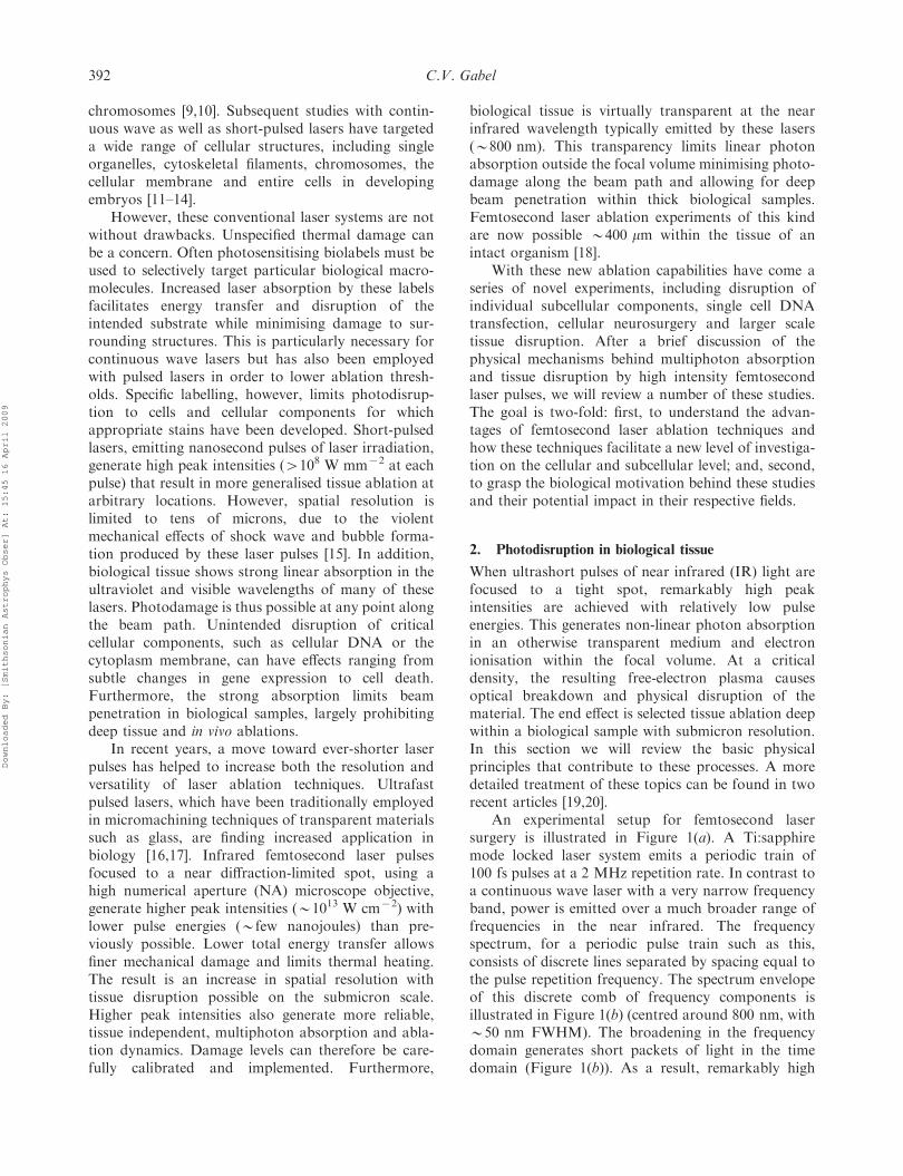
An experimental setup for femtosecond lasersurgery is illustrated in Figure 1(a). A Ti:sapphiremode locked laser system emits a periodic train of100 fs pulses at a 2 MHz repetition rate. In contrast toa continuous wave laser with a very narrow frequencyband, power is emitted over a much broader range offrequencies in the near infrared. The frequencyspectrum, for a periodic pulse train such as this,consists of discrete lines separated by spacing equal tothe pulse repetition frequency. The spectrum envelopeof this discrete comb of frequency components isillustrated in Figure 1(b) (centred around 800 nm, with*50 nm FWHM). The broadening in the frequencydomain generates short packets of light in the timedomain (Figure 1(b)). As a result, remarkably high
392 C.V. Gabel
Downloaded By: [Smithsonian Astrophys Obser] At: 15:45 16 April 2009

peak power is achieved for each pulse from relativelylow pulse energies and average laser power. Forexample, a typical 2 nJ pulse (see below) generates apeak power 420 kW. The 2 MHz beam passesthrough a pulse picker that blocks the majority ofthe pulses, reducing the repetition rate to 1 kHz. Thislower repetition rate ensures that each pulse actsindependently on the target and that energy deposi-tion does not accumulate over multiple pulses. Lowrepetition rates and minimal pulse energies are typicalof a number of experiments and will be the basis formost of the discussion below. However, other beamparameters, such as higher repetition rates(*80 MHz) with even lower pulse energies, are oftenused and will be discussed in the context of theirspecific applications. The kilohertz pulse train isdirected through the back of a standard compoundmicroscope where it is focused to a near diffractionlimited spot by a high NA objective (1.4 NA). Thecombination of the ultrashort pulse length and tightfocus results in peak light intensities great enough toinduce optical breakdown and physical disruptionwithin the focal volume. A precision X–Y–Z stagepositions the target point in the sample at the focalpoint of the laser and a high-speed shutter allows
brief exposure of the laser beam. A beam splitterwithin the microscope provides for simultaneous laserexposure and wide-field fluorescence microscopy. Insome cases a more sophisticated optical setup isemployed that allows simultaneous two-photon fluor-escent microscopy with the same femtosecond lasersource used for ablation [18,22].
With sufficiently high light intensity, incidentphotons are absorbed by electrons within the focalvolume exciting the electrons into a free or unboundstate. In a gaseous medium, the high light intensityresults in complete ionisation of the electrons. Incondensed matter, the electrons are excited to a ‘quasi-free’ state with enough kinetic energy to avoid captureby local potential energy barriers. The electronexcitation energy and optical breakdown propertiesof biological tissue mimic that of water, its primarycomponent. A single photon in the near-IR hassubstantially less energy than is needed for electronionisation in water (see below). The ionisation of asingle electron therefore depends on the collectiveabsorption of numerous photons that occurs throughthree possible mechanisms: simultaneous multiphotonabsorption, electron tunnelling, and impact ionisation(Figure 2).
Figure 1. Femtosecond laser ablation setup and beam characteristics. (a) A schematic diagram of a simple femtosecond laserablation system [21]. A 2 MHz train of 100 fs pulses is emitted by the Ti:sapphire laser oscillator. This is cut to a 1 kHz train by apulse picker and directed into the back of a standard inverted compound microscope. A high NA objective focuses the beam to adiffraction limited spot at the sample. (b) A continuous wave laser beam has a consistent intensity with a narrow frequencyspectrum (i.e. very narrow band of wavelengths) (top). A pulsed laser beam, however, concentrates the light and energy into shortpackets or pulses (bottom). The light intensity peaks at a very high level during each pulse and is zero otherwise. In the frequencydomain, the spectrum envelope shows significant power over a much broader band of wavelengths (i.e. frequencies) around acentral value. Shorter pulses produce higher peak intensities and broader spectrums for the same average laser power.
Contemporary Physics 393
Downloaded By: [Smithsonian Astrophys Obser] At: 15:45 16 April 2009

Incident laser pulses generate initial quasi-freeelectrons through multiphoton absorption and elec-tron tunnelling processes. Simultaneous absorptionof multiple photons allows an electron to gainsufficient energy to escape its local potential barrier.In the case of water, the electron ionisation energy is6.5 eV [23]. With 800 nm light, the energy perphoton will be E ¼ hc/l ¼ 1.56 eV (where h isPlanck’s constant, c is the speed of light, l is thewavelength). Thus, five photons are necessary to‘ionise’ a single electron (Figure 2(a)). This simulta-neous absorption requires high photon density (i.e.light intensity) and will be proportional to *I5
(where I is the light intensity). Electrons can alsoreach an ionised or quasi-free state by tunnellingthrough the local potential barrier (Figure 2(b)).With an unperturbed potential the likelihood of thisoccurring is extremely low. The high incidentelectromagnetic field of an intense laser beam,however, can sufficiently suppress the local potentialand greatly increase the tunnelling probability. Laserlight at long wavelengths (low frequencies) and high
intensities favour electron tunnelling, while moremoderate intensities and shorter wavelengths favourmultiphoton absorption [24]. With 800 nm light inwater, the transition from multiphoton absorption toelectron tunnelling is at an intensity of *1.3–1.9 6 1013 W cm72 [25,26]. This intensity is slightlymore than the *0.6 6 1013 W cm72 thresholdintensity found for optical breakdown in waterwith 100 fs pulses (see below). Thus, at threshold,multiphoton absorption is thought to predominatebut at higher intensities (or shorter pulse times)electron tunnelling could be significant [19]. Forfemtosecond pulses, the light intensity is sufficientlyhigh to make photonionisation by multiphotonabsorption and/or electron tunnelling very efficient.This generates numerous initial free electrons or‘seed’ electrons within the focal volume regardless ofthe local characteristics of the media. The result isreliable formation of a free electron plasma andcontrasts with longer pulse lengths that dependheavily on random thermal excitation or materialdefects to produce a few seed electrons [19,27].
Figure 2. Electron ionisation mechanisms. Electrons within the focal volume are ionised as a result of the high intensity laserlight by three possible mechanisms: (a) multiphoton absorption, in which a number of photons are absorbed simultaneously by asingle electron allowing it to overcome its ionisation energy. For near IR light in water this requires the absorption of fivephotons. (b) Electron tunnelling, where by the high electromagnetic field of the incident laser light temporarily suppresses thelocal potential barrier of the electron allowing it to tunnel free. (c) Avalanche ionisation, where a free electron absorbs energythrough a series of independent single photon events. Once its kinetic energy is high enough, a collision with a bound electronresults in two free electrons. These free electrons then linearly absorb photons and the process continues in a cascade oravalanche effect. For avalanche ionisation to occur, initial free electrons or ‘seed’ electrons resulting from multiphotonabsorption or electron tunnelling processes must be present in the sample.
394 C.V. Gabel
Downloaded By: [Smithsonian Astrophys Obser] At: 15:45 16 April 2009

Seed electrons initiate a process of avalancheionisation that further increases the free electrondensity in the material (Figure 2(c)). Once in anexcited state, free electrons linearly absorb incidentphotons and readily gain substantial kinetic energy.With sufficient energy, these electrons collide withbound electrons resulting in impact ionisation. Thetwo resulting free electrons then absorb additionalphotons and the process proceeds in a cascade oravalanche effect, rapidly increasing the number offree electrons in the sample. However, photonabsorption times are limited due to momentumconservation constraints, resulting in a doublingtime for free electrons of *13 fs [19,28]. Thus, forultrashort pulses, avalanche ionisation has a dimin-ished role compared to multiphoton effects [29]. Withlonger nanosecond pulses only a few seed electronsare produced and subsequent avalanche ionisationresults in a 1096 amplification of the free electrondensity. In contrast, a 100 fs pulse results inonly a 126 amplification due to avalancheionisation [19].
Multiphoton absorption mechanisms make itpossible to produce free electron distributions thatare significantly smaller than the diffraction limit. Asdiscussed above, the photon absorption rate is propor-tional to the light intensity (I) raised to the powerof the number of photons (n) necessary to causeionisation, *I n. Any multiphoton absorptionprofile will therefore be narrower than the focalvolume (Figure 3(a)). Moreover, the brief pulse timelimits the diffusion of free electrons during irradiation.Thus, the free electron distribution is proportional tothe photon absorption profile. The resulting I n
dependence of the free electron density has beenobserved experimentally for pulse energies close tooptical breakdown in dielectric materials [31]. Thediameter of the diffraction limited focal spot is given byd ¼ 1.22 l/NA. At l ¼ 800 nm and a NA of 1.3, thisequation gives a diameter of d ¼ 750 nm. If oneassumes a five-photon absorption process (*I5), theabsorption profile and therefore the free electrondistribution is reduced to a diameter of d ¼ 336 nm[19] (Figure 3(a)). This result agrees well with the
Figure 3. Femtosecond laser ablation exhibits high spatial resolution. (a) Multiphoton ionisation results in an absorptionprofile (and therefore free electron plasma distribution) below the diffraction limit. The diffraction limited spot for near IR(800 nm) light is approximated by a Gaussian distribution (750 nm FWHM). Linear photon adsorption would result in anabsorption profile which follows the intensity profile (*I, solid line). For multiphoton processes the absorption profile follows*In (where I is intensity and n is the number of photons involved). These profiles are progressively narrower for higher photonprocesses (*I2 long dashed line,*I5 short dashed line). For a five-photon process, the absorption profile is reduced to a width of*330 nm, which agrees with the experimental resolution for ablation damage. (b) Ablations at varying pulse energies offluorescently labelled actin fibres in a fixed cultured cell [30]. The fluorescence intensity profile along one fibre reveals the wellbehaved dependence on pulse energy as well as the submicron resolution of the technique. Fibre ablation (rather thenphotobleaching) was confirmed at these pulse energies through separate electron microscopy measurements.
Contemporary Physics 395
Downloaded By: [Smithsonian Astrophys Obser] At: 15:45 16 April 2009

experimentally observed limits of tissue disruption(d � 300 nm) [32].
An incident laser pulse drives electron ionisationuntil a critical free electron density is reached (*1021
cm73 [19]). At which point, optical breakdown occurs,resulting in physical disruption of the sample. For100 fs pulses focused into pure water, physical disrup-tion occurs at an intensity of *0.5 6 1013 W cm72
[19]. This value agrees with experimental ablationthresholds in biological tissue, which generate similarpeak intensities [22,32]. Due to the ultrashort irradia-tion time, minimal heat transfer occurs duringthe pulse. Electron cooling and heat transfer betweenthe free electrons and the surrounding media takeson the order of *10 ps or roughly 100 times longerthan the pulse length [27,33]. Energy deposition istherefore confined within the volume of the freeelectron distribution resulting in a large thermo-elasticpressure wave [15,33,34]. The high tensile stress of thisshock wave initiates cavitation and bubble formationat the centre of the focal volume. For femtosecondpulses near the threshold of cavitation, the resultingbubble is remarkably short lived (*10 ns) and isroughly the size of the free electron plasma distributiond * 300 nm [19,35]. The bubble size is a reproduciblefunction of the pulse energy, giving remarkable controlover the size of the disrupted volume as illustrated inFigure 3(b). Because the total energy per pulse isrelatively low, only a few nanojoules, the damageresulting from additional heat transfer is limited. For a100 fs pulse, Shen et al. calculated that 1.4 mm from thefocal point the temperature increased by only 10degrees Kelvin for a period of 2 ns [32]. Given limitedgeneral heating, it is the thermoelastic shock wave andbubble formation that constitute the physical disrup-tion of the targeted sample and allow the high spatialresolution. It is also important to note that for highNA objectives the power threshold for optical break-down occurs below the threshold for nonlinear lightpropagation. Thus, self-focusing and filamentation ofthe damage region is not an issue as it is with lower NAobjectives (51 NA) [17].
From the above considerations we can now graspthe mechanisms that give the femtosecond laser anumber of advantages over other forms of laserablation in biological samples. The lower pulse energyneeded for optical breakdown minimises excessiveheating and limits the mechanical shock wave andresulting cavitation to 51 mm in diameter. Thus,spatial resolution is improved over longer pulsedlasers. The multiphoton absorption at high peakintensities allows what is otherwise transparent materi-al to absorb the near IR light. This takes advantage ofa ‘diagnostic window’ in which linear absorption isrelatively low in biological tissues minimising damage
outside the focal volume and allowing for targetingdeep within a tissue sample. Finally, with femtosecondpulses the higher intensity results in reliable, sampleindependent plasma formation. This is in contrast tolonger pulse lengths that rely on material defects andthermal excitation in order to generate a few seedelectrons that trigger a dominant avalanche ionisationprocess. In such processes, plasma generation becomesstochastic and dependent on the abnormalities andproperties of the local tissue. With the femtosecondlaser we therefore have an accurate, reliable andversatile tool for selected ablation of biological tissue.
3. Subcellular laser surgery
The cell is the basic building block of life. Firstdescribed by Robert Hooke in 1665 using a rudimen-tary microscope, 350 years of research have produced acomplex, detailed description of cellular anatomy [36].In essence a cell consists of a lipid bi-layer or plasmamembrane, which encapsulates the cellular milieu orcytoplasm, a number of subcellular organelles and avast array of protein structures. Structural complexityranges from single celled procaryotic bacteria, consist-ing of one membrane bound compartment, to highlyspecialised eucaryotic cells within multicellular organ-isms. Typical eukaryotic cells measure 20–30 mm indiameter and possess numerous self-contained lipidvesicles or organelles. A cell’s shape, size and proteincontent can vary dramatically based on the cell’sspecific role in an organism. Femtosecond laserablation presents a new tool with which to investigatethis intracellular world. Researchers can disruptspecific cellular structures without damaging surround-ing structures or compromising cell viability. Physicaldissection of single components within living cellsprovides unique experimental advantages over bio-chemical and genetic techniques of traditional biology,that generally disrupt the function of specific proteinsthroughout the entire cell. As mentioned above,cellular and subcellular laser ablation has been anactive area of research since the 1960s [9,37]. Studieswith continuous wave and short-pulsed (nano andpicosecond) lasers have successfully targeted individualchromosomes, cytoskeletal filaments, as well as specificorganelles within living cells [11,12,38,39]. These lasersare not without drawbacks, however, often relying onphotosensitising dyes, lacking true submicron resolu-tion and generating unspecified collateral damage.Experiments within the past half-decade using femto-second lasers have brought a new level of precision andsophistication to biological studies.
Initial experiments demonstrated the potential forfemtosecond laser ablation in biological samples bydissecting single human chromosomes [40]. Lines scans
396 C.V. Gabel
Downloaded By: [Smithsonian Astrophys Obser] At: 15:45 16 April 2009

using a femtosecond laser beam generated completechromosome cuts less then 200 nm wide, while pointablations produced holes *250 nm in diameter. Thesefeatures are well below the *750 nm diffraction limitof the near IR laser light, demonstrating the increasedresolution of the multiphoton ablation technique.Given the high pulse rate, low pulse energies, andrelatively long exposure times (80 MHz pulse train,0.5 nJ per pulse and 40 ms exposures) used in theseexperiments, chromosome damage was most likely acumulative effect of a low-density electron plasmagenerated over numerous pulses (see discussion below).These initial dissections were performed with driedhuman chromosome samples, in part to allow forsubsequent scanning force microscopy of the resultingdamage. Similar experiments, however, within livingmammalian cells, suggests that DNA fragmentationcan be achieved in vivo as well [41].
Single organelles within living cells have also beentargeted using femtosecond lasers. Shen et al. success-fully ablated individual mitochondria within liveendothelial cells [32]. Mitochondria consist of self-contained membrane bound organelles roughly 1 mmin size. They function as the powerhouse of eucaryoticcells producing the vast majority the cell’s ATP(adenosine 50-triphosphate), its primary source ofchemical energy. Figure 4(a) shows fluorescentlylabelled mitochondria within a cultured endothelialcell. Disruption of individual mitochondria wasachieved by brief exposure to a 1 kHz pulse train(100 fs pulses, 2 nJ pulse energy), focused through a1.4 NA microscope objective. Irradiation with severalhundred individual pulses (i.e. *100 ms exposuretimes) ensured enough collective damage to reliablyrupture the target mitochondria. Neighbouring mito-chondria, however, 51 mm away appeared unaffectedby the procedure confirming the spatial resolution ofthe damage (Figures 4(b) and (c)). Post irradiation cellviability was tested using ethidium bromide staining. Ifthe cell plasma membrane has been compromised,ethidium bromide readily labels cellular DNA. How-ever, ethidium bromide does not penetrate intact livingcells. Mitochondrial ablations resulted in no additionalethidium bromide staining. On the other hand,specifically targeting the plasma membrane causedcell rupture and immediate staining. Watanabe et al.further confirmed ablation of individual mitochondriausing similar laser irradiation and successive fluores-cent labelling experiments [42]. In control experiments,fluorescently labelled mitochondria were photo-bleached using an Arþ laser (488 nm) and thensuccessfully relabelled. With femtosecond laser irradia-tion re-labelling was not possible, confirming photo-ablation rather than bleaching of the targetmitochondria. Similar experiments have been
performed in plant cell as well, resulting in successfulablation of individual chloroplasts while maintainingcell viability [43]. These experiments also confirmedablation resolution to within *400 nm throughelectromicroscopy of the targeted plant cell.
The physical characteristics of eucaryotic cells,such as shape, rigidity and motility are controlled bythe cell’s cytoskeleton, a web like structure of proteinfilaments spanning the cytoplasm (Figure 5(a)). Thecytoskeleton also helps mediate numerous cellularprocesses including subcellular organisation, cell divi-sion, outgrowth and differentiation [36]. Numerouslaser ablation studies have helped to shed light on theorganisation and function of this key cellular structure[11,12,38,39]. Recently femtosecond laser ablation hasbrought a new level of precision to this work. Using afemtosecond laser to target and snip individual stressfibres within a living cell, Kumar et al. were able tomake real time measurements of structural relaxationwithin the cell’s cytoskeleton [44]. Stress fibres,composed of bundles of associated actin and myosinfilaments, form a key element in the cytoskeleton.These fibres terminate at specific adhesion points onthe plasma membrane that act as anchors to thesurrounding extra-cellular matrix. The myosin compo-nent of these structures is a motor protein, capable ofundergoing specific conformational changes and gen-erating force on a molecular scale [45]. Within musclecells for example, myosin molecules collectively act onactin filaments to generate cellular contraction. Withinstress fibres it is thought that myosin actively generatestension along the fibre, which is then balanced byrigidity in the extra-cellular matrix and the remainderof the cytoskeleton [46].
Snipping a single stress fibre is followed byimmediate retraction of the severed ends as illustratedin Figure 5(a) [44]. Stress fibres are selectivelyvisualised in cultured endothelial cells by geneticallyencoded fluorescent proteins, which incorporate intothe fibres. Retraction of the severed ends appears to bedue to relaxation from a pre-stressed state rather thande-polymerisation of the filament into independentactin monomers. Indeed, the retraction is well mod-elled as the relaxation of a simple damped elastic fibre,i.e. a viscoelastic cable, following
L ¼ L0½1� exp ð�t=tÞ� þ da
(where L is the distance retracted, L0 is the asymptoticlimit of the retraction, t is time, t is the time constantof the material which depends on its dampingcoefficient and Young’s modulus, da is the length offibre initially ablated) (Figure 5(b)). By contrast,substantial de-polymerisation was observed in a recentstudy using a picosecond laser to snip individual
Contemporary Physics 397
Downloaded By: [Smithsonian Astrophys Obser] At: 15:45 16 April 2009

microtubules [12]. Microtubule filaments, another keyelement of the cytoskeletal network, were selectivelytargeted with an ultraviolet laser through enhancedfluorescence susceptibility. The resulting damage waseither not confined to the ablation point or left themicrotubules inherently unstable as they were seen to
disassemble for many microns away from the targetpoint. Femtosecond snipping appears to be morestable. Cutting close to a fibre bifurcation, for example,did not result in decomposition of the branch point butrather a retraction of the intact structure (Figure 5(a)).Fiducial marks can also be created along the target
Figure 4. Disruption of single organelles within an intact cell by femtosecond laser ablation [32]. (a) Mitochondria within acultured endothelial cell are labelled with a fluorescent biomarker. (b) The targeted mitochondrion before laser exposure. (c) Thesame image frame after laser exposure. The targeted mitochondrion has undergone complete photodisruption while neighbouringmitochondria (51 mm away) appear unaffected.
Figure 5. Dissection of subcellular cytoskeletal structures using femtosecond laser ablation [44]. (a) A fluorescently labelledstress fibre within a cultured endothelial cell is targeted (arrowhead, time ¼ 0 s). Immediately after laser exposure, a narrow gap(51 mm) appears at the target point (time ¼ 1 s). This gap expands as the severed ends of the stress fibre retract (time ¼ 5 s,time ¼ 15 s). Note that a bifurcation immediately above the cut point remains stable over time. This suggests that the fibre doesnot disassociate into individual actin monomers after laser surgery but rather the retraction is due to relaxation from a pre-stressed state. (b) The rate of retraction of the severed fibre ends follows that of a simple viscoelastic model (solid line).
398 C.V. Gabel
Downloaded By: [Smithsonian Astrophys Obser] At: 15:45 16 April 2009

fibre by reducing laser power and photobleaching thefilament at a specific point. When the filament is cut,the bleached marker is seen to move in tandem withthe severed end indicating relaxation of a strainedelastic fibre rather than disassembly of the structure.
The viscoelastic retraction of severed stress fibres isthe result of a ‘pre-stressed’ force balance between thecell’s cytoskeleton and the external matrix. Tension isactively established in the stress fibres through theoperation of myosin light chain molecules on the actinfilaments. This condition is evident from the substan-tial reduction in post snipping retraction in cellstreated with specific myosin blocking agents [44].Further evidence of a cell-wide force balance comesfrom the reorientation of cells cultured on a compliantexternal matrix. Snipping individual stress fibres inthese cells causes a substantial shift (*5 mm) in theentire cellular structure as counter-balanced forces ofthe extra-cellular matrix and the cytoskeleton reach anew equilibrium [44]. Biological tissue presents asimilarly compliant environment suggesting that activetension within the fibres plays an important role in thecell’s interaction with its surroundings. Thus, femtose-cond laser ablation is shedding light not only on thetension within single stress fibres, but also how thistension is generated and balanced against the rest ofthe cell and its surrounding environment.
4. Single cell transfection
An important technique in modern biological researchand medicine is the introduction of foreign DNA into
cells in order to express a gene of interest. This method,known as ‘transfection’ of DNA in eucaryotic cells,enables a wide range of genetic manipulations in thelab such as replacement of a defective gene, expressionof a particular biomarker or antibody, alteration of thecell’s physiological state or modulation of subsequentgene expression. In addition to being an importantexperimental method, clinicians are working towarddeveloping ‘gene therapy’ treatments that promise todeliver genes into patients’ cells in order to correctdisease states. Recently, femtosecond laser ablationhas emerged as an attractive technology to expand andimprove the repertoire of gene delivery methods.
During transfection, DNA must cross the plasmamembrane before the cell’s endogenous machinery willbegin expressing the encoded gene (Figure 6). How-ever, eucaryotic cells do not efficiently take up nakedDNA from the environment. Thus, the plasmamembrane must be temporarily breached to allowentry of the DNA from the surrounding media. Thisexchange must also be limited and brief to avoid killingthe cell. Numerous techniques have been implementedfor experimental transfection with varying success,including widely-used, lipid-based carriers of DNAthat facilitate passage of polar DNA moleculesthrough the hydrophobic plasma membrane. Anothermethod, electroporation, uses an electrical field togenerate small, short-lived perforations in cell mem-branes through which the DNA can pass. An alter-native approach for gene delivery is viral transduction[47,48]. The desired transgene is substituted into viralDNA and delivered to the target cells by the virus as
Figure 6. Optoporation with a femtosecond laser enables DNA transfection of single cells (schematic). The plasma membraneof a cell is targeted with a high repetition rate, low pulse energy beam. Brief exposure creates a temporary hole in the membranethrough which plasmid DNA can pass. Multiphoton absorption ensures that damage is imposed only within the focal volume(dark red) and not elsewhere along the beam path (light red). Once transfection has occurred the cell’s endogenous geneexpression apparatus produce the protein encoded in the foreign DNA. In this case a GFP fluorescent marker is expressed.
Contemporary Physics 399
Downloaded By: [Smithsonian Astrophys Obser] At: 15:45 16 April 2009

part of the natural process of infection. While highlyeffective in delivering the transgene, it is not withoutrisks. The potential integration of the viral DNA intothe genome of the host cells can result in genomemutagenesis and cancer. Furthermore, production ofthe modified viruses and safety concerns make thetechnique time consuming and costly. Simple plasmidDNA, on the other hand, does not share the same riskof genomic integration and is significantly less costly toproduce [49]. There is substantial interest, therefore, indeveloping none viral transfection methods such aslaser poration that are safer, faster, and cheaper.
By targeting the plasma membrane of a cell with afemtosecond laser, it is possible to briefly rupture themembrane without killing the cell. This laser generatedoptoporation allows DNA plasmids (small circles ofnaked DNA) in the external media to enter the cell aspart of the resulting fluid exchange (Figure 6). Tirlapurand Konig were the first to show successful transfectionof cultured cells, inducing expression of green fluores-cent protein (GFP) in individual targets cells [50]. Anumber of characteristics of femtosecond laser ablationmake it a potentially attractive technique over othertransfection methods. The high degree of spatialresolution and tunable damage level allow for a criticalbalance between maximising cell viability and generat-ing sufficient volume exchange with the externalenvironment. Minimal linear absorption of near-IRlight negates photodamage along the beam pathminimising collateral damage to the target and neigh-bouring cells, while also allowing for deep penetrationwithin intact tissue. Other transfection methods such aselectroporation and nanosecond UV laser ablation canresult in substantial collateral damage and tissuedegradation [13,48]. Laser poration also allows forsingle cell targeting, a potential advantage over electricaland chemical transfection that affect an entire popula-tion of cells.
Typical laser transfection experiments are per-formed with high pulse rates and relatively low pulseenergies. The pulse energies, ranging from a fraction of ananojoule to a few nanojoules, are generally below thethreshold for optical breakdown [50–52]. Long expo-sure times of *10 ms, with a *80 MHz pulse trainresult in total irradiation of roughly a million pulses.Damage, therefore, results not from the independentaction of individual pulses but from a cumulative effectof many subsequent pulses. Although below opticalbreakdown, the pulse intensities are thought togenerate a low density free electron plasma. Thesefree electrons can result in a photochemical effectcausing bond-breaking and eventual fragmentation ofbiomolecules [19]. Cumulative chemical damage as wellas possible heat build up within the targeted areaeventually leads to tissue disruption. With higher laser
intensities, brief transient bubbles are observed in-dicating physical rupture of the targeted membraneand effective DNA transfection [51,52]. Bubble sizeand duration can be adequately controlled by beamintensities and exposure times, allowing effective DNAtransfer across the membrane while minimising da-mage to the cell.
A number of recent experiments have investigatedthe efficiency and feasibility of femtosecond lasertransfection in detail. Lei et al. demonstrated laserporation in neuronal cultures by successfully stainingindividual neurons with a membrane impermeablefluorescent dye [53]. Steveson et al. rigorously investi-gated transfection efficiency and cell viability in culturedcells [51]. By varying laser power, 50–225 mW, andexposure time, 10–250 ms, with a 80 MHz, 120 fs pulsetrain, they were able to measure and optimise transfec-tion rates at *50% while keeping cell viability high,*70%. This result represented a 106-fold increase overspontaneous transfection rates (with no artificial pora-tion) and was comparable to other methods. In theseexperiments, successful optoporationwas attributed to abrief bubble formation in the plasma membrane. Thesetransient membrane breakdown events were readilyvisualised, with optimal conditions leading to *1 mmspots that lasted no longer than the exposure time.
Baumgart et al. further demonstrated the correla-tion between transient bubble formation and effectivetransfection [52]. By simultaneously performing fem-tosecond laser poration and patch clamp electrophy-siology on the same cell, they measured membranepotential before, during and after laser irradiation.Two regimes were found depending on the focus of thelaser beam relative to the membrane. In the first, laserexposure resulted in a *5 mV increase in membranepotential from a resting value of 745 mV, with noassociated bubble formation. In the second, bubbleformation was observed along with a larger 10–20 mVincrease in membrane potential. Substantial cellpenetration of an otherwise impermeable fluorescentdye was only associated with bubble formation events.From the potential drop, it is possible to estimate whatfraction of the cell volume was exchanged with theexternal media. The Nernst and Goldman equationsgovern the equilibrium membrane potential as abalance of voltage and concentration gradients acrossa cell membrane [54]. Based on these equations it canbe shown that, assuming constant cell volume, thefractional volume exchanged follows
aV¼ ð1� exp ð�DUmF RTÞÞð1� exp ðUmF=RTÞÞ
(where V is the cell volume, a is the exchanged volume,R is the gas constant, T is temperature, F is the
400 C.V. Gabel
Downloaded By: [Smithsonian Astrophys Obser] At: 15:45 16 April 2009

Faraday constant, Um is the initial membrane poten-tial, and DUm is the change in membrane potential)[52]. A *10 mV potential change thus gives afractional volume exchanged of *0.4. This numberwas verified by calibrated fluorescent measurements inwhich the concentration of fluorescent dye in laserporated cells varied between 0.33 and 0.44 of that inthe surrounding media. Such measurements allow forthe precise calibration and control of the amount ofmaterial transferred into the cell, and further elucidatethe mechanism behind femtosecond laser transfection.
Femtosecond laser transfection may prove to be aninnovative gene delivery method in important experi-mental systems such as stem cell cultures. Stem cellsare undifferentiated cells that can have the potential todifferentiate into multiple cell types. This character-istic, known as pluripotency, suggests that stem cellsmay be employed as therapeutics to replace various celltypes that have been destroyed by trauma or disease.To this end, stem cells have become a major focus inbasic and clinical research. A substantial challenge instem cell research, however, is gene delivery. Conven-tional transfection methods using chemical, mechan-ical and electroporation result in low transfection ratesand high cell mortality in these systems [55,56].Recent application of femtosecond laser transfectionhas shown promising possibilities for improvement inthis regard. Successful transfection in cultured humanstem cells has shown an impressive 70–80% transfec-tion efficiency while maintaining a 100% cell survivalrate [57]. As with other femtosecond laser transfectiontechniques, successful poration appears to be the resultof accumulative photodamage given the high pulserate, 76 MHz, and long exposure times, 50 ms (result-ing in *4 6 106 pulses). By pre-compensating fordispersion in the experimental optics, 12 fs pulsesemitted by a Ti:sapphire laser oscillator arrived at thesample no more than 20 fs long. This roughly tenfolddecrease in pulse duration over previous studiesresulted in a corresponding decrease in thresholdenergy (to *70 pJ per pulse) and may be critical inmaintaining stem cell viability. In a number of cases,transfected cells not only survived the procedure butalso maintained a high division rate and successfullypassed the transgene along to the daughter cells. Thus,a number of the critical requirements for successfulgene delivery in this potentially therapeutic systemappear to have been met.
For many applications of gene transfection andtherapy, DNA must be delivered into cells withinintact tissue. The precision and penetration depth offemtosecond laser ablation are well suited in thisregard. For example, recent experiments have demon-strated successful femtosecond laser poration andtransfection of individual cells within an intact zebra
fish embryo [58]. Targeted cells showed uptake of amembrane impermeable fluorescent dye but no addi-tional side effects in embryo development wereobserved [59]. Likewise researchers have recentlytransfected muscle and skin tissue within a livingmouse [60,61]. Naked plasmid DNA was injected intothe tissue, femtosecond laser exposure then triggeredtransfection within the targeted area resulting inexpression of the desired transgene. The penetrationof the near IR beam allowed for successful transfectionand expression of a GFP transgene 4400 mm belowthe surface of the tissue. In addition, no tissue damageor cell death was noted within the target or surround-ing area and no integration of the transfected DNA(‘genotoxicity’) was detected in the genome.
5. C. elegans neurosurgery
Submicron precision, non-tissue specific plasma gen-eration and deep tissue penetration make the femto-second laser an ideal tool for in vivo microdissection ofintact living animals. One system in which this hasproven particularly powerful is the soil dwellingnematode or roundworm, Caenorhabditis elegans.Using a femtosecond laser, researchers can successfullysnip individual nerve fibres within living worms withminimal damage to the surrounding tissue andneighbouring neurons. This capability is enablingdetailed study of nerve function and neuronal regen-eration in a basic model system.
Over the past few decades, C. elegans has emergedas a powerful model system in biological research. Theworm’s small size (*1 mm in length, *100 mm indiameter), short life cycle, and unique breedingcharacteristics make it ideal for genetic studies.Sequencing of the genome has comprehensivelydescribed the full complement of C. elegans genes,and an ever-increasing array of transgenic and gene-silencing tools are available to study gene function.Furthermore, the worm is transparent, allowingresearchers to observe, in real time, the developmentand morphology of any of the approximately 1000 cellsin the intact animal [14]. Studies in C. elegans have ledto important discoveries in many aspects of animalbiology including development, organogenesis, aging,and sensory transduction [62–64]. Many of thebiological pathways described in the worm have beensubsequently shown to have similar mechanisms inhuman cells. For example, the molecular mechanismselucidated for programmed cell death during wormdevelopment have provided the basic framework forthis process in mammals, including humans, where itplays important roles in development and cancer[65,66]. Likewise, discoveries of new classes of ‘smallRNA’ encoding genes, and characterised RNA
Contemporary Physics 401
Downloaded By: [Smithsonian Astrophys Obser] At: 15:45 16 April 2009

interference as a potent gene silencing tool, have alsobeen extended to humans [67,68].
Femtosecond laser surgery is opening new studiesof neurodissection and neuronal regeneration inC. elegans. As with other aspects of C. elegans biology,the worm’s simple, tractable nervous system sharesfundamental attributes with more complicated animalsand serves as a powerful platform for basic research.The worm’s nervous system is highly stereotyped,consisting of exactly 302 neurons that are invariantfrom worm to worm in the adult hermaphroditeanimal. Moreover, the entire network (neuron posi-tion, shape, and the connections between neurons) hasbeen completely mapped by serial section electronmicroscopy [69]. The simplicity of the C. elegansnervous system is in stark contrast to higher systems,such as mammals, which contain upward of 100 billionneurons. Traditionally, C. elegans research has reliedheavily on whole cell laser ablation, as a powerful toolby which to assay a neuron’s role in the nervous system[14,70]. Using conventional lasers, however, thistechnique is largely limited to killing entire neuronsin developing larval worms.
A nerve cell consists of a central cell body or somaand numerous extended branches (axons and den-drites) that spread through the surrounding tissue andmake specific synaptic connections with other neuronsand cells. In C. elegans these branches or nerve fibresmeasure *300 nm in diameter and can extend thebody length of the worm. Femtosecond laser ablationallows snipping of single nerve fibres deep (450 mm)within the tissue of an intact adult animal withoutdamaging the rest of the neuron or neighbouringneurons [72,73]. C. elegans small, transparent body,coupled with genetic expression of fluorescent markerproteins in specific neurons, allows in vivo imaging oftarget neurons before, during and after laser surgery,Figure 7(b). Yanik et al. were the first to demonstratefemtosecond laser surgery in C. elegans, successfullysnipping motor neurons within the body of the worm[72]. Remarkably, the severed neurons were seen to re-grow, eventually restoring nerve function. This post-surgical nerve growth in C. elegans has opened thepossibilities of exhaustive genetic and molecularstudies of in vivo neuronal regeneration.
Optimal beam characteristics for neural surgery inC. elegans are typically a *1 kHz pulse train at800 nm, with pulse energies ranging between 2–40 nJper pulse. Exposure times of *10 ms, allowing forirradiation of hundreds of pulses, result in reliableneuron dissection [22,73,74]. By targeting specificneurons expressing GFP, successful cuts are verifiedvisually by a stable *1 mm gap in the targeted nervefibre, Figure 7(b). It is also possible to photobleach theGFP within the target volume without cutting the
nerve fibre using lower laser irradiation. In such cases,however, the resulting visual gap at the target pointrecovers within a matter of minutes, as freely diffusingGFP within the cytoplasm disperses into the irradiatedvolume [22]. Bourgeois and Ben-Yakar demonstrated awell-behaved relation between the total number ofirradiant pulses and the threshold pulse energy neededfor successful nerve damage:
EN ¼ E1 þ ðE1 � E1ÞN�1=3
(where N is the number of pulses, EN is the pulseenergy at threshold for N pulses). This result follows asimilar relation found for damage imposed in dielectricmaterial and illustrates the collective effect of numer-ous pulses [74]. At low pulse energies close to damagethreshold, the nanoscale transient bubble measuring*200 nm (see above) and possible photochemicaleffects of individual pulses are unlikely to completelysever a 300–400 nm nerve fibre. To generate reliableneuron cuts, the collective damage of a large numberof pulses (4100) is necessary. At the same time, pulserates low enough to ensure energy dissipation betweenpulses helps minimise accumulative heating andcollateral damage. This low energy, multipulse techni-que generates superior accuracy and reliability overhigher pulse energies that successfully cut nerve fibreswith fewer pulses but generate larger zones ofdisruption [74].
Further evidence of cell specific laser disruption ofnerve fibres comes through fluorescent staining experi-ments [22,73]. Sensory neurons in the worm’s head andtail, that are exposed to the surrounding environmentthrough pores in the animals skin, readily take-up anddisplay fluorescent dyes introduced into the worm’senvironment. These dyes are incorporated into the cellmembrane at the sensory pores and then diffuse withinthe membrane to label the entire neuron. Looking at apair of sensory neurons in the worm’s tail, researchersfirst stained both neurons with a green fluorescent label.Laser surgery was then performed on the dendrite ofone neuron while leaving the other intact. Subsequentre-staining with a red fluorescent dye demonstrated cellspecific dissection, as the cell body of the targetedneuron did not display the second dye, while theuntargeted neuron was successfully labelled.
Physical dissection of individual neurons can resultin loss of nerve function and is facilitating the analysisof simple neural circuits in the worm. Yanik et al.found that, as expected, snipping a particular class ofmotor control neurons (the D-type motor neurons)disrupted the animal’s ability to crawl backwards [72].Chung et al. performed experiments snipping thermo-sensory neurons and testing the worm’s response totemperature stimuli [73]. They found that snipping the
402 C.V. Gabel
Downloaded By: [Smithsonian Astrophys Obser] At: 15:45 16 April 2009

dendritic branch of these neurons was equivalent tokilling the entire neuron. This effect confirmed that thethermosensory apparatus is localised in the dendritictip and that laser surgery completely blocked informa-tion flow down the dendrite to the rest of the neuron.Similar dissection of the sensory dendrites has locatedthe electrosensory apparatus of C. elegans to thesensory pores in the nose as well [75]. Interestingly,snipping thermal sensory neurons had no effect on thefunction of adjacent neurons. Osmosensory neurons,that allow the worm to sense and avoid regions of highosmolarity, have similar dendritic branches runningalong the same nerve bundle but were unaffected bythe surgeries [73]. This result further confirms thesingle neuron resolution of the technique and agreeswith visual observations showing clear disruption ofindividual nerve fibres within the dendritic bundle
without any apparent damage to neighbouring neu-rons as close as *500 nm away (Figure 7(c)).
Recent work on the neural circuitry controllingegg-laying in the worm has generated interestingresults [76]. The HSN neuron, which controls egg-laying, shows spontaneous activity when monitoredusing optical physiology techniques. By snipping theaxon of this neuron using a femtosecond laser,researchers isolated the cell body from the synapticinputs of other neurons. Neuron activity, however, wasunaffected, suggesting that, while presynaptic inputsmay modulate neuron behaviour, the base line activityis generated cell autonomously and is intrinsic to theHSN neuron. Detailed analysis of neuron interactionsuch as the above example, demonstrates the power ofsingle cell femtosecond laser dissection within thesimple neural anatomy of C. elegans.
Figure 7. In vivo single cell neurodissection within the nematode worm, C. elegans. (a) Its small size, transparent body, simplestereotyped nervous system and the larger array of genetic tools developed in the animal, make C. elegans a powerful modelsystem for basic biological research. (b) The precision and penetration depth of femtosecond laser ablation allow snipping ofindividual nerve fibres within intact animals. Specific cells are labelled within intact C. elegans using a genetically encoded GFPmarker (top). Successful ablations are evident from a stable visual break in the nerve fibre at the target point (arrow bottom).(c) A single nerve fibre within the dendritic bundle is targeted and successfully cut with out damaging neighbouring nerve fibres51 mm away [71].
Contemporary Physics 403
Downloaded By: [Smithsonian Astrophys Obser] At: 15:45 16 April 2009

Many C. elegans neurons re-grow over a period ofhours and days after laser surgery (Figure 8). Thiseffect is enabling the study of neuronal regeneration inthis powerful model system. Tragically, the mamma-lian central nervous system (CNS), the brain and thespinal cord, do not recover from traumatic neuronalinjury. In the United States alone, some five millionpeople suffer from traumatic brain and spinal cordinjury, while an additional five million suffer fromneural degenerative diseases. Research in traditionalvertebrate systems (i.e. rodents) has begun to focus onspecific molecular factors that control and regulatenerve recovery [77]. While a number of such signalling
factors have been identified, experiments are typicallycomplex and progress slowly. A typical experimentconsists of slashing the spinal cord of a mouse resultingin disruption of thousands of neurons and surroundingtissue. Studies designed to determine genes thatmodulate neural cell growth after injury are limitedby the experimental complexity of vertebrate systems.Inactivation of specific genes in vivo via gene knockoutor gene silencing requires months or years. Thesuperior genetic tools available in C. elegans, the speedwith which experiments can be performed, the simplestereotyped nervous system of the worm, and the singleneuron precision of femtosecond laser surgery all lead
Figure 8. In vivo neuronal regeneration in C. elegans after femtosecond laser surgery. (a) Part (i): an intact AVM touch sensoryneuron immediately before laser surgery. AVM morphology is conserved from animal to animal. The cell body is located in thecentre of the worm. A single axon extends down to the ventral nerve cord, which it then follows anteriorly to the head of theworm (see schematic below). Part (ii): immediately following femtosecond laser surgery, a clean break is seen at the target pointhalf way down the ventral projection of the axon (red arrow). Part (iii): 24 h after surgery substantial regenerative growth isobserved. The break point and severed distal end of the original axon are still evident (red arrow). The proximal severed end hassprouted new regenerative growth to the left, led by a large growth cone structure (yellow arrow). (b) Regenerative outgrowth isscored by marking the endpoints of the new branches after 24 h. The blue dot indicates the end of the primary branch (thelongest and brightest branch or the branch that successfully reaches the ventral nerve cord). Red dots indicate secondarybranches [21]. (c) Quantitative measurement of neuronal regeneration. A schematic diagram of the AVM neuron in C. elegans(top). The results scored from more than 20 AVM regeneration experiments in different animals are superimposed on the sameschematic plot of the worm (middle). As evident from the large number of blue dots at the bottom of the worm, AVMsuccessfully grows back to the ventral nerve cord *65% of the time in normal wild type adults. Substantial branching is alsoevident (red dots). A mutation in the unc-6 gene disrupts axon guidance during regeneration (results from more than 20 separateAVM surgeries) (bottom). Regenerative growth in these mutant worms reaches the ventral nerve cord only *30% of thetime [21].
404 C.V. Gabel
Downloaded By: [Smithsonian Astrophys Obser] At: 15:45 16 April 2009

to obvious advantages for the study of neuron re-growth. Since the vast majority of C. elegans geneshave direct counterparts in mammals and humans,discoveries within the humble worm will likely havedirect impact on our medical understanding of neuralregeneration.
Regenerative ability in C. elegans neurons varieswith neuron type. Motor neurons in the body of theworm, controlling body movement and egg-laying,readily re-grow as do mechanosensory and somechemosensory neurons. In contrast, many sensoryneurons in the head and tail of the animal do notshow significant re-growth [21,22,72,73]. This cellvariability is particularly striking given that vertebrateneurons display a range of regenerative abilities aswell. In higher vertebrates, the central nervous systemshows very limited regeneration after traumatic da-mage, while the peripheral nervous system (i.e. nervecells and processes in the limbs and main tissue of theanimal, PNS) will generally recover. It is possible thatinvestigations into the role of cell differentiation andexternal signalling factors in C. elegans neural regen-eration may lead to a better understanding of the CNSversus PNS dichotomy observed in higher organisms.
The precision of femtosecond laser surgery allowstargeting not only of a particular neuron but also ofspecific locations along the nerve fibre. The resultingregeneration depends in-part on the location of thedamage. For example, cut points close to the cell bodytend to result in new collateral branches extendingfrom the cell body, while cuts farther along the sameneuron result in regenerative outgrowth from thesevered end of the original fibre [21]. Wu et al. founda particularly interesting example of a branch point ina specific mechanosensory neuron beyond whichregeneration did not occur. In contrast, cuts proximalto the branch point did show substantial re-growth[78]. Such findings and subsequent study may againhave implications in higher vertebrates where neuronsthat extend from the CNS into the PNS, only showsubstantial regeneration within the latter.
To elucidate the molecular mechanisms underlyingneural regeneration, the precision of femtosecond lasersurgery is being combined with the power of C. elegansgenetics. A simple yet powerful approach is to testwell-characterised mutant worms lacking the activityof specific genes for their neuronal regenerative ability.The simple stereotyped pattern of the C. elegansnervous systems and the precision of femtosecondlaser ablation allows for repeated experiments acrossnumerous animals in which a specific target neuron isdissected at a specific location. The subsequent analysisof cell specific neuronal regeneration facilitates aquantitative comparison of many different wormstrains carrying mutations in an array of important
neural genes. This approach has identified severalgenes with roles in regeneration. For example, Wuet al. determined that worms lacking the vab-1 geneshowed more accurate neuronal regeneration. Thevab-1 gene encodes an enhrin protein that has beenshown to be a regenerative deterrent in mammals [78].Likewise, Gabel et al. performed a candidate screen ofmutant worms that lacked known axon guidancefactors, finding that much of the apparatus involvedin axon guidance during development plays a similarrole in regenerative axon guidance as well [21]. Theresults of over 20 separate surgeries of a particularneuron (the AVM, mechanosensory neuron) in bothwild type (normal) and mutant worms lacking afunctional unc-6 gene are shown in Figure 8(c). Theschematic scatter plots indicate the endpoints of theregenerating neurons relative to the cell body, 24 hoursafter laser surgery. The unc-6 gene clearly affectsregenerative guidance, as regenerating AVM neuronsin those mutant worms were much less likely to hittheir first target, the ventral nerve cord, at the bottomof the worm.
In addition to studying genes known to have neuralfunctions, a major goal of C. elegans research is toidentify novel genes that are important for regenera-tion. An unbiased approach to this work is to performmutagenesis screens, in which several thousand wormsare treated with a DNA mutagen to randomlyinactivate genes in the genome. The worms are thentested for deficiencies in regeneration, and the defectiveworms are isolated as carriers of mutations in neuralregeneration genes. Such an approach has beenextremely fruitful in identifying novel genes thatparticipate in an wide array of biological pathways inthe worm, ranging from programmed cell death duringdevelopment, to sensory transduction in touch neu-rons, to genes affecting aging [63,64]. However, thisapproach requires assessing the regenerative capabilityof thousands of mutagenised worms through indivi-dual laser surgery experiments. A large-scale screensuch as this is potentially a daunting task. Severalsystems are in development, however, that automatemuch of the labour involved in these experiments. Inparticular, microfluidic devices designed to automati-cally manipulate and restrain C. elegans for lasersurgery and imaging show great potential in increasingexperimental throughput [79–81]. These advances willbring the full power of C. elegans genetics to bear onthe study of neural regeneration.
6. In vivo laser surgery
Femtosecond laser ablation is proving to be aneffective surgical tool at larger length scales as well.This is true for a number of developing medical
Contemporary Physics 405
Downloaded By: [Smithsonian Astrophys Obser] At: 15:45 16 April 2009

applications, most notably improvements in laser eyesurgery, but also in basic biological studies. Large scalecellular dissection relies less on the ultimate resolutionof the technique and more on the increased penetrationdepth, minimal collateral damage and precise damagecontrol it provides. Here we will discuss two particularexamples that demonstrate the advantages of femtose-cond laser surgery for in vivo ablation studies.
Embryonic development of multicellular organisms isa complex process of cellular division, differentiation andchoreographed cellular movements resulting in anatomi-cal structure formation. Conventional laser ablation hasproven to be a powerful tool for embryogenic studies incertain systems. For example, selected cellular ablationswithin the small, transparent embryo of a developing C.elegans played a critical role in mapping the completedevelopmental lineage of the organism. Researcherssuccessfully tracked every cell division and eventual cellfate from the initial single celled zygote to the 959 somaticcells of the adult hermaphrodite [14]. The femtosecondlaser with its increased penetration depth and minimalcollateral damage broadens the potential application ofthese techniques to other systems, allowing the precisionablation of cells deep within an intact developingorganism.
Applying this technique to the fruit fly, Drosophilamelanogaster, a classic model system in developmentalbiology, Supetto et al. have generated dramatic results[82]. An isolated line scar across the embryo can haltglobal cellular movements throughout the entireorganism. The precision and versatility of femtosecondlaser ablation was critical for these experiments. Byvarying pulse energy (0.5–4 nJ) and total exposure (i.e.number of pulses absorbed per unit volume) of a76 MHz pulse train, ablation damage was successfullytuned to a critical level at variable depths within theembryo. As with C. elegans neurosurgery [74], theradius of damage relied more critically on pulse energythan on the total number of pulses absorbed.Exposures resulting in *5–6 mm bubbles lasting lessthan 5 s, successfully killed the cells within the targetvolume but left neighbouring cells unharmed. Cellswithin a few microns of the embryos outer membranecould be successfully targeted and destroyed withoutdamaging the membrane itself. At the same time, cellsas deep as *100 mm below the surface of the embryocould be ablated as well. Damage was imposedby line scans of the laser 50–100 mm in length and15–40 mm deep. The longer dissections running40–60% of the embryos length disrupted morphoge-netic cell movement over the entire organism, whileshorter dissections had more local effects. Given thecellular specificity of the damage and the rapid andbroad reaching effect on cellular movements, theseresults suggest that mechanical coupling and force
balance over the entire organism play a key role inorganisation and control of cellular movements duringembryogenisis.
The potentials of in vivo femtosecond laser ablationare equally well demonstrated by recent ‘microstroke’experiments performed in living rats. The vasculaturewithin the mammalian cerebral cortex supplies nu-trients and removes metabolites from a critical portionof the brain. A number of disorders such as Alzheimerdisease, dementia and small-undetected ‘silent’ strokeshave been linked to the disruption of microvessels inthis region, resulting in cognitive impairment [83–85].Using a femtosecond laser, Nishimura et al. selectivelytargeted and disrupted capillaries deep within thecortical tissue of a living rat, thus generating acontrolled model for small ischaemic strokes [18].Through a small surgical window in the rat’s skull(created using traditional craniotomy techniques),researchers were able to disrupt individual bloodvessels as much as 450 mm below the surface of thebrain in anaesthetised animals. Combining laserablation with two-photon imaging using the samelaser source, it was possible to optically measure bloodflow in the surrounding capillary network before andimmediately following vessel disruption. Fluorescein-dextran was injected into the blood stream to allowfluorescent imaging of the vasculature. Contrastbetween the fluorescent blood plasma and the un-labelled red blood cells allowed measurement of bloodflow rates [86]. The post-ablation flow patternsascertained by this method helped to measure theamount of vascular coupling and redundancies in thenetwork, an important element of stroke research.
The nature and extent of the capillary disruption inthese studies was precisely controlled by the exposuretime and pulse energies of a 1 kHz train of 100 fs laserpulses. With particularly high pulse energies (0.2–1 mJper pulse or 306 the measured damage threshold of0.03 mJ) only a few (1–10) pulses were necessary tocompletely rupture the target capillary (Figure 9(a)).This resulted in an initial spherical volume of disruptedtissue followed by a larger haemorrhaging of bloodinto the surrounding tissue. This initial disruption zonemay represent the cavitation size given the large pulseenergies. Its volume followed a well-behaved relation
V ¼ ð280 pLÞ exp ðEf=0:15 mJÞ
(where Ef is the energy per pulse estimated at the focalpoint. According to an exponential attenuation withfocal depth, Ef ¼ Ei exp (7z/‘), Ei is the incidentenergy, z is the depth, ‘ is the attenuation length whichis estimated at *200 mm for brain tissue [87]). Thiswell behaved relation between pulse energy andablation diameter demonstrates again the precision
406 C.V. Gabel
Downloaded By: [Smithsonian Astrophys Obser] At: 15:45 16 April 2009

with which damage can be administered with afemtosecond laser.
At lower pulse energies more subtle effects wereachieved. Exposure to 10–100 pulses with 0.1–0.5 mJper pulse resulted in extravasation or leaking of theblood into the surrounding tissue (Figure 9(b)).Initially, this extravasation was accompanied by areduction, but not complete stoppage, of blood flow inthe targeted capillary. Additional irradiation oftenresulted in complete occlusion of the capillary, thusmodelling an ischaemic blood clot (Figure 9(c)).Monitoring blood flow in the surrounding vessels,using two-photon microscopy, demonstrated littlechange in flow rate for vasculature immediately beforeor parallel to the clot point. Immediately following theclot, however, blood flow was substantially reduced. A450% reduction in flow rate was observed as many asfour branch points down stream of the clot point. Thisresult differs remarkably from the network of arterioles
on the surface of the cortex were high connectivitysuccessfully compensates for any blockage [86]. Tissuedamage was also apparent in the area of low flowfollowing the clot. The damage was evident fromdegradation of neighbouring neurons and elevatedlevels of oxidative stress (measured by hypoxia specificstaining techniques). Interestingly, a clinical treatmentfor strokes, hypervolumetric haemodilution [88], inwhich the viscosity of the blood is reduced by dilutionwith isotonic saline, substantially restored flow ratesbelow the clot point. Thus the laser-induced occlusionof blood vessels appears to have many of thecharacteristics of an ischaemic blood clot, allowingfor real time study of microstroke events.
7. Summary
Ultrashort infrared laser pulses, focused to a tightdiffraction limited spot, generate high peak intensities
Figure 9. Selected photodisruption of capillary microvessels deep (*450 mm) within the tissue of an intact rat brain [18]. Byvarying pulse energy and exposure times of a femtosecond laser beam, capillary damage can be selectively controlled. Images arebefore (top) and after (bottom) laser ablation. (a) Large pulse energies rupture the target capillary, causing a large core disruptionzone and substantial haemorrhaging. (b) Lower pulse energies produce transient leakage (extravasation) in an otherwise intactcapillary that sustains some blood flow. (c) Increased exposure time (i.e. more irradiant pulses) at low pulse energies causescomplete blockage of the capillary thus forming an intravascular clot.
Contemporary Physics 407
Downloaded By: [Smithsonian Astrophys Obser] At: 15:45 16 April 2009

with relatively low pulse energy. This results in multi-photon absorption, reliable plasma generation, opticalbreakdown and physical disruption within the targetvolume with minimal collateral damage. As we haveseen, the characteristics of this damagemechanismmakefemtosecond lasers particularly attractive for biologicalapplications. The submicron resolution and consistent,tissue independent, photodisruption are opening a newseries of subcellular ablation experiments. These studiesinclude the disruption of individual organelles as well asprecise dissection of the cytoskeletal network withinintact cells. Damage levels can be modulated based onbeam power, pulse rate and total exposure time. Thiscalibration is particularly advantageous in single celltransfection techniques where researchers can balancesuccessful optoporation with high viability of the targetcell. Increased penetration depth and submicron spatialresolution allow disruption of specific cells within intactanimals, thus enabling precision, in vivo laser surgery.C.elegans neurosurgery is a good example of this withdissection of single neurons spurring neuronal regenera-tion studies in this powerful model organism. Othersystems show great potential aswell, such as cell ablationduring embryogenisis and the creation of in vivo‘microstroke’ models in rats.
The same attributes that make femtosecond laserablation attractive in basic research translate tocommercial and medical uses as well. With improve-ments in the reliability, ease of use, and physical size ofcommercially available femtosecond laser systems,there has been increased interest in more conventionalapplications [89,90]. The most prominent application isan improvement in laser eye surgery. Femtosecondlasers are now being used in LASIK eye surgeryperforming precision cuts in the cornea previouslydone with mechanical incisions [91,92]. As discussedabove, single cell laser transfection techniques havepotential applications in gene therapy. Numerousother uses are emerging including skin transplantation,tattoo removal, dental surgery, targeting cancer cellsand arterial plaque removal [89,93]. As techniques andfemtosecond laser systems improve, the advantages ofultrashort laser ablation will no doubt eclipse conven-tional systems in a vast array of practical applicationsas well as in basic research.
Biological research has been given a powerful newtool. As femtosecond laser systems become moreaccessible, the number of applications will proliferate.The possibilities are, in essence, left to the imaginationand vision of the investigator. For example, detailedcell ablation studies are now possible in an arrayof developing animals. Zebra fish, which are anemerging vertebrate genetic system, present interestingpossibilities for neurosurgical studies. With a solidunderstanding of the ablation mechanism, the
experimental precision this allows, and the increasedaccessibility of femtosecond lasers, the table is set for agrowing number of laser ablation studies that will havea profound effect on a wide range of research fields. Itwill be interesting to watch this latest interplay betweenphysics and biology as this emerging technologyreaches it full potential.
Acknowledgements
The author would like to thank Samuel Chung, AravinthanSamuel and Harrison Gabel for critical review and commentson this manuscript. The author’s work was support in part bya grant from the Dana foundation.
Notes on contributor
Christopher V. Gabel is a post-doctoral fellow in the PhysicsDepartment at Harvard University. He received his Ph.D. inphysics from Harvard in 2003, working with Howard Bergon biophysical measurements of the bacterial flagellar motor.He currently works in Aravinthan Samuel’s lab usingfemtosecond laser ablation techniques to study neuronalregeneration and circuitry in C. elegans.
References
[1] J.D. Watson and F.H.C. Crick, Molecular structure ofnucleic acids, Nature 171 (1953), pp. 737–738.
[2] R.E. Rosalind and R.G. Gosling, Molecular config-uration in sodium thymonucleate, Nature 171 (1953),pp. 740–741.
[3] W. Strickler, J.H. Denk, and W.W. Webb, Two-photonlaser scanning fluorescence microscopy, Science 248(1990), pp. 73–76.
[4] K. Konig, Mutliphoton microscopy in life science,J. Microscopy 200 (2000), pp. 83–104.
[5] J.B. Pawley, Handbook of Biological Confocal Micro-scopy, Springer, New York, 1995.
[6] A. Ashkin and J.M. Dziedzic, Optical trapping andmanipulation of viruses and bacteria, Science 235 (1987),pp. 1517–1520.
[7] K. Neuman and S.M. Block, Optical trapping, Rev. Sci.Instrum. 75 (2004), pp. 2787–2809.
[8] E.A. Abbondanzier, W.J. Greenleaf, J.W. Shaevitz, R.Landick, and S.M. Block, Direct observation of base-pair stepping by RNA polymerase, Nature 438 (2005),pp. 460–465.
[9] R.L. Amy and R. Storb, Selective mitochondrial damageby a ruby laser microbeam: an electron microscopic study,Science 150 (1965), pp. 756–758.
[10] K.O. Greulich, Micromanipulation by Light in Biologyand Medicine, Springer, Boston, MA, 1999.
[11] M.W. Berns, J. Aist, J. Edwards, K. Strahs, J. Girton, P.McNeill, J.B. Rattner, M. Kitzes, M. Hammer-Wilson,L.-H. Liaw, A. Siemens, M. Koonce, S. Peterson, S.Brenner, J. Burt, R. Walter, P.J. Bryant, D. van Dyk, J.Coulombe, T. Cahill, and G.S. Berns, Laser micro-surgery in cell and developmental biology, Science 213(1981), pp. 505–513.
[12] E.L. Botvinick, V. Venugopalan, J.V. Shah, L.H. Liaw,and M.W. Berns, Controlled ablation of microtubulesusing a picosecond laser, Biophysical J. 87 (2004), pp.4203–4212.
408 C.V. Gabel
Downloaded By: [Smithsonian Astrophys Obser] At: 15:45 16 April 2009

[13] M. Tsukakoshi, S. Kurata, Y. Nomiya, Y. Ikawa, andT. Kasuya, A novel method of DNA transfection by lasermicrobeam cell surgery, Appl. Phys. B 35 (1984), pp.135–140.
[14] W.B. Wood, The Nematode Caenorhabditis Elegans,CSHL Press, Cold Spring Habor, New York, 1998.
[15] V. Venugopalan, A. Guerra III, K. Nahen, and A.Vogel, Role of Laser-induced plasma formation in pulsedcellular microsurgery and micromanipulation, Phys. Rev.Lett. 88 (2002), p. 078103(4).
[16] R.R. Gattass and E. Mazur, Femtosecond laser micro-machining in transparent materials, Nat. Phot. 2 (2008),pp. 219–225.
[17] C.B. Schaffer, A. Brodeur, J.F. Garcıa, and E. Mazur,Micromachining bulk glass by use of femtosecond laserpulses with nanojoule energy, Opt. Lett. 26 (2001), pp.93–95.
[18] N. Nishimura, C.B. Schaffer, B. Friedman, P.S. Tsai,P.D. Lyden, and D. Kleinfeld, Targeted insult to subsur-face cortical blood vessels using ultrashort laser pulses:three models of stroke, Nat. Meth. 3 (2006), pp. 99–108.
[19] A. Vogel, J. Noack, G. Huttman, and G. Paltauf,Mechanisms of femtosecond laser nanosurgery of cellsand tissues, Appl. Phy. B 81 (2005), pp. 1015–1047.
[20] A. Vogel and V. Venugopalan, Mechanisms of pulsedlaser ablation of biological tissues, Chem. Rev. 103(2003), pp. 577–644.
[21] C.V. Gabel, F. Antonie, C.-F Chuang, A.D.T. Samuel,and C. Chang, Distinct cellular and molecular mechan-isms mediate initial axon development and adult-stageaxon regeneration in C. elegans, Development 135(2008), pp. 1129–1136.
[22] M.F. Yanik, H. Cinar, H.N. Cinar, A. Gibby, A.D.Chisholm, Y. Jin, and A. Ben-Yakar, Nerve regenerationin Caenorhabditis elegans after femtosecond laser ax-otomy, Quant. Elect IEEE Quant. Elect. 12 (2006), pp.1283–1291.
[23] D.N. Nikogosyan, A.A. Oraevsky, and V.I. Pupasov,Two-photon ionization and dissociation of liquid waterby powerful laser UV radiation, Chem. Phys. 77 (1983),pp. 131–143.
[24] L.V. Keldysh, Ionization in the field of a strongelectromagnetic wave, Sov. Phys.-JETP 20 (1965), pp.1307–1314.
[25] A. Tien, S. Backus, H. Kapteyn, M. Murnane, and G.Mourou, Short-pulse laser damage in transparent materi-als as a function of pulse duration, Phys. Rev. Lett. 82(1999), pp. 3883–3886.
[26] C.L. Arnold, A. Heisterkamp, W. Ertmer, and H.Lubatschowski, Streak formation as side effect of opticalbreakdown during processing the bulk of transparent Kerrmedia with ultra-short laser pulses, Appl. Phys. B 80(2005), pp. 247–253.
[27] B.C. Stuart, M.D. Feit, S. Herman, A.M. Rubenchik,B.W. Shore, and M.D. Perry, Nanosecond-to-femtose-cond laser-induced breakdown in dielectrics, Phys. Rev. B53 (1996), pp. 1749–1761.
[28] N. Bloembergen, Laser-induced electric breakdown insolids, Quant. Elect. IEEE 10 (1974), pp. 375–386.
[29] B. Rethfeld, Unified model for the free-electron avalanchein laser-irradiated dielectrics, Phys. Rev. Lett. 92 (2004).187401(4).
[30] A. Heisterkamp, I.Z. Maxwell, E. Mazur, J.M. Under-wood, J.A. Nickerson, S. Kumar, and D.E. Ingber, Pulseenergy dependence of subcellular dissection by femtosecondlaser pulses, Opt. Exp. 13 (2005), pp. 3690–3696.
[31] S.S. Mao, F. Quere, S. Guizard, X. Mao, R.E. Russo,G. Petite, and P. Martin, Dynamics of femtosecond laserinteractions with dielectrics, Appl. Phys. A 79 (2004), pp.1695–1709.
[32] N. Shen, D. Datta, C.B. Schaffer, P. LeDuc, D.E.Ingber, and E. Mazur, Ablation of cytoskeletal filamentsand mitochondria in live cells using a femtosecond lasernanoscissor, Mech. Chem. Biosyst. 2 (2005), pp. 17–25.
[33] C.B. Schaffer, N. Nishimura, E.N. Glezer, A.M.-T.Kim, and E. Mazur, Dynamics of femtosecond laser-induced breakdown in water from femtoseconds tomicroseconds, Opt. Exp. 10 (2002), pp. 196–203.
[34] J. Noak, D.X. Hammer, G.D. Noojin, and B.A.Rockwell, Influence of pulse duration on mechanicaleffects after laser induced breakdown in water, J. Appl.Phys. 83 (1998), pp. 7488–7495.
[35] A. Vogel, N. Linz, S. Freidank, and G. Paltauf,Femtosecond-laser-induced nanocavitation in water: im-plications for optical breakdown threshold and cellsurgery, Phys. Rev. Lett. 100 (2008). 38102(4).
[36] B. Alberts, D. Bray, J. Lewis, M. Raff, K. Roberts, andJ.D. Watson, Molecular Biology of the Cell, GarlandScience, New York, 1999.
[37] M.W. Berns, R.S. Olson, and D.E. Rounds, In vitroproduction of chromosomal lesions with an argon lasermicrobeam, Nature 221 (1969), pp. 74–75.
[38] A. Khodjakov, R.W. Cole, B.R. Oakley, and C.L.Rieder, Centrosome-independent mitotic spindle forma-tion in vertebrates, Curr. Bio. 10 (2000), pp. 59–67.
[39] K.R. Strahs and M.W. Berns, Laser microirradiation ofstress fibers and intermediate filaments in non-musclecells from cultured rat heart, Exp. Cell Res. 119 (1979),pp. 31–45.
[40] K. Konig, J. Riemann, and W. Fritzsche, Nanodissectionof human chromosomes with near-infrared femtosecondlaser pulses, Opt. Lett. 26 (2001), pp. 819–821.
[41] U.K. Tirlapur and K. Konig, Femtosecond near-infrared laser pulse induced strand breaks inmammalian cells, Cell Mol. Biol. 47 (2001), online pub.OL131–134.
[42] W. Watanabe, N. Arakawa, S. Matsunaga, T. Higashi,K. Fukui, K. Isobe, and K. Itoh, Femtosecond laserdisruption of subcellular organelles in a living cell, Opt.Exp. 12 (2004), pp. 4203–4212.
[43] U.K. Tirapur and K. Konig, Femtosecond near-infraredpulses as a versatile non-invasive tool for intra-tissuenanoprocessing in plants without compromising viability,Plant J. 31 (2002), pp. 365–374.
[44] S. Kumar, I.Z. Maxwell, A. Heisterkamp, T.R. Polte,T.P. Lele, M. Salanga, E. Mazur, and D.E. Ingber,Viscoelastic retraction of single living stress fibers and itsimpact on cell shape, cytoskeletal organization, andextracellular matrix mechanics, Biophys. J. 90 (2006),pp. 3762–3773.
[45] J.S. Berg, B.C. Powell, and R.E. Cheney, A millennialmyosin census, Mol. Bio. Cell 12 (2001), pp. 780–794.
[46] D.E. Ingber, Tensegrity I. Cell structure and hierarchicalsystems biology, J. Cell Sci. 116 (2003), pp. 1157–1173.
[47] A. Pfeifer and I.M. Verma, Gene therapy: promises andproblems, Annu. Rev. Genomics Hum. Genet. 2 (2001),pp. 177–211.
[48] N. Somia and I.M. Verna, Gene therapy: trials andtribulations, Nat. Rev. Genet. 1 (2000), pp. 91–99.
[49] D.J. Wells, Gene therapy progress and prospects:electroporation and other physical methods, Gene Ther.11 (2004), pp. 1365–1369.
Contemporary Physics 409
Downloaded By: [Smithsonian Astrophys Obser] At: 15:45 16 April 2009

[50] U.K. Tirlapur and K. Konig, Targeted transfection byfemtosecond laser, Nature 418 (2002), pp. 290–291.
[51] D. Steveson, B. Agate, X. Tsampoula, P. Fischer,C.T.A. Brown, W. Sibbett, A. Riches, F. Gunn-Moore,and K. Dholakia, Femtosecond optical transfectionof cells: viability and efficiency, Opt. Exp. 14 (2006),pp. 7125–7133.
[52] J. Baumgart, W. Bintig, A. Ngezahayo, S. Willenbrock,H.M. Escobar, W. Ertmer, H. Lubatschowski, and A.Heisterkamp, Quantified femtosecond laser based opto-perforation of living GFSHR-17 and MTH53a cells, Opt.Exp. 16 (2008), pp. 3021–3031.
[53] M. Lei, H. Xu, H. Yang, and B. Yao, Femtosecondlaser-assisted microinjection into living neurons, J. Neu-roscience Meth. 174 (2008), pp. 215–218.
[54] D. Goldman, Potential, impedance, And rectification inmembranes, J. Gen. Physiol. 27 (1943), pp. 37–60.
[55] A. Hamm, N. Krott, I. Breibach, R. Blindt, and A.K.Bosserhoff, Efficient transfection method for primarycells, Tissue Eng. 8 (2002), pp. 235–245.
[56] H. Haleen-Smith, A. Derfoul, C. Okafor, R. Tuli, D.Olsen, D.J. Hall, and R.S. Tuan, Optimization of high-efficiency transfection of adult human mesenchymal stemcells in vitro, Mol. Biotech. 30 (2005), pp. 9–20.
[57] A. Uchugonova, K. Konig, R. Bueckle, A. Isemann,and G. Tempea, Targeted transfection of stem cells withsub-20 femtosecond laser pulses, Opt. Exp. 16 (2008), pp.9357–9364.
[58] V. Kohli, V. Robles, M.L. Cancela, J.P. Acker, A.J.Waskiewicz, and A.Y. Elezzabi, An alternative methodfor delivering exogenous material Into developingzebrafish embryos, Biotech. Bioeng. 98 (2007), pp.1230–1241.
[59] V. Kohli and A.Y. Elezzabi, Laser surgery of zebrafish(Danio rerio) embryos using femtosecond laser pulses:Optimal pararmeters for exogenous material delivery, andthe laser’s effect on short- and long-term development,BMC Biotech. 8 (2008), p. 7.
[60] E. Zeira, A. Manevitch, A. Khatchatouriants, O. Pappo,E. Hyam, M. Darash-Yahana, E. Tavor, A. Honigman,A. Lewis and E. Galun, Fetmosecond infrared laser – anefficient and safe in vivo gene delivery system forprolonged expression, Mol. Ther. 8 (2003), pp. 342–350.
[61] E. Zeira, A. Manevitch, Z. Manevitch, E. Kedar, M.Gropp, N. Daudi, R. Barsuk, M. Harati, H. Yotvat, P.J.Troilo, T.G. Griffiths II, S.J. Pacchione, D.F. Roden, Z.Niu, O. Nussbaum, G. Zamir, O. Papo, I. Hemo, A.Lewis, and E. Galun, Femtosecond laser; new intrader-mal DNA delivery method for efficient, long-term geneexpression and genetic immunization, FASEB J. 21(2007), pp. 3522–3533.
[62] D.S. Fay and J. Yochem, The SynMuv genes ofCaenorhabditis elegans in vulval development and beyond,Dev. Biol. 306 (2007), pp. 1–9.
[63] L. Bianchi and M. Driscoll, Protons at the gate: DEG/ENaC ion channels help us feel and remember, Neuron 34(2002), pp. 337–340.
[64] A. Antebi, Genetics of again in Caenorhabditis elegans,PLoS Genet. 3 (2007), pp. 1565–1571.
[65] G.V. Putcha and E.M. Johnson, Jr., Men are but worms:neuronal cell death in C. elegans and vertebrates, CellDeath Differ. 11 (2004), pp. 38–48.
[66] M.M. Metzstein, G.M. Standfield, and H.R. Horvitz,Genetics of programmed cell death in C. elegans: past,present and future, Trends Genet. 14 (1998), pp. 410–416.
[67] C.C. Mello and D. Conte, Jr., Revealing the world ofRNA interference, Nature 431 (2004), pp. 338–342.
[68] M. Kato and F.J. Slack, MicroRNAs: small moleculeswith big roles –C. elegans to human cancer, Biol. Cell. 100(2008), pp. 71–81.
[69] J.G. White, E. Southgate, J.N. Thomson, and S.Brenner, The structure of the nervous system of thenematode Caenorhabditis elegans, Phil. Trans. Roy. Soc.London B 314 (1986), pp. 1–340.
[70] J.E. Sulston and J.G. White, Regulation and cellautonomy during postembryonic development of Caenor-habditis elegans, Devel. Biol. 78 (1980), pp. 577–597.
[71] I. Maxell, S. Chung, and E. Mazur, Nanoprocessing ofsubcellular targets using femtosecond laser pulses, Med.Laser Appl. 20 (2005), pp. 193–200.
[72] M.F. Yanik, H. Cinar, H.N. Cinar, A.D. Chisholm, Y.Jin, and A. Ben-Yakar, Functional regeneration afterlaser axotomy, Nature 432 (2004), p. 822.
[73] S.H. Chung, D.A. Clark, C.V. Gabel, E. Mazur, andA.D.T. Samuel, The role of the AFD neuron in C. elegansthermotaxis analyzed using femtosecond laser ablation,BMC Neurosci. 7 (2006), p. 30.
[74] F. Bourgeois and A.A. Ben-Yakar, Femtosecond lasernanoaxotomy properties and their effect on axonal recoveryin C. elegans, Opt. Exp. 15 (2007), pp. 8521–8531.
[75] C.V. Gabel, H. Gabel, D. Pavlichin, A. Kao, D.A.Clark, and A.D.T. Samuel, Neural circuits mediateelectrosensory behavior in Caenorhabditis elegans, J.Neurosci. 27 (2007), pp. 7586–7596.
[76] M. Zhang, S.H. Chung, C. Fang-Yen, C. Craig, R.A.Kerr, H. Suzuki, A.D.T. Samuel, E. Mazur, and W.R.Schafer, A self-regulating feed-forward circuit controllingC. elegans egg-laying behavior, Curr. Bio. 18 (2008), pp.1445–1455.
[77] L.C. Case and M. Tessier-Lavigne, Regeneration of theadult central nervous system, Curr. Biol. 15 (2005), pp.R749–R753.
[78] Z. Wu, A. Ghosh-Roy, M.F. Yanik, J.Z. Zhang, Y. Jin,and A.D. Chisholm, Caenorhabditis elegans neuronalregeneration is influenced by life stage, ephrin signaling,and synaptic branching, PNAS 104 (2007), pp. 15132–15137.
[79] S.X. Guo, F. Bourgeois, T. Chokshi, N.J. Durr, M.A.Hilliard, N. Chronis, and A. Ben-Yakar, Femtosecondlaser nanoaxotomy lab-on-a- chip in vivo nerve regenera-tion studies, Nature Meth. 5 (2008), pp. 531–533.
[80] S.E. Hulme, S.S. Shevkoplyas, J. Apfeld, W. Fontana,and G.M. Whitesides, A microfabricated array of clampsfor immobilizing and imaging C. elegans, Lab Chip 7(2007), pp. 1515–1523.
[81] F. Zeng, C.B. Rohde, and M.F. Yanik, Sub-cellularprecision on-chip small-animal immobilization, multi-photon imaging and femtosecond-laser manipulation,Lap Chip 8 (2008), pp. 653–656.
[82] W. Supatto, D. Debarre, B. Moulia, E. Brouzes, J.-L.Martin, E. Farge, E. Beaurepaire, and K.V. Anderson,In vivo modulation of morphogenetic movements inDrosophila embryos with femtosecond laser pulses,PNAS 102 (2005), pp. 1047–1052.
[83] C. Iadecola, Neurovascular regulation in the normal brainand in Alzheimer’s disease, Nat. Rev. Neurosci. 5 (2004),pp. 347–360.
[84] K.M. Cullen, Z.Z. Kocsi, and J.J. Stone, Pericapillaryhaem-rich deposits: evidence for microhaemorrhages inaging human cerebral cortex, J. Cereb. Blood FlowMetab. 25 (2005), pp. 1656–1667.
410 C.V. Gabel
Downloaded By: [Smithsonian Astrophys Obser] At: 15:45 16 April 2009

[85] J.M. Wardlaw, P.A.G. Sandercock, M.S. Dennis, and J.Starr, Is breakdown of the blood-brain barrier responsiblefor lacunar stroke, leukoaraiosis, and dementia? Stroke 34(2003), pp. 806–812.
[86] C.B. Schaffer, B. Friedman, N. Nishimura, L.F.Schroeder, P.S. Tsai, F.F. Ebner, P.D. Lyden, and D.Kleinfeld, Two-photon imaging of cortical surface micro-vessels reveals a robust redistribution in blood flow aftervascular occlusion, PLOS Biol. 4 (2006), p. e22.
[87] D. Kleinfeld, P.P. Mitra, F. Helmchen, and W. Denk,Fluctuations and stimulus-induced changes in blood flowobserved in individual capillaries in layers 2 through 4 ofrat neocortex, PNAS 95 (1998), pp. 15741–15746.
[88] K. Asplund, Haemodilution for acute ischaemic stroke,Cochrane Database Syst. Rev. 3 (2005), pp. 1–38.
[89] J. Ouellette, Femtosecond laser prepare to break out ofthe laboratory, Phys. Today 61 (2008), pp. 36–38.
[90] P. Burrows, Is this really the next big thing? BusinessWeek, June 4 (2007).
[91] D.S. Durrie and G.M. Kezerian, Femtosecondlaser versus mechanical keratome flaps in wavefront-guided laser in situ keratomileusis: Prospectivecontralateral eye study, J. Cataract. Refract. Surg. 31(2005), pp. 120–126.
[92] H. Lubatschowski, G. Maatz, A. Heisterkamp, U.Hetzel, W. Drommer, H. Welling, and W. Ertmer,Application of ultrashort laser pulses for intrastromalrefractive surgery, Graefe’s Arch. Clin. Exp. Ophthal-mol. 238 (2000), pp. 33–39.
[93] P. Forrester, K. Bol, L. Lilge, and R. Marjoribanks,Effects of heat transfer and energy absorption in theablation of biological tissues by pulsetrain-burst(4100 MHz) ultrafast laser processing, Proc. SPIE6343 (2006), 63430J.
Contemporary Physics 411
Downloaded By: [Smithsonian Astrophys Obser] At: 15:45 16 April 2009
Related Documents