Fwfrul Filg ffilssww$frron m Because much can be learned from dissecting embalmed fetal pig specimens, they are frequently utilized in anatomy laboratories. Fetal pigs are purchased from biological supply houses and are spe- cially prepared for dissection. Excess embalming fluid should be drained from the packaged specimen prior to dissection. Examine your specimen and identify the umbilical cord attached to the ventral sudace of the abdomen. Locate the r'rvo rows of teats that extend the length of the abdomen. Determine the sex of your specimen. A male has a scrotal sac in the pelvic region ofthe body benveen the hind legs and a urogenital open- ing just caudal to the umbilical cord.The penis can be palpated as a muscular tubular structure just underneath the skin along the midline proceeding caudally from the urogenital opening.A female has a small fleshy genital papilla projecting from the urogenital opening, which is located immediately ventral to the anal open- mg. Before the muscles and viscera of a fetal pig can be studied, the specimeni skin has to be removed according to the following sug- gested guidelines. Figure 20.1 A ventral view of the suface anatomy of the fetal pig. 1. Nose 5. Scrotum Place your specimen on a dissecting tray ventral side up. Using a sharp scalpel, make a shallow incision through the skin extending from the chin caudally to the umbilical cord. Carefully continue your cut around one side of the umbilical cord. If vour soecimen is a male. make a diagonal cut from'the umbilical cord to the scrotum. If a female, continue a midventral incision from the umbilical cord to the genital papilla. Make an incision around the genitalia and tail. From the midventral incision, extend an incision down the medial surfaces of the forelegs to the hoofs and then do the same for the skin of the hindlegs. Make circular incisions around each of the hoofs. Following the ventral borders of the lower jaws, make extended cuts from the chin dorsolaterally to just below the ears. Grasp the cut edge of the skin and carefully remove it from your specimen. If the skin is difiicult to remove, grasp the cut edge of the skin with one hand and push on the muscle with the thumb of the other hand. After the specimen is skinned, the muscles can be seen more easily if the moisture on them is sponged away with a paper towel. The muscles of a fetal pig are extremely delicate and as you proceed to dissectyour specimen, make certain that you separatethe muscles along their natural boundaries.When transection of a muscle is necessary, carefully isolate the muscle from its attached connective tissue and make a clean cut acrossthe belly of the muscle, leaving the origin and insertion intact. At the end of the laboratory period, wrap your specimen in muslin cloth and store it in a tight, healry-dury plastic bag. Discard the skin that was removed from your speci- men, and the plastic shipment bag.Wet your specimen from time to time with a preservative solution (usually 2-3% phenol). Caution is necessary when using a phenol wetting solution as it is caustic and poisonous if misused or used in a concentrated form. 1. 5. A 2. Wrist 6. Ttil 9. Hoofofdigit 10. Umbilical cord 11. Knee 12. Ankle 3. Elbow 4. Teats 7. Nostril 8. Tongue

Welcome message from author
This document is posted to help you gain knowledge. Please leave a comment to let me know what you think about it! Share it to your friends and learn new things together.
Transcript

Fwfrul Filg ffilssww$frron mBecause much can be learned from dissecting embalmed fetal
pig specimens, they are frequently utilized in anatomy laboratories.Fetal pigs are purchased from biological supply houses and are spe-cially prepared for dissection. Excess embalming fluid should bedrained from the packaged specimen prior to dissection.
Examine your specimen and identify the umbilical cordattached to the ventral sudace of the abdomen. Locate the r'rvorows of teats that extend the length of the abdomen. Determinethe sex of your specimen. A male has a scrotal sac in the pelvicregion ofthe body benveen the hind legs and a urogenital open-ing just caudal to the umbilical cord.The penis can be palpated asa muscular tubular structure just underneath the skin along themidline proceeding caudally from the urogenital opening.A femalehas a small fleshy genital papilla projecting from the urogenitalopening, which is located immediately ventral to the anal open-mg.
Before the muscles and viscera of a fetal pig can be studied, thespecimeni skin has to be removed according to the following sug-gested guidelines.
Figure 20.1A ventral view of the suface anatomy of the fetal pig.
1. Nose 5. Scrotum
Place your specimen on a dissecting tray ventral side up.Using a sharp scalpel, make a shallow incision through theskin extending from the chin caudally to the umbilicalcord. Carefully continue your cut around one side of theumbilical cord. If vour soecimen is a male. make adiagonal cut from'the umbilical cord to the scrotum. If afemale, continue a midventral incision from the umbilicalcord to the genital papilla. Make an incision around thegenitalia and tail.
From the midventral incision, extend an incision downthe medial surfaces of the forelegs to the hoofs and thendo the same for the skin of the hindlegs. Make circularincisions around each of the hoofs. Following the ventralborders of the lower jaws, make extended cuts from thechin dorsolaterally to just below the ears.
Grasp the cut edge of the skin and carefully remove itfrom your specimen. If the skin is difiicult to remove,grasp the cut edge of the skin with one hand and push onthe muscle with the thumb of the other hand.
After the specimen is skinned, the muscles can be seenmore easily if the moisture on them is sponged away witha paper towel. The muscles of a fetal pig are extremelydelicate and as you proceed to dissect your specimen,make certain that you separate the muscles along theirnatural boundaries.When transection of a muscle isnecessary, carefully isolate the muscle from its attachedconnective tissue and make a clean cut across the belly ofthe muscle, leaving the origin and insertion intact.
At the end of the laboratory period, wrap your specimenin muslin cloth and store it in a tight, healry-dury plasticbag. Discard the skin that was removed from your speci-men, and the plastic shipment bag.Wet your specimenfrom time to time with a preservative solution (usually2-3% phenol). Caution is necessary when using a phenolwetting solution as it is caustic and poisonous if misusedor used in a concentrated form.
1.
5.
A
2. Wrist 6. Ttil9. Hoofofdigit
10. Umbilical cord1 1. Knee12. Ankle
3. Elbow4. Teats
7. Nostril8. Tongue

o
Gg.gEG
i la 3d;d E
TI U:I (d
e3€ e EE tsge€sFf;E'6 h.9 H 6OEfidd. ic . id+rr ;i id i i
G()b0
sdd d#H tr ; t 'Fx9trq6
.$; 'R B'* 8i,g $€ $E.9FFAq)l)
96r-dtoio
o
B
Ed3s,
4;H
€ ; d S dEX *
--9 - ' lrH 3 s€i* I E€,H H{- l
5Y tr- 6- :9(doOH5;rsa'F o".= o o t r
si ,gtE5f ioi
E;j-6lco$ro
10
t1,t2IJ
t4
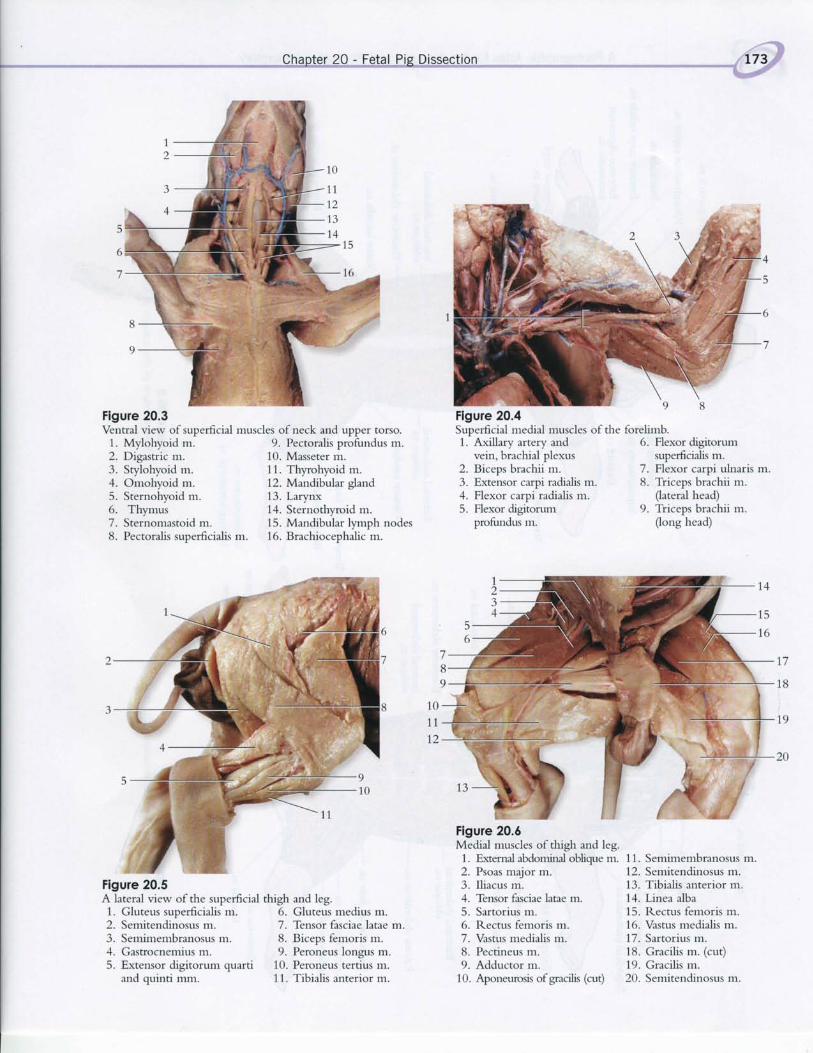
Flgure 20.3Ventral view of superficial muscles of neck and upper torso.
1. Mylohyoid m.2. Digastric m.3. Srylohyoid m.4. Omohyoid m.5. Sternohyoid m.6. Thymus7. Sternomastoid m.
9. Pectoralis profundus m.10. Masseter m.11. Thyrohyoid m.12. Mandibular gland13. Larynx14. Sternothyroid m.15. Mandibular lymph nodes
6. Gluteus medius m.7. tnsor fasciae latae m.8. Biceps femoris m.9. Peroneus longus m.
1. Axillary artery andvein, brachial plexus
2. Biceps brachii m.3. Exterxor carpi radialis m.4. Flexor carpi radialis m.5. Flexor digitorum
profundus m.
6. Flexor digrtorumsuperficialis m.
7. Flexor carpi ulnaris m.8. Triceps brachii m.
Qateral head)9. Triceps brachii m.
(long head)8. Pectoralis superficialis m. 16. Brachiocephalic m.
Figure 20.5A lateral view ofthe superficial thigh and leg.
Medial muscles of thigh and leg.1. External aMominal oblique m.2. Psoas major m.3. Iliacus m.4. Tensor fasciae latae m.5. Sartorius m.6. Rectus femoris m.7. Vastus medialis m.8. Pectineus m.9. Adductor m.
17
18
1011t2
1. Gluteus superficialis m.2. Semitendinosus m.3. Semimembranosus m.4. Gastrocnemius m.
11. Semimembranosus m.12. Semitendinosus m.13. Tibialis anterior m.14. Linea alba15. Rectus femoris m.16. Vastus medialis m.17. Sartorius m.18. Gracilis m. (cut)19. Gracilis m.5. Extensor digitorum quarti 10. Peroneus tertius m.
Figure 20.4Superficial medial muscles of the forelimb.
Flgure 20.6
and quinti mm. 11. Tibialis anterior m. 10. Aponeurosis of gracilis (cut) 20. Semitendinosus m.

;
-oq
C)U)
d!
.X
od
X '5o
.:Poox
o
o
o
d
d
d
g0d
s.
;
o
o
s!
-
do
9?5
i
-2
!d
9d
r i i ' i
;F F
F9 2A/- .=xd ql6o c
.a
XA
d
o
-
d;;
22!.=dA
v)i:
(t)
o
t
q(/)
bE;' {.1
2-
;s
a
6
06
!
.!.'|r
'a ; ;:F F
co Ix5 I; .v . -co o
?xts69?=XH
i
t
rJ
!
r2F
j j
.n o
23th3AE
d
'O t'
* :se! =L
o y5I atrb oFX XA14 H6
.j
do
a.i
I
Po
o
E
bo
d
,OJ
0
o
o
crRtEE=)ool !iE<
5b
,OJ
ot
C)
O
.Bct>di
obLC2cJct>E<
g
E
o(t)
q
qv)
j
o
qU)
r l
rFl
; i
o.6
98_, ! ! o
,4.
-
c)
o:
o.<
;
HF
=4eI
i
=_-2tr .EYxtroX-Y:GX' : g iEd h ?otr ! : c5d p h.g 't .eE=,4 r ! t r=HT
o.ror
Fxxt i
nYX
o
d
t r9c6
'69
c(h!a
.t)
!
ts6(A
!!
.,'1r
o
tu
th
d
o
6

3H'E 'E - c
€g:3sx'- ' - = FFE S€?9 H \ !hA
( / ) (n)JA
6r-dioic iid i iOl
o
o
YA
vk
Et s **. :EXcV,qa : ?.4q)-\ - -
_ 6i c.i + rri
bod6
- .d iE.; AX R=Eoott .H. r .E.q.q gFA\. / \J<
6F'-doio
oo
,o
o
6
B6q.9 d aa
R.i r : E E Fs E,tsflflS?ia
F. ' tdOlt . )$rn

i
hD
- -(>\S Ei l F He 5i .€EOFlr,<t4
!6Fdo.
o
'6
aac)d
c'Eedo I
EfgtE*e{ H s E= F io'5 I5 E 985 *; i66' j IPA , .. i". i +,' i
oa
d,Arh Ebg
-bo==f ,E.EE-rJ lJ ( ,
! . r i 6 NI
d
(JI
o
oF 6
- .9
+ci >x.xN= F ts E s-^
Ura>!! IE:eF-9.45 dF. l F; l i F lFtr> .F- 'zd ( \ t (O +

Flgure 20.12Thorax and neck regions ofthe fetal pig.
1. Larynx 5. Heart2. Thymus 6. Lung3. Lung4. Liver (cut)
7. Spleen (cut)
Figure 20.13A ventral view of the abdominal caviry of a fetal pig.1. Diaphragm2. Liver3. Gallbladder4. Umbilical vein
Figure 20.14Abdominal organs of the fetal pig.
1. Liver (cut)2. Small intestine3. Umbilical arteries4. Stomach (reflected)5. Spleen6. Pancreas
5. Small intestine6. Undescended testis7. Umbilical artery8. Urinary bladder
7. Kidney8. Large intestine9. (Jreter
10. Ductus (vas) deferens11. Urinary bladder

-.Af-h"e,J-eff?shls."Atles"-[sr"lhe.Arslqmy."en.C.P,.hr:jelp.s. Le.Fp-rstqry
60)0h
' I o 9.1.=HVHV
P o xc '
- 5.Y O O6Al l&&BO tr.) \O F.. O
d
o
d
0)E s!or
!k
.o: dEU9
AE 6.Ei{ * t E= Fdr a ) ' i : : :=YmJ* C.Y5 6'r .F cnnttr o
---=-c. . l
.o+
hgd
'5
da
-h, EEg.9 F
as Es: E ggJ HqEqqq S'UFJ<ts]- tJt lu-OO\Oic. lco+ro
€ si i i i -
dd
.ye9l |otsGFd
o atr 6E .= ' : ' l; 9t ?O r! O - i
.^ F B$5 st g *q > .1.= 'b0d ! >-tr._-*-=:5
- : Y. ,= 6 e- 6 d. igHnEF'6.Eetr-
: l i ; su g *Ef"qf"Y': . i E.X f l , \ ' , ' , \ ' tttl r
E<iNci+to\ot ' -@

Chapter 20 - Fetal Pig Dissection,.
t,1tr'l
Figure 20.17Urogenital systen.r of the fetal pig.1. Kidney2. Caudal (inferior) vena cava
3. (Jreter
4. Rectum (cut)5. Partially dissected testis
Renal veinDescending aortaDuctus deferensUrinary bladderUmbilical arteryEpididymis
Figure 20.18Urogenital system of the fetal pig.1. Umbilical cord 6.2. Right kidney 7.3. Ureter 8.4. Umbilical artery 9.5. Urinary bladder 10
PenisVas (ductus) deferensSpermatic cordRight testisEpididymis
6.7.8.9.
10.11.
Figure 20.19General structures of the fetalpig brain. Because the cerebrumis less deEned in pigs, the regionsare not known as lobes as theyare in humans.
1. Occipital region of cerebrum2. Cerebellum3. Medulla oblongata4. Spinal cord5. External acoustic meatus6. Longitudinal fissure7. Parietal region ofcerebrum8. Frontal region ofcerebrum9. Temporal region of cerebrum
10. Eye
hh
Related Documents