article nature genetics • volume 23 • october 1999 159 Fv2 encodes a truncated form of the Stk receptor tyrosine kinase Derek A. Persons 1 *, Robert F. Paulson 4 *, Melanie R. Loyd 2 , Mark T. Herley 2 , Sara M. Bodner 3 , Alan Bernstein 5 , Pamela H. Correll 4 & Paul A. Ney 2 *These authors contributed equally to this work. The Friend virus susceptibility 2 (Fv2) locus encodes a dominant host factor that confers susceptibility to Friend virus-induced erythroleukaemia in mice. We mapped Fv2 to a 1.0-Mb interval that also contained the gene (Ron) encoding the stem cell kinase receptor (Stk). A truncated form of Stk (Sf-stk), which was the most abundant form of Stk in Fv2-sensitive (Fv2 ss ) erythroid cells, was not expressed in Fv2 resistant (Fv2 rr ) cells. Enforced expression of Sf-stk conferred susceptibility to Friend disease, whereas targeted disruption of Ron caused resistance. We con- clude that the Fv2 locus encodes Ron, and that a naturally expressed, truncated form of Stk confers susceptibility to Friend virus-induced erythroleukaemia. Departments of 1 Experimental Hematology, 2 Biochemistry and 3 Pathology, St. Jude Children’s Research Hospital, Memphis, Tennessee 38105, USA. 4 Department of Veterinary Science, Pennsylvania State University, University Park, Pennsylvania 16802, USA. 5 Program in Molecular Biology and Cancer, Samuel Lunenfeld Research Institute and Department of Molecular and Medical Genetics, Mount Sinai Hospital, University of Toronto, Toronto, Ontario, Canada. Correspondence should be addressed to P.A.N. (e-mail: [email protected]). Introduction Mice infected with Friend virus complex develop acute ery- throblastosis that rapidly progresses to erythroleukaemia 1 . In the early stages of the disease, proliferating erythroblasts form discrete foci in the spleen that become confluent, causing mas- sive splenic enlargement 2 . Depending on the strain of virus (FVA or FVP), the proliferating erythroblasts undergo differen- tiation, and either anaemia or polycythaemia develops. In the later stages of the disease, clones of fully transformed erythrob- lasts emerge, leading to erythroleukaemia 3 . This progression from uncontrolled polyclonal proliferation to erythroleukaemia is accompanied by the acquisition of additional genetic muta- tions, including activation of Pu.1 (Spi-1), and inactivation of p53 (refs 4,5). The multistage nature of Friend disease has made it an important experimental model to identify the viral and host cellular events involved in leukaemic transformation and malignant progression 6 . Friend virus complex consists of a replication-competent helper virus, Friend murine leukaemia virus (F-MuLV) and a replication-defective virus, spleen focus-forming virus 7 (SFFV). Acute erythroblastosis in the early stages of Friend disease is caused by SFFV (refs 8,9). In contrast to other acutely oncogenic retroviruses, SFFV does not contain a mutated cellular proto- oncogene 10,11 . Rather, pathogenicity depends on a chimaeric retroviral envelope protein, gp55 (ref. 12). The mechanism of gp55-mediated erythroblastosis involves constitutive activation of the erythropoietin receptor (Epor). Membrane-bound gp55 dimers, representing less than 5% of gp55 protein produced, are required for pathogenicity 13 . gp55 from the polycythaemic strain of SFFV associates with and activates the mouse Epor in Ba/F3 cells 14 . Furthermore, gp55 can replace erythropoietin (Epo) in supporting the formation of erythroid colonies in vitro, but only in the presence of the Epor (ref. 15). A number of host genes have been identified that affect suscep- tibility to Friend disease. These can be divided into several cate- gories based on the mechanism of resistance. The first group consists of genes that interfere with the infection of target cells by Friend virus. Fv4 encodes an endogenous retroviral envelope protein that blocks cell-surface receptors 16 . Fv1 encodes a gag- related protein that interferes with the retroviral life cycle by an unknown mechanism 17 . The second group consists of genes that alter the immune response to Friend virus infection. Fv3 affects susceptibility to immunosuppression by F-MuLV, and two H2- linked loci, Rfv1 and Rfv2, affect the recovery from Friend virus infection 18,19 . The third group consists of genes that affect the progression of Friend disease. Fv5 determines whether FVP causes anaemia or polycythaemia in certain strains of mice 20 . Fv3 and Fv5 have not been clearly established as independent loci governing the response to Friend virus infection. Mutations in the genes flexed-tail (f), Mgf and Kit impede the progression of Friend disease 21–23 . These genes are required for normal erythro- poiesis, emphasizing the importance of the normal erythropoi- etic machinery for the development of Friend disease. Fv2 is another host gene that affects the progression of Friend disease 24 . Fv2 does not interfere with retroviral entry into cells or with the retroviral life cycle 25 . Rather, Fv2 appears to determine whether SFFV-infected erythroblasts proliferate in response to gp55. Experiments in chimaeric mice with Fv2 rr and Fv2 ss cells demonstrated that Friend virus infection causes selective expan- sion of Fv2 ss erythroid cells 26 . Thus, the effects of Fv2 are cell autonomous, not a property of the cellular environment. There is indirect evidence that Fv2 and gp55 interact, and that interaction of these proteins with Epor causes erythroid proliferation. Fv2- mediated resistance can be circumvented by deletions in the ecotropic domain of gp55, but the same mutants are less active than wild-type gp55 in Fv2 ss mice 27 . To understand the role of © 1999 Nature America Inc. • http://genetics.nature.com © 1999 Nature America Inc. • http://genetics.nature.com

Welcome message from author
This document is posted to help you gain knowledge. Please leave a comment to let me know what you think about it! Share it to your friends and learn new things together.
Transcript

article
nature genetics • volume 23 • october 1999 159
Fv2 encodes a truncated form ofthe Stk receptor tyrosine kinaseDerek A. Persons1*, Robert F. Paulson4*, Melanie R. Loyd2, Mark T. Herley2, Sara M. Bodner3,Alan Bernstein5, Pamela H. Correll4 & Paul A. Ney2
*These authors contributed equally to this work.
The Friend virus susceptibility 2 (Fv2) locus encodes a dominant host factor that confers susceptibility to Friend
virus-induced erythroleukaemia in mice. We mapped Fv2 to a 1.0-Mb interval that also contained the gene (Ron)
encoding the stem cell kinase receptor (Stk). A truncated form of Stk (Sf-stk), which was the most abundant form
of Stk in Fv2-sensitive (Fv2ss) erythroid cells, was not expressed in Fv2 resistant (Fv2rr) cells. Enforced expression of
Sf-stk conferred susceptibility to Friend disease, whereas targeted disruption of Ron caused resistance. We con-
clude that the Fv2 locus encodes Ron, and that a naturally expressed, truncated form of Stk confers susceptibility
to Friend virus-induced erythroleukaemia.
Departments of 1Experimental Hematology, 2Biochemistry and 3Pathology, St. Jude Children’s Research Hospital, Memphis, Tennessee 38105, USA.4Department of Veterinary Science, Pennsylvania State University, University Park, Pennsylvania 16802, USA. 5Program in Molecular Biology and Cancer,Samuel Lunenfeld Research Institute and Department of Molecular and Medical Genetics, Mount Sinai Hospital, University of Toronto, Toronto, Ontario,Canada. Correspondence should be addressed to P.A.N. (e-mail: [email protected]).
IntroductionMice infected with Friend virus complex develop acute ery-throblastosis that rapidly progresses to erythroleukaemia1. Inthe early stages of the disease, proliferating erythroblasts formdiscrete foci in the spleen that become confluent, causing mas-sive splenic enlargement2. Depending on the strain of virus(FVA or FVP), the proliferating erythroblasts undergo differen-tiation, and either anaemia or polycythaemia develops. In thelater stages of the disease, clones of fully transformed erythrob-lasts emerge, leading to erythroleukaemia3. This progressionfrom uncontrolled polyclonal proliferation to erythroleukaemiais accompanied by the acquisition of additional genetic muta-tions, including activation of Pu.1 (Spi-1), and inactivation ofp53 (refs 4,5). The multistage nature of Friend disease has madeit an important experimental model to identify the viral andhost cellular events involved in leukaemic transformation andmalignant progression6.
Friend virus complex consists of a replication-competenthelper virus, Friend murine leukaemia virus (F-MuLV) and areplication-defective virus, spleen focus-forming virus7 (SFFV).Acute erythroblastosis in the early stages of Friend disease iscaused by SFFV (refs 8,9). In contrast to other acutely oncogenicretroviruses, SFFV does not contain a mutated cellular proto-oncogene10,11. Rather, pathogenicity depends on a chimaericretroviral envelope protein, gp55 (ref. 12). The mechanism ofgp55-mediated erythroblastosis involves constitutive activationof the erythropoietin receptor (Epor). Membrane-bound gp55dimers, representing less than 5% of gp55 protein produced, arerequired for pathogenicity13. gp55 from the polycythaemic strainof SFFV associates with and activates the mouse Epor in Ba/F3cells14. Furthermore, gp55 can replace erythropoietin (Epo) insupporting the formation of erythroid colonies in vitro, but onlyin the presence of the Epor (ref. 15).
A number of host genes have been identified that affect suscep-tibility to Friend disease. These can be divided into several cate-gories based on the mechanism of resistance. The first groupconsists of genes that interfere with the infection of target cells byFriend virus. Fv4 encodes an endogenous retroviral envelopeprotein that blocks cell-surface receptors16. Fv1 encodes a gag-related protein that interferes with the retroviral life cycle by anunknown mechanism17. The second group consists of genes thatalter the immune response to Friend virus infection. Fv3 affectssusceptibility to immunosuppression by F-MuLV, and two H2-linked loci, Rfv1 and Rfv2, affect the recovery from Friend virusinfection18,19. The third group consists of genes that affect theprogression of Friend disease. Fv5 determines whether FVPcauses anaemia or polycythaemia in certain strains of mice20. Fv3and Fv5 have not been clearly established as independent locigoverning the response to Friend virus infection. Mutations inthe genes flexed-tail (f), Mgf and Kit impede the progression ofFriend disease21–23. These genes are required for normal erythro-poiesis, emphasizing the importance of the normal erythropoi-etic machinery for the development of Friend disease.
Fv2 is another host gene that affects the progression of Frienddisease24. Fv2 does not interfere with retroviral entry into cells orwith the retroviral life cycle25. Rather, Fv2 appears to determinewhether SFFV-infected erythroblasts proliferate in response togp55. Experiments in chimaeric mice with Fv2rr and Fv2ss cellsdemonstrated that Friend virus infection causes selective expan-sion of Fv2ss erythroid cells26. Thus, the effects of Fv2 are cellautonomous, not a property of the cellular environment. There isindirect evidence that Fv2 and gp55 interact, and that interactionof these proteins with Epor causes erythroid proliferation. Fv2-mediated resistance can be circumvented by deletions in theecotropic domain of gp55, but the same mutants are less activethan wild-type gp55 in Fv2ss mice27. To understand the role of
© 1999 Nature America Inc. • http://genetics.nature.com©
199
9 N
atu
re A
mer
ica
Inc.
• h
ttp
://g
enet
ics.
nat
ure
.co
m

article
160 nature genetics • volume 23 • october 1999
Fv2 in erythroid proliferation and leukaemic transformation, weundertook the positional cloning of Fv2. Here we report that Fv2encodes Stk, a member of the Met subfamily of receptor tyrosinekinases. Specifically, we show that susceptibility to Friend diseaseis conferred by expression of a truncated form of Stk that lacks anextracellular ligand-binding domain.
ResultsMapping and cloning the Fv2 intervalFv2 is located on the distal arm of mouse chromosome 9, appro-ximately 61 cM from the centromere28,29. We mapped Fv2 in theBXD series of recombinant inbred strains of mice30. The progen-itor strains for this series are C57BL/6 (Fv2rr) and DBA/2 (Fv2ss).We confirmed the previously reported strain distribution patternof Fv2 and typed two strains not included in the original series31
(Fig. 1a). To map Fv2, we selected 15 simple sequence lengthpolymorphisms (SSLPs) from a group of markers (a bin) nearFv2, established in a generic cross of mice32. Most of theseshowed a single crossover with Fv2 in the BXD2 strain and felltelomeric to Fv2 (below Fv2 in Fig. 1a). Some mapped still fur-ther telomeric of Fv2 (D9Mit200, D9Mit213, D9Mit20, D9Mit349and D9Mit243). To obtain markers on both sides of Fv2, we stu-died SSLPs from an adjacent bin, namely D9Mit51, D9Mit78 andD9Mit80. These SSLPs mapped centromeric to Fv2 (above Fv2 inFig. 1a). Of all the markers tested, only D9Mit359 matched thestrain distribution pattern of Fv2. These results indicated thatFv2 is in the interval between these two bins, which is 1.1 cM inthe MIT backcross. Our results agree with those of a recent back-cross that placed Fv2 between D9Mit78 and D9Mit212 (ref. 29).
To clone the Fv2 interval, we used bacterial artificial chromo-somes (BACs) to walk from D9Mit359 to the nearest recombi-nant markers on either side (Fig. 1b). For these experiments, werelied on a map provided by the MIT mouse genetic and physicalmapping project consisting of YAC contigs anchored to SSLPs
(ref. 32). D9Mit359 was identified in a YAC contig (WC-1111) that included D9Mit184 as well as several non-polymor-phic markers (23.MHAa89f11, X74736, 25.mHAa8b1 and38.MMHAP67FLE5). These markers were used to probe 129Sv/Jand C57BL/6 genomic DNA BAC libraries. We assembled fourBACs into a contig that spanned the Fv2 interval. On the telom-eric end, BAC 167/j20 contained the nearest recombinant SSLP(D9Mit184). A CA-repeat in the ubiquitin-specific protease gene(Unp (CA)n) also mapped telomeric of the Fv2 interval, estab-lishing this as the nearest recombinant marker on the telomericside. On the centromeric end, we identified sequences that over-lapped with the human lung cancer tumour-suppressor generegion33. The gene encoding the α-subunit of retinal transducin(Gnat1) is located 100 kb centromeric of this junction and is thenearest recombinant marker on the centromeric side.
Ron is a candidate gene for Fv2Pulse-field gel electrophoresis showed that the BACs ranged120–200 kb. Based on the number of BACs and their size, we esti-mated the Fv2 interval to be less than 1.0 Mb. To identify candi-date genes for Fv2, HindIII fragments of BACs in the Fv2 regionwere randomly subcloned and sequenced. We identified 12known genes by this approach; all of their human homologueswere located in the syntenic region of human chromosome3p21.3. One of the genes we identified was Ron. Mouse Stk is amember of the Met subfamily of receptor tyrosine kinases34. Stkis closely related to RON (its human homologue), avian v-seaand mouse Met (88%, 70% and 66% identical in the kinasedomain, respectively). The relationship to v-sea increased ourinterest in Stk, because v-sea causes erythroblastosis and anaemiain chickens35.
Stk was cloned from Lin–KIT+Sca+ mouse bone marrow cellsby PCR with degenerate primers to conserved sequences in thetyrosine kinase domain34. Ron has 19 exons36 (Fig. 2a), but in
Fig. 1 Fv2 is located between markers Gnat1 and D9Mit184. a, Strain distribution pat-tern of Fv2 and other markers in the BXD series of recombinant inbred strains. Thestrains (01–32) are shown vertically across the top. Markers are shown on the left. ‘D’indicates an allele from the DBA/2J (Fv2ss) progenitor strain. ‘B’ indicates an allele fromthe C57BL/6J (Fv2rr) strain. The bracket on the left shows a BAC contig that spans theFv2 interval from Gnat1 to D9Mit184. b, Map of the Fv2 interval. The BAC contig over-laps the human lung cancer tumour-suppressor gene region on the centromeric side.All BACs are from 129/SvJ DNA, except for BAC 197/f4, which is from C57BL/6J. Genesand markers that have been localized to BACs are shown vertically. Genes and theirprotein products are: CISH, cytokine inducible SH2-containing protein; Ron, stem cellkinase receptor; Ube1l, ubiquitin activating enzyme-E1 related protein; Traip, TRAF-interacting protein; Camkl, calmodulin binding kinase-like protein; Bsn, bassoon; Apeh,aminoacyl peptide hydrolase; Hgfl, hepatocyte growth factor-like protein; Dag1, dys-troglycan; Tctal, T-cell leukaemia translocation altered gene-like; and Unp, ubiquitinspecific protease.
a b
Ro
n
Trai
p
Bsn
Hg
fl
© 1999 Nature America Inc. • http://genetics.nature.com©
199
9 N
atu
re A
mer
ica
Inc.
• h
ttp
://g
enet
ics.
nat
ure
.co
m

article
nature genetics • volume 23 • october 1999 161
mouse erythroleukaemia (MEL) cells, the major expressedmRNA encodes a truncated form of the receptor, which starts inintron 10 (ref. 34). This short form of Stk (Sf-stk) lacks almostthe entire extracellular domain, but retains the transmembraneand tyrosine kinase domains. In primary fetal liver cells, Sf-stk isthe major expressed form of the gene (Fig. 2b). Notably, Sf-stkwas expressed in the fetal liver cells of BALB/c (Fv2ss), but notC57BL/6 (Fv2rr), mice. To test the association between Fv2 sus-ceptibility and Sf-stk expression, we performed RT-PCR on bonemarrow RNA from a panel of inbred strains of mice. Sf-stk wasexpressed in all Fv2ss strains (Fig. 2c). In contrast, Sf-stk expres-sion was decreased or absent in C57BL/6 (Fv2rr) and relatedstrains. Full-length Stk was expressed in all strains, regardless ofFv2 status. Thus, Fv2 susceptibility correlates with expression ofSf-stk in adult bone marrow cells, which are the target cells forFriend virus infection.
The strain-specific difference in Sf-stk expression we identifiedmay be due to a difference in activity of an alternate Stk pro-moter. To examine this possibility, we linked 1 kb of DNA con-taining the putative Sf-stk promoter to the luciferase gene andstably transfected the construct into MEL cells. DNA from anFv2ss strain (129/SvJ) had promoter activity that was sixfoldhigher than that from an Fv2rr strain (C57BL/6; Fig. 2d). Thisregion contains consensus binding sites for Sp1, Ets, Myb and
Gata proteins (Fig. 2e). Sequence analysis showed that threenucleotides were deleted from C57BL/6 DNA compared with129/SvJ. This deletion included the 5´ end of the published Sf-stkcDNA, as well as part of a consensus WGATAR binding site34. Todetermine if this deletion was associated with resistance at theFv2 locus, we screened a panel of 48 different strains and sub-strains of mice. The deletion was present exclusively in C57BL/6(Fv2rr) and related strains. When the strain distribution patternof this deletion was determined in the BXD series, it co-segre-gated with Fv2. Furthermore, when genomic DNA samples froma published backcross of 425 mice were screened for this deletion,it co-segregated with Fv2 (ref. 29, and data not shown). Theseexperiments confirm that Fv2 and Ron are genetically tightlylinked, and suggest that a 3-nt deletion in the Sf-stk promotercauses decreased Sf-stk expression and Fv2 resistance in C57BL/6and related strains.
Fv2 resistance was originally described in C57BL/6 mice buthas also been reported in Mus spretus, wild-derived mice37.Because M. spretus are genetically distinct from C57BL/6 mice,this provided an opportunity to further test the correlationbetween Sf-stk expression and Fv2 susceptibility. Sf-stk expres-sion was decreased in M. spretus bone marrow cells (Fig. 2c). Thethree nucleotides that were deleted in C57BL/6 strain DNA werepresent in M. spretus DNA, but there was a point mutation in a
b
Fig. 2 Sf-stk is not expressed in Fv2rr erythroidcells. a, Organization of mouse Ron. Exons arenumbered. TM is the transmembrane domain.Arrows show the 5´ end of the full-length (Stk)and truncated Ron cDNA (Sf-stk). b, Northernblot of E14.5 fetal liver poly(A) enriched RNAfrom BALB/c (Fv2ss) and C57BL/6 (Fv2rr) strains,probed with Sf-stk cDNA, then reprobed with β-actin. The full-length transcript (Stk) is 4.8 kb,and the short transcript (Sf-stk) is 1.9 kb. c, RT-PCR with primers specific for RNA encoding full-length Stk (Stk) and short-form Stk (Sf-stk). Thesource of RNA was mouse bone marrow fromeach of the strains shown across the top. d, Rela-tive promoter activity of nt 9,684–10,684 of Ron(ref. 36), from C57BL/6 (B6) and 129/SvJ (129)DNA, determined in pools of stably transfectedMEL cells (20 clones per pool, 5 pools per con-struct). e, Sequence of the putative Sf-stk pro-moter. The arrow marks the first nucleotide ofthe published Sf-stk cDNA (ref. 34). Consensustranscription factor binding sites are shown byboxes. The black box shows 3 nt that are deletedfrom the DNA of C57BL/6 and related strains.Intron 10 is underlined. The first two amino acidsof the predicted Sf-stk protein are shown (M,T).
a
c
d
e
M. s
pret
us
relative luciferase activity
Sf-stk
Sf-stk
Sf-stk
Stk
–179
–109
–39
+32
© 1999 Nature America Inc. • http://genetics.nature.com©
199
9 N
atu
re A
mer
ica
Inc.
• h
ttp
://g
enet
ics.
nat
ure
.co
m
article
162 nature genetics • volume 23 • october 1999
Myb consensus binding site (TAACGGTT→TAAAGGTT) thatalters a contact for Myb binding38. Introduction of this mutationinto 129/SvJ DNA reduced activity of the Sf-stk promoter to thelevel of C57BL/6 in MEL cells (data not shown). These resultsstrengthen the correlation between Sf-stk expression and Fv2susceptibility and suggest that M. spretus have an allele of Fv2r
that is distinct from C57BL/6.
Stk is required for susceptibility to Friend diseaseThe absence of Sf-stk transcripts in C57BL/6 erythroid cellsraised the possibility that Sf-stk expression is required for suscep-
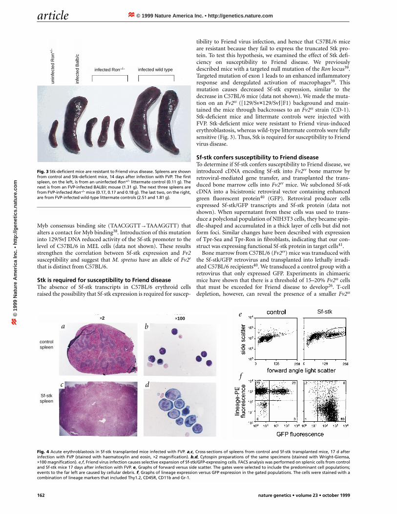
tibility to Friend virus infection, and hence that C57BL/6 miceare resistant because they fail to express the truncated Stk pro-tein. To test this hypothesis, we examined the effect of Stk defi-ciency on susceptibility to Friend disease. We previouslydescribed mice with a targeted null mutation of the Ron locus39.Targeted mutation of exon 1 leads to an enhanced inflammatoryresponse and deregulated activation of macrophages39. Thismutation causes decreased Sf-stk expression, similar to thedecrease in C57BL/6 mice (data not shown). We made the muta-tion on an Fv2ss ([129/Sv×129/SvJ]F1) background and main-tained the mice through backcrosses to an Fv2ss strain (CD-1).Stk-deficient mice and littermate controls were injected withFVP. Stk-deficient mice were resistant to Friend virus-inducederythroblastosis, whereas wild-type littermate controls were fullysensitive (Fig. 3). Thus, Stk is required for susceptibility to Friendvirus disease.
Sf-stk confers susceptibility to Friend diseaseTo determine if Sf-stk confers susceptibility to Friend disease, weintroduced cDNA encoding Sf-stk into Fv2rr bone marrow byretroviral-mediated gene transfer, and transplanted the trans-duced bone marrow cells into Fv2rr mice. We subcloned Sf-stkcDNA into a bicistronic retroviral vector containing enhancedgreen fluorescent protein40 (GFP). Retroviral producer cellsexpressed Sf-stk/GFP transcripts and Sf-stk protein (data notshown). When supernatant from these cells was used to trans-duce a polyclonal population of NIH3T3 cells, they became spin-dle-shaped and accumulated in a thick layer of cells but did notform foci. Similar changes have been described with expressionof Tpr-Sea and Tpr-Ron in fibroblasts, indicating that our con-struct was expressing functional Sf-stk protein in target cells41.
Bone marrow from C57BL/6 (Fv2rr) mice was transduced withthe Sf-stk/GFP retrovirus and transplanted into lethally irradi-ated C57BL/6 recipients40. We transduced a control group with aretrovirus that only expressed GFP. Experiments in chimaericmice have shown that there is a threshold of 15–20% Fv2ss cellsthat must be exceeded for Friend disease to develop26. T-celldepletion, however, can reveal the presence of a smaller Fv2ss
Fig. 3 Stk-deficient mice are resistant to Friend virus disease. Spleens are shownfrom control and Stk-deficient mice, 14 days after infection with FVP. The firstspleen, on the left, is from an uninfected Ron+/– littermate control (0.11 g). Thenext is from an FVP-infected BALB/c mouse (1.31 g). The next three spleens arefrom FVP-infected Ron–/– mice (0.17, 0.17 and 0.18 g). The last two, on the right,are from FVP-infected wild-type littermate controls (2.51 and 1.81 g).
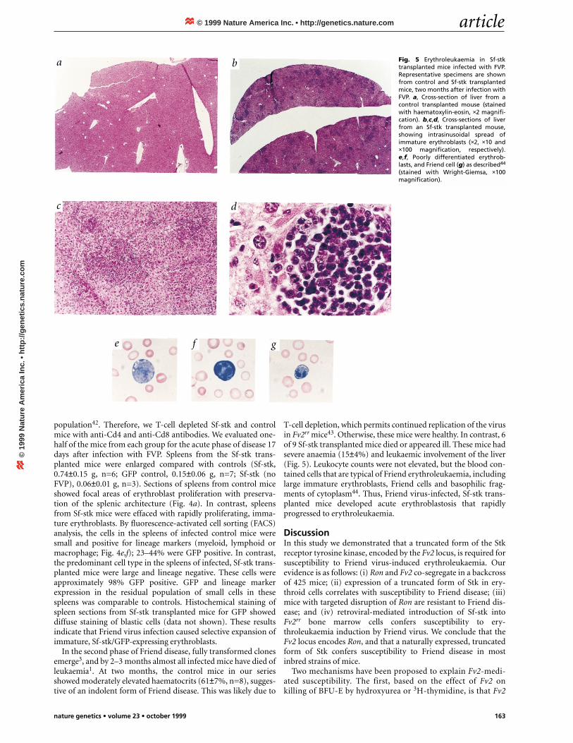
Fig. 4 Acute erythroblastosis in Sf-stk transplanted mice infected with FVP. a,c, Cross-sections of spleens from control and Sf-stk transplanted mice, 17 d afterinfection with FVP (stained with haematoxylin and eosin, ×2 magnification). b,d, Cytospin preparations of the same specimens (stained with Wright-Giemsa,×100 magnification). e,f, Friend virus infection causes selective expansion of Sf-stk/GFP-expressing cells. FACS analysis was performed on splenic cells from controland Sf-stk mice 17 days after infection with FVP. e, Graphs of forward versus side scatter. The gates were selected to include the predominant cell populations;events to the far left are caused by cellular debris. f, Graphs of lineage expression versus GFP expression in the gated populations. The cells were stained with acombination of lineage markers that included Thy1.2, CD45R, CD11b and Gr-1.
unin
fect
ed R
on+
/–
infe
cted
Bal
b/c
infected Ron–/– infected wild type
×2 ×100
a b
c d
e
f
Sf-stkspleen
Sf-stk
controlspleen
© 1999 Nature America Inc. • http://genetics.nature.com©
199
9 N
atu
re A
mer
ica
Inc.
• h
ttp
://g
enet
ics.
nat
ure
.co
m
article
nature genetics • volume 23 • october 1999 163
population42. Therefore, we T-cell depleted Sf-stk and controlmice with anti-Cd4 and anti-Cd8 antibodies. We evaluated one-half of the mice from each group for the acute phase of disease 17days after infection with FVP. Spleens from the Sf-stk trans-planted mice were enlarged compared with controls (Sf-stk,0.74±0.15 g, n=6; GFP control, 0.15±0.06 g, n=7; Sf-stk (noFVP), 0.06±0.01 g, n=3). Sections of spleens from control miceshowed focal areas of erythroblast proliferation with preserva-tion of the splenic architecture (Fig. 4a). In contrast, spleensfrom Sf-stk mice were effaced with rapidly proliferating, imma-ture erythroblasts. By fluorescence-activated cell sorting (FACS)analysis, the cells in the spleens of infected control mice weresmall and positive for lineage markers (myeloid, lymphoid ormacrophage; Fig. 4e,f); 23–44% were GFP positive. In contrast,the predominant cell type in the spleens of infected, Sf-stk trans-planted mice were large and lineage negative. These cells wereapproximately 98% GFP positive. GFP and lineage markerexpression in the residual population of small cells in thesespleens was comparable to controls. Histochemical staining ofspleen sections from Sf-stk transplanted mice for GFP showeddiffuse staining of blastic cells (data not shown). These resultsindicate that Friend virus infection caused selective expansion ofimmature, Sf-stk/GFP-expressing erythroblasts.
In the second phase of Friend disease, fully transformed clonesemerge3, and by 2–3 months almost all infected mice have died ofleukaemia1. At two months, the control mice in our seriesshowed moderately elevated haematocrits (61±7%, n=8), sugges-tive of an indolent form of Friend disease. This was likely due to
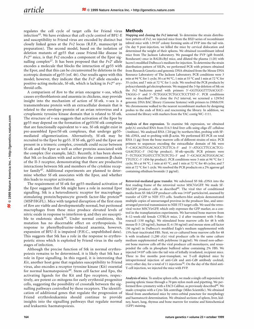
T-cell depletion, which permits continued replication of the virusin Fv2rr mice43. Otherwise, these mice were healthy. In contrast, 6of 9 Sf-stk transplanted mice died or appeared ill. These mice hadsevere anaemia (15±4%) and leukaemic involvement of the liver(Fig. 5). Leukocyte counts were not elevated, but the blood con-tained cells that are typical of Friend erythroleukaemia, includinglarge immature erythroblasts, Friend cells and basophilic frag-ments of cytoplasm44. Thus, Friend virus-infected, Sf-stk trans-planted mice developed acute erythroblastosis that rapidlyprogressed to erythroleukaemia.
DiscussionIn this study we demonstrated that a truncated form of the Stkreceptor tyrosine kinase, encoded by the Fv2 locus, is required forsusceptibility to Friend virus-induced erythroleukaemia. Ourevidence is as follows: (i) Ron and Fv2 co-segregate in a backcrossof 425 mice; (ii) expression of a truncated form of Stk in ery-throid cells correlates with susceptibility to Friend disease; (iii)mice with targeted disruption of Ron are resistant to Friend dis-ease; and (iv) retroviral-mediated introduction of Sf-stk intoFv2rr bone marrow cells confers susceptibility to ery-throleukaemia induction by Friend virus. We conclude that theFv2 locus encodes Ron, and that a naturally expressed, truncatedform of Stk confers susceptibility to Friend disease in mostinbred strains of mice.
Two mechanisms have been proposed to explain Fv2-medi-ated susceptibility. The first, based on the effect of Fv2 onkilling of BFU-E by hydroxyurea or 3H-thymidine, is that Fv2
Fig. 5 Erythroleukaemia in Sf-stktransplanted mice infected with FVP.Representative specimens are shownfrom control and Sf-stk transplantedmice, two months after infection withFVP. a, Cross-section of liver from acontrol transplanted mouse (stainedwith haematoxylin-eosin, ×2 magnifi-cation). b,c,d, Cross-sections of liverfrom an Sf-stk transplanted mouse,showing intrasinusoidal spread ofimmature erythroblasts (×2, ×10 and×100 magnification, respectively).e,f, Poorly differentiated erythrob-lasts, and Friend cell (g) as described44
(stained with Wright-Giemsa, ×100magnification).
a b
c d
e f g
© 1999 Nature America Inc. • http://genetics.nature.com©
199
9 N
atu
re A
mer
ica
Inc.
• h
ttp
://g
enet
ics.
nat
ure
.co
m
article
164 nature genetics • volume 23 • october 1999
regulates the cell cycle of target cells for Friend virusinfection45. We have evidence that cell-cycle control of BFU-Eand susceptibility to Friend virus are encoded by distinct butclosely linked genes at the Fv2 locus (R.F.P., manuscript inpreparation). The second model, based on the isolation ofdeletion mutants of gp55 that cause Friend-like disease inFv2rr mice, is that Fv2 encodes a component of the Epor sig-nalling complex27. It has been proposed that the Fv2r alleleencodes a molecule that blocks the interaction of gp55 withthe Epor, and that this can be circumvented by deletions in theecotropic domain of gp55 (ref. 46). Our results agree with thismodel; however, they indicate that the Fv2s allele encodes apositive-acting molecule, Sf-stk, which is lacking in Fv2rr ery-throid cells.
A comparison of Ron to the avian oncogene v-sea, whichcauses erythroblastosis and anaemia in chickens, may provideinsight into the mechanism of action of Sf-stk. v-sea is atransmembrane protein with an extracellular domain that isrelated to the envelope protein of an avian retrovirus and acytoplasmic tyrosine kinase domain that is related to Sf-stk.The structure of v-sea suggests that activation of the Epor bygp55 may depend on the formation of gp55/Sf-stk complexesthat are functionally equivalent to v-sea. Sf-stk might exist inpre-assembled Epor/Sf-stk complexes, that undergo gp55-mediated oligomerization. Alternatively, Sf-stk may berecruited to the Epor by gp55. If Sf-stk, gp55 and the Epor arepresent in a trimeric complex, crosstalk could occur betweenSf-stk and the Epor as well as other proteins associated withthe complex (for example, Jak2). Recently, it has been shownthat Stk co-localizes with and activates the common β-chainof the Il-3 receptor, demonstrating that there are productiveinteractions between Stk and members of the cytokine recep-tor family47. Additional experiments are planned to deter-mine whether Sf-stk associates with the Epor, and whetherthis leads to receptor activation.
The requirement of Sf-stk for gp55-mediated activation ofthe Epor suggests that Stk might have a role in normal Eporsignalling. Stk is a heterodimeric receptor for macrophage-stimulating protein/hepatocyte growth factor-like protein48
(MSP/HGFL). Mice with targeted disruption of the first exonof Ron are viable and developmentally normal, but peritonealmacrophages from these mice produce elevated levels ofnitric oxide in response to interferon-γ, and they are suscepti-ble to endotoxic shock39. Under normal conditions, thismutation has no discernable effect on erythropoiesis. Inresponse to phenylhydrazine-induced anaemia, however,expansion of BFU-E is impaired (P.H.C., unpublished data).This suggests that Stk has a role in the response to erythro-poietic stress which is exploited by Friend virus in the earlystages of infection.
Although the precise function of Stk in normal erythro-poiesis remains to be determined, it is likely that Stk has arole in Epor signalling. In this regard, it is interesting thatKit, another host gene that regulates susceptibility to Friendvirus, also encodes a receptor tyrosine kinase (Kit) essentialfor normal haematopoiesis49. Stem cell factor and Epo, theactivating ligands for the Kit and Epo receptors, respec-tively, are potent co-mitogens for early erythroid progenitorcells, suggesting the possibility of crosstalk between the sig-nalling pathways controlled by these receptors. The identifi-cation of additional genes that regulate the progression ofFriend erythroleukaemia should continue to provideinsights into the signalling pathways that regulate normaland leukaemic haematopoiesis.
MethodsMapping and cloning the Fv2 interval. To determine the strain distribu-tion pattern of Fv2, we injected mice from the BXD series of recombinantinbred mice with 1.9×10³ colony forming units of FVP into the tail vein.On day 9 post-injection, we killed the mice by cervical dislocation anddetermined the weight of their spleens. We obtained recombinant inbredmice from The Jackson Laboratory. We passaged the FVP (gift fromM.Bondurant) once in BALB/cByJ mice, and diluted the plasma (1:20) withIscove’s modified Dulbecco’s medium for injection. To determine the straindistribution pattern of SSLPs, we performed PCR with primers obtainedfrom Research Genetics and genomic DNA obtained from the Mouse DNAResource Laboratory of The Jackson Laboratory. PCR conditions were 3min at 94 oC for 1 cycle; 30 s at 94 oC, 1 min at 55 oC and 1 min at 72 oC for35 cycles; and 7 min at 72 oC for 1 cycle. We resolved the PCR products bypolyacrylamide gel electrophoresis. We mapped the 3-bp deletion of Stk onthe Fv2 backcross panel with primers 5´–GGTGGGTTTAACGGT-TAGGG–3´ and 5´–TCTGGGCTCTGCCTCCTTAT–3´. PCR conditionswere as described29. To clone the Fv2 interval, we screened a 129/SvJgenomic DNA BAC library (Genome Systems) with primers to D9Mit359.We chromosome walked to the nearest recombinant markers by designingprobes to the ends of BACs and rescreening the library. In addition, wescreened the library with markers from the YAC contig WC-1111.
Analysis of Ron expression. To examine Stk expression, we obtainedpoly(A) enriched RNA from E14.5 fetal liver cells using oligo dT cellulose(Ambion). We analysed RNA (∼ 10 µg) by northern blot, probing with SF-Stk cDNA, and re-probing with β-actin. We performed RT-PCR on totalRNA (1 µg) from the bone marrow cells of different strains of mice. PCRprimers to sequences encoding the extracellular domain of Stk were5´–CAGCAGTGGACAGCCTGTTCA–3´ and 5´–ATGCCTTCCACTCG-GAAGTGC–3´ (542-bp product). Sf-stk–specific PCR primers were5´–TCTGGCTGATCCTTCTGTCTG–3´ and 5´–GCAGCAGTGGGACACTTGTCC–3´ (456-bp product). PCR conditions were 3 min at 94 oC for 1cycle; 30 s at 94 oC, 1 min at 65 oC, and 1 min at 72 oC for 40 cycles; and 7min at 72 oC for 1 cycle. We resolved the PCR products on a 2% agarose gelcontaining ethidium bromide (1 µg/ml).
Retroviral-mediated gene transfer. We subcloned SF-Stk cDNA into thefirst reading frame of the retroviral vector MSCVirGFP. We made SF-Stk/GFP producer cells as described40. The viral titer of conditionedmedia from SF-Stk/GFP producer cells was 1×10⁶ particles/ml, assessed bytransfer of GFP to NIH 3T3 cells. Southern-blot analysis demonstratedmultiple copies of unrearranged provirus in the producer line, and unre-arranged proviral transmission to NIH 3T3 target cells. We used the retro-viral vector MSCVirGFP, which only expresses the GFP marker, as a con-trol in the transplantation experiments. We harvested bone marrow from8–12-week-old female C57BL/6 mice, 2 d after treatment with 5-fluo-rouracil (150 mg/kg). We stimulated bone marrow cells for 48 h withmouse Il-3 (20 ng/ml), human IL-6 (50 ng/ml) and mouse stem cell factor(50 ng/ml) in Dulbecco’s modified Eagle’s medium supplemented with15% heat-inactivated FBS. Next, we co-cultured bone marrow cells for 48h with irradiated (1,200 cGy) viral producer cells in the same culturemedium supplemented with polybrene (6 µg/ml). We rinsed non-adher-ent bone marrow cells off the viral producer cell monolayers, and resus-pended the cells in phosphate buffered saline containing 2% FBS. Weinjected 5×10⁶ cells into the tail vein of lethally irradiated, recipient mice.Three to five months post-transplant, we T-cell depleted mice byintraperitoneal injection of anti-Cd4 and anti-Cd8 antibody cocktail,every other day, for a total of 11 injections50. On the day of the third anti-T-cell injection, we injected the mice with FVP.
Analysis of mice. To analyse spleen cells, we made a single cell supension bypassing splenic tissue through a 70 µm nylon mesh and pipetting. We per-formed flow cytometry with a FACS Calibur, as previously described40. Wemade cytospins with a Cyto-Tek centrifuge (Miles Scientific). We obtainedblood from anesthetized mice by retro-orbital puncture for morphologyand haematocrit determination. We obtained sections of spleen, liver, kid-ney, heart, lung, thymus and bone marrow for routine and histochemical(GFP) staining.
© 1999 Nature America Inc. • http://genetics.nature.com©
199
9 N
atu
re A
mer
ica
Inc.
• h
ttp
://g
enet
ics.
nat
ure
.co
m
article
nature genetics • volume 23 • october 1999 165
Stk-deficient mice. Stk-deficient mice have been described39. Stk expres-sion is disrupted by partial deletion of the first exon, and insertion of lacZand a neomycin resistance cassette. We injected Stk-deficient mice and lit-termate controls with FVP at 5 months of age. We killed mice 14 d afterinjection with FVP to evaluate their spleens.
AcknowledgementsWe thank A. Troutman and J. Swift for technical support; J. Riberdy andP. Doherty for anti-Cd4 and anti-Cd8 antibody; J. Ihle, M. Bondurant and
M. Hayman for reviewing the manuscript; and A. Nienhuis and G. Grosveldfor additional support. This work was supported by NIH Cancer CenterSupport Grant No. P30 CA21765, the American Lebanese Syrian AssociatedCharities (ALSAC), an American Society of Hematology Junior FacultyScholarship Award to P.H.C. and a Terry Fox Program Project Grant from theNational Cancer Institute of Canada to A.B.
Received 23 July; accepted 31 August 1999.
1. Friend, C. Cell-free transmission in adult swiss mice of a disease having thecharacter of a leukemia. J. Exp. Med. 105, 307–318 (1956).
2. Axelrad, A.A. & Steeves, R.A. Assay for Friend leukemia virus: rapid quantitativemethod based on enumeration of macroscopic spleen foci in mice. Virology 24,513–518 (1964).
3. Mager, D., MacDonald, M.E., Robson, I.B., Mak, T.W. & Bernstein, A. Clonalanalysis of the late stages of erythroleukemia induced by two distinct strains ofFriend leukemia virus. Mol. Cell. Biol. 1, 721–730 (1981).
4. Moreau-Gachelin, F., Tavitian, A. & Tambourin, P. Spi-1 is a putative oncogene invirally induced murine erythroleukaemias. Nature 331, 277–280 (1988).
5. Mowat, M., Cheng, A., Kimura, N., Bernstein, A. & Benchimol, S. Rearrangementsof the cellular p53 gene in erythroleukaemic cells transformed by Friend virus.Nature 314, 633–636 (1985).
6. Ben-David, Y. & Bernstein, A. Friend virus-induced erythroleukemia and themultistage nature of cancer. Cell 66, 831–834 (1991).
7. Steeves, R.A. Editorial: spleen focus-forming virus in Friend and Rauscherleukemia virus preparations. J. Natl Cancer Inst. 54, 289-297 (1975).
8. Wolff, L. & Ruscetti, S. Malignant transformation of erythroid cells in vivo byintroduction of a nonreplicating retrovirus vector. Science 228, 1549–1552 (1985).
9. Berger, S.A., Sanderson, N., Bernstein, A. & Hankins, W.D. Induction of the earlystages of Friend erythroleukemia with helper-free Friend spleen focus-formingvirus. Proc. Natl Acad. Sci. USA 82, 6913–6917 (1985).
10. Clark, S.P. & Mak, T.W. Complete nucleotide sequence of an infectious clone ofFriend spleen focus-forming provirus: gp55 is an envelope fusion glycoprotein.Proc. Natl Acad. Sci. USA 80, 5037–5041 (1983).
11. Amanuma, H., Katori, A., Obata, M., Sagata, N. & Ikawa, Y. Complete nucleotidesequence of the gene for the specific glycoprotein (gp55) of Friend spleen focus-forming virus. Proc. Natl Acad. Sci. USA 80, 3913–3917 (1983).
12. Aizawa, S. et al. Env-derived gp55 gene of Friend spleen focus-forming virusspecifically induces neoplastic proliferation of erythroid progenitor cells. EMBO J.9, 2107–2116 (1990).
13. Kabat, D. Molecular biology of Friend viral erythroleukemia. Curr. Top. Microbiol.Immunol. 148, 1–42 (1989).
14. Li, J.P., D’Andrea, A.D., Lodish, H.F. & Baltimore, D. Activation of cell growth bybinding of Friend spleen focus-forming virus gp55 glycoprotein to theerythropoietin receptor. Nature 343, 762–764 (1990).
15. Constantinescu, S.N. et al. The anemic Friend virus gp55 envelope protein induceserythroid differentiation in fetal liver colony-forming units-erythroid. Blood 91,1163–1172 (1998).
16. Kai, K., Sato, H. & Odaka, T. Relationship between the cellular resistance to Friendmurine leukemia virus infection and the expression of murine leukemia virus-gp70-related glycoprotein on cell surface of BALB/c-Fv-4wr mice. Virology 150,509–512 (1986).
17. Best, S., Le Tissier, P., Towers, G. & Stoye, J.P. Positional cloning of the mouseretrovirus restriction gene Fv1. Nature 382, 826–829 (1996).
18. Kumar, V., Resnick, P., Eastcott, J.W. & Bennett, M. Mechanism of geneticresistance to Friend virus leukemia in mice. V. Relevance of Fv-3 gene in theregulation of in vivo immunosuppression. J. Natl Cancer Inst. 61, 1117–1123(1978).
19. Chesebro, B. & Wehrly, K. Rfv-1 and Rfv-2, two H-2-associated genes thatinfluence recovery from Friend leukemia virus-induced splenomegaly. J. Immunol.120, 1081–1085 (1978).
20. Shibuya, T. & Mak, T.W. A host gene controlling early anaemia or polycythaemiainduced by Friend erythroleukaemia virus. Nature 296, 577–579 (1982).
21. Axelrad, A. Genetic and cellular basis of susceptibility or resistance to Friendleukemia virus infection in mice. Can. Cancer Conf. 8, 313–343 (1969).
22. Steeves, R.A., Bennett, M., Mirand, E.A. & Cudkowicz, G. Genetic control by the Wlocus of susceptibility to (Friend) spleen focus-forming virus. Nature 218, 372–374(1968).
23. Bennett, M., Steeves, R.A., Cudkowicz, G., Mirand, E.A. & Russell, L.B. Mutant Slalleles of mice affect susceptibility to Friend spleen focus-forming virus. Science162, 564–565 (1968).
24. Lilly, F. Fv-2: identification and location of a second gene governing the spleen focusresponse to Friend leukemia virus in mice. J. Natl Cancer Inst. 45, 163–169 (1970).
25. Geib, R.W., Dizik, M., Anand, R. & Lilly, F. Infection and transformation of Fv-2rr
erythroprogenitor cells with Friend virus. Virus Res. 8, 327–333 (1987).26. Behringer, R.R. & Dewey, M.J. Cellular site and mode of Fv-2 gene action. Cell 40,
441–447 (1985).27. Hoatlin, M.E. et al. Deletions in one domain of the Friend virus-encoded
membrane glycoprotein overcome host range restrictions for erythroleukemia.J. Virol. 69, 856–863 (1995).
28. Blake, J.A., Eppig, J.T., Richardson, J.E. & Davisson, M.T. The Mouse GenomeDatabase (MGD): a community resource. Status and enhancements. The MouseGenome Informatics Group. Nucleic Acids Res. 26, 130–137 (1998).
29. Paulson, R.F. & Bernstein, A. A genetic linkage map of the mouse chromosome 9region encompassing the Friend virus susceptibility gene 2. Mamm. Genome 9,381–384 (1998).
30. Taylor, B.A. Origins of Inbred Mice (ed. Morse, H.C.) 423–438 (Academic Press,New York, 1978).
31. Eliott, R.W., Hohman, C., Romijko, C., Louis, P. & Lilly, F. Electrophoresis (ed.Catsimpoolas, N.) 261–274 (Elsevier North Holland Biomedical Press, New York,1978).
32. Dietrich, W. et al. A genetic map of the mouse suitable for typing intraspecificcrosses. Genetics 131, 423–447 (1992).
33. Wei, M.H. et al. Construction of a 600-kilobase cosmid clone contig andgeneration of a transcriptional map surrounding the lung cancer tumorsuppressor gene (TSG) locus on human chromosome 3p21.3: progress toward theisolation of a lung cancer TSG. Cancer Res. 56, 1487–1492 (1996).
34. Iwama, A., Okano, K., Sudo, T., Matsuda, Y. & Suda, T. Molecular cloning of anovel receptor tyrosine kinase gene, STK, derived from enriched hematopoieticstem cells. Blood 83, 3160–3169 (1994).
35. Smith, D.R., Vogt, P.K. & Hayman, M.J. The v-sea oncogene of avianerythroblastosis retrovirus S13: another member of the protein-tyrosine kinasegene family. Proc. Natl Acad. Sci. USA 86, 5291–5295 (1989).
36. Waltz, S.E. et al. Characterization of the mouse Ron/Stk receptor tyrosine kinasegene. Oncogene 16, 27–42 (1998).
37. Kozak, C.A. Analysis of wild-derived mice for Fv-1 and Fv-2 murine leukemia virusrestriction loci: a novel wild mouse Fv-1 allele responsible for lack of host rangerestriction. J. Virol. 55, 281–285 (1985).
38. Ogata, K. et al. Solution structure of a specific DNA complex of the Myb DNA-binding domain with cooperative recognition helices. Cell 79, 639–648 (1994).
39. Correll, P.H. et al. Deregulated inflammatory response in mice lacking theSTK/RON receptor tyrosine kinase. Genes Funct. 1, 69–83 (1997).
40. Persons, D.A. et al. Enforced expression of the GATA-2 transcription factor blocksnormal hematopoiesis. Blood 93, 488–499 (1999).
41. Santoro, M.M., Collesi, C., Grisendi, S., Gaudino, G. & Comoglio, P.M. Constitutiveactivation of the RON gene promotes invasive growth but not transformation.Mol. Cell. Biol. 16, 7072–7083 (1996); erratum: 17, 1758 (1997).
42. Behringer, R.R. & Dewey, M.J. Cellular site and mode of Fv-2 gene action. II.Conditional protection of Fv-2ss cells by admixture with Fv-2rr cells. Exp. Hematol.17, 330–334 (1989).
43. Van der Gaag, H.C. & Axelrad, A.A. Friend virus replication in normal andimmunosuppressed C57BL/6 mice. Virology 177, 837–839 (1990).
44. Metcalf, D., Furth, J. & Buffett, R. Pathogenesis of mouse leukemia caused byFriend virus. Cancer Res. 19, 52–58 (1959).
45. Suzuki, S. & Axelrad, A.A. Fv-2 locus controls the proportion of erythropoieticprogenitor cells (BFU-E) synthesizing DNA in normal mice. Cell 19, 225–236(1980).
46. Hoatlin, M.E. & Kabat, D. Host-range control of a retroviral disease: Frienderythroleukemia. Trends Microbiol. 3, 51–57 (1995).
47. Mera, A., Suga, M., Ando, M., Suda, T. & Yamaguchi, N. Induction of cell shapechanges through activation of the IL-3 common β-chain receptor by the RONreceptor-type tyrosine kinase. J. Biol. Chem. 274, 15766–15774 (1999).
48. Gaudino, G. et al. RON is a heterodimeric tyrosine kinase receptor activated bythe HGF homologue MSP. EMBO J. 13, 3524–3532 (1994).
49. Chabot, B., Stephenson, D.A., Chapman, V.M., Besmer, P. & Bernstein, A. Theproto-oncogene c-kit encoding a transmembrane tyrosine kinase receptor mapsto the mouse W locus. Nature 335, 88–89 (1988).
50. Riberdy, J.M. et al. Protection against a lethal avian influenza A virus in amammalian system. J. Virol. 73, 1453–1459 (1999).
© 1999 Nature America Inc. • http://genetics.nature.com©
199
9 N
atu
re A
mer
ica
Inc.
• h
ttp
://g
enet
ics.
nat
ure
.co
m
Related Documents

![Fv2 resnick 5e [solucionario]](https://static.cupdf.com/doc/110x72/53ff95e78d7f724c088b4689/fv2-resnick-5e-solucionario.jpg)


![DBPIA-NURIMEDIAacml.gnu.ac.kr/download/Publications/28.pdf39 6 2011. 6 Navier-Stokes Eulerian Eulerian Bourgault [7] 01 FLUENT* FENSAP-ICE 3.1 Truncated Flapped Flapol Truncated Truncated](https://static.cupdf.com/doc/110x72/6064d2c624aba96be8533943/dbpia-39-6-2011-6-navier-stokes-eulerian-eulerian-bourgault-7-01-fluent-fensap-ice.jpg)
