Fusion of uniluminal vascular spheroids: a model for assembly of blood vessels Paul A. Fleming 1 , W. Scott Argraves 1 , Carmine Gentile 1 , Adrian Neagu 2,3 , Gabor Forgacs 3,4,5 , and Christopher J. Drake 1 1 Regenerative Medicine and Cell Biology, Medical University of South Carolina, Charleston, SC 29455 2 Victor Babes University of Medicine and Pharmacy Timisoara, P-ta Eftimie Murgu Nr. 2, 300041 Timisoara, Romania 3 Department of Physics and Biology, University of Missouri Columbia, MO 65201. 4 Department of Biological Sciences, University of Missouri Columbia, MO 65201. 5 Department of Biomedical Engineering, University of Missouri Columbia, MO 65201. Abstract Here, we evaluated the self-assembly properties of uniluminal vascular spheroids having outer layers of vascular smooth muscle cells and a contiguous inner layer of endothelial cells lining a central lumen. We showed that while pairs of uniluminal vascular spheroids suspended in culture medium fused to form a larger diameter spheroidal structure, spheroids in collagen hydrogels formed elongated structures. These findings highlight the potential use of uniluminal vascular spheroids as modules to engineer blood vessels. We also demonstrate that uniluminal vascular spheroid fusion conforms to models describing the coalescence of liquid drops. Furthermore, the fusion of uniluminal vascular spheroids in vitro closely resembled the in vivo process by which the descending aorta forms from the fusion of the paired dorsal aortae during embryonic development. Together, the findings indicate that tissue liquidity underlies uniluminal vascular spheroid fusion and that in vivo anastomosis of blood vessels may involve a similar mechanism. Keywords spheroids; tissue liquidity; blood vessels; dorsal aorta; angiogenesis; differential adhesion; anastomosis Introduction Recently, Gentile et al. (2008) described the generation of uniluminal vascular spheroids from embryonic mouse allantois tissue cultured in hanging drops with VEGF supplemented medium. The resulting uniluminal vascular spheroids exhibited morphological, immunohistochemical, and physiological properties of a blood vessel. Uniluminal vascular spheroids are characterized by an outer layer of smooth muscle cells (SMCs) expressing smooth muscle myosin, SM22-α, and SMαA and an inner endothelial cell (EC) layer expressing PECAM-1, CD34, VE-cadherin which lines the central lumen. Furthermore, the Address correspondence to: Christopher J. Drake, Ph.D., Department of Cell Biology and Anatomy, Charleston, SC 29425, Tel.: (843) 792-1692, Fax: (843) 792-0664, [email protected]. NIH Public Access Author Manuscript Dev Dyn. Author manuscript; available in PMC 2011 September 20. Published in final edited form as: Dev Dyn. 2010 February ; 239(2): 398–406. doi:10.1002/dvdy.22161. NIH-PA Author Manuscript NIH-PA Author Manuscript NIH-PA Author Manuscript

Welcome message from author
This document is posted to help you gain knowledge. Please leave a comment to let me know what you think about it! Share it to your friends and learn new things together.
Transcript
Fusion of uniluminal vascular spheroids: a model for assemblyof blood vessels
Paul A. Fleming1, W. Scott Argraves1, Carmine Gentile1, Adrian Neagu2,3, GaborForgacs3,4,5, and Christopher J. Drake1
1Regenerative Medicine and Cell Biology, Medical University of South Carolina, Charleston, SC294552Victor Babes University of Medicine and Pharmacy Timisoara, P-ta Eftimie Murgu Nr. 2, 300041Timisoara, Romania3Department of Physics and Biology, University of Missouri Columbia, MO 65201.4Department of Biological Sciences, University of Missouri Columbia, MO 65201.5Department of Biomedical Engineering, University of Missouri Columbia, MO 65201.
AbstractHere, we evaluated the self-assembly properties of uniluminal vascular spheroids having outerlayers of vascular smooth muscle cells and a contiguous inner layer of endothelial cells lining acentral lumen. We showed that while pairs of uniluminal vascular spheroids suspended in culturemedium fused to form a larger diameter spheroidal structure, spheroids in collagen hydrogelsformed elongated structures. These findings highlight the potential use of uniluminal vascularspheroids as modules to engineer blood vessels. We also demonstrate that uniluminal vascularspheroid fusion conforms to models describing the coalescence of liquid drops. Furthermore, thefusion of uniluminal vascular spheroids in vitro closely resembled the in vivo process by which thedescending aorta forms from the fusion of the paired dorsal aortae during embryonic development.Together, the findings indicate that tissue liquidity underlies uniluminal vascular spheroid fusionand that in vivo anastomosis of blood vessels may involve a similar mechanism.
Keywordsspheroids; tissue liquidity; blood vessels; dorsal aorta; angiogenesis; differential adhesion;anastomosis
IntroductionRecently, Gentile et al. (2008) described the generation of uniluminal vascular spheroidsfrom embryonic mouse allantois tissue cultured in hanging drops with VEGF supplementedmedium. The resulting uniluminal vascular spheroids exhibited morphological,immunohistochemical, and physiological properties of a blood vessel. Uniluminal vascularspheroids are characterized by an outer layer of smooth muscle cells (SMCs) expressingsmooth muscle myosin, SM22-α, and SMαA and an inner endothelial cell (EC) layerexpressing PECAM-1, CD34, VE-cadherin which lines the central lumen. Furthermore, the
Address correspondence to: Christopher J. Drake, Ph.D., Department of Cell Biology and Anatomy, Charleston, SC 29425, Tel.: (843)792-1692, Fax: (843) 792-0664, [email protected].
NIH Public AccessAuthor ManuscriptDev Dyn. Author manuscript; available in PMC 2011 September 20.
Published in final edited form as:Dev Dyn. 2010 February ; 239(2): 398–406. doi:10.1002/dvdy.22161.
NIH
-PA Author Manuscript
NIH
-PA Author Manuscript
NIH
-PA Author Manuscript
ECs and SMCs of uniluminal vascular spheroids, like their counterparts in blood vessels,form a functional unit that was capable of regulating vascular tone (Gentile et al., 2008).
Uniluminal vascular spheroids have a number of potential utilities. The fact that uniluminalvascular spheroids are generated from mouse embryonic allantoic tissue affords theopportunity to investigate the consequences of genetic mutation or exogenous agents onaspects of blood vessel formation recapitulated during spheroid morphogenesis. This couldinclude studies of integrated SMC-EC signaling in response to gene mutation or biologicalagents. Uniluminal vascular spheroids could be used as modules to engineer blood vessels.A requirement for this usage is the ability to join uniluminal vascular spheroids together.
While a number of studies have shown that tissue spheroids can fuse (Gordon et al., 1972;Jakab et al., 2004; Perez-Pomares and Foty, 2006), the fusion of spheroids having a largecentral lumen, such as uniluminal vascular spheroids, has not been described. Herein, wehave investigated the ability of uniluminal vascular spheroids to fuse and preserve themorphological architecture of the original uniluminal vascular spheroids. The findings notonly highlight the potential use of uniluminal vascular spheroid fusion as an approach toengineer blood vessels, they also provide insights into the fundamental mechanism by whichblood vessels form in vivo.
ResultsUniluminal vascular spheroids fuse to form a single, larger diameter spheroid
As described by Gentile et al. (2008), treatment of E8.5 mouse allantois-derived spheroidswith VEGF in hanging drop culture results in the formation of uniluminal vascular spheroids(Fig. 1A and B). These spheroids are characterized by an outer layer of smooth muscle cellsexpressing SM22α, and an inner cavity lined by ECs expressing -CD34 (Fig. 1B). Toinvestigate the capacity of uniluminal vascular spheroids to fuse, pairs of uniluminalvascular spheroids of equal size were placed in contact with one another in hanging dropculture and incubated for various periods of time. As early as one hour in culture, theuniluminal vascular spheroids became joined together, such that when they were physicallymanipulated they moved as one structure (Fig. 2A). Within 1–3 h of culture, amorphological change in joined pairs of spheroids was evident, such that pairs appearedpeanut shaped (Fig. 2B). Within 3–8 h of culture, the peanut shaped structures transitionedinto ovoid structures (Fig. 2C). Finally, between 8 and 12 h, the ovoid structures transitionedinto spheroids (Fig. 2D).
To evaluate the impact of the fusion process on the endothelial and smooth musclecomponents, uniluminal vascular spheroids were examined at various stages in the processof fusion after labeling with antibodies to the EC protein, PECAM-1, and the SMC protein,SMαA. Within the first hour of culture, SMαA-expressing cells adjoining uniluminalvascular spheroids are seen to be closely associated in the flat disc shaped plane of contact(Fig. 2E). Closer examination of the interface between adjoining uniluminal vascularspheroids revealed that the outer layers of SMαA-expressing cells of adjoining uniluminalvascular spheroids appeared to be separate (Fig. 2I). There was no apparent intermingling ofSMαA-expressing cells in adjoining uniluminal vascular spheroids. This interpretation wassupported by examination of the fusion of uniluminal vascular spheroids pairs labeled witheither green fluorescent or red fluorescent dye, which showed a paucity of intermingling ofgreen and red cells of the fusing spheroids along the interfacial plane (Fig. 3).
Within 1–3 h of culture, the interfacial disc of SMαA-expressing cells had increased in size(Fig. 2F and J). As was observed at earlier periods of culture, the SMαA-expressing cells ofadjoining uniluminal vascular spheroids appeared to be separate (Fig. 2J). Within 3–8 h of
Fleming et al. Page 2
Dev Dyn. Author manuscript; available in PMC 2011 September 20.
NIH
-PA Author Manuscript
NIH
-PA Author Manuscript
NIH
-PA Author Manuscript
culture, the interfacial disc of SMαA-expressing cells, which was previously continuous,was disrupted at the center of the disc (Fig. 2K, arrow; Supplemental Figure 1). In theSupplemental Figure 1, we capture an apparent fusion of the endothelial layers of theadjacent spheroids in the space created by the disruption of the SMαA-expressing celllayers. A disruption in the continuity of the endothelial layers associated with interfacingSMαA-expressing cells is evident in the fusing spheroids shown in Figure 2G (arrow).Between 8 and 12 h, no vestige of either the SMαA-expressing cell layers or EC layers thatcomprised the interface disc at earlier time periods was evident, and the resulting compositestructure resembled the architecture of the spheroids from which it was generated (Fig. 2Hand L). To establish that composite spheroids possessed a single central lumen, fast greendye was injected into the center of a spheroid. The injected dye was observed to fill a centralspace, the boundaries of which were defined by the thickness of the smooth muscle andendothelial layers, as evidenced by light microscopy (Fig. 4).
Uniluminal vascular spheroid fusion conforms to a tissue liquidity modelPrevious studies examining the fusion of solid tissue spheroids composed of smooth musclecells revealed that their fusion conforms to a model of the coalescence of highly viscousliquid drops (Flenner et al., 2007). To evaluate if uniluminal vascular spheroid fusion alsoconforms to models describing the coalescence of immiscible liquid drops, we compared thetime (t) evolution of the radius (r0) of the circular contact area between merging uniluminalvascular spheroids at 1-hour intervals for a period of 12 h. Assuming volume conservation,the coalescence of two identical, highly viscous liquid drops can be described by the formula[r(t) /R0]2 = 22/3 (1−e−t/τ) where R0 is the initial radius of identical spheroid pair (Fig. 5).The characteristic fusion time τ = 1.9ηR0/γ. Here r(t), R0, γ and η denote respectively theinstantaneous radius of the contact disk between fusing spheroids, their initial radius, surfacetension and viscosity. The expression for τ is consistent with earlier theoretical results forthe coalescence of highly viscous liquid drops (Frenkel, 1945).
We next studied the process of uniluminal vascular spheroid fusion using liquidity-basedMonte Carlo simulations (Metropolis et al., 1953; Graner and Glazier, 1992; Glazier andGraner, 1993; Jakab et al., 2004; Neagu et al., 2005). To simulate the fusion of twouniluminal vascular spheroids, sites on a three dimensional lattice were associated with avariable σ, which accounted for the possible occupancy of the site by one of the fourexperimental materials; the external medium/culture medium (σ = 1), SMCs (σ = 2), ECs (σ= 3) or the internal medium/lumen (σ = 4). In the Monte Carlo steps (MCS), each cell at aninterface had the opportunity to move once, exchanging its position with a neighbor chosenby chance. Figure 6 shows snapshots of the states obtained in the simulation, depictinginterior and exterior views of the spheroids. The initial state (Fig. 6A and E) is depicted as alattice representation of two adjacent uniluminal vascular spheroids, each of 30 cell sizes indiameter. The outer layer of each spheroid is made up of 4975 model cells (red) and theinner layer (green) in made up of 2018 model cells (green). Within 5×10⁴ MCS, partialfusion emerges (Fig. 6B and F). After 45×104 MCS, the lumens fuse (Fig. 6C and G), andthe equilibrium state (Fig. 6D and H) of minimal adhesive energy is reached in about 2×10⁶
MCS.
Casting spheroids in collagen hydrogels acts to modulate uniluminal vascular spheroidfusion
We next evaluated the consequence of performing uniluminal vascular spheroid fusionunder conditions in which the spheroids are capable of engaging in adhesive interactionswith their culture environment. Pairs of spheroids were cast in a hydrogel composed of typeI collagen. In contrast to the spherical structure generated by fusion of pairs of spheroids inhanging drop culture for 12 h (Fig. 7A), the culture in collagen hydrogels resulted in the
Fleming et al. Page 3
Dev Dyn. Author manuscript; available in PMC 2011 September 20.
NIH
-PA Author Manuscript
NIH
-PA Author Manuscript
NIH
-PA Author Manuscript
formation of an ovoid structure (Fig. 7B). Longer periods of culture in the collagenhydrogels (i.e., 24 h) showed that ovoid shape structure was maintained (data not shown).Immunolabeling of the ovoids with antibodies to PECAM-1 and SMαA, revealed thepresence of a continuous PECAM-1 expressing endothelium lining an ovoid shaped lumensurrounded by a SMαA-expressing smooth muscle cell layer. Fast green dye was injectedinto the center of the ovoid shape structures, and filled a central space consistent with therebeing a single central lumen (Fig. 7C and D).
Fusion of uniluminal vascular spheroids recapitulates aspects of the in vivo fusion of thebilateral dorsal aortae
To determine whether the process of fusion of uniluminal vascular spheroids bore similarityto the process by which large caliber vessels are formed in vivo, we analyzed the formationof the descending aorta during development. In higher vertebrates, the descending aorta isformed in a cranial to caudal direction by the joining of bilateral blood vessels called thedorsal aortae. The cranial to caudal progression makes it possible to study the temporalevolution of this process. We therefore performed DIC image analysis of the forming aortain serial cross sections of E9.5 mouse embryos. In the caudal region of the embryo, thepaired aortae can be seen as separate blood vessels, along either side of the midline (Fig. 8A,asterisks). Moving cranially, the two aortae can be seen to be closely juxtaposed along themidline (Fig. 8B, asterisks). Further in the cranial direction, progressive steps in the fusionof the aortae could be observed (Fig. 8C–F), including a step in which the joined aortae, asobserved in the cross section, exhibit a peanut shaped structure with an intervening cellularseptum (Fig. 8C, asterisks). This septum gradually becomes discontinuous (Fig. 8D,arrowhead) and eventually is absent in more cranial sections (Fig. 8E–F). It is also evidentin the more cranial section that the shape of the fusing aortae transitions from an ovoid to around structure (Fig. 8E–F).
DiscussionHerein we show that spheroids having an outer layer of SMCs and an EC-lined centrallumen, when juxtaposed, fuse to form larger diameter spheroids. The configuration of celllayers in the resulting composite spheroid resembles that of each of the starting spheroids.The mechanism that acts to allow this fusion process to occur in a manner in which tissueorganization is retained and intermixing of SMCs and ECs is prevented appears consistentwith the principles underlying the differential adhesion hypothesis (DAH) (Steinberg, 1963;Steinberg, 1970; Foty et al., 1994; Forgacs et al., 1998; Foty and Steinberg, 2005; Steinberg,2007). This hypothesis states that cells rearrange to maximize their interadhesive bondingwhile minimizing the free energy of the system.
Our finding that the fusion of uniluminal vascular spheroids conforms to a model describingthe coalescence of liquid drops is also consistent with the DAH, as well as with other studiesshowing that multicellular systems display liquid-like behaviors. For example, tissues (e.g.,limb bud mesoderm, proepicardium, and cardiac cushion tissue) assume a spherical shapewhen placed in hanging drop culture (Heintzelman et al., 1978; Perez-Pomares et al., 2006;Jakab et al., 2008), tissue spheroids display liquid-like viscoelastic properties (Foty et al.,1994; Foty et al., 1996; Forgacs et al., 1998; Krieg et al., 2008; Schötz et al., 2008) andtissue spheroids coalesce when brought together (Perez-Pomares et al., 2006; Jakab et al.,2008). At the molecular level, the coalescence involves adhesive interactions mediated byproteins such as cadherins (Steinberg and Takeichi, 1994). Indeed, using the system we havedemonstrated that the process of uniluminal vascular spheroid fusion is dependent on proteinsynthesis (Supplemental Figure 2). The in vitro system that we have defined should allowfurther molecular dissection of the process of fusion of uniluminal vascular spheroids.
Fleming et al. Page 4
Dev Dyn. Author manuscript; available in PMC 2011 September 20.
NIH
-PA Author Manuscript
NIH
-PA Author Manuscript
NIH
-PA Author Manuscript
We have also shown that morphological similarities exist between uniluminal vascularspheroid fusion and the anastomosis of large caliber blood vessels during development. Thedorsal aorta is well known to form through the fusion of two separate blood vessels, thepaired dorsal aortae. Mechanistically, the two blood vessels, which are initially on eitherside of the embryonic axis, are brought to the midline through embryo folding eventsassociated with the formation of the gut (Fishman and Chien, 1997; Grapin-Botton, 2005).Then, as we show, the paired dorsal aortae fuse along the embryonic axis in a cranial tocaudal direction. Based on our studies of the fusion of uniluminal vascular spheroids, it issuggestive that the in vivo fusion of blood vessels can be mimicked in vitro.
Previous studies of embryonic vasculogenesis showed that small caliber endothelial bloodvessels join together to form a primordial vascular network (Drake and Jacobson, 1988;Poole and Coffin, 1988; Noden, 1989; Drake et al., 1997). These findings raised the questionas to whether fusion was restricted to nascent blood vessels lacking a smooth muscle layer.The results presented here suggest that a SMC layer is not an impediment to fusion. Thiswas certainly evident with the fusion of uniluminal vascular spheroids and appears to be thecase in vivo with the fusion of the paired dorsal aortae. Indeed, previous studies have shownthat the dorsal aortae are invested with cells expressing SMC markers (e.g., smooth muscleα-actinin and fibulin-1) (Hungerford et al., 1996; Hungerford et al., 1997). The process bywhich the SMCs at the interface of adjoining uniluminal vascular spheroids or blood vesselsare displaced remains to be established. Examination of static images showing theprogression of fusion of uniluminal vascular spheroids suggests that the SMCs are displaced(pushed or pulled) toward the outer margins of the interfacial disc. The underlying forcesultimately disrupt the interfacial SMC and EC layers.
The vascular fusion principles established through these studies may be applicable to thefusion of blood vessels occurring in all types of neovascular settings (i.e., normal andpathological angiogenesis and anastomosis). Large diameter blood vessels may be formedfrom fusion of adjacent small diameter blood vessels in a manner similar to the formation ofthe dorsal aorta. For example, at early stages during the formation of the endocardium,multiple endothelial tubes can be observed at the midline within the forming heart (Drake etal., 2006). These tubes are later transformed into a single blood vessel, the definitiveendocardium. The vascular fusion principle may also be applicable to formation of bloodvessel connections associated with sprouting. For example, in the context of embryonicblood vessel formation, others and we have described the process by which luminizedendothelial sprouts extending from one blood vessel within a primary vascular networkconnect with an adjacent blood vessel (Drake and Jacobson, 1988; Drake and Little, 1995;Drake et al., 1997; Kamei et al., 2006). The mechanism by which a luminized vascularsprout physically connects to another blood vessel has not been elucidated. We speculatethat the interface between a luminized sprout extending to an adjacent blood vessel isanalogous to the interface between two uniluminal vascular spheroids, or the interfacebetween the dorsal aortae (Supplemental Figure 3). Adhesive bonding and motility of thecells from the merging vascular structures lead to rearrangement of ECs at the interfaceplane. The corresponding reduction in the free energy of the system drives the joining of thesprout and existing vessel.
A number of approaches to fabricate blood vessels are currently being pursued. Theseinclude seeding of ECs or EC progenitors onto natural scaffolds (i.e., collagen, laminin andMatrigel), which act to promote organization of the cells into nascent endothelial tubes(Montesano et al., 1983; Kubota et al., 1988; Levenberg et al., 2002; Ferreira et al., 2007),the use of natural or synthetic polymeric tubular scaffolds which are seeded with ECs andsmooth muscle cells (Weinberg and Bell, 1986; Niklason et al., 1999; Watanabe et al.,2001), the use of sheets of vascular cells which are formed into tubes (L'Heureux et al.,
Fleming et al. Page 5
Dev Dyn. Author manuscript; available in PMC 2011 September 20.
NIH
-PA Author Manuscript
NIH
-PA Author Manuscript
NIH
-PA Author Manuscript
1998; L'Heureux et al., 2006) and a bioprinting approach based on the fusion of cellularspheroids or tubes (Norotte et al., 2009). The findings presented here are suggestive ofconditions that permit the formation of luminized tubular vessel segments from modulesarranged in a linear fashion. Indeed, we show that fusion of uniluminal vascular spheroidsembedded in a collagen gel leads to the formation of elongated structures that retainmorphological features of blood vessels. Linear structures of varying diameter could beproduced through fusion of uniluminal vascular spheroid modules of controlled diameter.Indeed, we have shown that the process of uniluminal vascular spheroid fusion is scalable(Supplemental Figure 4), allowing the formation of uniluminal vascular spheroids of anydesired diameter. Thus, employing pre-fabricated uniluminal vascular spheroids as buildingblocks together with a defined three-dimensional adhesive substrate (e.g., ECM) represents anew approach to engineering of diversely sized blood vessels.
Experimental ProceduresAnimals and reagents
Timed-pregnant mice were purchased from Harlan (Indianapolis, IN). Protocols governinganimal use in this study were submitted to and approved by the MUSC Institutional AnimalCare and Use Committee. Further, MUSC has maintained ongoing, full accreditation fromAAALAC since November 1987. Rat antibodies to mouse PECAM-1 were obtained fromBD PharMingen (San Diego, CA). Rat antibodies to mouse CD34 were purchased fromResearch Diagnostics Inc. (Flanders, NJ). Cy3 conjugated anti-smooth muscle alpha actin(SMαA, clone 1A4) was purchased from Sigma-Aldrich (St. Louis, MO). Rabbit anti-mouseSM22-alpha (SM22α) was purchased from Abcam (Cambridge, MA) and rabbit anti-humansmooth muscle myosin was purchased from Biomedical Technologies, Inc. (Stoughton,MA). Fluorescently conjugated secondary antibodies were obtained from JacksonImmunoResearch Labs, Inc. (West Grove, PA). Recombinant human VEGF165 was obtainedfrom R&D Systems (Minneapolis, MN).
The generation and fusion of uniluminal vascular spheroidsUniluminal vascular spheroids were generated as previously described (Gentile et al., 2008).Fusion of uniluminal vascular spheroids in hanging drops was achieved by placing pairs ofvascular spheroids into 25 µl of VEGF-supplemented (0.3–1µg/ml) culture medium(DMEM supplemented with 10% FBS (Gibco), 1% penicillin/streptomycin/L-glutamine(Gibco)) and then spotting the spheroid-medium droplet onto the inverted lid of a 60mmPetri dish. The lids were then re-inverted, placed onto culture dishes and the vascularspheroids cultured for 18 h (37°C, 5% CO2) in a humidified chamber.
Fusion of uniluminal vascular spheroids in three-dimensional hydrogels was achieved bycasting pairs of spheroids in drops of unpolymerized type I collagen. The collagen (1.3 mg/ml) was prepared on ice by combining 0.4 ml of rat tail-derived type I collagen (3.13 mg/ml;BD Biosciences, Bedford, MA) with 0.5 ml of culture medium and 0.05 ml of sterile water.Two vascular spheroids were pipetted into a 25 µl drop of the collagen solution on a 35mmtissue culture dish. The dish was inverted allowing the spheroids to move to the bottom ofthe drop before the gel polymerized. Once collagen polymerization was complete, the dishwas inverted and culture medium supplemented with 0.3 µg/ml rhVEGF was slowly addedso as not to dislodge the hydrogels. The hydrogel-embedded spheroids were then incubatedfor 18 h at 37°C, 5% CO2. Fixation and immunolabeling of spheroids were accomplished aspreviously described (Gentile et al., 2008).
Fleming et al. Page 6
Dev Dyn. Author manuscript; available in PMC 2011 September 20.
NIH
-PA Author Manuscript
NIH
-PA Author Manuscript
NIH
-PA Author Manuscript
Microscopic analysis of individual and fused uniluminal vascular spheroidsConventional fluorescence or differential interference contrast images of uniluminalvascular spheroids and the structures generated by their fusion were obtained at 22°C usinga Leica DMR research grade microscope equipped with a SPOT-RT camera (VashawScientific, Raleigh, NC). Images were acquired using SPOT-RT 3.5.7 software. Laserconfocal microscopic images were acquired using Zeiss 5X/0.15 and 10X/0.30 objectives ona BioRad MRC 1024 laser-scanning confocal microscope (BioRad, Microscopy Division,Cambridge, MA) equipped with Lasersharp 2000 software (BioRad Cell Science Division,Hemel Hempstead, UK) or using a 20X/0.70 HC PL APO objective on a Leica TCS SP2AOBS equipped with Leica confocal software (Leica Microsystems, Inc., Exton, PA).Confocal z-series were projected using Image J 1.31v (National Institutes of Health).Montages of microscopic images were generated using Adobe Photoshop 7.0 software(Adobe Systems, Inc., San Jose, CA).
Microinjection of fused uniluminal vascular spheroidsSpheroids generated from the fusion of two uniluminal vascular spheroids in hanging dropculture were placed into a 60 mm Petri dish containing Dulbecco’s phosphate bufferedsaline supplemented with 0.7 mM CaCl2 and 0.5 mM MgCl2 (EPBS; 22°C). The tip of aglass micropipette containing fast green dye was inserted into the spheroid and ~1–2 µl ofdye (0.25% in EPBS) was microinjected into the central cavity using a pneumatically drivenPico-Injector (Medical Systems, Greenvale, NY).
Fusion of fluorescently labeled uniluminal vascular spheroidsUniluminal vascular spheroids of similar diameters were transferred into Petri dishescontaining either 5 ml of 5 mM Cell Tracker Green CMFDA fluorescent dye (InvitrogenMolecular Probes; Eugene, OR) in complete medium (DMEM supplemented with 10% FBS(Gibco), 1% penicillin/streptomycin/L-glutamine (Gibco)) or 5 ml of 5 mM Cell TrackerRed CMTPX fluorescent dye (Invitrogen Molecular Probes; Eugene, OR) in completemedium at 37°C in 5% CO2. The medium was agitated at 5-minute intervals. Following a 45min incubation period, the spheroids were rinsed in complete medium (30 min; 37°C, 5%CO2 in air), and then rinsed in sterile EPBS (2 min; 37°C). Pairs of labeled spheroids, onewith red fluorescent dye and one with green fluorescent dye, were placed into hanging dropsas described above. At different times during the fusion process, pairs of fusing spheroidswere removed from culture and fixed in 4% paraformaldehyde in dPBS (25°C, 20 min).After fixation, the fused spheroids were rinsed in dPBS, mounted on glass slides andexamined using a confocal microscope.
Studies of shape evolution during uniluminal vascular spheroid fusionPairs of uniluminal vascular spheroids were incubated over 14 h in culture. Lightmicroscopic images were used to measure R0 (i.e., the average radii in µm of the pair at timezero) and the instantaneous radius, r(t) of the circular interfacial area between pairs of fusingspheroids at hourly intervals during the culture. Using this data, [r(t)/R0]2 was plotted asfunction of time and fit to the function (1−e−t/τ) (Flenner et al., 2007).
Modeling the fusion of uniluminal vascular spheroidsMonte Carlo simulation of the fusion of two uniluminal vascular spheroids was performedusing methods similar to those used to simulate the fusion of two homogenous cellularaggregates (Flenner et al., 2007). Sites on a three-dimensional lattice were associated with avariable σ, which accounted for the possible occupancy of the site by one of the fourmaterials, culture medium (σ = 1), smooth muscle cells (σ = 2), ECs (σ = 3) and the lumen
Fleming et al. Page 7
Dev Dyn. Author manuscript; available in PMC 2011 September 20.
NIH
-PA Author Manuscript
NIH
-PA Author Manuscript
NIH
-PA Author Manuscript
(σ = 4). The interaction energy of the system is given by, where J is themagnitude of the energy of interaction between the particles that are immediately adjacent toone another, r and r′ label lattice sites and <, > denotes that the sum runs over first, secondand third nearest neighbors. The terms in the above sum may take either of the 10 valuesJ(i,j) = −εij (i = 1,2,3,4; j = 1,2,3,4), where the ε’s are elements of a symmetric matrix, positiveparameters that account for the strengths of all possible pair wise interactions between thefour phases. Separating interfacial energy (the energy of particles along the (six) interfacesbetween the four possible phases) and the bulk energy (the energy of everything that is not
along the interface) we obtain (up to an irrelevant additive constant), , where thesummation is over only such pairs of indices in which i < j (6 terms), Nij stands for the totalnumber of bonds between the corresponding phases and γij = (εii + εjj)/2 −εij is proportionalto the interfacial tension between phases i and j (Israelachvili, 1997). Structure formationwas simulated by the Monte Carlo method using the Metropolis algorithm (Metropolis et al.,1953). During a Monte Carlo step (MCS), each cell at an interface (but not the medium orlumen particle) had the opportunity to move once, exchanging its position with a neighborchosen by chance. The energy change, ΔE, corresponding to each move was calculated andthe new configuration accepted with a probability P =1 if ΔE ≤ 0 or P = exp(−βΔE) if ΔE >0. Here β = 1/ET is a measure of the spontaneous, cytoskeleton-driven motion of cells. Theaverage biological fluctuation energy of a cell, ET, was shown earlier to be analogous to thethermal energy, kBT, of true liquid molecules (Mombach et al., 1995). Its value depends oncell type and has been previously estimated for mixed populations of chick embryonicneural and pigmented retinal cells (Beysens et al., 2000). In the simulations only thedimensionless combinations γ̃ij = γij/ET appear.
Immunohistological analysis of mouse dorsal aorta formationE9.5 embryos were dissected from timed pregnant females, fixed in 4% paraformaldehyde(45 min., 22°C), and processed for histological analysis as follows. Embryos were removedfrom fixative, rinsed in PBSA (10 min., 22°C), and then dehydrated through a series ofethanol solutions (70%, 95%, 100%, 100%; 20 min each). Following dehydration, theembryos were infiltrated first with a 1:1 mixture of 100% ethanol and Histoclear II clearingsolvent (National Diagnostics, Atlanta, GA) for 30 minutes (22°C), followed by infiltrationwith 100% Histoclear II clearing solvent for 40 minutes (22°C). The embryos were theninfiltrated with paraffin using Histoclear:paraffin mixtures (3:1, 1:1, 1:3; 0:4, 1 hour each,60°C). The paraffin embedded embryos were then sectioned (3 µm) using a Leica RM 2125microtome (Vashaw Scientific), mounted on glass slides, cleared, and cover slipped fordifferential interference contrast microscopic analysis.
Supplementary MaterialRefer to Web version on PubMed Central for supplementary material.
AcknowledgmentsThis work was supported by the NIH HL57375, NIH HL80168 (CJD); NIH HL061873, HL095067 (WSA), theNational Science Foundation Grant EEC-0244045 and FIBR-0526854 (A.N. and G.F.). The authors thank Dr.Vladimir Mironov for helpful discussions during the course of these studies. The authors also thank Cynthia K.Gittinger for her assistance with histology and Brent A. Wilkerson for his assistance with confocal microscopy.
Fleming et al. Page 8
Dev Dyn. Author manuscript; available in PMC 2011 September 20.
NIH
-PA Author Manuscript
NIH
-PA Author Manuscript
NIH
-PA Author Manuscript
ReferencesBeysens DA, Forgacs G, Glazier JA. Cell sorting is analogous to phase ordering in fluids. Proc Natl
Acad Sci U S A. 2000; 97:9467–9471. [PubMed: 10944216]Drake CJ, Brandt SJ, Trusk TC, Little CD. TAL1/SCL is expressed in endothelial progenitor cells/
angioblasts and defines a dorsal-to-ventral gradient of vasculogenesis. Dev Biol. 1997; 192:17–30.[PubMed: 9405094]
Drake CJ, Jacobson AG. A survey by scanning electron microscopy of the extracellular matrix andendothelial components of the primordial chick heart. The Anatomical Record. 1988; 222:391–400.[PubMed: 3228207]
Drake CJ, Little CD. Exogenous vascular endothelial growth factor induces malformed and hyperfusedvessels during embryonic neovascularization. Proc Natl Acad Sci U S A. 1995; 92:7657–7661.[PubMed: 7543999]
Drake CJ, Wessels A, Trusk T, Little CD. Elevated vascular endothelial cell growth factor affectsmesocardial morphogenesis and inhibits normal heart bending. Dev Dyn. 2006; 235:10–18.[PubMed: 16170779]
Ferreira LS, Gerecht S, Shieh HF, Watson N, Rupnick MA, Dallabrida SM, Vunjak-Novakovic G,Langer R. Vascular progenitor cells isolated from human embryonic stem cells give rise toendothelial and smooth muscle like cells and form vascular networks in vivo. Circ Res. 2007;101:286–294. [PubMed: 17569886]
Fishman MC, Chien KR. Fashioning the vertebrate heart: earliest embryonic decisions. Development.1997; 124:2099–2117. [PubMed: 9187138]
Flenner E, Marga F, Neagu A, Kosztin I, Forgacs G. Relating biophysical properties across scales.Curr. Top. Dev. Biol. 2007; 81:320–328.
Forgacs G, Foty RA, Shafrir Y, Steinberg MS. Viscoelastic properties of living embryonic tissues: aquantitative study. Biophys J. 1998; 74:2227–2234. [PubMed: 9591650]
Foty RA, Forgacs G, Pfleger CM, Steinberg MS. Liquid properties of embryonic tissues: Measurementof interfacial tensions. Physical Review Letters. 1994; 72:2298–2301. [PubMed: 10055839]
Foty RA, Pfleger CM, Forgacs G, Steinberg MS. Surface tensions of embryonic tissues predict theirmutual envelopment behavior. Development. 1996; 122:1611–1620. [PubMed: 8625847]
Foty RA, Steinberg MS. The differential adhesion hypothesis: a direct evaluation. Dev Biol. 2005;278:255–263. [PubMed: 15649477]
Frenkel J. Viscous flow of crystalline bodies under the action of surface tension. Journal of Physics(USSR). 1945; 9:385–391.
Gentile C, Fleming PA, Mironov V, Argraves KM, Argraves WS, Drake CJ. VEGF-mediated fusion inthe generation of uniluminal vascular spheroids. Dev Dyn. 2008; 237:2918–2925. [PubMed:18816835]
Glazier JA, Graner F. Simulation of the differential adhesion driven rearrangement of biological cells.Phys Rev E Stat Phys Plasmas Fluids Relat Interdiscip Topics. 1993; 47:2128–2154. [PubMed:9960234]
Gordon R, Goel NS, Steinberg MS, Wiseman LL. A rheological mechanism sufficient to explain thekinetics of cell sorting. J Theor Biol. 1972; 37:43–73. [PubMed: 4652421]
Graner F, Glazier JA. Simulation of biological cell sorting using a two-dimensional extended Pottsmodel. Phys Rev Lett. 1992; 69:2013–2016. [PubMed: 10046374]
Grapin-Botton, A. Int J Dev Biol. Vol. 49. 2005. Antero-posterior patterning of the vertebrate digestivetract: 40 years after Nicole Le Douarin's PhD thesis; p. 335-347.
Heintzelman KF, Phillips HM, Davis GS. Liquid-tissue behavior and differential cohesiveness duringchick limb budding. J Embryol Exp Morphol. 1978; 47:1–15. [PubMed: 722227]
Hungerford JE, Hoeffler JP, Bowers CW, Dahm LM, Falchetto R, Shabanowitz J, Hunt DF, Little CD.Identification of a novel marker for primordial smooth muscle and its differential expressionpattern in contractile vs noncontractile cells. J Cell Biol. 1997; 137:925–937. [PubMed: 9151694]
Hungerford JE, Owens GK, Argraves WS, Little CD. Development of the aortic vessel wall as definedby vascular smooth muscle and extracellular matrix markers. 1996; 178:375–392.
Israelachvili, JN. Intermolecular and Surface Forces. London: Academic Press; 1997.
Fleming et al. Page 9
Dev Dyn. Author manuscript; available in PMC 2011 September 20.
NIH
-PA Author Manuscript
NIH
-PA Author Manuscript
NIH
-PA Author Manuscript

Jakab K, Damon B, Marga F, Doaga O, Mironov V, Kosztin I, Markwald R, Forgacs G. Relating celland tissue mechanics: implications and applications. Dev. Dyn. 2008; 237:2438–2449. [PubMed:18729216]
Jakab K, Neagu A, Mironov V, Markwald RR, Forgacs G. Engineering biological structures ofprescribed shape using self-assembling multicellular systems. Proc Natl Acad Sci U S A. 2004;101:2864–2869. [PubMed: 14981244]
Kamei M, Saunders WB, Bayless KJ, Dye L, Davis GE, Weinstein BM. Endothelial tubes assemblefrom intracellular vacuoles in vivo. Nature. 2006; 442:453–456. [PubMed: 16799567]
Krieg M, Arboleda-Estudillo Y, Puech PH, Kafer J, Graner F, Muller DJ, Heisenberg CP. Tensileforces govern germ-layer organization in zebrafish. Nat Cell Biol. 2008; 10:429–436. [PubMed:18364700]
Kubota Y, Kleinman HK, Martin GR, Lawley TJ. Role of laminin and basement membrane in themorphological differentiation of human endothelial cells into capillary-like structures. J Cell Biol.1988; 107:1589–1598. [PubMed: 3049626]
L'Heureux N, Dusserre N, Konig G, Victor B, Keire P, Wight TN, Chronos NA, Kyles AE, GregoryCR, Hoyt G, Robbins RC, McAllister TNA. Human tissue-engineered blood vessels for adultarterial revascularization. Nat Med. 2006; 12:361–365. [PubMed: 16491087]
L'Heureux N, Paquet S, Labbe R, Germain L, Auger FA. A completely biological tissue-engineeredhuman blood vessel. Faseb J. 1998; 12:47–56. [PubMed: 9438410]
Levenberg S, Golub JS, Amit M, Itskovitz-Eldor J, Langer R. Endothelial cells derived from humanembryonic stem cells. Proc Natl Acad Sci U S A. 2002; 99:4391–4396. [PubMed: 11917100]
Metropolis N, Rosenbluth AW, Rosenbluth MN, Teller AH, Teller E. Equation of State Calculationsby Fast Computing Machines. J. Chem. Phys. 1953:1087–1092.
Mombach JC, Glazier JA, Raphael RC, Zajac M. Quantitative comparison between differentialadhesion models and cell sorting in the presence and absence of fluctuations. Physical ReviewLetters. 1995; 75:2244–2247. [PubMed: 10059250]
Montesano R, Orci L, Vassalli P. In vitro rapid organization of endothelial cells into capillary-likenetworks is promoted by collagen matrices. J Cell Biol. 1983; 97:1648–1652. [PubMed: 6630296]
Neagu A, Jakab K, Jamison R, Forgacs G. Role of physical mechanisms in biological self-organization. Phys Rev Lett. 2005; 95:178104. [PubMed: 16383876]
Niklason LE, Gao J, Abbott WM, Hirschi KK, Houser S, Marini R, Langer R. Functional arteriesgrown in vitro. Science. 1999; 284:489. [PubMed: 10205057]
Noden DM. Embryonic origins and assembly of blood vessels. Am Rev Respir Dis. 1989; 140:1097–1103. [PubMed: 2478056]
Norotte C, Marga FS, Niklason LE, Forgacs G. Scaffold-free vascular tissue engineering usingbioprinting. Biomaterials. 2009; 30:5910–5917. [PubMed: 19664819]
Perez-Pomares JM, Foty RA. Tissue fusion and cell sorting in embryonic development and disease:biomedical implications. Bioessays. 2006; 28:809–821. [PubMed: 16927301]
Perez-Pomares JM, Mironov V, Guadix JA, Macias D, Markwald RR, Munoz-Chapuli R. In vitro self-assembly of proepicardial cell aggregates: an embryonic vasculogenic model for vascular tissueengineering. Anat Rec A Discov Mol Cell Evol Biol. 2006; 288:700–713. [PubMed: 16761281]
Poole TJ, Coffin JD. Developmental angiogenesis: quail embryonic vasculature. Scanning Microsc.1988; 2:443–448. [PubMed: 3285464]
Schötz E-M, Burdine RD, Jülicher F, Steinberg MS, Heisenberg C-P, Foty RA. Quantitativedifferences in tissue surface tension influence zebrafish germ layer positioning. HSFP J. 2008;2:42–56.
Steinberg MS. Reconstruction of tissues by dissociated cells. Some morphogenetic tissue movementsand the sorting out of embryonic cells may have a common explanation. Science. 1963; 141:401–408. [PubMed: 13983728]
Steinberg MS. Does differential adhesion govern self-assembly processes in histogenesis? Equilibriumconfigurations and the emergence of a hierarchy among populations of embryonic cells. 1970;173:395–433.
Steinberg MS. Differential adhesion in morphogenesis: a modern view. Curr Opin Genet Dev. 2007;17:281–286. [PubMed: 17624758]
Fleming et al. Page 10
Dev Dyn. Author manuscript; available in PMC 2011 September 20.
NIH
-PA Author Manuscript
NIH
-PA Author Manuscript
NIH
-PA Author Manuscript
Steinberg MS, Takeichi M. Experimental specification of cell sorting, tissue spreading, and specificspatial patterning by quantitative differences in cadherin expression. Proc Natl Acad Sci U S A.1994; 91:206–209. [PubMed: 8278366]
Watanabe M, Shin'oka T, Tohyama S, Hibino N, Konuma T, Matsumura G, Kosaka Y, Ishida T, ImaiY, Yamakawa M, Ikada Y, Morita S. Tissue-engineered vascular autograft: inferior vena cavareplacement in a dog model. Tissue Eng. 2001; 7:429–439. [PubMed: 11506732]
Weinberg CB, Bell E. A blood vessel model constructed from collagen and cultured vascular cells.Science. 1986; 231:397–400. [PubMed: 2934816]
Fleming et al. Page 11
Dev Dyn. Author manuscript; available in PMC 2011 September 20.
NIH
-PA Author Manuscript
NIH
-PA Author Manuscript
NIH
-PA Author Manuscript
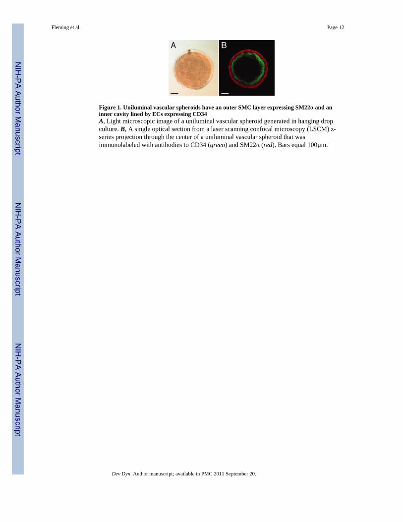
Figure 1. Uniluminal vascular spheroids have an outer SMC layer expressing SM22α and aninner cavity lined by ECs expressing CD34A, Light microscopic image of a uniluminal vascular spheroid generated in hanging dropculture. B, A single optical section from a laser scanning confocal microscopy (LSCM) z-series projection through the center of a uniluminal vascular spheroid that wasimmunolabeled with antibodies to CD34 (green) and SM22α (red). Bars equal 100µm.
Fleming et al. Page 12
Dev Dyn. Author manuscript; available in PMC 2011 September 20.
NIH
-PA Author Manuscript
NIH
-PA Author Manuscript
NIH
-PA Author Manuscript
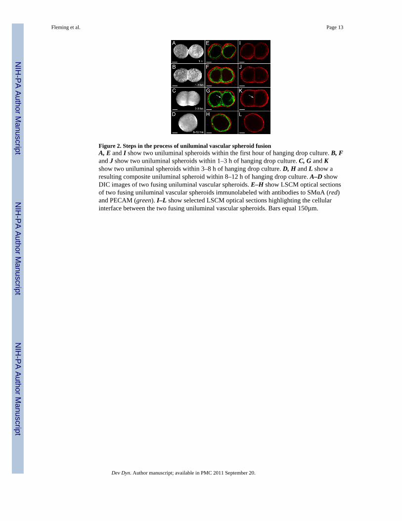
Figure 2. Steps in the process of uniluminal vascular spheroid fusionA, E and I show two uniluminal spheroids within the first hour of hanging drop culture. B, Fand J show two uniluminal spheroids within 1–3 h of hanging drop culture. C, G and Kshow two uniluminal spheroids within 3–8 h of hanging drop culture. D, H and L show aresulting composite uniluminal spheroid within 8–12 h of hanging drop culture. A–D showDIC images of two fusing uniluminal vascular spheroids. E–H show LSCM optical sectionsof two fusing uniluminal vascular spheroids immunolabeled with antibodies to SMαA (red)and PECAM (green). I–L show selected LSCM optical sections highlighting the cellularinterface between the two fusing uniluminal vascular spheroids. Bars equal 150µm.
Fleming et al. Page 13
Dev Dyn. Author manuscript; available in PMC 2011 September 20.
NIH
-PA Author Manuscript
NIH
-PA Author Manuscript
NIH
-PA Author Manuscript
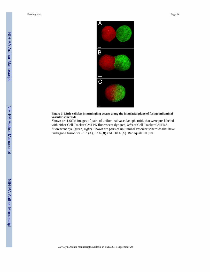
Figure 3. Little cellular intermingling occurs along the interfacial plane of fusing uniluminalvascular spheroidsShown are LSCM images of pairs of uniluminal vascular spheroids that were pre-labeledwith either Cell Tracker CMTPX fluorescent dye (red, left) or Cell Tracker CMFDAfluorescent dye (green, right). Shown are pairs of uniluminal vascular spheroids that haveundergone fusion for ~1 h (A), ~3 h (B) and ~18 h (C). Bar equals 100µm.
Fleming et al. Page 14
Dev Dyn. Author manuscript; available in PMC 2011 September 20.
NIH
-PA Author Manuscript
NIH
-PA Author Manuscript
NIH
-PA Author Manuscript

Figure 4. Spheroids resulting from the fusion of two uniluminal vascular spheroids possess asingle central lumenLight microscopic images of a spheroid resulting from the fusion of two uniluminal vascularspheroids before (A) and after (B) injection of fast green dye into the central cavity. Theincrement of time between the image shown A and B is <1 sec. Bars equal 100µm.
Fleming et al. Page 15
Dev Dyn. Author manuscript; available in PMC 2011 September 20.
NIH
-PA Author Manuscript
NIH
-PA Author Manuscript
NIH
-PA Author Manuscript
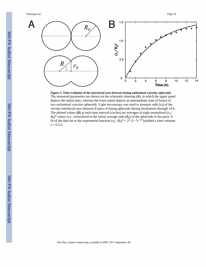
Figure 5. Time evolution of the interfacial area between fusing uniluminal vascular spheroidsThe measured parameters are shown on the schematic drawing (A), in which the upper paneldepicts the initial state, whereas the lower panel depicts an intermediate state of fusion oftwo uniluminal vascular spheroids. Light microscopy was used to measure radii (r0) of thecircular interfacial area between 8 pairs of fusing spheroids during incubations through 14 h.The plotted values (B) at each time interval (circles) are averages of eight normalized (r0 /R0)2 values (i.e., normalized to the initial average radii (R0) of the spheroids in the pair). Afit of the data set to the exponential function (r0 / R0)2 = 22 (1−3e−t/τ)yielded a time constantτ = 6.3 h.
Fleming et al. Page 16
Dev Dyn. Author manuscript; available in PMC 2011 September 20.
NIH
-PA Author Manuscript
NIH
-PA Author Manuscript
NIH
-PA Author Manuscript
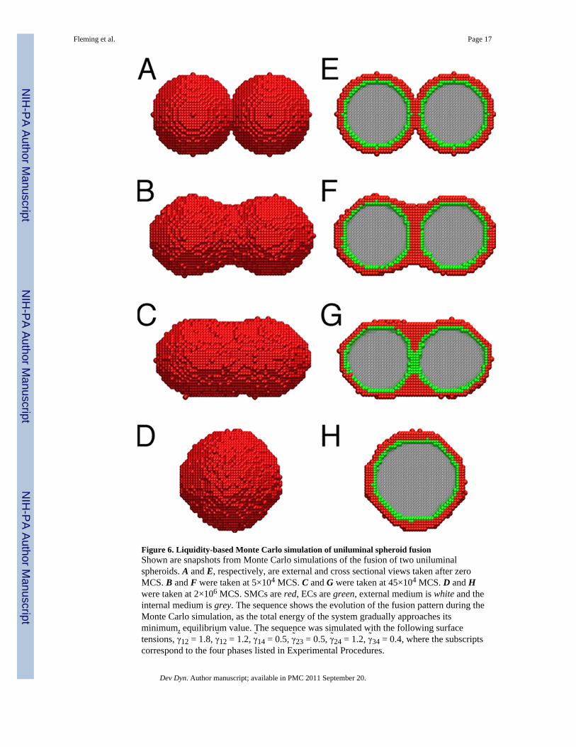
Figure 6. Liquidity-based Monte Carlo simulation of uniluminal spheroid fusionShown are snapshots from Monte Carlo simulations of the fusion of two uniluminalspheroids. A and E, respectively, are external and cross sectional views taken after zeroMCS. B and F were taken at 5×10⁴ MCS. C and G were taken at 45×104 MCS. D and Hwere taken at 2×10⁶ MCS. SMCs are red, ECs are green, external medium is white and theinternal medium is grey. The sequence shows the evolution of the fusion pattern during theMonte Carlo simulation, as the total energy of the system gradually approaches itsminimum, equilibrium value. The sequence was simulated with the following surfacetensions, γ̃12 = 1.8, γ̃12 = 1.2, γ̃14 = 0.5, γ̃23 = 0.5, γ̃24 = 1.2, γ̃34 = 0.4, where the subscriptscorrespond to the four phases listed in Experimental Procedures.
Fleming et al. Page 17
Dev Dyn. Author manuscript; available in PMC 2011 September 20.
NIH
-PA Author Manuscript
NIH
-PA Author Manuscript
NIH
-PA Author Manuscript
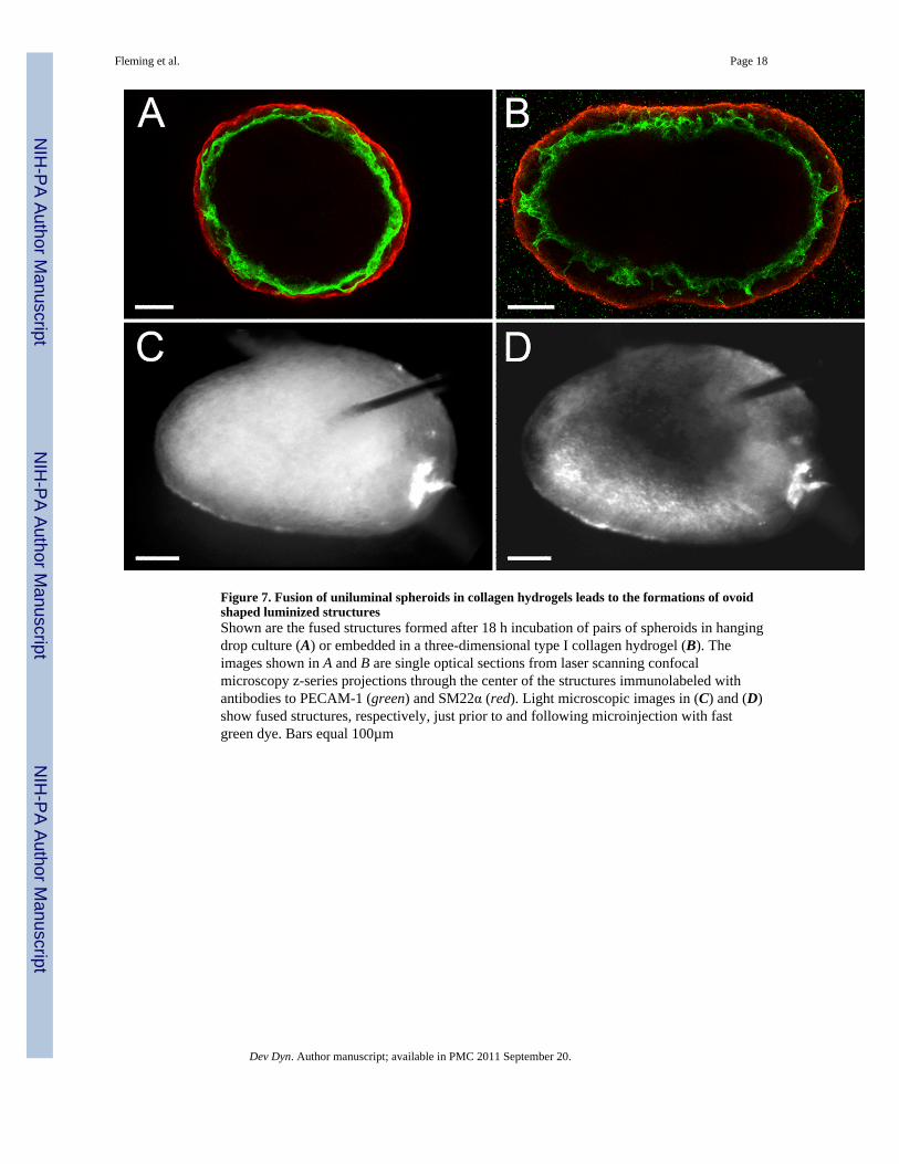
Figure 7. Fusion of uniluminal spheroids in collagen hydrogels leads to the formations of ovoidshaped luminized structuresShown are the fused structures formed after 18 h incubation of pairs of spheroids in hangingdrop culture (A) or embedded in a three-dimensional type I collagen hydrogel (B). Theimages shown in A and B are single optical sections from laser scanning confocalmicroscopy z-series projections through the center of the structures immunolabeled withantibodies to PECAM-1 (green) and SM22α (red). Light microscopic images in (C) and (D)show fused structures, respectively, just prior to and following microinjection with fastgreen dye. Bars equal 100µm
Fleming et al. Page 18
Dev Dyn. Author manuscript; available in PMC 2011 September 20.
NIH
-PA Author Manuscript
NIH
-PA Author Manuscript
NIH
-PA Author Manuscript
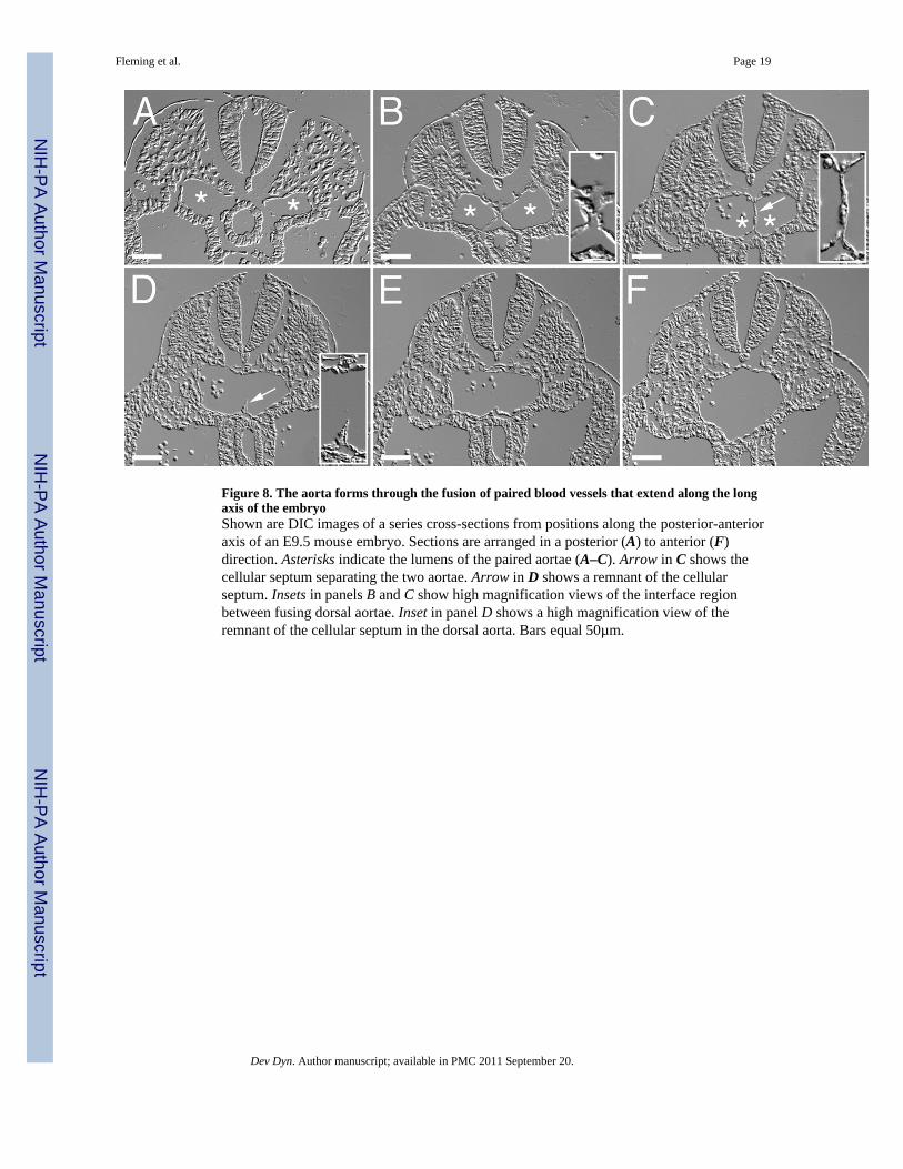
Figure 8. The aorta forms through the fusion of paired blood vessels that extend along the longaxis of the embryoShown are DIC images of a series cross-sections from positions along the posterior-anterioraxis of an E9.5 mouse embryo. Sections are arranged in a posterior (A) to anterior (F)direction. Asterisks indicate the lumens of the paired aortae (A–C). Arrow in C shows thecellular septum separating the two aortae. Arrow in D shows a remnant of the cellularseptum. Insets in panels B and C show high magnification views of the interface regionbetween fusing dorsal aortae. Inset in panel D shows a high magnification view of theremnant of the cellular septum in the dorsal aorta. Bars equal 50µm.
Fleming et al. Page 19
Dev Dyn. Author manuscript; available in PMC 2011 September 20.
NIH
-PA Author Manuscript
NIH
-PA Author Manuscript
NIH
-PA Author Manuscript
Related Documents