This article was downloaded by: [Judith Phoku] On: 02 October 2012, At: 02:40 Publisher: Taylor & Francis Informa Ltd Registered in England and Wales Registered Number: 1072954 Registered office: Mortimer House, 37-41 Mortimer Street, London W1T 3JH, UK Food Additives & Contaminants: Part A Publication details, including instructions for authors and subscription information: http://www.tandfonline.com/loi/tfac20 Fusarium infection of maize and maize-based products and exposure of a rural population to fumonisin B 1 in Limpopo Province, South Africa J.Z. Phoku a , M.F. Dutton a , P.B. Njobeh a b , M. Mwanza a , M.A. Egbuta a & C.A. Chilaka a a Food, Environment and Health Research Group, Faculty of Health Sciences, University of Johannesburg, P.O. Box 17011, Doornfontein 2028, Gauteng, South Africa b Department of Food Technology and Biotechnology, Faculty of Science, University of Johannesburg, P.O. Box 17011, Doornfontein Campus, 2028, Gauteng, South Africa Accepted author version posted online: 06 Sep 2012.Version of record first published: 11 Sep 2012. To cite this article: J.Z. Phoku, M.F. Dutton, P.B. Njobeh, M. Mwanza, M.A. Egbuta & C.A. Chilaka (2012): Fusarium infection of maize and maize-based products and exposure of a rural population to fumonisin B 1 in Limpopo Province, South Africa, Food Additives & Contaminants: Part A, DOI:10.1080/19440049.2012.708671 To link to this article: http://dx.doi.org/10.1080/19440049.2012.708671 PLEASE SCROLL DOWN FOR ARTICLE Full terms and conditions of use: http://www.tandfonline.com/page/terms-and-conditions This article may be used for research, teaching, and private study purposes. Any substantial or systematic reproduction, redistribution, reselling, loan, sub-licensing, systematic supply, or distribution in any form to anyone is expressly forbidden. The publisher does not give any warranty express or implied or make any representation that the contents will be complete or accurate or up to date. The accuracy of any instructions, formulae, and drug doses should be independently verified with primary sources. The publisher shall not be liable for any loss, actions, claims, proceedings, demand, or costs or damages whatsoever or howsoever caused arising directly or indirectly in connection with or arising out of the use of this material.

Welcome message from author
This document is posted to help you gain knowledge. Please leave a comment to let me know what you think about it! Share it to your friends and learn new things together.
Transcript

This article was downloaded by: [Judith Phoku]On: 02 October 2012, At: 02:40Publisher: Taylor & FrancisInforma Ltd Registered in England and Wales Registered Number: 1072954 Registered office: Mortimer House,37-41 Mortimer Street, London W1T 3JH, UK
Food Additives & Contaminants: Part APublication details, including instructions for authors and subscription information:http://www.tandfonline.com/loi/tfac20
Fusarium infection of maize and maize-based productsand exposure of a rural population to fumonisin B1 inLimpopo Province, South AfricaJ.Z. Phoku a , M.F. Dutton a , P.B. Njobeh a b , M. Mwanza a , M.A. Egbuta a & C.A. Chilaka aa Food, Environment and Health Research Group, Faculty of Health Sciences, University ofJohannesburg, P.O. Box 17011, Doornfontein 2028, Gauteng, South Africab Department of Food Technology and Biotechnology, Faculty of Science, University ofJohannesburg, P.O. Box 17011, Doornfontein Campus, 2028, Gauteng, South Africa
Accepted author version posted online: 06 Sep 2012.Version of record first published: 11Sep 2012.
To cite this article: J.Z. Phoku, M.F. Dutton, P.B. Njobeh, M. Mwanza, M.A. Egbuta & C.A. Chilaka (2012): Fusarium infectionof maize and maize-based products and exposure of a rural population to fumonisin B1 in Limpopo Province, South Africa,Food Additives & Contaminants: Part A, DOI:10.1080/19440049.2012.708671
To link to this article: http://dx.doi.org/10.1080/19440049.2012.708671
PLEASE SCROLL DOWN FOR ARTICLE
Full terms and conditions of use: http://www.tandfonline.com/page/terms-and-conditions
This article may be used for research, teaching, and private study purposes. Any substantial or systematicreproduction, redistribution, reselling, loan, sub-licensing, systematic supply, or distribution in any form toanyone is expressly forbidden.
The publisher does not give any warranty express or implied or make any representation that the contentswill be complete or accurate or up to date. The accuracy of any instructions, formulae, and drug doses shouldbe independently verified with primary sources. The publisher shall not be liable for any loss, actions, claims,proceedings, demand, or costs or damages whatsoever or howsoever caused arising directly or indirectly inconnection with or arising out of the use of this material.

Food Additives & Contaminants: Part A2012, 1–9, iFirst
Fusarium infection of maize and maize-based products and exposure of a rural population to
fumonisin B1 in Limpopo Province, South Africa
J.Z. Phokua*, M.F. Duttona, P.B. Njobehaby, M. Mwanzaa, M.A. Egbutaa and C.A. Chilakaa
aFood, Environment and Health Research Group, Faculty of Health Sciences, University of Johannesburg, P.O. Box 17011,Doornfontein 2028, Gauteng, South Africa; bDepartment of Food Technology and Biotechnology, Faculty of Science,University of Johannesburg, P.O. Box 17011, Doornfontein Campus, 2028, Gauteng, South Africa
(Received 19 August 2011; final version received 29 June 2012)
Fusarium species (spp.) and fumonisin B1 (FB1) contaminations were monitored in maize and porridge consumedby a rural population of Limpopo Province, South Africa. Faecal samples were also analysed for FB1 as a means ofestimating the degree of dietary exposure to this mycotoxin. In total, 142 samples of maize (n¼ 54), porridge (47)and faeces (41) were screened for Fusarium spp. using a serial dilution technique followed by DNA sequencing,while FB1 was further screened and quantified by thin-layer chromatography (TLC) and high-performance liquidchromatography (HPLC), respectively. At least four species of Fusarium were identified, of which F. verticillioideswas themost prevalent in all three sample types analysed. The contamination levels of FB1 were significantly higherin 87% of maize sampled (range¼ 101–53,863 mg kg�1) as compared with porridge (74% incidence rate;range¼ 0.2–20mg kg�1) and faecal samples (100% incidence rate; range¼ 0.3–464mg kg�1). Thus, it can bededuced that the level of human exposure to FB1 via the consumption of maize was high as several samplescontained levels exceeding 1000 mg kg�1, which was strongly supported by the levels found in faecal samples.Further data revealed that a high proportion of FB1 is destroyed or removed by processing maize into porridge. Asmaize porridge is consumed as a staple, the low levels found provide ameans to limit exposure to FB1. Levels of FB1
found in the faeces which were higher indicate that other foods contaminated with the toxin are also consumed.
Keywords: mycotoxins; Fusarium; commodity; fumonisin; rural population; exposure
Introduction
From studies conducted to assess the exposure ofAfrican rural and informal settlement populations, it
has become clear that they consume on a daily basis
foods contaminated with a wide range of mycotoxins
(Sibanda et al. 1997). It follows that at some stage the
foods or the commodities used to process the final
product must have been infected with mycotoxin-
producing fungi (Marasas et al. 1979). In African rural
areas, it has been shown that all these possibilities
occur and that certain processing practices may exac-
erbate the situation, e.g. the brewing of beverages from
contaminated cereals (Shephard et al. 2005). A major
contributor to this situation is the wide propagation of
maize (corn) in Sub-Saharan Africa as a cereal staple
(Dutton 2009), which is a New World crop (McCann
2005) and in several ways unsuited to African condi-
tions and practices. In rural South Africa, e.g. Venda,
Limpopo Province, maize is a staple food. It is also
processed to locally brewed alcoholic (umqcobhothi)
and non-alcoholic (magewu) beers. All these products
are consumed daily, sometimes almost to the exclusion
of other commodities, because of the impoverished
nature of these communities.A major problem using maize as a staple is that it is
prone to infection with various Fusarium spp., inparticular, F. verticillioides (Sydenham, Gelderblom,et al. 1990), which together with F. proliferatum are theprincipal producers of the fumonisins (FBs) andconsequently are found in maize worldwide, sometimesat very high levels (Bullerman and Tsai 1994). Theubiquitous nature of FBs should not detract from thefact that Fusarium, which infects maize in the field and/or in stores (Fandohan et al. 2005), can produce a widerange of mycotoxins differing in chemical diversity andhence toxicity. Fusarium also produces the trichothe-cenes, some of which are highly toxic, such as T-2 toxinand deoxynivalenol (DON) as well as zearalenone(ZEA) (D’Mello 2000).
Of the FBs, fumonisins B1 (FB1), B2 (FB2) and B3
(FB3) are principal members, with the most commonlyoccurring and in highest quantities being FB1.These are all esters of a tricarballylic acid and an
*Corresponding author. Email: [email protected] address: Department of Food Technology and Biotechnology, Faculty of Science, University of Johannesburg,P.O. Box 17011, Doornfontein Campus, 2028, Gauteng, South Africa.
ISSN 1944–0049 print/ISSN 1944–0057 online
� 2012 Taylor & Francis
http://dx.doi.org/10.1080/19440049.2012.708671
http://www.tandfonline.com
Dow
nloa
ded
by [
Judi
th P
hoku
] at
02:
40 0
2 O
ctob
er 2
012
aminopentol (a derivative of pentahydroxyeicosane)
(Bezuidenhout et al. 1988) which mimics sphingoid
bases in their chemical structure and consequently
compete with them for enzymes involved in sphingo-
lipid biosynthesis, effectively blocking sphingolipid
production (Merrill et al. 1993). Hence, the toxicolog-
ical effects of FB1 are subtle, affecting cellular metab-
olism by interfering with important signalling pathways
and membrane effects (Dutton 2003). A cause for
concern is the suspected link between the high FB1 level
in maize and the increased incidence of human
oesophageal cancer (Sydenham, Thiel, et al. 1990).
However, the evidence is not certain. Work has shown
that FB1, being a polar compound, is poorly absorbed
from the gastrointestinal tract and is rapidly excreted,
mainly via faeces, from material passing unabsorbed
from the gastrointestinal tract and through the bile
(Shephard, Thiel, Sydenham, Vleggaar, et al. 1994).This study was conducted in a rural area of South
Africa to ascertain the degree of rural exposure to FB1,
where previous studies in the country have shown that
FB1 commonly occurs in maize for human consump-
tion (Dutton 1996).
Materials and methods
Study area and sampling
A total of 142 samples of maize (n¼ 54), porridge (47)
and faeces (41) were obtained from Mapate village of
Limpopo Province (Venda), South Africa, and ana-
lysed in this study. To be specific, maize samples were
randomly collected from the field and storage facilities
(both field and storage good maize were separated
from the mouldy maize by the householders) belonging
to different households and porridge samples from
ready dish plates, while faecal samples were obtained
from individuals belonging to these households less
than 24 h after ingestion of maize products. Food
samples (about 200 g each) were collected by thorough
mixing to obtain a representative sample, put in sealed
plastic bags placed in cooler boxes, and taken to the
Food, Environment and Health Research Group
(FEHRG), University of Johannesburg, South Africa,
where they were preserved at�4�C until analysed. Prior
to analysis, maize samples were milled (particle size of
about 10 mm) using a laboratorymill (IKAM20,Merck,
Darmstadt, Germany), while the porridge and faecal
samples were freeze-dried and further crushed into
powder using a pestle and mortar. The study was
approved by the Faculty of Health Sciences Research
Ethical Committee, University of Johannesburg (Ref
No. HDC01/01/2009).
Materials and reagents
All chemicals and reagents were at least of analyticalgrade, unless otherwise specified:
. Antibiotics: streptomycin and chlorampheni-col (Sigma, Aldrich, Steinheim, Germany).
. Culture media: Czapek yeast agar (CYA),malt extract agar (MEA), potato dextroseagar (PDA and Ohio Agricultural andExperimental Station agar (OAESA).
. Fungal/bacterial DNA extraction kit (ZymoResearch Corporation, CA, USA).
. ITS-1 and ITS-4 primers were synthesised byInqaba Biotechnological Industries (Pretoria,South Africa).
. Equipment used for PCR and DNA sequenc-ing included the ABI 9700 PCR system andABI 3500XL sequencer (both from AppliedBiosystems, Foster City, CA, USA), while thereaction included the ABI Big Dye TerminatorCycle Sequencing kit Version 3.1 dye (AppliedBiosystems).
. SAX solid-phase extraction cartridge (10mlcapacity containing 500mg sorbent; VarianBond Elut, Harbor City, CA, USA) which waspreviously conditioned using 5ml methanol(CH3OH) followed by 5ml methanol/H2O(CH3OH:H2O) (1:3, v/v).
. FB1 standard was purchased from PROMECUnit of MRC, Cape Town, South Africa.
. Thin-layer chromatography (TLC): reversed-phase (C18) TLC plates, Whatman LKC18
with pre-concentration zone (Merck).. Mobile phases: TLC-methanol/4% aqueous
potassium chloride (70:30, v/v); HPLC-metha-nol/sodium di-hydrogen phosphate (80:20, v/v) (pH was adjusted to 3.4 using 1M sodiumhydroxide or 0.1M glacial acetic acid).
. HPLC equipment: HPLC Shimadzu Corpora-tion (Kyoto, Japan) equipped with an LC-20AB liquid chromatograph, CBM-20A com-munication bus module, LC-20AB degasser,CTO-20A column oven, SIL-20A auto sam-pler, RF-10AxL fluorescence detector, RID-10A refractive index detector and SPD-M20Aphotodiode array detector linked to LC solu-tions version 1.22 Software Release.
. Derivatising agent: for TLC, 1ml of 0.1Mborate buffer was prepared by dissolving3.81 g sodium tetraborate in 10ml distilledwater and fluorescamine solution (Sigma, St.Louis, MO, USA) prepared by dissolving0.4mg fluorescamine in 1ml of acetonitrile(1:1, v/v). For HPLC, o-phthaldialdehyde(OPA) reagent prepared by dissolving 40mgof o-phthaldialdehyde in 1ml CH3OH and
2 J.Z. Phoku et al.
Dow
nloa
ded
by [
Judi
th P
hoku
] at
02:
40 0
2 O
ctob
er 2
012

diluted with 5ml 0.1M sodium tetraborate(Na2B2O4) and 50 ml mercaptoethanol.
Laboratory analyses
Mycological analysis
Fungal isolation. The mycological analytical proce-dures were performed under aseptic conditions accord-ing to Kaufman et al. (1963). Samples (1 g each) weredissolved in 9ml sterile Ringer’s solution and seriallydiluted in sterile Ringer’s solution down to 10�6.Aliquots (1ml) of the dilutions were transferred ontosterile Petri plates containing 20ml of molten OhioAgricultural and Experimental Station agar (OAESA).Following streaking using a sterile bent glass rod,plates were then incubated at 28�C for 7 days duringwhich time they were inspected for fungal colonies.Fungal colony counts were done on the dilution agarplates to determine the colony-forming unit per g ofsample (CFUg�1). After microscopic identification,individual fungal isolates were subcultured on potatodextrose agar (PDA), malt extract agar (MEA) andCzapek yeast extract agar (CYA) for 7 days at 28�Cand after which time isolates were further identified.Identification of Fusarium isolates was performedusing the identification keys of Nelson et al. (1983)and Klich and Pitt (1988). The mycelia of isolates wereharvested, freeze-dried and preserved at �4�C for PCRand DNA analyses.
DNA extraction, PCR and sequencing. In a case wherefungal identification by the conventional method wasnot possible or further confirmation in some circum-stances was required, isolates were sent to InqabaBiotechnological Industries (Pty) Ltd, Pretoria, SouthAfrica, for DNA extraction, PCR and sequencing.Genomic DNA analysis was performed on the pre-served mycelia using a Fungal/Bacterial DNA extrac-tion kit (ZymoResearch Corporation) as recommendedby the manufacturer. Prior to analysis, isolates werethawed for about 2–3 h at room temperature and thenDNA was extracted using 60mg of sample. PCRanalysis was done by isolating from the DNA sample,the internal transcribed spacer (ITS) region of the fungalDNA using the forward (ITS-1) and reverse (ITS-4)primers. The primer sequences were designed in theconserved 30 and 50 regions, i.e. ITS-1: 50-TCC GTAGGT GAA CCT GCG G-30 (forward) and ITS-4: 50-TCC TCC GCT TAT TGA TAT GC-30 (reverse).
PCR was carried out according to the manufac-turer’s specifications with Lucigen EconoTaq PlusGreen (Lucigen Corporation, Middleton, USA) on anABI 9700 PCR system. An automatedDNA sequencingon the PCR products was performed using the
ABI 3500XL sequencer. The sequencing reaction usedincluded the ABI BigDye Terminator Cycle Sequencingkit Version 3.1 dye (Applied Biosystems) and thepreviously used ITS primers. The ITS sequences of thefungal species obtained in this study alongside othersequences of fungi available in literature were blasted(Altschul et al. 1997).
Analysis of fumonisins
Sample extraction and clean-up procedures. The ana-lytical procedures followed for the extraction andclean-up of FB1 in maize and porridge were based onthat described by Shephard and Sewram (2004).Accordingly, a finely ground maize or freeze-driedporridge sample (20 g) was mixed with 100ml metha-nol/water (75:25, v/v) and placed on a mechanicalshaker for 60min. The mixture was centrifuged at 500 gfor 10min at 4�C. The supernatant was obtained andadjusted to pH 5.8 using 1M sodium hydroxide or0.1M glacial acetic acid, wherever necessary. Analiquot (10ml) of extract was passed through apreviously conditioned SAX solid-phase extractioncartridge at a flow rate of 2mlmin�1, while notallowing the column to dry out. The column wasthen sequentially washed with 5 ml methanol/water(75:25, v/v) and 3ml methanol. FB1 was eluted using10ml methanol/glacial acetic acid (99:1, v/v) at a flowrate of 1mlmin�1 and the content dried under a streamof N2 gas at 60
�C.For faecal samples, the extraction and clean-up
method of FB1 was performed according to Cheluleet al. (2000). A sample of freeze-dried powdered faeces(1.5 g) of sample was mixed with 15ml of 0.1Methylene diamine tetraacetic acid (EDTA) in a centri-fuge tube and vortexed for 1min. The pH of eachextract was adjusted to 5.2 and the mixture centrifugedat 2000 g for 10min at 4�C. The supernatant wasremoved and the extraction repeated (2�). The com-bined supernatant was acidified to pH 2.9–3.2 with 5Mhydrochloric acid and centrifuged at 4000 g for 10min.An aliquot of 10ml was applied to a SAX columnpreviously conditioned with 5ml of water. The sorbentwas sequentially washed with 5 ml water, followed by5ml methanol/water (1:3, v/v) and finally with 3ml ofmethanol/water (1:1, v/v). FB1 was eluted using 15mlof methanol, evaporated under a stream of N2 gas at60�C and stored at 4�C for further analysis.
Reverse-phase thin-layer chromatography (TLC).Screening for the presence of FB1 in extracts wasperformed by TLC according to Shephard and Sewram(2004). TLC was prepared by placing a large (24 cmdiameter) filter paper in the TLC tank containingmethanol/4% aqueous potassium chloride (70:30, v/v)
Food Additives & Contaminants: Part A 3
Dow
nloa
ded
by [
Judi
th P
hoku
] at
02:
40 0
2 O
ctob
er 2
012
developing solution and allowing at least 4 h forequilibration. The dried FB extract was re-dissolvedusing 200 ml of methanol and an aliquot or FB1
standard solution (25ml) was derivatised with 25 mleach of 0.1M borate buffer and fluorescamine solutionand allowed to stand for 1min at room temperature.FB1 was then separated on a reversed-phase (C18) TLCplate (Whatmann LKC18 with pre-concentration zone;Merck) by spotting 15 ml of the mixture within the pre-concentrated zone on the plate, dried with a gentlestream of warm air and placed into the equilibratedTLC tank. The plates were allowed to develop to atleast 10 cm above the pre-concentrated zone, afterwhich time they were removed, dried under a stream ofwarm air and examined under long wavelength ultra-violet light (350mm). The blue fluorescent spot of FB1
on TLC was recorded and the retardation factor (RF)of the individual spot was calculated as the proportionof the distance travelled by the analyte to that of thedeveloping solvent, which was then compared withthat obtained for FB1 standard solution. Extracts wereagain dried as previously described using N2 gas.
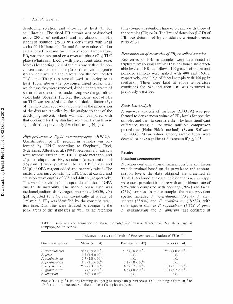
High-performance liquid chromatography (HPLC).Quantification of FB1 present in samples was per-formed by HPLC according to Shephard, Thiel,Sydenham, Alberts, et al. (1994). Accordingly, extractswere reconstituted in 1ml HPLC grade methanol and25 ml of aliquot or FB1 standard (concentration of0.5 mgml�1) were pipetted into an HPLC vial and250 ml of OPA reagent added and properly mixed. Themixture was injected into the HPLC set at excited andemission wavelengths of 335 and 440 nm, respectively.This was done within 1min upon the addition of OPAdue to its instability. The mobile phase used wasmethanol/sodium di-hydrogen phosphate (80:20, v/v)(pH adjusted to 3.4), run isocratically at a rate of1mlmin�1. FB1 was identified by the constant reten-tion time. Quantities were deduced by comparing thepeak areas of the standards as well as the retention
time (found at retention time of 6.3min) with those ofthe samples (Figure 2). The limit of detection (LOD) ofFB1 was determined by considering a signal-to-noiseratio of 3:1.
Determination of recoveries of FB1 on spiked samples
Recoveries of FB1 in samples were determined intriplicate by spiking samples that contained no detect-able levels of FB1 as follows: 100 g each of maize andporridge samples were spiked with 400 and 160 mg,respectively, and 1.5 g of faecal sample with 400 mg inmethanol. These were kept at room temperatureconditions for 24 h and then FB1 was extracted aspreviously described.
Statistical analysis
A one-way analysis of variance (ANOVA) was per-formed to derive mean values of FB1 levels for positivesamples and then to compare them by least significantdifference using all pairwise multiple comparisonprocedures (Holm–Sidak method) (Systat SoftwareInc. 2006). Mean values among sample types weredeemed to have significant differences if p� 0.05.
Results
Fusarium contamination
Fusarium contamination of maize, porridge and faeceswas determined based on the prevalence and contam-ination levels; the data obtained are presented inTable 1. As found, the data indicate that Fusarium spp.were most prevalent in maize with an incidence rate of92% when compared with porridge (28%) and faecal(27%) samples. In maize samples the most prevalentspecies included F. verticillioides (70.3%), F. oxy-sporum (25.9%) and F. proliferatum (18.5%), withother species such as F. sambucinum (3.7%) F. poae,F. graminearum and F. dimerum that occurred at
Table 1. Fusarium contamination in maize, porridge and human faeces from Mapate village inLimpopo, South Africa.
Dominant species
Incidence rate (%) and levels of Fusarium contamination (CFUg�1)a
Maize (n¼ 54) Porridge (n¼ 47) Faeces (n¼ 41)
F. verticillioides 70.3 (2.5� 106) 27.6 (2.8� 106) 29.2 (4.6� 105)F. poae 3.7 (4.4� 105) n.d. n.d.F. sambucinum 3.7 (2.8� 105) n.d. n.d.F. proliferatum 18.5 (2.1� 106) 2.1 (5.0� 106) n.d.F. oxysporum 25.9 (2.3� 106) 6.3 (5.7� 105) 12.1 (3.1� 105)F. graminearum 3.7 (3.3� 106) 6.3 (4.0� 106) 12.1 (3.7� 105)F. dimerum 1.8 (2.3� 105) n.d. n.d.
Notes: aCFUg�1 is colony-forming unit per g of sample (in parentheses). Dilution ranged from 10�1 to10�6; n.d., not detected. n is the number of samples analysed.
4 J.Z. Phoku et al.
Dow
nloa
ded
by [
Judi
th P
hoku
] at
02:
40 0
2 O
ctob
er 2
012

extremely lower levels in 1.8% of the samples analysed.Although F. graminearum was less frequently isolatedfrom maize, there was heavy contamination in thoseinfected samples, at a mean level of 3.3� 106CFUg�1,closely followed by F. verticillioides (2.5� 106CFUg�1),and F. oxysporum and F. proliferatum whose meanlevels were 2.3� 106 and 2.1� 106CFUg�1, respec-tively (Table 1). For other isolates recovered frommaize, lower mean contamination levels were found, aswas the case for F. sambucinum (2.8� 105), F. poae(4.4� 105) or F. dimerum (2.3� 105)CFUg�1.
Similar isolates of Fusarium were also foundcontaminating porridge samples in the same manneras observed for maize, but generally at much lowerincidence rates except for F. graminearum whoseincidence rate of 6.3% was surprisingly higher thanthat found for maize. Accordingly, F. verticillioides(27.6%), F. oxysporum (6.3%) and F. proliferatum(2.1%) were isolated. Equally surprising, porridgesamples analysed in this study had much higher meancontamination levels of 2.8� 106, 5.0� 106 and4.0� 106CFUg�1 for F. verticillioides, F. proliferatumand F. graminearum, respectively, when compared withthose of their maize counterparts. As for faecalsamples, a lesser number of Fusarium spp. was iden-tified including 29.2% for F. verticillioides, 12.1% forF. oxysporum and 12.1% for F. graminearum isolatedat mean contamination levels that ranged between3.1� 105 and 4.6� 105CFUg�1 of sample analysed(Table 1).
Fumonisin contamination
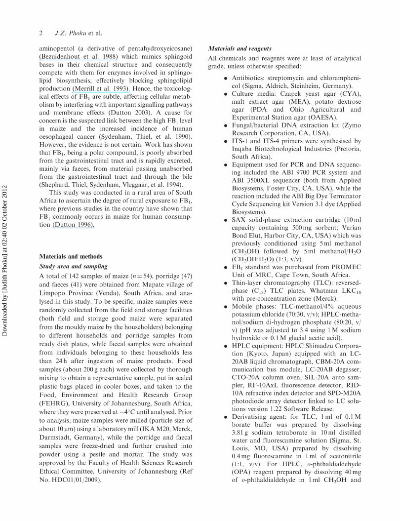
Screening for the presence of FB1 in samples via TLCafter extraction and clean-up procedures revealed thatseveral samples were positive with incidence rates of72%, 36% and 54% for maize, porridge and faecalsamples, respectively. Figure 1 shows a TLC plate
containing a blue fluorescent spot of FB1 in positivesamples as viewed under a UV spectrum (wavelengthof 362 nm). FB1 was identified by comparing thefluorescence intensity of spots under ultraviolet light(RF¼ 0.35) for samples with that of FB1 standard (A)on the same plate, as presented in Figure 1. High liquidchromatography was performed to determine levels ofFB1 in maize, porridge and faecal samples. Recoveriesof FB1 were also performed and repeated three timesfor each sample type to confirm the stability of theresults. The percentage recoveries were found to be95.3%� 2.1% for maize, 81.0%� 1.0% for porridgeand 85.3%� 2.1% for faecal samples. Figure 2 showsthe chromatograms of FB1.
Data obtained on the incidence and levels of FB1
(unadjusted data based on recoveries rates) in samplesare shown in Table 2. A significant variation wasobserved on the levels of FB1 with reference to sampletypes. In this regard, maize had a significantly(p< 0.001) much higher mean level (8189mg kg�1)than that of porridge (6 mg kg�1) or faecal(86 mg kg�1) samples (Table 2). FB1 levels in maizeranged from 101 to 53,863 mg kg�1 in 87% of positivesamples, 74% of porridge samples contained FB1 atlevels ranging from 0.2 to 20 mg kg�1, while all faecalsamples analysed had FB1 contents in the range of 0.3–464 mg kg�1. The study showed all the sample typesanalysed had high incidence rates of FB1. Whileporridge samples contained FB1 far below theEuropean Union acceptable limit of 1000 mg kg�1 inmaize products intended for human consumption, over69% of maize samples contained FB1 above this limit.Although no significant differences were found in thelevels of FB1 in porridge and faecal samples, dataindicate faecal samples had higher FB1 levels whencompared with those of porridge.
Discussion
TLC was used as a useful preliminary screening toconfirm the presence of FB1 in samples, while HPLCwas performed to determine the levels of the toxinpresent in each sample. Results by TLC showed thatonly a few of the samples showed negative results, andthis was confirmed when analysed by HPLC (Table 2).The study revealed that high levels of FB1 were foundin maize from Venda, with a majority of samplesexceeding by several times the set levels of 1000mg kg�1
in maize or maize-based products for human con-sumption (European Commission 2007). The studyfurther indicated that a much higher proportion of thismycotoxin appears to be destroyed and/or removed byprocessing the maize to porridge.
The processing of maize-meal into porridge inMapate village is, however, carried out by soakingmaize-meal in water overnight and subsequently
Figure 1. Reversed-phase TLC plate showing FB1 band(arrow): (A) FB1 standard; (B–F) contaminated maizesamples and (G) uncontaminated sample.
Food Additives & Contaminants: Part A 5
Dow
nloa
ded
by [
Judi
th P
hoku
] at
02:
40 0
2 O
ctob
er 2
012

(a) (b)
(c) (d)
0.0 1.0 2.0 3.0 4.0 5.0 6.0 7.0 min
0
5
10
15
20
25
30
35
40
mVDetector A:Ex:335nm,Em:440nm
1/F
B1/
6.20
9
0.0 1.0 2.0 3.0 4.0 5.0 6.0 7.0 min
0.0
2.5
5.0
7.5
10.0
12.5
15.0
17.5
20.0
22.5
25.0mVDetector A:Ex:335nm,Em:440nm
1/F
B1/
6.11
2
0.0 2.5 5.0 7.5 10.0
0
1
2
3
4
mVDetector A:Ex:335nm,Em:440nm
1/F
B1/
6.36
5
0.0 2.5 5.0 7.5 10.0
0
1
2
3
4
5
mVDetector A:Ex:335nm,Em:440nm
1/F
B1/
6.36
1
Figure 2. Chromatograms of fumonisin B1: (a) standard (10mgml�1, 40 ml of standard injected), (b) faecal sample, (c) porridgesample, and (d) maize sample (20 ml of extract injected) at retention times of 6.2, 6.1 and 6.3min.
Table 2. Incidence and levels of FB1 in foods and faecal samples from Mapate village in Limpopo, South Africa.
Sample typeNumber ofsamples
Number ofpositive samples
Percentagefrequency
Range(mg kg�1) Mean*
Maize 54 49 87 101–53,863 8189a
Porridge 47 41 74 0.2–20 6b
Faeces 41 41 100 0.3–464 86b
Source of variation DF SS MS F PB/n groups 2 2,014,827,012.6 1,007,413,506.3 16.149 <0.001Residual 126 77,859,977,549.7 62,380,774.2Total 128 9,874,804,562.360
Notes: Mean values not sharing the same superscript are significantly different at p< 0.001; *mean values are for positivesamples.DF, degree of freedom; SS, sum of square; MS, mean square; F, F-value; P, probability.
6 J.Z. Phoku et al.
Dow
nloa
ded
by [
Judi
th P
hoku
] at
02:
40 0
2 O
ctob
er 2
012

discarding the water including any floating fractionprior to cooking. This suggests that a proportion of FBis eliminated since this toxin has been shown to bewater soluble (Park et al. 2004). We observed in ourstudy that this process followed for porridge making,significantly (p< 0.001) reduced FB1 levels by approx-imately 90%.
This is an important finding that needs furtherinvestigation, especially as porridge is widely con-sumed by this population, thus lowering the degree ofhuman exposure to FB1. The explanation of such afinding seems to involve the overnight soaking ofmaize-meal in water, in which the water is oftendiscarded prior to cooking. FB1 present in certainfoodstuffs is also affected due to some food prepara-tion processes. Jackson et al. (1997) showed that up to43% of FB1 spiked in corn meal was lost after bakingat 175–200�C for 20min. In addition, Fandohan et al.(2005) found that about 93% of the FB level in maizewas lost via sorting, washing and fermentation. Theseare very important findings as the above-mentionedmeasures can be utilised seriously to reduce humanexposure to FB. However, limited information exists onattempts to reduce mycotoxin contamination in maizeusing traditional processing methods in Africa.In Benin, about 40 different maize fermentation pro-cessing methods have been recorded (Hounhouiganet al. 1993; Adegoke et al. 1994; Fandohan et al. 2005).
Chelule et al. (2000) reported the presence of FB1 inhuman faeces obtained from a rural population inSouth Africa, with seven of the 20 analysed sampleshaving FB1 levels that ranged from 6000 to20,000 mg kg�1. Studies (Shephard, Thiel, Sydenham,Alberts, et al. 1994) have shown that FB1 is poorlyabsorbed into the body via the alimentary canal as asubstantial amount is excreted via faeces. Results onthe analysis of faecal samples revealed higher FB1
levels when compared with those of porridge presentedin Table 2. This may be attributed to the fact that FB1
intake by the same population could be attributed toother sources. For example, home-brewed maize-basedbeer is widely consumed and enjoyed by subsistencefarmers in the rural population of areas South Africaon a daily basis (Shephard et al. 2005).
The genus Fusarium is considered as one of themost important pathogenic and toxin-producingfungal genera worldwide that affect mainly maize.There are several reports from South Africa on thecontamination of maize by a number of Fusarium spp.,especially those members belonging to the F. verticil-lioides complex. In this study different Fusarium spp.were isolated and identified with F. verticillioideswidely distributed in all the samples at high contam-ination levels. This species from a number of reports isthe most important Fusarium spp. in maize, not onlybecause of its pathogenic potentials to both plants andanimal species, but also because of its ability to
produce several mycotoxins in Africa (Gelderblomet al. 1988; Sydenham, Gelderblom, et al. 1990; Dutton1996; Marasas 2001) and elsewhere in the world(Nelson 1993; Nelson et al. 1993; Desjardins 2006).A major potential of F. verticillioides rests in its abilityto produce FB1 in food commodities that has beenlinked to increased prevalence of oesophageal cancer inthe Transkei region of South Africa (Marasas et al.1988; Sydenham, Thiel, et al. 1990), some parts ofChina (Chu and Li 1994), and in Central and EasternAfrica (Burkitt et al. 1970). The mode of this linkage,however, is not completely understood and no exactcause-and-effect relationship has been proved in thisdisease.
What is also of concern from this study whenconsidering the previously mentioned observations isthat most of the tested samples of maize and porridgehad a co-occurrence of more than one Fusarium spp.(Table 1), which therefore suggests, possibly, co-contamination of more than one mycotoxin, especiallythose considered significant with regards to health andthe economy. Of course, such mycotoxins of Fusariumorigin include the trichothecenes (mainly deoxynivale-nol, zearalenone and T-2 toxin). This considerationdoes not take into account that other genera of fungithat are capable of producing other mycotoxins areknown to be present in rural maize (Dutton 1996). Inaddition to F. verticillioides, other Fusarium spp.identified in the study were F. graminearum, F. poaeand F. oxysporum, amongst others. The presence ofthese species shows a possibility of the tested maize andmaize-based products to contain such significantmycotoxins as ZEA and trichothecences (DON andT-2). It is becoming apparent that combinations ofsuch mycotoxins in rural African diets are problematicbecause of the possibility of them exerting somesynergistic and additive effects (D’Mello et al. 1999;Placinta et al. 1999) in humans.
It thus follows that because maize and maize-basedproducts are consumed daily in the rural areas, theseresult in high FB intake. It can then be concluded thatthe rural population of Limpopo in South Africa isexposed to its toxic effects on an on-going basis.However, such levels may be reduced considerablyduring processing of maize-meal into porridge. Basedon the data obtained herein, it would thus be recom-mended to process maize into porridge before it isconsumed; nonetheless, this requires further investiga-tion. Results on the analysis of faecal samples revealeda higher FB1 level when compared with those ofporridge. This may be attributed to the fact that FB1
intake by the same population could be attained fromother sources. For example, home-brewed maize-basedbeer (such as umqobothi) is widely consumed andenjoyed by subsistence farmers in the rural populationof areas of South Africa on a daily basis (Shephardet al. 2005). However, in Mapate village in Limpopo
Food Additives & Contaminants: Part A 7
Dow
nloa
ded
by [
Judi
th P
hoku
] at
02:
40 0
2 O
ctob
er 2
012

Province home-brewed beer is also consumed; in thiscase the home-brewed beer was proved to contain highlevels of FB1. Although the fermentation process isusually involved in the production of this local beer,the raw material utilised is mainly mouldy home-grownmaize, which could be another source of the FB1.When setting daily tolerable levels of FB1 in foodsin South Africa, it is imperative to take into accountthe eating habits especially of those from rural com-munities. It is equally important to necessitate themanagement of mycotoxins in foods, especially in therural environment.
Acknowledgements
The authors wish to thank the University of Johannesburgvia the New Generation Scholarship (NGS), MedicalResearch Council (MRC) and National ResearchFoundation (NRF) of South Africa for funding this study.The contribution of Dr Makun Hussaini and other membersof the Food, Environment and Health Research Group(FEHRG) is duly acknowledged. An appreciation is alsoextended to Chief (Mr Ratshibvumo Ravele) and thepopulation of Mapate Village, Limpopo Province, SouthAfrica, for their support during sampling.
References
Adegoke GO, Otumu EJ, Akanni AO. 1994. Influence of
grain quality heat and processing time on the reduction of
aflatoxin B1 levels in tuwo and ogi two cereal based
products. Plant Food Human Nutr. 45:113–117.Altschul SF, Madden TL, Alejandro A, Schaffer AA,
Zhang J, Zhang Z, Miller W, Lipman JDL. 1997.
Gapped BLAST and PSI-BLAST: a new generation of
protein database search programs. Nucleic Acids Res.
25:3389–3402.
Bezuidenhout SC, Gelderblom WCA, Gorst-Allman RMM,
Marasas WFO, Spiteller G, Vleggaar R. 1988. Structure
elucidation of the fumonisins, mycotoxins from Fusarium
moniliforme. J Chem Soc Chem Commun. 11:743–745.Bullerman LB, Tsai WY. 1994. Incidence and levels of
Fusarium moniliforme, Fusarium proliferatum and fumoni-
sins in corn and corn based foods and feeds. J Food Prot.
57:541–546.Burkitt DO, Standfield JP, Church JC. 1970. A medical
research safari: fruits and frustrations. Cent Afr J Med.
16:197–201.
Chelule PK, Gqaleni N, Chuturgoon AA, Dutton MF. 2000.
The determination of fumonisin B1 in human faeces:
a short term marker for assessment of exposure.
Biomakers. 5:1–8.Chu FS, Li GY. 1994. Simultaneous occurrence of fumonisin
B1 and other mycotoxins in mouldy corn collected from
the People’s Republic of China in regions of high
incidences of oesophageal cancer. Appl Environ
Microbiol. 60:847–852.D’Mello JPF. 2000. Antinutritional factors and mycotoxins.
In: D’Mello JPF, editor. Farm animal metabolism and
nutrition. Wallingford (UK): CAB International.
p. 383–403.D’Mello JPF, Placinta CM, MacDonald AMC. 1999.
Fusarium mycotoxins: a review of global implications for
animal health, welfare and productivity. Anim Feed Sci
Technol. 80:183–205.Desjardins AE. 2006. Introduction to Fusarium mycotoxins.
In: Fusarium mycotoxins chemistry, genetics, and biology.
St. Paul (MN): APS Press. p. 1–9.Dutton MF. 1996. Fumonisins, mycotoxins of increasing
importance: their nature and their effects. Pharmacol Ther.
70:137–161.Dutton MF. 2003. Mycotoxins in South Africa. Adv Appl
Microbiol. 53:213–241.Dutton MF. 2009. The African Fusarium/maize disease.
Mycotox Res. 25:29–39.
European Commission. 2007. Commission Regulation (EC)
No 1126/2007 of 28 September 2007 amending Regulation
(EC) No 1881/2006 setting maximum levels for certain
contaminants in foodstuffs as regards Fusarium toxins in
maize and maize products. Off J Eur Union. 255:14–17.Fandohan P, Gnolonfin B, Hell K, Marasas WFO,
Wingfield MJ. 2005. Natural occurrence of Fusarium and
subsequent fumonisin contamination in preharvest and
stored maize in Benin West Africa. Int J Food Microbiol.
99:173–183.Gelderblom WCA, Jaskiewicz K, Marasas WF, Thiel PG,
Horak RM, Vleggar R, Kriek NP. 1988. Fumonisins novel
mycotoxins with cancer promoting activity produced by
Fusarium moniliforme. Appl Environ Microbiol.
54:1806–1811.Hounhouigan DJ, Nout MJR, Nago CM, Houben JH,
Rombouts FM. 1993. Composition and microbiological
and physical attributes of mawe, a fermented maize dough
from Benin. Int J Food Sci Technol. 28:513–517.Jackson LS, Katta SK, Fingerhut DD, DeVries JW,
Bullerman LB. 1997. Effects of baking and frying on the
fumonisin B1 content of cornbased foods. J Agric Food
Chem. 45:4800–4805.Kaufman DD, Williams LE, Sumner CB. 1963. Effect of
plating medium and incubation temperature on growth of
fungi in soil dilution plate. Can J Microbiol. 63:622–625.
Klich MA, Pitt JI. 1988. A laboratory guide to common
Aspergillus species and their telemorphs. North Ryde
(NSW): CSIRO Division of Food Research.Marasas WHO. 2001. Discovery and occurrence of the
fumonisins: a historical perspective. Environ Health
Perspect. 109:239–243.
Marasas WFO, Kellerman TS, Gelderblom WC, Coetzer JA,
Thiel PG, van der Lugt JJ. 1988. Leuko-encephalomalacia
in a horse induced by fumonisin B1 isolated from Fusarium
moniliforme. Onderstepoort J Vet Res. 55:197–203.Marasas WFO, Kriek NPJ, Wiggins VM, Steyn PS,
Towers DK, Hastie TJ. 1979. Incidence, geographic
distribution and toxigenicity of Fusarium species in South
African corn. Phytopathol. 69:1181–1185.
McCann JC. 2005. Maize and grace: Africa’s encounter with
a new world crop. Cambridge (MA): Harvard University
Press.Merrill AH, Wang E, Gilchrist DG, Riley RT. 1993.
Fumonisins and other inhibitors of de novo sphingolipid
biosynthesis. Adv Lipid Res. 26:215–234.
8 J.Z. Phoku et al.
Dow
nloa
ded
by [
Judi
th P
hoku
] at
02:
40 0
2 O
ctob
er 2
012

Nelson PE. 1993. Taxonomy and biology of Fusariummoniliforme. Mycopathol. 117:29–36.
Nelson PE, Desjardins AE, Plattner RD. 1993. Fumonisins,mycotoxins produced by Fusarium species: biology, chem-istry, and significance. Annu Rev Phytopathol. 31:233–252.
Nelson PE, Toussoun TA, Marasas WFO. 1983. Fusarium
species an illustrated manual for identification. UniversityPark (Pennsylvania): The Pennsylvania State UniversityPress, 193p.
Park JW, Scott PM, Lau BP, Lewis DA. 2004. Analysis ofheat-processed maize foods for fumonisins and boundfumonisins. Food Addit Contam. 21:1168–1178.
Placinta CM, D’Mello JPF, Macdonald AMC. 1999.A review of worldwide contamination of cereal grainsand animal feed with Fusariummycotoxins. Anim Feed SciTechnol. 78:21–37.
Shephard GS, Sewram V. 2004. Determination of themycotoxin fumonisin B1 in maize by reversed-phase thin-layer chromatography: a collaborative study. Food Addit
Contam. 21:498–505.Shephard GS, Thiel PG, Sydenham EW, Vleggaar R,Alberts JF. 1994. Determination of the mycotoxin fumoni-
sin B1 and identification of its partially hydrolysed
metabolites in the faeces of nonhuman primates. FoodChem Toxicol. 32:23–29.
Shephard GS, Thiel PG, Sydenham EW, Alberts JF,Gelderblom WCA. 1994. Fate of single dose of the14C-labelled mycotoxin, fumonisin B1, in rats. Toxins.30:768–770.
Shephard GS, van der Westhuizen L, Gatyeni PM,Katerere DR, Marasas WFO. 2005. Do fumonisin myco-toxins occur in wheat? J Agric Food Chem. 53:9293–9296.
Sibanda L, Marovatsanga LT, Pestka JJ. 1997. Review ofmycotoxin work in Sub-Saharan Africa. Food Control.8:21–29.
Sydenham EW, GelderblomWCA, Thiel PG, Marasas WFO.1990. Evidence for the natural occurrence of fumonisin B1 amycotoxin produced by Fusarium moniliforme in corn.J Agric Food Chem. 38:285–290.
Sydenham EW, Thiel PG, Marasas WFO, Shephard GS,van Schalkwyk DJ, Koch KR. 1990. Natural occurrenceof some Fusarium mycotoxins in corn from low and
high oesophageal cancer prevalence areas of the Transkei,Southern Africa. J Agric Food Chem. 38:1900–1903.
Systat Software Inc. 2006. SigmaStat 3.5 for Windows. Point
Richmond (CA): Systat Software Inc.
Food Additives & Contaminants: Part A 9
Dow
nloa
ded
by [
Judi
th P
hoku
] at
02:
40 0
2 O
ctob
er 2
012
Related Documents