Fundamentals of Biochemistry Fourth Edition Chapter 6 Proteins: Three-Dimensional Structure Copyright © 2013 by John Wiley & Sons, Inc. All rights reserved. Donald Voet • Judith G. Voet • Charlotte W. Pratt

Fundamentals of Biochemistry Fourth Edition Chapter 6 Proteins: Three-Dimensional Structure Copyright © 2013 by John Wiley & Sons, Inc. All rights reserved.
Dec 22, 2015
Welcome message from author
This document is posted to help you gain knowledge. Please leave a comment to let me know what you think about it! Share it to your friends and learn new things together.
Transcript
Fundamentals of Biochemistry
Fourth Edition
Chapter 6Proteins: Three-Dimensional Structure
Copyright © 2013 by John Wiley & Sons, Inc. All rights reserved.
Donald Voet • Judith G. Voet • Charlotte W. Pratt
Chapter 6Secondary Structure
Key Concepts 6.1• The planar character of the peptide group limits the conformational flexibility of the polypeptide chain.• The α helix and the β sheet allow the polypeptide chain to adopt favorable φ and ψ angles and to form hydrogen bonds.• Fibrous proteins contain long stretches of regular secondary structure, such as the coiled coils in α keratin and the triple helix in collagen.• Not all polypeptide segments form regular secondary structure such as α helices or β sheets.
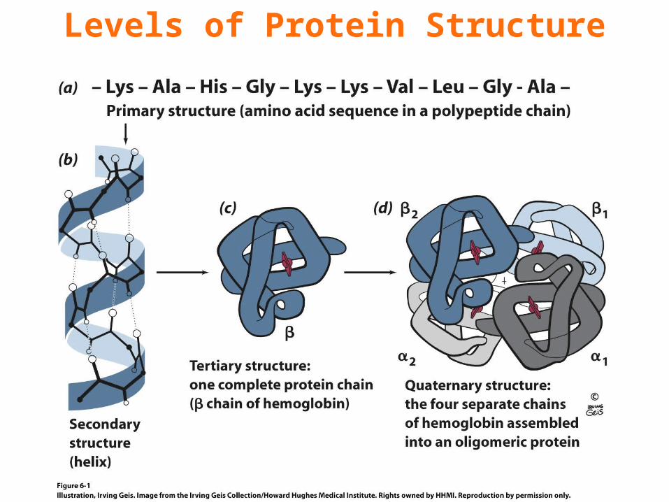
Levels of Protein Structure
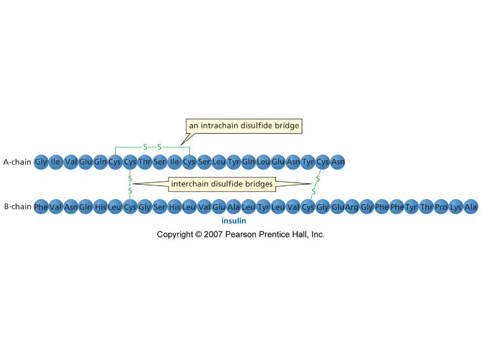
1° Structure
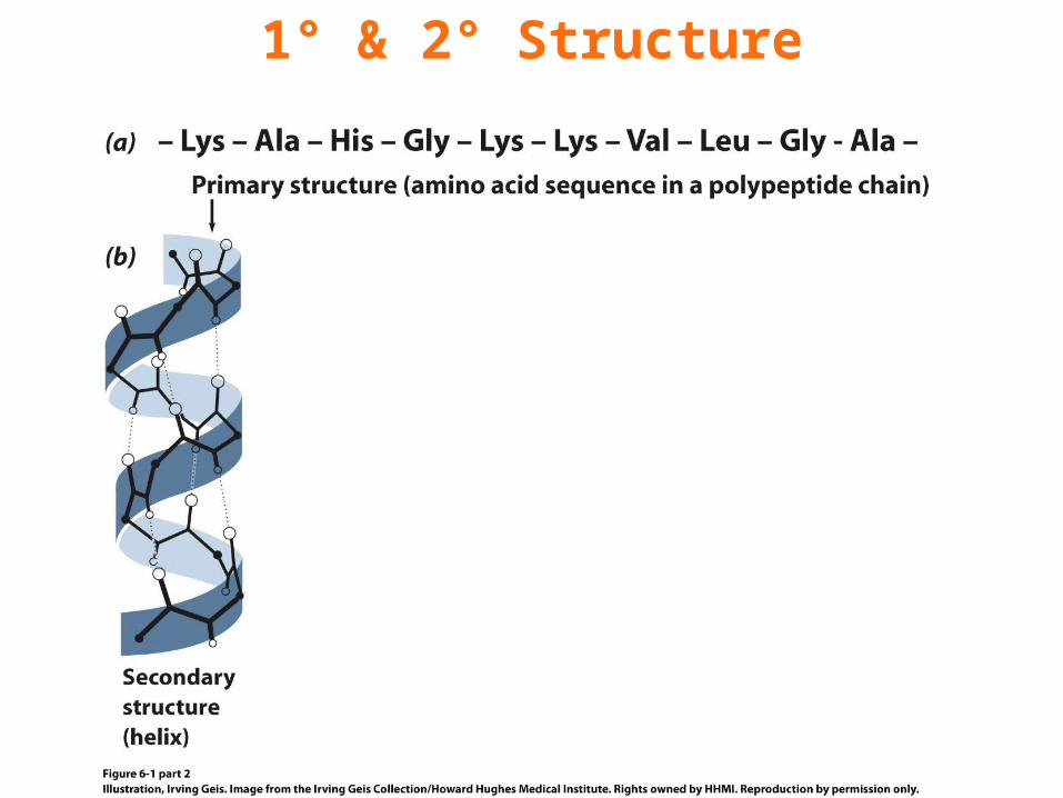
1° & 2° Structure
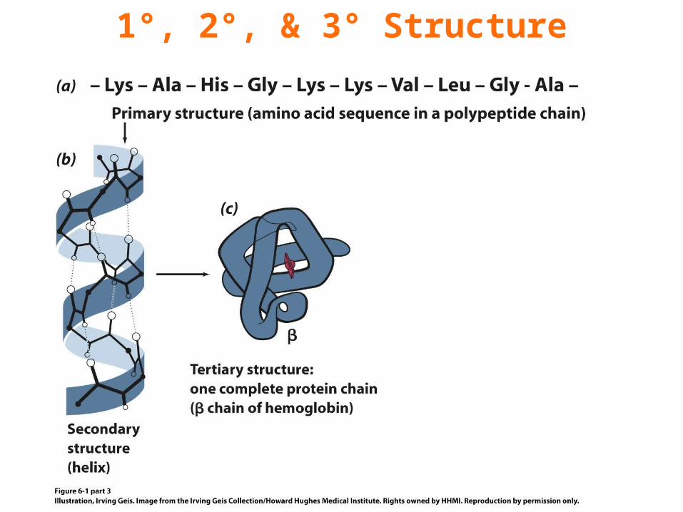
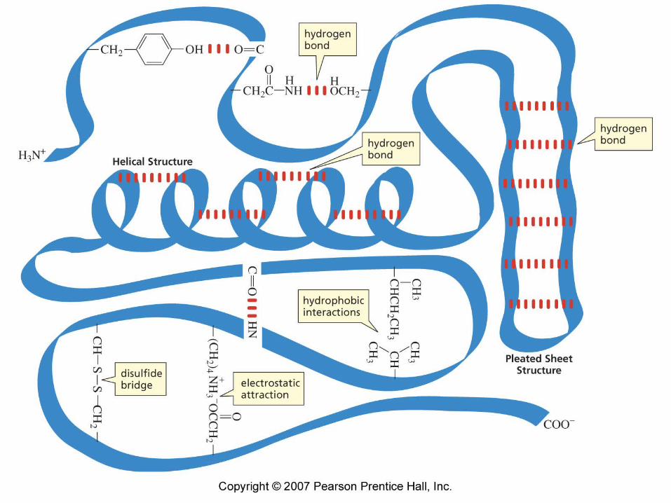
1°, 2°, & 3° Structure
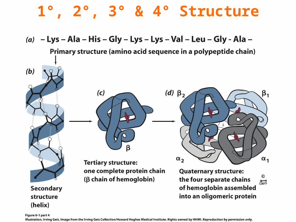
1°, 2°, 3° & 4° Structure
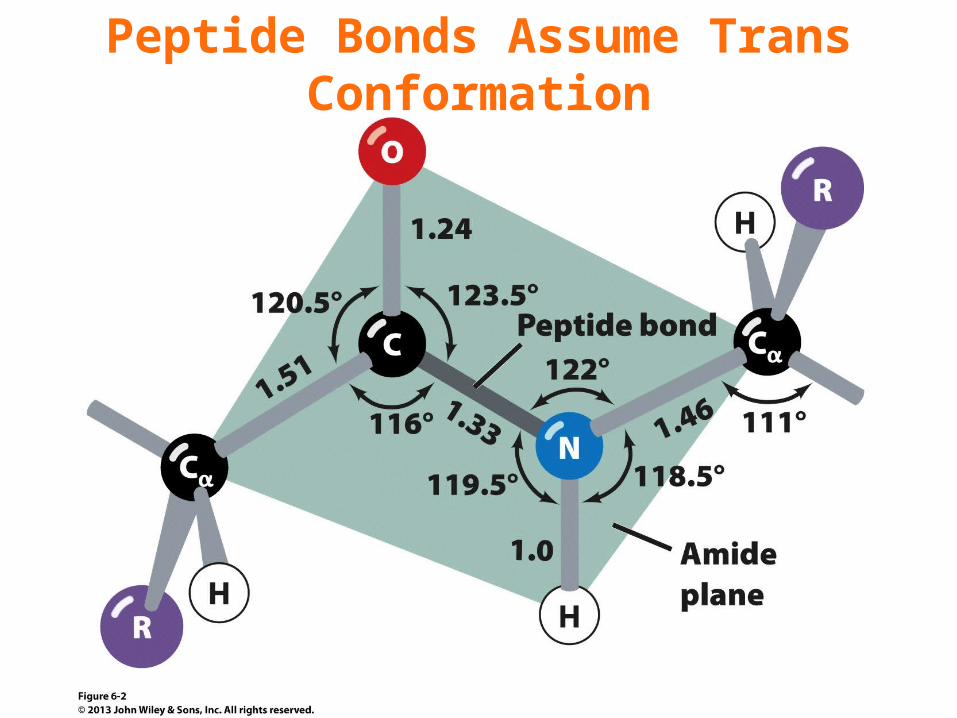
Peptide Bonds Assume Trans Conformation
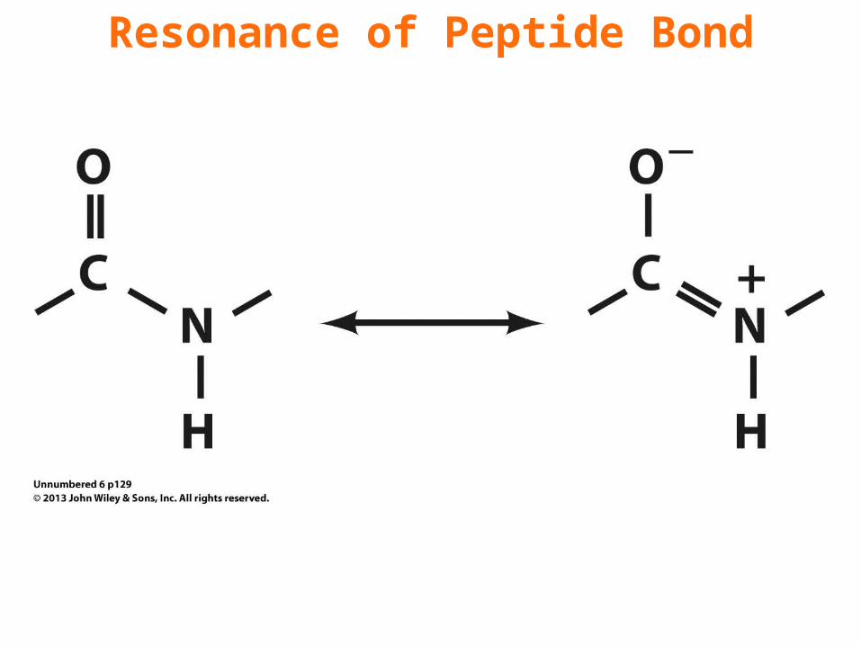
Resonance of Peptide Bond
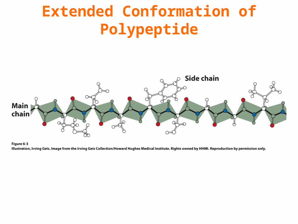
Extended Conformation of Polypeptide
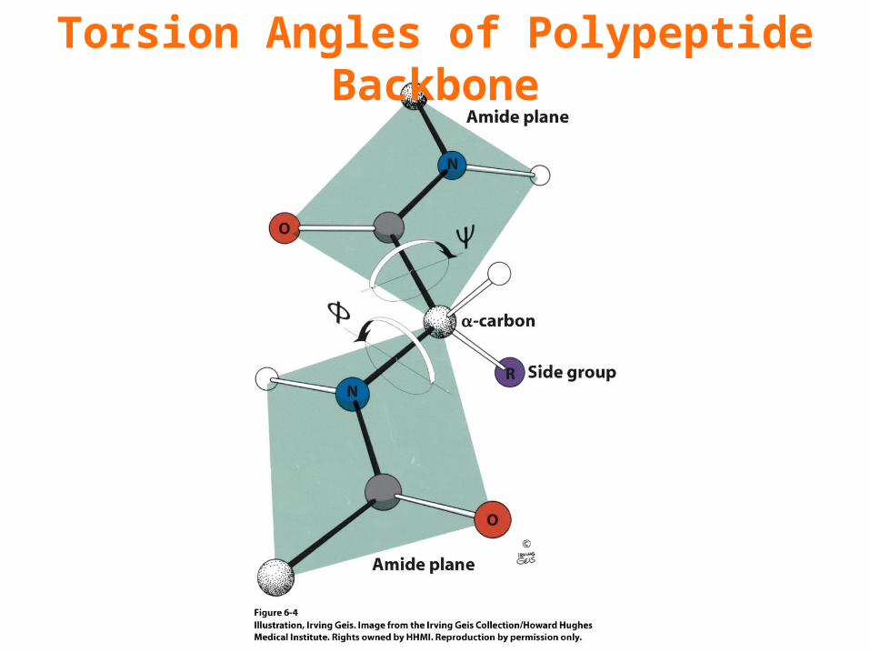
Torsion Angles of Polypeptide Backbone
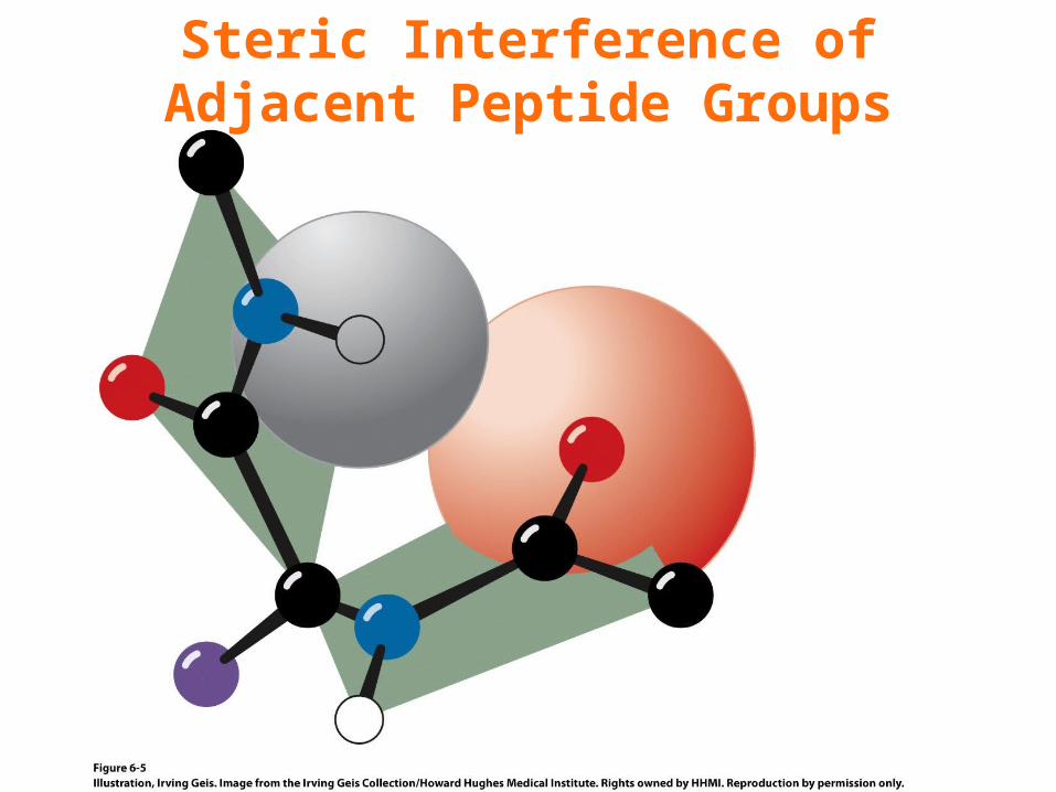
Steric Interference ofAdjacent Peptide Groups
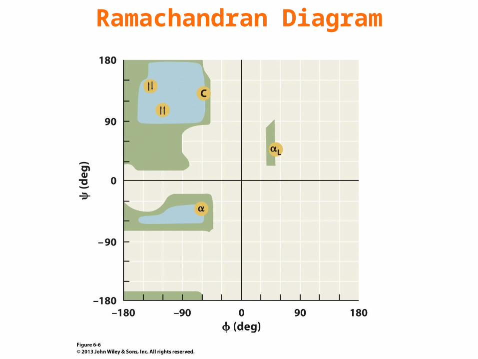
Ramachandran Diagram
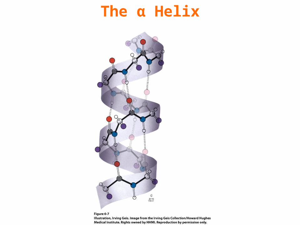
The α Helix
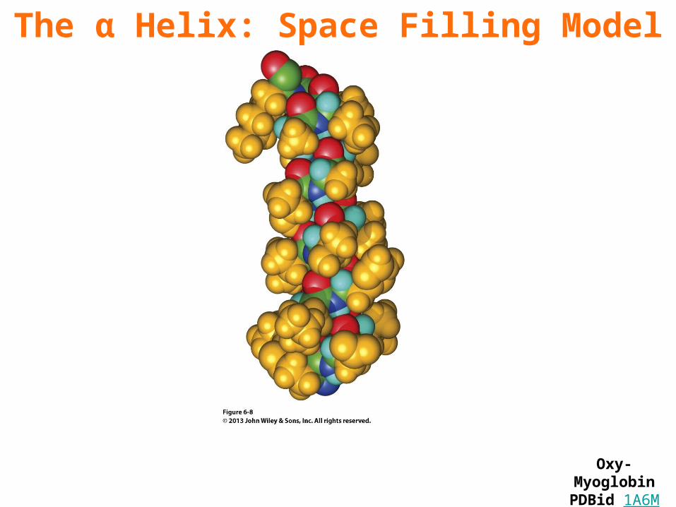
The α Helix: Space Filling Model
Oxy-MyoglobinPDBid 1A6M
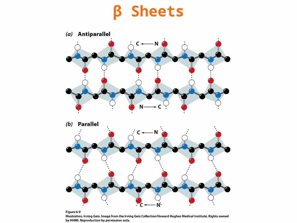
β Sheets
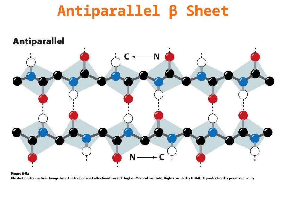
Antiparallel β Sheet
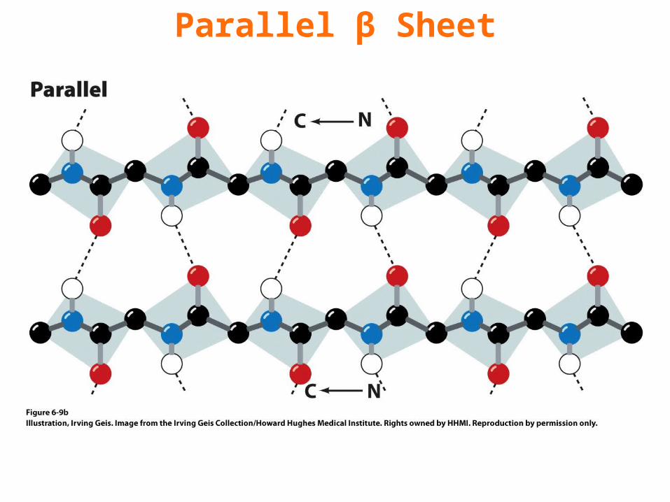
Parallel β Sheet
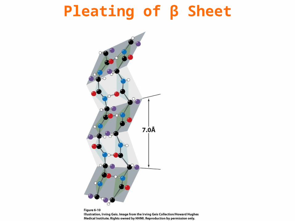
Pleating of β Sheet
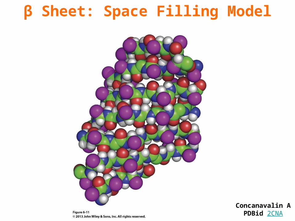
β Sheet: Space Filling Model
Concanavalin APDBid 2CNA
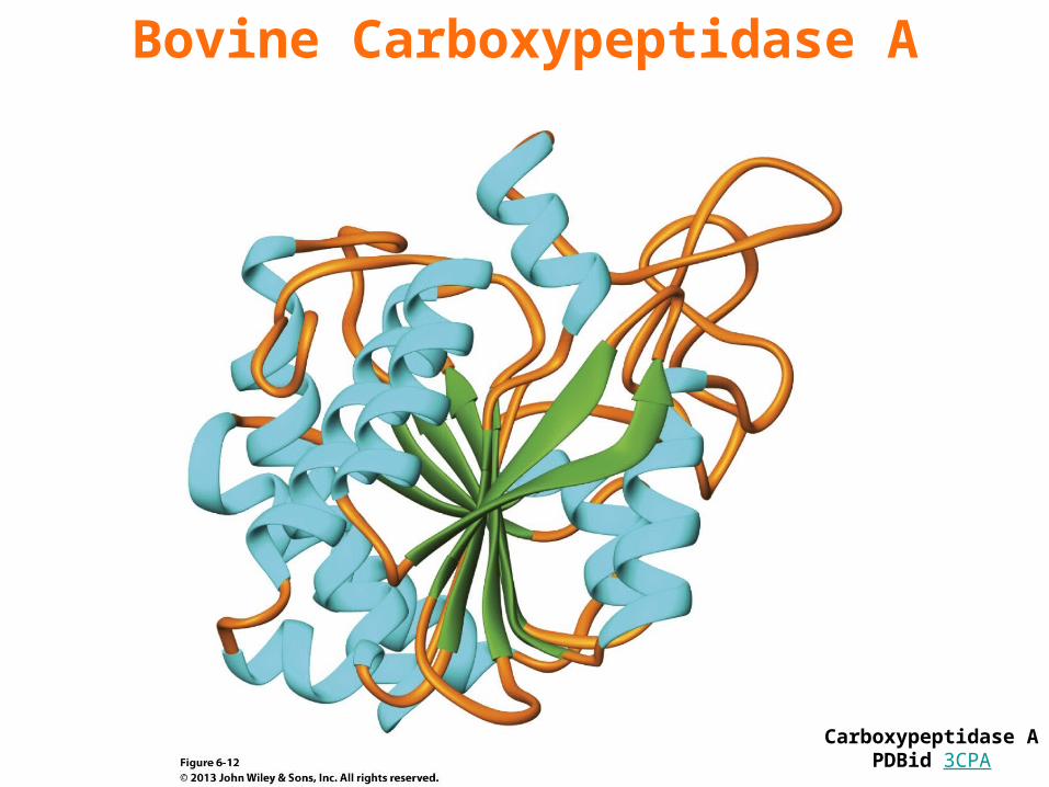
Bovine Carboxypeptidase A
Carboxypeptidase APDBid 3CPA
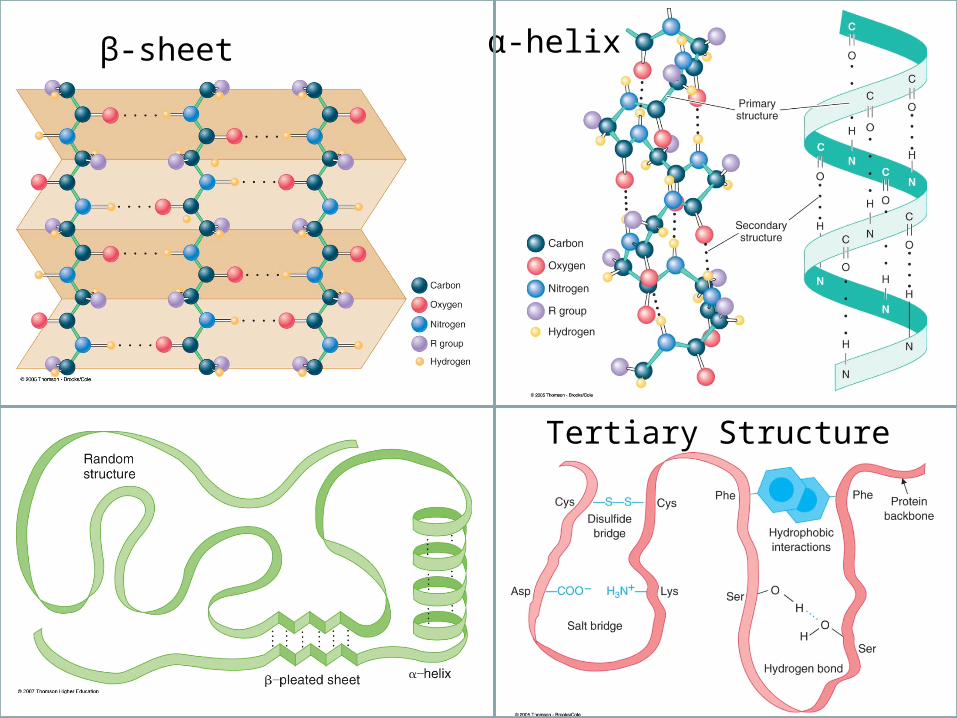
α-helixβ-sheet
Tertiary Structure
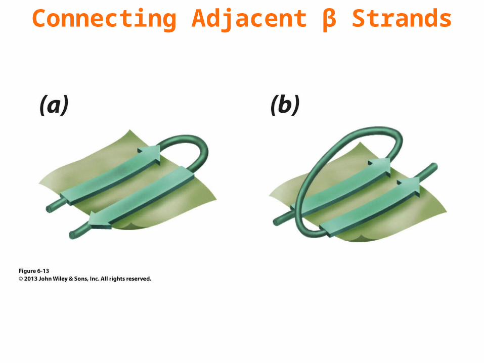
Connecting Adjacent β Strands
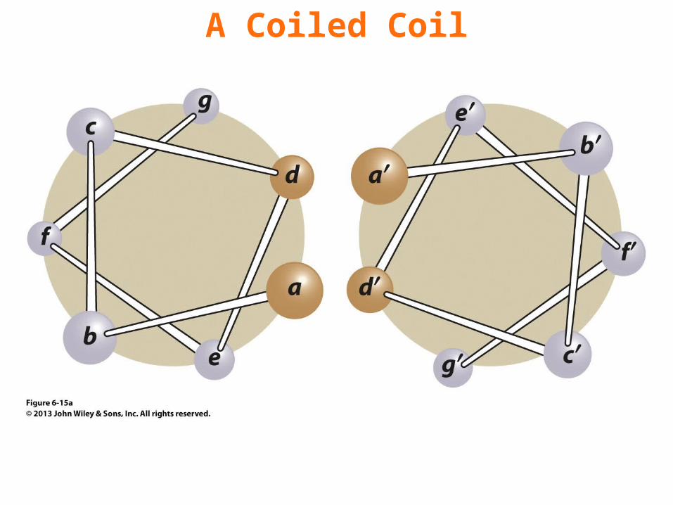
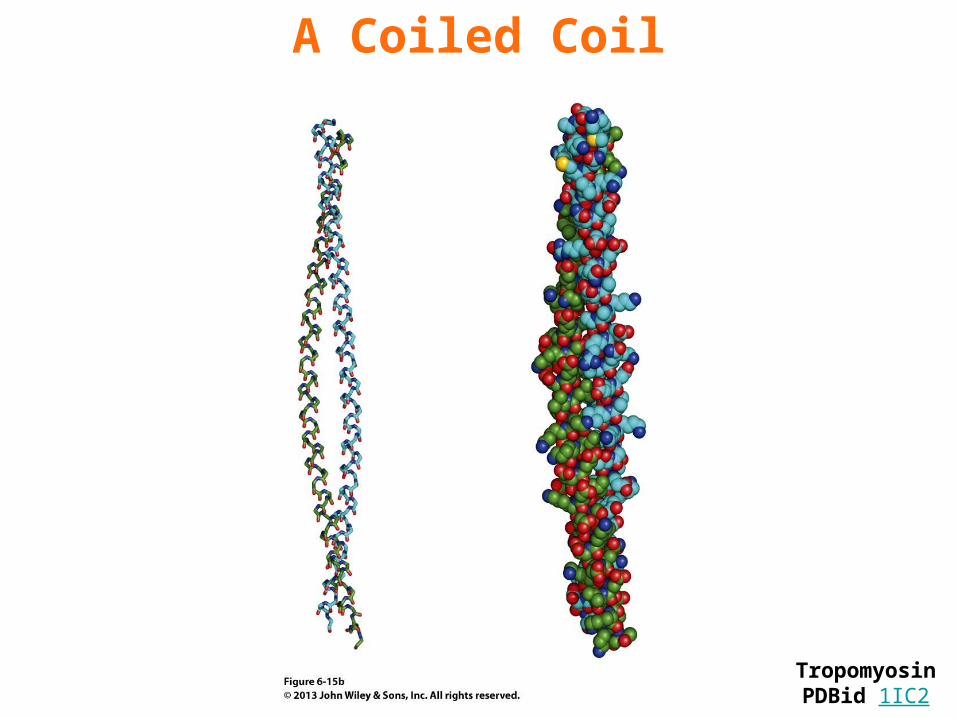
A Coiled Coil
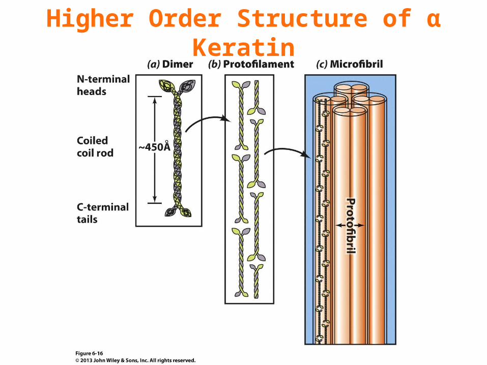
Higher Order Structure of α Keratin
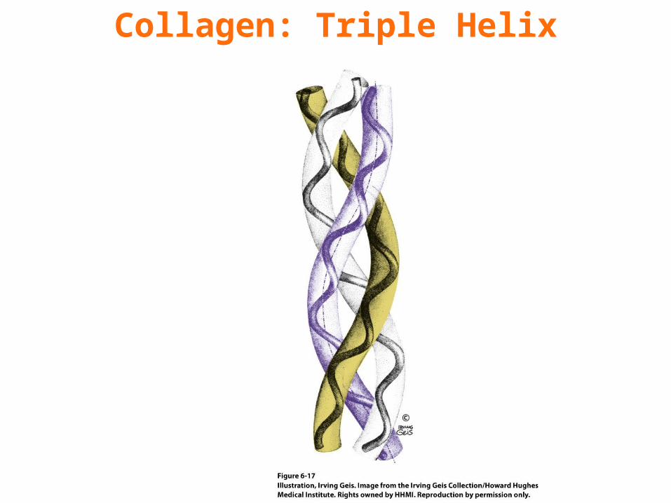
Collagen: Triple Helix
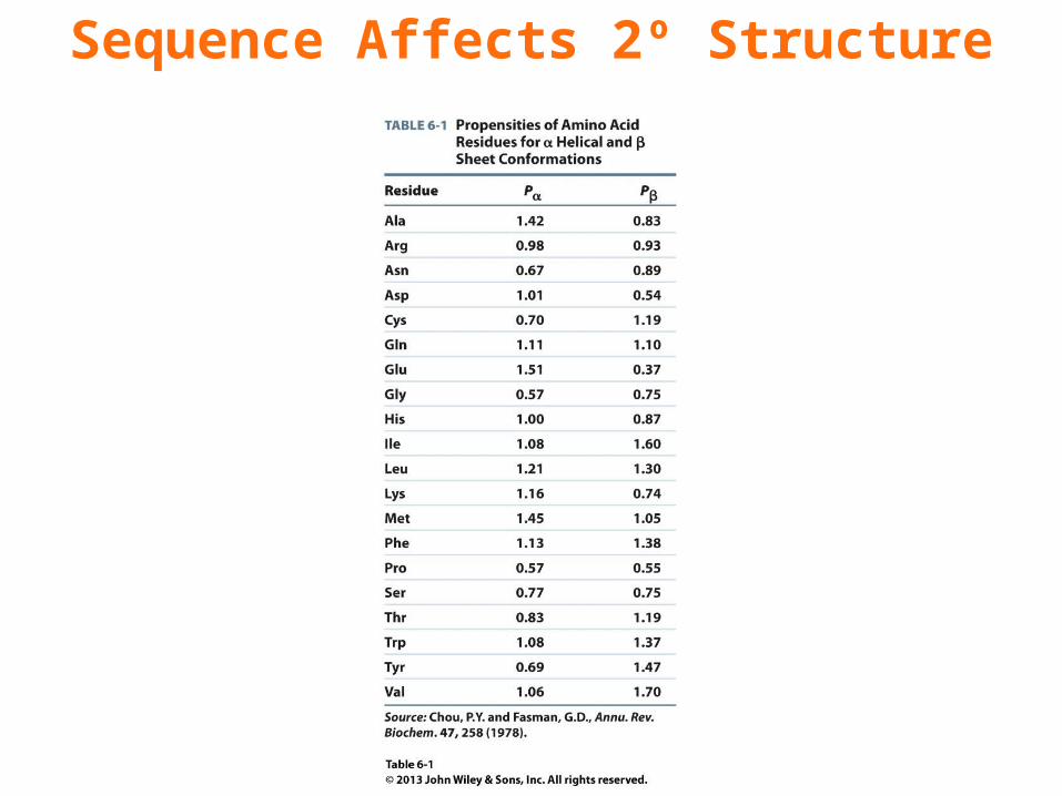
Sequence Affects 2º Structure
Chapter 6Tertiary Structure
Key Concepts 6.2 • X-Ray crystallography and NMR spectroscopy are used to determine the positions of atoms in proteins.• Nonpolar residues tend to occur in the protein interior and polar residues on the exterior.• A protein’s tertiary structure consists of secondary structural elements that combine to form motifs and domains.• Over time, a protein’s structure is more highly conserved than its sequence.• Bioinformatics databases store macromolecular structure coordinates. Software makes it possible to visualize proteins and compare their structural features.
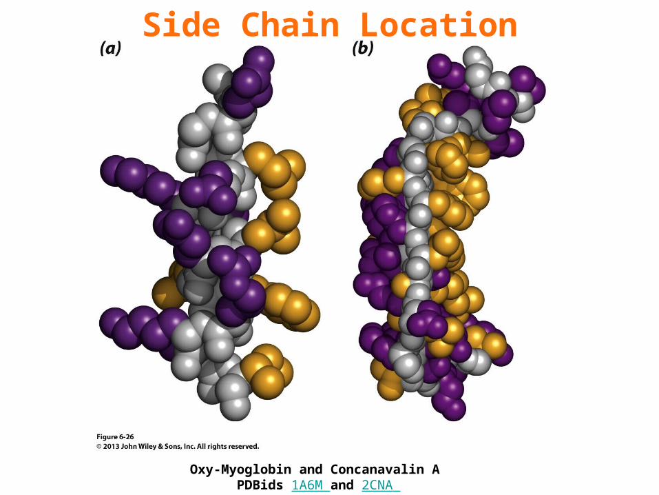
Side Chain Location
Oxy-Myoglobin and Concanavalin A PDBids 1A6M and 2CNA
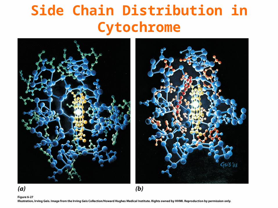
Side Chain Distribution in Cytochrome
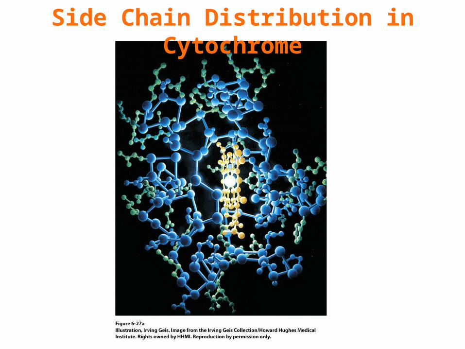
Side Chain Distribution in Cytochrome
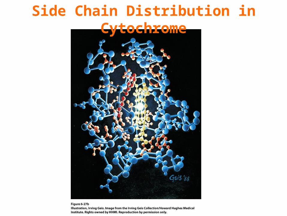
Side Chain Distribution in Cytochrome
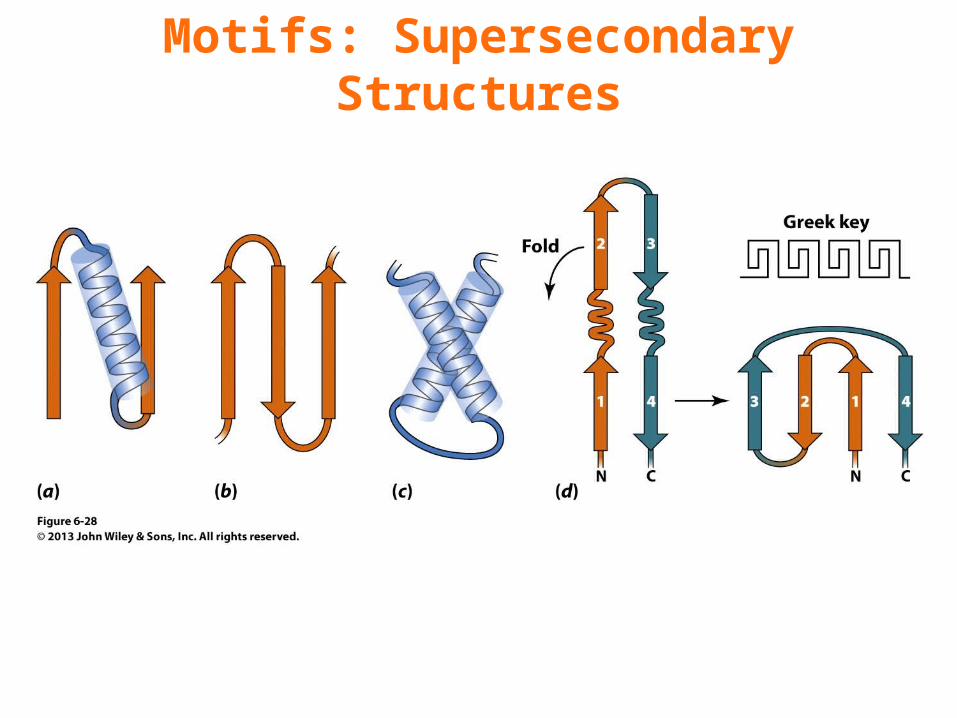
Motifs: Supersecondary Structures
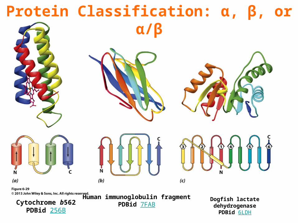
Protein Classification: α, β, or α/β
Cytochrome b562PDBid 256B
Human immunoglobulin fragmentPDBid 7FAB
Dogfish lactate dehydrogenasePDBid 6LDH
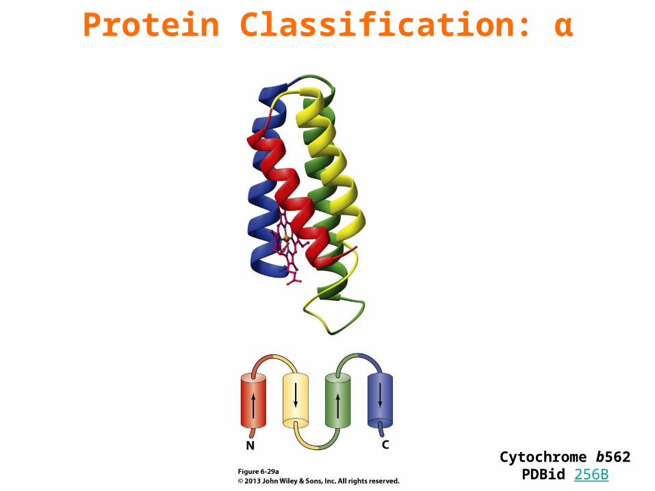
Protein Classification: α
Cytochrome b562PDBid 256B
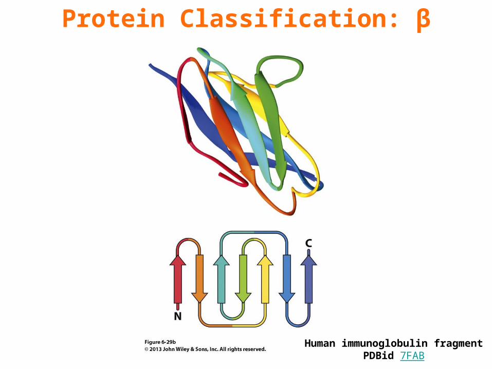
Protein Classification: β
Human immunoglobulin fragmentPDBid 7FAB
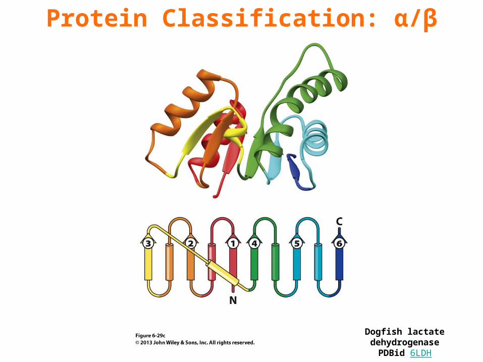
Protein Classification: α/β
Dogfish lactate dehydrogenasePDBid 6LDH
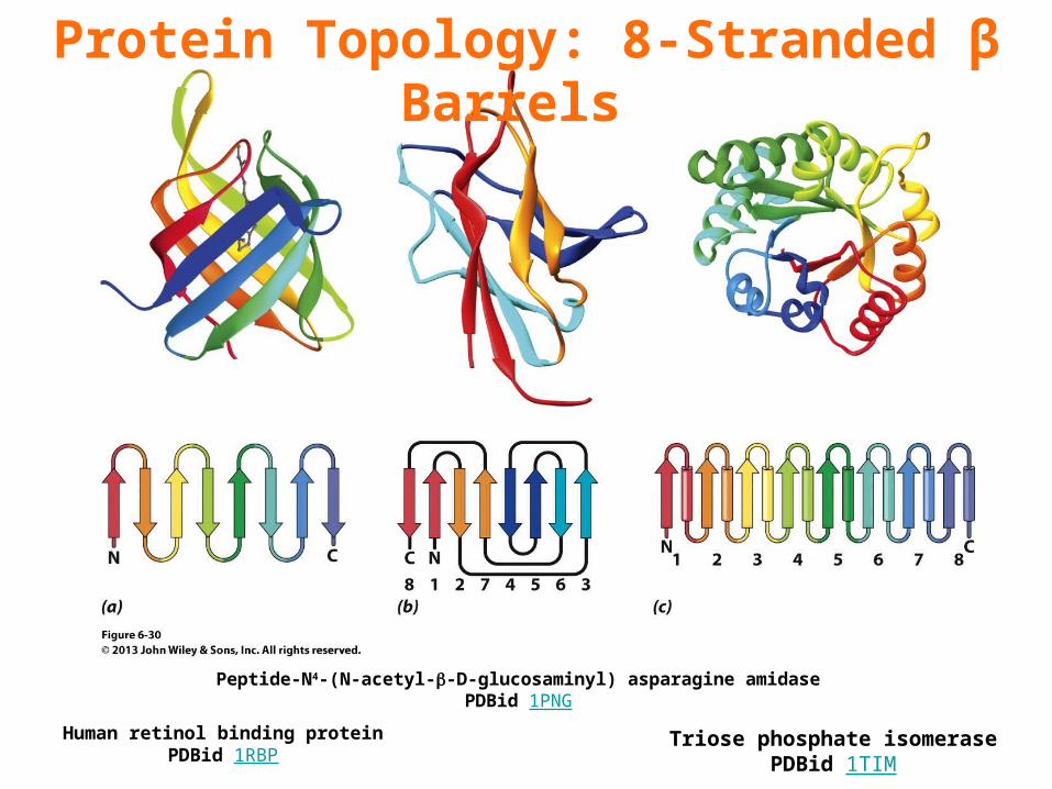
Protein Topology: 8-Stranded β Barrels
Human retinol binding proteinPDBid 1RBP
Peptide-N4-(N-acetyl--D-glucosaminyl) asparagine amidasePDBid 1PNG
Triose phosphate isomerasePDBid 1TIM
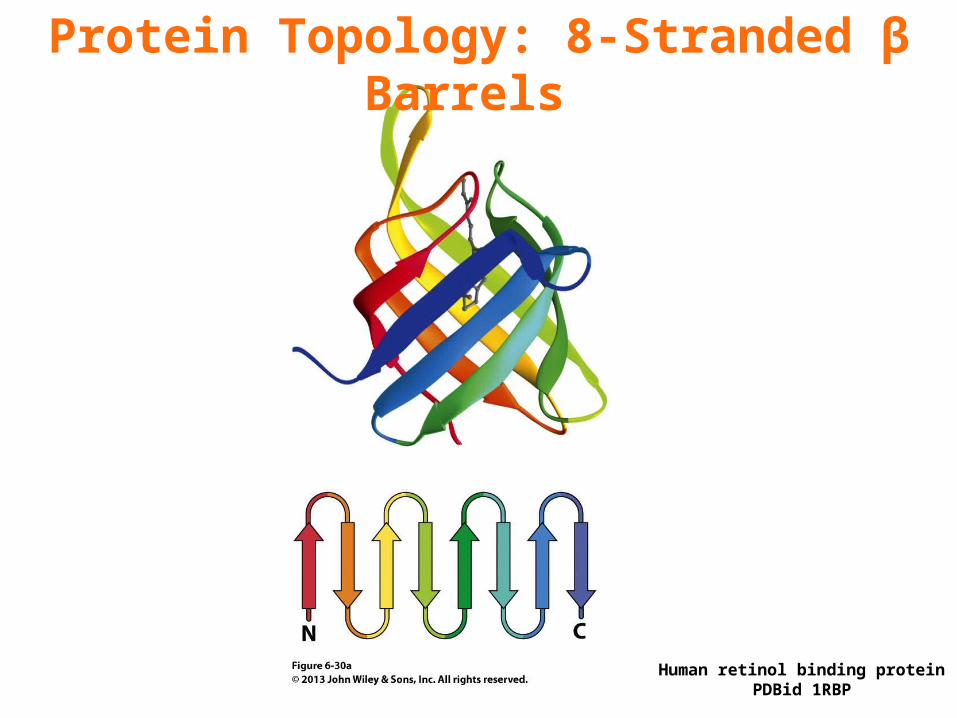
Protein Topology: 8-Stranded β Barrels
Human retinol binding proteinPDBid 1RBP
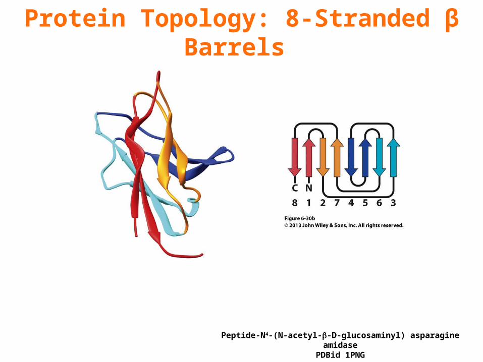
Protein Topology: 8-Stranded β Barrels
Peptide-N4-(N-acetyl--D-glucosaminyl) asparagine amidasePDBid 1PNG

Protein Topology: 8-Stranded β Barrels
Triose phosphate isomerasePDBid 1TIM
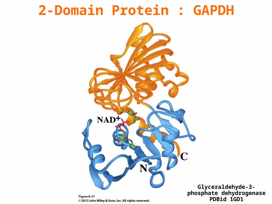
2-Domain Protein : GAPDH
Glyceraldehyde-3-phosphate dehydrogenasePDBid 1GD1
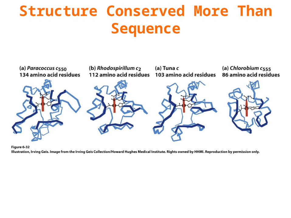
Structure Conserved More Than Sequence
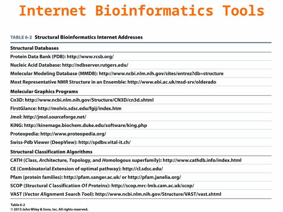
Internet Bioinformatics Tools
Chapter 6Quaternary Structure & Symmetry
Key Concepts 6.3 • Some proteins contain multiple subunits, usually arranged symmetrically.
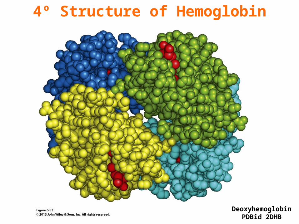
4º Structure of Hemoglobin
DeoxyhemoglobinPDBid 2DHB
Chapter 6Protein Stability
Key Concepts 6.4 • Protein stability depends primarily on hydrophobic effects and secondarily on electrostatic interactions.• A protein that has been denatured may undergo renaturation.• Protein structures are flexible and may include unfolded regions.
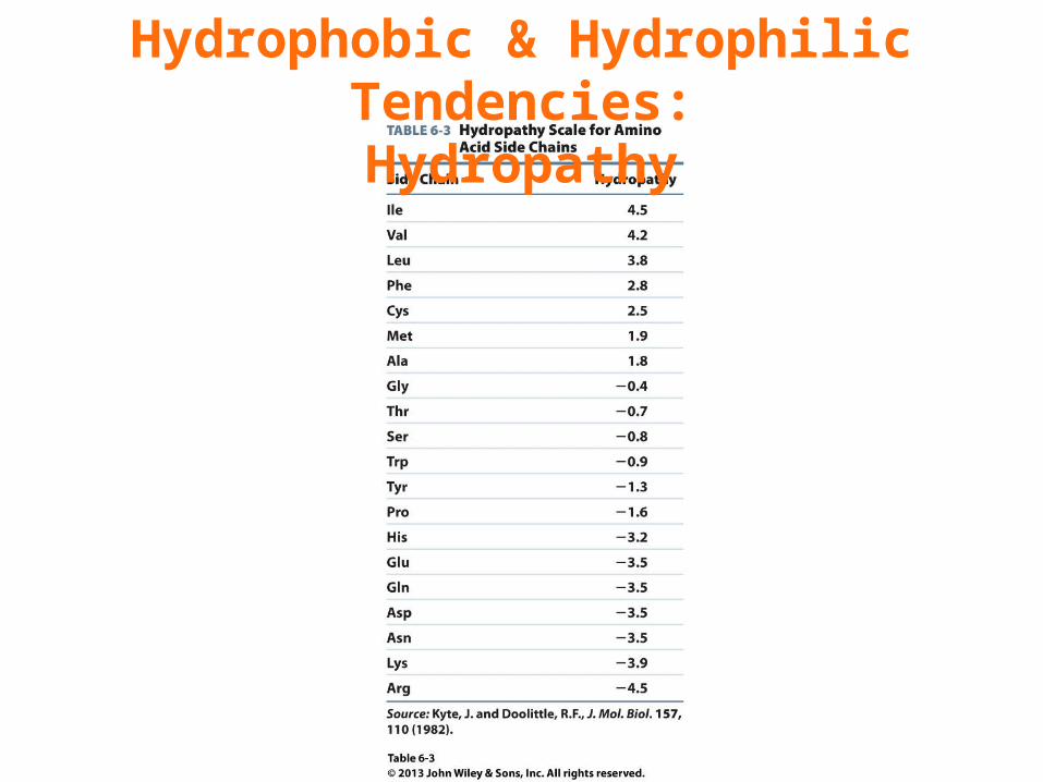
Hydrophobic & Hydrophilic Tendencies:Hydropathy
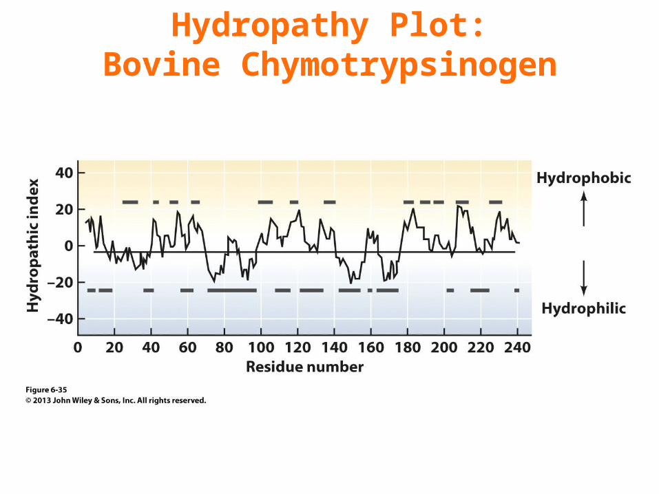
Hydropathy Plot:Bovine Chymotrypsinogen
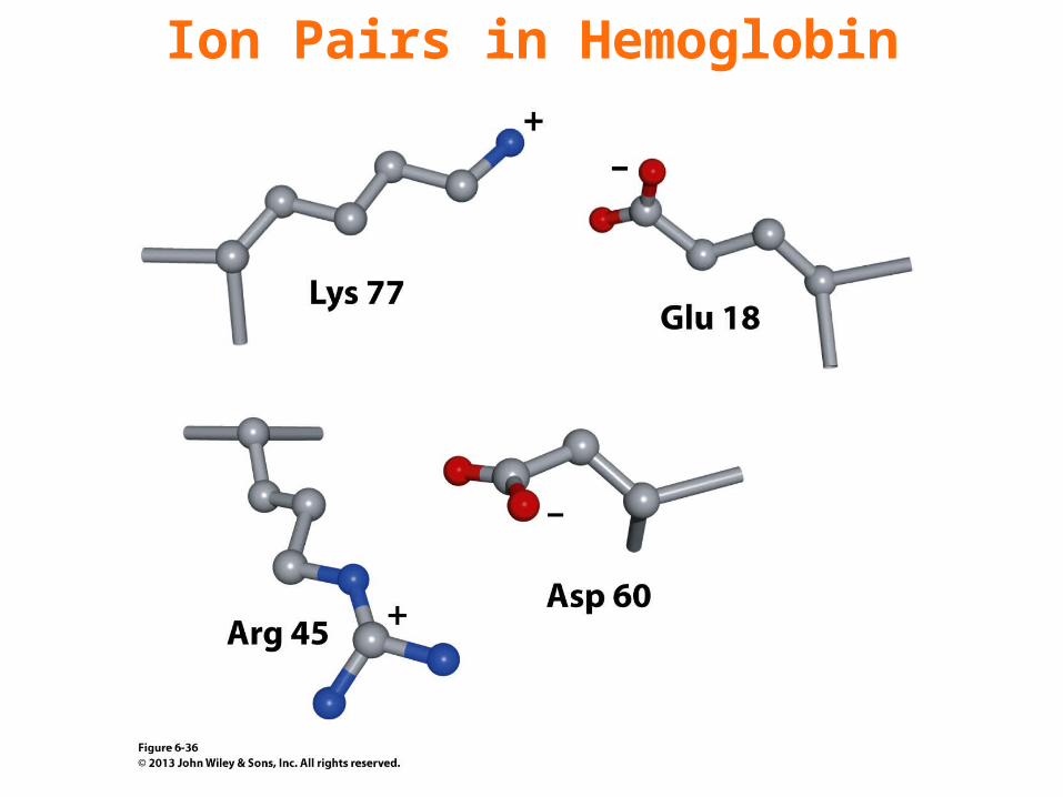
Ion Pairs in Hemoglobin
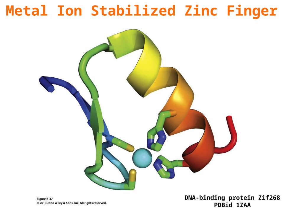
Metal Ion Stabilized Zinc Finger
DNA-binding protein Zif268PDBid 1ZAA
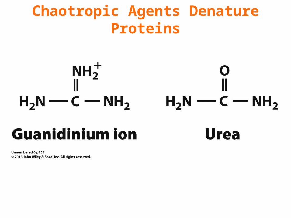
Chaotropic Agents Denature Proteins
Chapter 6Protein Stability
Checkpoint 6.4• Describe the hydropathic index plot for a fibrous protein such as collagen or keratin.• Describe the forces that stabilize proteins, and rank their relative importance.• Summarize the results of Anfinsen’s experiment with RNase A.• Why would it be advantageous for a protein or a segment of a protein to lack defined secondary or tertiary structure?

Chapter 6Protein Folding
Key Concepts 6.5 • A folding protein follows a pathway from high energy and high entropy to low energy and low entropy.• Protein disulfide isomerase catalyzes disulfide bond formation.• A variety of molecular chaperones assist protein folding via an ATP-dependent bind-and-release mechanism.• Amyloid diseases result from protein misfolding.• The misfolded proteins form fibrils containing extensive β structure.
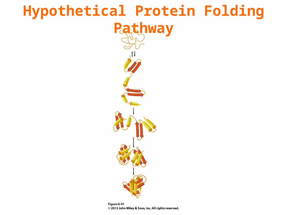
Hypothetical Protein Folding Pathway
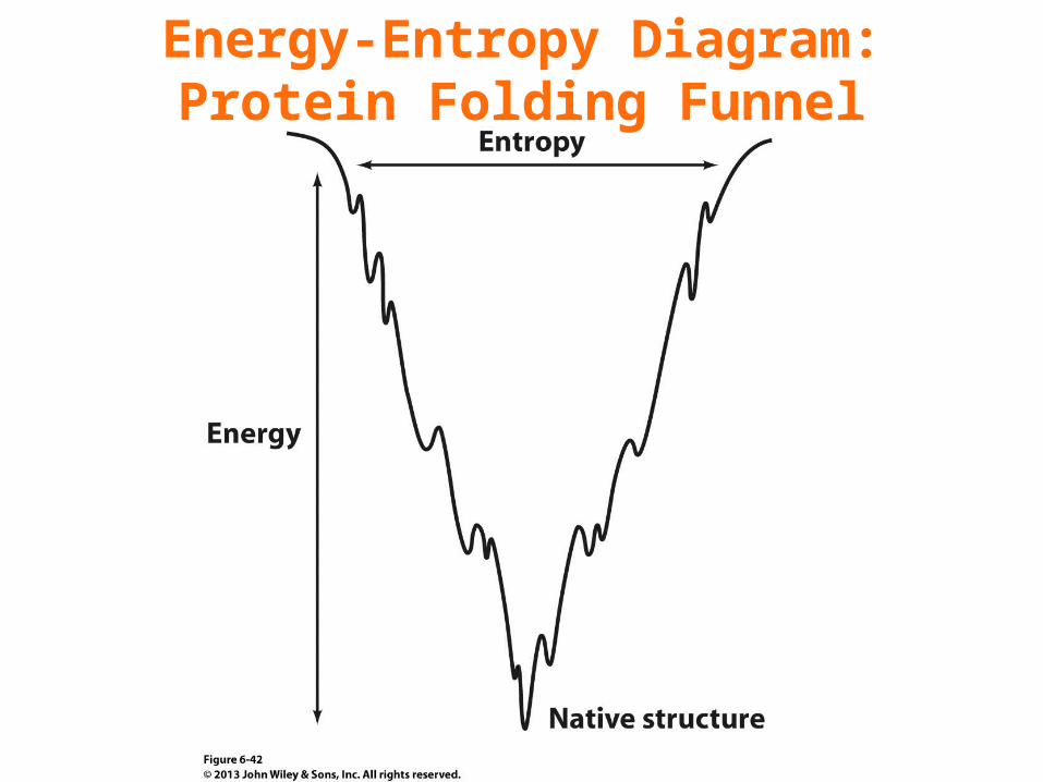
Energy-Entropy Diagram:Protein Folding Funnel
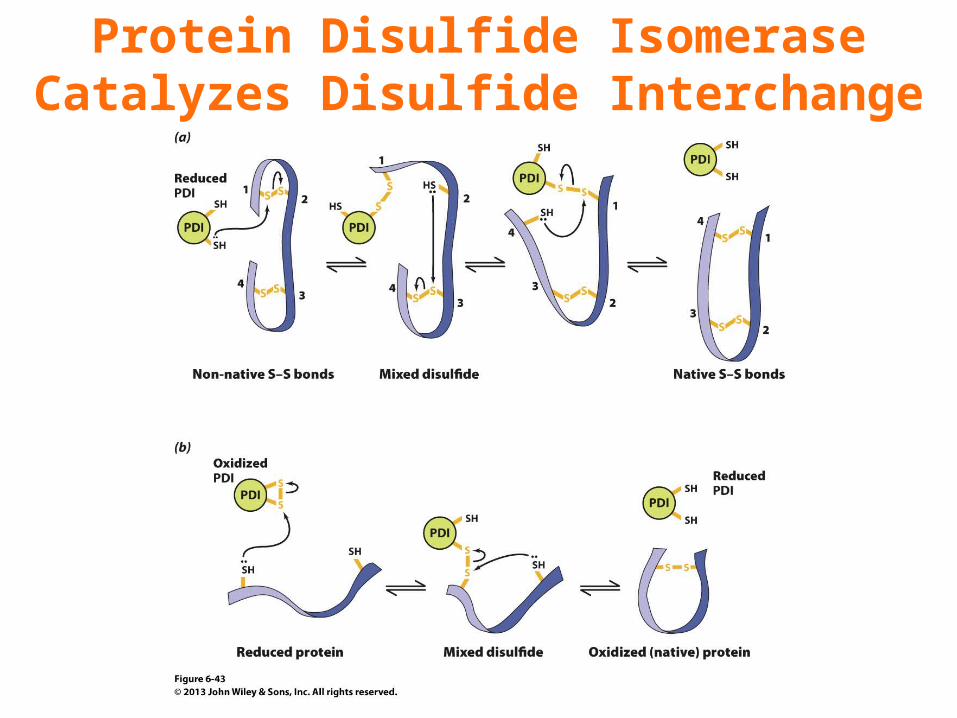
Protein Disulfide IsomeraseCatalyzes Disulfide Interchange
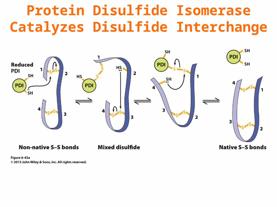
Protein Disulfide IsomeraseCatalyzes Disulfide Interchange
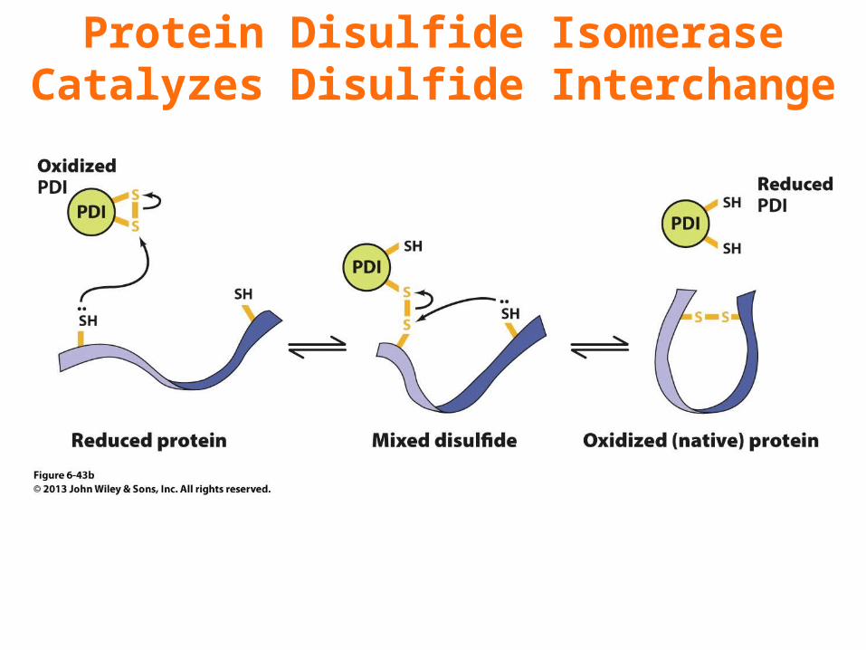
Protein Disulfide IsomeraseCatalyzes Disulfide Interchange
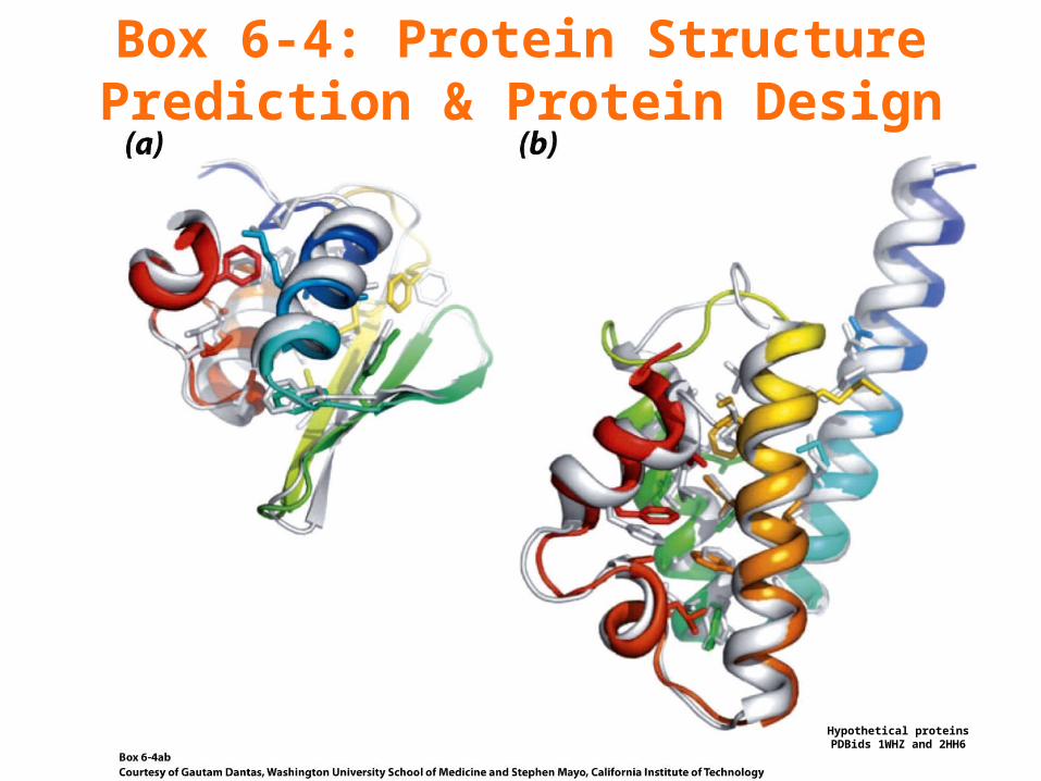
Box 6-4: Protein Structure Prediction & Protein Design
Hypothetical proteinsPDBids 1WHZ and 2HH6
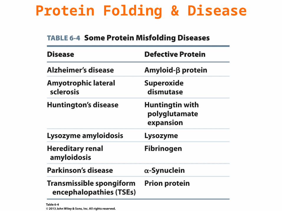
Protein Folding & Disease
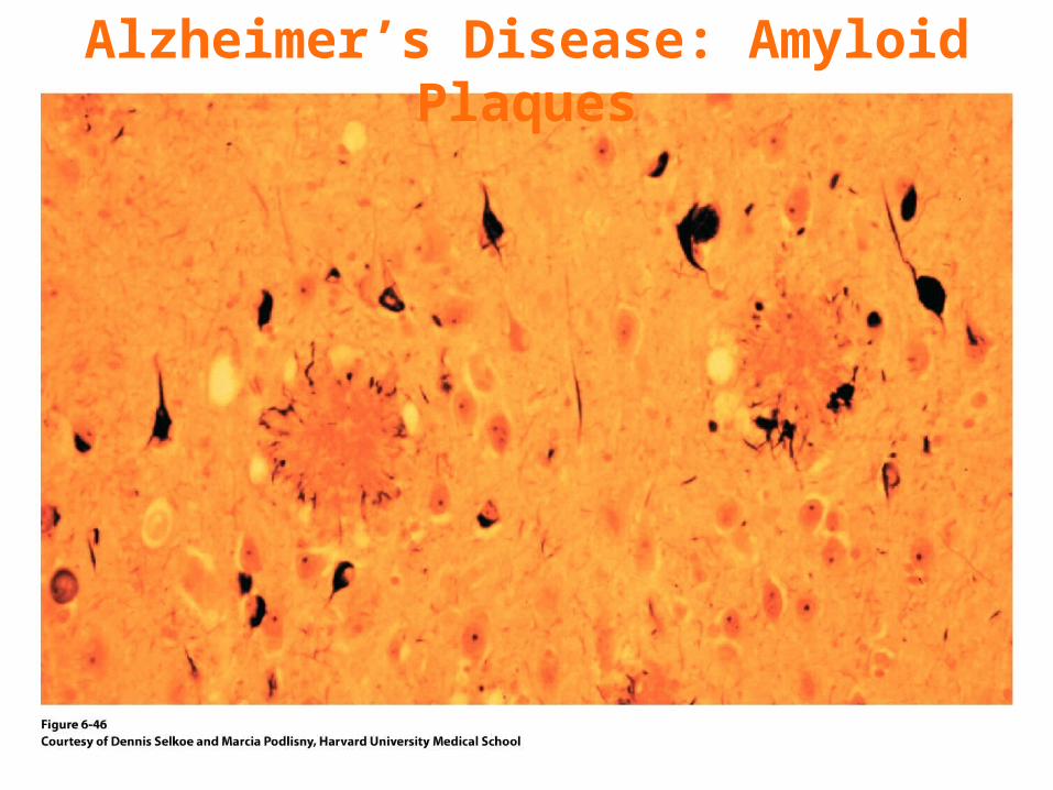
Alzheimer’s Disease: Amyloid Plaques
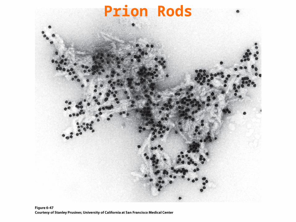
Prion Rods
Related Documents


