Chapter 21 The Respiratory System 1 Basic A & P II Dr. L. Bacha Chapter Outline (Martini & Nath 2010) read An Introduction to the Respiratory System Functions of the Respiratory System read about the five basic functions of the respiratory system Organization of the Respiratory System observe the components of the respiratory system in Fig. 21-1 what does the upper respiratory system consist of? what does the upper lower respiratory system include? what are alveoli? The Respiratory Mucosa the respiratory mucosa is a mucous membrane; the mucosa that lines the nasal cavity and trachea (and other parts of the respiratory system) is formed by: a. a ciliated pseudostratified columnar epithelium with goblet cells b. underlying connective tissue, called the lamina propria, that is well vascularized functions: the mucous membrane filters , warms , and moistens the air - air is warmed as heat radiates from the numerous blood vessels in the lamina propria - air is moistened as water evaporates from the mucus produced by goblet cells - air is filtered as particles (dust, pollen, bacteria, etc.) become trapped in the sticky blanket of mucus that covers the cilia of the epithelium - the cilia sweep the mucus and trapped debris toward the pharynx, where it is swallowed The Respiratory Defense System read about the respiratory defense system on p. 711

Welcome message from author
This document is posted to help you gain knowledge. Please leave a comment to let me know what you think about it! Share it to your friends and learn new things together.
Transcript

Chapter 21 The Respiratory System 1
Basic A & P II Dr. L. Bacha Chapter Outline (Martini & Nath 2010)
read An Introduction to the Respiratory System
Functions of the Respiratory System
read about the five basic functions of the respiratory system
Organization of the Respiratory System
observe the components of the respiratory system in Fig. 21-1
what does the upper respiratory system consist of?
what does the upper lower respiratory system include? what are alveoli? The Respiratory Mucosa
the respiratory mucosa is a mucous membrane; the mucosa that lines the nasal cavity and trachea (and other parts of the respiratory system) is formed by:
a. a ciliated pseudostratified columnar epithelium with goblet cells
b. underlying connective tissue, called the lamina propria, that is well vascularized
functions: the mucous membrane filters, warms, and moistens the air
- air is warmed as heat radiates from the numerous blood vessels in the lamina propria
- air is moistened as water evaporates from the mucus produced by goblet cells
- air is filtered as particles (dust, pollen, bacteria, etc.) become trapped in the sticky blanket of mucus that covers the cilia of the epithelium
- the cilia sweep the mucus and trapped debris toward the pharynx, where it is swallowed
The Respiratory Defense System read about the respiratory defense system on p. 711

Chapter 21 The Respiratory System 2
The Nose, Nasal Cavity, and Paranasal Sinuses
what is the other name for the external nares? what cavity do they open into?
features of the NASAL CAVITY:
1. define nasal vestibule:
2. the nasal septum divides what?
- the posterior part of the nasal septum is formed by bone
- the anterior portion of the nasal septum (toward the tip of the nose) is formed by what?
3. paranasal sinuses
- sinuses are air-filled cavities lined with mucous membrane present in some of the bones of the skull; they open into the nasal cavity and act as resonating chambers for sound
- their mucous secretions, aided by tears, help to do what?
4. olfactory epithelium - contains olfactory receptors in the superior portion of the nasal cavity, which are responsible for our sense of what?
5. three pairs of nasal conchae (also called turbinate bones) are curved, shelf-like bones covered with mucous membrane that curl in from the lateral walls of the nasal cavity
- name the three pairs of nasal conchae: - name the three pairs of narrow grooves through which air flows to pass through the nasal
cavity:
- the nasal conchae and meatuses increase surface area of the mucous membrane, and force the air to swirl around, which increases contact of the air with the mucous membrane
6. nasolacrimal ducts – open into the nasal cavity inferior to the inferior nasal choncha - do you remember what secretion the nasolacrimal ducts carry?
7. the nasal cavity is separated from the oral cavity by what part of the palate? 8. the nasal cavity opens into the nasopharynx through a common opening known as what?
As you study the organs and their specific
parts, examine their locations in the Figures in the textbook.

Chapter 21 The Respiratory System 3
The Pharynx
There are three regions of the pharynx: nasopharynx
◦ the superior portion that extends from the internal nares to the uvula (the tip of the soft palate) and it is separated from the oral cavity by what structure?
◦ it is a passageway for air only
◦ name the tonsil that it contains in its posterior wall:
◦ it has the openings of the auditory tubes that lead to the cavity of the middle ear and equalize pressure in the cavity of the middle ear with the outside atmosphere
oropharynx
◦ extends between what?
◦ it serves as a common passageway for food, drink and air
laryngopharynx
◦ includes the portion between what structures?

Chapter 21 The Respiratory System 4
• name the narrow opening into the larynx:
Cartilages and Ligaments of the Larynx
• the wall of the larynx has 9 cartilages (see. Fig. 21-4), some of the cartilages are listed here:
thyroid cartilage - large, anterior piece
cricoid cartilage - inferior ring like piece of cartilage - serves as a landmark for an emergency airway called a tracheotomy
epiglottis - a large, leaf-like structure supported by elastic cartilage; the broad superior part is
unattached
during swallowing, what happens to the larynx and the epiglottis, and what does this action prevent?
arytenoid cartilages - they attach to the vocal folds and intrinsic laryngeal muscles
SOUND PRODUCTION
• there are two pairs of folds of mucous membrane in the larynx:
(1) vestibular folds (ventricular folds; false vocal cords)
the superior, inelastic pair
they function in holding the breath against pressure in the thoracic cavity, as might occur when straining to lift a heavy object; they adduct during swallowing to help prevent food and drinks from entering the glottis
(2) vocal folds (true vocal cords)
the vocal folds are very elastic (numerous elastic ligaments
as air is directed against the vocal folds, they vibrate and produce sound waves
• sound waves originate from the vibration of the vocal folds, but numerous other structures are necessary for converting the sound into recognizable speech and for amplification and resonance
- amplication and resonance occur within what structures?
Chapter 21 The Respiratory System 5
The Trachea (Fig. 21-6)
• the trachea is a passageway for air; what is the other name for the trachea?
• the trachea extends between the larynx and primary bronchi • wall of the trachea: lined by a mucous membrane like that of the nasal cavity: ciliated, pseudostratified
columnar with goblet cells contains 15 - 20 pieces of hyaline cartilages called tracheal cartilages
- what is the function of the cartilages?
- what is the shape of the cartilages? - why does the open portion face posteriorly (toward the esophagus)?
The Primary Bronchi
• the distal part of the trachea divides into a right primary bronchus and a left primary bronchus (plural is “bronchi”)
• each primary bronchus enters a lung and branches into a lobe of the lung as a secondary bronchus
• each primary bronchus passes into a lung through a groove along the medial surface of each lung; what is the groove called?
- the structures that pass through this groove form the root of the lung
• each lung is a blunt cone - what is the superior tip of each lung called?
- what is the broad, inferior concave portion called?
• locate the apex and base of each lung on Fig. 21-7
Chapter 21 The Respiratory System 6
Lobes and Surfaces of the Lungs
• the lungs are paired organs in the thoracic cavity; they are highly distensible and elastic
• lobes and fissures (Fig. 21-7)
fissures are CT filled spaces that divide the lungs into lobes
right lung the right lung has how many lobes? list the lobes of the right lung:
left lung the left lung has how many lobes? list the lobes of the left lung:
• each lobe is subdivided into microscope lobules • the lungs are made up of the components of the bronchial tree
The Bronchi, Bronchioles, Alveolar Ducts, and Alveoli (I have grouped the information
from page 720 to 722 here:)
• the primary bronchi and their branches form what?
The Bronchial Tree:
1. the distal part of the trachea divides into a right primary bronchus and a left primary bronchus (plural is “bronchi”); each primary bronchus enters a lung and branches into a lobe of the lung as a secondary bronchus
2. secondary bronchi - what are secondary bronchi also known as?
- each secondary bronchus branches within a lobe of a lung and forms a tertiary bronchus
3. tertiary bronchi - what are tertiary bronchi also called?
- each supplies a unit within a lobe called a bronchopulmonary segment and eventually branches into bronchioles
4. bronchioles - bronchioles branch many times within a lobe of the lung - there are many different types of bronchioles
5. eventually, bronchioles lead into alveolar sacs, which are formed entirely by clusters of alveoli
(we will get back to alveoli; they are very important…!)

Chapter 21 The Respiratory System 7
Alveoli (up to page 722)
singular is “alveolus”; see Figs. 21-9, 21-10 and 21-11
each lung contains about how many alveoli?
the numerous alveoli give the lungs what type of appearance?
what is associated with each alveolus?
what are the capillaries surrounded by?
gas exchange occurs in the lungs between air in the alveoli and blood in pulmonary capillaries
alveoli are lined mainly by cells called pneumocytes type I - what type of epithelial cells are pneumocytes type I cells?
what is the function of the alveolar macrophages?
less numerous pneumocytes type II also line the alveoli - what are these cells also called?
- what do the type II cells produce?
what does surfactant reduce?
- without surfactant, what would happen to the alveoli?
structure of bronchi vs. bronchioles: bronchi - have an epithelium, bundles of smooth muscle and plates of hyaline cartilage;
are larger in diameter
bronchioles - have an epithelium, bundles of smooth muscle; no hyaline cartilage
Have ever used latex
gloves? They have
powder inside, so that
the fingers stick together.
The powder decreases
surface tension, just like
surfactant of the alveoli!
If the fingers of the gloves
adhere, could you get
your hands in them
easily? If the alveoli
collapsed, could air flow
in and out of them?
Chapter 21 The Respiratory System 8
RESPIRATORY MEMBRANE
what occurs across the respiratory membrane of alveoli? the gases diffuse between the air in the alveolus and blood in the pulmonary capillary
the three layers of the respiratory membrane are:
(1) the simple squamous epithelial cells (pneumocytes type I) of the alveolus
(2) two fused basement membranes: the basement membrane of the alveolus and the basement membrane of the pulmonary capillary
(3) the simple squamous epithelial cells (endothelial cells) of the pulmonary capillary
The Blood Supply to the Lungs there are two sets of arteries that supply the lungs:
(1) pulmonary arteries - do they carry oxygenated blood or deoxygenated blood?! - are they part of the pulmonary or systemic circuit?
(2) bronchial arteries - branch from the aorta - do they carry oxygenated blood or deoxygenated blood?! - are they part of the pulmonary or systemic circuit?
The Pleural Cavities and Pleural Membranes
each lung occupies a pleural cavity which is lined by what? name the two layers of the pleura and indicate what each layer covers:
Chapter 21 The Respiratory System 9
pleural cavity = the potential space between the visceral and parietal pleura
- it contains a thin film of fluid called pleural fluid that is secreted by the serous membranes
functions of pleural fluid: (1) reduces friction between the serous membranes, so that they can slide easily over one
another during breathing (2) increases surface tension and causes the parietal pleura and visceral pleurae to adhere
to each other!
what is the primary function of pulmonary ventilation?
The Movement of Air
in pulmonary ventilation, air flows between the atmosphere and the alveoli of the lungs it flows because of alternating pressure differences created by contraction and relaxation of
muscles of respiration
Gas Pressure and Volume (Boyle’s Law)
Boyle’s Law: the pressure of a gas in a closed container is inversely proportional to the volume of the container (at a constant temperature); this means that: ◦ if the volume of a chamber decreases, pressure of the gas inside increases or decreases? ◦ if the volume of a chamber increases, pressure of the gas inside increases or decreases?
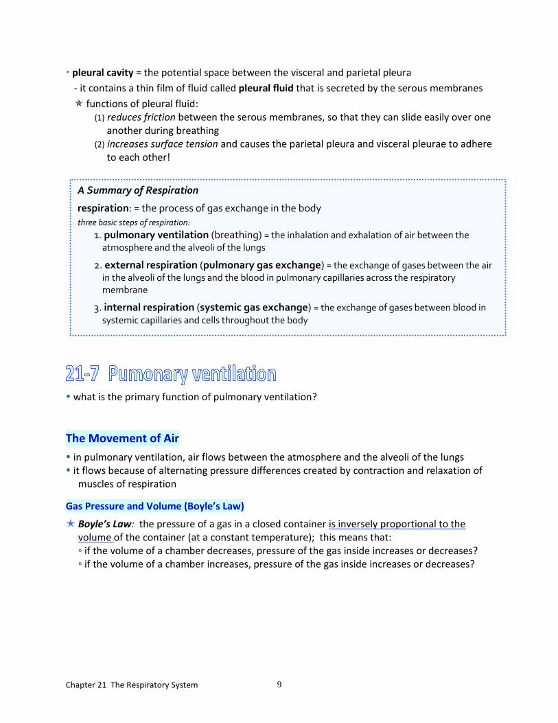
A Summary of Respiration
respiration: = the process of gas exchange in the body
three basic steps of respiration:
1. pulmonary ventilation (breathing) = the inhalation and exhalation of air between the atmosphere and the alveoli of the lungs
2. external respiration (pulmonary gas exchange) = the exchange of gases between the air in the alveoli of the lungs and the blood in pulmonary capillaries across the respiratory membrane
3. internal respiration (systemic gas exchange) = the exchange of gases between blood in
systemic capillaries and cells throughout the body

Chapter 21 The Respiratory System 10
Gas Pressure and Volume read about pressure and airflow to the lungs on page 727
Compliance the compliance of the lungs is an indication of what?
the greater the compliance, the easier it is to what? list the three factors affecting compliance:
Pressure Changes during Inhalation and Exhalation
There are three pressures involved in pulmonary ventilation: (1) atmospheric pressure = the pressure in the atmosphere; = 760 mmHg
(2) intrapulmonary pressure - define intrapulmonary pressure (p. 728): - it varies from 758 mm Hg (during inspiration) to 762 mm Hg (during expiration)
(3) intrapleural pressure - define intrapleural pressure (p. 728): - it varies from 754 mm Hg (during inspiration) to 756 mm Hg (during expiration) - so, how does it compare to the other two pressures (atmospheric and intrapulmonary)?
The Mechanics of Breathing (page 730)
Inhalation ◦ inhalation (inspiration)= breathing in
◦ for air to flow into the lungs, the pressure within the alveoli must become lower than atmospheric pressure
◦ quiet (unforced) inhalation involves contraction of two muscles:
diaphragm a dome shaped muscle (when relaxed); innervated by the phrenic nerve when the diaphragm contracts, it flattens and lengthens the thoracic cavity
external intercostals contraction elevates the ribs and increases the diameter of the thoracic cavity
Chapter 21 The Respiratory System 11
Exhalation exhalation (expiration) = breathing out
the process of normal exhalation said to be passive because it does not involve contraction of muscles; it begins when the muscles of inspiration (diaphragm and external intercostals) relax - as a result, the volume of the thoracic cavity, and therefore the lungs, decreases to its original volume
• • • • • My Summary of Events in Pulmonary Ventilation • • • • •
1. AT THE END OF EXPIRATION AND BEFORE THE NEXT INSPIRATION
Atmospheric pressure (760 mmHg) is equal to intrapulmonary pressure (760 mmHg), and there is no net movement of air.
2. INHALATION the respiratory center in the medulla oblongata
of the brain initiates nerve impulses to muscles of inspiration
Contraction of the diaphragm Contraction of external intercostal muscles Diaphragm lowers Rib cage raises and rotates Length of thoracic cavity increases Diameter of thoracic cavity increases
VOLUME of the THORACIC CAVITY INCREASES The volume within the lungs INCREASES as the LUNGS EXPAND
INTRAPULMONARY PRESSURE DECREASES (to 758 mm Hg) and IS LOWER THAN ATMOSPHERIC PRESSURE (760)
AIR FLOWS INTO the ALVEOLI of the lungs from the atmosphere
At the end of inspiration, the intrapulmonary pressure becomes equal to atmospheric pressure (760 mmHg), and there is no movement of air.
Inspiration is an active process, because it involves muscle contraction. Deep inspiration (forced inspiration) involves additional muscles to further increase the size of the thoracic cavity. This causes a greater difference between intrapulmonary pressure and atmospheric pressure, so that more air flows into the lungs.
The lungs expand (are distended) as the volume of the thoracic cavity increases because: 1. they adhere to the thoracic wall due to surface tension between the parietal and
visceral pleura
2. a decrease in intrapleural pressure (from 756 to 750 mm Hg) causes the alveoli to expand

Chapter 21 The Respiratory System 12
3. EXHALATION
Nerve impulses from the respiratory center in the medulla of the brain stop, so that contraction of the diaphragm and external intercostal muscles ends
Relaxation of diaphragm Relaxation of external intercostals Diaphragm raises Rib cage lowers (back to resting position) (back to resting position) Length of thoracic cavity decreases Diameter of thoracic cavity decreases to original length to original diameter
VOLUME of the THORACIC CAVITY DECREASES (back to original volume) THE LUNGS DECREASE to ORIGINAL SIZE and volume within the lungs DECREASES
due to elastic recoil of the lungs
INTRAPULMONARY PRESSURE INCREASES (to 762 mm Hg) and is GREATER THAN ATMOSPHERIC PRESSURE (760 mmHg)
AIR FLOWS OUT of the ALVEOLI of the lungs to the atmosphere
At the end of expiration, the intrapulmonary pressure becomes equal to atmospheric pressure (760 mmHg), and there is no movement of air.
Normal expiration is a passive process, because it does not involve muscle contraction. Deep expiration (forced expiration) involves muscle contraction to further decrease the size of the thoracic cavity. This causes a greater difference between intrapulmonary pressure and atmospheric pressure, so that more air flows out of the lungs.
The lungs do not collapse completely during expiration because:
1. Surfactant is produced by type II pneumocytes cells (septal cells) and it decreases the surface tension of the alveoli.
2. The lungs are kept slightly inflated because of the negative intrapleural pressure. (Remember the intrapleural pressure is always less than the intrapulmonary pressure and atmospheric pressure.)
• • • • • • • • • • • • • • • • • • • • • • • • • • • • • •
Respiratory Rates and Volumes
define respiratory rate (p. 732):
- what is the normal respiratory rate of a resting adult?

Chapter 21 The Respiratory System 13
a spirometer is an apparatus commonly used to measure the volume of air exchanged during breathing
define resting tidal volume:
◦ what is the average normal resting tidal volume? Inspiratory reserve volume = the amount of air that can be forcefully inhaled after a normal tidal volume inhalation
if you inhale normally, then force in as much air as you can, (try it!) the volume that you forcefully inhaled is the inspiratory reserve volume!
Expiratory reserve volume = the amount of air that can be forcefully exhaled after a normal tidal volume exhalation if you exhale normally, then force out as much air as you can, the volume that you forcefully exhaled is the expiratory reserve volume!
Residual volume define residual volume: so, do the lungs empty of air after you exhale as much as you can?!

Chapter 21 The Respiratory System 14
Dalton’s Law and Partial Pressures
define partial pressure of a gas: write an equation to show this relationship for the atmosphere: O2 will diffuses across a permeable membrane from high to low partial pressure of O2 CO2 will diffuses across a permeable membrane from an area of high to low PCO2
Here is my way to present gas exchange, covered on pages 736 and 737:
external respiration (pulmonary gas exchange) = the exchange of gases between the alveoli of the lungs and the blood in pulmonary capillaries across the respiratory membrane
Study Fig. 21-19a on p. 736 to help you find the answers to these questions:
1. In the deoxygenated blood pumped from the right ventricle of the heart to the alveolar (pulmonary) capillaries in the lungs, what is the partial pressure of O2?
- what is the partial pressure of CO2?
2. In the air in the alveolus, what is the partial pressures of O2? of CO2?
Now focus on one gas at a time:
a. compare the partial pressure of O2 in the deoxygenated blood and in the air in the alveolus
◦ where is the PO2 higher?
◦ which way will O2 diffuse, into or out of the pulmonary capillaries?
b. compare the partial pressure of CO2 in the deoxygenated blood and in the air of the alveolus
◦ where is the PCO2 higher?
◦ which way will CO2 diffuse, into or out of the pulmonary capillaries?
3. In the blood that leaves the pulmonary capillaries and returns to the left side of the heart, what is the partial pressure of O2? of CO2?
◦ is this blood oxygenated or deoxygenated?

Chapter 21 The Respiratory System 15
List (and read about) the 5 reasons why gas exchange at the respiratory membrane is efficient (the headings in italics on p.735 to 736):
1.
2.
3.
4.
5.
systemic gas exchange is the exchange of gases between blood in systemic capillaries and cells throughout the body
return to Fig. 21-19b:
4. In the oxygenated blood pumped from the left ventricle of the heart to the systemic capillaries that supply the systemic cells throughout the body (these could be cells in a muscle in your leg, in the CT of the stomach, in the choroid of the eye, etc, etc,!!)
what is the partial pressure of O2? of CO2? 5. In the systemic cell, what is the partial pressure of O2? of CO2?
Now focus on one gas at a time:
a. compare the partial pressure of O2 in the oxygenated blood and in the systemic cell ◦ where is the PO2 higher? ◦ which way will O2 diffuse, into or out of the systemic capillaries?
b. compare the partial pressure of CO2 in the oxygenated blood and in the systemic cell ◦ where is the PCO2 higher? ◦ which way will CO2 diffuse, into or out of the systemic capillaries?
6. In the blood that leaves the systemic capillaries and returns to the right side of the heart, what is the partial pressure of O2? of CO2?
◦ is this blood oxygenated or deoxygenated?

Chapter 21 The Respiratory System 16
Now, back to following the textbook, page 737…
Oxygen Transport
oxygen is not highly soluble in water only about what percent of oxygen molecules are in solution in the plasma?
the rest (98.5%) of oxygen molecules are bound to what? - specifically, to what?
write and be able to interpret the equation on page 737 that shows oxyhemoglobin
formation:
Is the reaction reversible?
define hemoglobin saturation:
list the four most important environmental factors affecting hemoglobin (i.e., how much O2 it transports; how saturated it is with O2):
Hemoglobin and PO2
the oxygen-hemoglobin saturation curve is a graph that relates what? this curve is not so bad, if you think about this…the more oxygen in the blood (higher PO2),
the more O2 will be bound to hemoglobin (the more saturated it will be with O2) -conversely, with less O2 in the blood (lower PO2), hemoglobin will be less saturated with O2
examine the curve in Fig. 21-20 and note that:
1. when PO2 is high, hemoglobin binds with large amounts of O2 and is almost 100% saturated - in what vessels is PO2 high, pulmonary arteries or pulmonary veins? - in what vessels is PO2 high, systemic arteries or systemic veins?
2. in deoxygenated blood with a PO2 of 40, hemoglobin is about what percent saturated? - so, is any O2 still bound to hemoglobin in the deoxygenated blood?
- in what vessels is PO2 low, pulmonary arteries or pulmonary veins? - in what vessels is PO2 low, systemic arteries or systemic veins?
and transport
of me, too!

Chapter 21 The Respiratory System 17
Hemoglobin and pH
- when pH drops (a rise in H+ concentration, such as due to an increase in PCO2) in active tissues, the shape of hemoglobin changes. As a result of this change, does hemoglobin release oxygen more readily, or bind to it more tightly?
Hemoglobin and Temperature - as temperature rises, what happens to the amount of O2 released from Hb?
Carbon Dioxide Transport
When carbon dioxide diffuses into the blood of systemic capillaries, it is transported in the blood in three forms:
1. Dissolved in plasma as CO2
what percent of CO2 is dissolved in the plasma as CO2 (see Fig. 21-23 for the answer!)?
2. Bound to hemoglobin
what percent of the CO2 transported in the blood is bound to hemoglobin in RBCs (see Fig.
21-23 for the answer!)?
specifically, the CO2 binds to the amino acids of the protein part (globin part) of hemoglobin, forming carbaminohemoglobin
3. Bicarbonate ions 70% of the CO2 transported in the blood is converted to bicarbonate ions and carried in
the plasma in that form
we will write the equation that shows this in class: observe what is shown in reference to the transport of O2 and CO2 in Fig. 21-24
the nervous system controls respiration automatically to meet the body’s metabolic demands without your conscious concern
Respiratory Center
the respiratory center in the medulla oblongata and pons controls nerve impulses going to the diaphragm and external intercostal muscles to initiate inspiration
I have summarized here what you should know about control of respiration; too much detail in the book…!

Chapter 21 The Respiratory System 18
Regulation of the Respiratory Center
factors that influence the respiratory center to effect the rate and depth of respiration:
1. Cerebral cortex
◦ has a connection with the respiratory center
◦ means we can voluntarily control our breathing for a short period of time
2. Chemical stimuli
◦ we have chemoreceptors sensitive to the chemical composition of blood which respond to PO2, PCO2, and pH of the blood; they are most sensitive to PCO2
◦ PCO2 is the most important stimulus for respiration!!!!! “Don’t try this at home”, but… if you held your breath and tried to never breath again, you would involuntarily start to breath, because the rising blood level of CO2, (not the falling level of O2 in the blood!) stimulates chemical receptors that signal the respiratory center of the brain to initiate respiration!
3. Inflation reflex
◦ stretch receptors in the lungs are stimulated during inspiration
◦ impulses go to the respiratory center which terminates inspiration and allows expiration
◦ this prevents over inflation of the lungs
see pages 745 to 746 to define:
hypercapnia -
hypoventilation - hypocapnia -
hyperventilation -
apnea -
THE END!
H+ stimulates chemoreceptors in aortic bodies and carotid bodies
impulses to Respiratory Center, which sends impulses to muscles of inspiration to increase depth and rate of pulmonary ventilation
more CO2 is exhaled
e.g. hypoventilation causes PCO2
(hypercapnia), in arterial blood which causes H+
Related Documents