Functionalization of Nanofibrillated Cellulose with Silver Nanoclusters: Fluorescence and Antibacterial Activity Isabel Dı ´ez, Paula Eronen, Monika O ¨ sterberg, Markus B. Linder, Olli Ikkala, Robin H. A. Ras* Introduction Cellulose [1] has attracted interest already long as a widely abundant and sustainable raw material, but more recently also as it is a source of native cellulose nanofibers, also called nanofibrillated cellulose (NFC) (for a comprehensive recent review, see ref. [2] ). Different types of native cellulose nanofibers exist, having thicknesses in the nanometer range and different lengths, but they all contain the favorable cellulose I crystalline structure, which allows attractive mechanical properties. [3–5] For example, recently it has been shown that cellulose nanocrystals from tunicates have a very high modulus of ca. 150 GPa. [6] Lower values are reported for cellulose nanofibers. [7,8] The feasible mechanical properties are due to the parallel grossly hydrogen-bonded polysaccharide chains within the native crystalline assemblies. Importantly, the cellulose I crystal structure does not form if dissolution steps are incorporated in the processes. It requires specific processing to produce nanofibers in which the native cellulose I crystalline form is preserved. Long native cellulose nanofibers have been disintegrated from the hierarchical structure of the macroscopic wood fibers by several methods, such as mechanical treatments, chemo-mechanical methods, (2,2,6,6-tetramethylpiperidin-1-yl)oxyl (TEMPO)-mediated oxidation and combination of mild enzymatic hydrolysis combined with high-pressure homogenization, whereas Full Paper Dr. I. Dı ´ez, Prof. O. Ikkala, Dr. R. H. A. Ras Molecular Materials, Department of Applied Physics, Aalto University (formerly Helsinki University of Technology), P.O. Box 15100, FIN-02150 Espoo, Finland E-mail: robin.ras@aalto.fi I. Dı ´ez Current address: Liquid Crystals and Polymers Group, Departamento de Fı ´sica de la Materia Condensada, Facultad de Ciencias, Universidad de Zaragoza, C./Pedro Cerbuna 12, 50009, Zaragoza, Spain P. Eronen, Dr. M. O ¨ sterberg Forest Products Surface Chemistry Group, Department of Forest Products Technology, School of Chemical Technology, Aalto University, P.O. Box 16300, FIN-00076 Aalto, Espoo, Finland Dr. M. B. Linder VTT Technical Research Center of Finland, Biotechnology, Tietotie 2, FIN-02044, Espoo, Finland Native cellulose nanofibers are functionalized using luminescent metal nanoclusters to form a novel type of functional nanocellulose/nanocluster composite. Previously, various types of cellulose fibers have been functionalized with large, non-luminescent metal nanoparticles. Here, mechanically strong native cellulose nanofibers, also called nanofibrillatedcellulose (NFC), microfibrillatedcellulose (MFC) ornanocellulose, disintegrated from macroscopic cellu- lose pulp fibers are used as support for small and fluorescent silver nanoclusters. The functionalization occurs in a supramolecular manner, mediated by poly(methacrylic acid) that protects nanoclusters while it allows hydrogen bonding with cellulose, leading to composites with fluorescence and antibacterial activity. Macromol. Biosci. 2011, 11, 1185–1191 ß 2011 WILEY-VCH Verlag GmbH & Co. KGaA, Weinheim wileyonlinelibrary.com DOI: 10.1002/mabi.201100099 1185

Welcome message from author
This document is posted to help you gain knowledge. Please leave a comment to let me know what you think about it! Share it to your friends and learn new things together.
Transcript
Full Paper
Functionalization of Nanofibrillated Cellulosewith Silver Nanoclusters: Fluorescence andAntibacterial Activity
Isabel Dıez, Paula Eronen, Monika Osterberg, Markus B. Linder, Olli Ikkala,Robin H. A. Ras*
Native cellulose nanofibers are functionalized using luminescent metal nanoclusters to form anovel type of functional nanocellulose/nanocluster composite. Previously, various types ofcellulose fibers have been functionalized with large, non-luminescent metal nanoparticles.Here, mechanically strong native cellulose nanofibers, also called nanofibrillatedcellulose(NFC), microfibrillatedcellulose (MFC) ornanocellulose, disintegrated from macroscopic cellu-lose pulp fibers are used as support for small and fluorescent silver nanoclusters. Thefunctionalization occurs in a supramolecular manner,mediated by poly(methacrylic acid) that protectsnanoclusters while it allows hydrogen bonding withcellulose, leading to composites with fluorescence andantibacterial activity.
Introduction
Cellulose[1] has attracted interest already long as a widely
abundant and sustainable raw material, but more recently
also as it is a source of native cellulose nanofibers, also called
Dr. I. Dıez, Prof. O. Ikkala, Dr. R. H. A. RasMolecular Materials, Department of Applied Physics, AaltoUniversity (formerly Helsinki University of Technology), P.O. Box15100, FIN-02150 Espoo, FinlandE-mail: [email protected]. DıezCurrent address: Liquid Crystals and Polymers Group,Departamento de Fısica de la Materia Condensada, Facultad deCiencias, Universidad de Zaragoza, C./Pedro Cerbuna 12, 50009,Zaragoza, SpainP. Eronen, Dr. M. OsterbergForest Products Surface Chemistry Group, Department of ForestProducts Technology, School of Chemical Technology, AaltoUniversity, P.O. Box 16300, FIN-00076 Aalto, Espoo, FinlandDr. M. B. LinderVTT Technical Research Center of Finland, Biotechnology, Tietotie2, FIN-02044, Espoo, Finland
Macromol. Biosci. 2011, 11, 1185–1191
� 2011 WILEY-VCH Verlag GmbH & Co. KGaA, Weinheim wileyonlin
nanofibrillated cellulose (NFC) (for a comprehensive recent
review, see ref.[2]). Different types of native cellulose
nanofibers exist, having thicknesses in the nanometer
range and different lengths, but they all contain the
favorable cellulose I crystalline structure, which allows
attractive mechanical properties.[3–5] For example, recently
it has been shown that cellulose nanocrystals from
tunicates have a very high modulus of ca. 150 GPa.[6] Lower
values are reported for cellulose nanofibers.[7,8] The feasible
mechanical properties are due to the parallel grossly
hydrogen-bonded polysaccharide chains within the native
crystalline assemblies. Importantly, the cellulose I crystal
structure does not form if dissolution steps are incorporated
in the processes. It requires specific processing to produce
nanofibers in which the native cellulose I crystalline form is
preserved. Long native cellulose nanofibers have been
disintegrated from the hierarchical structure of the
macroscopic wood fibers by several methods, such as
mechanical treatments, chemo-mechanical methods,
(2,2,6,6-tetramethylpiperidin-1-yl)oxyl (TEMPO)-mediated
oxidation and combination of mild enzymatic hydrolysis
combined with high-pressure homogenization, whereas
elibrary.com DOI: 10.1002/mabi.201100099 1185

1186
www.mbs-journal.de
I. Dıez, P. Eronen, M. Osterberg, M. B. Linder, O. Ikkala, R. H. A. Ras
short rod-like cellulose whiskers are obtained by acid
hydrolysis.[4,5,9–16] Bacterial cellulose offers another route
to native cellulose nanofibers.[1] The native cellulose
nanofibers have inspired to pursue towards various
functionalities. They allow, e.g., strong nanocomposites,
nanopaper, thin films by layer-by-layer assemblies, trans-
parent films, ductile aerogels, low gas-barrier properties
and wetting modifications.[17–32] On the other hand,
various types of nanoparticles have been assembled on
the surfaces of macroscopic and nanoscopic cellulose fibers
to allow functionalities, such as antibacterial or catalytic
activity and permanent colors.[33–36]
In this work, we aim to explore possibilities to bind metal
objects even smaller than nanoparticles, i.e., nanoclusters
consisting of only a few atoms, on native cellulose
nanofibers. As a characteristic example, silver is an
interesting material. Silver nanoparticles show surface
plasmon resonance[37] and in catalytic processes it is
desirable that the silver particles are small since the surface-
to-volume ratio becomes larger, resulting in an enhanced
reactivity.[38,39] Also antibacterial properties are obtained
when binding silver nanoparticles to cellulose.[40] More
generally, now it is established that going down in size to
small silver nanoclusters (smaller than 2 nm), composed
only of very few atoms, unusual properties are obtained
when compared to bulk silver or even silver nanoparticles,
for example regarding fluorescence, see recent
reviews.[41,42] The bright fluorescence together with the
large Stokes shift, the subnanometre size and the low
toxicity are the reasons why silver nanoclusters have been
studied in regard to biolabelling.[43,44] Fluorescent silver
nanoclusters (AgNC) in water solutions are very stable
when protected by poly(methacrylic acid) (PMAA)[45] and
they display additionally novel properties such as electro-
chemiluminescence.[46] The optical properties of AgNC are
strongly affected by the environment, pointing to the
extraordinary sensitivity of the nanoclusters towards the
environment.[46–48] Whereas the antibacterial properties of
silver nanoparticles have been studied extensively,[49–51]
the behavior of fluorescent silver nanoclusters against
bacteria has not been described.
Here, we show a facile route to bind silver nanoclusters
on native cellulose nanofibers. Besides the characteristic
fluorescent properties, the NFC/AgNC composite exhibits a
pronounced antibacterial activity, probably due to the large
surface area to volume of NFC that favors considerable
adsorption of the tiny silver clusters.
Experimental Section
Birch hardwood cellulose disintegrated by mechanical shearing in
a high-pressure fluidizer (Microfluidics M-110Y, Microfluidics
Corp.Newton, MA) was used. The material passed through the
Macromol. Biosci. 201
� 2011 WILEY-VCH Verlag Gmb
fluidizer 16 times and had a concentration of 1.7 wt% for films used
for optical and antibacterial properties. For QCM-D films, cellulose
passed through the fluidizer 30 times and had a final concentration
of 1.64 wt%. Trimethylsilyl cellulose (TMSC) was synthesized from
cellulose powder from spruce (Fluka). Silver nitrate was purchased
from Riedel-de Haen (>99.8%), and poly(methacrylic acid) (PMAA,
Mw¼ 100 000 g �mol�1) and polyethyleneimine (PEI, Mw¼50 000–
100 000 g �mol�1) from Polysciences. All chemicals were used as
received. Water was purified by a Milli-Q system (Millipore).
NFC films (for optical measurements and bacterial test) were
cast onto glass substrates and dried at about 40 8C. Silver
nanoclusters solutions were synthesized from AgNO3 and PMAA
at molar ratios Ag/methacrylic acid (MAA) of 2, 4, 8 as described by
Dıez et al.[46] Composites of NFC and fluorescent silver nanoclusters
were prepared by immersing free-standing NFC films into aqueous
silver nanocluster solutions under shaking in darkness. After
several hours, the films were thoroughly rinsed with water and
dried at ambient conditions.
Fluorescence spectra were obtained with a Varian Cary Eclipse
fluorescence spectrometer. Scan speed was 120 nm �min�1.
NFC films (for QCM-D) were prepared on silica-coated quartz
crystals (QSX303, Q-Sense) by applying the method described
previously[52] except that PEI was used as the anchoring layer.
Langmuir-Schaefer cellulose films were prepared by deposition of
TMSC on polystyrene spin-coated gold crystals (QSX301, Q-Sense)
using the horizontal dipping system (KSV Instruments LTD,
Helsinki, Finland.[53] 30 layer films were deposited and converted
to cellulose prior stabilization by desilylation in 10 wt% HCl-vapor
for 5 min. Both films were stabilized in MilliQ-water overnight, and
measurements were not started until stable baseline was acquired.
A solution of PMAA-protected silver nanoclusters (molar ratio
Ag:MAA 2:1) and a solution of PMAA (2.5 mg �mL�1) were pumped
through the chambers at constant flow rate of 0.1 mL �min�1 and
the temperature was kept constant at 24 8C.
The binding of AgNC to NFC was studied with an E4 quartz
crystal microbalance with dissipation (QCM-D), manufactured by
Q-Sense AB, Vastra Frolunda, Sweden. The instrument measures
the resonance frequency of oscillation of the quartz crystal with
fundamental frequency (5 MHz) and selected overtones (15, 25, 35,
45, 55 and 75 MHz). The adsorbed mass was estimated from the
frequency change using the Sauerbrey equation.[54] This equation is
strictly valid only for rigid layers and in addition also the bound
water is included in the apparent mass detected using QCM-D.
However, for comparison, the equation is very useful.
The dried NFC films prepared on QCM-D crystals were
characterized with atomic force microscopy (AFM) using a
Nanoscope IIIa Multimode scanning probe microscope (Digital
Instruments, Santa Barbara, CA). Imaging was performed in room
temperature using tapping mode and standard silicon cantilevers
(NSC15/AIBS, MikroMasch, Tallinn, Estonia) with resonance
frequency around 325 kHz. After measuring, images were flattened
to correct for the nonlinearity of the scanner movement.
The effect of NFC/AgNC films on bacteria was tested by growing
Escherichia coli (XL-1 Blue) in LB-medium and plating on LB-agarose
Petri dishes.[55] A series of plates with ten-fold dilutions were
prepared (10�1, 10�2, 10�3, 10�4, 10�5). Dried NFC/AgNC films were
placed on the agarose gel and incubated at 37 8C for 24 h. Images of
the plates were recorded and analyzed. For comparison, a pure NFC
1, 11, 1185–1191
H & Co. KGaA, Weinheim www.MaterialsViews.com

Figure 1. Photographs of an NFC film functionalized with fluor-escent silver nanoclusters under (a) white light and (b) UV-light.
Functionalization of Nanofibrillated Cellulose with Silver Nanoclusters: . . .
www.mbs-journal.de
was also tested, after being immersed in pure water solution for
60 h, similarly as the NFC/AgNC films, to ensure comparable rinse
of the bactericide contained in the nanocellulose solution.
Figure 2. Excitation (recorded at the emission of 620 nm) andemission (recorded at the excitation of 530 nm) spectra of NFCfilms functionalized with fluorescent silver nanoclusters.(a) Films dipped for 24 h in AgNC solutions with molar ratiosAg:MAA 8:1, 4:1 and 2:1. (b) Films dipped in AgNC solutions withmolar ratio Ag:MAA 4:1 for 8, 24 and 60 h.
Results and Discussion
The preparation of aqueous solutions of AgNC protected by
PMAA was reported elsewhere.[46] Briefly, a solution of
silver salt is mixed with a solution of PMAA and the mixture
is subsequently irradiated with white light, until pink
fluorescent silver nanoclusters are formed. For the pre-
paration of NFC/AgNC composite, NFC films were casted
onto glass substrates and dried at about 40 8C. The free-
standing films were dipped into a fluorescent silver
nanocluster solution for several hours. After carefully
rinsing with water, we observed that the films had a
homogeneous pink colouration and were luminescent, as
shown in Figure 1, thus suggesting that the AgNC/PMAA
adducts were successfully connected to NFC.
Optical Properties
Optical characterization demonstrates that the emission
intensity and emission wavelength of the NFC/AgNC
composite are strongly affected by various synthetic
parameters. Here, we will discuss two factors, the first
one is the molar ratio Ag:MAA of the AgNC solution used to
dip the NFC films. As shown in Figure 2a, the emissive
properties of the composite are quite strong when the
films were immersed in AgNC solutions with molar ratios
Ag:MAA 4:1 and 8:1, but quite weak when immersed in
solutions with molar ratio 2:1. These data correspond well
with the intensity of the starting solutions, which increases
with the molar ratio Ag:MAA. The emission wavelength of
the solid composites is located at about 622 nm for all the
molar ratios, whereas for the AgNC solutions a red shift
with increasing Ag:MAA was reported.[46] In both cases the
excitation wavelength is constant for all the molar ratios
described here, i.e., in solution the Stokes shift increases
www.MaterialsViews.com
Macromol. Biosci. 2011
� 2011 WILEY-VCH Verlag Gmb
with concentration, whereas in films it remains constant
and smaller, which could be attributed to a smaller
interaction nanoclusters/medium in the solid matrix
compared to that in the liquid.
A second factor affecting the optical properties is the
dipping time. As shown in Figure 2b for films dipped in a
AgNC solution with molar ratio Ag:MAA 4:1 the emission
intensity of the films increases with the immersion time.
Mechanism of Composite Formation Between NativeCellulose Nanofibers and Silver Nanoclusters
There are a few possible mechanisms for how AgNC are
bound to the cellulose surface. The binding of AgNC to NFC
could initially be explained in terms of silver affinity. AgNC
solutions were prepared using AgNO3 as precursor and
PMAA as the initial scaffold. AgNC in solution cannot exist
as such; a protecting scaffold is required for their formation
and stabilization, preventing that fluorescent silver
nanoclusters aggregate to form larger nanoparticles.
Nevertheless, it has been demonstrated that fluorescent
, 11, 1185–1191
H & Co. KGaA, Weinheim1187
1188
www.mbs-journal.de
I. Dıez, P. Eronen, M. Osterberg, M. B. Linder, O. Ikkala, R. H. A. Ras
AgNC in solution can transfer from one scaffold to another.
For example, AgNC can migrate under the proper conditions
from poly(acrylic acid) to oligonucleotides[56] or as described
in our previous publications from crowded PMAA chains
to empty chains[46] or to oligopeptides,[57] keeping their
fluorescence. Additionally it is well known that silver is
strongly attracted by cellulose and this has been widely
used to synthesize large and plasmonic silver nanoparti-
cles.[50,58–60] In this regard, the formation of the composite
NFC/AgNC could hypothetically be due to a transfer of
AgNC from PMAA chains to cellulose nanofibers. However,
this hypothesis is unlikely because the transfer of AgNC
reported in literature is typically accompanied with a large
shift in the optical bands, whereas in the present case, the
shift observed in the emission peak of AgNC when mixed
with NFC dispersions is small (not shown). Since PMAA
contains –COOH groups and NFC contains –OH groups as
well as –COOH groups due to hemicelluloses, the possibility
of hydrogen bonding between PMAA and NFC has to be
considered. If the NFC film attracts PMAA chains, which are
at the same time bound to AgNC, the resultant NFC film will
contain both PMAA and fluorescent AgNC. In order to
provide evidence for this mechanism of binding, QCM
measurements were carried out.
Figure 3a shows the evolution of the adsorbed mass
versus time for AgNC adsorption on an NFC film. The NFC
film was equilibrated in water overnight and at t¼ 10 min
an aqueous solution containing AgNC (protected by PMAA)
was pumped into the chamber for about 20 min. After that,
the film was rinsed with pure water (t¼ 32 min). When the
AgNC protected by polymer are introduced into the
chamber, the film starts to adsorb the solutes quickly and
after few minutes the binding kinetics slow down
considerably. The strong mass increase is partly due to
the adsorption of AgNC as demonstrated before in Figure 1
and 2. The role of PMAA in the adsorbed mass becomes clear
in a control experiment, where only a PMAA solution was
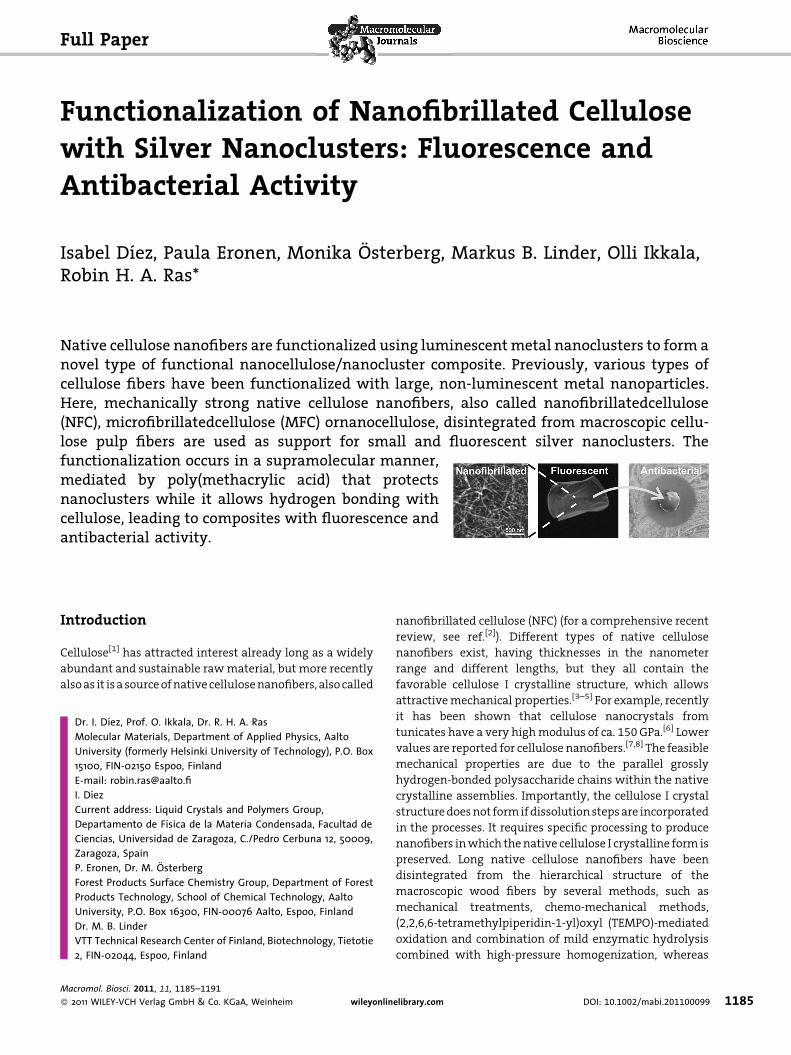
Figure 3. (a) Plot of the adsorbed mass versus time measuredby QCM for adsorption of PMAA (black) and PMAA-protectedAgNC (molar ratio Ag:MAA 2:1) (grey) from solutions on anultrathin NFC film. Solutions were pumped in the chamber att¼ 10 min and rinsed at t¼ 32 min. (b) AFM image of the finalNFC/AgNC film.
Macromol. Biosci. 201
� 2011 WILEY-VCH Verlag Gmb
directed onto an NFC film (black curve). Also in this case the
QCM detected adsorption, which means that not only AgNC
adsorb onto the NFC film but also PMAA. The mass bound
to the NFC film is larger when AgNC are present in the
solution. A comparison of the mass adsorbed by an NFC
film at t¼ 20 min when using a solution of the pure
PMAA (224 ng � cm�2) and a solution of AgNC/PMAA
(550 ng � cm�2) indicates that the stoichiometry of the
adsorbed fluorescent material does not correspond to the
stoichiometry of the AgNC solution. Since the NFC film
adsorbs 224 ng � cm�2 of pure PMAA, the adsorption of
AgNC would be expected to be 784 ng � cm�2, because the
AgNC solution was prepared with a mass ratio Ag:MAA
2.5:1 (molar ratio Ag:MAA 2:1). Instead, a lower value was
measured (550 ng � cm�2). However, the adsorbed mass
values are only indicative since the QCM also detects bound
water and the amount of bound water in the NFC film may
change due to adsorption of AgNC and also the different
ionic strengths in PMAA and AgNC-containing solution
might affect the swollen state of the NFC film and
adsorption.[52]
This suggests that hydrogen bonding between PMAA
and NFC facilitates the functionalization of NFC with
fluorescent silver nanoclusters. Additional experiments
were performed with Langmuir-Schaefer cellulose model
films prepared from dissolved cellulose derivative,[53,61] i.e.,
TMSC. It has the advantages of a smooth surface structure
together with well-defined chemistry. Similar results were
obtained, indicating that the hydrogen bonds might occur
preferentially with the –OH groups of cellulose instead of
the –COOH groups from hemicelluloses.
Dried NFC films were imaged using AFM before and after
adsorption of PMAA-protected fluorescent silver nanoclus-
ters and no significant differences could be noticed. In
Figure 3b the AFM image of the composite NFC/AgNC film is
presented. The thin NFC fibers are still clearly observable
and no hints of PMAA coils or large aggregated silver
nanoparticles can be seen. The absence of large silver
particles, although cellulose could act as reducing agent,[60]
indicates that silver nanoclusters are well protected by the
PMAA.
Antibacterial Properties
The antibacterial behavior of the composites NFC/AgNC
were tested against E. coli bacteria by the halo method. The
composite films for antibacterial testing were prepared by
dipping NFC films into AgNC solutions with molar ratios
Ag:MAA 8:1, 4:1 and 2:1. For comparison, a pure NFC film
was also tested. The agar plates were smeared with E. coli
bacteria in dilutions from 10�1 to 10�5. After allowing the
bacteria to grow for 24 h at 37 8C, photographs were
recorded. Figure 4 shows an example of the antibacterial
behavior of the composite NFC/AgNC prepared from molar
1, 11, 1185–1191
H & Co. KGaA, Weinheim www.MaterialsViews.com

Figure 4. Antibacterial properties of NFC films functionalized withfluorescent silver nanoclusters. Escherichia coli bacteria concen-tration 10�4. Films dipped for 60 h in AgNC solutions with molarratios Ag:MAA 8:1 (left) and Ag:MAA 2:1 (right); unmodified NFCfilm (center down).
Table 1. Ratio of the surface area without bacteria over thesurface area covered by NFC/AgNC film.
Ratio
Ag:MAAa)
Bacteria
dilutionb)
Surface area
ratio
8:1 10�5 5.0
10�4 4.9
10�3 4.3
10�2 3.5
10�1 2.8
4:1 10�5 5.3
10�4 5.4
10�3 4.9
10�2 4.1
10�1 2.7
2:1 10�5 3.9
10�4 4.8
10�3 4.4
10�2 3.7
10�1 2.8
a)Molar ratio of AgNC solution used to prepare the composites;b)Serial dilution of bacteria.
Functionalization of Nanofibrillated Cellulose with Silver Nanoclusters: . . .
www.mbs-journal.de
ratios Ag:MAA 8:1 and 2:1, compared to the behavior of an
NFC film. After incubation, the NFC film shows no
antibacterial activity, the bacteria grow freely all around
the film. In contrast, around the NFC/AgNC films a large
halo free of bacteria can be observed. After incubation,
the initially pink NFC/AgNC films were partly metalized,
indicating further reduction and aggregation of silver.
Nevertheless, silver ions or nanoclusters were released from
the film into the gel preventing the growth of bacteria in a
large area around the films. The surface without bacterial
growth compared to the surface of the NFC/AgNC film for all
the samples tested is collected in Table 1. The antibacterial
activity is comparable for all the Ag:MAA ratios tested, just
slightly larger for the ratio 4:1. The largest area in the agar
plate where bacterial growth was completely inhibited is
5.4 times larger than the area of the film causing the effect
(Ag:MAA 4:1 and solution of bacteria diluted to 10�4). In this
case the thickness of the inhibition halo was 5 mm whereas
the radius of the film was only 3.2 mm, which is a much
larger effect than reported in literature. For instance, for
silver impregnated in glass the values reported are halo
5 mm, film 12.5 mm,[62] for silver impregnated in P2O5/SiO2
they are halo 4.5 mm, film 5.5 mm,[63] for silver solution
they are halo 2.5 mm, solution 4 mm,[64] and for AgBr/
poly[(4-vinylpyridine)-co-(4-vinyl-N-hexylpyridinium bro-
mide)]-coated paper they are halo 2 mm, film 5.5 mm.[65]
However, we point out that the comparison of the halos
with literature values might not be taken entirely
quantitative since also other factors can affect dissolution
of silver ions, such as time, temperature and pH of the agar.
www.MaterialsViews.com
Macromol. Biosci. 2011
� 2011 WILEY-VCH Verlag Gmb
The release of silver ions or AgNC from the NFC/AgNC
film to the gel to prevent bacterial growth takes place only
in the presence of water as revealed by the following
experiment. When a pink composite NFC/AgNC film is
immersed for several days in solvents such as ethanol,
methanol, tetrahydrofuran or chloroform, the film keeps
the pink color and the fluorescence. In contrast, when
immersed in water the film partly looses the pink color
while releasing pink AgNC to the solution. The released
AgNC in solution are very stable indicating that they are
still protected by PMAA, also released from the film.
Conclusion
Native cellulose nanofibers disintegrated from macroscopic
cellulose hardwood fibers were successfully functionalized
with fluorescent silver nanoclusters by simply dipping a
nanocellulose film into a solution of silver nanoclusters
protected by PMAA. The mechanism of binding of silver
nanoclusters to nanocellulose was discussed and PMAA,
carrying silver nanoclusters, was suggested to hydrogen
bond to nanocellulose or/and to the residual hemicellu-
loses, and thus act as mediator in the functionalization. This
leads to a novel type of supramolecular native cellulose
nanofiber/nanocluster adduct. The composite nanocellu-
, 11, 1185–1191
H & Co. KGaA, Weinheim1189

1190
www.mbs-journal.de
I. Dıez, P. Eronen, M. Osterberg, M. B. Linder, O. Ikkala, R. H. A. Ras
lose-nanosilver retains the appealing properties of both
components. The high surface area of the nanofibrils favors
a significant adsorption of silver nanoclusters that can be
released in aqueous medium. The released silver will
prevent bacterial growth in a surface area fivefold larger
than the area of the film. In conclusion, we have shown
that the metal nanocluster/nanocellulose composites are
feasible. We expect that such materials are useful in,
e.g., wound-healing pads and in more general for other
functionalities by selecting different metal nanoclusters.
Acknowledgements: Janne Laine (Aalto Univ.), Timo Koskinen(UPM) and Antti Laukkanen (UPM) are acknowledged fordiscussions. We acknowledge the Finnish Funding Agency forTechnology and Innovation (TEKES) and Academy of Finlandfor funding. This work has been made within the Finnish Centerfor Nanocellulosic Technologies (partnership between UPM, AaltoUniversity and VTT).
Received: March 11, 2011; Published online: July 4, 2011; DOI:10.1002/mabi.201100099
Keywords: bactericides; luminescence; nanocelluloses; nanofi-bers; nanoclusters
[1] D. Klemm, B. Heublein, H.-P. Fink, A. Bohn, Angew. Chem., Int.Ed. 2005, 44, 3358.
[2] S. J. Eichhorn, A. Dufresne, M. Aranguren, N. E. Marcovich, J. R.Capadona, S. J. Rowan, C. Weder, W. Thielemans, M. Roman,S. Renneckar, W. Gindl, S. Veigel, J. Keckes, H. Yano, K. Abe,M. Nogi, A. N. Nakagaito, A. Mangalam, J. Simonsen, A. S.Benight, A. Bismarck, L. A. Berglund, T. Peijs, J. Mater. Sci. 2010,45, 1.
[3] S. Ahola, M. Osterberg, J. Laine, Cellulose 2008, 15, 303.[4] H. Yano, J. Sugiyama, A. N. Nakagaito, M. Nogi, T. Matsuura,
M. Hikita, K. Handa, Adv. Mater. 2005, 17, 153.[5] M. Paakko, M. Ankerfors, H. Kosonen, A. Nykanen, S. Ahola,
M. Osterberg, J. Ruokolainen, J. Laine, P. T. Larsson, O. Ikkala,T. Lindstrom, Biomacromolecules 2007, 8, 1934.
[6] S. Iwamoto, W. Kai, A. Isogai, T. Iwata, Biomacromolecules2009, 10, 2571.
[7] D. G. Hepworth, D. M. Bruce, J. Mater. Sci. 2000, 35, 5861.[8] G. Guhados, W. Wan, J. L. Hutter, Langmuir 2005, 21, 6642.[9] A. F. Turbak, F. W. Snyder, K. R. Sandberg, J. Appl. Polym. Sci.:
Appl. Polym. Symp. 1983, 37, 815.[10] F. W. Herrick, R. L. Casebier, J. K. Hamilton, K. R. Sandberg,
J. Appl. Polym. Sci.: Appl. Polym. Symp. 1983, 37, 797.[11] K. Fleming, D. G. Gray, S. Matthews, Chem. Eur. J. 2001, 7,
1831.[12] A. N. Nakagaito, H. Yano, Appl. Phys. A: Mater. Sci. Process.
2004, 78, 547.[13] K. Abe, S. Iwamoto, H. Yano, Biomacromolecules 2007, 8, 3276.[14] R. Saito, Macromolecules 2001, 34, 4299.
Macromol. Biosci. 201
� 2011 WILEY-VCH Verlag Gmb
[15] M. Henriksson, G. Henriksson, L. A. Berglund, T. Lindstrom,Eur. Polym. J. 2007, 43, 3434.
[16] G. Siqueira, J. Bras, A. Dufresne, Biomacromolecules 2008, 10,425.
[17] J. R. Capadona, O. van den Berg, L. A. Capadona, M. Schroeter,S. J. Rowan, D. J. Tyler, C. Weder, Nat. Nanotechnol. 2007, 2,765.
[18] M. Henriksson, L. A. Berglund, P. Isaksson, T. Lindstrom,T. Nishino, Biomacromolecules 2008, 9, 1579.
[19] J. R. Capadona, K. Shanmuganathan, D. J. Tyler, S. J. Rowan,C. Weder, Science 2008, 319, 1370.
[20] M. Nogi, H. Yano, Adv. Mater. 2008, 20, 1849.[21] L. Wagberg, G. Decher, M. Norgren, T. Lindstrom,
M. Ankerfors, K. Axnas, Langmuir 2008, 24, 784.[22] M. Paakko, J. Vapaavuori, R. Silvennoinen, H. Kosonen,
M. Ankerfors, T. Lindstrom, L. A. Berglund, O. Ikkala, SoftMatter 2008, 4, 2492.
[23] H. Fukuzumi, T. Saito, T. Wata, Y. Kumamoto, A. Isogai,Biomacromolecules 2009, 10, 162.
[24] O. Ikkala, R. H. A. Ras, N. Houbenov, J. Ruokolainen, M. Paakko,J. Laine, M. Leskela, L. A. Berglund, T. Lindstrom, G. ten Brinke,H. Iatrou, N. Hadjichristidis, C. F. J. Faul, Faraday Discuss. 2009,143, 95.
[25] M. Nogi, S. Iwamoto, A. N. Nakagaito, H. Yano, Adv. Mater.2009, 20, 1.
[26] C. Aulin, J. Netrval, L. Wagberg, T. Lindstrom, Soft Matter 2010,6, 3298.
[27] H. Liu, D. Liu, F. Yao, Q. Wu, Bioresour. Technol. 101, 5685.[28] S. Dong, M. Roman, J. Am. Chem. Soc. 2007, 129, 13810.[29] A. Walther, J. Timonen, I. Dıez, A. Laukkanen, O. Ikkala,
Adv. Mater. 2011, DOI: 10.1002/adma.201100580.[30] M. Kettunen, R. J. Silvennoinen, N. Houbenov, A. Nykanen,
J. Ruokolainen, J. Sainio, V. Pore, M. Kemell, M. Ankerfors,T. Lindstrom, M. Ritala, R. H. A. Ras, O. Ikkala, Adv. Funct.Mater. 2011, 21, 510.
[31] J. T. Korhonen, P. Hiekkataipale, J. Malm, M. Karppinen,O. Ikkala, R. H. A. Ras, ACS Nano 2011, 5, 1967.
[32] H. Jin, M. Kettunen, A. Laiho, H. Pynnonen, J. Paltakari,A. Marmur, O. Ikkala, R. H. A. Ras, Langmuir 2011, 27, 1930.
[33] G. K. Hyde, K. J. Park, S. M. Stewart, J. P. Hinestroza, G. N.Parsons, Langmuir 2007, 23, 9844.
[34] B. H. Dong, J. P. Hinestroza, ACS Appl. Mater. Interfaces 2009, 1,797.
[35] J. P. Hinestroza, Mater. Today 2007, 10, 64.[36] C. Chang, J. Peng, L. Zhang, D.-W. Pang, J. Mater. Chem. 2009,
19, 7771.[37] K. A. Willets, R. P. Van Duyne, Ann. Rev. Phys. Chem. 2007, 58,
267.[38] A. R. Tao, S. Habas, P. Yang, Small 2008, 4, 310.[39] N. R. Jana, T. K. Sau, T. Pal, J. Phys. Chem. B 1998, 103, 115.[40] H. Dong, D. Wang, G. Sun, J. P. Hinestroza, Chem. Mater. 2008,
20, 6627.[41] I. Dıez, R. H. A. Ras, ‘‘Few-Atom Silver Clusters as Fluorescent
Reporters’’, in Advanced Fluorescence Reporters in Chemistryand Biology II, (Ed., A. P. Demchenko), Springer, Heidelberg2010, p. 307.
[42] I. Dıez, R. H. A. Ras, Nanoscale 2011, 3, 1963.[43] J. Zheng, R. M. Dickson, J. Am. Chem. Soc. 2002, 124, 13982.[44] J. Yu, S. Choi, C. I. Richards, Y. Antoku, R. M. Dickson, Photo-
chem. Photobiol. 2008, 84, 1435.[45] L. Shang, S. Dong, Chem. Commun. 2008, 1088.[46] I. Dıez, M. Pusa, S. Kulmala, H. Jiang, A. Walther, A. S.
Goldmann, A. H. E. Muller, O. Ikkala, R. H. A. Ras, Angew.Chem., Int. Ed. 2009, 48, 2122.
1, 11, 1185–1191
H & Co. KGaA, Weinheim www.MaterialsViews.com
Functionalization of Nanofibrillated Cellulose with Silver Nanoclusters: . . .
www.mbs-journal.de
[47] W. Guo, J. Yuan, Q. Dong, E. Wang, J. Am. Chem. Soc. 2009, 132,932.
[48] I. Dıez, H. Jiang, R. H. A. Ras, ChemPhysChem 2010, 11,3100.
[49] I. Sondi, B. Salopek-Sondi, J. Colloid Interface Sci. 2004, 275,177.
[50] W. K. Son, J. H. Youk, T. S. Lee, W. H. Park, Macromol. RapidCommun. 2004, 25, 1632.
[51] M. Rai, A. Yadav, A. Gade, Biotechnol. Adv. 2009, 27, 76.[52] S. Ahola, J. Salmi, L. S. Johansson, J. Laine, M. Osterberg,
Biomacromolecules 2008, 9, 1273.[53] T. Tammelin, T. Saarinen, M. Osterberg, J. Laine, Cellulose
2006, 13, 519.[54] G. Sauerbrey, Zeitsch. Phys. 1959, 155, 206.[55] J. Sambrook, E. F. Fritsch, T. Maniatis, Molecular Cloning: A
Laboratory Manual, 2nd edition, Cold Spring Harbor Labora-tory, Cold Spring Harbor, New York 1989.
[56] J. Yu, S. Choi, R. M. Dickson, Angew. Chem., Int. Ed. 2009,48, 318.
www.MaterialsViews.com
Macromol. Biosci. 2011
� 2011 WILEY-VCH Verlag Gmb
[57] I. Dıez, H. Hahn, O. Ikkala, H. G. Borner, R. H. A. Ras, Soft Matter2010, 6, 3160.
[58] S. Ifuku, M. Tsuji, M. Morimoto, H. Saimoto, H. Yano, Bioma-cromolecules 2009, 10, 2714.
[59] J. H. He, T. Kunitake, T. Watanabe, Chem. Commun. 2005, 795.[60] J. Cai, S. Kimura, M. Wada, S. Kuga, Biomacromolecules 2008,
10, 87.[61] E. Kontturi, M. Osterberg, Model Cellulosic Surfaces, American
Chemical Society, Washington DC 2009, p. 57.[62] M. Miola, S. Ferraris, S. Nunzio, P. F. Robotti, G. Bianchi,
G. Fucale, G. Maina, M. Cannas, S. Gatti, A. Masse, C. V.Brovarone, E. Verne, J. Mater. Sci.: Mater. Med. 2009, 20,741.
[63] H. R. Liu, Q. Chen, L. Song, R. F. Ye, J. Y. Lu, H. P. Li, J. Non-Cryst.Solids 2008, 354, 1314.
[64] D. C. Tien, K. H. Tseng, C. Y. Liao, T. T. Tsung, Med. Eng. Phys.2008, 30, 948.
[65] V. Sambhy, M. M. MacBride, B. R. Peterson, A. Sen, J. Am.Chem. Soc. 2006, 128, 9798.
, 11, 1185–1191
H & Co. KGaA, Weinheim1191
Related Documents