Functional ultrasound imaging for assessment of extracellular matrix scaffolds used for liver organoid formation Ryan C. Gessner a , Ariel D. Hanson a, b,1 , Steven Feingold a, 1 , Avery T. Cashion a , Ana Corcimaru b , Bryant T. Wu a, b , Christopher R. Mullins e , Stephen R. Aylward e , Lola M. Reid b, c, d, ** , 2 , Paul A. Dayton a, d, * , 2 a UNC/NCSU Joint Department of Biomedical Engineering, UNC School of Medicine, Chapel Hill, NC 27599, USA b Department of Cell Biology and Physiology, UNC School of Medicine, Chapel Hill, NC 27599, USA c Program in Molecular Biology and Biotechnology, UNC School of Medicine, Chapel Hill, NC 27599, USA d Lineberger Cancer Center, UNC School of Medicine, Chapel Hill, NC 27599, USA e Kitware, Inc.,101 East Weaver St, Carrboro, NC 27510, USA article info Article history: Received 13 July 2013 Accepted 10 August 2013 Available online 4 September 2013 Keywords: Acoustic angiography Microbubble Extracellular matrix Scaffolds Liver Organoids abstract A method of 3D functional ultrasound imaging has been developed to enable non-destructive assess- ment of extracellular matrix scaffolds that have been prepared by decellularization protocols and are intended for recellularization to create organoids. A major challenge in organ decellularization is retaining patent micro-vascular structures crucial for nutrient access and functionality of organoids. The imaging method described here provides statistical distributions of flow rates throughout the tissue volumes, 3D vessel network architecture visualization, characterization of microvessel volumes and sizes, and delineation of matrix from vascular circuits. The imaging protocol was tested on matrix scaffolds that are tissue-specific, but not species-specific, matrix extracts, prepared by a process that preserved >98% of the collagens, collagen-associated matrix components, and matrix-bound growth factors and cytokines. Image-derived data are discussed with respect to assessment of scaffolds followed by proof-of-concept studies in organoid establishment using Hep3B, a human hepatoblast-like cell line. Histology showed that the cells attached to scaffolds with patent vasculature within minutes, achieved engraftment at near 100%, expressed liver-specific functions within 24 h, and yielded evidence of pro- liferation and increasing differentiation of cells throughout the two weeks of culture studies. This im- aging method should prove valuable in analyses of such matrix scaffolds. Ó 2013 Elsevier Ltd. All rights reserved. 1. Introduction Liver transplantation is the primary treatment for end-stage liver disease [1]. Currently, more than 16,000 adults and children are in need of liver transplants. Unfortunately, the number of livers available for transplantation are in short supply [2]. An alternative to organ transplantation is to support patients using an extracor- poreal liver-assist device (LAD). A LAD is a bioreactor comprised of liver cells harvested from donor livers and incorporated into a network of hollow dialysis fibers that mimic blood vessels, which can connect to the patient and thus serve as a bioartificial liver [3,4]. Such bioartificial liver devices provide temporary relief for one to two weeks or until an organ is available for transplantation. They cannot be used longer, since liver cells seeded into all extant forms of bioreactors attach and deposit extracellular matrix and other cellular components onto the hollow fibers, causing “fouling” or clogging of the fibers’ pores, limiting the life span of the device [5]. A more robust alternative is to develop human liver organoids that can be incorporated into a LAD to enable hemodialysis; this provides a more stable and fully functional bioartificial liver in which vascular channels are provided by the native extracellular * Corresponding author. UNC/NCSU Joint Department of Biomedical Engineering, Lineberger Cancer Center, Chapel Hill, NC 27599-6136, USA. ** Corresponding author. Department of Cell Biology and Physiology, Program in Molecular Biology and Biotechnology, Lineberger Cancer Center, Chapel Hill, NC 27599-7038, USA. E-mail addresses: [email protected] (R.C. Gessner), adhanson@ email.unc.edu (A.D. Hanson), [email protected] (S. Feingold), zackcashion@ gmail.com (A.T. Cashion), [email protected] (A. Corcimaru), btwu10@ gmail.com (B.T. Wu), [email protected] (C.R. Mullins), [email protected] (S.R. Aylward), [email protected] (L.M. Reid), [email protected] (P.A. Dayton). 1 Co-equal second authors. 2 Co-equal senior authors. Contents lists available at ScienceDirect Biomaterials journal homepage: www.elsevier.com/locate/biomaterials 0142-9612/$ e see front matter Ó 2013 Elsevier Ltd. All rights reserved. http://dx.doi.org/10.1016/j.biomaterials.2013.08.033 Biomaterials 34 (2013) 9341e9351

Welcome message from author
This document is posted to help you gain knowledge. Please leave a comment to let me know what you think about it! Share it to your friends and learn new things together.
Transcript

lable at ScienceDirect
Biomaterials 34 (2013) 9341e9351
Contents lists avai
Biomaterials
journal homepage: www.elsevier .com/locate/biomater ia ls
Functional ultrasound imaging for assessment of extracellular matrixscaffolds used for liver organoid formation
Ryan C. Gessner a, Ariel D. Hanson a,b,1, Steven Feingold a,1, Avery T. Cashion a,Ana Corcimaru b, Bryant T. Wu a,b, Christopher R. Mullins e, Stephen R. Aylward e,Lola M. Reid b,c,d,**,2, Paul A. Dayton a,d,*,2
aUNC/NCSU Joint Department of Biomedical Engineering, UNC School of Medicine, Chapel Hill, NC 27599, USAbDepartment of Cell Biology and Physiology, UNC School of Medicine, Chapel Hill, NC 27599, USAc Program in Molecular Biology and Biotechnology, UNC School of Medicine, Chapel Hill, NC 27599, USAd Lineberger Cancer Center, UNC School of Medicine, Chapel Hill, NC 27599, USAeKitware, Inc., 101 East Weaver St, Carrboro, NC 27510, USA
a r t i c l e i n f o
Article history:Received 13 July 2013Accepted 10 August 2013Available online 4 September 2013
Keywords:Acoustic angiographyMicrobubbleExtracellular matrixScaffoldsLiverOrganoids
* Corresponding author. UNC/NCSU Joint DepartmeLineberger Cancer Center, Chapel Hill, NC 27599-6136** Corresponding author. Department of Cell BiologMolecular Biology and Biotechnology, Lineberger Ca27599-7038, USA.
E-mail addresses: [email protected] (email.unc.edu (A.D. Hanson), [email protected] (A.T. Cashion), [email protected] (B.T. Wu), [email protected]@kitware.com (S.R. Aylward), [email protected] (P.A. Dayton).
1 Co-equal second authors.2 Co-equal senior authors.
0142-9612/$ e see front matter � 2013 Elsevier Ltd.http://dx.doi.org/10.1016/j.biomaterials.2013.08.033
a b s t r a c t
A method of 3D functional ultrasound imaging has been developed to enable non-destructive assess-ment of extracellular matrix scaffolds that have been prepared by decellularization protocols and areintended for recellularization to create organoids. A major challenge in organ decellularization isretaining patent micro-vascular structures crucial for nutrient access and functionality of organoids. Theimaging method described here provides statistical distributions of flow rates throughout the tissuevolumes, 3D vessel network architecture visualization, characterization of microvessel volumes andsizes, and delineation of matrix from vascular circuits. The imaging protocol was tested on matrixscaffolds that are tissue-specific, but not species-specific, matrix extracts, prepared by a process thatpreserved >98% of the collagens, collagen-associated matrix components, and matrix-bound growthfactors and cytokines. Image-derived data are discussed with respect to assessment of scaffolds followedby proof-of-concept studies in organoid establishment using Hep3B, a human hepatoblast-like cell line.Histology showed that the cells attached to scaffolds with patent vasculature within minutes, achievedengraftment at near 100%, expressed liver-specific functions within 24 h, and yielded evidence of pro-liferation and increasing differentiation of cells throughout the two weeks of culture studies. This im-aging method should prove valuable in analyses of such matrix scaffolds.
� 2013 Elsevier Ltd. All rights reserved.
1. Introduction
Liver transplantation is the primary treatment for end-stageliver disease [1]. Currently, more than 16,000 adults and children
nt of Biomedical Engineering,, USA.y and Physiology, Program inncer Center, Chapel Hill, NC
R.C. Gessner), adhanson@(S. Feingold), zackcashion@(A. Corcimaru), [email protected] (C.R. Mullins),
[email protected] (L.M. Reid),
All rights reserved.
are in need of liver transplants. Unfortunately, the number of liversavailable for transplantation are in short supply [2]. An alternativeto organ transplantation is to support patients using an extracor-poreal liver-assist device (LAD). A LAD is a bioreactor comprised ofliver cells harvested from donor livers and incorporated into anetwork of hollow dialysis fibers that mimic blood vessels, whichcan connect to the patient and thus serve as a bioartificial liver [3,4].Such bioartificial liver devices provide temporary relief for one totwo weeks or until an organ is available for transplantation. Theycannot be used longer, since liver cells seeded into all extant formsof bioreactors attach and deposit extracellular matrix and othercellular components onto the hollow fibers, causing “fouling” orclogging of the fibers’ pores, limiting the life span of the device [5].
A more robust alternative is to develop human liver organoidsthat can be incorporated into a LAD to enable hemodialysis; thisprovides a more stable and fully functional bioartificial liver inwhich vascular channels are provided by the native extracellular

R.C. Gessner et al. / Biomaterials 34 (2013) 9341e93519342
matrix components lined by endothelia. “Biomatrix scaffolds”,herein referred to as matrix scaffolds, are a particularly rich form ofextracellular matrix extracted from organs or tissue and derivedfrom gentle delipidation and perfusion of high salt buffers to keepall collagens and their associated factors insoluble [15]. The orga-noids can be formed by preparing matrix scaffolds from decellu-larized livers and then recellularizing the scaffolds with humancells. These matrix scaffolds can be recellularized in two stages:first, the vascular channels are recellularized with endotheliathrough which medium and then blood can be perfused; and sec-ond, the rest of the matrix is recellularized with a combination ofhepatic and mesenchymal stem cell populations that will matureinto fully functional liver parenchymal cells along with theirmesenchymal cell partners. Current efforts are making use of hu-man hepatic cell lines for human liver organoid formation toestablish optimal recellularization protocols. Successful protocolswill then be used with freshly isolated human hepatic andmesenchymal stem cell populations and endothelia.
For recellularized scaffolds to yield a human liver organoidable to support patients as a bioartificial liver, cell functions mustbe comparable to those of normal human livers. Since cellseeding and organoid functionality are directly related to thepatency and structure of micro-vascular matrix remnants in thescaffold, there is a crucial need for non-destructive assessment ofthe structural characteristics of the scaffold, particularly itsvascular matrix. Without adequate perfusion, the process ofreseeding matrix scaffolds with new cells cannot be accom-plished, since this process relies on fluid transport through thematrix remnants of the vascular bed for the delivery of the cells.Also, after cells have been engrafted throughout the scaffolds,their continued functions depend on a long-term delivery ofnutrients and oxygen. For this reason, a method to image boththe anatomy and flow within the sample in a non-destructivemanner is highly desirable.
There are many methods currently employed to image tissuescaffolds, including scanning and transmission electron micro-scopy (SEM and TEM), optical microscopy [6], magnetic resonance(MR) imaging and microscopy [7], computed tomography (CT) [8],optical coherence tomography (OCT) [9], and Doppler ultrasound[10]. The selection of any one modality will always yield inherenttradeoffs such as cost, invasiveness to the sample, field of view,resolution, acquisition time, and type of information gleaned.From this list, the imaging modalities that can non-invasivelyimage a 3D scaffold with a significant thickness are MR, CT, andultrasound. MR and CT are widely available in both clinical andresearch contexts. These modalities have the best field of view,although they require expensive hardware (particularly MR im-aging). MR can also require long image acquisition times. On theother hand, CT suffers from poor soft-tissue contrast and can causeradiation damage to cells. Ultrasound has many benefits over MRand CT in that it is real-time, relatively inexpensive, non-invasive,does not use ionizing radiation, and has excellent soft-tissuecontrast. In addition, ultrasound is able to assess multipledifferent qualities of a tissue volume (applicable to both in vivovolumes and in vitro matrix scaffolds), including tissue structurewith standard b-mode [11], mechanical stiffness [12], micro-vascular perfusion architecture [13], and parametric perfusionrate [14]. One possible challenge hindering ultrasound’s utilityfor scaffold perfusion assessment to date has likely been themodality’s limited field of view, allowing for freehand visualiza-tion of different 2D slices, or small 3D sub-volumes, but tradi-tionally not visualization or quantitation of a large field of view.Our objective in this study was to explore the application of ul-trasound to perform 3D visualization and quantification ofperfusion throughout a matrix scaffold.
In these studies, we have developed a protocol to enabledetailed assessment of vascular structural and functional charac-teristics within scaffolds in a non-destructive manner. We had twoobjectives: the first was to explore the application of ultrasound toperform 3D visualization and quantification of perfusionthroughout an extracellular matrix scaffold; the second was todemonstrate, using a hepatoblast-like cell line, that the imagingassessments can identify scaffolds that will be successful forcreating human liver organoids.
2. Materials and methods
2.1. Decellularization of rat livers
Wistar rats (weights 250e300 g) were obtained from Charles River Laboratories,Wilmington, MA, and housed in animal facilities handled by the University of NorthCarolina (UNC) Division of Laboratory Animal Management. They were fed ad libi-tum until used for experiments. All experimental work was approved by and per-formed in accordance with the UNC Institutional Animal Use and Care Committeeguidelines.
The protocol for decellularizing livers to produce matrix scaffolds has beendescribed previously [15]. Images of tissue in the process of decellularization aregiven in the online supplement Fig. S1, and results using this protocol are comparedto results using other decellularization protocols (Table S1). Male rats were anes-thetized with Ketamine-Xylazine, and their abdominal cavity opened. The portalvein was cannulated with a 20-gauge catheter to provide a perfusion inlet to thevasculature of the liver, and the vena cava was transected to provide an outlet forperfusion. The liver was removed from the abdominal cavity and placed in aperfusion bioreactor. The blood was removed by flushing the liver with 300 ml ofserum-free DMEM/F12 (Gibco, Grand Island, NY). A delipidation buffer, comprised of36 U/L of phospholipase A2 in 1% sodium deoxycholate (Fisher, Pittsburgh, PA) wasused to remove plasma and nuclear membranes, and was perfused through the liverfor w30 min (up to an hour) or until the tissue became transparent.
This was followed by perfusion for 90 min with a high salt buffer (NaCL). Sol-ubility constants for known collagen types in liver are such that 3.4 M NaCL isadequate to keep them all in an insoluble state, along with any matrix componentsand cytokine/growth factors bound to the collagens or the collagen-bound matrixcomponents. The liver was rinsed for 15 min with serum-free DMEM/F12 to elimi-nate the delipidation buffer and then followed by perfusion with 100 ml of DNase(1mg per 100ml; Fisher, Pittsburgh, PA) and RNase (5mg per 100ml; Sigma Aldrich,St. Louis, MO) to remove any residual contaminants of nucleic acids from the scaf-fold. The final step was to rinse the scaffolds with serum-free DMEM/F12 for 1 h toeliminate any residual salt or nucleases. Images are provided in Fig. S1. The decel-lularized liver scaffolds were stored overnight at 4 �C and perfused with serum-freeDME/F12 basal media at 3 ml/min via a peristaltic pump (Masterflex, ColeeParmer,Vernon Hills, IL) before the imaging study was performed. Prior to an imaging study,the scaffold was transferred from the perfusion bioreactor into the sample imagingchamber (Fig. 1). When in the sample imaging chamber, perfusion was maintainedat 4 ml/min through the matrix scaffold remnant of the portal vein via the sameperistaltic pump.
2.2. Contrast imaging
An overview of the image data processing workflow is provided (Fig. 2). Flashreplenishment imaging was performed using an Acuson Sequoia 512 equipped witha 15L8 transducer (Siemens Medical Solutions USA Inc, Mountain View, CA). The“CPS Capture” software algorithm was used to measure perfusion time. The 3Dimages of the liver matrix scaffold were acquired by scanning the transducer in theelevational direction using a linear stage and motion controller (UTS150PP andESP300, Newport, Irvine, CA) interfaced through LabVIEW (National Instruments,Austin, TX) as described by Feingold et al. [14]. Perfusion imageswere parametricallymapped to contrast arrival times between 1 and 10 s. These images were stored inDICOM format with JPEG compression and analyzed offline in MATLAB (Mathworks,Natick, MA). Perfusion times within the regions of interest were assessed.
Acoustic angiography was performed on a prototype dual frequency probe [16]with imaging parameters previously described [13]. The imaging system was aVisualSonics Vevo770 (Toronto, ON, Canada), with pulses emitted at 4 MHz at1.23 MPa, and echoes received on a 30 MHz transducer with 100% bandwidth afterbeing passed through a 15 MHz high pass filter to remove non-contrast signal.Three-dimensional images were acquired with the VisualSonics 1D linear motionstage with inter-frame distance of 100 mm to yield nearly isotropic voxels. Imageswere acquired with a frame rate of 2 Hz, with 5 frames averaged at each location.High resolution b-mode images were also acquired with the Vevo770 system usingthe same imaging parameters, except the transmit frequency changed to 30 MHz.After imaging, data was exported from the ultrasound system as 8 bit uncom-pressed AVIs. The microvessels were then segmented from these images using analgorithm originally designed for human magnetic resonance angiography images,
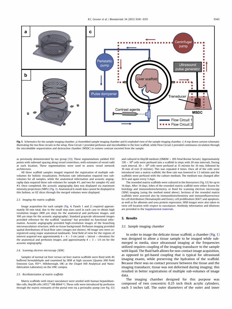
Fig. 1. Schematics for the sample imaging chamber. a) Assembled sample imaging chamber and b) exploded view of the sample imaging chamber. c) A top-down cartoon schematicillustrating the two flow circuits in the setup. Flow Circuit 1 provided perfusion and microbubbles to the liver scaffold, while Flow Circuit 2 provided continuous circulation throughthe microbubble sequestration and destruction chamber (MSDC) to remove contrast excreted from the sample.
R.C. Gessner et al. / Biomaterials 34 (2013) 9341e9351 9343
as previously demonstrated by our group [13]. These segmentations yielded XYZpoints with subvoxel spacing along vessel centerlines, with estimates of vessel radiiat each location. These segmentations were used to assess vessel networkarchitecture.
All three scaffold samples imaged required the registration of multiple sub-volumes for holistic visualization. Perfusion rate information required two sub-volumes for all samples, while the anatomical information and acoustic angiog-raphy data required three sub-volumes for sample #1, and two for samples #2 and#3. Once completed, the acoustic angiography data was displayed via maximumintensity projections (MIPs) (Fig. 3). Anatomical b-mode data cannot be displayed inthis fashion, so XZ slices through the merged volumes were displayed.
2.3. Imaging the matrix scaffolds
Image acquisition for each sample (Fig. 4, Panels 1 and 2) required approxi-mately 50 min total, due to the small step sizes used in each case to obtain highresolution images (800 mm steps for the anatomical and perfusion images, and100 mm steps for the acoustic angiography). Standard grayscale ultrasound imagesprovided reference for the scaffold “anatomy” but provided no functional infor-mation. Acoustic angiography provided high-resolution images of the branchingmicrovasculature structure, with no tissue background. Perfusion imaging providedspatial distributions of local flow rates (images not shown). All image sets were co-registered using major anatomical landmarks. Total field of view for the regions ofinterest acquired was approximately 4 � 4 � 3 cm (axial � lateral � elevation) forthe anatomical and perfusion images, and approximately 4 � 3 � 1.4 cm for theacoustic angiography.
2.4. Scanning electron microscopy (SEM)
Samples of normal rat liver versus rat liver matrix scaffolds were fixed with 4%buffered formaldehyde and examined by SEM at high vacuum (Quanta 200 FieldEmission Gun, FEI�, Hillsborough, OR) at the Chapel Hill Analytical and Nano-fabrication Laboratory on the UNC campus.
2.5. Recellularization of matrix scaffolds
Matrix scaffolds with intact vasculature were seeded with human hepatoblast-like cells, Hep3B cells (ATCC� HB-8064�). These cells were introduced by perfusionthrough the matrix remnants of the portal vein via a peristaltic pump (see Fig. S3)
and cultured in Hep3B medium (DMEM þ 10% Fetal Bovine Serum). Approximately130 � 106 cells were perfused into a scaffold in steps with 20 min intervals. Duringeach interval, 30 � 106 cells were perfused at 15 ml/min for 10 min, followed by10 min of rest (0 ml/min). This was repeated 4 times. Once all of the cells wereintroduced into a matrix scaffold, the flow rate was lowered to 1.3 ml/min and thescaffolds were perfused with the culture medium. The medium was changed after24 h and again every 3 days.
The reseeded matrix scaffolds were cultured in the bioreactors (Fig. S3) for up to14 days. After 14 days, lobes of the reseeded matrix scaffold were either frozen forhistology and immunohistochemistry, or fixed for scanning electron microscopy(SEM) imaging (using the method noted above). Sections of the reseeded matrixscaffolds were assessed also by immunohistochemistry and immunofluorescencefor cell distribution (Hematoxylin and Eosin), cell proliferation (Ki67) and apoptosis,as well as for albumin and urea protein expression. SEM images were also taken toview cell location with respect to vasculature. Antibody information and dilutionsare provided in the Supplemental materials.
3. Results
3.1. Sample imaging chamber
In order to image the delicate tissue scaffold, a chamber (Fig. 1)was designed to allow a tissue sample to be imaged while sub-merged in media, since ultrasound imaging at the frequenciesutilized requires coupling of the imaging transducer to the samplewith liquid. The fluid bath allows for non-contact image acquisition,as opposed to gel-based coupling that is typical for ultrasoundimaging exams, while preserving the hydration of the scaffold.Because there was no contact pressure between the tissue and theimaging transducer, tissue was not deformed during imaging; thisresulted in better registrations of multiple sub-volumes of imagedata.
The imaging chamber designed for this purpose wascomposed of two concentric 0.25 inch thick acrylic cylinders,each 3 inches tall. The outer diameters of the outer and inner

Fig. 2. The orientation of the matrix scaffold sample as viewed from above with the following visible lobes labeled: a) left lateral lobe (LLL), inferior right lobe (IRL), anterior caudatelobe (ACL), posterior caudate lobe (PCL), superior right lobe (SRL), and median lobe (ML). The LLL was the lobe imaged in this study. Lobes were identified in this figure via anavailable surgical guide. b) Orientation of the imaging sub-volumes relative to the tissue sample. XY dimensions were lateral and axial axes within the ultrasound coordinate space,with the Z-axis being the elevational scan direction. c) Schematic explaining the registration of multiple 3D volumes from three distinct ultrasound imaging approaches into thefinal composite volume.
R.C. Gessner et al. / Biomaterials 34 (2013) 9341e93519344
cylinders were 6 and 5 inches, respectively. The outer cylinderwas mounted to the 6 � 6 � 0.3 inch acrylic base of the imagingchamber using acrylic glue. The inner cylinder was fit to theimaging chamber’s base over a Vaseline-lubricated silicone O-ring to allow for quick coupling with a tight seal. Though notimplemented in this study, this O-ring design also enables bi-directional imaging orientations (i.e. it is possible to rotate theinner chamber and image the contralateral side of the tissuesample). The interior cylinder served several purposes. It pro-vided a frame for the tissue sample support webbing, made from5.0 silk sutures (Ethicon, Somerville, NJ). This support webbingheld the sample suspended in the interior of chamber. Additionalsuture was loosely tethered over the top of the tissue to preventflotation or shifting during the imaging study. The interior cyl-inder of the sample imaging chamber also allowed for efficientbuffer circulation but limited turbulence near the sample.
Preliminary studies showed that the peristaltic pump, whichpowered flow circuit #1, caused a slight periodicity in the flow ratethrough the scaffold samples as a result of the pump’s rotary wheeldesign; this affected perfusion measurements. To prevent thisartifact, a pulse dampener (Model 07596-20, ColeeParmer, VernonHills, IL) was placed between the output from the peristaltic pumpand the input to the sample. Preliminary studies also showed that
contrast agent exiting the sample into the surrounding fluid afterperfusing through the portal circuit resulted in a decrease in imagequality over time as contrast agent floated between the imagingtransducer and the sample. To prevent this, a clearance fluid circuitwas implemented (Fig. 1). This circulated the fluid surrounding thescaffold sample though a microbubble sequestration and destruc-tion chamber (MSDC) before reinjection into the imaging chamber.The MSDC was a 2 L Erlenmeyer flask in which a 1 MHz unfocusedpiston transducer was suspended (Valpey-Fisher, Hopkinton, MA)to facilitate contrast destruction. The 1 MHz piston transducer waspulsed at 10 Hzwith a pressure of 460 kPa via a pulser (Model 801A,Ritec, Warwick RI). Under these conditions, free contrast agent inthe solution flowing through the chamber was destroyed and thusremoved from the circulating media. Media surrounding the scaf-fold was continually pumped through this chamber at 1 ml/minusing a centrifugal pump (Model PQ-12, Greylor, Cape Coral, FL)powered by an external DC power supply (Model DIGI360, ElectroIndustries, Westbury, NY). Four nylon luer fittings were attached tothe outer cylinder for coupling the sample imaging chamber to thetwo flow circuits. All fluid circuits used 0.125 inch inner diameterTygon tubing, except between the catheter entering the scaffoldsample and the outer cylinder of the imaging chamber (0.062 inchin diameter).
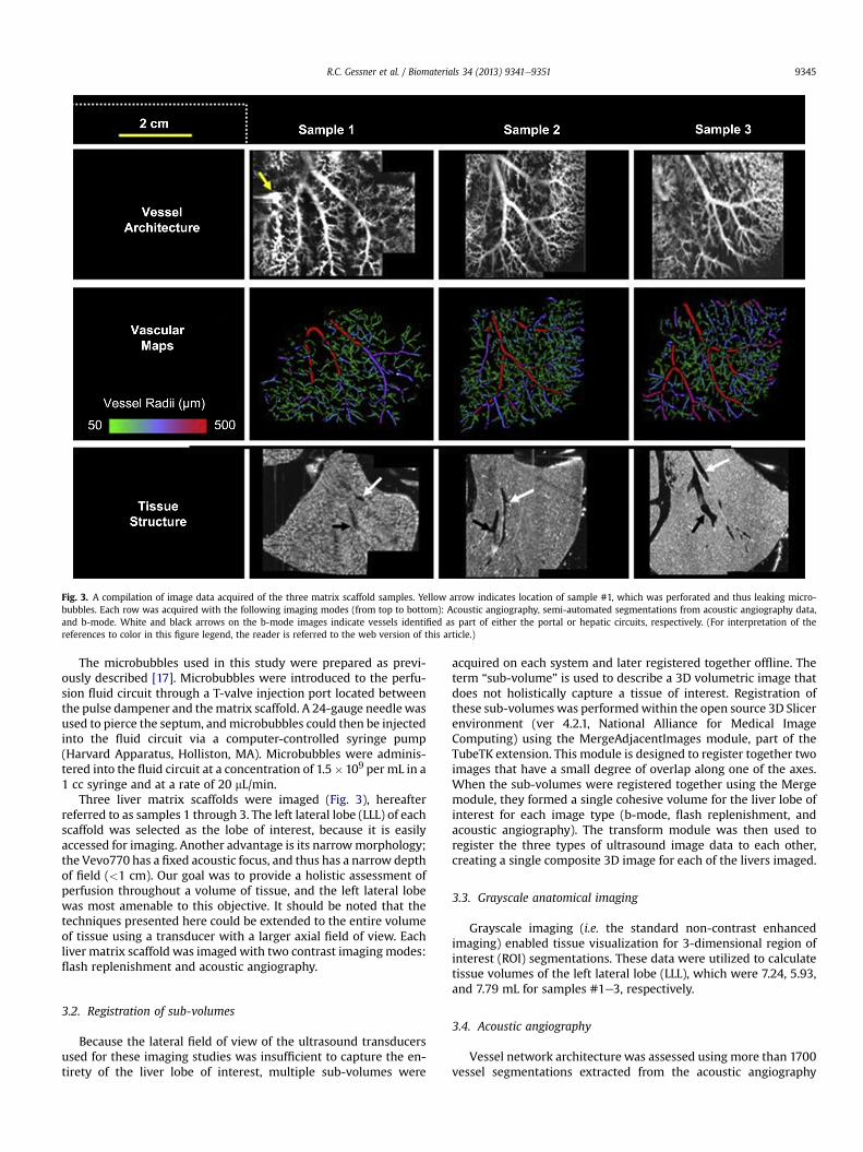
Fig. 3. A compilation of image data acquired of the three matrix scaffold samples. Yellow arrow indicates location of sample #1, which was perforated and thus leaking micro-bubbles. Each row was acquired with the following imaging modes (from top to bottom): Acoustic angiography, semi-automated segmentations from acoustic angiography data,and b-mode. White and black arrows on the b-mode images indicate vessels identified as part of either the portal or hepatic circuits, respectively. (For interpretation of thereferences to color in this figure legend, the reader is referred to the web version of this article.)
R.C. Gessner et al. / Biomaterials 34 (2013) 9341e9351 9345
The microbubbles used in this study were prepared as previ-ously described [17]. Microbubbles were introduced to the perfu-sion fluid circuit through a T-valve injection port located betweenthe pulse dampener and thematrix scaffold. A 24-gauge needlewasused to pierce the septum, andmicrobubbles could then be injectedinto the fluid circuit via a computer-controlled syringe pump(Harvard Apparatus, Holliston, MA). Microbubbles were adminis-tered into the fluid circuit at a concentration of 1.5�109 per mL in a1 cc syringe and at a rate of 20 mL/min.
Three liver matrix scaffolds were imaged (Fig. 3), hereafterreferred to as samples 1 through 3. The left lateral lobe (LLL) of eachscaffold was selected as the lobe of interest, because it is easilyaccessed for imaging. Another advantage is its narrowmorphology;the Vevo770 has a fixed acoustic focus, and thus has a narrow depthof field (<1 cm). Our goal was to provide a holistic assessment ofperfusion throughout a volume of tissue, and the left lateral lobewas most amenable to this objective. It should be noted that thetechniques presented here could be extended to the entire volumeof tissue using a transducer with a larger axial field of view. Eachliver matrix scaffold was imaged with two contrast imaging modes:flash replenishment and acoustic angiography.
3.2. Registration of sub-volumes
Because the lateral field of view of the ultrasound transducersused for these imaging studies was insufficient to capture the en-tirety of the liver lobe of interest, multiple sub-volumes were
acquired on each system and later registered together offline. Theterm “sub-volume” is used to describe a 3D volumetric image thatdoes not holistically capture a tissue of interest. Registration ofthese sub-volumeswas performedwithin the open source 3D Slicerenvironment (ver 4.2.1, National Alliance for Medical ImageComputing) using the MergeAdjacentImages module, part of theTubeTK extension. This module is designed to register together twoimages that have a small degree of overlap along one of the axes.When the sub-volumes were registered together using the Mergemodule, they formed a single cohesive volume for the liver lobe ofinterest for each image type (b-mode, flash replenishment, andacoustic angiography). The transform module was then used toregister the three types of ultrasound image data to each other,creating a single composite 3D image for each of the livers imaged.
3.3. Grayscale anatomical imaging
Grayscale imaging (i.e. the standard non-contrast enhancedimaging) enabled tissue visualization for 3-dimensional region ofinterest (ROI) segmentations. These data were utilized to calculatetissue volumes of the left lateral lobe (LLL), which were 7.24, 5.93,and 7.79 mL for samples #1e3, respectively.
3.4. Acoustic angiography
Vessel network architecture was assessed using more than 1700vessel segmentations extracted from the acoustic angiography

Fig. 4. Panel 1. aec) 3D renderings of the matrix acoustic angiography data. Color is defined as either inside (green) or outside (purple) of the manually defined tissue border. d)Quantitative assessments of vascular network volume, length, and vascularity ratio. Data are normalized to sample #1. Panel 2. Perfusion comparisons between the three liverscaffolds evaluated from the flash-replenishment imaging. (Top) Area normalized vessel size histograms computed from the vessel segmentations. (Bottom) Area normalizedperfusion rate histograms. (For interpretation of the references to color in this figure legend, the reader is referred to the web version of this article.)
R.C. Gessner et al. / Biomaterials 34 (2013) 9341e93519346
image data. These data were rendered in 3D and color coded basedon whether vessels are inside or outside the manually definedtissue boundary (Fig. 4, Panels 1 and 2). Different numbers of ves-sels were extracted from each sample: for sample #1 (N ¼ 415vessel segmentations), sample #2 (N ¼ 702 vessel segmentations),
and sample #3 (N ¼ 671 vessel segmentations). The volumes ofthese vessel network segmentations were computed to be 142.6,226.6, and 241.3 ml, and the total lengths were computed to be1.0245, 1.6177, and 1.4646 m for samples #1, #2, and #3,respectively.

R.C. Gessner et al. / Biomaterials 34 (2013) 9341e9351 9347
The vascularity ratios were 1.97%, 3.82%, and 3.10% for samples#1, #2, and #3, respectively (Fig. 4, Panel 1d). Although thesemetrics revealed sample #1 to contain fewer perfused vessels (bothin total volume of the vessel network, and as a proportion of totalvolume of the matrix scaffold) the distribution of vessel sizes weresimilar between all three samples (Fig. 4, Panel 2 e top).
3.5. Perfusion imaging
The perfusion values within each volume were assessed usingregions of interest as defined based on anatomical data. Histogramsof the perfused pixels within the scaffold volumes were computedand plotted as a function of perfusion time (Fig. 4, Panel 2 e bot-tom). Samples #2 and #3 had similarly shaped histograms withsimilar mean perfusion times of 3.128� 1.923 s, and 3.017� 1.677 s(mean m � s), respectively. Sample #1, on the other hand, had anegatively skewed monotonically decreasing histogram with amean perfusion of 1.381�1.328 s. This result was in alignment withthe differences in the perfusion images, which could be qualita-tively observed; sample #1 had a larger proportion of fasterperfusing regions than samples #2 and #3 (Fig. 4).
3.6. Scanning electron microscopy of intact liver vs. matrix scaffolds
Scanning electron microscopy (SEM) of liver versus matrixscaffolds (Fig. 5, Panel 1, 5a and 5b) indicated that the decellulari-zation protocol preserves the liver’s histological infrastructure, andthat the macro- and micro-vascular channels are clearly intact inthe liver matrix scaffolds (see arrows). These SEM images arecomplemented by our prior studies showing that all knownextracellular matrix components, including all known collagentypes, are present and preserved at >98% of the levels found innormal liver, and that the scaffolds are negligible for nucleic acidsand for cytoplasmic components [15]. All known matrix compo-nents in liver versus scaffolds were assessed by immunohisto-chemistry and found to be in their correct histological sites,meaning that the liver acinar zonation (zones 1e3) is preserved interms of gradients of matrix components [15,18].
3.7. Engraftment efficiency of scaffold recellularization
As a proof-of-concept towards human liver organoid formation,matrix scaffolds with patent vasculature were reseeded withapproximately 130 � 106 Hep3b cells, human hepatoblast-like cellsof a hepatic cell line (ATCC� HB-8064�). This cell line was estab-lished by Barbara Knowles and associates from a tumor from an 8-year old patient [19] and has been extensively characterized withrespect to liver-specific gene expression by Darlington and associ-ates [20]. The Hep3B cells were introduced into the matrix scaffoldby perfusion through thematrix remnants of the portal vein using aperistaltic pump. The cells attached within minutes, and engraft-ment efficiency was near 100% by the end of the seeding process.
Over the course of 14 days, the medium from bioreactors withreseeded scaffolds was collected at varying time points and assayedby ELISA for secreted products (Fig. 6). At the end of the 14 dayculture period, scaffolds were evaluated by SEM, histology andimmunohistochemistry. SEM images (Fig. 5, Panel 1) show cells thathave engrafted onto or into the matrix scaffold. Hematoxylin andeosin stained sections revealed a wide cell distribution throughoutthe scaffolds (Fig. 5, Panel 2, 5a). The seeded cells actively prolif-erated throughout the 14 days of culture, as demonstrated by Ki67staining (Fig. 5, Panel 2, 5b), but they did not show any evidence ofapoptosis (data not shown). In addition, the reseeded cellsexpressed liver-specific proteins in patterns correlated with theirknown locations in the normal liver acinus in vivo. Albumin (Fig. 5,
Panel 2, 5c) was found in all of the cells but at higher levels in re-gions of the scaffolds correlated with acinar zones 2 and 3, whereasepithelial cell adhesion molecule, EpCAM (Fig. 5, Panel 2, 5d), wasexpressed only by cells clustered near the matrix remnants ofportal triads, acinar zone 1. EpCAM is a marker of hepatic stem/progenitors {15}. Hep3B cells have phenotypic traits indicating thatthey are hepatoblast-like [20]; normal hepatoblasts are found onlyin zone 1 of the pediatric and adult liver acinus, and are at a lineagestage in which EpCAM is a key phenotypic trait.
4. Discussion
We describe a method of imaging for the evaluation of matrixextracts prepared by decellularization of tissues. Our techniquesmay provide important information regarding vascular patency,which is particularly important if such scaffolds are to be used assubstrata for cell populations as part of organoid formation. Theultrasound images described here provide insights into thecomplexity of the matrix remnants of the liver architecture and ofthe vascular channels. The dimensions of the channels can bequantified precisely using these ultrasound technologies andcorresponding segmentation algorithms, enabling the identifica-tion of categories of blood vessels for which such dimensions areknown (up to 100 mm). Furthermore, these techniques provide anon-destructive method to assess vascular functionality.
When using ultrasound, contrast agents are utilized to imageand quantify flow inmicrovessels, due to the poor acoustic contrastfrom blood. Microbubble contrast agents are the predominantlyused vascular contrast agent for ultrasound. They are micron-sizedlipid encapsulated gaseous spheres which, when immersed in afluid, provide a strongly reflective interface and thus a high degreeof contrast in an ultrasound image. Microbubble contrast agents(diameters typically between 1 and 5 mm) are much larger thanvascular fenestrations, which allow them to remain within theluminal space. At the same time, they are small enough to passthrough capillary beds. Since their rheology is similar to red bloodcells [21], they are a suitable contrast agent for spatial mapping ofvascular channel networks within matrix scaffold networks,thereby serving as surrogate markers for mapping cell movementwithin the scaffold.
In addition to traditional “grayscale” ultrasound to providereference images of the scaffold physical structure, two differentcontrast-enhanced ultrasound techniques were utilized in thiswork to provide functional information about matrix scaffoldmicrovasculature. The first technique, referred to as “flash replen-ishment”, or “dynamic contrast-enhanced perfusion imaging”(DCE-PI), allows relative blood perfusion to be spatially mapped byassessing the speed at which contrast agents refill a sample volumeafter clearance [22]. While this technique is not new, it has notpreviously been performed in this type of scaffold imagingapplication.
The other contrast imaging approach implemented in thisstudy is a technique we refer to as “acoustic angiography”. Thisnewly developed imaging method differs from traditional ul-trasound imaging in that it is designed to visualize vascular andmicro-vascular structure at high resolution rather than tissueanatomy [16,23]. Unlike flash replenishment, acoustic angiog-raphy does not yet have the ability to parametrically mapperfusion rates. However, it can provide detailed visualization ofvessel architecture and provide a high resolution dataset fromwhich vessels can be segmented and quantitatively mapped(vessel network volume, length, etc.). High resolution acousticangiography requires a new type of transducer developed by ourlab with collaborators in F. Stuart Foster’s group at the Universityof Toronto, Sunnybrook [16] that is not yet available on

Fig. 5. Panel 1. Scanning electron microscopy (SEM) of a) normal adult rat liver fixed with 4% paraformaldehyde and subjected to SEM; b) normal rat liver matrix scaffold. The majorand minor vascular channels are evident in both the liver and in the scaffold. Empty spaces are visible where parenchymal cells previously resided (thin arrow). Major vascular
R.C. Gessner et al. / Biomaterials 34 (2013) 9341e93519348

R.C. Gessner et al. / Biomaterials 34 (2013) 9341e9351 9349
commercial ultrasound systems. The protocol utilized for thiswork involves three imaging scans: the first defines the scaf-fold’s physical structure (anatomy) with standard “b-mode” orgrayscale ultrasound as is commonly used clinically; the secondmaps vessel architecture with high resolution acoustic angiog-raphy for vascular network visualization and quantitation; thethird maps perfusion rates with flash replenishment. All threestudies could be performed sequentially with appropriate sys-tem hardware.
The analyses of these scaffolds by imaging modalities provideevidence for the complexity of the matrix remnants of the liverarchitecture and of the vascular channels, as well as evidence of thescaffolds’ biological efficacy. These analyses also provide assistancewith recellularization protocols. The dimensions of the channelscan be quantified precisely using these ultrasound technologies andcorresponding segmentation algorithms; this enables identificationof categories of blood vessels for which such dimensions areknown.
From a quantitative standpoint, sample #1 was noticeablydifferent from samples #2 and #3 in both the acoustic angiographyand perfusion images. Sample #1 illustrated a similar vessel sizedistribution to the other two samples, but showed a grosslyreduced perfusion rate. This was due to a defect in sample #1 e thescaffold was damaged, resulting in several low resistance outlets forflow exiting the system. Although wewere not able to perceive thisdamage prior to imaging, it was clearly reflected in the functionaldata. One perforated outlet was clearly visible within the acousticangiography image set (Figs. 3 and 4, Sample 1, yellow arrow).
Furthermore, during contrast infusion, itwas possible to delineatevessels as part of either the hepatic or portal circulatory networkswithin the tissue volume (Fig. 3, white vs. black arrows, respectively).Because contrast was infused though the portal vein, the hepaticcircuit did not contain contrast and thus was not visualized underacoustic angiography. When the b-mode volumes were registered tothe acoustic angiography volumes, flow voids in the b-mode, whichwere not perfused within the acoustic angiography dataset, could beclassifiedascomponentsof thehepatic circuit. Alternatively,wecouldhave perfused the hepatic andportal circuits separately, although thiswas not done in our experiments for simplicity.
The ultrasound approaches we describe offer a non-destructive,high resolution, and potentially inexpensive technique for visuali-zation of scaffold perfusion. Additionally, these approaches offer abetter depth of penetration into the scaffold than optical imagingapproaches, allowing for the visualization of the entirety of the LLL(at the expense of both axial and lateral resolution, which is worsethan systems imaging at optical wavelengths).
While the advantages of this protocol are numerous, there arealso several drawbacks to this approach. Currently, the imagingtechnology presented for the acoustic angiography vessel mapping isnot commercially available. Also, while several groups have madestrides toward calibrating flash replenishment imaging against goldstandards for perfusion rate [22,24], the technique currently pro-vides only relative quantitative measures for perfusion. This isadequate for assessing differences in regions within a given sample,
channels are also visible (thick arrow); c) low magnification image of rat liver matrix scaffthroughout the scaffolds. This attachment occurs within minutes of seeding the cells and rfication image of rat liver matrix scaffold showing Hep3b cells that have attached, spread an(thick white arrow) the lumen of the vessel wall and perhaps pass through the fenestrae (teosin stained sections revealed a cell distribution throughout the matrix scaffolds. b) The semagnified version in b’). c) The Hep3b cells expressed peak albumin levels (albumin ¼ greeliver acinus. d) Cells bound to matrix remnants of the portal triads (zone 1) expressed albumstrong in the cells bound in these regions but weak or negligible if in matrix regions associatest cells are a cell line and yet they apparently still have the capacity to show some deginterpretation of the references to color in this figure legend, the reader is referred to the
such as would be necessary for identifying pockets of occlusion. Itdoes not yet allow determination of absolute flow rates(mL s�1 cm�3) within the scaffold without prior calibration based onthe tissue volume. Finally, our technique was relatively slow, due tothe 1-D form factor of our transducers, which were able to imageonly a single 2-D image slice at a time and required us to mechani-cally scan the transducers across the tissue volume. The 2-D matrixultrasound transducers now in development are able to acquireentire image volumes at a much higher frame rate, although trans-ducers required to perform real-time 3D contrast imaging at theresolution described here are not yet commercially available.
The rapid and successful recellularization of the scaffolds withthe Hep3b cells demonstrated the patency and functionality of thematrix remnants of the vascular network. It is interesting that,although the vascular walls remain sufficiently patent to constrain1-5 micron microbubble contrast agents, the cells were still able tocross the matrix remnants of the vascular channels and engraftinto all parts of the matrix, including that associated withparenchymal cells (Fig. 5, Panel 1, 5c). It is unclear how the cellswere able to engraft after seeding by perfusion into a decellular-ized liver, since the matrix remnants of the vascular channels wereintact. Many researchers who have successfully seeded cells intodecellularized organs used decellularization protocols thatdestroyed facets of the vascular matrix [25]. Of those using pro-tocols resulting in patent vascular channels ([26], it has never beenexplained how the cells are able to breach patent vascular matrixwalls and engraft. Baptista et al. [26] hypothesized that theirmethod of decellularization by harsh detergents followed bydistilled water perfusion may have caused structural damage tothe vascular architecture such as thinning of the matrix or creatingsmall holes; this might, in turn, have allowed cells to migrateacross the permeabilized walls.
We have confirmed that our decellularization method results inintact scaffolds without evidence of a defect: there were nobreaches of the vascular matrix greater than 5 microns prior toseeding. An alternative explanation for the cells’ ability to breachmatrix walls and engraft derives from hepatocyte transplantationstudies [27,28]. In these studies it was shown that sinusoidalendothelial cells can shift to allow the hepatocytes access to largerfenestrae through which they achieve access and integrate into theparenchymal cell plates. SEM imaging of our reseeded scaffoldssuggest that cells are attaching to and engaging the matrix andperhaps are able to squeeze through residual fenestrae left behindafter decellularization (Fig. 5, Panel 1, 5d, arrows).
Non-destructive 3D perfusion imaging of decellularized organsand tissues could have significant value in tissue engineering in-vestigations and also in tracking recellularization. Contrast ultra-sound techniques have been shown to have utility in measuringmicrovasculature structural information and relative flow ratecorrelated with anatomical orientation in tissue scaffolds. Therecellularization studies presented here indicate that cells aresuccessfully being delivered and engrafting into the matrix rem-nants of the liver parenchyma by perfusion seeding (Fig. 5, Panel 2).This perfusion seeding method is only possible in a patent vascular
old reseeded with Hep3b cells. Large numbers of cells are found bound to the matrixesults in near 100% engraftment by the end of the seeding process; d) Higher magni-d are forming classic cellular extensions and connections with each other. They engagehin white arrow) that remain following decellularization. Panel 2. a) Hematoxylin andeded cells actively proliferated through day 14, demonstrated by Ki67 staining (seen inn, DAPI ¼ blue) in regions of the matrix scaffolds correlated with zones 2 and 3 of thein more weakly than elsewhere on the matrix. By contrast, EpCAM expression was quiteted with zones 2 and 3 (EpCAM ¼ green, DAPI ¼ blue). This is surprising, given that theree of zonation of functions if bound to specific regions of the matrix scaffolds. (Forweb version of this article.)
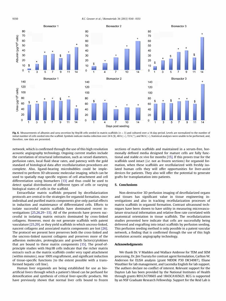
Fig. 6. Measurements of albumin and urea secretion by Hep3B cells seeded in matrix scaffolds (n ¼ 3) and cultured over a 14-day period. Levels are normalized to the number ofinitial number of cells seeded into the scaffold. Symbols indicate media collection over 24 h ($), 48 h (�), 72 h (*), and 96 h (þ). Statistical analyses were unable to be performed, and,therefore, raw data are presented.
R.C. Gessner et al. / Biomaterials 34 (2013) 9341e93519350
network, which is confirmed through the use of this high resolutionacoustic angiography technology. Ongoing current studies includethe correlation of structural information, such as vessel diameters,perfusion rates, local fluid shear rates, and patency with the goldstandard of histological data after recellularization procedures arecomplete. Also, ligand-bearing microbubbles could be imple-mented to perform 3D ultrasonic molecular imaging, which can beused to spatially map specific regions of cell attachment and celldifferentiation using biomarkers [13] and thus could be used todetect spatial distributions of different types of cells or varyingbiological states of cells in the scaffold.
Extracellular matrix scaffolds prepared by decellularizationprotocols are central to the strategies for organoid formation, sinceindividual and purified matrix components give only partial effectsin induction and maintenance of differentiated cells. Efforts toisolate successful matrix scaffolds have dominated recent in-vestigations [25,26,29e33]. All of the protocols have proven suc-cessful in isolating matrix extracts dominated by cross-linkedcollagens. However, most do not generate scaffolds with patentvasculature [25,29], or they yield scaffolds inwhich uncross-linked,nascent collagens and associated matrix components are lost [26].The protocol we present here preserves both the cross-linked andthe uncross-linked nascent collagens and preserves more of theadhesion molecules, proteoglycans and growth factors/cytokinesthat are bound to these matrix components [15]. The proof-of-principle studies with Hep3B cells indicate that the richer matrixextracts such as the matrix scaffolds confer very rapid attachment(within minutes), near 100% engraftment, and significant inductionof tissue-specific functions (to the extent possible with a trans-formed hepatic cell line).
Human liver organoids are being established for use as bio-artificial livers through which a patient’s blood can be perfused fordetoxification and synthesis of critical liver-specific products. Wehave previously shown that normal liver cells bound to frozen
sections of matrix scaffolds and maintained in a serum-free, hor-monally defined media designed for mature cells are fully func-tional and stable ex vivo for months [15]. If this proves true for thescaffolds used intact (i.e. not as frozen sections) for organoid for-mation, when these scaffolds are recellularized with freshly iso-lated human cells they will offer opportunities for liver-assistdevices for patients. They also will offer the potential to generategrafts for transplantation into patients.
5. Conclusions
Non-destructive 3D perfusion imaging of decellularized organsand tissues has significant value in tissue engineering in-vestigations and also in tracking recellularization processes ofmatrix scaffolds in organoid formation. Contrast ultrasound tech-niques have been shown to have utility in measuring microvascu-lature structural information and relative flow rate correlated withanatomical orientation in tissue scaffolds. The recellularizationstudies presented here indicate that cells are successfully beingdelivered and engrafting into intact scaffolds by perfusion seeding.This perfusion seeding method is only possible in a patent vascularnetwork, a finding that is confirmed through the use of this highresolution acoustic angiography technology.
Acknowledgments
We thank Dr. V Madden and Wallace Ambrose for TEM and SEMprocessing, Dr. Jim Tsuruta for contrast agent formulation, CarltonW.Anderson for ELISA analysis (grant NIDDK P30 DK34987), ElianeWauthier for lab management, and Lucendia English for lab support.The authors declare no conflict of interest. Relevant support for theDayton Lab has been provided by the National Institutes of Healththrough grants R01CA170665 and 1R43CA165621. RCG is supportedby an NSF Graduate Research Fellowship. Support for the Reid Lab is
R.C. Gessner et al. / Biomaterials 34 (2013) 9341e9351 9351
derived from an SRA from Vesta Therapeutics (Bethesda, MD), anSRA from the Hamner Institute (a subcontract of a grant from theDow Chemical Company), and from an NCI grant (CA016086). Patentapplications on matrix scaffolds have been filed, are owned by UNCand have been licensed to PhoenixSongs Biologicals (Branford, CT) fornon-clinical uses and to Vesta Therapeutics (Bethesda, MD) forclinical programs. Kitware, Inc. has been provided funding throughthe National Institutes of Health 1R43CA165621.
Appendix A. Supplementary data
Supplementary data related to this article can be found at http://dx.doi.org/10.1016/j.biomaterials.2013.08.033.
References
[1] Puppi J, Strom SJ, Hughes RD, Bansal S, Castell JV, Dagher I, et al. Improving thetechniques for human hepatocyte transplantation: report from a consensusmeeting in London. Cell Transplant 2012;21:1e10.
[2] Smith JM, Biggins SW, Haselby DG, Kim WR, Wedd J, Lamb K, et al. Kidney,pancreas and liver allocation and distribution in the United States. Am JTransplant 2012;12:3191e212.
[3] Gerlach JC, Zeilinger K, Patzer JF. Bioartificial liver systems: why, what,whither? (Review). Regen Med 2008;3:575e95.
[4] Catapano G, Patzer JF, Gerlach JC. Transport advances in disposable bio-reactors for liver tissue engineering. Adv Biochem Eng Biotechnol2010;115:117e43.
[5] Macdonald J, Griffin J, Kubota H, Griffith L, Fair J, Reid LM. Bioartificial livers.In: Kuhtreiber WM, Lanza RP, Chick WL, editors. Cell encapsulation technol-ogy and therapeutics. Boston: Birkhauser; 1999. p. 252e86.
[6] Smith LE, Smallwood R, MacNeil S. A comparison of imaging methodologiesfor 3D tissue engineering. Microsc Res Tech 2010;73:1123e33.
[7] Xu H, Othman SF, Magin RL. Monitoring tissue engineering using magneticresonance imaging. J Biosci Bioeng 2008;106:515e27.
[8] Young S, Kretlow JD, Nguyen C, Bashoura AG, Baggett LS, Jansen JA, et al.Microcomputed tomography characterization of neovascularizationin bone tissue engineering applications. Tissue Eng Part B Rev 2008;14:295e306.
[9] Liang X, Graf BW, Boppart SA. Imaging engineered tissues using structural andfunctional optical coherence tomography. J Biophotonics 2009;2:643e55.
[10] McGuigan AP, Sefton MV. Vascularized organoid engineered by modularassembly enables blood perfusion. Proc Natl Acad Sci U S A 2006;103:11461e6.
[11] Foster FS, Zhang MY, Zhou YQ, Liu G, Mehi J, Cherin E, et al. A new ultrasoundinstrument for in vivo microimaging of mice. Ultrasound Med Biol 2002;28:1165e72.
[12] Behler RH, Nichols TC, Zhu H, Merricks EP, Gallippi CM. ARFI imaging fornoninvasive material characterization of atherosclerosis part II: towardin vivo characterization. Ultrasound Med Biol 2009;35:278e95.
[13] Gessner RC, Aylward SR, Dayton PA. Mapping microvasculature with acousticangiography yields quantifiable differences between healthy and tumor-bearing tissue volumes in a rodent model. Radiology 2012;264:733e40.
[14] Feingold S, Gessner R, Guracar IM, Dayton PA. Quantitative volumetricperfusion mapping of the microvasculature using contrast ultrasound. InvestRad 2010;45:669e74.
[15] Wang Y, Cui C, Miguez P, Yamauchi M, Costello J, Wauthier E, et al. Lineagerestriction of hepatic stem cells to mature fates is made efficient by tissue-specific biomatrix scaffolds. Hepatology 2011;53:293e305.
[16] Gessner R, Lukacs M, Lee M, Cherin E, Foster FS, Dayton PA. High-resolution,high-contrast ultrasound imaging using a prototype dual-frequency trans-ducer: in vitro and in vivo studies. IEEE Trans Ultrason Ferroelectr Freq Control2010;57:1772e81.
[17] Streeter JE, Herrera-Loeza SG, Neel NF, Yeh JJ, Dayton PA. A comparativeevaluation of ultrasound perfusion imaging, molecular imaging, and volumemeasurements in evaluating the response to therapy. Tech Cancer Res Treat2013;12:311e21.
[18] Reid LM, Fiorino AS, Sigal SH, Brill S, Holst PA. Extracellular matrix gra-dients in the space of Disse: relevance to liver biology. Hepatology1992;15:1198e203.
[19] Knowles BB, Howe CC, Aden DP. Human hepatocellular carcinoma cell linessecrete the major plasma proteins and hepatitis B surface antigen. Science1980;209:497e9.
[20] Darlington GJ, Kelly JH, Buffone GJ. Growth and hepatospecific gene expres-sion of human hepatoma cells in a defined medium. In Vitro Cell Dev Biol1987;23:349e54.
[21] Lindner JR, Song J, Jayaweera AR, Sklenar J, Kaul S. Microvascular rheology ofdefinity microbubbles after intra-arterial and intravenous administration.J Am Soc Echocardiogr 2002;15:396e403.
[22] Wei K, Jayaweera AR, Firoozan S, Linka A, Skyba DM, Kaul S. Quantificationof myocardial blood flow with ultrasound-induced destruction of micro-bubbles administered as a constant venous infusion. Circulation 1998;97:473e83.
[23] Kruse DE, Ferrara KW. A new imaging strategy using wideband transientresponse of ultrasound contrast agents. IEEE Trans Ultrason Ferroelectr FreqControl 2005;52:1320e409.
[24] Kogan P, Johnson KA, Feingold S, Garrett N, Guracar I, Arendshorst WJ, et al.Validation of dynamic contrast-enhanced ultrasound in rodent kidneys as anabsolute quantitative method for measuring blood perfusion. Ultrasound MedBiol 2011;37:900e8.
[25] Badylak SF, Taylor D, Uygun K. Whole-organ tissue engineering: decellulari-zation and recellularization of three-dimensional matrix scaffolds. Annu RevBiomed Eng 2011;13:27e53.
[26] Baptista PM, Siddiqui MM, Lozier G, Rodriguez SR, Atala A, Soker S. The use ofwhole organ decellularization for the generation of a vascularized liverorganoid. Hepatology 2011;53:604e17.
[27] Gupta S, Rajvanshi P, Sokhi R, Slehria S, Yam A, Kerr A, et al. Entry and inte-gration of transplanted hepatocytes in rat liver plates occur by disruption ofhepatic sinusoidal endothelium. Hepatology 1999;29:509e19.
[28] Malhi H, Irani A, Volenberg I, Schilsky ML, Gupta S. Early celltransplantation in LEC rats modeling Wilson’s disease eliminateshepatic copper with reversal of liver disease. Gastroenterology 2002;122:438e47.
[29] Badylak S, Arnoczky S, Plouhar P, Haut R, Mendenhall V, Clarke R, et al.Naturally occurring extracellular matrix as a scaffold for musculoskeletalrepair. Clin Orthop Relat Res 1999:S333e43.
[30] Badylak SF. The extracellular matrix as a scaffold for tissue reconstruction.Semin Cell Dev Biol 2002;13:377e83.
[31] Uygun B, Soto-Gutierrez A, Yagi H, Izamis M, Guzzardi MA, Shulman C, et al.Organ re-engineering through development of a transplantable recellularizedliver graft using decellularized liver matrix. Nat Med 2010;16:814e20.
[32] Taylor DA, Atkins BZ, Hungspreugs P, Jones TR, Reedy MC, Hutcheson KA, et al.Regenerating functional myocardium: improved performance after skeletalmyoblast transplantation. Nat Med 1998;4:929e33.
[33] Baptista PM, Orlando G, Mirmalek-Sani S, Siddiqui M, Atala A, Soker S. Wholeorgan decellularization-a tool for bioscaffold fabrication and organ bioengi-neering. Conf Proc IEEE Eng Med Biol Soc 2009:6526e9.
Related Documents