Proc. Natl. Acad. Sci. USA Vol. 95, pp. 13935–13940, November 1998 Neurobiology Functional redundancy of acetylcholinesterase and neuroligin in mammalian neuritogenesis (antisenseyband 4.1 homologsyneuroligin–neurexin interactionsyPC12yneurite) MIRTA GRIFMAN,NILLY GALYAM,SHLOMO SEIDMAN, AND HERMONA SOREQ* Department of Biological Chemistry, Institute of Life Sciences, Hebrew University of Jerusalem, 91904, Jerusalem, Israel Communicated by Aron Arthur Moscona, The University of Chicago, Chicago, IL, September 18, 1998 (received for review July 16, 1998) ABSTRACT Accumulated evidence attributes noncata- lytic morphogenic activitie(s) to acetylcholinesterase (AChE). Despite sequence homologies, functional overlaps between AChE and catalytically inactive AChE-like cell surface adhe- sion proteins have been demonstrated only for the Drosophila protein neurotactin. Furthermore, no mechanism had been proposed to enable signal transduction by AChE, an extra- cellular enzyme. Here, we report impaired neurite outgrowth and loss of neurexin Ia mRNA under antisense suppression of AChE in PC12 cells (AS-ACHE cells). Neurite growth was partially rescued by addition of recombinant AChE to the solid substrate or by transfection with various catalytically active and inactive AChE variants. Moreover, overexpression of the homologous neurexin I ligand, neuroligin-1, restored both neurite extension and expression of neurexin Ia. Differential PCR display revealed expression of a novel gene, nitzin, in AS-ACHE cells. Nitzin displays 42% homology to the band 4.1 protein superfamily capable of linking integral membrane proteins to the cytoskeleton. Nitzin mRNA is high throughout the developing nervous system, is partially colocalized with AChE, and increases in rescued AS-ACHE cells. Our findings demonstrate redundant neurite growth-promoting activities for AChE and neuroligin and implicate interactions of AChE- like proteins and neurexins as potential mediators of cytoar- chitectural changes supporting neuritogenesis. A strong body of evidence attributes morphogenic activities to the acetylcholine-hydrolyzing enzyme acetylcholinesterase (AChE), especially in association with neurite outgrowth (reviewed in ref. 1). An evolutionarily conserved capacity of AChE to promote process extension was observed in avian, amphibian, and mammalian primary neurons (2–4) and in rat glioma cells (5). In neuroblastoma cells, modulated expression of AChE revealed a direct correlation between AChE levels and neurite outgrowth (6). However, the molecular and cell- ular mechanism(s) by which AChE exerts its neuritogenic activities remain to be elucidated. Repeated observations that process-promoting activities of AChE are insensitive to certain active site inhibitors (2, 3) and to genetically engineered inactivation of its hydrolytic activity (4) demonstrated their noncatalytic nature. Together with the sequence homologies observed for AChE and several known cell-adhesion proteins, these findings hinted at a role for AChE in cell adhesion- related processes. Furthermore, they suggested that the neu- ritogenic function of AChE might be fulfilled, in some cir- cumstances, by one of the catalytically inactive, AChE- homologous cell surface proteins. Among the proteins carrying large AChE-like extracellular domains are Drosophila neurotactin (7), gliotactin (8), and the rat neuroligins (9). Unlike AChE, however, the cholinesterase- like proteins all possess a transmembrane region and a pro- truding cytoplasmic domain. As such, they are capable of transducing growth signals directly into the cell. In contrast, it is unclear how AChE might induce intracellular signals leading to neurite growth. We therefore considered the possibility that AChE may act by competing with members of the cholinest- erase-like family for extracellular binding to common ligands such as neurexins. Neuroligins constitute a multigenic family of brain-specific AChE-homologous proteins that have been suggested to exert overlapping functions in mediating recog- nition processes between neurons (10). Neuroligins bind to a specific subset of neurexins, polymorphic neuronal cell surface proteins believed to play a role in neuronal differentiation and axogenesis (9, 11). Neurexin Ib was shown to interact with rat neuroligin to induce heterotypic cell adhesion (12). Thus, neuroligin–neurexin interrelationships could be important in inter-neuronal recognition pathways regulating axon pathfind- ing. We previously reported that overexpressed transgenic AChE suppressed neurexin Ib production in embryonic mouse motoneurons in vivo (13). These results were taken to indicate crosstalk between AChE and neurexins during development and strengthened the notion that AChE and neuroligin act on common elements. To address the question of whether the cholinesterase-like proteins display overlapping functionality, we established a loss-of-function model in which AChE could be replaced by candidate substitutes. Here, we report that rat phaeochromo- cytoma (PC12) cells stably transfected with DNA-encoding antisense AChE cRNA (AS-ACHE cells) display severe AChE and neurexin Ia mRNA depletion. After nerve growth factor (NGF)-stimulated differentiation, AS-ACHE cells exhibit an aberrant phenotype characterized by attenuated neuritogen- esis. Neuritogenesis was partially restored not only by AChE, but by neuroligin, which also rescued lost neurexin Ia expres- sion. Using a nonbiased differential display analysis, we dis- covered a member of the band 4.1 protein superfamily the expression of which correlated with the ability of the rescued cells to extend neurites in response to NGF. Together, this work demonstrates functional redundancy for AChE and neuroligin and suggests that broad familial interactions be- tween neurexins and AChE-like proteins mediate cell–cell recognition processes important for regulating neuronal cyto- architecture. MATERIALS AND METHODS Vector Construction. A fragment of rat AChE cDNA was amplified by reverse transcription (RT)-PCR, by using primers The publication costs of this article were defrayed in part by page charge payment. This article must therefore be hereby marked ‘‘advertisement’’ in accordance with 18 U.S.C. §1734 solely to indicate this fact. © 1998 by The National Academy of Sciences 0027-8424y98y9513935-6$2.00y0 PNAS is available online at www.pnas.org. Abbreviations: AChE, acetylcholinesterase (protein); ACHE, acetyl- cholinesterase (gene); AS-ACHE, antisense oligodeoxynucleotide- treated PC12 cells; ERM, ezrin, radixin, moesin protein family; MAGUK, membrane-associated guanylate cylcase kinase; NGF, nerve growth factor; RT-PCR, reverse transcription–PCR. Data deposition: The sequence reported in this paper has been deposited in the GenBank database (accession no. AF087945). *To whom reprint requests should be addressed. e-mail: soreq@shum. huji.ac.il. 13935

Welcome message from author
This document is posted to help you gain knowledge. Please leave a comment to let me know what you think about it! Share it to your friends and learn new things together.
Transcript

Proc. Natl. Acad. Sci. USAVol. 95, pp. 13935–13940, November 1998Neurobiology
Functional redundancy of acetylcholinesterase and neuroligin inmammalian neuritogenesis
(antisenseyband 4.1 homologsyneuroligin–neurexin interactionsyPC12yneurite)
MIRTA GRIFMAN, NILLY GALYAM, SHLOMO SEIDMAN, AND HERMONA SOREQ*Department of Biological Chemistry, Institute of Life Sciences, Hebrew University of Jerusalem, 91904, Jerusalem, Israel
Communicated by Aron Arthur Moscona, The University of Chicago, Chicago, IL, September 18, 1998 (received for review July 16, 1998)
ABSTRACT Accumulated evidence attributes noncata-lytic morphogenic activitie(s) to acetylcholinesterase (AChE).Despite sequence homologies, functional overlaps betweenAChE and catalytically inactive AChE-like cell surface adhe-sion proteins have been demonstrated only for the Drosophilaprotein neurotactin. Furthermore, no mechanism had beenproposed to enable signal transduction by AChE, an extra-cellular enzyme. Here, we report impaired neurite outgrowthand loss of neurexin Ia mRNA under antisense suppression ofAChE in PC12 cells (AS-ACHE cells). Neurite growth waspartially rescued by addition of recombinant AChE to the solidsubstrate or by transfection with various catalytically activeand inactive AChE variants. Moreover, overexpression of thehomologous neurexin I ligand, neuroligin-1, restored bothneurite extension and expression of neurexin Ia. DifferentialPCR display revealed expression of a novel gene, nitzin, inAS-ACHE cells. Nitzin displays 42% homology to the band 4.1protein superfamily capable of linking integral membraneproteins to the cytoskeleton. Nitzin mRNA is high throughoutthe developing nervous system, is partially colocalized withAChE, and increases in rescued AS-ACHE cells. Our findingsdemonstrate redundant neurite growth-promoting activitiesfor AChE and neuroligin and implicate interactions of AChE-like proteins and neurexins as potential mediators of cytoar-chitectural changes supporting neuritogenesis.
A strong body of evidence attributes morphogenic activities tothe acetylcholine-hydrolyzing enzyme acetylcholinesterase(AChE), especially in association with neurite outgrowth(reviewed in ref. 1). An evolutionarily conserved capacity ofAChE to promote process extension was observed in avian,amphibian, and mammalian primary neurons (2–4) and in ratglioma cells (5). In neuroblastoma cells, modulated expressionof AChE revealed a direct correlation between AChE levelsand neurite outgrowth (6). However, the molecular and cell-ular mechanism(s) by which AChE exerts its neuritogenicactivities remain to be elucidated. Repeated observations thatprocess-promoting activities of AChE are insensitive to certainactive site inhibitors (2, 3) and to genetically engineeredinactivation of its hydrolytic activity (4) demonstrated theirnoncatalytic nature. Together with the sequence homologiesobserved for AChE and several known cell-adhesion proteins,these findings hinted at a role for AChE in cell adhesion-related processes. Furthermore, they suggested that the neu-ritogenic function of AChE might be fulfilled, in some cir-cumstances, by one of the catalytically inactive, AChE-homologous cell surface proteins.
Among the proteins carrying large AChE-like extracellulardomains are Drosophila neurotactin (7), gliotactin (8), and the
rat neuroligins (9). Unlike AChE, however, the cholinesterase-like proteins all possess a transmembrane region and a pro-truding cytoplasmic domain. As such, they are capable oftransducing growth signals directly into the cell. In contrast, itis unclear how AChE might induce intracellular signals leadingto neurite growth. We therefore considered the possibility thatAChE may act by competing with members of the cholinest-erase-like family for extracellular binding to common ligandssuch as neurexins. Neuroligins constitute a multigenic familyof brain-specific AChE-homologous proteins that have beensuggested to exert overlapping functions in mediating recog-nition processes between neurons (10). Neuroligins bind to aspecific subset of neurexins, polymorphic neuronal cell surfaceproteins believed to play a role in neuronal differentiation andaxogenesis (9, 11). Neurexin Ib was shown to interact with ratneuroligin to induce heterotypic cell adhesion (12). Thus,neuroligin–neurexin interrelationships could be important ininter-neuronal recognition pathways regulating axon pathfind-ing. We previously reported that overexpressed transgenicAChE suppressed neurexin Ib production in embryonic mousemotoneurons in vivo (13). These results were taken to indicatecrosstalk between AChE and neurexins during developmentand strengthened the notion that AChE and neuroligin act oncommon elements.
To address the question of whether the cholinesterase-likeproteins display overlapping functionality, we established aloss-of-function model in which AChE could be replaced bycandidate substitutes. Here, we report that rat phaeochromo-cytoma (PC12) cells stably transfected with DNA-encodingantisense AChE cRNA (AS-ACHE cells) display severe AChEand neurexin Ia mRNA depletion. After nerve growth factor(NGF)-stimulated differentiation, AS-ACHE cells exhibit anaberrant phenotype characterized by attenuated neuritogen-esis. Neuritogenesis was partially restored not only by AChE,but by neuroligin, which also rescued lost neurexin Ia expres-sion. Using a nonbiased differential display analysis, we dis-covered a member of the band 4.1 protein superfamily theexpression of which correlated with the ability of the rescuedcells to extend neurites in response to NGF. Together, thiswork demonstrates functional redundancy for AChE andneuroligin and suggests that broad familial interactions be-tween neurexins and AChE-like proteins mediate cell–cellrecognition processes important for regulating neuronal cyto-architecture.
MATERIALS AND METHODSVector Construction. A fragment of rat AChE cDNA was
amplified by reverse transcription (RT)-PCR, by using primers
The publication costs of this article were defrayed in part by page chargepayment. This article must therefore be hereby marked ‘‘advertisement’’ inaccordance with 18 U.S.C. §1734 solely to indicate this fact.
© 1998 by The National Academy of Sciences 0027-8424y98y9513935-6$2.00y0PNAS is available online at www.pnas.org.
Abbreviations: AChE, acetylcholinesterase (protein); ACHE, acetyl-cholinesterase (gene); AS-ACHE, antisense oligodeoxynucleotide-treated PC12 cells; ERM, ezrin, radixin, moesin protein family;MAGUK, membrane-associated guanylate cylcase kinase; NGF,nerve growth factor; RT-PCR, reverse transcription–PCR.Data deposition: The sequence reported in this paper has beendeposited in the GenBank database (accession no. AF087945).*To whom reprint requests should be addressed. e-mail: soreq@shum.
huji.ac.il.
13935

designed for the E6 exon of mouse AChE (positions 1728 and1832, Genbank accession no. x56518). The amplification prod-uct was directly cloned into the pCR3 vector (Invitrogen)according to the manufacturer’s instructions. The orientationof the insert was determined by informative restriction anal-yses by using XmnI and PstI (New England Biolabs) and itsnucleotide sequence confirmed in an ABI-377 automatedsequencer (Perkin–Elmer). A pCR3 vector containing anunknown irrelevant DNA fragment served as a control.
Cell Lines and Transfections. PC12 rat pheochromocytomacells were grown as previously described (14). Tissue cultureplates or coverslips were coated with 10 mgyml collagen typeIV (Sigma) and for rescue studies also with the same concen-tration of recombinant human AChE (Sigma). Transient trans-fections were performed with lipofectamine (GIBCOyBRL)according to the manufacturer’s instructions. For stable trans-fections, cells were incubated in medium containing 800 mgymlG418 (GIBCOyBRL) for a period of 30 days and thenmaintained with 400 mgyml G418.
PCR Analyses. RNA extraction and semiquantitative RT-PCR amplifications were performed as previously described(14) by using the following specific primers: mE6 1832(1),59-TCT GGA CGA GGC GGA GCG CC-39 and pCR3759(2), 59-AGA TGC ATG CTC GAG CGG CC-39, toamplify the AS-E6 RNA. To amplify AChE mRNA, we usedmAC 1361(1) 59-CCG GGT CTA TGC CTA CAT CTT TGAA-39 (upstream primer), mAC 1844(2) 59-CAC AGG TCTGAG CAG CGC TCC TGC TTG CTA-39, and mI4 74(2)59-TTG CCG CCT TGT GCA TTC CCT-39, the downstreamprimers specific for E6-AChE and I4-AChE, respectively.Isoform-specific neurexin and neuroligin primers were asfollows (numbers correspond to nucleotide position and Gen-bank accession no., respectively): aI(1) 4175–4198 and aI(2)4667–4647, m96374; bI(1) 978–997 and bI(2) 1113–1090,m96375; aII(1) 2311–2330 and aII(2) 2503–2482, m96376;bII(1) 881–900 and bII(2) 1011–991, m96377; aIII(1) 4166–4185, aIII(2) 4390–4369, bIII(1) 1179–1198 and bIII(2)1431–1411, L14851; neurolig1(2) 762–786 and neurolig1(2)1497–1477, U22952; neurolig2(1) 2962–2981 and neu-rolig2(2) 3636–3615, U41662; neurolig3(1) 700–719 andneurolig3(2) 1169–1148, U41663.
The primers for specific amplification of differentially dis-played transcripts were: collagentV 69(1), 59-CTC CAG ATGACA CAA ACA AAA C-39; collagentV 301(2), 59-CTC TCCTGT CTC CAG ATT GC-39; and nitzin 57(1), 59-AAT GCCAGG AAA GAC TTG AAG ACA C-39; nitzin 498(2),59-CTT GAT GAG GGA AGA GTT TGC ATA C-39. Neu-ronal cell adhesion molecule and actin primers were as de-scribed (5). Mouse b-tubulin and rat synaptophysin primerswere as follows: btub 197(1), 59-CGG AGA GCA ACA TGAATG AC-39; btub 365(2), 59-AAA GAC CAA TGC TGGAGG AC-39; rsyn 212(1), 59-CCT TCA GGC TGC ACCAAG TGT ACT TTG ATG-39; and rsyn 660(2), 59-CACGAA CCA TAA GTT GCC AAC CCA GAG CAC-39.
Resultant PCR products obtained from similar amounts ofRNA were removed at intervals of three PCR cycles, electro-phoresed on agarose gels and stained with ethidium bromide.
Differential Display. Differential display was performed byusing a modification of the original Welsh protocol (15) asdetailed elsewhere (16) by using the following primers: 410,59-AGG TGA CGT GGG ACA CT-39; TATA, 59-CCT CCGCGA GAT CAT CT-39; 599(2), 59-ACG ACT TTC ACAGAC GG-39; R15, 59-AGA GTG CAG GCC ATG-39; and17259, 59-GGT GAA GTT AGC GCA AG-39.
Biochemical AChE Analyses. Protein extracts were obtainedfrom analyzed cells by homogenization in PBS supplementedwith 1% Triton and 10 mgyml of leupeptin and aprotinin (Sigma).Homogenates were centrifuged at 13,000 3 g in a microfuge andthe supernatants kept frozen for subsequent use. AChE activitywas determined as described (17).
In Situ Hybridization. High resolution nonradioactive in situhybridization was performed on 7-mm paraffin embeddedsections from three individual whole newborn mice, using apreviously reported AChEcRNA probe (13) or 10 mgyml of the50-mer 29-O-methyl 59-biotinylated nitzin cRNA probe, 59-UGG CAG UGU CUU CAA GUC UUU CCU GGC AUUCUC AUC GCU GGU AUA AUC UC-39, as previouslydescribed (13). Dewaxed sections were treated with proteinaseK. Hybridization in 50% formamide and 53 SSC, pH 4.5, wasat 52°C for 18 hr, and washes in 23 SSC were at 60°C.
RESULTS
AChE Suppression Is Associated with a Partially ReversibleNeuritogenic Deficit. To achieve potent long-term suppressionof AChE production, we transfected PC12 cells with thepCRAS-E6 vector, which directs the expression of a 132-bpfragment of exon 6 from the rat ACHE gene in the antisenseorientation. Eight independent stably transfected clones wereselected, each displaying different expression levels of AChEcRNA. Four of these clones revealed significant reductions inthe catalytic activity of AChE (of 12, 30, 70, and 80%).AChEcRNA thus suppressed AChE activity in PC12 cellsconsiderably more effectively than similarly targeted antisenseoligonucleotides (14). The latter clone, with maximal suppres-sion of AChE activity, was termed AS-ACHE and was used forfurther analyses. RT-PCR analysis by using primers selectivefor exon 6 (E6) or pseudointron 4 (I4) in the ACHE generevealed a reduction in AChE-E6 mRNA and the completesuppression of AChE-I4 mRNA transcripts in AS-ACHE cells(Fig. 1A and data not shown). Furthermore, AChE catalyticactivity, which was suppressed by 80% in AS-ACHE cells ascompared with the parental PC12 cell line, was not signifi-cantly enhanced by NGF-triggered differentiation. In contrast,the original PC12 cells display a 50% increase in AChE activitywithin 24 hr of NGF treatment (ref. 14 and Fig. 1B). Whendecorated with polyclonal anti-AChE antibodies, NGF-treatedAS-ACHE cells revealed drastic reduction in AChE-immunofluorescent labeling as compared with the parentalPC12 cells (data not shown).
AS-ACHE cells displayed reduced neurite frequency afterNGF treatment compared with the parent PC12 cells (2.2 60.9 vs. 5.0 6 1.0 neuritesycell; P , 0.01, two-tailed t test) (Fig.1 C and D). To examine the reversibility of this phenotype, wecoated the collagen matrix on which the cells were grown withhighly purified and catalytically active recombinant humanAChE (rhAChE). After 3 days in the presence of rhAChE andNGF, neurite frequency increased to 3.5 6 1.1 (P , 0.01) (Fig.1D), with cell bodies becoming more round. RecombinantrhAChE also had a small but not statistically significant neuritegrowth promoting effect on PC12 cells (5.2 6 1.2 as comparedwith 5.0 6 1.0 neuritesycell).
Transfected AChE and Neuroligin Rescue Neuritogenesis inAS-ACHE Cells. The deficient neurite outgrowth displayed byAS-ACHE cells might be related to lost catalytic or noncata-lytic properties of AChE, or both. To test these possibilities, wetransiently transfected AS-ACHE cells with plasmids encodingthe synaptic form of AChE (AChE-E6, 17), genetically inac-tivated AChE (AChE-In, 4), the catalytically inactive AChEhomolog neuroligin-1 (9) or a plasmid encoding the mitochon-drial protein StAR (18) as a control. The initial neuritefrequency of AS-ACHE cells was 1.9 6 0.7 neuritesycell inthese experiments and remained low (2.4 6 0.1 neuritesycell)after transient transfection with the irrelevant StAR plasmid.However, transfection with AChE-E6, AChE-E6-In, and neu-roligin-1 enhanced neurite frequency to 3.3 6 0.1, 3.3 6 0.1,and 3.7 6 0.5 neuritesycell, respectively (P , 0.01, Fig. 2). Thesimilar extent of rescue achieved with ACHE-E6 andACHE-In attested to the noncatalytic nature of AChE’sneuritogenic activity in PC12. Moreover, the partial rescue
13936 Neurobiology: Grifman et al. Proc. Natl. Acad. Sci. USA 95 (1998)
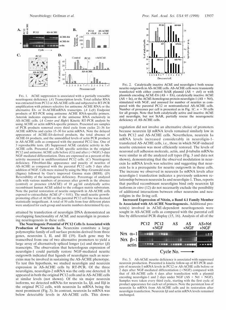
attained by transfection of neuroligin DNA demonstrated anoverlapping functionality of AChE and neuroligin in promot-ing neuritogenesis in these cells.
The Neuritogenic Potential of PC12 Cells Is Associated withProduction of Neurexin Ia. Neurexins constitute a largepolymorphic family of cell surface proteins derived from threegenes, neurexins I, II, and III (19). Each gene may betranscribed from one of two alternative promoters to yield alarge array of alternatively spliced longer (a) and shorter (b)transcripts. The observation that heterologous expression ofneuroligin-1 could partially restore NGF-mediated neuriteoutgrowth indicated that ligands of neuroligins such as neur-exins may be involved in sustaining the AS-ACHE phenotype.To test this hypothesis, we studied neuroligin and neurexinexpression in AS-ACHE cells by RT-PCR. Of the threeneuroligins, neuroligin-2 mRNA was the only one detected. Itappeared in both the original PC12 cells and in AS-ACHE cellsat similar levels (not shown). Of the six major neurexinisoforms, we detected mRNAs for neurexin Ia, Ib, and IIb inthe original PC12 cells, with neurexin Ia mRNA being themost prominent (Fig. 3). In contrast, neurexin Ia mRNA wasbelow detectable levels in AS-ACHE cells. This down-
regulation did not involve an alternative choice of promotersbecause neurexin Ib mRNA levels remained similarly low inboth PC12 and AS-ACHE cells. Nevertheless, neurexin IamRNA levels increased considerably in neuroligin-1-transfected AS-ACHE cells, i.e., those in which NGF-inducedneurite extension was most efficiently restored. The levels ofneuronal cell adhesion molecule, actin, and b-tubulin mRNAswere similar in all the analyzed cell types (Fig. 3 and data notshown), demonstrating that the observed modulation in neur-exin Ia mRNA levels was selective and suggesting that neur-exin Ia is a prerequisite for neurite outgrowth in these cells.The increase we observed in neurexin Ia mRNA levels afterneuroligin-1 transfection indicates a previously unknown re-lationship between neurexin Ia and neuroligin-1. Thus, reportsthat purified recombinant neuroligins bind only neurexin Ibisoforms in vitro (12) do not necessarily exclude the possibilityof additional interactions between other neurexins and neu-roligins in the living cell.
Increased Expression of Nitzin, a Band 4.1 Family MemberIs Associated with AS-ACHE Neuritogenesis. Additional pro-tein(s) involved in AChE-dependent neuritogenesis weresought in AS-ACHE cells as compared with the parental cellline by differential PCR display (15, 16). Analysis of all of the
FIG. 1. AChE suppression is associated with a partially rescuableneuritogenic deficiency. (A) Transcription levels. Total cellular RNAwas extracted from PC12 or AS-ACHE cells and subjected to RT-PCRamplification with primers selective for antisense ACHE RNA or thealternative E6- or I4-ACHEmRNA transcripts. (A Left) Endpointproducts of RT-PCR using antisense ACHE RNA-specific primers.Asterisk indicates expression of the antisense RNA exclusively inAS-ACHE cells. (A Center and Right) Kinetic RT-PCR analysis byusing ACHE or actin mRNA-specific primers. Presented are samplesof PCR products removed every third cycle from cycles 21–36 forACHE mRNAs and cycles 15–30 for actin mRNA. Note the delayedappearance of ACHE-E6-derived products, the total absence ofACHE-I4 products, and the unmodified levels of actin PCR productsin AS-ACHE cells as compared with the parental PC12 line. One of3 reproducible tests. (B) Suppressed AChE catalytic activity in AS-ACHE cells. Presented are AChE specific activities in the originalPC12 and antisense ACHE cells before (Ct) and after (1NGF) 3-daysNGF-mediated differentiation. Data are expressed as a percent of theactivity measured in undifferentiated PC12 cells. (C) Neuritogenicdeficiency. Fibroblast-like appearance and paucity of neurites ofAS-ACHE as compared with the parental PC12 cells 3 days afteraddition of NGF. Cells were stained by using the May–Grunwald stain(Sigma) followed by Gurr’s improved Giemsa stain (BDH). (D)Reversibility of the neuritogenic deficiency. Percentage of analyzedcells with various numbers of processes after growth in the absence(control) or presence (rhAChE) of 10 mgyml catalytically activerecombinant human AChE added to the collagen matrix substratum.Note the partial restoration of neurite outgrowth in AS-ACHE cellsexposed to extracellular AChE (P , 0.01). The small neurite growth-promoting effect of AChE on the parental PC12 cell line was deemedstatistically insignificant. A total of 50 cells from four different plateswere analyzed for each group and neurite numbers determined by eye.
FIG. 2. Catalytically inactive AChE and neuroligin-1 both rescueneurite outgrowth in AS-ACHE cells. AS-ACHE cells were transientlytransfected with either control StAR plasmid (AS 1 ctrl) or withplasmids encoding AChE-E6 (AS 1 E6), catalytically inactive AChE(AS 1 In), or the AChE-homologous protein neuroligin-1 (AS 1 Nrl),stimulated with NGF, and assessed for number of neurites as com-pared with the parental PC12 or nontransfected AS-ACHE cells.Number of processes per cell is presented as in Fig. 1C. n 5 50 cellsfor all groups. Note that both catalytically active and inactive AChEand neuroligin, but not StAR, partially rescue the neurogeneticdeficiency of AS-ACHE cells.
FIG. 3. AS-AChE neurite deficiency is associated with suppressedneurexin production. Presented is kinetic follow-up of RT-PCR anal-ysis of neurexin I mRNA levels in PC12 or AS-ACHE cells before or2 days after NGF-mediated differentiation (1NGF) compared withthat of AS-ACHE cells 4 days after transfection with a plasmidencoding neuroligin-1 and 2 days under NGF (AS 1 Nrl 1 NGF).Samples were taken every third cycle, starting with the first cycle ofproduct appearance for each set of primers. Note the persistent loss ofneurexin Ia mRNA from AS-ACHE cells and its restoration afterneuroligin transfection. Neurexin Ib and actin mRNA levels remainedunchanged.
Neurobiology: Grifman et al. Proc. Natl. Acad. Sci. USA 95 (1998) 13937

differentially displayed PCR products (average length: 482 6191 bp) revealed seven transcripts that were prominently up-or down-regulated in AS-ACHE as compared with PC12 cells.The corresponding PCR fragments were cloned, sequenced,and sought in several databases. Five of these transcripts areyet unknown or presented weak homologies to known genes.The two others were rat procollagen type V-a2 and a protein,we termed nitzin, a putative member of the cytoarchitectureband 4.1 superfamily of proteins (20). RT-PCR amplificationof procollagen type V-a2 and nitzin mRNAs, using specificprimers, confirmed that these two mRNAs are detectable onlyin AS-ACHE cells (data not shown).
The band 4.1 superfamily includes the ezrin, radixin, moesin(ERM) family of proteins (20) involved in the intracellularanchoring of cell membrane proteins to the cytoskeleton. Thenucleotide sequence of nitzin mRNA displays 55% homologyto mammalian ERM mRNAs and 42% homology to band 4.1genes. Moreover, amino acid motifs corresponding to consen-sus sequence blocks 3 and 4 (PROSITE accession nos.BL0060C and BL0060D, respectively) and motifs 4 and 5 of theERM family (PR00661) are all preserved in nitzin. A search of
expressed sequence tag collections revealed that the humannitzin gene is highly homologous to that from rat (Fig. 4A).
The tentative identification of nitzin as a cytoarchitectureprotein raised the possibility that its expression is modulatedunder NGF-induced neuritogenesis. Nitzin mRNA levels werebelow detection in PC12 cells before and after differentiationbut high in naive AS-ACHE cells and down-regulated duringthe abortive neuritogenesis induced in these cells in thepresence of NGF (Fig. 4B). Increased nitzin mRNA levelsparalleled the efficiency of rescue observed after NGF-treatment of AS-ACHE cells transfected with AChE variantsor neuroligin (Fig. 4C). This result indicated a role for nitzinin the cytoskeletal remodeling that provided for rescue ofneurite extension in transfected AS-ACHE cells. To unravelthe potential scope of such roles at the level of the developingorganism in vivo, we analyzed nitzin’s pattern of expression inseveral tissues where various levels of AChE mRNA weredetected in newborn mice by in situ hybridization and com-pared it to that of AChE. Nitzin and AChE mRNA signals werehigh and partially colocalized in the developing cerebellum,hippocampus, and retina, three tissues where a neuritogenic
FIG. 4. Expression of nitzin, a band 4.1 protein family member, is associated with AS-ACHE neuritogenesis. (A) Sequence alignment. Partialamino acid sequences from mouse band 4.1, human moesin, rat nitzin, and the corresponding human expressed sequence tag homolog to nitzin.Band 4.1 family consensus sequences are marked in red, ERM consensus sequences in blue, and nitzinyexpressed sequence tag homologous residuesin green. Band 4.1 signature no. 2 in sequence blocks 3 and 4 are marked by red brackets, and ERM family motifs IV and V are enclosed in blueboxes. (B) Transcriptional modulations. Nitzin mRNA levels were evaluated by RT-PCR in naive and NGF-differentiated PC12 and AS-ACHEcells, and in AS-ACHE cells transfected with a control plasmid (AS 1 ctrl), or plasmids encoding AChE-E6 (AS 1 E6), catalytically inactivatedAChE (AS 1 In) or neuroligin-1 (AS 1 Nrl) as in Fig. 3. Samples were removed as described in the Fig. 3 legend. Note the pronounced expressionof nitzin in AS-ACHE but not PC12 cells, its suppression under NGF, and the neuritogenic-associated increases in rescued AS-ACHE cells, underconditions in which actin mRNA levels remained unchanged.
13938 Neurobiology: Grifman et al. Proc. Natl. Acad. Sci. USA 95 (1998)

role for AChE was suggested (1). However, expression ofnitzin also was observed in liver, thymus, and kidney tubules(Fig. 5 and data not shown), where no overlaps could be foundwith AChE expression and no developmental role(s) weresuggested for AChE. These findings limit the predicted asso-ciation between nitzin and AChE to the nervous system, yetsuggest a more general role for nitzin in the cytoarchitecturalchanges involved with the development of several cells andorgans.
DISCUSSION
We studied the neuritogenic activities of AChE by using apartially reversible AChE loss-of-function model in PC12 cellsexpressing antisense AChE cRNA. Stable, most effectivesuppression of AChE in PC12 cells imposed a block to normalNGF-mediated differentiation that was characterized by al-tered cytoarchitecture, a paucity of neurites, loss of neurexinIa mRNA, and altered expression of a band 4.1 superfamilymember, nitzin. Our findings that heterologous expression ofneuroligin-1 restored morphological characteristics and geneexpression patterns associated with normal differentiationdemonstrate a functional redundancy of AChE and neuroliginin activating a critical morphogenetic pathway in these cells.Together with the sequence homologies shared by AChE,
neuroligins, neurotactin, and gliotactin, our data thereforesuggest that AChE and the various cholinesterase-like proteinsinteract with an overlapping set of heterotypic ligands such asneurexins and neurexin-like proteins.
Modest quantities of AChEcRNA transcripts suppressedAChE production in stably transfected PC12 cells more effec-tively than similarly targeted antisense oligodeoxynucleotides(14). This probably reflects more efficient formation of doublestranded mRNA-cRNA hybrids by the longer cRNA chains,which reduced AChE levels below the amounts needed tosupport neuritogenesis. It was shown previously that the coredomain of AChE could replace the homologous extracellulardomain of neurotactin to generate a functional chimera (21).Thus, the cholinesterasic domain appears to play a conservedrole in ligand recognition. Nevertheless, a membrane-associated form of the intact AChE polypeptide could notsubstitute for neurotactin in mediating heterotypic cell adhe-sion. Thus, the transmembrane and cytoplasmic elementspresent in the AChE-like proteins—but absent in AChE—appear indispensable in translating some ligand-binding inter-actions to changes in cytoarchitecture. In that case, competi-tive interactions between AChE and neurexins could serve aunique role in regulating growth processes associated withneuroligin–neurexin interactions.
As our cells were grown at low density on a collagen matrix,our observations in PC12 cells must reflect autologous inter-actions between substratum-bound or transgene-derivedAChE, neuroligin, and a common ligand, possibly neurexin Ia.These and similar studies may therefore suggest that lateral cismembrane interactions between AChE, neuroligin, and neur-exin mediate neuritogenic processes in some neuronal celltypes. Yet, even if supported by additional in vitro studies, thishypothesis does not exclude in vivo situations in which het-erotypic trans cell–cell interactions could predominate. In-deed, both AChE and neurexins are expressed in the devel-oping nervous system and are considered to play central rolesin establishing neuronal connectivity. Nevertheless, the par-tially reversible nature of the AS-ACHE phenotype demon-strates a degree of plasticity in AChE-mediated morphoge-netic processes. As such, we might predict a role for noncata-lytic AChE activities in neuronal remodeling in the adultnervous system as well. Consistent with this hypothesis, werecently have observed high expression of AChE to be asso-ciated with modulated dendrite branching in AChE transgenicmice (22). Long-term accumulation of AChE was furtherdetected in mice subjected to acute psychological stress (23),also known to induce persistent changes in brain circuitry. Inthis light, the reduced AChE levels observed in the adrenalmedulla of Alzheimer’s disease patients (24) may predictmodified innervation of the medulla.
a-Neurexins contain cytoplasmic tails with sequences ho-mologous to the glycophorin-C intracellular domain whichbinds the band 4.1 protein. In addition, they possess a con-served four-amino acid motif that functions as a recognitionsequence for the PDZ domains of membrane-associated guan-ylate cyclase kinase (MAGUK) proteins (25). Complexesformed by the association of a glycophorin-C like domain, aband 4.1 protein and a MAGUK, were described in mamma-lian erythrocytes and Drosophila septate junctions (26, 27, andreviewed in ref. 19). In both cases, glycophorin-C, band 4.1,and MAGUK-related elements participate in mediating thecytoarchitectural organization that determines biological func-tion. In Drosophila septate junctions, genomic knock-out ofeither the neurexin-like (27) or cholinesterase-like (8) com-ponent caused a similar disturbance in the integrity of theblood–nerve barrier. Thus, evolutionarily diverse interactionsbetween AChE-like proteins and neurexin-like proteins canserve to mediate multiform intercellular recognition processesregulating cellular morphology. In the nervous system, mod-ular compositions of neurexin-band 4.1-MAGUK-related
FIG. 5. Partially overlapping expression patterns for nitzin andAChE mRNAs in developing mouse tissues. High resolution in situhybridization results for various tissues from newborn FVByN mice byusing AChE (Left) and nitzin- (Right) specific probes (see Materialsand Methods). From top to bottom: hippocampus, cerebellum, retina,and kidney. Note primarily overlapping expression patterns in bothcholinergic (e.g., hippocampus) and noncholinergic brain regions (e.g.,cerebellum), partial overlap in the retina, where AChE is notablyinvolved with embryonic development (1) and no AChE expression inthe kidney, which expresses high levels of nitzin.
Neurobiology: Grifman et al. Proc. Natl. Acad. Sci. USA 95 (1998) 13939

complexes might similarly promote cytoskeletal arrange-mentsyrearrangements in either autologous or heterologousfashions through interactions with members of the cholinest-erase-related family of cell surface proteins. The apparentlydifferent band 4.1 elements in parental PC12 cells and ‘‘res-cued’’ AS-ACHE cells, in which nitzin levels remain high,supports the notion of variable compositions of these com-plexes. In that case, AChE could regulate neurite outgrowththrough competitive interactions with different neurexin com-plexes.
Currently approved drugs for the treatment of Alzheimer’sdisease patients are all designed to suppress the catalyticactivity of AChE (28). This is aimed at improving cholinergicneurotransmission under the acetylcholine deficiency causedby loss of cholinergic neurons. However, conventional AChEinhibitors induce increases in the amount of AChE protein(23). They may therefore facilitate the deleterious effects ofnoncatalytic morphogenic activities of AChE in the dementedbrain, such as the promotion of b-amyloid aggregation (23, 29).To reduce AChE protein levels in the brain, it is important todevelop therapeutic antisense strategies targeted againstACHE mRNA (14). By preventing the production of AChE,antisense-ACHE agents, even if less effective than the trans-fection-delivered AChEcRNA, would improve cholinergicneurotransmission while simultaneously ameliorating the non-catalytic effects of this multifunctional protein (30). That theneuritogenic deficiencies induced by antisense AChE suppres-sion can be reversed by AChE noncatalytic homologues sup-ports the plausibility of antisense based therapies for thetreatment of neurodegenerative diseases associated with cho-linergic malfunction.
We are grateful to Dr. T. Sudhof (Dallas) for neuroligin 1 DNA, toDr. I. Silman (Rehovot) for anti-AChE antibodies, and to M. Sternfeldand Dr. R. Broide (Jerusalem) for help with experiments. We alsothank Prof. F. Eckstein and Dr. D. Glick for critical evaluation of thismanuscript. This study was supported by grants from the German–Israeli Foundation (I-0512-206), the Israel Science Foundation (590y97), the United States Army (17.97.I.7007), the Zelman CowenFoundation, and Ester Neurosciences, Ltd.
1. Layer, P. G. & Willbold, E. (1995) Prog. Histochem. Cytochem. 29,1–99.
2. Small, D. H., Reed, G., Whitefield, B. & Nurcombe, V. (1995)J. Neurosci. 15, 144–151.
3. Holmes, C., Jones, S. A., Budd, T. C. & Greenfield, S. A. (1997)J. Neurosci. Res. 49, 207–218.
4. Sternfeld, M., Ming, G.-L., Song, H.-L., Sela, K., Poo, M.-M. &Soreq, H. (1998) J. Neurosci. 18, 1240–1249.
5. Karpel, R., Sternfeld, M., Ginzberg, D., Guhl, E., Graessmann,A. & Soreq, H. (1996) J. Neurochem. 66, 114–123.
6. Koenigsberger, C., Chiappa, S. & Brimijoin, S. (1997) J. Neuro-chem. 69, 1389–1397.
7. De la Escalera, S., Bockamp, E. O., Moya, F., Piovant, M. &Jimenez, F. (1990) EMBO J. 9, 3593–3601.
8. Auld, V. J., Fetter, R. D., Broadie, K. & Goodman, C. S. (1995)Cell 81, 757–767.
9. Ichtchenko, K., Hata, Y., Nguyen, T., Ullrich, B., Missler, M.,Moomaw, C. & Sudhof, T. C. (1995) Cell 81, 435–443.
10. Ichtchenko, K., Nguyen, T. & Sudhof, T. C. (1996) J. Biol. Chem.271, 2676–2682.
11. Puschel, A. W. & Betz, H. (1995) J. Neurosci. 15, 2849–2856.12. Nguyen, T. & Sudhof, T. C. (1997) J. Biol. Chem. 272, 26032–
26039.13. Andres, C., Beeri, R., Friedman, A., Lev-Lehman, E., Henis, S.,
Timberg, R., Shani, M. & Soreq, H. (1997) Proc. Natl. Acad. Sci.USA 94, 8173–8178.
14. Grifman, M. & Soreq, H. (1997) Antisense Nucl. Acid Drug Dev.7, 351–359.
15. Welsh, J., Chada, K., Dalal, S. S., Cheng, R., Ralph, D. &McClelland, M. (1992) Nucleic Acids Res. 20, 4965–4970.
16. Grifman, M., Lev-Lehman, E., El-Tamer, A., Ginzberg, D.,Hanin, I. & Soreq, H. (1996) Neuroscience Protocols 20, 1–11.
17. Seidman, S., Sternfeld, M., Ben Aziz-Aloya, R., Timberg, R.,Kaufer-Nachum, D. & Soreq, H. (1995) Mol. Cell. Biol. 15,2993–3002.
18. Clark, B. J., Wells, J., King, S. R. & Stocco, D. M. (1994) J. Biol.Chem. 269, 28314–28322.
19. Missler, M. & Sudhoff, T. C. (1998) Trends Genet. 14, 20–26.20. Tsukita, S., Yonemura, S. & Tsukita, S. (1997) Trends Biochem.
Sci. 22, 53–58.21. Darboux, I., Barthalay, Y., Piovant, M. & Hipeau-Jacquotte, R.
(1996) EMBO J. 15, 4835–4843.22. Beeri, R., LeNovere, N., Mervis, R., Huberman, T., Grauer, E.,
Changeux, J. P. & Soreq, H. (1997) J. Neurochem. 69, 2441–2451.23. Kaufer, D., Friedman, A., Seidman, S. & Soreq, H. (1998) Nature
(London) 393, 373–377.24. Appleyard, M. E. & McDonald, B. (1991) Lancet 338, 1085–1086.25. Songyang, Z., Fanning, A. S., Fu, C., Xu, J., Marfatia, S. M.,
Chishti, A. H., Crompton, A., Chan, A. C., Anderson, J. M. &Cantley, L. C. (1997) Science 275, 73–77.
26. Correas, I., Leto, T. L., Speicher, D. W. & Marchesi, V. T. (1986)J. Biol. Chem. 261, 3310–3315.
27. Baumgartner, S., Littleton, J. T., Broadie, K., Bhat, M. A.,Harbecke, R., Lengyel, J. A., Chiquet-Ehrismann, R., Prokop, A.& Bellen, H. J. (1996) Cell 87, 1059–1068.
28. Knapp, M. J., Knopman, D. S., Solomon, P. R., Pendlebury,W. W., Davis, C. S. & Gracon, S. I. (1994) J. Am. Med. Assoc. 271,985–991.
29. Inestrosa, N. C., Alvarez, A., Perez, C. A., Moreno, R. D.,Vicente, M., Linker, C., Casanueva, O. I., Soto, C. & Garrido, J.(1996) Neuron 16, 881–891.
30. Grifman, M., Lev-Lehman, E., Ginzberg, D., Eckstein, F., Zakut,H. & Soreq, H. (1997) in Concepts in Gene Therapy, eds. Strauss,M. & Barranger, J. A. (de Gruyter, Berlin), pp. 141–167.
13940 Neurobiology: Grifman et al. Proc. Natl. Acad. Sci. USA 95 (1998)
Related Documents