Functional Reconstitution of ESCRT-III Assembly and Disassembly Suraj Saksena, 1 Judit Wahlman, 2 David Teis, 1 Arthur E. Johnson, 2,3,4, * and Scott D. Emr 1, * 1 Weill Institute for Cell and Molecular Biology and Department of Molecular Biology and Genetics, Weill Hall, Cornell University, Ithaca, NY 14853, USA 2 Department of Molecular and Cellular Medicine, Texas A&M Health Science Center, College Station, TX 77843-1114, USA 3 Department of Chemistry 4 Department of Biochemistry and Biophysics Texas A&M University, College Station, TX 77843, USA *Correspondence: [email protected] (A.E.J.), [email protected] (S.D.E.) DOI 10.1016/j.cell.2008.11.013 SUMMARY Receptor downregulation in the MVB pathway is mediated by the ESCRT complexes. ESCRT-III is composed of four protein subunits that are mono- meric in the cytosol and oligomerize into a protein lattice only upon membrane binding. Recent studies have shown that the ESCRT-III protein Snf7 can form a filament by undergoing homo-oligomerization. To examine the role of membrane binding and of interac- tions with other ESCRT components in initiating Snf7 oligomerization, we used fluorescence spectroscopy to directly detect and characterize the assembly of the Snf7 oligomer on liposomes using purified ESCRT components. The observed fluorescence changes reveal an obligatory sequence of membrane-protein and protein-protein interactions that generate the active conformation of Snf7. Also, we demonstrate that ESCRT-III assembly drives membrane deforma- tion. Furthermore, using an in vitro disassembly assay, we directly demonstrate that Vps24 and Vps2 function as adaptors in the ATP-dependent membrane disas- sembly of the ESCRT-III complex by recruiting the AAA ATPase Vps4. INTRODUCTION A number of seemingly unrelated biological processes such as multivesicular body (MVB) biogenesis, cytokinesis, and retroviral budding require the ESCRT (endosomal sorting complex required for transport) machinery. In contrast to the formation of secretory and endocytic vesicles, where membrane budding occurs into the cytosol, MVB formation requires membrane budding away from the cytosol. The cellular machinery respon- sible for MVB formation under normal and pathological condi- tions is comprised of a subset of vacuolar protein sorting (Vps) gene products that were first implicated in receptor downregula- tion via the MVB pathway (Hurley, 2008; Saksena et al., 2007; Williams and Urbe, 2007). The majority of these Vps proteins are constituents of five separate heteromeric protein complexes called ESCRT-0, ESCRT-I, ESCRT-II, ESCRT-III, and the Vps4 AAA-ATPase complex (Hurley, 2008; Williams and Urbe, 2007). These complexes are transiently recruited from the cytoplasm to the endosomal membrane, where they function sequentially in sorting ubiquitinated transmembrane proteins into the MVB pathway. During the process of MVB sorting, ESCRT-0, ESCRT-I, and ESCRT-II are recruited to the endosomal membrane as stable hetero-oligomeric complexes from the cytosol. In contrast, the ESCRT-III proteins (Vps20, Snf7, Vps24, and Vps2) remain monomeric in the cytosol, and only upon membrane binding oligomerize into an ESCRT-III lattice of indeterminate stoichiom- etry. The fact that the ESCRT-III complex is comprised of four protein subunits that undergo a membrane-dependent mono- mer to hetero-oligomer transition raises a number of mechanistic questions, including (1) what is the order of membrane recruit- ment and assembly for each of the ESCRT-III proteins and (2) what are the molecular signals that prevent premature assembly of the ESCRT-III complex in the cytosol and direct assembly on the membrane? Genetic and biochemical studies in yeast have provided some clues regarding the order of membrane recruitment of the indi- vidual ESCRT-III proteins. Vps20 is the first ESCRT-III protein to associate with ESCRT-II on the endosome. Endosomal recruit- ment of the Vps24-Vps2 subcomplex appears to be dependent on the Vps20-Snf7 subcomplex, thereby indicating that the two subcomplexes are recruited sequentially (Teis et al., 2008). Recy- cling of membrane-bound ESCRT complexes into the cytosol has been shown to involve interactions between Vps2 and the AAA-ATPase Vps4 (Obita et al., 2007; Stuchell-Brereton et al., 2007). Although it is challenging to determine the precise stoichiom- etry and molecular architecture of a membrane-bound protein lattice, studies on hSnf7-1 (CHMP4A) have shown that overex- pressed hSnf7-1/CHMP4A spontaneously forms membrane- attached filaments 5 nm in width that curve and self-associate to create circular arrays that can promote or stabilize negative membrane curvature and outward budding of plasma membrane tubules (Hanson et al., 2008). These data indicate that a single ESCRT-III protein can form an ESCRT-III lattice, presumably by Snf7 binding to itself. This possibility is further supported by observations that Snf7 is the most abundant ESCRT-III subunit Cell 136, 97–109, January 9, 2009 ª2009 Elsevier Inc. 97

Welcome message from author
This document is posted to help you gain knowledge. Please leave a comment to let me know what you think about it! Share it to your friends and learn new things together.
Transcript

Functional Reconstitutionof ESCRT-III Assembly and DisassemblySuraj Saksena,1 Judit Wahlman,2 David Teis,1 Arthur E. Johnson,2,3,4,* and Scott D. Emr1,*1Weill Institute for Cell and Molecular Biology and Department of Molecular Biology and Genetics, Weill Hall, Cornell University,
Ithaca, NY 14853, USA2Department of Molecular and Cellular Medicine, Texas A&M Health Science Center, College Station, TX 77843-1114, USA3Department of Chemistry4Department of Biochemistry and Biophysics
Texas A&M University, College Station, TX 77843, USA
*Correspondence: [email protected] (A.E.J.), [email protected] (S.D.E.)
DOI 10.1016/j.cell.2008.11.013
SUMMARY
Receptor downregulation in the MVB pathway ismediated by the ESCRT complexes. ESCRT-III iscomposed of four protein subunits that are mono-meric in the cytosol and oligomerize into a proteinlattice only upon membrane binding. Recent studieshave shown that the ESCRT-III protein Snf7 can forma filament by undergoing homo-oligomerization. Toexamine the role of membrane binding and of interac-tions with other ESCRT components in initiating Snf7oligomerization, we used fluorescence spectroscopyto directly detect and characterize the assembly ofthe Snf7 oligomer on liposomes using purified ESCRTcomponents. The observed fluorescence changesreveal an obligatory sequence of membrane-proteinand protein-protein interactions that generate theactive conformation of Snf7. Also, we demonstratethat ESCRT-III assembly drives membrane deforma-tion. Furthermore, usingan invitro disassemblyassay,we directly demonstrate that Vps24 and Vps2 functionas adaptors in the ATP-dependent membrane disas-sembly of the ESCRT-III complex by recruiting theAAA ATPase Vps4.
INTRODUCTION
A number of seemingly unrelated biological processes such as
multivesicular body (MVB) biogenesis, cytokinesis, and retroviral
budding require the ESCRT (endosomal sorting complex
required for transport) machinery. In contrast to the formation
of secretory and endocytic vesicles, where membrane budding
occurs into the cytosol, MVB formation requires membrane
budding away from the cytosol. The cellular machinery respon-
sible for MVB formation under normal and pathological condi-
tions is comprised of a subset of vacuolar protein sorting (Vps)
gene products that were first implicated in receptor downregula-
tion via the MVB pathway (Hurley, 2008; Saksena et al., 2007;
Williams and Urbe, 2007). The majority of these Vps proteins
are constituents of five separate heteromeric protein complexes
called ESCRT-0, ESCRT-I, ESCRT-II, ESCRT-III, and the Vps4
AAA-ATPase complex (Hurley, 2008; Williams and Urbe, 2007).
These complexes are transiently recruited from the cytoplasm
to the endosomal membrane, where they function sequentially
in sorting ubiquitinated transmembrane proteins into the MVB
pathway.
During the process of MVB sorting, ESCRT-0, ESCRT-I, and
ESCRT-II are recruited to the endosomal membrane as stable
hetero-oligomeric complexes from the cytosol. In contrast, the
ESCRT-III proteins (Vps20, Snf7, Vps24, and Vps2) remain
monomeric in the cytosol, and only upon membrane binding
oligomerize into an ESCRT-III lattice of indeterminate stoichiom-
etry. The fact that the ESCRT-III complex is comprised of four
protein subunits that undergo a membrane-dependent mono-
mer to hetero-oligomer transition raises a number of mechanistic
questions, including (1) what is the order of membrane recruit-
ment and assembly for each of the ESCRT-III proteins and (2)
what are the molecular signals that prevent premature assembly
of the ESCRT-III complex in the cytosol and direct assembly on
the membrane?
Genetic and biochemical studies in yeast have provided some
clues regarding the order of membrane recruitment of the indi-
vidual ESCRT-III proteins. Vps20 is the first ESCRT-III protein to
associate with ESCRT-II on the endosome. Endosomal recruit-
ment of the Vps24-Vps2 subcomplex appears to be dependent
on the Vps20-Snf7 subcomplex, thereby indicating that the two
subcomplexes are recruited sequentially (Teis et al., 2008). Recy-
cling of membrane-bound ESCRT complexes into the cytosol
has been shown to involve interactions between Vps2 and the
AAA-ATPase Vps4 (Obita et al., 2007; Stuchell-Brereton et al.,
2007).
Although it is challenging to determine the precise stoichiom-
etry and molecular architecture of a membrane-bound protein
lattice, studies on hSnf7-1 (CHMP4A) have shown that overex-
pressed hSnf7-1/CHMP4A spontaneously forms membrane-
attached filaments 5 nm in width that curve and self-associate
to create circular arrays that can promote or stabilize negative
membrane curvature and outward budding of plasma membrane
tubules (Hanson et al., 2008). These data indicate that a single
ESCRT-III protein can form an ESCRT-III lattice, presumably by
Snf7 binding to itself. This possibility is further supported by
observations that Snf7 is the most abundant ESCRT-III subunit
Cell 136, 97–109, January 9, 2009 ª2009 Elsevier Inc. 97

in yeast (Teis et al., 2008) and that most Snf7 isoforms self-asso-
ciate in yeast two-hybrid analyses (reviewed in Saksena et al.,
2007).
ESCRT-III assembly is spatially restricted to the endosomal
membrane by the upstream ESCRT complexes (ESCRT-0, -I,
and -II), which selectively bind ubiquitin-modified transmem-
brane cargo and the endosomally enriched lipid phosphatidyli-
nositol 3-phosphate (PI3P). There is growing evidence that
activation of ESCRT-III subunits for assembly into the
membrane-bound ESCRT-III complex requires a conformational
switch. All of the ESCRT-III subunits are similar in size (221–241
residues) and domain organization, with N-terminal basic and
C-terminal acidic regions (Saksena et al., 2007). Hence, the
crystal structure of hVps24/CHMP3 (residues 9–183) reveals
structural elements that are probably common to all ESCRT-III
subunits (Muzio1 et al., 2006). Five alpha helices (a1–a5) span
most of the N-terminal two-thirds of the protein and are con-
nected by a relatively long linker to a sixth short predicted helix
(a6) near the C terminus (a6 and the loop linking a5 to a6 were
not seen in the crystal structure, indicating conformational flexi-
bility). The most prominent structural element observed in the
crystal structure is the N-terminal helical hairpin that is 70 A
long and composed of helices a1 and a2, which are critical for
membrane binding and homo- or heterodimerization events
(Muzio1 et al., 2006). The C-terminal region that includes helix
a5, the microtubule-interacting and transport (MIT)-interacting
region (MIR), and a6 constitutes a putative autoinhibitory region
that is thought to interact with helix a2 of the core and thereby
block homo- or heterodimerization of ESCRT-III components
(Shim et al., 2007). Such intramolecular interactions may there-
fore prevent ESCRT-III-ESCRT-III intermolecular association
and thereby maintain the ESCRT-III proteins as metastable
monomers in the cytosol in a ‘‘closed’’ state. ESCRT-III protein
assembly may then occur only upon recruitment to endosomes,
where the autoinhibitory intramolecular interaction is lost during
a putative ‘‘closed’’ to ‘‘open’’ state transition that now permits
ESCRT-III proteins to associate. The molecular trigger for the
proposed release of this inhibition and ESCRT-III assembly on
endosomal membranes remains unclear.
Here, we have employed primarily fluorescence spectroscopy
to directly examine the molecular requirements for the assembly
and disassembly of a Snf7 (ESCRT-III) polymer in vitro using
purified components. Probes at several different positions
were monitored spectroscopically under near-native aqueous
conditions to detect changes in probe environment and also their
sensitivity to different ESCRT components and membranes. The
observed spectral changes indicate that upon membrane
binding (1) the conformations of both Vps20 and Snf7 change;
(2) these conformations are further altered upon association
with, respectively, Vps25 and Vps20; (3) most of the surface of
the Snf7 molecule is in contact with the membrane surface; (4)
the C-terminal end of Snf7 moves adjacent to the bilayer except
in mutants with restricted conformational flexibility; (5) Snf7 mole-
cules oligomerize only in the presence of both membranes and
Vps20; and (6) this oligomerization event is critical for membrane
deformation. The combined data strongly suggest that these
ESCRT-III interactions dictate an obligatory sequence of
assembly reactions during formation of the ESCRT-III complex.
98 Cell 136, 97–109, January 9, 2009 ª2009 Elsevier Inc.
Furthermore, we demonstrate that disassembly of the
membrane-bound Snf7 oligomer requires Vps24 and Vps2,
which function as adaptors for ESCRT-III disassembly by
capping the Snf7 oligomer and providing an entry point for the
AAA-ATPase Vps4.
RESULTS
Experimental StrategyTo monitor the conformation, environment, and interactions of a
single protein during the assembly and disassembly of the multi-
component, membrane-bound ESCRT-III complex, the Cys-free
Vps20 and Snf7 proteins were each modified by the replacement
of a single amino acid with Cys. Cys substitutions were made at
sites that lie within domains predicted to have functional impor-
tance (autoinhibitory loop, membrane binding, etc.) and are
solvent exposed (based on the hVps24 crystal structure) to
enhance the efficiency of covalent modification. Importantly,
the introduction of a single Cys at different locations in monocys-
teine mutants had no effect on the folding and function of Vps20
or Snf7 since cells expressing the different cysteine mutants were
able to sort MVB cargo (GFP-CPS) to the vacuolar lumen (Fig-
ure S1 available online). A fluorescent 7-nitrobenz-2-oxa-1,3-
diazole (NBD) dye was then covalently attached to the Cys in
each derivative. NBD is small, uncharged, and its O and N atoms
have sufficient polar character for the dye to be soluble and exist
stably in both aqueous and nonaqueous environments. More-
over, NBD is environmentally sensitive, and its spectral proper-
ties are dramatically different in aqueous and nonaqueous
environments (Johnson, 2005; Shepard et al., 1998). When
NBD moves from an aqueous milieu (e.g., the surface of a soluble
protein) to a hydrophobic environment (e.g., the nonpolar interior
of a membrane or protein), its emission intensity and fluores-
cence lifetime (t) increase, while its wavelength of maximum
emission (lem max) shifts to the blue (but not always, see the
Supplemental Data). To distinguish the nonpolar membrane
interior from a nonpolar environment inside a folded protein or
at the nonpolar interface of associated proteins or domains,
one can diagnostically identify the former by collisions between
NBD probes and nitroxide collisional quenching moieties that
are restricted to locations within the nonpolar core of the bilayer.
Thus, by continuously monitoring in real time the spectral proper-
ties of an NBD-labeled protein (emission intensity, fluorescence
lifetime, accessibility to quenchers, anisotropy, FRET) before,
during, and after it associates with other proteins or membranes,
one can directly quantify both kinetic and thermodynamic param-
eters of these interactions. In addition, since each protein
contains NBD at only a single location, this approach provides
an opportunity to characterize the structural changes that occur
at specific sites in Vps20 and Snf7. The different NBD-labeled
and Rhodamine (Rh)-labeled mutants of Vps20 and Snf7 are
denoted as Vps20/Snf7n-NBD/Rh, where n denotes the location
of the cysteine residue modified with NBD or Rh.
Vps20 Binding to MembranesESCRT-III assembly is initiated by membrane binding of Vps20
(Teis et al., 2008). To detect Vps20 binding to a membrane
surface, NBD probes were positioned at four different sites in

monocysteine mutants of Vps20: residue 7 in the unstructured
N-terminal loop, 61 in the a2 helix, and residues 170 and 190
on either side of the a5 helix (Figure 1F). The fluorescence
emission of each of these NBD-labeled Vps20 derivatives was
then characterized before and after the addition of liposomes
comprised of phosphatidylcholine, phosphatidylserine, and
phosphatidylinositol at molar ratios of 80:15:5 (hereafter termed
PC/PS/PI). No change in emission was observed for probes
located at position 7 upon exposure to PC/PS/PI (Figure 1A).
However, an �6-fold increase in intensity and a 13 nm blue shift
in the lem max was observed for the NBD probe at residue 61 after
liposome addition (Figure 1B). Such substantial changes show
that the NBD dye at residue 61 moves from an aqueous to
nonaqueous milieu upon addition of membranes, a conclusion
verified by a large increase in its fluorescence lifetime (data not
shown).
When Vps2061-NBD was titrated with PC/PS/PI, the emission
intensity increased and reached a plateau (Figure 1C), thereby
suggesting that all Vps20 had bound to the membrane. To test
A B
C D
E F
Figure 1. Vps20 Binding to Membranes
(A and B) Fluorescence emission spectra of
750 nM Vps207-NBD (A) and 1 mM Vps2061-NBD (B)
before (black) and after (red) addition of excess
(1.6 mM; see Figure 1C) PC/PS/PI.
(C) Titration of 1 mM Vps2061-NBD with liposomes
reveals that fluorescence-detected (lem = 530 nm)
binding is complete after addition of 1.2 mM PC/
PS/PI.
(D) Sepharose CL-2B chromatography of 800 nM
Vps2061-NBD that had been preincubated with
either 1.5 mM PC/PS/PI (red) or no liposomes
(black). Protein was detected by NBD emission
intensity and liposomes by light scattering (cyan).
The elution profile of liposomes that were not
exposed to protein was identical to the profile
shown in this and later figures (data not shown).
(E) The ratios of NBD emission intensities at 530 nm
are shown for each derivative with (Fbound) and
without (Ffree) membranes.
(F) Cartoon representation of the water-soluble
hVps24 monomer. On the basis of the sequence
similarities of the ESCRT-III proteins, Vps20, Snf7,
and Vps2 are believed to share structural features
with the solved crystal structure of hVps24. Helices
a1–5 and the N and C termini of the protein are
labeled. Residues that were mutated to cysteine
for labeling with NBD are depicted in cyan and
labeled accordingly. Helix a6 and the a5-a6 loop
have been manually sketched.
this conclusion by an independent
technique, we incubated a sample of
Vps2061-NBD with liposomes and then
purified it by gel filtration chromatog-
raphy. Since essentially all of the NBD
fluorescence was found to coelute in the
void volume with the liposomes (detected
by light scattering) and very little eluted
with free Vps20 assayed in the absence
of liposomes (Figure 1D), more than
95% of the Vps2061-NBD was bound to liposomes after elution.
Thus, the membrane-dependent spectral changes do indeed
result from Vps20 binding to a membrane surface.
Large increases in intensity were also observed when excess
liposomes were added to Vps20 with an NBD probe at either
residue 170 or 190 (Figure 1E). When each of these derivatives
was titrated with PC/PS/PI, the binding curves were similar to
that of Figure 1C (Figure S2; data not shown). Thus, the similar
PC/PS/PI-dependent spectral changes of probes at residues
61, 170, and 190 suggest that the probes near a2 and a5 are
exposed to the nonpolar bilayer. This conclusion was confirmed
directly when the intensity of each probe was reduced by colli-
sions with quenchers restricted to the nonpolar membrane
core (Figures S3B–S3F) (Johnson, 2005). Interestingly, although
the spectral changes for the probe at residue 7 in the unstruc-
tured Vps20 N-terminal domain are slight upon PC/PS/PI addi-
tion, the fluorescence lifetime of this probe is somewhat high
(<t> = �4 ns; aqueous t = 1–2 ns, and nonpolar t = 7–9 ns for
NBD-labeled protein) and its lem max is somewhat low (538 nm;
Cell 136, 97–109, January 9, 2009 ª2009 Elsevier Inc. 99

typical range = 520–546 nm for nonpolar to aqueous) even in the
absence of membranes, which indicates that this probe is in
a relatively nonpolar environment when Vps20 is free in solution.
Vps25 Binds to Membrane-Bound Vps20When either Vps2061-NBD or Vps20170-NBD was titrated with
Vps25, a protein in the ESCRT-II complex that directly interacts
with Vps20, little change in NBD emission intensity was observed
(Figures 2A–2C). These small spectral changes reveal that the
probes located at positions 61 and 170 in Vps20 do not experi-
ence significant changes in environment upon exposure to free
Vps25 in solution and/or that Vps20 and Vps25 associate only
weakly in solution. Probes at residues 7 and 190 were also insen-
sitive to Vps20 exposure to Vps25 (Figure 2F; Figure S4B). Simi-
larly, when Vps20170-NBD was first bound to liposomes and then
titrated with Vps25, the NBD fluorescence signal was unchanged
by Vps25 (Figure S4A; Figures 2E and 2F). However, when Vps25
was added to membrane-bound Vps2061-NBD, a large additional
BA
C D
E F
Figure 2. Vps20 Interactions with the Vps25
Component of ESCRT-II
(A and B) Fluorescence emission scans of 1 mM
Vps20170-NBD D (A) and Vps2061-NBD (B) are shown
in buffer (black) and after addition of 30 mM Vps25
(red).
(C) The dependence of the emission intensities
(F; lex = 468, lem = 530 nm) of 1 mM Vps20170-NBD
(black) and Vps2061-NBD (red) on Vps25 addition
are shown; F0 is the intensity in the absence of
Vps25.
(D) Emission scans of 1 mM Vps2061-NBD are shown
in buffer (black), after addition of excess 1.5 mM
liposomes (red), and after the addition of 30 mM
Vps25 to Vps2061-NBD preincubated with 1.5 mM
liposomes (cyan).
(E) The dependence of the emission intensities
(F; lex = 468, lem = 530 nm) of 1 mM Vps20170-NBD
(black) and Vps2061-NBD (red) on Vps25 concentra-
tion is shown after the Vps20 derivatives were incu-
bated with 1.5 mM liposomes for 30 min at 22�C
before Vps25 addition; F0 is the intensity in the
absence of Vps25.
(F) Summary of changes in the emission intensities
of different NBD-labeled Vps20 mutants upon
incubation with liposomes (red bar), Vps25 alone
(dark blue bar), or liposomes and Vps25 (cyan
bar). In each case, F0 represents emission intensity
of the protein in buffer, and F represents emission
intensity of the protein after incubation either with
liposomes, Vps25, or liposomes and Vps25.
increase of NBD emission intensity was
observed that was Vps25 dependent
(Figures 2D–2F). These data strongly
suggest that Vps25 and Vps20 associate
on a membrane, but not in solution.
Furthermore, the spectroscopic insensi-
tivity of both free and membrane-bound
Vps20170-NBD to the presence of Vps25
suggests that a significant conformational
change alters the environment of a probe
at residue 61, but not at 170, upon association with Vps25 on the
membrane.
When excess Vps25 was added to membrane-bound
Vps20190-NBD, a small emission intensity decrease was observ-
ed (Figure S4C), in contrast to the large increase seen with
Vps2061-NBD (Figure 2D). In addition, little change was detected
with the Vps207-NBD derivative (Figure 2F). Thus, the use of
multiple fluorescent probes reveals that the environments of
Vps20 residues 61, 170, and 190 are altered upon membrane
binding and that the environments of the probes at both residues
61 and 190 are changed when Vps25 binds to the membrane-
bound Vps20 (Figure 2F). It must be emphasized that the exper-
iments described above were carried out with concentrations of
PC/PS/PI at which all of the NBD-labeled Vps20 is membrane
bound (Figure 1C). Hence, the changes in NBD intensity upon
Vps25 addition reflect changes in the local environment of
NBD as a result of Vps25-Vps20 interactions rather than changes
in the amount of membrane-bound Vps20. It must also be noted
100 Cell 136, 97–109, January 9, 2009 ª2009 Elsevier Inc.

that the size of a spectral change does not necessarily correlate
with the magnitude of the conformational change that altered
dye environment. Thus, one cannot conclude that the
membrane- and Vps25-induced structural changes in Vps20
were smaller near residue 170 than residue 61. However, the
most important observation is that the binding of Vps25 to
Vps20 changed its conformation at two sites that are well sepa-
rated in the predicted Vps20 structure (based on the hVps24
crystal structure) (Figure 1F). Furthermore, the simultaneous
binding of both Vps25 and a membrane surface elicited a unique
Vps20 conformation.
Snf7 Binding to MembranesTo characterize Snf7 binding to liposomes, nine monocysteine
Snf7 mutants were prepared and examined as above with
Vps20. With the exception of Snf714-NBD, addition of PC/PS/PI
to a Snf7 derivative elicited a blue shift in its lem max and an
increase in its t (data not shown). Furthermore, collisional
A B
C D
E F
Figure 3. Snf7 Binding to Membranes
(A and B) Fluorescence emission spectra of
540 nM Snf781-NBD (A) and 300 nM Snf7200-NBD
(B) before (black) and after (red) addition of an
excess (1.5 mM; see Figure 1C) of PC/PS/PI.
(C) The ratios of NBD emission intensities at
530 nm are shown for each derivative with (Fbound)
and without (Ffree) membranes.
(D) Cartoon representation of the water-soluble
hVps24 monomer as in Figure 1F, but here
showing the locations of the probes used in the
Snf7 experiments (cyan).
(E) Titration of 300 nM Snf7200-NBD with liposomes
reveals that fluorescence-detected (lem = 530 nm)
binding is complete after addition of 1.2 mM
PC/PS/PI.
(F) Sepharose CL-2B chromatography of 540 nM
Snf781-NBD that had been preincubated with either
1.5 mM PC/PS/PI (red) or no liposomes (black).
Protein was detected by NBD emission intensity
and liposomes by light scattering (cyan).
quenching experiments (Johnson, 2005)
demonstrated that these spectral
changes resulted from the exposure of
the NBD probes (except at residue 14) to
the nonpolar coreof the bilayer (Figure S5).
However, as is evident by comparison of
emission scans of Snf781-NBD (Figure 3A)
and Snf7200-NBD (Figure 3B) in the pres-
ence and absence of PC/PS/PI, the
magnitudes of the membrane-dependent
intensity increases varied considerably
(Figure 3C). Hence, the environments of
probes at different locations, even those
that are as spatially close as K14, K21,
and K35 in the a1 helix, P191 and P200
in the a5- a6 loop, and E81 and S93 in the
a2 helix, differ for free and/or membrane-
bound Snf7 (Figure 3D). To ensure that
the binding of protein to liposomes was
complete in each of these samples, we titrated PC/PS/PI into
the Snf7 proteins and measured intensities only at liposome
concentrations that gave maximal intensities (Figure 3E; Fig-
ure S6; data not shown). Thus, the different Fbound/Ffree ratios
suggest that the membrane-dependent structural changes vary
for different Snf7 domains and even for sites within the same
domain. When examined with gel filtration, Snf7 binding to
liposomes was found to differ from that of Vps20. Whereas
more than 95% of the Vps20 remained bound to the PC/PS/PI
liposomes after the chromatography (Figure 1D), less than 50%
of Snf781-NBD (Figure 3F) and the other eight Snf7 derivatives
(data not shown) remained bound to liposomes. This result was
not due to partial inactivation of the Snf781-NBD proteins because
the same proteins were completely bound to the same liposomes
under different conditions (see below). Instead, the reduced Snf7
binding to liposomes after gel filtration indicates that the
membrane association and dissociation rates are faster for Snf7
than for Vps20, even though maximal fluorescence-detected
Cell 136, 97–109, January 9, 2009 ª2009 Elsevier Inc. 101

binding was obtained at similar PC/PS/PI concentrations for
equivalent concentrations of Snf7 and Vps20.
Vps20 Appears to Bind to One End of Snf7When NBD-labeled Snf7 was titrated with Vps20 in solution,
probes positioned at opposite ends of Snf7 (Figure 3D) re-
sponded very differently. The emission intensity of an NBD at
residue 200 was completely unaffected by the addition of
Vps20 (Figure S7A). In contrast, the intensity of the probe at
residue 58 increased substantially (Figure 4A), by Vps20 alloste-
rically altering the local Snf7 conformation near residue 58 and/or
by Vps20 binding and covering the surface of Snf7 near residue
58. Thus, although one probe is spectroscopically insensitive to
Vps20 binding to Snf7, the dependence of the Snf758-NBD
spectral change on Vps20 concentration shows that Vps20
and Snf7 associate in solution (Figure 4B). Such an association
is not thought to occur between Vps20 and Snf7 in solution,
but the spectral data show that such binding does occur at the
A B
D
F
C
E
Figure 4. Vps20 Binding to Membrane-
Bound Snf7
(A and B) Fluorescence emission spectra of
300 nM Snf7200-NBD (A) and 2.2 mM Snf758-NBD
(B) before (black) and after (red) addition of Vps20.
(C and D) Titration (lem = 530 nm) of 300 nM
Snf7200-NBD (black) and 2.2 mM Snf758-NBD (red)
with Vps20 either before (C) or after (D) incubation
with 1.5 mM PC/PS/PI.
(E) Emission spectra of 2.2 mM Snf758-NBD bound
to 1.5 mM PC/PS/PI before (red) and after (cyan)
addition of 2.7 mM Vps20. For comparison, the
spectrum of 2.2 mM Snf758-NBD in buffer is shown
(black).
(F) Sepharose CL-2B chromatography of samples
containing 540 nM Snf781-NBD and 2.7 mM Vps20
that had been preincubated with either 1.5 mM
PC/PS/PI (red) or no liposomes (black). Protein
was detected by NBD emission intensity and
liposomes by light scattering (cyan).
nonphysiological protein concentrations
used in our experiments.
Interestingly, the spectral data indicate
that Vps20 binds similarly to both free
and membrane-bound Snf7. Vps20 addi-
tion did not significantly alter emission of
both free (Figure 4B) and membrane-
bound (Figure 4C) Snf7200-NBD, even
though the probe at residue 200 was
very sensitive to Snf7 binding to the
membrane (Figures3B and 3C). The probe
at residue 58 was also sensitive to Snf7
binding to the membrane (Figure 3C), but
its environment was further altered by
the binding of Vps20 to membrane-bound
Snf7 (Figures 4C and 4D). Given the
similarities in the magnitudes of the
Snf758-NBD intensity changes and their
dependence on Vps20 concentration
(Figures 4B and 4C), it appears that the extent of Vps20 binding
to free and membrane-bound Snf7 is similar. Since Vps20 binding
to Snf7 appears to be unaffected by whether or not Snf7 is bound
to a liposome, it suggests that Snf7 interactions with Vps20 and
membranes are uncoupled and noncooperative. However,
a Vps20 effect on Snf7 binding to liposomes was revealed by
gel filtration. WhenSnf781-NBD was incubated with both liposomes
and Vps20 and then chromatographed, essentially all (>95%) of
the Snf7 was found bound to the liposomes (Figure 4E). Thus,
Vps20 markedly stabilizes Snf7 binding to membranes, as is
clearly evident by comparison of the elution profiles of Snf7 in lipo-
somal samples that either contain (Figure 4E) or lack (Figure 3F)
Vps20. These results demonstrate that Vps20 functions to stabi-
lize Snf7 binding to the membrane, presumably by changing the
Snf7 conformation and reducing its rate of dissociation from the
membrane.
As was true with Snf7200-NBD (Figure 4B), titration of Snf735-NBD
with Vps20 had little effect on NBD emission intensity both in
102 Cell 136, 97–109, January 9, 2009 ª2009 Elsevier Inc.

the presence and absence of PC/PS/PI (Figures S7C and
S7D). Hence, the probe at residue 35 was not especially sensitive
to Vps20 binding to Snf7. However, Vps20-dependent emission
intensity increases were detected for both free and membrane-
bound Snf781-NBD, though the spectral changes were small,
especially for membrane-bound Snf781-NBD (Figures S7B–S7D).
The probes between a5 and a6 (residue 200) and near the
middle of the a1 helix (residues 35 and 21; data not shown) de-
tected Snf7 binding to membranes, but not to Vps20. In contrast,
probes at residue 58, at the other end of the protein (according to
the hVps24 crystal structure), and at residue 81 in a2 were sensi-
tive to Snf7 binding to both membranes and Vps20. These data
strongly suggest that the short Snf7 a1-a2 loop and the a2 helix
are sensitive to Vps20 binding both in solution and on the
membrane surface, while Vps20 binding has no detectable effect
on the conformation or environment of the a5-a6 loop or of a1 for
either free or membrane-bound Snf7.
Vps25 Does Not Detectably Bind to Snf7Since Vps25 binding to Vps20 on the membrane alters Vps20
conformation (Figures 2D–2F), we also determined whether
Vps25 binding to Snf7 could be detected spectroscopically.
No Vps25-dependent emission intensity changes were detected
for both free and membrane-bound Snf758-NBD (Figure 4F). Simi-
larly, no change in emission was detected when the NBD probe
was placed at any of the other eight probe locations in Snf7 (data
not shown). Since Snf7 labeled at multiple sites did not detect-
ably interact with Vps25, it therefore appears that, as suggested
by genetic studies, the protein-protein interactions that lead to
the formation of the ESCRT assemblies occur in an obligatory
sequence: Vps25 in ESCRT-II interacts with Vps20 on the
membrane, and then Vps20 in turn promotes Snf7 binding to
the membrane.
Snf7 Oligomerization RequiresBoth Membranes and Vps20The association of Snf7 into polymeric assemblies was detected
directly through theuseofa variationof thefluorescenceresonance
energy transfer (FRET) technique involving two samples of Snf7
E81C, one labeled with an NBD donor dye and the other with
a Rh acceptor dye. When Snf781-NBD and Snf781-Rh were mixed in
solution at final concentrations of 320 nM and 4.2 mM, respectively,
no FRETwasdetected (Table S1). Similarly, noFRETwas observed
with equimolar mixtures of Snf721-NBD and Snf721-Rh or of Snf721-
NBD and Snf781-Rh (Table S1). Thus, no dimers or higher oligomers
of Snf7 were detected in solution at this concentration.
However, when liposomes were added to the sample, some
FRET was observed between each of these pairs of labeled
proteins (Table S1). Thus, upon binding to the liposomal surface,
some donor-labeled Snf7 are positioned sufficiently close to
acceptor-labeled Snf7 for FRET to be detected. But when
Vps20 was added to these samples, a large increase in FRET
was observed in each case (Table S1). The maximum FRET effi-
ciencies (E) differed for Snf7 pairs labeled at different sites (Table
S1), as would be expected. These differences were not due to
incomplete binding of Snf7 to the membrane because the
Vps20, Snf7, and liposome concentrations used in the FRET
experiments were in excess of that required to maximize Snf7
binding (Figures 4D and 3E). It is therefore clear that Vps20
strongly promotes the oligomerization of Snf7 on the membrane
surface and that both membranes and Vps20 are required for
maximal association of Snf7 molecules. This conclusion is
dramatically supported by the absence of FRET when the
Vps20PW mutant, a variant of Vps20 that is conformationally
restricted at the C terminus (see Snf7PW results below), is added
to the sample instead of wild-type Vps20 (Table S1).
Proper determination of E in a membrane-containing sample
always requires at least four samples. Emission spectra for
donor-containing (D), donor- and acceptor-containing (DA),
acceptor-containing (A), and blank (B) samples containing
Vps20, liposomes, and Snf781-C labeled with NBD, Rh, or nothing
are shown in Figure 5A. E was quantified by comparison of the
donor emission intensities at 530 nm in the normalized net D
and DA spectra (Figure 5B). In the absence of Vps20, the donor
emission intensity was nearly the same in the normalized net D
and DA spectra (Figure S8). The much higher FRET efficiency
observed in the presence of Vps20 (Table S1) is probably due
to Vps20-dependent conformational changes in Snf7 that
stabilize its binding to the membrane surface (cf. Figure 4E)
and/or stabilize its binding to itself to form multimeric assemblies.
The requirements of Vps20 and membranes for inducing Snf7
oligomerization were also observed in a nonspectroscopic assay
that detected Snf7 oligomerization in vitro via velocity sedimen-
tation centrifugation on 10%–40% glycerol gradients. The
majority of Snf7 sediments in a low molecular weight fraction
(Mr < 156 kD) upon incubation with soluble Vps20 or liposomes
alone (Figure 5C, i and ii). Consistent with the FRET data, we
found that addition of Vps20 to a mixture of Snf7 and liposomes
triggered Snf7 oligomerization that was exquisitely sensitive to
the molar ratio of Vps20 to Snf7. When equimolar Vps20 and
Snf7 were mixed in the presence of liposomes, a small amount
of Snf7 sedimented in the high molecular weight fractions
(Figure 5C, iii). Since the majority of Snf7 sedimented in the low
molecular weight fraction under these conditions, equimolar
Vps20 was inefficient in nucleating Snf7 oligomerization,
presumably because each Snf7 monomer was bound to
Vps20, and no Snf7 monomers are available for oligomerization.
The Vps20:Snf7 molar ratio in yeast cells is �1:10 (Teis et al.,
2008). When Vps20 and Snf7 were mixed at the physiological
ratio in the presence of liposomes and sedimented, Snf7 was
found in low molecular weight fractions containing monomeric
Snf7 and/or Vps20-bound Snf7, and also in high molecular weight
fractions (�440 kD) that contain Snf7 polymers (Figure 5C, iv).
Since a 1:10 molar ratio drove Snf7 oligomerization, a 1:20
Vps20:Snf7 molar ratio was tested. Under these conditions,
instead of two distinct species of membrane-bound Snf7 (mono-
mer and oligomer), Snf7 was smeared over the gradient in what
appears to be a heterogeneous mixture of different-sized Snf7
polymers (Figure 5C, vii). Thus, Vps20 nucleates Snf7 oligomeri-
zation on membranes, but the size of the oligomers is dictated by
the relative concentrations of Vps20 and Snf7.
The a5-a6 Loop and Key Residues in and around a4 AreInvolved in Snf7 Oligomerization and FunctionTo test whether displacement of the autoinhibitory loop (a5-a6
loop) upon membrane binding (indicated by collisional quenching
Cell 136, 97–109, January 9, 2009 ª2009 Elsevier Inc. 103
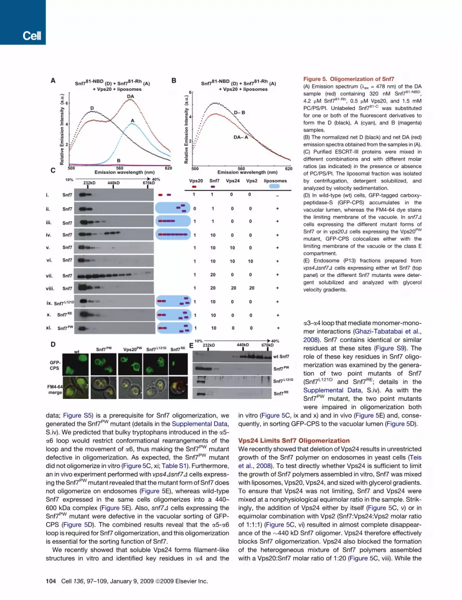
data; Figure S5) is a prerequisite for Snf7 oligomerization, we
generated the Snf7PW mutant (details in the Supplemental Data,
S.iv). We predicted that bulky tryptophans introduced in the a5-
a6 loop would restrict conformational rearrangements of the
loop and the movement of a6, thus making the Snf7PW mutant
defective in oligomerization. As expected, the Snf7PW mutant
did not oligomerize in vitro (Figure 5C, xi; Table S1). Furthermore,
an in vivo experiment performed with vps4Dsnf7D cells express-
ing the Snf7PW mutant revealed that the mutant form of Snf7 does
not oligomerize on endosomes (Figure 5E), whereas wild-type
Snf7 expressed in the same cells oligomerizes into a 440–
600 kDa complex (Figure 5E). Also, snf7D cells expressing the
Snf7PW mutant were defective in the vacuolar sorting of GFP-
CPS (Figure 5D). The combined results reveal that the a5-a6
loop is required for Snf7 oligomerization, and this oligomerization
is essential for the sorting function of Snf7.
We recently showed that soluble Vps24 forms filament-like
structures in vitro and identified key residues in a4 and the
BA
C
D E
Figure 5. Oligomerization of Snf7
(A) Emission spectrum (lex = 478 nm) of the DA
sample (red) containing 320 nM Snf781-NBD,
4.2 mM Snf781-Rh, 0.5 mM Vps20, and 1.5 mM
PC/PS/PI. Unlabeled Snf781-C was substituted
for one or both of the fluorescent derivatives to
form the D (black), A (cyan), and B (magenta)
samples.
(B) The normalized net D (black) and net DA (red)
emission spectra obtained from the samples in (A).
(C) Purified ESCRT-III proteins were mixed in
different combinations and with different molar
ratios (as indicated) in the presence or absence
of PC/PS/PI. The liposomal fraction was isolated
by centrifugation, detergent solubilized, and
analyzed by velocity sedimentation.
(D) In wild-type (wt) cells, GFP-tagged carboxy-
peptidase-S (GFP-CPS) accumulates in the
vacuolar lumen, whereas the FM4-64 dye stains
the limiting membrane of the vacuole. In snf7D
cells expressing the different mutant forms of
Snf7 or in vps20D cells expressing the Vps20PW
mutant, GFP-CPS colocalizes either with the
limiting membrane of the vacuole or the class E
compartment.
(E) Endosome (P13) fractions prepared from
vps4Dsnf7D cells expressing either wt Snf7 (top
panel) or the different Snf7 mutants were deter-
gent solubilized and analyzed with glycerol
velocity gradients.
a3-a4 loop that mediate monomer-mono-
mer interactions (Ghazi-Tabatabai et al.,
2008). Snf7 contains identical or similar
residues at these sites (Figure S9). The
role of these key residues in Snf7 oligo-
merization was examined by the genera-
tion of two point mutants of Snf7
(Snf7L121D and Snf7RE; details in the
Supplemental Data, S.iv). As with the
Snf7PW mutant, the two point mutants
were impaired in oligomerization both
in vitro (Figure 5C, ix and x) and in vivo (Figure 5E) and, conse-
quently, in sorting GFP-CPS to the vacuolar lumen (Figure 5D).
Vps24 Limits Snf7 OligomerizationWe recently showed that deletion of Vps24 results in unrestricted
growth of the Snf7 polymer on endosomes in yeast cells (Teis
et al., 2008). To test directly whether Vps24 is sufficient to limit
the growth of Snf7 polymers assembled in vitro, Snf7 was mixed
with liposomes, Vps20, Vps24, and sized with glycerol gradients.
To ensure that Vps24 was not limiting, Snf7 and Vps24 were
mixed at a nonphysiological equimolar ratio in the sample. Strik-
ingly, the addition of Vps24 either by itself (Figure 5C, v) or in
equimolar combination with Vps2 (Snf7:Vps24:Vps2 molar ratio
of 1:1:1) (Figure 5C, vi) resulted in almost complete disappear-
ance of the �440 kD Snf7 oligomer. Vps24 therefore effectively
blocks Snf7 oligomerization. Vps24 also blocked the formation
of the heterogeneous mixture of Snf7 polymers assembled
with a Vps20:Snf7 molar ratio of 1:20 (Figure 5C, viii). While the
104 Cell 136, 97–109, January 9, 2009 ª2009 Elsevier Inc.

exact mechanism of Vps24 prevention of Snf7 oligomerization is
not yet clear, it seems likely that Vps24 ‘‘caps’’ or prevents poly-
merization by binding to Snf7. The prevention of Snf7 oligomer-
ization appears to be solely a function of Vps24 because in the
absence of Vps24, equimolar (to Snf7) Vps2 did not block Snf7
oligomerization (data not shown).
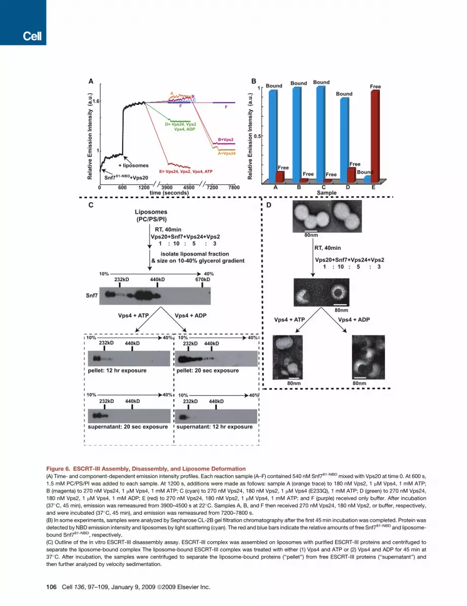
Vps24, Vps2, and Vps4 Mediate Disassemblyof Membrane-Bound Snf7 OligomersTo monitor disassembly of the membrane-bound Snf7 oligomer
spectroscopically, we mixed six parallel reactions (A–F) contain-
ing Snf781-NBD mixed with Vps20 and monitored NBD emission.
After 600 s, PC/PS/PI was added to each sample to initiate
Vps20-induced Snf7 oligomerization. The resulting intensity
changes were equivalent for samples A–F and are shown by
a single black trace in Figure 6A. At 1200 s, the samples received
different combinations of purified proteins. After a 37�C, 45 min
incubation, little to no change in NBD intensity was observed in
samples lacking Vps24 (A), Vps2 (B), or a functional Vps4 (C).
In contrast, the NBD intensity increases were nearly reversed
in a sample containing Vps24, Vps2, Vps4, and ATP (E), while
a much smaller decrease in NBD emission was observed when
ADP replaced ATP (D). Similar results were obtained when
samples were prepared with Snf721-NBD, Snf735-NBD, or
Snf7215-NBD instead of Snf781-NBD (data not shown).
To determine whether the large drop in NBD emission intensity
reflects Vps4-mediated release of NBD-labeled Snf7 from the
membrane into the aqueous milieu, we analyzed samples by
gel filtration after the 37�C incubation. Nearly all of the NBD emis-
sion in the complete sample (E) coeluted with liposome-free Snf7
(red bar in Figure 6B), and very little coeluted in the void volume
with the liposomes (detected by light scattering; blue bar in
Figure 6B), thereby showing efficient Snf7 release from the lipo-
somes. In contrast, when the other samples (A–D) were analyzed
by gel filtration, nearly all of the NBD emission coeluted with
liposomes in the void volume (Figure 6B; data not shown).
Since membrane-bound Snf7 was not released into the
aqueous milieu in samples lacking only Vps24 or Vps2, the
missing proteins were then added to A and B, respectively. After
45 min at 37�C, the lowered NBD intensity shows that Vps4-
mediated Snf7 disassembly now occurred in each sample
(Figure 6A; the reduced intensity decrease compared to sample
E presumably results from ATP hydrolysis during the second
37�C incubation). Thus, efficient release/disassembly of
membrane-bound Snf7 requires Vps24, Vps2, the ATPase
activity of Vps4, and ATP.
Disassembly of liposome-bound Snf7 oligomers was also
examined by glycerol gradient sizing assays. ESCRT-III
complexes were assembled on liposomes in vitro by the combi-
nation of Vps20, Snf7, Vps24, and Vps2 at their previously deter-
mined physiological molar ratio of 1:10:5:3, and their �400 kD
mass (as judged by immunoblotting with Snf7 [Figure 6C] or
Vps24 [data not shown] antibodies) was very similar to the
masses of ESCRT-III complexes that assemble on endosomes
in vivo (Teis et al., 2008) (no upstream ESCRT components
were required for assembly presumably because of the high
ESCRT-III protein concentrations used in the assay). The lipo-
some fraction was isolated by centrifugation and split in two.
One half was mixed with Vps4 and ATP, and the other half
received equivalent amounts of Vps4 and ADP. After another
45 min at 37�C incubation, the liposomes were separated from
soluble proteins by centrifugation, and both the pellet and super-
natant fractions were further analyzed by glycerol gradient
sizing. As shown in Figure 6C, the liposome-bound �400 kD
ESCRT-III complex was disassembled by exposure to Vps4
and ATP. Most Snf7 was detected in the supernatant-derived
fractions by immunoblotting with Snf7 antibodies (20 s expo-
sure), whereas Snf7 could only be detected in the pellet-derived
fractions after long exposure (12 hr). Thus, Vps4- and ATP-medi-
ated release of Snf7 from the liposomes into the supernatant was
very efficient. Interestingly, Vps4 appears to disassemble the
membrane-bound Snf7 oligomers into monomers because
Snf7 present in the supernatant was found in the low molecular
weight fractions of the glycerol gradient (Figure 6C).
The ATP dependence of the disassembly reaction was shown
by the inability of the ATPase inactivated Vps4 E233Q to disas-
semble the membrane-bound Snf7 oligomer even in the pres-
ence of ATP (data not shown). Also, Vps4-mediated disassembly
of membrane-bound Snf7 oligomers was much less efficient with
ADP than with ATP since the majority of Snf7 was found in the
pellet-derived fractions (20 s exposure); only a small amount of
Snf7 was found in the supernatant after a 12 hr exposure (Fig-
ure 6C). The size of the membrane-bound ESCRT-III complex
was slightly smaller after than before treatment with Vps4 and
ADP. We attribute this to one round of Vps4-mediated disas-
sembly that utilizes the ATP that may remain bound to Vps4
during purification. Such an effect would also explain the small
drop in NBD emission observed in sample D in our spectro-
scopic Snf7 disassembly assay (Figure 6A). Consistent with the
spectral data, no Vps4- and ATP-mediated disassembly of the
membrane-bound Snf7 oligomer was observed in the absence
of either Vps24 or Vps2 (data not shown).
ESCRT-III Assembly Induces Membrane DeformationTo determine whether the assembly and disassembly of the Snf7
oligomer induces membrane deformation, we analyzed liposome
morphology at different stages of the ESCRT-III disassembly
assay using negative stain electron microscopy (EM). Untreated
PC/PS/PI liposomes prepared by extrusion have a uniform,
spherical appearance with a diameter of 80–100 nm (Figure 6D).
Upon exposure to the ESCRT-III complex (Vps20, Snf7, Vps24,
Vps2), a distinct change in morphology was observed for
�90% of the liposomes. ESCRT-III-bound liposomes had an
inward invaginated appearance, and the size of the invagination
was �40 nm in diameter. This deformation was observed only
when liposomes were treated with all four ESCRT-III proteins:
incubation with individual ESCRT-III proteins did not induce any
detectable liposome deformation (data not shown). Also, no lipo-
some morphology changes were observed when mutants of Snf7
that do not oligomerize (Snf7PW, Snf7L121D, and Snf7RE) were
mixed with Vps20, Vps24, and Vps2 (data not shown). These
data reveal that Snf7 oligomerization is critical for membrane
invagination.
Importantly, when ESCRT-III-bound liposomes were exposed
to Vps4 and ATP, the near normal spherical morphology
of the liposomes was restored (Figure 6D). In contrast,
Cell 136, 97–109, January 9, 2009 ª2009 Elsevier Inc. 105
C
A
D
B
Figure 6. ESCRT-III Assembly, Disassembly, and Liposome Deformation
(A) Time- and component-dependent emission intensity profiles. Each reaction sample (A–F) contained 540 nM Snf781-NBD mixed with Vps20 at time 0. At 600 s,
1.5 mM PC/PS/PI was added to each sample. At 1200 s, additions were made as follows: sample A (orange trace) to 180 nM Vps2, 1 mM Vps4, 1 mM ATP;
B (magenta) to 270 nM Vps24, 1 mM Vps4, 1 mM ATP; C (cyan) to 270 nM Vps24, 180 nM Vps2, 1 mM Vps4 (E233Q), 1 mM ATP; D (green) to 270 nM Vps24,
180 nM Vps2, 1 mM Vps4, 1 mM ADP; E (red) to 270 nM Vps24, 180 nM Vps2, 1 mM Vps4, 1 mM ATP; and F (purple) received only buffer. After incubation
(37�C, 45 min), emission was remeasured from 3900–4500 s at 22�C. Samples A, B, and F then received 270 nM Vps24, 180 nM Vps2, or buffer, respectively,
and were incubated (37�C, 45 min), and emission was remeasured from 7200–7800 s.
(B) In some experiments, samples were analyzed by Sepharose CL-2B gel filtration chromatography after the first 45 min incubation was completed. Protein was
detected by NBD emission intensity and liposomes by light scattering (cyan). The red and blue bars indicate the relative amounts of free Snf781-NBD and liposome-
bound Snf781-NBD, respectively.
(C) Outline of the in vitro ESCRT-III disassembly assay. ESCRT-III complex was assembled on liposomes with purified ESCRT-III proteins and centrifuged to
separate the liposome-bound complex The liposome-bound ESCRT-III complex was treated with either (1) Vps4 and ATP or (2) Vps4 and ADP for 45 min at
37�C. After incubation, the samples were centrifuged to separate the liposome-bound proteins (‘‘pellet’’) from free ESCRT-III proteins (‘‘supernatant’’) and
then further analyzed by velocity sedimentation.
106 Cell 136, 97–109, January 9, 2009 ª2009 Elsevier Inc.

Figure 7. Speculative Model for the ESCRT-III Reaction CycleESCRT-III assembly is initiated when Vps20 binds to Vps25 on the membrane surface. In the second stage, Snf7 binds to the membrane-bound Vps25-Vps20
complex, and this triggers the formation of a membrane-bound Snf7 oligomer. The Snf7 oligomer traps cargo into a localized sorting domain and induces membrane
deformation. Vps24 caps the Snf7 oligomer, and interactions between Vps2 and Vps4 initiate membrane disassembly of the ESCRT-III complex (see Discussion).
ESCRT-III-bound liposomes retained their invaginated appear-
ance when treated with Vps4 and ADP. The reversibility of
ESCRT-III-induced liposome deformation upon treatment with
Vps4 and ATP, coupled with the other data presented above,
demonstrates directly that liposome morphology is dictated by
the assembly and disassembly of the ESCRT-III complex.
DISCUSSION
The ordered [ESCRT-II (Vps25)-Vps20-Snf7-Vps24-Vps2] as-
sembly and disassembly of the ESCRT-III complex has been re-
constituted in vitro with purified proteins, fluorescent-labeled
derivatives of those proteins, liposomes comprised of PC/PS/
PI, and multiple independent fluorescence and other techniques.
Specifically, our studies reveal that (1) Vps25 binds to Vps20, but
not to Snf7; (2) both Vps20 and Snf7 bind to a membrane surface;
(3) Vps25 binding to membrane-bound Vps20 alters its confor-
mation; (4) Vps20 binds the N-terminal end of membrane-bound
Snf7, and this association both stabilizes Snf7 binding to the
membrane surface and alters its conformation; (5) Vps20 nucle-
ates Snf7 oligomerization on the membrane surface; (6) Vps20
stabilizes and/or changes the conformation of the Snf7 oligomers;
(7) Vps24 caps membrane-bound Snf7 oligomers; (8) Snf7 oligo-
merization is blocked by mutations just C-terminal of the a5 helix
and by mutation of critical residues in and around helix a4; (9)
membrane-bound ESCRT-III complex is disassembled by Vps4
in a process that requires Vps24, Vps2, and ATP; and (10) the lipo-
some-bound ESCRT-III complex induces membrane deforma-
tion, and Snf7 oligomerization is critical for this deformation.
When considered in toto, these results suggest that the ESCRT-
III functional cycle consists of an ordered multistep assembly
involving three distinct stages (filament/lattice nucleation, poly-
merization, and capping) (Teis et al., 2008), followed by an ATP-
dependent disassembly reaction. These complex, coupled, and
regulated structural changes ultimately mediate membrane
deformation, cargo sorting, and MVB vesicle formation (Figure 7).
ESCRT-III Assembly Involves an Obligatory Sequenceof Protein-Protein Interactions and ConformationalRearrangementsThe fluorescence-detected conformational changes in the a5-a6
loop strongly suggest that they are required to nucleate the
(D) Negative stain EM analyses of PC/PS/PI liposomes, ESCRT-III bound liposomes, ESCRT-III-bound liposomes treated with 1 mM Vps4 + 1 mM ATP, and
ESCRT-III-bound liposomes treated with 1 mM Vps4 + 1 mM ADP.
Cell 136, 97–109, January 9, 2009 ª2009 Elsevier Inc. 107

assembly of the ESCRT-III complex. Such a conformational
change was proposed earlier to mediate the association of
ESCRT-III subunits by an intramolecular autoinhibition of
ESCRT-III polymer formation (Shim et al., 2007). The autoinhibi-
tion model proposes that ESCRT-III subunits exist as metastable
‘‘closed’’ monomers in the cytosol because the C-terminal auto-
inhibitory region of the molecule (including a5 and a6) binds to
a2 of the core and thus prevents hetero- and homodimerization.
Consistent with this, our results indicate that Vps25 has no affinity
for Vps20 in solution (Figures 2A–2C; Figure S4B), there is no
energy transfer between NBD- and Rh-labeled Snf7 molecules
in solution (Table S1), and there is no oligomerization of Snf7 in
solution (Figure 5C, i). Implicit in the autoinhibition model is the
idea that membrane binding and/or interactions with ESCRT-II
generate the ‘‘open’’ state of the ESCRT-III subunit that is active
for assembly into the ESCRT-III lattice/filament. The spectral data
presented here show that NBD probes incorporated within the
a5-a6 loop of both Vps20 and Snf7 exhibit a dramatic increase
in NBD emission intensity upon membrane binding (Figures 1E,
3B, and 3C), and—more importantly—that NBD emission from
these sites can be efficiently quenched by membrane-restricted
quenchers (Figures S3C, S5B, and S3F). These data therefore
directly show that membrane binding of an ESCRT-III subunit is
accompanied by the movement of its C-terminal a5-a6 loop adja-
cent to the membrane surface. Rearrangement of the C-terminal
end of both Vps20 and Snf7 upon membrane binding may expose
a2 in each protein for homo- and heterodimerization interactions
and thereby activate the ESCRT-III subunit (‘‘closed’’ to ‘‘open’’
state transition) for continued assembly into the ESCRT-III lattice.
Consequently, mutations that block conformational flexibility
around the a5-a6 loop impair Snf7 oligomerization and function
(Figure 5C, xi; Figures 5D and 5E; Table S1).
Spectroscopic characterization of Vps25 interactions with
NBD-labeled Vps20 mutants revealed that the binding of
Vps25 to Vps20 changed its conformation at both the N and
Cterminus.Thus,althoughtheN-terminaldomainofVps20under-
goes a larger spectral change and perhaps a larger conforma-
tional change upon association with Vps25 (Figures 2D–2F;
Figures S4A and S4C), Vps25 binding clearly elicits a long-range
conformational change in Vps20 that extends throughout its
entire length (see the Supplemental Data). Similarly, spectro-
scopic examination of Snf7 mutants containing NBD probes at
multiple locations and domains within the molecule allowed us
to determine possible sites on Snf7 that bind to Vps20. Our
data indicate that the short Snf7 a1-a2 loop and the a2 helix
are sensitive to Vps20 binding both in solution and on the
membrane (Figures 4A–4D; Figures S7B–S7D). In contrast,
Vps20 binding had no detectable effect on the probe environ-
ment and presumably conformation of the C-terminal a5-a6
loop or of a1 for either free or membrane-bound Snf7 (Figures
4B and 4C; Figures S7A, S7C, and S7D). These results strongly
indicate that the heterodimerization interactions during the
assembly of the ESCRT-III lattice are mediated by interactions
involving the a2 helix and the short a1-a2 loop.
As was true with Vps20, Snf7 exhibited low affinity for Vps25 in
solution (Figure 4F). However, in contrast to Vps20, we also did
not detect binding of Vps25 to any of the membrane-bound
Snf7 monocysteine mutants (Figure 4F; data not shown). Specif-
108 Cell 136, 97–109, January 9, 2009 ª2009 Elsevier Inc.
ically worth noting is the probe at residue 58, which is sensitive to
Snf7 binding both to membranes (Figure 3C) and to Vps20
(Figure 4A–4D) but remains insensitive to Vps25 both in solution
and when liposome bound (Figure 4F). When combined, these
data indicate that an obligatory sequence of protein-protein inter-
actions mediates assembly of the ESCRT-III complex (Figure 7).
How then does the system regulate interactions between
the structurally similar ESCRT-III proteins that associate via a
common surface? The most likely explanation is that individual
interactions elicit unique conformational changes and that the
next component in the assembly pathway binds only to a specific
‘‘active/open’’ conformation. In this way, one can ensure that
ESCRT-III assembly occurs in the proper order because a
component can only associate with the precursor complex that
has the necessary conformation. The spectral changes detected
along the entire length of the Snf7 molecule upon membrane
binding and interactions with Vps20 correlate with alterations
in protein conformation that shift the functional/structural state
of the protein from one state to another (‘‘closed’’ to ‘‘open’’ state
transition) and thereby activate it for assembly into the ESCRT-III
lattice (Figure 7).
Specific Function for Each Proteinin the ESCRT-III Reaction CycleThe assembly of ESCRT-III on a membrane surface has striking
mechanistic analogies to the assembly of filaments like actin or
tubulin, where ESCRT-III appears to form a polarized filament
that assembles in a sequential fashion with distinct nucleation
and capping sites (Figure 7). Our results indicate that Vps20 plays
a pivotal role in the assembly of a membrane-bound Snf7 polymer
by stabilizing the binding of Snf7 derivatives to the membrane
surface (Figure 4E) and by nucleating Snf7 oligomerization either
bystabilizing the binding of Snf7 to itself to formmultimericassem-
blies and/or by altering the conformation of membrane-bound
Snf7 to allow the assembly of an oligomer (Figures 5A–5E;
Figure S8; Table S1). Interestingly, the ability of Vps20 to nucleate
Snf7 oligomerization invitro (Figure 5A–5C) and invivo (Figure S10)
appears to be independent of its N-terminal myristoylation.
Vps20-induced Snf7 oligomerization on the membrane appears
to be critical for membrane deformation, as judged by the fact that
Snf7 mutants that do not oligomerize (in vitro and in vivo) are
defective in inducing changes in liposome morphology in vitro
and in MVB sorting in vivo (Figure 5C, ix–xi; Figures 5D and 5E;
data not shown). How ESCRT-III induced liposome deformation
in vitro relates to MVB formation in vivo remains an open
question. However, the strong dependence of the observed lipo-
some deformation on Snf7 oligomerization (Figure 5C; data not
shown) and the Vps4-mediated reversibility of the deformation
(Figure 6D) suggest a direct link between Snf7 oligomerization
and membrane deformation. In addition to inducing membrane
deformation, Snf7 oligomers could form rings, similar to the ring-
like spiral filaments generated at the plasma membrane in
mammalian cells overexpressing hSnf7 (Hanson et al., 2008). A
Snf7 membrane-associated ring/spiral could encircle cargo mole-
cules and keep them committed to MVB sorting by confining the
integral membrane cargo proteins to a sorting domain from which
they cannot escape (Snf7 filaments tightly associate with the
membrane surface) (Figure 7). Recent work has shown that Snf7

oligomers (filaments) associated with the endosomal membrane
may playa direct role incargosequestrationpossiblyby restricting
lateral movement of cargo molecules (Teis et al., 2008).
Our data suggest that Vps24 and Vps2 serve as adaptors for
Vps4-mediated disassembly of the Snf7 oligomer (Figure 6A).
The data presented here (Figures 5C and 6) and results of
in vivo studies (Teis et al., 2008; Obita et al., 2007; Stuchell-
Brereton et al., 2007) are consistent with a model in which
Vps24 terminates oligomerization, possibly by capping Snf7
oligomers and/or by recruiting Vps2 to the Snf7 oligomer in order
to initiate Vps4-mediated disassembly (Figure 7). The Vps24-
Vps2 ‘‘capping’’ complex may itself be in the form of a short
mixed oligomer (Lata et al., 2008), thereby providing multiple
binding sites (Vps2 MIM domain) for Vps4 recruitment via the
Vps4 MIT domain (Obita et al., 2007). Although not addressed
by the present study, we speculate that the Vps4 AAA-ATPase
may drive the sequential release/disassembly of Snf7 molecules
from one end of the Snf7 filament ring/spiral. This would result in
closure of the Snf7 ring/spiral and concentration of cargo
encircled by the ring (Figure 7). The Vps4 dodecamer could
stabilize the ever-shrinking Snf7 ring via a combination of strong
interactions with Vps2 and weak interactions with Snf7 mediated
by the N-terminal MIT domain on each Vps4 subunit (Obita et al.,
2007). The closure of the Snf7 ring/spiral may also drive invagina-
tion and fission of the sorting domain/vesicle, thereby directly
coupling ESCRT-III disassembly to cargo sorting and vesicle
formation (purse string model). Clearly, this speculative hypoth-
esis will require considerable new experimentation. However, it
does offer a framework that could explain the mechanism by
which ESCRT-III can sort cargo (even after the Ub sorting signal
has been cleaved), deform the membrane, and ultimately release
the invaginated MVB vesicle into the lumen of the endosome.
EXPERIMENTAL PROCEDURES
Purification and Labeling of ESCRT-III Proteins
Recombinant Vps20 and Snf7 were expressed in C41(DE3) cells and purified
with a combination of Ni2+-affinity and ion exchange chromatography. Purified
proteins were labeled with NBD as described earlier (Shepard et al., 1998).
Details of the protein purification and NBD-modification procedures are
presented in the Supplemental Data.
Liposome Preparation
Liposomes containing 100 mol% 1-palmitoyl-2-oleoyl-sn-glycero-3-phospho-
choline (POPC; Avanti) or 80 mol% POPC, 15 mol% egg-phosphatidyl serine
(PS), and 5 mol% egg-phosphatidyl inositol (PI) were prepared by extrusion
through a Liposofast extruder with 100 nm pore polycarbonate membranes
as before (Shepard et al., 1998).
Fluorescence Spectroscopy
All intensity and anisotropy measurements were carried out in 50 mM HEPES
(pH 7.5) and 100 mM NaCl at 22�C with the same steady-state instrumentation
and procedures described previously (Shepard et al., 1998). Emission spectra
for NBD-labeled proteins were recorded in 4 mm 3 4 mm quartz cuvettes
coated with POPC to minimize adsorption. See the Supplemental Data for
further details on fluorescence spectroscopy, fluorescence microscopy, and
FRET experiments.
Glycerol Velocity Gradients
Endosome-enriched P13 fractions were prepared and solubilized with 0.5%
Tween-20 and analyzed with a glycerol velocity gradient as before (Teis
et al., 2008). Analysis details are given in the Supplemental Data.
SUPPLEMENTAL DATA
Supplemental Data include Supplemental Results, Supplemental Discussion,
Supplemental Experimental Procedures, 11 figures, and one table and can
be found with this article online at http://www.cell.com/supplemental/
S0092-8674(08)01442-6.
ACKNOWLEDGMENTS
We are grateful to Y. Miao, Y. Shao, and S. Weys for technical assistance;
B. Judson for EM expertise; and E.V. MacGurn for assistance with graphics.
S.S. and D.T. were supported by fellowships from American Heart Association
(AHA 0826060D) and HFSP (LT00634/2006-L), respectively. This work
was supported by National Institutes of Health grant GM26494 and the
Robert A. Welch Foundation (Chair Grant BE-0017) (A.E.J.) and funds from
the Weill Institute for Cell and Molecular Biology (S.D.E.).
Received: June 24, 2008
Revised: September 22, 2008
Accepted: November 4, 2008
Published: January 8, 2009
REFERENCES
Ghazi-Tabatabai, S., Saksena, S., Short, J.M., Pobbati, A.V., Veprintsev, D.B.,
Crowther, R.A., Emr, S.D., Egelman, E.H., and Williams, R.L. (2008). Structure
and disassembly of filaments formed by the ESCRT-III subunit Vps24.
Structure 16, 1345–1356.
Hanson, P.I., Roth, R., Lin, Y., and Heuser, J.E. (2008). Plasma membrane
deformation by circular arrays of ESCRT-III protein filaments. J. Cell Biol.
180, 389–402.
Hurley, J.H. (2008). ESCRT complexes and the biogenesis of multivesicular
bodies. Curr. Opin. Cell Biol. 20, 4–11.
Johnson, A.E. (2005). Fluorescence approaches for determining protein
conformations, interactions and mechanisms at membranes. Traffic 6,
1078–1092.
Lata, S., Schoehn, G., Jain, A., Pires, R., Piehler, J., Gottlinger, H.G., and
Weissenhorn, W. (2008). Helical structures of ESCRT-III are disassembled
by VPS4. Science 321, 1354–1357.
Muzio1, T., Pineda-Molina, E., Ravelli, R.B., Zamborlini, A., Usami, Y., Gottlin-
ger, H., and Weissenhorn, W. (2006). Structural basis for budding by the
ESCRT-III factor CHMP3. Dev. Cell 10, 821–830.
Obita, T., Saksena, S., Ghazi-Tabatabai, S., Gill, D.J., Perisic, O., Emr, S.D.,
and Williams, R.L. (2007). Structural basis for selective recognition of
ESCRT-III by the AAA ATPase Vps4. Nature 449, 735–739.
Saksena, S., Sun, J., Chu, T., and Emr, S.D. (2007). ESCRTing proteins in the
endocytic pathway. Trends Biochem. Sci. 32, 561–573.
Shepard, L.A., Heuck, A.P., Hamman, B.D., Rossjohn, J., Parker, M.W., Ryan,
K.R., Johnson, A.E., and Tweten, R.K. (1998). Identification of a membrane-
spanning domain of the thiol-activated pore-forming toxin Clostridium perfrin-
gens perfringolysin O: an a-helical to beta-sheet transition identified by
fluorescence spectroscopy. Biochemistry 37, 14563–14574.
Shim, S., Kimpler, L.A., and Hanson, P.I. (2007). Structure/function analysis
of four core ESCRT-III proteins reveals common regulatory role for extreme
C-terminal domain. Traffic 8, 1068–1079.
Stuchell-Brereton, M.D., Skalicky, J.J., Kieffer, C., Karren, M.A., Ghaffarian, S.,
and Sundquist, W.I. (2007). ESCRT-III recognition by VPS4 ATPases. Nature
449, 740–744.
Teis, D., Saksena, S., and Emr, S.D. (2008). Ordered assembly of the ESCRT-III
complex on endosomes is required to sequester cargo during MVB formation.
Dev. Cell 15, 578–589.
Williams, R.L., and Urbe, S. (2007). The emerging shape of the ESCRT
machinery. Nat. Rev. Mol. Cell Biol. 8, 355–368.
Cell 136, 97–109, January 9, 2009 ª2009 Elsevier Inc. 109
Related Documents