Journal of Steroid Biochemistry & Molecular Biology 85 (2003) 209–219 Functional properties of the N-terminal region of progesterone receptors and their mechanistic relationship to structure Glenn S. Takimoto ∗ , Lin Tung, Hany Abdel-Hafiz, Michael G. Abel, Carol A. Sartorius, Jennifer K. Richer, Britta M. Jacobsen, David L. Bain, Kathryn B. Horwitz Department of Medicine and The Molecular Biology Program, University of Colorado Health Sciences Center, Denver, CO 80262, USA Abstract Progesterone receptors (PR) are present in two isoforms, PR-A and PR-B. The B-upstream segment (BUS) of PR-B is a 164 amino acid N-terminal extension that is missing in PR-A and is responsible for the functional differences reported between the two isoforms. BUS contains an activation function (AF3) which is defined by a core domain between residues 54–154 whose activity is dependent upon a single Trp residue and two LXXLL motifs. We have also identified sites both within and outside of BUS that repress the strong synergism between AF3 and AF1 in the N-terminal region and AF2 in the hormone binding domain. One of these repressor sites is a consensus binding motif for the small ubiquitin-like modifier protein, SUMO-1 ( 387 IKEE). The DNA binding domain (DBD) structure is also important for function. When BUS is linked to the glucocorticoid receptor DBD, AF3 activity is substantially attenuated, suggesting that binding to a DNA response element results in allosteric communication between the DBD and N-terminal functional regions. Lastly, biochemical and biophysical analyses of highly purified PR-B and PR-A N-terminal regions reveal that they are unstructured unless the DBD is present. Thus, the DBD stabilizes N-terminal structure. We propose a model in which the DBD through DNA binding, and BUS through protein–protein interactions, stabilize active receptor conformers within an ensemble distribution of active and inactive conformational states. This would explain why PR-B are stronger transactivators than PR-A. © 2003 Elsevier Science Ltd. All rights reserved. Keywords: Progesterone receptor; Transcription; Synergism; Cofactors; Structure; Activation domain; Repression; SUMO 1. Introduction Steroid receptors regulate a complex network of path- ways controlling development, differentiation and home- ostasis. These nuclear proteins were defined as having a modular structure with distinct domains that can function independently. Specific functions have been ascribed to each domain, including hormone binding, DNA binding and transcriptional activation. With the exception of estrogen receptor (ER), the high degree of sequence similar- ity between DNA binding (DBD) and hormone binding Abbreviations: BUS, B-upstream segment; AF, activation function; PR, progesterone receptors; ER, estrogen receptors; GR, glucocorticoid receptors; AR, androgen receptors; DBD, DNA binding domain; HBD, hormone binding domain; NT, N-terminal; BDN, BUS-DBD-nuclear lo- calization signal; PRE, progesterone response element; GRE, glucocor- ticoid response element; ERE, estrogen response element; IF, inhibitory function; SUMO, small ubiquitin-like modifier Presented at the 11th International Congress on Hormonal Steroids and Hormones and Cancer, ICHS & ICHC, Fukuoka, Japan, 21–25 Oc- tober 2002. ∗ Corresponding author. Tel.: +1-303-315-8850; fax: +1-303-315-4525. E-mail address: [email protected] (G.S. Takimoto). (HBD) domains within the steroid receptor family belies the diverse array of gene promoters and metabolic path- ways that they regulate [1]. We and others have speculated that this diversity is subserved by the N-terminal regions of the receptor proteins, which are highly dissimilar in both sequence (<15% sequence identity) and size (184 and 566 amino acids for ER and the progesterone recep- tor B-isoform (PR-B), respectively). Several recent studies have provided suggestive functional and structural evidence that N-terminal regions do indeed play a critical role in determining which gene promoters and, therefore, which functional pathways are regulated by these receptors [2–7]. PR provide an ideal model system to probe the mechanis- tic details by which receptor N-terminal regions modulate function. First, they exist in two isoforms, PR-A and PR-B, which have large N-terminal regions of 402 and 566 amino acids, respectively, that are identical in sequence except that PR-A are missing the far 164 residue N-terminal B-upstream segment (BUS) region. This conveniently provides a dis- crete physical entity upon which to focus functional, bio- chemical and biophysical studies. Transcription activation as well as repression functions have been identified within the N-terminal regions, some of which are unique to the BUS 0960-0760/$ – see front matter © 2003 Elsevier Science Ltd. All rights reserved. doi:10.1016/S0960-0760(03)00197-3

Welcome message from author
This document is posted to help you gain knowledge. Please leave a comment to let me know what you think about it! Share it to your friends and learn new things together.
Transcript
Journal of Steroid Biochemistry & Molecular Biology 85 (2003) 209–219
Functional properties of the N-terminal region of progesterone receptorsand their mechanistic relationship to structure�
Glenn S. Takimoto∗, Lin Tung, Hany Abdel-Hafiz, Michael G. Abel, Carol A. Sartorius,Jennifer K. Richer, Britta M. Jacobsen, David L. Bain, Kathryn B. Horwitz
Department of Medicine and The Molecular Biology Program, University of Colorado Health Sciences Center, Denver, CO 80262, USA
Abstract
Progesterone receptors (PR) are present in two isoforms, PR-A and PR-B. The B-upstream segment (BUS) of PR-B is a 164 amino acidN-terminal extension that is missing in PR-A and is responsible for the functional differences reported between the two isoforms. BUScontains an activation function (AF3) which is defined by a core domain between residues 54–154 whose activity is dependent upon asingle Trp residue and two LXXLL motifs. We have also identified sites both within and outside of BUS that repress the strong synergismbetween AF3 and AF1 in the N-terminal region and AF2 in the hormone binding domain. One of these repressor sites is a consensus bindingmotif for the small ubiquitin-like modifier protein, SUMO-1 (387IKEE). The DNA binding domain (DBD) structure is also important forfunction. When BUS is linked to the glucocorticoid receptor DBD, AF3 activity is substantially attenuated, suggesting that binding to aDNA response element results in allosteric communication between the DBD and N-terminal functional regions. Lastly, biochemical andbiophysical analyses of highly purified PR-B and PR-A N-terminal regions reveal that they are unstructured unless the DBD is present. Thus,the DBD stabilizes N-terminal structure. We propose a model in which the DBD through DNA binding, and BUS through protein–proteininteractions, stabilize active receptor conformers within an ensemble distribution of active and inactive conformational states. This wouldexplain why PR-B are stronger transactivators than PR-A.© 2003 Elsevier Science Ltd. All rights reserved.
Keywords:Progesterone receptor; Transcription; Synergism; Cofactors; Structure; Activation domain; Repression; SUMO
1. Introduction
Steroid receptors regulate a complex network of path-ways controlling development, differentiation and home-ostasis. These nuclear proteins were defined as having amodular structure with distinct domains that can functionindependently. Specific functions have been ascribed toeach domain, including hormone binding, DNA binding andtranscriptional activation. With the exception of estrogenreceptor� (ER�), the high degree of sequence similar-ity between DNA binding (DBD) and hormone binding
Abbreviations: BUS, B-upstream segment; AF, activation function;PR, progesterone receptors; ER, estrogen receptors; GR, glucocorticoidreceptors; AR, androgen receptors; DBD, DNA binding domain; HBD,hormone binding domain; NT, N-terminal; BDN, BUS-DBD-nuclear lo-calization signal; PRE, progesterone response element; GRE, glucocor-ticoid response element; ERE, estrogen response element; IF, inhibitoryfunction; SUMO, small ubiquitin-like modifier
� Presented at the 11th International Congress on Hormonal Steroidsand Hormones and Cancer, ICHS & ICHC, Fukuoka, Japan, 21–25 Oc-tober 2002.
∗ Corresponding author. Tel.:+1-303-315-8850; fax:+1-303-315-4525.E-mail address:[email protected] (G.S. Takimoto).
(HBD) domains within the steroid receptor family beliesthe diverse array of gene promoters and metabolic path-ways that they regulate[1]. We and others have speculatedthat this diversity is subserved by the N-terminal regionsof the receptor proteins, which are highly dissimilar inboth sequence (<15% sequence identity) and size (184and 566 amino acids for ER� and the progesterone recep-tor B-isoform (PR-B), respectively). Several recent studieshave provided suggestive functional and structural evidencethat N-terminal regions do indeed play a critical role indetermining which gene promoters and, therefore, whichfunctional pathways are regulated by these receptors[2–7].
PR provide an ideal model system to probe the mechanis-tic details by which receptor N-terminal regions modulatefunction. First, they exist in two isoforms, PR-A and PR-B,which have large N-terminal regions of 402 and 566 aminoacids, respectively, that are identical in sequence except thatPR-A are missing the far 164 residue N-terminal B-upstreamsegment (BUS) region. This conveniently provides a dis-crete physical entity upon which to focus functional, bio-chemical and biophysical studies. Transcription activation aswell as repression functions have been identified within theN-terminal regions, some of which are unique to the BUS
0960-0760/$ – see front matter © 2003 Elsevier Science Ltd. All rights reserved.doi:10.1016/S0960-0760(03)00197-3

210 G.S. Takimoto et al. / Journal of Steroid Biochemistry & Molecular Biology 85 (2003) 209–219
region present in PR-B[8–12]. Despite similarities betweenthe two PR isoforms, the transcriptional phenotype of PR-Aand PR-B on both synthetic and endogenous promoters isvery different. PR-B are generally strong transcriptional ac-tivators, while PR-A are weak activators and exhibit strongerrepressor properties[13,14]. Moreover, recent gene arraystudies in our laboratory show that PR-A and PR-B regulaterelatively distinct gene programs in vivo[15,16].
In the present study, we have precisely mapped activatorand repressor activities within the N-terminal regions of bothreceptors, which, we propose, are responsible for their di-verse functional responses. This includes delineation of thecore activation domain within activation function 3 (AF3)of BUS that is required for the high degree of AF3 syner-gism with AF1 and AF2 domains[3]. We also identified aconsensus binding motif for the small ubiquitin-like modi-fier protein, SUMO-1, that tightly controls AF synergism inboth isoforms[17,18]. Finally, we initiated biochemical andbiophysical studies using highly purified N-terminal regionsof both PR-A and PR-B to assess their structural properties.Our findings show that the N-terminal regions are largelyunstructured. However, these regions assume an orderedconformation when the DNA binding domain is present,and assume further structural changes when the proteinsare bound to a DNA response element. Additionally, thepresence of BUS reduces heterogeneity in the ensemble dis-tribution of N-terminal conformations, suggesting that thereis an enrichment of active conformers when BUS is present.We speculate that these data provide a mechanistic basis toexplain functional differences between PR-A and PR-B.
2. Materials and methods
2.1. Recombinant plasmids and site-directed mutagenesis
Human PR-B (pSG5-hPR1), PR-A (pSG5-hPR2) andER� (pSG5-ER�) were kind gifts of P. Chambon. Construc-tion of NT-B, NT-A, and DBD in the pSG5 mammalianexpression vector have been described[9]. The site-directedmutants in PR-B, mL1, mL2, mL1/mL2, and W140A wereconstructed as described[3]. Construction of site-directedmutants PR-B K388R and PR-A K388R have been de-scribed [4]. PR-B K5W/K7W were constructed by PCRheteroduplex-based mutagenesis and screened by introduc-ing a translationally silentXhoI restriction site adjacent tothe mutated residues. BDGRN was constructed by replac-ing the RsrII /KpnI fragment (residues 556–645) in BDNwith the corresponding human GR DBD fragment (residues430–519). BDGR/PRN was constructed by introducing trans-lationally silentBglII andPstI sites at the borders of the corePR DBD (residues 565 and 630) and subcloning annealedoligonucleotides representing the corresponding N- andC-terminal GR flanking sequences (residues 430–440 and507–519, respectively) into the resultingRsrII–BglII andPstI–KpnI sites. The plasmid pJJ521[19] was constructed
by inserting anEcoRI fragment containing the Gal 1.10 pro-moter from yeast strain (Sc4816) into the plasmid Yeplac181[20]. The plasmid pJJ/BDN was constructed by insertingBDN downstream of the galactose inducible promoter.
2.2. Random mutagenesis
We used a random mutagenesis technique in which re-duced/loss of function mutant AF3 cDNAs were selected.The 492bp cDNA encoding BUS was mutagenized by amodified PCR technique[21] that randomly introduces basesubstitutions into the amplified cDNA target. Plasmid DNAfrom random clones was sequenced to determine an aver-age mutation rate of 2.5% and a range of 6–15 nucleotidechanges per fragment. Remaining PCR product was ligatedinto the pJJ(BDN) vector. Aliquots of the ligation mix werecotransformed with the reporter plasmid, pSX26.1, and thecontrol plasmid, pGA702, into the yeast strain RS299 by thelithium acetate method[22]. Yeast colonies were assayed forAF3 activity by the agar overlay assay method[23]. A to-tal of 35,000 yeast colonies were screened and 210 coloniesdemonstrated the reduced/loss of function phenotype. Mu-tant BUS plasmids were selected and sequenced. Sequenceanalysis/alignment and protein translation/alignment againstthe wildtype BUS region of PR was performed using theDNA∗ analysis suite of software programs (DNASTAR Inc.,Madison, WI).
2.3. Protein expression
Expression of all cDNA constructs was confirmed by tran-sient transfection into COS-1 or HeLa cells as described[3].Cell extracts were prepared from transfected cells; proteinswere resolved by SDS-PAGE; transferred to nitrocellulose,probed with monoclonal or polyclonal antibodies; and visu-alized by enhanced chemiluminescense as described[3].
2.4. Transcription assays
HeLa cells were transfected by calcium phosphateco-precipitation with designated amounts of expression vec-tors and a PRE2-TATA or ERE2-TATA luciferase promoter–reporter plasmid as described[3,4]. Cells were treated ornot with hormone, harvested, lysed, and luciferase activitywas measured and normalized to luciferase activity gener-ated from a cotransfected SV40Renilla luciferase plasmidas described[3].
3. Results
3.1. AF3 synergizes with AF1 and AF2 and maps tospecific residues within BUS
In Fig. 1, we compared transcriptional activities of PRconstructs expressing one or more AFs, using a synthetic

G.S. Takimoto et al. / Journal of Steroid Biochemistry & Molecular Biology 85 (2003) 209–219 211
Fig. 1. AF3 synergizes with AF1 and AF2 at physiological receptor con-centrations. Shown is a direct comparison of transcriptional activities oftransiently transfected PR-B, PR-A, NT-B, NT-A, and BDN. PR-B con-tains AF3/AF1/AF2; PR-A contains AF1/AF2; NT-B contains AF3/AF1;NT-A contains AF1; and BDN contains AF3. BUS is the B-upstream seg-ment consisting of the far N-terminal 164 residues of PR-B. DBD is theDNA binding domain between residues 556–641. HBD is the hormonebinding domain between residues 641–933. HeLa cells were transientlytransfected with 10–500 ng of cDNA and 1�g PRE2-TATA luciferase re-porter plasmid. Transfections with full-length PR-B and PR-A were treatedwith 10 nM R5020. The HBD deleted constructs are constitutively active.For full-length receptors, transcriptional activity represents the differencebetween hormone-induced and basal (no hormone) levels. Basal activityis less than 5% of the corresponding values obtained with hormone. Dataare plotted as firefly luciferase units normalized toRenilla luciferase in-ternal control. Shown are representative experiments performed at leastthree times. Each point is the average of duplicate samples.
promoter containing tandem progesterone response elements(PRE2-TATA luciferase). We tested constructs that expressedAF1 alone (NT-A), and AF1+AF2 (PR-A). Both constructselicited very low transcriptional activity at cDNA doses be-tween 5 and 250 ng. Additionally, a C-terminal constructcontaining only AF2 that consisted of the DBD, hinge andHBD, also had little activity in this assay system (not shown).When BUS/AF3 was fused to NT-A and PR-A, produc-ing NT-B and PR-B, respectively, a strong synergistic in-crease in activity was observed at all doses. Even BUS alonefused to the DBD had substantial activity, but only at thehighest cDNA concentration (250 ng) tested. We concludethat at lower concentrations, AF3 has little intrinsic activ-ity but synergizes strongly with AF1 and AF2 either aloneor together. At higher concentrations, intrinsic AF3 activityincreases to high levels and PR-B exhibits self-squelchingwhich obscures the strong synergism.
We had previously analyzed a series of BUS deletion mu-tants, as well as random and site-directed BUS mutants inan effort to precisely map the AF3 domain[3]. These stud-
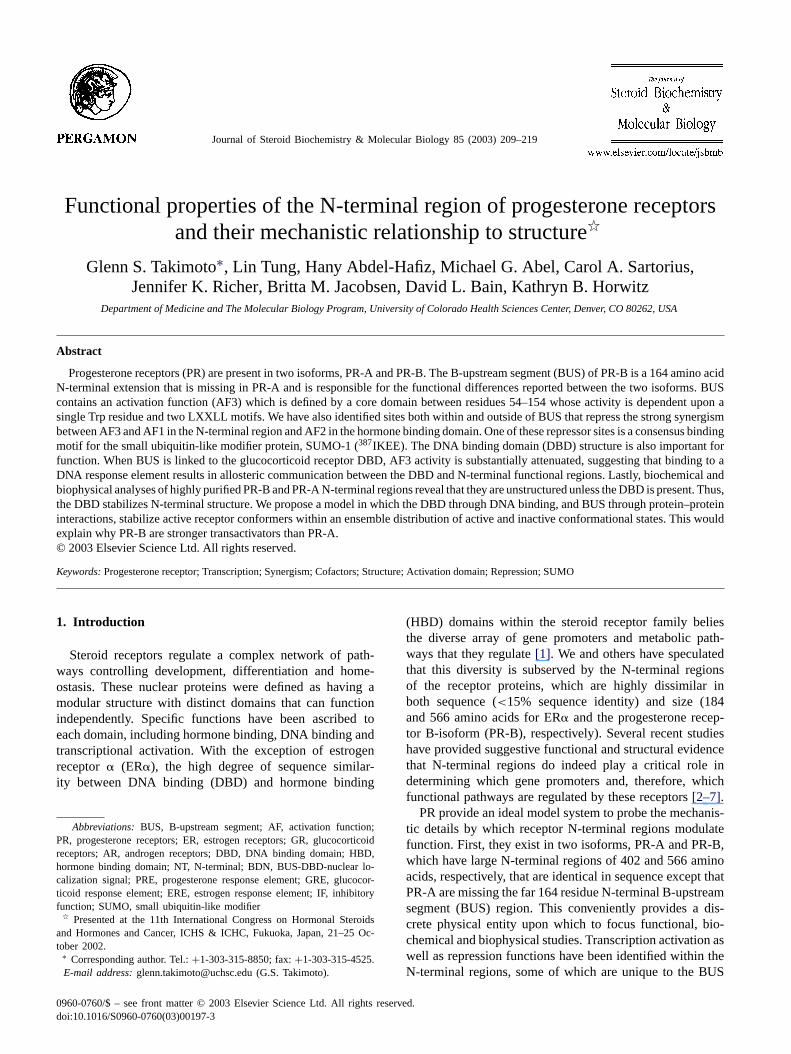
ies identified a core AF3 domain between residues 54–154and three specific sites within this core that defined AF3activity. These sites include140Trp and two LXXLL mo-tifs designated L1 and L2. As shown inFig. 2, mutationof L1, L2 or 140Trp reduced transcriptional activity on aPRE2-TATA luciferase promoter–reporter by 80% or more.Combined mutation of L1 and L2 sites reduced activity by90%, to a level approximately equivalent to the very low ac-tivity of PR-A on the PRE2-TATA promoter. These data sug-gest that inactivation of AF3-related activity can produce areceptor with a transcriptional phenotype resembling that ofPR-A.
Our previous deletion mutagenesis studies had mapped arepressor region within the first 25 residues of BUS. Usinga PCR-based random mutagenesis approach, we have nowidentified two highly hydrophilic residues,5Lys and7Lys,that produce a greater than 10-fold increase in transcrip-tional activity when mutated to a highly hydrophobic Trpresidue. Inactivation of AF3 by mutating L1, L2 and140Trpcompletely prevented this activity increase, suggesting thatthe repressor function in BUS is linked to AF3-dependenttranscriptional activity (not shown). Finally, the losses of ac-tivity with mL1, mL2 and W140A and gains of activity withthe K5W/K7W mutants were identical in BDN (BUS linkeddirectly to the DBD) and full-length PR-B backgrounds (notshown), suggesting that these alterations of BUS functionaldomains affected both intrinsic and synergistic properties ofAF3 activity.
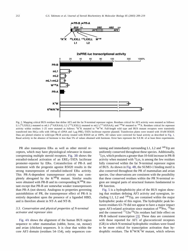
3.2. The SUMO-1 binding motif is responsible for bothautoinhibitory and transrepressor properties of PR
The autoinhibitory property of PR has been defined asthe increased transcriptional activity seen upon deletion ofa large region (IF) common to PR-A and PR-B locatedN-terminal to AF1 [9]. Recently, studies by Giangrandeet al. [10], Huse et al.[11] and our laboratory[4] mappedthis autoinhibitory function to subregions within IF. Withina subregion of IF containing the autoinhibitory function(residues 375–397), we identified a SUMO-1 consensusbinding motif, 387IKEE [17]. When a point mutation wasintroduced into this motif (387IREE) in PR-B and PR-A,greater than 10-fold (PR-B) and 6–7-fold (PR-A) increasesin transcriptional activity were observed from a PRE2-TATAluciferase promoter–reporter (Fig. 3A). The fact that thePR-B K388R mutant remains a much stronger activator thanthe corresponding PR-A K388R mutant indicates that theintact SUMO-1 binding motif in PR-B tightly controls thestrong AF3 synergism with AF1 and AF2. Interestingly,K388R mutant-dependent increases in transcriptional activ-ity are hormone-dependent and completely absent in NT-Aand NT-B constructs lacking the HBD[4]. We have alsoshown that the K388R mutation results in a complete loss ofcovalent attachment of the SUMO-1 protein to PR-A andPR-B, which is consistent with a single SUMO-1 bindingmotif present in the PR protein[4].
212 G.S. Takimoto et al. / Journal of Steroid Biochemistry & Molecular Biology 85 (2003) 209–219
Fig. 2. Mapping critical BUS residues that define AF3 and the far N-terminal repressor region. Residues critical for AF3 activity were mutated as follows:L1 (55LXXLL) mutated to mL1 (55AXXAA); L2 ( 115LXXLL) mutated to mL2 (115AXXAA); and 140W mutated to140A. Residues critical for repressoractivity within residues 1–25 were mutated as follows:5K7K mutated to5W7W. Full-length wild type and BUS mutant receptors were transientlytransfected into HeLa cells with 100 ng of cDNA and 1�g PRE2-TATA luciferase reporter plasmid. Transfection plates were treated with 10 nM R5020.Data are plotted relative to wild-type PR-B activity treated with R5020 set at 100%. All values were corrected for basal activity as described inFig. 1.Basal activity in the absence of hormone is less than 5% of values obtained with hormone. Error bars represent the S.E.M. of at least three experiments.
PR also transrepress ER� as well as other steroid re-ceptors, which may have physiological relevance in tissuescoexpressing multiple steroid receptors.Fig. 3B shows theestradiol-induced activation of an ERE2-TATA luciferasepromoter–reporter by ER�. Cotransfection of PR-A andtreatment with the progestin agonist R5020 results in thestrong transrepression of estradiol-induced ER� activity.This PR-A-dependent transrepressor activity was com-pletely abrogated by the K388R mutant. Similar resultswere obtained with PR-B and its corresponding K388R mu-tant except that PR-B are somewhat weaker transrepressorsthan PR-A (not shown). Analogous to properties governingautoinhibition of PR, the transrepressor effect of PR-A isentirely dependent upon the presence of a liganded HBD,and is therefore absent in NT-A and NT-B.
3.3. Conservation and physical properties of N-terminalactivator and repressor sites
Fig. 4A shows the alignment of the human BUS regionsequence to other mammalian (rabbit, horse, rat, mouse)and avian (chicken) sequences. It is clear that within thecore AF3 domain (residues 54–154), only sequences con-
taining and immediately surrounding L1, L2 and140Trp areuniformly conserved throughout these species. Additionally,7Lys, which produces a greater than 10-fold increase in PR-Bactivity when mutated with5Lys, is among the few residuesfully conserved within the far N-terminal repressor regionof BUS. As shown inFig. 4B, the SUMO-1 binding motif isalso conserved throughout the PR of mammalian and avianspecies. Our observations are consistent with the possibilitythat these conserved residues within the PR N-terminal re-gion are integral parts of structural features fundamental toPR function.
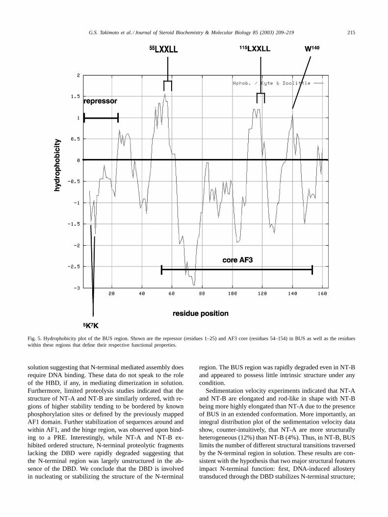
Fig. 5 is a hydrophobicity plot of the BUS region show-ing that residues defining AF3 activity and synergism, in-cluding L1, L2 and140Trp, are contained within the majorhydrophobic peaks of this region. The hydrophilic peak be-tween residues 63–79 did not appear to have a major impactupon AF3-related activation since mutation of69Pro,70Serand the conserved77Gln/78Gln residues had little effect onPR-B induced transcription[3]. These data are consistentwith those reported for AF1 of glucocorticoid receptors(GR) in which N-terminal hydrophobic residues were foundto be more critical for transcription activation than hy-drophilic residues. The K5W/K7W mutant, which relieves
G.S. Takimoto et al. / Journal of Steroid Biochemistry & Molecular Biology 85 (2003) 209–219 213
Fig. 3. Autoinhibition and transrepressor functions of PR require the SUMO-1 binding motif. HeLa cells were transiently transfected with 100 ng ofcDNA and 1�g PRE2-TATA luciferase reporter plasmid. (A) Plates transfected with either wild-type (PR-B, PR-A) or mutant (PR-A K388R, PR-BK388R) cDNAs were treated with 10 nM R5020 and corrected for basal activity as described inFig. 1. Activity is expressed as fold change as comparedto wild-type PR-B. (B) All plates were transfected with 50 ng ER� cDNA alone and treated with 10 nM estradiol (E2) or ER� plus 50 ng PR-A or thePR-A K388R mutant cDNAs and treated with 10 nM E2 plus 10 nM R5020. Estrogenic activity is expressed as a percentage of ER� alone set at 100%.
the repressor function in the first 25 residues of BUS, resideswithin the major hydrophilic peak of this repressor region.
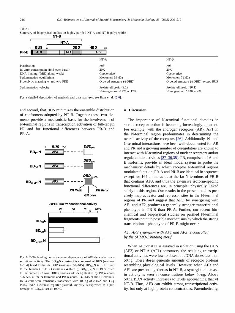
3.4. AF3 activity is DBD context-dependent
Fig. 6 shows transcriptional activities of AF3 in BDNand BDN chimeras that were tested on a PRE2-TATA lu-ciferase promoter–reporter. The chimeras were constructedby swapping either the PR DBD core region with the cor-responding GR DBD core, or the PR DBD flanking regionswith the corresponding GR DBD flanking sequences. Thecore region is defined by the DBD fragment that was usedto obtain a high resolution NMR solution structure[24], andthe limits of the flanking sequences were defined by the ex-tended region originally demarcated as the “C” region of thefull-length receptors plus four additional C-terminal residues
comprising the PR nuclear localization signal[25]. Swap-ping the core PR DBD for the core GR DBD had no effecton transcriptional activity. However, swapping the core andflanking sequences of the core PR DBD for the GR DBDreduced activity by 70–75%. Both the N- and C-terminalflanking sequences of GR contributed to this reduction inactivity (not shown). Sequence identity between the PR andGR DBD core is 90%, whereas it is 20–40% in the flankingregions. These data suggest that PR sequences in the DBDflanking regions are necessary for AF3 activity. Moreover,from unpublished studies in our laboratory, we know thatsubstituting the PR DBD with heterologous DBDs such asthe GAL4 DBD or the ER� DBD also results in a substan-tial loss of AF3 activity. Taken together, these observationssupport the hypothesis that expression of AF3-related func-tional activities can be allosterically regulated by specific

214 G.S. Takimoto et al. / Journal of Steroid Biochemistry & Molecular Biology 85 (2003) 209–219
Fig. 4. Residues responsible for AF3 activator and N-terminal repressor properties of PR are highly conserved. Sequence alignment of various speciesshown for the BUS (A) and SUMO-1 binding (B) regions. Conserved residues throughout the species listed are shown in red in the consensus sequenceand include residues comprising5K7K, L1 (55LXXLL), L2 ( 115LXXLL), 140W (A); and the SUMO-1 binding motif (387IKEE) (B).
amino acid residues within the DBD region, and, therefore,upon recognition of context-specific DNA response elementsequences at a gene promoter. Clearly for PR, and probablyfor other transcription factors, analysis of their AFs throughuse of heterologous DBDs may yield skewed information.
3.5. Biochemical and biophysical studies providea mechanistic basis for explaining differencesin N-terminal function
In recent studies using highly purified NT-B and NT-A,we performed biochemical and biophysical analyses thatyielded the results summarized inTable 1 [5,6]. First, NT-A
and NT-B purified to greater than 95% homogeneity weretranscriptionally competent in a cell-free in vitro transcrip-tion assay. Nitrocellulose filter binding studies indicated thatboth NT-A and NT-B bind cooperatively, and with similaraffinities, to a single PRE oligonucleotide whose sequencewas obtained from the GRE/PRE of the tyrosine amino-transferase gene promoter[38]. The DBD alone, however,bound weakly with a 10-fold lower affinity than NT-A andNT-B (not shown) suggesting that N-terminal regions com-mon to both PR-A and PR-B are responsible together withthe DBD, for cooperative interactions between the recep-tors and their cognate response elements. By sedimentationequilibrium analysis, NT-A and NT-B were monomeric in
G.S. Takimoto et al. / Journal of Steroid Biochemistry & Molecular Biology 85 (2003) 209–219 215
Fig. 5. Hydrophobicity plot of the BUS region. Shown are the repressor (residues 1–25) and AF3 core (residues 54–154) in BUS as well as the residueswithin these regions that define their respective functional properties.
solution suggesting that N-terminal mediated assembly doesrequire DNA binding. These data do not speak to the roleof the HBD, if any, in mediating dimerization in solution.Furthermore, limited proteolysis studies indicated that thestructure of NT-A and NT-B are similarly ordered, with re-gions of higher stability tending to be bordered by knownphosphorylation sites or defined by the previously mappedAF1 domain. Further stabilization of sequences around andwithin AF1, and the hinge region, was observed upon bind-ing to a PRE. Interestingly, while NT-A and NT-B ex-hibited ordered structure, N-terminal proteolytic fragmentslacking the DBD were rapidly degraded suggesting thatthe N-terminal region was largely unstructured in the ab-sence of the DBD. We conclude that the DBD is involvedin nucleating or stabilizing the structure of the N-terminal
region. The BUS region was rapidly degraded even in NT-Band appeared to possess little intrinsic structure under anycondition.
Sedimentation velocity experiments indicated that NT-Aand NT-B are elongated and rod-like in shape with NT-Bbeing more highly elongated than NT-A due to the presenceof BUS in an extended conformation. More importantly, anintegral distribution plot of the sedimentation velocity datashow, counter-intuitively, that NT-A are more structurallyheterogeneous (12%) than NT-B (4%). Thus, in NT-B, BUSlimits the number of different structural transitions traversedby the N-terminal region in solution. These results are con-sistent with the hypothesis that two major structural featuresimpact N-terminal function: first, DNA-induced allosterytransduced through the DBD stabilizes N-terminal structure;
216 G.S. Takimoto et al. / Journal of Steroid Biochemistry & Molecular Biology 85 (2003) 209–219
Table 1Summary of biophysical studies on highly purified NT-A and NT-B polypeptides
NT-A NT-B
Purification >95 >95In vitro transcription (fold over basal) 20X 20XDNA binding (DBD alone, weak) Cooperative CooperativeSedimentation equilibrium Monomer: 59 kDa Monomer: 71 kDaProteolytic mapping w and w/o PRE Ordered structure (+DBD) Ordered structure (+DBD) except BUS
Sedimentation velocity Prolate ellipsoid (9:1) Prolate ellipsoid (20:1)Heterogenous:�S20,w 12% Homogenous:�S20,w 4%
For a detailed description of methods and data analyses, see Bain et al.[5,6].
and second, that BUS minimizes the ensemble distributionof conformers adopted by NT-B. Together these two ele-ments provide a mechanistic basis for the involvement ofN-terminal regions in transcription activation of full-lengthPR and for functional differences between PR-B andPR-A.
Fig. 6. DNA binding domain context dependence of AF3-dependent tran-scriptional activity. The BDPRN construct is composed of BUS (residues1–164) fused to the PR DBD (residues 556–645); BDGRN is BUS fusedto the human GR DBD (residues 430–519); BDGR/PRN is BUS fusedto the human GR core DBD (residues 441–506) flanked by PR residues556–565 at the N-terminus and PR residues 632–645 at the C-terminus.HeLa cells were transiently transfected with 100 ng of cDNA and 1�gPRE2-TATA luciferase reporter plasmid. Activity is expressed as a per-centage of BDPRN set at 100%.
4. Discussion
The importance of N-terminal functional domains insteroid receptor action is becoming increasingly apparent.For example, with the androgen receptors (AR), AF1 inthe N-terminal region predominates in determining theoverall activity of the receptors[26]. Additionally, N- andC-terminal interactions have been well-documented for ARand PR and a growing number of coregulators are known tointeract with N-terminal regions of nuclear receptors and/orregulate their activities[27–30,35]. PR, comprised of A andB isoforms, provide an ideal model system to probe themechanistic details by which receptor N-terminal regionsmodulate function. PR-A and PR-B are identical in sequenceexcept for 164 amino acids at the far N-terminus of PR-Bthat contains AF3, and thus the extensive isoform-specificfunctional differences are, in principle, physically linkedsolely to this region. Our results in the present studies pre-cisely map activator and repressor sites in the N-terminalregions of PR and suggest that AF3, by synergizing withAF1 and AF2, produces a generally stronger transcriptionalphenotype in PR-B than PR-A. Further, our recent bio-chemical and biophysical studies on purified N-terminalfragments point to possible mechanisms by which the strongtranscriptional phenotype of PR-B might occur.
4.1. AF3 synergism with AF1 and AF2 is controlledby the SUMO-1 binding motif
When AF3 or AF1 is assayed in isolation using the BDN(AF3) or NT-A (AF1) constructs, the resulting transcrip-tional activities were low to absent at cDNA doses less than50 ng. These doses generate amounts of receptor proteinsresembling physiological levels. However, when AF3 andAF1 are present together as in NT-B, a synergistic increasein activity is seen at concentrations below 50 ng. Above50 ng BDN activity increases to levels approaching that ofNT-B. Thus, AF3 can exhibit strong transcriptional activ-ity, but only at high protein concentrations. Parenthetically,

G.S. Takimoto et al. / Journal of Steroid Biochemistry & Molecular Biology 85 (2003) 209–219 217
these data point to the importance of understanding recep-tor concentrations when evaluating transcriptional activitiesin vitro. AF3 synergizes with AF1 and AF2 when bothare present as seen by comparing the activities of PR-B(AF3, AF1, AF2) with PR-A (AF1, AF2) and BDN (AF3) atdoses less than 50 ng. Interestingly, this synergism is highlytempered by the presence of the SUMO-1 binding motif,387IKEE, located in the N-terminal region common to bothPR-B and PR-A. When this motif is mutated to387IREE, theactivity of PR-B increases by greater than 10-fold, whereasthe activity of NT-B is unchanged. These findings suggestthat at intracellular receptor levels that more closely resem-ble physiological, the potential for AF3 to synergize withAF1 and AF2 is tightly controlled. Under conditions wherePR-B are not sumoylated or under-sumoylated, extremelyhigh levels of synergism-dependent activation could in the-ory be achieved. This could represent a mechanism wherebygene expression levels could be rapidly turned up or down.Synergism between AF1 and AF2 is also controlled bysumoylation. However, in the absence of AF3 the maximumpotential for AF synergism is a fraction of that seen withnon-sumoylated PR-B.
As with AR, the activities of PR N-terminal constructssuch as NT-B and BDN at higher doses make a more pro-nounced contribution to the transcriptional phenotype ofPR-B than the C-terminal HBD region containing AF2,which has little intrinsic activity on promoters examinedso far [26]. However, our results with the K388R SUMO-1binding mutant suggest that maximal AF synergy is inti-mately dependent upon C-terminal sequences within a lig-anded HBD. Additionally, recent unpublished studies in ourlaboratory demonstrate that AF3 related synergy requiresmultiple tandem PREs and is inoperative on a synthetic min-imal promoter construct with a single palindromic responseelement. It is conceivable that the arrangement, type andnumber of heterologouscis-acting promoter elements adja-cent to a single PRE may play a critical role in the expres-sion of AF3-related synergy on endogneous promoters.
4.2. Repressor properties of PR are relatedto synergy control
We have presented compelling evidence that the autoin-hibitory properties of PR-A and PR-B are intimately linkedto the SUMO-1 binding or “synergy control” motif[18].Interestingly, when we tested the ability of PR to transre-press ER�, we obtained similar results. Both PR-A, andto a lesser extent PR-B, inhibit the transcriptional activ-ity of estradiol-induced ER� on an ERE2-TATA luciferasepromoter–reporter. This effect requires the liganded HBD ofPR and an intact SUMO-1 binding motif analogous to therequirements for synergy control in full-length receptors. Itdoes not appear that this effect is due to squelching of lim-ited coregulator pools since transrepression occurs at con-centrations of PR well below the levels eliciting maximaltranscriptional responses. Whether PR are able to bring teth-
ered synergy control factors to the ER�-regulated promotercomplex remains to be investigated.
A second repressor region in the far N-terminus of BUSwas also identified. Conversion of hydrophilic5Lys and7Lys to hydrophobic Trp residues resulted in 10-fold in-creases in transcriptional activities of PR-B, comparable tothose seen with the SUMO-1 binding mutant. However,this increased activity was not dependent upon a ligandedHBD since BDN lacking an HBD exhibited the same in-creased activity as PR-B. Since the increased activity wasabrogated by mutation of L1, L2 and140Trp in BUS (datanot shown), the mechanism of repression may be more di-rectly linked to the overall physical properties of AF3 andBUS.
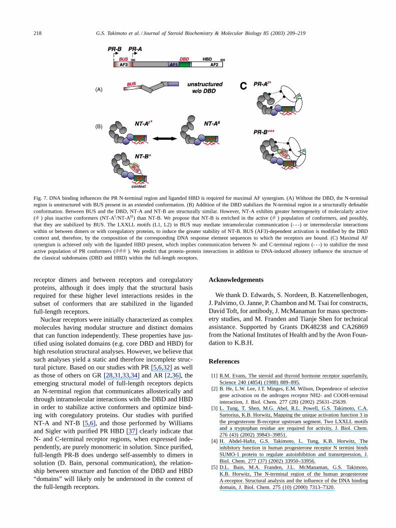
4.3. A model for N-terminal linked activation
We propose the model shown inFig. 7 based uponmechanistic insights provided by biochemical and biophys-ical analyses and novel functional properties of the PRN-terminal regions. As shown in part A, the regions ofPR-A and PR-B N-terminal to the DBD are largely un-structured, with BUS assuming an extended, non-globularconformation. Ordered structure (part B) is induced by thepresence of the DBD, and further stabilization is achieved,particularly for sequences around and within AF1 and thehinge domains (blue), upon binding to DNA. The intrinsicas well as synergistic transcriptional activities attributed toAF3 in BUS is DBD context-dependent and therefore isintimately linked to the number and specific sequence ofresponse elements. The LXXLL motifs (L1, L2) as wellas 140Trp in BUS define AF3-related activity and maymediate intra- or intermolecular interactions. The unusualpresence of two LXXLL motifs in BUS (motifs that aremore commonly found on coactivators) suggest that BUSis involved in protein–protein interactions that remain tobe defined. We view BUS as a “tethered coregulator” andpredict that other transcription factors may have similardomains. Finally, as shown in part B, the greater hetero-geneity seen with the ensemble distribution of NT-A (12%)as compared to NT-B (4%) conformers leads us to pro-pose that, mole for mole, a higher proportion of NT-Bmolecules exist in an active () conformation resultingin the generally stronger transcriptional phenotype of thisisoform.
Fig. 7Cillustrates the critical role of the liganded HBD inthe tight control of potentially high levels of AF synergismin PR-A ( ) and particularly AF3-dependent synergism inPR-B ( ), as seen by the effects of desumoylation ofthe full-length receptors. This figure illustrates the likeli-hood that the N-terminal region, particularly BUS, and theC-terminal HBD participate in critical intramolecular inter-actions that minimize the ensemble distribution of receptorsto a conformation(s) eliciting optimal AF synergism. Lastly,this figure does not address the additional structural com-plexity imparted by intermolecular interactions between
218 G.S. Takimoto et al. / Journal of Steroid Biochemistry & Molecular Biology 85 (2003) 209–219
Fig. 7. DNA binding influences the PR N-terminal region and liganded HBD is required for maximal AF synergism. (A) Without the DBD, the N-terminalregion is unstructured with BUS present in an extended conformation. (B) Addition of the DBD stabilizes the N-terminal region in a structurally definableconformation. Between BUS and the DBD, NT-A and NT-B are structurally similar. However, NT-A exhibits greater heterogeneity of molecularly active( ) plus inactive conformers (NT-AI /NT-AII ) than NT-B. We propose that NT-B is enriched in the active () population of conformers, and possibly,that they are stabilized by BUS. The LXXLL motifs (L1, L2) in BUS may mediate intramolecular communication (- - -) or intermolecular interactionswithin or between dimers or with coregulatory proteins, to induce the greater stability of NT-B. BUS (AF3)-dependent activation is modified by the DBDcontext and, therefore, by the composition of the corresponding DNA response element sequences to which the receptors are bound. (C) Maximal AFsynergism is achieved only with the liganded HBD present, which implies communication between N- and C-terminal regions (- - -) to stabilize the mostactive population of PR conformers ( ). We predict that protein–protein interactions in addition to DNA-induced allostery influence the structure ofthe classical subdomains (DBD and HBD) within the full-length receptors.
receptor dimers and between receptors and coregulatoryproteins, although it does imply that the structural basisrequired for these higher level interactions resides in thesubset of conformers that are stabilized in the ligandedfull-length receptors.
Nuclear receptors were initially characterized as complexmolecules having modular structure and distinct domainsthat can function independently. These properties have jus-tified using isolated domains (e.g. core DBD and HBD) forhigh resolution structural analyses. However, we believe thatsuch analyses yield a static and therefore incomplete struc-tural picture. Based on our studies with PR[5,6,32]as wellas those of others on GR[28,31,33,34]and AR[2,36], theemerging structural model of full-length receptors depictsan N-terminal region that communicates allosterically andthrough intramolecular interactions with the DBD and HBDin order to stabilize active conformers and optimize bind-ing with coregulatory proteins. Our studies with purifiedNT-A and NT-B [5,6], and those performed by Williamsand Sigler with purified PR HBD[37] clearly indicate thatN- and C-terminal receptor regions, when expressed inde-pendently, are purely monomeric in solution. Since purified,full-length PR-B does undergo self-assembly to dimers insolution (D. Bain, personal communication), the relation-ship between structure and function of the DBD and HBD“domains” will likely only be understood in the context ofthe full-length receptors.
Acknowledgements
We thank D. Edwards, S. Nordeen, B. Katzenellenbogen,J. Palvimo, O. Janne, P. Chambon and M. Tsai for constructs,David Toft, for antibody, J. McManaman for mass spectrom-etry studies, and M. Franden and Tianje Shen for technicalassistance. Supported by Grants DK48238 and CA26869from the National Institutes of Health and by the Avon Foun-dation to K.B.H.
References
[1] R.M. Evans, The steroid and thyroid hormone receptor superfamily,Science 240 (4854) (1988) 889–895.
[2] B. He, L.W. Lee, J.T. Minges, E.M. Wilson, Dependence of selectivegene activation on the androgen receptor NH2- and COOH-terminalinteraction, J. Biol. Chem. 277 (28) (2002) 25631–25639.
[3] L. Tung, T. Shen, M.G. Abel, R.L. Powell, G.S. Takimoto, C.A.Sartorius, K.B. Horwitz, Mapping the unique activation function 3 inthe progesterone B-receptor upstream segment. Two LXXLL motifsand a tryptophan residue are required for activity, J. Biol. Chem.276 (43) (2002) 39843–39851.
[4] H. Abdel-Hafiz, G.S. Takimoto, L. Tung, K.B. Horwitz, Theinhibitory function in human progesterone receptor N termini bindsSUMO-1 protein to regulate autoinhibition and transrepression, J.Biol. Chem. 277 (37) (2002) 33950–33956.
[5] D.L. Bain, M.A. Franden, J.L. McManaman, G.S. Takimoto,K.B. Horwitz, The N-terminal region of the human progesteroneA-receptor. Structural analysis and the influence of the DNA bindingdomain, J. Biol. Chem. 275 (10) (2000) 7313–7320.

G.S. Takimoto et al. / Journal of Steroid Biochemistry & Molecular Biology 85 (2003) 209–219 219
[6] D.L. Bain, M.A. Franden, J.L. McManaman, G.S. Takimoto, K.B.Horwitz, The N-terminal region of human progesterone B-receptors:biophysical and biochemical comparison to A-receptors, J. Biol.Chem. 276 (26) (2001) 23825–23831.
[7] R. Kumar, E.B. Thompson, Transactivation functions of the N-terminal domains of nuclear hormone receptors: protein folding andcoactivator interactions, Mol. Endocrinol. 17 (1) (2003) 1–10.
[8] C.A. Sartorius, M.Y. Melville, A.R. Hovland, L. Tung, G.S.Takimoto, K.B. Horwitz, A third transactivation function (AF3)of human progesterone receptors located in the unique N-terminalsegment of the B-isoform, Mol. Endocrinol. 8 (10) (1994) 1347–1360.
[9] A.R. Hovland, R.L. Powell, G.S. Takimoto, L. Tung, K.B. Horwitz,An N-terminal inhibitory function, IF, suppresses transcription by theA-isoform but not the B-isoform of human progesterone receptors,J. Biol. Chem. 273 (10) (1998) 5455–5460.
[10] P.H. Giangrande, G. Pollio, D.P. McDonnell, Mapping andcharacterization of the functional domains responsible for thedifferential activity of the A and B isoforms of the human pro-gesterone receptor, J. Biol. Chem. 272 (52) (1997) 32889–32900.
[11] B. Huse, S.B. Verca, P. Matthey, S. Rusconi, Definition of anegative modulation domain in the human progesterone receptor,Mol. Endocrinol. 12 (9) (1998) 1334–1342.
[12] M.E. Meyer, A. Pornon, J.W. Ji, M.T. Bocquel, P. Chambon, H.Gronemeyer, Agonistic and antagonistic activities of RU486 on thefunctions of the human progesterone receptor, EMBO J. 9 (12) (1990)3923–3932.
[13] L. Tung, M.K. Mohamed, J.P. Hoeffler, G.S. Takimoto, K.B.Horwitz, Antagonist-occupied human progesterone B-receptorsactivate transcription without binding to progesterone response ele-ments and are dominantly inhibited by A-receptors, Mol. Endocrinol.7 (10) (1993) 1256–1265.
[14] P.H. Giangrande, E.A. Kimbrel, D.P. Edwards, D.P. McDonnell, Theopposing transcriptional activities of the two isoforms of the humanprogesterone receptor are due to differential cofactor binding, Mol.Cell. Biol. 20 (9) (2000) 3102–3115.
[15] J.K. Richer, B.M. Jacobsen, N.G. Manning, M.G. Abel, D.M. Wolf,K.B. Horwitz, Differential gene regulation by the two progesteronereceptor isoforms in human breast cancer cells, J. Biol. Chem. 277 (7)(2002) 5209–5218.
[16] B.M. Jacobsen, J.K. Richer, S.A. Schittone, K.B. Horwitz, Newhuman breast cancer cells to study progesterone receptor isoformratio effects and ligand-independent gene regulation, J. Biol. Chem.277 (31) (2002) 27793–27800.
[17] H. Poukka, U. Karvonen, O.A. Janne, J.J. Palvimo, Covalent modi-fication of the androgen receptor by small ubiquitin-like modifier 1(SUMO-1), Proc. Natl. Acad. Sci. U.S.A. 97 (26) (2000) 14145–14150.
[18] J.A. Iniguez-Lluhi, D. Pearce, A common motif within the negativeregulatory regions of multiple factors inhibits their transcriptionalsynergy, Mol. Cell. Biol. 20 (16) (2000) 6040–6050.
[19] C.S. Winkley, M.J. Keller, J.A. Jaehning, A multicomponentmitochondrial RNA polymerase fromSaccharomyces cerevisiae, J.Biol. Chem. 260 (26) (1985) 14214–14223.
[20] R.D. Gietz, A. Sugino, New yeast-Escherichia colishuttle vectorsconstructed with in vitro mutagenized yeast genes lacking six basepair restriction sites, Gene 74 (2) (1988) 527–534.
[21] D. Muhlrad, R. Hunter, R. Parker, A rapid method for localizedmutagenesis of yeast genes, Yeast 8 (2) (1992) 79–82.
[22] R.D. Gietz, Transforming yeast with DNA, Methods Mol. Cell. Biol.5 (1995) 255–269.
[23] S.P. Bohen, K.R. Yamamoto, Isolation of Hsp90 mutants by screeningfor decreased steriod receptor function, Proc. Natl. Acad. Sci. U.S.A.90 (23) (1993) 11424–11428.
[24] T. Hard, E. Kellenbach, R. Boelens, B.A. Maler, K. Dahlman, L.P.Freedman, J. Carlstedt-Duke, K.R. Yamamoto, J.A. Gustafsson, R.Kaptein, Solution structure of the glucocorticoid receptor DNA-binding domain, Science 249 (4965) (1990) 157–160.
[25] A. Guiochon-Mantel, H. Loosfelt, P. Lescop, S. Sar, M. Atger, M.Perrot-Applanat, E. Milgrom, Mechanisms of nuclear localizationof the progesterone receptor: evidence for interaction betweenmonomers, Cell 57 (7) (1989) 1147–1154.
[26] J.A. Simental, M. Sar, M.V. Lane, F.S. French, E.M. Wilson,Transcriptional activation and nuclear targeting signals of the humanandrogen receptor, J. Biol. Chem. 266 (1) (1991) 510–518.
[27] S.A. Onate, V. Boonyaratanakornkit, T.E. Spencer, S.Y. Tsai, M.J.Tsai, D.P. Edwards, B.W. O’Malley, The steroid receptor coactivator-1 contains multiple receptor interacting and activation domains thatcooperatively enhance the activation function 1 (AF1) and AF2domains of steroid receptors, J. Biol. Chem. 273 (20) (1998) 12101–12118.
[28] R. Kumar, J.C. Lee, D.W. Bolen, E.B. Thompson, The conformationof the glucocorticoid receptor af1/tau1 domain induced by osmolytebinds co-regulatory proteins, J. Biol. Chem. 276 (21) (2001) 18146–18152.
[29] T. Ikonen, J.J. Palvimo, O.A. Janne, Interaction between theamino- and carboxyl-terminal regions of the rat androgen receptormodulates transcriptional activity and is influenced by nuclearreceptor coactivators, J. Biol. Chem. 272 (47) (1997) 29821–29828.
[30] R. Metivier, G. Penot, G. Flouriot, F. Pakdel, Synergism betweenERalpha transactivation function 1 (AF-1) and AF-2 mediated bysteroid receptor coactivator protein-1: requirement for the AF-1alpha-helical core and for a direct interaction between the N-and C-terminal domains, Mol. Endocrinol. 15 (11) (2001) 1953–1970.
[31] R. Kumar, I.V. Baskakov, G. Srinivasan, D.W. Bolen, J.C. Lee,E.B. Thompson, Interdomain signaling in a two-domain fragment ofthe human glucocorticoid receptor, J. Biol. Chem. 274 (35) (1999)24737–24741.
[32] M.J. Tetel, P.H. Giangrande, S.A. Leonhardt, D.P. McDonnell, D.P.Edwards, Hormone-dependent interaction between the amino- andcarboxyl-terminal domains of progesterone receptor in vitro and invivo, Mol. Endocrinol. 13 (6) (1999) 910–924.
[33] A. Warnmark, J.A. Gustafsson, A.P. Wright, Architectural principlesfor the structure and function of the glucocorticoid receptor tau1 core activation domain, J. Biol. Chem. 275 (20) (2000) 15014–15018.
[34] K. Dahlman-Wright, H. Baumann, I.J. McEwan, T. Almlof, A.P.Wright, J.A. Gustafsson, T. Hard, Structural characterization ofa minimal functional transactivation domain from the humanglucocorticoid receptor, Proc. Natl. Acad. Sci. U.S.A. 92 (5) (1995)1699–1703.
[35] A. Warnmark, A. Wikstrom, A.P. Wright, J.A. Gustafsson, T. Hard,The N-terminal regions of estrogen receptor alpha and beta areunstructured in vitro and show different TBP binding properties, J.Biol. Chem. 276 (49) (2001) 45939–45944.
[36] J. Reid, S.M. Kelly, K. Watt, N.C. Price, I.J. McEwan,Conformational analysis of the androgen receptor amino-terminaldomain involved in transactivation. Influence of structure-stabilizingsolutes and protein–protein interactions, J. Biol. Chem. 277 (22)(2002) 20079–20086.
[37] S.P. Williams, P.B. Sigler, Atomic structure of progesteronecomplexed with its receptor, Nature 393 (6683) (1998) 392–396.
[38] H.M. Jantzen, U. Strahle, B. Gloss, F. Stewart, W. Schmid, M.Boshart, R. Miksicek, G. Schutz, Cooperativity of glucocorticoidresponse elements located far upstream of the tyrosine aminotrans-ferase gene, Cell 49 (1) (1987) 29–38.
Related Documents