Functional Interleukin-17 Receptor A is Expressed in the Central Nervous System and Upregulated in Experimental Autoimmune Encephalomyelitis a Jayasri Das Sarma 1 , Bogoljub Ciric 1 , Ryan Marek 1 , Sanjoy Sadhukhan 1 , Jasmine Shafagh 1 , Denise C. Fitzgerald 1 , Kenneth S. Shindler 2 and A. M. Rostami 1* 1 Department of Neurology, Thomas Jefferson University, Philadelphia, PA 19107. 2 Department of Ophthalmology, University of Pennsylvania, Scheie Eye Institute and FM Kirby Center for Molecular Ophthalmology, Philadelphia, PA 19104. Running title: Expression and signaling of IL-17RA in the CNS A.M. Rostami, M.D, Ph.D 900 Walnut Street 200JHN Department of Neurology Thomas Jefferson University Philadelphia, PA 19107, USA Tel: 215-955-8100 Fax: 215-955-1390 E-mail: [email protected] a This work was supported by a grant from NIH to AMR (5R01 NS048435) and the M.E.Groff Surgical Medical Research and Education Charitable Trust to JDS (F76401)

Welcome message from author
This document is posted to help you gain knowledge. Please leave a comment to let me know what you think about it! Share it to your friends and learn new things together.
Transcript
Functional Interleukin-17 Receptor A is Expressed in the Central Nervous System and
Upregulated in Experimental Autoimmune Encephalomyelitisa
Jayasri Das Sarma1, Bogoljub Ciric1, Ryan Marek1, Sanjoy Sadhukhan1, Jasmine
Shafagh1, Denise C. Fitzgerald1, Kenneth S. Shindler2 and A. M. Rostami1*
1Department of Neurology, Thomas Jefferson University, Philadelphia, PA 19107.
2Department of Ophthalmology, University of Pennsylvania, Scheie Eye Institute and FM
Kirby Center for Molecular Ophthalmology, Philadelphia, PA 19104.
Running title: Expression and signaling of IL-17RA in the CNS
A.M. Rostami, M.D, Ph.D
900 Walnut Street
200JHN
Department of Neurology
Thomas Jefferson University
Philadelphia, PA 19107, USA
Tel: 215-955-8100
Fax: 215-955-1390
E-mail: [email protected]
aThis work was supported by a grant from NIH to AMR (5R01 NS048435) and the M.E.Groff
Surgical Medical Research and Education Charitable Trust to JDS (F76401)
Abstract:
Interleukin-17A (IL-17A) is founding member of a novel family of inflammatory cytokines that
plays a critical role in the pathogenesis of many autoimmune diseases, including multiple
sclerosis (MS) and its animal model, experimental autoimmune encephalomyelitis (EAE). IL-
17A signals through IL-17RA, which is expressed in most peripheral tissues; however,
expression of IL-17RA in the central nervous system (CNS) and its role in CNS inflammation
are not well understood. Here we report constitutive expression of functional IL-17RA in mouse
CNS tissue. Specifically, CNS astrocytes and microglia express IL-17RA, and IL-17A treatment
induces biological responses in these cells in vitro. In response to exogenous IL-17A treatment,
microglia and astrocytes significantly upregulate MCP-1, MCP-5, MIP-2 and KC chemokine
secretion. Exogenous IL-17A does not significantly alter the constitutive expression of IL-17RA
mRNA in glial cells, suggesting that upregulation of chemokines by glial cells is due to IL-17A
signaling through constitutively expressed IL-17RA. IL-17RA expression is significantly
increased in the CNS of mice with EAE compared to healthy mice. Our findings suggest that IL-
17RA signaling in glial cells can play a significant role in autoimmune inflammation of the CNS
and may be a potential pathway to target for therapeutic interventions.
Introduction: IL-17A (also known as IL-17) was described more than a decade ago (1), but became a major
focus of research only recently, after a novel IL-17A-producing Th lineage (Th17) was
discovered (2-6). Th17 cells are generated in response to polarizing cytokines including TGFβ,
IL-6, IL-23, IL-1β and TNF with apparent species specific variations (7-10). Like other
inflammatory cytokines, IL-17A has both protective and pathogenic roles. IL-17A is important
for host defense against infectious organisms (11-14). However, elevated IL-17A in several
autoimmune diseases including MS/EAE (15-17), contributes to disease pathogenesis.
Deficiency or neutralization of IL-17A in EAE reduces disease susceptibility and clinical
severity (18). IL-17A can induce the expression of a range of inflammatory mediators, and thus
modulates the activities of inflammatory cells (19, 20) through production of numerous
cytokines and chemokines involved in inflammatory responses (21).
Infiltration of inflammatory cells and encephalitogenic T cells in the CNS is the hallmark of
EAE (22). IL-17A expression is increased in lymphocytes derived from EAE mice (23), and
anti-IL-17A antibody treatment during the recovery phase in a relapsing remitting EAE model
delays the onset and reduces incidence and severity of relapses (24). In human MS patients, IL-
17A mRNA and protein are increased in both brain lesions and mononuclear cells isolated from
blood and cerebrospinal fluid (25, 26). Recently, Kebir et al demonstrated that IL-17A produced
by Th17 cells is detectable at the blood brain barrier (BBB) in MS lesions, and that IL-17A can
promote BBB disruption in vitro (27).
IL-17A functions through a distinct ligand-receptor signaling system (28). IL-17RA is a widely
expressed receptor identified as a mammalian counter structure for HVS13 and subsequently
shown to bind IL-17A with high affinity (29). Leukocytes from mice lacking IL-17RA fail to
bind IL-17A, and antibodies against IL-17RA inhibit the activity of IL-17A on human epithelial
cells, indicating that IL-17RA is critical for IL-17A function (30, 31). Recently it has been
demonstrated in infectious models in which neutrophils are crucial for host defense, that IL-
17RA deficiency results in reduced chemokine levels and reduced neutrophil numbers, and
resistance to infection (12, 13, 31). IL-17RA signaling is implicated in both innate and adaptive
elements of infectious and autoimmune diseases (15); however, little is known about its signaling
in the CNS. One reason may be that IL-17RA is expressed in the CNS at a very low level.
Expression of IL-17RA in the CNS of healthy human subjects is undetectable by
immunofluorescence but the receptor was expressed in CNS endothelial cells within heavily
infiltrated MS lesions (27). Given the important role that IL-17A plays in autoimmune diseases
of the CNS, it is important to understand responses of CNS cells to IL-17RA signaling. Here, we
have investigated expression and function of IL-17RA in healthy and inflamed mouse CNS
tissues both in vitro and in vivo.
We report here that mouse CNS tissues express IL-17RA and the level of expression increases in
the CNS of mice with EAE. We also demonstrate in vitro that astrocytes and microglia in
isolated culture express IL-17RA. The expression level of IL-17RA in microglia/macrophages
is higher compared to astrocytes. Treatment of astrocyte cultures (devoid of microglia) and
microglia cultures (devoid of astrocytes) with exogenous recombinant mouse IL-17A protein
showed functional activation of IL-17RA signaling as demonstrated by increased secretion of
several chemokines (MCP-1, MCP-5, MIP-2 and KC).
Materials and methods:
Mice: Eight-week-old and time pregnant mice C57Bl/6 mice were purchased from the Jackson
Laboratory (Bar Harbor, ME). All animal procedures and care were conducted in accordance
with approved ethical guidance under the auspices of the Thomas Jefferson University Animal
Care and Use Committee. IL-17RA deficient mice on the C57BL/6 background were used as
negative control (31). IL-17RA deficient mice on the C57BL/6 background were kindly provided
by David Abraham (Thomas Jefferson University, Philadelphia) with permission from Amgen
(Seattle, WA).
Induction of EAE: Mice were injected subcutaneously with 100 µg myelin oligodendroglial
glycoprotein (MOG35-55) peptide (MEVGWYRSPFSRVVHLYRNGK) in complete Freund’s
adjuvant containing 4 mg/ml Mycobacterium tuberculosis H37Ra (Difco, MI) at two sites on the
back. 200 ng pertussis toxin was given intraperitonially on day 0 and 2 post-immunization (p.i.).
Mice were scored daily according to a 0-5 scale as follows: partial limp tail, 0.5; full limp tail, 1;
limp tail and waddling gait, 1.5; paralysis of one hind limb, 2; paralysis of one hind limb and
partial paralysis of the other hind limb, 2.5; paralysis of both hind limbs, 3; ascending paralysis,
3.5, paralysis of trunk, 4; moribund, 4.5; death, 5 (32). At 20 days p.i. (peak of disease; score 3)
tissues were collected for mRNA extraction and histology.
Histology: Mice were perfused transcardially with PBS followed by PBS containing 4%
paraformaldehyde (PFA). Spleen, brain and spinal cord tissues were collected, post-fixed in 4%
PFA overnight at room temperature (RT) and embedded in paraffin. 5 µm sections were
processed and stained with Hematoxylin and eosin (H&E) for assessment of inflammation and
with Luxol Fast Blue (LFB) for demyelination. Sections were assessed as follows (32);
Inflammation: 0, none; 1, a few inflammatory cells; 2, organization of perivascular infiltrates;
and 3, increasing severity of perivascular cuffing with extension into the adjacent tissue;
Demyelination: 0, none; 1, rare foci; 2, a few areas of demyelination; 3, large (confluent) areas of
demyelination.
Isolation of neonatal astrocytes and microglia: Primary cultures of mixed glial from day 0
newborn mice were prepared as described previously (33), with minor modifications. Briefly,
following the removal of meninges, brain tissues were minced with a pasteur pipette and passed
through 100 µm nylon mesh followed by a wash and centrifugation (300 x g for 10 min). The
pellet was resuspended with a pasteur pipette, passed through a 70µm nylon mesh, followed by a
second wash and centrifugation (300 x g for 10 min). Following dilutions with astrocyte-specific
medium {Dulbeco’s essential medium containing 1% penicillin-streptomycin, 0.2mM L-
glutamine and 10% fetal calf serum (FCS)}, cells were plated and grown in a humidified
incubator at 37º C. Cells were cultured until day 10, with a medium change on day 4, then every
2-3 days. To culture astrocytes free from microglia and to obtain pure microglial cultures,
feeding of mixed glial cultures was stopped for the following 12-14 days. Cultures were then
rigorously agitated for 30-40 min in an orbital incubator shaker at 200 rpm at 37ºC to detach
cells adhering to the astrocyte monolayer. Thereafter, cells suspended in the medium were
collected and plated (8 x10⁵ cells/ml; 1.5 ml per chamber slide (Nunc, Rochester, NY). After 15
min, non-adherent cells were discarded and adherent cells were maintained in medium specified
for astrocyte culture. Following this procedure, cells were 98-99% positive for CD11b
(microglia/macrophage marker) and were negative for glial fibrillary acidic protein (GFAP),
indicating a very high enrichment in microglia. Adherent astrocyte monolayers from the original
culture were trypsinized and resuspended in astrocyte specific medium at 8x10⁵ cells/ml; and 2
ml were plated on each well of 6 well culture plates. Sub-cultured astrocytes were 85% positive
for glial fibrillary acidic protein (GFAP) by immunofluorescence and 60-80 % by flow
cytometry.
IL-17A treatment in vitro: Functional studies were performed either on confluent microglia
subcultures obtained after 24 hr, or astrocyte subcultures obtained after 72hr hrs of plating. On
the day of stimulation, media were removed and cells were washed with PBS. Recombinant
mouse IL-17A (10ng/ml or 1-100 ng/ml where indicated) was added to the selected culture
wells. Non-stimulated sister cultures were used as controls throughout the studies. Culture
supernatants were collected at 3, 6, 12, 24 and 48 hr time points.
Immunofluorescence: Cells were processed by double label immunofluorescence for recognition
of microglia and astrocytes. CD11b was used as microglia/macrophage surface marker; whereas
GFAP was used as an intracellular astrocytic marker. Unfixed cells were incubated with
biotinylated anti-CD11b primary antibody for 30 min at RT followed by Cy3 conjugated
streptavidin secondary antibody for 30 min. Cultures were then rinsed with Ham’s F12, fixed in
95% ethanol/5% acetic acid (vol/vol) at -20°C for 10 min and washed in Ham’s F12
(Invitrogen). For GFAP staining, cells were washed 3 times with PBS, followed by PBS with
0.5% Triton X-100 and PBS with 0.5% Triton X-100 and 2% heat-inactivated goat serum. Cells
were incubated with polyclonal GFAP antisera (DAKO, Carpinteria, CA) for 30 min, washed,
and labeled with Cy2-conjugated goat anti-rabbit IgG. Cells were then washed, mounted into
Mowiol, and visualized by fluorescence microscopy (Olympus I X-80) with a 20 PlanApo oil
immersion objective (1.0 numerical aperture). Images were acquired with a SensiCamQE High
Performance CCD Camera.
Flow cytometry: Glial cell cultures were harvested and washed in buffer containing 1% FCS,
0.1% NaN3 in PBS, and stained with an APC-conjugated antibody to CD11b for 20 min in the
dark at 4ºC. Cells were washed, fixed and permeabilized using Fix and Perm ® cell
permebilization reagents (Caltag Laboratories, Burlingame, CA). Cells were then stained for
intracellular GFAP with polyclonal anti-GFAP antibody and PE-conjugated goat anti-rabbit IgG.
Search light chemokine arrays: Levels of 29 analytes including cytokines, chemokines, growth
factors and matrix metalloproteinases (MMPs) (Table I) in supernatants of cultures either treated
with IL-17A or non-stimulated, were assayed using a SearchLight Multiplex Sandwich ELISA
according to the manufacturer’s instructions.
Extraction of RNA and synthesis of cDNA: Tissue RNA and cellular RNA was extracted with
RNeasy Midi or Mini kits (Qiagen, Chatsworth, CA) respectively according to the
manufacturers’ recommendations. The purity of total RNA was assessed using a NanoDrop®
ND-100 spectrophotometer (NanoDrop Technologies, Wilmington, DE). One µg of total RNA
was used to synthesize cDNA with high capacity cDNA archive kit (Applied Biosystems Inc.,
Foster, CA) according to the manufacturers’ instructions.
Absolute quantification by real time PCR: Quantitative Real-Time (RT)-PCR was performed on
the ABI PRISM 7000 Sequence Detection System using TaqMan® Universal PCR Master Mix
(Applied Biosystems) and TaqMan® Gene Expression Assays primer/probe (Applied
Biosystems; Assay ID: Mm00434214_m1-from exon boundary 1-2) according to the
manufacturer’s specifications. Additional primer probe was also selected from exon boundary
3-4 (Assay ID. Mm01183143_m1) for amplification as this region is disrupted in IL-17RA
deficient mice. To generate a standard curve for quantification of templates, cDNA constructs
either from exon boundary 1-2 or 3-4 were cloned into pGEM® T Easy vector (Promega,
Madison, WI) and verified by double strand sequencing. Respective cDNA constructs were
serially diluted 7 times at a ratio of 1:10. Thus, the dynamic range for each gene was from 12 to
12,000,000 copies. Samples were analyzed in triplicate and experiments performed three times.
Amplification data were analyzed with ABI Prism Sequence Detection Software 2.1 (Applied
Biosystems).
Statistics: 2-tailed, Student’s Welch corrected t tests (for parametric data) were used for
statistical analysis. Differences were considered significant if * p < 0.05.
Results
IL-17RA is constitutively expressed in CNS tissues - IL-17RA is expressed in most tissues
examined to date, although little is known about its expression in the CNS. To investigate if IL-
17RA is expressed in normal CNS tissues, we harvested brain, spinal cord and, as a positive
control, spleen, from 10-week-old C57BL/6 female mice. RNA was extracted and cDNA
synthesized for quantitative RT-PCR. Pearson’s correlation coefficient of the standard curve
generated from serially-diluted cDNA constructs of pGEMT-IL-17RA exon boundary 1-2 was
0.99. IL-17RA was expressed in both brain and spinal cord with slightly higher levels detected
in brain. Levels of IL-17RA mRNA in normal CNS were approximately 5-fold lower than that
of normal spleen (Fig. 1A). To reconfirm the expression of IL-17RA in CNS, we constructed
another standard curve using a plasmid expressing the IL-17RA gene from exon boundary 3 - 4,
and used IL-17RA deficient mice (in which IL-17RA gene is disrupted between exon boundary 4
-11) (31) as a negative control. Pearson’s correlation coefficient of the standard curve generated
from serially-diluted cDNA constructs of pGEMT-IL-17RA exon boundary 3-4 was 0.98. No
detectable amplification was observed in samples from IL-17RA deficient mice, while IL-17RA
mRNA was again detected in brain and spinal cord of wild-type C57BL/6 mice (Fig. 1B). These
results demonstrate that normal mouse CNS tissues constitutively express IL-17RA.
IL-17RA expression is upregulated in inflamed CNS - Mounting evidence suggests that IL-17A
causes pathology in autoimmunity, but little is known about mechanisms of IL-17RA signaling.
To examine if CNS inflammation alters IL-17RA expression locally, we utilized the EAE model
induced in C57BL/6 mice with MOG35-55. As shown in Fig. 2A, these mice developed the
classical clinical profile of chronic EAE. Spinal cords were harvested at the peak of disease (day
20) for histopathological studies and RNA extraction. In agreement with clinical findings, we
observed inflammatory demyelinating lesions in EAE mice (Fig. 2D-G). Quantitative RT-PCR
using a standard curve (expressing the gene from the exon boundary 1 - 2) demonstrated nearly
5-fold more IL-17RA expression in EAE spinal cords than healthy controls (Fig. 2H). These
results suggest that inflamed CNS may have heightened responsiveness to IL-17A.
Glial cells express IL-17RA - To determine whether distinct CNS cell types express IL-17RA,
we used in vitro cell cultures thereby averting the influence of infiltrating peripheral immune
cells as seen in EAE. We specifically examined glial cell cultures because astrocytes and
microglia in the CNS play significant roles in the development of both innate and adaptive
immune responses in the CNS (34). Using day 0 neonatal CNS tissue we first established mixed
glial cultures containing both astrocytes and microglia (Fig. 3A). Enriched sub-cultures were
then established with astrocytes free of microglia, or microglia free of astrocytes (Fig. 3B-C
respectively). We verified isolated culture purities by flow cytometry (Fig. 3D-F) and found that
microglial cultures were 98-99% pure. Astrocyte cultures were 60-80% GFAP-positive by flow
cytometry, more than 85% pure by immunofluorescence, and devoid of CD11b positive cells.
RNA was extracted from glial cultures (mixed glia, microglia or astrocytes); cDNA was
synthesized and analyzed by RT-PCR using probes from exon boundary 1-2. IL-17RA was
expressed in all glial culture systems with highest expression in microglial cultures (Fig. 3G).
To further investigate cell-specific expression of IL-17RA in the CNS, we performed in situ
hybridization and immunofluorescence on brain, spinal cord and spleen. However, IL-17RA
expression in CNS tissue sections was undetectable, whereas spleen cells showed expression at
both mRNA (by in situ hybridization) and protein levels (by immunofluorescence) (data not
shown), suggesting that IL-17RA expression in normal CNS cells is below the in situ detection
limit
Glial cells transduce IL-17A signal in vitro –To study the functional responsiveness of glial
cells to IL-17A without the complex influence of inflammatory networks present during
pathogenesis, we treated glial cultures with exogenous IL-17A. Using a multiplex array system
we examined secretion of 29 different analytes including cytokines, chemokines, matrix
metalloproteases (MMPs) and growth factors (Table I) by glial cells cultured for 12 hr in the
presence or absence of exogenous IL-17A (10 ng/ml). Microglia and astrocytes each
constitutively expressed several chemokines (MCP-1, MCP-5, MIP-2, MIP-1α, MIP-3β, KC and
RANTES) (data not shown). IL-17A treatment significantly upregulated the expression of MCP-
1, MCP-5, MIP-2 and KC (Fig. 4A-H). MIP-1α, MIP-3β and RANTES expression were not
significantly affected by IL-17A in either astrocyte or microglia cultures, and no significant
cytokine upregulation was observed either (data not shown). While TGFβ and MMPs were
constitutively expressed by astrocytes and microglia, exogenous IL-17A treatment did not
significantly alter this expression (data not shown). We also treated cells with IL-17A at a
concentration range of 1-100 ng/ml and examined the secretion of analytes (Table I) at various
time points. We observed maximal upregulation of several chemokines when IL-17A was used at
10 ng/ml at the 12 hr time point, with no difference between 10 and 100 ng/ml IL-17A
treatments from 12 to 48 hr (data not shown).
Exogenous IL-17A does not alter IL-17RA expression in glial cultures – To ensure that
changes in chemokine expression induced by IL-17A were due to signaling through
constitutively expressed IL-17RA, as opposed to an increase of IL-17RA expression, we
evaluated the influence of IL-17A treatment on IL-17RA expression in astrocytes and microglia.
IL-17A did not significantly alter the constitutive expression of IL-17RA mRNA (p > 0.05) (Fig.
5). This infers that upregulation of chemokines by glial cells was due to exogenous IL-17A
signaling through constitutively expressed IL-17RA.
Discussion
Increasing evidence suggests that IL-17A and Th17 cells play a major role in autoimmune
inflammation, but there are gaps in our understanding of IL-17RA signaling mechanisms. IL-
17RA is expressed in most tissues examined to date and activates many of the same signaling
cascades as innate cytokines such as TNFα and IL-1β (35, 36). Thus IL-17A is considered an
important bridging molecule between the adaptive and innate immune systems (15, 37).
Furthermore, emerging knowledge regarding IL-17A/IL-17RA signaling in numerous tissues
suggests a broader role in health and disease beyond the immune system. Given this importance
of IL-17RA signaling, it is of particular interest to understand the role of IL17RA signaling in
the CNS of mice with EAE.
In our present study we demonstrated that the healthy mouse CNS constitutively expresses IL-
17RA. To investigate cell-specific expression of IL-17RA in healthy mouse CNS in vivo we
performed in situ hybridization and immunofluorescence on brain, spinal cord and spleen tissue
sections. IL-17RA expression in CNS cells on tissue section was undetectable, whereas spleen
cells showed detectable expression. Our detection of IL-17RA mRNA expression in whole CNS
tissues by RT-PCR suggests that the expression of IL-17RA in healthy mouse CNS cells is
below the in situ detection limit. Indeed, in human studies, Kebir et al. also were unable to
detect IL-17RA expression in situ in healthy CNS (27). They did demonstrate, however, that IL-
17RA is expressed on CNS endothelial cells in MS lesions.
In agreement with Kebir et al.(27), we also observed that, in comparison to healthy mice, the
expression of IL-17RA is significantly increased in the CNS of mice with EAE. This is of
particular relevance to MS and EAE as Th17 cells and IL-17A have been implicated in disease
pathogenesis (2, 5). As the CNS in the EAE model contains peripheral immune cells that have
infiltrated during the inflammatory process, it is likely that increased expression of IL-17RA is
partly due to the abundance of these cells, but increased IL-17RA expression may also be due to
increased expression in resident CNS cells. In either case, increased IL-17RA expression in the
inflamed CNS suggests a heightened responsiveness to IL-17A signaling. In vivo CNS cell-
specific detection of IL-17RA either by in situ hybridization or by immunofluorescence in EAE
mice was uninterpretable, possibly due to inflammatory cell infiltration altering cytoachitecture
(data not shown).
Therefore we used in vitro purified glial cell culture models, free of peripheral immune cells, to
study the functional responsiveness of glial cells to IL-17A treatment without the complex
influence of inflammatory networks. We observed constitutive expression of the IL-17RA in
resting astrocytes and microglia. Although produced primarily by T cells, IL-17A is known to
trigger a variety of target cells to secrete inflammatory mediators, including chemokines,
cytokines and cell surface receptors (28). We verified that IL-17RA expression on glial cells is
functional by treating these cultures with exogenous IL-17A and examining the expression of a
range of targets serving as surrogate markers of IL-17RA signaling. We chose not to activate
these cultures with bacterial products or potent endogenous activators of inflammation (such as
TNF-α or IFN-gamma) so as not to obscure the constitutive profile of IL-17RA expression and
function in glial cells. Our functional studies demonstrate that IL-17A treatment significantly
upregulated the expression of MCP-1, MCP-5, MIP-2 and KC in both purified astrocyte and
microglia cultures. Moreover, upregulation of chemokines by glial cells was exclusively due to
exogenous IL-17A signaling through constitutively expressed IL-17RA as exogenous IL-17
treatment did not significantly alter the expression level of IL-17RA mRNA in microglia or
astrocyte cultures.
These results suggest IL-17A may exert some of its proinflammatory effect through direct
interaction with its receptor on glial cells to regulate expression of several chemokines. Some of
these chemokines are known to play a role in amplifying the inflammatory reaction in EAE/MS
(38). Moreover, the upregulation of these chemokines demonstrates that IL-17RA expressed on
astrocytes and microglia is functional, and likely has biological significance in CNS
inflammation. In mice with EAE, IL-17A may be secreted from CD4+ T cells/Th17 infiltrating
cells and bind to IL-17RA on CNS resident glial cells, which in turn can secrete chemokines that
attract a range of other inflammatory cells, such as KC and MIP-2 that are known to recruit
neutrophils (39). Moreover, glial cells may be part of the cellular machinery that IL-17A uses in
the CNS to steer local inflammation.
Together, our studies demonstrate that both astrocytes and microglia are responsive to IL-17A.
However, full functional stimulation by IL-17A may require additional inflammatory signals
(e.g. IFN-γ, TNF-α, IL-1β, LPS) not present our in vitro system. Indeed, the cellular response
elicited in glial cells by IL-17A will likely differ depending on the inflammatory status of the
tissue. In addition, cross communication between IL-17A and other cytokine signaling systems
would likely modify the response of glial cells to IL-17A. Infiltration of IL-17A-secreting T
cells has clearly been demonstrated to be a pathogenic event in EAE. The resultant cellular and
chemokine milieu and its effect on IL-17RA signaling in glial cells warrant detailed study in the
future. Nonetheless, here we have demonstrated for the first time that IL-17RA is expressed
constitutively in mouse CNS, is upregulated during EAE, and is expressed on astrocytes and
microglia suggesting a role for glial IL-17A signaling in mediating CNS inflammation.
Acknowledgements: The authors thank Elsa Aglow for histological assistance.
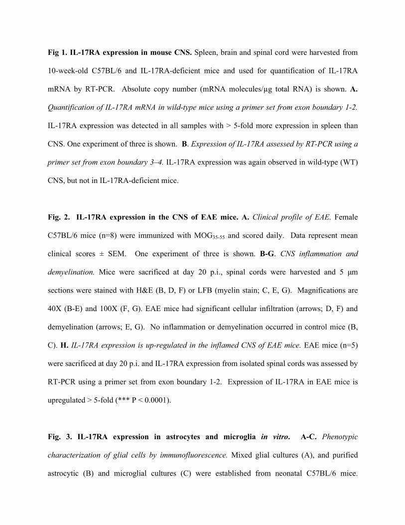
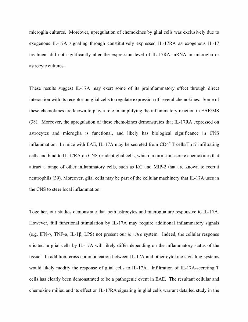
Fig 1. IL-17RA expression in mouse CNS. Spleen, brain and spinal cord were harvested from
10-week-old C57BL/6 and IL-17RA-deficient mice and used for quantification of IL-17RA
mRNA by RT-PCR. Absolute copy number (mRNA molecules/µg total RNA) is shown. A.
Quantification of IL-17RA mRNA in wild-type mice using a primer set from exon boundary 1-2.
IL-17RA expression was detected in all samples with > 5-fold more expression in spleen than
CNS. One experiment of three is shown. B. Expression of IL-17RA assessed by RT-PCR using a
primer set from exon boundary 3–4. IL-17RA expression was again observed in wild-type (WT)
CNS, but not in IL-17RA-deficient mice.
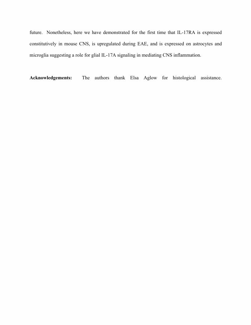
Fig. 2. IL-17RA expression in the CNS of EAE mice. A. Clinical profile of EAE. Female
C57BL/6 mice (n=8) were immunized with MOG35-55 and scored daily. Data represent mean
clinical scores ± SEM. One experiment of three is shown. B-G. CNS inflammation and
demyelination. Mice were sacrificed at day 20 p.i., spinal cords were harvested and 5 µm
sections were stained with H&E (B, D, F) or LFB (myelin stain; C, E, G). Magnifications are
40X (B-E) and 100X (F, G). EAE mice had significant cellular infiltration (arrows; D, F) and
demyelination (arrows; E, G). No inflammation or demyelination occurred in control mice (B,
C). H. IL-17RA expression is up-regulated in the inflamed CNS of EAE mice. EAE mice (n=5)
were sacrificed at day 20 p.i. and IL-17RA expression from isolated spinal cords was assessed by
RT-PCR using a primer set from exon boundary 1-2. Expression of IL-17RA in EAE mice is
upregulated > 5-fold (*** P < 0.0001).
Fig. 3. IL-17RA expression in astrocytes and microglia in vitro. A-C. Phenotypic
characterization of glial cells by immunofluorescence. Mixed glial cultures (A), and purified
astrocytic (B) and microglial cultures (C) were established from neonatal C57BL/6 mice.
Cultures were stained with anti-GFAP antibody (astrocytic marker; green) and anti-CD11b
(microglial marker; red) and counterstained with nuclear stain DAPI (blue). Mixed glial cultures
primarily consist of astrocytes (70-80%) and microglia (5-10%); whereas, purified astrocyte
cultures consist of 80-90% GFAP-positive cells. Purified microglial cultures are 98-99%
CD11b-positive. D-F. Flow cytometry. Glial cells were immunostained for flow cytometric
analysis. Mixed glial cultures (D) contain both GFAP- and CD11b-positive cells. Astrocyte
cultures were free from microglia (< 0.5%) (E) and microglial cultures free of astrocytes (<
0.5%) (F). G. IL-17RA expression in vitro. mRNA was extracted from glial cultures and IL-
17RA expression was quantified by RT-PCR using a primer set from exon boundary 1-2. Data
represent the mean expression of total IL-17RA mRNA from isolated cultures from three
different batches of donors. IL-17RA is expressed 4-fold higher in microglia compared to
astrocytes (***p < 0.0001). Mixed glial culture confers more expression of IL-17RA mRNA in
comparison to astrocyte cultures devoid of microglia (*p = 0.0329).
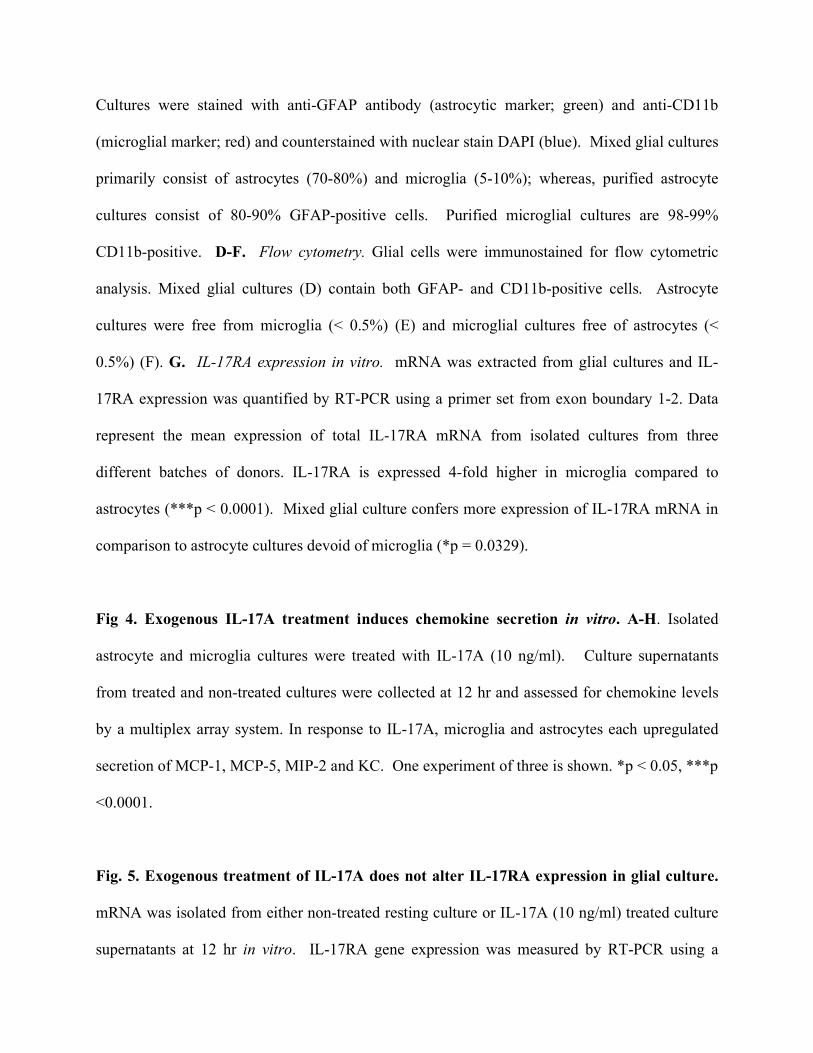
Fig 4. Exogenous IL-17A treatment induces chemokine secretion in vitro. A-H. Isolated
astrocyte and microglia cultures were treated with IL-17A (10 ng/ml). Culture supernatants
from treated and non-treated cultures were collected at 12 hr and assessed for chemokine levels
by a multiplex array system. In response to IL-17A, microglia and astrocytes each upregulated
secretion of MCP-1, MCP-5, MIP-2 and KC. One experiment of three is shown. *p < 0.05, ***p
<0.0001.
Fig. 5. Exogenous treatment of IL-17A does not alter IL-17RA expression in glial culture.
mRNA was isolated from either non-treated resting culture or IL-17A (10 ng/ml) treated culture
supernatants at 12 hr in vitro. IL-17RA gene expression was measured by RT-PCR using a
primer set from exon boundary 1-2. Data represent the mean expression from three different
non- treated and IL-17A- treated culture batches ± SEM. IL-17A treatment did not alter IL-
17RA expression in neonatal glial cells (*p > 0.05).
References
1. Rouvier, E., M. F. Luciani, M. G. Mattei, F. Denizot, and P. Golstein. 1993. CTLA-8,
cloned from an activated T cell, bearing AU-rich messenger RNA instability sequences,
and homologous to a herpesvirus saimiri gene. J Immunol 150:5445.
2. Langrish, C. L., Y. Chen, W. M. Blumenschein, J. Mattson, B. Basham, J. D. Sedgwick,
T. McClanahan, R. A. Kastelein, and D. J. Cua. 2005. IL-23 drives a pathogenic T cell
population that induces autoimmune inflammation. J Exp Med 201:233.
3. Harrington, L. E., R. D. Hatton, P. R. Mangan, H. Turner, T. L. Murphy, K. M. Murphy,
and C. T. Weaver. 2005. Interleukin 17-producing CD4+ effector T cells develop via a
lineage distinct from the T helper type 1 and 2 lineages. Nat Immunol 6:1123.
4. Park, H., Z. Li, X. O. Yang, S. H. Chang, R. Nurieva, Y. H. Wang, Y. Wang, L. Hood, Z.
Zhu, Q. Tian, and C. Dong. 2005. A distinct lineage of CD4 T cells regulates tissue
inflammation by producing interleukin 17. Nat Immunol 6:1133.
5. Bettelli, E., M. Oukka, and V. K. Kuchroo. 2007. T(H)-17 cells in the circle of immunity
and autoimmunity. Nat Immunol 8:345.
6. Bettelli, E., Y. Carrier, W. Gao, T. Korn, T. B. Strom, M. Oukka, H. L. Weiner, and V.
K. Kuchroo. 2006. Reciprocal developmental pathways for the generation of pathogenic
effector TH17 and regulatory T cells. Nature 441:235.
7. Chen, Z., and J. J. O'Shea. 2007. Regulation of IL-17 production in human lymphocytes.
Cytokine
8. Weaver, C. T., R. D. Hatton, P. R. Mangan, and L. E. Harrington. 2007. IL-17 family
cytokines and the expanding diversity of effector T cell lineages. Annu Rev Immunol
25:821.
9. Veldhoen, M., R. J. Hocking, C. J. Atkins, R. M. Locksley, and B. Stockinger. 2006.
TGFbeta in the context of an inflammatory cytokine milieu supports de novo
differentiation of IL-17-producing T cells. Immunity 24:179.
10. Sutton, C., C. Brereton, B. Keogh, K. H. Mills, and E. C. Lavelle. 2006. A crucial role for
interleukin (IL)-1 in the induction of IL-17-producing T cells that mediate autoimmune
encephalomyelitis. J Exp Med 203:1685.
11. Yu, J. J., M. J. Ruddy, G. C. Wong, C. Sfintescu, P. J. Baker, J. B. Smith, R. T. Evans,
and S. L. Gaffen. 2007. An essential role for IL-17 in preventing pathogen-initiated bone
destruction: recruitment of neutrophils to inflamed bone requires IL-17 receptor-
dependent signals. Blood 109:3794.
12. Huang, W., L. Na, P. L. Fidel, and P. Schwarzenberger. 2004. Requirement of
interleukin-17A for systemic anti-Candida albicans host defense in mice. J Infect Dis
190:624.
13. Ye, P., P. B. Garvey, P. Zhang, S. Nelson, G. Bagby, W. R. Summer, P.
Schwarzenberger, J. E. Shellito, and J. K. Kolls. 2001. Interleukin-17 and lung host
defense against Klebsiella pneumoniae infection. Am J Respir Cell Mol Biol 25:335.
14. Kelly, M. N., J. K. Kolls, K. Happel, J. D. Schwartzman, P. Schwarzenberger, C. Combe,
M. Moretto, and I. A. Khan. 2005. Interleukin-17/interleukin-17 receptor-mediated
signaling is important for generation of an optimal polymorphonuclear response against
Toxoplasma gondii infection. Infect Immun 73:617.
15. Kolls, J. K., and A. Linden. 2004. Interleukin-17 family members and inflammation.
Immunity 21:467.
16. Vaknin-Dembinsky, A., K. Balashov, and H. L. Weiner. 2006. IL-23 is increased in
dendritic cells in multiple sclerosis and down-regulation of IL-23 by antisense oligos
increases dendritic cell IL-10 production. J Immunol 176:7768.
17. Kotake, S., N. Udagawa, N. Takahashi, K. Matsuzaki, K. Itoh, S. Ishiyama, S. Saito, K.
Inoue, N. Kamatani, M. T. Gillespie, T. J. Martin, and T. Suda. 1999. IL-17 in synovial
fluids from patients with rheumatoid arthritis is a potent stimulator of osteoclastogenesis.
J Clin Invest 103:1345.
18. Komiyama, Y., S. Nakae, T. Matsuki, A. Nambu, H. Ishigame, S. Kakuta, K. Sudo, and
Y. Iwakura. 2006. IL-17 plays an important role in the development of experimental
autoimmune encephalomyelitis. J Immunol 177:566.
19. Forlow, S. B., J. R. Schurr, J. K. Kolls, G. J. Bagby, P. O. Schwarzenberger, and K. Ley.
2001. Increased granulopoiesis through interleukin-17 and granulocyte colony-
stimulating factor in leukocyte adhesion molecule-deficient mice. Blood 98:3309.
20. Linden, A., and M. Adachi. 2002. Neutrophilic airway inflammation and IL-17. Allergy
57:769.
21. Jeffery, J. Y., Gaffen, S. L. 2008. Interleukin-17: A novel inflammatory cytokine that
bridges innate and adaptive immunity. Frontiers in Biosciences 13:170.
22. McFarland, H. F., and R. Martin. 2007. Multiple sclerosis: a complicated picture of
autoimmunity. Nat Immunol 8:913.
23. Zhang, G. X., B. Gran, S. Yu, J. Li, I. Siglienti, X. Chen, M. Kamoun, and A. Rostami.
2003. Induction of experimental autoimmune encephalomyelitis in IL-12 receptor-beta 2-
deficient mice: IL-12 responsiveness is not required in the pathogenesis of inflammatory
demyelination in the central nervous system. J Immunol 170:2153.
24. Chen, Y., C. L. Langrish, B. McKenzie, B. Joyce-Shaikh, J. S. Stumhofer, T.
McClanahan, W. Blumenschein, T. Churakovsa, J. Low, L. Presta, C. A. Hunter, R. A.
Kastelein, and D. J. Cua. 2006. Anti-IL-23 therapy inhibits multiple inflammatory
pathways and ameliorates autoimmune encephalomyelitis. J Clin Invest 116:1317.
25. Lock, C., G. Hermans, R. Pedotti, A. Brendolan, E. Schadt, H. Garren, A. Langer-Gould,
S. Strober, B. Cannella, J. Allard, P. Klonowski, A. Austin, N. Lad, N. Kaminski, S. J.
Galli, J. R. Oksenberg, C. S. Raine, R. Heller, and L. Steinman. 2002. Gene-microarray
analysis of multiple sclerosis lesions yields new targets validated in autoimmune
encephalomyelitis. Nat Med 8:500.
26. Matusevicius, D., P. Kivisakk, B. He, N. Kostulas, V. Ozenci, S. Fredrikson, and H.
Link. 1999. Interleukin-17 mRNA expression in blood and CSF mononuclear cells is
augmented in multiple sclerosis. Mult Scler 5:101.
27. Kebir, H., K. Kreymborg, I. Ifergan, A. Dodelet-Devillers, R. Cayrol, M. Bernard, F.
Giuliani, N. Arbour, B. Becher, and A. Prat. 2007. Human TH17 lymphocytes promote
blood-brain barrier disruption and central nervous system inflammation. Nat Med
13:1173.
28. Moseley, T. A., D. R. Haudenschild, L. Rose, and A. H. Reddi. 2003. Interleukin-17
family and IL-17 receptors. Cytokine Growth Factor Rev 14:155.
29. Yao, Z., W. C. Fanslow, M. F. Seldin, A. M. Rousseau, S. L. Painter, M. R. Comeau, J. I.
Cohen, and M. K. Spriggs. 1995. Herpesvirus Saimiri encodes a new cytokine, IL-17,
which binds to a novel cytokine receptor. Immunity 3:811.
30. McAllister, F., A. Henry, J. L. Kreindler, P. J. Dubin, L. Ulrich, C. Steele, J. D. Finder, J.
M. Pilewski, B. M. Carreno, S. J. Goldman, J. Pirhonen, and J. K. Kolls. 2005. Role of
IL-17A, IL-17F, and the IL-17 receptor in regulating growth-related oncogene-alpha and
granulocyte colony-stimulating factor in bronchial epithelium: implications for airway
inflammation in cystic fibrosis. J Immunol 175:404.
31. Ye, P., F. H. Rodriguez, S. Kanaly, K. L. Stocking, J. Schurr, P. Schwarzenberger, P.
Oliver, W. Huang, P. Zhang, J. Zhang, J. E. Shellito, G. J. Bagby, S. Nelson, K. Charrier,
J. J. Peschon, and J. K. Kolls. 2001. Requirement of interleukin 17 receptor signaling for
lung CXC chemokine and granulocyte colony-stimulating factor expression, neutrophil
recruitment, and host defense. J Exp Med 194:519.
32. Fitzgerald, D. C., B. Ciric, T. Touil, H. Harle, J. Grammatikopolou, J. Das Sarma, B.
Gran, G. X. Zhang, and A. Rostami. 2007. Suppressive effect of IL-27 on
encephalitogenic Th17 cells and the effector phase of experimental autoimmune
encephalomyelitis. J Immunol 179:3268.
33. Gingras, M., V. Gagnon, S. Minotti, H. D. Durham, and F. Berthod. 2007. Optimized
protocols for isolation of primary motor neurons, astrocytes and microglia from
embryonic mouse spinal cord. J Neurosci Methods 163:111.
34. Bailey, S. L., P. A. Carpentier, E. J. McMahon, W. S. Begolka, and S. D. Miller. 2006.
Innate and adaptive immune responses of the central nervous system. Crit Rev Immunol
26:149.
35. Shen, F., M. J. Ruddy, P. Plamondon, and S. L. Gaffen. 2005. Cytokines link osteoblasts
and inflammation: microarray analysis of interleukin-17- and TNF-alpha-induced genes
in bone cells. J Leukoc Biol 77:388.
36. Schwandner, R., K. Yamaguchi, and Z. Cao. 2000. Requirement of tumor necrosis factor
receptor-associated factor (TRAF)6 in interleukin 17 signal transduction. J Exp Med
191:1233.
37. Tato, C. M., A. Laurence, and J. J. O'Shea. 2006. Helper T cell differentiation enters a
new era: le roi est mort; vive le roi! J Exp Med 203:809.
38. Ransohoff, R. M., L. Liu, and A. E. Cardona. 2007. Chemokines and chemokine
receptors: multipurpose players in neuroinflammation. Int Rev Neurobiol 82:187.
39. Kobayashi, Y. 2006. Neutrophil infiltration and chemokines. Crit Rev Immunol 26:307.
Related Documents