Personalized Medicine and Imaging Functional Ex Vivo Assay to Select Homologous Recombination–Deficient Breast Tumors for PARP Inhibitor Treatment Kishan A.T. Naipal 1 , Nicole S. Verkaik 1 , Najim Ameziane 2 , Carolien H.M. van Deurzen 3 , Petra ter Brugge 4 , Matty Meijers 5 , Anieta M. Sieuwerts 6 , John W. Martens 6 , Mark J. O'Connor 7 , Harry Vrieling 5 , Jan H.J. Hoeijmakers 1 , Jos Jonkers 4 , Roland Kanaar 1,8,9 , Johan P. de Winter 2† , Maaike P. Vreeswijk 5,10 , Agnes Jager 6 , and Dik C. van Gent 1 Abstract Purpose: Poly(ADP-ribose) polymerase (PARP) inhibitors are promising targeted treatment options for hereditary breast tumors with a homologous recombination (HR) deficiency caused by BRCA1 or BRCA2 mutations. However, the functional consequence of BRCA gene mutations is not always known and tumors can be HR deficient for other reasons than BRCA gene mutations. Therefore, we aimed to develop a functional test to determine HR activity in tumor samples to facilitate selection of patients eligible for PARP inhibitor treatment. Experimental design: We obtained 54 fresh primary breast tumor samples from patients undergoing surgery. We determined their HR capacity by studying the formation of ionizing radiation induced foci (IRIF) of the HR protein RAD51 after ex vivo irradiation of these organotypic breast tumor samples. Tumors showing impaired RAD51 IRIF formation were subjected to genetic and epigenetic analysis. Results: Five of 45 primary breast tumors with sufficient numbers of proliferating tumor cells were RAD51 IRIF formation deficient (11%, 95% CI, 5%–24%). This HR defect was significantly associated with triple-negative breast cancer (OR, 57; 95% CI, 3.9–825; P ¼ 0.003). Two of five HR-deficient tumors were not caused by mutations in the BRCA genes, but by BRCA1 promoter hypermethylation. Conclusion: The functional RAD51 IRIF assay faithfully identifies HR-deficient tumors and has clear advantages over gene sequencing. It is a relatively easy assay that can be performed on biopsy material, making it a powerful tool to select patients with an HR-deficient cancer for PARP inhibitor treatment in the clinic. Clin Cancer Res; 20(18); 4816–26. Ó2014 AACR. Introduction Breast cancer is the most common female cancer and the leading cause of cancer-related deaths in women (1). Great improvements have been made in breast cancer treatment with targeted therapies in estrogen receptor (ER)–positive and human epidermal growth factor receptor 2 (HER2)–positive tumors for adjuvant settings as well as treatment of metastatic breast cancer (2, 3). However, triple-negative breast cancers (TNBC), which do not express ER, progesterone receptor (PR), or HER2, have a relatively poor prognosis because of the absence of an effective targeted treatment regimen. A particular type of TNBCs arises in familial cases of breast cancer, especially BRCA1 mutation carriers. BRCA1 muta- tion–associated tumors are predominantly high-grade TNBC (4). Interestingly, TNBCs arising in BRCA1 mutation carriers and sporadic TNBCs share clinicopathologic and molecular characteristics. Therefore, similar etiology is pro- posed for these groups of breast cancer (5–7). Hereditary breast cancer associated with BRCA1 or BRCA2 mutations 1 Department of Genetics, Erasmus University Medical Center, Rotter- dam, the Netherlands. 2 Department of Clinical Genetics, VU University Medical Center, Amsterdam, the Netherlands. 3 Department of Pathol- ogy, Erasmus University Medical Center, Rotterdam, the Netherlands. 4 Division of Molecular Pathology, Netherlands Cancer Institute, Amster- dam, the Netherlands. 5 Department of Toxicogenetics, Leiden Univer- sity Medical Center, Leiden, the Netherlands. 6 Department of Medical Oncology, Erasmus University Medical Center, Rotterdam, the Nether- lands. 7 AstraZeneca, iMed Oncology, Macclesfield, Cheshire, United Kingdom. 8 Cancer Genomics Center Netherlands, Erasmus University Medical Center, Rotterdam, the Netherlands. 9 Department of Radiation Oncology, Erasmus University Medical Center, Rotterdam, the Nether- lands. 10 Department of Human Genetics, Leiden University Medical Center, Leiden, the Netherlands. Note: Supplementary data for this article are available at Clinical Cancer Research Online (http://clincancerres.aacrjournals.org/). †Deceased. Corresponding Author: Dik C. van Gent, Erasmus University Medical Center, P.O. Box 2040, 3000 CA Rotterdam, the Netherlands, Phone: 31-10-7043932; Fax: 31-10-7044743; E-mail: [email protected] doi: 10.1158/1078-0432.CCR-14-0571 Ó2014 American Association for Cancer Research. Clinical Cancer Research Clin Cancer Res; 20(18) September 15, 2014 4816 on July 10, 2018. © 2014 American Association for Cancer Research. clincancerres.aacrjournals.org Downloaded from Published OnlineFirst June 24, 2014; DOI: 10.1158/1078-0432.CCR-14-0571

Welcome message from author
This document is posted to help you gain knowledge. Please leave a comment to let me know what you think about it! Share it to your friends and learn new things together.
Transcript
Personalized Medicine and Imaging
Functional Ex Vivo Assay to Select HomologousRecombination–Deficient Breast Tumors for PARPInhibitor Treatment
Kishan A.T. Naipal1, Nicole S. Verkaik1, Najim Ameziane2, Carolien H.M. van Deurzen3, Petra ter Brugge4,Matty Meijers5, Anieta M. Sieuwerts6, John W. Martens6, Mark J. O'Connor7, Harry Vrieling5,Jan H.J. Hoeijmakers1, Jos Jonkers4, Roland Kanaar1,8,9, Johan P. de Winter2†, Maaike P. Vreeswijk5,10,Agnes Jager6, and Dik C. van Gent1
AbstractPurpose: Poly(ADP-ribose) polymerase (PARP) inhibitors are promising targeted treatment options for
hereditary breast tumors with a homologous recombination (HR) deficiency caused by BRCA1 or BRCA2
mutations. However, the functional consequence of BRCA genemutations is not always known and tumors
can be HR deficient for other reasons than BRCA gene mutations. Therefore, we aimed to develop a
functional test to determine HR activity in tumor samples to facilitate selection of patients eligible for PARP
inhibitor treatment.
Experimental design: We obtained 54 fresh primary breast tumor samples from patients undergoing
surgery. We determined their HR capacity by studying the formation of ionizing radiation induced foci
(IRIF) of the HR protein RAD51 after ex vivo irradiation of these organotypic breast tumor samples. Tumors
showing impaired RAD51 IRIF formation were subjected to genetic and epigenetic analysis.
Results: Five of 45 primary breast tumors with sufficient numbers of proliferating tumor cells were
RAD51 IRIF formation deficient (11%, 95%CI, 5%–24%). This HR defect was significantly associated with
triple-negative breast cancer (OR, 57; 95%CI, 3.9–825;P¼0.003). Twoof fiveHR-deficient tumorswerenot
caused by mutations in the BRCA genes, but by BRCA1 promoter hypermethylation.
Conclusion: The functional RAD51 IRIF assay faithfully identifies HR-deficient tumors and has clear
advantages over gene sequencing. It is a relatively easy assay that can be performed on biopsy material,
making it a powerful tool to select patients with an HR-deficient cancer for PARP inhibitor treatment in the
clinic. Clin Cancer Res; 20(18); 4816–26. �2014 AACR.
IntroductionBreast cancer is the most common female cancer and the
leading cause of cancer-related deaths in women (1). Greatimprovements have been made in breast cancer treatmentwith targeted therapies in estrogen receptor (ER)–positive andhuman epidermal growth factor receptor 2 (HER2)–positivetumors for adjuvant settings as well as treatment ofmetastaticbreast cancer (2, 3). However, triple-negative breast cancers(TNBC), which do not express ER, progesterone receptor(PR), or HER2, have a relatively poor prognosis because ofthe absence of an effective targeted treatment regimen.
Aparticular typeof TNBCs arises in familial cases of breastcancer, especially BRCA1 mutation carriers. BRCA1 muta-tion–associated tumors are predominantly high-gradeTNBC (4). Interestingly, TNBCs arising in BRCA1mutationcarriers and sporadic TNBCs share clinicopathologic andmolecular characteristics. Therefore, similar etiology is pro-posed for these groups of breast cancer (5–7). Hereditarybreast cancer associated with BRCA1 or BRCA2 mutations
1Department of Genetics, Erasmus University Medical Center, Rotter-dam, the Netherlands. 2Department of Clinical Genetics, VU UniversityMedical Center, Amsterdam, the Netherlands. 3Department of Pathol-ogy, Erasmus University Medical Center, Rotterdam, the Netherlands.4Division of Molecular Pathology, Netherlands Cancer Institute, Amster-dam, the Netherlands. 5Department of Toxicogenetics, Leiden Univer-sity Medical Center, Leiden, the Netherlands. 6Department of MedicalOncology, Erasmus University Medical Center, Rotterdam, the Nether-lands. 7AstraZeneca, iMed Oncology, Macclesfield, Cheshire, UnitedKingdom. 8Cancer Genomics Center Netherlands, Erasmus UniversityMedical Center, Rotterdam, the Netherlands. 9Department of RadiationOncology, Erasmus University Medical Center, Rotterdam, the Nether-lands. 10Department of Human Genetics, Leiden University MedicalCenter, Leiden, the Netherlands.
Note: Supplementary data for this article are available at Clinical CancerResearch Online (http://clincancerres.aacrjournals.org/).
†Deceased.
Corresponding Author: Dik C. van Gent, Erasmus University MedicalCenter, P.O. Box 2040, 3000 CA Rotterdam, the Netherlands, Phone:31-10-7043932; Fax: 31-10-7044743; E-mail: [email protected]
doi: 10.1158/1078-0432.CCR-14-0571
�2014 American Association for Cancer Research.
ClinicalCancer
Research
Clin Cancer Res; 20(18) September 15, 20144816
on July 10, 2018. © 2014 American Association for Cancer Research. clincancerres.aacrjournals.org Downloaded from
Published OnlineFirst June 24, 2014; DOI: 10.1158/1078-0432.CCR-14-0571

show defects in the DNA damage response (DDR), whichencompasses DNA damage repair, cell-cycle checkpointsignaling and apoptosis.BRCA1orBRCA2deficiency resultsin impaired double-strand break (DSB) repair by homolo-gous recombination (HR) and chromosomal instability,which may contribute to carcinogenesis.Dysregulation of DDR processes is a common phenom-
enon in cancers, sometimes highly associatedwith a specifictype or subtype of cancer, for example, mismatch repairdefects in colon carcinoma, cancer associatedwith crosslinkrepair defects in Fanconi anemia (FA), and HR defects inhereditary breast cancer (8). Interestingly, DDR defects arenot only important to understand the carcinogenic process,but may also be utilized to optimize therapy response. Adefective DDR pathway in tumor cells can cause dependen-cy on another specific back up mechanism that allowscellular survival. This provides options for therapeuticintervention: specific targeting of this back up system canresult in selective tumor cell death, a phenomenon referredto as "synthetic lethality" (9, 10). The major advantage ofthis approach is efficient tumor-specific cell killing withfewer adverse effects for the patient, because normal cells donot depend exclusively on the targeted pathway and there-forewill survive the treatment. An exciting example of such asynthetic lethal approach is treatment of BRCA-deficienttumors with poly(ADP-ribose) polymerase (PARP) inhibi-tors (11–13).PARP activity contributes toward signaling the presence
of single-strand DNA breaks and base damage by attachingpoly(ADP)ribose moieties to histones and other proteins,including itself, at the site of damage, which results inefficient repair of these types of DNA damage (14). Inhibit-ing PARP activity in proliferating cells results in excessivesingle strand and/or base lesions, which cause the collapseof replication forks and DSBs if encountered by the repli-cation machinery (15, 16). Repair of these stalled replica-
tion fork-induced DSBs specifically requires HR (17, 18). Ifleft unrepaired these types of lesions accumulate and causecell death (17, 18). Therefore, HR-deficient cells associatedwith BRCA1 or BRCA2mutations are extremely sensitive toPARP inhibition (8, 11, 12).
PARP inhibitor treatment showed a very effective antitu-mor activity in patients with BRCA mutation–associatedcancers in phase I and II clinical trials (13, 19–21). Fur-thermore, the toxic side-effects commonly associated withconventional chemotherapywere relativelymild after PARPinhibitor treatment. Several non-BRCA mutation–associat-ed tumor cell lines are also sensitive to this specific type oftreatment, thereby extending the potential clinical applica-tion of PARP inhibitors. These cells had defects in genes,which also lead to impaired HR and/or cell-cycle check-points (22–26). These defects are also detected in humanbreast cancers (27). Unfortunately, in spite of promisingexperimental and clinical data, PARP inhibitors have not yetmade it to breast cancer treatment in the clinic. One likelyexplanation for this is the lack of a marker for patientselection (except for known BRCA mutation status),because targeted treatment with PARP inhibitors can onlybe successful in a well-defined patient population andtherefore selection of the appropriate patient populationbefore treatment is very important.
HR activity in a tumor cell is probably themost importantfactor to predict whether treatment with PARP inhibitorswill be successful (28). HR is mediated by the RAD51protein that forms a nucleoprotein filament that is able tocarry out the crucial strand exchange step ofHR. The BRCA2protein delivers RAD51 to DNA DSBs, where it can bedetected as foci in the nucleus. Formation of RAD51 ion-izing radiation induced foci (IRIF) can therefore be used as aconvenient and highly informative test for HR function.Cells deficient in BRCA1, BRCA2, or a number of other HRfactors do not or inefficiently form RAD51 IRIF, suggestingthat this read out can be used as a PARP inhibitor sensitivitymarker (28, 29). Here, we describe how ex vivo RAD51 IRIFformation capacity in primary breast tumor specimens canidentify HR-deficient tumors.
Materials and MethodsPatient-derived xenografts
Xenograft models were initiated by implanting freshpatient-derived tumor tissue subcutaneously in the thighof immunocompromised mice. Tumors were allowed togrow out up to 2 cm in diameter and were subsequentlyisolated for further experiments. To maintain the specificmodel in vivo, tumors were isolated from mice, sliced intosmaller sections, and implanted in other immunocompro-mised mice. BRCA1 and BRCA2 status in tumors wasanalyzed by immunoblotting and exon sequencing (P. terBrugge and J. Jonkers; unpublished data).
Clinical breast cancer specimensFresh breast tumor tissue was obtained from patients
undergoing wide local excision or amputation for breast
Translational RelevanceThis functional assay will facilitate selection of
patients with (breast) cancer for poly(ADP-ribose) poly-merase (PARP) inhibitor treatment. As this is a func-tional test, not only knownBRCA1 andBRCA2mutationcarriers will be identified, but also tumors with defects inother genes of the same genetic pathway and epigeneticforms of gene silencing. The assay can be performed onbiopsy material, making it a powerful tool to selectadditional patients for ongoing and future clinical trialswith PARP inhibitors. In addition to this relatively short-term translational achievement, the assaywill in the longrun be useful to select patients for PARP inhibitor treat-ment in general oncology practice for breast cancer, aswell as other tumor types. We expect that the number ofpatients that benefit from this promising targeted cancertherapy can be increased significantly by implementingthe RAD51 ionizing radiation induced foci assay.
Functional Assay for PARP Inhibitor Treatment
www.aacrjournals.org Clin Cancer Res; 20(18) September 15, 2014 4817
on July 10, 2018. © 2014 American Association for Cancer Research. clincancerres.aacrjournals.org Downloaded from
Published OnlineFirst June 24, 2014; DOI: 10.1158/1078-0432.CCR-14-0571

cancer at Erasmus Medical Center (Erasmus MC), Haven-ziekenhuis Rotterdam, and Leiden University Medical Cen-ter (LUMC) Leiden, The Netherlands. After resection, thetissuewasdirectly transported to the Pathology department.After macroscopic investigation and determination oftumor areas for diagnostic purposes by a pathologist, leftover tumor tissue was used for research purposes accordingto the code of proper secondary use of human tissue in theNetherlands establishedby theDutch FederationofMedicalScientific Societies and approved by the local MedicalEthical committees. Specimens were coded anonymouslyin a way that they were not traceable back to the patient bylaboratoryworkers. Patients receiving neo-adjuvant chemo-or radiotherapy were excluded.
Tissue culture systemResearch samples were obtained within 4 hours after
surgical resection and kept at 4�C during transport to thelaboratory in breast medium (30) containing a 2:1 mixtureof Dulbecco’s modified Eagle’s medium (DMEM) withoutphenol red and nutrient mixture F-12 (HAM) supplemen-ted with 2% fetal bovine serum (FBS), hydrocortisone(0.3 mg/mL; Sigma), insulin (4 mg/mL; Sigma), transferrin(4mg/mL; Sigma), 3,30,5-triiodothyronine (1ng/mL; Sigma),cholera toxin (7 ng/mL; Sigma), epidermal growth factor(8 ng/mL; Sigma), adenine (0.2 mg/mL; Sigma). Excess fattissue was discarded using surgical tools and tumor speci-mens were depending on tissue quantity and type of exper-iment, sliced into 300-mm slices using a Leica vibratome1200S or sliced manually into approximately 2-mm slices.No differences in visualization of RAD51 IRIFwere observedbetween these slicingmethods (data not shown). Slices weredirectly incubated in breast medium and irradiated with5 Gy g-radiation using a 137Cs source (0.7 Gy/min). Incu-bation and irradiation of samples was performed within6 hours after surgical resection. Subsequently, sampleswere incubated at 37�C and 5% CO2 on a rotating plat-form (60 rpm) for 2 hours. Afterward, they were fixed in37% neutrally buffered formalin for at least 24 hours atroom temperature and subsequently embedded in paraffin(overnight procedure). Microscopy sections of 4 mm weregenerated and subjected to immunofluorescent staining(Supplementary Fig. S1).
Immunofluorescent stainingSections were deparaffinized using xylene and hydrated
with declining concentrations of ethanol. Target antigenretrieval was performed using DAKO Antigen Retrievalbuffer (pH 9.0 for RAD51 and pH 6.9 for others), whichwas heated to 100�C for 15 minutes. Cells were permea-bilized using phosphate buffered saline (PBS) with 0.2%Triton X-100 for 20minutes. For RAD51–geminin costain-ing, an additional DNase (1,000 U/mL; Roche Diagnos-tics) incubation was performed at 37�C for 1 hour. Block-ing was achieved using PBS with 2% FBS and 1% bovineserum albumin (BSA). Primary antibodies [anti-RAD51(GeneTex clone14B4 GTX70230) 1/200, anti-geminin(Proteintech Group 10802-1-AP) 1/400, anti-cleaved cas-
pase-3 (Cell Signaling Technology 9664S) 1/100], anti-gH2AX (Millipore 2310355) 1/500, anti-53BP1 (NovusBiologicals NB100-304) 1/500, anti-P63 (Ventana clone4A4 790-4509, ready to use)] were diluted in blockingbuffer and incubated for 90minutes at room temperature.Secondary Alexa Fluor 594 or 488 antibodies were used tovisualize the primary antibody. Sections were mountedusing Vectashield mounting medium with DAPI. For P63staining, the primary antibodies were detected using DABchromogen.
Scoring of RAD51 fociTumor cells/areas were determined by morphology on a
serial hematoxylin and eosin (H&E) stained section of thesame tumor slice that was used for RAD51 foci analysis.Geminin-positive cells were counted manually. A cell wasconsidered positive for geminin if the complete nucleuswasstained by the geminin antibody. These cells were scored forthe presence of RAD51 foci. A cell was considered positivefor RAD51 foci if more than 5 nuclear foci were detected.The percentages of RAD51 foci–positive cells in the gemi-nin-positive population were calculated. Approximately100 geminin-positive cells were counted, unless sectionshad fewer geminin-positive cells, in which case at least 30cells were counted in each tumor sample. To generate errorbars, the standard error was estimated assuming a binomialdistribution.
Statistical analysisStatistical analysis were all 2-sided and performed using
IBM SPSS statistics v21.
Next-generation sequencingGenomic DNAwas isolated from fresh-frozen samples of
tumors using the Nucleospin Tissue Kit (Macherey-Nagel)according to themanufacturer’s protocol. The percentage oftumor cells was determined by H&E staining of 5-mmcryosections of the same sample. A custom Haloplex (Agi-lent) Kit was used to enrich the coding regions of specificgenes using 200 ng of genomic DNA to obtain sequencinglibraries. Samples were tagged with a unique barcode andpooled before pair-ended sequencing 150 bp on the Illu-mina Miseq platform. SureCall software (Agilent) was usedto detect variants. Validation of the identified mutationswas performed by Sanger sequencing.
BRCA1 promoter methylation and copy numbervariation analysis by MS-MLPA
To assess promoter methylation of BRCA1, 2 MS-MLPAKits (MRC-Holland) were used, each containing a differentprobe (31). Twenty-five nanograms of DNA was denaturedfor 10 minutes at 98�C and subsequently cooled down to25�C. After addition of SALSA Probe-mix and MLPA buffersamples were incubated for 1 minute at 95�C followed byhybridization for 16 hours at 60�C.Next, samples were splitand ligated with (methylation test) or without (copy num-ber test) the additionofHhaI enzyme for 30minutes at 49�Cand then heated for 5 minutes at 98�C.
Naipal et al.
Clin Cancer Res; 20(18) September 15, 2014 Clinical Cancer Research4818
on July 10, 2018. © 2014 American Association for Cancer Research. clincancerres.aacrjournals.org Downloaded from
Published OnlineFirst June 24, 2014; DOI: 10.1158/1078-0432.CCR-14-0571

SALSA PCR-buffer and polymerase mix (includingdNTPs, SALSA polymerase, and PCR primers) were addedand samples were subjected to the following PCR reactionfor 35 cycles: 30 seconds at 95�C; 30 seconds at 60�C; 60seconds at 72�C. Finally, the PCR reaction was incubated at72�C for 20 minutes. The amplified PCR products wereseparated by electrophoresis on anABI PRISM310 fragmentanalyzer (Applied Biosystems) and analyzed using Gene-marker analysis software (Softgenetics).
In situ detection of BRCA1 RNAIn situ detection of BRCA1 mRNA was performed using
RNAScope (ACD) using standard protocols as described bythemanufacturer (32). BRCA1 probes were purchased fromthe same company. In short, paraffin sections were depar-affinized with xylene and samples were subjected to pre-treatment steps described by the manufacturer. Next,hybridization of target probes [BRCA1 along with POLR2A(positive control) and DapB (negative control)] wasachieved by incubating samples with specific probes for 2hours at 40�C. Subsequently, several amplification stepswere performed using specific amplification buffers toamplify the hybridized probe signal and visualization ofthis signal was achieved using Fast Red dye. Samples werecounterstained with Mayer’s hematoxylin solution andmounted using EcoMount mounting medium.
ResultsEx vivo RAD51 IRIF formation in xenograft tumorsCells in the SorG2phase of the cell cycle formRAD51 foci
after DSB induction by IR. We adapted the immunofluo-rescence detection of these IRIF for thin breast tumor tissueslices and investigatedwhetherwe could distinguish tumorswith known defects in BRCA1 or BRCA2 from other tumorsby analyzing RAD51 IRIF formation. To validate our pro-cedures, we used a collection of BRCA-positive and -nega-tive human to mouse xenograft tumor models. Conditionsfor culturing tissue slices derived from these xenografttumors were optimized (Naipal and colleagues, in prepa-ration), slices were irradiated ex vivo, fixed after 2 hours andRAD51 IRIF formation was analyzed (Supplementary Fig.S1). The 2-hour time point was optimal for RAD51 IRIFformation (ref. 33 and Supplementary Fig. S2A). As RAD51IRIF are only expected to occur in S- or G2-phase cells, wealso identified this cell population in the tumor slices bystaining for the cell-cycle marker geminin (refs. 34and 35; Fig. 1A). Indeed, RAD51 IRIF–positive nuclei wereonly found in irradiated geminin-positive cells (ref. 35 andSupplementary Fig. S2B).In a blinded experimental setup we found all tumors
without a known DDR defect (n¼ 4) to display prominentRAD51 IRIF in more than 50% of geminin-positive cells(Fig. 1B). On the other hand, we observed less than 10% ofgeminin-positive cells exhibited RAD51 IRIF in organotypicslices from tumors with known BRCA1 or BRCA2 (n ¼ 8)defects. BRCA1 deficiency caused by a frame shift mutationor a BRCA1 promoter hypermethylation both displayed
absence of RAD51 foci in xenograft tumors, showing thatgenetic and epigenetic modes of gene inactivation havesimilar RAD51 foci–deficient phenotypes in this assay. Incells without geminin expression, we did not detect RAD51IRIF and tumors proficient for BRCA1 or BRCA2 that werenot irradiated formed RAD51 foci in less than 10% ofgeminin-positive cells (Fig. 1A and B), showing that theassay is specific for induction of foci by IR in S- andG2-phasecells.
Ex vivo RAD51 IRIF formation in human breast tumorsWe investigated whether this assay could also be used for
clinical tumor specimens with a known BRCA1 defect. Weobtained a tumor biopsy from a patient carrying a germlineBRCA1 mutation who had developed a retrosternal recur-rence after previous primary breast cancer treatment. In thistumor, only 11% of geminin-positive cells displayedRAD51 IRIF, whereas tumor slices from unselected primarytumors (n ¼ 5) showed RAD51 IRIF in more than 50% ofthe geminin-positive cells (Fig. 2A and B). Results from thexenografts and the patient biopsy indicate that RAD51 IRIFcan be identified in ex vivo irradiated organotypic tumorslices and that this assay can be used to discriminate HR-deficient and HR-proficient tumors.
After validation, we used this assay to identify HR defectsin clinical breast cancer specimens. We collected 54 (che-motherapy na€�ve) tumor samples obtained from patientsthat underwent breast cancer surgery and generated orga-notypic tumor slices. Information about BRCA mutationstatus, family history, and pathology reports were unknownto the investigators during the analysis of the tumor sam-ples. Pathology reports from corresponding tumors wereobtained afterward (Supplementary Table S1). Themajorityof tumors were histologically classified as ductal carcinoma(82%; n¼ 44)whereas 15% (n¼ 8)was classified as lobularcarcinoma. In addition, 93% (n ¼ 50) of the samplesexpressed either ER, PR, or HER2 receptor and 7% (n ¼4) of tumors had no expression of these 3 receptors (TNBC)as determined by immunohistochemical analysis. Ninetumor samples contained very low numbers of geminin-expressing cells and could therefore not be analyzed. Therewas no specific correlation between low geminin expressionand pathologic tumor characteristics (Supplementary TableS2), suggesting that this was the result of coincidentalsampling of tumor areas that were less proliferative orexhibited rapid decrease in proliferation after resection. Intotal, 45 tumor samples contained sufficient numbers ofgeminin-positive cells for RAD51 IRIF analysis.
Clinicopathologic characteristics of RAD51 IRIF–negative tumors
Based on results of xenograft experiments, RAD51 IRIFformation was considered normal (positive) when morethan 5 foci per nucleus were present in more than 50% ofgeminin-positive cells, whereas RAD51 IRIF formation wasconsidered impaired (negative) when less than 20% ofgeminin-expressing cells contained more than 5 RAD51IRIF per cell. Using these criteria, 5 tumors of 45 (11%;
Functional Assay for PARP Inhibitor Treatment
www.aacrjournals.org Clin Cancer Res; 20(18) September 15, 2014 4819
on July 10, 2018. © 2014 American Association for Cancer Research. clincancerres.aacrjournals.org Downloaded from
Published OnlineFirst June 24, 2014; DOI: 10.1158/1078-0432.CCR-14-0571

95% CI, 5%–24%) showed impaired RAD51 IRIF forma-tion (Fig. 3). We excluded technical reasons for the absenceof RAD51 foci in tumor cells by showing formation ofgH2AX and 53BP1 nuclear foci after irradiation (Sup-plementary Fig. S3A). Furthermore, normal RAD51 IRIFformation was detected in geminin-positive normal breastepithelium, stroma or fat tissue within the same tissuesection (Supplementary Fig. S4). Lobular carcinomas inthis cohort did not show impaired RAD51 IRIF (0 of 7),whereas tumors from the histologic subtypes classified asductal carcinoma (3 of 35) or other (not classified ductal orlobular, 2 of 2) had impaired focus formation (Table 1). Inaddition, RAD51 IRIF–negative tumors were frequentlyclassified as grade 3 carcinomas, although this correlationwas not statistically significant (P¼0.419; Table 1). Three of5 RAD51 IRIF–negative tumors were TNBC whereas the 2other tumors expressed the ER (Table 1). Interestingly, 3 of 4TNBC tumors in this cohort displayed impairedRAD51 IRIFformation, indicating that HR defects were more frequentin TNBC than receptor-positive BC (OR, 57; 95% CI,3.9–825.4; P ¼ 0.003; Table 1).
One of the 45 tumors (sample #30) in the cohort dis-played great variability in RAD51 IRIF formation. RAD51IRIF–positive cells (>5 foci/cell) were clearly recognized,but lower in number compared with other tumors. Strik-ingly, in some regions of the tumor RAD51 IRIF–positivecells were completely absent although geminin-positivecells were present in high numbers, similar to that of othertumors (data not shown). Overall RAD51 IRIF wereobserved in 38%of geminin-positive cells (Fig. 3). Notably,this tumor sample was derived from a 102-year-old patient(Supplementary Table S1); the oldest patient in this cohort.All other tumors showed little variability in RAD51 IRIFformation, indicating that analysis of a small area of thetumor will in most cases be sufficient to accurately deter-mine RAD51 IRIF formation proficiency.
Ex vivo PARP inhibitor sensitivity in RAD51 IRIF–negative tumor
One RAD51 IRIF–negative tumor (sample #20) wasincubated ex vivo with the PARP inhibitor, Olaparib. After96 hours treatment, this tumor sample showed a clearly
0
10
20
30
40
50
60
70
80
90
100
3 4 5 6 8 9 10
Gem
inin
-pos
itive
cel
lsw
ith R
AD
51 fo
ci (
%)
statusBRCA2 status
Tumor code X1 X1 X2 X2 X3 X3 X4 X5 X6 X6 X7 X7
+ +m+
m+
d+
d+
+–
+–
++
++
++
++
RAD51/ dapiRAD51/Geminin/
dapiRAD51/Geminin
20 µm
Zoom in
BRCAproficient
0 Gy
BRCAproficient
5 Gy
BRCAdeficient
5 Gy
– –BRCA1
B
A
Figure 1. RAD51 IRIF in xenograftbreast tumors. A, representativepictures of tumor cells in slices ofBRCA-deficient and BRCA-proficient xenograft tumors.RAD51 IRIF are present in themajority of geminin-positive cells inBRCA-proficient tumors but not inBRCA-deficient tumors. Tumorsamples were subjected to 5 Gy gradiation, cultured at 37�C andfixed 2 hours after irradiation. Rightcolumn of images represent single-cell enlargements of cells in whiteboxes; blue, DAPI; green, geminin;and red, RAD51; scale bar, 20 mm.B, quantification of RAD51 IRIF inxenograft tumor samples. At least30 geminin-positive cells werecounted for each sample. �, geneinactivation caused by BRCA1mutation; m, gene inactivationcaused by BRCA1 promoterhypermethylation; d, nomutation inBRCA1 but absent BRCA1expression on immunoblot. Causeunknown. Same tumor codesrepresent duplicate analysis of thesame xenograft tumor but indifferent mice at different times.Error bars indicate standard errorassuming binomial distribution.
Naipal et al.
Clin Cancer Res; 20(18) September 15, 2014 Clinical Cancer Research4820
on July 10, 2018. © 2014 American Association for Cancer Research. clincancerres.aacrjournals.org Downloaded from
Published OnlineFirst June 24, 2014; DOI: 10.1158/1078-0432.CCR-14-0571

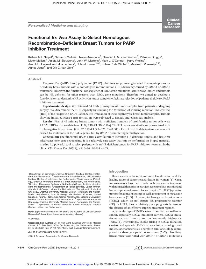
altered morphology with many picnotic nuclei, comparedwith the untreated tumor slice (Fig. 4A). The tumor cellnuclei in the treated sample were either larger in size orfragmented, shrunken, and hyperchromatic. Interestingly,the morphology of the normal mammary ducts (identifiedby their typical double layer of glandular cells and sur-rounding P63-positivemyoepithelial cells) was not affectedby Olaparib treatment (Fig. 4B). Subsequent staining of thesections showed increased levels of the apoptotic markercleaved caspase-3 after 96 hours of exposure to Olaparib(Fig. 4C). In 5 tumors with normal formation of RAD51IRIF, the altered morphology and induction of cleavedcaspase-3 was not noticed after this treatment, stronglysuggesting that induction of apoptosis was because of PARPinhibitor treatment and not caused by declining tissueviability (Fig. 4A and C).PARP inhibition results in DSBs during replication. This
was shown by the formation of gH2AX and 53BP1 nuclearfoci (Supplementary Fig. S3B). As a consequence weexpected an activated HR pathway and thus formation ofRAD51 foci in HR-proficient cells after PARP inhibitortreatment. Accordingly, RAD51 foci induced by Olaparibtreatment were present in normal mammary epitheliumand not in tumor cells in the same tissue slice (Fig. 4B).These results indicate that the functional HR defect in thesetumor cells caused sensitivity to PARP inhibitors, whereasnormal epithelial cells were not affected, opening perspec-
tives for using this assay as a functional test for clinicalsensitivity.
Genetic and epigenetic analysis of RAD51 IRIF–negative tumors
Subsequently, we determined the basis for impairedRAD51 IRIF formation in the tumors by genetic analysisfor the BRCA1 and BRCA2 genes in these tumors. Wesequencedmore than 99%of the BRCA1, BRCA2, and TP53exons and flanking intron sequences and found that 3RAD51 IRIF–negative tumors harbored a mutation in theBRCA2 gene (Table 2). Twoof these tumorswere ERpositiveand onewas a TNBC (Table 2). In sample #2, we identified aG to Tmutation at the splice donor site of intron 15 (c.7617þ 1G > T, NM_000059) causing aberrant splicing withskipping of exon 15 as a result. This mutation was detectedhemi/homozygously in the tumor, whereas normal breasttissue from the same patient was heterozygous for thismutation. This showed that the patient carried a germlinemutation in the BRCA2 gene (Supplementary Fig. S5A).Sample #54 harbored a known pathogenic missense muta-tion (c.9154C > T, p.Arg3052Trp, NM_000059) in theBRCA2 gene. Interestingly, this specific sample wasobtained from the only male patient in this cohort. Theother BRCA2mutationwas detected in sample #32, a TNBC(Table 2). This specificmutation (c.517G >C, p.Gly173Arg,NM_000059) at the intron–exon junction alters the first
Gem
inin
-pos
itive
cel
lsw
ith R
AD
51 fo
ci (
%)
1 2 3 4 5
BRCA1–/
–
RAD51/dapiRAD51/Geminin/
dapiRAD51/Geminin
20 µm
Zoom in
BRCA
patient5 Gy
Controlpatient
5 Gy
0
20
40
60
80
100B
A
mutation
Figure2. ValidationofRAD51 IRIF inhuman breast tumors. A, impairedRAD51 IRIF formation in BRCA1-deficient breast cancer.Immunofluorescent imagesshowing the absence of RAD51foci in BRCA-mutated tumorsample in contrast to a primarytumor from another patient. Tumorsamples were subjected to 5 Gy gradiation, cultured at 37�C andfixed 2 hours after irradiation. Rightcolumnof images represent single-cell enlargements of cells in whiteboxes; blue, DAPI; green, geminin;and red, RAD51; scale bar, 20 mm.B, quantification of RAD51 IRIF intumor samples displays lowerformation of foci in a BRCAdeficient tumor than in 5unselected tumors. All tumorsamples were collected shortlyafter each other from the clinic andstained simultaneously. At least 30geminin-positive cells werecounted for each sample. Errorbars indicate standard errorassuming binomial distribution.
Functional Assay for PARP Inhibitor Treatment
www.aacrjournals.org Clin Cancer Res; 20(18) September 15, 2014 4821
on July 10, 2018. © 2014 American Association for Cancer Research. clincancerres.aacrjournals.org Downloaded from
Published OnlineFirst June 24, 2014; DOI: 10.1158/1078-0432.CCR-14-0571

nucleotide of exon 7, whichmight abrogate splicing of exon7. Gene sequencing of the 2 remaining tumors (sample #1and #20), both TNBC, did not reveal mutations in theBRCA1 andBRCA2 genes but did reveal amutation in TP53,which is very often detected in TNBC (ref. 36; Table 2).
BRCA1 promoter hypermethylation can lead to reducedBRCA1 protein expression and lack of RAD51 IRIF forma-
tion (ref. 37 and Fig. 1). Therefore, we analyzed hyper-methylation of the BRCA1 promoter in the RAD51 IRIF–negative tumors. Sample #1 and #20, both displayed hyper-methylation in the promoter sequence of the BRCA1 gene(Table 2 and Supplementary Fig. S5B). This was notobserved in normal mammary tissue from the samepatients, suggesting that impaired RAD51 IRIF formation
Normal RAD51 IRIF
Intermediate RAD51 IRIF
Impaired RAD51 IRIF
n = 45
Gem
inin
-pos
itive
cel
ls w
ith R
AD
51 fo
ci (
%)
0
10
20
30
40
50
60
70
80
90
100
Figure 3. RAD51 IRIF in 45 primarybreast tumor samples. ImpairedRAD51 IRIF formation wasdetected in 5 of 45 primary breasttumor samples. Each barrepresents the quantification ofRAD51 IRIF in a tumor sample. Atleast 30 geminin-positive cells pertumor were counted. A cell wasconsidered positive for RAD51IRIF ifmore than5 fociwerepresentin the nucleus. Normal RAD51IRIF >50%, intermediate RAD51IRIF ¼ 20% to 50%, impairedRAD51 IRIF <20%.
Table 1. Clinicopathologic comparison of normal RAD51 IRIF versus impaired RAD51IRIF tumor samples
Normal RAD51 IRIF Impaired RAD51 IRIF
n ¼ 44 n ¼ 39 n ¼ 5 P
Histologic subtypeDuctal carcinoma 32 (82%) 3 (60%)Lobular carcinoma 7 (18%) 0 (0%)Other 0 (0%) 2 (40%) 0.014a
Histologic grade1 6 (15%) 0 (0%)2 16 (41%) 1 (20%)3 17 (44%) 4 (80%) 0.419
Receptor statusER/PRþ 35 (90%) 2 (40%)ER/PR� 4 (10%) 3 (60%) 0.023a
HER2þ 5 (13%) 0 (0%)HER2� 34 (87%) 5 (100%) 1.000TN 1 (3%) 3 (60%)ER/PR/HER2þ 38 (97%) 2 (40%) 0.003a
Tumor size (ø cm) (median–range) 2.7–11.7 4.8–4.4 0.767Age (y) at surgery (median–range) 63–56 74–16 0.405
NOTE: For categorical data, the P values were calculated using the Fisher exact test and for continuous data (age and tumor size)P values were calculated using the Mann–Whitney test. One intermediate RAD51 IRIF tumor is not represented in this table.aStatistically significant differences (P < 0.05).
Naipal et al.
Clin Cancer Res; 20(18) September 15, 2014 Clinical Cancer Research4822
on July 10, 2018. © 2014 American Association for Cancer Research. clincancerres.aacrjournals.org Downloaded from
Published OnlineFirst June 24, 2014; DOI: 10.1158/1078-0432.CCR-14-0571
in the tumor was caused by BRCA1 promoter hypermethy-lation (Supplementary Fig. S5B). BRCA1 promoter meth-ylationwasnot detected in theother 3RAD51 IRIF–negativetumors, nor in 10 random RAD51 IRIF-positive tumors(data not shown). To assess the functionality of BRCA1promoter hypermethylation, we performed in situ detectionof BRCA1 mRNA. As expected, we did not detect BRCA1mRNA in sample #1 and #20, whereas BRCA1 mRNA wasreadily observed in unmethylated tumors (SupplementaryFig. S6 and data not shown). This confirms that BRCA1promoter methylation caused BRCA1 silencing andimpaired RAD51 IRIF formation.The remaining TNBCwith normal RAD51 IRIF formation
(sample #62) did not harbor a mutation in BRCA1 or
BRCA2, neither did it showhypermethylation of the BRCA1promoter (Table 2). The only sample that showed interme-diate levels of RAD51 IRIF also did not harbor amutation inBRCA1 or BRCA2 and showed no hypermethylation of theBRCA1 promoter (Table 2). In conclusion, all tumors withimpaired RAD51 IRIF in this cohort were found to harborBRCA1 or BRCA2 defects.
DiscussionHerewe describe an assay to identifyHR-deficient tumors
based onRAD51 IRIF formation inorganotypic tumor slicesex vivo. The assay was validated in xenograft tumors withdefective or normal BRCA1/2 gene expression, where the
Impaired RAD51IRIF
Normal RAD51IRIF
A
20 µm
Morphology
Impaired RAD51IRIF
Normal RAD51IRIF
C
40 µm
Cleaved caspase-3
Morphology P63 RAD51/
Geminin/dapiB
40 µm 10 µm
Zoom in
96 hoursuntreated
control
96 hours10 µmol/L
olaparib
96 hoursuntreated
control
96 hours10 µmol/L
olaparib
Normal mammary epithelium
Tumor cells
Figure 4. Ex vivo sensitivity forolaparib in RAD51 IRIF–negativetumor. Cytotoxic response of aRAD51 IRIF–negative tumor toPARP inhibitor treatment ex vivo. A,tumor sample #20 with impairedRAD51 IRIF formation displaysaltered morphology and picnoticnuclei after a 96-hour incubationwith 10 mmol/L olaparib comparedwith a breast tumor with normalRAD51 IRIF formation. B, differentregions from the same tumor slice(#20) incubated with 10 mmol/Lolaparib for 96 hours. Normalmammary epithelium, supported byP63 staining, displays normalnuclear morphology. Also,formation of RAD51 foci is normal inthese cells, whereas tumor cells donot form RAD51 foci. Right columnof images represent single-cellenlargements of cells in whiteboxes; brown, P63; blue, DAPI;green, geminin; and red, RAD51. C,tumor cells display high expressionof the apoptotic marker, cleavedcaspase-3, in response totreatment; blue, DAPI; red, cleavedcaspase-3.
Table 2. Genetic analysis of tumor samples
Sample number Receptor status BRCA1 BRCA2 TP53 RAD51 IRIF
1 TNBC Promoter methylation Normal c.581T > C Impaired20 TNBC Promoter methylation Normal c.1024C > T Impaired32 TNBC Normal c.517G > C c.154C > T Impaired
p.Gly173Arg62 TNBC Normal Normal c.532dup Normal2 ERþ Normal c.7617 þ 1G > T Normal Impaired
54 ERþ Normal c.9154C > T Normal Impairedp.Arg3052Trp
30 ERþ Normal Normal c.711G > A Intermediate
Abbreviations: ERþ, estrogen receptor–positive breast cancer; normal, no mutation and no BRCA1 promoter methylation.
Functional Assay for PARP Inhibitor Treatment
www.aacrjournals.org Clin Cancer Res; 20(18) September 15, 2014 4823
on July 10, 2018. © 2014 American Association for Cancer Research. clincancerres.aacrjournals.org Downloaded from
Published OnlineFirst June 24, 2014; DOI: 10.1158/1078-0432.CCR-14-0571

absence of RAD51 IRIF perfectly correlated with BRCA genestatus. We used this approach to identify a subgroup of HR-deficient tumors in patients with primary breast cancer andfound that approximately 10%ofprimarybreast tumorshasa clearly impaired HR repair capacity based on this assay.This percentage is lower than some estimates described inother publications, which report that up to 25%of sporadicbreast cancers have a BRCAness phenotype and might berelated to a possible HR deficiency (38). Other researchgroups report even higher percentages of primary breasttumors to have impaired HR based on a RAD51 focusformation assay (35, 39, 40). There are several explanationsfor these discrepancies.
One explanation could be that there are differences in themethods used to induce, visualize, and characterize RAD51foci in tumor samples. In some studies, the foci wereinduced by DNA damage caused by in vivo administrationof chemotherapy topatientswithbreast cancer (35, 40). Thefoci were visualized in tumor biopsies of these patientsobtained 24 hours after the first dose of chemotherapy.This method might result in an overestimation of HR-deficient tumors, because efficient DNA repair will resultin relatively low levels of residual foci at 24 hours aftertreatment (33, 41). Therefore, a low number of residualRAD51 foci does not necessarily mean impaired focusformation. In contrast, we induced DNA damage by IR oftumor samples ex vivo and subsequent culturing for 2 hoursbefore RAD51 foci were visualized. In cell culture and ex vivotissue culture, the number of RAD51 foci peaks 2 hours afterDNA damage treatment (33, 41). Thus, in contrast todetecting RAD51 foci 24 hours after the in vivo administra-tion of chemotherapy, our assay specifically detects theability to formRAD51 foci in the tumor samples. Moreover,after robust validation of the ex vivo irradiation approach,we can state with high confidence that this assay faithfullydiscriminates HR-deficient from HR-proficient tumors.
Another methodological difference that could lead to anoverestimation of HR-deficient tumors is the fact that someresearch groups analyze the formation of RAD51 foci with-out adjusting for proliferating cells in the tumor samples. AsHR is only active during the S andG2 phases of the cell cycle,RAD51 focus formation is only expected in these cell-cyclephases. Thus, tumor samples with very few cells in the S–G2
phases will have low levels of RAD51 foci, but these tumorsare not necessarily impaired in RAD51 IRIF formation.Therefore, we only score RAD51 foci in cells expressinggeminin, which is a marker for the S and G2 phases of thecell cycle (34, 35, 42, 43). We identified some tumorsamples having no or very few cells with geminin expres-sion. Thismight be a result of a very low-proliferating tumorarea or coincidental sampling of a part of the tumor thatrapidly declined in proliferation after surgical resection.Therefore, we exclude samples with low geminin expression(less than 30 geminin-positive cells) from quantitativeanalysis to prevent inappropriate designation of tumors asHR deficient.
HR defects in ER-positive BC might be indicative for aBRCA2 mutation (38). The fact that we identify BRCA2
mutations in the 2 HR-deficient ER-positive tumors istherefore within expectations. On the other hand, TNBCmore frequently harbor a mutation in BRCA genes andamong TNBC the incidence of BRCA1 mutations is higherthan BRCA2 mutations (38, 44, 45). In the RAD51 IRIF-negative TNBCs, we did not identify mutations in theBRCA1 gene, but instead found hypermethylation of theBRCA1 promoter as a cause for the RAD51 IRIF defect.Therefore, the absence of RAD51 IRIF in the tumor could inall cases be explained by the deficiency of BRCA1 or BRCA2.Thus, mutation screening of BRCA1 and BRCA2 in combi-nation with methylation analysis of the BRCA1 promoter,would have been sufficient to identify these tumors. How-ever, other causes forHRdeficiency inprimaryBChavebeendescribed in literature (22, 46, 47). These specific defects areprobably less frequently observed in the population but areexpected to be present when screening a larger cohort ofprimary BC by the RAD51 IRIF assay.
Nevertheless, the observed frequency of RAD51 IRIFdeficiency in TNBC suggests that this subgroup of breasttumors should benefit the most from PARP inhibitor treat-ment.However, in a phase II clinical trial, treatment of non–BRCA-associated advanced TNBC with a daily dose of Ola-parib did not result in objective responses (21). There areseveral possible explanations for the lack of response. Thesample sizewas small and it is likely thatnot all TNBCareHRdeficient. In addition, the fact that the patients included inthis clinical trial had previously been treated with severalcycles and types of chemotherapy, probably with DNA-damaging agents, might have caused a selection of resistanttumor cells that are also resistant to PARP inhibitors.
A possible mechanism leading to PARP inhibitor resis-tance in BRCA1-deficient cells is 53BP1 loss, which alsorestores RAD51 focus formation (48). Other mechanismscausing resistance to PARP inhibitors are secondary muta-tions inBRCA genes that are able to restore the open readingframe and result in transcription of functional isoforms ofBRCAproteins (49).We therefore argue that the ex vivo assaywill also be a very useful tool to discriminate tumors thatacquired resistance by these mechanisms.
Currently, phase III clinical trials are being conducted fordifferent PARP inhibitors. However, patient selection isbased on germline BRCA mutations. Other assays to deter-mine HR status have been proposed and certain trials takethis into account, for example, Myriad’s HRD assay. Theadvantage of this assay is that it can be performed onformalin-fixed paraffin-embedded (FFPE) material and nofresh viable tissue is needed. It measures loss of heterozy-gosity caused by HR deficiency in the tumor (50). This givesa historic overview of genomic aberrations acquired by thetumor over time. Although the RAD51 IRIF assay can onlybe performed on fresh tumor material, it provides a func-tional analysis ofHR at themoment of sampling, thatmightalso discriminate tumors that have acquired resistance toPARP inhibitors or other DNA damaging drugs.
Concluding,we show that functional assessment ofHR inbreast tumors, by ex vivo determination of RAD51 IRIFformation in organotypic slices, provides a unique chance
Naipal et al.
Clin Cancer Res; 20(18) September 15, 2014 Clinical Cancer Research4824
on July 10, 2018. © 2014 American Association for Cancer Research. clincancerres.aacrjournals.org Downloaded from
Published OnlineFirst June 24, 2014; DOI: 10.1158/1078-0432.CCR-14-0571

to identify a sizeable fraction ofHR-deficient tumors amongunselected primary breast tumors. This has a clear advan-tage over gene sequencing as more than only BRCA muta-tion associated tumors are identified as HR deficient. Basedon the study presented here, we expect approximately 10%of all patients with breast cancer to be eligible for PARPinhibitor treatment. Furthermore, other chemotherapeutictreatments, causing DNA damage that requires HR for itsrepair, could also be considered for this subgroup of mam-mary tumors. Most notably, the interstrand crosslinkingagent cis-Platin or the topoisomerase I inhibitor Doxoru-bicin are expected to have efficient cell killing capacity inthis category of tumors. Therefore, this assay grants uniqueopportunities to select patients for clinical trials with PARPinhibitors and to facilitate optimal selection of currentstandard treatment options.
Disclosure of Potential Conflicts of InterestNo potential conflicts of interest were disclosed.
Authors' ContributionsConception and design: K.A.T. Naipal, N.S. Verkaik, M.J. O’Connor,H. Vrieling, J.H.J. Hoeijmakers, R. Kanaar, M.P. Vreeswijk, A. Jager, D.C.van GentDevelopment of methodology: K.A.T. Naipal, N.S. Verkaik, M. Meijers,H. Vrieling, M.P. Vreeswijk, A. Jager, D.C. van GentAcquisitionofdata (provided animals, acquired andmanagedpatients,provided facilities, etc.): K.A.T. Naipal, N.S. Verkaik, C.H.M. van Deurzen,P. ter Brugge, A.M. Sieuwerts, J.W. Martens, J. Jonkers, M.P. Vreeswijk
Analysis and interpretation of data (e.g., statistical analysis, biosta-tistics, computational analysis):K.A.T.Naipal, N.S. Verkaik,N. Ameziane,M.Meijers, J.W.Martens, H. Vrieling, M.P. Vreeswijk, A. Jager, D.C. vanGent,J. de WinterWriting, review, and/or revision of the manuscript: K.A.T. Naipal, N.S.Verkaik, N. Ameziane, C.H.M. van Deurzen, J.W. Martens, H. Vrieling, J.H.J.Hoeijmakers, R. Kanaar, A. Jager, D.C. van Gent, J. de WinterAdministrative, technical, or material support (i.e., reporting or orga-nizing data, constructing databases): K.A.T. Naipal, N.S. Verkaik,N. Ameziane, M. MeijersStudy supervision: R. Kanaar, M.P. Vreeswijk, A. Jager, D.C. van Gent
AcknowledgmentsThe authors thankDrs. R.A. Tollenaar,W.E.Mesker, andV.T. Smit (Leiden
University Medical Center) for the collection of patient tumormaterial, N.C.Turner (Institute of Cancer Research, UK) for assistance with the RAD51/geminin immunofluorescence staining, G. Verjans and S. Getu (Departmentof Viroscience, Erasmus, MC) for assisting with in situ RNA detection assays,and J. Bartek and J. Bartkova (Danish Cancer Society) for useful discussions.J.H.J. Hoeijmakers acknowledges support from the Royal Academy of Artsand Sciences of the Netherlands (academia professorship) and an advancedresearch grant from the European Research Council.
Grant SupportThe research leading to these results has received funding from the
European Community’s Seventh Framework Programme (FP7/2007–2013) under grant agreement No. HEALTH-F2-2010-259893 and from theDutch Cancer Society (grant EMCR 2008-4045 and a Ride for the RosesCancer Research Grant).
The costs of publication of this article were defrayed in part by the pay-ment of page charges. This article must therefore be hereby marked advertise-ment in accordance with 18 U.S.C. Section 1734 solely to indicate this fact.
Received March 7, 2014; revised May 6, 2014; accepted June 2, 2014;published OnlineFirst June 24, 2014.
References1. ParkinDM,BrayF, Ferlay J, Pisani P.Global cancer statistics, 2002.CA
Cancer J Clin 2005;55:74–108.2. Campos SM,Winer EP. Hormonal therapy in postmenopausal women
with breast cancer. Oncology 2003;64:289–99.3. Madarnas Y, Trudeau M, Franek JA, McCready D, Pritchard KI,
Messersmith H. Adjuvant/neoadjuvant trastuzumab therapy in womenwith HER-2/neu-overexpressing breast cancer: a systematic review.Cancer Treat Rev 2008;34:539–57.
4. Turner NC, Reis-Filho JS. Basal-like breast cancer and the BRCA1phenotype. Oncogene 2006;25:5846–53.
5. Lips EH,Mulder L, Oonk A, van der Kolk LE, Hogervorst FB, Imholz AL,et al. Triple-negative breast cancer: BRCAness and concordance ofclinical features with BRCA1-mutation carriers. Br J Cancer 2013;108:2172–7.
6. Joosse SA, Brandwijk KI, Mulder L, Wesseling J, Hannemann J,Nederlof PM. Genomic signature of BRCA1 deficiency in sporadicbasal-like breast tumors. Genes Chromosomes Cancer 2011;50:71–81.
7. Turner NC, Reis-Filho JS, Russell AM, Springall RJ, Ryder K, Steele D,et al. BRCA1 dysfunction in sporadic basal-like breast cancer. Onco-gene 2007;26:2126–32.
8. Curtin NJ. DNA repair dysregulation from cancer driver to therapeutictarget. Nat Rev Cancer 2012;12:801–17.
9. Bouwman P, Jonkers J. The effects of deregulated DNA damagesignalling on cancer chemotherapy response and resistance. Nat RevCancer 2012;12:587–98.
10. Rehman FL, Lord CJ, Ashworth A. Synthetic lethal approaches tobreast cancer therapy. Nat Rev Clin Oncol 2010;7:718–24.
11. Bryant HE, Schultz N, ThomasHD, Parker KM, Flower D, Lopez E, et al.Specific killing of BRCA2-deficient tumours with inhibitors of poly(ADP-ribose) polymerase. Nature 2005;434:913–7.
12. Farmer H, McCabe N, Lord CJ, Tutt AN, Johnson DA, Richardson TB,et al. Targeting the DNA repair defect in BRCA mutant cells as atherapeutic strategy. Nature 2005;434:917–21.
13. Fong PC, Boss DS, Yap TA, Tutt A, Wu P, Mergui-Roelvink M, et al.Inhibition of poly(ADP-ribose) polymerase in tumors fromBRCAmuta-tion carriers. N Engl J Med 2009;361:123–34.
14. de Murcia G, Menissier de Murcia J. Poly(ADP-ribose) polymerase: amolecular nick-sensor. Trends Biochem Sci 1994;19:172–6.
15. Plummer ER. Inhibition of poly(ADP-ribose) polymerase in cancer. CurrOpin Pharmacol 2006;6:364–8.
16. Helleday T. The underlying mechanism for the PARP and BRCAsynthetic lethality: clearing up the misunderstandings. Mol Oncol2011;5:387–93.
17. Helleday T. Homologous recombination in cancer development, treat-ment and development of drug resistance. Carcinogenesis 2010;31:955–60.
18. Petermann E,OrtaML, IssaevaN, Schultz N, Helleday T. Hydroxyurea-stalled replication forks become progressively inactivated and requiretwo different RAD51-mediated pathways for restart and repair. MolCell 2010;37:492–502.
19. Tutt A, Robson M, Garber JE, Domchek SM, Audeh MW, Weitzel JN,et al. Oral poly(ADP-ribose) polymerase inhibitor olaparib in patientswith BRCA1 or BRCA2 mutations and advanced breast cancer: aproof-of-concept trial. Lancet 2010;376:235–44.
20. Gelmon KA, Tischkowitz M, Mackay H, Swenerton K, Robidoux A,Tonkin K, et al. Olaparib in patients with recurrent high-grade serous orpoorly differentiated ovarian carcinoma or triple-negative breast can-cer: a phase 2,multicentre, open-label, non-randomised study. LancetOncol 2011;12:852–61.
21. SandhuSK,OmlinA,HylandsL,MirandaS,Barber LJ, RiisnaesR, et al.Poly (ADP-ribose) polymerase (PARP) inhibitors for the treatment ofadvanced germline BRCA2 mutant prostate cancer. Ann Oncol2013;24:1416–8.
22. McCabe N, Turner NC, Lord CJ, Kluzek K, Bialkowska A, Swift S, et al.Deficiency in the repair of DNAdamage by homologous recombinationand sensitivity to poly(ADP-ribose) polymerase inhibition. Cancer Res2006;66:8109–15.
Functional Assay for PARP Inhibitor Treatment
www.aacrjournals.org Clin Cancer Res; 20(18) September 15, 2014 4825
on July 10, 2018. © 2014 American Association for Cancer Research. clincancerres.aacrjournals.org Downloaded from
Published OnlineFirst June 24, 2014; DOI: 10.1158/1078-0432.CCR-14-0571

23. Lord CJ, McDonald S, Swift S, Turner NC, Ashworth A. A high-throughput RNA interference screen for DNA repair determinants ofPARP inhibitor sensitivity. DNA Repair (Amst) 2008;7:2010–9.
24. Turner NC, Lord CJ, Iorns E, Brough R, Swift S, Elliott R, et al. Asynthetic lethal siRNA screen identifying genesmediating sensitivity toa PARP inhibitor. EMBO J 2008;27:1368–77.
25. Murai J, Huang SY, Das BB, Renaud A, Zhang Y, Doroshow JH, et al.Trappingof PARP1andPARP2byclinical PARP inhibitors.CancerRes2012;72:5588–99.
26. Dedes KJ, Wilkerson PM, Wetterskog D, Weigelt B, Ashworth A, Reis-Filho JS. Synthetic lethality of PARP inhibition in cancers lackingBRCA1 and BRCA2 mutations. Cell Cycle 2011;10:1192–9.
27. CancerGenomeAtlasN.Comprehensivemolecular portraits of humanbreast tumours. Nature 2012;490:61–70.
28. MukhopadhyayA, Elattar A,CerbinskaiteA,WilkinsonSJ,DrewY,KyleS, et al. Development of a functional assay for homologous recombi-nation status in primary cultures of epithelial ovarian tumor andcorrelation with sensitivity to poly(ADP-ribose) polymerase inhibitors.Clin Cancer Res 2010;16:2344–51.
29. Oplustilova L, Wolanin K, Mistrik M, Korinkova G, Simkova D, BouchalJ, et al. Evaluation of candidate biomarkers to predict cancer cellsensitivity or resistance to PARP-1 inhibitor treatment. Cell Cycle2012;11:3837–50.
30. Garvin S, Nilsson UW, Huss FR, Kratz G, Dabrosin C. Estradiolincreases VEGF in human breast studied by whole-tissue culture. CellTissue Res 2006;325:245–51.
31. Nygren AO, Ameziane N, Duarte HM, Vijzelaar RN, Waisfisz Q, HessCJ, et al.Methylation-specificMLPA (MS-MLPA): simultaneous detec-tion of CpG methylation and copy number changes of up to 40sequences. Nucleic Acids Res 2005;33:e128.
32. WangF, Flanagan J, SuN,WangLC,Bui S,NielsonA, et al. RNAscope:a novel in situ RNA analysis platform for formalin-fixed, paraffin-embedded tissues. J Mol Diagn 2012;14:22–9.
33. van Veelen LR, Essers J, van de Rakt MW, Odijk H, Pastink A,Zdzienicka MZ, et al. Ionizing radiation-induced foci formation ofmammalian Rad51 and Rad54 depends on the Rad51 paralogs, butnot on Rad52. Mutat Res 2005;574:34–49.
34. Wohlschlegel JA, Kutok JL, Weng AP, Dutta A. Expression of gemininas amarker of cell proliferation in normal tissues andmalignancies. AmJ Pathol 2002;161:267–73.
35. Graeser M, McCarthy A, Lord CJ, Savage K, Hills M, Salter J, et al. Amarker of homologous recombination predicts pathologic completeresponse to neoadjuvant chemotherapy in primary breast cancer. ClinCancer Res 2010;16:6159–68.
36. Dumay A, Feugeas JP, Wittmer E, Lehmann-Che J, Bertheau P, EspieM, et al. Distinct tumor protein p53 mutants in breast cancer sub-groups. Int J Cancer 2013;132:1227–31.
37. Drew Y, Mulligan EA, Vong WT, Thomas HD, Kahn S, Kyle S, et al.Therapeutic potential of poly(ADP-ribose) polymerase inhibitorAG014699 in human cancers with mutated or methylated BRCA1 orBRCA2. J Natl Cancer Inst 2011;103:334–46.
38. Turner N, Tutt A, Ashworth A. Hallmarks of 'BRCAness' in sporadiccancers. Nat Rev Cancer 2004;4:814–9.
39. Willers H, Taghian AG, LuoCM, TreszezamskyA, Sgroi DC, Powell SN.Utility of DNA repair protein foci for the detection of putative BRCA1pathway defects in breast cancer biopsies. Mol Cancer Res2009;7:1304–9.
40. Asakawa H, Koizumi H, Koike A, Takahashi M, Wu W, Iwase H, et al.Prediction of breast cancer sensitivity to neoadjuvant chemotherapybased on status of DNA damage repair proteins. Breast Cancer Res2010;12:R17.
41. van Veelen LR, Cervelli T, van de Rakt MW, Theil AF, Essers J, KanaarR. Analysis of ionizing radiation-induced foci of DNA damage repairproteins. Mutat Res 2005;574:22–33.
42. McGarry TJ, KirschnerMW.Geminin, an inhibitor of DNA replication, isdegraded during mitosis. Cell 1998;93:1043–53.
43. Sundara Rajan S, Hanby AM, Horgan K, Thygesen HH, Speirs V. Thepotential utility of geminin as a predictive biomarker in breast cancer.Breast Cancer Res Treat 2014;143:91–8.
44. Gonzalez-Angulo AM, Timms KM, Liu S, Chen H, Litton JK, Potter J,et al. Incidence and outcome of BRCA mutations in unselectedpatients with triple receptor-negative breast cancer. Clin Cancer Res2011;17:1082–9.
45. Hartman AR, Kaldate RR, Sailer LM, Painter L, Grier CE, EndsleyRR, et al. Prevalence of BRCA mutations in an unselectedpopulation of triple-negative breast cancer. Cancer 2012;118:2787–95.
46. Mendes-Pereira AM,Martin SA, BroughR,McCarthy A, Taylor JR, KimJS, et al. Synthetic lethal targeting of PTEN mutant cells with PARPinhibitors. EMBO Mol Med 2009;1:315–22.
47. Williamson CT,Muzik H, Turhan AG, Zamo A, O'ConnorMJ, Bebb DG,et al. ATM deficiency sensitizes mantle cell lymphoma cells to poly(ADP-ribose) polymerase-1 inhibitors. Mol Cancer Ther 2010;9:347–57.
48. Bunting SF, Callen E,WongN,ChenHT, Polato F, GunnA, et al. 53BP1inhibits homologous recombination in Brca1-deficient cells by block-ing resection of DNA breaks. Cell 2010;141:243–54.
49. Edwards SL, Brough R, Lord CJ, Natrajan R, Vatcheva R, Levine DA,et al. Resistance to therapy caused by intragenic deletion in BRCA2.Nature 2008;451:1111–5.
50. Abkevich V, Timms KM, Hennessy BT, Potter J, Carey MS, Meyer LA,et al. Patterns of genomic loss of heterozygosity predict homologousrecombination repair defects in epithelial ovarian cancer. Br J Cancer2012;107:1776–82.
Clin Cancer Res; 20(18) September 15, 2014 Clinical Cancer Research4826
Naipal et al.
on July 10, 2018. © 2014 American Association for Cancer Research. clincancerres.aacrjournals.org Downloaded from
Published OnlineFirst June 24, 2014; DOI: 10.1158/1078-0432.CCR-14-0571
2014;20:4816-4826. Published OnlineFirst June 24, 2014.Clin Cancer Res Kishan A.T. Naipal, Nicole S. Verkaik, Najim Ameziane, et al. Deficient Breast Tumors for PARP Inhibitor Treatment
− Assay to Select Homologous RecombinationEx VivoFunctional
Updated version
10.1158/1078-0432.CCR-14-0571doi:
Access the most recent version of this article at:
Material
Supplementary
http://clincancerres.aacrjournals.org/content/suppl/2014/06/28/1078-0432.CCR-14-0571.DC1
Access the most recent supplemental material at:
Cited articles
http://clincancerres.aacrjournals.org/content/20/18/4816.full#ref-list-1
This article cites 50 articles, 9 of which you can access for free at:
Citing articles
http://clincancerres.aacrjournals.org/content/20/18/4816.full#related-urls
This article has been cited by 5 HighWire-hosted articles. Access the articles at:
E-mail alerts related to this article or journal.Sign up to receive free email-alerts
Subscriptions
Reprints and
To order reprints of this article or to subscribe to the journal, contact the AACR Publications Department at
Permissions
Rightslink site. Click on "Request Permissions" which will take you to the Copyright Clearance Center's (CCC)
.http://clincancerres.aacrjournals.org/content/20/18/4816To request permission to re-use all or part of this article, use this link
on July 10, 2018. © 2014 American Association for Cancer Research. clincancerres.aacrjournals.org Downloaded from
Published OnlineFirst June 24, 2014; DOI: 10.1158/1078-0432.CCR-14-0571
Related Documents