Paraplegia 25 (1987) 279-288 1987 International Medical Society of Paraplegia Functional Electrical Stimulation: Current Status and Future Prospects of Applications to the Neuromuscular System in Spinal Cord Injury P. Hunter Peckham, Ph.D. Departments of Biomedical Engineering and Orthopaedics, Highland View Hospital; Spinal Injury Center, Case Western Reserve University and Veterans Administra tion Medical Center, Cleveland, Ohio U.S.A. Summary Functional electrical stimulation (FES) is a means of eliciting activation of the nervous system in order to elicit either a therapeutic or functional effect. The results of research performed over the past 10 years has provided a scientffic knowledge base for clinical studies. The results of clinical studies demonstrate the viability of utilising FES to restore function to the spinal injured individual. This article focuses on applications to the neuromuscular system which are relevant to the care of these individuals. Introduction Functional electrical stimulation (FES) is a technique of eliciting controlled neural activation through the application of low levels of electrical current. The objective of FES is to obtain responses in the nervous system to achieve move ment or sensation. In the former case, the desired purpose is to elicit a motor response (muscle contraction) through activation of a specific group of nerve fibres, typically using fibres of peripheral nerves. This may be achieved by activation of motor efferent or through reflex pathways by activation of sensory afferent nerve fibres. The technique may also include activation of sensory nerve fibres for the purpose of eliciting a perceived response in the skin which may be used to provide sensory cues to the asensory extremity. Functional electrical stimulation includes both therapeutic applications and applications to obtain functional restoration of movement. Therapeutic appli cations include those purposes which are employed to halt or reverse the progress of a disabling condition. Generally, the time period for these applica tions is relatively short lived (maximal periods of several years). Functional restoration of movement includes the applications to enhance manipulation in the upper extremity or locomotion in the lower extremity. These applications generally are expected to be utilised for the lifetime of the user, since the state Correspondence to: Department of Orthopaedics, Cleveland Metropolitan General/Highland View Hospital, 3395 Scranton Road, Cleveland, Ohio, 44109, U.S.A.

Welcome message from author
This document is posted to help you gain knowledge. Please leave a comment to let me know what you think about it! Share it to your friends and learn new things together.
Transcript

Paraplegia 25 (1987) 279-288 (C\ 1987 International Medical Society of Paraplegia
Functional Electrical Stimulation: Current Status and Future Prospects of Applications to the Neuromuscular System in Spinal Cord Injury
P. Hunter Peckham, Ph.D.
Departments of Biomedical Engineering and Orthopaedics, Highland View Hospital;
Spinal Injury Center, Case Western Reserve University and Veterans Administra
tion Medical Center, Cleveland, Ohio U.S.A.
Summary
Functional electrical stimulation (FES) is a means of eliciting activation of the
nervous system in order to elicit either a therapeutic or functional effect. The results
of research performed over the past 10 years has provided a scientific knowledge
base for clinical studies. The results of clinical studies demonstrate the viability of
utilising FES to restore function to the spinal injured individual. This article focuses
on applications to the neuromuscular system which are relevant to the care of these
individuals.
Introduction
Functional electrical stimulation (FES) is a technique of eliciting controlled
neural activation through the application of low levels of electrical current. The
objective of FES is to obtain responses in the nervous system to achieve move
ment or sensation. In the former case, the desired purpose is to elicit a motor
response (muscle contraction) through activation of a specific group of nerve
fibres, typically using fibres of peripheral nerves. This may be achieved by
activation of motor efferent or through reflex pathways by activation of sensory
afferent nerve fibres. The technique may also include activation of sensory
nerve fibres for the purpose of eliciting a perceived response in the skin which
may be used to provide sensory cues to the asensory extremity.
Functional electrical stimulation includes both therapeutic applications and
applications to obtain functional restoration of movement. Therapeutic appli
cations include those purposes which are employed to halt or reverse the
progress of a disabling condition. Generally, the time period for these applica
tions is relatively short lived (maximal periods of several years). Functional
restoration of movement includes the applications to enhance manipulation in
the upper extremity or locomotion in the lower extremity. These applications
generally are expected to be utilised for the lifetime of the user, since the state
Correspondence to: Department of Orthopaedics, Cleveland Metropolitan General/Highland View Hospital, 3395 Scranton Road, Cleveland, Ohio, 44109, U.S.A.
280 PARAPLEGIA
of the neurological injury generally is stable at the onset of the implementation
and remains so despite the intervention. Other applications of FES which are
outside the scope of this paper include activation of the diaphragm for respira
tory pacing and control of the urinary bladder.
Methods
Electrical stimulation is delivered to the tissue via electrodes which may be
placed either on the skin surface, or subcutaneously. Skin surface electrodes are
generally used in therapeutic stimulation, and have undergone significant develop
ment. Electrodes are now commercially available which provide excellent elec
trical contact to the skin, and may be applied without removal or irritation, for
days. The general availability of such electrodes, as well as the availability of
external stimulator devices, has made clinical studies of therapeutic uses of
electrical stimulation quite common.
Subcutaneous electrodes generally have been used for functional restoration.
Electrodes of this type may either be placed in or on the muscle, known as
intramuscular or epimysial, or around or adjacent to the nerve. In all cases,
the stimulation elicits contraction of the muscle primarily through activation
of peripheral afferent nerve fibres, rather than by direct muscle excitation.
In our laboratories, intramuscular electrodes are inserted percutaneously for
periods of months to years providing an excellent means of eliciting a focused
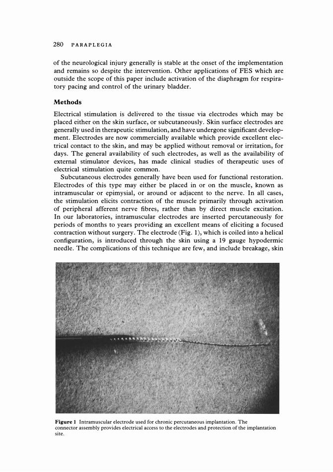
contraction without surgery. The electrode (Fig. 1), which is coiled into a helical
configuration, is introduced through the skin using a 19 gauge hypodermic
needle. The complications of this technique are few, and include breakage, skin
Figure 1 Intramuscular electrode used for chronic percutaneous implantation. The connector assembly provides electrical access to the electrodes and protection of the implantation site.

FUNCTION AL ELECTRICAL STIMULA TION 281
irritation and possible infection, although only one electrode of several thousand
implanted has become infected (Marsolais, 1986). This approach has proven to
be an ideal means to enable development of quite sophisticated and complex
clinical systems for manipulation and locomotion without subjecting the patient
to surgical intervention for electrode or stimulator implantation.
Applications of electrical stitnulation
Therapeutic applications
Electrical stimulation has been employed for a variety of therapeutic purposes
in the musculoskeletal system. These applications include muscle strengthening,
relief of spasticity, reversal of joint contracture and muscle adhesions, and cor
rection of spinal curvature.
Muscles which are atrophied due to disuse have been demonstrated to increase
force and fatigue resistance in response to electrical activation. The alterations
in force and fatigue resistance are accompanied by a change in the metabolic
profile of the muscle. Assuming a chronic, low frequency (on the order of 10
Hz) application of stimuli, the atrophied type II muscle fibres of the disuse
atrophied muscle will change toward type I fibre which is larger in diameter
(Mortimer, 1981). While optimal paradigms of activity have not been identified,
the changes that are induced are used to advantage in the case of muscle
weakness. Examples of this approach are in maintaining muscle strength during
immobilisation for fracture, restoration of voluntary function in paretic
muscles, and preventing joint subluxation in stroke. In spinal cord injury,
muscle stimulation has been reported to improve blood flow through the muscle,
thus decreasing stasis and the likelihood of pulmonary embolus. This should be
particularly relevant in cases of prolonged immobilisation.
The use of electrical stimulation for muscle strengthening is an essential
element of functional activation of totally paralysed muscles as well. Muscles
that are unable to generate and sustain satisfactory performance prior to the
conditioning stimulation can thus be made to alter their metabolism to perform
at levels which are sufficient for clinical usage. Subjects have reported satisfac
tion with resultant cosmetic benefit of increased girth which results from the
muscle hypertrophy, which many feel improves appearance. Virtually every
investigator involved in utilising electrical stimulation for restoration of func
tional movement incorporates a muscle strengthening program in the clinical
protocol.
Functional applications
A second major area of FES concerns applications to restore movement and
control, which is sometimes referred to as functional neuromuscular stimulation,
or FNS. This area is primarily experimental at present, although clinical devices
are provided on individual bases in some centres. Major advances have been
made in both technological development and clinical applications.
FNS systems involve interaction between the user, the device he uses to
deliver the stimulus, and the action generated. Thus, he must perform some
action to control the device, which then generates the appropriate stimulus
patterns which are applied to the muscles to move the extremity. For FNS
282 PARAPLEGIA
applications, most systems utilise four or more channels of stimulation. The
stimulus generated is regulated by the action of the user, thus requiring the
presence of an adequate source for generation of this command control signal.
Systems of this type are known as open loop systems, since the only alterations
in the action of the device are those imposed by the user. Open loop systems
comprise the vast majority of all devices which have been implemented clinically.
Standing and locomotion in paraplegia. Studies on paraplegic standing
and locomotion are in progress in at least 17 laboratories worldwide including
Austria, England, France, Israel, Japan, Netherlands, Scotland, U.S.A., Wales
and Yugoslavia. Cybulski, et ai., (1984), reviewed development in this applica
tion through 1984. Most subjects involved in the studies have had complete
thoracic lesions between T6 and T12. Kralj and Bajd (1983) developed a surface
stimulation technique which is employed in many laboratories. Using two
channels of stimulation on each leg, both standing and reciprocal walking have
been demonstrated. One channel directly activates the knee extensor muscles;
the second activates a flexion withdrawal of the hip, knee, and ankle by activation
of the peroneal, sural, or saphenous nerves (usually the former site is used).
Standing is achieved by activation of the knee extensors and upper body lifting.
A stride is taken by maintaining stimulation of the knee extensor with one leg
and initiating the flexion reflex in the opposite. When the hip is flexed, the knee
extensor is activated and maintained through the stance phase. Upon double
limb support the subject moves the walking support and triggers the movement
in the opposite limb. Trunk stability during stance is achieved by hyperexten
sion of the hips, forming a C-curve posture. Walking is assisted with either a
rolling or reciprocating walker or crutches, which contain switches to supply
the trigger command inputs to initiate stimulation. A similar technique of acti
vation is used in most laboratories. Generally a function (e.g. Stand, Walk,
Climb) is selected by the user and then he initiates the action which is subse
quently indicated by a second presentation on the display (e.g. Left Leg). This
triggers a sequence of stimulus trains which have been preprogrammed into the
device, to activate the muscles to the proper level and in the proper sequences to
move the limb through the desired trajectory.
The surface stimulation technique has been used extensively at many centres,
since it utilises a noninvasive stimulation technique and activates the minimal
number of muscle groups required for walking with the fewest number of
channels of stimulation. Recently in Ljubljana, Yugoslavia, a single channel
implantable stimulator has also been implanted near the peroneal nerve in a
paraplegic subject, with the flexor pattern elicited with this technique as well.
Petrofsky has studied walking with subjects wearing external electrodes on up to
12 muscles per leg and external goniometers at the hip, knee, ankle, and shoes
with pressure sensors for feedback signals. This system has been reduced to a
clinically simpler system by incorporating a reciprocating orthosis and electrodes
for stimulation of quadriceps and iliacus incorporated in a wearable garment.
Holle and Thoma (1984) have implanted multichannel receiver-stimulators,
with electrodes applied to the femoral and infragluteal nerves for knee extensor
(quadriceps) and hip extension (gluteus maximus). Reciprocal walking has been
achieved. Walking studies using implantable devices reported by Brindley
have apparently not been expanded.

FUNCTIONAL ELECTRICAL STIMULA TION 283
Marsolais, et ai., (1983) has utilised the percutaneous electrode technique to
access up to 16 muscles of each extremity, as well as using three channels of
surface electrodes for trunk support. Movement provided is hip flexion, ex
tension, and abduction, knee flexion and extension, and ankle plantar and
dorsiflexion. Subjects have demonstrated the ability to stand, walk on level
ground and slight ramps, climb and descend stairs, and step sideways. Figure
2 is a photograph of one subject presently involved in the research program.
Figure 2 Complete paraplegic subject demonstrating stair climbing with implanted electrode system. (Photo compliments of Dr E. B. Marsolais, Veterans Administration Medical Center and Case Western Reserve University).
This system uses preprogrammed sequences of stimulation to produce each
movement trajectory. These are selected from the menu sequence displayed on
a liquid crystal display of the stimulator unit. Most movements have utilised
open loop control, but feedback control has been incorporated in the knee and
hip position.
Incomplete injuries have been studied most extensively by Andrews in
Glasgow and Bajd in Ljubljana. The residual voluntary function retained is
different and not predictable from one individual to the next because of the
nature of the injury. In general, subjects have had considerably more extensive
involvement in one extremity than the other, and efforts have been focused on
the more involved side. Because many of these subjects retain some sensory

284 PARAPLEGIA
function on the involved limb, they may be aware of limb placement and joint
stability. The approach has been to utilise surface peroneal stimulation alone or
combined with stimulation of the knee extensor to initiate and maintain a syn
thetic gait pattern. Andrews also utilised a sensor in the calf strap of an ankle
foot orthosis for feedback control in regulation of the knee extension moment.
This ensured that the knee did not collapse during stance.
The clinical acceptance of synthetic walking systems will be measured, to a
large extent, upoq their ease and reliability in use and their efficiency. At the
present time only limited experience on small populations of subjects have been
obtained at any single centre. While the results to date demonstrate impressive
function, the objective is to reduce the energy expenditure that is required
during walking, while providing security that malfunction will not result in a
fall and potential fracture. Many approaches have been proposed: closed loop
regulation to ensure repeatable function; postural switching to share activity
among alternative muscle groups; external orthoses to limit movement and
support weight; implanted systems to minimise external harnessing and provide
activation of deep muscle groups; command signals which are synchronously
generated by activities of the intended gait; etc. While FNS walking systems to
this day have had limited applicability, as the results of experimental research
are applied in clinical trials, the resolution of some of the most fundamental
issues will be resolved. Clinical studies must demonstrate that the technique
allows safe functional usage. Potential problems such as joint effusion and
degeneration, pressure sores, and bone fracture must be identified at an early
time so as to not compromise the user. While the use of FNS for functional
walking is in its infancy, the basic techniques of stimulation and control are now
developed to a level where their clinical applicability may now begin to be
realised. The clinical demonstrations performed to date illustrate substantially
enhanced function which is unavailable through alternative sources. Despite the
many potential problems to be encountered and unknowns to be resolved in
applying these techniques of FNS to provide synthesised walking, the future of
this technique appears very promising.
Functional control of the upper extremity. Studies in the upper ex
tremity have focused on development of grasp-release function. Both surface
and implanted techniques are employed, but it appears that the isolated activa
tion of quite small muscles is better suited to implanted techniques. Studies
performed in our laboratories have focused on providing controlled prehension
release in the C5 and C6 tetraplegic using implanted electrodes. These subjects
generally have bilateral loss of distal muscle function, but voluntary control of
the shoulder and elbow flexion. Some subjects may have some voluntary wrist
extension. Elbow extension, wrist flexion, and prehension-release is absent.
Sensation in the hand is absent or severely impaired.
Neuroprostheses for restoration of controlled prehension-release have been
implemented in 26 subjects and undergoing various stages of outpatient clinical
trials for 7 years. The neuroprosthesis provides control of two types of grasp;
palmar prehension (or three jaw chuck pinch) and lateral prehension (or key
grip). Generally eight muscles are used, four or five for each grasp. The subject
proportionally controls both the position of his fingers and thumb and the grasp
force by a single command control source, generally the position of the shoulder

FUNCTION AL ELECTRICAL STIM ULA TION 285
Figure 3 Complete cervical level tetraplegic with FES system for restoration of grasp-release function.
opposite to the limb being stimulated. Figure 3 shows one of the subjects
involved in the evaluation of this system. The clinical system operates as an
open loop system, with the visual feedback used to ensure adequate grasp on an
object.
Chronically indwelling percutaneous electrodes are used for implantation of
muscles providing finger and thumb flexion and extension and thumb abduction
and adduction. Surgical reconstruction has been performed on the hand in
three subjects to enhance the function provide with FNS through stabilisation
of the interphalangeal joint of the thumb and synchronisation of the fingers in
flexion and extension movement by tenodeses of the Flexor digitorum super
ficialis, Flexor digitorum profundus, and Extensor digitorum.
Performance of subjects using the FNS system has been evaluated in activities
of daily living, including tests of isolated basic tasks, co-ordinated tasks, and
integrated tasks. Basic tasks performed by the subjects include grasp release or
utensils, books, writing instruments, telephone, cups, etc. Integrated tasks
include pouring, washing, diskette handling, teeth brushing. Some subjects
demonstrated the ability to perform advanced tasks such as threading a needle
and self-catheterisation. Tasks that involved transitions (i.e. acquiring an ob
ject, position the object, holding for use, and replacing the object) were able to
be accomplished regularly by subjects using FNS. The functional performance
demonstrated by the subjects provides them with independence of hand function
that was not achieved with the wrist hand orthosis which was available for

286 PARAPLEGIA
alternative use. With FNS, they were able to perform more tasks indepen
dently and lost no abilities as a result of implementing the FNS system. We
believe these results demonstrate a major advance in the treatment of the high
level tetraplegic hand.
Implantable stimulator devices also have been used in upper extremity appli
cations by Pasniczek in Warsaw and more recently in our laboratory by Keith
and in that of Brindley (D. Rushton, personal communication).
Control of elbow extension has been demonstrated in the C5 tetraplegic in
our laboratory. Stimulation of the lateral and medial heads of the triceps muscle
was regulated with the position of the arm in abduction. Thus, as the subject
reached to greater angles above his head, the stimulation level was increased to
provide sufficient force to fully extend the elbow. If an intermediate position of
the elbow was desired, the subject could reach that position by voluntary con
traction of his elbow flexors. Combining the hand system with that of the elbow
is expected to further enhance independent function.
Advanced FNS systems
More advanced FNS systems which are under development incorporate closed
loop feedback and substitutional sensory feedback. Closed loop feedback systems
internally correct for differences between the desired action and the actual action
generated by the stimulation (e.g. joint position or contact force). Thus, more
repeatable movement can be generated since there is internal compensation for
changes such muscle activation properties, (e.g. fatigue), or external disturb
ances (e.g. gravity or an object impeding movement). The control systems which
perform the corrections must function over a wide range of conditions, and
have been extensively researched by Crago and Chizeck (1985). At present,
satisfactory performance of the controllers and improvement in the actual clini
cal function has been demonstrated in restricted conditions of the laboratory
environment for regulation of motions limited complexity in humans.
Substitutional sensory feedback systems augment function of the FNS system
by providing the user with cutaneous sensory cues which are related to the
movement or force in the limb. This is important, since the user generally has
a sensory deficit in his extremities, and must commit substantial conscious
effort toward monitoring the elicited movement to insure that it is sufficient
to perform the intended function. Szeto and Riso (1987) have recently reviewed
this field. Riso has studied paradigms for generating cutaneous sensation by
subcutaneously placed electrodes. Subjects have demonstrated the ability to
accurately recognise cutaneous stimuli with high resolution at adequate rates
and with stability of the response over time. Some aspects of these techniques
have been incorporated in the upper extremity system for control of grasp
release, and more extensive integration of this technique into the clinical systems
may be anticipated to lead to more confident and higher quality performance
with the FNS system.
Sensors are required to provide the sources of information in both closed
loop feedback and sensory feedback. Crago et al., (1986) has recently reviewed
the sensor requirements for FNS. For the upper extremity, joint position and
contact force information is required. For the lower extremity, joint position,
foot-floor contact, and crutch reactions are needed. At present, few sensor

FUNCTIONAL ELECTRICAL STIMULATION 287
systems meet the investigational needs, although substantial improvement on
all of these sensors has been made. For example, in the upper extremity a single
element force sensor has been developed· which can be worn over the contact
area of the thumb and fingers, and a two degree of freedom joint position sensor
is used. These sensors are intended primarily for laboratory evaluation, but
ultimately the type of information they provide must be available from internal
sensors which are not un encumbering to the user. Development of sensor
technology thus may be expected to be a substantial effort in implementation of
more advanced FNS systems.
Conclusion
The future development of functional electrical stimulation may be expected
to proceed along several pathways; technical, biological sciences, and clinical
sciences. Technical development will include the development of improved
sensors, stimulators, electrodes, and so forth. The natural progression of
technical development toward smaller, lower power processors with greater
computational capabilities will assist the development of clinical hardware.
Development of the biological sciences will provide far greater understanding
of neural events, such as mechanisms of neural excitation, and information
available from chronic neural recordings in the peripheral and central nervous
system. This information will be used to develop more selective electrodes,
to provide alternative sources of feedback and command control information,
etc. Clinical efforts will be the beneficiary of these findings, as we learn to
apply increasingly sophisticated neural prostheses to provide increasingly more
refined movements.
The challenge of incorporating neural prosthetic technology to solve the
clinical problems is substantial. Clinicians and scientists, with the users, must
collaborate closely in defining reasonable clinical objectives, identifying the
approach to specific problems, and in implementing the solutions. FES should
not be expected to resolve complex disorders of the central nervous system in
isolation of other treatment modalities. Rather, it must be used in combination
with these modalities such as therapy, surgery, orthotics, and other assistive
aids. Both clinical and basic science advances in FES are introduced regularly.
These results demonstrate that the challenge is being met and provide good
reason for cautious optimism regarding the significance of FES in rehabilitation
of the spinal cord injured.
Acknowledgement
This research was supported in part by the National Institute of Health, Neural Prosthesis Program (N01-NS-3-2334), the National Institute on Disability and Rehabilitation Research (9008300118) and the Veterans Administration Research and Development Service.
References
BENTON LA, BAKER LL, BOWMAN BR, et al. Functional Electrical Stimulation-A Practical Clinical Guide. Rancho Los Amigos REC, Downey, CA
CRAGO PE, CHIZECK HI, NEUMAN MR, et al. Sensors for use in Functional Neuromuscular Stimulation. IEEE Transactions on Biomedical Engineering 33:256-268

288 PARAPLEGIA
CYBULSKI GC, PENN RD, JAEGER RJ 1984 Lower Extremity Functional Neuromuscular Stimulation in Cases of Spinal Cord Injury. Neurosurgery 15:132-146
HAMBRECHT FT, RESWICK JB 1977 Functional Electrical Stimulation: Applications in Neural Prostheses. New York: Marcel Dekker.
HANDA Y, ICHIE M, HANDA T, et al. 1985 Control of the Paralyzed Hand by a ComputerControlled FES System. Proceedings of the Seventh IEEE-EMBS conference 322-326
HOLLE J, FREY M, GRUBER H, et al. 1984 Functional Electrostimulation of Paraplegics: Experimental Investigations and First Clinical Experience with an Implantable Stimulation Device. Orthopaedics 7(7):1145-1160
KRALJ A, BAJD T, TURK R 1983 Gait restoration in paraplegic patients: A Feasibility Demonstration using multichannel surface electrode FES. Journal of Rehabilitation Research and Development 20(BPRI0-38):3-20.
MARSOLAIS EB, KOBETIC R 1983 Functional Walking in Paralyzed Patients by Means of Electrical Stimulation. Clinical Orthopaedics 175:30-36
MARSOLAIS EB, KOBETIC R 1986 Implantation Techniques and Experience with Percutaneous Intramuscular Electrodes in the Lower Extremities. Journal of Rehabilitation Research and Development 23:6:1-8
MORTIMER JT 1981 Motor prosthesis in Handbook of Physiology, Section I: The Nervous System, Vol. 2, Part I, Chap. 5, BROOKS VB (ed.). Am. Physiological Soc., Bethesda, MD 155-187
PETROFSKY JE, PHILLIPS CA 1985 Closed-loop Control of Movement of Skeletal Muscle. CRC Critical Reviews in Biomedical Engineering 13:35-96
SZETO A YI, RISO RR 1987 Sensory Feedback Using Electrical Stimulation. In: SMITH RV, LESLIE JH (eds), Rehabilitation Engineering. CRC Press, Boca Raton, FLA
WILHERE GF, CRAGO PE, CHIZECK HJ 1985 Design and Evaluation of a Digital Closed-Loop Controller for the Regulation of Muscle Force by Recruitment Modulation. IEEE Transactions on Biomedical Engineering 32:668-676
Related Documents