ORIGINAL ARTICLE Functional diversity of bacteria in a ferruginous hydrothermal sediment Kim M Handley 1 , Christopher Boothman 1 , Rachel A Mills 2 , Richard D Pancost 3 and Jonathan R Lloyd 1 1 School of Earth, Atmospheric and Environmental Sciences, The University of Manchester, Manchester, UK; 2 School of Ocean and Earth Science, National Oceanography Centre, University of Southampton, Southampton, UK and 3 Organic Geochemistry Unit, Bristol Biogeochemistry Research Centre, School of Chemistry, University of Bristol, Bristol, UK A microbial community showing diverse respiratory processes was identified within an arsenic-rich, ferruginous shallow marine hydrothermal sediment (20–40 1C, pH 6.0–6.3) in Santorini, Greece. Analyses showed that ferric iron reduction with depth was broadly accompanied by manganese and arsenic reduction and FeS accumulation. Clone library analyses indicated the suboxic–anoxic transition zone sediment contained abundant Fe(III)- and sulfate-reducing Deltaproteobacteria, whereas the overlying surface sediment was dominated by clones related to the Fe(II)-oxidizing zetaproteobacterium, Mariprofundus ferroxydans. Cultures obtained from the transition zone were enriched in bacteria that reduced Fe(III), nitrate, sulfate and As(V) using acetate or lactate as electron donors. In the absence of added organic carbon, bacteria were enriched that oxidized Fe(II) anaerobically or microaerobically, sulfide microaerobically and aerobically and As(III) aerobically. According to 16S rRNA gene analyses, enriched bacteria represented a phylogenetically wide distribution. Most probable number counts indicated an abundance of nitrate-, As(V)- and Fe(III) (s,aq) -reducers, and dissolved sulfide-oxidizers over sulfate-reducers, and FeS-, As(III)- and nitrate-dependent Fe(II)-oxidisers in the transition zone. It is noteworthy that the combined community and geochemical data imply near-surface microbial iron and arsenic redox cycling were dominant biogeochemical processes. The ISME Journal advance online publication, 22 April 2010; doi:10.1038/ismej.2010.38 Subject Category: geomicrobiology and microbial contributions to geochemical cycles Keywords: microbial community; functional diversity; geochemical cycling; enrichment cultivation; ferruginous; arsenic; redox Introduction Marine hydrothermal sediments are thought to host an abundance of bacteria and archaea that exploit the steep geochemical gradients formed at the confluence of reduced, metal-rich hydrothermal fluids and oxidized seawater (Jannasch and Mottl, 1985; Zierenberg et al., 2000; Luther et al., 2001). However, information on the metabolic diversity of prokaryotes and their effect on redox cycling remains limited, not the least in iron-rich deposits. Hydrothermal deposits dominated by iron-rich mineral phases are found in the vicinity of seafloor spreading centers, intra-plate seamounts and island arcs. A majority of these have been identified in deep-sea environments (reviewed in Rona, 1988), but shallow marine examples are also known (for example, Santorini, Aegean Volcanic Arc; Ambitle Island, Papua New Guinea; Holm, 1987; Alt, 1988; Pichler and Veizer, 1999). Formation proceeds either by the in situ precipitation of iron from ascending hydrothermal fluids or the fall out of iron oxides formed in hydrothermal plumes (Alt, 1988; Mills et al., 1993), with temperatures typically ranging from that of ambient seawater to 100 1C (see references in Little et al., 2004). These ferruginous deposits are of particular interest owing to their ability to scavenge and retain trace elements from seawater (German et al., 1991), as well as the analogies that may be drawn between their development and that of Banded Iron Forma- tions, which formed under oxygen limitation during the Precambrian (Krapez et al., 2003). The oxidation of Fe(II) within contemporary iron oxide deposits at circumneutral pH is increasingly attributed to bacteria that catalyze the transformation of Fe(II) to Fe(III) under anaerobic or microaerobic conditions. Currently, much of the evidence for marine micro- bial Fe(II) oxidation is based on the association of stalk- or sheath-like structures within hydrothermal Received 20 November 2009; revised 11 February 2010; accepted 1 March 2010 Correspondence: Current address: KM Handley, Department of Earth and Planetary Sciences, University of California, Berkeley, 307 McCone Hall, Berkeley, CA, USA. E-mail: [email protected] The ISME Journal (2010), 1–13 & 2010 International Society for Microbial Ecology All rights reserved 1751-7362/10 $32.00 www.nature.com/ismej

Welcome message from author
This document is posted to help you gain knowledge. Please leave a comment to let me know what you think about it! Share it to your friends and learn new things together.
Transcript
ORIGINAL ARTICLE
Functional diversity of bacteria in a ferruginoushydrothermal sediment
Kim M Handley1, Christopher Boothman1, Rachel A Mills2, Richard D Pancost3 andJonathan R Lloyd1
1School of Earth, Atmospheric and Environmental Sciences, The University of Manchester, Manchester, UK;2School of Ocean and Earth Science, National Oceanography Centre, University of Southampton,Southampton, UK and 3Organic Geochemistry Unit, Bristol Biogeochemistry Research Centre, School ofChemistry, University of Bristol, Bristol, UK
A microbial community showing diverse respiratory processes was identified within an arsenic-rich,ferruginous shallow marine hydrothermal sediment (20–40 1C, pH 6.0–6.3) in Santorini, Greece.Analyses showed that ferric iron reduction with depth was broadly accompanied by manganese andarsenic reduction and FeS accumulation. Clone library analyses indicated the suboxic–anoxictransition zone sediment contained abundant Fe(III)- and sulfate-reducing Deltaproteobacteria,whereas the overlying surface sediment was dominated by clones related to the Fe(II)-oxidizingzetaproteobacterium, Mariprofundus ferroxydans. Cultures obtained from the transition zone wereenriched in bacteria that reduced Fe(III), nitrate, sulfate and As(V) using acetate or lactate as electrondonors. In the absence of added organic carbon, bacteria were enriched that oxidized Fe(II)anaerobically or microaerobically, sulfide microaerobically and aerobically and As(III) aerobically.According to 16S rRNA gene analyses, enriched bacteria represented a phylogeneticallywide distribution. Most probable number counts indicated an abundance of nitrate-, As(V)- andFe(III)(s,aq)-reducers, and dissolved sulfide-oxidizers over sulfate-reducers, and FeS-, As(III)-and nitrate-dependent Fe(II)-oxidisers in the transition zone. It is noteworthy that the combinedcommunity and geochemical data imply near-surface microbial iron and arsenic redox cycling weredominant biogeochemical processes.The ISME Journal advance online publication, 22 April 2010; doi:10.1038/ismej.2010.38Subject Category: geomicrobiology and microbial contributions to geochemical cyclesKeywords: microbial community; functional diversity; geochemical cycling; enrichment cultivation;ferruginous; arsenic; redox
Introduction
Marine hydrothermal sediments are thought to hostan abundance of bacteria and archaea that exploitthe steep geochemical gradients formed at theconfluence of reduced, metal-rich hydrothermalfluids and oxidized seawater (Jannasch and Mottl,1985; Zierenberg et al., 2000; Luther et al., 2001).However, information on the metabolic diversity ofprokaryotes and their effect on redox cyclingremains limited, not the least in iron-rich deposits.Hydrothermal deposits dominated by iron-richmineral phases are found in the vicinity of seafloorspreading centers, intra-plate seamounts and islandarcs. A majority of these have been identified indeep-sea environments (reviewed in Rona, 1988),but shallow marine examples are also known (for
example, Santorini, Aegean Volcanic Arc; AmbitleIsland, Papua New Guinea; Holm, 1987; Alt, 1988;Pichler and Veizer, 1999). Formation proceeds eitherby the in situ precipitation of iron from ascendinghydrothermal fluids or the fall out of iron oxidesformed in hydrothermal plumes (Alt, 1988; Millset al., 1993), with temperatures typically rangingfrom that of ambient seawater to 100 1C (seereferences in Little et al., 2004).
These ferruginous deposits are of particular interestowing to their ability to scavenge and retain traceelements from seawater (German et al., 1991), aswell as the analogies that may be drawn betweentheir development and that of Banded Iron Forma-tions, which formed under oxygen limitation duringthe Precambrian (Krapez et al., 2003). The oxidationof Fe(II) within contemporary iron oxide deposits atcircumneutral pH is increasingly attributed tobacteria that catalyze the transformation of Fe(II) toFe(III) under anaerobic or microaerobic conditions.Currently, much of the evidence for marine micro-bial Fe(II) oxidation is based on the association ofstalk- or sheath-like structures within hydrothermal
Received 20 November 2009; revised 11 February 2010; accepted1 March 2010
Correspondence: Current address: KM Handley, Department ofEarth and Planetary Sciences, University of California, Berkeley,307 McCone Hall, Berkeley, CA, USA.E-mail: [email protected]
The ISME Journal (2010), 1–13& 2010 International Society for Microbial Ecology All rights reserved 1751-7362/10 $32.00
www.nature.com/ismej
iron deposits that are reminiscent of the Fe(II)oxidizers Gallionella and Leptothrix, respectively(for example, Alt, 1988; Juniper and Fouquet, 1988;Hanert, 2002; Kennedy et al., 2003; Little et al.,2004). Both of these taxa are better known fromterrestrial environments. Only a very limited num-ber of marine Fe(II) oxidizers have been isolated todate, namely the zetaproteobacterium Mariprofun-dus ferrooxydans and several alphaproteobacteriaand gammaproteobacteria strains related to thegenera Hyphomonas and Marinobacter (Emersonand Moyer, 2002; Edwards et al., 2003a; Rogerset al., 2003; Lysnes et al., 2004; Emerson et al., 2007).
The nature of other hypothetical biogeochemicalprocesses in this type of deposit may include themicrobial respiration of Fe(III), SO4
2� and NO3�, and
the biogeochemical cycling of trace elements (forexample, Mn, Cu, U, Cd, As, Ag and Au) typicallyenriched in hydrothermal deposits (cf. Glynn et al.,2006; Severmann et al., 2006). Many of these traceelements may be reduced or oxidized by prokaryotesto conserve energy for cell growth or maintenance,or for toxicity resistance (reviewed in Lloyd, 2003).Of these, arsenic is highly enriched in numerousferruginous deposits (Cronan, 1972; Rona, 1988),including those at Santorini and Tutum Bay, PapuaNew Guinea (Varnavas and Cronan, 1988; Price andPichler, 2005). Although the microbial reduction ofAs(V) and the oxidation of As(III) have been studiedextensively in terrestrial freshwater and geothermalbrine environments (Lloyd and Oremland, 2006),little is currently known regarding these processesin marine settings.
In this study, we examine the phylogeneticdiversity and geochemical effect of prokaryoticactivity within a temperate shallow marine hydro-thermal sediment at Santorini, Greece that is iron-and arsenic-rich (Varnavas and Cronan, 1988). Thesediment was examined using microbiological,molecular phylogenetic and geochemical techni-ques. In particular, enrichment culturing was usedto analyze the capacity of the indigenous prokar-yotic community to transform a range of geochemi-cally important inorganic species (that is, Fe, Mn,As, S and N).
Materials and methods
Sampling and analytical methodsSamples were collected from a shallow embayment(X0.3 m water depth, 20–40 1C; Bostrom andWidenfalk, 1984) on the western margin of NeaKameni island, within the Santorini flooded cal-dera. Several cores were extracted in a spatiallyconfined area using hand-push tubes (10 cm internaldiameter � 50 cm long), and sealed with air-tightcaps. One core was selected for geochemicalanalyses. Down-core Eh and pH measurements,initial enrichment inoculations and sedimentsectioning for geochemical analyses (at 5 cm intervals
from 0–35 cm depth) were conducted directly aftercollection under N2 in a glove bag. Sedimentwas transported immediately to Manchester on ice,where pore-waters were separated by centrifugation(4100 rpm, 4 min), and filtered (0.45mm) under N2.Pore-water was acidified with HNO3 for dissolvedelement analyses, or kept anaerobic and chilled forrapid analysis of anions and cations.
Total pore-water element concentrations weremeasured by ICP-AES (Perkin-Elmer Optima 5300Dual View, Waltham, MA, USA). Anions weremeasured using a Dionex DX600 ion chromatograph,fitted with a high-capacity ion exchange column(AS9-HC) and an AG9-HC guard column (Sunnyvale,CA, USA), eluted at 1.4 ml min�1 with 12 mM
isocratic Na2CO3 (2600–2800 psi). Dissolved organiccarbon was measured using Shimadzu TOC5050A total carbon analyzer (Milton Keynes, UK).Ammonium concentrations were measured afterreaction with Nessler’s reagent (at 420 nm). Ironspecies were quantified by the ferrozine method(Lovley and Phillips, 1986, 1987; Anderson andLovley, 1999).
Solid-phase elements were quantified by an AxiosSequential Wavelength-dispersive X-ray fluores-cence spectrometer (XRF; PANalytical, Almelo,The Netherlands). Total carbon was determined induplicate by flash combustion of freeze-dried finelypowdered samples using a Carlo Erba EA 1108elemental analyzer (CE Elantech, Lakewood, NJ,USA). Total inorganic carbon was determined on aCoulomat 702 C/S analyzer (Strohlein, Karst,Germany). The difference between total carbon andtotal inorganic carbon was taken to be total organiccarbon. Sediment was imaged with a Philips FEG-XL30 environmental scanning electron microscope(Eindhoven, The Netherlands), fitted with a PRISMEDS detector and Spirit software (PGT, Princeton,NJ, USA). Samples were imaged unwashed orwashed (� 3) with MilliQ H2O, with or withoutfixation by 2.5% glutaraldehyde.
Enrichment cultivationAnaerobic, microaerobic and aerobic enrichmentmedia were inoculated in triplicate with 10% (v/v)wet sediment from the suboxic to anoxic transitionzone (5–20 cm depth), incubated in the dark at 25 1Cfor X1 month, and subcultured repeatedly beforecommunity composition analysis. An anaerobicmarine minimal medium (MMM) (described inHandley et al., 2009a), supplemented with 10 mM
acetate or lactate as the electron donor and carbonsource, was used for the reduction of 10 mM
amorphous Fe(III) oxyhydroxide (FeOOH) (Lovleyand Phillips, 1986), soluble Fe(III)-nitrilotriaceticacid (NTA) or nitrate (KNO3), or 5 mM As(V)(Na2HAsO4.7H2O). Fe(III)-NTA stock (100 mM) wasprepared as described by (Fredrickson et al., 2000),sparged with N2 and filter sterilized. FeSO4.7H2O(20 mM) with KNO3 (10 mM) was used to test
Functional diversity of bacteriaKM Handley et al
2
The ISME Journal
anaerobic Fe(II) oxidation. Arsenite oxidizers wereenriched in aerobic MMM using 5 mM Na3AsO2,acidified with HCl to pH 6.8–7.0. Marine PostgateMedium B, with 20 mM acetate or 30 mM lactate wasused for sulfate reducer growth (Postgate, 1984).
Oxygen gradient tubes using FeS plugs and anagarose slush overlayer of Modified Mineral Wolfe’sMedium in artificial seawater were used to cultivatemicroaerophilic Fe(II) oxidizers following the meth-ods given by Kucera and Wolfe (1957) and modifica-tions by Hanert (1992) and Emerson and Moyer(1997, 2002), with vitamins and minerals as forMMM. Tubes targeting sulfide oxidizers received1% agarose plugs containing 10 mM neutralizedNa2S and 25% anoxic artificial seawater (Jannaschet al., 1985). The overlayer consisted of artificialseawater, 1 ml l�1 vitamins and minerals (MMM),0.001% phenol red (Jannasch et al., 1985) and 0.1%agarose. A 25 ml stab inoculum was added to over-layers 24 h after formation.
No organic carbon was added to media usinginorganic electron donors.
Most probable number counts (MPN)Enumeration of different microbial functional groupswas achieved through the MPN method, using10-fold serial dilutions from 10�1 to 10�8. Media forcultures was as described for enrichment cultures,with no organic carbon added to media withinorganic electron donors, but acetate and lactateadded in combination to media requiring organicelectron donors. Cell numbers were estimated fromthe three-tube table published in de Man (1983).
Isolation of bacteriaSulfate reducers were isolated from enrichmentcultures anaerobically using Postgate Medium Eagar plates with acetate or lactate (Postgate, 1984).Anaerobic Fe(III)-NTA MMM agar plates were usedto isolate from a lactate-dependant FeOOH enrich-ment culture. Fe(III)-reducing bacteria from Fe(III)-NTA and FeOOH/acetate enrichment cultures wereisolated on Luria–Bertani agar plates. As(V)- andAs(III)-oxidizers were isolated on Luria–Bertani andMarinobacter medium (DSMZ 970) agar plates.Anaerobic nitrate MMM agar plates were used toisolate from the lactate-metabolizing nitrate enrich-ment. Dual-layer plates, based on the gradient tubemethod, but substituting 1.5% agarose in both layers(cf. Wirsen et al., 2002) were used to isolate aerobicsulfide-oxidizers. The metabolism of each isolate wassubsequently tested in the appropriate liquid medium.
Analytical techniques for detection of growth on targetsubstratesThe reduction or oxidation of iron in cultureswere determined by the ferrozine method, andwere also evident owing to: the visible formation
of the white ferric iron-bearing mineral vivianite[Fe3(PO4)2.8H2O] (confirmed by XRD) from Fe(III)-NTA; the transformation of FeOOH from orange to adark-brown or green precipitate; or the change ofFe(II)/NO3
� medium from green to orange. Ferrousiron oxidation in FeS gradient tubes was indicatedby a visible horizon of cell growth stained with anorange ferric precipitate. Similarly, Na2S oxidationwas indicated by a horizon of cell growth, and colorchange in phenol red (to pale yellow). Sulfatereduction was evident from the development ofFeS. Nitrate reduction was established by anincrease in optical density, and NO3
�, NO2� and
NH4þ concentrations. Arsenate and arsenite levels
were measured by the molybdenum blue colori-metric method (see Handley et al., 2009b).
DNA extraction and amplificationGenomic DNA from sediments, enrichment cultures(X3rd subculture of the initial 10% v/v dilutions),and sulfate-reducing colonies imbued with FeS wasextracted using the PowerSoil DNA Isolation Kit(MO BIO Laboratories Inc., Carlsbad, CA, USA).DNA from other isolates was amplified directlyfrom colonies. Bacterial 16S rRNA genes wereamplified using the primers 8F and 519R and PCRsettings according to Holmes et al., (2002) with10-min final extensions. Dissimilatory sulfitereductase (dsr) gene primers, DSR1F and DSR4R(Wagner et al., 1998) were also used for analysis ofsulfate-reducing communities. Reaction conditionswere: 94 1C for 1 min; 35 cycles of 94 1C for 30 s,54 1C for 1 min, and 72 1C for 2 min; and 72 1C for10 min. PCR products were purified using a QIA-quick purification kit (Qiagen, Crawley, UK), andverified on an ethidium bromide stained 1% agarosetris-borate-EDTA gel.
Cloning and restriction fragment length polymorphismanalysis (RFLP)Purified PCR products were cloned using InvitrogenTA Cloning and Top10 kits (Paisley, UK). Colonieswere screened using the vector primers 1F (50-AGTGTGCTGGAATTCGGCTT-30) and 1R (50-ATATCTGCAGAATTCGGCTT-30). Between 47 and 57 clones weresampled from enrichment libraries, 57 and 100clones were sampled from surface and transitionzone sediment libraries, respectively, and 6 and 25from dsr gene libraries. Sequence diversity wasdetermined by RFLP (Weidner et al., 1996) analysisusing endonucleases EcoRI or Sau3A and MspI(Roche Diagnostics, Lewes, UK), and separationwithin 3% agarose tris-borate-EDTA gels.
DNA sequencing and phylogenetic analysisDNA from pure cultures and multiple representa-tives of each RFLP pattern were sequenced using thereverse PCR primers and the ABI Prism BigDye
Functional diversity of bacteriaKM Handley et al
3
The ISME Journal
Terminator v1.1 Cycle Sequencing Kit (AppliedBiosystems, Warrington, UK). Electrophoresis wasperformed using an ABI Prism 3100 Genetic Analy-zer. Sequences were verified for chimeras usingChimera Check v. 2.7, RDP-II (Cole et al., 2003). Thephylogenetic affiliations of the nucleotide partialsequences were determined using BLAST analysis(Altschul et al., 1990). Sequences were aligned withphylogenetically related sequences from GenBank,and a neighbor-joining phylogenetic tree was con-structed with 1000 boot-strap replicates, based onevolutionary distances estimated using MEGA v4(http://www.megasoftware.net/index.html) with theMaximum Composite Likelihood method (Tamuraet al., 2004, 2007).
Sequence accession numbersSequences are deposited in GenBank under thefollowing accession numbers: EU983110–EU983129(surface sediment), EU983130–EU983155 (transitionzone sediment), EU983213–EU983262 (enrichmentcultures) and EU983263–EU983274 (pure cultures).
Intergenic spacer profilingDNA fingerprinting of down-core sediment bacterialcommunities was undertaken by amplifying the 16S(small subunit) and 23S (large subunit) rRNAintergenic spacer region, using the primers SD-Bact-1522-b-s-20 and LD-Bact-132-a-A-18 (Normandet al., 1996; Ranjard et al., 2000). These primersyield approximately 20 and 130 bp of 16S and 23SrRNA genes (Ranjard et al., 2000), respectively,along with the intergenic spacer region, whichranges from approximately 150–1500 bp in prokar-yotes (for example, Fisher and Triplett, 1999;Cardinale et al., 2004). PCR settings were: 94 1C for3 min; 35 cycles of 94 1C for 1 min, 55 1C for 30 s, and72 1C for 1min; and 72 1C for 5min. Purified productwas separated within a 3% agarose tris-borate-EDTA gel.
Results and discussion
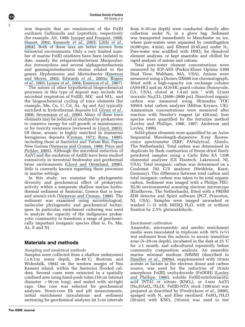
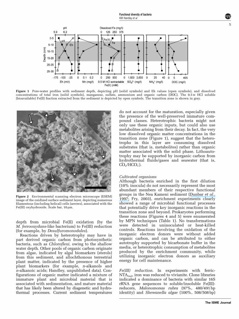
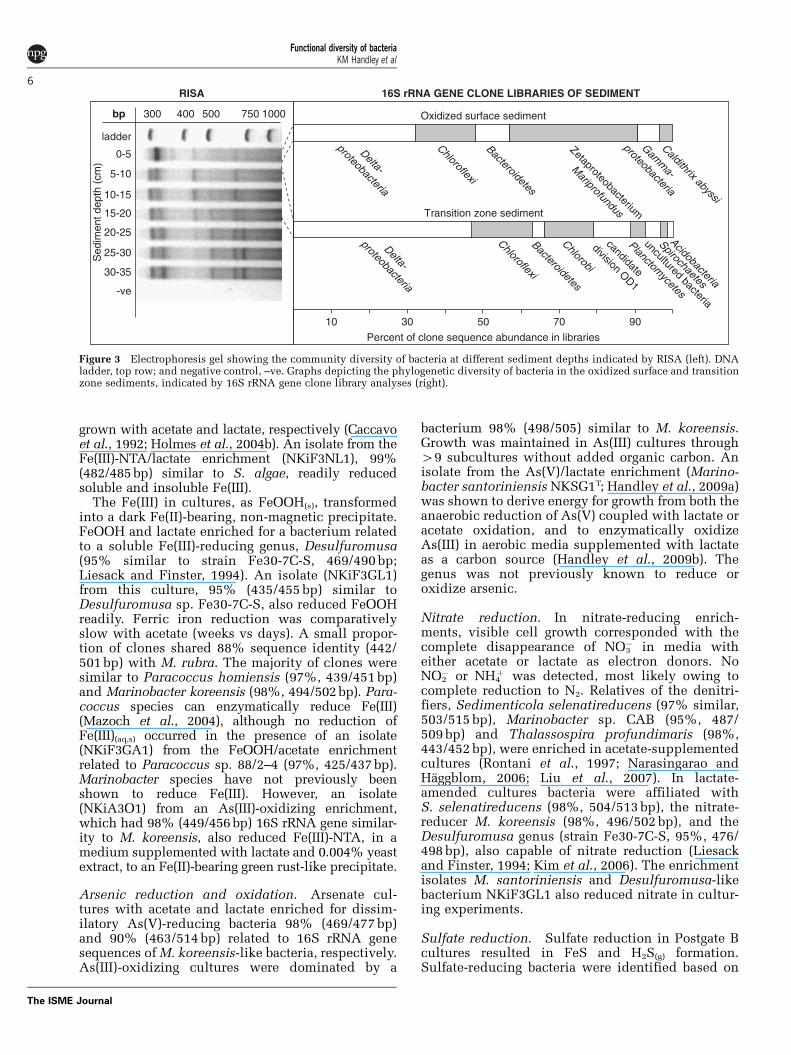
Sediment geochemistryThe sediment was 25 1C during sampling (February2006), owing to warm geothermal fluids, andcomprised three distinct geochemical and miner-alogical zones as it graded downwards from suboxicto anoxic. Suboxic surface sediment (zone one,0–5cm depth) was largely characterized by Fe(II)oxidation and Eh values near zero (Figure 1). Thissurface sediment was unconsolidated and rust-colored with a filamentous microtexture (Figure 2)typical of iron oxide deposits (cf. Little et al., 2004and references therein). Deeper anoxic sedimentwas marked by increasingly negative Eh values, andthe reduction of alternative electron acceptors, suchas Fe(III) and Mn(IV). The suboxic to anoxictransition zone (zone two, approximately 5–20 cmdepth, Eh �60 to �140 mV) graded from poorly to
well-consolidated green–brown mud with localized(predominantly millimeter-scale) areas of blackprecipitate, most likely FeS(am), which continuedthrough the lower anoxic zone (zone three, 20–35 cmdepth). From X35 cm depth the sediment wasgravel-rich.
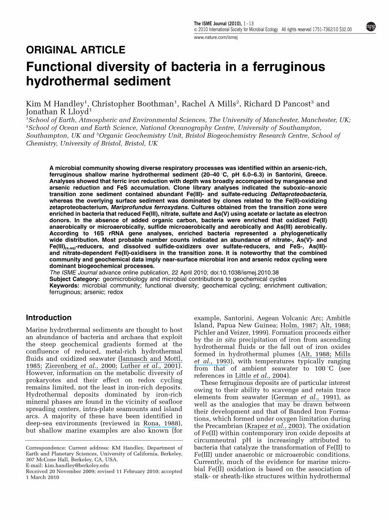
Sediment contained 44–52% Fe, 349–424 ppmAs and minor enrichments of Mo (48–56 ppm), P(0.2–1.5%) and S (0.3–0.5%) (Supplementary Tables1 and 2). Silicon values were low, although notunusual for similar hydrothermal deposits, andelements typically enriched in hydrothermal sedi-ments, such as Mn, Zn and Cu, were depleted (cf. ElWakeel and Riley, 1961; Bostrom and Widenfalk,1984; Alt, 1988). Inorganic carbon comprised 1.0%of the sediment at 0–5 cm depth, 4.0% at 5–20 cm,and 3.4% at 20–30 cm. Organic carbon was 0.1%,0.5% and 0.3% at the respective sediment depths.
Pore-water was significantly enriched indissolved Fe (144.2–316.8 mg l�1), and containedelevated levels of V (1.07–1.2 mg l�1), Mn (0.01–0.27 mg l�1), Si (6.34–17.21mgl�1) and P (0–0.95mgl�1)(Supplementary Table 3). Peaks in pore-water Mnand Fe (as ferrous iron) occurred at 5–10 cm and10–15 cm depth, respectively, while seawater con-centrations of sulfate (2680–3040 mg l�1) remainedconstant over the 30 cm depth analyzed (Figure 1).High levels of ammonium (20–36 mg l�1) were alsopresent throughout the core (Figure 1, Supplemen-tary Table 4). No nitrate or nitrite were detected.Total dissolved inorganic carbon concentrationswere consistently low (0–1 mg l�1), whereas dis-solved organic carbon was substantially elevated inthe surface sediment (495 mg l�1) (Figure 1, Supple-mentary Table 4).
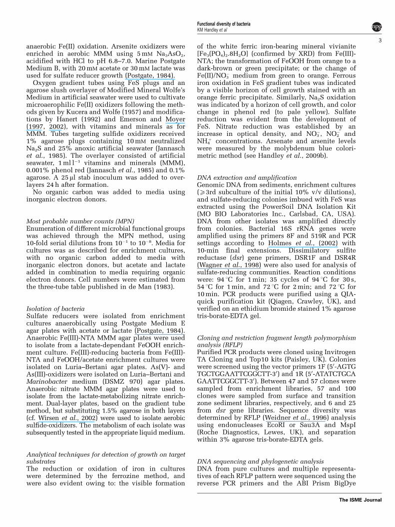
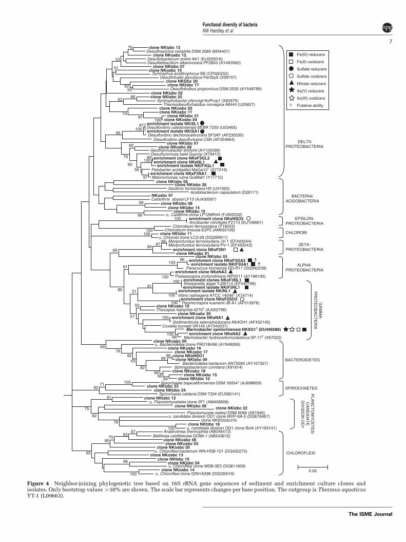
In situ bacterial community and carbon sourcesResults from ribosomal intergenic spacer analysis(RISA) illustrate a gradational shift in the bacterialcommunity structure with increasing sedimentdepth (Figure 3). An exploration of the surfacesediment (0–5 cm depth), based on 16S rRNA geneclone sequencing after RFLP sorting, suggests thecommunity was dominated by bacteria related to themarine Fe(II)-oxidizing zetaproteobacterium, Mari-profundus ferrooxydans (33% library abundance;Emerson and Moyer, 2002; Emerson et al., 2007), theFe(III)- and nitrate-reducing deltaproteobacterium,Geothermobacter sp. (23%; Kashefi et al., 2003) andChloroflexi (16%; Figures 3 and 4, SupplementaryTable 5). In the transition zone sediment (5–20 cmdepth) bacterial community, the greatest proportionof clone sequences (46%) were most similar toDeltaproteobacteria, with 23% of these sequencessimilar to bacterial species within the Desulfuromo-nadales, an order characterized by Fe(III)- and S0-reducers (Lovley et al., 2004), and 16% similar to theDesulfobulbus genus, known for sulfate and Fe(III)reduction (for example, Sass et al., 2002; Holmeset al., 2004a). These data suggest a broad shift with
Functional diversity of bacteriaKM Handley et al
4
The ISME Journal
depth from microbial Fe(II) oxidation (by theM. ferrooxydans-like bacterium) to Fe(III) reduction(for example, by Desulfuromonodales).
Reactions driven by heterotrophy may have inpart derived organic carbon from photosyntheticbacteria, such as Chloroflexi, owing to the shallowwater depth. Other pools of organic carbon originatefrom algae, indicated by algal biomarkers (sterols)from this sediment, and allochthonous terrestrialplant matter, indicated by the presence of higherplant biomarkers (for example, n-alkanols andn-alkanoic acids; Handley, unpublished data). Con-figurations of organic matter indicated a mixture ofimmature plant and microbial material that isassociated with sedimentation, and mature materialthat has likely been altered by diagenetic and hydro-thermal processes. Current sediment temperatures
do not account for the maturation, especially giventhe presence of the well-preserved immature com-pound classes. Heterotrophic bacteria might notonly use these organic inputs, but could also usemetabolites arising from their decay. In fact, the verylow dissolved organic matter concentrations in thetransition zone (Figure 1), suggest that the hetero-trophs in this layer are consuming dissolvedsubstrates (that is, metabolites) rather than organicmatter associated with the solid phase. Lithoauto-trophy may be supported by inorganic carbon fromhydrothermal fluids/gases and seawater (that is,CO2/HCO3
�).
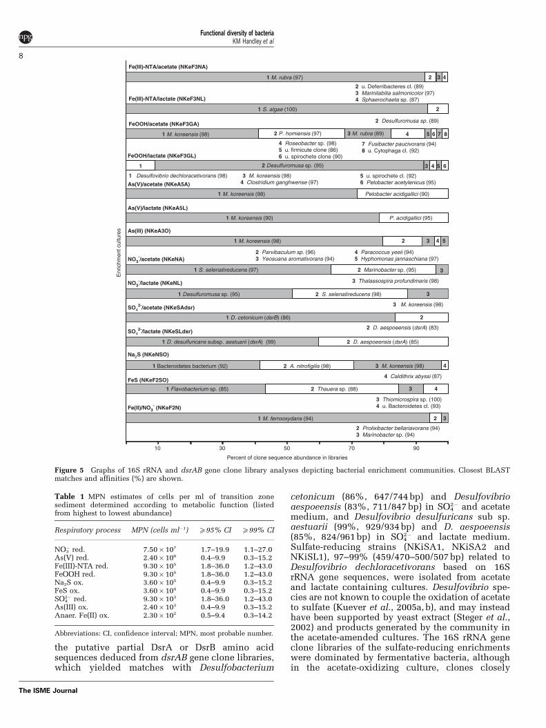
Cultivated organismsAlthough bacteria enriched in the first dilution(10% inocula) do not necessarily represent the mostabundant members of their respective functionalgroups in the Nea Kameni sediment (Dunbar et al.,1997; Fry, 2003), enrichment experiments clearlyshowed a range of microbial functional processesthat potentially drive key inorganic reactions in thetransition zone and beyond. Prokaryotes performingthese reactions (Figures 4 and 5) were enumeratedby MPN techniques (Table 1). No transformationswere detected in uninoculated or heat-killedcontrols. Reactions involving the oxidation of theinorganic electron donors were without addedorganic carbon, and can be attributed to eitherautotrophy supported by bicarbonate buffer in themedia, or heterotrophic consumption of metabolitesproduced by the enrichment community, whileutilizing inorganic electron donors as auxiliaryenergy for cell maintenance.
Fe(III) reduction. In experiments with ferric-NTA(aq), iron was reduced to vivianite. Clone librariesindicated a dominance of bacteria with similar 16SrRNA gene sequences to soluble/insoluble Fe(III)-reducers, Malonomonas rubra (97%, 480/491 bpidentity) and Shewanella algae (100%, 508/508 bp)
Figure 1 Pore-water profiles with sediment depth, depicting pH (solid symbols) and Eh values (open symbols), and dissolvedconcentrations of total iron (solid symbols), manganese, sulfate, ammonium and organic carbon (DOC). The 0.5 M HCl soluble(bioavailable) Fe(II) fraction extracted from the sediment is depicted by open symbols. The transition zone is shown in gray.
Figure 2 Environmental scanning electron microscope (ESEM)image of the oxidized surface sediment layer, depicting numerousfilamentous (including helical) cells (arrows), associated with theFe(III) oxyhydroxide. Scale bar, 10mm.
Functional diversity of bacteriaKM Handley et al
5
The ISME Journal
grown with acetate and lactate, respectively (Caccavoet al., 1992; Holmes et al., 2004b). An isolate from theFe(III)-NTA/lactate enrichment (NKiF3NL1), 99%(482/485 bp) similar to S. algae, readily reducedsoluble and insoluble Fe(III).
The Fe(III) in cultures, as FeOOH(s), transformedinto a dark Fe(II)-bearing, non-magnetic precipitate.FeOOH and lactate enriched for a bacterium relatedto a soluble Fe(III)-reducing genus, Desulfuromusa(95% similar to strain Fe30-7C-S, 469/490 bp;Liesack and Finster, 1994). An isolate (NKiF3GL1)from this culture, 95% (435/455 bp) similar toDesulfuromusa sp. Fe30-7C-S, also reduced FeOOHreadily. Ferric iron reduction was comparativelyslow with acetate (weeks vs days). A small propor-tion of clones shared 88% sequence identity (442/501 bp) with M. rubra. The majority of clones weresimilar to Paracoccus homiensis (97%, 439/451 bp)and Marinobacter koreensis (98%, 494/502 bp). Para-coccus species can enzymatically reduce Fe(III)(Mazoch et al., 2004), although no reduction ofFe(III)(aq,s) occurred in the presence of an isolate(NKiF3GA1) from the FeOOH/acetate enrichmentrelated to Paracoccus sp. 88/2–4 (97%, 425/437 bp).Marinobacter species have not previously beenshown to reduce Fe(III). However, an isolate(NKiA3O1) from an As(III)-oxidizing enrichment,which had 98% (449/456 bp) 16S rRNA gene similar-ity to M. koreensis, also reduced Fe(III)-NTA, in amedium supplemented with lactate and 0.004% yeastextract, to an Fe(II)-bearing green rust-like precipitate.
Arsenic reduction and oxidation. Arsenate cul-tures with acetate and lactate enriched for dissim-ilatory As(V)-reducing bacteria 98% (469/477 bp)and 90% (463/514 bp) related to 16S rRNA genesequences of M. koreensis-like bacteria, respectively.As(III)-oxidizing cultures were dominated by a
bacterium 98% (498/505) similar to M. koreensis.Growth was maintained in As(III) cultures through49 subcultures without added organic carbon. Anisolate from the As(V)/lactate enrichment (Marino-bacter santoriniensis NKSG1T; Handley et al., 2009a)was shown to derive energy for growth from both theanaerobic reduction of As(V) coupled with lactate oracetate oxidation, and to enzymatically oxidizeAs(III) in aerobic media supplemented with lactateas a carbon source (Handley et al., 2009b). Thegenus was not previously known to reduce oroxidize arsenic.
Nitrate reduction. In nitrate-reducing enrich-ments, visible cell growth corresponded with thecomplete disappearance of NO3
� in media witheither acetate or lactate as electron donors. NoNO2
� or NH4þ was detected, most likely owing to
complete reduction to N2. Relatives of the denitri-fiers, Sedimenticola selenatireducens (97% similar,503/515 bp), Marinobacter sp. CAB (95%, 487/509 bp) and Thalassospira profundimaris (98%,443/452 bp), were enriched in acetate-supplementedcultures (Rontani et al., 1997; Narasingarao andHaggblom, 2006; Liu et al., 2007). In lactate-amended cultures bacteria were affiliated withS. selenatireducens (98%, 504/513 bp), the nitrate-reducer M. koreensis (98%, 496/502 bp), and theDesulfuromusa genus (strain Fe30-7C-S, 95%, 476/498 bp), also capable of nitrate reduction (Liesackand Finster, 1994; Kim et al., 2006). The enrichmentisolates M. santoriniensis and Desulfuromusa-likebacterium NKiF3GL1 also reduced nitrate in cultur-ing experiments.
Sulfate reduction. Sulfate reduction in Postgate Bcultures resulted in FeS and H2S(g) formation.Sulfate-reducing bacteria were identified based on
0-5
5-10
15-20
10-15
30-35
25-30
20-25
-ve
400300 750500 1000bp
ladder
Oxidized surface sediment
Zetaproteobacterium
Mariprofundus
Delta-
proteobacteria
Chloroflexi
Caldithrix abyssi
Bacteroidetes
Transition zone sediment
Chlorobi
Spirochaetes
uncultured bacteria
Planctomycetes
Acidobacteria
candidate
division OD1
Delta-
proteobacteria
Chloroflexi
Bacteroidetes
Gamm
a-
proteobacteria
10 30 50 70 90
Percent of clone sequence abundance in libraries
RISA 16S rRNA GENE CLONE LIBRARIES OF SEDIMENTS
edim
ent d
epth
(cm
)
Figure 3 Electrophoresis gel showing the community diversity of bacteria at different sediment depths indicated by RISA (left). DNAladder, top row; and negative control, –ve. Graphs depicting the phylogenetic diversity of bacteria in the oxidized surface and transitionzone sediments, indicated by 16S rRNA gene clone library analyses (right).
Functional diversity of bacteriaKM Handley et al
6
The ISME Journal
Desulfosarcina variabilis DSM 2060 (M34407) clone NKosbc 12
Desulfobacterium anilini AK1 (EU020016) Desulfatibacillum alkenivorans PF2803 (AY493562)
clone NKtzbc 07 clone NKosbc 19
Syntrophus aciditrophicus SB (CP000252) Desulfofustis glycolicus PerGlyS (X99707)
clone NKtzbc 20 clone NKtzbc 17
Desulfobulbus propionicus DSM 2032 (AY548789) clone NKtzbc 02 clone NKtzbc 25
Syntrophobacter pfennigii KoProp1 (X82875) Thermodesulforhabdus norvegica A8444 (U25627)
clone NKosbc 02clone NKosbc 11
clone NKtzbc 21 clone NKosbc 04
enrichment isolate NKiSA1 Desulfovibrio caledoniensis SEBR 7250 (U53465) enrichment isolate NKiSL1
Desulfovibrio dechloracetivorans SF3AF (AF230530) Desulfovibrio desulfuricans CSN (AF354664)
clone NKtzbc 01 clone NKosbc 08
Geothermobacter ehrlichii (AY155599)
enrichment clone NKeF3GL2 enrichment clone NKeNL1
enrichment isolate NKiF3GL1 Pelobacter acidigallici MaGa12T (X77216) enrichment clone NKeF3NA1 Malonomonas rubra GraMal1 (Y17712)
clone NKtzbc 11 u. Chlorobi clone LC3-28 (DQ289911)
Chlorobium ferrooxidans (Y18253) Chlorobium limicola E2P2 (AM050126)
Mariprofundus ferrooxydans JV-1 (EF493244) Mariprofundus ferrooxydans PV-1 (EF493243)
enrichment clone NKeF2N1 clone NKosbc 01
clone NKtzbc 03 enrichment clone NKeF3GA2 enrichment isolate NKiF3GA1
Paracoccus homiensis DD-R11 (DQ342239) enrichment clone NKeNA3 Thalassospira profundimaris WP0211 (AY186195)
enrichment clone NKeNA2 Marinobacter hydrocarbonoclasticus SP.17T (X67022)
Marinobacter santoriniensis NKSG1T (EU496088)
enrichment clone NKeNA1 Sedimenticola selenatireducens AK4OH1 (AF432145)
clone NKosbc 10 Thiocapsa halophila 4270T (AJ002796)
clone NKosbc 20
enrichment clone NKeF2SO3 Thiomicrospira kuenenii JB-A1 (AF013978)
enrichment isolate NKiNL1 Vibrio natriegens ATCC 14048T (X74714)
Shewanella algae YJ06114 (EF542799) enrichment clones NKeF3NL1
enrichment isolate NKiF3NL1
clone NKtzbc 05 clone NKtzbc 26
Geothrix fermentans H5 (U41563) Acidobacterium capsulatum (D26171)
NKosbc 07 Caldothrix abyssi LF13 (AJ430587)
clone NKtzbc 15
clone NKtzbc 08 clone NKtzbc 14
enrichment clone NKeNSO2 Arcobacter nitrofigilis F2173 (EU106661)
clone NKosbc 09 u. Bacteroidetes clone PRD18H06 (AY948069)
clone NKosbc 16 clone NKosbc 17
clone NKeNSO1 clone NKtzbc 09
Bacteriodetes bacterium ANT9285 (AY167327) Sphingobacterium comitans (X91814)
clone NKosbc 18 clone NKosbc 15
clone NKtzbc 10
clone NKtzbc 18 u. candidate division OD1 clone Bol4 (AY193141)
clone NKSGtzbcl16 u. candidate division OD1 clone MVP-6A-5 (DQ676467)
Planctomycete staleyi DSM 6068 (X81946)
clone NKtzbc 06 clone NKtzbc 22
clone NKtzbc 12 u. Planctomycetales clone 2F1 (AM406808)
clone NKosbc 13
Anaerolinea thermophila (AB046413) Bellilinea caldifistulae GOMI-1 (AB243672)
clone NKosbc 06 clone NKosbc 03
clone NKosbc 05 u. Chloroflexi bacterium WN-HSB-121 (DQ432275)
clone NKtzbc 19 clone NKtzbc 04 u. Chloroflexi clone MSB-3E5 (DQ811859)
clone NKosbc 14 u. Chloroflexi clone 02N1A296 (DQ330016)
Spirochaeta caldaria DSM 7334 (EU580141) clone NKtzbc 24
Spirochaeta bajacaliforniensis DSM 16054T (AJ698859) clone NKtzbc 23
clone NKtzbc 13
Desulfuromusa bakii Gyprop (X79412)
Coxiella burnetii VR145 (AY342037)
u. Caldithrix clone LP12MR44 (FJ902532)
0.05
GA
MM
A-
PR
OT
EO
BA
CT
ER
IA
ZETA-PROTEOBACTERIA
BACTEROIDETES
DELTA-PROTEOBACTERIA
EPSILON-PROTEOBACTERIA
BACTERIA/ACIDOBACTERIA
CHLOROBI
ALPHA-PROTEOBACTERIA
PLA
NC
TO
MY
CE
TE
S/
CA
ND
IDA
TE
DIV
ISIO
N O
D1
SPIROCHAETES
CHLOROFLEXI
100
100
96100
100
100
100
87100
100
69100
100
100
100
100
100
100
66100
99
9897
99
9999
99
6456
9682
6392
7899
98
97
97
95
7192
91
91
88
61
50
57
5185
6783
7585
5680
78
76
68
76
62
75
74
69
65
5962
62
53
51
Fe(III) reducers
Fe(II) oxidizers
Sulfate reducers
Sulfide oxidizers
Nitrate reducers
As(V) reducers
As(III) oxidizers
Putative ability ?
? ?
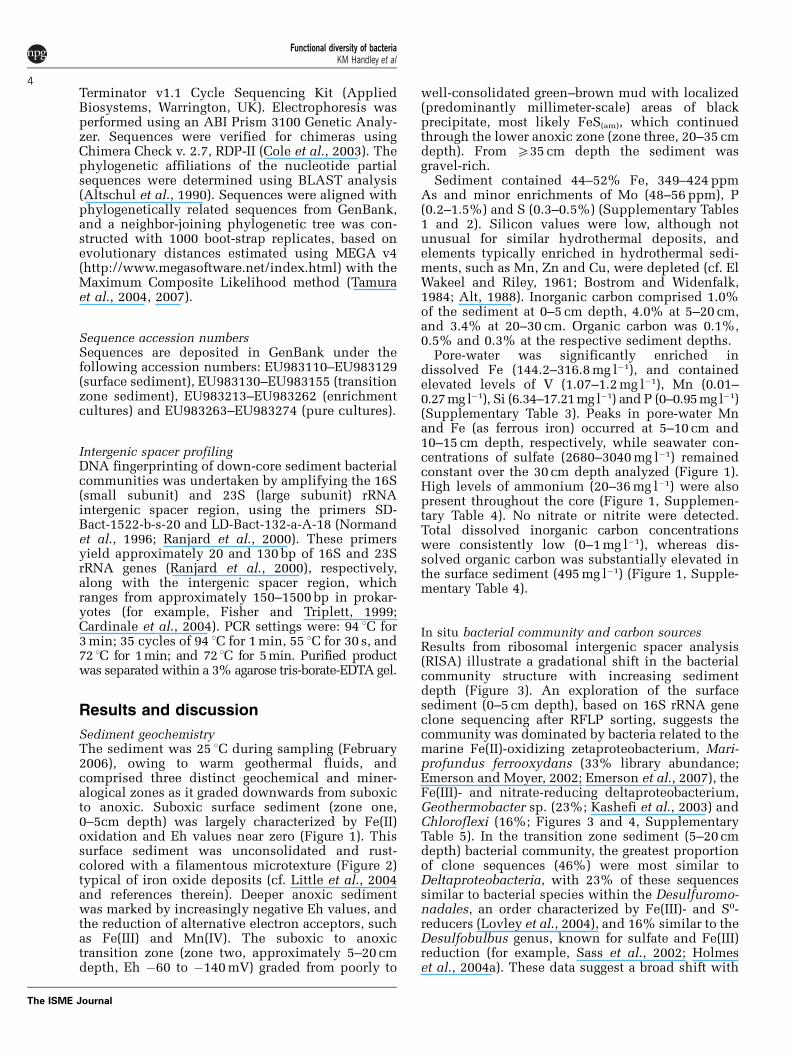
Figure 4 Neighbor-joining phylogenetic tree based on 16S rRNA gene sequences of sediment and enrichment culture clones andisolates. Only bootstrap values 450% are shown. The scale bar represents changes per base position. The outgroup is Thermus aquaticusYT-1 (L09663).
Functional diversity of bacteriaKM Handley et al
7
The ISME Journal
the putative partial DsrA or DsrB amino acidsequences deduced from dsrAB gene clone libraries,which yielded matches with Desulfobacterium
cetonicum (86%, 647/744 bp) and Desulfovibrioaespoeensis (83%, 711/847 bp) in SO4
2� and acetatemedium, and Desulfovibrio desulfuricans sub sp.aestuarii (99%, 929/934 bp) and D. aespoeensis(85%, 824/961 bp) in SO4
2� and lactate medium.Sulfate-reducing strains (NKiSA1, NKiSA2 andNKiSL1), 97–99% (459/470–500/507 bp) related toDesulfovibrio dechloracetivorans based on 16SrRNA gene sequences, were isolated from acetateand lactate containing cultures. Desulfovibrio spe-cies are not known to couple the oxidation of acetateto sulfate (Kuever et al., 2005a, b), and may insteadhave been supported by yeast extract (Steger et al.,2002) and products generated by the community inthe acetate-amended cultures. The 16S rRNA geneclone libraries of the sulfate-reducing enrichmentswere dominated by fermentative bacteria, althoughin the acetate-oxidizing culture, clones closely
10 30 50 70 90
Fe(II)/NO3- (NKeF2N)
FeS (NKeF2SO)
Na2S (NKeNSO)
SO42-/lactate (NKeSLdsr)
SO42-/acetate (NKeSAdsr)
NO3-/lactate (NKeNL)
NO3-/acetate (NKeNA)
As(III) (NKeA3O)
As(V)/lactate (NKeA5L)
As(V)/acetate (NKeA5A)
Fe(III)-NTA/lactate (NKeF3NL)
Fe(III)-NTA/acetate (NKeF3NA)
FeOOH/lactate (NKeF3GL)
FeOOH/acetate (NKeF3GA)
1 M. rubra (97)
2 Desulfuromusa sp. (89)
1 S. algae (100)
1 M. koreensis (98)
2 u. Deferribacteres cl. (89)3 Marinilabilia salmonicolor (97)4 Sphaerochaeta sp. (87)
7 Fusibacter paucivorans (94)8 u. Cytophaga cl. (92)
1 Desulfovibrio dechloracetivorans (98) 3 M. koreensis (98) 4 Clostridium ganghwense (97)
2 Desulfuromusa sp. (95)
4 Paracoccus yeeii (94)5 Hyphomonas jannaschiana (97)
Pelobacter acidigallici (90)1 M. koreensis (98)
P. acidigallici (95)1 M. koreensis (90)
1 M. koreensis (98)
3 Thalassospira profundimaris (98)
3 M. koreensis (98)
2 D. aespoeensis (dsrA) (83)
2 D. aespoeensis (dsrA) (85)
1 S. selenatireducens (97)
1 Desulfuromusa sp. (95)
1 D. cetonicum (dsrB) (86)
1 D. desulfuricans subsp. aestuarii (dsrA) (99)
4 Caldithrix abyssi (87)
3 Thiomicrospira sp. (100)4 u. Bacteroidetes cl. (93)
2 Prolixibacter bellariavorans (94)3 Marinobacter sp. (94)
1 Bacteroidetes bacterium (92)
1 Flavobacterium sp. (85) 2 Thauera sp. (88)
1 M. ferrooxydans (94)
2 Marinobacter sp. (95)
2 P. homiensis (97)
2 S. selenatireducens (98)
2 A. nitrofigilis (98) 3 M. koreensis (98)
3 M. rubra (89)
2 Parvibaculum sp. (96)3 Yeosuana aromativorans (94)
3
2
4
4
2
3
3
3
2 3
2
54
3 4 5 61
5 6 7 84
3 42
5 u. spirochete cl. (92)6 Pelobacter acetylenicus (95)
4 Roseobacter sp. (98)5 u. firmicute clone (86)6 u. spirochete clone (90)
Percent of clone sequence abundance in libraries
Enr
ichm
ent c
ultu
res
Figure 5 Graphs of 16S rRNA and dsrAB gene clone library analyses depicting bacterial enrichment communities. Closest BLASTmatches and affinities (%) are shown.
Table 1 MPN estimates of cells per ml of transition zonesediment determined according to metabolic function (listedfrom highest to lowest abundance)
Respiratory process MPN (cells ml�1) X95% CI X99% CI
NO3� red. 7.50�107 1.7–19.9 1.1–27.0
As(V) red. 2.40�106 0.4–9.9 0.3–15.2Fe(III)-NTA red. 9.30�105 1.8–36.0 1.2–43.0FeOOH red. 9.30�105 1.8–36.0 1.2–43.0Na2S ox. 3.60�105 0.4–9.9 0.3–15.2FeS ox. 3.60�104 0.4–9.9 0.3–15.2SO4
2� red. 9.30�103 1.8–36.0 1.2–43.0As(III) ox. 2.40�103 0.4–9.9 0.3–15.2Anaer. Fe(II) ox. 2.30�102 0.5–9.4 0.3–14.2
Abbreviations: CI, confidence interval; MPN, most probable number.
Functional diversity of bacteriaKM Handley et al
8
The ISME Journal
related to Desulfosarcina variabilis (98%, 512/519 bp) and Desulfobacterium niacin (98%, 506/515 bp) were also present as minor components ofthe library (data not shown).
Sulfide and Fe(II) oxidation. The Na2S gradientmedium developed an aerobic horizon of cellgrowth that was accompanied by acidification ofthe medium, turning the pH indicator pale yellow. Asignificant proportion of library clones were relatedto Arcobacter nitrofigilis (98%, 482/487 bp), thegenus of which includes autotrophic sulfide-oxidi-zers (for example, Wirsen et al., 2002). An isolate ofthe Arcobacter-like bacterium (NKiNSO1) was alsoobtained on sulfide over-layer plates. In the FeSgradient medium, distinct rust-colored opaque cellhorizons formed mid-way between the FeS plug andthe air-agarose interface. No distinct horizonsformed in uninoculated media. Abiotic oxidationresulted in broad, diffuse Fe(III) bands. The enrich-ment was equally dominated by clones yieldingsequence matches to Flavobacterium sp. AKB-2008-JO5 (85%, 434/506 bp), and the autotrophic sulfur-oxidizing genus, Thiomicrospira (strain JB-A1,100%, 506/506 bp; for example, Kuenen and Veld-kamp, 1972; Brinkhoff and Muyzer, 1997). Analysesof cloned 16S rRNA genes from the anaerobic Fe(II)-oxidizing/nitrate-reducing enrichment culture re-vealed it was dominated by a bacterium most closelyrelated to the autotrophic Fe(II)-oxidizer, Maripro-fundus ferrooxydans JV-1 (94%, 476/505 bp;Emerson et al., 2007). Growth of the enrichmentthrough successive subcultures (410 times)occurred without organic carbon supplementation.Nitrate-dependant Fe(II) oxidation was also attainedby the FeS enrichment and M. santoriniensis(Handley et al., 2009a) with 5 mM acetate. Auto-trophy was not confirmed for any Fe(II)- or sulfide-oxidizing enrichment culture bacterium.
Implications for biogeochemical processesNeutrophilic Fe(II) iron-oxidizing bacteria arethough contribute to the formation of hydrothermalferruginous deposits, and alteration of iron sulfidesand exposed spreading ridge basalt (for example,Emerson and Moyer, 2002; Kennedy et al., 2003;Edwards et al., 2003a, b; Little et al., 2004), andbacteria phylogenetically related to Fe(II)-oxidizingGallionella ferruginea are known to be present indeep-sea vent sediments (Rogers et al., 2003).Nevertheless, there is increasing evidence that otherbacteria also have a major role in marine Fe(II)oxidation, in particular zetaproteobacteria and Mar-inobacter species (Rogers et al., 2003; Edwards et al.,2003a; Lysnes et al., 2004; Emerson et al., 2007; Katoet al., 2009; M. Muller, personal communication;this study). Structures resembling the freshwaterFe(II) oxidizers Gallionella and Leptothrix havepreviously been recognized in iron-rich mud ofPalaea Kameni, the island adjacent to Nea Kameni at
Santorini (Hanert, 2002), but neither were identifiedusing molecular techniques in this study. In con-trast, bacteria related to the Fe(II)-oxidizing stalk-former, M. ferrooxydans (Emerson et al., 2007), wereabundant in the Nea Kameni surface-most sediment(Figure 5), and other bacteria potentially alsocontribute to Fe(II) oxidation within this sediment,such as rod-shaped Marinobacter species (Handleyet al., 2009a).
The geochemistry and microbiology of the NeaKameni sediment are consistent with observationsthat Fe(II)-oxidizing bacteria are important in thedevelopment of ferruginous sediments. However,results also illustrate the importance of otherbiogeochemical processes in these sediments, nota-bly, complete redox cycling of iron. Ferruginousdeposits also contain enrichments of other elementsof interest, with high levels of arsenic not beinguncommon. In sediments at Tutum Bay, similar tothose at Santorini, Price and Pichler (2005) foundonly a very small proportion of arsenic wasassociated with the easily extractable, ‘bioavailable’iron oxide fraction. Ferric iron-reducing bacteria,however, are able to consume more recalcitrantforms of iron as well—although at a slower rate—potentially freeing up structurally bound arsenic.Arsenic in the Nea Kameni sediment was found tobe over 60% As(V) in the near surface, but only 5%As(V) in the transition zone (Handley et al., unpub-lished XAS study). This significant change in theproportion of solid-phase As(III) and As(V) betweenthe surface and transition zones occurs in thepresence of, and likely owing to bacteria capable ofredox cycling arsenic. We know of only one otherexample of arsenic metabolism (dissimilatory ar-senate reduction) recorded in a deep-sea hydrother-mal environment (iron sulfide black smoker ventchimney, Takai et al., 2003).
Biogeochemical zonesGeochemical cycling in marine sediments character-istically occurs across the oxic-anoxic gradient bythe transformation of redox couples in order ofdecreasing energy yield, for example, O2/H2O, NO3
�/NO2
�, Mn(IV)/Mn(II), Fe(III)/Fe(II), SO42�/H2S and
then CO2/CH4, producing biogeochemical stratifica-tion with depth (Froelich et al., 1979; Jørgensen,2006). Geochemical data indicated the successivereduction of Mn(IV), Fe(III) and some sulfate (toFeS) with depth in the transition zone sediment atNea Kameni (cf. Burdige, 2006; Jørgensen, 2006).Sediment-bound arsenic was reduced in the transi-tion zone as described above. It is also likely thatseawater nitrate was reduced rapidly at the sedi-ment-water interface, but was not detected at the5 cm resolution of our analyses. The relative abun-dances of organisms enriched tend to correspondwith sediment geochemistry. MPNs are subject tomedia-biases, and may underestimate abundances;however, results broadly suggest a higher likelihood
Functional diversity of bacteriaKM Handley et al
9
The ISME Journal
for microbial As(V), Fe(III) and nitrate reduction andsulfide oxidation over sulfate reduction and As(III)and Fe(II) oxidation within the transition zone(Table 1).
Iron and manganese transformations. Neutrophi-lic microbial oxidation of Fe(II) coupled withoxygen or nitrate reduction, while forming rust-colored iron-oxyhydroxides, out-competes abioticoxidation at low oxygen fugacities and producessufficient energy to support growth (for example,Straub et al., 1996; Supplementary Table 6). Sharpincreases in first aqueous manganese and then ironin the deeper transition zone imply the onset ofmicrobial Mn(IV) and then Fe(III) reduction. Insupport of this, a range of bacteria capable of bothFe(III) and Mn(IV) respiration (for example, Shewa-nella species, Lovley et al., 2004) were present in thetransition zone. The depth-related order in whichthese species were reduced corresponds with therespective energetics of each transformation, where-by the Mn(IV)-Mn(II) transformation tends to beenergetically more favorable (for example, Thaueret al., 1977; Supplementary Table 6), and Mn(IV) isconsequently used in preference to Fe(III) whereboth substrates are present.
Arsenic oxidation and reduction. Both the enrich-ment and isolation of As(III)-oxidizing bacteriasuggests As(V) in the surface sediment may beattributed to the enzymatic oxidation of As(III),arising from mixotrophy or chemolithotrophycoupled with O2 or an alternative electron acceptor.In contrast, the abiotic oxidation of As(III) can beexceptionally slow (months–years) even under fullyaerobic conditions (Smedley and Kinniburgh, 2002,and references therein). Bacteria also are able toaccess, and reduce, structurally bound and surfi-cially complexed As(V) associated with sedimentsand minerals (for example, Newman et al., 1997;Zobrist et al., 2000), potentially accounting forAs(III) in the transition zone. Arsenate respirationgenerates energy yields similar to those for Fe(III)reduction (Supplementary Table 6), although thesevalues will be dependent on several environmentalfactors including concentrations of the electronacceptors and mineralogical constraints (Cuttinget al., 2009). Arsenate reduction to As(III) has beenshown to occur before, concomitant with, orsubsequent to Fe(III) reduction, depending on themodel system studied (for example, Ahmann et al.,1997; Zobrist et al., 2000; Islam et al., 2004;Campbell et al., 2006). Conversely, the release ofiron-bound As(III), after As(V) reduction, may occuralongside or subsequent to Fe(III) reduction (Islamet al., 2004).
Sulfur cycling. It is unclear from available datawhether sulfide oxidation occurs primarily in thesurface or transition zone sediment, althoughthe greater abundance of sulfide-oxidizers over
sulfate-reducers in the transition zone (Table 1)may indicate the latter. Evidence for microbialsulfide oxidation in the transition zone sediment isalso afforded by the cultivation of sulfide-oxidizersclosely related to Arcobacter and Thiomicrospira.Concentrations of hydrothermally generated H2S areeither minor or non-existent in the Santorini hydro-thermal sediments (Bostrom and Widenfalk, 1984;Minissale et al., 1997). However, sulfide may also begenerated by microbial sulfate reduction.
Consistently high levels of pore-water sulfatethroughout the sediment column suggests sulfate-reducing conditions are never fully attained over thesampled sediment depth, even where putative FeSvisibly formed within the lower portion of thetransition zone. This is corroborated by the lownumbers estimated for transition zone sulfate-redu-cers (Table 1). Sulfur concentrations in the sedimentdid not increase with depth, suggesting FeS wasrelatively negligible. Low levels of sulfate reductionmay be due to the relatively high Eh values (above�200 mV; Postgate, 1984), and the inexhaustion ofother electron acceptors, notably Fe(III), whichpossess a higher energy yield (Thauer et al., 1977;Supplementary Table 6).
Nitrate reduction. Rapid reduction of seawater-derived nitrate by bacteria, immediately at or belowsediment-water interfaces, forms part of the char-acteristic geochemical profile within marine sedi-ments (Froelich et al., 1979). It is plausible that thisprocess also occurs at the Nea Kameni sediment-water interface, owing to the absence of detectableseawater nitrate, and abundant nitrate-reducingbacteria in the sediment. The high reducing capacityof nitrate, mean it is the inorganic electron acceptortypically used after oxygen depletion (Supplemen-tary Table 6), and may be coupled with the oxidationof organic carbon, or a range of inorganic electrondonors, such as Fe(II), As(III) or sulfide (for example,Straub et al., 1996; Oremland et al., 2002; Cardosoet al., 2006).
Conclusions
Considering the extent to which microorganisms arecapable of catalyzing geochemical reactions, it ispalpable that while ferruginous deposits undoubt-edly serve as an important substrate for microbialFe(II) oxidation, results here illustrate they also arehome to a range of other geochemically significantmicrobial processes. Bacteria cultivated from theSantorini sediment transformed a range of relevantmarine and hydrothermal inorganic electron donorsand acceptors, particularly Fe(II), Fe(III), As(V) andAs(III), but also nitrate, sulfide and sulfate. Findingsalso contribute to the limited information currentlyavailable on bacteria performing Fe(II) and As(III)oxidation and As(V) reduction in the marineenvironment, owing especially to the cultivation of
Functional diversity of bacteriaKM Handley et al
10
The ISME Journal
novel bacteria able to transform both these chemicalspecies.
Acknowledgements
Funding for this work was provided by the BIOTRACS, ESTMarie Curie Fellowship (European Union) and ORSAS(HEFCE). The Santorini Port Authority, S Monteiro,M Muller, A Bewsher, P Lythgoe and S Caldwell assistedin sampling or analyses. Helpful advice was provided byS P Varnavas, I Head and K Morris.
References
Ahmann D, Krumholz LR, Hemond HF, Lovley DR, MorelFMM. (1997). Microbial mobilization of arsenic fromsediments of the aberjona watershed. Environ SciTechnol 31: 2923–2930.
Alt JC. (1988). Hydrothermal oxide and nontronitedeposits on seamounts in the eastern Pacific.Mar Geol 81: 227–239.
Altschul SF, Gish W, Miller W, Myers EW, Lipman DJ.(1990). Basic local alignment search tool. J Mol Biol215: 403–410.
Anderson RT, Lovley DR. (1999). Napthalene and benzenedegradation under Fe(III)-reducing conditions in pet-roleum-contaminated aquifers. Biorem J 3: 121–135.
Bostrom K, Widenfalk L. (1984). The origin of iron-richmuds at the Kameni islands, Santorini, Greece.Chem Geol 42: 203–218.
Burdige DJ. (2006). Geochemistry of Marine Sediments.Princeton University Press: Princeton, NJ. pp 609.
Brinkhoff T, Muyzer G. (1997). Increased species diversityand extended habitat range of sulfur-oxidizing Thio-microspira spp. Appl Environ Microbiol 63: 3789–3796.
Caccavo Jr F, Blakemore RP, Lovley DR. (1992). Ahydrogen-oxidizing, Fe(III)-reducing microorganismfrom the Great Bay Estuary, New Hampshire.Appl Environ Microbiol 58: 3211–3216.
Campbell KM, Malasarn D, Saltikov CW, Newman DK,Hering JG. (2006). Simultaneous microbial reductionof iron(III) and arsenic(V) in suspensions of hydrousferric oxide. Environ Sci Technol 40: 5950–5955.
Cardinale M, Brusetti L, Quatrini P, Borin S, Puglia AM,Rizzi A et al. (2004). Comparison of different primersets for use in automated ribosomal intergenic spaceranalysis of complex bacterial communities.Appl Environ Microbiol 70: 6147–6156.
Cardoso RB, Sierra-Alvarez R, Rowlette P, Flores ER,Gomez J, Field JA. (2006). Sulfide oxidation underchemolithoautotrophic denitrifying conditions.Biotechnol Bioeng 95: 1148–1157.
Cole JR, Chai B, Marsh TL, Farris RJ, Wang Q, Kulam SA,Chandra S, McGarrell DM, Schmidt TM, Garrity GM,Tiedje JM. (2003). The Ribosomal Database Project(RDP-II): previewing a new autoaligner that allowsregular updates and the new prokaryotic taxonomy.Nucleic Acids Res 31: 442–443.
Cronan DS. (1972). The Mid-Atlantic Ridge near 45 1C N,XVII: Al, As, Hg, and Mn in ferruginous sedimentsfrom the Median Valley. Can J Earth Sci 9: 319–322.
Cutting RS, Coker VS, Fellowes JW, Lloyd JR, Vaughan DJ.(2009). Mineralogical and morphological constraintson Fe(III) reduction by Geobacter sulfurreducens.Geochim Cosmochim Acta 73: 4004–4022.
de Man JC. (1983). MPN tables, corrected. Eur J ApplMicrobiol Biotechnol 17: 301–305.
Dunbar J, White S, Forney L. (1997). Genetic diversitythrough the looking glass: effect of enrichment bias.Appl Environ Microbiol 63: 1326–1331.
Edwards KJ, Rogers DR, Wirsen CO, McCollom TM.(2003a). Isolation and characterization of novel psy-chrophilic, neutrophilic, Fe-oxidizing, chemolithoau-totrophic a- and g-Proteobacteria from the deep sea.Appl Environ Microbiol 69: 2906–2913.
Edwards KJ, McCollom TM, Konishi H, Buseck PR.(2003b). Seafloor bioalteration of sulphide minerals:results from in situ incubation studies. GeochimCosmochim Acta 67: 2843–2856.
El Wakeel SK, Riley JP. (1961). Chemical and mineralogi-cal studies of deep-sea sediments. Geochim Cosmo-chim Acta 25: 110–146.
Emerson D, Moyer C. (1997). Isolation and characteriza-tion of novel iron-oxidizing bacteria that grow at cir-cumneutral pH. Appl Environ Microbiol 63: 4784–4792.
Emerson D, Moyer C. (2002). Neutrophilic Fe-oxidizingbacteria are abundant at the Loihi Seamount hydro-thermal vents and play a major role in Fe oxidedeposition. Appl Environ Microbiol 68: 3085–3093.
Emerson D, Rentz JA, Lilburn TG, Davis RE, Aldrich H,Chan C et al. (2007). A novel lineage of acteriainvolved in formation of marine Fe-oxidizing micro-bial mat communities. PLoS ONE 2: e667.
Fisher MM, Triplett EW. (1999). Automated approach forribosomal intergenic spacer analysis of microbialdiversity and its application to freshwater bacterialcommunities. Appl Environ Microbiol 65: 4630–4636.
Fredrickson JK, Kostandarithes HM, Li SW, Plymale AE,Daly MJ. (2000). Reduction of Fe(III), Cr(VI), U(VI),and Tc(VII) by Deinococcus radiodurans R1.Appl Environ Microbiol 66: 2006–2011.
Froelich PN, Klinkhammer GP, Bender ML, Luedtke NA,Heath GR, Cullen D et al. (1979). Early oxidation oforganic matter in pelagic sediments of the easternequatorial Atlantic: suboxic diagenesis. GeochimCosmochim Acta 43: 1075–1090.
Fry JC. (2003). Culture-dependent microbiology. In: BullAT (ed). Microbial Diversity and Bioprospecting.ASM Press: Washington, DC. pp 80–87.
German CR, Campbell AC, Edmond JM. (1991). Hydro-thermal scavenging at the Mid-Atlanctic Ridge:modification of trace element dissolved fluxes.Earth Planet Sci Lett 107: 101–114.
Glynn S, Mills RA, Palmer MR, Pancost RD, Severmann S,Boyce AJ. (2006). The role of prokaryotes in supergenealteration of submarine hydrothermal sulfides.Earth Planet Sci Lett 244: 170–185.
Handley KM, Hery M, Lloyd JR. (2009a). Marinobactersantoriniensis sp. nov., a novel arsenate respiring andarsenite oxidizing bacterium isolated from hydrother-mal sediment at Santorini, Greece. Int J Syst EvolMicrobiol 59: 886–892.
Handley KM, Hery M, Lloyd JR. (2009b). Redox cyclingof arsenic by the hydrothermal marine bacteriumMarinobacter santoriniensis. Environ Microbiol 11:1601–1611.
Hanert HH. (1992). The Genus Gallionella. In: Balows A,Truper HG, Dworkin M, Harder W, Schleifer K-H (eds).
Functional diversity of bacteriaKM Handley et al
11
The ISME Journal
The Prokaryotes 2nd edn. Springer-Verlag: New York.p 4126.
Hanert HH. (2002). Bacterial and chemical iron oxidedeposition in a shallow bay on Palaea Kameni,Santorini, Greece: microscopy, electron probe micro-analysis, and photometry of in situ experiments.Geomicrobiol J 19: 317–342.
Holm NG. (1987). Biogenic influences on the geochemistryof certain ferruginous sediments of hydrothermalorigin. Chem Geol 63: 45–57.
Holmes DE, Bond DR, Lovley DR. (2004a). ElectronTransfer by Desulfobulbus propionicus to Fe(III)and graphite electrodes. Appl Environ Microbiol 70:1234–1237.
Holmes DE, Finneran KT, O’Neil RA, Lovley DR. (2002).Enrichment of members of the family Geobacteraceaeassociated with stimulation of dissimilatory metalreduction in uranium-contaminated aquifer sedi-ments. Appl Environ Microbiol 68: 2300–2306.
Holmes DE, Nevin KP, Lovley DR. (2004b). Comparison of16S rRNA, nifD, recA, gyrB, rpoB and fusA geneswithin the family Geobacteraceae fam. nov. Int J SystEvol Microbiol 54: 1591–1599.
Islam FS, Gault AG, Boothman C, Polya DA, Charnock JM,Chatterjee D et al. (2004). Role of metal-reducingbacteria in arsenic release from Bengal delta sedi-ments. Nature 430: 68–71.
Jannasch HW, Mottl MJ. (1985). Geomicrobiology of deep-sea hydrothermal vents. Science 229: 717–725.
Jannasch HW, Wirsen CO, Nelson DC, Robertson LA.(1985). Thiomicrospira crunogena sp. nov., a colorless,sulfur-oxidizing bacterium from a deep-sea hydrother-mal vent. Int J Syst Bacteriol 35: 422–424.
Jørgensen BB. (2006). Bacteria and marine biogeochem-istry. In: Schulz HD, Zabel M (eds). Marine Geochem-istry 2nd edn. Springer: New York. pp 169–206.
Juniper SK, Fouquet Y. (1988). Filamentous iron-silicadeposits from modern and ancient hydrothermal sites.Can Mineral 26: 859–869.
Kashefi K, Holmes DE, Baross JA, Lovley DR. (2003).Thermophily in the Geobacteraceae: Geothermobacterehrlichii gen. nov., sp. nov., a novel thermophilicmember of the Geobacteraceae from the ‘Bag City’hydrothermal vent. Appl Environ Microbiol 69:2985–2993.
Kato S, Kobayashi C, Kakegawa T, Yamagishi A. (2009).Microbial communities in iron-silica-rich microbialmats at deep-sea hydrothermal fields of the southernMariana Trough. Environ Microbiol 11: 2094–2111.
Kennedy CB, Scott SD, Ferris FG. (2003). Characterizationof bacteriogenic iron oxide deposits from AxialVolcano, Juan de Fuca Ridge, northeast Pacific Ocean.Geomicrobiol J 20: 199–214.
Kim B-Y, Weon H-Y, Yoo S-H, Kim J-S, Kwon S-W,Stackebrandt E et al. (2006). Marinobacter koreensissp. nov., isolated from sea sand in Korea. Int J SystEvol Microbiol 56: 2653–2656.
Krapez B, Barley ME, Pickard AL. (2003). Hydrothermaland resedimented origins of the precursor sedimentsto banded iron formation: sedimentological evidencefrom the Early Palaeoproterozoic Brockman Super-sequence of Western Australia. Sedimentology 50:979–1011.
Kucera S, Wolfe RS. (1957). A selective enrichment methodfor Gallionella Ferruginea. J Bacteriol 74: 344–349.
Kuenen JG, Veldkamp H. (1972). Thiomicrospirapelophila, gen. n., sp. n., a new obligately chemolitho-
trophic colourless sulfur bacterium. Antonie Leeu-wenhoek 38: 241256.
Kuever J, Rainey FA, Widdel F. (2005a). Family I.Desulfovibrionaceae fam. nov. In: Brenner DJ, KriegNR, Staley JT, Garrity GM (eds). Bergey’s Manual ofSystematic Bacteriology, The Proteobacteria 2nd edn.Springer-Verlag: New York. pp 926–938.
Kuever J, Rainey FA, Widdel F. (2005b). Family I.Desulfobacteraceae. In: Brenner DJ, Krieg NR, StaleyJT, Garrity GM (eds). Bergey’s Manual of SystematicBacteriology, The Proteobacteria 2nd edn. Springer-Verlag: New York. pp 959–960.
Liesack W, Finster K. (1994). Phylogenetic analysis of fivestrains of gram-negative, obligately anaerobic, sulfur-reducing bacteria and description of Desulfirornusagen. nov., including Desulfuromusa kysingii sp. nov.,Desulfurornusa bakii sp. nov., and Desulfuromusasuccinoxidans sp. nov. Int J Syst Bacteriol 44:753–758.
Little CTS, Glynn SEJ, Mills RA. (2004). Four-hundred-and-ninety-million-year record of bacteriogenic ironoxide precipitation at sea-floor hydrothermal vents.Geomicrobiol J 21: 415–429.
Liu C, Wu Y, Li L, Ma Y, Shao Z. (2007). Thalassospiraxiamenensis sp. nov. and Thalassospira profundi-maris sp. nov. Int J Syst Evol Microbiol 57: 316–320.
Lloyd JR. (2003). Microbial reduction of metals andradionuclides. FEMS Microbiol Ecol 27: 411–425.
Lloyd JR, Oremland RS. (2006). Microbial transformationsof arsenic in the environment: from soda lakes toaquifers. Elements 2: 85–90.
Lovley DR, Holmes DE, Nevin KP. (2004). DissimilatoryFe(III) and Mn(IV) reduction. Adv Microb Physiol 49:219–286.
Lovley DR, Phillips EJP. (1986). Organic matter miner-alization with reduction of ferric iron in anaerobicsediments. Appl Environ Microbiol 51: 683–689.
Lovley DR, Phillips EJP. (1987). Rapid assay for micro-bially reducible ferric iron in aquatic sediments.Appl Environ Microbiol 53: 1536–1540.
Luther III GW, Rozan TF, Taillefert M, Nuzzio DB, Di MeoC, Shank TM et al. (2001). Chemical speciation driveshydrothermal vent ecology. Nature 410: 813–816.
Lysnes K, Thorseth IH, Steinsbu BO, vreas L, Torsvik T,Pedersen RB. (2004). Microbial community diversityin seafloor basalt from the Arctic spreading ridges.FEMS Microbiol Ecol 50: 213–230.
Mazoch J, Tesarik R, Sedlacek V, Kucera I, Turanek J.(2004). Isolation and biochemical characterization oftwo soluble iron(III) reductases from Paracoccusdenitrificans. Eur J Biochem 271: 553–562.
Mills RA, Elderfield H, Thomson J. (1993). A dual originfor the hydrothermal component in a metalliferoussediment core from the Mid-Atlantic Ridge. J GeophysRes 98: 9671–9681.
Minissale A, Duchi V, Kolios N, Nocenti M, Verrucchi C.(1997). Chemical patterns of thermal aquifers inthe volcanic islands of the Aegean Arc, Greece.Geothermics 26: 501–518.
Narasingarao P, Haggblom M. (2006). Sedimenticolaselenatireducens, gen. nov., sp. nov., an anaerobicselenate-respiring bacterium isolated from estuarinesediment. Syst Appl Microbiol 29: 382–388.
Newman DK, Kennedy EK, Coates JD, Ahmann D, Ellis DJ,Lovley DR et al. (1997). Dissimilatory arsenate andsulfate reduction in Desulfotomaculum auripigmen-tum sp. nov. Arch Microbiol 168: 380–388.
Functional diversity of bacteriaKM Handley et al
12
The ISME Journal
Normand P, Ponsonnet C, Nesme X, Neyra M, simonet P.(1996). ITS analysis of prokaryotes. In: Akkermans DL,van Elsas JD, de Bruijn FJ (eds). Molecular MicrobialEcology Manual. Kluwer Academic Publishers:London. pp 1–12.
Oremland RS, Hoeft SE, Santini JM, Bano N, HollibaughRA, Hollibaugh JT. (2002). Anaerobic oxidation ofarsenite in Mono Lake water and by a facultative,arsenite-oxidising chemoautotroph, strain MLHE-1.Appl Environ Microbiol 68: 4795–4802.
Pichler T, Veizer J. (1999). Precipitation of Fe(III) oxyhydr-oxide deposits from shallow-water hydrothermalfluids in Tutum Bay, Ambitle Island, Papua NewGuinea. Chem Geol 162: 15–31.
Postgate JR. (1984). The Sulphate-Reducing Bacteria2nd edn. Cambridge University Press: Cambridge,UK.
Price RE, Pichler T. (2005). Distribution, speciation andbioavailability of arsenic in a shallow-water submar-ine hydrothermal system, Tutum Bay, Ambitle Island,PNG. Chem Geol 224: 122–135.
Ranjard L, Poly F, Combrisson J, Richaume A, Gourbiere F,Thioulouse J et al. (2000). Heterogeneous cell densityand genetic structure of bacterial pools associatedwith various soil microenvironments as determined byenumeration and DNA fingerprinting approach(RISA). Microb Ecol 39.
Rogers DR, Santelli CM, Edwards KJ. (2003). Geomicro-biology of deep-sea deposits: estimating communitydiversity from low-temperature seafloor rocks andminerals. Geobiology 1: 109–117.
Rona PA. (1988). Hydrothermal mineralization at oceanicridges. Can Mineral 26: 431–465.
Rontani J-F, Gilewicz M, Michotey V, Zeng TL, Bonin PC,Bertrand J-C. (1997). Aerobic and anaerobic metabo-lism of 6,10,14-trimethylpentadecan-2-one by a deni-trifying bacterium isolated from marine sediments.Appl Environ Microbiol 63: 636–643.
Sass A, Rutters H, Cypionka H, Sass H. (2002). Desulfo-bulbus mediterraneus sp. nov., a sulfate-reducingbacterium growing on mono- and disaccharides.Arch Microbiol 177: 468–474.
Severmann S, Mills RA, Palmer MR, Telling JP, Cragg B,Parkes RJ. (2006). The role of prokaryotes in subsur-face weathering of hydrothermal sediments: a com-bined geochemical and microbiological investigation.Geochim Cosmochim Acta 70: 1677–1694.
Smedley PL, Kinniburgh DG. (2002). A review of thesource, behaviour and distribution of arsenic innatural waters. Appl Geochem 17: 517–568.
Steger JL, Vincent C, Ballard JD, Krumholz LR. (2002).Desulfovibrio sp. genes involved in the respiration ofsulfate during metabolism of hydrogen and lactate.Appl Environ Microbiol 68: 1932–1937.
Straub KL, Benz M, Schink B, Widdel F. (1996). Anaero-bic, nitrate-dependent microbial oxidation of ferrousiron. Appl Environ Microbiol 62: 1458–1460.
Takai K, Kobayashi H, Nealson KH, Horikoshi K. (2003).Deferribacter desulfuricans sp. nov., a novel sulfur-,nitrate- and arsenate-reducing thermophile isolatedfrom a deep-sea hydrothermal vent. Int J Syst EvolMicrobiol 53: 839–846.
Tamura K, Dudley J, Nei M, Kumar S. (2007). MEGA4:molecular evolutionary genetics analysis (MEGA)software version 4.0. Mol Biol Evol 24: 1596–1599.
Tamura K, Nei M, Kumar S. (2004). Prospects for inferringvery large phylogenies by using the neighbor-joiningmethod. Proc Natl Acad Sci USA 101: 11030–11035.
Thauer RK, Jungermann K, Decker K. (1977). Energyconservation in chemotrophic anaerobic bacteria.Bacteriol Rev 41: 100–180.
Varnavas SP, Cronan DS. (1988). Arsenic, antimony andbismuth in sediments and waters from the Santorinihydrothermal field, Greece. Chem Geol 67: 295–305.
Wagner M, Roger AJ, Flax JL, Brusseau GA, Stahl DA.(1998). Phylogeny of dissimilatory sulfite reductasessupports an early origin of sulfate respiration.J Bacteriol 180: 2975–2982.
Weidner S, Walter A, Puhler A. (1996). Diversity of uncul-tured microorganisms associated with the seagrassHalophila stipulacea estimated by restriction fragmentlength polymorphism analysis of PCR-amplified 16SrRNA genes. Appl Environ Microbiol 62: 766–771.
Wirsen CO, Sievert SM, Cavanaugh CM, Molyneaux SJ,Ahmad A, Taylor LT et al. (2002). Characterization ofan autotrophic sulfide-oxidizing marine Arcobactersp. that produces filamentous sulfur. Appl EnvironMicrobiol 68: 316–325.
Zierenberg RA, Adams MWW, Arp AJ. (2000). Life inextreme environments: hydrothermal vents. Proc NatlAcad Sci USA 97: 12961–12962.
Zobrist J, Dowdle PR, Davis JA, Oremland RS. (2000).Mobilization of arsenite by dissimilatory reduction ofadsorbed arsenate. Environ Sci Technol 34: 4747–4753.
Supplementary Information accompanies the paper on The ISME Journal website (http://www.nature.com/ismej)
Functional diversity of bacteriaKM Handley et al
13
The ISME Journal
Related Documents