Functional connectivity during Stroop task performance Ben J. Harrison, a,b, * Marnie Shaw, a Murat Yqcel, b,c,d, * Rosemary Purcell, b,c Warrick J. Brewer, b,d Stephen C. Strother, e Gary F. Egan, f,g James S. Olver, h Pradeep J. Nathan, a and Christos Pantelis b,f,g a Brain Sciences Institute, Swinburne University of Technology, Melbourne, Australia b Melbourne Neuropsychiatry Centre (Sunshine Hospital), Department of Psychiatry, The University of Melbourne, Melbourne, Australia c Applied Schizophrenia Division, The Mental Health Research Institute of Victoria, Melbourne, Australia d ORYGEN Research Centre, and Department of Psychiatry, The University of Melbourne, Melbourne, Australia e Department of Radiology, Department of Neurology, University of Minnesota, Minneapolis, MN, USA f Howard Florey Institute, The University of Melbourne, Melbourne, Australia g Centre for Neuroscience, The University of Melbourne, Melbourne, Australia h Centre for Positron Emission Tomography, and Department of Psychiatry, Austin Hospital, The University of Melbourne, Melbourne, Australia Received 5 April 2004; revised 29 July 2004; accepted 23 August 2004 Available online 11 November 2004 Using covariance-based multivariate analysis, we examined patterns of functional connectivity in rCBF on a practice-extended version of the Stroop color-word paradigm. Color-word congruent and incon- gruent conditions were presented in six AB trials to healthy subjects during 12 H 2 15 O PET scans. Analyses identified two reproducible canonical eigenimages (CE) from the PET data, which were converted to a standard Z score scale after cross-validation resampling and correction for random subject effects. The first CE corresponded to practice-dependent changes in covarying rCBF that occurred over early task repetitions and correlated with improved behavioral performance. This included many regions previously implicated by PET and fMRI studies of this task, which we suggest may represent two bparallelQ networks: (i) a cingulo-frontal system that was initially engaged in selecting and mapping a task-relevant response (color naming) when the attentional demands of the task were greatest; and (ii) a ventral visual processing stream whose concurrent decrease in activity represented the task-irrelevant inhibition of word reading. The second CE corresponded to a consistent paradigmatic effect of Stroop interference on covarying rCBF. Coactivations were located in dorsal and ventral prefrontal regions as well as frontopolar cortex. This pattern supports existing evidence that prefrontal regions are involved in maintaining atten- tional control over conflicting response systems. Taken together, these findings may be more in line with theoretical models that emphasize a role for practice in the emergence of Stroop phenomena. These findings may also provide some additional insight into the nature of anterior cingulate- and prefrontal cortical contributions to imple- menting cognitive control in the brain. D 2004 Elsevier Inc. All rights reserved. Keywords: Stroop task; Attention; Interference; Cognitive control; Inhi- bition; Anterior cingulate; Prefrontal cortex; Functional connectivity; Multivariate analysis; PET Introduction Stroop interference is undoubtedly one of, if not the most studied phenomena in cognitive psychology and remains at the cornerstone of investigations into human selective attention and the top-down control of behavior (Banich et al., 2001; Cohen et al., 1990; Miller and Cohen, 2001; Posner and Petersen, 1990). While many variants of the interference paradigm now exist (MacLeod, 1991), the basic principle that was made eponymous by Stroop (1935) is largely unchanged; that is, word reading—a highly prepotent learned ability—interferes with color naming. This effect is most striking when a color-word noun, for example, the word dREDT is printed in blue ink and the task is to name the word’s color. Interference is characterized by the slowed response to naming these incongruent words compared to neutral- or color-congruent stimuli. Stroop facilitation on the other hand characterizes the speeded response to naming color-congruent words, for example, dREDT printed in red ink, compared to color-neutral stimuli. Popular connectionist models of the Stroop task have argued that interference and facilitation can be understood as inherent features of a parallel distributed processing (PDP) network, that is, dboth reflect the outcome of the same competitive processesT 1053-8119/$ - see front matter D 2004 Elsevier Inc. All rights reserved. doi:10.1016/j.neuroimage.2004.08.033 * Corresponding authors. Melbourne Neuropsychiatry Centre (Sun- shine Hospital), 176 Furlong Road, PO Box 294, St. Albans, Victoria, Australia, 3021. Fax: +61 3 8345 0599. E-mail addresses: [email protected] (B.J. Harrison)8 [email protected] (M. Yqcel). Available online on ScienceDirect (www.sciencedirect.com.) www.elsevier.com/locate/ynimg NeuroImage 24 (2005) 181 – 191

Welcome message from author
This document is posted to help you gain knowledge. Please leave a comment to let me know what you think about it! Share it to your friends and learn new things together.
Transcript

www.elsevier.com/locate/ynimg
NeuroImage 24 (2005) 181–191
Functional connectivity during Stroop task performance
Ben J. Harrison,a,b,* Marnie Shaw,a Murat Yqcel,b,c,d,* Rosemary Purcell,b,c
Warrick J. Brewer,b,d Stephen C. Strother,e Gary F. Egan,f,g James S. Olver,h
Pradeep J. Nathan,a and Christos Pantelisb,f,g
aBrain Sciences Institute, Swinburne University of Technology, Melbourne, AustraliabMelbourne Neuropsychiatry Centre (Sunshine Hospital), Department of Psychiatry, The University of Melbourne, Melbourne, AustraliacApplied Schizophrenia Division, The Mental Health Research Institute of Victoria, Melbourne, AustraliadORYGEN Research Centre, and Department of Psychiatry, The University of Melbourne, Melbourne, AustraliaeDepartment of Radiology, Department of Neurology, University of Minnesota, Minneapolis, MN, USAfHoward Florey Institute, The University of Melbourne, Melbourne, AustraliagCentre for Neuroscience, The University of Melbourne, Melbourne, AustraliahCentre for Positron Emission Tomography, and Department of Psychiatry, Austin Hospital, The University of Melbourne, Melbourne, Australia
Received 5 April 2004; revised 29 July 2004; accepted 23 August 2004
Available online 11 November 2004
Using covariance-based multivariate analysis, we examined patterns
of functional connectivity in rCBF on a practice-extended version of
the Stroop color-word paradigm. Color-word congruent and incon-
gruent conditions were presented in six AB trials to healthy subjects
during 12 H215O PET scans. Analyses identified two reproducible
canonical eigenimages (CE) from the PET data, which were
converted to a standard Z score scale after cross-validation
resampling and correction for random subject effects. The first CE
corresponded to practice-dependent changes in covarying rCBF that
occurred over early task repetitions and correlated with improved
behavioral performance. This included many regions previously
implicated by PET and fMRI studies of this task, which we suggest
may represent two bparallelQ networks: (i) a cingulo-frontal system
that was initially engaged in selecting and mapping a task-relevant
response (color naming) when the attentional demands of the task
were greatest; and (ii) a ventral visual processing stream whose
concurrent decrease in activity represented the task-irrelevant
inhibition of word reading. The second CE corresponded to a
consistent paradigmatic effect of Stroop interference on covarying
rCBF. Coactivations were located in dorsal and ventral prefrontal
regions as well as frontopolar cortex. This pattern supports existing
evidence that prefrontal regions are involved in maintaining atten-
tional control over conflicting response systems. Taken together, these
findings may be more in line with theoretical models that emphasize a
role for practice in the emergence of Stroop phenomena. These
findings may also provide some additional insight into the nature of
1053-8119/$ - see front matter D 2004 Elsevier Inc. All rights reserved.
doi:10.1016/j.neuroimage.2004.08.033
* Corresponding authors. Melbourne Neuropsychiatry Centre (Sun-
shine Hospital), 176 Furlong Road, PO Box 294, St. Albans, Victoria,
Australia, 3021. Fax: +61 3 8345 0599.
E-mail addresses: [email protected] (B.J. Harrison)8
[email protected] (M. Yqcel).Available online on ScienceDirect (www.sciencedirect.com.)
anterior cingulate- and prefrontal cortical contributions to imple-
menting cognitive control in the brain.
D 2004 Elsevier Inc. All rights reserved.
Keywords: Stroop task; Attention; Interference; Cognitive control; Inhi-
bition; Anterior cingulate; Prefrontal cortex; Functional connectivity;
Multivariate analysis; PET
Introduction
Stroop interference is undoubtedly one of, if not the most studied
phenomena in cognitive psychology and remains at the cornerstone
of investigations into human selective attention and the top-down
control of behavior (Banich et al., 2001; Cohen et al., 1990; Miller
and Cohen, 2001; Posner and Petersen, 1990). While many variants
of the interference paradigm now exist (MacLeod, 1991), the basic
principle that was made eponymous by Stroop (1935) is largely
unchanged; that is, word reading—a highly prepotent learned
ability—interferes with color naming. This effect is most striking
when a color-word noun, for example, the word dREDT is printed inblue ink and the task is to name the word’s color. Interference is
characterized by the slowed response to naming these incongruent
words compared to neutral- or color-congruent stimuli. Stroop
facilitation on the other hand characterizes the speeded response to
naming color-congruent words, for example, dREDT printed in red
ink, compared to color-neutral stimuli.
Popular connectionist models of the Stroop task have argued
that interference and facilitation can be understood as inherent
features of a parallel distributed processing (PDP) network, that is,
dboth reflect the outcome of the same competitive processesT
B.J. Harrison et al. / NeuroImage 24 (2005) 181–191182
(Carter et al., 1995), p. 226. Within this framework, overlapping
(neural) pathways mediate word reading and color naming.
Reading pathways have greater strength due to prior experience,
which facilitates processing in color-naming pathways when color-
word stimuli are congruent, but interferes with color naming when
they are incongruent. Because word reading naturally holds
precedence over color naming, attention needs to be biased
towards weaker pathways for the naming of incongruent stimuli
to occur. With practice, this response mapping can be learned and
the relative strength of color-naming pathways can be increased
(for a detailed description, see Cohen et al., 1990).
While this emphasis on emergent properties in PDP models has
been recognized as crucial to their success in explaining Stroop
phenomena (MacLeod, 1991), it remains unclear how such
processes translate to neural systems that are involved in perform-
ing this task. The strongest evidence for an anatomical basis to the
Stroop effect comes from PET and fMRI studies in healthy subjects
(e.g., Carter et al., 1995; Leung et al., 2000; Pardo et al., 1990;
Peterson et al., 1999). Most recently, these studies have focused on
anterior cingulate (ACC) and dorsolateral prefrontal cortex
(DLPFC) as responsible for implementing cognitive attentional
control during Stroop interference (e.g., Banich et al., 2000;
MacDonald et al., 2000; Milham et al., 2001). This work has
addressed theories of ACC function related to conflict monitoring
(Botvinick et al., 1999, 2001; Carter et al., 1998, 1999, 2000;
Cohen et al., 2000; Kerns et al., 2004); error detection (see reviews
by Bush et al., 2000; Gehring and Knight, 2000); and response
selection (Erickson et al., 2004; Milham et al., 2003a,b; Paus,
2001), as well as putative executive functions of the DLPFC
(MacDonald et al., 2000; Miller and Cohen, 2001).
Though informative, these concepts are relevant to only a small
number of sampled regions, where it is clear that Stroop perform-
ance engages many other functionally important sites, including
inferior prefrontal and parietal cortices and visual association areas
(Peterson et al., 1999). There are also apparent differences among
regions in the nature of their optimized response to practice-based
repetition on this task (Bench et al., 1993; Bush et al., 1998;
McKeown et al., 1998; Milham et al., 2003b). Notably, an
involvement of the ACC appears to decrease after periods of
initial task performance relative to increased activity in DLPFC
(Erickson et al., 2004). This finding has demonstrated more
explicitly a suspected parcellation of function between these two
regions in implementing cognitive control (Cohen et al., 2000).
The aim of the current study is to extend observations of regionally
specific adaptation on the Stroop task, but within the context of
larger scale neurocognitive networks. This is intended to comple-
ment existing ideas of the functional neuroanatomy of Stroop
interference while considering the theoretical implications of task
practice in the emergence of Stroop phenomena.
Attempts to characterize large-scale networks during cognitive
task performance have been reported in many PET and fMRI
studies already (Bullmore et al., 1996; Fletcher et al., 1996;
Frutiger et al., 2000; Horwitz et al., 1995; Moeller and Strother,
1991; Shaw et al., 2002). These studies have generally relied on
multivariate statistical approaches to identify patterns of spatially
correlated activity in scans—an approach now often called
bfunctional connectivityQ (Friston et al., 1993; for recent discus-
sions, see Horwitz, 2003; Lee et al., 2003). This includes
exploratory methods such as principle (PCA) and independent
components analysis (ICA) and more model-driven techniques
such as canonical variates analysis (CVA) and partial least squares
(PLS; for a review, see Petersson et al., 1999). While the benefits
of covariance-based approaches over the standard univariate
approach (i.e., general linear model; GLM) are known (Lukic et
al., 2002), their mainstream application has been limited by
comparison. This is largely because multivariate techniques have
lacked in established methods for inferential testing; that is, they
result in descriptive spatial image patterns or eigenimages, as
opposed to the quantitative statistical parametric map (SPM; i.e.,
SPM99; Wellcome Department of Cognitive Neurology, London,
UK, http://www.fil.ion.ucl.ac.uk/spm). However, techniques now
including CVA and PLS have been adapted to generate voxelwise
variance estimates or Z scores, which allow for a direct comparison
to results obtained with univariate approaches (Lin et al., 2003;
Strother et al., 2002).
Currently, two studies have applied multivariate analyses to
explore covariance patterns associated with Stroop interference. In
one study, PCA was used to examine the correlation of regions on
several eigenimage patterns that were hypothesized to represent
specialized (anatomical) components in a PDP network (Peterson et
al., 1999). Perhaps consistent with the conflict-monitoring hypoth-
esis, ACC subregions correlated on each eigenimage component
supporting the proposed evaluative role for this region in monitoring
response pathways (Carter et al., 1998). While this was an appealing
study because it attempted to combine statistical and cognitive
principles of connectionist processing, the authors comment on the
potentially low reproducibility of their findings due to small subjects
numbers per variables in the PCA. In addition, these findings make
no comment on potential practice-related changes in activity that
might be expected to alter the interpretation of some regions in their
proposed network. This latter point is significant in view of findings
from a single-subject validation study of ICA, which showed that
medial frontal (ACC) and lateral occipital activities decreased across
Stroop trials compared to sustained activities in DLPFC and parietal
foci (McKeown et al., 1998). However, like PCA, ICA has
traditionally been a noninferential technique, which makes compar-
ing these two studies to the existing corpus of Stroop neuroimaging
literature difficult.
In the present study, we attempted to combine the benefits of
regional statistical quantification (i.e., SPM), with the benefits of
covariance-based multivariate analysis to model functional con-
nectivity during Stroop task performance. To achieve this
objective, we used NPAIRS (nonparametric, prediction, activation,
influence, and reproducibility resampling) to perform multivariate
(CVA) resampling of PET Stroop activation data. For a detailed
description of the NPAIRS framework, refer to Strother et al.
(2002; http://www.neurovia.umn.edu/incweb/npairs_info.html).
Using NPAIRS, we aimed to test more explicitly which brain
regions form spatially distributed networks involved in Stroop
performance and what components of these networks demonstrate
change associated with task practice.
Methods
Subjects
Nine healthy volunteers (seven male and two female; mean
age 27.4 F 9.1 years) were recruited for the current study, which
was approved by the Austin and Repatriation Hospital Human
Ethics Committee. Subjects were all carefully screened for no
history of neurological, psychiatric, or substance abuse disorders.

B.J. Harrison et al. / NeuroImage 24 (2005) 181–191 183
All subjects were predominantly right handed as assessed
according to the Edinburgh Handedness Inventory (Oldfield,
1971) and presented with normal visual acuity and color vision.
The mean estimated IQ, using the National Adult Reading Test
(NART; Nelson and O’Connell, 1978) was 115 F 6 (range =
105–123), and all subjects had received at least some tertiary-
level education. Written informed consent was obtained from all
subjects.
Stroop task
Subjects completed a practice-extended version of the Stroop
color-word paradigm that has been previously adapted for use in
PET and fMRI experiments (Bench et al., 1993; Pardo et al., 1990;
Peterson et al., 1999). This consisted of sequential- congruent (A)
and incongruent (B) condition trials where each trial corresponded
to a continuous 60 s PET scan. Twelve trials were acquired in total
using a 6AB task design. For each condition, 36 stimulus words
were presented consecutively 3 mm above a fixation point (white
cross) for 1300 ms (SI) with an interstimulus interval (ISI) of 350
ms. Subjects were not practiced on the task before commencing
PET scanning.
Under the congruent condition, one of four words (green, red,
yellow, and blue) written in lower case letters appeared with equal
probability. In this condition, the color of the print and the meaning
of the word were compatible. Under the incongruent condition, the
same four words were presented; however, the color of the print
and the meaning of the word were not compatible (e.g., the word
dblueT written in red, green, or yellow print). For the incongruent
condition, all combinations of incongruent dprint colorT and dwordmeaningT were equally probable. The same instructions were given
twice, once before the first scan trial (congruent condition) and
once before the second scan trial (incongruent condition). Subjects
were instructed to keep their eyes fixed on the central cross
throughout the entire testing session and that words would appear
above the fixation point. The task instructions specified that
subjects attend to and name as quickly as possible the color of the
print in which the word was written without reading the word. In
this paradigm, the congruent Stroop condition serves as a
bbaselineQ task to which the lower order processing of stimuli
can be matched to bactiveQ incongruent task condition, that is,
when examining bhigher orderQ effects of Stroop interference (for
further discussion, see Peterson et al., 1999), pp. 1238–1239.
Both conditions of the Stroop task were presented on a
computer monitor located approximately 30 cm from the subject
while they were lying in position in the PET scanner. Voice onset
latencies were recorded with a microphone that was fixed to the
subject’s mask and was not visible to the subject. Following the
presentation of each stimulus, the latency of the subject’s response
was recorded. The software for presentation of stimuli and the
recording of responses was written in-house and has a 1-ms
resolution. Responses were also recorded with a portable tape
recorder that was out of the subject’s view. We determined the
mean latency of subject’s responses for each of the six congruent
and six incongruent conditions. Responses that were not clearly
recorded, were abnormally fast (b100 ms), or were abnormally
slow (N1200 ms) were excluded from analysis, accounting for
approximately 5% of total responses made. We also calculated the
number of errors made during the 12 Stroop scan trials. These were
defined as errors due to misses (omissions) and errors due to
incorrect verbalizations (commissions).
Data acquisition
PET scans were obtained using a Siemens/CT1 951R ECAT
PET scanner, which acquires 31 transaxial slices across an axial
field of view of 10.8 cm. Head movement was restricted using a
customized thermoplastic mask, and a transmission scan was
acquired using a 68Ge/68Ga rotating rod source to enable correction
of the emission scans for self-attenuation in the subject’s head. For
each subject, an initial scan using a tracer H215O infusion (100
MBq) was performed to determine the time delay from the
commencement of infusion to detection of radioactivity in the
subject’s brain; typically 40–60 s. A 40-s H215O infusion (mean
activity per infusion = 370 F 60 MBq) was administered per scan
using a highly reproducible automated water generator (Tochon-
Danguy et al., 1995). This produced a monotonically increasing
brain count-rate for 70 F 5 s.
Twelve dynamic scans (six activation and six baseline scans)
were acquired for each subject with the scanner operating in 3-D
acquisition mode. Each scan consisted of two frames having
durations of 30 and 120 s, respectively. Data from the initial 30-s
acquisition frame were acquired to enable a background correction
of the residual activity from the preceding scan to be applied to the
data acquired in the second frame (foreground frame). The scan
acquisition was commenced to synchronize frame—two with the
increasing brain count—rate. The 60-s visual paradigm was
presented 10 s before the commencement of acquisition frame-
two, followed by 60 s of fixation during the residual decay of the
foreground frame. The average radiation dose per subject was
4.6 F 0.8 mSv (Smith et al., 1994). PET images were recon-
structed using a 3-D image reconstruction algorithm (Kinahan and
Rogers, 1989) resulting in data volumes with 128 � 128 � 31
voxels (each of 2.43 � 2.43 � 3.375 mm3). A high-resolution T1-
weighted MRI scan was also acquired for each subject (GE Signa
1.5 T scanner).
Preprocessing strategy
To minimize head movement artifact, spatial realignment of the
individual PET images was performed using SPM99 (Wellcome
Department of Cognitive Neurology, London, UK; http://www.fil.
ion.ucl.ac.uk/spm). Coregistration and spatial normalization was
then performed using FSL (http://www.fmrib.ox.ac.uk/fsl/
index.html). Brain extraction tool (BET) removed nonbrain matter
(scalp editing) from each individual’s T1-weighted MRI scan.
FMRIB’s linear registration tool (FLIRT) performed a two-stage
coregistration process using linear, rigid body (df = 12) trans-
formations. This involved coregistering each subject’s realigned
mean PET image to their T1-weighted MRI, and then normalizing
these coregistered images into standard neuroanatomical space
(Montreal Neurological Institute; MNI-ICBM152).
An averaged group T1-weighted MRI from the subject’s T1
images was used to mask out nonbrain activity following
smoothing with a 3-D 12 mm FWHM Gaussian filter in SPM99.
This anatomical MRI was also used to display NPAIRS/CVA
results.
Statistical analysis
Behavioral
Vocalized reaction times (RTs) and error scores were analyzed
by comparing the first and last half of experimental trials using a

Table 1
Mean reaction time (RT) performance
Average First 3AB Last 3AB
Congruent RT 592.8 (76.8) 621.6 (88.2) 557.5 (58.4)
Incongruent RT 774.0 (60.6) 791.9 (61.2) 759.5 (61.8)
Interference RT 181.1 (64.9) 170.3 (85.9) 202.3 (46.5)
Interference (%) 23.4 21.5 26.5
Values in parentheses are standard deviations.
B.J. Harrison et al. / NeuroImage 24 (2005) 181–191184
repeated measures analysis of variance (ANOVA) with the factors;
condition (congruent; incongruent) by time (trials 1–3; trials 4–6).
This was done to examine effects of practice over early and later
stages of Stroop task performance as reported in recent studies
(Erickson et al., 2004; Milham et al., 2003b). We also calculated
trial-by-trial Stroop interference RT scores (i.e., incongruent–
congruent conditions) and analyzed for change across early and
later trials.
PET rCBF
To measure functional connectivity during Stroop task perform-
ance, NPAIRS/CVA was used to investigate the inherent spatial
covariance structure of the PET images. For a detailed description
of the NPAIRS package and its application to PET data, see
Strother et al. (2002; see also Frutiger et al., 2000; Shaw et al.,
2002). Initially, all scans were volume mean normalized (VMN) by
dividing each voxel’s value by the mean value across all voxels
within the specified brain mask. The mean value from each
subject’s images was then subtracted from each voxel in their
individual scans (mean subject removal; MSR). This preprocessing
strategy is aimed to reduce individual subject differences while
maximizing sensitivity to within-subject effects (Frutiger et al.,
2000). NPAIRS then performed singular value decomposition
(SVD) on this input data structure, reducing its dimensionality to
the first 20 principle components (PCs). Twenty PC’s were chosen
based upon previous PET studies where this approach has resulted
in superior spatial pattern reproducibility (Shaw et al., 2002; M.
Shaw, personal communication). CVA was then performed on this
ddenoisedT data structure.
Using NPAIRS/CVA, data can be flexibly assigned to n
separate classes according to the experimental effects of interest
(Shaw et al., 2002). For this study, we grouped each scan into its
own experimental class (i.e., 12 classes from the six congruent–
incongruent AB pairs) to determine the principle sources of
covariance in the PET data. This data-driven approach is also
useful for exploring interactions between subject’s brain state and
time; an a priori effect of practice that we anticipated from
cognitive theory and neuroimaging studies of the Stroop task.
Motivated by the results of this 12-class model, we then performed
a more confirmatory two-class CVA, classifying each scan by
brain state, that is, averaging congruent versus incongruent trials
(see further).
The canonical variates (CVs) produced by NPAIRS can be
described as maximizing between-class covariance in the data
relative to the within-class error covariance (i.e., signal-to-noise),
enabling the identification of experimental effects (Strother et al.,
2002). CVs are derived successively until the full dimensionality
of the between class covariance is represented (up to n � 1
classes) where the number of significant CVs reflects the number
of significant (orthogonal) sources of covariance in the data.
Corresponding canonical eigenimages (CEs) therefore reflect the
spatial regions that are most important for explaining these
sources of modeled covariance (Friston et al., 1996; Kustra and
Strother, 2001).
For CEs in the present study, NPAIRS cross-validation
resampling was used to determine the reproducibility of measured
effects after 50 randomizations of the data using the class
structures, 12 or 2. CEs from each of the split-half groupings
were then correlated against each other in a scatter plot and PCA
was performed on the voxel values defined by the two images. The
projection of each voxel’s value onto the major axis of the PCA
was then used to define the breproducibleQ signal for that voxel.Projections on the minor axis of the PCA defined an uncorrelated
noise distribution, whose standard deviation gave a pooled
variance estimate that is used to transform the voxelwise
reproducible signal values into a Z score CE, or rSPM. The
probability values corresponding to these Z scores are corrected
random between-subject effects (Kustra, 2000). For the purposes of
comparison to existing Stroop literature, we defined activity as
significant if reaching a commonly reported peak height proba-
bility of Puncorrected b 0.001.
For the two significant CV/CE dimensions that were identified,
Pearson’s product moment correlations (one tailed, simple regres-
sion) were calculated in Statistical Package for the Social Sciences
(SPSS, version 11) between subject’s CV scores and vocalized RT
performances during the six incongruent trials (i.e., six points per
subject). Although the validity of global performance metrics such
as RT is limited in brain-behavioral models of practice or learning
effects (Frutiger et al., 2000), RT responses on incongruent/conflict
trials have been demonstrated to correlate with changes in brain
activity on the Stroop task (MacDonald et al., 2000).
Results
Behavioral
Practice significantly improved subject’s vocalized RT perform-
ances in the second half of Stroop trials compared to the first half of
trials for both the congruent [F(1,8) = 9.29, P b 0.05] and
incongruent conditions [F(1,8) = 5.01, P b 0.05]. While the
magnitude of this change was not significantly different between
conditions [F(1,8) = 2.16, P b 0.18], the mean difference in RT
performance during incongruent versus congruent trials corre-
sponded to a significant Stroop interference effect [F(1,40) = 99.39,
P b 0.001]. Subjects performed with a mean RT cost of +186.1 ms
(+23.4%) during the incongruent Stroop trials, which was
unaffected by task practice [F(1,8) = 1.72, P b 0.23]. Rates of
error were low across the 12 AB trials; however, there was a trend
for more errors to be made during the incongruent (11.9%)
compared to congruent (3.5%) condition [F(1,8) = 3.75, P b
0.08]. For both conditions, fewer errors were committed in the last
half compared to the first half of Stroop trials [F(1,8) = 5.95, P b
0.04]. Mean RT scores are presented in Table 1.
NPAIRS/CVA
The 12-class CVA produced two CVs that accounted for most
of the covariance in this model, Fig. 1a. The first CV (CV1)
produced a canonical correlation of 0.94 and accounted for 49.6%
of covariance. The second CV (CV2) produced a canonical
correlation of 0.82 and accounted for 13.0% of covariance.

Fig. 1. (a) NPAIRS canonical subspace plots for the 12-class CVA model; left = CV1; right = CV2. (b) NPAIRS canonical subspace plot for the two-class CVA.
B.J. Harrison et al. / NeuroImage 24 (2005) 181–191 185
From Fig. 1a (left plot), it is clear that CV1 represents a
monotonic effect of time across scans presumably due to task
practice. This effect appears to be driven by changes in rCBF
occurring over the first half of the experimental trials compared to
latter trials. By contrast, CV2 reflects a difference between the
scans belonging to the two experimental conditions, congruent and
incongruent (Fig. 1a, right plot). To model this effect further, we
applied the restricted two-class CVA model that averaged the
congruent versus incongruent scans. This produced one significant
CV with a canonical correlation of 0.78. From this canonical
subspace plot (Fig. 1b), it can be seen that CV scores are uniformly
higher in the incongruent versus congruent condition(s). The
covariance discriminating between these conditions is therefore
variance specific to incongruent task performance or bStroopinterference.Q
For these results, we report on two canonical Z score eigenimage
patterns (CEs; see Table 2). The first CE (CE1) corresponds to the
bpractice-relatedQ effect identified by the 12-class model (CV1; Fig.
1a). The second CE (CE2) corresponds to the binterferenceQ effectidentified by the two-class model (Fig. 1b). Although there was no
qualitative difference between the 12- and 2-class CVA results that
represented this interference effect, Z scores in the 2-class CE were
moderately improved by averaging the task conditions together as a
simple linear discriminant function, that is, independent of scan-to-
scan covariance, time/practice effects.
Eigenimage patterns
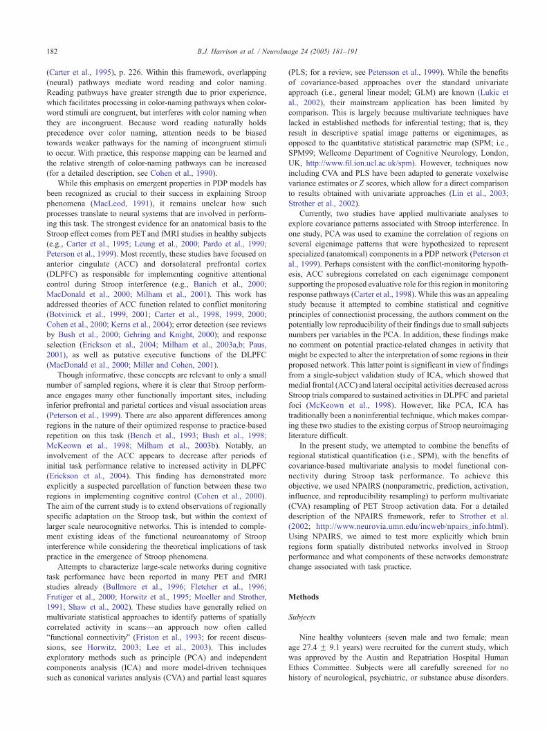
For CE1 (Figs. 2A and B), significant increases in rCBF
that occurred primarily over the first three paired-task trials
were observed in left inferior frontal cortex, bilateral primary
motor and left supplementary motor areas, right dorsal/
paralimbic and ventral anterior cingulate cortex, left posterior
cingulate gyrus, bilateral lingual gyri, visual striate cortex, and
left insula. For CE1, significant decreases in rCBF over the
first three task trials were observed bilaterally in extrastriate
cortex (fusiform gyrus) and the inferior temporal lobe, right
medial temporal and parahippocampal gyri, left orbitofrontal
cortex, bilateral superior frontal gyri, right thalamus, and left
cerebellum.
For CE2 (Fig. 2A), significant increases in rCBF associated
with Stroop interference were observed in the right orbital and
medial frontal gyrus, left middle and lateral prefrontal cortex, left
supplementary motor area, cerebellum, and right insula. For CE2,
significant decreases in rCBF during interference were observed
bilaterally in the medial temporal lobe, left inferior parietal lobe
and right posterior cingulate cortex, left ventral anterior and
subgenual cingulate gyrus, superior frontal gyrus, and cerebellum.
While we report on canonical Z scores for this deactivation
component in CE2, discussion of their functional implications will
be limited.

Table 2
Canonical Z score activations for the 12- and 2-class CVAs
Significant activations approximate region Brodmann Peak voxel-level activation Z score
areax y z
CE1: Practice-related increases
Inferior frontal gyrus 11 �23 17 �20 4.25
Medial frontal gyrus 6 �6 �22 52 3.34
Precentral gyrus 4 �15 �32 54 4.09
4 �46 �18 38 3.53
3 34 �28 50 3.70
4 40 �24 44 3.40
Anterior cingulate gyrus 32 6 0 40 3.33
25 12 36 �18 3.23
Posterior cingulate gyrus 31 �11 �34 21 3.33
Lingual gyrus 18 �6 �96 �8 3.83
18 4 �96 �10 3.53
Striate cortex 17 2 �100 �10 3.43
Insula �40 �16 6 3.26
CE1: Practice-related decreases
Fusiform gyrus 19 �30 �66 �16 5.25
37 �42 �22 �22 3.75
37 42 �46 �24 4.63
37 52 �46 �22 4.21
Inferior temporal gyrus 20 �56 �42 �22 4.18
20 52 �14 �28 3.60
20 46 22 �20 4.94
Medial temporal gyrus 21 60 �44 �22 4.34
21 46 1 �24 3.53
Parahippocampal gyrus 36 30 �20 �30 3.85
28 20 8 �26 3.66
36 28 17 �14 3.81
Orbital frontal gyrus 11 �8 17 �32 3.08
Superior frontal gyrus 10 �38 61 �2 3.44
10 2 70 �10 3.24
6 6 14 62 3.60
Thalamus 2 �8 8 3.09
Cerebellum �34 �64 �24 3.82
CE2: Interference-related activations
Orbital frontal gyrus 47 48 34 �16 4.39
Superior frontal gyrus 6 �6 6 58 3.99
Post central gyrus 3 �23 �28 58 3.78
Medial frontal gyrus 10 34 64 �10 3.72
Middle frontal gyrus 46 �42 16 18 2.99
Insula 32 �24 14 3.04
Cerebellum 6 �76 �35 4.90
�62 �68 �30 4.41
CE2: Interference-related deactivations
Temporopolar 38 �40 �8 �22 5.70
38 24 �4 �30 4.42
Middle temporal gyrus 21 �52 �22 �14 3.70
20 48 �12 �16 3.11
20 46 �68 19 3.47
Angular gyrus 40 �42 �64 30 3.76
Precuneus 7 �1 64 35 3.09
Cuneus 30 �4 �70 6 3.24
Posterior cingulate gyrus 31 2 �32 36 3.95
Subgenual gyrus 25 0 14 �24 3.61
Anterior cingulate gyrus 32 �8 34 �8 2.99
Superior frontal gyrus 8 26 24 52 4.35
8 �10 38 48 3.62
Cerebellum 44 �66 �42 3.47
NB: 12-Class CVA results = CE1; 2-class CVA results = CE2.
B.J. Harrison et al. / NeuroImage 24 (2005) 181–191186
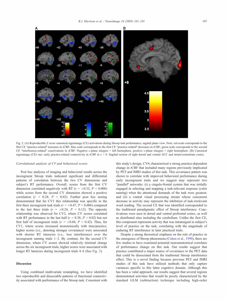
Fig. 2. (A) Reproducible Z score canonical eigenimage (CE) activations during Stroop task performance; sagittal plane view. Note: red scale corresponds to the
first CE bpractice-relatedQ increases in rCBF; blue scale corresponds to the first CE bpractice-relatedQ decreases in rCBF; green scale corresponds to the second
CE binterference-relatedQ coactivations in rCBF. Negative x-plane integers = left hemisphere, positive x-plane integers = right hemisphere. (B) Canonical
eigenimage (CE) one: early practice-related connectivity in rCBF at x = 8. Sagittal section of right dorsal and ventral ACC and striate/extrastriate cortex.
B.J. Harrison et al. / NeuroImage 24 (2005) 181–191 187
Correlational analysis of CV and behavioral scores
Post hoc analyses of imaging and behavioral results across the
incongruent Stroop trials indicated significant and differential
patterns of correlation between the two CV dimensions and
subject’s RT performance. Overall, scores from the first CV
dimension correlated negatively with RT (r = �0.33, P b 0.008)
while scores from the second CV dimension showed a positive
correlation (r = 0.28, P b 0.02). Further post hoc testing
demonstrated that for CV1 this relationship was specific to the
first three incongruent task trials (r = �0.47, P b 0.006) compared
to the last three trials (r = �0.24, P b 0.12). The opposite
relationship was observed for CV2, where CV scores correlated
with RT performance in the last half (r = 0.38, P b 0.02) but not
first half of incongruent trials (r = �0.04, P b 0.42). Thus, for
CV1, where scores increased monotonically with time/practice,
higher scores (i.e., denoting stronger covariance) were associated
with shorter RT latencies (i.e., less interference) over the
incongruent naming trials 1–3. By contrast, for the second CV
dimension, where CV scores showed relatively minimal change
across the six incongruent trials, higher scores were associated with
longer RT latencies during incongruent trials 4–6 (See Fig. 3).
Discussion
Using combined multivariate resampling, we have identified
two reproducible and dissociable patterns of functional connectiv-
ity associated with performance of the Stroop task. Consistent with
this study’s design, CVA characterized a strong practice-dependent
change in rCBF that included many regions previously implicated
by PET and fMRI studies of this task. This covariance pattern was
shown to correlate with improved behavioral performance during
early incongruent trials and we suggest may represent two
bparallelQ networks: (i) a cingulo-frontal system that was initially
engaged in selecting and mapping a task-relevant response (color
naming) when the attentional demands of the task were greatest;
and (ii) a ventral visual processing stream whose concurrent
decrease in activity may represent the inhibition of task-irrelevant
word reading. The second CE that was identified corresponded to
the traditional paradigmatic effect of Stroop interference. Coac-
tivations were seen in dorsal and ventral prefrontal cortex, as well
as distributed sites including the cerebellum. Unlike the first CE,
this component represents activity that was intransigent to subject’s
level of practice on the task, correlating with the magnitude of
enduring RT interference in later practiced trials.
Despite a strong theoretical emphasis on the role of practice in
the emergence of Stroop phenomena (Cohen et al., 1990), there are
few studies to have examined potential neuroanatomical correlates
of performance change on this task. Our results suggest that
practice contributed a major source of covariance to the PET data
that could be dissociated from the traditional Stroop interference
effect. This is a novel finding because previous PET and fMRI
studies of this task have utilized methods that only capture
variances specific to this latter cognitive domain. Although this
has been a valid approach, our results suggest that several regions
demonstrated activities that would be poorly characterized by the
standard GLM (subtraction) technique including high-order

Fig. 3. Correlation of reaction time (RT) and canonical variate (CV) scores on incongruent naming trials. For CV1 (left plot), there is a significant negative
correlation of RT and CV scores specific to first the three incongruent trials (r = �0.47, P b 0.006); while for CV2, there was a significant positive correlation
of RT and CV scores specific to the last three incongruent trials (r = 0.38, P b 0.02).
B.J. Harrison et al. / NeuroImage 24 (2005) 181–191188
regions such as the dorsal/paralimbic ACC and modulated lower
order visual areas that are involved in general stimulus processing.
These visual areas in particular may have value when interpreting
modulated effects of task practice, as conceptually they form basic
components of those bparallelQ response pathways that are selectedor inhibited during color naming versus word reading performance
(Cohen et al., 1990).
Interpretively, the first CE corresponds to regions that
demonstrated a covarying increase or decrease in their activities
predominantly over early task trials and whose activities were
associated with an improved color naming response as correlated
on incongruent naming trials. Because CV scores were undiffer-
entiated between the two task conditions and because RT
performance improved generally as a function of task practice,
this CE appears to represent common regions supporting the
emergence of a strengthened behavioral response. This would align
with the instructional set of both task conditions, which empha-
sized task-relevant color naming over task-irrelevant word reading.
However, it makes intuitive sense that the nature of coactivations
may be more characteristic of performance during the difficult
incongruent trials (i.e., as indexed behaviorally by significantly
longer RTs and more response errors). This is supported by a
previous PET study, which demonstrated that when congruent and
incongruent conditions were contrasted to color-neutral trials,
respectively, both conditions showed a similar pattern of activated
and deactivated regions (e.g., ACC, extrastriate visual areas) but a
greater magnitude of evoked activity during incongruent task
performance (Carter et al., 1995; see also Bench et al., 1993).
Considering this, we suggest that the first CE reflects most
parsimoniously activities that contributed to an improved color-
naming response via processes of increased attentional inhibition
as opposed to an improvement due to enhanced facilitation on
color-congruent trials.
Corresponding to the first CE is a notable involvement of the
ACC among a network of regions that increased in activity over
early Stroop trials (Figs. 2A and B). The spatial distribution of this
eigenimage pattern includes the right dorsal ACC activation that
has been identified in previous Stroop studies (Bush et al., 1998;
Carter et al., 1995; Kerns et al., 2004; Milham et al., 2003a,b;
Yucel et al., 2002), as well as additional areas of the cingulate
complex including ventral ACC and dorsal posterior cingulate
gyrus. This pattern of functional connectivity is highly compatible
with the known anatomical connectivity of the cingulate regions
(Vogt et al., 1995) and regions also demonstrating strong
covariance, in particular, the primary and supplementary motor
areas and left inferior frontal gyrus (Paus, 2001).
Coactivation of the posterior dorsal ACC and precentral gyrus
may suggest an involvement of the cingulate motor system (Picard
and Strick, 1996). This system has a recognized role in response
selection processes and appears to activate in functional imaging
studies irrespective of the response modality of the task at hand, for
example, manual motor (Barch et al., 2001; Koski and Paus, 2000).
There is also a recognized relationship between the ventral ACC
and left inferior frontal gyrus that has been previously validated
with PET and covariance-based connectivity analysis (Koski and
Paus, 2000). The functional coupling of these regions is believed to
form part of the neural circuitry responsible for vocalization (Paus,
2001), which has particular relevance to the current study where
the mode of responding to Stroop stimuli was vocal, as opposed to
the manual response paradigms often used in fMRI. It could also
be speculated that coactivation of the posterior cingulate gyrus and
left lingual gyrus may represent the task-relevant selection of color
from the compound Stroop stimuli. Although the posterior
cingulate gyrus has been largely implicated in visuospatial
attention (Mesulam et al., 2001; Vogt et al., 1992), there is
evidence that this region also participates in color form discrim-
ination (Gulyas et al., 1994), while the left lingual gyrus is more
commonly a region associated with the selective attentional
processing of color (Corbetta et al., 1991; Lueck et al., 1989).
Involvement of the dorsal ACC in this predominantly cingulo-
frontal network is of particular interest given its putative role in
evaluative response processes, such as, conflict monitoring (e.g.,
Carter et al., 1998) and error detection (e.g., Gehring and Knight,
2000). Most recently, it has been suggested that the ACC monitors
for conflict or error in response pathways during initial task
performance, which contributes to the implementation of cognitive
control in DLPFC when selecting between alternative responses is
difficult (Milham et al., 2003a). This has hinged on observations
during Stroop performance that the ACC is activated under
conditions requiring response-level optimization as opposed to
nonresponse (Milham et al., 2001, 2003a) and that activity within
this region decreases as the level of response conflict/error is
reduced with practice and/or control is established in DLPFC
(Erickson et al., 2004; Milham et al., 2003b). Our results are not

B.J. Harrison et al. / NeuroImage 24 (2005) 181–191 189
inconsistent with this, although they indicate that habituation
occurred only after this network initially increased in activity over
early task repetitions. While this may relate to differences in the
modeled variance between our study and previous studies, this may
also suggest that the dorsal ACC formed part of a more distributed
cingulo-frontal network that was involved in both response
evaluation processes (i.e., conflict monitoring) as well as mapping
or consolidating task-relevant responses when cognitive control
was lowest. Though speculative, this is in keeping with the pattern
of functional connectivity that we have described, that is, action
monitoring in dorsal ACC; response mapping in posterior cingulate
and visual cortex; and response execution (vocalization) via ventral
ACC-inferior frontal cortex. Following these initial coactivations,
regions such as the dorsal ACC may become less critically
involved in this response network, while other and/or different
regions may support the further consolidation (automatization) of
naming responses with practice.
Central to our interpretation of the first CE is that practice on
the Stroop task led to an improvement in RT performance related to
increased inhibitory processing during early incongruent trials.
Corresponding to the first CE are also regions that demonstrated a
covarying decrease in rCBF with practice on this task. Though it is
difficult to partial out nonspecific effects of task adaptation (i.e.,
due to decreasing emotional salience and stimulus novelty), we
suggest that this reflects more specifically the inhibition of word
reading responses within a ventral-visual processing stream. In
previous studies, deactivation of the left lateral extrastriate cortex
has been interpreted as a probable site for the inhibition of task-
irrelevant processing on the Stroop task because of its hypothe-
sized role in coding orthographic-lexical information (Carter et al.,
1995; see also Buckner et al., 1995; Petersen et al., 1988).
Additional areas of this CE also have hypothesized roles in
processing word form and meaning, including the right fusiform
gyrus and bilateral parahippocampal gyri (Corbetta et al., 1991;
Demb et al., 1995; Price, 1998; Raichle et al., 1994). Therefore, at
least conceptually, this practice-related decrease of functional
connectivity within ventral visual regions supports the nature of
competitive processing that has been advocated in PDP models
(Cohen et al., 1990).
If this ventral stream holds true as a site of task-irrelevant
inhibition on the Stroop task, then it begs the question as to what
regions are responsible for generating this source of inhibitory
control. It could be suggested that a reciprocal relationship exists
between the bparallelQ networks that we have described, where a
cingulo-frontal system participates in task-relevant response
selection or mapping and task-irrelevant response inhibition (Paus
et al., 1993). However, in current models of Stroop performance,
cognitive control is marshaled as a seemingly independent
moderator of response pathways, either signaled into action by
its own regulative mechanisms or in response to feedback
(evaluation) from performance monitoring functions of the ACC
(Botvinick et al., 2001). From recent functional imaging studies of
this task, evidence has implicated the DLPFC as responsible for
implementing this inhibitory bias over processing in posterior
cortical regions (Banich et al., 2001; Milham et al., 2003a). This is
illustrated well by Milham et al. (2002) who reported that in the
event of less activation of the mid-DLPFC, and hence less
inhibition of task-irrelevant processes, a more extensive activation
of these same ventral regions occurred during Stroop interference.
Corresponding to the second CE, we note coactivations of the
left DLPFC, right orbital frontal gyrus, and right frontopolar
cortex. These regions showed consistent task-related connectivity
during Stroop interference, which was present at the behavioral
level across the six incongruent trials. It was also found that this
pattern most closely aligned with subject’s RT performance during
later incongruent trials, correlating with the magnitude of enduring
response conflict. This pattern supports recent suggestions that the
DLPFC bmaintainsQ attentional control over Stroop performance
after initial stages of response optimization with practice (Erickson
et al., 2004; Milham et al., 2003b). The coupling of the DLPFC to
other regions including orbital prefrontal and frontopolar cortex
also suggests that such actions appear to engage a more
distributed PFC network perhaps consistent with other recent
studies of the functional anatomy of cognitive control (Badre and
Wagner, 2004). It is interesting to note that the cerebellum
demonstrated significant coactivation in this second CE pattern.
The cerebellum has been previously implicated in studies of
practice effects and automaticity in cognitive performance
(Burnod, 1991; Grafton et al., 1992; Seitz et al., 1990) and
typically shows decreased activity between unpracticed and
practiced trials (e.g., Friston et al., 1992). However, that the
cerebellum and frontal regions were engaged consistently by
incongruent Stroop trials probably reflects that performance of the
task remained effortful and never fully automatized, that is,
hundreds of trials are typically needed to reduce the amount of RT
interference (MacLeod, 1991).
In closing, the current study provides a novel characterization
of the effects of practice and interference on functional connectiv-
ity in rCBF associated with performance the Stroop task. These
findings largely compliment existing studies that have utilized this
task to examine more specific roles for the ACC and DLPFC in
cognitive control, although they suggest that the contribution of
these regions is worth considering in the context of more
distributed brain systems. Despite apparent consistencies between
our results and results obtained with fMRI, there are obvious
limitations to this study inherent with the use of PET (i.e., poor
temporal resolution). For instance, our results do not comment on
the dynamic adaptivity of the ACC and DLPFC regions during
response conflict or error processing, which has been reported in
recent event-related functional imaging studies (e.g., Botvinick et
al., 1999; Kerns et al., 2004). The use of PET to examine extended
effects of practice on this task also necessitated that we use a
simple blocked paradigm of congruent and incongruent trials,
which is not appropriate for modeling other response parameters
such as Stroop facilitation effects. However, for the purpose of
characterizing gradual changes in brain activity that may contribute
to the reorganization of reading versus naming responses that
emerge slowly with practice on this task (Cohen et al., 1990;
MacLeod, 1991), we would suggest the current approach was
sufficiently suited. A particular benefit of PET in this scenario was
our ability to examine one of the basic tenets of true Stroop task
performance, namely, vocalization. In fMRI studies of this task,
subjects are often pretrained on alternative response parameters
before scanning (e.g., button-box associations) because of the
motion-artifact associated with vocalized movement. This practice
may in turn lead to studies underestimating the responsivity of
certain regions such as the ACC when averaging actual (scanned)
task performances (for a recent discussion see Erickson et al.,
2004). In the current study, practice not only contributed the
greatest source of variance to our data, but it was those initial
changes in activity resulting from practice that were arguably the
most meaningful.

B.J. Harrison et al. / NeuroImage 24 (2005) 181–191190
Acknowledgments
Supported by a National Health and Medical Research
Council (NHMRC) grant 970599, the NHMRC Brain Research
Network. The authors thank Drs. Phyllis Chua and Simon
Collinson for their assistance with task and subject preparation
and image acquisition. They also thank colleagues and staff from
the Department of Nuclear Medicine, Center for PET, Austin
Hospital Melbourne.
References
Badre, D., Wagner, A.D., 2004. Selection, integration, and conflict
monitoring; assessing the nature and generality of prefrontal cognitive
control mechanisms. Neuron 41, 473–487.
Banich, M.T., Milham, M.P., Atchley, R.A., Cohen, N.J., Webb, A.,
Wszalek, T., Kramer, A.F., Liang, Z., Barad, V., Gullett, D., Shah, C.,
Brown, C., 2000. Prefrontal regions play a predominant role in
imposing an attentional dsetT, evidence from fMRI. Brain Res. Cogn.
Brain Res. 10, 1–9.
Banich, M.T., Milham, M.P., Jacobson, B.L., Webb, A., Wszalek, T.,
Cohen, N.J., Kramer, A.F., 2001. Attentional selection and the
processing of task-irrelevant information, insights from fMRI exami-
nations of the Stroop task. Prog. Brain Res. 134, 459–470.
Barch, D.M., Braver, T.S., Akbudak, E., Conturo, T., Ollinger, J.,
Snyder, A., 2001. Anterior cingulate cortex and response conflict:
effects of response modality and processing domain. Cereb. Cortex 11,
837–848.
Bench, C.J., Frith, C.D., Grasby, P.M., Friston, K.J., Paulesu, E.,
Frackowiak, R.S., Dolan, R.J., 1993. Investigations of the functional
anatomy of attention using the Stroop test. Neuropsychologia 31,
907–922.
Botvinick, M., Nystrom, L.E., Fissell, K., Carter, C.S., Cohen, J.D., 1999.
Conflict monitoring versus selection-for-action in anterior cingulate
cortex. Nature 402, 179–181.
Botvinick, M.M., Braver, T.S., Barch, D.M., Carter, C.S., Cohen, J.D.,
2001. Conflict monitoring and cognitive control. Psychol. Rev. 108,
624–652.
Buckner, R.L., Petersen, S.E., Ojemann, J.G., Miezin, F.M., Squire, L.R.,
Raichle, M.E., 1995. Functional anatomical studies of explicit and
implicit memory retrieval tasks. J. Neurosci. 15, 12–29.
Bullmore, E.T., Rabe-Hesketh, S., Morris, R.G., Williams, S.C., Gregory,
L., Gray, J.A., Brammer, M.J., 1996. Functional magnetic resonance
image analysis of a large-scale neurocognitive network. NeuroImage 4,
16–33.
Burnod, Y., 1991. Organizational levels of the cerebral cortex: an integrated
model. Acta Biotheor. 39, 351–361.
Bush, G., Whalen, P.J., Rosen, B.R., Jenike, M.A., McInerney, S.C., Rauch,
S.L., 1998. The counting Stroop, an interference task specialized for
functional neuroimaging-validation study with functional MRI. Hum.
Brain Mapp. 6, 270–282.
Bush, G., Luu, P., Posner, M.I., 2000. Cognitive and emotional influences
in anterior cingulate cortex. Trends Cogn. Sci. 4, 215–222.
Carter, C.S., Mintun, M., Cohen, J.D., 1995. Interference and facilitation
effects during selective attention, an H215O PET study of Stroop task
performance. NeuroImage 2, 264–272.
Carter, C.S., Braver, T.S., Barch, D.M., Botvinick, M.M., Noll, D., Cohen,
J.D., 1998. Anterior cingulate cortex, error detection, and the online
monitoring of performance. Science 280, 747–749.
Carter, C.S., Botvinick, M.M., Cohen, J.D., 1999. The contribution of the
anterior cingulate cortex to executive processes in cognition. Rev.
Neurosci. 10, 49–57.
Carter, C.S., Macdonald, A.M., Botvinick, M., Ross, L.L., Stenger, V.A.,
Noll, D., Cohen, J.D., 2000. Parsing executive processes, strategic vs.
evaluative functions of the anterior cingulate cortex. Proc. Natl. Acad.
Sci. U. S. A. 97, 1944–1948.
Cohen, J.D., Dunbar, K., McClelland, J.L., 1990. On the control of
automatic processes, a parallel distributed processing account of the
Stroop effect. Psychol. Rev. 97, 332–361.
Cohen, J.D., Botvinick, M., Carter, C.S., 2000. Anterior cingulate and
prefrontal cortex, who’s in control? Nat. Neurosci. 3, 421–423.
Corbetta, M., Miezin, F.M., Dobmeyer, S., Shulman, G.L., Petersen, S.E.,
1991. Selective and divided attention during visual discriminations of
shape, color, and speed, functional anatomy by positron emission
tomography. J. Neurosci. 11, 2383–2402.
Demb, J.B., Desmond, J.E., Wagner, A.D., Vaidya, C.J., Glover, G.H.,
Gabrieli, J.D., 1995. Semantic encoding and retrieval in the left inferior
prefrontal cortex, a functional MRI study of task difficulty and process
specificity. J. Neurosci. 15, 5870–5878.
Erickson, K.I., Milham, M.P., Colcombe, S.J., Kramer, A.F., Banich, M.T.,
Webb, A., Cohen, N.J., 2004. Behavioral conflict, anterior cingulate
cortex, and experiment duration: implications of diverging data. Hum.
Brain Mapp. 21, 98–107.
Fletcher, P.C., Dolan, R.J., Shallice, T., Frith, C.D., Frackowiak, R.S.,
Friston, K.J., 1996. Is multivariate analysis of PET data more revealing
than the univariate approach? Evidence from a study of episodic
memory retrieval. NeuroImage 3, 209–215.
Friston, K.J., Frith, C.D., Passingham, R.E., Liddle, P.F., Frackowiak, R.S.,
1992. Motor practice and neurophysiological adaptation in the
cerebellum: a positron tomography study. Proc. R. Soc. Lond. B Biol.
Sci. 248 (1323), 223–228.
Friston, K.J., Frith, C.D., Liddle, P.F., Frackowiak, R.S., 1993. Functional
connectivity, the principal-component analysis of large (PET) data sets.
J. Cereb. Blood Flow Metab. 13, 5–14.
Friston, K.J., Frith, C.D., Fletcher, P., Liddle, P.F., Frackowiak, R.S., 1996.
Functional topography, multidimensional scaling and functional con-
nectivity in the brain. Cereb. Cortex 6, 156–164.
Frutiger, S.A., Strother, S.C., Anderson, J.R., Sidtis, J.J., Arnold, J.B.,
Rottenberg, D.A., 2000. Multivariate predictive relationship between
kinematic and functional activation patterns in a PET study of
visuomotor learning. NeuroImage 12, 515–527.
Gehring, W.J., Knight, R.T., 2000. Prefrontal-cingulate interactions in
action monitoring. Nat. Neurosci. 3, 516–520.
Grafton, S.T., Mazziotta, J.C., Presty, S., Friston, K.J., Frackowiak, R.S.,
Phelps, M.E., 1992. Functional anatomy of human procedural learning
determined with regional cerebral blood flow and PET. J. Neurosci. 12,
2542–2548.
Gulyas, B., Heywood, C.A., Popplewell, D.A., Roland, P.E., Cowey, A.,
1994. Visual form discrimination from color or motion cues: functional
anatomy by positron emission tomography. Proc. Natl. Acad. Sci.
U. S. A. 91, 9965–9969.
Horwitz, B., 2003. The elusive concept of brain connectivity. NeuroImage
19, 466–470.
Horwitz, B., McIntosh, A.R., Haxby, J.V., Grady, C.L., 1995. Network
analysis of brain cognitive function using metabolic and blood flow
data. Behav. Brain Res. 66, 187–193.
Kerns, J.G., Cohen, J.D., MacDonald III, A.W., Cho, R.Y., Stenger, V.A.,
Carter, C.S., 2004. Anterior cingulate conflict monitoring and adjust-
ments in control. Science 303, 1023–1026.
Kinahan, P.E., Rogers, J.G., 1989. Analytic 3D image reconstruction using
all detected events. IEEE Trans. Nucl. Sci. 36, 984–986.
Koski, L., Paus, T., 2000. Functional connectivity of the anterior cingulate
cortex within the human frontal lobe, a brain-mapping meta-analysis.
Exp. Brain Res. 133, 55–65.
Kustra R., 2000. Statistical analysis of medical images with applications to
neuroimaging. PhD Thesis, University of Toronto. http://www.ustat.
utoronto.ca/~rafal/thesis/.ps.gz.
Kustra, R., Strother, S., 2001. Penalized discriminant analysis of [15O]-
water PET brain images with prediction error selection of smooth-
ness and regularization hyperparameters. IEEE Trans. Med. Imag.
20, 376–387.

B.J. Harrison et al. / NeuroImage 24 (2005) 181–191 191
Lee, L., Harrison, L.M., Mechelli, A., 2003. A report of the functional
connectivity workshop, Dusseldorf 2002. NeuroImage 19, 457–465.
Leung, H.C., Skudlarski, P., Gatenby, J.C., Peterson, B.S., Gore, J.C., 2000.
An event-related functional MRI study of the Stroop color word
interference task. Cereb. Cortex 10, 552–560.
Lin, F.H., McIntosh, A.R., Agnew, J.A., Eden, G.F., Zeffiro, T.A.,
Belliveau, J.W., 2003. Multivariate analysis of neuronal interactions
in the generalized partial least squares framework: simulations and
empirical studies. NeuroImage 20, 625–642.
Lueck, C.J., Zeki, S., Friston, K.J., Deiber, M.P., Cope, P., Cunningham,
V.J., Lammertsma, A.A., Kennard, C., Frackowiak, R.S., 1989. The
color center in the cerebral cortex of man. Nature 340, 386–389.
Lukic, A.S., Wernick, M.N., Strother, S.C., 2002. An evaluation of methods
for detecting brain activations from functional neuroimages. Artif.
Intell. Med. 25, 69–88.
MacDonald III, A.W., Cohen, J.D., Stenger, V.A., Carter, C.S., 2000.
Dissociating the role of the dorsolateral prefrontal and anterior cingulate
cortex in cognitive control. Science 288, 1835–1838.
MacLeod, C.M., 1991. Half a century of research on the Stroop effect, an
integrative review. Psychol. Bull. 109, 163–203.
McIntosh, A.R., 1999. Mapping cognition to the brain through neural
interactions. Memory 7, 523–548.
McKeown, M.J., Jung, T.P., Makeig, S., Brown, G., Kindermann, S.S., Lee,
T.W., Sejnowski, T.J., 1998. Spatially independent activity patterns in
functional MRI data during the Stroop color-naming task. Proc. Natl.
Acad. Sci. U. S. A. 95, 803–810.
Mesulam, M.M., Nobre, A.C., Kim, Y.H., Parrish, T.B., Gitelman, D.R.,
2001. Heterogeneity of cingulate contributions to spatial attention.
NeuroImage 13, 1065–1072.
Milham, M.P., Banich, M.T., Webb, A., Barad, V., Cohen, N.J., Wszalek,
T., Kramer, A.F., 2001. The relative involvement of anterior cingulate
and prefrontal cortex in attentional control depends on nature of
conflict. Brain Res. Cogn. Brain Res. 12, 467–473.
Milham, M.P., Erickson, K.I., Banich, M.T., Kramer, A.F., Webb, A.,
Wszalek, T., Cohen, N.J., 2002. Attentional control in the aging
brain, insights from an fMRI study of the Stroop task. Brain Cogn.
49, 277–296.
Milham, M.P., Banich, M.T., Barad, V., 2003a. Competition for priority in
processing increases prefrontal cortex’s involvement in top-down
control, an event-related fMRI study of the Stroop task. Brain Res.
Cogn. Brain Res. 17, 212–222.
Milham, M.P., Banich, M.T., Claus, E.D., Cohen, N.J., 2003b. Practice-
related effects demonstrate complementary roles of anterior cingulate
and prefrontal cortices in attentional control. NeuroImage 18, 483–493.
Miller, E.K., Cohen, J.D., 2001. An integrative theory of prefrontal cortex
function. Annu. Rev. Neurosci. 24, 167–202.
Moeller, J.R., Strother, S.C., 1991. A regional covariance approach to the
analysis of functional patterns in positron emission tomographic data.
J. Cereb. Blood Flow Metab. 11, A121–A135.
Nelson, H.E., O’Connell, A., 1978. Dementia, the estimation of premorbid
intelligence levels using the New Adult Reading Test. Cereb. Cortex 14,
234–244.
Oldfield, R.C., 1971. The assessment and analysis of handedness: the
Edinburgh Inventory. Neuropsychologia 9, 97–113.
Pardo, J.V., Pardo, P.J., Janer, K.W., Raichle, M.E., 1990. The anterior
cingulate cortex mediates processing selection in the Stroop attentional
conflict paradigm. Proc. Natl. Acad. Sci. U. S. A. 87, 256–259.
Paus, T., 2001. Primate anterior cingulate cortex, where motor control, drive
and cognition interface. Nat. Rev., Neurosci. 2, 417–424.
Paus, T., Petrides, M., Evans, A.C., Meyer, E., 1993. Role of the human
anterior cingulate cortex in the control of oculomotor, manual, and
speech responses, a positron emission tomography study. J. Neuro-
physiol. 70, 453–469.
Petersen, S.E., Fox, P.T., Posner, M.I., Mintun, M., Raichle, M.E., 1988.
Positron emission tomographic studies of the cortical anatomy of
single-word processing. Nature 331, 585–589.
Peterson, B.S., Skudlarski, P., Gatenby, J.C., Zhang, H., Anderson, A.W.,
Gore, J.C., 1999. An fMRI study of Stroop word-color interference,
evidence for cingulate subregions subserving multiple distributed
attentional systems. Biol. Psychiatry 45, 1237–1258.
Petersson, K.M., Nichols, T.E., Poline, J.B., Holmes, A.P., 1999.
Statistical limitations in functional neuroimaging: I. Non-inferential
methods and statistical models. Philos. Trans. R. Soc. Lond., B Biol.
Sci. 354, 1239–1260.
Picard, N., Strick, P.L., 1996. Motor areas of the medial wall: a review of
their location and functional activation. Cereb. Cortex 6, 342–353.
Posner, M.I., Petersen, S.E., 1990. The attention system of the human brain.
Annu. Rev. Neurosci. 13, 25–42.
Price, C.J., 1998. The functional anatomy of word comprehension and
production. Trends Cogn. Sci. 2, 2288–2981.
Raichle, M.E., Fiez, J.A., Videen, T.O., MacLeod, A.M., Pardo, J.V., Fox,
P.T., Petersen, S.E., 1994. Practice-related changes in human brain
functional anatomy during nonmotor learning. Cereb. Cortex 4, 8–26.
Seitz, R.J., Roland, E., Bohm, C., Greitz, T., Stone-Elander, S., 1990. Motor
learning in man: a positron emission tomographic study. NeuroReport
1, 57–60.
Shaw, M.E., Strother, S.C., McFarlane, A.C., Morris, P., Anderson, J.,
Clark, C.R., Egan, G.F., 2002. Abnormal functional connectivity in
posttraumatic stress disorder. NeuroImage 15, 661–674.
Smith, T., Tong, C., Lammertsma, A.A., Butler, K.R., Schnorr, L., Watson,
J.D.G., Ramsay, S., Clark, J.C., Jones, T., 1994. Dosimetry of
intravenously administered oxygen-15 labeled water in man: a model
based on experimental human data from 21 subjects. Eur. J. Nucl. Med.
21, 1126–1134.
Stroop, R., 1935. Studies of interference in serial verbal reactions. J. Exp.
Psychol. 18, 643–662.
Strother, S.C., Anderson, J., Hansen, L.K., Kjems, U., Kustra, R., Sidtis, J.,
Frutiger, S., Muley, S., LaConte, S., Rottenberg, D., 2002. The
quantitative evaluation of functional neuroimaging experiments, the
NPAIRS data analysis framework. NeuroImage 15, 747–771.
Tochon-Danguy, H.J., Clark, J.C., Janus, A., Sachinidis, J.I., 1995.
Technical performance and operating procedure of a bedside [15O]water
infuser. J. Label. Comp. Radiopharm. 37, 662–664.
Vogt, B.A., Finch, D.M., Olson, C.R., 1992. Functional heterogeneity in
cingulate cortex, the anterior executive and posterior evaluative regions.
Cereb. Cortex 2, 435–443.
Vogt, B.A., Nimchinsky, E.A., Vogt, L.J., Hof, P.R., 1995. Human cingulate
cortex, surface features, flat maps, and cytoarchitecture. J. Comp.
Neurol. 359, 490–506.
Yqcel, M., Pantelis, C., Stuart, G.W., Wood, S.J., Maruff, P., Velakoulis, D.,
Pipingas, A., Crowe, S.F., Tochon-Danguy, H.J., Egan, G.F., 2002.
Anterior cingulate activation during Stroop task performance: a PET to
MRI coregistration study of individual patients with schizophrenia. Am.
J. Psychiatry 159, 251–254.
Related Documents