Molecular Biology of the Cell Vol. 13, 469 – 479, February 2002 Functional Characterization and Localization of the Aspergillus nidulans Formin SEPA □ V Kathryn E. Sharpless and Steven D. Harris* † Department of Microbiology, University of Connecticut Health Center, Farmington, Connecticut 06030-3205 Submitted July 18, 2001; Revised October 23, 2001; Accepted November 5, 2001 Monitoring Editor: John Pringle Formins are a family of multidomain scaffold proteins involved in actin-dependent morphoge- netic events. In Aspergillus nidulans, the formin SEPA participates in two actin-mediated processes, septum formation and polarized growth. In this study, we use a new null mutant to demonstrate that SEPA is required for the formation of actin rings at septation sites. In addition, we find that a functional SEPA::GFP fusion protein localizes simultaneously to septation sites and hyphal tips, and that SEPA colocalizes with actin at each site. Using live imaging, we show that SEPA localization at septation sites and hyphal tips is dynamic. Notably, at septation sites, SEPA forms a ring that constricts as the septum is deposited. Moreover, we demonstrate that actin filaments are required to maintain the proper localization pattern of SEPA, and that the amino-terminal half of SEPA is sufficient for localization at septation sites and hyphal tips. In contrast, only localization at septation sites is affected by loss of the sepH gene product. We propose that specific morpho- logical cues activate common molecular pathways to direct SEPA localization to the appropriate morphogenetic site. INTRODUCTION The actin cytoskeleton functions in numerous cellular pro- cesses, including cell motility, organelle and vesicle trans- port, morphogenesis, and cytokinesis. To perform these multiple tasks, the actin cytoskeleton is controlled by nu- merous regulatory and accessory proteins that direct the polymerization of actin monomers into filaments and the cross-linking of filaments into a network (Ayscough, 1998; Chen et al., 2000). Assembly and organization of the actin cytoskeleton is linked to environmental and intracellular signals by multiple pathways (Schmidt and Hall, 1998). Key regulators of these pathways are members of the Rho family of low molecular weight GTPases. Rho GTPases are mem- brane-bound proteins that act as molecular switches to relay spatial and temporal information to effectors that reorganize the actin cytoskeleton (Tanaka and Takai, 1998). Among the effectors of Rho GTPases are proteins that contain multiple protein–protein interaction domains and appear to function as molecular scaffolds (Bishop and Hall, 2000). Scaffold pro- teins integrate incoming signals with actin cytoskeleton dy- namics by interacting with both the signaling proteins and actin-binding proteins. Examples include the ezrin/radixin/ moesin, enabled/vasodilator-stimulated phosphoprotein, Wiskott-Aldrich syndrome protein/Wiskott-Aldrich syn- drome protein-interacting protein, and formin families of proteins (Frazier and Field, 1997; Beckerle, 1998; Wasser- man, 1998; Bretscher, 1999; Ramesh et al., 1999; Zeller et al., 1999; Mullins, 2000). Formins are conserved from fungi to humans and are characterized by the presence of two conserved carboxy- terminal regions, the FH1 and FH2 domains (Emmons et al., 1995). The proline-rich FH1 domain interacts with SH3 and WW domain-containing proteins, as well as with the actin monomer-binding protein profilin (Chan et al., 1996; Manseau et al., 1996; Uetz et al., 1996; Bedford et al., 1997; Chang et al., 1997; Evangelista et al., 1997; Imamura et al., 1997; Watanabe et al., 1997; Kamei et al., 1998). The FH2 domain interacts with the actin-binding proteins Bud6p and elongation factor 1 (Evangelista et al., 1997; Umikawa et al., 1998), as well as with Smy1p, a kinesin-related protein that may function in actin filament-based transport (Kikyo et al., 1999). Three other domains located in the amino-terminal half of formins may be conserved: the Rho GTPase-binding site (Kohno et al., 1996; Evangelista et al., 1997; Imamura et al., 1997; Watanabe et al., 1997), the FH3 domain (Petersen et al., 1995), and the Spa2p-binding domain (Fujiwara et al., 1998). The FH3 domain and Spa2p-binding domain are Article published online ahead of print. Mol. Biol. Cell 10.1091/ mbc.01– 07-0356. Article and publication date are at www.molbiol- cell.org/cgi/doi/10.1091/mbc.01– 07-0356. † Present address: Plant Science Initiative, University of Nebraska, N234 Beadle Center, Lincoln, NE 68588-0660. □ V Online version of this article contains video material for some figures. Online version available at www.molbiolcell.org. * Corresponding author. E-mail address: [email protected]. Abbreviations used: aa, amino acid; GFP, green fluorescent pro- tein; ts, temperature-sensitive. © 2002 by The American Society for Cell Biology 469

Welcome message from author
This document is posted to help you gain knowledge. Please leave a comment to let me know what you think about it! Share it to your friends and learn new things together.
Transcript

Molecular Biology of the CellVol. 13, 469–479, February 2002
Functional Characterization and Localization of theAspergillus nidulans Formin SEPA□V
Kathryn E. Sharpless and Steven D. Harris*†
Department of Microbiology, University of Connecticut Health Center, Farmington, Connecticut06030-3205
Submitted July 18, 2001; Revised October 23, 2001; Accepted November 5, 2001Monitoring Editor: John Pringle
Formins are a family of multidomain scaffold proteins involved in actin-dependent morphoge-netic events. In Aspergillus nidulans, the formin SEPA participates in two actin-mediated processes,septum formation and polarized growth. In this study, we use a new null mutant to demonstratethat SEPA is required for the formation of actin rings at septation sites. In addition, we find thata functional SEPA::GFP fusion protein localizes simultaneously to septation sites and hyphal tips,and that SEPA colocalizes with actin at each site. Using live imaging, we show that SEPAlocalization at septation sites and hyphal tips is dynamic. Notably, at septation sites, SEPA formsa ring that constricts as the septum is deposited. Moreover, we demonstrate that actin filamentsare required to maintain the proper localization pattern of SEPA, and that the amino-terminal halfof SEPA is sufficient for localization at septation sites and hyphal tips. In contrast, only localizationat septation sites is affected by loss of the sepH gene product. We propose that specific morpho-logical cues activate common molecular pathways to direct SEPA localization to the appropriatemorphogenetic site.
INTRODUCTION
The actin cytoskeleton functions in numerous cellular pro-cesses, including cell motility, organelle and vesicle trans-port, morphogenesis, and cytokinesis. To perform thesemultiple tasks, the actin cytoskeleton is controlled by nu-merous regulatory and accessory proteins that direct thepolymerization of actin monomers into filaments and thecross-linking of filaments into a network (Ayscough, 1998;Chen et al., 2000). Assembly and organization of the actincytoskeleton is linked to environmental and intracellularsignals by multiple pathways (Schmidt and Hall, 1998). Keyregulators of these pathways are members of the Rho familyof low molecular weight GTPases. Rho GTPases are mem-brane-bound proteins that act as molecular switches to relayspatial and temporal information to effectors that reorganizethe actin cytoskeleton (Tanaka and Takai, 1998). Among theeffectors of Rho GTPases are proteins that contain multipleprotein–protein interaction domains and appear to function
as molecular scaffolds (Bishop and Hall, 2000). Scaffold pro-teins integrate incoming signals with actin cytoskeleton dy-namics by interacting with both the signaling proteins andactin-binding proteins. Examples include the ezrin/radixin/moesin, enabled/vasodilator-stimulated phosphoprotein,Wiskott-Aldrich syndrome protein/Wiskott-Aldrich syn-drome protein-interacting protein, and formin families ofproteins (Frazier and Field, 1997; Beckerle, 1998; Wasser-man, 1998; Bretscher, 1999; Ramesh et al., 1999; Zeller et al.,1999; Mullins, 2000).
Formins are conserved from fungi to humans and arecharacterized by the presence of two conserved carboxy-terminal regions, the FH1 and FH2 domains (Emmons et al.,1995). The proline-rich FH1 domain interacts with SH3 andWW domain-containing proteins, as well as with the actinmonomer-binding protein profilin (Chan et al., 1996;Manseau et al., 1996; Uetz et al., 1996; Bedford et al., 1997;Chang et al., 1997; Evangelista et al., 1997; Imamura et al.,1997; Watanabe et al., 1997; Kamei et al., 1998). The FH2domain interacts with the actin-binding proteins Bud6p andelongation factor 1� (Evangelista et al., 1997; Umikawa et al.,1998), as well as with Smy1p, a kinesin-related protein thatmay function in actin filament-based transport (Kikyo et al.,1999). Three other domains located in the amino-terminalhalf of formins may be conserved: the Rho GTPase-bindingsite (Kohno et al., 1996; Evangelista et al., 1997; Imamura etal., 1997; Watanabe et al., 1997), the FH3 domain (Petersen etal., 1995), and the Spa2p-binding domain (Fujiwara et al.,1998). The FH3 domain and Spa2p-binding domain are
Article published online ahead of print. Mol. Biol. Cell 10.1091/mbc.01–07-0356. Article and publication date are at www.molbiol-cell.org/cgi/doi/10.1091/mbc.01–07-0356.
† Present address: Plant Science Initiative, University of Nebraska,N234 Beadle Center, Lincoln, NE 68588-0660.
□V Online version of this article contains video material for somefigures. Online version available at www.molbiolcell.org.
* Corresponding author. E-mail address: [email protected] used: aa, amino acid; GFP, green fluorescent pro-tein; ts, temperature-sensitive.
© 2002 by The American Society for Cell Biology 469
thought to regulate the localization of formins to sites of cellsurface remodeling (Petersen et al., 1998; Ozaki-Kuroda et al.,2001).
Phenotypic characterization of formin mutants has pro-vided evidence that formins are required for actin function.For example, during cytokinesis, actin rings fail to form inSchizosaccharomyces pombe cdc12 and Drosophila melanogasterdia mutants (Chang et al., 1997; Afshar et al., 2000). In con-trast, actomyosin rings form at the mother/bud neck inSaccharomyces cerevisiae bni1 mutants, but they do not con-tract (Vallen et al., 2000). Furthermore, actin fails to localizeto tips of mating projections during conjugation in bni1 andS. pombe fus1 mutants (Evangelista et al., 1997; Petersen et al.,1998). Consistent with their role in actin cytoskeletal orga-nization, most formins colocalize with actin at sites of po-larized growth or with the actin ring during cytokinesis(Chang et al., 1997; Evangelista et al., 1997; Fujiwara et al.,1998; Petersen et al., 1998; Swan et al., 1998; Afshar et al.,2000; Ozaki-Kuroda et al., 2001).
Conidiospores of the filamentous fungus Aspergillus nidu-lans undergo a series of morphogenetic events during ger-mination (Harris, 1997). Initially, conidiospores undergo aperiod of isotropic swelling, which is followed by a switch toapical growth and the subsequent emergence of a germ tube.Apical (tip) growth is maintained throughout the life of ahypha, and, unlike what occurs in budding or fission yeast,it is not interrupted during septation. Septation occurs oncecells have satisfied a size requirement and completed at leastone round of mitosis (Wolkow et al., 1996). Experiments withcytochalasin A, an actin-depolymerizing drug, demonstratethat actin filaments are required for both apical growth andseptation (Harris et al., 1994; Torralba et al., 1998). Consistentwith its function, actin localizes to both hyphal tips andseptation sites (Harris et al., 1994). Like actin, the sepA geneproduct functions in a number of morphogenetic processes;the temperature-sensitive (ts) sepA1 mutant fails to septate atrestrictive temperature, displays a defective tip growth pat-tern, and has abnormally wide hyphae (Morris, 1976). Mo-lecular characterization has revealed that sepA encodes aformin (Harris et al., 1997).
In this study, we characterize the role of SEPA duringseptum formation and tip growth by constructing a new nullallele and by determining the localization pattern of a func-tional SEPA::GFP fusion protein. In addition, we provideinitial insight into the mechanisms underlying the simulta-neous targeting of SEPA to distinct structures within hyphalcells.
MATERIALS AND METHODS
Aspergillus Strains and Growth MethodsStrains used in this study are described in Table 1. All geneticmanipulations were performed as described previously (Harris etal., 1994). The following media were used: MAG (2% dextrose, 2%malt extract, 0.2% peptone, trace elements, and vitamins; pH 6.5),YGV (2% dextrose, 0.5% yeast extract, and vitamins), and MN (1%dextrose, nitrate salts, trace elements, and biotin; pH 6.5). Arginine(1 mM), uridine (5 mM), and uracil (10 mM) were added as needed.Trace elements, nitrate salts, and vitamins were added as describedin the appendix to Kafer (1977). For solid media, 1.5% agar wasadded.
DNA TechniquesSubcloning was performed using standard methods (Sambrook etal., 1989) except that the TOPO TA Cloning kit (Invitrogen, Carsl-bad, CA) was used to subclone polymerase chain reaction (PCR)fragments of sepA1 into pCR2.1-TOPO. PCRs were carried out usingVent or Taq polymerase (Invitrogen). GC-rich PCR was performedusing the Advantage-GC Genomic PCR kit (CLONTECH, Palo Alto,CA) or the PCRX Enhancer System (Invitrogen). PCR primer se-quences are available on request. Sequencing and oligonucleotidesynthesis were performed by the Molecular Core Facility at theUniversity of Connecticut Health Center. The sepA sequence hasbeen updated in GenBank (accession number U83658).
Isolation of DNA from A. nidulans and transformations wereperformed using standard procedures (Timberlake, 1990; Oakleyand Osmani, 1993). Southern blots were analyzed using digoxige-nin-labeled probes and nonradioactive detection (Roche MolecularBiochemicals, Indianapolis, IN).
Plasmid ConstructsThe plasmid containing sepA::gfp, pKES59, was constructed in mul-tiple steps. A unique NotI site that replaced the sepA stop codon wasconstructed using PCR to amplify a 333-base pair fragment of sepA.This fragment was subcloned into pYESTrp (Invitrogen) by using aninternal SphI site and the NotI site from the primer. Using the SphIsite and a KpnI site 3� to the NotI site, the fragment was ligated intopKES1, a plasmid containing most of sepA (HindIII to SphI) (Harriset al., 1997). The resulting plasmid, pKES30, contains the entire sepAgene (5422 base pairs without the stop codon) followed by uniqueNotI and KpnI sites, plus 964 base pairs of upstream sequence. PCRwas used to incorporate NotI sites on both ends of gfp from pMCB32(Fernandez-Abalos et al., 1998). pMCB32 contains a codon-modifiedversion of green fluorescent protein (GFP), which is optimized forexpression in mammals and plants and carries the S65T substitu-tion. gfp was ligated in-frame with sepA in the NotI site of pKES30,resulting in the plasmid pKES46.
Table 1. A. nidulans strains used in this study
Strain Genotype Source
A28a pabaA6 biA1 FGSCa
AH13 wA3; argB2; chaA1 Lab stockAJM68 pyrG89; wA3; sepH1; pyroA4 Bruno et al., 2001ALH1 sepA4�Bm Harris et al., 1997AML9 pyrG89 pabaA1; argB2 Lab stockAML13 pabaA1 yA2; argB2; chaA1? Lab stockASH35 sepA1 yA2; argB2 Harris et al., 1997ASH162 pyrG89 pabaA1 yA2 Lab stockAKS6 sepA6�FH�argB; wA2; argB2;
chaA1This study
AKS64 sepA1 yA2; sepA�gfp�argB argB2 This studyAKS65 sepA1 yA2; sepA�gfp�argB argB2 This studyAKS70 sepA�gfp�pyr-4; pyrG89 pabaA1
yA2This study
AKS71 sepA�gfp�pyr-4; pyrG89; wA3;sepH1; pyroA4
This study
AKS76 yA2; sepA�gfp�argB argB2; chaA1 This studyAKS82 sepA(aa1-838)�gfp�pyr-4; pyrG89
pabaA1 yA2This study
AKS84 sepA(aa1-132)�gfp�pyr-4; pyrG89pabaA1 yA2
This study
a Fungal Genetics Stock Center, Department of Microbiology, Uni-versity of Kansas Medical Center, Kansas City, KS 66160-7420.
K.E. Sharpless and S.D. Harris
Molecular Biology of the Cell470

On the 5� end, a KpnI site was incorporated just 3� of the HindIIIsite by PCR of a 982-base pair fragment of sepA. This fragment wassubcloned into pKES46 by using an internal SnaBI site and theHindIII site, resulting in a plasmid (pKES58) containing sepA::gfpflanked by KpnI sites. pKES59 was constructed by subcloning thesepA::gfp gene fusion by using the KpnI sites into the pyr-4–contain-ing vector pRG3 (Waring et al., 1989).
Another plasmid containing sepA::gfp, pKES56, was also con-structed from pKES46. pKES56 contains a truncated allele of argBthat will target integration of sepA::gfp to the argB locus. The trun-cated allele of argB was made by PCR with pSDW194 (James et al.,1999). The truncated argB gene was then cloned into pCR2.1-TOPOresulting in pKES55. pKES56 was constructed by subcloning thetruncated argB gene from pKES55 into pKES46 (sepA::gfp).
pKES20 was constructed by subcloning an �3.5-kb SacI fragmentfrom pON48 (Harris et al., 1997) into pYESTrp (Invitrogen). Thisfragment contains the last 2779 base pairs of sepA and 0.7 kb ofdownstream sequence.
The sepA disruption plasmid pKES14 was constructed in threesteps. First, a 5� piece of sepA from pKES1 (a 1.3-kb HindIII-BamHIfragment) was ligated into pUC18, resulting in plasmid pKES12.Second, a BamHI fragment from pSalArgB (Miller et al., 1987) con-taining the argB gene was ligated into the BamHI site of pKES12,resulting in plasmid pKES13. Third, pKES14 was constructed byligating a 3� piece of sepA from pON48 (a 3-kb MfeI-EcoRI fragment)into the EcoRI site of pKES13.
pKES63 and pKES64, two plasmids containing 5� sepA fragmentsfused to gfp, were constructed from pKES58. A 1.3-kb KpnI-BamHI 5�fragment of sepA was subcloned from pKES58 into pRG3, resultingin plasmid pKES61. PCR of pMCB32 was performed to incorporatea 5� BamHI site and a 3� SphI site onto the ends of gfp. The PCRfragment containing gfp was subcloned into pCR2.1-TOPO, result-ing in plasmid pKES62. gfp from pKES62 was subsequently sub-cloned downstream of the sepA fragment in pKES61 by using theBamHI and SphI sites, resulting in plasmid pKES63. pKES63 con-tains 0.96 kb of upstream sequence plus 399 base pairs of sepAsequence fused in-frame to gfp, encoding a predicted fusion proteinof 371 amino acids (aa) (132 aa of SEPA plus 239 aa of GFP). pKES64was constructed by subcloning a 2115-base pair BamHI sepA frag-ment into the BamHI site of pKES63. pKES64 contains 0.96 kb ofupstream sequence plus 2514 base pairs of sepA sequence fusedin-frame to gfp, encoding a predicted fusion protein of 1077 aa (838aa of SEPA plus 239 aa of GFP). The strains AKS84 and AKS82(Figure 8A) were obtained by transforming strain ASH162 withpKES63 and pKES64, respectively.
Immunofluorescence Microscopy and Live ImagingCoverslips with adherent cells were processed for microscopy andstained with Calcofluor (American Cyanamid, Wayne, NJ) andHoechst 33258 (Molecular Probes, Eugene, OR) to visualize septaand nuclei, respectively (Harris et al., 1994). Immunofluorescencemicroscopy for detection of the actin cytoskeleton was performedusing standard protocols (Harris et al., 1999). Mouse C4 monoclonalanti-actin antibody (ICN Biomedicals, Aurora, OH) diluted at 1:400was used as the primary antibody. Texas Red-conjugated goat an-timouse antibodies diluted at 1:100 were used as secondary anti-bodies (Jackson Immunoresearch Laboratories, West Grove, PA).
SEPA::GFP was observed by fluorescence microscopy with a stan-dard fluorescein isothiocyanate filter, and images were capturedusing an Axioplan charge-coupled device camera. Slides were alsoviewed using an Olympus BX60 microscope. Images were pro-cessed and printed using Adobe Photoshop and Adobe Illustrator(Adobe Systems, Mountain View, CA) and Microsoft PowerPoint(Microsoft, Redmond, WA). Counts of septa, SEPA at tips and SEPArings are based on the means of at least three separate experimentsin which the number of cells counted (n) usually equaled 100.Cytochalasin A (Sigma, St. Louis, MO) was used at a final concen-tration of 2 �g/ml from a 1 mg/ml stock made in dimethyl sulfox-
ide (DMSO). We were unable to determine the localization of SEPAin two sep mutants, sepD5 and sepG1, because GFP is no longerstable at the lowest temperature that these mutants fail to form septa(i.e., 42°C).
Live imaging was performed using an Olympus IX70 invertedfluorescence microscope with a HiQ fluorescein filter set and 100-WHg lamp. Cells were grown in YGV on coverslips sealed over a holepunched out of a Petri dish on a Bioptechs heated stage as describedpreviously (Xiang et al., 2000). Images were obtained using a Prince-ton Instruments 5-MHz MicroMax cooled charge-coupled devicecamera system and IPLab software (Scanalytics, Fairfax, VA). Imagesequences were then converted into Quicktime. Live imaging ofSEPA::GFP at hyphal tips was acquired from 13-h and older hyphae(28°C).
RESULTS
The sepA1 Temperature-sensitive Mutation Lies inHighly Conserved Amino Acid in FH2 DomainThe sepA gene was originally identified by the ts sepA1mutation (Morris, 1976). The nature and location of thismutation were determined by sequencing pooled PCR reac-tions of genomic DNA from a sepA1 strain, ASH35. Tolocalize the sepA1 mutation, ASH35 was first transformedwith fragments of sepA to determine which sequences canrepair the mutation. Ts� transformants were obtained usingpKES20, a plasmid containing the last 2778 bases of sepA(our unpublished results). This region of sepA1 was there-fore cloned and sequenced to identify the mutation. A singlemutation was found, a T:A-to-C:G transition at base pair4106, which changes amino acid 1369 from leucine (TTA;wild-type) to serine (TCA) (Figure 1A). Notably, this hydro-phobic-to-hydrophilic substitution lies in a highly conserved
Figure 1. Location of the sepA1 mutation. (A) Schematic drawingof SEPA depicting the FH3, FH1, and FH2 domains. An arrowindicates the location within the FH2 domain of the L1369S muta-tion. (B) Alignment of formin family members highlighting theconservation of the hydrophobic residue (boldface) altered in sepA1.Sequences were aligned using the CLUSTALw program.
A. nidulans Formin SEPA
Vol. 13, February 2002 471

residue within the FH2 domain (Figure 1B). The sepA1 mu-tation may disrupt protein–protein interactions of the FH2domain, or the sepA1 protein product may be unfolded anddegraded at high temperature.
sepA Is Required for SeptationIn previous work, a sepA disruption strain, ALH1(sepA4�Bm), was constructed and shown to have a ts growthdefect (Harris et al., 1997). In addition, it was found thatsepA4�Bm strains produce septa after a long delay. Becausethe ts sepA1 allele never produces septa at the restrictivetemperature, we questioned whether the sepA4�Bm straindisplays the true null phenotype. To test this, we con-structed a new sepA disruption strain in which a larger pieceof the gene, including the conserved FH1 and FH2 regions,was replaced. The new sepA6�FH strains are missing a 4668-base pair BamHI-MfeI fragment, whereas the sepA4�Bmstrain is missing a 2135-base pair BamHI fragment (Figure2A).
The new sepA disruption strains were constructed bytransforming strain AH13 with uncut pKES14. We screened250 Arg� transformants for restricted colonial growth be-
cause sepA1 strains exhibit this phenotype. Three transfor-mants displayed restricted colonial growth and failed toproduce conidiating colonies. These transformants(sepA6�FH) were propagated as heterokaryons with AML13(argB). Growth of the two types of spores harvested from theheterokaryon can be controlled using different media; bothwild-type and sepA6�FH hyphae grow in media supple-mented with arginine such as YGV (Figure 2B), whereas inMN media, only sepA6�FH spores grow (Figure 2C, inset).Analysis of each transformant by Southern blotting of DNAobtained from strains grown in MN and YGV confirmed thatsepA had been replaced with argB in sepA6�FH hyphae (ourunpublished results). We thus conclude that sepA is not anessential gene, although it is required for conidiation.
Similar to sepA4�Bm strains, the sepA6�FH disruptantsdisplay dichotomous branching (split tips) and contain hy-phae that are 1.5–2.5 times wider than normal (Figure 2, Band C). However, unlike sepA4�Bm colonies, sepA6�FH col-onies display restricted colonial growth at all temperatures.Moreover, sepA6�FH strains do not septate, even after �40 hof growth (Figure 2C), whereas sepA4�Bm strains (Figure2D) and wild-type strains (Figure 2E) contain multiple septaafter shorter periods of growth. From these observations, weconclude that SEPA is required for septum formation.
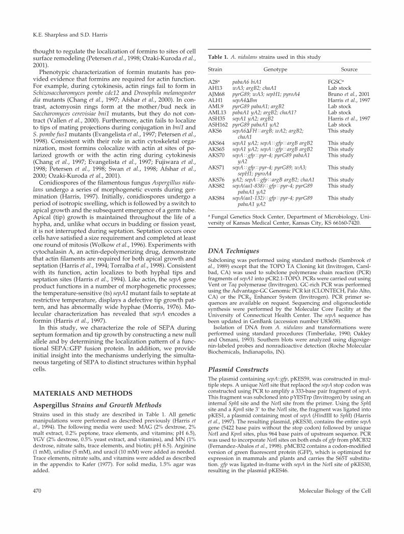
sepA Is Required for Actin Ring FormationSeptation in A. nidulans is preceded by the formation andconstriction of an actin ring (Momany and Hamer, 1997).Because sepA is required for septation, we next askedwhether it plays a role in actin ring formation. A mixedspore population from the sepA6�FH heterokaryon was in-oculated into selective MN media, allowing only sepA6�FHspores to germinate, and actin was visualized by immuno-fluorescence after 20 h. In wild-type cells, actin localizes tohyphal tips, in rings at septation sites, and in cortical spots(Figure 3A). However, in sepA6�FH strains, actin was ob-served only at hyphal tips and in cortical spots; no actinrings were observed (n � 100; Figure 3, B and C). Thus,SEPA is required for actin ring formation, but not for actinlocalization at hyphal tips and cortical spots.
SEPA Displays Dynamic Localization at SeptationSites and Hyphal TipsBased on its role in septation and polarized growth, wepredicted that SEPA would localize to sites of septation andto hyphal tips. To test these predictions, we constructedplasmids encoding SEPA::GFP fusion proteins (see MATE-RIALS AND METHODS). pKES56 also contains a truncatedversion of argB to target integration to the argB locus, andpKES59 contains the Neurospora crassa pyr-4 gene, which cancomplement the A. nidulans pyrG89 mutation. These con-structs were transformed into strains ASH35 (sepA1) andASH162 (wild-type), respectively. Analysis of Southern blotsrevealed that two ASH35 transformants, AKS64 and AKS65,contain a single copy of sepA::gfp integrated at the argB locus(our unpublished results). Because these transformantsgrow and septate like wild-type at the restrictive tempera-ture (our unpublished results), we conclude that SEPA::GFPis functional.
The strains transformed with pKES59 displayed differ-ences in brightness. These differences may be due to differ-
Figure 2. Comparison of two sepA deletion mutants, sepA4�Bmand sepA6�FH. (A) Schematic drawing of sepA indicating the se-quences replaced by argB in sepA4�Bm and sepA6�FH. (B)sepA6�FH and SepA� hyphae from a mixed spore population har-vested from the AKS6/AML13 heterokaryon grown for 18 h in YGVmedium and stained to visualize nuclei. An arrow indicates asepA6�FH germling surrounded by wild-type hyphae. (C)sepA6�FH fails to produce septa and displays dichotomous branch-ing. Strain AKS6 grown for 44 h in MN medium and stained toshow nuclei and septa. Inset shows hyphae stained only to visualizenuclei. (D) sepA4�Bm septates after a delay. Arrows indicate septathat have formed in ALH1 (sepA4�Bm) grown for 17 h. (E) Multiplesepta (arrows) in a wild-type strain (A28) after 24 h of growth. Bars,12 �m.
K.E. Sharpless and S.D. Harris
Molecular Biology of the Cell472
ent numbers of plasmid integration events, resulting in vari-able levels of expression of SEPA::GFP. We isolated strainsexpressing SEPA::GFP at a level sufficient for good imaging.For example, one of these strains, AKS70, was shown by South-ern blotting to contain more than five copies of the sepA::gfpconstruct (our unpublished results). To ensure that the patternof SEPA::GFP localization in these strains was not an artifact ofoverexpression, we also examined strain AKS76, a daughter ofa cross between AKS65 and AH13 (note that AKS76 was usedbecause it possesses a wild-type allele at the sepA locus; Table1). The SEPA::GFP localization pattern in AKS76 was indistin-guishable from the pattern exhibited by the brighter strains(compare Figure 4A with B). In addition, we have observedseemingly aberrant localization of SEPA::GFP when it is over-expressed under the inducible alcA promoter (our unpublishedresults). Because we do not observe a similar aberrant localiza-tion pattern in strains containing multiple copies of sepA::gfp,we conclude that these strains display normal localization ofSEPA, only brighter.
In AKS70, SEPA::GFP localized faintly to the cytoplasmand brightly to the tips of hyphae and at septation sites(Figure 4B). Localization to both sites occurred simulta-neously during septation. SEPA::GFP was found at hyphaltips in 100% of cells (n � 100), typically as a crescent (Figure4B); 80% of the cells also showed a bright spot near the tip.SEPA::GFP was seen at sites of septation in 20% of cells (n �100) after 13 h of growth at 28°C.
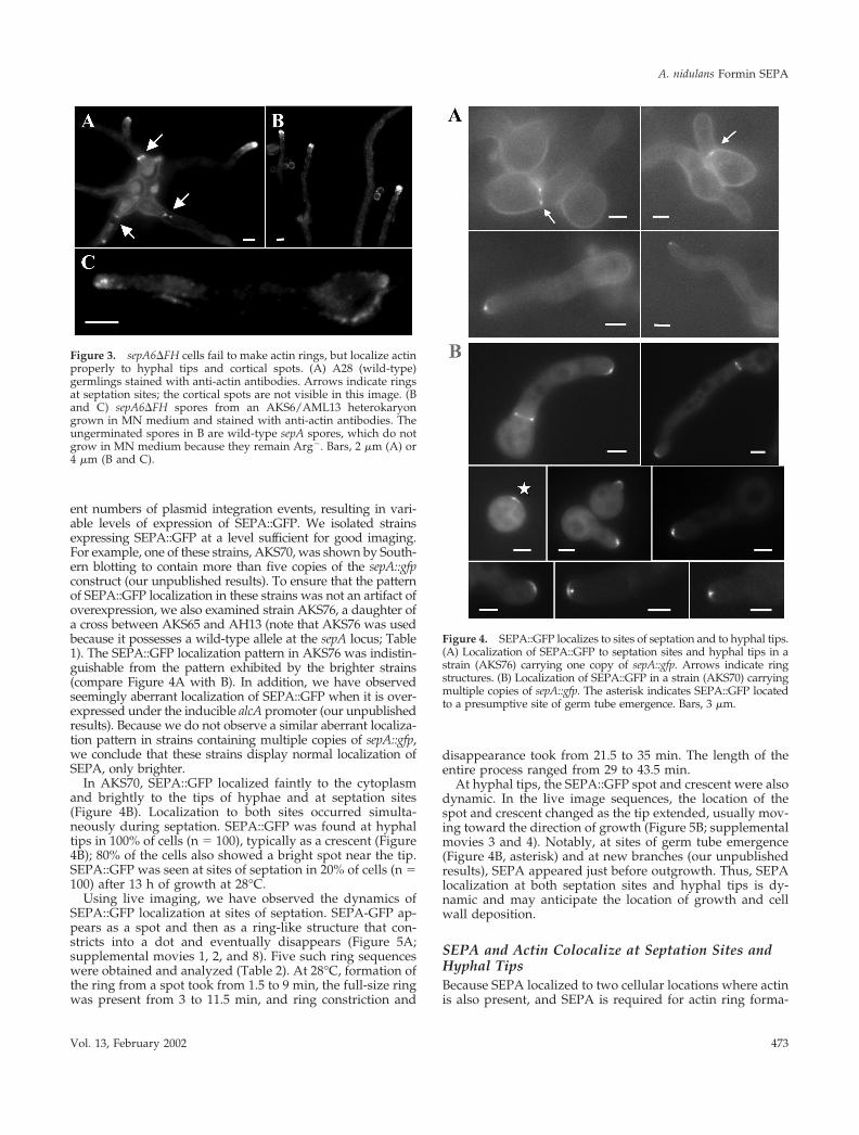
Using live imaging, we have observed the dynamics ofSEPA::GFP localization at sites of septation. SEPA-GFP ap-pears as a spot and then as a ring-like structure that con-stricts into a dot and eventually disappears (Figure 5A;supplemental movies 1, 2, and 8). Five such ring sequenceswere obtained and analyzed (Table 2). At 28°C, formation ofthe ring from a spot took from 1.5 to 9 min, the full-size ringwas present from 3 to 11.5 min, and ring constriction and
disappearance took from 21.5 to 35 min. The length of theentire process ranged from 29 to 43.5 min.
At hyphal tips, the SEPA::GFP spot and crescent were alsodynamic. In the live image sequences, the location of thespot and crescent changed as the tip extended, usually mov-ing toward the direction of growth (Figure 5B; supplementalmovies 3 and 4). Notably, at sites of germ tube emergence(Figure 4B, asterisk) and at new branches (our unpublishedresults), SEPA appeared just before outgrowth. Thus, SEPAlocalization at both septation sites and hyphal tips is dy-namic and may anticipate the location of growth and cellwall deposition.
SEPA and Actin Colocalize at Septation Sites andHyphal TipsBecause SEPA localized to two cellular locations where actinis also present, and SEPA is required for actin ring forma-
Figure 3. sepA6�FH cells fail to make actin rings, but localize actinproperly to hyphal tips and cortical spots. (A) A28 (wild-type)germlings stained with anti-actin antibodies. Arrows indicate ringsat septation sites; the cortical spots are not visible in this image. (Band C) sepA6�FH spores from an AKS6/AML13 heterokaryongrown in MN medium and stained with anti-actin antibodies. Theungerminated spores in B are wild-type sepA spores, which do notgrow in MN medium because they remain Arg�. Bars, 2 �m (A) or4 �m (B and C).
Figure 4. SEPA::GFP localizes to sites of septation and to hyphal tips.(A) Localization of SEPA::GFP to septation sites and hyphal tips in astrain (AKS76) carrying one copy of sepA::gfp. Arrows indicate ringstructures. (B) Localization of SEPA::GFP in a strain (AKS70) carryingmultiple copies of sepA::gfp. The asterisk indicates SEPA::GFP locatedto a presumptive site of germ tube emergence. Bars, 3 �m.
A. nidulans Formin SEPA
Vol. 13, February 2002 473
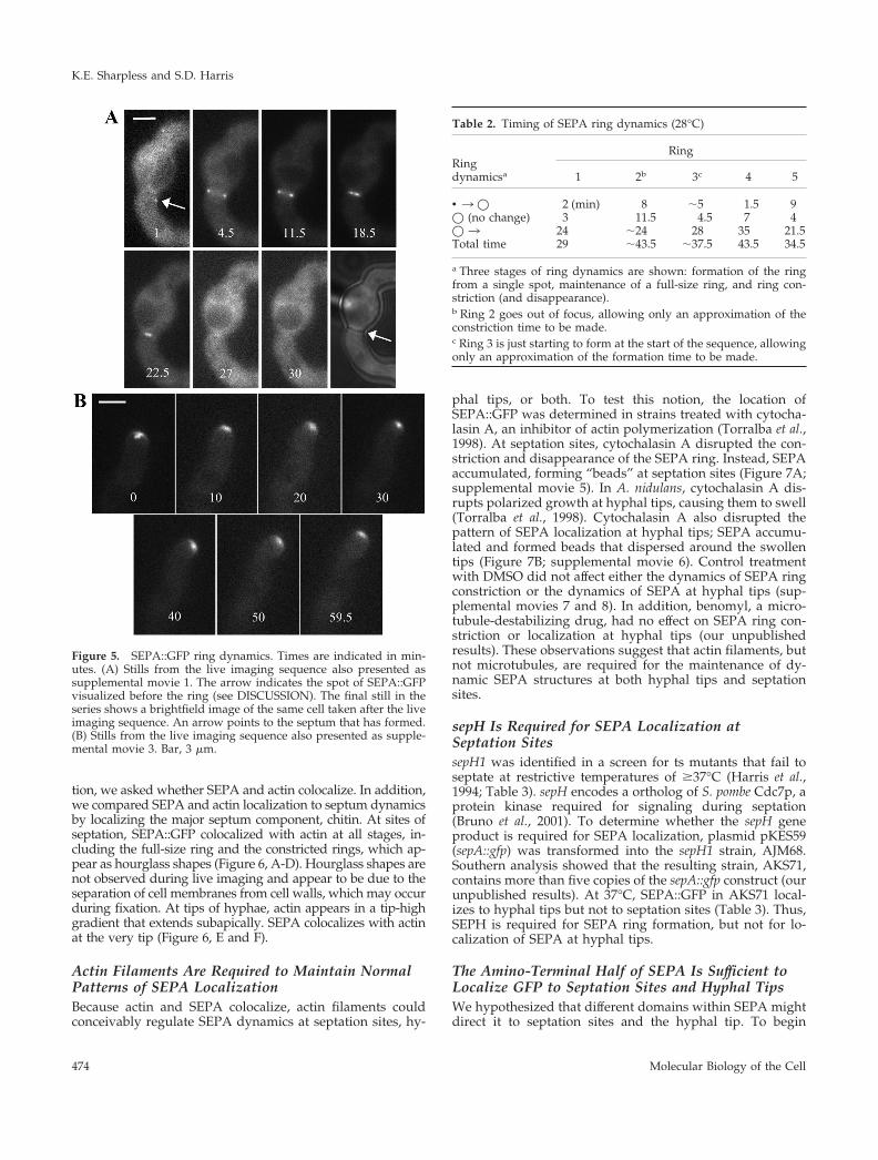
tion, we asked whether SEPA and actin colocalize. In addition,we compared SEPA and actin localization to septum dynamicsby localizing the major septum component, chitin. At sites ofseptation, SEPA::GFP colocalized with actin at all stages, in-cluding the full-size ring and the constricted rings, which ap-pear as hourglass shapes (Figure 6, A-D). Hourglass shapes arenot observed during live imaging and appear to be due to theseparation of cell membranes from cell walls, which may occurduring fixation. At tips of hyphae, actin appears in a tip-highgradient that extends subapically. SEPA colocalizes with actinat the very tip (Figure 6, E and F).
Actin Filaments Are Required to Maintain NormalPatterns of SEPA LocalizationBecause actin and SEPA colocalize, actin filaments couldconceivably regulate SEPA dynamics at septation sites, hy-
phal tips, or both. To test this notion, the location ofSEPA::GFP was determined in strains treated with cytocha-lasin A, an inhibitor of actin polymerization (Torralba et al.,1998). At septation sites, cytochalasin A disrupted the con-striction and disappearance of the SEPA ring. Instead, SEPAaccumulated, forming “beads” at septation sites (Figure 7A;supplemental movie 5). In A. nidulans, cytochalasin A dis-rupts polarized growth at hyphal tips, causing them to swell(Torralba et al., 1998). Cytochalasin A also disrupted thepattern of SEPA localization at hyphal tips; SEPA accumu-lated and formed beads that dispersed around the swollentips (Figure 7B; supplemental movie 6). Control treatmentwith DMSO did not affect either the dynamics of SEPA ringconstriction or the dynamics of SEPA at hyphal tips (sup-plemental movies 7 and 8). In addition, benomyl, a micro-tubule-destabilizing drug, had no effect on SEPA ring con-striction or localization at hyphal tips (our unpublishedresults). These observations suggest that actin filaments, butnot microtubules, are required for the maintenance of dy-namic SEPA structures at both hyphal tips and septationsites.
sepH Is Required for SEPA Localization atSeptation SitessepH1 was identified in a screen for ts mutants that fail toseptate at restrictive temperatures of �37°C (Harris et al.,1994; Table 3). sepH encodes a ortholog of S. pombe Cdc7p, aprotein kinase required for signaling during septation(Bruno et al., 2001). To determine whether the sepH geneproduct is required for SEPA localization, plasmid pKES59(sepA::gfp) was transformed into the sepH1 strain, AJM68.Southern analysis showed that the resulting strain, AKS71,contains more than five copies of the sepA::gfp construct (ourunpublished results). At 37°C, SEPA::GFP in AKS71 local-izes to hyphal tips but not to septation sites (Table 3). Thus,SEPH is required for SEPA ring formation, but not for lo-calization of SEPA at hyphal tips.
The Amino-Terminal Half of SEPA Is Sufficient toLocalize GFP to Septation Sites and Hyphal TipsWe hypothesized that different domains within SEPA mightdirect it to septation sites and the hyphal tip. To begin
Figure 5. SEPA::GFP ring dynamics. Times are indicated in min-utes. (A) Stills from the live imaging sequence also presented assupplemental movie 1. The arrow indicates the spot of SEPA::GFPvisualized before the ring (see DISCUSSION). The final still in theseries shows a brightfield image of the same cell taken after the liveimaging sequence. An arrow points to the septum that has formed.(B) Stills from the live imaging sequence also presented as supple-mental movie 3. Bar, 3 �m.
Table 2. Timing of SEPA ring dynamics (28°C)
Ringdynamicsa
Ring
1 2b 3c 4 5
• 3 � 2 (min) 8 �5 1.5 9� (no change) 3 11.5 4.5 7 4� 3 24 �24 28 35 21.5Total time 29 �43.5 �37.5 43.5 34.5
a Three stages of ring dynamics are shown: formation of the ringfrom a single spot, maintenance of a full-size ring, and ring con-striction (and disappearance).b Ring 2 goes out of focus, allowing only an approximation of theconstriction time to be made.c Ring 3 is just starting to form at the start of the sequence, allowingonly an approximation of the formation time to be made.
K.E. Sharpless and S.D. Harris
Molecular Biology of the Cell474
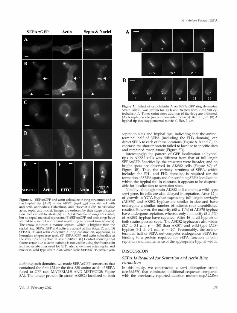
defining such domains, we made SEPA::GFP constructs thatcontained the first 132 or the first 838 amino acids of SEPAfused to GFP (see MATERIALS AND METHODS; Figure8A). The longer protein (in strain AKS82) localized to both
septation sites and hyphal tips, indicating that the amino-terminal half of SEPA (including the FH3 domain), candirect SEPA to each of these locations (Figure 8, B and C). Incontrast, the shorter protein failed to localize to specific sitesand remained cytoplasmic (Figure 8D).
Interestingly, the pattern of GFP localization at hyphaltips in AKS82 cells was different from that of full-lengthSEPA::GFP. Specifically, the crescents were broader, and nobright spots are observed in AKS82 cells (Figure 8C; cf.Figure 4B). Thus, the carboxy terminus of SEPA, whichincludes the FH1 and FH2 domains, is required for theformation of SEPA spots and for confining SEPA localizationwithin the hyphal tip. In contrast, it appears to be dispens-able for localization to septation sites.
Notably, although strain AKS82 still contains a wild-typesepA gene, its cells are also delayed in septation. After 12 hof growth in YGV, hyphae expressing full-length sepA::gfp(AKS70) and AKS82 hyphae are similar in size and haveundergone a similar number of mitoses (our unpublishedresults). However, the majority (60 � 11%) of AKS70 hyphaehave undergone septation, whereas only a minority (8 � 5%)of AKS82 hyphae have septated. After 16 h, all hyphae ofboth strains possess septa. The ASK82 hyphae are also wider(3.7 � 0.1 �m; n � 20) than AKS70 and wild-type (A28)hyphae (3.1 � 0.1 �m; n � 20). Presumably, the amino-terminal half of SEPA out-competes endogenous SEPA forbinding to a protein required for SEPA function in bothseptation and maintenance of the appropriate hyphal width.
DISCUSSION
SEPA Is Required for Septation and Actin RingFormationIn this study, we constructed a sepA disruption strain(sepA6�FH) that eliminates additional sequence comparedwith the previously reported deletion mutant (sepA4�Bm;
Figure 6. SEPA::GFP and actin colocalize in ring structures and atthe hyphal tip. (A-D) Strain AKS70 (sepA::gfp) was stained withanti-actin antibodies, Calcofluor, and Hoechst 33258 to visualizeactin, septa, and nuclei. Images are ordered by their stage of septa-tion from earliest to latest. (A) SEPA::GFP and actin rings are visible,but no septal material is present. (B) SEPA::GFP and actin rings havestarted to constrict and a faint septal ring is present (arrowheads).The arrow indicates a mature septum, which is brighter than theseptal ring; SEPA::GFP and actin are absent at this stage. (C and D)SEPA::GFP and actin colocalize during constriction, appearing ashourglass shapes (see text). (E) SEPA::GFP and actin colocalize atthe very tips of hyphae in strain AKS70. (F) Control showing thatfluorescence due to actin staining is not visible using the fluoresceinisothiocyanate filter used for GFP. Also shown are actin, septa, andnuclei in wild-type strain A28, which lacks SEPA::GFP. Bars, 3 �m.
Figure 7. Effect of cytochalasin A on SEPA::GFP ring dynamics.Strain AKS70 was grown for 13 h and treated with 2 mg/ml cy-tochalasin A. Times (min) since addition of the drug are indicated.(A) A septation site (see supplemental movie 5). Bar, 1.5 �m. (B) Ahyphal tip (see supplemental movie 6). Bar, 3 �m.
A. nidulans Formin SEPA
Vol. 13, February 2002 475

Harris et al., 1997). We have found that sepA6�FH mutantsfail to septate and can only form tiny colonies that lackconidia. In contrast, sepA4�Bm mutants undergo septationafter a lengthy delay and fail to produce normal coloniesonly at higher temperatures (Harris et al., 1997). We there-fore conclude that sepA is required for the formation ofsepta, but is not essential for vegetative growth in A. nidu-lans. This phenotype is similar to that described for the otherlate-acting sep mutants in A. nidulans (i.e., sepD, sepG, andsepH; Harris et al., 1994; Bruno et al., 2001). It is not knownhow sepA4�Bm mutants are able to septate despite missingthe 5� half of the gene. It is possible that residual expressionof the 3� half of the gene is sufficient to allow septation.
By analyzing the phenotype of sepA6�FH, we have foundthat SEPA is required for the formation of actin rings atseptation sites. This observation is consistent with the
known roles of other formins in promoting cytokinesis(Wasserman, 1998). In contrast, actin localizes to the hyphaltip in the absence of SEPA, although the polarity defectsobserved in sepA mutants suggest that the tip-associatedactin may not function in a normal manner. Accordingly,although other pathways may be available for recruitingactin to the hyphal tip, we propose that SEPA is required forthe overall fidelity of actin function at this site.
Dynamic Localization of SEPA at Septation Sitesand Hyphal TipsIn this study, we have found that SEPA localizes to septationsites and hyphal tips. Simultaneous localization to distinctsubcellular sites is a unique feature of SEPA that is notshared by other fungal formins. The localization patternmirrors that of actin, which is also located simultaneously atseptation sites and hyphal tips in A. nidulans (Harris, 1997).In contrast, in yeast cells, actin relocalizes from cell tips todivision sites concomitant with septation. Accordingly,yeast formins either localize to sites of cytokinesis or polar-ized growth, but not both at the same time (Chang et al.,1997; Petersen et al., 1998; Ozaki-Kuroda et al., 2001). Theability of SEPA to simultaneously organize distinct actinstructures at different sites implies that its localization issubject to strict spatial and temporal control.
Using live imaging, we have determined the dynamics ofSEPA at both septation sites and hyphal tips. At septationsites, the SEPA ring forms quickly after the initial appear-ance of an asymmetric SEPA spot. The presence of coiled-coil regions in SEPA (Harris et al., 1997) suggests that thespot may contain multimers that subsequently organize intoa higher order ring structure. In S. pombe, Cdc12p also ap-pears as a cortical spot before forming a medial ring (Chang,1999). However, unlike SEPA, the Cdc12p spot is mobile andpersists for a much longer period of time.
Like Cdc12p rings (Chang et al., 1997), SEPA rings con-strict coincident with the deposition of septal wall material.In contrast, Bni1p rings do not constrict during septation inS. cerevisiae (Ozaki-Kuroda et al., 2001). Regardless ofwhether formin ring structures undergo constriction duringseptum formation, they are likely to guide the formation andsubsequent contraction of actomyosin rings at septation sites(Vallen et al., 2000). The colocalization of SEPA and actinrings at all stages of constriction is consistent with thisnotion. The contraction of actomyosin rings is presumed toguide membrane insertion and wall deposition during sep-tum formation (Vallen et al., 2000). However, it also remains
Table 3. sepH is required for SEPA localization to septation sites
StrainTemperaturea
(°C)% of Cells withSEPA at tipsb
% of Cells withSEPA ringsb
% of Cellswith septab
AKS70 (wt) 28 100–0 20.3–4.4 35–13.037 99–0.6 17.3–5.7 82–2.6
AKS71 (sepH1) 28 99–0.6 14–1.7 18.7–3.237 99–0.6 0.3–0.3 0–0
a Cells were grown in YGV for 12 h at 28°C or 10 h at 37°C.b Counts are the mean � standard error of the mean of three independent experiments (n � 100).
Figure 8. Amino terminal half of SEPA is sufficient for localizationto both septation sites and hyphal tips. (A) Diagrams of threeSEPA::GFP constructs (see MATERIALS AND METHODS). (B andC) Nearly normal localization (see text) of the amino terminal half ofSEPA in strain AKS82. Bars, 4 �m. (D) Failure of a shorter amino-terminal fragment (in strain AKS84) to localize. Bar, 3 �m.
K.E. Sharpless and S.D. Harris
Molecular Biology of the Cell476

possible that ring dynamics are driven by the force of cen-tripetal cell wall deposition.
Although SEPA is found transiently in a ring structureduring septation, it localizes continuously at sites of polar-ized growth. The dynamic localization of SEPA at hyphaltips generally correlates with the direction of tip extension.Similarly, in S. cerevisiae, the location of Bni1p in bud tipscorresponds with the direction of bud growth (Ozaki-Ku-roda et al., 2001). The pattern of localization at hyphal tips isa crescent usually subtended by a bright spot. The crescentof SEPA appears to be located at hyphal tips in a thinsubcortical layer, whereas the bright spot is found just be-hind or within the crescent. The spot may colocalize with adense collection of vesicles found at hyphal tips known asthe Spitzenkorper, which is thought to be an organizingcenter for vesicles that are targeted to the growing tip (Bar-tnicki-Garcia et al., 1989). Notably, movement of the Spitzen-korper has also been shown to correlate with changes in thedirection of hyphal extension (Riquelme et al., 1998).
Actin Filaments Function Interdependently withSEPAWe have found that actin filaments are required to maintainthe proper pattern of SEPA localization at septation sites andhyphal tips. Specifically, treatment of hyphae with cytocha-lasin A causes preexisting SEPA structures to collapse intobead-like patches that remain at those sites. In contrast,maintenance of SEPA structures is not affected by the loss ofmicrotubule integrity. Because SEPA is required for actinring formation and is presumably involved in organizingactin structures at the hyphal tip, these observations suggestthat actin filaments and SEPA function in an interdependentmanner. For example, profilin, or another actin-associatedprotein capable of interacting with SEPA, may promote orstabilize interactions between SEPA and filamentous actin.Alternatively, the septins, a conserved family of GTP-bind-ing proteins capable of forming filaments (Trimble, 1999;Momany et al., 2001), may coordinate the localization ofSEPA and actin. The observation that yeast Bni1p displaysgenetic interactions with a septin supports this notion(Longtine et al., 1996).
SEPH Functions Upstream of SEPA and Actin RingFormation at Septation SitesIn A. nidulans, signals activating cytokinesis are thought toemanate from mitotic nuclei, which somehow determineand/or activate the septation site (Wolkow et al., 1996).SEPH, by analogy to the role of Cdc7p in S. pombe, may bepart of the signaling pathway that determines the timingand/or location of septum formation (McCollum andGould, 2001). We have shown that SEPH function is re-quired for SEPA localization at septation sites. Consistentwith its role upstream of SEPA, sepH strains also do not formactin rings at restrictive temperature (our unpublished re-sults; Bruno et al., 2001). Based on these observations, wesuggest that SEPH may function in a regulatory pathwaythat coordinates actin ring formation with the events ofmitosis. Notably, the role of SEPH is different than that of itsorthologs, S. pombe Cdc7p and S. cerevisiae Cdc15p. NeitherCdc7p nor Cdc15p control actin ring formation; rather, both
proteins are components of pathways that regulate actinring constriction (McCollum and Gould, 2001).
The Amino-Terminal Half of SEPA Localizes toSeptation Sites and as a Wider Crescent at HyphalTipsWe have found that SEPA is targeted to septation sites andhyphal tips through sequences in its amino terminus. Be-cause these sequences include the entire FH3 domain, wepropose that it is primarily responsible for targeting GFP tothese sites (Petersen et al., 1998). Moreover, our observationimplies that the protein(s) that binds the FH3 domain is alsolocated at septation sites and hyphal tips. Other domains ofSEPA may modify the localization pattern. Consistent withthis idea, we have found that the amino-terminal half ofSEPA is not sufficient to direct GFP to a tight crescent athyphal tips or to form the subtending bright spot. Presum-ably, interactions between the FH1 or FH2 domains andlocalized components of the actin cytoskeleton facilitate therecruitment of SEPA to specific sites. This may also explainthe ability of sepA4�Bm hyphae, which possess only the FH1and FH2 domains, to septate after a lengthy delay. In addi-tion, we have found that expression of the amino-terminalhalf of SEPA delays septation and causes hyphae to appearwider than normal. These dominant negative effects mayoccur because the amino-terminal half of SEPA interfereswith the ability of endogenous SEPA to interact with anFH3-binding protein. Similarly, it has been found that over-expression of formin FH3 constructs in S. pombe and inhuman macrophages interferes with their normal function(Petersen et al., 1998; Yayoshi-Yamamoto et al., 2000).
Summary
Our findings support a model in which SEPA acts in amultiprotein complex to control the dynamic assembly anddisassembly of functionally distinct actin structures. Wesuggest that the recruitment of SEPA to septation sites andhyphal tips is directed by specific morphological cues.Downstream of these cues, we propose that similar path-ways involving Rho GTPases and septins ensure that SEPAforms the appropriate dynamic structure. Further analysis ofthe requirements for SEPA localization should reveal howfungal hyphae simultaneously direct growth at spatiallydistinct sites.
ACKNOWLEDGMENTS
We thank Ron Morris and his laboratory members for the use oftheir microscope, charge-coupled device, and computer software toobtain the live imaging sequences. We also thank John Pringle andPeter Kraus for constructive comments that greatly improved themanuscript. This work was supported by a grant from the NationalScience Foundation (MCB-9723711).
REFERENCES
Afshar, K., Stuart, B., and Wasserman, S.A. (2000). Functional anal-ysis of the Drosophila Diaphanous FH protein in early embryonicdevelopment. Development 127, 1887–1897.
Ayscough, K.R. (1998). In vivo functions of actin-binding proteins.Curr. Opin. Cell Biol. 10, 102–111.
A. nidulans Formin SEPA
Vol. 13, February 2002 477

Bartnicki-Garcia, S., Hergert, F., and Gierz, G. (1989). Computersimulation of fungal morphogenesis and the mathematical basis forhyphal (tip) growth. Protoplasma 153, 46–57.
Beckerle, M.C. (1998). Spatial control of actin filament assembly:lessons from Listeria. Cell 95, 741–748.
Bedford, M.T., Chan, D.C., and Leder, P. (1997). FBP WW domainsand the Abl SH3 domain bind to a specific class of proline-richligands. EMBO J. 16, 2376–2383.
Bishop, A.L., and Hall, A. (2000). Rho GTPases and their effectorproteins. Biochem. J. 348, 241–255.
Bretscher, A. (1999). Regulation of cortical structure by the ezrin-radixin-moesin protein family. Curr. Opin. Cell Biol. 11, 109–116.
Bruno, K.S., Morrell, J.L., Hamer, J.E., and Staiger, C.S. (2001). SEPH,a Cdc7p ortholog from Aspergillus nidulans, functions upstream ofactin ring formation during cytokinesis. Mol. Microbiol. 42, 3–12.
Chan, D.C., Bedford, M.T., and Leder, P. (1996). Formin bindingproteins bear WWP/WW domains that bind proline-rich peptidesand functionally resemble SH3 domains. EMBO J. 15, 1045–1054.
Chang, F. (1999). Movement of a cytokinesis factor Cdc12p to thesite of cell division. Curr. Biol. 9, 849–852.
Chang, F., D. Drubin, D., and Nurse, P. (1997). Cdc12p, a proteinrequired for cytokinesis in fission yeast, is a component of the celldivision ring and interacts with profilin. J. Cell Biol. 137, 169–182.
Chen, H., Bernstein, B.W., and Bamburg, J.R. (2000). Regulatingactin-filament dynamics in vivo. Trends Biochem. Sci. 25, 19–23.
Emmons, S., Phan, H., Calley, J., Chen, W., James, B., and Manseau,L. (1995). cappuccino, a Drosophila maternal effect gene required forpolarity of the egg and embryo, is related to the vertebrate limbdeformity locus. Genes Dev. 9, 2482–2494.
Evangelista, M., Blundell, K., Longtine, M.S., Chow, C.J., Adames,N., Pringle, J.R., Peter, M., and Boone, C. (1997). Bni1p, a yeastformin linking Cdc42p and the actin cytoskeleton during polarizedmorphogenesis. Science 276, 118–122.
Fernandez-Abalos, J.M., Fox, H., Pitt, C., Wells, B., and Doonan, J.H.(1998). Plant-adapted green fluorescent protein is a versatile vitalreporter for gene expression, protein localization and mitosis in thefilamentous fungus Aspergillus nidulans. Mol. Microbiol. 27, 121–130.
Frazier, J.A., and Field, C.M. (1997). Actin cytoskeleton: are FHproteins local organizers? Curr. Biol. 7, R414–R417.
Fujiwara, T., Tanaka, K., Mino, A., Kikyo, M., Takahashi, K.,Shimizu, K., and Takai, Y. (1998). Rho1p-Bni1p-Spa2p interactions:implications in localization of Bni1p at the bud site and regulation ofthe actin cytoskeleton in Saccharomyces cerevisiae. Mol. Biol. Cell 9,1221–1233.
Harris, S.D. (1997). The duplication cycle in Aspergillus nidulans.Fungal Genet. Biol. 22, 1–13.
Harris, S.D., Hamer, L., Sharpless, K.E., and Hamer, J.E. (1997). TheAspergillus nidulans sepA gene encodes an FH1/2 protein involved incytokinesis and the maintenance of cellular polarity. EMBO J. 16,3474–3483.
Harris, S.D., Hofmann, A.F., Tedford, H.W., and Lee, M.P. (1999).Identification and characterization of genes required for hyphalmorphogenesis in the filamentous fungus Aspergillus nidulans. Ge-netics 151, 1015–1025.
Harris, S.D., Morrell, J.L., and Hamer, J.E. (1994). Identification andcharacterization of Aspergillus nidulans mutants defective in cytoki-nesis. Genetics 136, 517–532.
Imamura, H., Tanaka, K., Hihara, T., Umikawa, M., Kamei, T.,Takahashi, K., Sasaki, T., and Takai, Y. (1997). Bni1p and Bnr1p:downstream targets of the Rho family small G-proteins which in-
teract with profilin and regulate actin cytoskeleton in Saccharomycescerevisiae. EMBO J. 16, 2745–2755.
James, S.W., et al. (1999). nimO, an Aspergillus gene related to bud-ding yeast Dbf4, is required for DNA synthesis and mitotic check-point control. J. Cell Sci. 112, 1313–1324.
Kafer, E. (1977). Meiotic and mitotic recombination in Aspergillusand its chromosomal aberrations. Adv. Genet. 19, 33–131.
Kamei, T., Tanaka, K., Hihara, T., Umikawa, M., Imamura, H.,Kikyo, M., Ozaki, K., and Takai, Y. (1998). Interaction of Bnr1p witha novel Src homology 3 domain-containing Hof1p. J. Biol. Chem.273, 28341–28345.
Kikyo, M., Tanaka, K., Kamei, T., Ozaki, K., Fujiwara, T., Inoue, E.,Takita, Y., Ohya, Y., and Takai, Y. (1999). An FH domain-containingBnr1p is a multifunctional protein interacting with a variety ofcytoskeletal proteins in Saccharomyces cerevisiae. Oncogene 18, 7046–7054.
Kohno, et al. (1996). Bni1p implicated in cytoskeletal control is aputative target of Rho1p small GTP binding protein in Saccharomycescerevisiae. EMBO J. 15, 6060–6068.
Longtine, M.S., DeMarini, D.J., Valencik, M.L., Al-Awar, O.S., Fares,H., De Virgilio, C., and Pringle, J.R. (1996). The septins: roles incytokinesis and other processes. Curr. Opin. Cell Biol. 8, 106–119.
Manseau, L., Calley, J., and Phan, H. (1996). Profilin is required forposterior patterning of the Drosophila oocyte. Development 122,2109–2116.
McCollum, D., and Gould, K.L. (2001). Timing is everything: regu-lation of mitotic exit and cytokinesis by the MEN and SIN. TrendsCell Biol. 11, 89–95.
Miller, B.L., Miller, K.Y., Roberti, K.A., and Timberlake, W.E. (1987).Position-dependent and -independent mechanisms regulate cell-specific expression of the SpoC1 gene cluster of Aspergillus nidulans.Mol. Cell. Biol. 7, 427–434.
Momany, M., and Hamer, J.E. (1997). Relationship of actin, micro-tubules, and crosswall synthesis during septation in Aspergillusnidulans. Cell Motil. Cytoskeleton 38, 373–384.
Momany, M., Zhao, J., Lindsey, R., and Westfall, P.J. (2001). Char-acterization of the Aspergilus nidulans septin (asp) gene family. Ge-netics 157, 969–977.
Morris, N.R. (1976). Mitotic mutants of Aspergillus nidulans. Genet.Res. 26, 237–254.
Mullins, R.D. (2000). How WASP-family proteins, and the Arp2/3complex convert intracellular signals into cytoskeletal structures.Curr. Opin. Cell Biol. 12, 91–96.
Oakley, B.R., and Osmani, S.A. (1993). Cell cycle analysis using thefilamentous fungus Aspergillus nidulans. In: The Cell Cycle: A Prac-tical Approach, ed. P. Fantes and R. Brooks, New York, NY: IRLPress, 127–142.
Ozaki-Kuroda, K., Yamamoto, Y., Nohara, H., Kinoshita, M., Fuji-wara, T., Irie, K., and Takai, Y. (2001). Dynamics localization andfunction of Bni1p at the sites of directed growth in Saccharomycescerevisiae. Mol. Cell. Biol. 21, 827–839.
Petersen, J., Nielsen, O., Egel, R., and Hagan, I.M. (1998). FH3, adomain found in formins, targets the fission yeast formin Fus1 to theprojection tip during conjugation. J. Cell Biol. 141, 1217–1228.
Petersen, J., Weilguny, D., Egel, R., and Nielsen, O. (1995). Charac-terization of fus1 of Schizosaccharomyces pombe: a developmentallycontrolled function needed for conjugation. Mol. Cell. Biol. 15, 3697–3707.
Ramesh, N., Anton, I.M., Martinez-Quiles, N., and Geha, R.S. (1999).Waltzing with WASP. Trends Cell Biol. 9, 15–19.
K.E. Sharpless and S.D. Harris
Molecular Biology of the Cell478

Riquelme, M., Reynaga-Pena, C.G., Gierz, G., and Bartnicki-Garcia,S. (1998). What determines growth direction in fungal hyphae?Fungal Genet. Biol. 24, 101–109.
Sambrook, J., Fritsch, E.F., and Maniatis,T. (1989). Molecular clon-ing: a laboratory manual. Cold Spring Harbor, NY: Cold SpringHarbor Laboratory Press.
Schmidt, A., and Hall, M.N. (1998). Signaling to the actin cytoskel-eton. Annu. Rev. Cell Dev. Biol. 14, 305–338.
Swan, K.A., Severson, A.F., Carter, J.C., Martin, P.R., Schnabel, H.,Schnabel, R., and Bowerman, B. (1998). cyk-1: a C. elegans FH generequired for a late step in embryonic cytokinesis. J. Cell Sci. 111,2017–2027.
Tanaka, K., and Takai, Y. (1998). Control of reorganization of theactin cytoskeleton by Rho family small GTP-binding proteins inyeast. Curr. Opin. Cell Biol. 10, 112–116.
Timberlake, W.E. (1990). Molecular genetics of Aspergillus develop-ment. Annu. Rev. Genet. 24, 5–36.
Torralba, S., Raudaskoski, M., Pedregosa, A.M., and Laborda, F.(1998). Effect of cytochalasin A on apical growth, actin cytoskeletonorganization and enzyme secretion in Aspergillus nidulans. Microbi-ology 144, 45–53.
Trimble, W.S. (1999). Septins: a highly conserved family of mem-brane-associated GTPases with functions in cell division and be-yond. J. Membr. Biol. 169, 75–81.
Uetz, P., Fumagalli, S., James, D., and Zeller, R. (1996). Molecularinteraction between limb deformity proteins (formins) and Src fam-ily kinases. J. Biol. Chem. 271, 33525–33530.
Umikawa, M., Tanaka, K., Kamei, T., Shimizu, K., Imamura, H.,Sasaki, T., and Takai, Y. (1998). Interaction of Rho1p target Bni1p
with F-actin-binding elongation factor 1�: implication in Rho1p-regulated reorganization of the actin cytoskeleton in Saccharomycescerevisiae. Oncogene 16, 2011–2016.
Vallen, E.A., Caviston, J., and Bi, E. (2000). Roles of Hof1p, Bni1p,Bnr1p, and Myo1p in cytokinesis in Saccharomyces cerevisiae. Mol.Biol. Cell 11, 593–611.
Waring, R.B., May, G.S., and Morris, N.R. (1989). Characterization ofan inducible expression system in Aspergillus nidulans using alcAand tubulin-encoding genes. Gene 79, 119–130.
Wasserman, S. (1998). FH proteins as cytoskeletal organizers.Trends Cell Biol. 8, 111–115.
Watanabe, N., Madaule, P., Reid, T., Ishizaki, T., Watanabe, G.,Kakizuka, A., Saito, Y., Nakao, K., Jockusch, B.M., and Narumiya, S.(1997). p140mDia, a mammalian homolog of Drosophila diaphanous,is a target protein for Rho small GTPase and is a ligand for profilin.EMBO J. 16, 3044–3056.
Wolkow, T.D., Harris, S.D., and Hamer, J.E. (1996). Cytokinesis inAspergillus nidulans is controlled by cell size, nuclear positioning andmitosis. J. Cell Sci. 109, 2179–2188.
Xiang, X., Han, G., Winkelmann, D.A., Zuo, W., and Morris, N.R.(2000). Dynamics of cytoplasmic dynein in living cells and the effectof a mutation in the dynactin complex actin-related protein Arp1.Curr. Biol. 10, 603–606.
Yayoshi-Yamamoto, S., Taniuchi, I., and Watanabe, T. (2000). FRL, anovel formin-related protein, binds to Rac and regulates cell motil-ity and survival of macrophages. Mol. Cell. Biol. 20, 6872–6881.
Zeller, R., Haramis, A.G., Zuniga, A., McGuigan, C., Dono, R.,Davidson, G., Chabanis, S., and Gibson, T. (1999). Formin defines alarge family of morphoregulatory genes and their functions in es-tablishment of the polarising region. Cell Tissue Res. 296, 85–93.
A. nidulans Formin SEPA
Vol. 13, February 2002 479
Related Documents