Functional and Morphological Adaptation to Peptidoglycan Precursor Alteration in Lactococcus lactis * □ S Received for publication, May 12, 2010 Published, JBC Papers in Press, June 4, 2010, DOI 10.1074/jbc.M110.143636 Marie Deghorain ‡ , Laetitia Fontaine ‡1 , Blandine David ‡ , Jean-Luc Mainardi §¶ **, Pascal Courtin ‡‡ , Richard Daniel §§ , Jeff Errington §§ , Alexei Sorokin ¶¶ , Alexander Bolotin ¶¶ , Marie-Pierre Chapot-Chartier ‡‡ , Bernard Hallet ‡2 , and Pascal Hols ‡3 From ‡ Biochimie et Ge ´ne ´tique Mole ´culaire Bacte ´rienne, Institut des Sciences de la Vie, Universite ´ Catholique de Louvain, Louvain-la-Neuve 1348, Belgium, § INSERM, U872, LRMA, Equipe 12 du Centre de Recherche des Cordeliers, Paris F-75006, France, the ¶ Universite ´ Pierre et Marie Curie, UMR S 872, Paris F-75006, France, the Universite ´ Paris Descartes, UMR S872, Paris F-75006, France, the **Assistance Publique-Ho ˆpitaux de Paris, Ho ˆpital Europe ´en Georges Pompidou, Paris F-75015, France, ‡‡ INRA, UMR1319 Micalis, Domaine de Vilvert, F-78352 Jouy-en-Josas Cedex, France, the §§ Institute for Cell and Molecular Biosciences, The Medical School, University of Newcastle upon Tyne, Newcastle upon Tyne NE2 4HH, United Kingdom, and the ¶¶ Genome Analysis Team ANALGEN, TGU MICALIS, CRJ INRA, Bat. 440, Domaine de Vilvert, F-78352 Jouy-en-Josas Cedex, France Cell wall peptidoglycan assembly is a tightly regulated process requiring the combined action of multienzyme complexes. In this study we provide direct evidence showing that substrate transformations occurring at the different stages of this process play a crucial role in the spatial and temporal coordination of the cell wall synthesis machinery. Peptidoglycan substrate alter- ation was investigated in the Gram-positive bacterium Lactococcus lactis by substituting the peptidoglycan precursor biosynthesis genes of this bacterium for those of the vancomy- cin-resistant bacterium Lactobacillus plantarum. A set of L. lac- tis mutant strains in which the normal D-Ala-ended precursors were partially or totally replaced by D-Lac-ended precursors was generated. Incorporation of the altered precursor into the cell wall induced morphological changes arising from a defect in cell elongation and cell separation. Structural analysis of the muropep- tides confirmed that the activity of multiple enzymes involved in peptidoglycan synthesis was altered. Optimization of this altered pathway was necessary to increase the level of vancomycin resis- tance conferred by the utilization of D-Lac-ended peptidoglycan precursors in the mutant strains. The implications of these findings on the control of bacterial cell morphogenesis and the mechanisms of vancomycin resistance are discussed. Peptidoglycan (or murein) is a major stress-bearing compo- nent of the bacterial cell wall. This polymer is made of glycan chains interconnected by covalent cross-links between short peptides. It acts as an exoskeleton and plays a key role in deter- mining and maintaining cell shape throughout the cell cycle. The first steps of peptidoglycan biosynthesis occur in the cytoplasm where specific enzymes catalyze the formation of theuridinediphospho-n-acetylmuramyl-pentapeptide(UDP- MurNAc-pentapeptide) 4 precursor (1). After the addition of n-acetyl-glucosamine (GlcNAc), this precursor is transported through the plasma membrane and incorporated into the pep- tidoglycan by penicillin-binding proteins (PBPs) (2). High molecular weight PBPs act in transglycosylation and transpep- tidation reactions required for peptidoglycan polymers forma- tion and cross-linking, whereas low molecular weight PBPs are suggested to be carboxypeptidases and endopeptidases involved in peptide chains processing during peptidoglycan synthesis and maturation. PBPs collaborate with peptidoglycan hydrolases, possibly through the assembly of multienzymatic complexes, which together constitute the peptidoglycan bio- synthesis machinery (3–7). Bacterial cell morphology is the result of a tightly regulated process coupling peptidoglycan synthesis with cell growth and cell division. Proper cell shape requires proper localization and activity of the peptidoglycan biosynthesis machinery. PBP localization studies performed in bacilli and cocci suggest that recruitment of these proteins is mediated by structural deter- minants of the cytoskeleton (e.g. the FtsZ ring at the septum and actin-like filaments along the longitudinal axis of the cell) (3, 8, 9). Substrate availability also appears to be important for proper localization and activity of PBPs, as was reported for Esche- richia coli (10) and more recently for Streptococcus pneumoniae (11) and Staphylococcus aureus (12). Although the exact mech- anism of this control is still unclear, it has been suggested that PBP localization depends on the activity of the carboxypepti- dases that trim the peptidoglycan pentapeptide chains, provid- * This work was supported by Fonds Spe ´ ciaux de la Recherche (Universite ´ Catholique de Louvain), Fonds National de la Recherche Scientifique, and Actions de Recherche Concerte ´ es (Communaute ´ Franc ¸aise de Belgique). This work was also supported by a grant from the Biotechnology and Bio- logical Sciences Research Council, Swindon, United Kingdom, and a short- term European Molecular Biology Organization fellowship (to M. D.). □ S The on-line version of this article (available at http://www.jbc.org) contains supplemental Tables S1–S5 and Fig. 1. 1 Held a doctoral fellowship from Fonds pour la Formation a ` la Recherche dans I’Industrie et dans l’Agriculture. 2 To whom correspondence should be addressed. Tel.: 32-10-47-91-60; Fax: 32-10-47-31-09; E-mail: [email protected]. 3 A research associate at Fonds National de la Recherche Scientifique. 4 The abbreviations used are: MurNAc, N-acetylmuramic acid; GlcNAc, N-acetylglucosamine; PBP, penicillin-binding protein; MIC, minimal inhib- itory concentration; Van-FL, vancomycin BODIPY-FL conjugate; ddl Lc , D-Ala-D-Ala ligase gene from L. lactis subsp. cremoris; ddl Lp , D-Ala-D-Lac ligase gene from L. plantarum; ddlA St , D-Ala-D-Lac ligase gene from S. ther- mophilus; ldhD Lh , D-Lac dehydrogenase gene from L. helveticus; aad, D-Ala- D-Ala dipeptidase gene from L. plantarum; VanX A , D-Ala-D-Ala dipeptidase gene from E. faecium; HPLC, high pressure liquid chromatography; MALDI- TOF, matrix-assisted laser desorption ionization time-of-flight; ORF, open reading frame; MS, mass spectroscopy; D-Lac, D-lactate. THE JOURNAL OF BIOLOGICAL CHEMISTRY VOL. 285, NO. 31, pp. 24003–24013, July 30, 2010 © 2010 by The American Society for Biochemistry and Molecular Biology, Inc. Printed in the U.S.A. JULY 30, 2010 • VOLUME 285 • NUMBER 31 JOURNAL OF BIOLOGICAL CHEMISTRY 24003 by guest on April 22, 2016 http://www.jbc.org/ Downloaded from by guest on April 22, 2016 http://www.jbc.org/ Downloaded from by guest on April 22, 2016 http://www.jbc.org/ Downloaded from by guest on April 22, 2016 http://www.jbc.org/ Downloaded from by guest on April 22, 2016 http://www.jbc.org/ Downloaded from by guest on April 22, 2016 http://www.jbc.org/ Downloaded from by guest on April 22, 2016 http://www.jbc.org/ Downloaded from by guest on April 22, 2016 http://www.jbc.org/ Downloaded from by guest on April 22, 2016 http://www.jbc.org/ Downloaded from by guest on April 22, 2016 http://www.jbc.org/ Downloaded from

Welcome message from author
This document is posted to help you gain knowledge. Please leave a comment to let me know what you think about it! Share it to your friends and learn new things together.
Transcript
Functional and Morphological Adaptation to PeptidoglycanPrecursor Alteration in Lactococcus lactis*□S
Received for publication, May 12, 2010 Published, JBC Papers in Press, June 4, 2010, DOI 10.1074/jbc.M110.143636
Marie Deghorain‡, Laetitia Fontaine‡1, Blandine David‡, Jean-Luc Mainardi§¶�**, Pascal Courtin‡‡, Richard Daniel§§,Jeff Errington§§, Alexei Sorokin¶¶, Alexander Bolotin¶¶, Marie-Pierre Chapot-Chartier‡‡, Bernard Hallet‡2,and Pascal Hols‡3
From ‡Biochimie et Genetique Moleculaire Bacterienne, Institut des Sciences de la Vie, Universite Catholique de Louvain,Louvain-la-Neuve 1348, Belgium, §INSERM, U872, LRMA, Equipe 12 du Centre de Recherche des Cordeliers, Paris F-75006,France, the ¶Universite Pierre et Marie Curie, UMR S 872, Paris F-75006, France, the �Universite Paris Descartes, UMR S872,Paris F-75006, France, the **Assistance Publique-Hopitaux de Paris, Hopital Europeen Georges Pompidou, Paris F-75015, France,‡‡INRA, UMR1319 Micalis, Domaine de Vilvert, F-78352 Jouy-en-Josas Cedex, France, the §§Institute for Cell and MolecularBiosciences, The Medical School, University of Newcastle upon Tyne, Newcastle upon Tyne NE2 4HH, United Kingdom, and the¶¶Genome Analysis Team ANALGEN, TGU MICALIS, CRJ INRA, Bat. 440, Domaine de Vilvert, F-78352 Jouy-en-Josas Cedex, France
Cell wall peptidoglycan assembly is a tightly regulated processrequiring the combined action of multienzyme complexes. Inthis study we provide direct evidence showing that substratetransformations occurring at the different stages of this processplay a crucial role in the spatial and temporal coordinationof thecell wall synthesis machinery. Peptidoglycan substrate alter-ation was investigated in the Gram-positive bacteriumLactococcus lactis by substituting the peptidoglycan precursorbiosynthesis genes of this bacterium for those of the vancomy-cin-resistant bacteriumLactobacillus plantarum.Aset ofL. lac-tismutant strains in which the normal D-Ala-ended precursorswere partially or totally replaced by D-Lac-ended precursors wasgenerated. Incorporation of the altered precursor into the cellwall induced morphological changes arising from a defect in cellelongation andcell separation. Structural analysis of themuropep-tides confirmed that the activity of multiple enzymes involved inpeptidoglycan synthesis was altered. Optimization of this alteredpathway was necessary to increase the level of vancomycin resis-tance conferred by the utilization of D-Lac-ended peptidoglycanprecursors in themutantstrains.The implicationsof these findingson thecontrolofbacterial cellmorphogenesis and themechanismsof vancomycin resistance are discussed.
Peptidoglycan (or murein) is a major stress-bearing compo-nent of the bacterial cell wall. This polymer is made of glycanchains interconnected by covalent cross-links between shortpeptides. It acts as an exoskeleton and plays a key role in deter-mining and maintaining cell shape throughout the cell cycle.
The first steps of peptidoglycan biosynthesis occur in thecytoplasm where specific enzymes catalyze the formation oftheuridinediphospho-n-acetylmuramyl-pentapeptide(UDP-MurNAc-pentapeptide)4 precursor (1). After the addition ofn-acetyl-glucosamine (GlcNAc), this precursor is transportedthrough the plasma membrane and incorporated into the pep-tidoglycan by penicillin-binding proteins (PBPs) (2). Highmolecular weight PBPs act in transglycosylation and transpep-tidation reactions required for peptidoglycan polymers forma-tion and cross-linking, whereas low molecular weight PBPsare suggested to be carboxypeptidases and endopeptidasesinvolved in peptide chains processing during peptidoglycansynthesis andmaturation. PBPs collaborate with peptidoglycanhydrolases, possibly through the assembly of multienzymaticcomplexes, which together constitute the peptidoglycan bio-synthesis machinery (3–7).Bacterial cell morphology is the result of a tightly regulated
process coupling peptidoglycan synthesis with cell growth andcell division. Proper cell shape requires proper localization andactivity of the peptidoglycan biosynthesis machinery. PBPlocalization studies performed in bacilli and cocci suggest thatrecruitment of these proteins is mediated by structural deter-minants of the cytoskeleton (e.g. the FtsZ ring at the septumandactin-like filaments along the longitudinal axis of the cell) (3, 8,9). Substrate availability also appears to be important for properlocalization and activity of PBPs, as was reported for Esche-richia coli (10) andmore recently for Streptococcus pneumoniae(11) and Staphylococcus aureus (12). Although the exact mech-anism of this control is still unclear, it has been suggested thatPBP localization depends on the activity of the carboxypepti-dases that trim the peptidoglycan pentapeptide chains, provid-* This work was supported by Fonds Speciaux de la Recherche (Universite
Catholique de Louvain), Fonds National de la Recherche Scientifique, andActions de Recherche Concertees (Communaute Francaise de Belgique).This work was also supported by a grant from the Biotechnology and Bio-logical Sciences Research Council, Swindon, United Kingdom, and a short-term European Molecular Biology Organization fellowship (to M. D.).
□S The on-line version of this article (available at http://www.jbc.org) containssupplemental Tables S1–S5 and Fig. 1.
1 Held a doctoral fellowship from Fonds pour la Formation a la Recherchedans I’Industrie et dans l’Agriculture.
2 To whom correspondence should be addressed. Tel.: 32-10-47-91-60; Fax:32-10-47-31-09; E-mail: [email protected].
3 A research associate at Fonds National de la Recherche Scientifique.
4 The abbreviations used are: MurNAc, N-acetylmuramic acid; GlcNAc,N-acetylglucosamine; PBP, penicillin-binding protein; MIC, minimal inhib-itory concentration; Van-FL, vancomycin BODIPY-FL conjugate; ddlLc,D-Ala-D-Ala ligase gene from L. lactis subsp. cremoris; ddlLp, D-Ala-D-Lacligase gene from L. plantarum; ddlASt, D-Ala-D-Lac ligase gene from S. ther-mophilus; ldhDLh, D-Lac dehydrogenase gene from L. helveticus; aad, D-Ala-D-Ala dipeptidase gene from L. plantarum; VanXA, D-Ala-D-Ala dipeptidasegene from E. faecium; HPLC, high pressure liquid chromatography; MALDI-TOF, matrix-assisted laser desorption ionization time-of-flight; ORF, openreading frame; MS, mass spectroscopy; D-Lac, D-lactate.
THE JOURNAL OF BIOLOGICAL CHEMISTRY VOL. 285, NO. 31, pp. 24003–24013, July 30, 2010© 2010 by The American Society for Biochemistry and Molecular Biology, Inc. Printed in the U.S.A.
JULY 30, 2010 • VOLUME 285 • NUMBER 31 JOURNAL OF BIOLOGICAL CHEMISTRY 24003
by guest on April 22, 2016
http://ww
w.jbc.org/
Dow
nloaded from
by guest on April 22, 2016
http://ww
w.jbc.org/
Dow
nloaded from
by guest on April 22, 2016
http://ww
w.jbc.org/
Dow
nloaded from
by guest on April 22, 2016
http://ww
w.jbc.org/
Dow
nloaded from
by guest on April 22, 2016
http://ww
w.jbc.org/
Dow
nloaded from
by guest on April 22, 2016
http://ww
w.jbc.org/
Dow
nloaded from
by guest on April 22, 2016
http://ww
w.jbc.org/
Dow
nloaded from
by guest on April 22, 2016
http://ww
w.jbc.org/
Dow
nloaded from
by guest on April 22, 2016
http://ww
w.jbc.org/
Dow
nloaded from
by guest on April 22, 2016
http://ww
w.jbc.org/
Dow
nloaded from

ing the appropriate acceptor substrates for high molecularweight transpeptidases (10, 11) or eliminate these side chains,thereby preventing new peptidoglycan synthesis in specificregions of the cell wall (11, 12). Likewise, proper recruitment ofcarboxypeptidases and other peptidoglycan hydrolases mayalso depend on the specific location of their substrate duringcell growth (11, 13).In this study the flexibility of the cell wall biosynthesis
machinery toward a new substrate was examined in the Gram-positive bacterium Lactococcus lactis. In terms of shape andPBP content, L. lactis is closely related to other ovoid cocci,such as streptococci and enterococci, including a number ofpathogenic species. The primary structure of L. lactis pepti-doglycan monomer is GlcNAc-MurNAc-L-Ala-�-D-Glu (orisoglutamine)-L-Lys-D-Ala-D-Ala, with a D-Asn (or a D-Asp)cross-bridge residue attached to L-Lys (14–17). The D-AlaC-terminal residue of L. lactis peptidoglycan peptide chainswas changed to D-Lac. To this end, specific genes involved inpeptidoglycan precursor synthesis from Lactobacillus planta-rum, another lactic acid bacterium, were transferred andexpressed in L. lactis (Fig. 1). In L. plantarum, the peptide chainended with D-Ala-D-Lac, which makes this bacterium naturallyresistant to vancomycin due to the fact that D-Ala-D-Lac ter-mini are not targets for the antibiotic (18, 19). The resultingL. lactismutants produced different levels of precursors termi-nated by D-Ala-D-Lac instead of D-Ala-D-Ala and showed differ-ent levels of resistance to vancomycin. Utilization of the newsubstrate forced L. lactis peptidoglycan biosynthetic pathwayto adapt, leading to significant changes in peptidoglycan struc-ture and cell morphology.
EXPERIMENTAL PROCEDURES
Bacterial Strains, Plasmids, and Growth Conditions—Thebacterial strains and plasmids used in this study are listed insupplemental Table S1. The L. lactis subsp. cremoris strainNZ3900 and its derivatives were grown at 28 °C in M17 broth(BD Biosciences) containing 0.5% glucose (M17-glucose).Strains carrying the expression vectors pGIM020, pGIM022,pGIM023, pGIM024, pGIM025, and pGIM026 were grown inthe presence of 10 �g/ml chloramphenicol, whereas their ddlLcD-Ala-D-Ala ligase mutant derivatives MD001, MD002,MD003, MD004, and MD006 were selected and cultured on 5�g/ml chloramphenicol and 5 �g/ml erythromycin (Sigma).For induction of genes under the control of the nisA expressionsignals, nisin A (Sigma) was used at a concentration of 0.5ng/ml. D-Lactate (15 mM; Fluka) was added in the cultures,except for strains expressing the ldhDLh D-Lac dehydrogenasegene from Lactobacillus helveticus.Analysis of growth dependence of the ddlLc mutants toward
nisin and D-Lac was performed as followed; overnight cultureswere grown in the presence of nisin (0.5 ng/ml), D-Lac (15mM),erythromycin (5 �g/ml), and chloramphenicol (5 �g/ml),washed once, and diluted at an A600 nm of 0.05 in M17-glucosebroth containing erythromycin and chloramphenicol supple-mented with nisin and D-Lac or just nisin.Construction of Expression Plasmids—For pGIM020, a DNA
fragment containing the ddlLp D-Ala-D-Lac ligase open readingframe (ORF) and its ribosome binding site was amplified from
L. plantarum strainNCIMB8826with primersDDLPLXP3 andDDLPLXP4 (supplemental Table S1). The 1187-bp PCR frag-ment was digested with BamHI and ScaI and inserted into thepNZ2650 plasmid (20) digested with BamHI and PvuII. Theresulting plasmid (pGIM020) contains ddlLp under the controlof the PnisA promoter followed by the L. plantarum ldhL tran-scriptional terminator. For co-expressing ddlLpwith the ldhDLhgene of L. helveticus, a 1434-bp fragment encompassing theldhDLhORF and promoter sequences was PCR-amplified fromL. helveticus CNRZ32 with primers PNZLDHD1-PNZLDHD2(supplemental Table S1), digested with XbaI and ScaI, andinserted into the XbaI/NspI-digested pGIM020 plasmid toyield pGIM022. Construction of the pGIM023 vector for co-expressing ddlLp and the aad D-Ala-D-Ala dipeptidase genefrom L. plantarum as a bicistronic unit under the control of thePnisA promoter was previously described (18). The same proce-dure was used for the construction of pGIM024 to co-expressddlLpwith theVanXA D-Ala-D-Ala dipeptidase gene of the van-comycin-resistant Enterococcus faecium strain BM4147. A671-bp fragment encompassing the ribosome binding site andORF of VanXA was amplified from pAT83 plasmid (21) usingprimers VANX1 and VANX2 (supplemental Table S1). Theresulting PCR product was digestedwith BsrGI and BamHI andcloned into similarly digested pGIM020.For the complementation control plasmid pGIM026 ex-
pressing the ddlASt D-Ala-D-Ala ligase gene from Streptococcusthermophilus, primers SthddlABam1 and SthddlAKpnI(supplemental Table S1) were used to amplify the ddlASt ribo-some binding site and ORF directly from S. thermophilusLMG18311 colonies. The resulting 1104-bp PCR fragment wasdigested with BamHI and KpnI and inserted into pGIM020 cutwith the same enzymes, replacing the ddlLp insert and placingddlASt under the transcriptional control of the PnisA promoter.Construction of L. lactis Knock-out Mutants—Disruption of
the ddlLc ligase gene was carried out by homologous recom-bination-mediated integration of the disruptive plasmidpGIL001. For pGIL001 construction, an internal 574-bp frag-ment of ddlLc was generated by PCR using the LCDDLDISR1and LCDDLDISR2 primers (supplemental Table S1). The frag-ment was digested with PstI and XbaI and cloned into thesuicide plasmid pGIZ907 (19) digested with NsiI and XbaI.Chromosomal integration of pGIL001 generated 5�- and 3�-ter-minal-truncated copies of ddlLc separated by vector sequences.These include an erythromycin resistance gene as well as anoutward-facing promoter PLdh (19) to ensure expression of themurF gene located downstream of ddlLcin L. lactis. Inactivationof the dacA gene was carried out using the disruptive plasmidpGIBD006. APCR fragment containing the dacAORFwas gen-erated using the BLD_DacAUpNcoI and BLD_DacADownSacIprimers (supplemental Table S1) and cloned into thepGMTEasy vector (Invitrogen). The resulting plasmid wasdigested with SphI and KasI to provide an internal fragment ofthe dacA gene that was then cloned into pUC18Ery (22). Chro-mosomal integration of pGIBD006 produced two truncatedcopies of the dacA gene separated by vector sequences.Determination of Vancomycin Resistance Level and Popula-
tion Analysis—Minimal inhibitory concentrations (MICs) ofvancomycin displayed by the wild type and mutant strains of
Reprogramming Peptidoglycan Biosynthesis in L. lactis
24004 JOURNAL OF BIOLOGICAL CHEMISTRY VOLUME 285 • NUMBER 31 • JULY 30, 2010
by guest on April 22, 2016
http://ww
w.jbc.org/
Dow
nloaded from
L. lactis were determined by the E-test method (AB-Biodisk).Cells were plated on solid medium and incubated for 24 h at28 °C in the presence of an E-test strip containing a gradient ofvancomycin (from 0.016 to 256 �g/ml). MIC values are indi-cated by the position where the edge of the inhibition ellipseintersects the side of the strip. For population heterogeneityanalysis, serial cultures of the primary mutant MD003 and thecontrol strains MD006 and NZ3900 (pNZ8048) were per-formed by 100-fold diluting overnight-grown cultures into 10ml of fresh medium every day (�10 generations intervals). Ateach passage diluted samples were plated on increasing con-centrations of vancomycin (from 0 to 1024 �g/ml), and sub-populations displaying different levels of vancomycin resis-tance were estimated by expressing the number of colonyforming units obtained for each antibiotic concentration permlof culture and A600 nm unit.Preparation and Analysis of UDP-linked Peptidoglycan
Precursors—Cells were grown in M17-glucose broth supple-mentedwith antibiotics, nisin, and D-Lac (when appropriate) toanA600 nm of 0.7 and treatedwith bacitracin (200�g/ml; Sigma)for 90 min before harvesting by centrifugation. Peptidoglycanprecursors were then extracted with 20% trichloroacetic acidand analyzed by reverse-phase high pressure liquid chromatog-raphy (HPLC) as previously described (19).Determination of Peptidoglycan Composition—The refer-
ence strain used, PH8076, is an erythromycin-resistant L. lactisstrain isogenic to NZ3900.5 The erythromycin resistancemarker carried by this strainwas inserted togetherwith thenisRand nisK regulatory genes in the pepN locus using the integra-tion vector pNZ9573 (23). This strain also contains a mutationin the�-acetolactate decarboxylase gene aldB that has no effecton cell wall peptidoglycan composition. PH8076 was trans-formed with the pNZ8048 empty vector and used as a wild typereference for muropeptide analysis.Peptidoglycan from L. lactis strains PH8076 (pNZ8048),
MD003, andMD003-128 was prepared as previously described(14, 16). Purified peptidoglycan (4mg dry weight in 500 �l) wasthen digested with mutanolysin (2500 units/ml; Sigma) in 25mM sodium phosphate buffer (pH 5.5) for 19 h at 37 °C underrotational shaking. Muropeptides were reduced with sodiumborohydride and separated by reverse phase HPLC using aHypersil ODS column (C18, 250 � 4.6 mm; 5 �m; ThermoHy-persil-Keystone) at 50 °C as described by Courtin et al. (14).Fractions were collected, and 1 �l of the fractions containingmuropeptides of interest was analyzed by matrix-assisted laserdesorption ionization time-of-flight (MALDI-TOF)mass spec-trometry with a Voyager DE STR mass spectrometer (AppliedBiosystems) with �-cyano-4-hydroxycinnamic acid matrix.The structures of themuropeptides I and II were determined bymass spectrometry as described in Arbeloa et al. (24) using anelectrospray time-of-flight mass spectrometer operating inpositive mode (Qstar Pulsar I, Applied Biosystems).Microscopic Imaging and Van-FL Staining—Van-FL staining
was essentially performed as previously described by Danieland Errington (25). Cells from an overnight culture were
diluted into fresh M17-glucose medium and grown to earlyexponential phase (A600 nm � 0.2–0.3) at 30 °C. Culture sam-ples (200 �l) were incubated for 30 min with a mixture of equalamount of vancomycin BODIPY-FL conjugate (MolecularProbes) and unlabeled vancomycin (Sigma) at a final concen-tration of 3 �g/ml. Cells were fixed in 1.6% formaldehyde (inphosphate-buffered saline) andmounted on poly-lysine-coatedslides (25). 4�,6-Diamidino-2-phenylindole (0.5 �g/ml; Sigma)was added for nucleoid staining. Images were taken using aSonyCoolSnap HQ cooled CCD camera (Roper Scientific Ltd)attached to a Zeiss Axiovert microscope. For filamentationinduction by methicillin treatment, the antibiotic was added toexponentially growing cultures (A600 nm � 0.1) at concentra-tions of 0.1 and 0.5 �g/ml, and cells were incubated at 28 °C for4.5 h before microscope observation.Complete Genome Sequencing of MD003-128 L. lactis
Mutant—For SOLiD sequencing, approximately 5 �g of chro-mosomal DNAwas sheared and size-selected to an average sizeof 100 bp. P1 and P2 adaptors were ligated and amplified for 15cycles; 0.2 pg/�l double-strandedDNA librarywas added to theemulsion with one billion beads according to manufacturerinstructions. From 11 million positive beads, corresponding totheMD003-128 strain DNA, 4.6 millions (42%) matched to theL. lactisMG1363 sequence (26) used as reference for the anal-ysis byCorona-Lite (Applied Biosystems) orMAQversion 0.7.1software (27). It produced 231 million nucleotides correspond-ing to a total coverage of 91.6 of the 2,529,478-bp genome. 127kb (5%) of 2.5 Mb were not covered, and 30.9 kb (1.2%) weredetermined with ambiguity. Usually the latter were the regionsof genome poorly covered with the sequencing reads. 29 kb ofthese had coverage of less than 5,most of the others less than 10.It should also bementioned thatwith the data produced and theanalysis tools thatwere applied, wewere not able to detect smallindel-like mutations.
RESULTS
Production of D-Lac-ended Peptidoglycan Precursors inL. lactis—A key enzyme for determining the C-terminal com-position of the peptidoglycan peptide chain is the Ddl ligasethat links together the two last residues before their incorpora-tion into the precursor by theMurF enzyme (Fig. 1). In L. lactis,the ddlLc ligase is specific for D-Ala-D-Ala dipeptides formation,whereas the ddlLp ligase of L. plantarum synthesizes D-Ala-D-Lac depsipeptides (Fig. 1). To produce D-Lac-ended peptidogly-can precursors in L. lactis, the L. plantarum ddlLp gene wascloned as a transcriptional fusion with the nisin-inducible pro-moter PnisA and introduced in NZ3900, a L. lactis strain con-taining the nisR and nisK regulatory genes required for nisininduction (Fig. 1a, see also “Experimental Procedures”).Because L. lactis exclusively produces the L-isomer of lactate,D-Lac (15 mM) was added to the culture medium to serve as asubstrate for the ddlLp ligase. Alternatively, ddlLp was co-ex-pressed with the ldhDLh gene encoding the D-Lac dehydrogen-ase of L. helveticus to produce D-Lac directly within the cell bypyruvate conversion.Precursors terminated by D-Ala-D-Lac were not detected in
the cytoplasmof the strain overexpressing ddlLp alone (NZ3900(pGIM020), Table 1). However, they represented 25% of the5 P. Hols, unpublished data.
Reprogramming Peptidoglycan Biosynthesis in L. lactis
JULY 30, 2010 • VOLUME 285 • NUMBER 31 JOURNAL OF BIOLOGICAL CHEMISTRY 24005
by guest on April 22, 2016
http://ww
w.jbc.org/
Dow
nloaded from
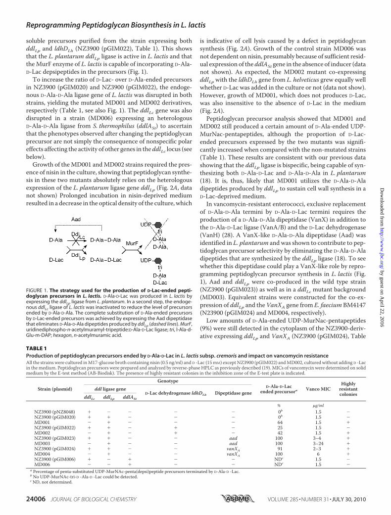
soluble precursors purified from the strain expressing bothddlLp and ldhDLh (NZ3900 (pGIM022), Table 1). This showsthat the L. plantarum ddlLp ligase is active in L. lactis and thattheMurF enzyme of L. lactis is capable of incorporating D-Ala-D-Lac depsipeptides in the precursors (Fig. 1).
To increase the ratio of D-Lac- over D-Ala-ended precursorsin NZ3900 (pGIM020) and NZ3900 (pGIM022), the endoge-nous D-Ala-D-Ala ligase gene of L. lactis was disrupted in bothstrains, yielding the mutated MD001 and MD002 derivatives,respectively (Table 1, see also Fig. 1). The ddlLc gene was alsodisrupted in a strain (MD006) expressing an heterologousD-Ala-D-Ala ligase from S. thermophilus (ddlASt) to ascertainthat the phenotypes observed after changing the peptidoglycanprecursor are not simply the consequence of nonspecific polareffects affecting the activity of other genes in theddlLc locus (seebelow).Growth of theMD001 andMD002 strains required the pres-
ence of nisin in the culture, showing that peptidoglycan synthe-sis in these two mutants absolutely relies on the heterologousexpression of the L. plantarum ligase gene ddlLp (Fig. 2A, datanot shown) Prolonged incubation in nisin-deprived mediumresulted in a decrease in the optical density of the culture, which
is indicative of cell lysis caused by a defect in peptidoglycansynthesis (Fig. 2A). Growth of the control strain MD006 wasnot dependent on nisin, presumably because of sufficient resid-ual expression of theddlASt gene in the absence of inducer (datanot shown). As expected, the MD002 mutant co-expressingddlLpwith the ldhDLh gene from L. helveticus grew equally wellwhether D-Lac was added in the culture or not (data not show).However, growth of MD001, which does not produces D-Lac,was also insensitive to the absence of D-Lac in the medium(Fig. 2A).Peptidoglycan precursor analysis showed that MD001 and
MD002 still produced a certain amount of D-Ala-ended UDP-MurNac-pentapeptides, although the proportion of D-Lac-ended precursors expressed by the two mutants was signifi-cantly increased when compared with the non-mutated strains(Table 1). These results are consistent with our previous datashowing that the ddlLp ligase is bispecific, being capable of syn-thesizing both D-Ala-D-Lac and D-Ala-D-Ala in L. plantarum(18). It is, thus, likely that MD001 utilizes the D-Ala-D-Aladipeptides produced by ddlLp to sustain cell wall synthesis in aD-Lac-deprived medium.
In vancomycin-resistant enterococci, exclusive replacementof D-Ala-D-Ala termini by D-Ala-D-Lac termini requires theproduction of a D-Ala-D-Ala dipeptidase (VanX) in addition tothe D-Ala-D-Lac ligase (VanA/B) and the D-Lac dehydrogenase(VanH) (28). A VanX-like D-Ala-D-Ala dipeptidase (Aad) wasidentified in L. plantarum and was shown to contribute to pep-tidoglycan precursor selectivity by eliminating the D-Ala-D-Aladipeptides that are synthesized by the ddlLp ligase (18). To seewhether this dipeptidase could play a VanX-like role by repro-gramming peptidoglycan precursor synthesis in L. lactis (Fig.1), Aad and ddlLp were co-produced in the wild type strain(NZ3900 (pGIM023)) as well as in a ddlLc mutant background(MD003). Equivalent strains were constructed for the co-ex-pression of ddlLp and theVanXA gene from E. faecium BM4147(N23900 (pGIM024) and MD004, respectively).Low amounts of D-Ala-ended UDP-MurNac-pentapeptides
(9%) were still detected in the cytoplasm of the NZ3900-deriv-ative expressing ddlLp and VanXA (NZ3900 (pGIM024), Table
FIGURE 1. The strategy used for the production of D-Lac-ended pepti-doglycan precursors in L. lactis. D-Ala-D-Lac was produced in L. lactis byexpressing the ddlLp ligase from L. plantarum. In a second step, the endoge-nous ddlLc ligase of L. lactis was inactivated to reduce the level of precursorsended by D-Ala-D-Ala. The complete substitution of D-Ala-ended precursorsby D-Lac-ended precursors was achieved by expressing the Aad dipeptidasethat eliminates D-Ala-D-Ala dipeptides produced by ddlLp (dashed lines). MurF,uridinediphospho-n-acetylmuramyl-tripeptide:D-Ala-D-Lac ligase; tri, l-Ala-d-Glu-m-DAP; hexagon, n-acetylmuramic acid.
TABLE 1Production of peptidoglycan precursors ended by D-Ala-D-Lac in L. lactis subsp. cremoris and impact on vancomycin resistanceAll the strains were cultured inM17-glucose broth containing nisin (0.5 ng/ml) and D -Lac (15mM) except NZ3900 (pGIM022) andMD002, cultured without adding D -Lacin the medium. Peptidoglycan precursors were prepared and analyzed by reverse-phase HPLC as previously described (19). MICs of vancomycin were determined on solidmedium by the E-test method (AB-Biodisk). The presence of highly resistant colonies in the inhibition zone of the E-test plate is indicated.
Strain (plasmid)
GenotypeD-Ala-D-Lac
ended precursora Vanco MICHighlyresistantcolonies
ddl ligase geneD-Lac dehydrogenase ldhDLh Dipeptidase gene
ddlLc ddlLp ddlASt
% �g/mlNZ3900 (pNZ8048) � � � � � 0b 1.5 �NZ3900 (pGIM020) � � � � � 0b 1.5 �MD001 � � � � � 64 1.5 �NZ3900 (pGIM022) � � � � � 25 1.5 �MD002 � � � � � 42 1.5 �NZ3900 (pGIM023) � � � � aad 100 3–4 �MD003 � � � � aad 100 3–24 �NZ3900 (pGIM024) � � � � vanXA 91 2–3 �MD004 � � � � vanXA 100 6 �NZ3900 (pGIM006) � � � � � NDc 1.5 �MD006 � � � � � NDc 1.5 �
a Percentage of penta-substituted UDP-MurNAc-penta(depsi)peptide precursors terminated by D-Ala-D -Lac.b No UDP-MurNAc-tri-D -Ala-D -Lac could be detected.c ND, not determined.
Reprogramming Peptidoglycan Biosynthesis in L. lactis
24006 JOURNAL OF BIOLOGICAL CHEMISTRY VOLUME 285 • NUMBER 31 • JULY 30, 2010
by guest on April 22, 2016
http://ww
w.jbc.org/
Dow
nloaded from

1), but they were totally undetectable in the strain co-express-ing ddlLp and aad (NZ3900 (pGIM023)) and in the ddlLcmutant derivatives MD003 and MD004 (Table 1). This showsthat the Aad dipeptidase of L. plantarum is at least as efficientas the enterococcal VanXdipeptidase in reorienting the biosyn-thesis of peptidoglycan precursors when expressed in a heter-ologous host. Growth ofMD003 andMD004was dependent onthe presence of both nisin and D-Lac in the culture medium(Fig. 2B, data not shown), indicating that D-Lac-ended pepti-doglycan precursors have become essential for cell wall synthe-sis in these two mutants.Changing the Peptidoglycan Precursor Is Not Sufficient to
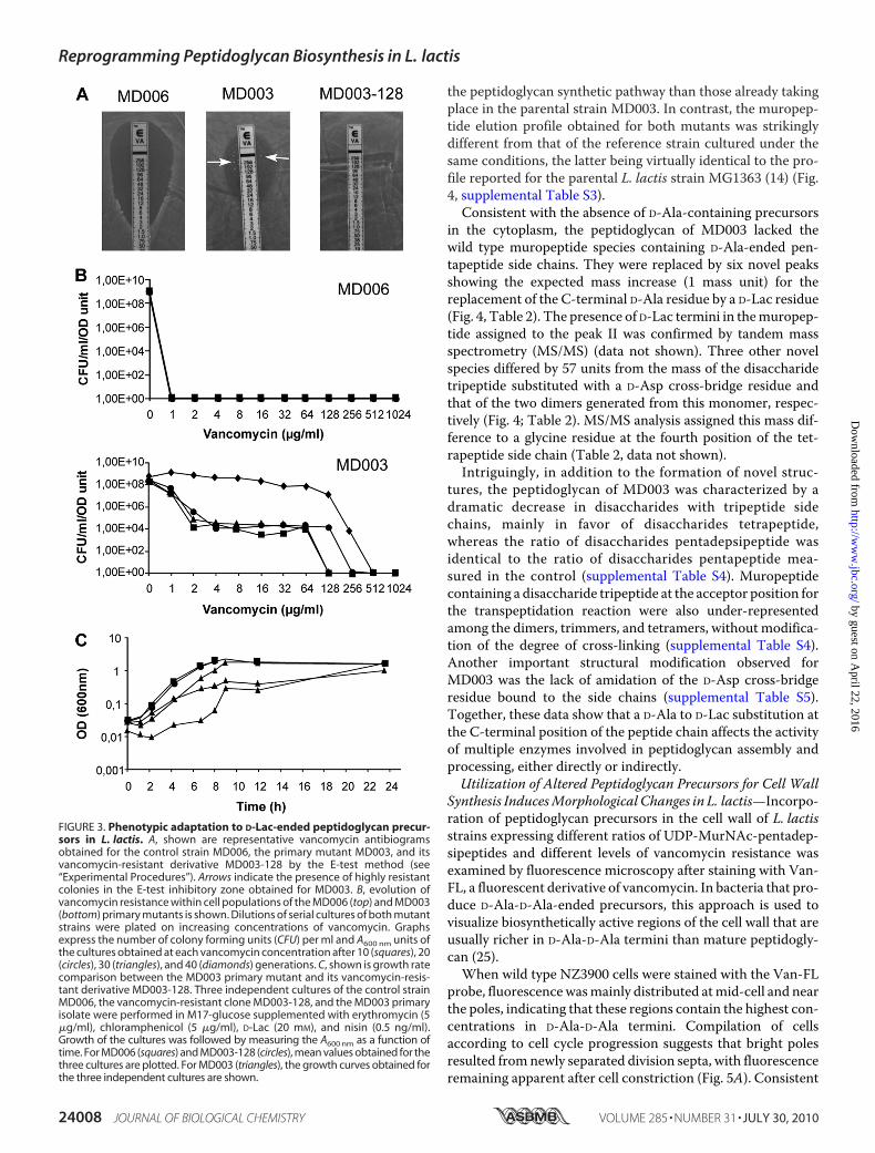
Confer aHigh Level of Resistance toVancomycin—Processing ofUDP-MurNAc-pentadepsipeptides by the cell wall biosynthe-sis machinery was first examined by looking at the effect ofmodulating the pool of D-Ala/D-Lac-ended precursors on thelevel of resistance to vancomycin expressed by the differentstrains of L. lactis (Table 1). All the strains producing amixtureof D-Lac- and D-Ala-ended precursors remained sensitive tovancomycin as determined by the E-test method (Table 1, see“Experimental Procedures”). Intriguingly, the completereplacement of D-Ala-ended UDP-MurNAc-pentapeptides byD-Lac-ended UDP-MurNac-pentadepsipeptides only slightlyincreased the resistance level, giving minimal vancomycininhibitory concentrations ranging from3 to 24�g/ml (Table 1).However, inspection of the plates after 24 h of further incuba-tion revealed that all the primary mutant strains that producedUDP-MurNAc-pentadepsipeptides (except NZ3900 (pGIM022),which accumulated the lowest amount of these modified pre-cursors), gave rise to isolated colonies in the inhibition zone ofthe E-test (Table 1, Fig. 3A). Population analysis performed byplating serial dilutions of a MD003 culture on increasing con-centrations of vancomycin revealed that derivatives expressing
higher levels of resistance accumulated during growth and upto completely dominated the cell population after �40 genera-tions (Fig. 3B). No accumulation of vancomycin-resistantderivatives was observed for the wild type strain or for theMD006 reference strain exclusively producing D-Ala-endedpeptidoglycan precursors (Fig. 3B, data not shown).MD003-derived clones that were recovered from the inhibi-
tion zone of the E-tests or fromprolonged cultures showedhighlevels of vancomycin resistance, withMIC values exceeding 256�g/ml (Fig. 3A). Their growth was dependent on the presencenisin and D-Lac in the culture medium, showing that pepti-doglycan synthesis in these variants still relied on the activity ofthe exogenous ddlLp ligase (data not shown). To get furtherinsight on the origin of the observed change in resistance level,plasmid DNA extracted from several independent sub-cloneswas back-introduced into NZ3900 cells, and the endogenousddlLc ligase genewas then inactivated as described above for theinitial MD003 mutant. This new generation of mutants repro-duced the phenotype of the parental strain, giving rise to highlyresistant cells emerging from a relatively low level of resistancebackground. This demonstrates that the increased vancomycinresistance displayed by the MD003 sub-clones did not resultfrom a mutation affecting the ddlLp/aad expression vector but,rather, from some genetic and/or physiological adaptation thatoccurred during cultivation of the primary mutant so as toimprove the fitness of the bacterial cells. Supporting this con-clusion, the growth rate of “adapted” vancomycin-resistantsub-clones was indistinguishable from that of the control strainMD006, whereas independent cultures performedwith the pri-mary MD003 mutant showed delayed and sometimes mul-tiphasicgrowthcurves indicativeofbacterialpopulationhetero-geneity (Fig. 3C).To get further insight on the mechanism that led to this
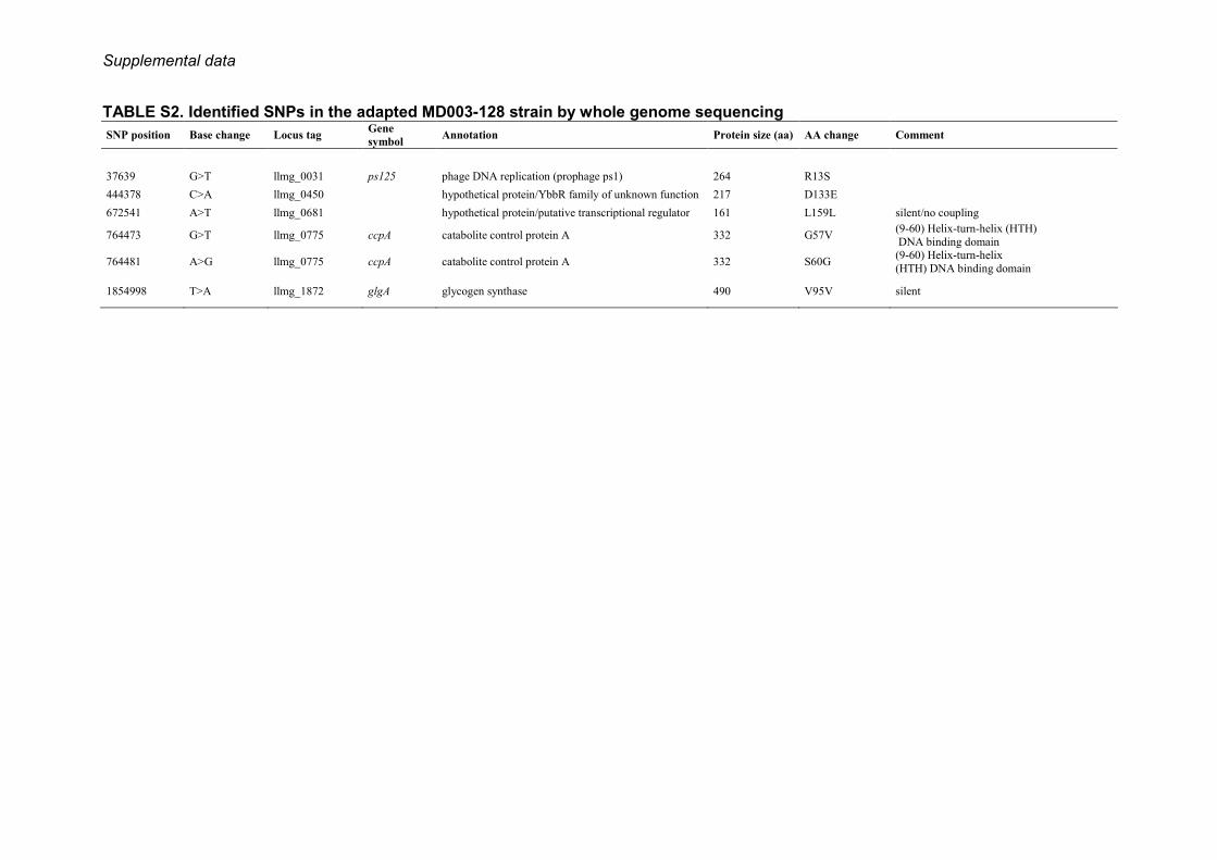
physiological adaptation of L. lactis cells, the completegenome of a selected vancomycin-resistant MD003 deriva-tive (MD003-128) was sequenced by high throughput SOLiDsequencing (see “Experimental Procedures”. The resultrevealed six changes compared with the wild type strainMG1363, including two silent mutations (supplementalTable S2). Two of the sense mutations affected the amino acidsequence of the catabolite control protein A, a global metabolicregulator of L. lactis (29). The remaining twomutations were ina phage protein and in a protein of unknown function, respec-tively (supplemental Table S2). Thus, none of the specificchanges found in the MD003-128 genome affected a geneknown to be directly involved in cell wall biosynthesis.
D-Ala/D-Lac Substitution of the C Terminus of PeptidoglycanPeptide Precursors Leads to Major Modifications of Peptidogly-can Structure—Todetermine the effect of changing the precur-sors on peptidoglycan structure, muropeptides extracted fromthe cell wall of the MD003 primary mutant and the highly van-comycin-resistant MD003-128 derivative were analyzed byreverse-phaseHPLC andMALDI-TOFmass spectrometry (14)(see “Experimental Procedures”). Intriguingly, peptidoglycancomposition established for MD003-128 was essentially thesame as that ofMD003 (data not shown). This indicates that theadaptive process that led to increased vancomycin resistance inMD003-128 was not accompanied with further alterations of
FIGURE 2. Growth dependence of the MD001 and MD003 mutants towardnisin and D-lactate. The L. lactis MD001 (A) and MD003 (B) mutants werecultured in M17-glucose in the presence and absence of D-Lac (15 mM) andnisin (0.5 ng/ml), shown as closed and open triangles, respectively, and in thepresence of nisin (0.5 ng/ml) without D-Lac (open circles). Growth of the cul-tures was followed by measuring the A600 nm.
Reprogramming Peptidoglycan Biosynthesis in L. lactis
JULY 30, 2010 • VOLUME 285 • NUMBER 31 JOURNAL OF BIOLOGICAL CHEMISTRY 24007
by guest on April 22, 2016
http://ww
w.jbc.org/
Dow
nloaded from
the peptidoglycan synthetic pathway than those already takingplace in the parental strain MD003. In contrast, the muropep-tide elution profile obtained for both mutants was strikinglydifferent from that of the reference strain cultured under thesame conditions, the latter being virtually identical to the pro-file reported for the parental L. lactis strain MG1363 (14) (Fig.4, supplemental Table S3).Consistent with the absence of D-Ala-containing precursors
in the cytoplasm, the peptidoglycan of MD003 lacked thewild type muropeptide species containing D-Ala-ended pen-tapeptide side chains. They were replaced by six novel peaksshowing the expected mass increase (1 mass unit) for thereplacement of the C-terminal D-Ala residue by a D-Lac residue(Fig. 4, Table 2). The presence of D-Lac termini in themuropep-tide assigned to the peak II was confirmed by tandem massspectrometry (MS/MS) (data not shown). Three other novelspecies differed by 57 units from the mass of the disaccharidetripeptide substituted with a D-Asp cross-bridge residue andthat of the two dimers generated from this monomer, respec-tively (Fig. 4; Table 2). MS/MS analysis assigned this mass dif-ference to a glycine residue at the fourth position of the tet-rapeptide side chain (Table 2, data not shown).Intriguingly, in addition to the formation of novel struc-
tures, the peptidoglycan of MD003 was characterized by adramatic decrease in disaccharides with tripeptide sidechains, mainly in favor of disaccharides tetrapeptide,whereas the ratio of disaccharides pentadepsipeptide wasidentical to the ratio of disaccharides pentapeptide mea-sured in the control (supplemental Table S4). Muropeptidecontaining a disaccharide tripeptide at the acceptor position forthe transpeptidation reaction were also under-representedamong the dimers, trimmers, and tetramers, without modifica-tion of the degree of cross-linking (supplemental Table S4).Another important structural modification observed forMD003 was the lack of amidation of the D-Asp cross-bridgeresidue bound to the side chains (supplemental Table S5).Together, these data show that a D-Ala to D-Lac substitution atthe C-terminal position of the peptide chain affects the activityof multiple enzymes involved in peptidoglycan assembly andprocessing, either directly or indirectly.Utilization of Altered Peptidoglycan Precursors for Cell Wall
Synthesis InducesMorphological Changes in L. lactis—Incorpo-ration of peptidoglycan precursors in the cell wall of L. lactisstrains expressing different ratios of UDP-MurNAc-pentadep-sipeptides and different levels of vancomycin resistance wasexamined by fluorescence microscopy after staining with Van-FL, a fluorescent derivative of vancomycin. In bacteria that pro-duce D-Ala-D-Ala-ended precursors, this approach is used tovisualize biosynthetically active regions of the cell wall that areusually richer in D-Ala-D-Ala termini than mature peptidogly-can (25).When wild type NZ3900 cells were stained with the Van-FL
probe, fluorescencewasmainly distributed atmid-cell and nearthe poles, indicating that these regions contain the highest con-centrations in D-Ala-D-Ala termini. Compilation of cellsaccording to cell cycle progression suggests that bright polesresulted fromnewly separated division septa, with fluorescenceremaining apparent after cell constriction (Fig. 5A). Consistent
FIGURE 3. Phenotypic adaptation to D-Lac-ended peptidoglycan precur-sors in L. lactis. A, shown are representative vancomycin antibiogramsobtained for the control strain MD006, the primary mutant MD003, and itsvancomycin-resistant derivative MD003-128 by the E-test method (see“Experimental Procedures”). Arrows indicate the presence of highly resistantcolonies in the E-test inhibitory zone obtained for MD003. B, evolution ofvancomycin resistance within cell populations of the MD006 (top) and MD003(bottom) primary mutants is shown. Dilutions of serial cultures of both mutantstrains were plated on increasing concentrations of vancomycin. Graphsexpress the number of colony forming units (CFU) per ml and A600 nm units ofthe cultures obtained at each vancomycin concentration after 10 (squares), 20(circles), 30 (triangles), and 40 (diamonds) generations. C, shown is growth ratecomparison between the MD003 primary mutant and its vancomycin-resis-tant derivative MD003-128. Three independent cultures of the control strainMD006, the vancomycin-resistant clone MD003-128, and the MD003 primaryisolate were performed in M17-glucose supplemented with erythromycin (5�g/ml), chloramphenicol (5 �g/ml), D-Lac (20 mM), and nisin (0.5 ng/ml).Growth of the cultures was followed by measuring the A600 nm as a function oftime. For MD006 (squares) and MD003-128 (circles), mean values obtained for thethree cultures are plotted. For MD003 (triangles), the growth curves obtained forthe three independent cultures are shown.
Reprogramming Peptidoglycan Biosynthesis in L. lactis
24008 JOURNAL OF BIOLOGICAL CHEMISTRY VOLUME 285 • NUMBER 31 • JULY 30, 2010
by guest on April 22, 2016
http://ww
w.jbc.org/
Dow
nloaded from

with this persistence of fluores-cence, �15% of L. lactis muropep-tides contain intact, D-Ala-D-Ala-ended pentapeptide side chains thatare not engaged in cross-linkingreactions (14). This pattern is, thus,slightly different from that previ-ously reported for S. pneumoniaewhere 100% of the pentapeptideside chains are processed during cellwall synthesis (25, 30).Van-FL staining was still observed
for theMD001 strain producing bothD-Ala- and D-Lac-ended precursors(Fig. 5B). However, the staining pat-tern appeared to bemuchmore het-erogeneous than that seen for thewild type strain, suggesting differ-ential utilization and/or processingof the peptidoglycan precursors indifferent cells. A specific class ofelongated cells (representing 22% ofthe cell population, n � 119)showed an asymmetrically posi-tioned dark band dividing the cellinto two unequal compartments(Fig. 5B). This morphologicalanomaly was sometimes associatedwith mis-positioned nucleoids as
revealed by 4�,6-diamidino-2-phenylindole staining. On aver-age,MD001 cells were slightly shorter and thicker than thewildtype cells, giving them a swollen appearance (compare Fig. 5, Aand B, Table 3). In addition, daughter cells often remainedattached to each other after cell division, generating shortchains that were not or rarely observedwith thewild type strain(Fig. 5, supplemental Fig. S1).These morphological alterations were found strongly accen-
tuated in the MD003 mutant (Fig. 5C) and even further pro-nounced in the vancomycin-resistant derivative MD003-128(Fig. 5D). These two strains were not stained by the Van-FLprobe, consistent with the fact that D-Ala-D-Lac-ended precur-sors are the only substrates available for cell wall synthesis.However, MD003 and MD003-128 cells showed an increasedtendency to adopt a spherical or almost spherical shape (Fig. 5,C and D) characterized by a reduced length/width ratio com-pared with the wild type cells (Table 3). The defect in cell sep-aration was also more pronounced for MD003 and MD003-128, resulting in the formation of long and curly chains (Fig. 5and supplemental Fig. S1). In contrast, the MD006 mutantexpressing the D-Ala-D-Ala ligase of S. thermophilus producednormal and well individualized ovoid cells comparable with thewild type cells (data not shown). This demonstrates that theprogressive alteration of cell shape and cell separation observedwith the MD001 and MD003 (MD003-128) mutants is thedirect consequence of an increased utilization of D-Lac-endedprecursors for cell wall synthesis.Implication of Penicillin-binding Proteins—Muropeptides
analysis showed that a distinctive alteration of MD003 and
FIGURE 4. Effect of changing the precursor on peptidoglycan structure. Peptidoglycan extracted from thereference strain PH8076 (pNZ8048) (top) and from the D-Lac-ended precursor-producing MD003 mutant (bot-tom) were digested with the mutanolysin muramidase and analyzed by HPLC as described under “Experimen-tal Procedures.” The chemical structures assigned to the identified picks of muropeptides (numbered) are givenin supplemental Table S3 and Table 2.
TABLE 2Molecular masses and proposed structures for muropeptidespresent (roman numbers) and absent (arabic numbers) from thepeptidoglycan of the MD003 mutant strain
PeakaMass (M�Na)�
IdentificationcObserved Calculatedb
6 NDd 990.46 Penta10 NDd 1105.49 Penta-D12 NDd 1104.50 Penta-NI 1020.52 NAe Tetra/Gly-DII 1106.48 1106.49 Penta/D -Lac-DIII 1105.55 1105.51 Penta/D -Lac-N25 NDd 2097.95 Penta-N-Tetra-Df
27 NDd 2096.96 Penta-N-Tetra-NIV 2098.95 2098.95 Penta/D -Lac-N-Tetra-Df
V 2012.89 NAe Tetra/Gly-N-Tetra-Df
VI 2011.99 NAe Tetra/Gly-N-Tetra-NVII 2097.86 2097.97 Penta/D -Lac-N-Tetra-N35b NDd 3090.42 Penta-N-Tetra-N-Tetra-Df
36 NDd 3089.44 Penta-N-Tetra-N-Tetra-NVIII 3091.31 3091.42 Penta/D -Lac-N-Tetra-N-Tetra-Df
IX 3090.41 3090.44 Penta/D -Lac-N-Tetra-N-Tetra-N39 NDd 4081.90 Penta-N-Tetra-N-Tetra-N-Tetra-N
a Peak numbers refer to Fig. 4.b Sodiatedmolecular ionswere themost abundant onMALDI-TOFmass spectra forall muropeptides.m/z values correspond to monoisotopic masses.
c Penta, disaccharide pentapeptide (L-Ala-D-iGln-L-Lys-D-Ala-D-Ala; iGln is iso-glu-tamine); Tetra, disaccharide tetrapeptide (L-Ala-D-iGln-L-Lys-D-Ala); Tetra/Gly,disaccharide tetrapeptide (L-Ala-D-iGln-L-Lys-Gly); Penta/D-Lac, disaccharidepentadepsipeptide (L-Ala-D-iGln-L-Lys-D-Ala-D-Lac); disaccharide, GlcNAc-MurNAc; D, D-Asp; N, D-Asn; iGln, �-amidated isoGlu (or �Glu).
d ND, not determined. MALDI-TOF masses were determined only for MD003;structures from the control peptidoglycanwere assigned by comparing peaks withretention times observed for the corresponding muropeptides previously identi-fied in the parental strain MG1363 (14).
e NA, not applicable.f Positions of N and D in peptide chains of oligomeric forms are arbitrarily given.
Reprogramming Peptidoglycan Biosynthesis in L. lactis
JULY 30, 2010 • VOLUME 285 • NUMBER 31 JOURNAL OF BIOLOGICAL CHEMISTRY 24009
by guest on April 22, 2016
http://ww
w.jbc.org/
Dow
nloaded from

MD003-128 peptidoglycan is the decrease in disaccharideswithtripeptide side chains and the concomitant increase of disac-charides with tetrapeptide side chains. A similar enrichment of
tetrapeptides at the expense of tri-peptides was previously reportedfor a L. lactis mutant deficient forthe L,D-carboxypeptidase DacB(VES2065) (14). This suggests thatchanging the nature of the pepti-doglycan precursors may havealtered the activity of L. lactis car-boxypeptidases. Analysis of theL. lactis genome revealed the pres-ence of two carboxypeptidases. Inaddition to DacB, L. lactis encodesDacA, a protein homologous toPBP3 from S. pneumoniae (31). AdacA mutant strain (BLD006) wasconstructed, and the morphologyof both the dacA and dacBmutants was analyzed. The dacAmutant produced cells that werespherical or almost spherical asobserved for MD003 (Table 3; Fig.6), whereas dacB cells were thickerthan the wild type cells and tendedto remain associated into chains(Table 3; Fig. 6). Together thesephenotypes are reminiscent ofthose observed for the MD003 andMD003-128 precursor mutants(Fig. 6; see also Fig. 5).To get further insight on the
mechanism that led to the observedmorphological defects of themutant strains, bacterial cells weretreated with methicillin, a �-lactamantibiotic known to specificallyblock PBPs involved in cell division(32). When incubated with increas-ing concentrations of methicillin,the wild type and control strainMD006 stopped dividing, generat-ing elongated cells reminiscent ofbacterial filaments (Fig. 6A). In con-trast, cells of the MD003 andMD003-128 mutants remained
more compact and started to swell to adopt a puffy phenotype(Fig. 6, B and C). A similar phenotype was observed with thedacAmutant and to a lower extentwith thedacBmutant (Fig. 6,D and E). These results demonstrate that the cell elongationand division processes can be separated in L. lactis and thatchanging the peptidoglycan precursors affects cell elongation,presumably by perturbing the activity of specific PBPs, includ-ing carboxypeptidases.
DISCUSSION
This study reports the production of peptidoglycan precur-sors ending by D-Ala-D-Lac instead of D-Ala-D-Ala in L. lactisthrough the heterologous expression of genes involved in pep-tidoglycan synthesis from L. plantarum. Modification of the
FIGURE 5. Van-FL staining and morphology of L. lactis mutant strains expressing different proportionsof D-Lac-ended versus D-Ala-ended peptidoglycan precursors. For each strain micrographs of representa-tive cells visualized by Van-FL staining (upper panels) and 4�,6-diamidino-2-phenylindole staining (lower panels)are presented. Scale bars: 1 �m. A, wild type NZ3900 cells transformed with the empty expression vectorpNZ8048 are shown. Cells are numbered according to cell cycle progression. Arrowheads show brighter fluo-rescence spots that appear at the division site and then separate at a later stage of cell division to occupy a polarposition in the newborn cells. A schematic representation of the different stages of the cell cycle is shown onthe left, with the Van-FL-stained zones and spots colored gray. B, cells of the MD001 mutant producing amixture of D-Ala-ended and D-Lac-ended peptidoglycan precursors are shown. Arrows indicate morphologicaldefects associated with dark areas not stained by the Van-FL probe. C, cells of the MD003 mutant exclusivelyproducing D-Lac-ended peptidoglycan precursors are shown. Contrast of the fluorescence images wasincreased to visualize the position of the cell in the field. D, shown is a representative cell chain formed by thehighly vancomycin resistant MD003 derivative MD003-128.
TABLE 3Effect of peptidoglycan precursors modification on cell shape(length/width ratio)
Strain Length average � ICa Width average � ICa Length/Widthb
�m �mNZ3900 1.7 � 0.05 0.7 � 0.01 2.4MD006 1.9 � 0.05 0.8 � 0.02 2.4MD001 1.6 � 0.05 0.8 � 0.01 2.0MD003 1.5 � 0.06 0.9 � 0.02 1.6MD003–128 1.3 � 0.04 1.0 � 001 1.3BLD006 1.3 � 0.04 1.0 � 0.02 1.3VES2065 1.7 � 0.06 0.97 � 0.02 1.8
an � 200 cells. IC, interval of confidence, � � 0.01.b Ratio of average cell length to average cell width.
Reprogramming Peptidoglycan Biosynthesis in L. lactis
24010 JOURNAL OF BIOLOGICAL CHEMISTRY VOLUME 285 • NUMBER 31 • JULY 30, 2010
by guest on April 22, 2016
http://ww
w.jbc.org/
Dow
nloaded from

last residue of the stem peptide strongly affected cell wall pep-tidoglycan composition and cell morphology. Cells becamespherical instead of ovoid and remained assembled into chains.Our data support the view that peptidoglycan compositionplays a central role in the coordination of cell wall biosynthesisand processing enzymes.Cell Wall Biosynthesis Reprogramming and Morphogenesis;
Regulation by the Substrate—L. lactismutants exclusively pro-ducing D-Lac-ended precursors are viable, showing that the cellwall synthesis machinery can accept the surrogate precursors.Analysis of the muropeptide composition of the MD003mutant showed that all the D-Ala-ended pentapeptide sidechains were replaced by an equivalent amount of pentadep-sipeptide side chains ending by D-Ala-D-Lac. In addition, thelevel of cross-linking was not affected by the precursor alter-ation. These results show that modified stem peptides can effi-ciently be used as a donor but also as an acceptor for transpep-tidation in L. lactis. By comparison, D-Ala-D-Lac termini werenot detected in the peptidoglycan extracted from vancomycin-resistant strains of enterococci and S. aureus expressing the
vanA/B gene cluster, although thelevel of cross-linking was un-changed (33–37). It was proposedthat removing D-Lac from the pep-tide chains is necessary in these bac-terial species to supply transpepti-dases with an appropriate acceptorsubstrate, which seems not to be thecase in L. lactis (33, 37).Although the level of transpepti-
dation was not impaired in themutant strains, changing the lastresidue of the peptidoglycan pre-cursor peptide chain significantlymodified the muropeptide compo-sition of the bacterium. Severalactivities involved in the processingand/or exportation of the pepti-doglycan precursors may be alteredin the mutants, whereas structuralchanges in the muropeptides mayhave affected the activity of otherenzymes acting at a later stage ofpeptidoglycan synthesis and matu-ration.Wepropose that altering oneof these specific activities or thecoordination of multiple compo-nents of the cell wall machinery isresponsible for the morphologicalchanges observed in the L. lactisprecursor mutants.A marked difference that was
revealed by the muropeptide analy-sis is the decrease in amidation ofthe D-Asp cross-bridge in theMD003mutant comparedwith wildtype L. lactis. Although little isknown on the physiological signifi-
cance of peptidoglycan amidation during cell wall synthesis, theasparagine synthase (AsnH) responsible for D-Asp amidationwas recently identified in L. lactis (17). Amidation is thought tooccur after D-Asp incorporation into the peptidoglycan precur-sor by a D-Asp-specific ligase (16, 17). A D-Ala to D-Lac substi-tution at the fifth position of the stem peptide may, therefore,interfere with this reaction by affecting the structure of its sub-strate. However, the lack of amidation alone cannot explain themorphological defects that characterize the mutant strainsexpressing D-Lac-ended precursors, as an asnH null mutantthat is completely deficient for D-Asp amidation exhibits nor-mal cell shape.6In contrast, disruption of the carboxypeptidase genes dacA
and dacBmarkedly affected cell morphology producing pheno-types that phenocopied those of MD003 and MD003-128.Mutation in the dacB gene was shown to alter the ratio of trip-eptide versus tetrapeptide disaccharides as we observed in the
6 M. Deghorain, unpublished data.
FIGURE 6. Effect of methicillin on cell morphology. Exponentially growing cells of the control strain MD006(A), the MD003 primary mutant (B), the highly vancomycin-resistant MD003-derived clone MD003-128, and thecarboxypeptidase dacA (BLD006; D) and dacB (VES2065; E) mutants were incubated for 4h30 with the indicatedconcentrations of methicillin before be examined by light microscopy. Scale bar � 3�M.
Reprogramming Peptidoglycan Biosynthesis in L. lactis
JULY 30, 2010 • VOLUME 285 • NUMBER 31 JOURNAL OF BIOLOGICAL CHEMISTRY 24011
by guest on April 22, 2016
http://ww
w.jbc.org/
Dow
nloaded from
precursormutants (14). The DacA protein of L. lactis is homol-ogous to the PBP3 carboxypeptidase of S. pneumoniae. PBP3was proposed to trim the last residue of pentapeptides all overthe cell except in the equatorial zone. This activity is required toproperly position high molecular weight PBPs in this region byproviding them with the appropriate donor substrates for thetranspeptidation reaction (11). As a consequence, disruption ofPBP3 was found to alter the morphology of S. pneumoniae asweobservedhere for thedacAmutant ofL. lactis (31). It is, thus,likely that changing the structure of the precursor altered theactivity of peptidoglycan processing enzymes such as DacBand/orDacA carboxypeptidases. Thismay in turn have affectedthe recruitment of other PBPs. Heterogeneous distribution ofcarboxypeptidases and/or transpeptidases in the cell wall ofL. lactis is supported by the Van-FL staining pattern obtainedfor the wild type strain, with some regions of the wall appearingmore fluorescent than others at different stages of the cell cycle.The morphological defects observed for the MD003 mutant
of L. lactis are also reminiscent of those reported for S. ther-mophilusmutants deficient in the transpeptidase PBP2b or thetransmembrane RodA protein, two enzymes thought to be spe-cifically involved in cell elongation (38).We show here that cellelongation and cell division can be uncoupled in L. lactis bytreating the cells with methicillin. Wild type bacteria in whichcell division was blocked by the antibiotic sustained active lon-gitudinal growth, giving rise to long filaments. This process wasseverely compromised in theMD003 andMD003-128mutants,indicating that changing the nature of the peptidoglycan pre-cursor specifically perturbed the cell elongation machinery.This may occur directly by affecting the activity of key enzymesof the elongation process (e.g. PBP2b) or indirectly by alteringtheir recruitment within the cell wall (e.g. throughDacA and/orDacB activity). Changing the substrate may also affected theactivity and/or location of other peptidoglycan-processingenzymes, such as peptidoglycanhydrolases that are required forcell separation, as was reported in E. coli (13). Elucidation of theexact mechanism that is responsible for the observed morpho-logical changes in the precursor mutants of L. lactis willundoubtedly provide new insight into how the different com-ponents of the cell wall biosynthesis machinery cooperate dur-ing cell morphogenesis in ovococci and how their activity iscontrolled by substrate processing during peptidoglycansynthesis.Resistance to Vancomycin; Further Adaptation to Peptidogly-
can Reprogramming Is Required—Key enzymes for specificD-Lac incorporation into the cell wall of L. plantarum in-clude the D-lactate dehydrogenase LdhDLp, the D-Ala-D-Lacligase ddlLp, and the D-Ala-D-Ala dipeptidase Aad (18, 19, 39).Unlike their enterococcal van homologues, the genes encodingthese different activities are not clusteredwithin an operon, andthey appear to have a more fundamental role in cell physiologythan being simply required for vancomycin resistance (18, 19,39).We, therefore, proposed that genes functionally equivalentto those of L. plantarum were initially recruited by glycopep-tide producers to protect themselves against the antibiotic andwere subsequently transmitted to enterococci and other bacte-rial species to confer resistance to vancomycin (18). In supportof this view, we show here that the co-expression of these met-
abolic genes in L. lactis allows peptidoglycan precursor synthe-sis to be remodeled, just as the expression of the vancomycinresistance vanA or vanB operons in enterococci.However, an intriguing finding of this study is the fact that
vancomycin resistance is not an immediate consequence ofchanging the precursor but the result of an adaptive processafter its utilization for cell wall synthesis. Similar observationswere made when the vanHAX resistance operon from E. fae-cium BM4147 was transferred in L. lactis (data not shown),indicating that the ability to develop vancomycin resistancedepends on properties of the bacterial cells and not on thegenes that were used to alter precursor biosynthesis. Con-sistent with this, the growth rate of vancomycin resistantMD003 derivatives was significantly improved when com-pared with that of the primary mutant, suggesting that cellswere adapted to better utilize the new peptidoglycan sub-strate, thereby increasing their fitness. Sequencing of theMD003-128 complete genome revealed that this adaptationprocess did not directly affect enzymes specifically involvedin peptidoglycan synthesis or cell wall processing (sup-plemental Table S2). However, two mutations were detectedin catabolite control protein A, a pleiotropic regulatorknown to regulate multiple cellular processes including vir-ulence and the level of antibiotics resistance in S. aureus(40). Thus, these data support the conclusion that pheno-typic adaptation leading to high levels of vancomycin resist-ance resulted from global optimization of L. lactis cell wallbiosynthetic machinery rather than from specific alterationsof the peptidoglycan synthetic pathway. Also supporting thisview is the finding that the muropeptide composition of theadapted MD003-128 strain is unchanged when comparedwith that of the mother strain MD003.The impact that the nature of the precursor may have on
peptidoglycan synthesis and cell morphology together with therequirement for further optimization of the biosynthesismachinery has an important and more general implication onvancomycin resistance. Not all bacteria may exhibit the sameflexibility to cope with altered peptidoglycan precursors, andonemay, therefore, anticipate that some species would bemoreprone to become resistant to vancomycin than others.
Acknowledgments—We warmly thank J. Delcour for helpful discus-sions and scientific advice. We are grateful to M. Arthur for theMS/MS determination of muropeptides I and II and to A. Marnef forthe construction of the MD006 strain. We thank Dr. S. Kulakauskasfor help in L. lactisMD003-128 sequencing andDr. S. Kennedy andN.Galleron (MetaQuant sequencing platform) for preparing the ampli-con libraries and for preliminary data analysis. Sequencing ofMD003-128 was supported in part by the programAIP BioRessources2009 of INRA (project SOLIMUT).
REFERENCES1. Barreteau, H., Kovac, A., Boniface, A., Sova, M., Gobec, S., and Blanot, D.
(2008) FEMS Microbiol. Rev. 32, 168–2072. Sauvage, E., Kerff, F., Terrak,M., Ayala, J. A., and Charlier, P. (2008) FEMS
Microbiol. Rev. 32, 234–2583. Carballido-Lopez, R., and Formstone, A. (2007)Curr. Opin.Microbiol. 10,
611–6164. den Blaauwen, T., de Pedro, M. A., Nguyen-Disteche, M., and Ayala, J. A.
Reprogramming Peptidoglycan Biosynthesis in L. lactis
24012 JOURNAL OF BIOLOGICAL CHEMISTRY VOLUME 285 • NUMBER 31 • JULY 30, 2010
by guest on April 22, 2016
http://ww
w.jbc.org/
Dow
nloaded from
(2008) FEMS Microbiol. Rev. 32, 321–3445. Holtje, J. V. (1998)Microbiol. Mol. Biol. Rev. 62, 181–2036. Vollmer, W., Joris, B., Charlier, P., and Foster, S. (2008) FEMS Microbiol.
Rev. 32, 259–2867. Zapun, A., Vernet, T., and Pinho, M. G. (2008) FEMS Microbiol. Rev. 32,
345–3608. Cabeen, M. T., and Jacobs-Wagner, C. (2007) J. Cell Biol. 179, 381–3879. Scheffers, D. J., and Pinho, M. G. (2005) Microbiol. Mol. Biol. Rev. 69,
585–60710. Begg, K. J., Takasuga, A., Edwards, D. H., Dewar, S. J., Spratt, B. G., Adachi,
H., Ohta, T., Matsuzawa, H., and Donachie,W. D. (1990) J. Bacteriol. 172,6697–6703
11. Morlot, C., Noirclerc-Savoye, M., Zapun, A., Dideberg, O., and Vernet, T.(2004)Mol. Microbiol. 51, 1641–1648
12. Pinho, M. G., and Errington, J. (2005)Mol. Microbiol. 55, 799–80713. Priyadarshini, R., Popham, D. L., and Young, K. D. (2006) J. Bacteriol. 188,
5345–535514. Courtin, P., Miranda, G., Guillot, A., Wessner, F., Mezange, C., Doma-
kova, E., Kulakauskas, S., and Chapot-Chartier, M. P. (2006) J. Bacteriol.188, 5293–5298
15. Schleifer, K. H., and Kandler, O. (1972) Bacteriol. Rev. 36, 407–47716. Veiga, P., Piquet, S., Maisons, A., Furlan, S., Courtin, P., Chapot-Chartier,
M. P., and Kulakauskas, S. (2006)Mol. Microbiol. 62, 1713–172417. Veiga, P., Erkelenz, M., Bernard, E., Courtin, P., Kulakauskas, S., and
Chapot-Chartier, M. P. (2009) J. Bacteriol. 191, 3752–375718. Deghorain, M., Goffin, P., Fontaine, L., Mainardi, J. L., Daniel, R., Err-
ington, J., Hallet, B., and Hols, P. (2007) J. Bacteriol. 189, 4332–433719. Goffin, P., Deghorain, M., Mainardi, J. L., Tytgat, I., Champomier-Verges,
M. C., Kleerebezem, M., and Hols, P. (2005) J. Bacteriol. 187, 6750–676120. Hols, P., Kleerebezem, M., Schanck, A. N., Ferain, T., Hugenholtz, J., Del-
cour, J., and de Vos, W. M. (1999) Nat. Biotechnol. 17, 588–59221. Arthur, M., Molinas, C., and Courvalin, P. (1992) J. Bacteriol. 174,
2582–259122. van Kranenburg, R., Marugg, J. D., van Swam, I. I., Willem, N. J., and de
Vos, W. M. (1997)Mol. Microbiol. 24, 387–39723. de Ruyter, P. G., Kuipers, O. P., and de Vos, W. M. (1996) Appl. Environ.
Microbiol. 62, 3662–366724. Arbeloa, A., Hugonnet, J. E., Sentilhes, A. C., Josseaume, N., Dubost, L.,
Monsempes, C., Blanot, D., Brouard, J. P., and Arthur, M. (2004) J. Biol.Chem. 279, 41546–41556
25. Daniel, R. A., and Errington, J. (2003) Cell 113, 767–77626. Wegmann, U., O’Connell-Motherway, M., Zomer, A., Buist, G., Shear-
man, C., Canchaya, C., Ventura,M., Goesmann, A., Gasson,M. J., Kuipers,O. P., van Sinderen, D., and Kok, J. (2007) J. Bacteriol. 189, 3256–3270
27. Li, H., Ruan, J., and Durbin, R. (2008) Genome Res. 18, 1851–185828. Arthur, M., Depardieu, F., Reynolds, P., and Courvalin, P. (1996) Mol.
Microbiol. 21, 33–4429. Zomer, A. L., Buist, G., Larsen, R., Kok, J., and Kuipers, O. P. (2007) J.
Bacteriol. 189, 1366–138130. Ng, W. L., Kazmierczak, K. M., andWinkler, M. E. (2004)Mol. Microbiol.
53, 1161–117531. Schuster, C., Dobrinski, B., and Hakenbeck, R. (1990) J. Bacteriol. 172,
6499–650532. Lleo,M.M., Canepari, P., and Satta, G. (1990) J. Bacteriol. 172, 3758–377133. de Jonge, B. L., Handwerger, S., and Gage, D. (1996) Antimicrob. Agents
Chemother. 40, 863–86934. Bouhss, A., Josseaume, N., Severin, A., Tabei, K., Hugonnet, J. E., Shlaes,
D., Mengin-Lecreulx, D., Van Heijenoort, J., and Arthur, M. (2002) J. Biol.Chem. 277, 45935–45941
35. de Jonge, B. L., Gage, D., andHandwerger, S. (1996)Microb. Drug Resist. 2,225–229
36. Billot-Klein, D., Shlaes, D., Bryant, D., Bell, D., van Heijenoort, J., andGutmann, L. (1996) Biochem. J. 313, 711–715
37. Severin, A., Tabei, K., Tenover, F., Chung, M., Clarke, N., and Tomasz, A.(2004) J. Biol. Chem. 279, 3398–3407
38. Thibessard, A., Fernandez, A., Gintz, B., Leblond-Bourget, N., and De-caris, B. (2002) J. Bacteriol. 184, 2821–2826
39. Ferain, T., Hobbs, J. N., Jr., Richardson, J., Bernard, N., Garmyn, D., Hols,P., Allen, N. E., and Delcour, J. (1996) J. Bacteriol. 178, 5431–5437
40. Seidl, K., Stucki, M., Ruegg, M., Goerke, C., Wolz, C., Harris, L., Berger-Bachi, B., and Bischoff, M. (2006) Antimicrob. Agents Chemother. 50,1183–1194
Reprogramming Peptidoglycan Biosynthesis in L. lactis
JULY 30, 2010 • VOLUME 285 • NUMBER 31 JOURNAL OF BIOLOGICAL CHEMISTRY 24013
by guest on April 22, 2016
http://ww
w.jbc.org/
Dow
nloaded from
Supplemental data
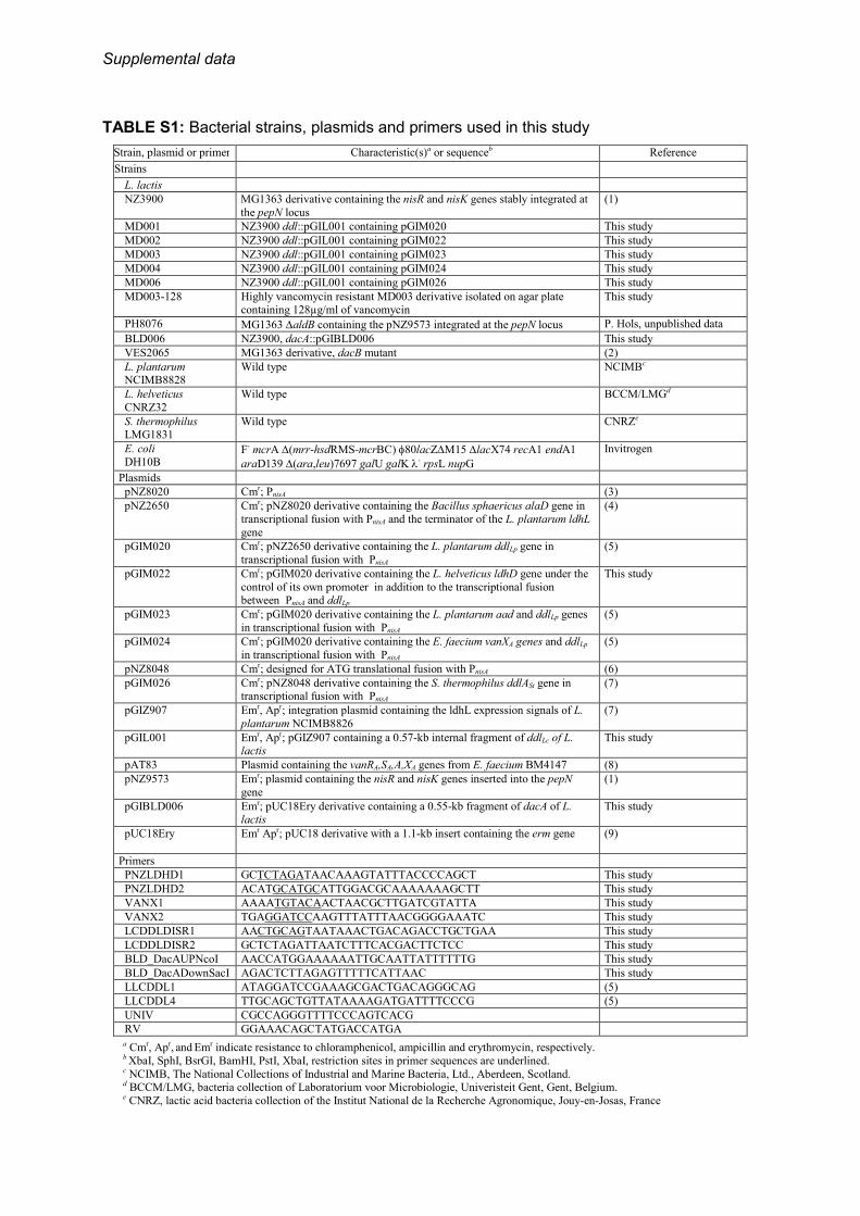
TABLE S1: Bacterial strains, plasmids and primers used in this study
Strain, plasmid or primer Characteristic(s)a or sequenceb Reference
Strains
L. lactis
NZ3900 MG1363 derivative containing the nisR and nisK genes stably integrated at
the pepN locus
(1)
MD001 NZ3900 ddl::pGIL001 containing pGIM020 This study
MD002 NZ3900 ddl::pGIL001 containing pGIM022 This study
MD003 NZ3900 ddl::pGIL001 containing pGIM023 This study
MD004 NZ3900 ddl::pGIL001 containing pGIM024 This study
MD006 NZ3900 ddl::pGIL001 containing pGIM026 This study
MD003-128 Highly vancomycin resistant MD003 derivative isolated on agar plate containing 128µg/ml of vancomycin
This study
PH8076 MG1363 ∆aldB containing the pNZ9573 integrated at the pepN locus P. Hols, unpublished data
BLD006 NZ3900, dacA::pGIBLD006 This study
VES2065 MG1363 derivative, dacB mutant (2)
L. plantarum NCIMB8828
Wild type NCIMBc
L. helveticus
CNRZ32
Wild type BCCM/LMGd
S. thermophilus LMG1831
Wild type CNRZe
E. coli
DH10B F- mcrA ∆(mrr-hsdRMS-mcrBC) φ80lacZ∆M15 ∆lacX74 recA1 endA1
araD139 ∆(ara,leu)7697 galU galK λ- rpsL nupG
Invitrogen
Plasmids
pNZ8020 Cmr; PnisA (3)
pNZ2650 Cmr; pNZ8020 derivative containing the Bacillus sphaericus alaD gene in
transcriptional fusion with PnisA and the terminator of the L. plantarum ldhL
gene
(4)
pGIM020 Cmr; pNZ2650 derivative containing the L. plantarum ddlLp gene in
transcriptional fusion with PnisA
(5)
pGIM022 Cmr; pGIM020 derivative containing the L. helveticus ldhD gene under the
control of its own promoter in addition to the transcriptional fusion between PnisA and ddlLp
This study
pGIM023 Cmr; pGIM020 derivative containing the L. plantarum aad and ddlLp genes
in transcriptional fusion with PnisA
(5)
pGIM024 Cmr; pGIM020 derivative containing the E. faecium vanXA genes and ddlLp
in transcriptional fusion with PnisA
(5)
pNZ8048 Cmr; designed for ATG translational fusion with PnisA (6)
pGIM026 Cmr; pNZ8048 derivative containing the S. thermophilus ddlASt gene in transcriptional fusion with PnisA
(7)
pGIZ907 Emr, Apr; integration plasmid containing the ldhL expression signals of L.
plantarum NCIMB8826
(7)
pGIL001 Emr, Apr; pGIZ907 containing a 0.57-kb internal fragment of ddlLc of L. lactis
This study
pAT83 Plasmid containing the vanRA,SA,A,XA genes from E. faecium BM4147 (8)
pNZ9573
Emr; plasmid containing the nisR and nisK genes inserted into the pepN
gene
(1)
pGIBLD006
Emr; pUC18Ery derivative containing a 0.55-kb fragment of dacA of L. lactis
This study
pUC18Ery Emr Apr; pUC18 derivative with a 1.1-kb insert containing the erm gene (9)
Primers
PNZLDHD1 GCTCTAGATAACAAAGTATTTACCCCAGCT This study
PNZLDHD2 ACATGCATGCATTGGACGCAAAAAAAGCTT This study
VANX1 AAAATGTACAACTAACGCTTGATCGTATTA This study
VANX2 TGAGGATCCAAGTTTATTTAACGGGGAAATC This study
LCDDLDISR1 AACTGCAGTAATAAACTGACAGACCTGCTGAA This study
LCDDLDISR2 GCTCTAGATTAATCTTTCACGACTTCTCC This study
BLD_DacAUPNcoI AACCATGGAAAAAATTGCAATTATTTTTTG This study
BLD_DacADownSacI AGACTCTTAGAGTTTTTCATTAAC This study
LLCDDL1 ATAGGATCCGAAAGCGACTGACAGGGCAG (5)
LLCDDL4 TTGCAGCTGTTATAAAAGATGATTTTCCCG (5)
UNIV CGCCAGGGTTTTCCCAGTCACG
RV GGAAACAGCTATGACCATGA
a Cmr, Apr, and Emr indicate resistance to chloramphenicol, ampicillin and erythromycin, respectively. b XbaI, SphI, BsrGI, BamHI, PstI, XbaI, restriction sites in primer sequences are underlined. c NCIMB, The National Collections of Industrial and Marine Bacteria, Ltd., Aberdeen, Scotland. d BCCM/LMG, bacteria collection of Laboratorium voor Microbiologie, Univeristeit Gent, Gent, Belgium. e CNRZ, lactic acid bacteria collection of the Institut National de la Recherche Agronomique, Jouy-en-Josas, France
Supplemental data
TABLE S2. Identified SNPs in the adapted MD003-128 strain by whole genome sequencing
SNP position Base change Locus tag Gene
symbol Annotation Protein size (aa) AA change Comment
37639 G>T llmg_0031 ps125 phage DNA replication (prophage ps1) 264 R13S
444378 C>A llmg_0450 hypothetical protein/YbbR family of unknown function 217 D133E
672541 A>T llmg_0681 hypothetical protein/putative transcriptional regulator 161 L159L silent/no coupling
764473 G>T llmg_0775 ccpA catabolite control protein A 332 G57V (9-60) Helix-turn-helix (HTH)
DNA binding domain
764481 A>G llmg_0775 ccpA catabolite control protein A 332 S60G (9-60) Helix-turn-helix
(HTH) DNA binding domain
1854998 T>A llmg_1872 glgA glycogen synthase 490 V95V silent
Supplemental data
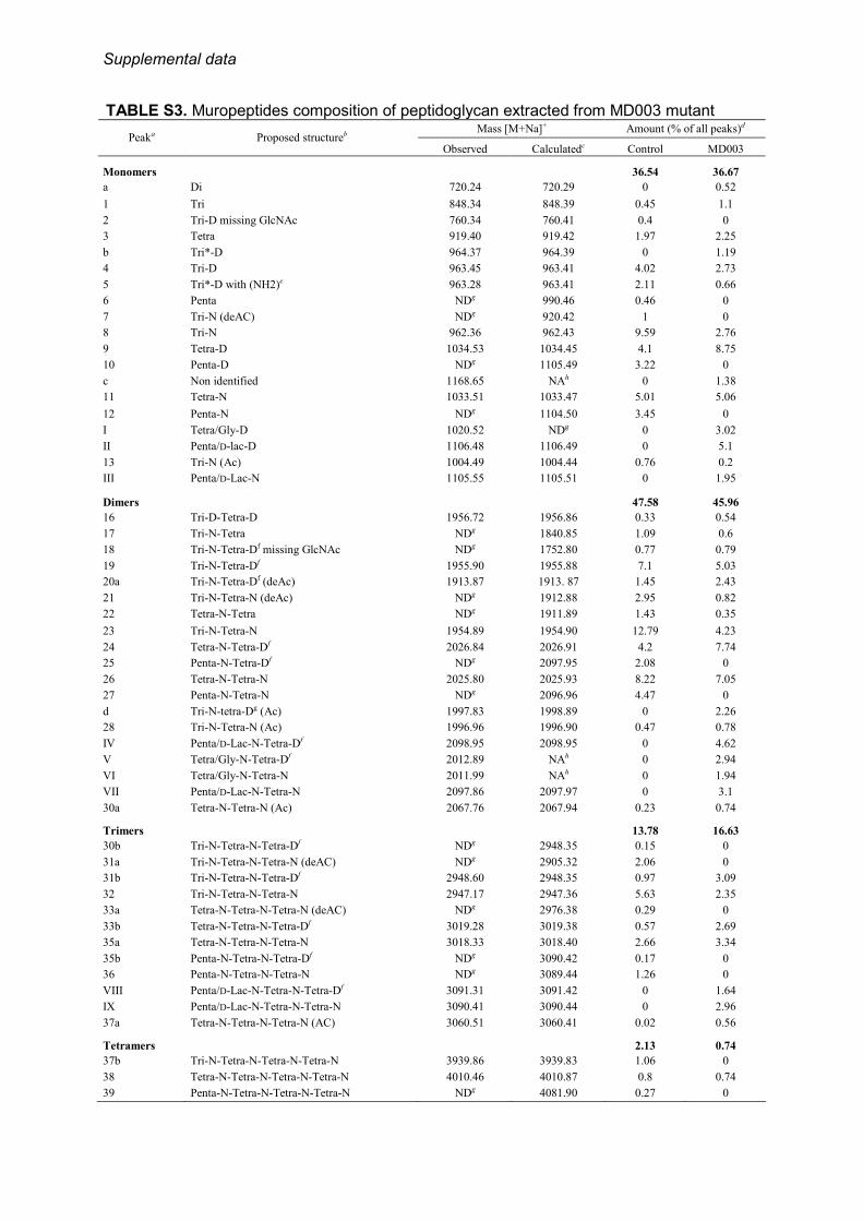
TABLE S3. Muropeptides composition of peptidoglycan extracted from MD003 mutant Mass [M+Na]+ Amount (% of all peaks)d
Peaka Proposed structureb Observed Calculatedc Control MD003
Monomers 36.54 36.67
a Di 720.24 720.29 0 0.52
1 Tri 848.34 848.39 0.45 1.1
2 Tri-D missing GlcNAc 760.34 760.41 0.4 0
3 Tetra 919.40 919.42 1.97 2.25
b Tri*-D 964.37 964.39 0 1.19
4 Tri-D 963.45 963.41 4.02 2.73
5 Tri*-D with (NH2)e 963.28 963.41 2.11 0.66
6 Penta NDg 990.46 0.46 0
7 Tri-N (deAC) NDg 920.42 1 0
8 Tri-N 962.36 962.43 9.59 2.76
9 Tetra-D 1034.53 1034.45 4.1 8.75
10 Penta-D NDg 1105.49 3.22 0
c Non identified 1168.65 NAh 0 1.38
11 Tetra-N 1033.51 1033.47 5.01 5.06
12 Penta-N NDg 1104.50 3.45 0
I Tetra/Gly-D 1020.52 NDg 0 3.02
II Penta/D-lac-D 1106.48 1106.49 0 5.1
13 Tri-N (Ac) 1004.49 1004.44 0.76 0.2
III Penta/D-Lac-N 1105.55 1105.51 0 1.95
Dimers 47.58 45.96
16 Tri-D-Tetra-D 1956.72 1956.86 0.33 0.54
17 Tri-N-Tetra NDg 1840.85 1.09 0.6
18 Tri-N-Tetra-Df missing GlcNAc NDg 1752.80 0.77 0.79
19 Tri-N-Tetra-Df 1955.90 1955.88 7.1 5.03
20a Tri-N-Tetra-Df (deAc) 1913.87 1913. 87 1.45 2.43
21 Tri-N-Tetra-N (deAc) NDg 1912.88 2.95 0.82
22 Tetra-N-Tetra NDg 1911.89 1.43 0.35
23 Tri-N-Tetra-N 1954.89 1954.90 12.79 4.23
24 Tetra-N-Tetra-Df 2026.84 2026.91 4.2 7.74
25 Penta-N-Tetra-Df NDg 2097.95 2.08 0
26 Tetra-N-Tetra-N 2025.80 2025.93 8.22 7.05
27 Penta-N-Tetra-N NDg 2096.96 4.47 0
d Tri-N-tetra-Dg (Ac) 1997.83 1998.89 0 2.26
28 Tri-N-Tetra-N (Ac) 1996.96 1996.90 0.47 0.78
IV Penta/D-Lac-N-Tetra-Df 2098.95 2098.95 0 4.62
V Tetra/Gly-N-Tetra-Df 2012.89 NAh 0 2.94
VI Tetra/Gly-N-Tetra-N 2011.99 NAh 0 1.94
VII Penta/D-Lac-N-Tetra-N 2097.86 2097.97 0 3.1
30a Tetra-N-Tetra-N (Ac) 2067.76 2067.94 0.23 0.74
Trimers 13.78 16.63
30b Tri-N-Tetra-N-Tetra-Df NDg 2948.35 0.15 0
31a Tri-N-Tetra-N-Tetra-N (deAC) NDg 2905.32 2.06 0
31b Tri-N-Tetra-N-Tetra-Df 2948.60 2948.35 0.97 3.09
32 Tri-N-Tetra-N-Tetra-N 2947.17 2947.36 5.63 2.35
33a Tetra-N-Tetra-N-Tetra-N (deAC) NDg 2976.38 0.29 0
33b Tetra-N-Tetra-N-Tetra-Df 3019.28 3019.38 0.57 2.69
35a Tetra-N-Tetra-N-Tetra-N 3018.33 3018.40 2.66 3.34
35b Penta-N-Tetra-N-Tetra-Df NDg 3090.42 0.17 0
36 Penta-N-Tetra-N-Tetra-N NDg 3089.44 1.26 0
VIII Penta/D-Lac-N-Tetra-N-Tetra-Df 3091.31 3091.42 0 1.64
IX Penta/D-Lac-N-Tetra-N-Tetra-N 3090.41 3090.44 0 2.96
37a Tetra-N-Tetra-N-Tetra-N (AC) 3060.51 3060.41 0.02 0.56
Tetramers 2.13 0.74
37b Tri-N-Tetra-N-Tetra-N-Tetra-N 3939.86 3939.83 1.06 0
38 Tetra-N-Tetra-N-Tetra-N-Tetra-N 4010.46 4010.87 0.8 0.74
39 Penta-N-Tetra-N-Tetra-N-Tetra-N NDg 4081.90 0.27 0

Supplemental data
a Peak numbers refer to Fig. 4. b Tri, disaccharide tripeptide (L-Ala-D-iGln-L-Lys); Tetra, disaccharide tetrapeptide (L-Ala-D-iGln-L-Lys-D-Ala);Tetra/Gly, disaccharide
tetrapeptide (L-Ala-D-iGln-L-Lys-Gly); Penta, disaccharide pentapeptide (L-Ala-D-iGln-L-Lys-D-Ala-D-Ala); Penta/D-Lac, disaccharide
pentadepsipeptide (L-Ala-D-iGln-L-Lys-D-Ala-D-Lac); disaccharide, GlcNAc-MurNAc; D, D-Asp; N, D-Asn; AC, acetylation; deAC,
deacetylation; (NH2), amidation; iGln, α-amidated isoGlu (or gGlu). c Sodiated molecular ions were the most abundant on MALDI-TOF mass spectra for all muropeptides. m/z values correspond to
monoisotopic masses. d Percentage of each peak was calculated as the ratio of the peak areas of all peaks identified in the table. e Position of amidation was not identified. Tri*, dissacharide tripeptide (L-Ala-D-iGlu-L-Lys). f Positions of N and D in either peptide chain are arbitrary. g ND, not determined. MALDI-TOF masses determined only for MD003, structures from the control peptidoglycan were assigned by
comparing peaks retention times with retention times observed for the corresponding muropeptides previously identified in the parental
strain MG1363 (2). h NA, not applicable.
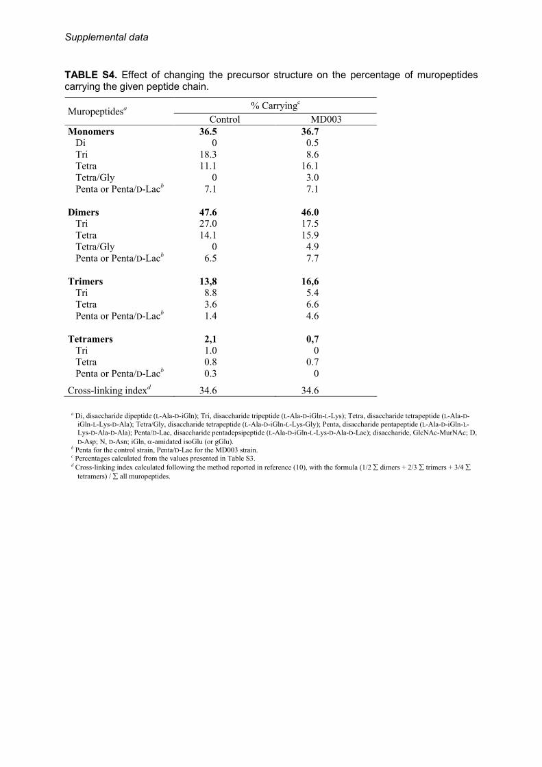
Supplemental data
TABLE S4. Effect of changing the precursor structure on the percentage of muropeptides carrying the given peptide chain.
a Di, disaccharide dipeptide (L-Ala-D-iGln); Tri, disaccharide tripeptide (L-Ala-D-iGln-L-Lys); Tetra, disaccharide tetrapeptide (L-Ala-D-
iGln-L-Lys-D-Ala); Tetra/Gly, disaccharide tetrapeptide (L-Ala-D-iGln-L-Lys-Gly); Penta, disaccharide pentapeptide (L-Ala-D-iGln-L-Lys-D-Ala-D-Ala); Penta/D-Lac, disaccharide pentadepsipeptide (L-Ala-D-iGln-L-Lys-D-Ala-D-Lac); disaccharide, GlcNAc-MurNAc; D,
D-Asp; N, D-Asn; iGln, α-amidated isoGlu (or gGlu).
b Penta for the control strain, Penta/D-Lac for the MD003 strain. c Percentages calculated from the values presented in Table S3. d Cross-linking index calculated following the method reported in reference (10), with the formula (1/2 ∑ dimers + 2/3 ∑ trimers + 3/4 ∑
tetramers) / ∑ all muropeptides.
% Carryingc
Muropeptidesa
Control MD003
Monomers 36.5 36.7
Di 0 0.5
Tri 18.3 8.6
Tetra 11.1 16.1
Tetra/Gly 0 3.0
Penta or Penta/D-Lacb 7.1 7.1
Dimers 47.6 46.0
Tri 27.0 17.5
Tetra 14.1 15.9
Tetra/Gly 0 4.9
Penta or Penta/D-Lacb 6.5 7.7
Trimers 13,8 16,6
Tri 8.8 5.4
Tetra 3.6 6.6
Penta or Penta/D-Lacb 1.4 4.6
Tetramers 2,1 0,7
Tri 1.0 0
Tetra 0.8 0.7
Penta or Penta/D-Lacb 0.3 0
Cross-linking indexd 34.6 34.6

Supplemental data
TABLE S5. Percentage of muropeptides carrying non-amidated (D-Asp) or amidated (D-Asn) cross-bridge residue as a monomer, or involved in dimers and trimersa.
% Carryingd
Muropeptidesb
Control MD003
Monomers
Tri-D 4.4 2.7
Tetra-D 4.1 8.8
Tetra/Gly-D 0 3.0
Penta-D or Penta/D-Lac-Dc 3.2 5.1
Total 11.7 19.6
Tri-N 11.3 3.0
Tetra-N 5.0 5.1
Penta-N or Penta/D-Lac-Nc 3.5 1.9
Total 21.9 10.0
Dimers
ds-D-ds-D 0.3 0.5
ds-N-ds-D 15.6 25.8
ds-N-ds-N 24.4 17.9
Trimers
ds-D-ds-D-ds-D 0 0
ds-N-ds-D-ds-D 0 0
ds-N-ds-N-ds-D 1.9 7.4
ds-N-ds-N-ds-N 11.9 9.2
a All the tetramers contained amidated aspartate (D-Asn) cross-bridge residue. bTri, disaccharide tripeptide (L-Ala-D-iGln-L-Lys); Tetra, disaccharide tetrapeptide (L-Ala-D-iGln-L-Lys-D-Ala); Tetra/Gly, disaccharide
tetrapeptide (L-Ala-D-iGln-L-Lys-Gly); Penta, disaccharide pentapeptide (L-Ala-D-iGln-L-Lys-D-Ala-D-Ala); Penta/D-Lac, disaccharide
pentadepsipeptide (L-Ala-D-iGln-L-Lys-D-Ala-D-Lac); disaccharide, GlcNAc-MurNAc; D, D-Asp; N, D-Asn; iGln, α-amidated isoGlu (or gGlu).
c Penta for the control strain, Penta/D-Lac for the MD003 strain. d Percentages calculated from the values presented in Table S3.
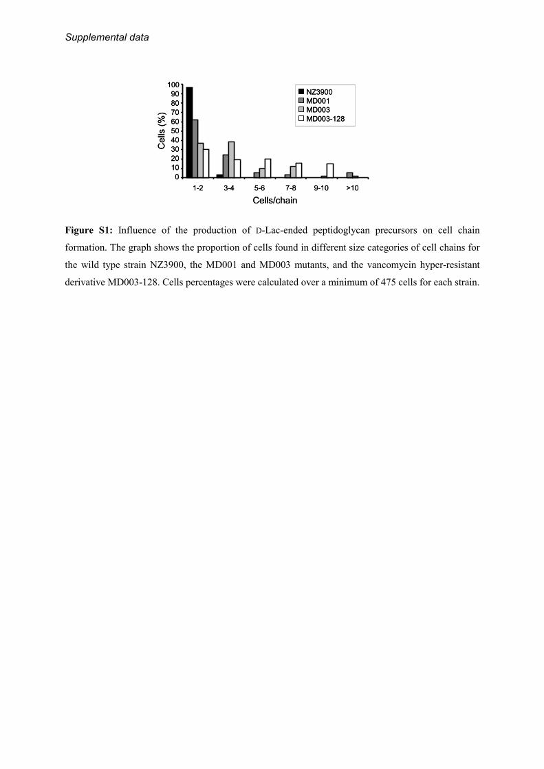
Supplemental data
Figure S1: Influence of the production of D-Lac-ended peptidoglycan precursors on cell chain
formation. The graph shows the proportion of cells found in different size categories of cell chains for
the wild type strain NZ3900, the MD001 and MD003 mutants, and the vancomycin hyper-resistant
derivative MD003-128. Cells percentages were calculated over a minimum of 475 cells for each strain.
Cells/chain
Cells
(%)
1-2 3-4 5-6 7-8 9-10 >10
0
10
20
30
40
50
60
70
80
90
100NZ3900
MD001
MD003
MD003-128
Cells/chain
Cells
(%)
1-2 3-4 5-6 7-8 9-10 >10
0
10
20
30
40
50
60
70
80
90
100NZ3900NZ3900
MD001MD001
MD003MD003
MD003-128MD003-128

Supplemental data
REFERENCES
1. de Ruyter, P. G., Kuipers, O. P., Beerthuyzen, M. M., Alen-Boerrigter, I., and de Vos, W. M.
(1996) J. Bacteriol. 178, 3434-3439
2. Courtin, P., Miranda, G., Guillot, A., Wessner, F., Mezange, C., Domakova, E., Kulakauskas, S.,
and Chapot-Chartier, M. P. (2006) J. Bacteriol. 188, 5293-5298
3. de Ruyter, P. G., Kuipers, O. P., and de Vos, W. M. (1996) Appl. Environ. Microbiol. 62, 3662-
3667
4. Hols, P., Kleerebezem, M., Schanck, A. N., Ferain, T., Hugenholtz, J., Delcour, J., and de Vos,
W. M. (1999) Nat. Biotechnol. 17, 588-592
5. Deghorain, M., Goffin, P., Fontaine, L., Mainardi, J. L., Daniel, R., Errington, J., Hallet, B., and
Hols, P. (2007) J. Bacteriol. 189, 4332-4337
6. Kuipers, O. P., de Ruyter, P. G. G. A., Kleerebezem, M., and de Vos, W. M. (1998) J.
Biotechnol. 64, 15-21
7. Goffin, P., Deghorain, M., Mainardi, J. L., Tytgat, I., Champomier-Verges, M. C., Kleerebezem,
M., and Hols, P. (2005) J. Bacteriol. 187, 6750-6761
8. Arthur, M., Molinas, C., and Courvalin, P. (1992) J. Bacteriol. 174, 2582-2591
9. van Kranenburg, R., Marugg, J. D., van Swam, I. I., Willem, N. J., and de Vos, W. M. (1997)
Mol. Microbiol. 24, 387-397
10. Glauner, B. (1988) Anal. Biochem. 172, 451-464
Marie-Pierre Chapot-Chartier, Bernard Hallet and Pascal HolsCourtin, Richard Daniel, Jeff Errington, Alexei Sorokin, Alexander Bolotin,
Marie Deghorain, Laetitia Fontaine, Blandine David, Jean-Luc Mainardi, PascalLactococcus lactisin
Functional and Morphological Adaptation to Peptidoglycan Precursor Alteration
doi: 10.1074/jbc.M110.143636 originally published online June 4, 20102010, 285:24003-24013.J. Biol. Chem.
10.1074/jbc.M110.143636Access the most updated version of this article at doi:
Alerts:
When a correction for this article is posted•
When this article is cited•
to choose from all of JBC's e-mail alertsClick here
Supplemental material:
http://www.jbc.org/content/suppl/2010/06/04/M110.143636.DC1.html
http://www.jbc.org/content/285/31/24003.full.html#ref-list-1
This article cites 40 references, 30 of which can be accessed free at
by guest on April 22, 2016
http://ww
w.jbc.org/
Dow
nloaded from
Related Documents


