Zurich Open Repository and Archive University of Zurich University Library Strickhofstrasse 39 CH-8057 Zurich www.zora.uzh.ch Year: 2015 Functional analysis of structurally diverged and reduced organelles in Giardia lamblia Rout, Samuel Posted at the Zurich Open Repository and Archive, University of Zurich ZORA URL: https://doi.org/10.5167/uzh-120991 Dissertation Published Version Originally published at: Rout, Samuel. Functional analysis of structurally diverged and reduced organelles in Giardia lamblia. 2015, University of Zurich, Faculty of Science.

Welcome message from author
This document is posted to help you gain knowledge. Please leave a comment to let me know what you think about it! Share it to your friends and learn new things together.
Transcript
Zurich Open Repository andArchiveUniversity of ZurichUniversity LibraryStrickhofstrasse 39CH-8057 Zurichwww.zora.uzh.ch
Year: 2015
Functional analysis of structurally diverged and reduced organelles inGiardia lamblia
Rout, Samuel
Posted at the Zurich Open Repository and Archive, University of ZurichZORA URL: https://doi.org/10.5167/uzh-120991DissertationPublished Version
Originally published at:Rout, Samuel. Functional analysis of structurally diverged and reduced organelles in Giardia lamblia.2015, University of Zurich, Faculty of Science.
Functional analysis of structurally diverged and reduced organelles
in Giardia lamblia
___________________________________________________________________________
Dissertation
zur
Erlangung der naturwissenschaftlichen Doktorwürde
(Dr. sc. nat.)
vorgelegt der
Mathematisch-Naturwissenschaftlichen Fakultät
der
Universität Zürich
von
Samuel Rout
aus
Bhubaneswar / India
Promotionskomitee
Prof. Dr. Adrian B. Hehl
(Vorsitz und Leitung der Dissertation)
Prof. Dr. Cornel Fraefel
Prof. Dr. Norbert Müller
Prof. Dr. Ueli Grossniklaus
Zürich, 2015
Functional analysis of structurally diverged and reduced organelles
in Giardia lamblia
________________________________________________________________________
Faculty of Science University of Zurich
Life Science Graduate School – Microbiology and Immunology PhD Program
University of Zurich
PhD Thesis
Submitted by
Samuel Rout
from Bhubaneswar, India
Thesis Supervisor:
Prof. Dr. Adrian B. Hehl
Thesis Co-supervisor:
Dr. Carmen Faso
Institute of Parasitology
Thesis Committee Members:
Prof. Dr. Cornel Fraefel
Prof. Dr. Norbert Müller
Prof. Dr. Ueli Grossniklaus
Zürich, 2015
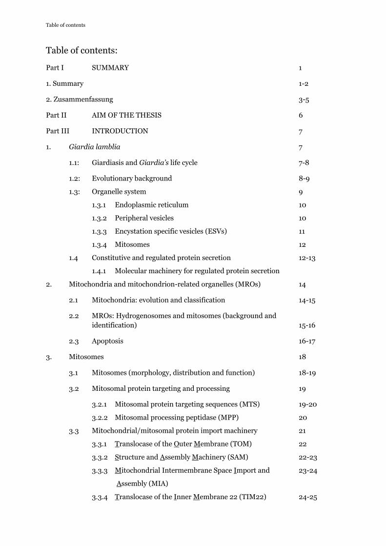
Table of contents
Table of contents:
Part I SUMMARY 1
1. Summary 1-2
2. Zusammenfassung 3-5
Part II AIM OF THE THESIS 6
Part III INTRODUCTION 7
1. Giardia lamblia 7
1.1: Giardiasis and Giardia’s life cycle 7-8
1.2: Evolutionary background 8-9
1.3: Organelle system 9
1.3.1 Endoplasmic reticulum 10
1.3.2 Peripheral vesicles 10
1.3.3 Encystation specific vesicles (ESVs) 11
1.3.4 Mitosomes 12
1.4 Constitutive and regulated protein secretion 12-13
1.4.1 Molecular machinery for regulated protein secretion
2. Mitochondria and mitochondrion-related organelles (MROs) 14
2.1 Mitochondria: evolution and classification 14-15
2.2 MROs: Hydrogenosomes and mitosomes (background and
identification) 15-16
2.3 Apoptosis 16-17
3. Mitosomes 18
3.1 Mitosomes (morphology, distribution and function) 18-19
3.2 Mitosomal protein targeting and processing 19
3.2.1 Mitosomal protein targeting sequences (MTS) 19-20
3.2.2 Mitosomal processing peptidase (MPP) 20
3.3 Mitochondrial/mitosomal protein import machinery 21
3.3.1 Translocase of the Outer Membrane (TOM) 22
3.3.2 Structure and Assembly Machinery (SAM) 22-23
3.3.3 Mitochondrial Intermembrane Space Import and 23-24
Assembly (MIA)
3.3.4 Translocase of the Inner Membrane 22 (TIM22) 24-25
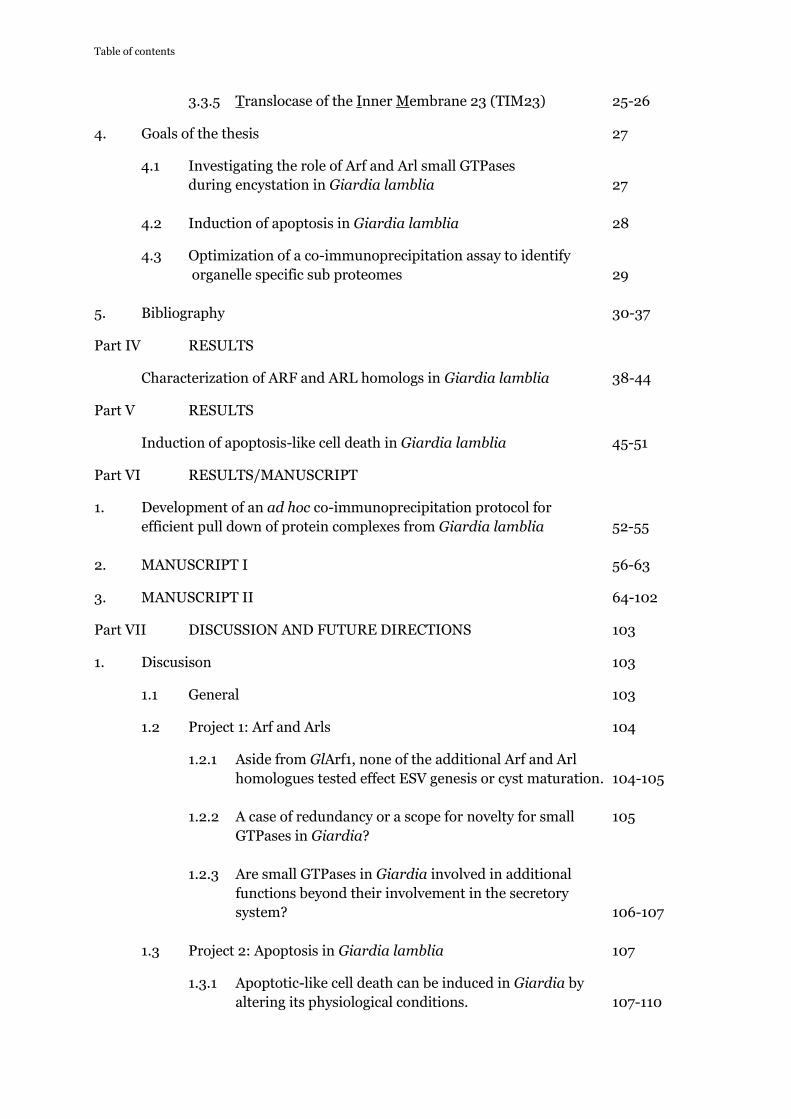
Table of contents
3.3.5 Translocase of the Inner Membrane 23 (TIM23) 25-26
4. Goals of the thesis 27
4.1 Investigating the role of Arf and Arl small GTPases
during encystation in Giardia lamblia 27
4.2 Induction of apoptosis in Giardia lamblia 28
4.3 Optimization of a co-immunoprecipitation assay to identify
organelle specific sub proteomes 29
5. Bibliography 30-37
Part IV RESULTS
Characterization of ARF and ARL homologs in Giardia lamblia 38-44
Part V RESULTS
Induction of apoptosis-like cell death in Giardia lamblia 45-51
Part VI RESULTS/MANUSCRIPT
1. Development of an ad hoc co-immunoprecipitation protocol for
efficient pull down of protein complexes from Giardia lamblia 52-55
2. MANUSCRIPT I 56-63
3. MANUSCRIPT II 64-102
Part VII DISCUSSION AND FUTURE DIRECTIONS 103
1. Discusison 103
1.1 General 103
1.2 Project 1: Arf and Arls 104
1.2.1 Aside from GlArf1, none of the additional Arf and Arl
homologues tested effect ESV genesis or cyst maturation. 104-105
1.2.2 A case of redundancy or a scope for novelty for small 105
GTPases in Giardia?
1.2.3 Are small GTPases in Giardia involved in additional
functions beyond their involvement in the secretory
system? 106-107
1.3 Project 2: Apoptosis in Giardia lamblia 107
1.3.1 Apoptotic-like cell death can be induced in Giardia by
altering its physiological conditions. 107-110
Table of contents
1.4 Project 3: Optimization of a co-immunoprecipitation assay to
identify organelle specific sub- proteomes. 110
1.4.1 A minimized mitosomal import machinery in Giardia: 110-114
reductionism at its best?
1.4.2 Diverged mitosome-ER contacts sites in Giardia? 115-116
1.4.3 Mitosome dynamics and a novel role for Giardia
dynamin related protein in mitosome morphogenesis. 116-119
2. Bibliography 119-123
PART VIII CONCLUSION 124-125
Acknowledgement 126
Curriculum Vitae 127-128
Part I: SUMMARY
PART I: SUMMARY
1. Summary
The proliferating trophozoite stage of the non-invasive protozoan parasite Giardia
lamblia (syn. G. intestinalis and G. duodenalis) is a highly polarized motile cell and a
tractable laboratory model organism to investigate fundamental cell biological questions. G.
lamblia belongs to the phylum Diplomonadida (kingdom Excavata), which was, until recently
one of the most extant branches of eukaryotic evolution. G. lamblia is the leading causative
agent for water borne parasite induced diarrheal disease worldwide. It not only affects
humans and animals but also causes significant morbidity and economic loss in the livestock
industry. Giardia has a two stage asexual life cycle comprising of a proliferating binucelate
tropozoite stage and an environmentally resistant cyst stage. Proliferating trophozoites
actively attach to gut epithelia with the help of a specialized ventral suction disc to avoid
elimination via peristalsis. Moreover, cells exhibit antigenic variation of their protein surface
coat to evade immuno-mediated clearing by the host, leading to chronicization of Giardia
infections. However, upon encountering changes in lipid concentration and increase in pH,
resident trophozoites undergo complex stage differentiation and transform into an
environmentally resistant cyst form which is shed with the feces. This process is defined as
“encystation”.
The simplification in cellular complexity observed in Giardia can be interpreted as a result of
massive reductive evolution as an adaptation to its ecological niche and parasitic lifestyle.
The endomembrane system in G. lamblia is of prime example and may serve as a model to
study protein trafficking. Transmission to a new host demands stage conversion from
trophozoite to cyst form. The encystation process requires transport of cyst wall proteins
(CWPs) from the endoplasmic reticulum (ER) to the plasma membrane (PM) to form the
protective extracellular matrix. Despite clear evidence for secretory activity, Giardia lacks a
canonical steady state Golgi apparatus. However, upon the triggering of encystation, de novo
generated encystation specific vesicles (ESVs) act as Golgi analogs by accumulating,
processing and sorting CWPs prior to regulated secretion. ESV neogenesis and maturation
depends on small GTPases such as GlSar1 and GlArf1. Over-expression of mutated GlSar1
and GlArf1 variants eliciting dominant negative effects impaired ESV genesis and maturation,
respectively. In the first part of my doctoral thesis, I investigated the role of 5 additional Arf
and Arl homologues by defining their cellular localization and possible function during
encystation. Our data confirms that Arf1 is likely the only family member involved in
encystation. The other homologues may either be redundant in relation to Arf1 or be involved
in other as-yet unidentified cellular process. New insights and/or novel tools are required to
obtain a more comprehensive view of Arf and Arl functions in G. lamblia.
1
Part I: SUMMARY
Encystation is a necessary mechanism for parasite transmission and may be viewed as an
escape route for Giardia when faced with unfavorable conditions within the host. However,
during infection, Giardia trophozoites reside at high densities in the host’s small intestine.
Surprisingly little inflammation and/or damage to epithelial cells is observed. One
explanation could reside in a form of programmed cell death (PCD) which limits the
stimulation of the immune system by deteriorating parasites. Therefore, in the second part of
my doctoral thesis, I investigated PCD-related events in Giardia using nutrient starvation
and heat shock treatment as physiological triggers. Exposure of phosphatidylserine (PS), ER
disintegration, nuclear condensation and DNA damage are hallmarks of Giardia’s form of
PCD, suggesting that this parasite harbors machinery for an apoptotic like cell death. Since
apoptosis in more complex eukaryotes is linked to mitochondrial function, we speculated that
Giardia’s mitosomes may be involved. These organelles, similarly to mitochondria, might
function as a highly sensitive central switch for monitoring cell health and integrating
possible death signals from within and without, beyond their established role in iron-sulfur
(Fe-S) protein maturation. However, to address this fundamental biological question and to
dissect the full range of mitosomal function, a comprehensive mitosomal proteome is
essential.
Despite indications of roles for giardial mitosomes beyond Fe-S maturation, little is known
regarding the protein repertoire of of these organelles, primarily due to 1) significant genomic
sequence divergence and 2) challenges in organelle purification. A case in point is the
organelle’s protein import machinery where only a poorly conserved translocon of the outer
membrane (GlTom40) has been identified to date. Therefore, during the last part of my
doctoral thesis I developed a customized co-immunoprecipitation (co-IP) assay to pull down
organelle-specific protein complexes with high efficiency. Using GlTom40 as bait protein,
this strategy led to the identification of 10 novel mitosomal localized proteins of unknown
function. Furthermore, reverse co-IP strategies using five novel mitosome-localized bait
proteins allowed for the building of a core membrane interactome and a complex interactome
network extending inwards to the organelle matrix as well as outwards to components of the
ER membrane and the cytoplasm. Our findings point towards a simplified outer membrane
import machinery in giardial mitosomes involving the translocon (GlTom40), a Giardia-
specific receptor (Gl29147) and a membrane anchored protein (Gl14939). In addition, we
propose the presence of a diverged mitosome-ER contact site based on the identification of 5
novel hypothetical proteins with dual localization at the ER and mitosomes. Furthermore, we
show for the first time association of the single giardial dynamin related protein with
mitosomes and provide direct evidence for its involvement in organelle morphogenesis and
homeostasis.
2
Part I: SUMMARY
2. Zusammenfassung
Das proliferative Trophozoiten-Stadium des nicht-invasiven, einzelligen Parasiten
Giardia lamblia (syn. G. intestinalis und G. duodenalis) ist eine stark polarisierte, motile
Zelle und ein geeigneter Modellorganismus zur Erforschung von zellbiologischen
Grundlagen. G. lamblia gehört zum Stamm der Diplomonadida (Excavata), welche bis vor
kurzem eine der einfachsten Gruppen der eukaryotischen Evolution darstellten. G. lamblia
ist weltweit der Hauptverursacher für Parasiten-induzierte Durchfallerkrankungen durch
verunreinigtes Wasser. Dies führt nicht nur zur gesundheitlichen Beeinträchtigung von
Mensch und Tier, sondern hat auch signifikante Morbidität und wirtschaftliche Verluste in
der landwirtschaftlichen Tierhaltung zur Folge. Der zwei Stadien umfassende asexuelle
Lebenszyklus von Giardia besteht aus einem sich vermehrenden zweikernigen Trophozoiten-
und einem gegen Umwelteinflüssen resistenten Zysten-Stadium. Erstere heften sich aktiv an
Dünndarmepithelzellen des Wirtes mit Hilfe einer spezialisierten Bauchhaftscheibe, um ihrer
Beseitigung durch peristaltische Bewegungen entgegenzuwirken. Darüber hinaus erlaubt die
Fähigkeit des Parasiten, seine Oberflächenantigene kontinuierlich zu verändern, sich der
Immunabwehr des Wirtes zu entziehen. Dies hat eine erfolgreiche Persistenz der Krankheit
zur Folge. Trotz der umfassenden Möglichkeiten von Giardia, sich dem Abwehrmechanismus
des Wirtes zu entziehen, ist der Einzeller nicht immun gegen Erhöhung von pH und
Gallenkonzentration. Unter solch ungünstigen Veränderung unterzieht sich der Parasit einer
Stadiums-Differenzierung und transformiert sich in die umweltresistente Zystenform, welche
mit dem Stuhl ausgeschieden wird. Dieser Prozess nennt sich „Enzystierung“.
Die Vereinfachung der zellulären Komplexität, welche bei Giardia beobachtet wird, kann als
Resultat von massiv reduktiver Evolution als Folge von Adaptation an seine ökologische
Nische und seine parasitische Lebensweise interpretiert werden. Das Endomembransystem
in G. lamblia ist ein erstklassiges Beispiel und dient als Model zur Erforschung von
intrazellulärem Proteintransport. Die Übertragung des Parasiten von einem Wirt zum
nächsten erfordert die oben genannte Stadiumskonversion von Trophozoit zur Zyste, die
Enzystierung. Dieser Prozess benötigt den Transport von Zystenwand-Material (CWM) vom
endoplasmatischen Retikulum (ER) zur Plasmamembran (PM), um die schützende
extrazelluläre Matrix zu bilden. Obwohl klare Beweise für eine gewisse sekretorische Aktivität
vorliegen, hat Giardia keinen klassischen Golgi-Apparat. Wie dem auch sei, während der
Einleitung der Enzystierung fungieren de novo generierte, enzystierungsspezifische Vesikel
(ESV) als Golgi-Analoge aufgrund ihrer Funktion zur Akkumulierung, Prozessierung und
Sortierung von CWM vor der regulierten Sekretion. Neogenese und Maturation von ESV sind
abhängig von kleinen GTPasen wie GlSar1 und GlArf1. Überexpression von mutiertem GlSar1
und GlArf1 resultieren in dominant negativen Effekten, wie beeinträchtigte Neogenese resp.
Reifung von ESV. Im ersten Teil meiner Doktorarbeit erforschte ich die Funktion von fünf
3
Part I: SUMMARY
zusätzlichen Arf und Arl Homologen, indem ich ihre zelluläre Lokalisation und mögliche
Rolle während der Enzystierung untersuchte. Unsere Daten bestätigen, dass Arf1 sehr
wahrscheinlich das einzige Mitglied der Arf-Familie ist, welches bei dem Prozess eine
Funktion innehat. Die anderen Homologen sind möglicherweise redundant in Bezug zu Arf1,
oder sie sind in einem bisher unbekannten zellulären Prozess involviert. Weitere
Erkenntnisse und/oder neue Instrumente sind vonnöten, um eine umfassendere Einsicht in
die Funktion von Arf und Arl in Giardia lamblia zu erlangen.
Enzystierung ist ein notwendiger Mechanismus für die Transmission von Parasiten, und
kann im Falle von ungünstigen Bedingungen im Wirt als Fluchtroute von Giardia betrachtet
werden. Während einer Infektion besiedeln Giardia Trophozoiten den Dünndarm in hoher
Konzentration, obwohl nur schwache Inflammationsanzeichen und/oder Schäden an Zellen
des Darmepithels beobachtet werden. Eine Erklärung hierfür könnte eine Form von
programmiertem Zelltod (PCD) sein, welche eine Immunantwort gegen die Parasiten dämpft.
Um diese These zu prüfen, untersuchte ich im zweiten Teil meiner Doktorarbeit PCD-
bezogene Ereignisse in Giardia. Hierfür benutzte ich Nährstofflimitation und Hitzeschock als
physiologische Auslöser. Externalisierung von Phosphatidylserin (PS), ER-Zerfall,
Kernkondensation und DNA-Schäden sind Kennzeichen der PCD-Form von Giardia, was auf
eine Maschinerie des Parasiten hinweist, die für eine Apoptose-ähnliche Form von Zelltod
verantwortlich ist. Die Tatsache, dass Apoptose in komplexeren Eukaryoten mitochondriale
Funktionen beinhaltet, lässt uns vermuten, dass auch Mitosomen von Giardia in diesen
Prozess involviert sind. Diese Organellen, die Ähnlichkeiten zu Mitochondrien aufweisen,
könnten nebst ihrer Rolle in der Fe-S Proteinreifung als hochsensitiven, zentralen Schalter
zur Überwachung vom Gesundheitszustand der Zelle und von möglichen Zelltod-Auslöser
intra- oder extrazellulären Ursprungs fungieren. Um diese fundamentale biologische Frage
zu untersuchen und somit sämtliche Funktionen von Mitosomen aufzudecken, ist ein
umfassendes mitosomales Proteom notwendig.
Nebst ihrer Rolle in der Fe-S Proteinmaturation ist wenig bekannt über das Proteinrepertoire
in Mitosomen. Dies hat einerseits zu tun mit signifikanter Divergenz genomischer
Sequenzen, andererseits mit Schwierigkeiten bezüglich der Aufreinigung von Organellen. Ein
Beispiel hierfür ist die mitosomale Proteinimport-Maschinerie, bei welcher bisher erst ein
schwach konserviertes Translocon der äusseren Membran (GlTom40) identifiziert werden
konnte. Aufgrund dessen entwickelte ich im letzten Teil meiner Doktorarbeit ein auf Giardia
angepasstes co-Immunpräzipitations (co-IP) Assay, um organellenspezifische
Proteinkomplexe mit hoher Effizienz zu isolieren. Durch die Verwendung von GlTom40 als
bait-Protein konnten dadurch zehn neue mitosomale Proteine identifiziert werden, deren
Funktion noch unbekannt ist. Zusätzlich wurden fünf neue mitosomale bait-Proteine für eine
inverse co-IP-Strategie verwendet, was einerseits zu einem Membran-Interaktom und
4
Part I: SUMMARY
andererseits zu einem complexen Interaktom führte, welches sich zusätzlich nach innen zur
Organellenmatrix als auch nach aussen zu Teilen der ER-Membran und zum Cytoplama
erstreckt. Unsere Resultate führen zu einer vereinfachten Proteinimport-Maschinerie der
äusseren Mitosomenmembran in Giardia, welche das Translocon (GlTom40), einen Giardia-
spezifischen Rezeptor (Gl29147) und ein membranverankertes Protein (Gl14939) enthält.
Zusätzlich stellen wir aufgrund fünf neu identifizierter, hypothetischer Proteine mit dualer
Lokalisation in ER und Mitosomen die Hypothese auf, dass eine divergierende Mitosom-ER-
Kontaktseite existiert. Wir konnten auch das erste Mal die Verbindung zwischen dem
einzigen Protein in Giardia, welches mit Dynamin verwandt ist, und Mitosomen zeigen. Dies
stellt einen direkten wissenschaftlichen Beweis für die Beteiligung des Proteins in
Morphogenese und Homöostase von Organellen dar.
5
Part II: AIM OF THE THESIS
Part II: AIM OF THE THESIS
Giardia lamblia has a very simple life cycle comprising of a motile trophozoite stage
that colonizes the host’s small intestine leading to the manifestation of the disease and an
environmentally resistant cyst form. The stage conversion is crucial for parasite survival
outside the host. Giardia trophozoites display a highly minimized compartment organization
which is not only functionally but also structurally reduced.
In absence of a steady state Golgi apparatus, de novo generated specialized organelles,
encystation specific vesicles (ESVs) function as a Golgi body analog where the cyst wall
material is sorted and matured prior to sequential deposition on the parasite surface forming
the resistant cyst wall. Regulated CWP trafficking is essential for the formation of the cyst
wall and in turn survival of the parasite, hence it is an essential stage in parasite
transmission. Additionally, Giardia lacks canonical mitochondria and possesses
mitochondrion-related organelles (mitosomes) which are implicated in iron-sulfur protein
maturation. Despite unambiguous evidence for functionally conserved protein import
machinery, there is massive divergence in the structural components involved in the
machinery.
The minimized organism, G. lamblia, because of its simple organization, provides a platform
for investigation of basic cellular functions and pathways which are difficult to study in
complex eukaryotes. Furthermore, it is also a useful model system to investigate principles of
reductive evolution, i.e. why and how adoption of a parasitic life-style leads to the loss of even
archetypical sub-cellular organelles, and which minimal machinery is necessary for
maintenance of fundamental cellular functions.
Therefore, the aims of my doctoral thesis were to investigate the membrane associated
factors involved in ESV maintenance and to identify the repertoire of mitosomal proteins
involved in the import machinery and facilitating inter-organellar communications which
would help us to unravel the functional range of these highly diverged organelles.
6
PART III: INTRODUCTION
Part III: INTRODUCTION
1 Giardia lamblia
1.1 Giardiasis and Giardia’s life cycle
Giardia lamblia (syn. G. intestinalis and G. duodenalis) is a protozoan parasite and is the
leading causative agent for non-bacterial water borne diarrhea worldwide. The discovery of
this non-invasive intestinal parasite dates back to 1681 by Dutch microscopist Anthony Van
Leeuwenhoek while he was analyzing his own stool samples. The parasite has a broad
vertebrate host range and causes significant morbidity and economic loss [1, 2]. Despite this
impact, giardiasis is a poorly understood disease and was included in the “WHO neglected
disease initiative” in 2004 [3]. Giardiasis is highly prevalent in developing countries ranging
from 20-30% as compared to developed countries, 2-7% [4]. The etiology of diarrhea in
giardiasis is thought to be a leak flux mechanism as a result of compromised epithelial barrier
function. The flagellated parasite causes acute and chronic diarrhea accompanied by nausea,
abdominal pain, weight loss and malabsorption. Although 5- nitroimidazole compounds are
used for treatment of giardiasis, treatment failures have been documented resulting in
chronic diarrhea in immunocompromised individuals and children [5, 6].
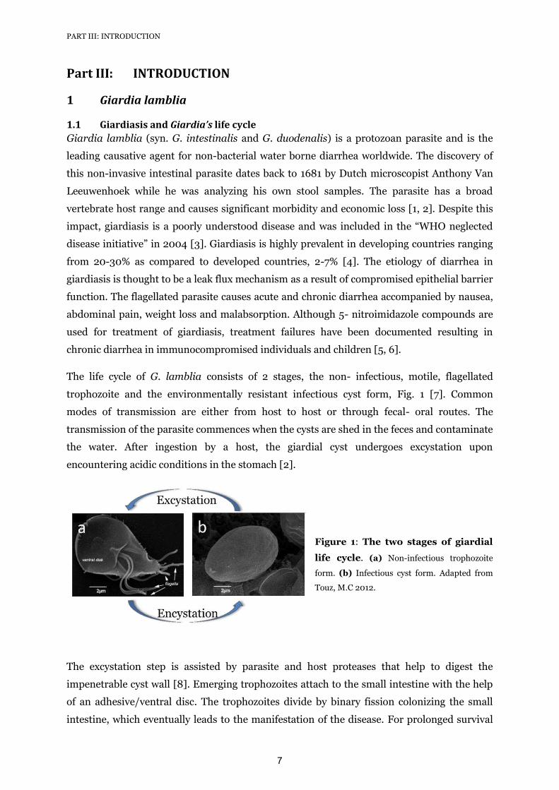
The life cycle of G. lamblia consists of 2 stages, the non- infectious, motile, flagellated
trophozoite and the environmentally resistant infectious cyst form, Fig. 1 [7]. Common
modes of transmission are either from host to host or through fecal- oral routes. The
transmission of the parasite commences when the cysts are shed in the feces and contaminate
the water. After ingestion by a host, the giardial cyst undergoes excystation upon
encountering acidic conditions in the stomach [2].
Figure 1: The two stages of giardial
life cycle. (a) Non-infectious trophozoite
form. (b) Infectious cyst form. Adapted from
Touz, M.C 2012.
The excystation step is assisted by parasite and host proteases that help to digest the
impenetrable cyst wall [8]. Emerging trophozoites attach to the small intestine with the help
of an adhesive/ventral disc. The trophozoites divide by binary fission colonizing the small
intestine, which eventually leads to the manifestation of the disease. For prolonged survival
7
PART III: INTRODUCTION
within the host the trophozoites exhibit antigenic variation of their protein surface coat to
evade clearing by the immune system.
Upon encountering changes in lipid concentration and increase in pH, trophozoites undergo
a complex stage differentiation process (encystation) to cysts which are then shed in the
environment by the host. However, the exact stimulus/stimuli that trigger encystation are
still unknown. The infection cycle is completed when the cysts are ingested by another host.
The complete life‐cycle including cyst formation and excystation (mimicking infection of a
new host after peroral uptake of cysts) can be reproduced in vitro [9].
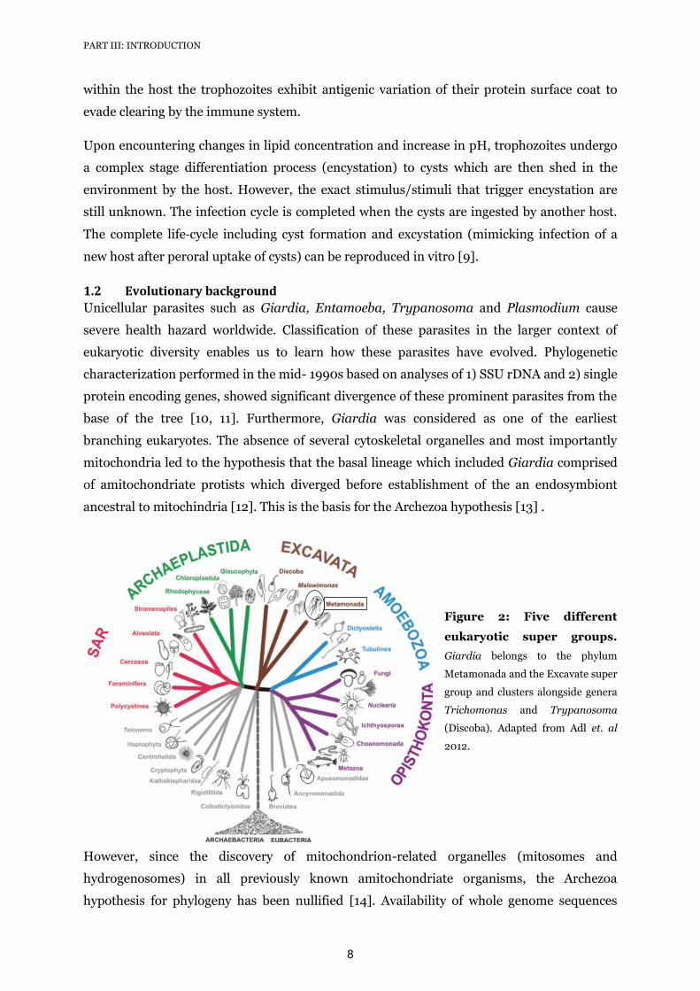
1.2 Evolutionary background
Unicellular parasites such as Giardia, Entamoeba, Trypanosoma and Plasmodium cause
severe health hazard worldwide. Classification of these parasites in the larger context of
eukaryotic diversity enables us to learn how these parasites have evolved. Phylogenetic
characterization performed in the mid- 1990s based on analyses of 1) SSU rDNA and 2) single
protein encoding genes, showed significant divergence of these prominent parasites from the
base of the tree [10, 11]. Furthermore, Giardia was considered as one of the earliest
branching eukaryotes. The absence of several cytoskeletal organelles and most importantly
mitochondria led to the hypothesis that the basal lineage which included Giardia comprised
of amitochondriate protists which diverged before establishment of the an endosymbiont
ancestral to mitochindria [12]. This is the basis for the Archezoa hypothesis [13] .
Figure 2: Five different
eukaryotic super groups.
Giardia belongs to the phylum
Metamonada and the Excavate super
group and clusters alongside genera
Trichomonas and Trypanosoma
(Discoba). Adapted from Adl et. al
2012.
However, since the discovery of mitochondrion-related organelles (mitosomes and
hydrogenosomes) in all previously known amitochondriate organisms, the Archezoa
hypothesis for phylogeny has been nullified [14]. Availability of whole genome sequences
8
PART III: INTRODUCTION
from diverged eukaryotes and phylogenetic analyses combined with comparative
ultrastructural data in the last decade has revolutionized our view of eukaryotic evolution,
leading to the clustering of organisms in 5 major phylogenetic groups, Fig. 2 [15]. G. lamblia
belongs to the Excavata super group and clusters along with genera Trichomonas and
Trypanosoma. The new phylogenetic tree clusters protozoan parasites in three eukaryotic
super groups; Excavate, Amoebozoa and SAR which are separated by 2 non- parasitic super
groups, Archaeplastida and Ophisthokonta, This suggests that the parasitic life style evolved
independently on separate occasions. Therefore, the presence of diverged parasitic life style
and analogous structures having similar function in organisms in different parasitic taxa are
a result of convergent evolution.
Interestingly, the modern era un-rooted phylogenetic tree reveals that the last eukaryotic
common ancestor (LECA) was likely a eukaryote harboring all essential organelles (including
mitochondria and stacked Golgi). Therefore the secondary loss of such organelles via
reductive evolution in basal eukaryotes is a consequence of parasitic life style.
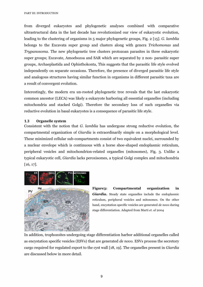
1.3 Organelle system
Consistent with the notion that G. lamblia has undergone strong reductive evolution, the
compartmental organization of Giardia is extraordinarily simple on a morphological level.
These minimized cellular sub-compartments consist of two equivalent nuclei, surrounded by
a nuclear envelope which is continuous with a horse shoe-shaped endoplasmic reticulum,
peripheral vesicles and mitochondrion-related organelles (mitosomes), Fig. 3. Unlike a
typical eukaryotic cell, Giardia lacks peroxisomes, a typical Golgi complex and mitochondria
[16, 17].
Figure3: Compartmental organization in
Giardia. Steady state organelles include the endoplasmic
reticulum, peripheral vesicles and mitosomes. On the other
hand, encystation specific vesicles are generated de novo during
stage differentiation. Adapted from Marti et. al 2004
In addition, trophozoites undergoing stage differentiation harbor additional organelles called
as encystation specific vesicles (ESVs) that are generated de novo. ESVs process the secretory
cargo required for regulated export to the cyst wall [18, 19]. The organelles present in Giardia
are discussed below in more detail.
9
PART III: INTRODUCTION
1.3.1 Endoplasmic reticulum
In a eukaryotic cell the secretory system comprises of the ER, Golgi apparatus and the
secretory vesicles. Giardia harbors a labyrinthine tubular vesicular network (TVN), studded
with ribosomes, reminiscent of the rough ER. Giardia’s ER extends from the peri nuclear
region towards the cell periphery, spanning the entire cell body [20]. Despite the
omnipresence of ER in the cytoplasmic space, the ER does not permeate the space occupied
by PVs [21]. Giardia ER was identified by localization studies using 3 known ER markers: 1)
immunoglobulin heavy chain-binding protein (BiP), 2) the 3 protein disulphide isomerases
(PDI) and 3) acid phosphatase [20, 22-25]. Furthermore, giardial ER possesses conserved
machinery for co-translational import of secreted proteins into the organelle’s lumen and
necessary chaperones such as PDIs, heat shock protein 70 (Hsp70) Binding Protein BiP, and
peptidyl-prolyl cis-trans isomerases to facilitate proper protein folding [26]. However,
Giardia lacks the machinery for the addition of N-linked glycans to secreted proteins.
Enzymes that add mannose and glucose to a dolichol precursor are also absent. Therefore, N-
glycosylation of proteins is restricted to the addition of 1 or 2 GlcNAc to asparagine which are
not further modified during secretory transport [27]. In line with this, Giardia lacks an ER
based N-glycan dependent quality control machinery for protein folding and degradation
[28]. However Giardia ER in the absence of a steady state Golgi apparatus is the cornerstone
organelle for both exocytosis and endocytosis. It has been regarded as a pluripotent
compartment either facilitating direct secretion of proteins to destination organelles/plasma
membrane (exocytosis) or helping in catalysis/processing of endocytosed material from the
extracellular milieu by peripheral vesicles [18, 29].
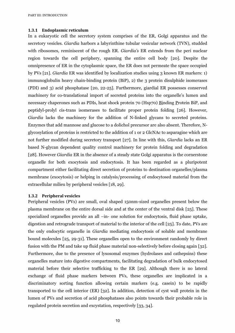
1.3.2 Peripheral vesicles
Peripheral vesicles (PVs) are small, oval shaped 150nm-sized organelles present below the
plasma membrane on the entire dorsal side and at the center of the ventral disk [25]. These
specialized organelles provide an all –in- one solution for endocytosis, fluid phase uptake,
digestion and retrograde transport of material to the interior of the cell [25]. To date, PVs are
the only endocytic organelle in Giardia mediating endocytosis of soluble and membrane
bound molecules [25, 29-31]. These organelles open to the environment randomly by direct
fusion with the PM and take up fluid phase material non-selectively before closing again [32].
Furthermore, due to the presence of lysosomal enzymes (hydrolases and cathepsins) these
organelles mature into digestive compartments, facilitating degradation of bulk endocytosed
material before their selective trafficking to the ER [29]. Although there is no lateral
exchange of fluid phase markers between PVs, these organelles are implicated in a
discriminatory sorting function allowing certain markers (e.g. casein) to be rapidly
transported to the cell interior (ER) [32]. In addition, detection of cyst wall protein in the
lumen of PVs and secretion of acid phosphatases also points towards their probable role in
regulated protein secretion and excystation, respectively [33, 34].
10
PART III: INTRODUCTION
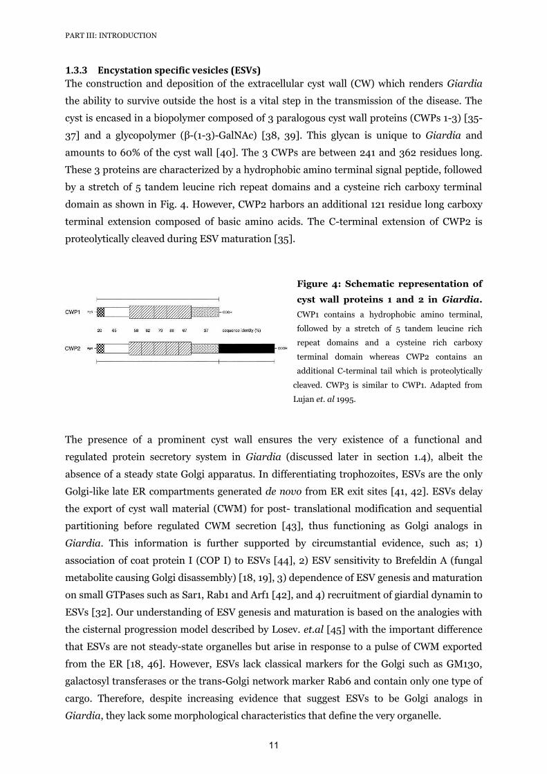
1.3.3 Encystation specific vesicles (ESVs)
The construction and deposition of the extracellular cyst wall (CW) which renders Giardia
the ability to survive outside the host is a vital step in the transmission of the disease. The
cyst is encased in a biopolymer composed of 3 paralogous cyst wall proteins (CWPs 1-3) [35-
37] and a glycopolymer (β-(1-3)-GalNAc) [38, 39]. This glycan is unique to Giardia and
amounts to 60% of the cyst wall [40]. The 3 CWPs are between 241 and 362 residues long.
These 3 proteins are characterized by a hydrophobic amino terminal signal peptide, followed
by a stretch of 5 tandem leucine rich repeat domains and a cysteine rich carboxy terminal
domain as shown in Fig. 4. However, CWP2 harbors an additional 121 residue long carboxy
terminal extension composed of basic amino acids. The C-terminal extension of CWP2 is
proteolytically cleaved during ESV maturation [35].
Figure 4: Schematic representation of
cyst wall proteins 1 and 2 in Giardia.
CWP1 contains a hydrophobic amino terminal,
followed by a stretch of 5 tandem leucine rich
repeat domains and a cysteine rich carboxy
terminal domain whereas CWP2 contains an
additional C-terminal tail which is proteolytically
cleaved. CWP3 is similar to CWP1. Adapted from
Lujan et. al 1995.
The presence of a prominent cyst wall ensures the very existence of a functional and
regulated protein secretory system in Giardia (discussed later in section 1.4), albeit the
absence of a steady state Golgi apparatus. In differentiating trophozoites, ESVs are the only
Golgi-like late ER compartments generated de novo from ER exit sites [41, 42]. ESVs delay
the export of cyst wall material (CWM) for post- translational modification and sequential
partitioning before regulated CWM secretion [43], thus functioning as Golgi analogs in
Giardia. This information is further supported by circumstantial evidence, such as; 1)
association of coat protein I (COP I) to ESVs [44], 2) ESV sensitivity to Brefeldin A (fungal
metabolite causing Golgi disassembly) [18, 19], 3) dependence of ESV genesis and maturation
on small GTPases such as Sar1, Rab1 and Arf1 [42], and 4) recruitment of giardial dynamin to
ESVs [32]. Our understanding of ESV genesis and maturation is based on the analogies with
the cisternal progression model described by Losev. et.al [45] with the important difference
that ESVs are not steady-state organelles but arise in response to a pulse of CWM exported
from the ER [18, 46]. However, ESVs lack classical markers for the Golgi such as GM130,
galactosyl transferases or the trans-Golgi network marker Rab6 and contain only one type of
cargo. Therefore, despite increasing evidence that suggest ESVs to be Golgi analogs in
Giardia, they lack some morphological characteristics that define the very organelle.
11
PART III: INTRODUCTION
1.3.4 Mitosomes
Mitosomes are mitochondrion-related organelles (MROs) and are the simplest form of
mitochondria. These organelles are devoid of organellar genome and are incapable of
generating ATP via oxidative phosphorylation. Consequently, iron- sulfur protein maturation
in the only metabolic pathway currently associated to these organelles. Since mitosomes are
the main focus of my doctoral thesis, these reduced organelles are discussed in more detail in
the next chapter.
1.4 Constitutive and regulated protein secretion
Until now two export pathways (constitutive and regulated) have been identified in Giardia
responsible for trafficking proteins directly from ER either to the PM or to the ESVs [18, 47,
48].
The constitutive protein secretory pathway is mainly responsible for secretion of 3 kinds of
proteins in trophozoites 1) proteins harboring a predicted signal sequence, 2) cysteine rich
non-variable proteins and 3) variant surface membrane proteins (VSPs) [21]. VSPs are
transmembrane anchored surface proteins that cover the whole trophozoite, providing a
protective layer [49, 50]. VSPs are targeted to the PM via the conserved C-terminal CRGKA
tail [18]. Although VSPs are trafficked directly from the ER to the PM [21, 47, 48], VSP
trafficking is sensitive to brefeldin A [19] indicating that VSP trafficking could be via COP I
derived vesicles. However such vesicles are not yet identified in Giardia trophozoites. Only
one out of the predicted 235- 275 VSPs coats the parasite membrane at a time [51]. VSPs are
composed of cysteine rich exo-domains which are released as soluble antigens into the
environment (VSP switching) facilitating antigenic variation of the surface coat thereby
helping the parasite evade the host immune system [52-54]. VSP switching/turnover
happens on average every 6-13 generations [55] and is regulated by RNA interference [56,
57]. Furthermore, apart from the approximately 300 VSPs, another 500 cysteine rich non-
variable proteins and proteins with a predicted signal sequence are targeted to the PM via the
constitutive secretory pathway [58].
The most distinctive secretion process is Giardia is however during encystation where the
CWM is packed in de novo generated ESVs, sorted, partitioned and deposited on the plasma
membrane [18, 19, 42]. CWPs are initially sorted from constitutively secreted proteins
already at ER exit sites [18, 41] and packed into ESVs prior to their regulated secretion. The
encystation process can be studied in vitro via several methods; however the two-step
encystation protocol is mostly preferred amongst all. This method is based on bile
deprivation for 48 hours followed by subsequent increase in pH and porcine bile [59, 60] .
The expression of CWM is stage specifically induced and the mRNA levels peak at 7 hours
post induction of encystation (hpie) [43]. The whole process of encystation lasts
approximately 20- 24 hours [46]. Briefly, at 2 hpie, CWM starts to appear at the ER, followed
12
PART III: INTRODUCTION
by the first round of sorting at the transitional ER where CWM is sorted from the constitutive
cargo. The CWM leaves the ER in a COP II dependent manner [41]. Subsequently the de novo
generated ESVs accumulate CWM and increase in size. CWM is delayed in ESVs allowing for
post translational modification of CWPs [43]. Specifically the 121 residue long carboxy
terminal extension of CWP2 undergoes proteolytic cleavage generating CWP2 (ΔN) and
CWP2 (ΔC) fragments. Around 8-12 hpie the CWM undergoes selective condensation where
the CWP3 and the C-terminal portion of CWP2 (ΔC) form the condensed core in the ESV
while CWP1 and the N-terminal portion of CWP2 (ΔN) form an outer fluid phase. The fluid
phase circulates between ESVs most likely via the ER [42]. 16-20 hpie marks the second
sorting event where the fluid phase is sorted away from the condensed core near the cell
periphery forming 2 different compartments. Subsequently the fluid phase is secreted first,
forming the outer layer of the cyst wall. The condensed core undergoes de-condensation and
is secreted slowly over hours, forming the inner layer [42].
1.4.1 Molecular machinery for regulated protein secretion
Due to reductive evolution Giardia has lost most of the molecular machinery required for
protein secretion including the Golgi apparatus which is considered the hub of the secretory
pathway. As mentioned above, ESVs act as Golgi analogs although they differ substantially
both structurally and biochemically when compared to a canonical Golgi.
Despite these differences, ESVs are sensitive to brefeldin A (a fungal metabolite that inhibits
Arf 1 and in turn causes Golgi disassembly in higher eukaryotes). Furthermore, proteins and/
or factors required for budding and fusion of transport vesicles such as coatomer proteins
(COP I, COP II), clathrin heavy chain, two adaptor proteins (AP1 and AP2/3) and small
GTPases such as Arf1 which are involved in COP I coated vesicles, Rabs and Sar1 have been
identified in the Giardia genome [61, 62].
In addition, 7 SNARE (soluble N-ethylmalemide- sensitive factor attachment protein
receptors) proteins have been also identified [44, 63]. Interestingly, it was demonstrated that
the ER resident chaperone Hsp70/Bip was retrieved back to the ER from ESV via KDEL
sequence suggesting COP I based retrograde protein trafficking. Furthermore identification
of proteasomal components during the early stages on encystation on ESV membranes points
towards a quality control step associated with a degradation process [64]. Taken together,
identification of all these proteins associated with ESVs makes these organelles stage
specifically regulated Golgi analogs and hints towards minimum machinery capable of
regulated secretion in Giardia lamblia.
13
PART III: INTRODUCTION
2: Mitochondria and mitochondrion-related organelles (MROs)
2.1 Mitochondria: evolution and classification
Mitochondria are organelles found in virtually all eukaryotic cells and function at the
crossroads of life and cell death. These organelles are not only the energy source of the cell
capable of performing a myriad of essential biochemical reactions but are also the key
triggers for apoptosis [65-69]. Based on the well-established endosymbiotic theory, the
mitochondrion was once a free-living prokaryote which was maintained as an organelle after
being engulfed by a eukaryotic cell [70-74]. This theory is corroborated by the level of
biochemical and physiological similarity of mitochondria to prokaryotic cells [13, 75-77].
Martin et. al proposed that the ability of the α-proteobacterium to generate free energy in
form of ATP and the inability of the host (eukaryotic ancestor) to do so might have been the
reason that drove the endosymbiosis event [78]. However phylogenetic analysis of the
mitochondrial ADP/ATP transporter does not support this hypothesis by placing the origin of
the transporter post endosymbiosis [79]. Therefore, alternative theories on the nature of the
driving force for the endosymbiosis event were proposed such as 1) the hydrogen hypothesis
[78], 2) The syntrophic hypothesis [80] and 3) The ox-tox hypothesis [72]. Van der Giezen et.
al have well documented the biochemical drivers for the above proposed hypotheses [81].
Regardless, the presence of mitochondrial DNA substantiates the endosymbiosis theory.
Phylogenetic analysis of mt-DNA and mt-rRNA sequences links the origin of mitochondria to
α-proteobacteria, more specifically to the order Rickettsiales [82, 83].
Following the endosymbiosis event, evolution of the eukaryotic lineage has led to 5 super-
groups based on phylogenetic analyses [15]. The presence of mitochondria or MROs is
ubiquitous in all [15]. Several studies provide evidence that the diversified MROs evolved
from the mitochondrial ancestor under selection by environmental habitats of the host
organisms [84]. Previously, eukaryotes were mostly classified into 3 categories, based on the
Archezoa hypothesis: Type 1: Primitive amitochondriate (no endosymbiosis) or secondarily
amitochondriate (loss of mitochondria), Type 2: Mitochondrial descendants having
compartmentalized energy metabolism (mitochondria/hydrogenosomes) and Type 3:
Mitochondrial descendants without compartmentalized energy metabolism (mitosomes) [84,
85]. However, given that the archezoal hypothesis has been nullified, eukaryotes belonging to
type 1 class have never been identified until now. More recently, Muller et. al differentiated
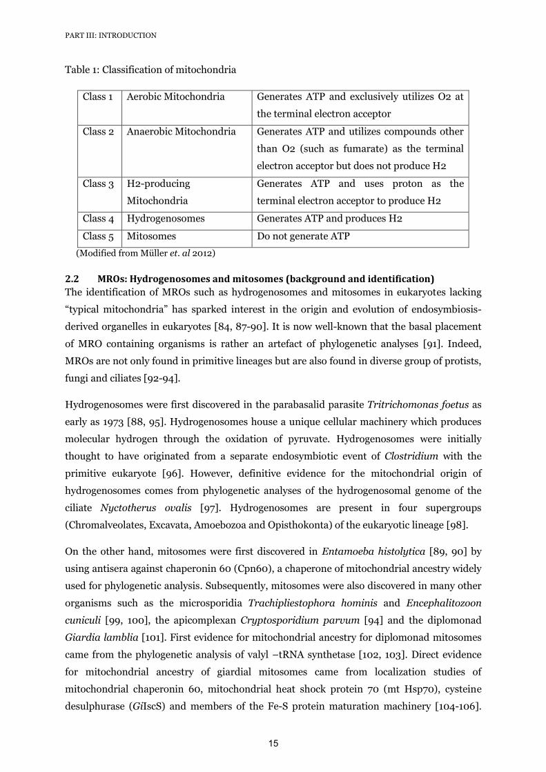
mitochondria in 5 different categories based on organelle biochemistry, energy metabolism
and functions, Table 1 [86]. Briefly, classes 1, 2 and 3 generate energy via the electron
transport chain (ETC), whereas class 4 and 5 lack such machinery for energy production.
14
PART III: INTRODUCTION
Table 1: Classification of mitochondria
Class 1 Aerobic Mitochondria Generates ATP and exclusively utilizes O2 at
the terminal electron acceptor
Class 2 Anaerobic Mitochondria Generates ATP and utilizes compounds other
than O2 (such as fumarate) as the terminal
electron acceptor but does not produce H2
Class 3 H2-producing
Mitochondria
Generates ATP and uses proton as the
terminal electron acceptor to produce H2
Class 4 Hydrogenosomes Generates ATP and produces H2
Class 5 Mitosomes Do not generate ATP
(Modified from Müller et. al 2012)
2.2 MROs: Hydrogenosomes and mitosomes (background and identification)
The identification of MROs such as hydrogenosomes and mitosomes in eukaryotes lacking
“typical mitochondria” has sparked interest in the origin and evolution of endosymbiosis-
derived organelles in eukaryotes [84, 87-90]. It is now well-known that the basal placement
of MRO containing organisms is rather an artefact of phylogenetic analyses [91]. Indeed,
MROs are not only found in primitive lineages but are also found in diverse group of protists,
fungi and ciliates [92-94].
Hydrogenosomes were first discovered in the parabasalid parasite Tritrichomonas foetus as
early as 1973 [88, 95]. Hydrogenosomes house a unique cellular machinery which produces
molecular hydrogen through the oxidation of pyruvate. Hydrogenosomes were initially
thought to have originated from a separate endosymbiotic event of Clostridium with the
primitive eukaryote [96]. However, definitive evidence for the mitochondrial origin of
hydrogenosomes comes from phylogenetic analyses of the hydrogenosomal genome of the
ciliate Nyctotherus ovalis [97]. Hydrogenosomes are present in four supergroups
(Chromalveolates, Excavata, Amoebozoa and Opisthokonta) of the eukaryotic lineage [98].
On the other hand, mitosomes were first discovered in Entamoeba histolytica [89, 90] by
using antisera against chaperonin 60 (Cpn60), a chaperone of mitochondrial ancestry widely
used for phylogenetic analysis. Subsequently, mitosomes were also discovered in many other
organisms such as the microsporidia Trachipliestophora hominis and Encephalitozoon
cuniculi [99, 100], the apicomplexan Cryptosporidium parvum [94] and the diplomonad
Giardia lamblia [101]. First evidence for mitochondrial ancestry for diplomonad mitosomes
came from the phylogenetic analysis of valyl –tRNA synthetase [102, 103]. Direct evidence
for mitochondrial ancestry of giardial mitosomes came from localization studies of
mitochondrial chaperonin 60, mitochondrial heat shock protein 70 (mt Hsp70), cysteine
desulphurase (GiIscS) and members of the Fe-S protein maturation machinery [104-106].
15
PART III: INTRODUCTION
Therefore, the absence of canonical mitochondria in Giardia can be explained by a secondary
loss due to parasitic adaptation to an anaerobic environment. Interestingly, the Spironucleus
genus (closest relatives to the Giardia genus known and investigated) harbors
hydrogenosomes [107, 108]. Based on phylogenetic data, Jerlstrom-Hultqvist, J et. al
proposed that within the diplomonad kingdom, mitosomes might have evolved from
hydrogenosomes and not from mitochondria directly due to the presence of hydrogenosome
components in the diplomonad ancestor [109, 110].
2.3 Apoptosis
Cell death plays a complementary but opposite role to mitosis in maintaining a cell
population, eliminating damaged cells and giving shape to an organism [111]. The term
apoptosis (a-po-toe-sis) was first used to describe a morphologically distinct form of cell
death [112]. Studies regarding apoptosis have boomed ever since the discovery of
programmed cell death (PCD) in Caenorhabditis elegans [113]. Cell deletion at specific stages
leading to development of organs and digits outlines the precision and importance of this
process. Apoptosis therefore has been accepted as the most distinctive form of PCD in
metazoans because of its obvious benefits towards multicellular life forms [114]. This
however doesn’t exclude the existence of other forms of cell death such as necrosis and
autophagy [115, 116]. Depending on the type of stimuli and/or degree of exposure to stimuli,
cells could either die by apoptosis or necrosis [117]. Apoptosis is accompanied by various
physiological and morphological changes in the cell. For instance, cell shrinkage (reduced
size, condensed cytoplasm and tightly packed organelles) and pyknosis (condensed
chromatin) are early symptoms of apoptosis and are visible by light microscopy. In metazoan
cells undergoing apoptosis, pyknosis is accompanied by fragmentation of genomic material
into oligonucleosomal fragments of 200bps leading to a characteristic DNA laddering pattern
[118]. Other morphological features include excessive plasma membrane blebbing and
separation of small cellular fragments generating “apoptotic bodies”. These apoptotic bodies
are then scavenged by macrophages or parenchymal cells and are degraded within
phagolysosomes [119]. Hallmark biochemical changes include phosphatidylserine (PS)
externalization, increase in intra-cellular Ca2+ levels, breakdown of PARP (DNA repair
enzyme) and mitochondrial dysfunction [120]. It is noteworthy that neither apoptosis nor
removal of apoptotic cells elicits any inflammatory response due to several reasons: 1) cells
do not burst, hence they do not release their cellular content into the environment; 2) they
are phagocytosed by macrophages, hence preventing secondary necrosis; 3) the scavenging
cells do not release/produce pro-inflammatory cytokines [119].
Apoptosis is a highly complex, sophisticated and energy demanding process. Current
research suggests the presence of 2 main apoptotic pathways: the extrinsic (via death
receptor) and the intrinsic route (via mitochondria). In fact, these two pathways could be
16
PART III: INTRODUCTION
interconnected. These different routes are activated by specific triggering signals in order to
begin a highly complex cascade of molecular events resulting in the characteristic cyto-
morphological apoptotic features leading to the demise of the cell (execution pathway). Since
MROs and, specifically, mitosomes are the organelles of interest during my doctoral thesis, I
briefly summarize the events in the intrinsic death pathway, with mitochondria (cell’s
Pandora’s Box [121],) being the cornerstone organelle [122]. Briefly, specific death stimuli
result in loss of mitochondrial membrane potential, opening of the mitochondrial transition
pore with release of pro-apoptotic proteins (cytochrome c and Smac/DIABLO) from the inner
membrane space into the cytosol. These factors then activate the caspase dependent
mitochondrial cell death pathway. Tight regulation of the intrinsic death pathway is carried
out by Bcl-2 family proteins [122, 123].
Several groups around the world in the past decade have performed numerous experiments
to show the presence of a caspase like execution pathway for cell death in MRO harboring
parasites such as Trichomonas and Giardia [124, 125]. A form of cell death with most if not
all features of canonical apoptotic death has been documented, for e.g. chromatin
condensation, PS externalization, TUNEL positive DNA fragmentation without any DNA
laddering. Furthermore, involvement of caspase like activity has also been reported by the
authors [126]. In 2009, Ghosh et. al demonstrated that Giardia trophozoites underwent a
form of PCD when subjected to H2O2, metronidazole, and upon exposure to a media devoid
of cysteine and ascorbic acid [127]. PS externalization and DNA degradation without a typical
electrophoretic laddering were observed as read outs for apoptotic like cell death.
Interestingly dying cells were negative for caspase activity and other proteases which could be
involved in the death machinery. In another study, when giardial trophozoites were treated
with the drug beta-lapachone (topoisomerase inhibitor); dying cells exhibited many if not all
features of apoptosis, such as cell shrinkage, chromatin condensation, membrane blebbing
and vacuolization. In addition, authors have also described autophagic cell death in Giardia
upon treatment with the drug beta-lapachone based on the appearance of large vacuoles and
LC3 staining (hallmark of autophagy) [128]. Similar results were replicated by Bagchi et. al in
2012, where the authors could demonstrate apoptotic like morphological changes in giardial
trophozoites upon exposure to H2O2. Interestingly, using in silico homology searches, authors
could also identify 3 three key players of autophagic cell death (TOR, ATG1 and ATG16)
[129].
17
PART III: INTRODUCTION
3 Mitosomes
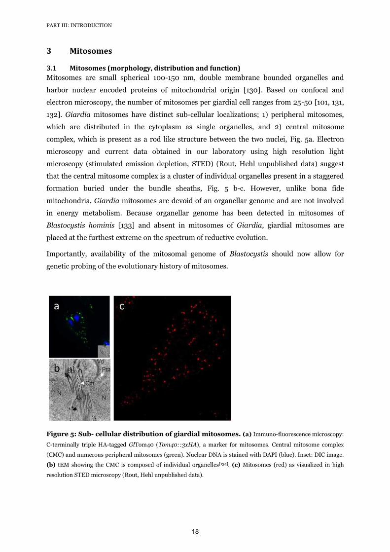
3.1 Mitosomes (morphology, distribution and function)
Mitosomes are small spherical 100-150 nm, double membrane bounded organelles and
harbor nuclear encoded proteins of mitochondrial origin [130]. Based on confocal and
electron microscopy, the number of mitosomes per giardial cell ranges from 25-50 [101, 131,
132]. Giardia mitosomes have distinct sub-cellular localizations; 1) peripheral mitosomes,
which are distributed in the cytoplasm as single organelles, and 2) central mitosome
complex, which is present as a rod like structure between the two nuclei, Fig. 5a. Electron
microscopy and current data obtained in our laboratory using high resolution light
microscopy (stimulated emission depletion, STED) (Rout, Hehl unpublished data) suggest
that the central mitosome complex is a cluster of individual organelles present in a staggered
formation buried under the bundle sheaths, Fig. 5 b-c. However, unlike bona fide
mitochondria, Giardia mitosomes are devoid of an organellar genome and are not involved
in energy metabolism. Because organellar genome has been detected in mitosomes of
Blastocystis hominis [133] and absent in mitosomes of Giardia, giardial mitosomes are
placed at the furthest extreme on the spectrum of reductive evolution.
Importantly, availability of the mitosomal genome of Blastocystis should now allow for
genetic probing of the evolutionary history of mitosomes.
Figure 5: Sub- cellular distribution of giardial mitosomes. (a) Immuno-fluorescence microscopy:
C-terminally triple HA-tagged GlTom40 (Tom40::3xHA), a marker for mitosomes. Central mitosome complex
(CMC) and numerous peripheral mitosomes (green). Nuclear DNA is stained with DAPI (blue). Inset: DIC image.
(b) tEM showing the CMC is composed of individual organelles[134]. (c) Mitosomes (red) as visualized in high
resolution STED microscopy (Rout, Hehl unpublished data).
18
PART III: INTRODUCTION
Nevertheless, Giardia mitosomes despite being highly reduced/modified are able to import
nuclear encoded proteins via conserved protein import pathways (discussed later) [90, 101,
131]. However, iron- sulfur protein (Fe-S) maturation is the only known function ascribed to
these organelles and has been shown to be indispensable for mitochondria and conserved
throughout evolution [135]. This could be a probable explanation for the retention of
mitosomes in Giardia. In fact, Giardia genome database searches and high throughout
proteomics studies revealed that Giardia harbors key components for the Fe-S protein
maturation machinery such as IscU (Gl50803_15196), cysteine desulphurase
(Gl50803_14519), mtHsp70 (Gl50803_14581), IscA2 (Gl50803_14821), ferredoxin
(Gl50803_27266), Nfu (EAA38809), DnaJ protein Jac1 (Gl50803_17030), Glutaredoxin 5
(Gl50803_2013) and GrpE (Gl50803_1376) [86, 136]. Surprisingly, frataxin is missing in the
Giardia genome. Frataxin is a Fe-binding protein that donates Fe to the IscU/IscS complex
and is invariably present in all eukaryotes including mitosome harboring E. cuniculi [17] and
C. parvum [137]. Furthermore, an ADP/ATP transporter or any machinery for ATP synthesis
has not been identified in Giardia mitosomes, despite the presence of 2 mitosomal proteins
(Cpn60 and mt Hsp70) having ATP-dependent activity [26, 136, 138]. Therefore, the source
of energy in Giardia mitosomes for: 1) import of nuclear encoded proteins into mitosomes, 2)
transfer of Fe-S cluster, remains elusive till date. Interestingly, a unique ADP/ATP
transporter which does not require membrane potential has been identified in E. histolytica
mitosomes [139].
3.2 Mitosomal protein targeting and processing
3.2.1 Mitosomal protein targeting sequences (MTS)
In all species known so far harboring a mitochondrion with an organellar genome, 98% of the
organellar protein repertoire is nuclear encoded and must be targeted to the organelle and its
compartments precisely with the help of specific targeting signals [140, 141]. The nuclear
encoded mitochondrial matrix proteins are synthesized with a N-terminal mitochondrial
targeting sequence (MTS) which is recognized by the receptors on the mitochondrial surface
(Tom20), subsequently transporting the preproteins to their final destination via translocases
of the outer and inner membranes [142, 143]. Some carrier proteins however lack the N-
terminal MTS and instead possess several internal mitochondrial targeting signals
distributed throughout the entire length of the protein which are recognized by the Tom70
receptor [142, 144].
Typical MTSs are composed of approximately 10-80 positively charged, hydrophobic and
hydroxylated amino acids [140, 145-147]. Apart from these characteristics MTSs have very
little primary sequence conservation amongst different phylogenetic groups [148]. In fact, the
MTS present in organisms harboring mitosomes and hydrogenosomes are comparatively
shorter than their eukaryotic homologs. Smid et. al have shown that the length of the MTS in
19
PART III: INTRODUCTION
these relic mitochondria containing organisms varies from 4-21 amino acids [149, 150].
Experimental evidence in the literature suggests that the MTS of mitosomal proteins in
Giardia, Entamoeba and of hydrogenosomal proteins in Trichomonas contains an arginine
residue which is located 2 residues upstream of the cleavage site, thus facilitating recognition
of the cleavage site [151, 152]. Surprisingly, in 2 independent studies performed in Giardia
and Saccharomyces cerevisiae, it was demonstrated that IscU protein was delivered to
mitosomes and mitochondria respectively regardless of the MTS, albeit with reduced
efficiency in its absence [132, 153]. These experiments suggested that the internal signals
present in mitochondrial proteins are sufficient for proper delivery; however a MTS increases
the efficiency of transport.
Matrix proteins harboring positively charged MTS are inserted via membrane potential
dependent Tim 23 translocon after which the MTS is cleaved by the mitochondrial processing
peptidases marking the final step of the import process. This processing step is important as
it ensures proper protein function and/or protein stability [154]. Subsequently the unfolded
protein is refolded in the matrix to its native structure with the help of the cpn60/cpn10
chaperone complex.
3.2.2 Mitosomal processing peptidase
Canonical mitochondrial processing peptidases (MPPs) comprise of 2-α and 2-β subunits and
work as a heterodimer and remain non-functional individually [148, 155]. Kitada et. al in
2007 proposed that the modern day MPP’s ancestor was probably a monomeric α-
proteobacterial peptidase because of high sequence similarity with Rickettsia prowazekii
peptidase [156]. During evolution, gene duplication events led to formation of α/β subunits
resulting in a heterodimeric MPP. Both subunits are responsible for different function; the α-
subunit infers substrate binding and release whereas the β-subunit is responsible for catalysis
process [149, 155]. The two subunits form a negatively charged cavity where the MTS is
processed by electrostatic interaction. Out of the few mitosomal and hydrogenosomal
proteins known/identified so far, a subset of the proteins are shown to possess a N-terminal
MTS responsible for proper targeting to the organelle [101, 131, 152]. In fact, Giardia harbors
a gene encoding a β-MPP and the product is localized to mitosomes [26, 132]. Giardia MPP is
unique in itself as it lacks the α-subunit and functions as a β-monomeric enzyme [149].
Furthermore, the recombinant Giardia β-MPP can process MTS harboring Giardia proteins
in vitro. Likewise a protein with limited similarity with S. cereviseae β-MPP has been
identified in genome of hydrogenosome containing Trichomonas vaginalis [157]. However,
the T. vaginalis MPP functions as a β-homodimer enzyme. Therefore, the presence of the
monomeric subunit β-peptidase in Giardia and homodimer β-peptidase in Trichomonas
points towards either a consequence of secondary reduction or retention of the ancestral
MPP [156].
20
PART III: INTRODUCTION
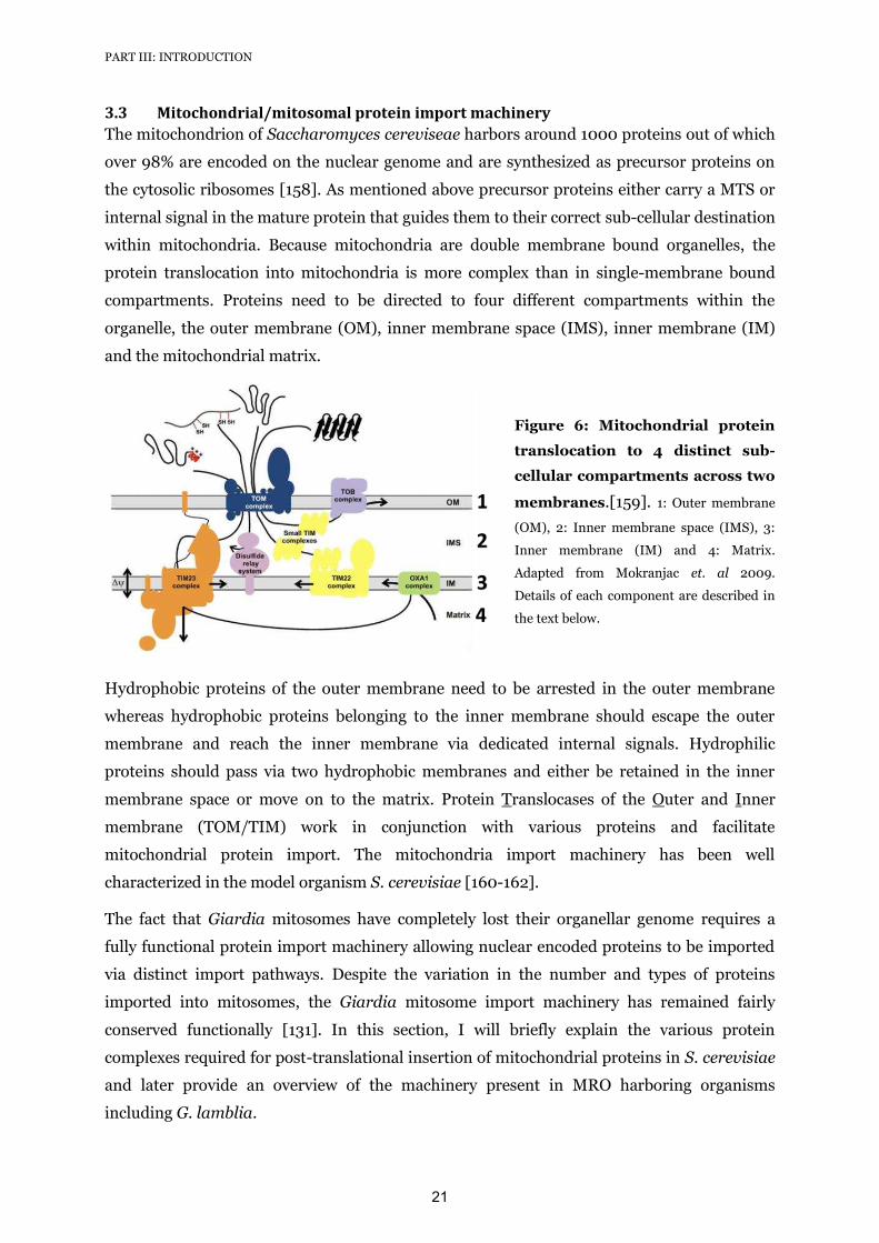
3.3 Mitochondrial/mitosomal protein import machinery
The mitochondrion of Saccharomyces cereviseae harbors around 1000 proteins out of which
over 98% are encoded on the nuclear genome and are synthesized as precursor proteins on
the cytosolic ribosomes [158]. As mentioned above precursor proteins either carry a MTS or
internal signal in the mature protein that guides them to their correct sub-cellular destination
within mitochondria. Because mitochondria are double membrane bound organelles, the
protein translocation into mitochondria is more complex than in single-membrane bound
compartments. Proteins need to be directed to four different compartments within the
organelle, the outer membrane (OM), inner membrane space (IMS), inner membrane (IM)
and the mitochondrial matrix.
Figure 6: Mitochondrial protein
translocation to 4 distinct sub-
cellular compartments across two
membranes.[159]. 1: Outer membrane
(OM), 2: Inner membrane space (IMS), 3:
Inner membrane (IM) and 4: Matrix.
Adapted from Mokranjac et. al 2009.
Details of each component are described in
the text below.
Hydrophobic proteins of the outer membrane need to be arrested in the outer membrane
whereas hydrophobic proteins belonging to the inner membrane should escape the outer
membrane and reach the inner membrane via dedicated internal signals. Hydrophilic
proteins should pass via two hydrophobic membranes and either be retained in the inner
membrane space or move on to the matrix. Protein Translocases of the Outer and Inner
membrane (TOM/TIM) work in conjunction with various proteins and facilitate
mitochondrial protein import. The mitochondria import machinery has been well
characterized in the model organism S. cerevisiae [160-162].
The fact that Giardia mitosomes have completely lost their organellar genome requires a
fully functional protein import machinery allowing nuclear encoded proteins to be imported
via distinct import pathways. Despite the variation in the number and types of proteins
imported into mitosomes, the Giardia mitosome import machinery has remained fairly
conserved functionally [131]. In this section, I will briefly explain the various protein
complexes required for post-translational insertion of mitochondrial proteins in S. cerevisiae
and later provide an overview of the machinery present in MRO harboring organisms
including G. lamblia.
21
PART III: INTRODUCTION
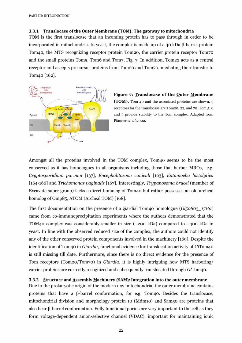
3.3.1 Translocase of the Outer Membrane (TOM): The gateway to mitochondria
TOM is the first translocase that an incoming protein has to pass through in order to be
incorporated in mitochondria. In yeast, the complex is made up of a 40 kDa β-barrel protein
Tom40, the MTS recognizing receptor protein Tom20, the carrier protein receptor Tom70
and the small proteins Tom5, Tom6 and Tom7, Fig. 7. In addition, Tom22 acts as a central
receptor and accepts precursor proteins from Tom20 and Tom70, mediating their transfer to
Tom40 [162].
Figure 7: Translocase of the Outer Membrane
(TOM). Tom 40 and the associated proteins are shown. 3
receptors for the translocase are Tom20, 22, and 70. Tom 5, 6
and 7 provide stability to the Tom complex. Adapted from
Pfanner et. al 2002.
Amongst all the proteins involved in the TOM complex, Tom40 seems to be the most
conserved as it has homologues in all organisms including those that harbor MROs, e.g.
Cryptosporidium parvum [137], Encephalitozoon cuniculi [163], Entamoeba histolytica
[164-166] and Trichomonas vaginalis [167]. Interestingly, Trypanosoma brucei (member of
Excavate super group) lacks a direct homolog of Tom40 but rather possesses an old archeal
homolog of Omp85, ATOM (Archeal TOM) [168].
The first documentation on the presence of a giardial Tom40 homologue (Gl50803_17161)
came from co-immunoprecipitation experiments where the authors demonstrated that the
TOM40 complex was considerably smaller in size (~200 kDa) compared to ~400 kDa in
yeast. In line with the observed reduced size of the complex, the authors could not identify
any of the other conserved protein components involved in the machinery [169]. Despite the
identification of Tom40 in Giardia, functional evidence for translocation activity of GlTom40
is still missing till date. Furthermore, since there is no direct evidence for the presence of
Tom receptors (Tom20/Tom70) in Giardia, it is highly intriguing how MTS harboring/
carrier proteins are correctly recognized and subsequently translocated through GlTom40.
3.3.2 Structure and Assembly Machinery (SAM): Integration into the outer membrane
Due to the prokaryotic origin of the modern day mitochondria, the outer membrane contains
proteins that have a β-barrel conformation, for e.g. Tom40. Besides the translocase,
mitochondrial division and morphology protein 10 (Mdm10) and Sam50 are proteins that
also bear β-barrel conformation. Fully functional porins are very important to the cell as they
form voltage-dependent anion-selective channel (VDAC), important for maintaining ionic
22
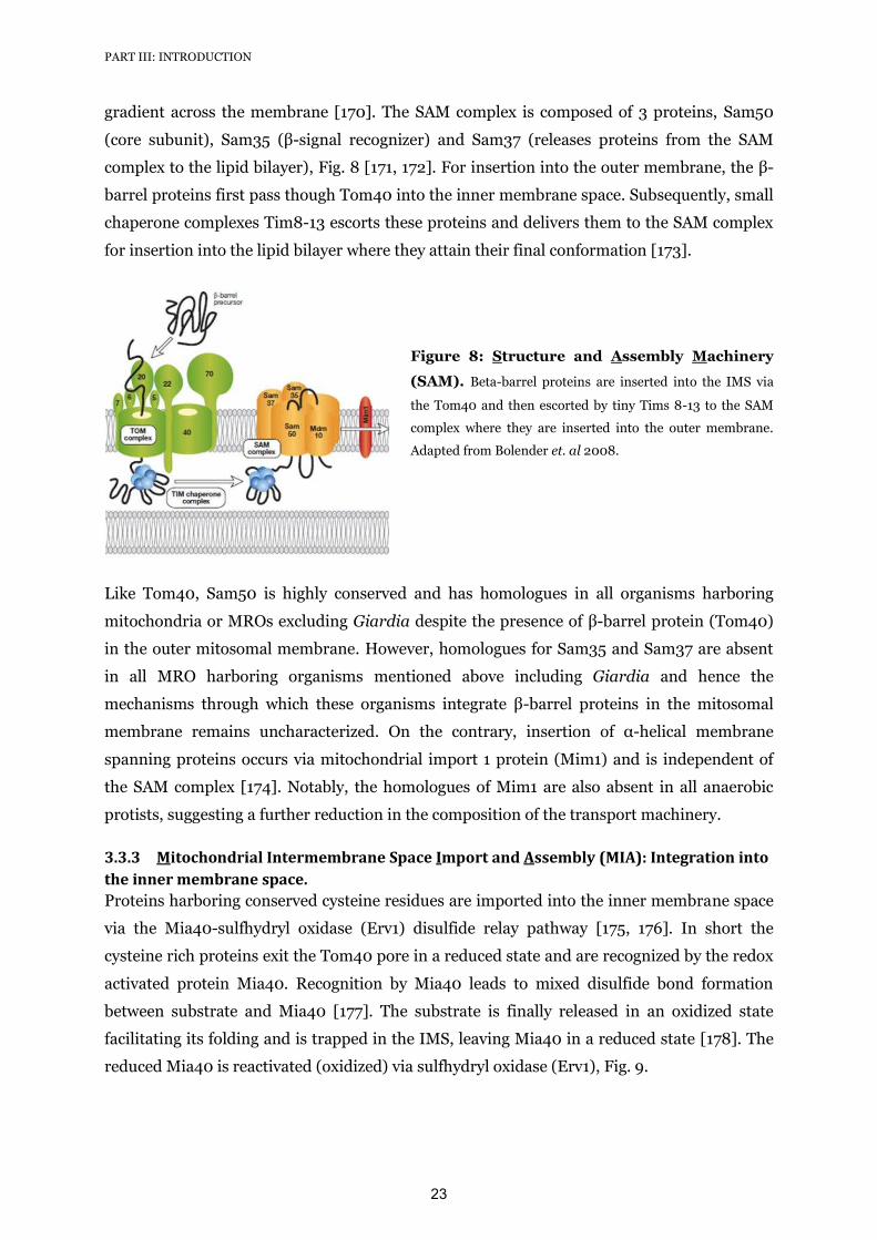
PART III: INTRODUCTION
gradient across the membrane [170]. The SAM complex is composed of 3 proteins, Sam50
(core subunit), Sam35 (β-signal recognizer) and Sam37 (releases proteins from the SAM
complex to the lipid bilayer), Fig. 8 [171, 172]. For insertion into the outer membrane, the β-
barrel proteins first pass though Tom40 into the inner membrane space. Subsequently, small
chaperone complexes Tim8-13 escorts these proteins and delivers them to the SAM complex
for insertion into the lipid bilayer where they attain their final conformation [173].
Figure 8: Structure and Assembly Machinery
(SAM). Beta-barrel proteins are inserted into the IMS via
the Tom40 and then escorted by tiny Tims 8-13 to the SAM
complex where they are inserted into the outer membrane.
Adapted from Bolender et. al 2008.
Like Tom40, Sam50 is highly conserved and has homologues in all organisms harboring
mitochondria or MROs excluding Giardia despite the presence of β-barrel protein (Tom40)
in the outer mitosomal membrane. However, homologues for Sam35 and Sam37 are absent
in all MRO harboring organisms mentioned above including Giardia and hence the
mechanisms through which these organisms integrate β-barrel proteins in the mitosomal
membrane remains uncharacterized. On the contrary, insertion of α-helical membrane
spanning proteins occurs via mitochondrial import 1 protein (Mim1) and is independent of
the SAM complex [174]. Notably, the homologues of Mim1 are also absent in all anaerobic
protists, suggesting a further reduction in the composition of the transport machinery.
3.3.3 Mitochondrial Intermembrane Space Import and Assembly (MIA): Integration into
the inner membrane space.
Proteins harboring conserved cysteine residues are imported into the inner membrane space
via the Mia40-sulfhydryl oxidase (Erv1) disulfide relay pathway [175, 176]. In short the
cysteine rich proteins exit the Tom40 pore in a reduced state and are recognized by the redox
activated protein Mia40. Recognition by Mia40 leads to mixed disulfide bond formation
between substrate and Mia40 [177]. The substrate is finally released in an oxidized state
facilitating its folding and is trapped in the IMS, leaving Mia40 in a reduced state [178]. The
reduced Mia40 is reactivated (oxidized) via sulfhydryl oxidase (Erv1), Fig. 9.
23
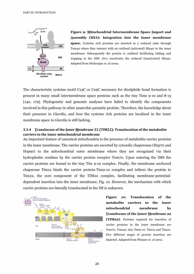
PART III: INTRODUCTION
Figure 9: Mitochondrial Intermembrane Space Import and
Assembly (MIA): Integration into the inner membrane
space. Cysteine rich proteins are inserted in a reduced state through
Tom40 where they interact with an oxidized (activated) Mia40 in the inner
membrane. Subsequently the protein is oxidized facilitating folding and
trapping in the IMS. Erv1 reactivates the reduced (inactivated) Mia40.
Adapted from Mokranjac et. al 2009.
The characteristic cysteine motif Cx3C or Cx9C necessary for disulphide bond formation is
present in many small intermembrane space proteins such as the tiny Tims 9-10 and 8-13
[140, 179]. Phylogenetic and genomic analyses have failed to identify the components
involved in this pathway in other anaerobic parasitic protists. Therefore, the knowledge about
their presence in Giardia, and how the cysteine rich proteins are localized in the inner
membrane space in Giardia is still lacking.
3.3.4 Translocase of the Inner Membrane 22 (TIM22): Translocation of the metabolite
carriers to the inner mitochondrial membrane
An important feature of canonical mitochondria is the presence of metabolite carrier proteins
in the inner membrane. The carrier proteins are escorted by cytosolic chaperones (Hsp70 and
Hsp90) to the mitochondrial outer membrane where they are recognized via their
hydrophobic residues by the carrier protein receptor Tom70. Upon entering the IMS the
carrier proteins are bound to the tiny Tim 9-10 complex. Finally, the membrane anchored
chaperone Tim12 binds the carrier protein-Tim9-10 complex and tethers the protein to
Tim22, the core component of the TIM22 complex, facilitating membrane-potential-
dependent insertion into the inner membrane, Fig. 10. However, the mechanism with which
carrier proteins are laterally translocated in the IM is unknown.
Figure 10: Translocation of the
metabolite carriers to the inner
mitochondrial membrane by
Translocase of the Inner Membrane 22
(TIM22). Proteins required for insertion of
carrier proteins in the inner membrane are
Tom70, Tom40, tiny Tim9-10, Tim12 and Tim22.
Five different stages of protein insertion are
depicted. Adapted from Pfanner et. al 2002.
24
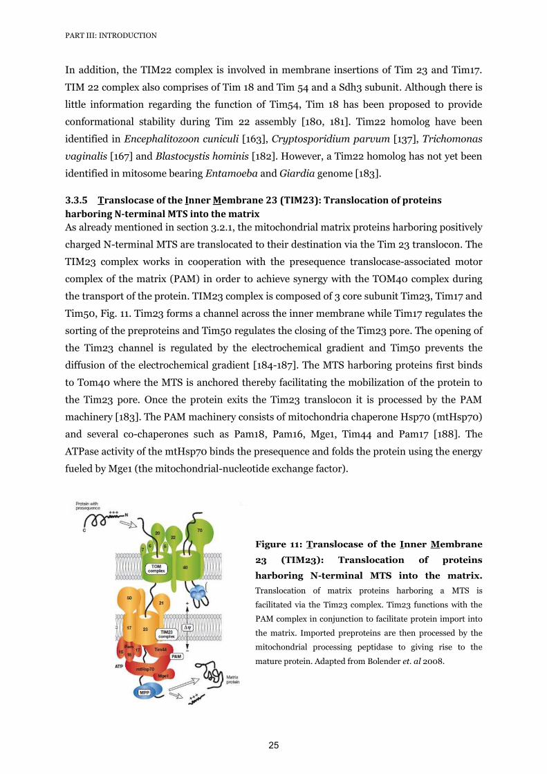
PART III: INTRODUCTION
In addition, the TIM22 complex is involved in membrane insertions of Tim 23 and Tim17.
TIM 22 complex also comprises of Tim 18 and Tim 54 and a Sdh3 subunit. Although there is
little information regarding the function of Tim54, Tim 18 has been proposed to provide
conformational stability during Tim 22 assembly [180, 181]. Tim22 homolog have been
identified in Encephalitozoon cuniculi [163], Cryptosporidium parvum [137], Trichomonas
vaginalis [167] and Blastocystis hominis [182]. However, a Tim22 homolog has not yet been
identified in mitosome bearing Entamoeba and Giardia genome [183].
3.3.5 Translocase of the Inner Membrane 23 (TIM23): Translocation of proteins
harboring N-terminal MTS into the matrix
As already mentioned in section 3.2.1, the mitochondrial matrix proteins harboring positively
charged N-terminal MTS are translocated to their destination via the Tim 23 translocon. The
TIM23 complex works in cooperation with the presequence translocase-associated motor
complex of the matrix (PAM) in order to achieve synergy with the TOM40 complex during
the transport of the protein. TIM23 complex is composed of 3 core subunit Tim23, Tim17 and
Tim50, Fig. 11. Tim23 forms a channel across the inner membrane while Tim17 regulates the
sorting of the preproteins and Tim50 regulates the closing of the Tim23 pore. The opening of
the Tim23 channel is regulated by the electrochemical gradient and Tim50 prevents the
diffusion of the electrochemical gradient [184-187]. The MTS harboring proteins first binds
to Tom40 where the MTS is anchored thereby facilitating the mobilization of the protein to
the Tim23 pore. Once the protein exits the Tim23 translocon it is processed by the PAM
machinery [183]. The PAM machinery consists of mitochondria chaperone Hsp70 (mtHsp70)
and several co-chaperones such as Pam18, Pam16, Mge1, Tim44 and Pam17 [188]. The
ATPase activity of the mtHsp70 binds the presequence and folds the protein using the energy
fueled by Mge1 (the mitochondrial-nucleotide exchange factor).
Figure 11: Translocase of the Inner Membrane
23 (TIM23): Translocation of proteins
harboring N-terminal MTS into the matrix.
Translocation of matrix proteins harboring a MTS is
facilitated via the Tim23 complex. Tim23 functions with the
PAM complex in conjunction to facilitate protein import into
the matrix. Imported preproteins are then processed by the
mitochondrial processing peptidase to giving rise to the
mature protein. Adapted from Bolender et. al 2008.
25
PART III: INTRODUCTION
MTS harboring proteins have been discovered in many anaerobic eukaryotes bearing MROs
including Giardia and the machinery required for their import into the matrix is variably
conserved in these eukaryotes. Many components of this machinery are found in the genera
Encephalitozoon, Cryptosporidium, Trichomonas, Blastocystis and in the anaerobic amoeba
Sawyeria marylandensis [137, 163, 167, 182, 189]. In Giardia, no direct homologue or
paralogue for components of the TIM23 complex has been detected. However, Giardia
possesses homologs for the components of PAM complex such as mtHsp70, Mge1, Pam18,
and Pam16 [136]. Recently, a Tim44 homologue has been identified in the Giardia genome
[190]. Therefore the identification of homologues for components of the TIM23 complex in
Giardia underlines the fact that the protein translocation into the mitosomal matrix occurs
via conserved pathways similar to their eukaryotic counterparts.
26
PART III: INTRODUCTION
4. Goals of the thesis
4.1 Investigating the role of Arf and ARF-like small GTPases during encystation in
Giardia lamblia
Giardia relies on highly regulated secretion machinery in order to form the protective extra-
cellular biopolymer (the cyst wall) essential for its transmission to a new host. De novo
generated encystation specific vesicles (ESVs) are responsible for maturation, sorting and
regulated secretion of cyst wall material (CWM). ESV genesis and maturation is dependent
on small GTPases such as Sar1 and Arf1 [42]. The G. lamblia homologue of Arf1
(Gl50803_7789) was shown to be recruited to ESVs during the later stages of this
differentiation process. Furthermore, over-expression of a non-functional Arf1 mutant
protein led to a “naked cyst” phenotype, suggestive of a block in ESV maturation which would
interfere with correct CWM secretion. These cysts lacked water resistance and thus were
presumably non-infective [42].
Using homology-based in silico searches, we identified 5 additional Arf and ARF- like protein
(Arl) homologues in the Giardia genome database. ORFs Gl50803_ 13930 and
Gl50803_7562 are Arf family members, ORFs Gl50803_4192, Gl50803_13523 and
Gl50803_13478 are Arl family members. Moreover, together with ORF Gl7789 which
encodes for GlArf1, ORF Gl7562 is the only other giardial Arf homologue known to have a
predicted N-myristoylation motif. This post-translational modification is a pre-requisite for
membrane recruitment of Arf family proteins to target membranes.
Since G. lamblia is a highly reduced eukaryote, the space for redundancy in such a
parsimonious system is quite narrow. Therefore we hypothesized that these additional Arf
and Arl proteins are also involved in CWM deposition and that a loss of function for these
proteins would inhibit cyst wall formation. To test this hypothesis, we begin by defining the
additional G. lamblia Arf and Arl homologues their localization within the cell and possible
role(s) in ESV maturation and functionality by expression of non-functional Arf and Arl
mutants exerting dominant negative effects.
A detailed investigation of the function of additional Arf and Arl homologues is pivotal in
understanding the exact role of these small GTPases in Giardia and would provide us with
novel molecular targets to prevent encystation, thereby reducing infectivity and parasite
spread.
27
PART III: INTRODUCTION
4.2 Induction of apoptosis in Giardia lamblia
Giardia mitosomes, even though reduced are nevertheless essential and have conserved
pathways for protein import [131]. In the course of an investigation of Tom/Tim-dependent
protein import into giardial mitosomes we fortuitously uncovered a dramatic and rapid form
of cell deterioration as a result of interference with mitosome protein import function upon
treatment of trophozoites with Mitoblock (an experimental compound targeting the Tim
import complex in mitochondria) (Hehl, unpublished data). Furthermore, recent reports
suggest that mitosomes not only play a role in Fe-S protein maturation but might be also
involved in a form of programmed cell death (PCD) [128].
Based on the following observations i.e., absence of a significant inflammatory response in
the gut epithelium upon infection, the possible control of parasite population density in the
intestine, and sensitivity of parasites to interference with mitosomal protein import
functions, we hypothesized that mitosomes not only fulfill essential metabolic functions but
also act as a highly sensitive central switch for monitoring cell health and for integrating
possible cell death signals in a simple/primitive form of PCD. PCD as a strategy to avoid large
scale necrosis and exposure of strong antigen to immune cells in the small intestine might be
an explanation for the surprisingly mild inflammatory response recorded even during chronic
infection.
Therefore, to test this hypothesis we aim to: 1) elicit cell death in culture and provide a
catalog of quantifiable changes which accompany this event and 2) provide criteria to
discriminate PCD-like phenomena and cell necrosis.
Characterization of PCD especially in basal protozoan parasite Giardia that does not possess
a mitochondrion would hint towards the existence of an antique cell death pathway related to
mitosomes and would shed light on the evolution of programmed cell death.
28
PART III: INTRODUCTION
4.3 Optimization of a co-immunoprecipitation assay to identify organelle specific sub
proteomes
Mitochondrion-related organelles in Giardia (mitosomes) contain no DNA, thus all
remaining endosymbiont-derived genes are localized in the nuclear genome. Their products
are translated on cytoplasmic ribosomes and subsequently imported into the organelle.
However, due to massive albeit selective sequence divergence in G. lamblia, traditional
strategies for identification of mitosome proteins based on homology-based in silico searches
fall short. Until now only 20 mitosomal proteins have been identified including only one
component of the conventional protein import machinery of the outer membrane
(GlTom40), although there is unambiguous experimental evidence for the presence of pre-
sequence dependent and independent protein import pathways in Giardia mitosomes [131].
Since Giardia mitosomes are positioned at the very extreme on the spectrum of reductive
evolution we hypothesized that these minimized organelles harbor not more than 100-150
proteins functioning in the matrix or present at the membrane facilitating protein import, or
establishing contact with the cellular compartments. To test our hypothesis we aim to
generate a comprehensive list of mitosome proteins and to analyze the molecular basis for
protein import. This would enable us to determine the functional range of these reduced
organelles and possibly to expand on the mechanisms for their maintenance and distribution.
Specifically, we aim to 1) establish an organelle centered co-immunoprecipitation (co-IP)
assay for efficient pull down of membrane and soluble complexes. 2) Perform a series of ad
hoc co-IP assays with validated Giardia proteins to identify components of the import
machinery and other non-conserved proteins which would enable us to create a GlTom40-
centered interactome that expands inwards towards the matrix and outwards towards the
cytosol.
A detailed proteome analysis of Giardia mitosomes is a must for careful speculation on the
vast array of functions that can be assigned to these reduced organelles. Furthermore,
comprehensive identification of mitosome proteins and characterization of protein import
dynamics are a prerequisite for uncovering a postulated link with PCD and/or cell
proliferation.
29
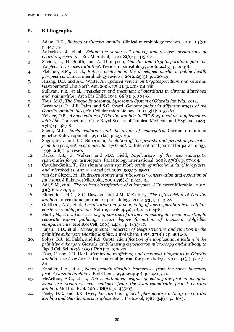
PART III: INTRODUCTION
5. Bibliography
1. Adam, R.D., Biology of Giardia lamblia. Clinical microbiology reviews, 2001. 14(3):
p. 447-75. 2. Ankarklev, J., et al., Behind the smile: cell biology and disease mechanisms of
Giardia species. Nat Rev Microbiol, 2010. 8(6): p. 413-22. 3. Savioli, L., H. Smith, and A. Thompson, Giardia and Cryptosporidium join the
'Neglected Diseases Initiative'. Trends in parasitology, 2006. 22(5): p. 203-8. 4. Fletcher, S.M., et al., Enteric protozoa in the developed world: a public health
perspective. Clinical microbiology reviews, 2012. 25(3): p. 420-49. 5. Huang, D.B. and A.C. White, An updated review on Cryptosporidium and Giardia.
Gastroenterol Clin North Am, 2006. 35(2): p. 291-314, viii. 6. Sullivan, P.B., et al., Prevalence and treatment of giardiasis in chronic diarrhoea
and malnutrition. Arch Dis Child, 1991. 66(3): p. 304-6. 7. Touz, M.C., The Unique Endosomal/Lysosomal System of Giardia lamblia. 2012. 8. Bernander, R., J.E. Palm, and S.G. Svard, Genome ploidy in different stages of the
Giardia lamblia life cycle. Cellular microbiology, 2001. 3(1): p. 55-62. 9. Keister, D.B., Axenic culture of Giardia lamblia in TYI-S-33 medium supplemented
with bile. Transactions of the Royal Society of Tropical Medicine and Hygiene, 1983. 77(4): p. 487-8.
10. Sogin, M.L., Early evolution and the origin of eukaryotes. Current opinion in genetics & development, 1991. 1(4): p. 457-63.
11. Sogin, M.L. and J.D. Silberman, Evolution of the protists and protistan parasites from the perspective of molecular systematics. International journal for parasitology, 1998. 28(1): p. 11-20.
12. Dacks, J.B., G. Walker, and M.C. Field, Implications of the new eukaryotic systematics for parasitologists. Parasitology international, 2008. 57(2): p. 97-104.
13. Cavalier-Smith, T., The simultaneous symbiotic origin of mitochondria, chloroplasts, and microbodies. Ann N Y Acad Sci, 1987. 503: p. 55-71.
14. van der Giezen, M., Hydrogenosomes and mitosomes: conservation and evolution of functions. J Eukaryot Microbiol, 2009. 56(3): p. 221-31.
15. Adl, S.M., et al., The revised classification of eukaryotes. J Eukaryot Microbiol, 2012. 59(5): p. 429-93.
16. Elmendorf, H.G., S.C. Dawson, and J.M. McCaffery, The cytoskeleton of Giardia lamblia. International journal for parasitology, 2003. 33(1): p. 3-28.
17. Goldberg, A.V., et al., Localization and functionality of microsporidian iron-sulphur cluster assembly proteins. Nature, 2008. 452(7187): p. 624-8.
18. Marti, M., et al., The secretory apparatus of an ancient eukaryote: protein sorting to separate export pathways occurs before formation of transient Golgi-like compartments. Mol Biol Cell, 2003. 14(4): p. 1433-47.
19. Lujan, H.D., et al., Developmental induction of Golgi structure and function in the primitive eukaryote Giardia lamblia. J Biol Chem, 1995. 270(9): p. 4612-8.
20. Soltys, B.J., M. Falah, and R.S. Gupta, Identification of endoplasmic reticulum in the primitive eukaryote Giardia lamblia using cryoelectron microscopy and antibody to Bip. J Cell Sci, 1996. 109 ( Pt 7): p. 1909-17.
21. Faso, C. and A.B. Hehl, Membrane trafficking and organelle biogenesis in Giardia lamblia: use it or lose it. International journal for parasitology, 2011. 41(5): p. 471-80.
22. Knodler, L.A., et al., Novel protein-disulfide isomerases from the early-diverging protist Giardia lamblia. J Biol Chem, 1999. 274(42): p. 29805-11.
23. McArthur, A.G., et al., The evolutionary origins of eukaryotic protein disulfide isomerase domains: new evidence from the Amitochondriate protist Giardia lamblia. Mol Biol Evol, 2001. 18(8): p. 1455-63.
24. Feely, D.E. and J.K. Dyer, Localization of acid phosphatase activity in Giardia lamblia and Giardia muris trophozoites. J Protozool, 1987. 34(1): p. 80-3.
30
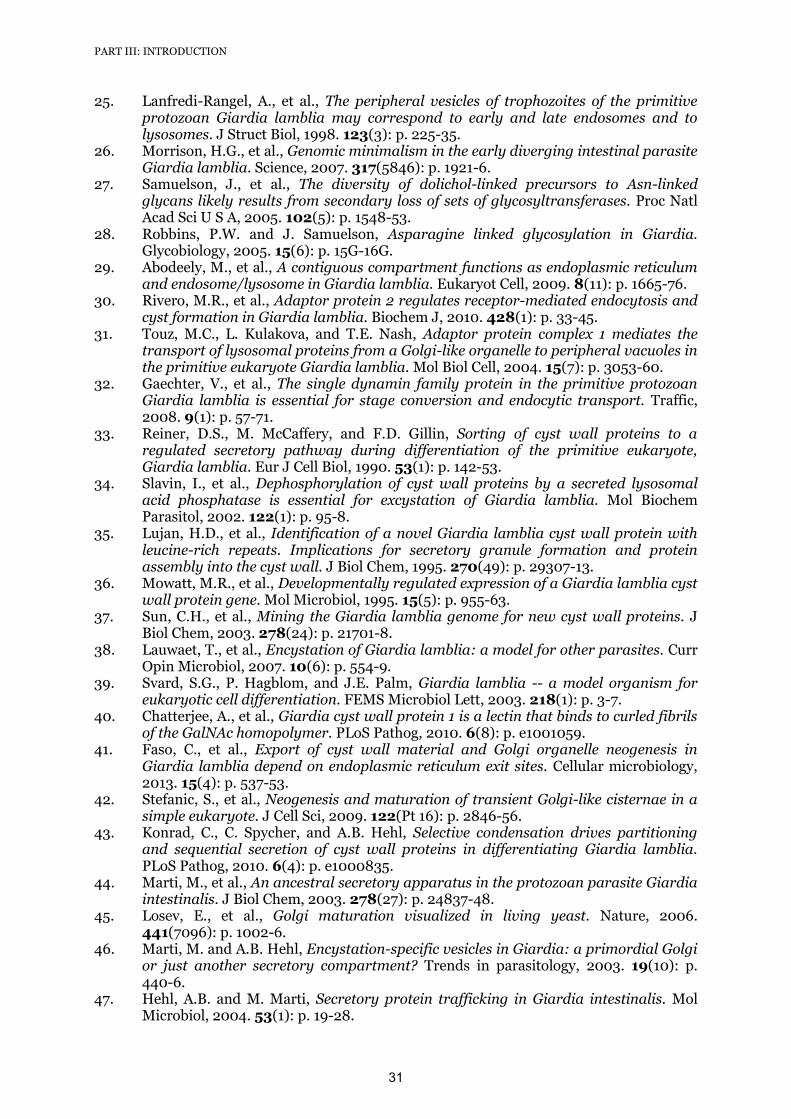
PART III: INTRODUCTION
25. Lanfredi-Rangel, A., et al., The peripheral vesicles of trophozoites of the primitive protozoan Giardia lamblia may correspond to early and late endosomes and to lysosomes. J Struct Biol, 1998. 123(3): p. 225-35.
26. Morrison, H.G., et al., Genomic minimalism in the early diverging intestinal parasite Giardia lamblia. Science, 2007. 317(5846): p. 1921-6.
27. Samuelson, J., et al., The diversity of dolichol-linked precursors to Asn-linked glycans likely results from secondary loss of sets of glycosyltransferases. Proc Natl Acad Sci U S A, 2005. 102(5): p. 1548-53.
28. Robbins, P.W. and J. Samuelson, Asparagine linked glycosylation in Giardia. Glycobiology, 2005. 15(6): p. 15G-16G.
29. Abodeely, M., et al., A contiguous compartment functions as endoplasmic reticulum and endosome/lysosome in Giardia lamblia. Eukaryot Cell, 2009. 8(11): p. 1665-76.
30. Rivero, M.R., et al., Adaptor protein 2 regulates receptor-mediated endocytosis and cyst formation in Giardia lamblia. Biochem J, 2010. 428(1): p. 33-45.
31. Touz, M.C., L. Kulakova, and T.E. Nash, Adaptor protein complex 1 mediates the transport of lysosomal proteins from a Golgi-like organelle to peripheral vacuoles in the primitive eukaryote Giardia lamblia. Mol Biol Cell, 2004. 15(7): p. 3053-60.
32. Gaechter, V., et al., The single dynamin family protein in the primitive protozoan Giardia lamblia is essential for stage conversion and endocytic transport. Traffic, 2008. 9(1): p. 57-71.
33. Reiner, D.S., M. McCaffery, and F.D. Gillin, Sorting of cyst wall proteins to a regulated secretory pathway during differentiation of the primitive eukaryote, Giardia lamblia. Eur J Cell Biol, 1990. 53(1): p. 142-53.
34. Slavin, I., et al., Dephosphorylation of cyst wall proteins by a secreted lysosomal acid phosphatase is essential for excystation of Giardia lamblia. Mol Biochem Parasitol, 2002. 122(1): p. 95-8.
35. Lujan, H.D., et al., Identification of a novel Giardia lamblia cyst wall protein with leucine-rich repeats. Implications for secretory granule formation and protein assembly into the cyst wall. J Biol Chem, 1995. 270(49): p. 29307-13.
36. Mowatt, M.R., et al., Developmentally regulated expression of a Giardia lamblia cyst wall protein gene. Mol Microbiol, 1995. 15(5): p. 955-63.
37. Sun, C.H., et al., Mining the Giardia lamblia genome for new cyst wall proteins. J Biol Chem, 2003. 278(24): p. 21701-8.
38. Lauwaet, T., et al., Encystation of Giardia lamblia: a model for other parasites. Curr Opin Microbiol, 2007. 10(6): p. 554-9.
39. Svard, S.G., P. Hagblom, and J.E. Palm, Giardia lamblia -- a model organism for eukaryotic cell differentiation. FEMS Microbiol Lett, 2003. 218(1): p. 3-7.
40. Chatterjee, A., et al., Giardia cyst wall protein 1 is a lectin that binds to curled fibrils of the GalNAc homopolymer. PLoS Pathog, 2010. 6(8): p. e1001059.
41. Faso, C., et al., Export of cyst wall material and Golgi organelle neogenesis in Giardia lamblia depend on endoplasmic reticulum exit sites. Cellular microbiology, 2013. 15(4): p. 537-53.
42. Stefanic, S., et al., Neogenesis and maturation of transient Golgi-like cisternae in a simple eukaryote. J Cell Sci, 2009. 122(Pt 16): p. 2846-56.
43. Konrad, C., C. Spycher, and A.B. Hehl, Selective condensation drives partitioning and sequential secretion of cyst wall proteins in differentiating Giardia lamblia. PLoS Pathog, 2010. 6(4): p. e1000835.
44. Marti, M., et al., An ancestral secretory apparatus in the protozoan parasite Giardia intestinalis. J Biol Chem, 2003. 278(27): p. 24837-48.
45. Losev, E., et al., Golgi maturation visualized in living yeast. Nature, 2006. 441(7096): p. 1002-6.
46. Marti, M. and A.B. Hehl, Encystation-specific vesicles in Giardia: a primordial Golgi or just another secretory compartment? Trends in parasitology, 2003. 19(10): p. 440-6.
47. Hehl, A.B. and M. Marti, Secretory protein trafficking in Giardia intestinalis. Mol Microbiol, 2004. 53(1): p. 19-28.
31
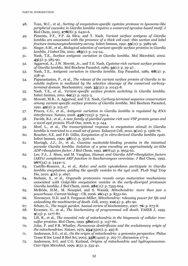
PART III: INTRODUCTION
48. Touz, M.C., et al., Sorting of encystation-specific cysteine protease to lysosome-like peripheral vacuoles in Giardia lamblia requires a conserved tyrosine-based motif. J Biol Chem, 2003. 278(8): p. 6420-6.
49. Pimenta, P.F., P.P. da Silva, and T. Nash, Variant surface antigens of Giardia lamblia are associated with the presence of a thick cell coat: thin section and label fracture immunocytochemistry survey. Infect Immun, 1991. 59(11): p. 3989-96.
50. Singer, S.M., et al., Biological selection of variant-specific surface proteins in Giardia lamblia. J Infect Dis, 2001. 183(1): p. 119-24.
51. Nash, T.E., Surface antigenic variation in Giardia lamblia. Mol Microbiol, 2002. 45(3): p. 585-90.
52. Aggarwal, A., J.W. Merritt, Jr., and T.E. Nash, Cysteine-rich variant surface proteins of Giardia lamblia. Mol Biochem Parasitol, 1989. 32(1): p. 39-47.
53. Nash, T.E., Antigenic variation in Giardia lamblia. Exp Parasitol, 1989. 68(2): p. 238-41.
54. Papanastasiou, P., et al., The release of the variant surface protein of Giardia to its soluble isoform is mediated by the selective cleavage of the conserved carboxy-terminal domain. Biochemistry, 1996. 35(31): p. 10143-8.
55. Nash, T.E., et al., Variant-specific surface protein switching in Giardia lamblia. Infect Immun, 2001. 69(3): p. 1922-3.
56. Mowatt, M.R., A. Aggarwal, and T.E. Nash, Carboxy-terminal sequence conservation among variant-specific surface proteins of Giardia lamblia. Mol Biochem Parasitol, 1991. 49(2): p. 215-27.
57. Prucca, C.G., et al., Antigenic variation in Giardia lamblia is regulated by RNA interference. Nature, 2008. 456(7223): p. 750-4.
58. Davids, B.J., et al., A new family of giardial cysteine-rich non-VSP protein genes and a novel cyst protein. PLoS One, 2006. 1: p. e44.
59. Morf, L., et al., The transcriptional response to encystation stimuli in Giardia lamblia is restricted to a small set of genes. Eukaryot Cell, 2010. 9(10): p. 1566-76.
60. Boucher, S.E. and F.D. Gillin, Excystation of in vitro-derived Giardia lamblia cysts. Infect Immun, 1990. 58(11): p. 3516-22.
61. Murtagh, J.J., Jr., et al., Guanine nucleotide-binding proteins in the intestinal parasite Giardia lamblia. Isolation of a gene encoding an approximately 20-kDa ADP-ribosylation factor. J Biol Chem, 1992. 267(14): p. 9654-62.
62. Lee, F.J., J. Moss, and M. Vaughan, Human and Giardia ADP-ribosylation factors (ARFs) complement ARF function in Saccharomyces cerevisiae. J Biol Chem, 1992. 267(34): p. 24441-5.
63. Castillo-Romero, A., et al., Rab11 and actin cytoskeleton participate in Giardia lamblia encystation, guiding the specific vesicles to the cyst wall. PLoS Negl Trop Dis, 2010. 4(6): p. e697.
64. Stefanic, S., et al., Organelle proteomics reveals cargo maturation mechanisms associated with Golgi-like encystation vesicles in the early-diverged protozoan Giardia lamblia. J Biol Chem, 2006. 281(11): p. 7595-604.
65. McBride, H.M., M. Neuspiel, and S. Wasiak, Mitochondria: more than just a powerhouse. Current biology : CB, 2006. 16(14): p. R551-60.
66. Newmeyer, D.D. and S. Ferguson-Miller, Mitochondria: releasing power for life and unleashing the machineries of death. Cell, 2003. 112(4): p. 481-90.
67. Schatz, G., The magic garden. Annual review of biochemistry, 2007. 76: p. 673-8. 68. Kroemer, G., et al., The biochemistry of programmed cell death. FASEB J, 1995.
9(13): p. 1277-87. 69. Lill, R., et al., The essential role of mitochondria in the biogenesis of cellular iron-
sulfur proteins. Biol Chem, 1999. 380(10): p. 1157-66. 70. John, P. and F.R. Whatley, Paracoccus denitrificans and the evolutionary origin of
the mitochondrion. Nature, 1975. 254(5500): p. 495-8. 71. Andersson, S.G., et al., On the origin of mitochondria: a genomics perspective. Philos
Trans R Soc Lond B Biol Sci, 2003. 358(1429): p. 165-77; discussion 177-9. 72. Andersson, S.G. and C.G. Kurland, Origins of mitochondria and hydrogenosomes.
Curr Opin Microbiol, 1999. 2(5): p. 535-41.
32
PART III: INTRODUCTION
73. Burger, G., M.W. Gray, and B.F. Lang, Mitochondrial genomes: anything goes. Trends Genet, 2003. 19(12): p. 709-16.
74. Lang, B.F., M.W. Gray, and G. Burger, Mitochondrial genome evolution and the origin of eukaryotes. Annu Rev Genet, 1999. 33: p. 351-97.
75. Gray, M.W., The evolutionary origins of organelles. Trends Genet, 1989. 5(9): p. 294-9.
76. Gray, M.W., Origin and evolution of mitochondrial DNA. Annu Rev Cell Biol, 1989. 5: p. 25-50.
77. Gray, M.W., et al., On the evolutionary origin of the plant mitochondrion and its genome. Proc Natl Acad Sci U S A, 1989. 86(7): p. 2267-71.
78. Martin, W. and M. Muller, The hydrogen hypothesis for the first eukaryote. Nature, 1998. 392(6671): p. 37-41.
79. Loytynoja, A. and M.C. Milinkovitch, Molecular phylogenetic analyses of the mitochondrial ADP-ATP carriers: the Plantae/Fungi/Metazoa trichotomy revisited. Proc Natl Acad Sci U S A, 2001. 98(18): p. 10202-7.
80. LóPez-García, P., Moreira, D The Syntrophy Hypothesis for the Origin of Eukaryotes. 2002.
81. van der Giezen, M., Endosymbiosis: past and present. Heredity (Edinb), 2005. 95(5): p. 335-6.
82. Gray, M.W., Mitochondrial evolution. Cold Spring Harb Perspect Biol, 2012. 4(9): p. a011403.
83. Thrash, J.C., et al., Phylogenomic evidence for a common ancestor of mitochondria and the SAR11 clade. Sci Rep, 2011. 1: p. 13.
84. van der Giezen, M. and J. Tovar, Degenerate mitochondria. EMBO Rep, 2005. 6(6): p. 525-30.
85. van der Giezen, M., S. Cox, and J. Tovar, The iron-sulfur cluster assembly genes iscS and iscU of Entamoeba histolytica were acquired by horizontal gene transfer. BMC Evol Biol, 2004. 4: p. 7.
86. Muller, M., et al., Biochemistry and evolution of anaerobic energy metabolism in eukaryotes. Microbiol Mol Biol Rev, 2012. 76(2): p. 444-95.
87. van der Giezen, M., J. Tovar, and C.G. Clark, Mitochondrion-derived organelles in protists and fungi. Int Rev Cytol, 2005. 244: p. 175-225.
88. Lindmark, D.G. and M. Muller, Hydrogenosome, a cytoplasmic organelle of the anaerobic flagellate Tritrichomonas foetus, and its role in pyruvate metabolism. J Biol Chem, 1973. 248(22): p. 7724-8.
89. Mai, Z., et al., Hsp60 is targeted to a cryptic mitochondrion-derived organelle ("crypton") in the microaerophilic protozoan parasite Entamoeba histolytica. Mol Cell Biol, 1999. 19(3): p. 2198-205.
90. Tovar, J., A. Fischer, and C.G. Clark, The mitosome, a novel organelle related to mitochondria in the amitochondrial parasite Entamoeba histolytica. Mol Microbiol, 1999. 32(5): p. 1013-21.
91. Embley, T.M. and R.P. Hirt, Early branching eukaryotes? Current opinion in genetics & development, 1998. 8(6): p. 624-9.
92. Embley, T.M., et al., Hydrogenosomes, mitochondria and early eukaryotic evolution. IUBMB Life, 2003. 55(7): p. 387-95.
93. Embley, T.M., et al., Mitochondria and hydrogenosomes are two forms of the same fundamental organelle. Philos Trans R Soc Lond B Biol Sci, 2003. 358(1429): p. 191-201; discussion 201-2.
94. Riordan, C.E., et al., Cryptosporidium parvum Cpn60 targets a relict organelle. Curr Genet, 2003. 44(3): p. 138-47.
95. Cerkasovová A, et al., Biochemical characterization of large granule fraction of Tritrichomonas foetus (strain KV1). Journal of Protozoology, 1973. 20(525).
96. Whatley, J.M., P. John, and F.R. Whatley, From extracellular to intracellular: the establishment of mitochondria and chloroplasts. Proc R Soc Lond B Biol Sci, 1979. 204(1155): p. 165-87.
97. Boxma, B., et al., An anaerobic mitochondrion that produces hydrogen. Nature, 2005. 434(7029): p. 74-9.
33

PART III: INTRODUCTION
98. Shiflett, A.M. and P.J. Johnson, Mitochondrion-related organelles in eukaryotic protists. Annu Rev Microbiol, 2010. 64: p. 409-29.
99. Williams, B.A., et al., A mitochondrial remnant in the microsporidian Trachipleistophora hominis. Nature, 2002. 418(6900): p. 865-9.
100. Williams, B.A., et al., Distinct localization patterns of two putative mitochondrial proteins in the microsporidian Encephalitozoon cuniculi. J Eukaryot Microbiol, 2008. 55(2): p. 131-3.
101. Tovar, J., et al., Mitochondrial remnant organelles of Giardia function in iron-sulphur protein maturation. Nature, 2003. 426(6963): p. 172-6.
102. Brown, J.R. and W.F. Doolittle, Root of the universal tree of life based on ancient aminoacyl-tRNA synthetase gene duplications. Proc Natl Acad Sci U S A, 1995. 92(7): p. 2441-5.
103. Hashimoto, T., et al., Secondary absence of mitochondria in Giardia lamblia and Trichomonas vaginalis revealed by valyl-tRNA synthetase phylogeny. Proc Natl Acad Sci U S A, 1998. 95(12): p. 6860-5.
104. Soltys, B.J. and R.S. Gupta, Presence and cellular distribution of a 60-kDa protein related to mitochondrial hsp60 in Giardia lamblia. J Parasitol, 1994. 80(4): p. 580-90.
105. Arisue, N., et al., Mitochondrial-type hsp70 genes of the amitochondriate protists, Giardia intestinalis, Entamoeba histolytica and two microsporidians. Parasitology international, 2002. 51(1): p. 9-16.
106. Morrison, H.G., et al., Giardia lamblia expresses a proteobacterial-like DnaK homolog. Mol Biol Evol, 2001. 18(4): p. 530-41.
107. Millet, C.O., et al., Mitochondria-derived organelles in the diplomonad fish parasite Spironucleus vortens. Exp Parasitol, 2013. 135(2): p. 262-73.
108. Williams, C.F., et al., Diversity in mitochondrion-derived organelles of the parasitic diplomonads Spironucleus and Giardia. Trends in parasitology, 2013. 29(7): p. 311-2.
109. Jerlstrom-Hultqvist, J., et al., Hydrogenosomes in the diplomonad Spironucleus salmonicida. Nat Commun, 2013. 4: p. 2493.
110. Martin, W., The missing link between hydrogenosomes and mitochondria. Trends Microbiol, 2005. 13(10): p. 457-9.
111. Jacobson, M.D., M. Weil, and M.C. Raff, Programmed cell death in animal development. Cell, 1997. 88(3): p. 347-54.
112. Wyllie, A.H., J.F. Kerr, and A.R. Currie, Cell death: the significance of apoptosis. International review of cytology, 1980. 68: p. 251-306.
113. Yuan, J. and H.R. Horvitz, A first insight into the molecular mechanisms of apoptosis. Cell, 2004. 116(2 Suppl): p. S53-6, 1 p following S59.
114. Welburn, S.C., M.A. Barcinski, and G.T. Williams, Programmed cell death in trypanosomatids. Parasitology today, 1997. 13(1): p. 22-6.
115. Fiers, W., et al., More than one way to die: apoptosis, necrosis and reactive oxygen damage. Oncogene, 1999. 18(54): p. 7719-30.
116. Sperandio, S., I. de Belle, and D.E. Bredesen, An alternative, nonapoptotic form of programmed cell death. Proceedings of the National Academy of Sciences of the United States of America, 2000. 97(26): p. 14376-81.
117. Elmore, S., Apoptosis: a review of programmed cell death. Toxicologic pathology, 2007. 35(4): p. 495-516.
118. Bortner, C.D., N.B. Oldenburg, and J.A. Cidlowski, The role of DNA fragmentation in apoptosis. Trends in cell biology, 1995. 5(1): p. 21-6.
119. Fadok, V.A. and P.M. Henson, Apoptosis: getting rid of the bodies. Current biology : CB, 1998. 8(19): p. R693-5.
120. Bratton, D.L., et al., Appearance of phosphatidylserine on apoptotic cells requires calcium-mediated nonspecific flip-flop and is enhanced by loss of the aminophospholipid translocase. The Journal of biological chemistry, 1997. 272(42): p. 26159-65.
121. Zamzami, N. and G. Kroemer, The mitochondrion in apoptosis: how Pandora's box opens. Nat Rev Mol Cell Biol, 2001. 2(1): p. 67-71.
34
PART III: INTRODUCTION
122. Brenner, C. and G. Kroemer, Apoptosis. Mitochondria--the death signal integrators. Science, 2000. 289(5482): p. 1150-1.
123. van Loo, G., et al., The role of mitochondrial factors in apoptosis: a Russian roulette with more than one bullet. Cell death and differentiation, 2002. 9(10): p. 1031-42.
124. Chose, O., et al., A form of cell death with some features resembling apoptosis in the amitochondrial unicellular organism Trichomonas vaginalis. Experimental cell research, 2002. 276(1): p. 32-9.
125. Mariante, R.M., et al., Hydrogen peroxide induces caspase activation and programmed cell death in the amitochondrial Tritrichomonas foetus. Histochemistry and cell biology, 2003. 120(2): p. 129-41.
126. Chose, O., et al., Cell death in protists without mitochondria. Annals of the New York Academy of Sciences, 2003. 1010: p. 121-5.
127. Ghosh, E., et al., Oxidative stress-induced cell cycle blockage and a protease-independent programmed cell death in microaerophilic Giardia lamblia. Drug design, development and therapy, 2009. 3: p. 103-10.
128. Correa, G., et al., Cell death induction in Giardia lamblia: effect of beta-lapachone and starvation. Parasitology international, 2009. 58(4): p. 424-37.
129. Bagchi, S., et al., Programmed cell death in Giardia. Parasitology, 2012. 139(7): p. 894-903.
130. Embley, T.M. and W. Martin, Eukaryotic evolution, changes and challenges. Nature, 2006. 440(7084): p. 623-30.
131. Regoes, A., et al., Protein import, replication, and inheritance of a vestigial mitochondrion. J Biol Chem, 2005. 280(34): p. 30557-63.
132. Dolezal, P., et al., Giardia mitosomes and trichomonad hydrogenosomes share a common mode of protein targeting. Proc Natl Acad Sci U S A, 2005. 102(31): p. 10924-9.
133. Nasirudeen, A.M. and K.S. Tan, Isolation and characterization of the mitochondrion-like organelle from Blastocystis hominis. J Microbiol Methods, 2004. 58(1): p. 101-9.
134. Hehl, A.B., et al., Bax function in the absence of mitochondria in the primitive protozoan Giardia lamblia. PLoS One, 2007. 2(5): p. e488.
135. Lill, R. and G. Kispal, Maturation of cellular Fe-S proteins: an essential function of mitochondria. Trends Biochem Sci, 2000. 25(8): p. 352-6.
136. Jedelsky, P.L., et al., The minimal proteome in the reduced mitochondrion of the parasitic protist Giardia intestinalis. PLoS One, 2011. 6(2): p. e17285.
137. Abrahamsen, M.S., et al., Complete genome sequence of the apicomplexan, Cryptosporidium parvum. Science, 2004. 304(5669): p. 441-5.
138. Franzen, O., et al., Draft genome sequencing of giardia intestinalis assemblage B isolate GS: is human giardiasis caused by two different species? PLoS Pathog, 2009. 5(8): p. e1000560.
139. Chan, K.W., et al., A novel ADP/ATP transporter in the mitosome of the microaerophilic human parasite Entamoeba histolytica. Current biology : CB, 2005. 15(8): p. 737-42.
140. Chacinska, A., et al., Importing mitochondrial proteins: machineries and mechanisms. Cell, 2009. 138(4): p. 628-44.
141. Clements, A., et al., The reducible complexity of a mitochondrial molecular machine. Proc Natl Acad Sci U S A, 2009. 106(37): p. 15791-5.
142. Neupert, W. and J.M. Herrmann, Translocation of proteins into mitochondria. Annual review of biochemistry, 2007. 76: p. 723-49.
143. Schatz, G. and B. Dobberstein, Common principles of protein translocation across membranes. Science, 1996. 271(5255): p. 1519-26.
144. Pfanner, N. and A. Geissler, Versatility of the mitochondrial protein import machinery. Nat Rev Mol Cell Biol, 2001. 2(5): p. 339-49.
145. Brix, J., K. Dietmeier, and N. Pfanner, Differential recognition of preproteins by the purified cytosolic domains of the mitochondrial import receptors Tom20, Tom22, and Tom70. J Biol Chem, 1997. 272(33): p. 20730-5.
35
PART III: INTRODUCTION
146. Meisinger, C., et al., Protein import channel of the outer mitochondrial membrane: a highly stable Tom40-Tom22 core structure differentially interacts with preproteins, small tom proteins, and import receptors. Mol Cell Biol, 2001. 21(7): p. 2337-48.
147. von Heijne, G., J. Steppuhn, and R.G. Herrmann, Domain structure of mitochondrial and chloroplast targeting peptides. Eur J Biochem, 1989. 180(3): p. 535-45.
148. Gakh, O., P. Cavadini, and G. Isaya, Mitochondrial processing peptidases. Biochim Biophys Acta, 2002. 1592(1): p. 63-77.
149. Smid, O., et al., Reductive evolution of the mitochondrial processing peptidases of the unicellular parasites trichomonas vaginalis and giardia intestinalis. PLoS Pathog, 2008. 4(12): p. e1000243.
150. Dolezal, P., et al., Evolution of the molecular machines for protein import into mitochondria. Science, 2006. 313(5785): p. 314-8.
151. Smutna, T., et al., Flavodiiron protein from Trichomonas vaginalis hydrogenosomes: the terminal oxygen reductase. Eukaryot Cell, 2009. 8(1): p. 47-55.
152. Bradley, P.J., et al., Targeting and translocation of proteins into the hydrogenosome of the protist Trichomonas: similarities with mitochondrial protein import. EMBO J, 1997. 16(12): p. 3484-93.
153. Gerber, J., et al., The yeast scaffold proteins Isu1p and Isu2p are required inside mitochondria for maturation of cytosolic Fe/S proteins. Mol Cell Biol, 2004. 24(11): p. 4848-57.
154. Mukhopadhyay, A., et al., Precursor protein is readily degraded in mitochondrial matrix space if the leader is not processed by mitochondrial processing peptidase. J Biol Chem, 2007. 282(51): p. 37266-75.
155. Saavedra-Alanis, V.M., et al., Rat liver mitochondrial processing peptidase. Both alpha- and beta-subunits are required for activity. J Biol Chem, 1994. 269(12): p. 9284-8.
156. Kitada, S., et al., A protein from a parasitic microorganism, Rickettsia prowazekii, can cleave the signal sequences of proteins targeting mitochondria. J Bacteriol, 2007. 189(3): p. 844-50.
157. Brown, M.T., et al., A functionally divergent hydrogenosomal peptidase with protomitochondrial ancestry. Mol Microbiol, 2007. 64(5): p. 1154-63.
158. Westermann, B. and W. Neupert, 'Omics' of the mitochondrion. Nat Biotechnol, 2003. 21(3): p. 239-40.
159. Mokranjac, D. and W. Neupert, Thirty years of protein translocation into mitochondria: unexpectedly complex and still puzzling. Biochim Biophys Acta, 2009. 1793(1): p. 33-41.
160. Chacinska, A., N. Pfanner, and C. Meisinger, How mitochondria import hydrophilic and hydrophobic proteins. Trends Cell Biol, 2002. 12(7): p. 299-303.
161. Pfanner, N. and K.N. Truscott, Powering mitochondrial protein import. Nat Struct Biol, 2002. 9(4): p. 234-6.
162. Pfanner, N. and N. Wiedemann, Mitochondrial protein import: two membranes, three translocases. Current opinion in cell biology, 2002. 14(4): p. 400-11.
163. Katinka, M.D., et al., Genome sequence and gene compaction of the eukaryote parasite Encephalitozoon cuniculi. Nature, 2001. 414(6862): p. 450-3.
164. Loftus, B., et al., The genome of the protist parasite Entamoeba histolytica. Nature, 2005. 433(7028): p. 865-8.
165. Loftus, B.J. and N. Hall, Entamoeba: still more to be learned from the genome. Trends in parasitology, 2005. 21(10): p. 453.
166. Makiuchi, T., et al., Novel TPR-containing subunit of TOM complex functions as cytosolic receptor for Entamoeba mitosomal transport. Sci Rep, 2013. 3: p. 1129.
167. Carlton, J.M., et al., Draft genome sequence of the sexually transmitted pathogen Trichomonas vaginalis. Science, 2007. 315(5809): p. 207-12.
168. Pusnik, M., et al., An essential novel component of the noncanonical mitochondrial outer membrane protein import system of trypanosomatids. Mol Biol Cell, 2012. 23(17): p. 3420-8.
36
PART III: INTRODUCTION
169. Dagley, M.J., et al., The protein import channel in the outer mitosomal membrane of Giardia intestinalis. Mol Biol Evol, 2009. 26(9): p. 1941-7.
170. Dukanovic, J. and D. Rapaport, Multiple pathways in the integration of proteins into the mitochondrial outer membrane. Biochim Biophys Acta, 2011. 1808(3): p. 971-80.
171. Lithgow, T. and A. Schneider, Evolution of macromolecular import pathways in mitochondria, hydrogenosomes and mitosomes. Philosophical Transactions of the Royal Society B-Biological Sciences, 2010. 365(1541): p. 799-817.
172. Kutik, S., et al., Dissecting membrane insertion of mitochondrial beta-barrel proteins. Cell, 2008. 132(6): p. 1011-24.
173. Habib, S.J., et al., Assembly of the TOB complex of mitochondria. J Biol Chem, 2005. 280(8): p. 6434-40.
174. Becker, T., et al., Biogenesis of the mitochondrial TOM complex: Mim1 promotes insertion and assembly of signal-anchored receptors. J Biol Chem, 2008. 283(1): p. 120-7.
175. Hell, K., The Erv1-Mia40 disulfide relay system in the intermembrane space of mitochondria. Biochim Biophys Acta, 2008. 1783(4): p. 601-9.
176. Stojanovski, D., et al., The MIA system for protein import into the mitochondrial intermembrane space. Biochim Biophys Acta, 2008. 1783(4): p. 610-7.
177. Mesecke, N., et al., A disulfide relay system in the intermembrane space of mitochondria that mediates protein import. Cell, 2005. 121(7): p. 1059-69.
178. Grumbt, B., et al., Functional characterization of Mia40p, the central component of the disulfide relay system of the mitochondrial intermembrane space. J Biol Chem, 2007. 282(52): p. 37461-70.
179. Milenkovic, D., et al., Identification of the signal directing Tim9 and Tim10 into the intermembrane space of mitochondria. Mol Biol Cell, 2009. 20(10): p. 2530-9.
180. Hwang, D.K., et al., Tim54p connects inner membrane assembly and proteolytic pathways in the mitochondrion. J Cell Biol, 2007. 178(7): p. 1161-75.
181. Gebert, N., et al., Dual function of Sdh3 in the respiratory chain and TIM22 protein translocase of the mitochondrial inner membrane. Mol Cell, 2011. 44(5): p. 811-8.
182. Denoeud, F., et al., Genome sequence of the stramenopile Blastocystis, a human anaerobic parasite. Genome Biol, 2011. 12(3): p. R29.
183. Makiuchi, T. and T. Nozaki, Highly divergent mitochondrion-related organelles in anaerobic parasitic protozoa. Biochimie, 2014. 100: p. 3-17.
184. Chacinska, A., et al., Mitochondrial presequence translocase: switching between TOM tethering and motor recruitment involves Tim21 and Tim17. Cell, 2005. 120(6): p. 817-29.
185. van der Laan, M., et al., Pam17 is required for architecture and translocation activity of the mitochondrial protein import motor. Mol Cell Biol, 2005. 25(17): p. 7449-58.
186. Meier, S., W. Neupert, and J.M. Herrmann, Conserved N-terminal negative charges in the Tim17 subunit of the TIM23 translocase play a critical role in the import of preproteins into mitochondria. J Biol Chem, 2005. 280(9): p. 7777-85.
187. Bauer, M.F., et al., Role of Tim23 as voltage sensor and presequence receptor in protein import into mitochondria. Cell, 1996. 87(1): p. 33-41.
188. Dudek, J., P. Rehling, and M. van der Laan, Mitochondrial protein import: common principles and physiological networks. Biochim Biophys Acta, 2013. 1833(2): p. 274-85.
189. Barbera, M.J., et al., Sawyeria marylandensis (Heterolobosea) has a hydrogenosome with novel metabolic properties. Eukaryot Cell, 2010. 9(12): p. 1913-24.
190. Martincova, E., et al., Probing the biology of Giardia intestinalis mitosomes using in vivo enzymatic tagging. Mol Cell Biol, 2015.
37
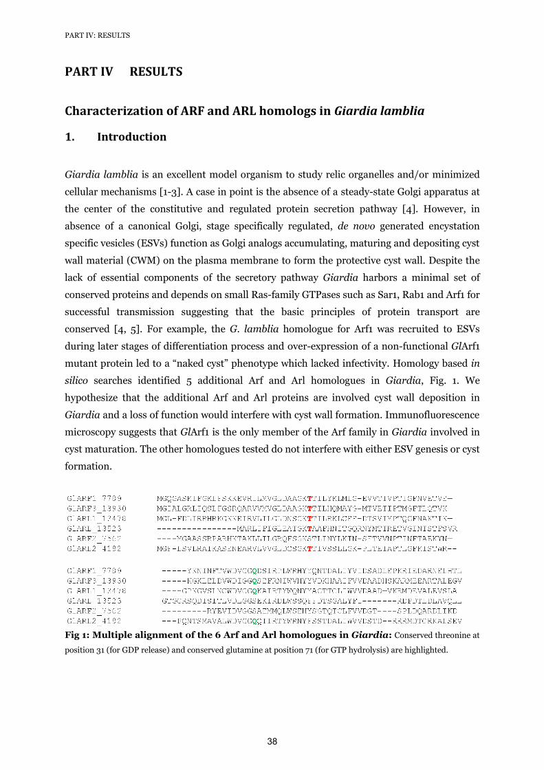
PART IV: RESULTS
PART IV RESULTS
Characterization of ARF and ARL homologs in Giardia lamblia
1. Introduction
Giardia lamblia is an excellent model organism to study relic organelles and/or minimized
cellular mechanisms [1-3]. A case in point is the absence of a steady-state Golgi apparatus at
the center of the constitutive and regulated protein secretion pathway [4]. However, in
absence of a canonical Golgi, stage specifically regulated, de novo generated encystation
specific vesicles (ESVs) function as Golgi analogs accumulating, maturing and depositing cyst
wall material (CWM) on the plasma membrane to form the protective cyst wall. Despite the
lack of essential components of the secretory pathway Giardia harbors a minimal set of
conserved proteins and depends on small Ras-family GTPases such as Sar1, Rab1 and Arf1 for
successful transmission suggesting that the basic principles of protein transport are
conserved [4, 5]. For example, the G. lamblia homologue for Arf1 was recruited to ESVs
during later stages of differentiation process and over-expression of a non-functional GlArf1
mutant protein led to a “naked cyst” phenotype which lacked infectivity. Homology based in
silico searches identified 5 additional Arf and Arl homologues in Giardia, Fig. 1. We
hypothesize that the additional Arf and Arl proteins are involved cyst wall deposition in
Giardia and a loss of function would interfere with cyst wall formation. Immunofluorescence
microscopy suggests that GlArf1 is the only member of the Arf family in Giardia involved in
cyst maturation. The other homologues tested do not interfere with either ESV genesis or cyst
formation.
Fig 1: Multiple alignment of the 6 Arf and Arl homologues in Giardia: Conserved threonine at
position 31 (for GDP release) and conserved glutamine at position 71 (for GTP hydrolysis) are highlighted.
38
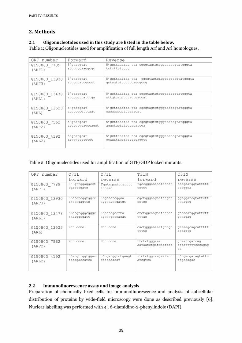
PART IV: RESULTS
2. Methods
2.1 Oligonucleotides used in this study are listed in the table below.
Table 1: Oligonucleotides used for amplification of full length Arf and Arl homologues.
ORF number Forward Reverse Gl50803_7789
(ARF1)
5’gcatgcat atgggccaaggcgc
5’gcttaattaa tta cgcgtagtctgggacatcgtatgggta tctcttcttccc
Gl50803_13930
(ARF3)
5’gcatgcat atgggcatcgccct
5’gcttaattaa tta cgcgtagtctgggacatcgtatgggta gctagtctccttccagcgccg
Gl50803_13478
(ARL1)
5’gcatgcat atggggttatttga
5’gcttaattaa cta cgcgtagtctgggacatcgtatgggta cttgtcagtcttactgaccat
Gl50803_13523
(ARL)
5’gcatgcat atggcgcgtttaat
5’gcttaattaa tta cgcgtagtctgggacatcgtatgggta cacagacgttgtaaacat
Gl50803_7562
(ARF2)
5’gcatgcat atgggtgcagccagct
5’gcttaattaa tca cgcgtagtctgggacatcgtatgggta aggctgctttggcacatcga
Gl50803_4192
(ARL2)
5’gcatgcat atgggctttctct
5’gcttaattaa tca cgcgtagtctgggacatcgtatgggta ccaaatagcagtctccaggtt
Table 2: Oligonucleotides used for amplification of GTP/GDP locked mutants.
ORF number Q71L forward
Q71L reverse
T31N forward
T31N reverse
Gl50803_7789
(ARF1)
5’gttggaggcctcgattcgatc
5’gatcgaatcgaggcctccaac
tgccgggaaaaataccat
tcttt
aaagaatggtattttt
cccggca
Gl50803_13930
(ARF3)
5’acatcggtggcctttccgagttc
5’gaactcggaa aggccaccgatgt
cgctgggaagaatacgatcctcc
ggaggatcgtattcttcccagcg
Gl50803_13478
(ARL1)
5’atgtgggcgggcttaaggcgatt
5’aatcgcctta agcccgcccacat
ctctggcaagaataccat
tttac
gtaaaatggtattctt
gccagag
Gl50803_13523
(ARL)
Not done Not done cactgggaaaaatgctgc
ttttc
gaaaagcagcattttt
cccagtg
Gl50803_7562
(ARF2)
Not done Not done ttctctgggaaa aataatctgatcaattac
gtaattgatcag attatttttcccagag
aa
Gl50803_4192
(ARL2)
5’atgttggtggacttcagaccatca
5’tgatggtctgaagtccaccaacat
5’ctctggcaagaatactatcgtca
5’tgacgatagtattcttgccagac
2.2 Immunofluorescence assay and image analysis
Preparation of chemically fixed cells for immunofluorescence and analysis of subcellular
distribution of proteins by wide-field microscopy were done as described previously [6].
Nuclear labelling was performed with 4', 6-diamidino-2-phenylindole (DAPI).
39
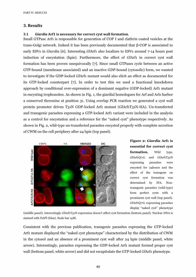
PART IV: RESULTS
3. Results
3.1 Giardia Arf1 is necessary for correct cyst wall formation.
Small GTPase Arf1 is responsible for generation of COP I and clathrin coated vesicles at the
trans-Golgi network. Indeed it has been previously documented that β-COP is associated to
early ESVs in Giardia [6]. Interesting GlArf1 also localizes to ESVs around 7-14 hours post
induction of encystation (hpie). Furthermore, the effect of GlArf1 in correct cyst wall
formation has been proven unequivocally [7]. Since small GTPases cycle between an active
GTP-bound (membrane associated) and an inactive GDP-bound (cytosolic) form, we wanted
to investigate if the GDP-locked GlArf1 mutant would also elicit an effect as documented for
its GTP-locked counterpart [7]. In order to test this we used a functional knockdown
approach by conditional over-expression of a dominant negative (GDP-locked) Arf1 mutant
in encysting trophozoites. As shown in Fig. 1, the giardial homologues for Arf and Arls harbor
a conserved threonine at position 31. Using overlap PCR reaction we generated a cyst wall
protein promoter driven T31N GDP-locked Arf1 mutant (GlArf1T31N-HA). Un-transfected
and transgenic parasites expressing a GTP-locked Arf1 variant were included in the analysis
as a control for encystation and a reference for the “naked cyst” phenotype respectively. As
shown in Fig. 2, wild-type un-transfected parasites encysted properly with complete secretion
of CWM on the cell periphery after 24 hpie (top panel).
Figure 2: Giardia Arf1 is
essential for correct cyst
formation. Wild type,
GlArf1Q71L and GlArf1T31N
expressing parasites were
encysted for 24hours and the
effect of the transgene on
correct cyst formation was
determined by IFA. Non-
transgenic parasites (wild-type)
form perfect cysts with a
prominent cyst wall (top panel).
GlArf1Q71L expressing parasites
display “naked cyst” phenotype
(middle panel). Interestingly GlArf1T31N expression doesn’t affect cyst formation (bottom panel). Nuclear DNA is
stained with DAPI (blue). Scale bar 1µM.
Consistent with the previous publication, transgenic parasites expressing the GTP-locked
Arf1 mutant displayed the “naked cyst phenotype” characterized by the distribution of CWM
in the cytosol and an absence of a prominent cyst wall after 24 hpie (middle panel, white
arrow). Interestingly, parasites expressing the GDP-locked Arf1 mutant formed proper cyst
wall (bottom panel, white arrow) and did not recapitulate the GTP-locked GlArf1 phenotype.
40
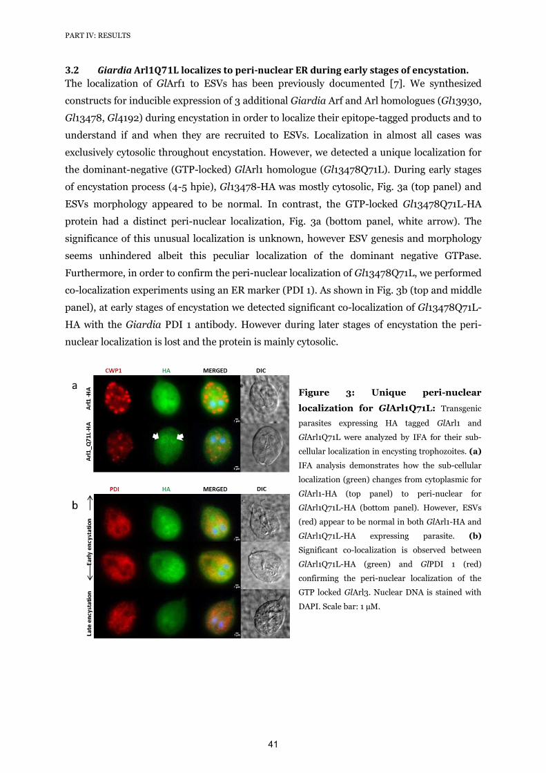
PART IV: RESULTS
3.2 Giardia Arl1Q71L localizes to peri-nuclear ER during early stages of encystation.
The localization of GlArf1 to ESVs has been previously documented [7]. We synthesized
constructs for inducible expression of 3 additional Giardia Arf and Arl homologues (Gl13930,
Gl13478, Gl4192) during encystation in order to localize their epitope-tagged products and to
understand if and when they are recruited to ESVs. Localization in almost all cases was
exclusively cytosolic throughout encystation. However, we detected a unique localization for
the dominant-negative (GTP-locked) GlArl1 homologue (Gl13478Q71L). During early stages
of encystation process (4-5 hpie), Gl13478-HA was mostly cytosolic, Fig. 3a (top panel) and
ESVs morphology appeared to be normal. In contrast, the GTP-locked Gl13478Q71L-HA
protein had a distinct peri-nuclear localization, Fig. 3a (bottom panel, white arrow). The
significance of this unusual localization is unknown, however ESV genesis and morphology
seems unhindered albeit this peculiar localization of the dominant negative GTPase.
Furthermore, in order to confirm the peri-nuclear localization of Gl13478Q71L, we performed
co-localization experiments using an ER marker (PDI 1). As shown in Fig. 3b (top and middle
panel), at early stages of encystation we detected significant co-localization of Gl13478Q71L-
HA with the Giardia PDI 1 antibody. However during later stages of encystation the peri-
nuclear localization is lost and the protein is mainly cytosolic.
Figure 3: Unique peri-nuclear
localization for GlArl1Q71L: Transgenic
parasites expressing HA tagged GlArl1 and
GlArl1Q71L were analyzed by IFA for their sub-
cellular localization in encysting trophozoites. (a)
IFA analysis demonstrates how the sub-cellular
localization (green) changes from cytoplasmic for
GlArl1-HA (top panel) to peri-nuclear for
GlArl1Q71L-HA (bottom panel). However, ESVs
(red) appear to be normal in both GlArl1-HA and
GlArl1Q71L-HA expressing parasite. (b)
Significant co-localization is observed between
GlArl1Q71L-HA (green) and GlPDI 1 (red)
confirming the peri-nuclear localization of the
GTP locked GlArl3. Nuclear DNA is stained with
DAPI. Scale bar: 1 µM.
41
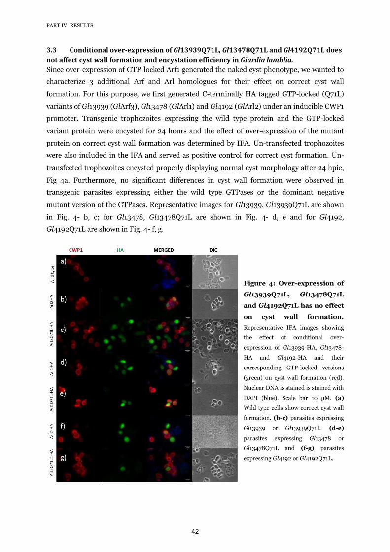
PART IV: RESULTS
3.3 Conditional over-expression of Gl13939Q71L, Gl13478Q71L and Gl4192Q71L does
not affect cyst wall formation and encystation efficiency in Giardia lamblia.
Since over-expression of GTP-locked Arf1 generated the naked cyst phenotype, we wanted to
characterize 3 additional Arf and Arl homologues for their effect on correct cyst wall
formation. For this purpose, we first generated C-terminally HA tagged GTP-locked (Q71L)
variants of Gl13939 (GlArf3), Gl13478 (GlArl1) and Gl4192 (GlArl2) under an inducible CWP1
promoter. Transgenic trophozoites expressing the wild type protein and the GTP-locked
variant protein were encysted for 24 hours and the effect of over-expression of the mutant
protein on correct cyst wall formation was determined by IFA. Un-transfected trophozoites
were also included in the IFA and served as positive control for correct cyst formation. Un-
transfected trophozoites encysted properly displaying normal cyst morphology after 24 hpie,
Fig 4a. Furthermore, no significant differences in cyst wall formation were observed in
transgenic parasites expressing either the wild type GTPases or the dominant negative
mutant version of the GTPases. Representative images for Gl13939, Gl13939Q71L are shown
in Fig. 4- b, c; for Gl13478, Gl13478Q71L are shown in Fig. 4- d, e and for Gl4192,
Gl4192Q71L are shown in Fig. 4- f, g.
Figure 4: Over-expression of
Gl13939Q71L, Gl13478Q71L
and Gl4192Q71L has no effect
on cyst wall formation.
Representative IFA images showing
the effect of conditional over-
expression of Gl13939-HA, Gl13478-
HA and Gl4192-HA and their
corresponding GTP-locked versions
(green) on cyst wall formation (red).
Nuclear DNA is stained is stained with
DAPI (blue). Scale bar 10 µM. (a)
Wild type cells show correct cyst wall
formation. (b-c) parasites expressing
Gl13939 or Gl13939Q71L. (d-e)
parasites expressing Gl13478 or
Gl13478Q71L and (f-g) parasites
expressing Gl4192 or Gl4192Q71L.
42
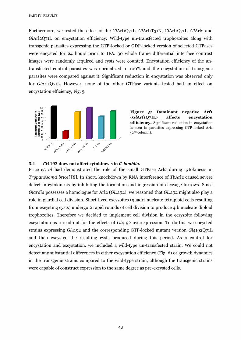
PART IV: RESULTS
Furthermore, we tested the effect of the GlArf1Q71L, GlArf1T31N, GlArl1Q71L, GlArl2 and
GlArl2Q71L on encystation efficiency. Wild-type un-transfected trophozoites along with
transgenic parasites expressing the GTP-locked or GDP-locked version of selected GTPases
were encysted for 24 hours prior to IFA. 30 whole frame differential interface contrast
images were randomly acquired and cysts were counted. Encystation efficiency of the un-
transfected control parasites was normalized to 100% and the encystation of transgenic
parasites were compared against it. Significant reduction in encystation was observed only
for GlArf1Q71L. However, none of the other GTPase variants tested had an effect on
encystation efficiency, Fig. 5.
Figure 5: Dominant negative Arf1
(GlArf1Q71L) affects encystation
efficiency. Significant reduction in encystation
is seen in parasites expressing GTP-locked Arf1
(2nd column).
3.4 Gl4192 does not affect cytokinesis in G. lamblia.
Price et. al had demonstrated the role of the small GTPase Arl2 during cytokinesis in
Trypanosoma bricei [8]. In short, knockdown by RNA interference of TbArl2 caused severe
defect in cytokinesis by inhibiting the formation and ingression of cleavage furrows. Since
Giardia possesses a homologue for Arl2 (Gl4192), we reasoned that Gl4192 might also play a
role in giardial cell division. Short-lived excyzoites (quadri-nucleate tetraploid cells resulting
from excysting cysts) undergo 2 rapid rounds of cell division to produce 4 binucleate diploid
trophozoites. Therefore we decided to implement cell division in the eczyzoite following
excystation as a read-out for the effects of Gl4192 overexpression. To do this we encysted
strains expressing Gl4192 and the corresponding GTP-locked mutant version Gl4192Q71L
and then excysted the resulting cysts produced during this period. As a control for
encystation and excystation, we included a wild-type un-transfected strain. We could not
detect any substantial differences in either excystation efficiency (Fig. 6) or growth dynamics
in the transgenic strains compared to the wild-type strain, although the transgenic strains
were capable of construct expression to the same degree as pre-excysted cells.
43
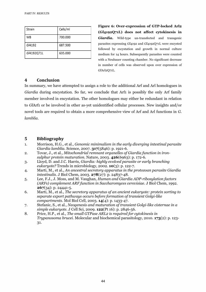
PART IV: RESULTS
Figure 6: Over-expression of GTP-locked Arl2
(Gl4192Q71L) does not affect cytokinesis in
Giardia. Wild-type un-transfected and transgenic
parasites expressing Gl4192 and Gl4192Q71L were encysted
followed by excystation and growth in normal culture
medium for 14 hours. Subsequently parasites were counted
with a Neubauer counting chamber. No significant decrease
in number of cells was observed upon over expression of
GlArl2Q71L.
4 Conclusion
In summary, we have attempted to assign a role to the additional Arf and Arl homologues in
Giardia during encystation. So far, we conclude that Arf1 is possibly the only Arf family
member involved in encystation. The other homologues may either be redundant in relation
to GlArf1 or be involved in other as-yet unidentified cellular processes. New insights and/or
novel tools are required to obtain a more comprehensive view of Arf and Arl functions in G.
lamblia.
5 Bibliography
1. Morrison, H.G., et al., Genomic minimalism in the early diverging intestinal parasite Giardia lamblia. Science, 2007. 317(5846): p. 1921-6.
2. Tovar, J., et al., Mitochondrial remnant organelles of Giardia function in iron-sulphur protein maturation. Nature, 2003. 426(6963): p. 172-6.
3. Lloyd, D. and J.C. Harris, Giardia: highly evolved parasite or early branching eukaryote? Trends in microbiology, 2002. 10(3): p. 122-7.
4. Marti, M., et al., An ancestral secretory apparatus in the protozoan parasite Giardia intestinalis. J Biol Chem, 2003. 278(27): p. 24837-48.
5. Lee, F.J., J. Moss, and M. Vaughan, Human and Giardia ADP-ribosylation factors (ARFs) complement ARF function in Saccharomyces cerevisiae. J Biol Chem, 1992. 267(34): p. 24441-5.
6. Marti, M., et al., The secretory apparatus of an ancient eukaryote: protein sorting to separate export pathways occurs before formation of transient Golgi-like compartments. Mol Biol Cell, 2003. 14(4): p. 1433-47.
7. Stefanic, S., et al., Neogenesis and maturation of transient Golgi-like cisternae in a simple eukaryote. J Cell Sci, 2009. 122(Pt 16): p. 2846-56.
8. Price, H.P., et al., The small GTPase ARL2 is required for cytokinesis in Trypanosoma brucei. Molecular and biochemical parasitology, 2010. 173(2): p. 123-31.
44
PART V: RESULTS
PART V RESULTS
Induction of apoptosis-like cell death in Giardia lamblia
1. Introduction
Programmed cell death (PCD) is a highly regulated cellular process that has been extensively
characterized in metazoans. PCD can be triggered by external and internal factors involving
several effectors and regulators [1]. Two major classes of PCD have been described; 1)
Apoptosis (type I PCD) is accompanied by specific morphological and biochemical changes
leading to the demise of the cell [2] and 2) Autophagy (type II PCD) which involves the
autophagosomal–lysosomal system and is responsible for engulfment of vesicles during
turnover of organelles [3]. PCD has also been documented in several unicellular eukaryotes
namely T. vaginalis, E. histolytica, Dictystelium, Blastocystis, Trypanosoma and Giardia
lamblia [4-7]. Most if not all of the above mentioned organisms do not possess canonical
mitochondria but instead harbor mitochondrion-related organelles (mitosomes and
hydrogenosomes). Therefore it is indeed intriguing to investigate the processes and factors
involved in apoptosis in MRO harboring organisms.
Giardia lamblia parasites reside in millions in the gut epithelium during an acute infection
but, in most cases of giardial infection, the parasites are eventually cleared by the host.
However, despite being present at high concentrations, they do not elicit any significant host
inflammatory response [8, 9]. We hypothesized that Giardia trophozoites undergo an
organized form of cell death; likely apoptosis-related, to avoid excessive exposure of antigenic
molecules to the host’s immune system and hence proliferate incognito in the host’s small
intestine. Interestingly, PCD in Giardia has been demonstrated previously using drugs like
beta-lapachone, H2O2 [10, 11]. However, since these drugs are not present under
physiological conditions we wanted to use: 1) nutrient starvation by growth in EBSS medium
(lacking L-cysteine and ascorbic) and 2) heat shock, as physiological insults to elicit apoptosis
in Giardia. Using annexin-FITC apoptosis assay and immunofluorescence microscopy we
could demonstrate an apoptosis-like cell death exhibiting many if not all characteristics of
apoptosis upon altercation of physiological conditions.
45
PART V: RESULTS
2. Methods
2.1 Induction of PCD
PCD was induced mainly by interfering with two physiological conditions; 1) Glucose
starvation and 2) heat shock.
1) For induction of PCD via glucose starvation, parasites were grown up to 75% confluency in
TYI-S-33 medium. Subsequently the medium was removed eliminating non-adherent cells
(dead/damaged cells) and was replaced by Earle's balanced salts solution (EBSS) medium
and incubated for 12-36 hours prior to harvesting for annexin binding assay or flow
cytometry analysis.
2) Heat treatment was another criterion used to elicit cell death in Giardia. Parasites were
grown up to 75% confluency in TYI-S-33 medium followed by incubation at varying
temperatures (39-42°C) in a water bath. Control parasites were incubated at 37°C and served
as negative control. Parasites were then harvested and processed accordingly for annexin
binding assay or flow cytometry analysis
2.2 Annexin- binding assay and microscopy analysis
Giardial trophozoites were induced for PCD as outlined above. Un-treated parasites were also
included as negative controls. The parasites were harvested and washed once by
centrifugation (900 x g for 10 mins and 4°C) with cold PBS. The parasites were processed as
described in FITC Annexin V/Dead Cell Apoptosis Kit with FITC annexin V and PI, for Flow
Cytometry, Cat no. V13242, Invitrogen. The pellet was re-suspended in 1oo μl of cold 1X
binding buffer to which 5 μl of annexin V-FITC (Invitrogen, UK) and 2 μl of propidium iodide
(PI) (1mg/ml) was added, followed by incubation in the dark at room temperature for 15
minutes. Subsequently the cells were washed with 1X annexin binding buffer and finally
analyzed either by flow cytometry (Beckman Coulter and Kaluza software) or by
epifluorescence microscopy. Microscopy analysis was performed on the standard
fluorescence microscopes Leica DM IRBE with MetaVue software version 5.0r1, or Nikon
Eclipse 80i with Openlab Improvision software 5.5.2 for data collection. WCIF ImageJ was
used for image processing.
2.3 DNA laddering (fragmentation)
Glucose starved and mock treated parasites were harvested followed by genomic DNA
isolation to detect DNA fragmentation. Samples were then run on a 1% agarose gel stained
with ethidium bromide to visualize DNA shearing and fragmentation.
2.4 Immunofluorescence Assay (IFA)
IFA was performed according to standard protocols [12].
46
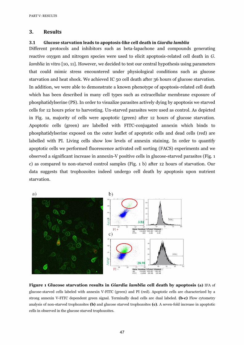
PART V: RESULTS
3. Results
3.1 Glucose starvation leads to apoptosis-like cell death in Giardia lamblia
Different protocols and inhibitors such as beta-lapachone and compounds generating
reactive oxygen and nitrogen species were used to elicit apoptosis-related cell death in G.
lamblia in vitro [10, 11]. However, we decided to test our central hypothesis using parameters
that could mimic stress encountered under physiological conditions such as glucose
starvation and heat shock. We achieved IC 50 cell death after 36 hours of glucose starvation.
In addition, we were able to demonstrate a known phenotype of apoptosis-related cell death
which has been described in many cell types such as extracellular membrane exposure of
phosphatidylserine (PS). In order to visualize parasites actively dying by apoptosis we starved
cells for 12 hours prior to harvesting. Un-starved parasites were used as control. As depicted
in Fig. 1a, majority of cells were apoptotic (green) after 12 hours of glucose starvation.
Apoptotic cells (green) are labelled with FITC-conjugated annexin which binds to
phosphatidylserine exposed on the outer leaflet of apoptotic cells and dead cells (red) are
labelled with PI. Living cells show low levels of annexin staining. In order to quantify
apoptotic cells we performed fluorescence activated cell sorting (FACS) experiments and we
observed a significant increase in annexin-V positive cells in glucose-starved parasites (Fig. 1
c) as compared to non-starved control samples (Fig. 1 b) after 12 hours of starvation. Our
data suggests that trophozoites indeed undergo cell death by apoptosis upon nutrient
starvation.
Figure 1 Glucose starvation results in Giardia lamblia cell death by apoptosis (a) IFA of
glucose-starved cells labeled with annexin V-FITC (green) and PI (red). Apoptotic cells are characterized by a
strong annexin V-FITC dependent green signal. Terminally dead cells are dual labeled. (b-c) Flow cytometry
analysis of non-starved trophozoites (b) and glucose starved trophozoites (c). A seven-fold increase in apoptotic
cells in observed in the glucose starved trophozoites.
47
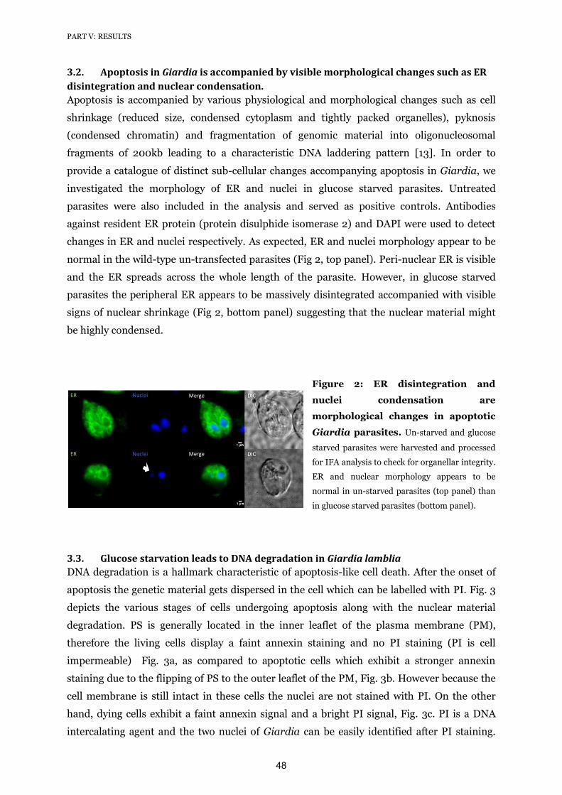
PART V: RESULTS
3.2. Apoptosis in Giardia is accompanied by visible morphological changes such as ER
disintegration and nuclear condensation.
Apoptosis is accompanied by various physiological and morphological changes such as cell
shrinkage (reduced size, condensed cytoplasm and tightly packed organelles), pyknosis
(condensed chromatin) and fragmentation of genomic material into oligonucleosomal
fragments of 200kb leading to a characteristic DNA laddering pattern [13]. In order to
provide a catalogue of distinct sub-cellular changes accompanying apoptosis in Giardia, we
investigated the morphology of ER and nuclei in glucose starved parasites. Untreated
parasites were also included in the analysis and served as positive controls. Antibodies
against resident ER protein (protein disulphide isomerase 2) and DAPI were used to detect
changes in ER and nuclei respectively. As expected, ER and nuclei morphology appear to be
normal in the wild-type un-transfected parasites (Fig 2, top panel). Peri-nuclear ER is visible
and the ER spreads across the whole length of the parasite. However, in glucose starved
parasites the peripheral ER appears to be massively disintegrated accompanied with visible
signs of nuclear shrinkage (Fig 2, bottom panel) suggesting that the nuclear material might
be highly condensed.
Figure 2: ER disintegration and
nuclei condensation are
morphological changes in apoptotic
Giardia parasites. Un-starved and glucose
starved parasites were harvested and processed
for IFA analysis to check for organellar integrity.
ER and nuclear morphology appears to be
normal in un-starved parasites (top panel) than
in glucose starved parasites (bottom panel).
3.3. Glucose starvation leads to DNA degradation in Giardia lamblia
DNA degradation is a hallmark characteristic of apoptosis-like cell death. After the onset of
apoptosis the genetic material gets dispersed in the cell which can be labelled with PI. Fig. 3
depicts the various stages of cells undergoing apoptosis along with the nuclear material
degradation. PS is generally located in the inner leaflet of the plasma membrane (PM),
therefore the living cells display a faint annexin staining and no PI staining (PI is cell
impermeable) Fig. 3a, as compared to apoptotic cells which exhibit a stronger annexin
staining due to the flipping of PS to the outer leaflet of the PM, Fig. 3b. However because the
cell membrane is still intact in these cells the nuclei are not stained with PI. On the other
hand, dying cells exhibit a faint annexin signal and a bright PI signal, Fig. 3c. PI is a DNA
intercalating agent and the two nuclei of Giardia can be easily identified after PI staining.
48

PART V: RESULTS
Terminally dead cells with completely degraded nuclear material (bold arrows) are shown in
Fig. 3d. Punctate PI staining is observed and in some cases the entire cytoplasm is stained
with PI since the nuclear material is completely degraded and dispersed throughout the cell.
Extensive DNA degradation (cytoplasmic PI staining) marks the “point of no return” and the
cells are destined to die silently.
Figure 3: Apoptosis results in nuclear material
degradation. Representative images showing varying
degrees of nuclear material degradation during apoptosis (a)
Living, (b) Apoptotic, (c) Dying and (d) Terminally dead.
3.4 DNA laddering and fragmentation
Genomic DNA fragmentation is a biochemical hallmark in apoptotic cells and is an
irreversible event in PCD. This step occurs before changes in plasma membrane permeability.
Glucose starved and un-starved parasites were subjected to genomic DNA analysis and were
analyzed on a 1% agarose gel. The typical recursive electrophoretic laddering pattern was not
seen in glucose starved parasites (Fig. 4b) as compared to unstarved parasites (Fig. 4a).
However, distinct DNA band (fragmented/sheared DNA) mainly in lower molecular weight
region was observed in the glucose starved parasites.
Figure 4: Apoptotic cells display an unusual DNA
banding pattern at low molecular size range.
Electrophoretic analysis of DNA degradation shows an accumulation
of fragmented DNA at 200 bps in glucose starved parasites (b),
whereas the un-starved have a single band for intact genomic DNA
(a).
49
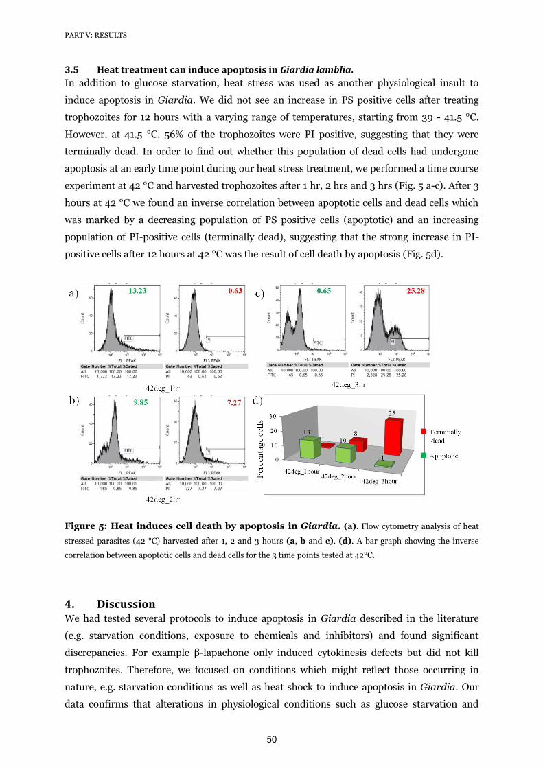
PART V: RESULTS
3.5 Heat treatment can induce apoptosis in Giardia lamblia.
In addition to glucose starvation, heat stress was used as another physiological insult to
induce apoptosis in Giardia. We did not see an increase in PS positive cells after treating
trophozoites for 12 hours with a varying range of temperatures, starting from 39 - 41.5 °C.
However, at 41.5 °C, 56% of the trophozoites were PI positive, suggesting that they were
terminally dead. In order to find out whether this population of dead cells had undergone
apoptosis at an early time point during our heat stress treatment, we performed a time course
experiment at 42 °C and harvested trophozoites after 1 hr, 2 hrs and 3 hrs (Fig. 5 a-c). After 3
hours at 42 °C we found an inverse correlation between apoptotic cells and dead cells which
was marked by a decreasing population of PS positive cells (apoptotic) and an increasing
population of PI-positive cells (terminally dead), suggesting that the strong increase in PI-
positive cells after 12 hours at 42 °C was the result of cell death by apoptosis (Fig. 5d).
Figure 5: Heat induces cell death by apoptosis in Giardia. (a). Flow cytometry analysis of heat
stressed parasites (42 °C) harvested after 1, 2 and 3 hours (a, b and c). (d). A bar graph showing the inverse
correlation between apoptotic cells and dead cells for the 3 time points tested at 42°C.
4. Discussion
We had tested several protocols to induce apoptosis in Giardia described in the literature
(e.g. starvation conditions, exposure to chemicals and inhibitors) and found significant
discrepancies. For example β-lapachone only induced cytokinesis defects but did not kill
trophozoites. Therefore, we focused on conditions which might reflect those occurring in
nature, e.g. starvation conditions as well as heat shock to induce apoptosis in Giardia. Our
data confirms that alterations in physiological conditions such as glucose starvation and
50
PART V: RESULTS
incubation temperature can induce apoptosis in Giardia lamblia with many if not all
characteristics of canonical apoptosis suggesting that programmed cell death pathway
involved in Giardia could be parasite-specific occurring within an infected host.
5. Bibliography
1. Brenner, C. and G. Kroemer, Apoptosis. Mitochondria--the death signal integrators. Science, 2000. 289(5482): p. 1150-1.
2. Elmore, S., Apoptosis: a review of programmed cell death. Toxicologic pathology, 2007. 35(4): p. 495-516.
3. Fiers, W., et al., More than one way to die: apoptosis, necrosis and reactive oxygen damage. Oncogene, 1999. 18(54): p. 7719-30.
4. Chose, O., et al., Cell death in protists without mitochondria. Annals of the New York Academy of Sciences, 2003. 1010: p. 121-5.
5. Bruchhaus, I., et al., Protozoan parasites: programmed cell death as a mechanism of parasitism. Trends in parasitology, 2007. 23(8): p. 376-83.
6. Nasirudeen, A.M. and K.S. Tan, Programmed cell death in Blastocystis hominis occurs independently of caspase and mitochondrial pathways. Biochimie, 2005. 87(6): p. 489-97.
7. Tan, K.S. and A.M. Nasirudeen, Protozoan programmed cell death--insights from Blastocystis deathstyles. Trends in parasitology, 2005. 21(12): p. 547-50.
8. Ringqvist, E., et al., Release of metabolic enzymes by Giardia in response to interaction with intestinal epithelial cells. Molecular and biochemical parasitology, 2008. 159(2): p. 85-91.
9. Roxstrom-Lindquist, K., et al., Giardia immunity--an update. Trends in parasitology, 2006. 22(1): p. 26-31.
10. Bagchi, S., et al., Programmed cell death in Giardia. Parasitology, 2012. 139(7): p. 894-903.
11. Correa, G., et al., Cell death induction in Giardia lamblia: effect of beta-lapachone and starvation. Parasitology international, 2009. 58(4): p. 424-37.
12. Marti, M. and A.B. Hehl, Encystation-specific vesicles in Giardia: a primordial Golgi or just another secretory compartment? Trends in parasitology, 2003. 19(10): p. 440-6.
13. Hacker, G., The morphology of apoptosis. Cell and tissue research, 2000. 301(1): p. 5-17.
51
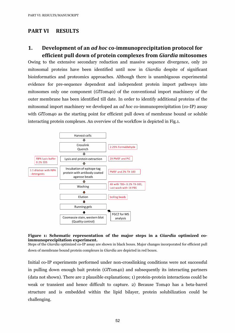
PART VI: RESULTS/MANUSCRIPT
PART VI RESULTS
1. Development of an ad hoc co-immunoprecipitation protocol for
efficient pull down of protein complexes from Giardia mitosomes Owing to the extensive secondary reduction and massive sequence divergence, only 20
mitosomal proteins have been identified until now in Giardia despite of significant
bioinformatics and proteomics approaches. Although there is unambiguous experimental
evidence for pre-sequence dependent and independent protein import pathways into
mitosomes only one component (GlTom40) of the conventional import machinery of the
outer membrane has been identified till date. In order to identify additional proteins of the
mitosomal import machinery we developed an ad hoc co-immunoprecipitation (co-IP) assay
with GlTom40 as the starting point for efficient pull down of membrane bound or soluble
interacting protein complexes. An overview of the workflow is depicted in Fig.1.
Figure 1: Schematic representation of the major steps in a Giardia optimized co-immunoprecipitation experiment. Steps of the Giardia optimized co-IP assay are shown in black boxes. Major changes incorporated for efficient pull
down of membrane bound protein complexes in Giardia are depicted in red boxes.
Initial co-IP experiments performed under non-crosslinking conditions were not successful
in pulling down enough bait protein (GlTom40) and subsequently its interacting partners
(data not shown). There are 2 plausible explanations; 1) protein-protein interactions could be
weak or transient and hence difficult to capture. 2) Because Tom40 has a beta-barrel
structure and is embedded within the lipid bilayer, protein solubilization could be
challenging.
52
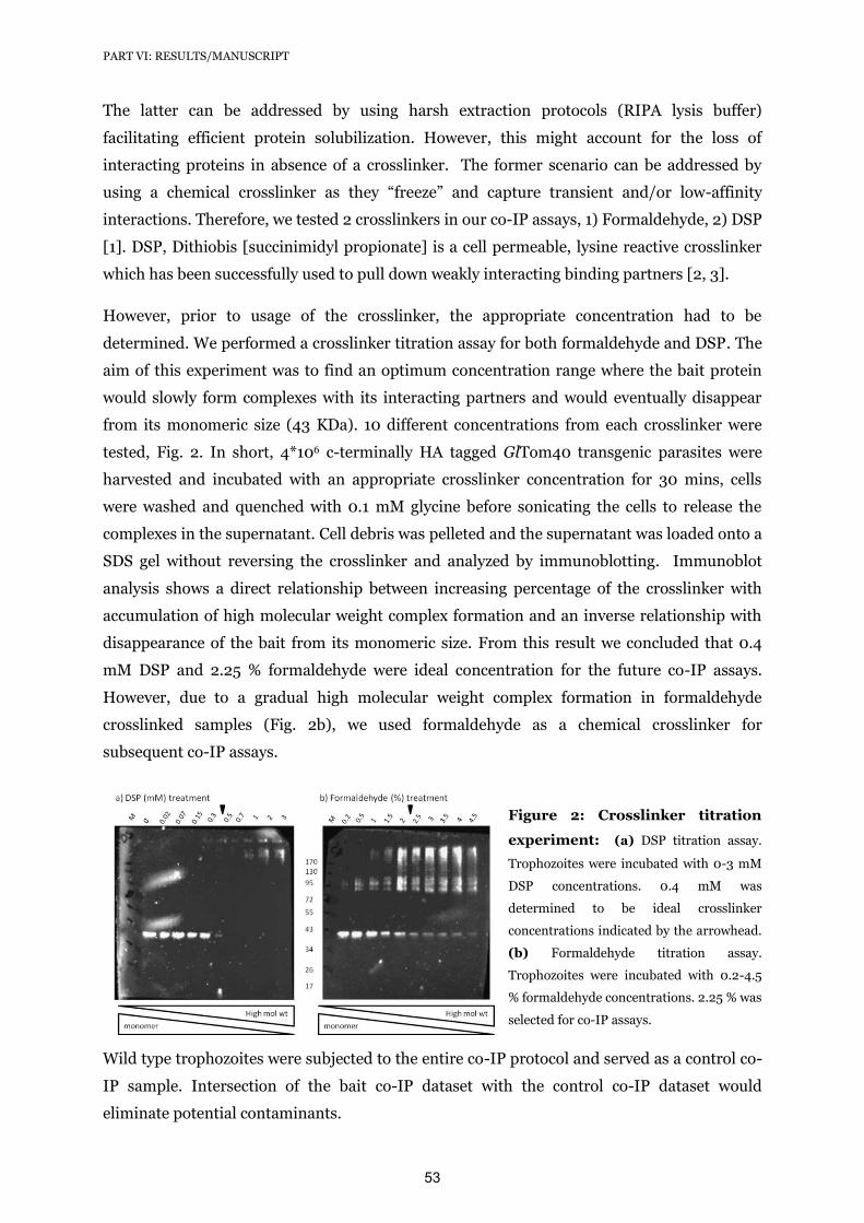
PART VI: RESULTS/MANUSCRIPT
The latter can be addressed by using harsh extraction protocols (RIPA lysis buffer)
facilitating efficient protein solubilization. However, this might account for the loss of
interacting proteins in absence of a crosslinker. The former scenario can be addressed by
using a chemical crosslinker as they “freeze” and capture transient and/or low-affinity
interactions. Therefore, we tested 2 crosslinkers in our co-IP assays, 1) Formaldehyde, 2) DSP
[1]. DSP, Dithiobis [succinimidyl propionate] is a cell permeable, lysine reactive crosslinker
which has been successfully used to pull down weakly interacting binding partners [2, 3].
However, prior to usage of the crosslinker, the appropriate concentration had to be
determined. We performed a crosslinker titration assay for both formaldehyde and DSP. The
aim of this experiment was to find an optimum concentration range where the bait protein
would slowly form complexes with its interacting partners and would eventually disappear
from its monomeric size (43 KDa). 10 different concentrations from each crosslinker were
tested, Fig. 2. In short, 4*106 c-terminally HA tagged GlTom40 transgenic parasites were
harvested and incubated with an appropriate crosslinker concentration for 30 mins, cells
were washed and quenched with 0.1 mM glycine before sonicating the cells to release the
complexes in the supernatant. Cell debris was pelleted and the supernatant was loaded onto a
SDS gel without reversing the crosslinker and analyzed by immunoblotting. Immunoblot
analysis shows a direct relationship between increasing percentage of the crosslinker with
accumulation of high molecular weight complex formation and an inverse relationship with
disappearance of the bait from its monomeric size. From this result we concluded that 0.4
mM DSP and 2.25 % formaldehyde were ideal concentration for the future co-IP assays.
However, due to a gradual high molecular weight complex formation in formaldehyde
crosslinked samples (Fig. 2b), we used formaldehyde as a chemical crosslinker for
subsequent co-IP assays.
Figure 2: Crosslinker titration
experiment: (a) DSP titration assay.
Trophozoites were incubated with 0-3 mM
DSP concentrations. 0.4 mM was
determined to be ideal crosslinker
concentrations indicated by the arrowhead.
(b) Formaldehyde titration assay.
Trophozoites were incubated with 0.2-4.5
% formaldehyde concentrations. 2.25 % was
selected for co-IP assays.
Wild type trophozoites were subjected to the entire co-IP protocol and served as a control co-
IP sample. Intersection of the bait co-IP dataset with the control co-IP dataset would
eliminate potential contaminants.
53
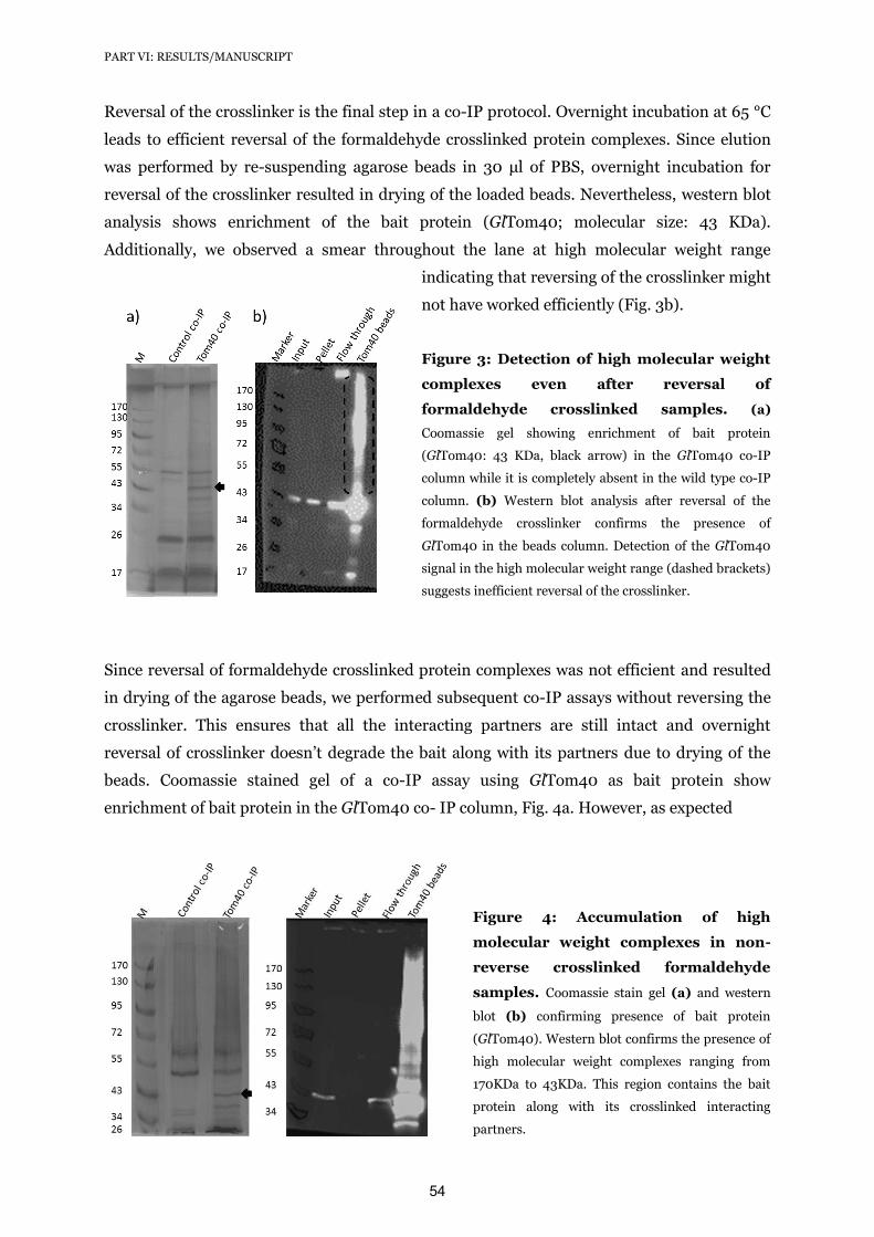
PART VI: RESULTS/MANUSCRIPT
Reversal of the crosslinker is the final step in a co-IP protocol. Overnight incubation at 65 °C
leads to efficient reversal of the formaldehyde crosslinked protein complexes. Since elution
was performed by re-suspending agarose beads in 30 μl of PBS, overnight incubation for
reversal of the crosslinker resulted in drying of the loaded beads. Nevertheless, western blot
analysis shows enrichment of the bait protein (GlTom40; molecular size: 43 KDa).
Additionally, we observed a smear throughout the lane at high molecular weight range
indicating that reversing of the crosslinker might
not have worked efficiently (Fig. 3b).
Figure 3: Detection of high molecular weight
complexes even after reversal of
formaldehyde crosslinked samples. (a)
Coomassie gel showing enrichment of bait protein
(GlTom40: 43 KDa, black arrow) in the GlTom40 co-IP
column while it is completely absent in the wild type co-IP
column. (b) Western blot analysis after reversal of the
formaldehyde crosslinker confirms the presence of
GlTom40 in the beads column. Detection of the GlTom40
signal in the high molecular weight range (dashed brackets)
suggests inefficient reversal of the crosslinker.
Since reversal of formaldehyde crosslinked protein complexes was not efficient and resulted
in drying of the agarose beads, we performed subsequent co-IP assays without reversing the
crosslinker. This ensures that all the interacting partners are still intact and overnight
reversal of crosslinker doesn’t degrade the bait along with its partners due to drying of the
beads. Coomassie stained gel of a co-IP assay using GlTom40 as bait protein show
enrichment of bait protein in the GlTom40 co- IP column, Fig. 4a. However, as expected
Figure 4: Accumulation of high
molecular weight complexes in non-
reverse crosslinked formaldehyde
samples. Coomassie stain gel (a) and western
blot (b) confirming presence of bait protein
(GlTom40). Western blot confirms the presence of
high molecular weight complexes ranging from
170KDa to 43KDa. This region contains the bait
protein along with its crosslinked interacting
partners.
54
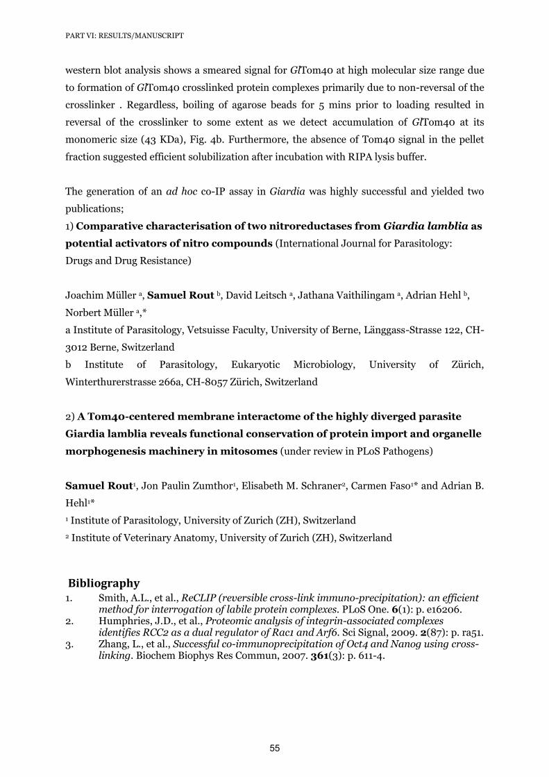
PART VI: RESULTS/MANUSCRIPT
western blot analysis shows a smeared signal for GlTom40 at high molecular size range due
to formation of GlTom40 crosslinked protein complexes primarily due to non-reversal of the
crosslinker . Regardless, boiling of agarose beads for 5 mins prior to loading resulted in
reversal of the crosslinker to some extent as we detect accumulation of GlTom40 at its
monomeric size (43 KDa), Fig. 4b. Furthermore, the absence of Tom40 signal in the pellet
fraction suggested efficient solubilization after incubation with RIPA lysis buffer.
The generation of an ad hoc co-IP assay in Giardia was highly successful and yielded two
publications;
1) Comparative characterisation of two nitroreductases from Giardia lamblia as
potential activators of nitro compounds (International Journal for Parasitology:
Drugs and Drug Resistance)
Joachim Müller a, Samuel Rout b, David Leitsch a, Jathana Vaithilingam a, Adrian Hehl b,
Norbert Müller a,*
a Institute of Parasitology, Vetsuisse Faculty, University of Berne, Länggass-Strasse 122, CH-
3012 Berne, Switzerland
b Institute of Parasitology, Eukaryotic Microbiology, University of Zürich,
Winterthurerstrasse 266a, CH-8057 Zürich, Switzerland
2) A Tom40-centered membrane interactome of the highly diverged parasite
Giardia lamblia reveals functional conservation of protein import and organelle
morphogenesis machinery in mitosomes (under review in PLoS Pathogens)
Samuel Rout1, Jon Paulin Zumthor1, Elisabeth M. Schraner2, Carmen Faso1* and Adrian B.
Hehl1*
1 Institute of Parasitology, University of Zurich (ZH), Switzerland
2 Institute of Veterinary Anatomy, University of Zurich (ZH), Switzerland
Bibliography
1. Smith, A.L., et al., ReCLIP (reversible cross-link immuno-precipitation): an efficient method for interrogation of labile protein complexes. PLoS One. 6(1): p. e16206.
2. Humphries, J.D., et al., Proteomic analysis of integrin-associated complexes identifies RCC2 as a dual regulator of Rac1 and Arf6. Sci Signal, 2009. 2(87): p. ra51.
3. Zhang, L., et al., Successful co-immunoprecipitation of Oct4 and Nanog using cross-linking. Biochem Biophys Res Commun, 2007. 361(3): p. 611-4.
55
PART VI: RESULTS/MANUSCRIPT
PART VI RESULTS (MANUSCRIPT I)
Comparative characterisation of two nitroreductases from Giardia lamblia as potential
activators of nitro compounds
This paper was an outcome of collaboration work together with Institute of Parasitology,
Bern. My contribution to this work consisted of performing co-immunoprecipitation
experiments, analysis of MS dataset (see page 5, Table 2 and 3) and writing of the
manuscript.
56
Comparative characterisation of two nitroreductases from Giardia
lamblia as potential activators of nitro compounds
Joachim Müller a, Samuel Rout b, David Leitsch a, Jathana Vaithilingam a, Adrian Hehl b,Norbert Müller a,*a Institute of Parasitology, Vetsuisse Faculty, University of Berne, Länggass-Strasse 122, CH-3012 Berne, Switzerlandb Institute of Parasitology, Eukaryotic Microbiology, University of Zürich, Winterthurerstrasse 266a, CH-8057 Zürich, Switzerland
A R T I C L E I N F O
Article history:
Received 5 February 2015
Received in revised form 12 March 2015
Accepted 16 March 2015
Available online 25 March 2015
Keywords:
Mode of action of nitro compounds
Functional assays
Nitroreduction
A B S T R A C T
Giardia lamblia is a protozoan parasite that causes giardiasis, a diarrhoeal disease affecting humans and
various animal species. Nitro drugs such as the nitroimidazole metronidazole and the nitrothiazolide
nitazoxanide are used for treatment of giardiasis. Nitroreductases such as GlNR1 and GlNR2 may play a
role in activation or inactivation of these drugs. The aim of this work is to characterise these two enyzmes
using functional assays. For respective analyses recombinant analogues from GlNR1 and GlNR2 were pro-
duced in Escherichia coli. E. coli expressing GlNR1 and GlNR2 alone or together were grown in the presence
of nitro compounds. Furthermore, pull-down assays were performed using HA-tagged GlNR1 and GlNR2
as baits. As expected, E. coli expressing GlNR1 were more susceptible to metronidazole under aerobic and
semi-aerobic and to nitazoxanide under semi-aerobic growth conditions whereas E. coli expressing GlNR2
were susceptible to neither drug. Interestingly, expression of both nitroreductases gave the same results
as expression of GlNR2 alone. In functional assays, both nitroreductases had their strongest activities on
the quinone menadione (vitamin K3) and FAD, but reduction of nitro compounds including the nitro drugs
metronidazole and nitazoxanidewas clearly detected. Full reduction of 7-nitrocoumarin to 7-aminocoumarin
was preferentially achieved with GlNR2. Pull-down assays revealed that GlNR1 and GlNR2 interacted in
vivo forming a multienzyme complex. These findings suggest that both nitroreductases are multifunc-
tional. Their main biological role may reside in the reduction of vitamin K analogues and FAD. Activation
by GlNR1 or inactivation by GlNR2 of nitro drugs may be the consequence of a secondary enzymatic ac-
tivity either yielding (GlNR1) or eliminating (GlNR2) toxic intermediates after reduction of these compounds.
© 2015 The Authors. Published by Elsevier Ltd on behalf of Australian Society for Parasitology. This is an
open access article under the CC BY-NC-ND license (http://creativecommons.org/licenses/by-nc-nd/4.0/).
1. Introduction
Giardia lamblia (syn.Giardia duodenalis;Giardia intestinalis), a flag-ellated protozoan, is themost common causative agent of persistentdiarrhoeaworldwide (Thompson, 2000; Robertson et al., 2010). Cur-rently, anti-giardial chemotherapy is performed using a couple ofeffective drugs, namely, the nitroheterocyclic drugs tinidazole, met-ronidazole, furazolidone,quinacrine, theaminoglycosideparomomycin,and the benzimidazole albendazole (Wright et al., 2003; Lalle, 2010).Furthermore, the nitrothiazolide nitazoxanide has been introducedas an alternative option (Hemphill et al., 2006).
As frequently hypothesised, metronidazole and other nitro drugsare reduced to a nitro-radical. According to one of these hypotheses,electrons are provided by the enzyme pyruvate:flavodoxin/ferredoxin
oxidoreductase (PFOR), representing a protein that is lacking in highereukaryotic cells (Brown et al., 1998; Horner et al., 1999). Referringto this model, the electrons are transferred via PFOR from pyruvateto ferredoxin. The resulting reduced ferredoxin is then re-oxidisedby ferredoxin:NAD-oxidoreductase transferring its electrons toNAD(P). The resulting NAD(P)H serves as a redox partner forsubsequent reactions such as the reduction of O2 by NAD(P)H oxidase(Brown et al., 1998). Nitro drugs may interfere in this pathway andcapture electrons directly from the reduced ferredoxin or from theNAD(P)H-oxidase. This process leads to the accumulation of toxicradicals that cause irreversible damage in the parasite. Furtherevidence for PFOR being a major target for nitro drugs in Giardia
comes from metronidazole-resistant isolates with lower PFORexpression levels (Upcroft and Upcroft, 2001).
Since a few years, however, evidence is emerging that PFORmaynot represent the exclusive target of nitro drugs in semi-aerobic oranaerobic pathogens. In the case of T. vaginalis, metronidazole andother nitroimidazoles were shown to covalently bind, and thus in-activate, proteins involved in the thioredoxin reductase pathway.Resistant cells compensate this blocking by re-regulating PFORs and
* Corresponding author. Institute of Parasitology, Vetsuisse Faculty Berne, University
of Berne, Länggass-Strasse 122, CH-3012 Berne, Switzerland. Tel.: +41 31 6312474;
fax: +41 31 6312477.
E-mail address: [email protected] (N. Müller).
http://dx.doi.org/10.1016/j.ijpddr.2015.03.001
2211-3207/© 2015 The Authors. Published by Elsevier Ltd on behalf of Australian Society for Parasitology. This is an open access article under the CC BY-NC-ND license
(http://creativecommons.org/licenses/by-nc-nd/4.0/).
International Journal for Parasitology: Drugs and Drug Resistance 5 (2015) 37–43
Contents lists available at ScienceDirect
International Journal for Parasitology:Drugs and Drug Resistance
journal homepage: www.elsevier.com/ locate / i jpddr
PART VI: RESULTS/MANUSCRIPT
57
other enzymes participating in oxidoreductive processes. Accord-ingly, down-regulation of PFOR seems not to be a prerequisite butrather a consequence of resistance formation (Leitsch et al., 2009).Moreover, our investigations on Giardia cell lines resistant to nitrodrugs have demonstrated that resistance is not necessarily linkedto down-regulation of PFOR (Müller et al., 2007a, 2008). Althoughsome nitro drugs are supposed to interact with PFOR in a directmanner, direct reduction of the nitro group via ferredoxin is ratherunlikely. Accordingly, catalysis of this reaction by nitroreductasesis a more realistic scenario (Roldán et al., 2008).
Nitroreductases belong to the enzymatic repertoire of many ar-chaebacteria and eubacteria (Nixon et al., 2002), where theycontribute to the assimilation of nitro compounds as carbon sources(Johnson and Spain, 2003; Luque-Almagro et al., 2006). From amech-anistic point of view, nitroreductases are divided into two classes,namely oxygen-sensitive and oxygen-insensitive nitroreductases(Roldán et al., 2008). Oxygen-sensitive nitroreductases transfer elec-trons one by one from NAD(P)H to the nitro group. In presence ofoxygen, the intermediate radicals are re-oxydised. Thus, there isNAD(P)H consumptionwithout nitroreduction, and the reaction looksmost like the one catalysed by a NAD(P)H oxydase. Oxygen-insensitive nitroreductases catalyse the full reduction of nitrocompounds into the corresponding amines by two-electron trans-fers. Also this type of reaction produces toxic intermediates, namely,nitroso or hydroxylamine intermediates (Moreno and Docampo,1985).
In anaerobic or microaerophilic pathogens, nitroreductases arealso well documented as resistance factors. In Helicobacter pylori,resistance to metronidazole is associated with loss-of-function mu-tations of the gene rdxA encoding an oxygen-insensitivenitroreductase (Goodwin et al., 1998), which reduces metronida-zole under anaerobic conditions (Olekhnovich et al., 2009). Othernitroreductases are found in enteric bacteria including Escherichia
coli (Lee et al., 1994; Zenno et al., 1996a, 1996b, 1996c; Guillén et al.,2009; Tavares et al., 2009; Yanto et al., 2010).
Nitroreductases have also been identified in microaerophilic oranaerobic eukaryotic parasites such as Entamoeba histolytica andG. lamblia. These organisms may have acquired the respective genesfrom prokaryotes by lateral transfer (Nixon et al., 2002). G. lamblia
(cloneWB C6) harbours two genes encoding nitroreductases GlNR1(accession N° EDO80257; Gl50803-22677, referred to as Fd-NR2 inthe Giardia database) and GlNR2 (accession N° XM_764091.1;Gl50803-6175, referred to as Fd-NR1 in the Giardia database). Thepolypeptide sequence of GlNR2 is rather similar to that one of GlNR1.Both proteins contain a ferredoxin domain with four Fe-S-clustersat their N-terminus and a nitro-FMN-reductase domain at theirC-terminus. Our previous results suggest that both enzymes havea different action on nitro drugs: GlNR1 behaving as an activator(Müller et al., 2007b; Nillius et al., 2011), GlNR2 more as aninactivator (Müller et al., 2013). The biological role of these enzymesis, however, completely unclear.
Here, we present results from functional assays showing that bothnitroreductases are multifunctional with strong quinone reduc-tase activities. Moreover, we show that both nitroreductases interactin vivo forming a multienzyme complex.
2. Materials and methods
2.1. Tissue culture media, biochemicals and drugs
If not otherwise stated, all biochemical reagents were from Sigma(St Louis, MO, USA). 7-Nitrocoumarin was purchased from SantaCruz Biotechnology (Dallas, Texas, USA). Nitazoxanide wassynthesised at the Department of Chemistry and Biochemistry,University of Berne (kindly provided by Ch. Leumann). Thenitroimidazole C17 was kindly provided by J. A. Upcroft (Molecular
Genetics Laboratory, Queensland Institute of Medical Research,Brisbane, Australia). CB1954 was purchased from Santa CruzBiotechnology. The compounds were kept as 100mM stock solutionsin DMSO at −20 °C.
2.2. Overexpression of recombinant GlNR1 and 2 in E. coli and
His-Tag-purification
Overexpression of recombinant GlNR1 and GlNR2 in E. coli BL21(DE3) and their purification by His-Tag-affinity-chromatography wasperformed as previously described (Müller et al., 2007b, 2013).
2.3. Overexpression of recombinant HA-tagged GlNR1 and 2 in
G. lamblia
Cloning of PCR-amplified GlNR1 and GlNR2 open reading framesinto the XbaI and PacI sites from vector pPacV-Integ (Jiménez-Garcíaet al., 2008) was essentially done as previously described (Mülleret al., 2007b, 2009). Briefly, GlNR1 and GlNR2-specific forward primercontained the XbaI site followed by the constitutive glutamate de-hydrogenase (GDH) promoter sequence (Davis-Hayman and Nash,2002) (Table S1). In the reverse primer, a sequence encoding threeconsecutive human influenza haemagglutinin (HA) tags was intro-duced 5′ of the PacI site (Table S1). PCRs for amplification of GlNR1and GlNR2 open reading frames, insertion of amplification prod-ucts into XbaI and PacI sites from pPacV thus yielding pPacV-GlNR1-3xHA or pPacV-GlNR2-3xHA, and transfection of G. lamblia WBC6with Swa1-linearised recombinant plasmids were performed as pre-viously described (Müller et al., 2007b, 2009).
2.4. Co-immunoprecipitation assay with HA-tagged nitroreductases
G. lamblia WBC6 GlNR1-3xHA and GlNR2-3xHA transgenictrophozoites were grown under anaerobic condition in triple flasks(Nunc, cat. 132867). The parasites were harvested by chilling in icewater for an hour followed by centrifugation (900 × g, 10 min, 4 °C),washed in 50 ml ice cold PBS, and counted in a Neubauer chamber.For co-immunoprecipitation assays, 109 parasites were then re-suspended in a 15-ml-Falcon-tube containing 5 ml of lysis buffer(RIPA) consisting of 50 mM Tris pH 7.4, 150 mM NaCl, 1% IGEPAL,0.5% sodium deoxycholate, 0.1% SDS, 10 mM EDTA supplementedwith 2 mM phenylmethylsulfonyl fluoride, PMSF and 1X ProteaseInhibitor cocktail, PIC (cat. No. 539131, Calbiochem USA), andsonicated twice (60 pulses, 2 output control, 30% duty cycle and 60pulses, 4 output control, 40% duty cycle). To solubilise the proteins,the Falcon tube was incubated on a rotating wheel (1 h, 4 °C). Celllysate was transferred into 1.5 ml microtubes and the supernatantcontaining the solubilised protein was collected after high-speedcentrifugation (14,000 × g, 10 min, 4 °C). The solubilised proteinfraction was diluted 1:1 with detergent-free RIPA lysis buffersupplemented with 2% Triton-X-100. To this diluted protein lysate,40 μl anti-HA agarose bead slurry from the Pierce HA Tag IP/Co-IPKit (Thermo Fisher Scientific, Rockford, Il.) were added and incubatedat 4 °C for 2 h on a rotating wheel in order to allow the HA-taggedproteins to bind to the agarose beads. Samples were pulse-centrifuged at 3500 g at 4 °C and 100 μl was stored as flow thoughcontrol. Samples were washed 4 times with 3 ml of Tris-BufferedSaline (TBS) supplemented with 0.1% Triton-X-100 and once with3 ml PBS. The agarose slurry was re-suspended in 350 μl PBS andtransferred into the spin column provided in the kit and pulse-fugedat 14,000 × g for 10 s at 4 °C. The agarose beads (boiled beads) werethen re-suspended in 30 μl PBS and transferred into a 1.5 mlmicrotubes and stored at −20 °C overnight for further analysis.
38 J. Müller et al./International Journal for Parasitology: Drugs and Drug Resistance 5 (2015) 37–43
PART VI: RESULTS/MANUSCRIPT
58
2.5. Protein analysis and sample preparation for mass spectrometry
For SDS-PAGE according to (Laemmli (1970), the samples collectedas described above were suspended in one volume of SDS-PAGEsample buffer containing 100mMdithiothreitol, and boiled for 5minfollowed by high speed centrifugation (14,000 × g, 10 min, RT). ForMS analysis, 25 μl of boiled bead sample were loaded on a 12%polyacrylamide gel under reducing conditions. The gel (see Fig. S1)was then stained with Instant blue (Expedeon, San Diego, CA),de-stainedwith sterile water, and subsequently sent to the FunctionalGenomics Center Zürich for mass spectrometry. For immunoblotanalysis, approximately 107 trophozoites were processed and sampleswere collected as described above. Immunoblots were performedwith rabbit anti-GlNR1 (Nillius et al., 2011) and mouse-anti-HA(Roche Diagnostics, Rotkreuz, Switzerland) antibodies as describedbefore (Nillius et al., 2011).
2.6. Mass spectrometry
Gel lanes (see Fig. S1) were cut in 8 equal sections. Each sectionwas further diced into smaller pieces and washed twice with 100 μlof 100mMammoniumbicarbonate/50%acetonitrile for15minat50 °C.The sections were dehydrated with 50 μl of acetonitrile. The super-natants of the washing and de-hydration steps were discarded.The gel pieceswere re-hydratedwith 20 μl trypsin solution (5 ng/ μlin 10 mM Tris/2 mM CaCl2 at pH 8.2) and 40 μl buffer (10 mMTris/2 mM CaCl2 at pH 8.2). Microwave-assisted digestion was per-formed for30minat 60 °Cwith themicrowavepower set to5W(CEMDiscover, CEMCorp., USA). Supernatantswere collected in fresh tubesand the gel pieces were extracted with 150 μl of 0.1% trifluoroaceticacid/50% acetonitrile. Supernatants were combined, dried, and thesamplesweredissolved in20 μl of 0.1% formic acidbeforebeing trans-ferred to the autosampler vials for liquid chromatography-tandemmass spectrometry, 7 to 9 μl were injected. Samples weremeasuredonaQ-exactivemass spectrometer (ThermoFisherScientific)equippedwith a nanoAcquity UPLC (Waters Corporation, Milford, MA). Pep-tides were trapped on a trap column (Symmetry C18, 5 μm,180 μm × 20mm, Waters Corporation) before they were separatedon a BEH300 C18, 1.7 μm, 75 μm × 150mm column (Waters Corpo-ration) by applying a gradient formedbetween solvent A (0.1% formicacid inwater) and solvent B (0.1% formic acid in acetonitrile). On themass spectrometer, a gradient starting at 1% solvent B and increas-ing to 40% within 60min was established. Database searches wereperformedusing theMASCOT search programagainst theGiardiada-tabase (http://giardiadb.org/giardiadb/) with a concatenated decoydatabase supplementedwith commonly observed contaminants andthe Swissprot database to increase size of the database. The identi-fiedhitswere then loadedonto theScaffoldViewerversion4 (ProteomeSoftware, Portland,USA) and thehitswerefilteredbasedonhigh strin-gency parameters, namely, minimal mascot score of 95 for peptideprobability, a protein probability of 95% and aminimum of 2 uniquepeptides per protein.
2.7. Quantification of nitroreductase activity
For enzymatic quantification, the nitroreductase activity wasmea-sured in 96-well microtiter plates containing 100 μl of a reactionmix containing buffer (50 mM Tris-Cl−, pH 7.0), 0.1 mM of the com-pounds to be tested, 0.5 mM thiazolyl blue tetrazolium (MTT),0.5 mM NADH or NADPH and 0.1 to 0.2 μg of the recombinantenzymes. The plates were incubated at 37 °C under aerobic condi-tions or in an anaerobic growth chamber. Substrate and enzymeblanks were included. After different time points, the reaction wasstopped by adding 100 μl of pure ethanol thus solubilising theproduct formed by the reduction of MTT, formazan. The absor-bance at 590 nm was read on a 96-well plate spectrophotometer
(Versamax; Molecular Devices, Sunnyvale, CA). After subtraction ofsubstrate and enzyme blanks, nitroreductase activity was ex-pressed as ΔA590/min/mg (Prochaska and Santamaria, 1988).
The reduction of 7-nitrocoumarin to 7-aminocoumarinwas quan-tified using the reaction mix as described above, but without MTT,with the same volumes and under the same conditions of incuba-tion. Enzyme and substrate blanks were included. The reaction wasstopped by adding 100 μl of 50 mM HCl. The resulting solution hadpH 2 resulting in full protonation of 7-aminocoumarin which wasquantified by fluorimetry with excitation at 365 nm and emissionat 455 nm (Wagner, 2009) using a 96-well-multimode plate reader(Enspire; Perkin-Elmer, Waltham, MA).
2.8. Determination of drug susceptibility in E. coli
Drug susceptibility of recombinant E. coli BL21 (DE3) lines(Invitrogen, Carlsbad, CA, USA) expressing either GlNR1 (recombinantplasmid pGlNR1), GlNR2 (pGlNR2), glucuronidase A (pGusA) alone(Nillius et al., 2011; Müller et al., 2013) or both nitroreductases (thisstudy, see below)were tested as described (Müller et al., 2013). Singlegene expression was achieved in vector system pET151 DirectionalTOPO® (Invitrogen) containing the ampicillin resistance marker forselection of transformants and allowing IPTG-inducible over-expression of recombinant proteins (see pET151 Directional TOPO®manual provided by the manufacturer) as described (Müller et al.,2013). In order to achieve double transfectants expressing bothnitroreductases, the following cloning strategies were chosen: (i) theentire pET151 Directional TOPO® expression cassettes carrying GlNR1and GusA open reading frames were amplified by PCR usingT7 forward (5′-TAATACGACTCACTATAGGG-3′) and T7 reverse(5′-TAGTTATTGCTCAGCGGTGG-3′) primers (annealing to regionsflanking the expression cassette of pET151 Directional TOPO®) andpGlNR1 and pGusA as DNA templates. (ii) PCR products were usedfor re-cloning of GlNR1 and GusA into pCR-Blunt II-TOPO®(Invitrogen) containing a kanamycin resistance marker. (iii) Thisre-cloning step provided plasmid constructs, pGlNR1-KanR andpGusA-KanR, suitable for subsequent transformation of ampicillin-resistant BL21 (DE3)/pGlNR2, and BL21 (DE3)/pGusA strains byselection for ampicillin (100 μg/ml)/kanamycin (50 μg/ml) double-resistant clones. E. coli BL21 (DE3) carrying pGlNR1, pGlNR2, pGusA,pNR1-KanR/pGlNR2 and pGusA-KanR/pGusA were tested underaerobic or microaerobic (5% O2, 10% CO2, 85% N2) conditions by aconventional disc diffusion agar procedure as described (Müller et al.,2013). Growth inhibition zone diameters were determined and theinhibition zone around the disc was calculated (in mm2).
2.9. Statistical methods
Statistical analysis of the results was done based on the tools fromthe open source software package R (R Core Team, 2012) Differ-ences exhibiting p values < 0.01 were considered significant.
3. Results
3.1. E. coli expressing recombinant GlNR1 and GlNR2 have different
susceptibilities to nitro drugs
We generated recombinant E. coli lines producing GlNR1 or GlNR2alone (Müller et al., 2013) or both (this study). As a control, we havecreated recombinant lines expressing glucuronidase A (Gus) alone(Müller et al., 2013) or double (Gus/Gus) (this study). In a pilot ex-periment, overproduction of proteins induced by IPTG stronglyreduced growth of E. coli. Accordingly, non-induced, recombinantE. coli cultures were chosen for growth inhibition assays as de-scribed earlier (Nillius et al., 2011). With these five strains, discdiffusion assays were performed with metronidazole, nitazoxanide
39J. Müller et al./International Journal for Parasitology: Drugs and Drug Resistance 5 (2015) 37–43
PART VI: RESULTS/MANUSCRIPT
59
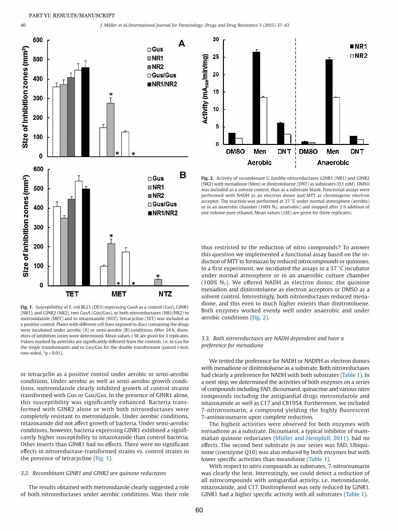
or tetracyclin as a positive control under aerobic or semi-aerobicconditions. Under aerobic as well as semi-aerobic growth condi-tions, metronidazole clearly inhibited growth of control strainstransformed with Gus or Gus/Gus. In the presence of GlNR1 alone,this susceptibility was significantly enhanced. Bacteria trans-formed with GlNR2 alone or with both nitroreductases werecompletely resistant to metronidazole. Under aerobic conditions,nitazoxanide did not affect growth of bacteria. Under semi-aerobicconditions, however, bacteria expressing GlNR1 exhibited a signifi-cantly higher susceptibility to nitazoxanide than control bacteria.Other inserts than GlNR1 had no effects. There were no significanteffects in nitroreductase-transformed strains vs. control strains inthe presence of tetracycline (Fig. 1).
3.2. Recombinant GlNR1 and GlNR2 are quinone reductases
The results obtained with metronidazole clearly suggested a roleof both nitroreductases under aerobic conditions. Was their role
thus restricted to the reduction of nitro compounds? To answerthis question we implemented a functional assay based on the re-duction of MTT to formazan by reduced nitrocompounds or quinones.In a first experiment, we incubated the assays in a 37 °C incubatorunder normal atmosphere or in an anaerobic culture chamber(100% N2). We offered NADH as electron donor, the quinonemenadion and dinitrotoluene as electron acceptors or DMSO as asolvent control. Interestingly, both nitroreductases reduced mena-dione, and this even to much higher extents than dinitrotoluene.Both enzymes worked evenly well under anaerobic and underaerobic conditions (Fig. 2).
3.3. Both nitroreductases are NADH dependent and have a
preference for menadione
We tested the preference for NADH or NADPH as electron donorswithmenadione or dinitrotoluene as a substrate. Both nitroreductaseshad clearly a preference for NADHwith both substrates (Table 1). Ina next step, we determined the activities of both enzymes on a seriesof compounds including FAD, dicoumarol, quinacrine and various nitrocompounds including the antigiardial drugs metronidazole andnitazoxanide as well as C17 and CB1954. Furthermore, we included7-nitrocoumarin, a compound yielding the highly fluorescent7-aminocoumarin upon complete reduction.
The highest activities were observed for both enzymes withmenadione as a substrate. Dicoumarol, a typical inhibitor of mam-malian quinone reductases (Müller and Hemphill, 2011), had noeffects. The second best substrate in our series was FAD. Ubiqui-none (coenzyme Q10) was also reduced by both enzymes but withlower specific activities than meandione (Table 1).
With respect to nitro compounds as substrates, 7-nitrocoumarinwas clearly the best. Interestingly, we could detect a reduction ofall nitrocompounds with antigiardial activity, i.e. metronidazole,nitazoxanide, and C17. Dinitrophenol was only reduced by GlNR1.GlNR1 had a higher specific activity with all substrates (Table 1).
Fig. 1. Susceptibility of E. coli BL21 (DE3) expressing GusA as a control (Gus), GlNR1
(NR1), and GlNR2 (NR2), two GusA (Gus/Gus), or both nitroreductases (NR1/NR2) to
metronidazole (MET) and to nitazoxanide (NTZ). Tetracycline (TET) was included as
a positive control. Plates with different cell lines exposed to discs containing the drugs
were incubated under aerobic (A) or semi-aerobic (B) conditions. After 24 h, diam-
eters of inhibition zones were determined. Mean values ± SE are given for 3 replicates.
Values marked by asterisks are significantly different from the controls, i.e. to Gus for
the single transformants and to Gus/Gus for the double transformant (paired t-test,
two-sided, *p < 0.01).
Fig. 2. Activity of recombinant G. lamblia nitroreductases GlNR1 (NR1) and GlNR2
(NR2) with menadione (Men) or dinitrotoluene (DNT) as substrates (0.1 mM). DMSO
was included as a solvent control, thus as a substrate blank. Functional assays were
performed with NADH as an electron donor and MTT as chromogenic electron
acceptor. The reaction was performed at 37 °C under normal atmosphere (aerobic)
or in an anaerobic chamber (100% N2; anaerobic) and stopped after 2 h addition of
one volume pure ethanol. Mean values (±SE) are given for three replicates.
40 J. Müller et al./International Journal for Parasitology: Drugs and Drug Resistance 5 (2015) 37–43
PART VI: RESULTS/MANUSCRIPT
60
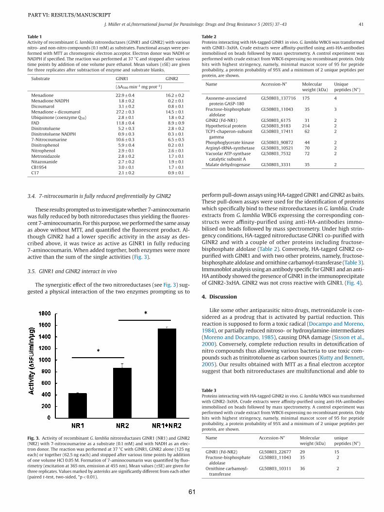
3.4. 7-nitrocoumarin is fully reduced preferentially by GlNR2
Theseresultspromptedus to investigatewhether7-aminocoumarinwas fully reduced by both nitroreductases thus yielding the fluores-cent7-aminocoumarin. For thispurpose,weperformedthesameassayas above without MTT, and quantified the fluorescent product. Al-though GlNR2 had a lower specific activity in the assay as des-cribed above, it was twice as active as GlNR1 in fully reducing7-aminocoumarin. When added together, both enzymes were moreactive than the sum of the single activities (Fig. 3).
3.5. GlNR1 and GlNR2 interact in vivo
The synergistic effect of the two nitroreductases (see Fig. 3) sug-gested a physical interaction of the two enzymes prompting us to
performpull-downassaysusingHA-taggedGlNR1andGlNR2asbaits.These pull-down assays were used for the identification of proteinswhich specifically bind to these nitroreductases in G. lamblia. Crudeextracts from G. lamblia WBC6 expressing the corresponding con-structs were affinity-purified using anti-HA-antibodies immo-bilised on beads followed by mass spectrometry. Under high strin-gency conditions, HA-tagged nitroreductase GlNR1 co-purifiedwithGlNR2 and with a couple of other proteins including fructose-bisphosphate aldolase (Table 2). Conversely, HA-tagged GlNR2 co-purified with GlNR1 and with two other proteins, namely, fructose-bisphosphate aldolase andornithine carbamoyl-transferase (Table 3).Immunoblot analysis usinganantibody specific forGlNR1andananti-HAantibody showed thepresenceof GlNR1 in the immunoprecipitateof GlNR2-3xHA. GlNR2 was not cross reactive with GlNR1. (Fig. 4).
4. Discussion
Like some other antiparasitic nitro drugs, metronidazole is con-sidered as a prodrug that is activated by partial reduction. Thisreaction is supposed to form a toxic radical (Docampo and Moreno,1984), or partially reduced nitroso- or hydroxylamine-intermediates(Moreno and Docampo, 1985), causing DNA damage (Sisson et al.,2000). Conversely, complete reduction results in detoxification ofnitro compounds thus allowing various bacteria to use toxic com-pounds such as trinitrotoluene as carbon sources (Kutty and Bennett,2005). Our results obtained with MTT as a final electron acceptorsuggest that both nitroreductases are multifunctional and able to
Table 1
Activity of recombinant G. lamblia nitroreductases (GlNR1 and GlNR2) with various
nitro- and non-nitro compounds (0.1 mM) as substrates. Functional assays were per-
formed with MTT as chromogenic electron acceptor. Electron donor was NADH or
NADPH if specified. The reaction was performed at 37 °C and stopped after various
time points by addition of one volume pure ethanol. Mean values (±SE) are given
for three replicates after subtraction of enzyme and substrate blanks.
Substrate GlNR1 GlNR2
(ΔA590 min−1 mg prot−1)
Menadione 22.9 ± 0.4 16.2 ± 0.2
Menadione NADPH 1.8 ± 0.2 0.2 ± 0.1
Dicoumarol 3.1 ± 0.2 0.8 ± 0.1
Menadione + dicoumarol 27.2 ± 0.3 14.5 ± 0.1
Ubiquinone (coenzyme Q10) 2.8 ± 0.1 1.8 ± 0.2
FAD 11.8 ± 0.4 8.9 ± 0.9
Dinitrotoluene 5.2 ± 0.3 2.8 ± 0.2
Dinitrotoluene NADPH 0.9 ± 0.3 0.3 ± 0.1
7-Nitrocoumarine 10.6 ± 0.3 6.5 ± 0.5
Dinitrophenol 5.9 ± 0.4 0.2 ± 0.1
Nitrophenol 2.9 ± 0.1 2.6 ± 0.1
Metronidazole 2.8 ± 0.2 1.7 ± 0.1
Nitazoxanide 2.7 ± 0.2 1.9 ± 0.1
CB1954 3.0 ± 0.1 1.7 ± 0.1
C17 2.1 ± 0.2 0.9 ± 0.1
Fig. 3. Activity of recombinant G. lamblia nitroreductases GlNR1 (NR1) and GlNR2
(NR2) with 7-nitrocoumarine as a substrate (0.1 mM) and with NADH as an elec-
tron donor. The reaction was performed at 37 °C with GlNR1, GlNR2 alone (125 ng
each) or together (62.5 ng each) and stopped after various time points by addition
of one volume HCl 0.05 M. Formation of 7-aminocoumarin was quantified by fluo-
rimetry (excitation at 365 nm, emission at 455 nm). Mean values (±SE) are given for
three replicates. Values marked by asterisks are significantly different from each other
(paired t-test, two-sided, *p < 0.01).
Table 2
Proteins interacting with HA-tagged GlNR1 in vivo. G. lamblia WBC6was transformed
with GlNR1-3xHA. Crude extracts were affinity-purified using anti-HA-antibodies
immobilised on beads followed by mass spectrometry. A control experiment was
performed with crude extract fromWBC6 expressing no recombinant protein. Only
hits with highest stringency, namely, minimal mascot score of 95 for peptide
probability, a protein probability of 95% and a minimum of 2 unique peptides per
protein, are shown.
Name Accession-N° Molecular
weight (kDa)
Unique
peptides (N°)
Axoneme-associated
protein GASP-180
GL50803_137716 175 4
Fructose-bisphosphate
aldolase
GL50803_11043 35 3
GlNR2 (Fd-NR1) GL50803_6175 31 2
Hypothetical protein GL50803_9183 214 2
TCP1-chaperon-subunit
gamma
GL50803_17411 62 2
Phosphoglycerate kinase GL50803_90872 44 2
Arginyl-tRNA-synthetase GL50803_10521 70 2
Vacuolar ATP-synthase
catalytic subunit A
GL50803_7532 72 2
Malate dehydrogenase GL50803_3331 35 2
Table 3
Proteins interacting with HA-tagged GlNR2 in vivo. G. lamblia WBC6was transformed
with GlNR2-3xHA. Crude extracts were affinity-purified using anti-HA-antibodies
immobilised on beads followed by mass spectrometry. A control experiment was
performed with crude extract fromWBC6 expressing no recombinant protein. Only
hits with highest stringency, namely, minimal mascot score of 95 for peptide
probability, a protein probability of 95% and a minimum of 2 unique peptides per
protein, are shown.
Name Accession-N° Molecular
weight (kDa)
unique
peptides (N°)
GlNR1 (Fd-NR2) GL50803_22677 29 15
Fructose-bisphosphate
aldolase
GL50803_11043 35 2
Ornithine carbamoyl-
transferase
GL50803_10311 36 2
41J. Müller et al./International Journal for Parasitology: Drugs and Drug Resistance 5 (2015) 37–43
PART VI: RESULTS/MANUSCRIPT
61
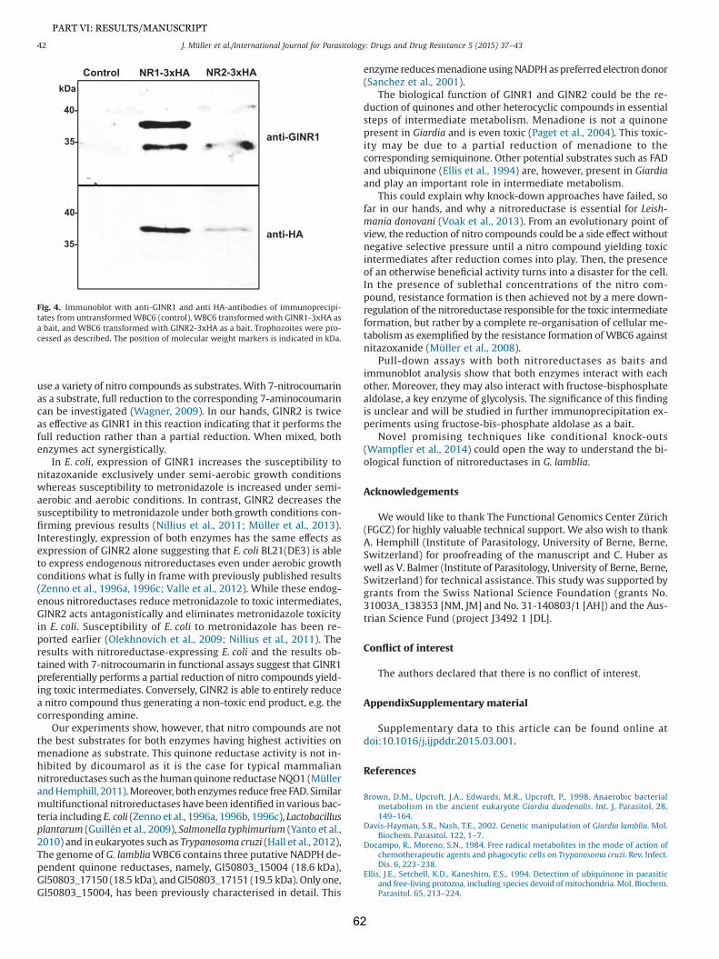
use a variety of nitro compounds as substrates.With 7-nitrocoumarinas a substrate, full reduction to the corresponding 7-aminocoumarincan be investigated (Wagner, 2009). In our hands, GlNR2 is twiceas effective as GlNR1 in this reaction indicating that it performs thefull reduction rather than a partial reduction. When mixed, bothenzymes act synergistically.
In E. coli, expression of GlNR1 increases the susceptibility tonitazoxanide exclusively under semi-aerobic growth conditionswhereas susceptibility to metronidazole is increased under semi-aerobic and aerobic conditions. In contrast, GlNR2 decreases thesusceptibility to metronidazole under both growth conditions con-firming previous results (Nillius et al., 2011; Müller et al., 2013).Interestingly, expression of both enzymes has the same effects asexpression of GlNR2 alone suggesting that E. coli BL21(DE3) is ableto express endogenous nitroreductases even under aerobic growthconditions what is fully in frame with previously published results(Zenno et al., 1996a, 1996c; Valle et al., 2012). While these endog-enous nitroreductases reduce metronidazole to toxic intermediates,GlNR2 acts antagonistically and eliminates metronidazole toxicityin E. coli. Susceptibility of E. coli to metronidazole has been re-ported earlier (Olekhnovich et al., 2009; Nillius et al., 2011). Theresults with nitroreductase-expressing E. coli and the results ob-tained with 7-nitrocoumarin in functional assays suggest that GlNR1preferentially performs a partial reduction of nitro compounds yield-ing toxic intermediates. Conversely, GlNR2 is able to entirely reducea nitro compound thus generating a non-toxic end product, e.g. thecorresponding amine.
Our experiments show, however, that nitro compounds are notthe best substrates for both enzymes having highest activities onmenadione as substrate. This quinone reductase activity is not in-hibited by dicoumarol as it is the case for typical mammaliannitroreductases such as the humanquinone reductaseNQO1 (MüllerandHemphill, 2011).Moreover, bothenzymes reduce freeFAD. Similarmultifunctional nitroreductases have been identified in various bac-teria including E. coli (Zenno et al., 1996a, 1996b, 1996c), Lactobacillus
plantarum (Guillén et al., 2009), Salmonella typhimurium (Yanto et al.,2010) and in eukaryotes such as Trypanosoma cruzi (Hall et al., 2012),The genome of G. lamblia WBC6 contains three putative NADPH de-pendent quinone reductases, namely, Gl50803_15004 (18.6 kDa),Gl50803_17150 (18.5 kDa), andGl50803_17151 (19.5 kDa). Only one,Gl50803_15004, has been previously characterised in detail. This
enzyme reducesmenadioneusingNADPHaspreferredelectrondonor(Sanchez et al., 2001).
The biological function of GlNR1 and GlNR2 could be the re-duction of quinones and other heterocyclic compounds in essentialsteps of intermediate metabolism. Menadione is not a quinonepresent in Giardia and is even toxic (Paget et al., 2004). This toxic-ity may be due to a partial reduction of menadione to thecorresponding semiquinone. Other potential substrates such as FADand ubiquinone (Ellis et al., 1994) are, however, present in Giardia
and play an important role in intermediate metabolism.This could explain why knock-down approaches have failed, so
far in our hands, and why a nitroreductase is essential for Leish-
mania donovani (Voak et al., 2013). From an evolutionary point ofview, the reduction of nitro compounds could be a side effect withoutnegative selective pressure until a nitro compound yielding toxicintermediates after reduction comes into play. Then, the presenceof an otherwise beneficial activity turns into a disaster for the cell.In the presence of sublethal concentrations of the nitro com-pound, resistance formation is then achieved not by a mere down-regulation of the nitroreductase responsible for the toxic intermediateformation, but rather by a complete re-organisation of cellular me-tabolism as exemplified by the resistance formation of WBC6 againstnitazoxanide (Müller et al., 2008).
Pull-down assays with both nitroreductases as baits andimmunoblot analysis show that both enzymes interact with eachother. Moreover, they may also interact with fructose-bisphosphatealdolase, a key enzyme of glycolysis. The significance of this findingis unclear and will be studied in further immunoprecipitation ex-periments using fructose-bis-phosphate aldolase as a bait.
Novel promising techniques like conditional knock-outs(Wampfler et al., 2014) could open the way to understand the bi-ological function of nitroreductases in G. lamblia.
Acknowledgements
Wewould like to thank The Functional Genomics Center Zürich(FGCZ) for highly valuable technical support. We also wish to thankA. Hemphill (Institute of Parasitology, University of Berne, Berne,Switzerland) for proofreading of the manuscript and C. Huber aswell as V. Balmer (Institute of Parasitology, University of Berne, Berne,Switzerland) for technical assistance. This study was supported bygrants from the Swiss National Science Foundation (grants No.31003A_138353 [NM, JM] and No. 31-140803/1 [AH]) and the Aus-trian Science Fund (project J3492 1 [DL].
Conflict of interest
The authors declared that there is no conflict of interest.
AppendixSupplementary material
Supplementary data to this article can be found online atdoi:10.1016/j.ijpddr.2015.03.001.
References
Brown, D.M., Upcroft, J.A., Edwards, M.R., Upcroft, P., 1998. Anaerobic bacterialmetabolism in the ancient eukaryote Giardia duodenalis. Int. J. Parasitol. 28,149–164.
Davis-Hayman, S.R., Nash, T.E., 2002. Genetic manipulation of Giardia lamblia. Mol.Biochem. Parasitol. 122, 1–7.
Docampo, R., Moreno, S.N., 1984. Free radical metabolites in the mode of action ofchemotherapeutic agents and phagocytic cells on Trypanosoma cruzi. Rev. Infect.Dis. 6, 223–238.
Ellis, J.E., Setchell, K.D., Kaneshiro, E.S., 1994. Detection of ubiquinone in parasiticand free-living protozoa, including species devoid of mitochondria. Mol. Biochem.Parasitol. 65, 213–224.
Control NR1-3xHA NR2-3xHA
anti-GlNR1
anti-HA
35-
40-
35-
40-
kDa
Fig. 4. Immunoblot with anti-GlNR1 and anti HA-antibodies of immunoprecipi-
tates from untransformedWBC6 (control), WBC6 transformed with GlNR1-3xHA as
a bait, and WBC6 transformed with GlNR2-3xHA as a bait. Trophozoites were pro-
cessed as described. The position of molecular weight markers is indicated in kDa.
42 J. Müller et al./International Journal for Parasitology: Drugs and Drug Resistance 5 (2015) 37–43
PART VI: RESULTS/MANUSCRIPT
62
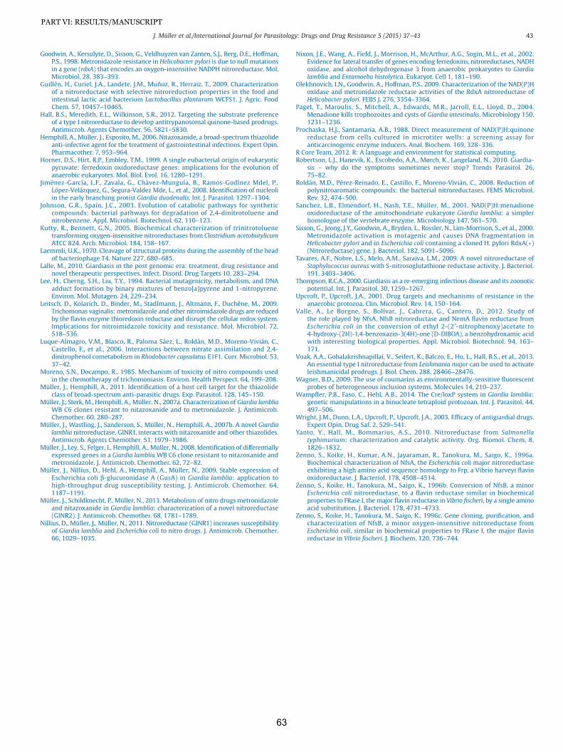
Goodwin, A., Kersulyte, D., Sisson, G., Veldhuyzen van Zanten, S.J., Berg, D.E., Hoffman,P.S., 1998. Metronidazole resistance in Helicobacter pylori is due to null mutationsin a gene (rdxA) that encodes an oxygen-insensitive NADPH nitroreductase. Mol.Microbiol. 28, 383–393.
Guillén, H., Curiel, J.A., Landete, J.M., Muñoz, R., Herraiz, T., 2009. Characterizationof a nitroreductase with selective nitroreduction properties in the food andintestinal lactic acid bacterium Lactobacillus plantarum WCFS1. J. Agric. FoodChem. 57, 10457–10465.
Hall, B.S., Meredith, E.L., Wilkinson, S.R., 2012. Targeting the substrate preferenceof a type I nitroreductase to develop antitrypanosomal quinone-based prodrugs.Antimicrob. Agents Chemother. 56, 5821–5830.
Hemphill, A., Müller, J., Esposito, M., 2006. Nitazoxanide, a broad-spectrum thiazolideanti-infective agent for the treatment of gastrointestinal infections. Expert Opin.Pharmacother. 7, 953–964.
Horner, D.S., Hirt, R.P., Embley, T.M., 1999. A single eubacterial origin of eukaryoticpyruvate: ferredoxin oxidoreductase genes: implications for the evolution ofanaerobic eukaryotes. Mol. Biol. Evol. 16, 1280–1291.
Jiménez-García, L.F., Zavala, G., Chávez-Munguía, B., Ramos-Godínez Mdel, P.,López-Velázquez, G., Segura-Valdez Mde, L., et al., 2008. Identification of nucleoliin the early branching protist Giardia duodenalis. Int. J. Parasitol. 1297–1304.
Johnson, G.R., Spain, J.C., 2003. Evolution of catabolic pathways for syntheticcompounds: bacterial pathways for degradation of 2,4-dinitrotoluene andnitrobenzene. Appl. Microbiol. Biotechnol. 62, 110–123.
Kutty, R., Bennett, G.N., 2005. Biochemical characterization of trinitrotoluenetransforming oxygen-insensitive nitroreductases from Clostridium acetobutylicum
ATCC 824. Arch. Microbiol. 184, 158–167.Laemmli, U.K., 1970. Cleavage of structural proteins during the assembly of the head
of bacteriophage T4. Nature 227, 680–685.Lalle, M., 2010. Giardiasis in the post genomic era: treatment, drug resistance and
novel therapeutic perspectives. Infect. Disord. Drug Targets 10, 283–294.Lee, H., Cherng, S.H., Liu, T.Y., 1994. Bacterial mutagenicity, metabolism, and DNA
adduct formation by binary mixtures of benzo[a]pyrene and 1-nitropyrene.Environ. Mol. Mutagen. 24, 229–234.
Leitsch, D., Kolarich, D., Binder, M., Stadlmann, J., Altmann, F., Duchêne, M., 2009.Trichomonas vaginalis: metronidazole and other nitroimidazole drugs are reducedby the flavin enzyme thioredoxin reductase and disrupt the cellular redox system.Implications for nitroimidazole toxicity and resistance. Mol. Microbiol. 72,518–536.
Luque-Almagro, V.M., Blasco, R., Paloma Sáez, L., Roldán, M.D., Moreno-Vivián, C.,Castello, F., et al., 2006. Interactions between nitrate assimilation and 2,4-dinitrophenol cometabolism in Rhodobacter capsulatus E1F1. Curr. Microbiol. 53,37–42.
Moreno, S.N., Docampo, R., 1985. Mechanism of toxicity of nitro compounds usedin the chemotherapy of trichomoniasis. Environ. Health Perspect. 64, 199–208.
Müller, J., Hemphill, A., 2011. Identification of a host cell target for the thiazolideclass of broad-spectrum anti-parasitic drugs. Exp. Parasitol. 128, 145–150.
Müller, J., Sterk, M., Hemphill, A., Müller, N., 2007a. Characterization of Giardia lamblia
WB C6 clones resistant to nitazoxanide and to metronidazole. J. Antimicrob.Chemother. 60, 280–287.
Müller, J., Wastling, J., Sanderson, S., Müller, N., Hemphill, A., 2007b. A novel Giardia
lamblia nitroreductase, GlNR1, interacts with nitazoxanide and other thiazolides.Antimicrob. Agents Chemother. 51, 1979–1986.
Müller, J., Ley, S., Felger, I., Hemphill, A., Müller, N., 2008. Identification of differentiallyexpressed genes in a Giardia lamblia WB C6 clone resistant to nitazoxanide andmetronidazole. J. Antimicrob. Chemother. 62, 72–82.
Müller, J., Nillius, D., Hehl, A., Hemphill, A., Müller, N., 2009. Stable expression ofEscherichia coli β-glucuronidase A (GusA) in Giardia lamblia: application tohigh-throughput drug susceptibility testing. J. Antimicrob. Chemother. 64,1187–1191.
Müller, J., Schildknecht, P., Müller, N., 2013. Metabolism of nitro drugs metronidazoleand nitazoxanide in Giardia lamblia: characterization of a novel nitroreductase(GlNR2). J. Antimicrob. Chemother. 68, 1781–1789.
Nillius, D., Müller, J., Müller, N., 2011. Nitroreductase (GlNR1) increases susceptibilityof Giardia lamblia and Escherichia coli to nitro drugs. J. Antimicrob. Chemother.66, 1029–1035.
Nixon, J.E., Wang, A., Field, J., Morrison, H., McArthur, A.G., Sogin, M.L., et al., 2002.Evidence for lateral transfer of genes encoding ferredoxins, nitroreductases, NADHoxidase, and alcohol dehydrogenase 3 from anaerobic prokaryotes to Giardia
lamblia and Entamoeba histolytica. Eukaryot. Cell 1, 181–190.Olekhnovich, I.N., Goodwin, A., Hoffman, P.S., 2009. Characterization of the NAD(P)H
oxidase and metronidazole reductase activities of the RdxA nitroreductase ofHelicobacter pylori. FEBS J. 276, 3354–3364.
Paget, T., Maroulis, S., Mitchell, A., Edwards, M.R., Jarroll, E.L., Lloyd, D., 2004.Menadione kills trophozoites and cysts of Giardia intestinalis. Microbiology 150,1231–1236.
Prochaska, H.J., Santamaria, A.B., 1988. Direct measurement of NAD(P)H:quinonereductase from cells cultured in microtiter wells: a screening assay foranticarcinogenic enzyme inducers. Anal. Biochem. 169, 328–336.
R Core Team, 2012. R: A language and environment for statistical computing.Robertson, L.J., Hanevik, K., Escobedo, A.A., Mørch, K., Langeland, N., 2010. Giardia-
sis – why do the symptoms sometimes never stop? Trends Parasitol. 26,75–82.
Roldán, M.D., Pérez-Reinado, E., Castillo, F., Moreno-Vivián, C., 2008. Reduction ofpolynitroaromatic compounds: the bacterial nitroreductases. FEMS Microbiol.Rev. 32, 474–500.
Sanchez, L.B., Elmendorf, H., Nash, T.E., Müller, M., 2001. NAD(P)H:menadioneoxidoreductase of the amitochondriate eukaryote Giardia lamblia: a simplerhomologue of the vertebrate enzyme. Microbiology 147, 561–570.
Sisson, G., Jeong, J.Y., Goodwin, A., Bryden, L., Rossler, N., Lim-Morrison, S., et al., 2000.Metronidazole activation is mutagenic and causes DNA fragmentation inHelicobacter pylori and in Escherichia coli containing a cloned H. pylori RdxA(+)(Nitroreductase) gene. J. Bacteriol. 182, 5091–5096.
Tavares, A.F., Nobre, L.S., Melo, A.M., Saraiva, L.M., 2009. A novel nitroreductase ofStaphylococcus aureus with S-nitrosoglutathione reductase activity. J. Bacteriol.191, 3403–3406.
Thompson, R.C.A., 2000. Giardiasis as a re-emerging infectious disease and its zoonoticpotential. Int. J. Parasitol. 30, 1259–1267.
Upcroft, P., Upcroft, J.A., 2001. Drug targets and mechanisms of resistance in theanaerobic protozoa. Clin. Microbiol. Rev. 14, 150–164.
Valle, A., Le Borgne, S., Bolívar, J., Cabrera, G., Cantero, D., 2012. Study ofthe role played by NfsA, NfsB nitroreductase and NemA flavin reductase fromEscherichia coli in the conversion of ethyl 2-(2′-nitrophenoxy)acetate to4-hydroxy-(2H)-1,4-benzoxazin-3(4H)-one (D-DIBOA), a benzohydroxamic acidwith interesting biological properties. Appl. Microbiol. Biotechnol. 94, 163–171.
Voak, A.A., Gobalakrishnapillai, V., Seifert, K., Balczo, E., Hu, L., Hall, B.S., et al., 2013.An essential type I nitroreductase from Leishmania major can be used to activateleishmanicidal prodrugs. J. Biol. Chem. 288, 28466–28476.
Wagner, B.D., 2009. The use of coumarins as environmentally-sensitive fluorescentprobes of heterogeneous inclusion systems. Molecules 14, 210–237.
Wampfler, P.B., Faso, C., Hehl, A.B., 2014. The Cre/loxP system in Giardia lamblia:genetic manipulations in a binucleate tetraploid protozoan. Int. J. Parasitol. 44,497–506.
Wright, J.M., Dunn, L.A., Upcroft, P., Upcroft, J.A., 2003. Efficacy of antigiardial drugs.Expert Opin. Drug Saf. 2, 529–541.
Yanto, Y., Hall, M., Bommarius, A.S., 2010. Nitroreductase from Salmonella
typhimurium: characterization and catalytic activity. Org. Biomol. Chem. 8,1826–1832.
Zenno, S., Koike, H., Kumar, A.N., Jayaraman, R., Tanokura, M., Saigo, K., 1996a.Biochemical characterization of NfsA, the Escherichia coli major nitroreductaseexhibiting a high amino acid sequence homology to Frp, a Vibrio harveyi flavinoxidoreductase. J. Bacteriol. 178, 4508–4514.
Zenno, S., Koike, H., Tanokura, M., Saigo, K., 1996b. Conversion of NfsB, a minorEscherichia coli nitroreductase, to a flavin reductase similar in biochemicalproperties to FRase I, themajor flavin reductase in Vibrio fischeri, by a single aminoacid substitution. J. Bacteriol. 178, 4731–4733.
Zenno, S., Koike, H., Tanokura, M., Saigo, K., 1996c. Gene cloning, purification, andcharacterization of NfsB, a minor oxygen-insensitive nitroreductase fromEscherichia coli, similar in biochemical properties to FRase I, the major flavinreductase in Vibrio fischeri. J. Biochem. 120, 736–744.
43J. Müller et al./International Journal for Parasitology: Drugs and Drug Resistance 5 (2015) 37–43
PART VI: RESULTS/MANUSCRIPT
63
PART VI: RESULTS/MANUSCRIPT
PART VI RESULTS (MANUSCRIPT II)
A Tom40-centered membrane interactome of the highly diverged parasite Giardia
lamblia reveals functional conservation of protein import and organelle morphogenesis
machinery in mitosomes
This paper summarizes the main part of my PhD thesis. I started working on this project
since the beginning of 2012 for 2 years. The concept of Giardia based co-
immunoprecipitation using G. lamblia Tom40 as a point of origin and (re)constructing a
GlTom40 centered interactome inwards towards the mitosomal matrix and outwards
towards the cytosol was developed together with my direct supervisor Prof. Dr. Adrian B.
Hehl and co-supervisor Dr. carmen Faso who supported me during all stages of the project
including writing of the manuscript.
My contribution to this work includes developing the co-IP protocol for membrane anchored
G. lamblia proteins, in silico analysis of the MS dataset and localization studies. I was
assisted in light and electron microscopy by Dr. Carmen Faso (Fig 5A-B), Prof. Dr. Adrian B.
Hehl (Fig 7), and Elisabeth M. Schraner (Fig 6I-J). Compilation of all the figures for the
manuscript was done by me.
Tandem mass spectrometry was performed in collaboration with the functional genomics
center Zurich (FGCZ).
64
PART VI: RESULTS/MANUSCRIPT
A Tom40-centered membrane interactome of the highly diverged parasite
Giardia lamblia reveals functional conservation of protein import and
organelle morphogenesis machinery in mitosomes
Samuel Rout1, Jon Paulin Zumthor1, Elisabeth M. Schraner2, Carmen Faso1* and Adrian B.
Hehl1*
1 Institute of Parasitology, University of Zurich (ZH), Switzerland
2 Institute of Veterinary Anatomy, University of Zurich (ZH), Switzerland
*Corresponding authors
Email: [email protected] (AH)
[email protected] (CF)
Abstract
Protozoan parasites of the genus Giardia are highly prevalent globally and infect a wide
range of vertebrate hosts including humans, but proliferation and pathology is restricted to
the small intestine. This narrow ecological specialization entailed extensive structural and
functional adaptations during host-parasite co-evolution. G. lamblia mitochondria-related
organelles (mitosomes) are at the farthest extreme on the spectrum of mitochondrial
reductive evolution with iron-sulphur protein maturation as the only identifiable biochemical
pathway. We describe construction of the extensively diverged mitosome-specific proteome
based entirely on protein-protein interactions, using the only identifiable component of a
predicted TOM/TIM protein import complex (Tom40) as a starting point. Using serial co-
immunoprecipitation assays and validation steps we extended a robust Tom40 core
interactome network outwards, revealing additional outer membrane proteins and candidate
links to membranes of the endoplasmic reticulum (ER), as well as inwards, identifying many
novel imported proteins in addition to the few annotated conserved metabolic factors and
chaperones. Live cell imaging revealed that the 30-40 organelles in a cell are highly
immobilized and do not form dynamic networks, which is also consistent with interactome
data suggesting physical links with the cytoskeleton, in particular with the basal body
complex. On the other hand, identification of small GTPases and factors with dual mitosome
and ER localization in the interactome suggested intimate connections with the ER.
Functional analysis of mitosomes showed conceptual conservation of protein import
although the machinery for translocation is diverged beyond recognition, as well as
association of the single G. lamblia dynamin-related protein with mitosomes and direct
evidence for its involvement in organelle morphogenesis. This study underscores the
65
PART VI: RESULTS/MANUSCRIPT
potential of this strategy for identification of proteins and machinery ab initio in a highly
diverged but clearly delimited organelle system.
Author Summary
Organelles with endosymbiotic origin are present in all extant eukaryotes and have
undergone considerable remodeling during > 1 billion years of evolution. Highly diverged
organelles such as mitosomes or plastids in some parasitic protozoa are the product of
extensive secondary reduction. They are sufficiently unique to generate interest as targets for
pharmacological intervention, in addition to providing a rich ground for evolutionary cell
biologists. The so-called mitochondria-related organelles (MROs) comprise mitosomes and
hydrogenosomes, with the former having lost any role in energy metabolism along with the
organelle genome. The mitosomes of the intestinal pathogen Giardia lamblia are the most
highly reduced MROs known and have proven difficult to investigate because of their
extreme divergence and their unique physical properties. Here, we implemented a novel
strategy aimed at systematic analysis of the organelle proteome by iterative expansion of a
protein-protein interaction network. We demonstrated the effectiveness of serial co-
immunoprecipitations combined with mass spectrometry analysis and in vivo validation for
generating an interactome network centered on a giardial Tom40 homolog. This iterative ab
initio proteome reconstruction provided protein-protein interaction data in addition to
identifying novel organelle proteins and functions. Building on this information we
investigated mitosome morphogenesis and organelle dynamics in living cells, and showed
that the single giardial dynamin plays a role in organelle replication.
Introduction
Since the single endosymbiotic event leading to establishment of mitochondria
approximately 2 billion years ago [1,2,3] these organelles have undergone massive changes
and have evolved into highly specialized and essential subcellular compartments in all
eukaryotes [4,5]. These changes comprise a dramatic size reduction, nuclear transfer of
organelle genomes, and a renewal of the proteome, which is synthesized almost entirely as
precursor proteins on cytosolic ribosomes [6,7,8,9,10,11,12,13] and imported from the
cytoplasm [14]. Mitochondria have been remodeled and/or restructured to very different
degrees in different species. Mitochondria-related organelles (MROs), i.e. hydrogenosomes
and mitosomes [15,16,17,18,19] in some protists lacking canonical mitochondria represent
extreme forms of reduction and/or divergence. The potential of highly diverged organelle-
specific pathways as targets for intervention has sparked research into the evolution of MROs
in single-celled organisms of all five eukaryotic supergroups [20,21]. Notably, the
microaerophilic protozoan pathogens Entamoeba histolytica [19], Giardia lamblia [22,23],
66
PART VI: RESULTS/MANUSCRIPT
Blastocystis hominis [24] as well as obligate intracellular parasites such as Cryptosporidium
parvum [25] and Encephalitozoon cuniculi [26] harbor highly reduced mitosomes.
Interestingly, recent investigation of MROs in Spironucleus salmonicida, a diplomonad and
the closest relative of G. lamblia belonging to the Excavata super-group, revealed that these
organelles are in fact hydrogenosomes [27]. Although it has been demonstrated that G.
lamblia mitosomes don’t produce hydrogen, this sheds a completely new light on the
evolution of MRO’s in diplomonads.
Proliferating G. lamblia trophozoites contain 20-50 double membrane-bounded 100
nm spherical mitosomes [22,23] devoid of an organellar genome [28,29,30,31]. Although not
proven experimentally, G. lamblia mitosomes are likely essential due to a subset of conserved
mitochondrial proteins required for iron- sulphur (Fe-S) protein maturation
[22,32,33,34,35]. Yeast genetic experiments suggested that Fe-S protein maturation, the only
function currently ascribable to G. lamblia mitosomes, is in fact the minimal essential
function of mitochondria [36]. Hence, these organelles have also generated considerable
interest as cell biological models to study extreme reductive evolution of MROs
[22,37,38,39,40,41,42]. However, due to massive, albeit selective sequence divergence in G.
lamblia, conventional strategies for identification of mitosome proteins based on homology-
based in silico searches fall short [26,28,32,43,44,45,46,47]. Moreover, proteome analyses
approaches have had very limited success due to the small size of the organelles and the
omnipresence of contaminating endoplasmic reticulum (ER) and cytoskeleton elements in
enriched mitosome fractions [33,48].
Nevertheless, there is unambiguous experimental evidence for the functional
conservation of the mitosomal protein import machinery [19,22,23]. The small subset of
structurally conserved mitosome proteins such as G. lamblia IscU, ferredoxin, Cpn60, IscS
and mtHsp70 are imported by transit peptide-dependent and -independent mechanisms
[22]. However, the predicted components of the TOM/TIM import apparatus are diverged
beyond recognition by state-of-the-art homology search tools or have been lost. Only one
subunit of the translocon in the outer mitochondrial (TOM) complex, a highly diverged
Tom40 homologue (GlTom40), has been identified [49].
Because most G. lamblia mitosome components have undergone extreme sequence
divergence or have been lost altogether, the vast knowledge about the molecular biology and
biochemistry of mitochondria cannot be directly applied to investigations into evolution,
morphogenesis, and function of these organelles. Hence, exploration of the range of G.
lamblia mitosome functions requires alternative strategies aimed at comprehensive
identification of their proteome and of essential Giardia-specific factors. We hypothesized
that the diminutive organelles harbored no more than 100-150 different proteins inside or
67
PART VI: RESULTS/MANUSCRIPT
associated with a clearly delineated cellular compartment. Thus, it should be possible to use a
putative GlTOM40 [22,33] as a starting point for identifying a core membrane interactome
and extend this in an iterative process to eventually encompass the whole organelle
proteome. The rationale for using GlTom40 as a starting point is the fact that it is the only,
albeit poorly, conserved outer membrane protein and plays a key role for the import of
organelle proteins. Using a series of co-immunoprecipitation assays we obtained a robust
GlTom40 interactome and extend this network in both directions, i.e. outwards onto the
cytoplasmic face of mitosomes, including peripheral membrane proteins such as a diverged
putative TOM receptor (GlTom40R), and inwards, encompassing the few known and many
novel imported organelle proteins. We used this information to probe mitosome
morphogenesis and function and to test constraints for import of nuclear encoded mitosome
proteins.
Materials and Methods
Giardia cell culture, induction of encystation, pulse-empty chase set-up and transfection
G. lamblia WBC6 (ATCC catalog number 50803) trophozoites were grown and harvested
using standard protocols [50]. Encystation was induced with the two-step method as
described previously [40,51]. Transgenic parasites were generated according to established
protocols by electroporation of linearized pPacV-Integ-based plasmid vectors prepared from
E. coli as described in [42]. After selection for puromycin resistance, transgenic G. lamblia
cell lines were cultured and analyzed without antibiotic.
Construction of expression vectors
All sequences of oligonucleotide primers for PCR used in this work are listed in S1 Table.
For cloning of C-terminally hemagglutinin (HA)-tagged proteins in Giardia, a vector PAC-
CHA was designed on the basis of the previously described vector pPacV-Integ [42], where
additional restriction sites were inserted. A detailed vector map can be found in S1 Fig.
A cyst wall protein 1 promoter (pCWP1)-driven G. lamblia ferredoxin (fd)-human
dihydrofolate reductase (DHFR) chimeric gene was generated by fusing two genes by
overlapping PCR: i) an intron-less fd mitosomal targeting signal (MTS) (MTSfdΔint) open
reading frame (ORF) was generated using primer pair 33 (S1 Table) with G. lamblia cDNA as
template, ii) a DHFR_HA minigene was generated using primer pair 34 (S1 Table) with a
cloned human DHFR cDNA as template. The fused product was digested with SpeI and PacI
and inserted in a PAC vector to yield construct pCWP1_MTSfdΔint-DHFR_HA.
68
PART VI: RESULTS/MANUSCRIPT
A pCwp1_ MTSfdΔint-DHFR_Neomycin resistance construct (without HA tag) was generated
for protein import block assays. Primer pair 35 (S1 Table) was used on pCwp1_ MTSfdΔint-
DHFR_HA as a template. The amplified product was digested with NsiI and PacI and ligated
into a vector containing a neomycin resistance cassette [52].
Co-immunoprecipitation with limited cross-linking
G. lamblia WBC6 and transgenic trophozoites expressing C-terminally HA tagged bait
proteins were harvested and subjected to immunofluorescence assay to confirm correct
subcellular distribution of bait proteins. Parasites were collected by centrifugation (900 x g,
10 minutes, 4 °C), washed in 50 ml of cold phosphate buffer saline solution (PBS) and
adjusted to 2 *107 cells .ml-1 in PBS (VWR Prolabo). The appropriate formaldehyde
concentration for cross-linking (2.25%) was determined by a titration assay (S2 Fig). For the
co-immunoprecipitation (co-IP) assays, 109 parasites were resuspended in 10 ml 2.25%
formaldehyde (in PBS) supplemented with 1 mM phenylmethylsulfonyl fluoride (PMSF;
SIGMA, Cat. No. P7626) and incubated for 30 minutes at room temperature (RT). Cells were
pelleted, washed once with 10 ml PBS, and quenched in 10 ml 100 mM glycine in PBS for 15
minutes at RT. The collected cells were then resuspended in 5 ml RIPA lysis buffer (50 mM
Tris pH 7.4, 150 mM NaCl, 1% IGEPAL, 0.5% sodium deoxycholate, 0.1% SDS, 10 mM EDTA)
supplemented with 2 mM PMSF and 1 x Protease Inhibitor cocktail (PIC, Cat. No. 539131,
Calbiochem USA) and sonicated twice using a Branson Sonifier with microtip (Branson
Sonifier 250, Branson Ultrasonics Corporation) with the following settings: 60 pulses, 2
output control, 30% duty cycle and 60 pulses, 4 output control, 40% duty cycle. The sonicate
was incubated on a rotating wheel for 1 h at 4 °C, aliquoted into 1.5 ml tubes and centrifuged
(14,000 x g, 10 minutes, 4 °C). The soluble protein fraction was mixed with an equal volume
detergent-free RIPA lysis buffer supplemented with 2% TritonX (TX)-100 (Fluka Chemicals)
and 40 μl anti-HA agarose bead slurry (Pierce, product # 26181). After binding of tagged
proteins to the beads at 4 °C for 2 h on a rotating wheel, beads were pulse-centrifuged and
washed 4 times with 3 ml Tris-Buffered Saline (TBS) supplemented with 0.1% TX-100 at 4
°C. After a final wash with 3 ml PBS the loaded beads were resuspended in 350 μl PBS,
transferred to a spin column (Pierce spin column screw cap, product # 69705, Thermo
Scientific) and centrifuged for 10 s at 4 °C. Elution was performed by resuspending beads in
30 μl of PBS. Dithiothreitol (DTT; 100mM; Thermo Scientific, Cat. # RO861) was added and
samples were boiled for 5 min followed by centrifugation (14,000 x g, 10 minutes, RT).
Protein analysis and sample preparation for mass spectrometry-based protein
identification
SDS-PAGE and immunoblotting analysis of input, flow-through, and eluate fractions was
performed on 4%-12% polyacrylamide gels under reducing conditions, (molecular weight
69
PART VI: RESULTS/MANUSCRIPT
marker Cat. No. 26616, Thermo Scientific, Lithuania). Transfer to nitrocellulose membranes
and antibody probing were done as described previously [53]. Gels for mass spectrometry
(MS) analysis were stained using Instant blue (Expedeon, Prod. # ISB1L) and de-stained with
sterile water.
Mass Spectrometry and protein identification
Stained gel lanes were cut into 8 equal sections. Each section was further diced into smaller
pieces and washed twice with 100 µl of 100 mM ammonium bicarbonate/ 50 % acetonitrile
for 15 min at 50 °C. The sections were dehydrated with 50 µl of acetonitrile. The gel pieces
were rehydrated with 20 µl trypsin solution (5 ng/µl in 10 mM Tris-HCl/ 2 mM CaCl2 at pH
8.2) and 40 µl buffer (10 mM Tris-HCl/ 2 mM CaCl2 at pH 8.2). Microwave-assisted digestion
was performed for 30 minutes at 60 °C with the microwave power set to 5 W (CEM Discover,
CEM corp., USA). Supernatants were collected in fresh tubes and the gel pieces were
extracted with 150 µl of 0.1% trifluoroacetic acid/ 50% acetonitrile. Supernatants were
combined, dried, and the samples were dissolved in 20 µl 0.1% formic acid before being
transferred to the autosampler vials for liquid chromatography-tandem MS (injection volume
7 to 9 µl). Samples were measured on a Q-exactive mass spectrometer (Thermo Scientific)
equipped with a nanoAcquity UPLC (Waters Corporation). Peptides were trapped on a
Symmetry C18, 5 µm, 180 µm x 20 mm column (Waters Corporation) and separated on a
BEH300 C18, 1.7 um, 75 µm x 150 mm column (Waters Corporation) using a gradient formed
between solvent A (0.1% formic acid in water) and solvent B (0.1% formic acid in
acetonitrile). The gradient started at 1% solvent B and the concentration of solvent B was
increased to 40% within 60 minutes. Following peptide data acquisition, database searches
were performed using the MASCOT search program against the G. lamblia database
(http://tinyurl.com/37z5zqp) with a concatenated decoy database supplemented with
commonly observed contaminants and the Swissprot database to increase database size. The
identified hits were then loaded onto the Scaffold Viewer version 4 (Proteome Software,
Portland, US) and filtered based on high stringency parameters (minimal mascot score of
95% for peptide probability, a protein probability of 95%, and a minimum of 2 unique
peptides per protein). For a description of relaxed conditions see “Results”.
In silico co-immunoprecipitation dataset analysis
Analysis of primary structure and domain architecture of putative mitosomal hypothetical
proteins was performed using the following tools and databases: MITOPROT
(http://ihg.gsf.de/ihg/mitoprot.html) and PSORTII (http://psort.hgc.jp/form2.html) for
subcellular localization prediction, TMHMM (http://www.cbs.dtu.dk/services/TMHMM/)
for transmembrane helix prediction, SMART (http://smart.embl-heidelberg.de/) for
prediction of patterns and functional domains, pBLAST for protein homology detection
70
PART VI: RESULTS/MANUSCRIPT
(http://blast.ncbi.nlm.nih.gov/Blast.cgi?PAGE=Proteins), HHPred
(http://toolkit.tuebingen.mpg.de/hhpred) for protein homology detection based on Hidden
Markov Model (HMM-HMM) comparison, and the Giardia Genome Database
(http://giardiadb.org/giardiadb/) to extract other/organism-specific information, e.g.
expression levels of the protein, predicted molecular size and nucleotide/protein sequence.
For functional domains predicted by SMART we used an e-value of 10e-5 as cutoff, and for
protein homologies predicted by pBLAST we accepted alignment scores above 80. However,
since G. lamblia homologs for eukaryotic proteins are highly diverged, we also considered
functional domain predictions associated to a lower e-value. Alignment scores between 50
and 80 were accepted only when pBLAST predictions were consistent with HHPred output.
Immunofluorescence analysis (IFA) and microscopy
Preparation of chemically fixed cells for immunofluorescence and analysis of subcellular
distribution of reporter proteins by wide-field and confocal microscopy were done as
described previously [42,53]. Nuclear labelling was performed with 4',6-diamidino-2-
phenylindole (DAPI).
Live-cell microscopy and fluorescence recovery after photobleaching (FRAP)
Transgenic G. lamblia trophozoites expressing GFP-GlTom40 or Gl29147-GFP were
harvested and prepared for imaging in PBS supplemented with 5 mM glucose (Cat. No.
49139, Fluka), 5 mM L-cysteine (Cat. No. C6852, Sigma) and 0.1 mM ascorbic acid (Cat. No.
95209, Fluka) at pH 7.1. FRAP and time-lapse series were performed as described previously
[53,54] .
Sample preparation for transmission electron microscopy
Transgenic trophozoites ectopically expressing wild type G. lamblia dynamin related protein
(GlDRP) (ORF Gl50803_14373) or the constitutively active (GTP-locked) GlDRP-K43E
variant under the control of the CWP1 promoter [54] were harvested 3 h post induction and
analyzed by transmission electron microscopy (TEM) as described previously [54].
Sub-cellular fractionation analysis
For sub-cellular fraction experiments, 4.106 GlDRP-HA and GlDRP-K43E-HA- expressing
transgenic cells were lysed by freeze-thawing and supernatant (soluble fraction) and pellet
(membrane fraction) were prepared by centrifugation at 14’000 x g for 10 minutes at 4 °C.
The HA-tagged proteins were detected by SDS-PAGE and Western blot using a rat anti-HA
mAb (clone 3F10, Roche) as described previously [53].
71
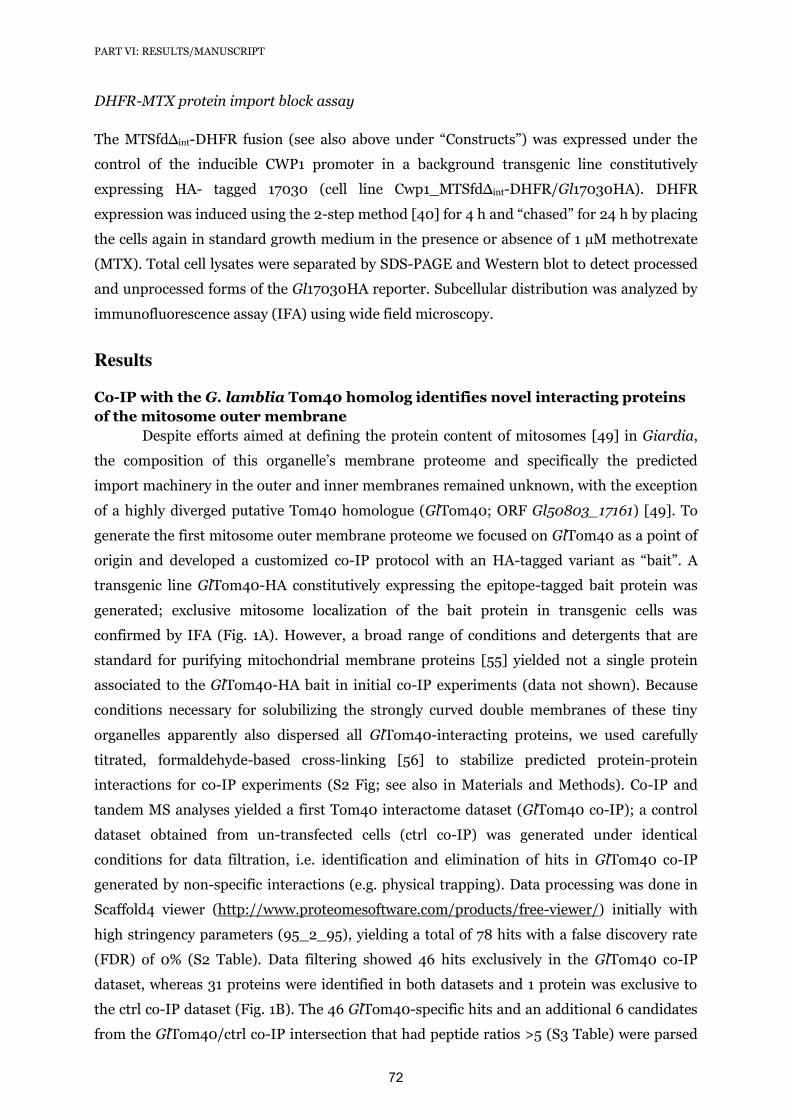
PART VI: RESULTS/MANUSCRIPT
DHFR-MTX protein import block assay
The MTSfdΔint-DHFR fusion (see also above under “Constructs”) was expressed under the
control of the inducible CWP1 promoter in a background transgenic line constitutively
expressing HA- tagged 17030 (cell line Cwp1_MTSfdΔint-DHFR/Gl17030HA). DHFR
expression was induced using the 2-step method [40] for 4 h and “chased” for 24 h by placing
the cells again in standard growth medium in the presence or absence of 1 µM methotrexate
(MTX). Total cell lysates were separated by SDS-PAGE and Western blot to detect processed
and unprocessed forms of the Gl17030HA reporter. Subcellular distribution was analyzed by
immunofluorescence assay (IFA) using wide field microscopy.
Results
Co-IP with the G. lamblia Tom40 homolog identifies novel interacting proteins
of the mitosome outer membrane
Despite efforts aimed at defining the protein content of mitosomes [49] in Giardia,
the composition of this organelle’s membrane proteome and specifically the predicted
import machinery in the outer and inner membranes remained unknown, with the exception
of a highly diverged putative Tom40 homologue (GlTom40; ORF Gl50803_17161) [49]. To
generate the first mitosome outer membrane proteome we focused on GlTom40 as a point of
origin and developed a customized co-IP protocol with an HA-tagged variant as “bait”. A
transgenic line GlTom40-HA constitutively expressing the epitope-tagged bait protein was
generated; exclusive mitosome localization of the bait protein in transgenic cells was
confirmed by IFA (Fig. 1A). However, a broad range of conditions and detergents that are
standard for purifying mitochondrial membrane proteins [55] yielded not a single protein
associated to the GlTom40-HA bait in initial co-IP experiments (data not shown). Because
conditions necessary for solubilizing the strongly curved double membranes of these tiny
organelles apparently also dispersed all GlTom40-interacting proteins, we used carefully
titrated, formaldehyde-based cross-linking [56] to stabilize predicted protein-protein
interactions for co-IP experiments (S2 Fig; see also in Materials and Methods). Co-IP and
tandem MS analyses yielded a first Tom40 interactome dataset (GlTom40 co-IP); a control
dataset obtained from un-transfected cells (ctrl co-IP) was generated under identical
conditions for data filtration, i.e. identification and elimination of hits in GlTom40 co-IP
generated by non-specific interactions (e.g. physical trapping). Data processing was done in
Scaffold4 viewer (http://www.proteomesoftware.com/products/free-viewer/) initially with
high stringency parameters (95_2_95), yielding a total of 78 hits with a false discovery rate
(FDR) of 0% (S2 Table). Data filtering showed 46 hits exclusively in the GlTom40 co-IP
dataset, whereas 31 proteins were identified in both datasets and 1 protein was exclusive to
the ctrl co-IP dataset (Fig. 1B). The 46 GlTom40-specific hits and an additional 6 candidates
from the GlTom40/ctrl co-IP intersection that had peptide ratios >5 (S3 Table) were parsed
72
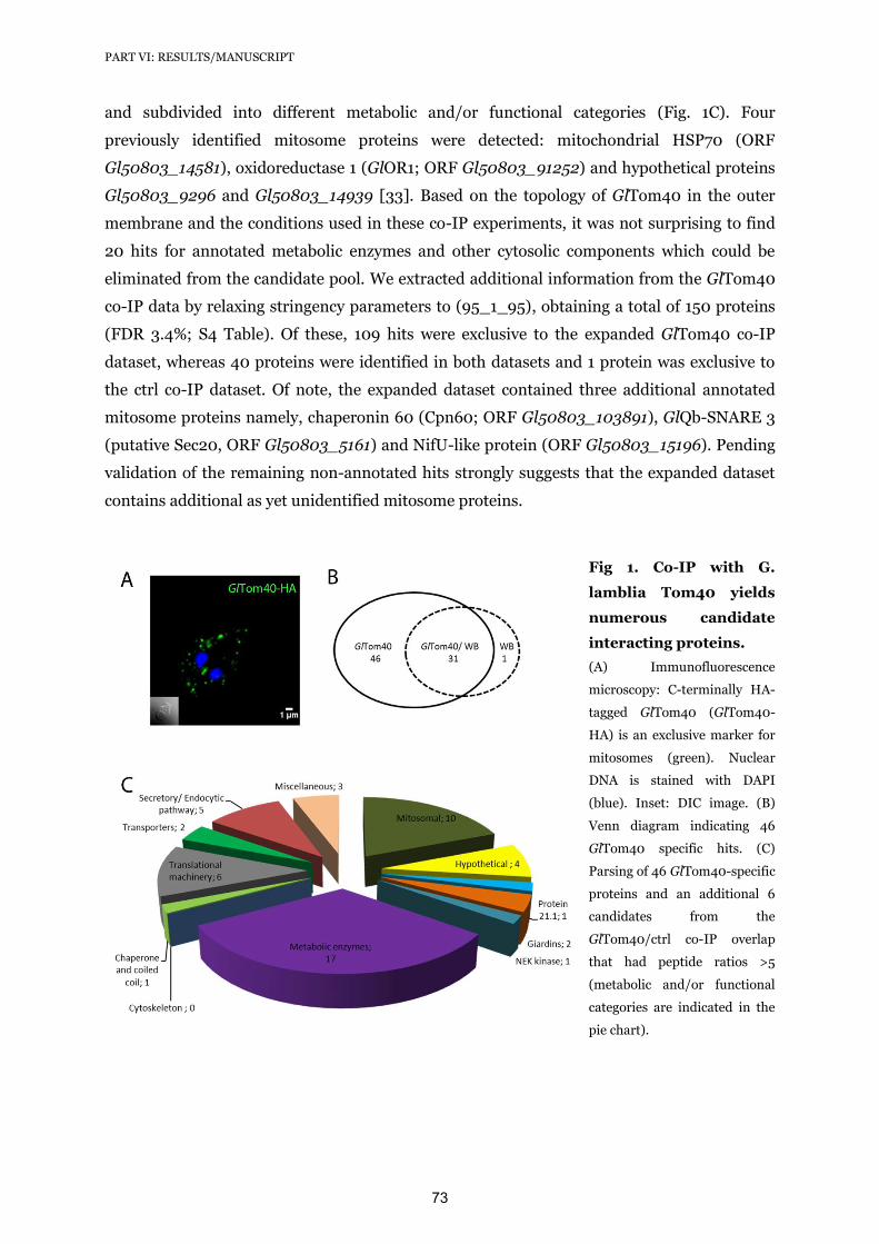
PART VI: RESULTS/MANUSCRIPT
and subdivided into different metabolic and/or functional categories (Fig. 1C). Four
previously identified mitosome proteins were detected: mitochondrial HSP70 (ORF
Gl50803_14581), oxidoreductase 1 (GlOR1; ORF Gl50803_91252) and hypothetical proteins
Gl50803_9296 and Gl50803_14939 [33]. Based on the topology of GlTom40 in the outer
membrane and the conditions used in these co-IP experiments, it was not surprising to find
20 hits for annotated metabolic enzymes and other cytosolic components which could be
eliminated from the candidate pool. We extracted additional information from the GlTom40
co-IP data by relaxing stringency parameters to (95_1_95), obtaining a total of 150 proteins
(FDR 3.4%; S4 Table). Of these, 109 hits were exclusive to the expanded GlTom40 co-IP
dataset, whereas 40 proteins were identified in both datasets and 1 protein was exclusive to
the ctrl co-IP dataset. Of note, the expanded dataset contained three additional annotated
mitosome proteins namely, chaperonin 60 (Cpn60; ORF Gl50803_103891), GlQb-SNARE 3
(putative Sec20, ORF Gl50803_5161) and NifU-like protein (ORF Gl50803_15196). Pending
validation of the remaining non-annotated hits strongly suggests that the expanded dataset
contains additional as yet unidentified mitosome proteins.
Fig 1. Co-IP with G.
lamblia Tom40 yields
numerous candidate
interacting proteins.
(A) Immunofluorescence
microscopy: C-terminally HA-
tagged GlTom40 (GlTom40-
HA) is an exclusive marker for
mitosomes (green). Nuclear
DNA is stained with DAPI
(blue). Inset: DIC image. (B)
Venn diagram indicating 46
GlTom40 specific hits. (C)
Parsing of 46 GlTom40-specific
proteins and an additional 6
candidates from the
GlTom40/ctrl co-IP overlap
that had peptide ratios >5
(metabolic and/or functional
categories are indicated in the
pie chart).
73
PART VI: RESULTS/MANUSCRIPT
Validation of the GlTom40 co-IP data
Because of the non-targeted nature of limited chemical cross-linking in co-IP assays we
performed careful validation of the datasets generated in these experiments based on two
criteria: i) subcellular localization of ectopically expressed, epitope-tagged candidates to
mitosomes, and ii) successful retrieval of the endogenous protein previously used as bait in
reverse co-IP experiments.
To test subcellular localization we selected 13 of the 109 candidate Tom40 interacting
proteins based on MASCOT scores or protein domains identified in HHPred. The 9
hypothetical proteins with the highest MASCOT scores and four additional candidates with
domains predicted to be involved in mitochondrial functions (S5 Table) were cloned as
endogenous promoter-driven C-terminally HA-tagged variants into a G. lamblia expression
vector and used to generate transgenic lines [57]. Using IFA of chemically fixed transgenic
cells, we confirmed mitosomal localization for 8 out of 9 candidates (Figs. 2A-2N).
Fig 2. Subcellular
localization of
candidate GlTom40
interaction partners.
(A-N) Immunofluorescence
microscopy: subcellular
localization of C-terminally
HA-tagged GlTom40 and 13
putative interaction partners
(green) falls into 3
categories: Typical mitosome
localization (A-E); dual
localization to mitosomes
and ER (F-I); no or
ambiguous mitosome
localization (J-N). Nuclear
DNA is stained with DAPI
(blue). Insets: DIC image.
(O) Partially validated
GlTom40 interactome
showing the bait protein
(orange sphere), matrix
proteins (purple), previously
identified and mitosome-
localized proteins (black),
and mitosome-localized
hypothetical proteins (blue).
The stringency parameters used for detection (high, medium, and relaxed) are represented by bold, dashed, and
dotted arrows, respectively.
74
PART VI: RESULTS/MANUSCRIPT
This high proportion underscores the quality of the GlTom40 co-IP dataset. Interestingly, 4
proteins without annotation (ORFs Gl50803_ 21943, 22587, 5785 and 9503) presented dual
localization (mitosome and ER) (Figs. 2F-2I). The cartoon in Fig. 2O shows a consolidated
depiction of a first GlTom40 interactome, which includes the 8 proteins localized to
mitosomes described above, as well as 4 previously identified matrix proteins and 3 newly
validated hypothetical proteins comprised in the list of GlTom40 interacting proteins.
To test the second validation criterion, we performed reverse co-IP experiments, using the
GlTom40 interaction partner with the highest MASCOT score (Gl50803_29147) as a first
bait. The gene codes for a predicted single-pass transmembrane protein of 133 amino acids
with mitosomal localization of an HA-tagged variant confirmed by fluorescence microscopy
(Fig. 2B). Its domain structure is reminiscent of Tom40 receptors found in mitochondria of
higher eukaryotes, i.e. a transmembrane helix in the N-terminal part of the protein followed
by a cytoplasmically exposed C-terminal domain as predicted by TMHMM
(http://www.cbs.dtu.dk/services/TMHMM/). Although experimental confirmation of a receptor
function is pending, we henceforth refer to the Gl50803_29147 product as GlTom40R. A cell
line, constitutively expressing HA-tagged GlTom40R as a mitosome-localized co-IP bait
protein produced a dataset of 221 GlTom40R co-IP exclusive proteins after filtering and
elimination of all hits intersecting with the ctrl co- IP fraction at high stringency parameters
(Fig. 3A; S6 Table). Importantly, the GlTom40R-HA bait protein and endogenous GlTom40
were detected with high MASCOT values, confirming the GlTom40 – GlTom40R interaction.
The 221 GlTom40R co-IP specific hits and an additional 20 candidates from the
GlTom40R/ctrl co-IP intersection that had peptide ratios >5 were parsed according to
different metabolic and/or functional categories (Fig. 3B; S7 Table). In addition to GlTom40,
the dataset contained several known mitosomal proteins, including matrix proteins HSP70
and GiOR1, cysteine desulfurase (IscS; Gl50803_14519), Cpn60, [2Fe-2S] ferredoxin
(Gl50803_27266) and NifU-like protein. Using high stringency criteria, we retrieved all 8
hypothetical proteins previously identified in the GlTom40 co-IP dataset and 4 additional
non-annotated candidate mitosome proteins listed in S8 Table (Fig. 3C-3F). Taken together,
combined subcellular localization experiments and a first reverse co-IP dataset using the
single-pass transmembrane GlTom40-interacting protein GlTom40R provided robust
validation of the experimental approach used to identify mitosome membrane proteins, and
has expanded the predicted mitosomal membrane and import machinery interactome to 22
proteins (Fig. 3G). Interestingly, this dataset contained two axoneme-associated GASP-180
proteins (Gl50803_137716 and Gl50803_16745) [58]with high MASCOT scores.
The two high-stringency co-IP datasets suggests a strong interaction between GlTom40 and
GlTom40R. Thus, hits appearing in the intersection of the two are considered to be
particularly informative (Fig. 3H). The curated list of hits after elimination of obvious
75
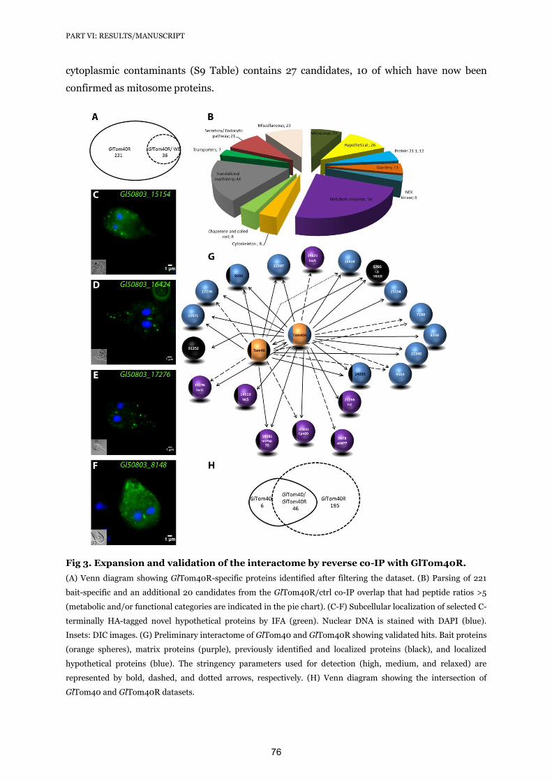
PART VI: RESULTS/MANUSCRIPT
cytoplasmic contaminants (S9 Table) contains 27 candidates, 10 of which have now been
confirmed as mitosome proteins.
Fig 3. Expansion and validation of the interactome by reverse co-IP with GlTom40R.
(A) Venn diagram showing GlTom40R-specific proteins identified after filtering the dataset. (B) Parsing of 221
bait-specific and an additional 20 candidates from the GlTom40R/ctrl co-IP overlap that had peptide ratios >5
(metabolic and/or functional categories are indicated in the pie chart). (C-F) Subcellular localization of selected C-
terminally HA-tagged novel hypothetical proteins by IFA (green). Nuclear DNA is stained with DAPI (blue).
Insets: DIC images. (G) Preliminary interactome of GlTom40 and GlTom40R showing validated hits. Bait proteins
(orange spheres), matrix proteins (purple), previously identified and localized proteins (black), and localized
hypothetical proteins (blue). The stringency parameters used for detection (high, medium, and relaxed) are
represented by bold, dashed, and dotted arrows, respectively. (H) Venn diagram showing the intersection of
GlTom40 and GlTom40R datasets.
76

PART VI: RESULTS/MANUSCRIPT
Co-IP experiments confirm GlTom40-specific protein-protein interactions and
reveal strong links with four additional mitosome proteins
In the absence of an absolute measure for strength/affinity of the physical interaction
between proteins in these interactomes we used MASCOT peptide scores as a non-exclusive
qualitative criterion for “interaction” in a preliminary manner. To determine a robust
GlTom40- and GlTom40R-centered core interactome consisting of the most frequently
detected interaction partners and, more importantly, to explore the boundaries of the
growing interaction network (Fig. 3G), we performed a series of additional co-IP experiments
using HA-tagged Qb-SNARE 4 (Gl50803_5785), GlIscS, and hypothetical proteins
Gl50803_9296 and Gl50803_14939 as baits. These proteins were chosen because they were
identified, in some cases exclusively, in both the GlTom40- and GlTom40R co-IP datasets
(S2 and S6 Tables), suggesting they may be most proximal to GlTom40. Furthermore,
mitosomal localization of all four HA-tagged bait proteins was confirmed by IFA (Fig. 2). As
was done previously, all bait-specific datasets were processed by filtering with a ctrl co-IP
dataset generated from un-transfected trophozoites and analyzed in MASCOT using high
stringency (95_2_95) and more relaxed (95_1_95) parameters.
Co-IP with the mitosome transmembrane protein Gl50803_14939
Gl50803_14939, which contains two transmembrane domains (TMD), was analyzed because
of its potential role as a component of the import complex. Using high stringency parameters
(95_2_95) 129 proteins with a FDR of 10% were detected with 93 candidates specific for the
Gl14939 co-IP dataset (S10 Table). Both GlTom40 and G. lamblia oxidoreductase 1 (GiOR1;
ORF Gl50803_91252) were detected with these parameters, in addition to several previously
identified hypothetical mitosome proteins (e.g. GlTom40R, Gl50803_10971 and
Gl50803_7188) (Fig. 4A). This further supports the idea that Gl50803_14939 is a significant
interacting partner of GlTom40 and GlTom40R.
Co-IP with the predicted outer membrane protein Qb-SNARE 4 (Gl50803_5785)
Qb-SNARE 4 has dual localization, (mitosome and ER; Fig. 2H) and was identified in both
the GlTom40 co-IP and the GlTom40R reverse co-IP datasets. This suggests that Qb-SNARE
4 may have a role in inter-organelle communication between mitosomes and the ER, and
potentially in protein/lipid transport [59,60]. We reasoned that identifying interaction
partners could shed light on the nature of physical contacts between mitosomes and other
membrane-bounded compartments. MS analysis of the Gl5785 co-IP dataset with medium
stringency parameters (95_1_95) yielded a total of 260 proteins with a FDR of 0% of which
157 were bait-specific after filtering with ctrl co-IP (S11 Table). The bait protein itself along
with Gl14939 were the only 2 proteins detected with high MASCOT values (≥ 5 peptide
score). Several non- annotated proteins, e.g., Gl50803_10971, GlTom40R, Gl50803_7188,
GlTom40 as well as Type III DnaJ protein Gl50803_9751 were detected with lower MASCOT
values (>1 but <5). Interestingly, another hypothetical protein in this dataset,
77
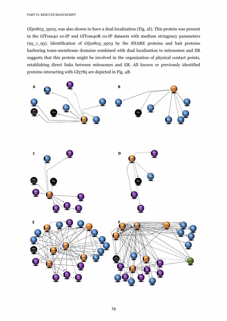
PART VI: RESULTS/MANUSCRIPT
Gl50803_9503, was also shown to have a dual localization (Fig. 2I). This protein was present
in the GlTom40 co-IP and GlTom40R co-IP datasets with medium stringency parameters
(95_1_95). Identification of Gl50803_9503 by the SNARE proteins and bait proteins
harboring trans-membrane domains combined with dual localization to mitosomes and ER
suggests that this protein might be involved in the organization of physical contact points,
establishing direct links between mitosomes and ER. All known or previously identified
proteins interacting with Gl5785 are depicted in Fig. 4B.
78
PART VI: RESULTS/MANUSCRIPT
Fig 4. Validation of the GlTom40 interactome by reverse co-IP using four additional
mitosome proteins as bait.
(A) Gl14939, (B) Gl5785, (C) GlIscS and (D) Gl9296 –derived interactomes. (E, F) Alternative depictions of the
cumulative interactome of proteins localizing to mitosomes generated with 6 bait proteins. Note the tight
association of GlTom40 with GlTom40R and Gl14939. (F) Matrix and soluble proteins are grouped at the bottom
left, proteins with dual localization (ER and mitosomes) and possibly involved in inter-organelle communication
are grouped at the top right. GlDRP is pulled down with all 6 bait proteins used in the co-IP assay and is also
included (green sphere). Bait proteins (orange spheres), matrix proteins (purple), previously identified and
localized proteins (black), and localized hypothetical proteins (blue). The stringency parameters used for detection
(high, medium, and relaxed) are represented by bold, dashed, and dotted arrows, respectively.
Co-IP with the matrix protein cysteine desulfurase (GlIscS)
Cysteine desulfurase (Gl50803_14519) is a mitosomal matrix protein and the central
component of the Fe-S assembly machinery [61]. All mitosomal matrix proteins including
GlIscS are translated in the cytoplasm and reach their final destination after unfolding and
translocation across the mitosome double membrane. Thus, this trafficking route (cytoplasm
– translocon – matrix) should be reflected in the protein-protein interactions of a co-IP
dataset with Gl14519-HA as bait. The MS dataset contained a total of 208 proteins using high
stringency parameters (95_2_95), with a FDR of 1.5%. After filtering with ctrl co-IP, 177 bait-
specific hits remained (S12 Table). Among those, we identified 5 known matrix proteins
namely, NifU-like protein, HSP70, [2Fe-2S] ferredoxin, Cpn60, and GiOR1. GlTom40 as the
sentinel protein for the translocon was detected only with relaxed stringency (50_1_50, FDR
of 30%). Seventy out of 177 hits were highly specific for GlIscS with ≥ 5 peptide counts.
Eighteen of those (25%) belong to the Protein 21.1 family. The biological function of this
cytoplasmic protein family in G. lamblia and the significant association to GlIscS is not
known [62]. The frequency of hits for 21.1 family members in the dataset might be relevant in
the context of stabilization of GlIscS in the cytoplasm before translocation and merits further
investigation. No additional candidate components of the import machinery (i.e. novel
proteins harboring TMDs) and/or novel or previously identified soluble proteins were
identified using Gl14519 as bait protein. However, 43 additional hypothetical proteins still
remain to be investigated for their localization and putative function. Fourteen out of 43
hypothetical proteins have high MASCOT values (peptide score of >5). Interestingly,
MITOPROT analysis suggests that six out of these fourteen proteins harbor an identifiable
MTS (S13 Table) and thus merit further investigation. All known/ previously identified
proteins interacting with Gl5785 at varying stringency parameters are depicted in Fig. 4C.
Co-IP with a hypothetical imported mitosome protein Gl50803_9296
Gl50803_9296 is a predicted soluble protein of unknown function localizing exclusively to
mitosomes (Fig. 2E). Despite this, MS data analysis performed at high stringency parameters
(95_2_95) yielded only 22 proteins with a FDR of 0% with 12 bait-specific hits. The bait
protein itself was by far the most significant hit in the dataset and under stringent analysis
79
PART VI: RESULTS/MANUSCRIPT
conditions the dataset contained no known mitosome proteins. Analysis with more relaxed
stringency parameters (90_1_90) yielded 85 proteins with a FDR of 6.2% with 47 bait-
specific identifications (S14 Table). Aside from the bait, 2 known mitosome proteins namely,
GlIscS and GlTom40 were identified. Taken together, the Gl9296 co-IP dataset suggests that
this mitosome protein doesn’t have an interactome enriched in matrix or membrane proteins
despite its clear-cut localization and considerable expression levels judged by the signal
obtained in Fig. 2E. Its localization might be in the intermembrane space, but remains to be
determined. The fact that the putative GlTom40R was identified once with a very low
stringency of 20_1_20 with a FDR of 51% suggested that Gl9296 and GlTom40R don’t
interact directly, but could be connected via bridging proteins. All identified Gl9296-
interacting proteins are depicted in Fig. 4D.
In summary, we have generated an extensive protein interaction network (Fig. 4E) from 6
independent co-IP assays using GlTom40 and 5 interaction partners (GlTom40R,
Gl50803_14939, Gl50803_5785, Gl50803_14519 and Gl50803_9296) selected as baits
based on i) number of peptide hits in the GlTom40 co-IP dataset and ii) confirmed
localization of epitope-tagged variants to mitosomes by IFA. Pending final validation of the
>200 novel candidate matrix-, outer membrane, and membrane-associated mitosome
components, the validated information from this series of co-IP experiments will be
instrumental to refine and complete the current map of the mitosome protein repertoire
shown in (Fig. 4E and 4F).
Pharmacological induction of a mitosome matrix-targeted DHFR complex
generates a protein import block and inhibits processing of an endogenous
reporter in mitosomes
The highly diverged GlTom40 orthologue in the G. lamblia genome is considered the
translocase across the outer mitosomal membrane. However, there are currently no reports
on the organelle-specific effects of interference on protein import into mitosomes. We tested
to what degree mitosome protein import is functionally conserved with respect to the
corresponding process in bona fide mitochondria by adapting the DHFR-folate analogue
system [63] to G. lamblia. Pre-sequence directed DHFR is a classical substrate used in
protein translocation studies due to its ability to fold irreversibly upon binding a folate
analog, e.g. MTX. Complexed with MTX, DHFR becomes unsuitable as a substrate for import
and blocks translocons, which results in a general blockage of organelle protein import [63].
Transfection of MTSfdΔint-DHFR into a Gl17030-HA background, i.e. a line expressing a HA-
tagged MTS-directed mitosomal reporter, allowed testing of the general effects of MTX-
induced import block. Importantly, G. lamblia lacks a DHFR homologue, and heterologous
over-expression of human DHFR did not result in any detectable phenotypic aberration or
change in growth rate (data not shown). We reasoned that the presence of MTX in
MTSfdΔint-DHFR expressing cells would lead to an import block with cytosolic accumulation
80
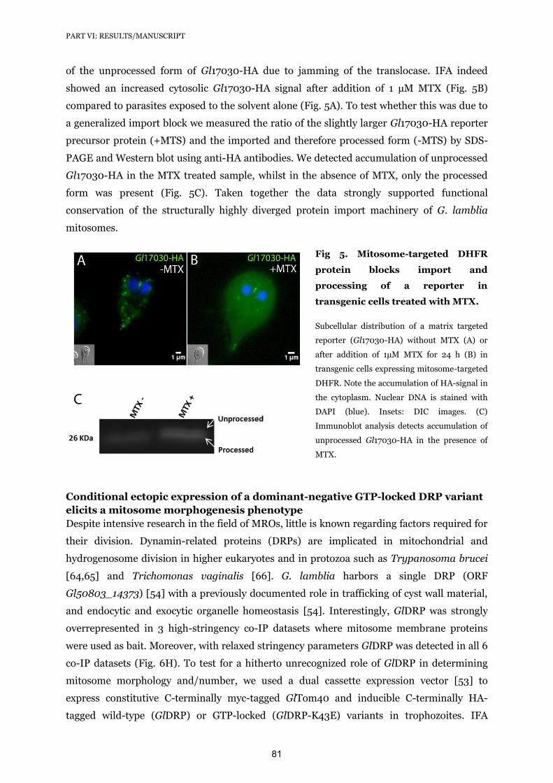
PART VI: RESULTS/MANUSCRIPT
of the unprocessed form of Gl17030-HA due to jamming of the translocase. IFA indeed
showed an increased cytosolic Gl17030-HA signal after addition of 1 µM MTX (Fig. 5B)
compared to parasites exposed to the solvent alone (Fig. 5A). To test whether this was due to
a generalized import block we measured the ratio of the slightly larger Gl17030-HA reporter
precursor protein (+MTS) and the imported and therefore processed form (-MTS) by SDS-
PAGE and Western blot using anti-HA antibodies. We detected accumulation of unprocessed
Gl17030-HA in the MTX treated sample, whilst in the absence of MTX, only the processed
form was present (Fig. 5C). Taken together the data strongly supported functional
conservation of the structurally highly diverged protein import machinery of G. lamblia
mitosomes.
Fig 5. Mitosome-targeted DHFR
protein blocks import and
processing of a reporter in
transgenic cells treated with MTX.
Subcellular distribution of a matrix targeted
reporter (Gl17030-HA) without MTX (A) or
after addition of 1µM MTX for 24 h (B) in
transgenic cells expressing mitosome-targeted
DHFR. Note the accumulation of HA-signal in
the cytoplasm. Nuclear DNA is stained with
DAPI (blue). Insets: DIC images. (C)
Immunoblot analysis detects accumulation of
unprocessed Gl17030-HA in the presence of
MTX.
Conditional ectopic expression of a dominant-negative GTP-locked DRP variant
elicits a mitosome morphogenesis phenotype
Despite intensive research in the field of MROs, little is known regarding factors required for
their division. Dynamin-related proteins (DRPs) are implicated in mitochondrial and
hydrogenosome division in higher eukaryotes and in protozoa such as Trypanosoma brucei
[64,65] and Trichomonas vaginalis [66]. G. lamblia harbors a single DRP (ORF
Gl50803_14373) [54] with a previously documented role in trafficking of cyst wall material,
and endocytic and exocytic organelle homeostasis [54]. Interestingly, GlDRP was strongly
overrepresented in 3 high-stringency co-IP datasets where mitosome membrane proteins
were used as bait. Moreover, with relaxed stringency parameters GlDRP was detected in all 6
co-IP datasets (Fig. 6H). To test for a hitherto unrecognized role of GlDRP in determining
mitosome morphology and/number, we used a dual cassette expression vector [53] to
express constitutive C-terminally myc-tagged GlTom40 and inducible C-terminally HA-
tagged wild-type (GlDRP) or GTP-locked (GlDRP-K43E) variants in trophozoites. IFA
81
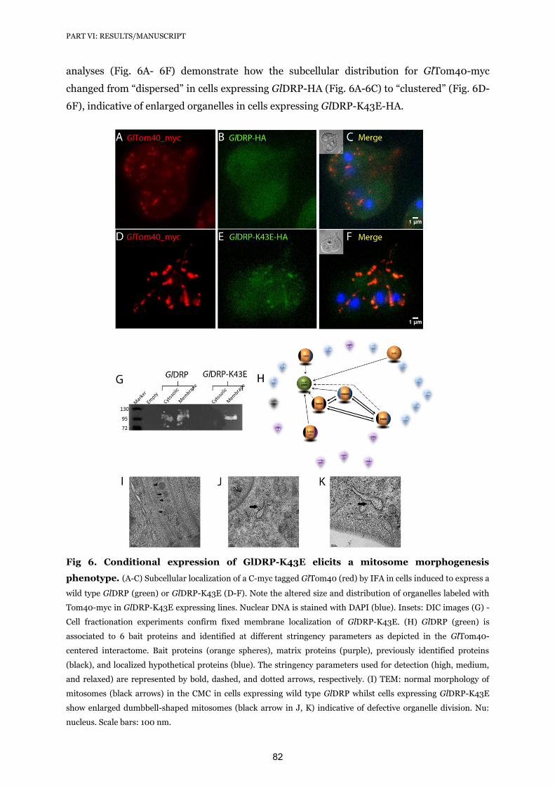
PART VI: RESULTS/MANUSCRIPT
analyses (Fig. 6A- 6F) demonstrate how the subcellular distribution for GlTom40-myc
changed from “dispersed” in cells expressing GlDRP-HA (Fig. 6A-6C) to “clustered” (Fig. 6D-
6F), indicative of enlarged organelles in cells expressing GlDRP-K43E-HA.
Fig 6. Conditional expression of GlDRP-K43E elicits a mitosome morphogenesis
phenotype. (A-C) Subcellular localization of a C-myc tagged GlTom40 (red) by IFA in cells induced to express a
wild type GlDRP (green) or GlDRP-K43E (D-F). Note the altered size and distribution of organelles labeled with
Tom40-myc in GlDRP-K43E expressing lines. Nuclear DNA is stained with DAPI (blue). Insets: DIC images (G) -
Cell fractionation experiments confirm fixed membrane localization of GlDRP-K43E. (H) GlDRP (green) is
associated to 6 bait proteins and identified at different stringency parameters as depicted in the GlTom40-
centered interactome. Bait proteins (orange spheres), matrix proteins (purple), previously identified proteins
(black), and localized hypothetical proteins (blue). The stringency parameters used for detection (high, medium,
and relaxed) are represented by bold, dashed, and dotted arrows, respectively. (I) TEM: normal morphology of
mitosomes (black arrows) in the CMC in cells expressing wild type GlDRP whilst cells expressing GlDRP-K43E
show enlarged dumbbell-shaped mitosomes (black arrow in J, K) indicative of defective organelle division. Nu:
nucleus. Scale bars: 100 nm.
82
PART VI: RESULTS/MANUSCRIPT
Consistent with this phenotype and in line with previous reports [54], the subcellular
distribution of HA-tagged GlDRP remained mostly cytosolic (Fig. 6B). Conversely, GlDRP-
K43E-HA showed a punctate distribution (Fig. 6E) and significant signal overlap with
GlTom40-myc (Fig. 6F), suggesting selective accumulation of GlDRP-K43E-HA on mitosome
membranes. We tested whether this marked association of ectopically expressed GlDRP-
K43E with organelle membranes compared to the wild type DRP variant in IFA could be
corroborated in cell fractionation experiments. Separation by SDS-PAGE and immunoblot
analysis revealed that epitope-tagged GlDRP-HA was almost equally distributed between the
“cytosolic” and “membrane” fraction, whereas the mutated variant GlDRP-K43E-HA was
detected only in the “membrane” fraction (Fig. 6G). This data was consistent with the
microscopical analysis in Fig. 6E and a strongly increased association with organelle
membranes compared to wild-type GlDRP-HA. To characterize the nature of the GlDRP-
K43E phenotype in more detail, we performed transmission electron microscopy of induced
transgenic cells. Cells expressing the GTP-locked GlDRP-K43E-HA variant frequently
presented elongated and tubular mitosome structures (Fig. 6J and 6K) compared to cells
expressing wild type GlDRP (Fig. 6I). This was consistent with IFA data in Fig. 6D and 6A
and suggestive of an organelle morphogenesis phenotype. Taken together, these data provide
an explanation for the frequent detection of GlDRP in co-IP datasets and strongly support a
transient association of GlDRP to these organelles. The phenotype elicited by expression of
GTP-locked GlDRP-K43E-HA suggests a previously unappreciated role for this GTPase in the
maintenance of mitosome integrity and organelle morphogenesis.
G. lamblia mitosomes are immobilized and do not form dynamic networks
Mitochondria in higher eukaryotes are highly dynamic organelle networks that move
in the cell via microtubules and microfilaments and undergo constant fission and fusion to
meet the energy requirements of the cell [67,68]. IFA and TEM analyses suggest that G.
lamblia mitosomes are very small spherical organelles with no evidence of network
formation. In addition, the mitosome population in each cell can be divided into peripheral
mitosomes (PM) distributed randomly in the cytoplasm and what has been dubbed the
central mitosome complex (CMC) [22]. The latter appear to form a grape-like cluster of
individual organelles of the size and shape of peripheral mitosomes that is closely and
permanently associated to the basal body complex between the two nuclei [22]. Interestingly,
these organelles remain separate despite their close proximity. The motility of this central
cluster is restricted to segregation with the duplicated basal body complex during cell division
[22]. However, no information is available on the spatial dynamics of peripheral mitosomes
in the cytoplasm. We investigated organelle dynamics in living cells by performing time lapse
microscopy of cells expressing GFP-tagged mitosome reporters. Conditional expression of N-
83
PART VI: RESULTS/MANUSCRIPT
terminally GFP-tagged GlTom40 with 3 h of induction followed by “chasing” newly-
synthesized GFP-Tom40 into mitosomes over 2-3 h in normal conditions can be used to label
organelles for imaging (Fig. 7A and 7B). Tracking of individual organelles over a period of
>30 min showed no significant cytoplasmic movement or changes in morphology (Fig. 7C),
suggesting that organelles neither move randomly nor are they transported directionally in
the cytoplasm along cytoskeleton structures. This is consistent with the lack of motor
proteins in any of the interactomes, suggesting that peripheral organelles do not undergo
frequent fusion and fission and may have lost the ability for fusion altogether. To test
whether mitosome outer membrane proteins are exchanged between organelles we
performed FRAP experiments on cells conditionally expressing GlTom40-GFP. Since
GlTom40-GFP is membrane-anchored, FRAP addresses the question how isolated mitosome
organelles are and whether they form membrane continuities. No recovery of fluorescence in
bleached CMC or PM organelles was detected (Fig. 7D- 7G), even when re-imaging bleached
areas after 20 min. Identical outcomes were observed in cells constitutively expressing GFP-
tagged GlTom40R (data not shown). GFP-tagged GlTom40R was expressed constitutively
from episomal (circular) expression vectors. The reporter localized exclusively to mitosome
organelles in all cases, but many cells showed a mitosome morphology dubbed “string”
phenotype suggestive of extensive elongation of organelles to large tubules (Fig. 7H). In many
cases, virtually all PMs had been replaced by a single long organelle with a diameter that
corresponded to that of an individual mitosome. FRAP analysis confirmed that these tubular
mitosomes were made from a single contiguous membrane (data not shown). Although the
“string” mitosome phenotype was compatible with survival of the parasites, many
trophozoites appeared to be delayed or even arrested in cytokinesis and had a typical heart-
shaped appearance (Fig. 7I) previously observed in cells which cannot complete cytokinesis
[69]. Because the tubular organelles ran through the non-divided part connecting both
daughter cells, we postulated that inability to divide mitosomes impairs completion of
cytokinesis.
84
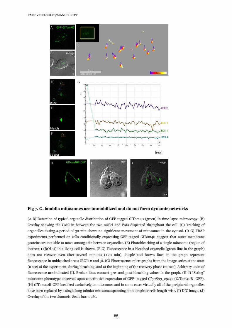
PART VI: RESULTS/MANUSCRIPT
Fig 7. G. lamblia mitosomes are immobilized and do not form dynamic networks
(A-B) Detection of typical organelle distribution of GFP-tagged GlTom40 (green) in time-lapse microscopy. (B)
Overlay showing the CMC in between the two nuclei and PMs dispersed throughout the cell. (C) Tracking of
organelles during a period of 30 min shows no significant movement of mitosomes in the cytosol. (D-G) FRAP
experiments performed on cells conditionally expressing GFP-tagged GlTom40 suggest that outer membrane
proteins are not able to move amongst/in between organelles. (E) Photobleaching of a single mitosome (region of
interest 1 (ROI 1)) in a living cell is shown. (F-G) Fluorescence in a bleached organelle (green line in the graph)
does not recover even after several minutes (>20 min). Purple and brown lines in the graph represent
fluorescence in unbleached areas (ROIs 2 and 3). (G) Fluorescence micrographs from the image series at the start
(0 sec) of the experiment, during bleaching, and at the beginning of the recovery phase (20 sec). Arbitrary units of
fluorescence are indicated [I]. Broken lines connect pre- and post-bleaching values in the graph. (H-J) “String”
mitosome phenotype observed upon constitutive expression of GFP- tagged Gl50803_29147 (GlTom40R- GFP).
(H) GlTom40R-GFP localized exclusively to mitosomes and in some cases virtually all of the peripheral organelles
have been replaced by a single long tubular mitosome spanning both daughter cells length-wise. (I) DIC image. (J)
Overlay of the two channels. Scale bar: 1 µM.
85
PART VI: RESULTS/MANUSCRIPT
Discussion
Eukaryotes that were previously considered to be amitochondriate are now known to
contain MROs such as hydrogenosomes and mitosomes [70,71]. Although there is a
consensus that MROs are evolutionarily derived from mitochondria, there is extensive
structural and functional divergence among organelles in different species. Bioinformatics
analyses show clearly that constraints for sequence divergence vary greatly, i.e. components
of the universal enzymatic machinery for the Fe-S protein maturation in MROs can be
detected in the nuclear genome using straightforward homology searches, whereas most
MRO membrane proteins have diverged beyond recognition if they were not lost altogether.
Three groups of membrane proteins are of particular interest in terms of organelle function
and propagation: the TOM/TIM complex and other protein import machinery, factors that
mediate interaction with the ER and the cytoskeleton, and transporters for ADP/ATP and
other building blocks. Attempts to discern the contribution of MROs to the cell’s metabolism
and catabolism and to understand why these organelles have been preserved even after they
lost their role in energy metabolism are confronted with two obstacles. Firstly, extensive
sequence divergence prevents identification of organelle proteins via homology-based
searches and secondly, the function of candidate factors identified by other means and
localized to organelle membranes can usually not be deduced based on existing structural
information from well-characterized mitochondrial homologs. In addition, the divergence
between orthologues of MRO proteins even in closely related species is very high. A case in
point is GlTOM40 whose sequence degeneration is so extensive that the identification of
orthologues in Giardia, Entamoeba or Spironucleus remains tentative despite the
constraints imposed by the beta barrel structure of these mitochondrial porins [44].
Unambiguous identification is even more difficult for highly diverged proteins that lack
clearly identifiable structural domains (e.g. a putative GlTim44 homolog, a component of the
TIM complex) [72].
G. lamblia mitosomes remain the smallest known and least characterized MROs. Systematic
identification of protein components using proteome analysis of enriched mitosome
preparations has proven challenging primarily due to extensive contamination and difficulty
to isolate organelles in sufficient amounts [33,48]
Here, we accounted for the paucity of data which may inform strategies for the systematic
identification of Giardia mitosome proteins by implementing an approach based on MS
analysis of chemically stabilized protein-protein interactions. Our starting premise was that
the diminutive mitosomes harbored no more than 100-150 different proteins inside or
associated with a clearly delineated cellular compartment. Using a putative GlTOM40 [22,33]
as a starting point, this study focused on organelle membrane components, in particular the
86
PART VI: RESULTS/MANUSCRIPT
mitosome protein import machinery whose function has been demonstrated but whose
composition remains unknown.
With only six bait proteins, the strategy based on iterative co-IP experiments allowed for
building a core membrane interactome and a complex interactome network extending
inwards to the organelle matrix as well as outwards to components of the ER membrane, the
axoneme cytoskeleton and the cytoplasm. The rationale is that with sufficient numbers of
reverse co-IP experiments using validated organelle proteins as baits, a comprehensive
organelle proteome can be built.
Co-IP with GlTom40 as bait and reverse co-IP performed with 5 subsequent new bait
proteins resulted in identification of 22 mitosomal proteins (annotated and hypothetical) out
of which 16 proteins were localized to mitosomes; 5 proteins displayed dual localization to
mitosomes and ER and one protein (Gl7188) showed a ER and PV pattern even though it was
pulled down exclusively with mitosome-localized bait proteins harboring TMDs. Based on
their localization pattern, we grouped these mitosome proteins into 2 categories (Fig. 4F).
The group at the bottom left with GlIscS at the center depict proteins with exclusive
mitosome localization suggesting that these proteins are mitosome residents (soluble or
membrane-anchored) whereas the group at the top right (Fig. 4F) includes all proteins with
dual localization, suggesting they might be at the junction between ER and mitosomes with a
potential role in establishing inter-organelle communication, e.g. for lipid transport.
GlTom40 and interaction partners GlTom40R and Gl14939: a minimized
mitosome protein import apparatus?
Following its identification as a prominent GlTom40 interaction partner, the single
pass membrane protein GlTom40R was the first bait protein selected for reverse co-IP in
order to validate the experimental procedure and to expand the GlTom40 interactome.
Importantly, the protein has a predicted N-terminal TMD similar to mitochondrial Tom40
receptors (Tom20 and Tom70) in Saccharomyces cerevisiae. Furthermore, this TMD is
preceded by a short N-terminal region and followed by a large C-terminal stretch. Ectopic
expression of the C-terminal portion of GlTom40R alone showed a distinct cytosolic
localization by IFA (data not shown), without a detectable phenotype. However, several in
silico analysis tools failed to predict a MTS at the protein’s N-terminus. Nevertheless, a C-
terminally GFP-tagged full-length variant determines the specific topology of this protein. We
have shown previously that GFP only fluoresces if exposed to the cytoplasm and never after
import into mitosomes ([22] and unpublished data). Therefore, the brightly fluorescing and
mitosome-localized GlTom40R-GFP fusion provided conclusive data and is a direct proof for
the proposed topology of GlTom40R. This transgenic line also provided an additional tool for
time lapse microscopy of mitosomes in trophozoites.
87
PART VI: RESULTS/MANUSCRIPT
Reverse co-IP using GlTom40R as bait pulled down GlTom40 with the most abundant
peptide counts and 220 additional specific proteins (annotated and hypothetical). So far 20
proteins have been validated by localization to mitosomes, allowing for a significant
expansion of the GlTom40/GlTom40R interactome. The topology of GlTom40R, its exclusive
mitosomal localization, and the repertoire of pulled down proteins further supports that
GlTom40R is a Tom40 accessory protein with a potential receptor function for protein
import. Specifically, the long cytoplasmically exposed C-terminal stretch interacts with
several imported matrix proteins independently of the presence of a predicted transit peptide
(S6 Table). To test this idea, we performed co-IP using the HA-tagged C-terminal part of
GlTom40R (amino acid 31-133), which has a cytoplasmic localization, under non-
crosslinking conditions. The resulting data is difficult to interpret, given that out of a total of
232 proteins detected at high stringency (95_2_95), 171 were specific to the C-terminal
GlTom40R with none of the previously known mitosomal resident proteins in the dataset.
However, upon analysis with medium stringency parameters (95_1_95), we identified only 2
novel hypothetical mitosomal proteins (Gl16424, Gl9503) in the C-terminal GlTom40R
specific dataset. This suggests that the cytoplasmic GlTom40R fragment does not recapitulate
the interaction properties of the full-length membrane anchored variant. Taken together, the
co-IP data suggest that i) capture of imported matrix proteins is context-dependent, i.e. likely
requires incorporation of the receptor domain into a TOM complex, and ii) interaction with
GlTom40 requires the domains on either side of the membrane anchor and possibly
additional accessory membrane proteins. Hence, the hypothesis of a receptor function for
GlTom40R remains to be tested directly to evaluate its exact role in proteins import.
Interestingly, although BLASTp yielded no strong homologs for GlTom40R, profile sequence
comparisons with HHpred showed homology to a “high potential iron sulfur protein” (p-
value 0.007). “High potential iron sulfur protein” in higher eukaryotes, also known as
mitoNEET, is an integral membrane protein localized at the outer membrane of
mitochondria and is responsible for transport of iron into mitochondria [73]. If we consider
that Fe-S protein maturation is the only metabolic pathway currently associated to G. lamblia
mitosomes, GlTom40R might also function as a mitoNEET homolog in G. lamblia.
Another GlTom40 interaction partner of special interest is Gl14939. This protein was
exclusively identified in the GlTom40 and the GlTom40R co-IP datasets, suggesting that
Gl14939 and GlTom40R may function as part of a complex. TMHMM predicts two TMDs at
its N-terminus, followed by a large C-terminal domain in Gl14939. Powerful HMMER-based
searches across several eukaryotic lineages, including the closely related diplomonad
Spironucleus salmonicida [74], yielded no orthologues for this protein (data not shown). To
date, there is no further information available indicating a function for Gl14939. However, a
recent study showed that Gl14939 (dubbed GiMOMP35) localizes at the outer mitosome
88
PART VI: RESULTS/MANUSCRIPT
membrane with its C- terminus in the cytosol [72]. Nonetheless, given that GlTom40 (the
translocon), GlTom40R, and Gl14939 (putative accessory protein) are outer membrane-
associated proteins and part of the same interactome, the data is consistent with a minimized
import apparatus whose core import machinery is composed of only these three proteins.
Likewise, a function in protein import of several other novel hypothetical proteins without
TMD, e.g. Gl10971, Gl9296, Gl8148, Gl17276, and Gl16424, remains to be elucidated. Reverse
co-IP using Gl9296 as bait protein pulled down GlIscS, GlIscA, GlHsp70, and outer
membrane proteins including GlTom40 and GlTom40R, indicating close association to the
import machinery and imported matrix proteins. Furthermore, Gl9296 has a predicted MTS
but protein sequence analysis using the SMART prediction tool identified no functional
domain of significance. Similarly, Gl9296 appears specific to the Giardia lineage. Given its
interaction profile, predicted import signal, and lack of homology to metabolic enzymes, we
hypothesize that Gl9296 is a matrix protein interacting with the import pore in mitosomes.
Recent developments in gene knockout strategies in G. lamblia [75] will facilitate the
definition of the role of these novel hypothetical proteins.
All 24 localized mitosome proteins (previously known and newly identified hypotheticals)
were parsed according to molecular function and biological process (S3A and S3B Figs) using
Blast2go (https://www.blast2go.com/). Metal ion, Fe-S, ATP, and protein binding were the
major molecular functions associated with these proteins. Interestingly, other biological
processes involving response to lipid and transmembrane transport were also identified with
significant p-values. An additional 93 candidates annotated as hypothetical proteins (from all
the 6 co-IP assays) were analyzed using Blast2go (S4A and S4B Figs). Binding and catalytic
activities were the 2 major GO terms associated to this group. Their potential involvement in
binding of lipids, flavin mono-nucleotide co-factor, metal ion, Fe-S cluster, calcium ion,
nucleotide, Fe and protein kinase will have to be tested experimentally. The number of
combined candidate mitosome proteins after elimination of obvious contaminants such as
cytoplasmic proteins is sufficiently large to suggest that mitosomes may have a role beyond
Fe-S protein maturation. Only recently the major function of E. histolytica mitosomes was
shown to be sulfate activation, and not Fe-S protein maturation as previously thought [44].
Although genes involved in this pathway are missing in other MRO-containing organisms
such as G. lamblia, T. vaginalis, and C. parvum, the Entamoeba example points to a wider
range of functions ascribable to mitosomes. This may even include general functions in stage-
differentiation as recently shown in E. histolytica whose mitosomes are essential for the
encystation process [76].
Mitosome-ER contact sites
Co-IP data identified proteins with dual localization at mitosomes and ER. Contact
between these organelles mediates at least two major functions, i.e. replication of mitosomes
89
PART VI: RESULTS/MANUSCRIPT
and transport associated to lipid biosynthesis. Thus far, we have identified five mitosome
proteins with dual localization potentially involved in inter-organelle communication (Fig.
4F). One of them is a transmembrane Qb-SNARE 4 (Gl50803_5785) [77] identified in
GlTom40 and GlTom40R co-IP datasets. IFA analysis localized a tagged variant to
mitosomes and parts of the ER.
For their biogenesis, mitochondria and MROs rely on lipid transfer from the ER, the central
site for phospholipid synthesis in the cell [78,79]. SNAREs are best known for mediating
membrane fusion in vesicular transport [80] In the context of mitochondria and the ER, they
function as components of so called ER-mitochondria encounter structures (ERMES). In
addition to being associated to mitochondrial protein import [81,82], ERMES fulfills an
essential function in inter-organelle lipid transport [81]. Phosphatidylserine is shuttled from
the ER to mitochondria through the ERMES complex where it is converted to
phosphatidylethanolamine (PE) by a decarboxylation reaction that generates most if not all
PE in mitochondria [81,83]. Whether this function is preserved in Giardia mitosomes is not
known, however, organelle biogenesis necessarily depends on ER-derived lipids which are
transported to mitosomes either by carrier proteins or via membrane contact sites. The latter
requires a tethering complex to facilitate phospholipid exchange between the two organelles.
We explored the idea that Qb-SNARE 4 is part of a larger complex mediating ER-mitosome
interaction by generating co-IP data. Indeed, in addition to outer membrane proteins such as
GlTom40, GlTom40R and Gl14939, 3 hypothetical proteins were identified in the dataset,
two of which, Gl9503 (3 TMDs) and Gl21943 (soluble), localized both to the ER and to
mitosomes. In addition, data mining identified a domain in Gl9503 with similarity to a yeast
“Maintenance of mitochondrial morphology” protein 1 (Mmm1) of the ERMES complex as
well as a predicted anhydrolase domain involved in lipid synthesis. HHpred analysis revealed
a link between Gl21943 and a beta barrel lipid binding protein MLN64 (e-value 0.0006) in H.
sapiens which facilitates cholesterol transport to mitochondria [84]. This preliminary data
points to an outer mitosomal membrane-associated complex in G. lamblia mitosomes
involved in generating ER-mitosome membrane contact sites analogous to ERMES.
However, unlike in the hydrogenosome-containing T. vaginalis [85], ERMES homologs have
not been identified in G. lamblia possibly due to extensive sequence divergence. Further
experiments are necessary to test whether the dually-localized proteins Gl21943, Gl22587,
Gl5785, Gl9503 and Gl15154 (Figs. 2F-2I and Fig. 3C) are indeed components of bona fide
ER-mitosome membrane contact sites.
Functional analysis of protein import into the mitosome matrix
Using transgenic lines co-expressing MTSfdΔint-DHFR and Gl17030-HA we induced a
protein import block by addition of the folate analog MTX. Accumulation of the reporter in
the cytoplasm upon MTX treatment shown in IFA (Fig. 5A and 5B) suggested blocked import
90
PART VI: RESULTS/MANUSCRIPT
of the reporter presumably as a result of clogging of the import pore. Consistent with this,
cells exposed to MTX presented accumulation of the unprocessed form of the reporter
Gl17030-HA in a Western blot analysis (Fig. 5C). Based on the interaction of MTX with the
mitosome targeted DHFR this is evidence for a blockage of the import pore analogous to
established assays in bona fide mitochondria [63]. The data also confirms the previously
tested notion that membrane translocation requires pre-proteins to remain in an unfolded
state [72]. Furthermore, it provides direct experimental evidence for the functional
conservation of the mitosome protein import pathway and the presence of a canonical
general insertion pore (GIP)/translocon in G. lamblia mitosomes. Given that GlTom40
localizes exclusively to mitosomes and is the only predicted beta barrel protein in G. lamblia,
this highly diverged protein is currently the only candidate for the mitosomal translocon.
Interestingly, recent reports on mitochondrial protein import in T. brucei (Excavata super-
group) identified an archaic TOM (ATOM) as the functional translocase at the outer
mitochondrial membrane [86]. ATOM is not phylogenetically related to canonical Tom40
proteins, suggesting there is more than one kind of eukaryotic translocase that can function
as an import pore. Whether this is also the case in G. lamblia remains to be investigated.
Mitosome dynamics and a novel role for GlDRP in mitosome morphogenesis
We had previously shown that replication and inheritance of the CMC is coordinated
in a cell cycle-dependent manner, whereas PMs divided stochastically [22]. The lack of a
system to track organelles in living trophozoites precluded addressing the question whether
mitosomes were motile and constituted a dynamic network of organelles. Development of
two GFP-tagged reporters GFP-GlTom40 and GlTom40R-GFP (this study) allowed for time-
lapse experiments to follow individual organelles in a cell. However, we found no evidence for
motility of organelles, neither in the CMC nor in PMs, even after prolonged observation (1.5
h). Moreover, FRAP experiments revealed no exchange of GFP-tagged membrane proteins
between organelles during the period of observation (Fig. 7F and 7G), which further
corroborated the relative isolation of mitosomes within the cytosol. The lack of motility,
inter-organelle contact and -interaction in Giardia mitosomes complicates investigation of
their replication and morphogenesis. The two most plausible scenarios for this are currently
the following: i) PMs are released from the CMC, which continuously produces new
organelles by elongation and fission to maintain a constant number of organelles in a cell-
cycle independent manner; ii) PMs and the CMC organelles replicate independently in a cell-
cycle independent and -dependent manner, respectively [22]. Although time-lapse
microscopy experiments did not provide evidence for either scenario, conditional expression
of a dominant-negative, constitutively active GlDRP-K43E revealed a distinct morphogenesis
phenotype (see also below) indicative of an organelle replication defect. Moreover, the
distinctive “string” mitosome phenotype in cells expressing GlTom40R-GFP clearly
demonstrated that mitosomes can assume an elongated, tubular morphology, which is a
91
PART VI: RESULTS/MANUSCRIPT
prerequisite for organelle division and replication. The implication is that G. lamblia
mitosomes retain at least the machinery for fission in which the mechanoenzyme DRP plays a
central role. Without the capability for fusion, this organelle “network” remains in a
maximally fragmented state. The “string” mitosome phenotype therefore reflects a lack of
fission, for example because the presence of the GFP-tagged protein on the surface interferes
with recruitment of DRP to the mitosome membrane.
As one of the key players in the regulation of mitochondrial fission, dynamin related protein
(DRP) is a mechanoenzyme conserved from yeast to vertebrates [87,88,89,90]. G. lamblia
harbors a single dynamin homologue (GlDRP) encoded by ORF Gl50803_14373; this protein
has been shown to play a major role in this parasite’s endocytic pathway and stage conversion
[54,91,92]. Transgenic parasites expressing the mutant GlDRP-K43E protein exhibited larger
and fewer mitosomes, compared to cells overexpressing wild type GlDRP (Fig. 6). This is in
line with the dominant-negative effect on mitochondrial fission elicited by the corresponding
mutation in DRPs in other organisms. To our knowledge, this is the first report on the
involvement of GlDRP in mitosome homeostasis, supporting the (at least partial) functional
conservation of mitochondrial and MRO fission [93,94,95,96]. The notion that G. lamblia
mitosome fission is functionally conserved is further substantiated by the identification of
ORF Gl22587 in the mitosome-specific co-IP datasets. This protein presents dual localization
to mitosomes and the ER. Interestingly, HMMER-based predictions relate Gl22587 to human
mitochondrial fission protein (Fis1, e-value 6.3E-05) which, along with mitochondrial fission
factor (Mff) and mitochondrial dynamics proteins (MiD 49 and MiD51), acts as a receptor to
recruit dynamin-related protein 1 (Drp1) to the mitochondrial surface [97,98]. The isolation
of Gl22587-interaction partners might lead to the identification of regulators and recruiting
factors for GlDRP, thereby shedding light on the composition of the mitosome fission
machinery and replication mechanism in Giardia.
Conclusion
Starting from the premise that mitosomes represent distinct cellular compartments with a
very limited number of components in close physical proximity, we used an iterative
approach based on co-IP experiments to generate a GlTom40-centered interactome network.
The ultimate purpose of this strategy is to build a full proteome, which delineates the full
complement of organelle proteins, peripherally associated factors, as well as interfaces with
the ER and the cytoskeleton. Although this strategy requires numerous rounds of sequential
co-IP and validation, it is highly informative because it produces interaction data in addition
to identifying novel proteins. Combined with testing of epitope-tagged variants of candidate
proteins for organelle localization as a straightforward validation criterion, serial co-IPs allow
unambiguous definition of the organelle-specific proteome, as well as interfaces with other
92
PART VI: RESULTS/MANUSCRIPT
cellular structures. A case in point is the complete lack of false-negative hits in the GlIscS co-
IP dataset, which contained all previously identified factors in addition to numerous novel
candidate matrix and inner membrane proteins. Due to the extreme sequence divergence in
nuclear-encoded genes for mitosome proteins, functional models for most components and
pathways cannot simply be replicated based on a mitochondrial blueprint, but require almost
complete ab initio (re)construction. Consequently, an exact and comprehensive delineation
of the organelle proteome is an indispensable prerequisite for any attempt at defining the
functional range of mitosome metabolism, as well as the organelle’s machinery for replication
and morphogenesis.
Acknowledgements
We thank Therese Michel for technical assistance. Dr. Peter Hunziker and his team at the
proteomics service facility of the Functional Genomics Center-Zürich are gratefully
acknowledged for their support in tandem mass spectrometry performance and analysis.
Prof. Robert Sinden at Imperial College, London (UK) is gratefully acknowledged for
providing the human DHFR-encoding plasmid. Dr. Chandra Ramakrishnan is gratefully
acknowledged for critical revision of the manuscript.
References
1. Gray MW, Burger G, Lang BF (1999) Mitochondrial evolution. Science 283: 1476-1481.
2. Hedges SB, Blair JE, Venturi ML, Shoe JL (2004) A molecular timescale of eukaryote evolution and
the rise of complex multicellular life. BMC Evol Biol 4: 2.
3. Yang D, Oyaizu Y, Oyaizu H, Olsen GJ, Woese CR (1985) Mitochondrial origins. Proc Natl Acad Sci
U S A 82: 4443-4447.
4. Olsen GJ, Woese CR, Overbeek R (1994) The winds of (evolutionary) change: breathing new life
into microbiology. J Bacteriol 176: 1-6.
5. Viale AM, Arakaki AK (1994) The chaperone connection to the origins of the eukaryotic
organelles. FEBS Lett 341: 146-151.
6. Bauer MF, Hofmann S, Neupert W, Brunner M (2000) Protein translocation into mitochondria:
the role of TIM complexes. Trends Cell Biol 10: 25-31.
7. Jensen RE, Johnson AE (2001) Opening the door to mitochondrial protein import. Nat Struct Biol
8: 1008-1010.
8. Koehler CM, Merchant S, Schatz G (1999) How membrane proteins travel across the
mitochondrial intermembrane space. Trends Biochem Sci 24: 428-432.
93
PART VI: RESULTS/MANUSCRIPT
9. Matouschek A, Pfanner N, Voos W (2000) Protein unfolding by mitochondria. The Hsp70 import
motor. EMBO Rep 1: 404-410.
10. Prokisch H, Scharfe C, Camp DG, 2nd, Xiao W, David L, et al. (2004) Integrative analysis of the
mitochondrial proteome in yeast. PLoS Biol 2: e160.
11. Reinders J, Zahedi RP, Pfanner N, Meisinger C, Sickmann A (2006) Toward the complete yeast
mitochondrial proteome: multidimensional separation techniques for mitochondrial
proteomics. J Proteome Res 5: 1543-1554.
12. Sickmann A, Reinders J, Wagner Y, Joppich C, Zahedi R, et al. (2003) The proteome of
Saccharomyces cerevisiae mitochondria. Proc Natl Acad Sci U S A 100: 13207-13212.
13. Pfanner N, Truscott KN (2002) Powering mitochondrial protein import. Nat Struct Biol 9: 234-
236.
14. Gabaldon T, Huynen MA (2004) Shaping the mitochondrial proteome. Biochim Biophys Acta
1659: 212-220.
15. Cerkasovová A, Lukasová G, Cerkasòv J, J K (1973) Biochemical characterization of large granule
fraction of Tritrichomonas foetus (strain KV1). Journal of Protozoology 20.
16. Lindmark DG, Muller M (1973) Hydrogenosome, a cytoplasmic organelle of the anaerobic
flagellate Tritrichomonas foetus, and its role in pyruvate metabolism. J Biol Chem 248:
7724-7728.
17. Mai Z, Ghosh S, Frisardi M, Rosenthal B, Rogers R, et al. (1999) Hsp60 is targeted to a cryptic
mitochondrion-derived organelle ("crypton") in the microaerophilic protozoan parasite
Entamoeba histolytica. Mol Cell Biol 19: 2198-2205.
18. Muller M (1993) The hydrogenosome. J Gen Microbiol 139: 2879-2889.
19. Tovar J, Fischer A, Clark CG (1999) The mitosome, a novel organelle related to mitochondria in
the amitochondrial parasite Entamoeba histolytica. Mol Microbiol 32: 1013-1021.
20. Shiflett AM, Johnson PJ (2010) Mitochondrion-related organelles in eukaryotic protists. Annu
Rev Microbiol 64: 409-429.
21. Adl SM, Simpson AG, Lane CE, Lukes J, Bass D, et al. (2012) The revised classification of
eukaryotes. J Eukaryot Microbiol 59: 429-493.
22. Regoes A, Zourmpanou D, Leon-Avila G, van der Giezen M, Tovar J, et al. (2005) Protein import,
replication, and inheritance of a vestigial mitochondrion. J Biol Chem 280: 30557-30563.
23. Tovar J, Leon-Avila G, Sanchez LB, Sutak R, Tachezy J, et al. (2003) Mitochondrial remnant
organelles of Giardia function in iron-sulphur protein maturation. Nature 426: 172-176.
24. Makiuchi T, Nozaki T (2014) Highly divergent mitochondrion-related organelles in anaerobic
parasitic protozoa. Biochimie 100: 3-17.
25. Riordan CE, Ault JG, Langreth SG, Keithly JS (2003) Cryptosporidium parvum Cpn60 targets a
relict organelle. Curr Genet 44: 138-147.
94
PART VI: RESULTS/MANUSCRIPT
26. Katinka MD, Duprat S, Cornillot E, Metenier G, Thomarat F, et al. (2001) Genome sequence and
gene compaction of the eukaryote parasite Encephalitozoon cuniculi. Nature 414: 450-453.
27. Jerlstrom-Hultqvist J, Einarsson E, Xu F, Hjort K, Ek B, et al. (2013) Hydrogenosomes in the
diplomonad Spironucleus salmonicida. Nature communications 4: 2493.
28. Abrahamsen MS, Templeton TJ, Enomoto S, Abrahante JE, Zhu G, et al. (2004) Complete
genome sequence of the apicomplexan, Cryptosporidium parvum. Science 304: 441-445.
29. Leon-Avila G, Tovar J (2004) Mitosomes of Entamoeba histolytica are abundant mitochondrion-
related remnant organelles that lack a detectable organellar genome. Microbiology 150:
1245-1250.
30. Turner G, Muller M (1983) Failure to detect extranuclear DNA in Trichomonas vaginalis and
Tritrichomonas foetus. J Parasitol 69: 234-236.
31. van der Giezen M, Sjollema KA, Artz RR, Alkema W, Prins RA (1997) Hydrogenosomes in the
anaerobic fungus Neocallimastix frontalis have a double membrane but lack an associated
organelle genome. FEBS Lett 408: 147-150.
32. Dolezal P, Smid O, Rada P, Zubacova Z, Bursac D, et al. (2005) Giardia mitosomes and
trichomonad hydrogenosomes share a common mode of protein targeting. Proc Natl Acad
Sci U S A 102: 10924-10929.
33. Jedelsky PL, Dolezal P, Rada P, Pyrih J, Smid O, et al. (2011) The minimal proteome in the
reduced mitochondrion of the parasitic protist Giardia intestinalis. PLoS One 6: e17285.
34. Lill R (2009) Function and biogenesis of iron-sulphur proteins. Nature 460: 831-838.
35. Craig EA, Voisine C, Schilke B (1999) Mitochondrial iron metabolism in the yeast Saccharomyces
cerevisiae. Biol Chem 380: 1167-1173.
36. Schilke B, Voisine C, Beinert H, Craig E (1999) Evidence for a conserved system for iron
metabolism in the mitochondria of Saccharomyces cerevisiae. Proc Natl Acad Sci U S A 96:
10206-10211.
37. Ankarklev J, Jerlstrom-Hultqvist J, Ringqvist E, Troell K, Svard SG (2010) Behind the smile: cell
biology and disease mechanisms of Giardia species. Nat Rev Microbiol 8: 413-422.
38. Morrison HG, McArthur AG, Gillin FD, Aley SB, Adam RD, et al. (2007) Genomic minimalism in
the early diverging intestinal parasite Giardia lamblia. Science 317: 1921-1926.
39. Davis-Hayman SR, Nash TE (2002) Genetic manipulation of Giardia lamblia. Molecular and
biochemical parasitology 122: 1-7.
40. Boucher SE, Gillin FD (1990) Excystation of in vitro-derived Giardia lamblia cysts. Infect Immun
58: 3516-3522.
41. Abodeely M, DuBois KN, Hehl A, Stefanic S, Sajid M, et al. (2009) A contiguous compartment
functions as endoplasmic reticulum and endosome/lysosome in Giardia lamblia. Eukaryot
Cell 8: 1665-1676.
95
PART VI: RESULTS/MANUSCRIPT
42. Stefanic S, Morf L, Kulangara C, Regos A, Sonda S, et al. (2009) Neogenesis and maturation of
transient Golgi-like cisternae in a simple eukaryote. J Cell Sci 122: 2846-2856.
43. Goldberg AV, Molik S, Tsaousis AD, Neumann K, Kuhnke G, et al. (2008) Localization and
functionality of microsporidian iron-sulphur cluster assembly proteins. Nature 452: 624-
628.
44. Mi-ichi F, Abu Yousuf M, Nakada-Tsukui K, Nozaki T (2009) Mitosomes in Entamoeba histolytica
contain a sulfate activation pathway. Proc Natl Acad Sci U S A 106: 21731-21736.
45. Putignani L, Tait A, Smith HV, Horner D, Tovar J, et al. (2004) Characterization of a
mitochondrion-like organelle in Cryptosporidium parvum. Parasitology 129: 1-18.
46. Sanderson SJ, Xia D, Prieto H, Yates J, Heiges M, et al. (2008) Determining the protein
repertoire of Cryptosporidium parvum sporozoites. Proteomics 8: 1398-1414.
47. Tsaousis AD, Kunji ER, Goldberg AV, Lucocq JM, Hirt RP, et al. (2008) A novel route for ATP
acquisition by the remnant mitochondria of Encephalitozoon cuniculi. Nature 453: 553-556.
48. Wampfler PB, Tosevski V, Nanni P, Spycher C, Hehl AB (2014) Proteomics of secretory and
endocytic organelles in Giardia lamblia. PLoS One 9: e94089.
49. Dagley MJ, Dolezal P, Likic VA, Smid O, Purcell AW, et al. (2009) The protein import channel in
the outer mitosomal membrane of Giardia intestinalis. Mol Biol Evol 26: 1941-1947.
50. Hehl AB, Marti M, Kohler P (2000) Stage-specific expression and targeting of cyst wall protein-
green fluorescent protein chimeras in Giardia. Mol Biol Cell 11: 1789-1800.
51. Morf L, Spycher C, Rehrauer H, Fournier CA, Morrison HG, et al. (2010) The transcriptional
response to encystation stimuli in Giardia lamblia is restricted to a small set of genes.
Eukaryot Cell 9: 1566-1576.
52. Hehl AB, Marti M, Kohler P (2000) Stage-specific expression and targeting of cyst wall protein-
green fluorescent protein chimeras in Giardia. Molecular biology of the cell 11: 1789-1800.
53. Konrad C, Spycher C, Hehl AB (2010) Selective condensation drives partitioning and sequential
secretion of cyst wall proteins in differentiating Giardia lamblia. PLoS Pathog 6: e1000835.
54. Gaechter V, Schraner E, Wild P, Hehl AB (2008) The single dynamin family protein in the
primitive protozoan Giardia lamblia is essential for stage conversion and endocytic
transport. Traffic 9: 57-71.
55. Pusnik M, Mani J, Schmidt O, Niemann M, Oeljeklaus S, et al. (2012) An essential novel
component of the noncanonical mitochondrial outer membrane protein import system of
trypanosomatids. Molecular biology of the cell 23: 3420-3428.
56. Rajala N, Hensen F, Wessels HJ, Ives D, Gloerich J, et al. (2015) Whole cell formaldehyde cross-
linking simplifies purification of mitochondrial nucleoids and associated proteins involved
in mitochondrial gene expression. PLoS One 10: e0116726.
96
PART VI: RESULTS/MANUSCRIPT
57. Jimenez-Garcia LF, Zavala G, Chavez-Munguia B, Ramos-Godinez Mdel P, Lopez-Velazquez G, et
al. (2008) Identification of nucleoli in the early branching protist Giardia duodenalis.
International journal for parasitology 38: 1297-1304.
58. Elmendorf HG, Rohrer SC, Khoury RS, Bouttenot RE, Nash TE (2005) Examination of a novel
head-stalk protein family in Giardia lamblia characterised by the pairing of ankyrin repeats
and coiled-coil domains. International journal for parasitology 35: 1001-1011.
59. Isenmann S, Khew-Goodall Y, Gamble J, Vadas M, Wattenberg BW (1998) A splice-isoform of
vesicle-associated membrane protein-1 (VAMP-1) contains a mitochondrial targeting
signal. Mol Biol Cell 9: 1649-1660.
60. Duman JG, Forte JG (2003) What is the role of SNARE proteins in membrane fusion? Am J
Physiol Cell Physiol 285: C237-249.
61. Bandyopadhyay S, Chandramouli K, Johnson MK (2008) Iron-sulfur cluster biosynthesis.
Biochemical Society transactions 36: 1112-1119.
62. Manning G, Reiner DS, Lauwaet T, Dacre M, Smith A, et al. (2011) The minimal kinome of
Giardia lamblia illuminates early kinase evolution and unique parasite biology. Genome
biology 12: R66.
63. Eilers M, Schatz G (1986) Binding of a specific ligand inhibits import of a purified precursor
protein into mitochondria. Nature 322: 228-232.
64. Morgan GW, Goulding D, Field MC (2004) The single dynamin-like protein of Trypanosoma
brucei regulates mitochondrial division and is not required for endocytosis. J Biol Chem
279: 10692-10701.
65. Chanez AL, Hehl AB, Engstler M, Schneider A (2006) Ablation of the single dynamin of T. brucei
blocks mitochondrial fission and endocytosis and leads to a precise cytokinesis arrest.
Journal of cell science 119: 2968-2974.
66. Wexler-Cohen Y, Stevens GC, Barnoy E, van der Bliek AM, Johnson PJ (2014) A dynamin-related
protein contributes to Trichomonas vaginalis hydrogenosomal fission. Faseb J 28: 1113-
1121.
67. Chan DC (2006) Mitochondria: dynamic organelles in disease, aging, and development. Cell 125:
1241-1252.
68. Chen H, Chan DC (2009) Mitochondrial dynamics--fusion, fission, movement, and mitophagy--in
neurodegenerative diseases. Human molecular genetics 18: R169-176.
69. Sonda S, Stefanic S, Hehl AB (2008) A sphingolipid inhibitor induces a cytokinesis arrest and
blocks stage differentiation in Giardia lamblia. Antimicrobial agents and chemotherapy 52:
563-569.
70. van der Giezen M (2009) Hydrogenosomes and mitosomes: conservation and evolution of
functions. J Eukaryot Microbiol 56: 221-231.
71. Aguilera P, Barry T, Tovar J (2008) Entamoeba histolytica mitosomes: organelles in search of a
function. Experimental parasitology 118: 10-16.
97
PART VI: RESULTS/MANUSCRIPT
72. Martincova E, Voleman L, Pyrih J, Zarsky V, Vondrackova P, et al. (2015) Probing the biology of
Giardia intestinalis mitosomes using in vivo enzymatic tagging. Mol Cell Biol.
73. Paddock ML, Wiley SE, Axelrod HL, Cohen AE, Roy M, et al. (2007) MitoNEET is a uniquely
folded 2Fe 2S outer mitochondrial membrane protein stabilized by pioglitazone. Proc Natl
Acad Sci U S A 104: 14342-14347.
74. Xu F, Jerlstrom-Hultqvist J, Einarsson E, Astvaldsson A, Svard SG, et al. (2014) The genome of
Spironucleus salmonicida highlights a fish pathogen adapted to fluctuating environments.
PLoS genetics 10: e1004053.
75. Wampfler PB, Faso C, Hehl AB (2014) The Cre/loxP system in Giardia lamblia: genetic
manipulations in a binucleate tetraploid protozoan. International journal for parasitology
44: 497-506.
76. Mi-Ichi F, Miyamoto T, Takao S, Jeelani G, Hashimoto T, et al. (2015) Entamoeba mitosomes
play an important role in encystation by association with cholesteryl sulfate synthesis. Proc
Natl Acad Sci U S A.
77. Elias EV, Quiroga R, Gottig N, Nakanishi H, Nash TE, et al. (2008) Characterization of SNAREs
determines the absence of a typical Golgi apparatus in the ancient eukaryote Giardia
lamblia. J Biol Chem 283: 35996-36010.
78. de Kroon AI, Dolis D, Mayer A, Lill R, de Kruijff B (1997) Phospholipid composition of highly
purified mitochondrial outer membranes of rat liver and Neurospora crassa. Is cardiolipin
present in the mitochondrial outer membrane? Biochim Biophys Acta 1325: 108-116.
79. Zinser E, Sperka-Gottlieb CD, Fasch EV, Kohlwein SD, Paltauf F, et al. (1991) Phospholipid
synthesis and lipid composition of subcellular membranes in the unicellular eukaryote
Saccharomyces cerevisiae. J Bacteriol 173: 2026-2034.
80. Kumar P, Guha S, Diederichsen U (2015) SNARE protein analog-mediated membrane fusion.
Journal of peptide science : an official publication of the European Peptide Society 21: 621-
629.
81. Tamura Y, Sesaki H, Endo T (2014) Phospholipid transport via mitochondria. Traffic 15: 933-945.
82. Yamano K, Tanaka-Yamano S, Endo T (2010) Tom7 regulates Mdm10-mediated assembly of the
mitochondrial import channel protein Tom40. J Biol Chem 285: 41222-41231.
83. Tatsuta T, Scharwey M, Langer T (2014) Mitochondrial lipid trafficking. Trends Cell Biol 24: 44-
52.
84. Rigotti A, Cohen DE, Zanlungo S (2010) STARTing to understand MLN64 function in cholesterol
transport. Journal of lipid research 51: 2015-2017.
85. Wideman JG, Gawryluk RM, Gray MW, Dacks JB (2013) The ancient and widespread nature of
the ER-mitochondria encounter structure. Mol Biol Evol 30: 2044-2049.
86. Mani J, Desy S, Niemann M, Chanfon A, Oeljeklaus S, et al. (2015) Mitochondrial protein import
receptors in Kinetoplastids reveal convergent evolution over large phylogenetic distances.
Nature communications 6: 6646.
98
PART VI: RESULTS/MANUSCRIPT
87. Zhao J, Lendahl U, Nister M (2013) Regulation of mitochondrial dynamics: convergences and
divergences between yeast and vertebrates. Cell Mol Life Sci 70: 951-976.
88. van der Bliek AM, Shen Q, Kawajiri S (2013) Mechanisms of mitochondrial fission and fusion.
Cold Spring Harb Perspect Biol 5.
89. Okamoto K, Shaw JM (2005) Mitochondrial morphology and dynamics in yeast and
multicellular eukaryotes. Annu Rev Genet 39: 503-536.
90. Elgass K, Pakay J, Ryan MT, Palmer CS (2013) Recent advances into the understanding of
mitochondrial fission. Biochim Biophys Acta 1833: 150-161.
91. Marti M, Li Y, Schraner EM, Wild P, Kohler P, et al. (2003) The secretory apparatus of an ancient
eukaryote: protein sorting to separate export pathways occurs before formation of
transient Golgi-like compartments. Mol Biol Cell 14: 1433-1447.
92. McArthur AG, Morrison HG, Nixon JE, Passamaneck NQ, Kim U, et al. (2000) The Giardia
genome project database. FEMS Microbiol Lett 189: 271-273.
93. Smirnova E, Shurland DL, Ryazantsev SN, van der Bliek AM (1998) A human dynamin-related
protein controls the distribution of mitochondria. The Journal of cell biology 143: 351-358.
94. Miyagishima SY, Nishida K, Mori T, Matsuzaki M, Higashiyama T, et al. (2003) A plant-specific
dynamin-related protein forms a ring at the chloroplast division site. Plant Cell 15: 655-665.
95. Nishida K, Takahara M, Miyagishima SY, Kuroiwa H, Matsuzaki M, et al. (2003) Dynamic
recruitment of dynamin for final mitochondrial severance in a primitive red alga. Proc Natl
Acad Sci U S A 100: 2146-2151.
96. Pan R, Hu J (2011) The conserved fission complex on peroxisomes and mitochondria. Plant
Signal Behav 6: 870-872.
97. Otera H, Mihara K (2011) Discovery of the membrane receptor for mitochondrial fission GTPase
Drp1. Small GTPases 2: 167-172.
98. Lee H, Yoon Y (2014) Mitochondrial fission: regulation and ER connection. Molecules and cells
37: 89-94.
99
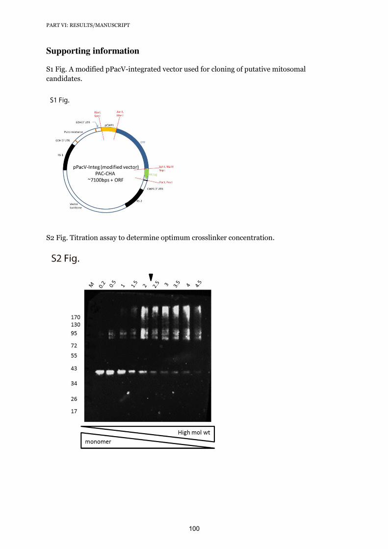
PART VI: RESULTS/MANUSCRIPT
Supporting information
S1 Fig. A modified pPacV-integrated vector used for cloning of putative mitosomal
candidates.
S2 Fig. Titration assay to determine optimum crosslinker concentration.
100
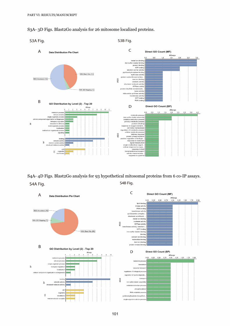
PART VI: RESULTS/MANUSCRIPT
S3A- 3D Figs. Blast2Go analysis for 26 mitosome localized proteins.
S4A- 4D Figs. Blast2Go analysis for 93 hypothetical mitosomal proteins from 6 co-IP assays.
101
PART VI: RESULTS/MANUSCRIPT
S1 Table. Oligonucleotides used in the study.
S2 Table. 95_2_95 analysis for GlTom40 co-IP dataset.
S3 Table. Parsing of 52 GlTom40 co-IP proteins.
S4 Table. 95_1_95 analysis for GlTom40 co-IP dataset.
S5 Table. Selected candidates for subcellular localization.
S6 Table. 95_2_95 analysis for GlTom40R co-IP dataset.
S7 Table. Parsing of 241 GlTom40R co-IP proteins.
S8 Table. List of mitosome localized proteins.
S9 Table. Overlap between GlTom40 and GlTom40R co-IP datasets.
S10 Table. 95_2_95 analysis for Gl14939 co-IP dataset.
S11 Table. 95_1_95 analysis for Gl5785 co-IP dataset.
S12 Table. 95_2_95 analysis for GlIscS co-IP dataset.
S13 Table. MITOPROT analysis for 14 hypothetical proteins in the GlIscS co-IP dataset.
S14 Table. 90_1_90 analysis for Gl9296 co-IP dataset.
102
PART VII: DISCUSSION AND FUTURE DIRECTIONS
PART VII DISCUSSION AND FUTURE DIRECTIONS
1. Discussion
1.1 General
Unicellular protozoan parasites are responsible for worldwide health problems. Giardia
lamblia is one of the leading causative agents for water borne non-bacterial diarrhea. G.
lamblia has a broad host range and not only affects humans but also causes severe morbidity
and economic loss in the livestock industry. Along with other parasitic protists such as
Trichomonas vaginalis and Entamoeba histolytica, Giardia has undergone reductive
evolution, frequently a hallmark of transition from free-living to parasitic lifestyle. These
adaptation and diverse biological processes are the results of the outcome due to long term
co-evolution with the host and are flaunted in the dramatic simplification of almost all
cellular systems and machineries. For e.g. the absence of a steady state Golgi apparatus and
the presence of mitochondrion-related organelles (mitosomes) (see below). Nevertheless, the
simplified cellular compartmentalization of Giardia makes for a useful platform to
investigate basic cellular functions and pathways which are challenging to dissect in complex
eukaryotes. Furthermore, it is also a practical model system to investigate principles of
reductive evolution, i.e. why and how adoption of a parasitic life-style leads to the loss of even
archetypical organelles, and which minimal machinery is maintained for fundamental
cellular functions.
During my doctoral thesis, I worked on two organelles in Giardia; the stage specifically
induced ESVs and the mitochondrion-related organelles mitosomes. I started with a project
to analyze the function of different homologues of small GTPase on ESV genesis and cyst
formation during the complex differentiation process. In my second project, I wanted to
characterize program cell death in Giardia. The main aim of this study was to assign giardial
mitosomes a novel function in program cell death by inducing apoptosis upon altering
conditions encountered by the parasite in physiological conditions such as nutrient starvation
and heat. The last part of my doctoral thesis was dedicated to optimizing a co-
immunoprecipitation assay (co-IP) enabling efficient pull down of organelle specific sub-
proteomes. The main aim of this part of my thesis was to characterize the mitosomal protein
import machinery and to identify other non-conserved proteins or factors responsible for
maintaining inter-organellar communication.
103
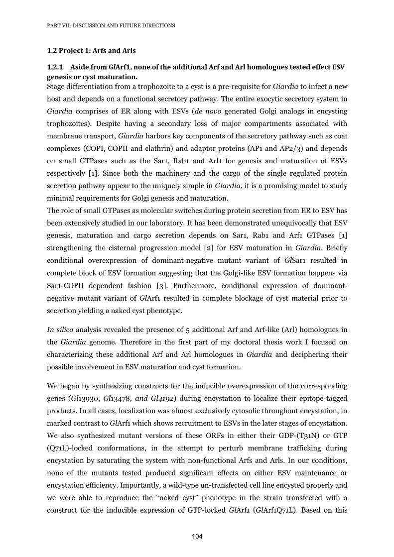
PART VII: DISCUSSION AND FUTURE DIRECTIONS
1.2 Project 1: Arfs and Arls
1.2.1 Aside from GlArf1, none of the additional Arf and Arl homologues tested effect ESV
genesis or cyst maturation.
Stage differentiation from a trophozoite to a cyst is a pre-requisite for Giardia to infect a new
host and depends on a functional secretory pathway. The entire exocytic secretory system in
Giardia comprises of ER along with ESVs (de novo generated Golgi analogs in encysting
trophozoites). Despite having a secondary loss of major compartments associated with
membrane transport, Giardia harbors key components of the secretory pathway such as coat
complexes (COPI, COPII and clathrin) and adaptor proteins (AP1 and AP2/3) and depends
on small GTPases such as the Sar1, Rab1 and Arf1 for genesis and maturation of ESVs
respectively [1]. Since both the machinery and the cargo of the single regulated protein
secretion pathway appear to the uniquely simple in Giardia, it is a promising model to study
minimal requirements for Golgi genesis and maturation.
The role of small GTPases as molecular switches during protein secretion from ER to ESV has
been extensively studied in our laboratory. It has been demonstrated unequivocally that ESV
genesis, maturation and cargo secretion depends on Sar1, Rab1 and Arf1 GTPases [1]
strengthening the cisternal progression model [2] for ESV maturation in Giardia. Briefly
conditional overexpression of dominant‐negative mutant variant of GlSar1 resulted in
complete block of ESV formation suggesting that the Golgi-like ESV formation happens via
Sar1-COPII dependent fashion [3]. Furthermore, conditional expression of dominant‐negative mutant variant of GlArf1 resulted in complete blockage of cyst material prior to
secretion yielding a naked cyst phenotype.
In silico analysis revealed the presence of 5 additional Arf and Arf-like (Arl) homologues in
the Giardia genome. Therefore in the first part of my doctoral thesis work I focused on
characterizing these additional Arf and Arl homologues in Giardia and deciphering their
possible involvement in ESV maturation and cyst formation.
We began by synthesizing constructs for the inducible overexpression of the corresponding
genes (Gl13930, Gl13478, and Gl4192) during encystation to localize their epitope-tagged
products. In all cases, localization was almost exclusively cytosolic throughout encystation, in
marked contrast to GlArf1 which shows recruitment to ESVs in the later stages of encystation.
We also synthesized mutant versions of these ORFs in either their GDP-(T31N) or GTP
(Q71L)-locked conformations, in the attempt to perturb membrane trafficking during
encystation by saturating the system with non-functional Arfs and Arls. In our conditions,
none of the mutants tested produced significant effects on either ESV maintenance or
encystation efficiency. Importantly, a wild-type un-transfected cell line encysted properly and
we were able to reproduce the “naked cyst” phenotype in the strain transfected with a
construct for the inducible expression of GTP-locked GlArf1 (GlArf1Q71L). Based on this
104
PART VII: DISCUSSION AND FUTURE DIRECTIONS
data, we conclude that Arf1 is probably the only Arf family member playing a key role in ESV
maintenance and trafficking. The other homologues may either be redundant/
complementary in relation to Arf1 or be involved in other as-yet novel unidentified cellular
processes.
A recent report highlighted the role of Arl2 during the cell cycle in T. brucei [4]. We tested
whether GlArl2 may play a similar role in G. lamblia cytokinesis. We did not record any
significant differences in cytokinesis in parasites expressing GTP-locked Gl4192 as compared
to untransfected parasites. We could not assign to giardial Arl2 a function in cytokinesis, as
seen in other protozoan species. Therefore, the only characterized Arf protein to date remains
GlArf1.
1.2.2 A case of redundancy or a scope for novelty for small GTPases in Giardia?
G. lamblia possesses 8 Rab proteins, 4 of which are well-conserved, a single Sar1 and 6 Arf
and Arl homologs [5, 6] (Rout, Faso and Hehl, unpublished data). Since the cellular
localization and molecular function of these GTPases are well conserved, they serve as tools
for characterizing compartment organization in the secretory pathway. Although distinct
phenotypes ranging from failure to undergo stage conversion to inability to secrete cyst wall
have been observed in Giardia upon over-expression of dominant negative Rab1, Sar1 and
Arf1, additional information regarding the localization and function of other homologs is still
missing. Since major molecular machineries and pathways have been streamlined in Giardia,
the scope of redundancy in such a parsimonious system is quite low.
Small GTPases are divided into 5 classes based on primary sequences, Ras, Rho/Rac, Rab,
Sar/ Arf and Ran [7]. The Rab GTPases constitute the largest group amongst all and are
implicated in vesicular traffic [8]. Multicellular organisms harboring a steady state Golgi
apparatus possess a larger repertoire of Rab GTPases compared to unicellular eukaryotes. A
case in point is the presence of 60 Rab genes in H. sapiens and 29 in both A. thaliana and D.
melanogaster, compared to 11 Rab genes identified in yeast harboring either stacked Golgi
(P. pastoris and S. pombe) or dispersed cisternae (S. cerevisiae) [9, 10]. Surprisingly, basal
eukaryotes such as E. histolytica and T. vaginalis that do not harbor a canonical Golgi
possess a large repertoire of Rab GTPases [11, 12]. Although a specific set of these GTPases
(Rab1, Rab5, Rab6, Rab7, Rab11) are highly conserved [13], not every GTPase has been
assigned a unique function in these species. However, the presence of unusual C-terminal
domain structure in E. histolytica Rab proteins points towards novel modification and
function [12].
Therefore, whether the presence of additional Arf and Rab homologs in Giardia is a case of
redundancy or if there are novel functions associated to these GTPases is yet to be
determined.
105
PART VII: DISCUSSION AND FUTURE DIRECTIONS
1.2.3 Are small GTPases in Giardia involved in additional functions beyond their
involvement in the secretory system?
Beyond their documented role in vesicular trafficking, small GTPases including Arfs and
Rabs have been implicated in at least 3 functions in higher eukaryotes; establishment of
ER/mitochondria contact sites, mitophagy and maintenance of mitochondrial homeostasis
and dynamics [14-16].
Arf1 GTPase in yeast and metazoans associates with mitochondria and ER-mitochondria
contact sites respectively [17] (novel localization for a Golgi GTPase) and interacts genetically
with Gem 1 (Rho-like GTPases negatively regulating ER-Mitochondria Encounter Structures
(ERMES in yeast)) [18]. The mitochondrial phenotype (hyper-connected mitochondria)
obtained in the Arf1/2 mutants are similar to those observed in Gem1 mutants in yeast [17].
Furthermore, the mitochondrial morphology in double mutants (Arf1/2 and Gem1) is
severely compromised than in individual mutants suggesting the presence of a novel Arf
dependent ER-mitochondria contact site besides the canonical ERMES in yeast. In addition,
Arf1 GTPase also recruits AAA-ATPase Cdc48 to yeast mitochondria for efficient removal of
Fzo1 (fusion GTPases) and other mitochondrial proteins. Cdc48 was shown to be involved in
quality control of mitochondrial outer membrane proteins. Likewise over-expression of
Arf1/2 mutants leads to impaired recruitment of Cdc48, leading to aberrant mitochondrial
phenotype [17]. On the other hand, Rab32 is shown to associate with mitochondria and over-
expression of GDP-locked variant resulted in collapse of mitochondria suggesting its role in
mitochondrial fission machinery [19].
The growing body of evidence points towards the involvement of the endomembrane system
(small GTPases) in mitochondrial dynamics [20]. Due to the above mentioned novel function
of the small GTPases (Arf1 and Rab32), the presence of five additional Arf and Arl homologs
and 8 Rab proteins in Giardia merits further investigation. Therefore, we hypothesize that
the additional small GTPases present in Giardia might be performing a novel function at
mitosomes or possess a Giardia specific function. This is supported by co-IP data showing
mitosomal bait specific enrichment for GlArf1, GlRab32 (Gl50803_16979) and an AAA-
ATPases (Gl50803_16867) homologs at high stringency parameters (95_2_95). Although
GlArf1 and AAA-ATPases do not localize to Giardia mitosomes in IFA studies (data not
shown), identification of these proteins in curated MS dataset suggests that a small fraction
of these proteins are in close association with mitosome membranes and/or hints towards a
putative novel role in establishing ER-mitosome contact sites or maintaining mitosomal
dynamics. Furthermore, although the role of GlArf1 has been established in cyst maturation,
little is known regarding its involvement in mitosome morphology and maintenance.
In order to address this question we propose to,
106
PART VII: DISCUSSION AND FUTURE DIRECTIONS
(1) Perform a thorough IFA analysis to determine the shape and morphology of mitosomes
using endogenous antibody (against GlIscU) in cell lines expressing the GTP- and GDP-
locked Arf and Arl homologues. In depth analysis will provide novel insights (if/any) into the
function of these homologues in Giardia. Availability of all the transgenic cell lines makes
this approach less time consuming.
(2) Previous experiments to determine the role of additional Arf and Arl homologs in cyst
maturation were performed using cell line expressing the dominant-negative mutants under
an inducible cyst wall protein (CWP) promoter. The cytoplasmic localization observed might
be a result of mistargeting of excess protein due to over-expression. Therefore, generation of
HA epitope-tagged variants under the control of their respective endogenous promoters is
necessary to access their correct localization within the cell.
(3) Apart from the function of GlRab1 in ESV development and cyst formation, little is known
regarding the role of GlRab32 in Giardia. Localization studies using epitope tagged GlRab32
and IFA analysis of transgenic parasites expressing GDP and GTP locked variants is essential
to determine if GlRab32 has any effect on mitosome morphology and/or distribution.
(4) Volpicelli-Daley et. al 2005, demonstrated that single knockdown of Arf1, 3, 4 and 5 were
not required at any step of membrane traffic in HeLa cells, however, every combination of
double knockdown elicited distinct defects along the secretory pathway demonstrating
specificity of Arf-couples at multiple steps [21]. Furthermore, Arf1/2 double mutants’ also
elicited a mitochondrial phenotype in yeast. Since Giardia harbors an additional Arf homolog
(Gl7562) with a predicted N-myristoylation motif, it is important to investigate the mitosome
phenotype upon over-expression of both Arf mutants in Giardia. Availability of vectors
allowing dual expression of both constructs makes this approach highly feasible.
(5) Lastly, involvement of these additional GTPase homologs in completely new Giardia
specific pathways cannot be excluded. Therefore, established assays to determine ER
morphology, PV morphology, clathrin recruitment and fluid phase uptake must be performed
in cell lines expressing the GTP- and GDP- locked mutants.
The result of these proposed experiments would answer the question if the additional
homologues for small GTPases present in Giardia are redundant or have a Giardia specific
novel function.
1.3 Project 2: Apoptosis in Giardia lamblia
1.3.1 Apoptotic like cell death can be induced in Giardia by altering its physiological
conditions.
PCD (apoptosis) has been best characterized in multicellular life forms based on the obvious
benefits it renders such as maintaining cell population, elimination of damaged cells or giving
107
PART VII: DISCUSSION AND FUTURE DIRECTIONS
shape to an organism [22, 23]. However, mounting evidences suggests that unicellular
organisms have developed an unusual cell death pathway and undergo PCD under certain
condition which is similar to their multicellular counterparts [24-26]. Based on the “original
sin hypothesis” for the evolution of PCD, key molecules and factors involved in the pathway
are located within the mitochondrion: the cornerstone organelle for PCD by apoptosis [27].
Therefore studies on unicellular organisms that do not harbor canonical mitochondria but
demonstrate apoptosis related phenomenon would help unravel conserved factors and
proteins involved in the process and will shed light on the evolution of PCD.
G. lamblia has undergone reductive evolution after changing from a free-living to a parasitic
life style and harbors mitosomes making it a useful model to study apoptosis related
processes. Despite present at high concentration during a chronic infection Giardia
trophozoites do not elicit any host inflammatory response [28, 29]. Therefore, we
hypothesized that Giardia trophozoites undergo an apoptosis-like cell death, which limits the
stimulation of the immune system by deteriorating the parasites. By altering physiological
conditions such as nutrient starvation and heat shock to trigger apoptosis, we demonstrate
unambiguously that Giardia exhibit many, if not all morphological and biochemical
characteristics of apoptosis-like cell death. Significant increase in phosphatidylserine (PS)
positive cells was observed in nutrient starved and heat shocked parasites as compared to
control parasites. PS exposure on the outer leaflet of the plasma membrane is a hallmark of
apoptotic cells [30]. PS exposure as an excellent strategy for immune silencing has been
demonstrated in protozoan parasites L. major, T. gondii and T. brucei [31] where exposure of
PS on apoptotic cells leads to recruitment of phagocytes which recognize the signals leading
to engulfment of apoptotic cells. However since apoptotic cells do not pose any danger, their
engulfment does not provoke any anti-microbial effector function of phagocytes. Rather this
step is accompanied by down regulation of pro-inflammatory and release of anti-
inflammatory cytokines leading to silent clearing [32-34].
In addition to PS exposure, our data clearly demonstrate ER disintegration and nuclear
material condensation in parasites induced to undergo apoptosis. However, analysis of the
electrophoretic profile of DNA from nutrient starved parasites did not show the typical
recursive DNA laddering pattern (caspase-dependent apoptosis) or high-molecular weight
banding (caspase-independent apoptosis). Rather we obtained sheared DNA in the low
molecular weight region. Atypical DNA fragmentation is seen in yeast cells undergoing
apoptosis and many other lower eukaryotes including dinoflagellate P. gatunense and is
linked to difference in chromatin arrangement [35, 36]. However, our data is in line with
hydrogenosome-bearing T. vaginalis where a low molecular weight DNA smear was observed
upon induction of apoptosis pointing towards the existence of an alternative DNA
fragmentation mechanism in these protozoan parasites [37].
108
PART VII: DISCUSSION AND FUTURE DIRECTIONS
The cell death pathway involved in MRO harboring organisms is still unclear; however the
end results are very much similar to studied pathways found in many unicellular organisms
and metazoans. Since mitosomes are mitochondrial relic organelles and derived from
endosymbiotic α-proteobacteria, we hypothesize that they might have some implications in
cell death by releasing harmful proteins in the cytosol upon exposure to damaging stimuli
like their eukaryotic counterpart. Although no bona fide effector homologs for any branch of
PCD have been detected in the Giardia genome due of strong sequence divergence, it does
not imply that such proteins do not exist since such analysis might be biased towards those
proteins which have significant homology. Therefore we propose to:
(1) Perform a whole proteome analysis of apoptosis induced and non-induced parasites.
Subtractive analysis of the proteome dataset would help discover Giardia specific abundant
proteins or factors which might be involved in apoptosis. In a more targeted approach we
would isolate nuclei from induced and non-induced trophozoites either by exploiting the
organelle purification system optimized in our laboratory [38] or by density gradient
centrifugation followed by differential proteomics analysis to identify novel Giardia specific
regulators or transcription factor that might be upregulated upon induction of apoptosis in
Giardia.
(2) We have shown that Giardia trophozoites undergo a form of apoptosis-related cell death
upon nutrient starvation and heat shock. Furthermore, a dramatic and rapid form of cell
deterioration was observed as a result of interference with mitosome protein import function
upon treatment of trophozoites with Mitoblock (an experimental compound targeting the
TIM import complex in mitochondria) (Hehl, unpublished data). Although there is indirect
evidence for the role of mitosomes in apoptosis in Giardia, direct evidence for its
involvement in the pathway is still missing. Therefore, we would exploit the DHFR-
methotrexate import block assay recently implemented in our laboratory (Rout, et.al 2015,
under review) to investigate whether a direct link is present between mitosomal protein
import and apoptosis in Giardia. Preliminary experiments show a marginal increase of PS-
positive cells in MTX-treated transgenic parasites expressing a mitosomal targeted DHFR
fusion construct compared to equivalent untreated parasites (data not shown). However,
large-scale quantification experiments using fluorescence activated cell sorting (FACS) will
determine to what extent this phenomenon is consistent and reproducible.
3) In addition, we would develop another strategy independent of the ligand-induced block
(DHFR- methotrexate) since this block is easily released upon dilution [39] and excess ligand
in the solution might account for off-target effect. Therefore we would generate a chimeric
construct harboring 1) a MTS at the N-terminus, 2) GST tag in the middle 3) a portion of
staphylococcal protein A (as a C-terminal block) [40, 41]. The ability of the C-terminal
109
PART VII: DISCUSSION AND FUTURE DIRECTIONS
staphylococcal protein A as an import blocking agent has been demonstrated [42].
Characterization of apoptosis related phenotype in transgenic parasites over-expressing such
a chimeric construct might also provide evidence to link mitosomes function and apoptosis in
Giardia.
Furthermore, because this chimeric construct blocks the completion of import due to a
translocation block, it spans the 4 different sub-cellular compartments in mitochondria (OM,
IMS, IM, and matrix). Such a scenario would provide identification of components of the
translocation machinery by affinity chromatography on IgG-Sepharose beads as an
alternative for the iterative co-IP assay (Rout, et. al 2015, under review).
Such endeavors will not only help us to decipher PCD in amitochondriate organisms but also
lead to an appreciation of the diversity of PCD across the eukaryotic spectrum.
1.4 Project 3: Optimization of a co-immunoprecipitation assay to identify organelle
specific sub- proteome.
1.4.1 A minimized mitosomal import machinery in Giardia: reductionism at its best?
Mitochondrion is one of the defining organelle of an eukaryotic cell. It is not only involved in
energy production by aerobic respiration, lipid synthesis but is also involved in several
biosynthetic pathways. However, a number of organisms (mostly anaerobic eukaryotes)
scattered throughout the 5 super-groups do not possess a canonical mitochondria but instead
harbor mitochondrial related organelles (MROs) such as hydrogenosomes and mitosomes
[43]. Although MROs are evolutionarily derived from mitochondria, there is extensive
structural and functional divergence among organelles in different species. Bioinformatics
analyses clearly demonstrate that the constraints for sequence divergence vary greatly
amongst the super-groups, for e.g. enzymes of the universal Fe-S protein maturation
machinery in MROs can be detected in the nuclear genome using straightforward in silico
homology searches, whereas most MRO membrane proteins have diverged beyond
recognition if they were not lost altogether. Additionally, despite unambiguous evidence for
conserved mitosomal protein import pathways in Giardia, the composition of the import
machinery remains unknowns. A case in point is GlTom40 (the only identified component of
the mitosome import machinery in Giardia) whose sequence degeneration is so extensive
that the identification of orthologues in Giardia, Entamoeba or Spironucleus still remains
tentative. This high divergence between orthologues of MRO proteins even in closely related
species makes systematic identification of protein components using proteome analysis very
challenging.
We hypothesized that the reduced Giardia mitosomes harbored no more than 100-150
different proteins, either involved in protein import or associated with a clearly delineated
cellular compartment facilitating inter-organellar communication. We accounted for the
110
PART VII: DISCUSSION AND FUTURE DIRECTIONS
paucity of mitosomal proteins by optimizing and implementing an approach based on
iterative co-IP experiments using GlTom40 as a starting point followed by mass spectrometry
(MS) analysis of chemically stabilized protein-protein interactions. This robust iterative
approach lead to identification of 22 mitosomal proteins (annotated and hypothetical) out of
which 16 proteins were localized to mitosomes; 5 proteins displayed dual localization to
mitosomes and ER and one protein showed a ER and PV pattern even though it was pulled
down exclusively with mitosome-localized bait proteins harboring TMDs (discussed below).
The two proteins of the proposed minimized mitosomal import machinery are discussed
below;
Gl29147, a single pass membrane protein was identified as a prominent GlTom40 interacting
protein. The protein has a predicted N-terminal TMD similar to mitochondrial Tom40
receptors (Tom 20 and Tom 70) in S. cerevisiae. Although several in silico analysis tools have
failed to identify a MTS at the protein’s N-terminus, ectopic expression of the C-terminal
portion of Gl21947 alone showed a distinct cytosolic localization by IFA (data not shown)
without a detectable phenotype suggesting that targeting signals are present in the N-
terminus of the protein. Additionally, C-terminally GFP-tagged full-length variant Gl29147-
GFP enabled us to perform time lapse microscopy of mitosomes in trophozoites for the first
time suggesting that Gl29147 is inserted in the mitosomal outer membrane in the type I
orientation, extruding the C-terminal domain to the cytosol similar to Tom 20 and Tom 70
[44]. Interestingly, reverse co-IP using Gl29147 as bait protein pulled down GlTom40 as the
most abundant membrane associated protein along with 220 additional specific proteins
(annotated and hypothetical) out of which 20 proteins have been validated by localization to
mitosomes, allowing for a significant expansion of the GlTom40-centered interactome.
Therefore, topology of Gl21947, its exclusive mitosomal localization, and the repertoire of
pulled down proteins indicate that Gl21947 is a GlTom40 accessory protein with a potential
receptor function for protein import (GlTom40R). However, the cytoplasmic GlTom40R
fragment alone does not recapitulate the interaction properties of the full-length membrane
anchored variant suggesting that the capture of imported matrix proteins requires
incorporation of the receptor domain into a TOM complex and requires additional factors or
domains on either side of the membrane anchor for proper functionality. However, the
hypothesis of a receptor function for GlTom40R remains to be tested directly to evaluate its
exact role in mitosomal protein import in Giardia.
Interestingly, although BLASTp yielded no strong homologues for GlTom40R, profile
sequence comparisons with HHpred showed homology to a “high potential iron sulfur
protein” (p-value 0.007). “High potential iron sulfur protein” in higher eukaryotes, also
known as mitoNEET, is an integral membrane protein localized at the outer membrane of
mitochondria and is responsible for transport of iron into mitochondria [45, 46]. If we
111
PART VII: DISCUSSION AND FUTURE DIRECTIONS
consider that Fe-S protein maturation is the only metabolic pathway currently associated to
G. lamblia mitosomes, GlTom40R might also function as a mitoNEET homolog in G.
lamblia. Alternatively, it wouldn’t be surprising if GlTom40R could be performing both
functions of a Tom receptor and mitoNEET simultaneously in this parasite. However, further
experiments needs to be performed in order to accurately assign a function to GlTom40R in
the mitosomal import machinery of G. lamblia.
Gl14939, a two TMD harboring protein was identified using GlTom40 and GlTom40R as
baits in co-IP and reverse co-IP experiments respectively. Exclusive detection of the protein
in both the datasets suggests that the three proteins (GlTom40, GlTom40R and Gl14939)
might function as part of a complex. Furthermore, HMMER-based searches across several
eukaryotic lineages fail to identify any orthologues for this protein. Consistent with this, there
is no functional information available on this protein until now. A recent study however
showed that Gl14939 (dubbed GiMOMP35) localizes at the outer mitosome membrane with
its C-terminus in the cytosol [47]. Nonetheless, given that GlTom40 (the translocon),
Gl29147 (GlTom40R), and Gl14939 (putative accessory protein) are outer membrane-
associated proteins and part of the same interactome, we conclude that Giardia possesses a
reduced mitosomal import machinery composed of the above three proteins.
Our conclusion is in line with the protein import machinery identified in other mitosome
bearing organisms such as E. histolytica and microsporidian E. cuniculi [48, 49]. The
presence of a single putative Tom receptor in Giardia is not an uncommon scenario in
mitosome bearing organisms. A case in point is the presence of a sole Tom 70 receptor in the
microsporidian E. cuniculi efficient enough for recognition and translocation of mitosomal
proteins through the E. cuniculi Tom 40 channel [50]. Another case of massive reduction is
seen in Trypanosoma mitochondria where only one translocon in the inner membrane
performs a “jack of all trades” function substituting Tim 17/22/ and 23 [50, 51]. In these
organisms however, extensive stripping of the protein import machinery to the essential is
explained by the apparent paucity of proteins imported into mitosomes. Nevertheless, the
most extreme case of reduction however is seen in anaerobic amoeba Mastigamoeba
balamuthi, where despite the identification of 21 mitochondrial proteins, no components of
the TOM, TIM or SAM complexes are found in the genome [52]. Based on the above
information, the absence of other conserved components of the mitosome import machinery
in Giardia can be explained in two possible ways: 1) extensive sequence divergence prevents
identification of organelle proteins via homology-based searches; 2) G. lamblia lacks
additional proteins of the import machinery simply due to overall mitochondrial reduction.
Nevertheless, our robust iterative co-IP approach for mitosomal protein identification also
yielded five novel hypothetical proteins without a predicted TMD (Gl10971, Gl9296, Gl8148,
112
PART VII: DISCUSSION AND FUTURE DIRECTIONS
Gl17276, and Gl16424). However their function and possible involvement in the import
machinery remains to be investigated. Gl9296 is of particular interest since it harbors an
identifiable MTS. Co-IP using Gl9296 as bait protein identified GlIscS, GlIscA, GlHsp70,
GlTom40 and GlTom40R suggesting close association of Gl9296 with few imported matrix
proteins and most importantly with the import machinery. Furthermore, Gl9296 appears to
be Giardia lineage specific as no other orthologues could be identified. Given its interaction
profile, predicted import signal, and lack of homology to metabolic enzymes, we hypothesize
that Gl9296 is a matrix protein and might be interacting at the trans-side for stabilization of
the import pore in Giardia mitosomes. However, this remains to be investigated.
In addition our serial co-IP strategy identified another 93 hypothetical proteins that remain
to be investigated for their localization and putative function. Interestingly, the major
function associated with E. histolytica mitosomes was shown to be sulfate activation rather
than Fe-S protein maturation [53]. Furthermore, Entamoeba mitosomes have been also
implicated in the encystation process [54]. Although genes involved in sulfate activation
pathway are absent in other MRO-containing organisms such as Giardia, Trichomonas and
Cryptosporidium, the Entamoeba example points to a wider range of functions ascribable to
mitosomes.
Based on the newly identified and novel validated mitosomal proteins we acquired so far
from our serial co-IP approach we hypothesize that giardial mitosome might be implicated in
other functions beyond Fe-S protein maturation. To test this hypothesis we propose to;
1) Perform expression and localization studies of epitope-tagged novel mitosomal candidates
after careful in silico analysis. Proteins with an identifiable MTS would be priority candidates
for this approach. We can make use of our proprietary expression system to integrate an
epitope-tagged variant of each candidate into the Giardia genome. Open reading frames and
~200 base pairs of upstream sequence including the endogenous promoter would be cloned
in frame in front of the HA-epitope tag sequence and ligated into the expression vector.
Usage of the endogenous promoter avoids problems of over expression since only a single
extra copy is introduced into the cells. However, if epitope tagging or ectopic expression
should lead to a lethal effect, we will use conditional expression under the control of a stage-
specific promoter which can be induced by changing culture medium conditions as a backup
strategy. Localization studies would be done by IFA analysis on chemically fixed cells using
monoclonal antibody which is specific for the HA epitope on the recombinant protein.
Furthermore, co-localization studies to confirm correct mitosomal localization for candidate
proteins can be done using a confirmed mitosome protein, i.e. GlIscU or GlIscS, to which a
polyclonal antibody is available (Rout, Hehl 2015, unpublished data).
113
PART VII: DISCUSSION AND FUTURE DIRECTIONS
2) Perform carbonate extraction assay to determine if the novel mitosome localized TMD
harboring proteins are indeed integral membrane proteins or peripheral membrane proteins.
Additionally, we would perform protease protection assay in order to determine the topology
of novel mitosome localized TMD harboring hypothetical protein candidates obtained from
the serial co-IP assays.
3) Analyze the function of selected candidates by exploiting the recent developments in gene
knockout strategy in Giardia based on the Cre/LoxP system [55]. Alternative strategies such
as CRISPR/CAS9 or knockdown by morpholinos can be used to analyze the effect of loss of
function of these proteins on mitosome (structural or functional). At present the CRISPR/
CAS9 method for genome editing in Giardia is being established in our laboratory. However,
knockdown by morpholino oligonucleotides is already established in Giardia [56].
Morpholinos role as translation-blocking nucleotides has been unambiguously proven in
Trypanosoma brucei [57]. A major advantage of this method is that morpholinos can be
electroporated into cells using a standard protocol where they remain stable and highly active
for days, although their concentration diminishes with each successive cell division.
Furthermore, the specificity of the suppression by the experimental morpholino can be
controlled in a concentration-dependent manner with a control morpholino which contains
five mispaired bases.
Additionally,
3) We would perform sufficient number of reverse co-IP assays with validated mitosomal
proteins as bait which would enable us to build a comprehensive Giardia mitosome organelle
proteome.
4) We have successfully GFP-tagged mitosomes using either GlTom40 or GlTom40R. The
type I topology of GlTom40R enables us to generate an organelle specific glutathione S-
transferase (GST) marker for organelle purification by affinity chromatography on
glutathione–Sepharose 4B beads as an alternative for our serial co-IP assay.
5) Establish a direct link between loss of mitosome function (import) and apoptosis in
Giardia. (Discussion, Section 1.3). Uncovering a possible link between PCD and the highly
degenerate mitochondrial remnants in this basal protozoan will reveal key information on the
evolution of mitosomes and PCD, and would provide a unique opportunity to explore the
breadth of eukaryotic diversity.
114
PART VII: DISCUSSION AND FUTURE DIRECTIONS
1.4.2 Diverged mitosome-ER contacts sites in Giardia?
Co-IP data identified five proteins (Gl5785, Gl9503, Gl21943, Gl22587, and Gl15154) with
dual localization suggesting that these proteins might be present at the junction between ER
and mitosomes and could be playing a role in establishing inter-organellar communication
between the two organelles. In yeast, the ER-mitochondria encounter structure (ERMES) is
responsible for bringing the two organelles in close proximity thereby facilitating lipid
transfer. Phosphatidylserine (PS) synthesized in the ER is shuttled to mitochondria through
the ERMES complex where it is converted to phosphotidylethanolamine (PE) by PS
decarboxylase (Psd1) located in the inner membrane. PE is then shuttled back to the ER
where is converted to phosphatidylcholine (PC) by different enzymes.
Because ERMES homologs are absent in metazoans [58], we hypothesized that there should
be a tethering complex establishing organelle contact site for lipid transfer since division of
mitosomes and maintenance of organelle membrane architecture require membrane lipids
from ER [59, 60]. SNAREs proteins are generally involved in mediating membrane fusion in
vesicular transport [61]. However the Giardia SNARE protein (Gl5785) particularly due to its
dual localization might be a part of such a tethering complex facilitating phospholipid
exchange between the two organelles by establishing membrane contact sites. Co-IP data
generated using Gl5785 as bait protein partially supported our hypothesis by identifying 3
novel hypothetical proteins Gl9503 (3 TMDs), Gl21943 and Gl10971. Gl9503 and Gl21943
had dual localization (ER and mitosome) and data mining for both proteins identified
domains with similarity to the yeast protein Mmm 1 of the ERMES complex and a beta barrel
lipid binding protein MLN64 (e-value 0.0006) in H. sapiens involved in cholesterol transport
to mitochondria [62] respectively. Additionally, Gl9503 also harbors an abhydrolase domain
implicated in lipid synthesis. Furthermore, Gl5785 also pulled down the putative core import
machinery in Giardia mitosomes (GlTom40, GlTom40R and Gl14939) which is in line with
the association of ERMES with components of the protein import machinery in yeast
mitochondria [63, 64]. Given the sub-cellular localization of the bait protein Gl5785, it is not
surprising that we might have pulled down additional proteins working at the interface
between these organelles facilitating either direct connection or working as bridging factors
between the two organelles.
In addition, identification and dual localization of Gl22587 further supports our hypothesis
for existence of a Giardia specific molecular tether between mitosomes and ER. HHpred
identified an F-GTPase activating protein with an e-value of 1.8E-05. Based on this data, we
can carefully speculate that Gl22587 might be a regulator of the putative contact site in
Giardia like the yeast GTPase (Gem 1) which functions as the master regulator of the ERMES
complex. Over-expression of a dominant negative variant of the Gl22587 would shed more
light on its molecular function and likely association with mitosomes.
115
PART VII: DISCUSSION AND FUTURE DIRECTIONS
Interestingly, ERMES beyond their dedicated involvement in lipid trafficking are also
implicated in regulation of mitochondrial fission, calcium signaling, mitophagy and
inflammasome activation [65]. Nonetheless, to this date, components of the ERMES complex
are not identified in metazoans and mitosome harboring organisms, however, ERMES
homologues have been identified in hydrogenosome containing organism such as T.
vaginalis [66]. However, identification of the SNARE protein (Gl5785) and several other
proteins such as Gl9503, Gl21943, Gl22587, and Gl15154 with dual localization suggest that
these proteins might be components of a Giardia specific highly diverged molecular tether
analogous to the ERMES in yeast.
Novel findings in the field of inter-organellar communication have revealed alternative
pathways for lipid exchange between ER and mitochondria [67, 68]. Authors demonstrated
that in the absence of an active ERMES, lipids are delivered to mitochondria via contact sites
between mitochondria and vacuoles in yeast model. The identification of Gl7188 in our co-IP
dataset and its atypical localization gives us an indication of a highly probable/alternative
scenario in G. lamblia despite the absence of vacuoles. Gl7188 is a TMD harboring protein
pulled down by baits present in the outer membrane in mitosomes (GlTom40, GlTom40R,
Gl14939 and Gl5785) and IFA analysis revealed that this protein is localized to ER and
peripheral vesicles prompting that these three organelles are in close proximity. In absence of
a canonical ERMES machinery and the need for membrane lipids for mitosomal homeostasis
it is tempting to speculate that membrane lipids might be exchanged between ER and
mitosomes via peripheral vesicles at the basal body complex where these three organelles are
in close association with each other. Gl7188 might be a bridging protein in-between
mitosomes and ER/PVs. Close association of ER and PVs in G. lamblia has been previously
documented [69]. Co-IP experiment using Gl7188 as bait protein and lipid binding assays
needs to be performed to determine its interaction partners and lipid binding ability.
However, the atypical localization of Gl7188 might also be an artefact of the epitope tag at the
C-terminus of the protein. Therefore, N-terminally epitope tagged variant needs to be
generated to check for its sub-cellular localizaiton .
1.4.3 Mitosome dynamics and a novel role for Giardia dynamin related protein in
mitosome morphogenesis
We have previously demonstrated the existence of two morphologically distinct classes of
mitosomes: 1) ~30 peripheral mitosomes (PMs) which are distributed in the cytoplasm
without a particular pattern, and 2) a central mitosome complex which looks like an
elongated organelle in fluorescence microscopy, but in fact is a tight bundle of spherical
organelles [70]. Furthermore, the CMC has a fixed localization at the basal body complex
between the two nuclei of Giardia and divides and segregates with the replicated basal body
116
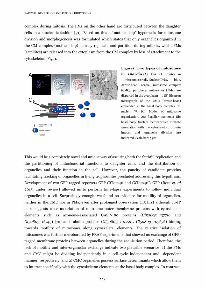
PART VII: DISCUSSION AND FUTURE DIRECTIONS
complex during mitosis. The PMs on the other hand are distributed between the daughter
cells in a stochastic fashion [71]. Based on this a “mother ship” hypothesis for mitosome
division and morphogenesis was formulated which states that only organelles organized in
the CM complex (mother ship) actively replicate and partition during mitosis, whilst PMs
(satellites) are released into the cytoplasm from the CM complex by loss of attachment to the
cytoskeleton, Fig. 1.
Figure1. Two types of mitosomes
in Giardia.(A) IFA of Cpn60 in
mitosomes (red). Nuclear DNA, blue.
Arrow-head: central mitosome complex
(CMC); peripheral mitosomes (PMs) are
dispersed in the cytoplasm [71]. (B) Electron
micrograph of the CMC (arrow-head)
embedded in the basal body complex. N:
nuclei [70]. (C) Model of mitosome
organization. Ax: flagellar axoneme; Bb:
basal body. Surface factors which mediate
association with the cytoskeleton, protein
import and organelle division are
indicated. Scale bar: 5 μm.
This would be a completely novel and unique way of assuring both the faithful replication and
the partitioning of mitochondrial functions to daughter cells, and the distribution of
organelles and their function in the cell. However, the paucity of candidate proteins
facilitating tracking of organelles in living trophozoites precluded addressing this hypothesis.
Development of two GFP-tagged reporters GFP-GlTom40 and GlTom40R-GFP (Rout et. al
2015, under review) allowed us to perform time-lapse experiments to follow individual
organelles in a cell. Surprisingly enough, we found no evidence for motility of organelles,
neither in the CMC nor in PMs, even after prolonged observation (1.5 hrs) although co-IP
data suggests close association of mitosome outer membrane proteins with cytoskeletal
elements such as axoneme-associated GASP-180 proteins (Gl50803_137716 and
Gl50803_16745) [72] and tubulin proteins (Gl50803_101291 , Gl50803_103676) hinting
towards motility of mitosomes along cytoskeletal elements. The relative isolation of
mitosomes was further corroborated by FRAP experiments that showed no exchange of GFP-
tagged membrane proteins between organelles during the acquisition period. Therefore, the
lack of motility and inter-organellar exchange indicate two plausible scenarios: 1) the PMs
and CMC might be dividing independently in a cell-cycle independent and -dependent
manner, respectively, and 2) CMC organelles possess surface determinants which allow them
to interact specifically with the cytoskeleton elements at the basal body complex. In contrast,
N
N
A B
C Bb
PMsCMBasal body complex
Ax
Ax
Cytoskeleton-binding
factor
Dividing mitosome
TIM/TOM
117
PART VII: DISCUSSION AND FUTURE DIRECTIONS
the surface of PMs does not appear to associate with the cytoskeleton, and they are randomly
distributed within the cytoplasm. Time lapse microscopy of synchronized transgenic Giardia
cultures expressing either GFP-GlTom40 or GlTom40R-GFP would address the first
scenario. Whereas development of an organelle purification strategy to selectively enrich
CMCs and PMs and analysis of whole organelle proteome would help identify novel surface
determinants for CMC that enables them to be closely associated to the basal body complex.
Interestingly, over-expression of a dominant-negative giardial dynamin mutant elicited an
organellar morphogenesis phenotype indicative of organelle replication defect (discussed
below). In addition, the occurrence of the “string” mitosome phenotype upon over-
expression of GlTom40R-GFP (Rout et. al 2015, under review) clearly suggested that
mitosomes assume an elongated, tubular morphology, which is a prerequisite for organelle
division and replication and that the “string” phenotype reflects a lack of fission.
Tight regulation of mitochondrial division is essential as survival of the cell depends on the
preservation of an adequate number of mitochondria after cytokinesis [73]. It has been
extensively documented that dynamin related protein (DRP) is a crucial mechanoenzyme
which is conserved from yeast to vertebrates and carries out mitochondrial fission [74-77]. G.
lamblia harbors a single dynamin homologue (GlDrp) encoded by ORF (Gl50803_14373)
and has been shown to play a major role in the endocytic pathway and stage conversion [78-
80]. However, little was known regarding the role of the single GlDRP in mitosomal division
and morphology. We have demonstrated for the first time the role of GlDRP in division and
maintenance of mitosome morphology in addition to its role in endocytosis and stage
conversion (Rout et. al 2015, under review). Transgenic parasites expressing GlDRP-K43E
(dominant-negative mutant) protein exhibited larger and fewer mitosomes as compared to
parasites expressing wild type GlDRP, supporting the role of single GlDRP in mitosomal
fission.
Our results are in line with the aberrant mitochondrial phenotype (long elongated
interconnected tubular networks) observed in higher eukaryotes upon over-expression of
dominant negative DRP [74, 81]. Interestingly, DRPs role in mitochondrial and
hydrogenosomal division has also been demonstrated in the protozoan parasites
Trypanosoma brucei [82, 83] and Trichomonas vaginalis [84], respectively. Therefore, our
data partially corroborates the mechanistic conservation of mitochondrial and MRO fission
[85-88].
Identification and mitosomal localization of a protein candidate (Gl50803_22587) further
substantiates the functional conservation of mitosomal fission machinery. HMMER-based
predictions relate this protein to a mitochondrial fission protein (Fis1, e value 6.3E-05) in H.
sapiens. Fis1, together with mitochondrial fission factor (Mff) and mitochondrial dynamic
118
PART VII: DISCUSSION AND FUTURE DIRECTIONS
proteins (MiD 49 and MiD 51) act as receptors that recruit Drp1 to the mitochondrial surface,
thus regulating fission mechanisms in eukaryotes [87, 89]. Therefore based on the
association of Gl22587 to mitosomal outer membrane proteins we hypothesize that it might
be involved in recruiting proteins together with GlDrp onto the mitosomal surface eliciting
the same function seen in higher eukaryotes. Co-IP assays using Gl22587 as bait protein are a
prerequisite for identification of regulating, recruiting and interacting partners for GlDrp.
Data obtained from these experiments would not only shed light on the composition of the
mitosome fission machinery and replication in Giardia but would also provide new targets
for development of therapeutic tools to prevent parasite transmission and hence giardiasis.
2. Bibliography
1. Stefanic, S., et al., Neogenesis and maturation of transient Golgi-like cisternae in a simple eukaryote. J Cell Sci, 2009. 122(Pt 16): p. 2846-56.
2. Losev, E., et al., Golgi maturation visualized in living yeast. Nature, 2006. 441(7096): p. 1002-6.
3. Faso, C., et al., Export of cyst wall material and Golgi organelle neogenesis in Giardia lamblia depend on endoplasmic reticulum exit sites. Cellular microbiology, 2013. 15(4): p. 537-53.
4. Price, H.P., et al., The small GTPase ARL2 is required for cytokinesis in Trypanosoma brucei. Molecular and biochemical parasitology, 2010. 173(2): p. 123-31.
5. Marti, M. and A.B. Hehl, Encystation-specific vesicles in Giardia: a primordial Golgi or just another secretory compartment? Trends in parasitology, 2003. 19(10): p. 440-6.
6. Stefanic, S., et al., Organelle proteomics reveals cargo maturation mechanisms associated with Golgi-like encystation vesicles in the early-diverged protozoan Giardia lamblia. J Biol Chem, 2006. 281(11): p. 7595-604.
7. Bourne, H.R., D.A. Sanders, and F. McCormick, The GTPase superfamily: a conserved switch for diverse cell functions. Nature, 1990. 348(6297): p. 125-32.
8. Stenmark, H., Rab GTPases as coordinators of vesicle traffic. Nature reviews. Molecular cell biology, 2009. 10(8): p. 513-25.
9. Suda, Y. and A. Nakano, The yeast Golgi apparatus. Traffic, 2012. 13(4): p. 505-10. 10. Pereira-Leal, J.B. and M.C. Seabra, The mammalian Rab family of small GTPases:
definition of family and subfamily sequence motifs suggests a mechanism for functional specificity in the Ras superfamily. Journal of molecular biology, 2000. 301(4): p. 1077-87.
11. Carlton, J.M., et al., Draft genome sequence of the sexually transmitted pathogen Trichomonas vaginalis. Science, 2007. 315(5809): p. 207-12.
12. Saito-Nakano, Y., et al., The diversity of Rab GTPases in Entamoeba histolytica. Experimental parasitology, 2005. 110(3): p. 244-52.
13. Lal, K., et al., Identification of a very large Rab GTPase family in the parasitic protozoan Trichomonas vaginalis. Molecular and biochemical parasitology, 2005. 143(2): p. 226-35.
14. Ackema, K.B., et al., The small GTPase Arf1 modulates mitochondrial morphology and function. The EMBO journal, 2014. 33(22): p. 2659-75.
15. Rabouille, C., Old dog, new tricks: Arf1 required for mitochondria homeostasis. The EMBO journal, 2014. 33(22): p. 2604-5.
16. Hamasaki, M., et al., Autophagosomes form at ER-mitochondria contact sites. Nature, 2013. 495(7441): p. 389-93.
119
PART VII: DISCUSSION AND FUTURE DIRECTIONS
17. Ackema, K.B., et al., The small GTPase Arf1 modulates mitochondrial morphology and function. EMBO J, 2014. 33(22): p. 2659-75.
18. Kornmann, B., C. Osman, and P. Walter, The conserved GTPase Gem1 regulates endoplasmic reticulum-mitochondria connections. Proceedings of the National Academy of Sciences of the United States of America, 2011. 108(34): p. 14151-6.
19. Alto, N.M., J. Soderling, and J.D. Scott, Rab32 is an A-kinase anchoring protein and participates in mitochondrial dynamics. The Journal of cell biology, 2002. 158(4): p. 659-68.
20. Altmann, K. and B. Westermann, Role of essential genes in mitochondrial morphogenesis in Saccharomyces cerevisiae. Mol Biol Cell, 2005. 16(11): p. 5410-7.
21. Volpicelli-Daley, L.A., et al., Isoform-selective effects of the depletion of ADP-ribosylation factors 1-5 on membrane traffic. Molecular biology of the cell, 2005. 16(10): p. 4495-508.
22. Jacobson, M.D., M. Weil, and M.C. Raff, Programmed cell death in animal development. Cell, 1997. 88(3): p. 347-54.
23. Evan, G. and T. Littlewood, A matter of life and cell death. Science, 1998. 281(5381): p. 1317-22.
24. Welburn, S.C., M.A. Barcinski, and G.T. Williams, Programmed cell death in trypanosomatids. Parasitology today, 1997. 13(1): p. 22-6.
25. Christensen, S.T., et al., Staurosporine-induced cell death in Tetrahymena thermophila has mixed characteristics of both apoptotic and autophagic degeneration. Cell biology international, 1998. 22(7-8): p. 591-8.
26. Christensen, S.T., et al., Signaling in unicellular eukaryotes. International review of cytology, 1998. 177: p. 181-253.
27. Ameisen, J.C., On the origin, evolution, and nature of programmed cell death: a timeline of four billion years. Cell death and differentiation, 2002. 9(4): p. 367-93.
28. Ringqvist, E., et al., Release of metabolic enzymes by Giardia in response to interaction with intestinal epithelial cells. Molecular and biochemical parasitology, 2008. 159(2): p. 85-91.
29. Roxstrom-Lindquist, K., et al., Giardia immunity--an update. Trends in parasitology, 2006. 22(1): p. 26-31.
30. Bratton, D.L., et al., Appearance of phosphatidylserine on apoptotic cells requires calcium-mediated nonspecific flip-flop and is enhanced by loss of the aminophospholipid translocase. The Journal of biological chemistry, 1997. 272(42): p. 26159-65.
31. Luder, C.G., et al., Impact of protozoan cell death on parasite-host interactions and pathogenesis. Parasites & vectors, 2010. 3: p. 116.
32. Meagher, L.C., et al., Phagocytosis of apoptotic neutrophils does not induce macrophage release of thromboxane B2. Journal of leukocyte biology, 1992. 52(3): p. 269-73.
33. Savill, J.S., et al., Macrophage phagocytosis of aging neutrophils in inflammation. Programmed cell death in the neutrophil leads to its recognition by macrophages. The Journal of clinical investigation, 1989. 83(3): p. 865-75.
34. Freire-de-Lima, C.G., et al., Apoptotic cells, through transforming growth factor-beta, coordinately induce anti-inflammatory and suppress pro-inflammatory eicosanoid and NO synthesis in murine macrophages. The Journal of biological chemistry, 2006. 281(50): p. 38376-84.
35. Vardi, A., et al., Programmed cell death of the dinoflagellate Peridinium gatunense is mediated by CO(2) limitation and oxidative stress. Current biology : CB, 1999. 9(18): p. 1061-4.
36. Madeo, F., et al., Oxygen stress: a regulator of apoptosis in yeast. The Journal of cell biology, 1999. 145(4): p. 757-67.
37. Chose, O., et al., Cell death in protists without mitochondria. Annals of the New York Academy of Sciences, 2003. 1010: p. 121-5.
38. Wampfler, P.B., et al., Proteomics of secretory and endocytic organelles in Giardia lamblia. PLoS One, 2014. 9(4): p. e94089.
120
PART VII: DISCUSSION AND FUTURE DIRECTIONS
39. Rassow, J., et al., Translocation arrest by reversible folding of a precursor protein imported into mitochondria. A means to quantitate translocation contact sites. The Journal of cell biology, 1989. 109(4 Pt 1): p. 1421-8.
40. Schnell, D.J. and G. Blobel, Identification of intermediates in the pathway of protein import into chloroplasts and their localization to envelope contact sites. The Journal of cell biology, 1993. 120(1): p. 103-15.
41. Schnell, D.J., F. Kessler, and G. Blobel, Isolation of components of the chloroplast protein import machinery. Science, 1994. 266(5187): p. 1007-12.
42. Schulke, N., N.B. Sepuri, and D. Pain, In vivo zippering of inner and outer mitochondrial membranes by a stable translocation intermediate. Proceedings of the National Academy of Sciences of the United States of America, 1997. 94(14): p. 7314-9.
43. van der Giezen, M., Hydrogenosomes and mitosomes: conservation and evolution of functions. J Eukaryot Microbiol, 2009. 56(3): p. 221-31.
44. Kanaji, S., et al., Characterization of the signal that directs Tom20 to the mitochondrial outer membrane. The Journal of cell biology, 2000. 151(2): p. 277-88.
45. Wiley, S.E., et al., The outer mitochondrial membrane protein mitoNEET contains a novel redox-active 2Fe-2S cluster. The Journal of biological chemistry, 2007. 282(33): p. 23745-9.
46. Wiley, S.E., et al., MitoNEET is an iron-containing outer mitochondrial membrane protein that regulates oxidative capacity. Proceedings of the National Academy of Sciences of the United States of America, 2007. 104(13): p. 5318-23.
47. Martincova, E., et al., Probing the biology of Giardia intestinalis mitosomes using in vivo enzymatic tagging. Mol Cell Biol, 2015.
48. Dolezal, P., et al., The essentials of protein import in the degenerate mitochondrion of Entamoeba histolytica. PLoS pathogens, 2010. 6(3): p. e1000812.
49. Waller, R.F., et al., Evidence of a reduced and modified mitochondrial protein import apparatus in microsporidian mitosomes. Eukaryotic cell, 2009. 8(1): p. 19-26.
50. Burri, L. and P.J. Keeling, Protein targeting in parasites with cryptic mitochondria. International journal for parasitology, 2007. 37(3-4): p. 265-72.
51. Schneider, A., D. Bursac, and T. Lithgow, The direct route: a simplified pathway for protein import into the mitochondrion of trypanosomes. Trends in cell biology, 2008. 18(1): p. 12-8.
52. Gill, E.E., et al., Novel mitochondrion-related organelles in the anaerobic amoeba Mastigamoeba balamuthi. Molecular microbiology, 2007. 66(6): p. 1306-20.
53. Mi-ichi, F., et al., Mitosomes in Entamoeba histolytica contain a sulfate activation pathway. Proc Natl Acad Sci U S A, 2009. 106(51): p. 21731-6.
54. Mi-Ichi, F., et al., Entamoeba mitosomes play an important role in encystation by association with cholesteryl sulfate synthesis. Proc Natl Acad Sci U S A, 2015.
55. Wampfler, P.B., C. Faso, and A.B. Hehl, The Cre/loxP system in Giardia lamblia: genetic manipulations in a binucleate tetraploid protozoan. International journal for parasitology, 2014. 44(8): p. 497-506.
56. Carpenter, M.L. and W.Z. Cande, Using morpholinos for gene knockdown in Giardia intestinalis. Eukaryotic cell, 2009. 8(6): p. 916-9.
57. Shi, H., C. Tschudi, and E. Ullu, Depletion of newly synthesized Argonaute1 impairs the RNAi response in Trypanosoma brucei. RNA, 2007. 13(7): p. 1132-9.
58. Wideman, J.G., et al., The ancient and widespread nature of the ER-mitochondria encounter structure. Molecular biology and evolution, 2013. 30(9): p. 2044-9.
59. de Kroon, A.I., et al., Phospholipid composition of highly purified mitochondrial outer membranes of rat liver and Neurospora crassa. Is cardiolipin present in the mitochondrial outer membrane? Biochim Biophys Acta, 1997. 1325(1): p. 108-16.
60. Zinser, E., et al., Phospholipid synthesis and lipid composition of subcellular membranes in the unicellular eukaryote Saccharomyces cerevisiae. J Bacteriol, 1991. 173(6): p. 2026-34.
61. Kumar, P., S. Guha, and U. Diederichsen, SNARE protein analog-mediated membrane fusion. Journal of peptide science : an official publication of the European Peptide Society, 2015. 21(8): p. 621-9.
121
PART VII: DISCUSSION AND FUTURE DIRECTIONS
62. Rigotti, A., D.E. Cohen, and S. Zanlungo, STARTing to understand MLN64 function in cholesterol transport. Journal of lipid research, 2010. 51(8): p. 2015-7.
63. Tamura, Y., H. Sesaki, and T. Endo, Phospholipid transport via mitochondria. Traffic, 2014. 15(9): p. 933-45.
64. Yamano, K., S. Tanaka-Yamano, and T. Endo, Tom7 regulates Mdm10-mediated assembly of the mitochondrial import channel protein Tom40. J Biol Chem, 2010. 285(53): p. 41222-31.
65. Marchi, S., S. Patergnani, and P. Pinton, The endoplasmic reticulum-mitochondria connection: one touch, multiple functions. Biochimica et biophysica acta, 2014. 1837(4): p. 461-9.
66. Wideman, J.G., et al., The ancient and widespread nature of the ER-mitochondria encounter structure. Mol Biol Evol, 2013. 30(9): p. 2044-9.
67. Lang, A., A.T. John Peter, and B. Kornmann, ER-mitochondria contact sites in yeast: beyond the myths of ERMES. Curr Opin Cell Biol, 2015. 35: p. 7-12.
68. Elbaz-Alon, Y., et al., A dynamic interface between vacuoles and mitochondria in yeast. Developmental cell, 2014. 30(1): p. 95-102.
69. Abodeely, M., et al., A contiguous compartment functions as endoplasmic reticulum and endosome/lysosome in Giardia lamblia. Eukaryot Cell, 2009. 8(11): p. 1665-76.
70. Hehl, A.B., et al., Bax function in the absence of mitochondria in the primitive protozoan Giardia lamblia. PLoS One, 2007. 2(5): p. e488.
71. Regoes, A., et al., Protein import, replication, and inheritance of a vestigial mitochondrion. J Biol Chem, 2005. 280(34): p. 30557-63.
72. Elmendorf, H.G., et al., Examination of a novel head-stalk protein family in Giardia lamblia characterised by the pairing of ankyrin repeats and coiled-coil domains. International journal for parasitology, 2005. 35(9): p. 1001-11.
73. Smirnova, E., et al., Dynamin-related protein Drp1 is required for mitochondrial division in mammalian cells. Mol Biol Cell, 2001. 12(8): p. 2245-56.
74. Zhao, J., U. Lendahl, and M. Nister, Regulation of mitochondrial dynamics: convergences and divergences between yeast and vertebrates. Cell Mol Life Sci, 2013. 70(6): p. 951-76.
75. van der Bliek, A.M., Q. Shen, and S. Kawajiri, Mechanisms of mitochondrial fission and fusion. Cold Spring Harb Perspect Biol, 2013. 5(6).
76. Okamoto, K. and J.M. Shaw, Mitochondrial morphology and dynamics in yeast and multicellular eukaryotes. Annu Rev Genet, 2005. 39: p. 503-36.
77. Elgass, K., et al., Recent advances into the understanding of mitochondrial fission. Biochim Biophys Acta, 2013. 1833(1): p. 150-61.
78. Gaechter, V., et al., The single dynamin family protein in the primitive protozoan Giardia lamblia is essential for stage conversion and endocytic transport. Traffic, 2008. 9(1): p. 57-71.
79. Marti, M., et al., The secretory apparatus of an ancient eukaryote: protein sorting to separate export pathways occurs before formation of transient Golgi-like compartments. Mol Biol Cell, 2003. 14(4): p. 1433-47.
80. McArthur, A.G., et al., The Giardia genome project database. FEMS Microbiol Lett, 2000. 189(2): p. 271-3.
81. Arimura, S., et al., Arabidopsis ELONGATED MITOCHONDRIA1 is required for localization of DYNAMIN-RELATED PROTEIN3A to mitochondrial fission sites. Plant Cell, 2008. 20(6): p. 1555-66.
82. Chanez, A.L., et al., Ablation of the single dynamin of T. brucei blocks mitochondrial fission and endocytosis and leads to a precise cytokinesis arrest. Journal of cell science, 2006. 119(Pt 14): p. 2968-74.
83. Morgan, G.W., D. Goulding, and M.C. Field, The single dynamin-like protein of Trypanosoma brucei regulates mitochondrial division and is not required for endocytosis. J Biol Chem, 2004. 279(11): p. 10692-701.
84. Wexler-Cohen, Y., et al., A dynamin-related protein contributes to Trichomonas vaginalis hydrogenosomal fission. Faseb J, 2014. 28(3): p. 1113-21.
85. Smirnova, E., et al., A human dynamin-related protein controls the distribution of mitochondria. The Journal of cell biology, 1998. 143(2): p. 351-8.
122
PART VII: DISCUSSION AND FUTURE DIRECTIONS
86. Miyagishima, S.Y., et al., A plant-specific dynamin-related protein forms a ring at the chloroplast division site. Plant Cell, 2003. 15(3): p. 655-65.
87. Nishida, K., et al., Dynamic recruitment of dynamin for final mitochondrial severance in a primitive red alga. Proc Natl Acad Sci U S A, 2003. 100(4): p. 2146-51.
88. Pan, R. and J. Hu, The conserved fission complex on peroxisomes and mitochondria. Plant Signal Behav, 2011. 6(6): p. 870-2.
89. Lee, H. and Y. Yoon, Mitochondrial fission: regulation and ER connection. Molecules and cells, 2014. 37(2): p. 89-94.
123
PART VIII: CONCLUSION
PART VIII CONCLUSION Reductive evolution is intimately associated with a parasitic life style. However, data
generated from my work regarding small GTPases such as Arf and Arls and from previous
publication suggest that Giardia despite having lost major metabolic pathways and proteins
have conserved the minimum machinery for organelle biogenesis and cyst formation
essential for survival and propagation. This phenomenon is unambiguously demonstrated by
the presence of de novo generated ESVs in absence of a steady state Golgi and the
dependence on small GTPases for neogenesis and maturation of ESVs. In the case of the
mitochondrion, reduction of the original endosymbiontic α-protobacterium which gave rise
to the organelle is compounded by loss of functions or adaptation to new environmental
conditions in parasitic protozoa [1]. Although this created machineries with extreme
minimizations and complete transfer of organelle DNA to the nucleus of the host, giardial
mitosomes are still retained. We can hypothesize that some basic functions imparted by this
organelle are essential for its retention. Beyond conservation at a functional level (Fe-S
protein maturation), Regoes et. al unambiguously demonstrated that protein import into
mitosomes occurs by pre-sequence dependent and independent pathways indicating that the
protein import pathway into mitosomes are conserved [2]. Furthermore, identification of
novel proteins through our iterative co-IP experiments with dual localization hints towards a
conservation of lipid transport machinery in Giardia mitosomes essential for synthesis of
phospholipids. In mammalian cells, the absence of a molecular tether (ERMES complex) is
compensated by the voltage dependent anion channel (VDAC) which forms stable
associations with the Ca2+ inositol trisphosphate (IP3) receptor as counterpart in the ER
membranes. The presence of the only beta-barrel protein, GlTom40 in the outer membrane
and its association with dually localized proteins might reflect a diverged form of contact site
facilitating inter-organellar contact between mitosomes and ER. Furthermore, involvement
of the only Giardia DRP in mitosomal division corroborates the function of DRPs in division
of membrane bound organelles such as mitochondria, chloroplasts, peroxisomes [3-5] and
therefore underlines the conservation at organelle morphogenesis level. Last but not the
least, existence of a rudimentary apoptosis pathway in Giardia to 1) eliminate excess
parasites without eliciting an immune response 2) as an altruistic behavior for survival of
best fit individuals upon nutrient starvation, hints towards conservation at the host-parasite
interaction level. In conclusion, characterization of the 2 reduced organelle systems (ESVs
and mitosomes) in the basal eukaryote Giardia clearly demonstrates that despite having
undergone secondary reduction due to the parasitic life style, essential protein factors or
pathways required for organelle morphogenesis and faithful transmission to a new host are at
least functionally conserved. These novel protein factors could turn out to be valuable targets
for treating giardiasis, the leading cause for parasite induced diarrhea world-wide.
124
PART VIII: CONCLUSION
Bibliography
1. Embley, T.M. and W. Martin, Eukaryotic evolution, changes and challenges. Nature, 2006. 440(7084): p. 623-30.
2. Regoes, A., et al., Protein import, replication, and inheritance of a vestigial mitochondrion. J Biol Chem, 2005. 280(34): p. 30557-63.
3. Smirnova, E., et al., A human dynamin-related protein controls the distribution of mitochondria. The Journal of cell biology, 1998. 143(2): p. 351-8.
4. Miyagishima, S.Y., et al., A plant-specific dynamin-related protein forms a ring at the chloroplast division site. The Plant cell, 2003. 15(3): p. 655-65.
5. Pan, R. and J. Hu, The conserved fission complex on peroxisomes and mitochondria. Plant signaling & behavior, 2011. 6(6): p. 870-2.
125
ACKNOWLEDGEMENTS
ACKNOWLEDGEMENTS
I thank all the people who were either involved directly in the project or contributed to my
work during my doctoral studies at the Institute of Parasitology in Zurich.
Firstly, I would like to thank my supervisor Prof. Dr. Adrian B. Hehl for taking me on board
and giving me a possibility to explore the amazing world of parasite biology. I thank him for
his patience and constant support in lab/written works and scientific discussions. I really
appreciated his friendliness and open-minded attitude in and outside of the laboratory.
I also thank my co-supervisor Dr. Carmen Faso for all the scientific ideas, interesting
discussions, and good advices during challenging situations and also for helping me during
the thesis writing.
Many thanks to my committee members, Prof. Dr. Cornel Fraefel, Prof. Dr. Norbert Müller
and Prof. Dr. Ueli Grossniklaus for supervising my thesis.
I thank Therese Michel for cloning many of my constructs for the project.
I would also like to thank the head of the institute, Prof. Dr. Peter Deplazes. Special thanks to
administration team for making my life easier with all the paper works. It was great to just
walk in with all the official documents and I really appreciated their helpful nature. I also
thank the entire IPZ team for a wonderful time at work and funny moments outside.
I would also like to thank all ex and/or current members of the Hehl group for a wonderful
time. Dr. Petra Wampfler for her systematic protocols, Dr. Paulin Zumthor for being a good
office mate and for teaching me Bündner duetsch, Jacqueline Ebneter and Lenka Chirnikova
for the lively environment. Furthermore, I really appreciated the fishing/biking/hiking trips
organized by Dr. Sasa Stefanic and the pep talk with Dr. Chandra Ramakrishnan.
I would like to convey my special thanks to Lynn Pisan for her constant support, comforting
words during stress times and for helping me with Microsoft office. I will cherish these
moments forever. I would also like to thank Prof. Dr. Peter Lüthy and his wife for their
amazing hospitality and help during the tough times with the immigration department at the
start of my doctoral thesis.
Last but not the least; I would like to convey my special thanks to my family for being with
me during these years away from home. Thank you for praying and supporting me
throughout. I would like to specially thank my papa and ma for staying awake late at night so
that I could return home and skype. I thank both my parents for patiently listening to me
during the skype calls when I would get upset on small things during my stressful doctoral
times and for all the love and good advices over these years.
126
Related Documents