HAL Id: tel-01938088 https://tel.archives-ouvertes.fr/tel-01938088 Submitted on 28 Nov 2018 HAL is a multi-disciplinary open access archive for the deposit and dissemination of sci- entific research documents, whether they are pub- lished or not. The documents may come from teaching and research institutions in France or abroad, or from public or private research centers. L’archive ouverte pluridisciplinaire HAL, est destinée au dépôt et à la diffusion de documents scientifiques de niveau recherche, publiés ou non, émanant des établissements d’enseignement et de recherche français ou étrangers, des laboratoires publics ou privés. Functional analysis of replication-competent primate lentivirus genomes driven by CAEV promoters : A new model to study latency and persistence Simaa Ahmid To cite this version: Simaa Ahmid. Functional analysis of replication-competent primate lentivirus genomes driven by CAEV promoters : A new model to study latency and persistence. Human health and pathology. Université Grenoble Alpes, 2017. English. NNT: 2017GREAV018. tel-01938088

Welcome message from author
This document is posted to help you gain knowledge. Please leave a comment to let me know what you think about it! Share it to your friends and learn new things together.
Transcript

HAL Id: tel-01938088https://tel.archives-ouvertes.fr/tel-01938088
Submitted on 28 Nov 2018
HAL is a multi-disciplinary open accessarchive for the deposit and dissemination of sci-entific research documents, whether they are pub-lished or not. The documents may come fromteaching and research institutions in France orabroad, or from public or private research centers.
L’archive ouverte pluridisciplinaire HAL, estdestinée au dépôt et à la diffusion de documentsscientifiques de niveau recherche, publiés ou non,émanant des établissements d’enseignement et derecherche français ou étrangers, des laboratoirespublics ou privés.
Functional analysis of replication-competent primatelentivirus genomes driven by CAEV promoters : A new
model to study latency and persistenceSimaa Ahmid
To cite this version:Simaa Ahmid. Functional analysis of replication-competent primate lentivirus genomes driven byCAEV promoters : A new model to study latency and persistence. Human health and pathology.Université Grenoble Alpes, 2017. English. �NNT : 2017GREAV018�. �tel-01938088�
i
THÈSE
Pour obtenir le grade de
DOCTEUR DE LA COMMUNAUTÉ UNIVERSITÉ
GRENOBLE ALPES
Spécialité : Virologie- Microbiologie-Immunologie
Arrêté ministériel : 25 mai 2016
Présentée par : Simaa AHMID
Thèse dirigée par Dr. Yahia CHEBLOUNE
préparée au sein du :
Laboratoire Pathogénèse et Vaccination Lentivirales (PAVAL)
dans l'École Doctorale Chimie et Sciences du Vivant-Grenoble
EDCSV
Analyse fonctionnelle de génomes lentiviraux de
primates réplicatifs sous le contrôle des promoteurs du
lentivirus caprin CAEV : Modèle d'étude pour la
latence et persistance des lentivirus.
Thèse soutenue publiquement le 10 avril 2017 devant le jury composé
de :
Prof. Christelle BRETON Professor, Molecular Glycobiology Group, Directrice de l'École Doctorale
Chimie et Sciences du Vivant CERMAV-CNRS Université Grenoble Alpes
(Présidente du jury)
Dr. Catherine LEMAIRE-VIEILLE Chargé de recherche, CNRS, Université Grenoble Alpes (Examinateur)
Prof. François VILLINGER Professeur, Université Lafayette, Louisiane (Rapporteur)
Prof. Michel PEPIN Professeur de Microbiologie / Immunologie / Pathologie Infectieuse chez
VetAgroSup - Campus Vétérinaire de Lyon (Rapporteur)
Dr. Jean GAGNON Directeur de recherche, Université Grenoble Alpes (Invité)

ii
Acknowledgements
"SEIGNEUR, DONNE-MOI (TOUJOURS) PLUS DE SAVOIR"
"Oh, Lord, Enrich me with Knowledge"
I would like to thank Dr. Y. CHEBLOUNE for accepting me in his lab, supervision of this
work, support and scientific advices.
I would like to thank Dr. J. GAGNON for his continuous support and scientific advices.
I would like to thank Prof. C. BRETON for her support
I would like to thank the members of my PhD committee and the members of this Jury.
I would like to thank all my teachers
I would like to thank my team members: Deepanwita BOSE, Dimitri MOMPELAT,
Abderrahim LAHROUSSI, and all my friends
I would like to thank the French and Iraq authorities for the scholarship.
Dedication
To my parents (god bless their soul)
To my husband AHMED Anmar
To my daughter ALZAIDAN Noor and my son ALZAIDAN Yahya
To my sisters and my brother
To my town (Mosul) and my country IRAQ.
iii
Abstract
Acquired Immuno-Deficiency Syndrome (AIDS) is a disease caused by
immunodeficiency viruses in human (HIV-1) and some animal species. The virus is a small
enveloped particle that has a single-strand RNA genome and belongs to the lentivirus genus
that belongs to the Retroviridae family. In human the virus infects and replicates mainly in cells
that express the CD4 on their surface. Since its apparition in human in 1982 the virus has
infected around 80 million individuals worldwide and caused the death of nearly half of them.
No vaccine exists but life expectancy of near half of HIV-1-infected individuals has been now
prolonged due to extensive highly active antiretroviral therapy (HAART). Because of the
complexity of the host/pathogen interactions that are associated with HIV-1 infection in human
and non-human primate models, a simple model system is strongly needed to ease the studies
aiming at better understanding the underlying mechanisms of increased pathogenesis of HIV-1
in human. A chimeric virus CAL-HIV-R1 was created in our laboratory by exchanging the long
terminal repeats (LTRs) of HIV with those of CAEV, a caprine lentivirus. Because these CAEV
LTRs have a constitutive promoter, which is independent of the trans-activator of transcription,
we expect that this chimeric virus should not undergo latency in memory CD4+ T cells. To
increase the potency of this chimera, serial passages on cultured human cells were performed.
Besides its primary receptor, CD4, HIV needs to interact with another molecule as a co-
receptor. Several infectious molecular clones of HIV-1 isolates pDNAs containing the complete
proviral genomes were received from the NIH AIDS Reagent Program Repository. Three of
these, namely pNL4-3, p89.6 and pWARO, were used to produce virus stocks following
transfection in the human HEK-293T cell line and used to infect a variety of cell lines such as:
1) GHOST cells that were used to examine the tropism for the co-receptor that were X4, X4/R5
and R5 respectively; 2) M8166 a fusogenic indicator cell line to evaluate the replication
competency, 3) TZM-bl to determine the infectivity titers of the viruses by scoring the blue
cells enabled by infections. A vaccine based on a chimeric DNA vector, CAL-SHIV-IN-, has
been developed in our laboratory and tested in macaques. A sero-neutralization assay was
performed on sera of macaques, which had been vaccinated with this vector and challenged in
parallel with control animals with a pathogenic virus. This assay was used to verify the presence
of neutralizing antibodies, but, unfortunately none could be detected
iv
Résumé
Le syndrome d'immunodéficience acquise (SIDA) est une maladie provoquée chez
l'homme par le virus de l'immunodéficience humaine (VIH), un lentivirus à ARN
monocaténaire qui infecte les cellules humaines qui expriment les CD4 à leur surface. Depuis
son apparition en 1982 chez l’homme, il y a eu environ 80 millions d'individus infectés dans le
monde et près de la moitié d'entre eux sont déjà décédés. Aucun vaccin n'existe actuellement
mais l'espérance de vie d’un grand nombre de patients est maintenant prolongée grâce au
développement et la disponibilité d'un traitement antirétroviral hautement actif (HAART en
anglais). En raison de la complexité des interactions hôte/pathogène liées à l'infection par le
VIH-1 chez l'homme et les modèles primates non-humains actuels, le développement d’un
modèle plus simple est nécessaire pour étudier et mieux comprendre les mécanismes sous-
jacents de l'augmentation de la pathogenèse du VIH-1 chez l’humain. Dans ce but, un virus
chimérique CAL-HIV-R1 a été construit dans notre laboratoire en échangeant les longues
séquences répétées terminales (LTR) du VIH par celles du CAEV, un lentivirus caprin. Parce
que ces LTR de CAEV ont un promoteur constitutif qui est indépendant du trans-activateur de
la transcription, ce virus chimérique ne devrait pas subir de latence dans les cellules T CD4+
mémoire. Pour rendre son efficacité réplicative plus performante, cette chimère a subi plusieurs
passages successifs sur des cellules humaines en culture. En plus de la présence de son récepteur
primaire, la protéine CD4, le VIH doit interagir avec une seconde molécule co-réceptrice pour
entrer dans la cellule hôte. Des clones moléculaires infectieux contenant des génomes proviraux
complets de plusieurs isolats de VIH-1 ont été reçus de la banque de produits "NIH AIDS
Reagent Program Repository". Trois d'entre eux, à savoir pNL4-3, p89.6 et pWARO, ont été
utilisés pour produire des stocks de virus après transfection des cellules de la lignée humaine
HEK-293T et utilisés pour infecter d’autres lignées cellulaires telles que : 1) des cellules
GHOST, utilisées pour examiner le tropisme des virus en fonction de leur utilisation des co-
récepteurs et qui sont respectivement X4, X4/R5 et R5; 2) la lignée cellulaire M8166, utilisée
comme cellules indicatrices du fait de ses propriétés fusogéniques, et qui sert à examiner les
capacités de réplication et enfin, 3) la lignée cellulaire TZM-bl utilisée pour évaluer le titre
infectieux des virus. Par ailleurs, un vaccin basé sur un vecteur ADN lentiviral chimérique, le
CAL-SHIV-IN-, a été développé au laboratoire et testé chez des macaques. Dans le cadre de
cette étude, un test de séro-neutralisation a été réalisé sur des échantillons de sérum des
macaques vaccinés avec ce vecteur, et des animaux témoins, pour examiner la présence
d'anticorps pouvant neutraliser le virus. Bien que des anticorps furent présents aucune capacité
neutralisante n'a pu être détectée.

v
Abbreviations :
Ad5 Adenovirus serotype 5
AIDS Acquired ImmunoDeficiency Syndrome
AM Alveolar Macrophage
APC Antigen Presenting Cell
APOBEC3G Apolipoprotein B mRNA-editing enzyme catalytic polypeptide-like 3G
ART Anti-Retroviral Therapy
BCFU Blue Cell Forming Units
BIV Bovine Immunodeficiency Virus
bNAbs Broadly Neutralizing Antibodies
BPS Bovine Paraplegic Syndrome
BST-2 Bone marrow Stromal antigen 2 (tetherin)
CA Capsid protein
CAEV Caprine Arthritis Encephalitis Virus
CCR5 C-C Chimiokine Receptor 5
CCR5-Δ32 CCR5 with gene deletion of 32 bp
CD4 Cluster of Differentiation 4
CMV Cytomegalovirus
CNS Central Nervous System
ConA Concanavalin A
CPE Cytopathic Effects
CSF Cerebral Spinal Fluid
CXCR4 Chemokine C-X-C motif Receptor 4
DCs Dendritic Cells
DMEM Dulbecco's Modified Eagle's Medium
DMSO Dimethyl Sulfoxide
DNA Deoxyribonucleic Acid
dsDNA Double-Stranded DNA
dscDNA Double-Stranded Complementary DNA
EDTA Ethylene Diamine Tetraacetic Acid
EIAV Equine Infectious Anemia Virus
ELISA Enzyme-Linked Immunosorbent Assay
Env Envelope glycoproteins
FBS Fetal Bovine Serum
FDA Food and Drug Administration
FDCs Follicular Dendritic Cells
FIV Feline Immunodeficiency Virus
Gag Group Antigens protein
GALT Gut-Associated Lymphoid Tissue
Gp Glycoprotein
HAART Highly Active AntiRetroviral Therapy
HEK Human Embryonic Kidney
HIV-1 Human Immunodeficiency Virus Type 1
HIV-2 Human Immunodeficiency Virus Type 2
HSPC Hematopoietic Stem Cells
HTLV-III Human T-Lymphotropic Virus Type III
ICTV International Committee on Taxonomy of Viruses
IFN Interferon
vi
IN Integrase
JDV Jembrana Disease Virus
Lac Z Lactose gene
LAV Lymphadenopathy-Associated Virus
LB Luria Broth medium
LTR Long Terminal Repeat
MA Matrix protein
MHC Major Histocompatibility Complex
MOI Multiplicity of Infection
mRNA Messenger Ribonucleic Acid
M-tropic Monocyte/Macrophage-tropic
MVA Modified vaccinia virus Ankara
MVC Maraviroc
MVV Maedi Visna Virus
Mx2 Myxovirus resistance protein 2
NAbs Neutralizing Antibodies
NC Nucleocapsid protein
Nef Negative Factor
NF-kB Nuclear Factor of kappa light polypeptide gene enhancer in B-cell
NIAID National Institute of Allergy and Infectious Diseases
NIH National Institute of Health
NK Natural Killer
NYVAC New York attenuated vaccinia virus
OPPV Ovine Progressive Pneumonia Virus
OWM Old World Monkeys
PBS Phosphate Buffered Saline
PFA Paraformaldehyde
PFU Plaque Forming Unit
PIC Pre-Integration Complex
PLV Puma Lentivirus
PMA Phorbol Myristate Acetate
Pol Polymerase
PPT Poly Purine Tract
PR Protease
PrEP Pre-Exposure Prophylaxis
R5 HIV strains that bind to co-receptor CCR5
rAD5 Recombinant Adenovirus type 5
Rev Regulator of Expression of Viral Proteins
RNA Ribonucleic Acid
Rpm Revolutions per minute
RPMI Roswell Park Memorial Institute medium
RRE Rev Response Element
RT Reverse Transcriptase
RTC Reverse Transcription Complex
SAIDS Simian Acquired ImmunoDeficiency Syndrome
SAMHD1 Sterile α Motif and HD Domain-Containing Protein 1
SFC Spot-Forming Cells (excreting IFNγ)
SHIV Simian/Human Immunodeficiency Virus
SIV Simian Immunodeficiency Virus
SIVcpz SIV of Chimpanzees

vii
SIVgor SIV of Gorilla
SIVmac SIV of Macaques
SIVsmm SIV of Sooty Mangabeys
Sp1 Specificity Protein 1
SRLVs Small Ruminant Lentivirus
ssRNA Single Stranded Ribonucleic Acid
SU Surface glycoprotein
TAE Tris-Acetate-EDTA buffer
TAR Trans-Activating Response element
Tat Trans-Activator of Transcription
TCID50 Tissue Culture Infectious Dose 50%
Tetherin Bone marrow Stromal antigen 2 (BST-2)
TF Transmitted/Founder
TIGEF T-Immortalized Goat Embryo Fibroblast
TM Transmembrane glycoprotein
TMB Tetramethylbenzidine
TNFR Tumor Necrosis Factor Receptor
tRNA Transfer Ribonucleic Acid
T-tropic T-Lymphocytes tropic
U3/U5 Unique regions 3’/5’ of LTR
V1/V2/V3 Variable regions of gp120
Vif Viral Infectivity Factor
Vpr Viral Protein R
Vpu Viral Protein Unique
Vpx Viral Protein X
VSV Vesicular Stomatitis Virus
WHO World Health Organization
X4 HIV strains that bind to co-receptor CXCR4
ZO-1 Zonula Occludens protein-1

viii
List of Tables
Table 1. HIV vaccine efficacy trials……………………………………………………..……31
Table 2. List of cell lines used in this work…………………………….…………………..…37
Table 3. Plasmids from the NIH AIDS Reagent Program Repository……………………..…46
Table 4. The tropism of viral clones………………………………………………………..…47
Table 5. Titration of viral stocks……………………………………………………….…...…51
List of Figures
Figure 1. Lentiviruses’ taxonomy …………………………………………………………...…3
Figure 2. Genome organizations of lentivirus……………………………………………….…5
Figure 3. Physical maps of small ruminant and human lentivirus genomes…………….…..…5
Figure 4. Simian origin of HIV-1 and HIV-2 in humans……………………………..…………8
Figure 5. Estimated number of people living with HIV in the world………………...…….…10
Figure 6. Schematic representation of the structure and the genome of HIV virion………......11
Figure 7. Schematic representation of 5’ and 3’LTR regions…………………………….…...12
Figure 8. Schematic presentation of the reverse transcription process……………………..…14
Figure 9. Schematic representation of the env trimer interaction with target cells receptor….15
Figure 10. Schematic presentation of Rev/RRE function in HIV-1 life cycle……………..….19
Figure 11. Structure of the TAR region from the 5’LTR…………………………………..….21
Figure 12. Schematic representation HIV tropism………………………………………….…24
Figure 13. HIV-1 replication cycle…………………………………………………….…..….25
Figure 14. Latency in CD4+ T cells…………………………………………..……........…….27
Figure 15. Chimeric lentivector DNA vaccine CAL-SHIV-IN¯……………………………...32
Figure 16. Organization of CAL-HIV-R1 pDNA…………………………………………..…43
Figure 17. Infectivity assay of selected HIV-1 strains on GHOST cells……………………...48
Figure 18. TZM-bl titration of HIV-1…………………………………………………............49
Figure 19. Synaptic study of replication in 174xCEM and M8166 cells………………….…..52
Figure 20. Adaptation by serial passages of CAL-HIV-R1…………………….......................54
Figure 21. Sero-neutralization activity in serum of macaques challenged with SIVmac251…..57
ix
Table des matières Acknowledgements ................................................................................................................................. ii
Abstract .................................................................................................................................................. iii
Résumé ................................................................................................................................................... iv
Abbreviations : ........................................................................................................................................ v
List of Tables ........................................................................................................................................ viii
List of Figures ...................................................................................................................................... viii
I. Summary .............................................................................................................................................. 1
II. Introduction......................................................................................................................................... 2
II.1. HIV/AIDS. ................................................................................................................................... 2
II.1.1. Nomenclature ........................................................................................................................ 2
II.1.2. Classification of HIV ............................................................................................................ 2
II.2. History of HIV ............................................................................................................................. 7
II.2.1. Origin of HIV-1 in humans ................................................................................................... 7
II.2.2. Human immunodeficiency virus type 2 (HIV-2): ................................................................. 8
II.3. HIV transmission ......................................................................................................................... 9
II.4.Epidemiology of HIV ................................................................................................................... 9
II.5. Structure of the viral particle and HIV Gene Structure ............................................................. 10
II.5.1. Viral Enzymes: ................................................................................................................... 12
II.5.2. Structural Proteins............................................................................................................... 15
II.5.3. Accessory and Regulatory Proteins .................................................................................... 17
II.6. HIV receptors ............................................................................................................................. 22
II.6.1. CD4 ..................................................................................................................................... 22
II.6.2. CCR5 .................................................................................................................................. 22
II.6.3. CXCR4 ............................................................................................................................... 22
II.6.4. HIV cellular tropism ........................................................................................................... 23
II.7. HIV life cycle ............................................................................................................................ 24
II.8. Latency ...................................................................................................................................... 26
II.8.1. General mechanisms of latency .......................................................................................... 27
II.8.2. Cellular and Anatomical Reservoirs of HIV. ...................................................................... 28
II.9. HIV vaccines ............................................................................................................................. 29
II.10. General aims ............................................................................................................................ 32
II.10.1. Specific Aim of the PhD ................................................................................................... 34
III. Material and methods ...................................................................................................................... 35
III.1. Amplification of HIV-1 plasmid DNAs ................................................................................... 35
III.2. Purification and control of plasmid DNA ................................................................................ 35

x
III.3. Large-scale purification of plasmid DNA ................................................................................ 36
III.4. Cell lines ................................................................................................................................... 37
III.5. Culture media ........................................................................................................................... 37
III.6. Thawing and freezing of cell lines ........................................................................................... 38
III.7. Cell maintenance ...................................................................................................................... 38
III.8. Transfection of HEK-293T cell line for production of viral stock ........................................... 39
III.9. Viral stock titration on M8166 ................................................................................................. 39
III.10. Viral stock titration on TZM-bl cell line ................................................................................ 40
III.11. Determination of viral tropism ............................................................................................... 40
III.12. Kinetics of HIV-1 replication in permissive T cell lines ........................................................ 41
III.13. Sero-neutralization using TZM-bl X-gal assay ...................................................................... 41
III.14. Sero-neutralization using ONE-Glo™ Luciferase assay ........................................................ 41
III.15. CAL-HIV-R1 plasmid preparation ......................................................................................... 42
III.16. Data interpretation .................................................................................................................. 44
IV. Results and Discussion .................................................................................................................... 45
IV.1. Tropism and infectivity ............................................................................................................ 45
IV.1.1. Plasmids ............................................................................................................................ 45
IV.1.2. Tropism ............................................................................................................................. 47
IV.1.3. Infectivity .......................................................................................................................... 49
IV.1.4. Production of viral stocks .................................................................................................. 50
IV.2. Kinetics study of replication .................................................................................................... 51
IV.3. Study of the chimeric lentivirus CAL-HIV-R1 ........................................................................ 52
IV.3.1. Rationale and hypothesis ................................................................................................... 52
IV.3.2. Objective ........................................................................................................................... 53
IV.3.3. Strategy ............................................................................................................................. 53
IV.3.4. Adaptation ......................................................................................................................... 54
IV.4. Antibody responses induced by CAL-SHIV-IN- ...................................................................... 55
V. Conclusions ...................................................................................................................................... 59
VI. References ....................................................................................................................................... 62
VII. Summary in French. ....................................................................................................................... 86
1
I. Summary
Acquired Immuno-Deficiency Syndrome (AIDS) is a disease caused by
immunodeficiency viruses in human (HIV-1) and some animal species. The virus is a small
enveloped particle that has a single-strand RNA genome and belongs to the lentivirus genus
that belongs to the Retroviridae family. In human the virus infects and replicates mainly in cells
that express the CD4 on their surface. Since its apparition in human in 1982, the virus has
infected around 80 million individuals worldwide and caused the death of nearly half of them.
No vaccine exists but life expectancy of near half of HIV-1-infected individuals has been now
prolonged due to extensive highly active antiretroviral therapy (HAART). Because of the
complexity of the host/pathogen interactions that are associated with HIV-1 infection in human
and non-human primate models, a simple model system is strongly needed to ease the studies
aiming at better understanding the underlying mechanisms of increased pathogenesis of HIV-1
in human. A chimeric virus CAL-HIV-R1 was created in our laboratory by exchanging the long
terminal repeats (LTRs) of HIV with those of CAEV, a caprine lentivirus. Because these CAEV
LTRs have a constitutive promoter, which is independent of the trans-activator of transcription,
we expect that this chimeric virus should not undergo latency in memory CD4+ T cells. To
increase the potency of this chimera, serial passages on cultured human cells were performed.
Besides its primary receptor, CD4, HIV needs to interact with another molecule as a co-
receptor. Several infectious molecular clones of HIV-1 isolates pDNAs containing the complete
proviral genomes were received from the NIH AIDS Reagent Program Repository. Three of
these, namely pNL4-3, p89.6 and pWARO, were used to produce virus stocks following
transfection in the human HEK-293T cell line and used to infect a variety of cell lines such as:
1) GHOST cells that were used to examine the tropism for the co-receptor that were X4, X4/R5
and R5 respectively; 2) M8166 a fusogenic indicator cell line to evaluate the replication
competency, 3) TZM-bl to determine the infectivity titers of the viruses by scoring the blue
cells enabled by infections. A vaccine based on a chimeric DNA vector, CAL-SHIV-IN-, has
been developed in our laboratory and tested in macaques. A sero-neutralization assay was
performed on sera of macaques, which had been vaccinated with this vector and challenged in
parallel with control animals with a pathogenic virus. This assay was used to verify the presence
of neutralizing antibodies, but, unfortunately none could be detected.

2
II. Introduction
II.1. HIV/AIDS.
II.1.1. Nomenclature
Acquired ImmunoDeficiency Syndrome (AIDS) induced by the human
immunodeficiency virus type 1 (HIV-1) infection is one of the leading causes of death
attributable to infectious diseases in adults and infants worldwide [1-2]. HIV infection induces
a state of chronic immune activation that progressively erodes the immune defenses and
severely depletes the CD4+ T cells, culminating in the development of AIDS, characterized by
life threatening opportunistic infections [3]. The first report of AIDS was in June 1982 in
conjunction with a clinical outbreak of Pneumocystis pneumonia among homosexual men, in
the United States[4], though the virus at the origin of the syndrome was only isolated from a
patient’s lymph node in 1983 at the Pasteur Institute, as the first Lenti/retrovirus in human.
While initially termed lymphadenopathy-associated virus (LAV) or human T-lymphotropic
virus type III (HTLV-III) [5-6], the International Committee on Taxonomy of Viruses (ICTV)
recommended its current identification as human immunodeficiency virus HIV in 1986 [7].
II.1.2. Classification of HIV
HIV is a single-stranded RNA-enveloped virus that belongs to the Lentivirus genus of
the Orthoretrovirinae subfamily and the Retroviridae family (Figure 1). There are other
lentiviruses of this genus that infect other vertebrates: Bovine immunodeficiency virus (BIV),
Equine infectious anemia virus (EIAV), Feline immunodeficiency virus (FIV), Puma lentivirus
of lion and puma (PLV), Maedi-visna virus (MVV) of sheep, Caprine arthritis encephalitis virus
(CAEV) and a large variety of viruses in monkeys termed Simian immunodeficiency virus
(SIV) [8-9].
II.1.2.1 Retroviridae family
Retroviruses are small enveloped RNA viruses that are found in many vertebrate
animals such as birds, fish and mammals [10]. The diameter of a retroviral virion is
approximately 80-100 nm [11]. The outer layer of the virus is a lipid bilayer originated from
the host cell membrane [12]. It covers all the surface of the spherical capsid inside of which
two single strand genomic RNA molecules of the virus are located. They are positive-sense and
approximately contain 7-11 kb [13]. All replication-competent retroviral genomes have three
structural genes called gag, pol and env which are enclosed between two long terminal repeats
3
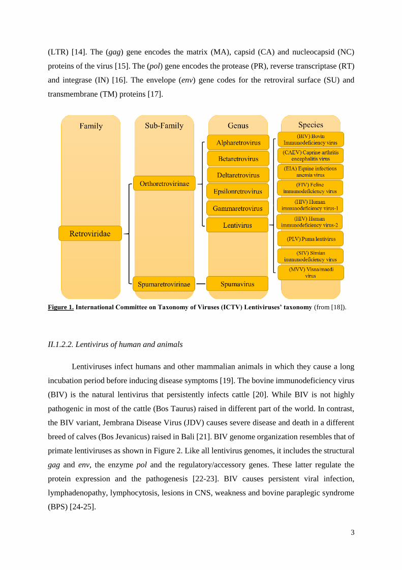
(LTR) [14]. The (gag) gene encodes the matrix (MA), capsid (CA) and nucleocapsid (NC)
proteins of the virus [15]. The (pol) gene encodes the protease (PR), reverse transcriptase (RT)
and integrase (IN) [16]. The envelope (env) gene codes for the retroviral surface (SU) and
transmembrane (TM) proteins [17].
Figure 1. International Committee on Taxonomy of Viruses (ICTV) Lentiviruses’ taxonomy (from [18]).
II.1.2.2. Lentivirus of human and animals
Lentiviruses infect humans and other mammalian animals in which they cause a long
incubation period before inducing disease symptoms [19]. The bovine immunodeficiency virus
(BIV) is the natural lentivirus that persistently infects cattle [20]. While BIV is not highly
pathogenic in most of the cattle (Bos Taurus) raised in different part of the world. In contrast,
the BIV variant, Jembrana Disease Virus (JDV) causes severe disease and death in a different
breed of calves (Bos Jevanicus) raised in Bali [21]. BIV genome organization resembles that of
primate lentiviruses as shown in Figure 2. Like all lentivirus genomes, it includes the structural
gag and env, the enzyme pol and the regulatory/accessory genes. These latter regulate the
protein expression and the pathogenesis [22-23]. BIV causes persistent viral infection,
lymphadenopathy, lymphocytosis, lesions in CNS, weakness and bovine paraplegic syndrome
(BPS) [24-25].
4
II.1.2.3. Equine infectious anemia virus (EIAV)
EIAV is a lentivirus that causes infection in horses and induces chronic infectious
diseases including recurrent anemia, weakness, thrombocytopenia and in rare cases
encephalopathies [27]. Virus transmission occurs through infected cells in blood [28-29] by
Tabanus fuscicostatus fly [30]. Vertical transmission of EIAV from infected mares to their foals
occasionally causes abortion [31]. The virus does not induce immunodeficiency-like disease
although there are multi-organ inflammatory disorders [32]. Tumor necrosis factor receptor
(TNFR) has been identified as the primary EIAV receptor [33]. EIAV infects and replicates
exclusively in the cells of the monocyte/macrophage lineage in vivo [34].
II.1.2.4. Feline Immunodeficiency Virus (FIV and PLV)
FIV is the natural lentivirus that infects domestic and a variety of wild cats, inducing an
immunodeficiency syndrome in infected animals [35]. PLV is the puma lentivirus that is a
variant of FIV infecting wild feline bobcats (Lynx rufus) and mountain lions (Puma concolor)
[36]. Unlike FIV-infected cats, PLV-infected hosts do not undergo pathogenesis like many SIVs
in their natural hosts [37]. FIV was first identified in 1986 in California [38]. The virus is tropic
for T-cells and replicates in feline kidney cells in vitro, it is found in blood, CSF and saliva, but
not in milk or colostrum. The initial phase of the disease is characterized by loss of appetite and
weight, depression, fever, lymphadenopathy and neutropenia. The end stage of the disease
includes loss of immune cells and proliferation of opportunistic infections that are associated
with death of infected animals [39]. FIV uses the CXCR4, CCR3 and CCR5 chemokine
receptors as entry receptors on mononuclear cells [40]. In contrast to primate lentiviruses FIV
does not use the CD4 as primary receptor and CXCR4 is considered the primary receptor for
entry in cells [41].
II.1.2.5. Ovine Lentivirus (OLV)
OLV is the natural lentivirus that was isolated from sheep and described as ovine
progressive pneumonia virus (OPPV) [42], or Maedi Visna Virus (MVV). The pathological
characteristics associated with OLV infections were reported in the beginning of the last century
[43]. Following an introduction in 1933 of Karakul sheep in Iceland imported from Germany,
two types of lethal disease syndromes were observed in local breed of sheep: Maedi (dyspnea
“pneumonia”) and Visna (wasting). Both diseases were caused by a lentivirus called MVV [44].

5
Figure 2. Genome organizations of lentivirus: equine (equine infectious anemia virus, EIAV), feline (feline
immunodeficiency virus, FIV), bovine (bovine immunodeficiency virus, BIV), caprine/ovine (caprine arthritis
encephalitis virus, CAEV, ovine maedi–visna virus, OMVV) (modified from [26]).
OLVs are macrophage-tropic lentiviruses that cause persistent infection in infected
sheep and the virus does not productively infect T lymphocytes [43, 45]. Infected sheep develop
interstitial progressive pneumonia, encephalitis, mastitis, arthritis and cachexia leading in some
cases to death because of prolonged starvation [46-47]. Virus transmission occurs mainly via
contaminated colostrum and milk from infected ewes to their newborn lambs [48].
Figure 3. Physical maps of small ruminant and human lentivirus genomes (modified from [66]).The genomes
contain in their extremities the 5’ and 3’ LTRs with the U3,R and U5 sequences that regulate the expression of
structural and regulatory/accessory genes represented in different colors and whose names are indicated.
II.1.2.6. Caprine Arthritis-Encephalitis Virus (CAEV)
CAEV is the second member of the small ruminant lentivirus group that was initially
reported in the late 1950s in Switzerland to be responsible of chronic arthritis in goats and then

6
years after it was associated disease in the central nervous system in CAEV-infected kids in
Germany [49]. Later on in the mid-1970s, Cork and colleagues described the disease in the
United States [50]. Later the virus was isolated from an adult goat suffering from chronic
arthritis, and described to be a retrovirus member of the genus Lentivirus [51]. The virus was
also simultaneously isolated from an encephalitis kid [52]. Like MVV in sheep, the productive
replication of CAEV is restricted in vivo to the monocyte/macrophage cell lineage and this
productive replication is dependent of the maturation of infected monocytes into macrophages
[53-54]. Although CAEV infection is subclinical, a small number of animals develop disease
syndromes including chronic polyarthritis in the joints and mastitis [55] but rarely encephalitis
in adults, whereas infected kids under six months develop encephalitis [56]. Dams transmit
CAEV to their kids via colostrum and milk [57-58]. The receptors and/or co-receptors of CAEV
are still unknown. CAEV is unable to infect human cells and the lack of functional receptors is
considered to be the main barrier that prevents CAEV from infecting human cells [59]. In
primate lentiviruses the functions of trans activating protein Tat have been well studied. This
protein is indispensable for the upregulation of transcription of the viral promoter in the LTR
and allowing the elongation of the transcripts [60-61]. In small ruminant lentiviruses including
CAEV, the LTR promoters were shown to induce virus expression constitutively and
independent from Tat transactivation [62]. The open reading frame previously called Tat of
CAEV and OLV/MVV does not encode a regulatory trans-activator protein of the LTR but
rather for an accessory protein that is structurally and functionally close to Vpr of primate
lentiviruses (Figure 3) [63-64]. In addition to Vpr-like, CAEV genome encodes Vif and Rev
regulatory proteins and the three structural proteins of gag, pol and env genes common to all
retroviruses [65]. Vif is necessary for effective in vivo virus replication and pathogenicity [66].
Rev trans-activation binds to Rev Response Element (RRE) an RNA structure [67]. The gene
rev encodes Rev protein that is necessary for cytoplasmic transport of un-spliced and single
spliced viral mRNA that are sequestered in nucleus [68].
II.1.2.7. Simian immunodeficiency virus SIV
This is a group of lentiviruses that infect a variety of species of non-human primates
[69]. SIVs are thought to be an old natural lentivirus reservoir in non-human primates that has
been the source of the spill over to human generating HIV-1 and HIV-2 [70-71]. SIVcpz from
chimpanzees (Pan troglodytes) and SIVgor from gorilla were found to be at the origin of the
cross species jumps to human generating HIV-1 [72-73], while SIVsmm from sooty mangabeys
7
(Cercocebus atys), generated HIV-2 [70]. SIVs are non-pathogenic in most of their own natural
hosts species [74] despite high levels of virus replication [75-76]. The mechanisms by which
these viruses remain non-pathogenic is unclear though several differences have been reported:
a) Natural hosts of SIV appear to limit immune activation [77] and the production of type I IFN
is limited to the early acute infection [78-79] which leads to b) an absence of chronic immune
activation and c) normal gastrointestinal homeostasis. Additional intriguing findings include
lower expression of CCR5 the SIV co-receptor on T cells [80], differential regulation of T cell
anergy [81], differential regulation of α4β7 on T cells [82] and a restriction of SIV replication
to T cells in vivo [83]. Experimental infection of non-natural hosts such as macaque species
with select isolates of SIVs has the ability to generate simian SAIDS with high levels of viremia,
CD4 depletion, in particular depletion of mucosal CD4+ T cells [84], immune impairment,
wasting and the occurrence of opportunistic infections [85]. Surprisingly, SIVs naturally infect
only African Old World monkeys (OWM) and apes from sub-Saharan Africa, but SIVs have
not been found in either Asian OWMs or New World monkeys [86].
II.2. History of HIV
It was believed that in 1960-1970, HIV-1 strains arrived in the USA from Congo. In an
early AIDS study, a Canadian airline steward referred to as "Patient 0" was thought to be
responsible for bringing HIV to North America [87]. However, data from a recent study indicate
that this is not the case [88]. The first case of HIV was declared in 1981. The virus inducing
AIDS was transmitted between homosexual men, who had begun dying of the disease [89].
II.2.1. Origin of HIV-1 in humans
HIV-1 is a zoonotic infectious pathogenic virus that arose from cross species infections
of simian immunodeficiency viruses (SIVs) naturally existing in reservoirs that were identified
to be in chimpanzees and gorillas. The subspecies Pan Troglodytes is the one naturally infected
with SIVcpz and experimental infections of these animals has now demonstrated that both
SIVcpz and HIV-1 infections can be associated with pathogenicity [90]. Genome detection and
sequencing have established that the endemic SIVcpz strains that P. t. troglodytes is a natural
reservoir of HIV-1 ancestor in Cameroon, where prevalence rates were found to be around 29
to 35%. Anti-SIV antibodies and viral genomes were detected in fecal and urine samples of
wild chimpanzees [91-92]. Phylogenetic analyses of SIVcpz sequences obtained from various
groups of chimpanzees have established that the infection has been present in this species for
8
over two centuries (Figure 4) [93]. HIV-1 is comprised of four viral lineages, termed groups M,
N, O, and P. Each resulted from independent cross-species transmissions of SIVcpz and SIVgor
[94]. The first group, M, originating from SIVcpz of Southern Cameroon is the cause of the
worldwide HIV-1 pandemic. Three other groups were then discovered in Cameroon; in 1990,
group O which represents less than 1% of global HIV-1 infections; in 1998, group N was
identified in a mere 13 cases. Finally, in 2009, group P was discovered in France, in a woman
and another person from Cameroon [93]. While HIV-1 group N is also from SIVcpz, the origin
of group O has been recently demonstrated to be from gorilla (SIVgor) from central Cameroon
and group P from SIVgor from southwestern Cameroon [95]. Previous studies have established
that SIVgor arose from SIVcpz [96].
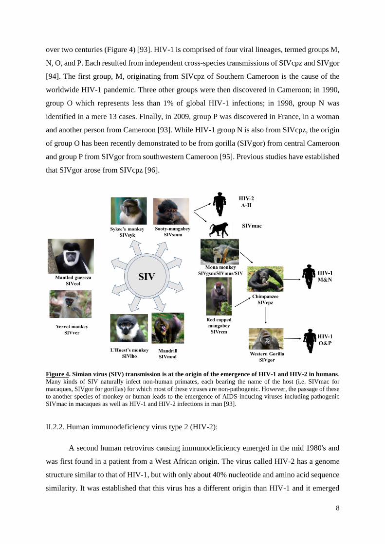
Figure 4. Simian virus (SIV) transmission is at the origin of the emergence of HIV-1 and HIV-2 in humans.
Many kinds of SIV naturally infect non-human primates, each bearing the name of the host (i.e. SIVmac for
macaques, SIVgor for gorillas) for which most of these viruses are non-pathogenic. However, the passage of these
to another species of monkey or human leads to the emergence of AIDS-inducing viruses including pathogenic
SIVmac in macaques as well as HIV-1 and HIV-2 infections in man [93].
II.2.2. Human immunodeficiency virus type 2 (HIV-2):
A second human retrovirus causing immunodeficiency emerged in the mid 1980's and
was first found in a patient from a West African origin. The virus called HIV-2 has a genome
structure similar to that of HIV-1, but with only about 40% nucleotide and amino acid sequence
similarity. It was established that this virus has a different origin than HIV-1 and it emerged
9
from a zoonotic cross species infection from sooty mangabey monkeys (SIVsmm) to human
[97-98]. HIV-2 is more commonly found in West Africa. In 1987, the first case in the United
States was in a West African woman diagnosed with central nervous system toxoplasmosis
[99]. There are eight HIV-2 groups: types A to H. Groups A and B are endemic in West Africa.
Although HIV-2 is similar to HIV-1 in its genome organization and replication, it lacks the
HIV-1 accessory vpu gene but encodes vpx [8] that facilitates the infection of resting CD4+ T
cells, via degradation of Sterile Alpha Motif and HD domain-containing protein 1 (SAMHD1),
the cell factor that prevent viral replication [100-101]. However in the absence of treatment,
HIV-2 mediated progression to disease is slower than upon infection with HIV-1 and
characterized with lower viral replication levels [102-103].
II.3. HIV transmission
HIV is transmitted through the exchange of body fluids from an infected to a non-
infected person [104-105]. Fluids exchange during unprotected intercourse has been shown to
be the most common route of HIV transmission [106]. However, HIV is also transmitted
through injection with contaminated needles or syringes used by infected individuals for drug
injection [107]. The efficiency of HIV transmission depends on the viral load and the virus type,
and whether the infection in transmitted either directly into the blood or onto a mucous
membrane [108]. Mother-to-child transmission from an infected mother to her baby during
pregnancy, delivery, or via breast feeding [109] is also an important route of transmission
particularly in children [110]. Throughout the breast-feeding period, the colostrum and breast
milk contain a variety of HIV-infected cells including CD4+ T cells and mammary epithelial
cells that are involved in virus transmission [111]. Other routes of HIV transmission include:
receiving HIV contaminated blood or blood products [112], organ and tissue transplants [113]
and accidental manipulation of infected products.
II.4.Epidemiology of HIV
Since the emergence of HIV-1 in human around 80 million individuals all over the world
were infected, nearly half of them have already died and the other half is living with the virus
[114-115]. The antiretroviral therapy (ART) helped not only HIV infected people to live longer,
to have a better quality of live, but also to decrease the transmission from HIV-positive to HIV
negative individuals. The great majority of HIV-infected individuals live in Sub Saharan Africa
and the accessibility of ART to these low-income countries has significantly slowed HIV
10
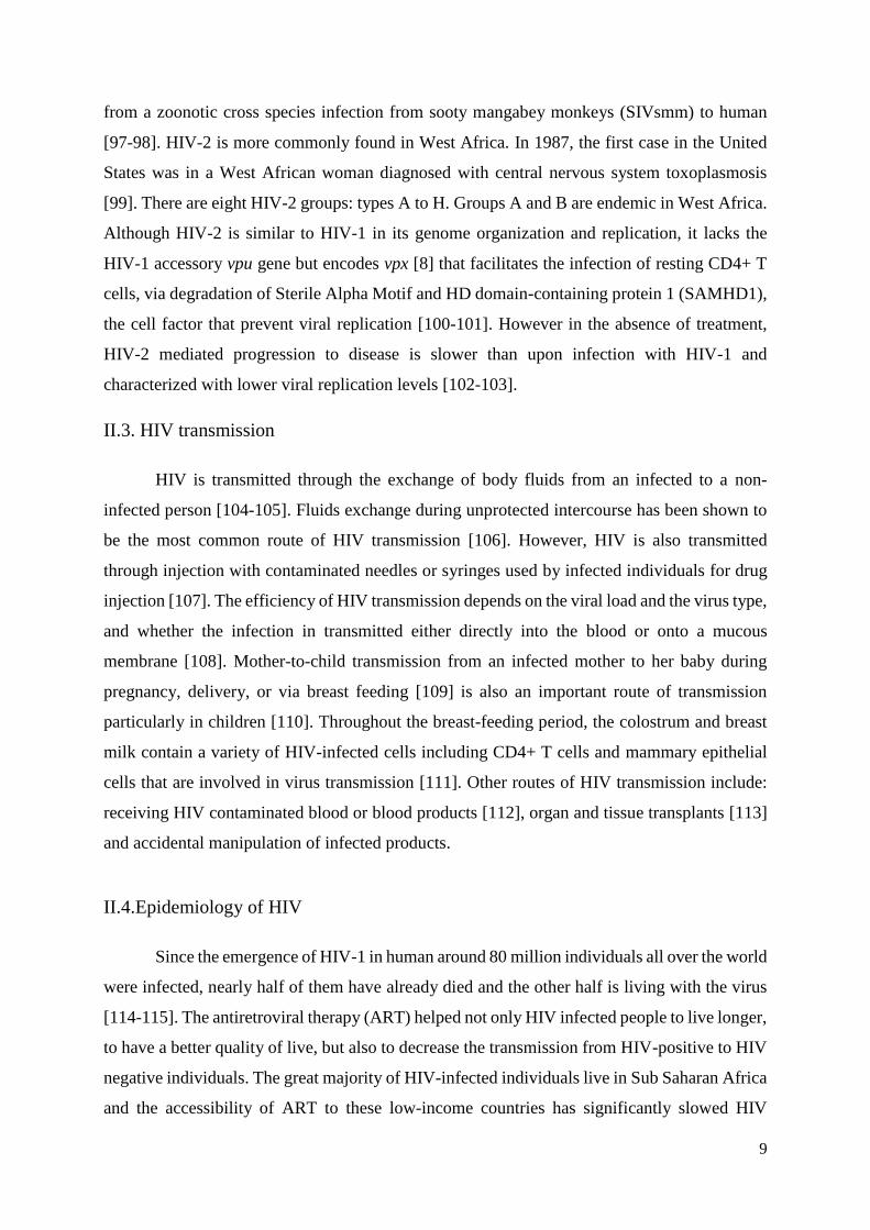
diffusion (Figure 5). Many strategies were developed in developing countries, including male
circumcision to slow down HIV-1 transmission [116], use of HAART during the pregnancy
and after delivery decreased mother to child transmission [117]. In addition ART for pre-
exposure prophylaxis (PrEP) was shown to decrease the risk of HIV acquisition [118].
Figure 5. Estimated number of people living with HIV in the world (taken from WHO).
II.5. Structure of the viral particle and HIV Gene Structure
As shown in Figure 6A the HIV viral particle is roughly spherical and coated with a
lipid bilayer derived from the host cell plasma membrane [119] when the newly formed virus
particle buds from the cell. The viral Env glycoproteins inserted at the surface are produced as
a precursor (gp160) [120], which is cleaved into the external surface gpl20 (SU) and the trans-
membrane gp4l (TM) [121]. Both proteins remain linked by non-covalent bonds and are
assembled as a trimer at the surface of the viral particles. Immature virus particles also have an
inner shell of Gag (Pr55) and Gag-Pol (Prl60) precursor group antigen polyproteins that are
subsequently cleaved by protease into functional mature subunits (Figure 6B) [122].

11
Figure 6. Schematic representation of (A) the structure and (B) the viral genome of HIV virion
(modified from [123]).
The cleavage of Gag (Pr55) precursor generates four functional domains (Figure 6B):,
the matrix protein (MA p17), capsid protein (CA p24), nucleocapsid proteins (NC p7 and p6)
which closes the narrow end of the viral core [124] that contains two identical molecules of
positive-sense strand genomic RNA [125]. The Gag-Pol (Prl60) precursor is subsequently
cleaved to produce the viral enzymes (Figure 6B): protease (PR pl0), reverse transcriptase (RT
p66/p51), and integrase (IN p32). Immature PR is less sensitive to protease inhibitors than free
mature PR enzyme [126].
Like all retroviruses, HIV encodes and incorporates into the virions enzymes that
convert the single stranded viral RNA into double stranded DNA which irreversibly integrates
12
into the chromosomal DNA of infected cells [127]. The viral genome contains also regulatory
and accessory genes (tat, rev, nef, vif, vpr and vpu) that express the virus regulatory and
accessory proteins (Figure 6B). These proteins play determinant roles in the modulation of virus
replication in infected cells and the pathogenesis in infected hosts.
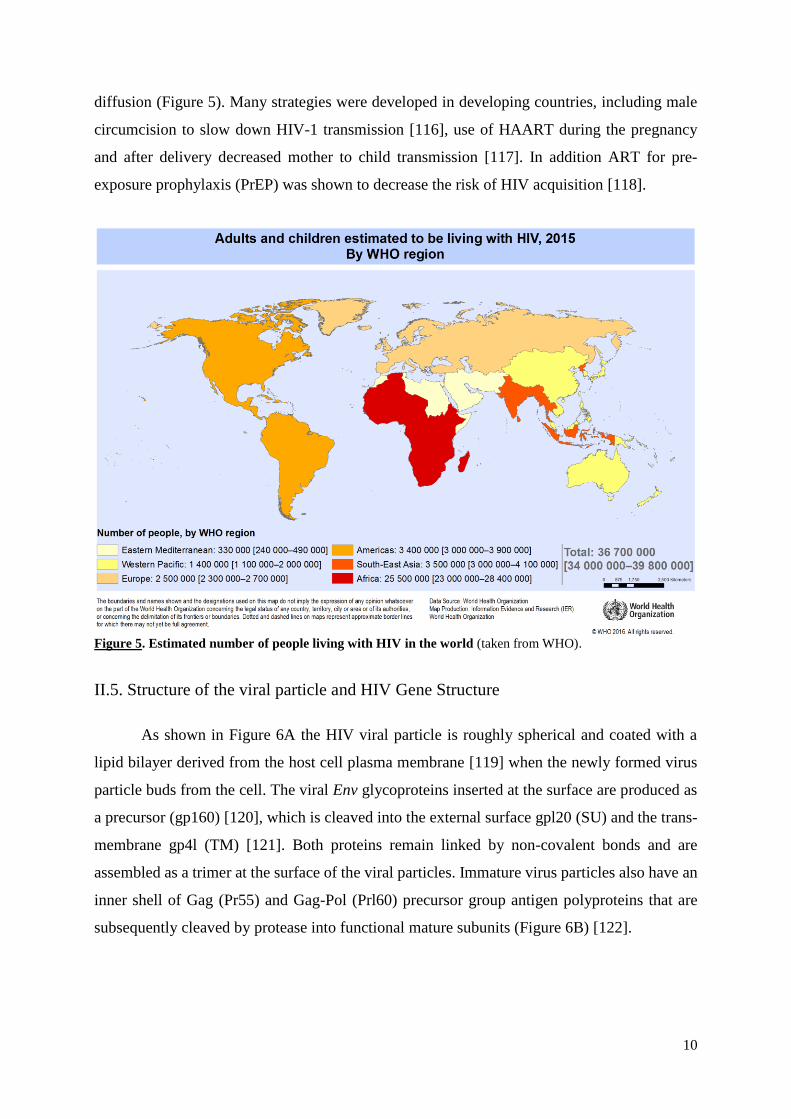
In addition to these structure regulatory and accessory genes, the RNA also contains
terminal sequences named long terminal repeat (LTR) (Figure 7). These are composed of the
unique U3 region, the repeat R element and the U5 region flanked by the transactivation
responsive element TAR and the primer-binding site in the 5’LTR. TAR is the target binding
sequence required for Tat transactivation activity [128]. The 3’LTR on the other hand is
preceded by the poly purine tract (PPT) [18]. This PPT segment is resistant to cleavage by
ribonuclease H and it serves as primer for the synthesis of the second DNA strand [129]. The
5' LTR harbors a promoter region for the polymerase II complex that initiates transcription of
the provirus, whereas the 3' LTR is needed for polyadenylation of the proviral mRNA and
provides transcriptional termination [130].
Figure 7. Schematic representation of regions of 5’LTR and 3’LTR which flank the proviral
sequence [131].
II.5.1. Viral Enzymes:
The pol gene encodes a protein precursor of the three viral enzymes, which are the
reverse transcriptase, the integrase and the protease. This precursor is cleaved by the auto
processing of the protease.
13
II.5.1.1. Reverse transcriptase (RT)
This multifunctional enzyme contains two enzymatic activities [132]. HIV-1 RT is a
heterodimer composed of two subunits of 66-kDa (p66) and 51-kDa (p51) derived from the
Gag-Pol (Prl60) precursor protein. The p66 subunit is responsible for polymerase and RNase H
activities and shows this activity also in the absence of the p51 subunit that serves as a structural
support [133]. RT contains the DNA polymerase activity that can copy either a RNA or DNA
template (Figure 8). It also has a RNase H activity that cleaves the DNA strand from the ssRNA
template then degrades the RNA if the RNA is part of an RNA/DNA duplex [134]. The RT
converts the +ssRNA genome of the virus into a double-stranded DNA (dsDNA) that can be
integrated into the genome of the host cell [135]. Many drugs used currently to fight HIV
infection target the activity of the RT.
II.5.1.2. Integrase (IN)
HIV-1 integrase is the enzyme that specifically and reproducibly integrates the HIV
dsDNA into that of the host as a provirus. In the structure of IN one can distinguish three
domains, the N-terminal domain which chelates zinc [136], the core domain which contains the
enzymatic activity, and the C-terminal domain which nonspecifically binds to DNA [137]. In
the cytoplasm of cells, IN binds to the extremities of the viral dsDNA at the specific sequences
of the attachment site located in the U5 and U3 at the ends of the 5’ and 3’ LTR regions [138].
This action is mediated by a stable nucleoprotein complex, the two ends of the viral DNA are
bridging by integrase making the synaptic complex [139]. Because of its N-terminal domain,
HIV-1 IN exists under several forms: monomer, dimer and tetramer [140]. The enzyme
undergoes two major catalytic activities: i) it processes the 3'-OH extremities of the viral
genome, ii) it inserts the viral DNA molecule into the infected host cellular chromosomal DNA.
Inserted DNA, called provirus, persists in the host cell serving as a template for viral gene
transcription followed by translation of viral proteins that are assembled to produce new
infectious virus particles [141]. As a reservoir, provirus can persist in a latent form lacking the
expression of any of the viral protein, making it incredibly difficult to fight [142-143].
14
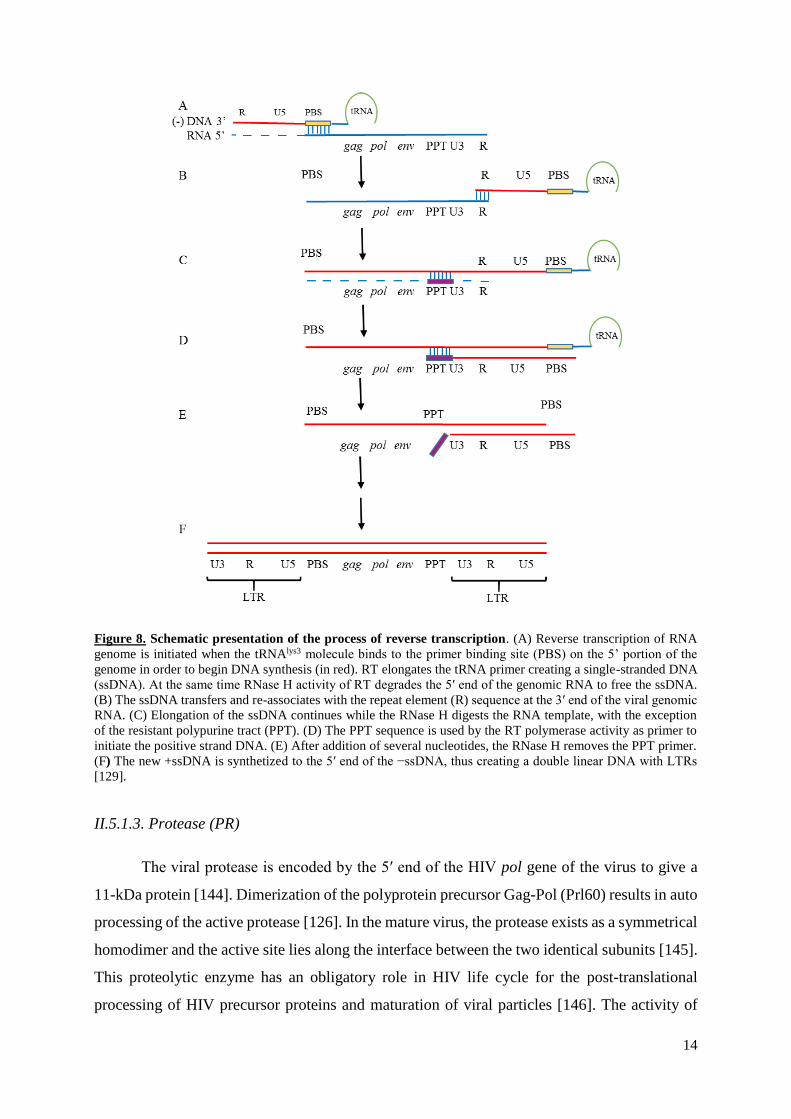
Figure 8. Schematic presentation of the process of reverse transcription. (A) Reverse transcription of RNA
genome is initiated when the tRNAlys3 molecule binds to the primer binding site (PBS) on the 5’ portion of the
genome in order to begin DNA synthesis (in red). RT elongates the tRNA primer creating a single-stranded DNA
(ssDNA). At the same time RNase H activity of RT degrades the 5′ end of the genomic RNA to free the ssDNA.
(B) The ssDNA transfers and re-associates with the repeat element (R) sequence at the 3′ end of the viral genomic
RNA. (C) Elongation of the ssDNA continues while the RNase H digests the RNA template, with the exception
of the resistant polypurine tract (PPT). (D) The PPT sequence is used by the RT polymerase activity as primer to
initiate the positive strand DNA. (E) After addition of several nucleotides, the RNase H removes the PPT primer.
(F) The new +ssDNA is synthetized to the 5′ end of the −ssDNA, thus creating a double linear DNA with LTRs
[129].
II.5.1.3. Protease (PR)
The viral protease is encoded by the 5′ end of the HIV pol gene of the virus to give a
11-kDa protein [144]. Dimerization of the polyprotein precursor Gag-Pol (Prl60) results in auto
processing of the active protease [126]. In the mature virus, the protease exists as a symmetrical
homodimer and the active site lies along the interface between the two identical subunits [145].
This proteolytic enzyme has an obligatory role in HIV life cycle for the post-translational
processing of HIV precursor proteins and maturation of viral particles [146]. The activity of

15
this enzyme is also essential for virus infectivity because without an effective protease, HIV
virions remain non-infectious [147]. Thus any mutation on functional domain or inhibition of
protease activity leads to disruption of replication and propagation of virus to other cells [148].
This central role of PR made it an attractive target for anti-HIV drugs, thus specific protease
inhibitors have been designed and are widely used to inhibit the protease [149].
II.5.2. Structural Proteins
II.5.2.1. Envelope glycoproteins (Env)
Envelope (Env) glycoproteins play a key role in HIV-1 diffusion of infection and
pathogenicity [150]. The envelope glycoprotein precursor 160 kDa (gp160) encoded by the env
gene from an unispliced RNA is cleaved by a cellular protease into two sub-units: the surface
glycoprotein of 120 kDa (SU gp120) and the transmembrane glycoprotein of 41 kDa (TM gp41)
[151]. Gp120 binds to the extracellular region of gp41 and is important for virus interaction
with the main CD4 receptor at the surface of target cells [152]. Gp120 contains also co-receptor
binding sites that determine the cell tropism of the virus [153].
Figure 9. Schematic representation of the envelope glycoprotein trimer and interaction with receptors of
target CD4+ cells (A) The target CD4+ cell and the virion with the gp120 and gp41 before binding; (B) The gp120
first bind to the CD4 receptor via its CD4 binding site and then to the co-receptor, either CCR5 or CXCR4; (C)
following this double binding conformational changes in the gp41 lead to the fusion of lipid bilayer membranes
of virus and target cell (adapted from [154]).
HIV-1 Env proteins are assembled as trimers that bind to cell-surface receptors CD4 on target
cells and subsequently lead to conformational changes in the glycoproteins [155-156]. These
changes involve shift in the V1 and V2 variable regions exposing the V3 region that then binds
to the co-receptor (CXCR4 or CCR5). This last interaction induces conformational changes in
gp41 and causes the exposure of the fusion peptide (Figure 9). This leads to the fusion of lipid
16
bilayer membranes of virus and target cell initiating the internalization of the capsid in the
cytoplasm of infected host cell [157]. Gp120 is considered the primary target of the humoral
immune response and specific neutralizing antibodies [158-159].
II.5.2.2. Group antigens (Gag)
Group antigen is the precursor of several major proteins: the matrix protein that lines
the inside of the viral envelope, capsid and nucleocapsid proteins that are important for genomic
RNA assembly and packaging (Figure 6).
Matrix (Ma) p17: This is a 17-kDa protein that originates from the cleavage of the amino-
terminal portion of the Gag precursor, Gag Pr55 at the early stage of infection [160]. The protein
is localized at the inner surface of the virion underneath the lipid bilayer membrane and has a
multifunctional crucial role for virion assembly [161]. It participates to the import of the pre-
integration complex (PIC) from the cytoplasm to the nucleus during the early stage of infection
[162]. It transports also the precursor Gag (Pr55) to the plasma membrane for virus assembly
[163].
Capsid (CA) p24: This is the major core protein of HIV that has a molecular weight of 24 kDa
and is also derived from Gag (Pr55). The capsid is a cone shape structure composed of about
250 hexamers of p24 and exactly 12 pentamers of the same protein at both conical ends [164].
Inside the capsid, there are the two strands of genomic viral RNA, the nucleocapsid (NC) and
all the enzymes necessary for replication [165]. The capsid p24 protein has been used as an
antiviral specific target and for HIV vaccine development [166]. HIV-1 Gag p24 antigen is
quantitatively the most abundant immunogenic element used for early stage diagnosis of HIV-
1 infection by enzyme-linked immunosorbent assay (ELISA) in the plasma [167].
Nucleocapsid (NC) p7: HIV-1 NC is a small 7-kDa protein, derived from the precursors Gag
(Pr55). NC sequence contains 55-amino acid with two zinc finger-binding domains. It is an
RNA-binding protein which facilitates both RNA rearrangements [168] and reverse
transcription [169]. The binding of NC with the viral RNA in the region of the psi (ψ) packaging
sequence leads to the incorporation of genomic viral RNA (gRNA) into the newly formed HIV-
1 particles [170]. For the initiation of the reverse transcription process, NC anneals the host cell
tRNALys3 primer onto the primer binding site in the leader region of the genomic RNA [171].
17
p6: The HIV-1 p6 is the carboxy-terminal of the cleaved precursors Gag (Pr55). It is involved
in the final separation step of nascent virions, where it facilitates virus release from the host
cells [172]. Gag p6 has also been found to be phosphorylated during HIV-1 infection and this
event may affect virus replication. Moreover, p6 mediates the incorporation of the viral Vpr
protein into new HIV-1 viral particles [173].
II.5.3. Accessory and Regulatory Proteins
II.5.3.1. Viral protein U (Vpu):
"Viral Protein Unique" is an 81 amino acids phosphorylated HIV-encoded accessory
protein with a molecular weight of 16 kDa. Vpu is a late viral protein expressed from a
bicistronic mRNA that encodes also HIV-1 envelope glycoproteins [174-176]. This protein is
almost unique to HIV-1 among primate immunodeficiency viruses. One of the major functions
of Vpu is to prevent superinfection of infected cells by degrading the CD4 receptor,
downregulating the newly synthesized CD4 receptor and major histocompatibility complex
(MHC) class I via the endoplasmic reticulum proteasomal degradation pathway [177-178]. The
second function of Vpu is at the level of efficient virus maturation by enhancement and
regulation of the release of progeny virions from the external surface of infected host cells
[179]. Vpu downregulates the antiviral factor bone marrow stromal antigen 2 (BST-2/tetherin),
that prevents HIV-1 release from the cell surface, by interacting with tetherin to remove it from
the plasma membrane [180].
II.5.3.2. Viral protein R (Vpr):
"Viral Protein R" is a late virus protein produced from a spliced mRNA that encodes the
accessory genes expressing maturation proteins Vpu, Vpr and Vif proteins as well as Env
glycoproteins [181]. Vpr contains 96 amino acids and has a molecular weight of 14-kDa. It is
a virion-associated protein that has multiple functions on virus and infected cells [182]. In early
stages of infection, Vpr is part of the complex of nuclear import that interacts with HIV-1
reverse-transcription and migration of pre-integration complex that transports the newly
synthesized viral DNA genome from the cytoplasm into the nucleus [182-183]. In the
pathogenesis of HIV-1 Vpr induces efficient replication of HIV-1 in non-dividing cells [184].
Vpr leads to cell arrest at the G2/M phase of cell division thereby inducing dysfunction and
apoptosis of infected cells [185]. Lack of Vpr or mutated Vpr that leads to decreased function
18
is associated with a slow disease progression [186]. Other functions were ascribed to Vpr like
modulation of the fidelity of viral reverse transcription and transactivation of the HIV-1 LTR
promoter and cellular genes [187], induction of cellular differentiation [188] or its interaction
with the p6 ensuring efficient Vpr packaging [189].
II.5.3.3. Viral infectivity factor (Vif):
Vif is a regulatory protein that is coded by all known lentivirus genomes of human and
animals. HIV-1 Vif is a 23-kD protein. Vif is essential for viral fitness, pathogenicity,
replication and productive infection in non-permissive cells mononuclear cells, macrophages.
It is absolutely necessary for productive infection in primary CD4+ T lymphocytes and
macrophages [190] where Vif interacts with the viral RNA and the NC for viral assembly and
packaging into viral particles [191]. Vif degrades and inhibits the enzymatic activity of the
cellular anti-viral enzyme apolipoprotein B mRNA-editing enzyme catalytic polypeptide-like
3G (APOBEC 3G) mainly but also other APOBEC-3 from the cytoplasm of infected cells, and
packaging into virion [192-193]. APOBEC-3 family enzymes act as potent antiviral restriction
factors to inhibit HIV-1 and other retroviruses [194].
II.5.3.4. Negative regulatory factor (Nef):
During the early stages of cell infection HIV-1 Nef is one of the first protein expressed
from a multi-spliced mRNA [195]. Nef is a 27-kDa protein that has multiple functions both on
virus life cycle and on host cells. Nef has multiple localizations, it is found both in the cytoplasm
and nucleus compartments, and secreted in exosomes [196]. Although Nef-defective HIV-1 is
replication competent, Nef is necessary for efficient virus replication and disease progression
in vivo [197]. One of the main functions of Nef is to downregulate the expression of surface
CD4 and Major Histocompatibility Complex-I molecules [198]. Nef enhances Gag localization
at the cell membrane and viral particle assembly, and facilitates viral transfer from cell-to-cell
[199]. Nef also enhances HIV-1 infection and replication in primary CD4+ T cells [200]. Nef
is indispensable for progression to AIDS in HIV infected patients since individuals infected
with nef-defective HIV-1 mutants do not progress to AIDS [201], as also demonstrated with
the Sydney blood bank cohort [202].
19
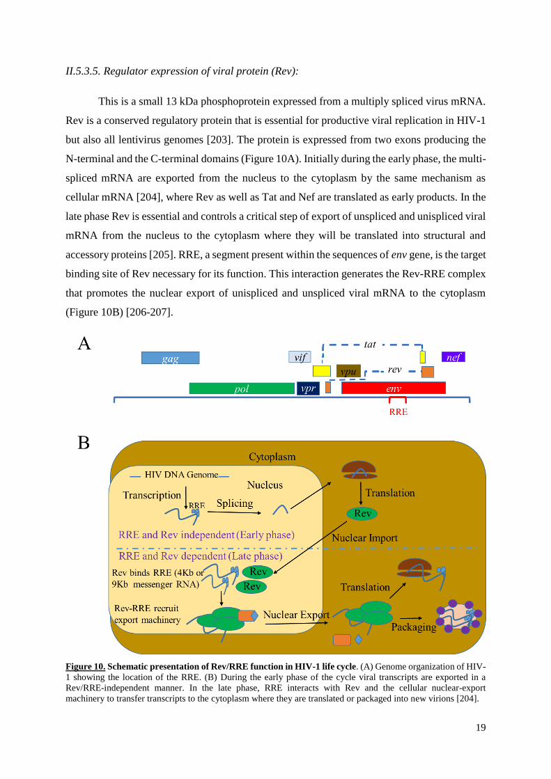
II.5.3.5. Regulator expression of viral protein (Rev):
This is a small 13 kDa phosphoprotein expressed from a multiply spliced virus mRNA.
Rev is a conserved regulatory protein that is essential for productive viral replication in HIV-1
but also all lentivirus genomes [203]. The protein is expressed from two exons producing the
N-terminal and the C-terminal domains (Figure 10A). Initially during the early phase, the multi-
spliced mRNA are exported from the nucleus to the cytoplasm by the same mechanism as
cellular mRNA [204], where Rev as well as Tat and Nef are translated as early products. In the
late phase Rev is essential and controls a critical step of export of unspliced and unispliced viral
mRNA from the nucleus to the cytoplasm where they will be translated into structural and
accessory proteins [205]. RRE, a segment present within the sequences of env gene, is the target
binding site of Rev necessary for its function. This interaction generates the Rev-RRE complex
that promotes the nuclear export of unispliced and unspliced viral mRNA to the cytoplasm
(Figure 10B) [206-207].
Figure 10. Schematic presentation of Rev/RRE function in HIV-1 life cycle. (A) Genome organization of HIV-
1 showing the location of the RRE. (B) During the early phase of the cycle viral transcripts are exported in a
Rev/RRE-independent manner. In the late phase, RRE interacts with Rev and the cellular nuclear-export
machinery to transfer transcripts to the cytoplasm where they are translated or packaged into new virions [204].

20
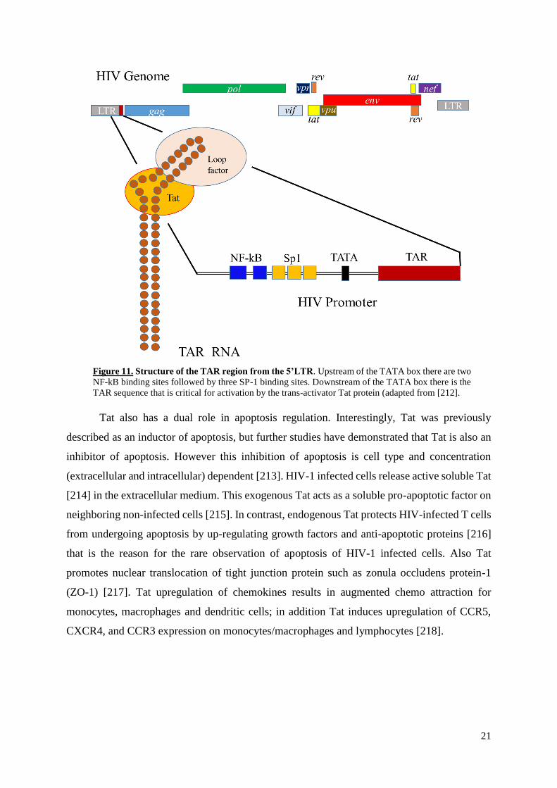
II.5.3.6. Trans-activator of transcription (Tat)
HIV-1 tat gene expresses a regulatory protein that upregulates HIV genome
transcription. Tat, which contains 86 aa with molecular weight of 14 kDa, is translated from
two exons located upstream and within the env gene; the minor exon codes for 14 amino acids,
while the major exon encodes 72 amino acids [208]. Tat is an essential virus protein in absence
of which the viral genome lacks the capacity of expression. This protein is necessary for
transcription initiation under control of HIV-1 long terminal repeat (LTR) and elongation of
transcripts of viral genes [209]. HIV-1 DNA provirus transcription begins with cellular factors
(NF-kB, Sp1, the TATA box binding protein) and RNA polymerase II binding to the 5' LTR
promoter region, forming a transcription complex that allows a low level of viral transcripts
production, that then translates to Tat, one of the early viral proteins (Figure 11) [210].
Following the initiation of viral RNA transcription Tat binds to TAR hairpin located at the 5’
and of viral RNA. This binding helps to increase transcription initiation and enhancement of
viral gene expression by augmenting the transcription efficiency of the viral LTR promoter and
highly enhances the efficiency of viral RNA transcription elongation by the cellular RNA
polymerase II [211]. Deletion or mutated genomes in tat gene or in the Tat target TAR
sequences dramatically decreases or cancels the transactivation activity of Tat and consequently
the virus replication.
21
Figure 11. Structure of the TAR region from the 5’LTR. Upstream of the TATA box there are two
NF-kB binding sites followed by three SP-1 binding sites. Downstream of the TATA box there is the
TAR sequence that is critical for activation by the trans-activator Tat protein (adapted from [212].
Tat also has a dual role in apoptosis regulation. Interestingly, Tat was previously
described as an inductor of apoptosis, but further studies have demonstrated that Tat is also an
inhibitor of apoptosis. However this inhibition of apoptosis is cell type and concentration
(extracellular and intracellular) dependent [213]. HIV-1 infected cells release active soluble Tat
[214] in the extracellular medium. This exogenous Tat acts as a soluble pro-apoptotic factor on
neighboring non-infected cells [215]. In contrast, endogenous Tat protects HIV-infected T cells
from undergoing apoptosis by up-regulating growth factors and anti-apoptotic proteins [216]
that is the reason for the rare observation of apoptosis of HIV-1 infected cells. Also Tat
promotes nuclear translocation of tight junction protein such as zonula occludens protein-1
(ZO-1) [217]. Tat upregulation of chemokines results in augmented chemo attraction for
monocytes, macrophages and dendritic cells; in addition Tat induces upregulation of CCR5,
CXCR4, and CCR3 expression on monocytes/macrophages and lymphocytes [218].

22
II.6. HIV receptors
II.6.1. CD4
CD4, the cluster of differentiation 4 receptor protein, was discovered in the late 1970s
and it was then called leu-3 and T4 [219]. This cell-surface glycoprotein contains four
immunoglobulin domains, a transmembrane hydrophobic segment and C terminal cytosolic part
[220]. The glycoprotein is present on the surface of white blood cells, T helper cells, T
lymphocytes, monocytes, macrophages and dendritic cells. CD4 receptor interacts directly with
the major histocompatibility complex class II molecule (MHC class II) on the surface of the
antigen-presenting cell (APC) and recognizes the foreign antigen, leading to T cell activation
[221]. CD4 serves as a human immunodeficiency virus (HIV) essential receptor for entry into
the host cell when its extracellular domain binds to gp120 the exterior protein of the envelope
surface [222]. This receptor is found on the surface of T helper cells, at all stages of
development, activation and function. Furthermore, after infection, the HIV accessory proteins
VpU and Nef downregulate CD4, thus preventing superinfection of infected cells [223].
II.6.2. CCR5
CCR5, also known as CD195, is a seven transmembrane segments protein located at the
surface of white blood cells where it acts as a receptor for chemokines [224]. CCR5 was
identified as a major co-receptor for macrophage-tropic (M-tropic) viruses such as HIV that
initially use CCR5 to enter and infect host cells [225]. CCR5 is predominantly expressed on T
lymphocytes, macrophages, dendritic cells [226]. In certain individuals the CCR5 gene
expresses a CCR5 co-receptor protein that contains a deletion of 32bp (CCR5-Δ32) which
prevents expression of the protein at the cell surface [227]. People that are homozygous for
CCR5∆32 are less susceptible to HIV infection, while for heterozygous people, the single copy
of the mutated gene provides some protection against infection and makes the disease less
severe if infection occurs [228].
II.6.3. CXCR4
CXCR4, also known as CD184, is another member of the chemokine receptor protein
family. Like CCR5, the protein has seven transmembrane regions but is located on the surface
of T cells [229]. This receptor, discovered in 1996, is expressed on leukocytes and
hematopoietic cells such as endothelial and epithelial cells [230]. It serves as co-receptor for T-

23
tropic HIV infection into permissive cells. HIV strains with X4 tropism are typically found late
in infection. Furthermore, AIDS is caused by both R5 and X4 strains, but the presence of X4
strain have been shown to accelerate depletion of CD4+T cells [231].
II.6.4. HIV cellular tropism
The main target cells of HIV infection and replication in infected hosts are the CD4+ T
cells [232]. Although the great majority of these cells are T lymphocytes,
monocyte/macrophage and dendritic cells are also important target of the virus [233]. ‘Viral
tropism’ is the preference of the virus to bind with one co-receptor among the others co-
receptors, in addition to CD4 the main receptor on the host cell surface [154]. Two
subcategories of these target cells are distinguished: the CD4+ that co-express the CCR5
chemokine receptor (CD4+/CCR5) and those co-expressing the CXCR4 chemokine receptor
(CD4+/CXCR4). Actually several HIV can also use other co-receptors such as CCR1, CCR2b,
CCR3, CCR8, CCR9, CX3CR1/V28, STRL-33/BONZO/CXCR6, GPR1, GPR15/BOB, APJ,
ChemR23, RDC1, and Leukotriene B4 receptor [154]. While the virus uses the CD4+/CCR5
cells localized mainly in the tissues to initiate the infection in the body and cause the chronic
phase of the infection [234], in the late stages of infection (AIDS stage) the virus switches its
tropism toward CD4+/CXCR4 T lymphocytes localized mainly in peripheral blood [235]. The
great majority of CD4+/CCR5 T cells are localized in the gut-associated lymphoid tissue
(GALT) [236] where the founder virus starts its replication prior to crossing the epithelium and
migrating to the lymph nodes [237]. Heterosexual transmission of HIV-1 is initiated by a
selected transmitter/founder (T/F) virus that generally uses the CCR5 as co-receptor [238]. This
type of transmission acts as a genetic bottleneck selection of a single viral variant that replicates
in the abundant CD4+/CCR5+ memory T lymphocytes of the GALT and then across the
mucosa to disseminate in target organs of the body [239]. During the chronic infection, the
swarm of viruses generated continuously by accumulation of mutations in the envelope use
mainly the CD4/CCR5 receptor/co-receptor to replicate in the CD4+ cells [240]. After several
years of chronic replication, there is a switch from the CCR5 co-receptor usage to two types of
viruses: the dual tropic CXCR4/CCR5 and the CXCR4 tropic [241]. This emergence is a result
of genetic variants that acquire amino acids in Env V3 loop that determine co-receptor usage
[242].
24
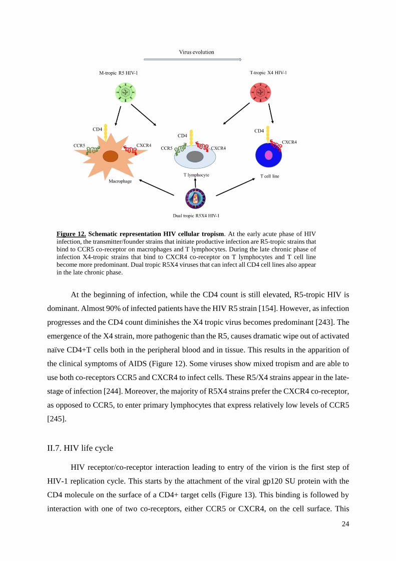
Figure 12. Schematic representation HIV cellular tropism. At the early acute phase of HIV
infection, the transmitter/founder strains that initiate productive infection are R5-tropic strains that
bind to CCR5 co-receptor on macrophages and T lymphocytes. During the late chronic phase of
infection X4-tropic strains that bind to CXCR4 co-receptor on T lymphocytes and T cell line
become more predominant. Dual tropic R5X4 viruses that can infect all CD4 cell lines also appear
in the late chronic phase.
At the beginning of infection, while the CD4 count is still elevated, R5-tropic HIV is
dominant. Almost 90% of infected patients have the HIV R5 strain [154]. However, as infection
progresses and the CD4 count diminishes the X4 tropic virus becomes predominant [243]. The
emergence of the X4 strain, more pathogenic than the R5, causes dramatic wipe out of activated
naïve CD4+T cells both in the peripheral blood and in tissue. This results in the apparition of
the clinical symptoms of AIDS (Figure 12). Some viruses show mixed tropism and are able to
use both co-receptors CCR5 and CXCR4 to infect cells. These R5/X4 strains appear in the late-
stage of infection [244]. Moreover, the majority of R5X4 strains prefer the CXCR4 co-receptor,
as opposed to CCR5, to enter primary lymphocytes that express relatively low levels of CCR5
[245].
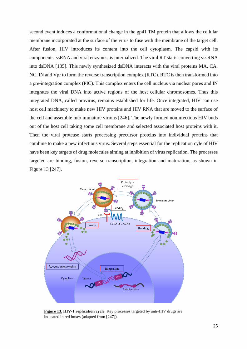
II.7. HIV life cycle
HIV receptor/co-receptor interaction leading to entry of the virion is the first step of
HIV-1 replication cycle. This starts by the attachment of the viral gp120 SU protein with the
CD4 molecule on the surface of a CD4+ target cells (Figure 13). This binding is followed by
interaction with one of two co-receptors, either CCR5 or CXCR4, on the cell surface. This
25
second event induces a conformational change in the gp41 TM protein that allows the cellular
membrane incorporated at the surface of the virus to fuse with the membrane of the target cell.
After fusion, HIV introduces its content into the cell cytoplasm. The capsid with its
components, ssRNA and viral enzymes, is internalized. The viral RT starts converting vssRNA
into dsDNA [135]. This newly synthesized dsDNA interacts with the viral proteins MA, CA,
NC, IN and Vpr to form the reverse transcription complex (RTC). RTC is then transformed into
a pre-integration complex (PIC). This complex enters the cell nucleus via nuclear pores and IN
integrates the viral DNA into active regions of the host cellular chromosomes. Thus this
integrated DNA, called provirus, remains established for life. Once integrated, HIV can use
host cell machinery to make new HIV proteins and HIV RNA that are moved to the surface of
the cell and assemble into immature virions [246]. The newly formed noninfectious HIV buds
out of the host cell taking some cell membrane and selected associated host proteins with it.
Then the viral protease starts processing precursor proteins into individual proteins that
combine to make a new infectious virus. Several steps essential for the replication cyle of HIV
have been key targets of drug molecules aiming at inhibition of virus replication. The processes
targeted are binding, fusion, reverse transcription, integration and maturation, as shown in
Figure 13 [247].
Figure 13. HIV-1 replication cycle. Key processes targeted by anti-HIV drugs are
indicated in red boxes (adapted from [247]).
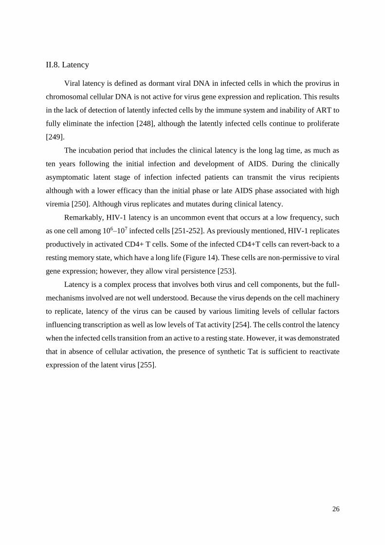
26
II.8. Latency
Viral latency is defined as dormant viral DNA in infected cells in which the provirus in
chromosomal cellular DNA is not active for virus gene expression and replication. This results
in the lack of detection of latently infected cells by the immune system and inability of ART to
fully eliminate the infection [248], although the latently infected cells continue to proliferate
[249].
The incubation period that includes the clinical latency is the long lag time, as much as
ten years following the initial infection and development of AIDS. During the clinically
asymptomatic latent stage of infection infected patients can transmit the virus recipients
although with a lower efficacy than the initial phase or late AIDS phase associated with high
viremia [250]. Although virus replicates and mutates during clinical latency.
Remarkably, HIV-1 latency is an uncommon event that occurs at a low frequency, such
as one cell among 106–107 infected cells [251-252]. As previously mentioned, HIV-1 replicates
productively in activated CD4+ T cells. Some of the infected CD4+T cells can revert-back to a
resting memory state, which have a long life (Figure 14). These cells are non-permissive to viral
gene expression; however, they allow viral persistence [253].
Latency is a complex process that involves both virus and cell components, but the full-
mechanisms involved are not well understood. Because the virus depends on the cell machinery
to replicate, latency of the virus can be caused by various limiting levels of cellular factors
influencing transcription as well as low levels of Tat activity [254]. The cells control the latency
when the infected cells transition from an active to a resting state. However, it was demonstrated
that in absence of cellular activation, the presence of synthetic Tat is sufficient to reactivate
expression of the latent virus [255].

27
Figure 14. Latency in CD4+ T cells. In the early stage of infection, HIV enters CD4+ T cell and
its genome is integrated into the host DNA. The activated infected T cells allow viral transcription,
virion assembly and production of new virus particles able to propagate the infection. A small
proportion of infected cells undergo transition from an active to a resting state contributing to the
formation of the latent reservoir.
II.8.1. General mechanisms of latency
There are two types of latency known for HIV-1 infection: the pre-integration latency
and the post-integration latency [256]. The pre-integration latency refers to the stage where
virus in infected cells undergo all the early stages of virus replication to produce the double-
stranded DNA but this latter fails to integrated into host genome and is not expressed as
extrachromosomal unit. This DNA will either be degraded or integrated later into the host cell
chromatin [257]. After HIV-1 enters host cell, the genomic viral RNA is converted into double-
stranded DNA by the reverse transcriptase, transported to the nucleus by the pre-integration
complex and integrated in the genome of host cell as a provirus. Therefore, pre-integration
latency may occasionally result from a defect of efficient reverse transcription activity,
inhibition of RT, ineffective transfer of PIC from the cytoplasm to the nucleus or by defect of
integration activity [258]. This type of pre-integration latency is very common in resting CD4+
T cells in untreated patients [259]. Pre-integration latency is influenced by interactions of host
restriction factors (RFs) including APOBEC3 that induces hypermutations in HIV genome G
to A [260]. To counteract this blocking activity to the virus replication, all lentiviruses including
HIV and SIV express Vif protein that promotes APOBEC3 degradation [193].
28
Many cellular factors interfere with viral replication processes. Sterile alpha motif
domain and HD domain-containing protein-1 (SAMHD1) is a cellular enzyme that has recently
been discovered. This protein prevents HIV replication by depleting the pool of dNTPs
available to the reverse transcriptase for viral DNA synthesis [261]. However, SIVmac/HIV-2
express the Vpx protein which promotes SAMHD1 degradation, thus preventing decrease of
dNTP concentration available for reverse transcription [262]. Myxovirus resistance protein 2
(Mx2) inhibits HIV-1 after reverse transcription and before proviral integration into the host
genome, because Mx2 stimulate IFN production that is known to inhibit virus replication [263].
The most important mechanism of latency is the post-integration latency. Following
transport of neosynthesised dsDNA by the pre-integration complex from the cytoplasm to the
nucleus, viral DNA integrates into the host genome as a provirus. The pre-integration complex
(PIC) consists of dsDNA, integrase (IN), reverse transcriptase (RT), matrix antigen (MA), viral
protein R (Vpr) and some host proteins like the transportins [264]. Following transport from
the cytoplasm to the nuclear pores, the viral DNA is translocated into the nucleus together with
the virus IN which will catalyze viral DNA integration into the host cell genome. In some
circumstances, in some cells the provirus stays dormant as a post-integration state of latency
[265].
Insertion of proviral HIV-1 DNA into the heterochromatin region of host cell support
the latency because it is densely packed and transcriptionally inactive [266]. Furthermore, HIV-
1 latency is supported by several mechanisms implicating transcription factors and post-
translational histone modifications enzymes [267]. Indeed histone deacetylases and histone
methyltransferases have been shown to reinforce latency [268-269].
II.8.2. Cellular and Anatomical Reservoirs of HIV.
Viral latency establishes at the early stage during acute infection in two types of
sanctuaries: cellular and anatomical [270]. There are many cell types involved in the persistence
of HIV-1 reservoir, which remains the major barrier to HIV-1 cure. HIV-1 infected resting
memory CD4+T cells that hold the provirus in latent stage represent the most important cellular
reservoir [271]. Other cellular reservoirs of HIV are circulating infected monocytes that require
to be differentiated into macrophages in tissues to activate virus replication [272-273];
hematopoietic stem cells (HSPC) in bone marrow that are precursor cells associated with self-
renewal and differentiation; they are considered to be long-lived reservoirs [274]. Furthermore,
dendritic cells (DCs) and follicular dendritic cells (FDCs) are important HIV reservoirs because
29
they have the ability to trap and store pathogens for long periods of time [275-276]. Other cell
types like epithelial cells (renal, mucosa and cervix), skin fibroblasts, bone marrow [277]
natural killer (NK) and memory stem cells [278] are also involved in latency and persistence of
HIV-1.
There are anatomical tissues serving as sanctuary reservoir compartments for HIV-1.
These tissues are immunologically protected and isolated by a barrier from the blood and the
lymphoid systems so that drugs used for treatment can poorly or cannot penetrate or metabolize
into active moieties [279]. Lymphoid organs, spleen, lymph nodes, and gut-associated
lymphoid tissues (GALT) are major sites of viral replication [280]. Liver, kidney, and lungs,
alveolar macrophages (AMs) in the respiratory tract are also suitable reservoirs for the virus as
they are infected early by HIV [281]. In the central nervous system (CNS) cell types like
mastocytes, astrocytes, and microglia are known to be infected. Microglia are brain resident
macrophages that are more resistant to apoptosis induced by HIV-1. They are important stable
hideouts for the virus, because of the long life span of macrophages compared to the short life
of activated CD4+ T cells, especially in the late stage of viral infection when CD4+ T cells are
largely depleted [282]. Furthermore, the blood–brain barrier limits the distribution of ART into
the CNS [283]. The genital tract is also one of the sanctuaries of HIV-1. In the male genital
tract, provirus DNA was detected in seminal cells [284]. The female reproductive tract is poorly
permissive to ART so that elimination of provirus from the female genital tract is inefficient
[285].
II.9. HIV vaccines
Since the discovery of HIV, many attempts to develop a safe, effective and durable
vaccine have been tried. So far, HIV vaccine preparations, either preventive or therapeutic, have
failed in part because of the complex biological properties of the virus [286]. Indeed, the life
cycle of the virus is associated with genetic and antigenic variabilities that render the host
adaptive immune response inefficient at controlling HIV infection [287]. In addition, the
integration/latency of HIV DNA ensure the persistence of virus infection and hiding to avoid
clearance by the immune system.
Several therapeutic or functional cure vaccines have been tested [288]. Some of these
are based on whole virus as an immunogen in its native structure but inactivated to render it
non-infectious [289]. One of these approaches uses inactivated HIV either by heat [290] or
chemical [291] treatment for killing the whole virus used as immunogen. Although the killed
30
virus strategy is probably safe for human, it is no longer supported because of the low
immunogenicity induced by this vaccine [292]. Neither heat nor chemical inactivation of HIV
was found to be immunogenic enough to elicit immune responses capable of preventing HIV
infection. In addition, this low immunogenicity requires adjuvants that are associated with
undesirable effects [293]. The second approach is based on the use of a live-attenuated virus.
Despite the encouraging initial results showing high levels of animal protection [294], the
development of live attenuated HIV vaccine was not considered because of safety problems
associated with this vaccine. The vaccine integrates in the host’s genome and induces persistent
replication. This is associated with reversion and emergence of new forms of pathogenic viruses
in newborns and some of adults, as found in preclinical studies [295]. Other vaccines are based
on proteins or peptide segments from the virus [296]. These immunogens consist either of
recombinant native proteins, such as gp120 or gp41 from the viral surface or transmembrane
respectively, or as fusion proteins such as gp140 [297].
Several HIV vaccine candidates follow a strategy consisting of viral vectors: here copies
of HIV genes are inserted into an attenuated viral or bacterial vector. Thus, the vectors express
HIV genes into the body in order to induce an immune response, because these vaccines can
express different HIV antigens [298]. Several vaccine delivery vectors have been tested and
used in clinical studies for a number of HIV vaccine preparations. As shown in Table 1,
adenovirus serotype 5 (Ad5) has been used in the Step, the Phambili and HVTN505 trials, as
well as canarypox virus (ALVAC) for the RV144 study. Other vectors have been tried such as
modified vaccinia virus Ankara (MVA), New York attenuated vaccinia virus (NYVAC) [299],
cytomegalovirus (CMV) [300] and vesicular stomatitis virus (VSV) [301]. The efficiency of
this strategy still remains to be fully proven (Table 1). Recently, RV144 was improved to
HVTN702 by changing env for that of clade C, which is predominant in Sub-Saharan Africa
and the south East Asia. In addition, the Alum adjuvant was replaced with the safer MF59
adjuvant that uses squalene. The phase III clinical trial is currently been tested in South Africa
[302].
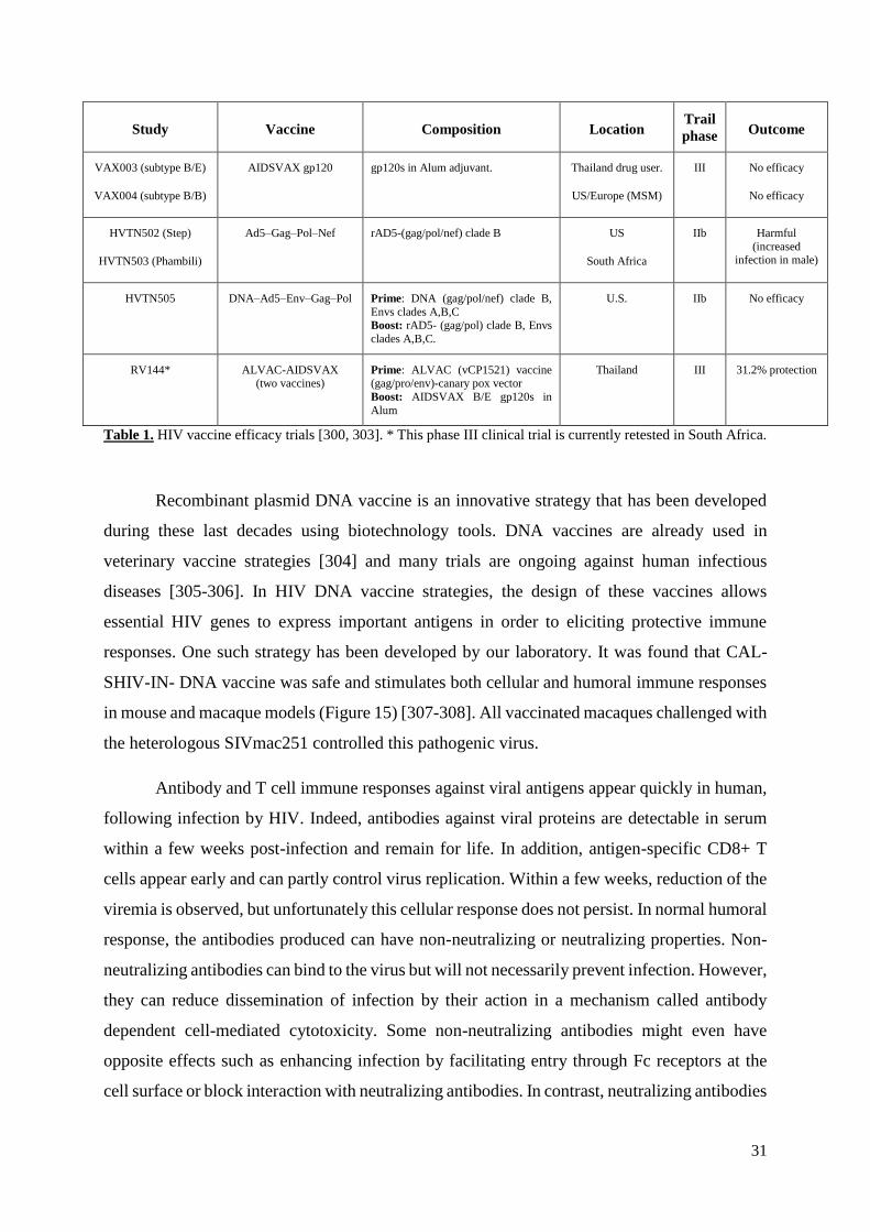
31
Study Vaccine Composition Location Trail
phase Outcome
VAX003 (subtype B/E)
VAX004 (subtype B/B)
AIDSVAX gp120
gp120s in Alum adjuvant.
Thailand drug user.
US/Europe (MSM)
III
No efficacy
No efficacy
HVTN502 (Step)
HVTN503 (Phambili)
Ad5–Gag–Pol–Nef
rAD5-(gag/pol/nef) clade B
US
South Africa
IIb
Harmful
(increased infection in male)
HVTN505
DNA–Ad5–Env–Gag–Pol
Prime: DNA (gag/pol/nef) clade B,
Envs clades A,B,C Boost: rAD5- (gag/pol) clade B, Envs
clades A,B,C.
U.S.
IIb
No efficacy
RV144*
ALVAC-AIDSVAX (two vaccines)
Prime: ALVAC (vCP1521) vaccine (gag/pro/env)-canary pox vector
Boost: AIDSVAX B/E gp120s in
Alum
Thailand
III
31.2% protection
Table 1. HIV vaccine efficacy trials [300, 303]. * This phase III clinical trial is currently retested in South Africa.
Recombinant plasmid DNA vaccine is an innovative strategy that has been developed
during these last decades using biotechnology tools. DNA vaccines are already used in
veterinary vaccine strategies [304] and many trials are ongoing against human infectious
diseases [305-306]. In HIV DNA vaccine strategies, the design of these vaccines allows
essential HIV genes to express important antigens in order to eliciting protective immune
responses. One such strategy has been developed by our laboratory. It was found that CAL-
SHIV-IN- DNA vaccine was safe and stimulates both cellular and humoral immune responses
in mouse and macaque models (Figure 15) [307-308]. All vaccinated macaques challenged with
the heterologous SIVmac251 controlled this pathogenic virus.
Antibody and T cell immune responses against viral antigens appear quickly in human,
following infection by HIV. Indeed, antibodies against viral proteins are detectable in serum
within a few weeks post-infection and remain for life. In addition, antigen-specific CD8+ T
cells appear early and can partly control virus replication. Within a few weeks, reduction of the
viremia is observed, but unfortunately this cellular response does not persist. In normal humoral
response, the antibodies produced can have non-neutralizing or neutralizing properties. Non-
neutralizing antibodies can bind to the virus but will not necessarily prevent infection. However,
they can reduce dissemination of infection by their action in a mechanism called antibody
dependent cell-mediated cytotoxicity. Some non-neutralizing antibodies might even have
opposite effects such as enhancing infection by facilitating entry through Fc receptors at the
cell surface or block interaction with neutralizing antibodies. In contrast, neutralizing antibodies
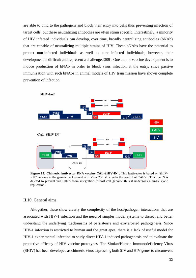
32
are able to bind to the pathogens and block their entry into cells thus preventing infection of
target cells, but these neutralizing antibodies are often strain specific. Interestingly, a minority
of HIV infected individuals can develop, over time, broadly neutralizing antibodies (bNAb)
that are capable of neutralizing multiple strains of HIV. These bNAbs have the potential to
protect non-infected individuals as well as cure infected individuals; however, their
development is difficult and represent a challenge.[309]. One aim of vaccine development is to
induce production of bNAb in order to block virus infection at the entry, since passive
immunization with such bNAbs in animal models of HIV transmission have shown complete
prevention of infection.
Figure 15. Chimeric lentivector DNA vaccine CAL-SHIV-IN¯. This lentivector is based on SHIV-
KU2 genome in the genetic background of SIVmac239. it is under the control of CAEV LTRs. the IN is
deleted to prevent viral DNA from integration in host cell genome thus it undergoes a single cycle
replication.
II.10. General aims
Altogether, these show clearly the complexity of the host/pathogen interactions that are
associated with HIV-1 infection and the need of simpler model systems to dissect and better
understand the underlying mechanisms of persistence and exacerbated pathogenesis. Since
HIV-1 infection is restricted to human and the great apes, there is a lack of useful model for
HIV-1 experimental infection to study direct HIV-1 induced pathogenesis and to evaluate the
protective efficacy of HIV vaccine prototypes. The Simian/Human Immunodeficiency Virus
(SHIV) has been developed as chimeric virus expressing both SIV and HIV genes to circumvent

33
the restrictions of HIV-1 in macaque cells, thereby creating a new model for HIV pathogenesis
and vaccines [310-311]. The first SHIV genomes that have been developed were bearing the
HIV-1 envelope of CXCR-4 tropic isolates into the backbone of SIVmac [312-313]. These
chimeric genomes have provided the proof of the principle that these SHIVs do productively
and persistently infect a variety of macaques [314]. However, since the virus genomes were
bearing CXCR4 tropic envelopes from isolates that emerge at late stages of HIV infection
associated with AIDS replicate preferentially in activated CD4+ T cells in the periphery, these
SHIVs were found to induce rapid induction of late stage pathogenesis in macaques [315].
Indeed, infection of macaque with pathogenic CXCR4-tropic SHIV induces a rapid (2-3 weeks)
loss of all CD4+ T cells. This helped to elucidate many of the mechanisms involved in HIV-1-
induced depletion of CD4+ T cells both in the periphery and in tissues [316]. It helped also to
test the efficacy of some vaccine prototypes [314]. However, these genomes failed to help
dissect the early mechanisms of host pathogen interaction that are known to be initiated by R5-
tropic viruses or to test the efficacy of vaccine prototypes against the acquisition/transmission
of these viruses [317-318]. Moreover these CXCR4 SHIVs were found to be easy to neutralize
and do not mimic the mucosal bottleneck selection associated with the transmitter/founders.
Many efforts were then made and are ongoing to generate R5-tropic chimeric viruses that
efficiently initiate the early stages of HIV-1 infection to dissect the early mechanisms and to
evaluate the efficacy of vaccines prototypes to induce protective immunity that blocks the virus
entry into target cells [319]. Although there are limited numbers of CCR5-tropic SHIVs that
have been developed, most of these viruses are of the clade B that is not the most abundant and
not bearing envelopes of transmitter/founders [320] with the exception of one [321].
On another hand, since HIV-1 has developed many strategies to undergo latency in a
variety of target cell types in which expression of virus genome is blocked by both virus and
cellular factors and mechanisms, these studies are very difficult to conduct with HIV-1.
Therefore, there is a need of chimeric viruses that are unable to undergo latency or at least that
use less complex mechanisms, for better understanding and developing more efficient drugs
that will cure HIV-1 infection.
In this context, our lab has developed novel chimeric lentivirus genomes using properties
from non-primate animal lentiviruses. Indeed, earlier studies from members of our lab have
established that small ruminant lentivirus (SRLVs) genomes are driven by constitutive LTR
promoters that are independent from Tat-transactivation [62, 322]. Furthermore, a gene of
SRLVs first reported as tat was found to be a Vpr-like both at structural and functional levels
[63]. Since these viruses were never associated with induction of immunodeficiency in their
34
hosts, even when they jump species in wildlife ruminant or in experimentally cross species
infected animals, they were defined as naturally attenuated lentiviruses [322-323]. Members of
our lab hypothesized that primate lentivirus genomes under transcriptional control of SRLV
constitutive LTR promoters will cause infection, replication, latency, persistence and
pathogenic properties different from the parental SIV. These properties will facilitate the studies
of primate lentiviruses and the development of innovative vaccines against HIV-1. In this
context, our lab has developed chimeric lentivector DNA vaccines under control of CAEV
LTRs and has shown that they are immunogenic both in mouse and macaque models of HIV
vaccines [307-308].
II.10.1. Specific Aim of the PhD
The main aim of this PhD project is to use the tools developed in our lab for studying
the host HIV-1 interactions leading to the complex pathogenesis. In addition, we wanted to
examine the humoral immune responses induced in macaques that have been vaccinated with a
lentivector DNA vaccine and challenged with a heterologous pathogenic SIVmac.
35
III. Material and methods
III.1. Amplification of HIV-1 plasmid DNAs
This work was done mainly on fourteen HIV molecular clones received from the NIH
AIDS Reagent Program Repository. These plasmids contain the complete or partial proviral
genome of different isolates of HIV-1 with different properties. They have been isolated from
different patients in various parts of the world. These infectious molecular clones are: pLAI.2,
pLconsnefSN, p89.6, pNef-ER, pWCML249, pcDNA3.1SF2NefF195R, pZM249M,
pSTCOr1, pWARO, pRGH-WT, pRGH-IntegraseD116A, pRGH-CMV-ΔCMV, pRGH-CMV-
ΔU3, and pNL4-3. Each clone was transformed in E.coli JM109 competent bacteria as follows.
In a 1.5 ml Eppendorf tube 45 µl of E.coli competent bacteria (JM109, >108 CFU/µg of uncut
plasmid DNA, Promega) were used for transformation with 5 µl (containing one ng) of plasmid
DNA. After incubation on ice for 30 min, a heat shock was performed by incubating the
bacterial suspension at 42°C for 90 sec. Following addition of 800 µl antibiotic-free LB, the
sample was put in a shaker incubator at 32°C for 1.5-2 h then centrifuged 3 min /5000 rpm. Part
of the supernatant (600 µl) was removed and the pellet resuspended. Volumes of 150 µl and 50
µl were plated on separate Luria broth (LB) agar plates containing suitable antibiotic and
incubated at 32°C for 24 h. Colonies were picked, transferred to 15 ml tubes that contained 5
ml LB with suitable antibiotic and incubated in a shaker incubator overnight at 32°C.
III.2. Purification and control of plasmid DNA
The pDNA from three clones for each of the fourteen molecular clones was purified by
mini prep using NucleoSpin® Plasmid EasyPure Kit (MACHEREY-NAGEL) according to the
manufacturer instructions. Briefly, bacteria from 1.5 ml of overnight culture with the adequate
antibiotic were collected by centrifugation (11000 rpm for 30 sec) and resuspended into 150 µl
of the resuspension buffer A1 supplemented with RNase. Cell suspension were lysed by
addition of 250 µl of lysis buffer A2, the tubes inverted five times and then incubated for 5 min
at RT. This lysis/denaturation step was stopped by adding 350 µl of the neutralization buffer
A3 and mixing thoroughly by inverting until the lysate turned “milky” colorless. The samples
were cleared by centrifugation at 11000 rpm for 3-5 min. The DNA containing supernatant was
loaded onto a NucleoSpin® Plasmid EasyPure column and centrifuged at 1000-2000 rpm for
30 sec. After washing with 450 µl 70% ethanol solution corresponding to wash buffer AQ, the
DNA was eluted with 50 µl of elution buffer AE, by centrifugation at 11000 rpm for 1 min.

36
Each sample of DNA purified by mini prep was subjected to two selected digestions
with restriction enzymes (BamHI and EcoRI) for characterization. Three samples (15 µl) were
used for each purified DNA: one is used as uncut control while two were digested with suitable
enzymes as follows. In an Eppendorf tube containing the DNA, we added 2 µl of 10x digestion
buffer (different for the different enzymes), H2O for a final volume of 19µl (2 µl) and 1 µl
restriction enzyme. After incubation for one hour at 370C (temperature differs for different
enzymes), 4 µl of loading buffer (8 ml Tris-Acetate-EDTA buffer (TAE), 1 ml glycerol and a
2 ml of bromophenol blue solution) are added and the total volume is loaded on 1% agarose gel
in parallel with 1kb ladder marker DNA. The electrophoresis was performed at 100 volt for 30-
60 min and bands were visualized following staining with ethidium bromide. To choose the
bacteria that harbor the right clones, the band pattern obtained were compared with the
theoretical maps provided by the NIH AIDS Reagent Program.
III.3. Large-scale purification of plasmid DNA
The chosen clones were grown overnight at 30-32°C in a shaker incubator in flasks
containing 500-1000 ml of LB medium supplemented with the antibiotic. Bacterial cultures
were harvested by centrifugation at 4500-6000 rpm for 15 min at 40C. The cell pellets were
resuspended in 8-16 ml (for midi prep) or 12-24 ml (for maxi prep) of resuspension buffer that
contains RNase. Bacteria were lysed following addition of 8-16 ml (for midi) or 12-24 ml (for
maxi) of lysing buffer, homogenized gently and incubated for 5 min at RT. Neutralization buffer
was added (8-16 ml for midi or 12-24 ml for maxi), inverted three times and incubated on ice
for 5 min, then centrifuged at 12000 rpm for 10 min at 4oC. The supernatants were filtrated
gently on the NucleoBond® Xtra column filters previously equilibrated using buffer (15 ml for
midi, 35 ml for maxi). After the NucleoBond®Xtra cotton pre-filter was washed with 5 ml (for
midi) or 10 ml (for maxi) of filtration buffer, the pre-filter was discarded and 35 ml (for midi)
or 90 ml (for maxi) of Endo wash buffer were added. The DNA was eluted with 5 ml (for midi)
or 15 ml (for maxi) of elution buffer, precipitated with isopropyl alcohol (3.5 ml for midi, 10.5
ml for maxi) and collected by centrifugation at 15000 rpm for 30 min at RT. The DNA pellet
was washed with 70% ethanol (2 ml for midi, 5 ml for maxi), centrifuged at 15000 for 10 min
at RT and dried for 20-30 min. the DNA was solubilized in 200-800 µl (for midi) or 400-1000
µl (for maxi) of sterile H2O. The DNA concentration was determined by spectrophotometry at
260 nm (see Table 3). The integrity and quality of each purified DNA by midi prep was
evaluated using three selected endonuclease digestions with restriction enzymes (BamHI,

37
EcoRI and PstI) and non-digested and digested samples of DNAs were separated in 1% agarose
gel.
III.4. Cell lines
Many cell lines were used in the study some of them are adherent others are non-
adherent as shown in the Table below.
Cell line Culture
medium
Type
Description
HEK-293T
DMEM
Adherent
Human Embryonic Kidney cell line
TZM-bl
DMEM
Adherent HeLa cell line with luciferase and ß-
galactosidase genes under the
control of HIV-1 promoter. Indicator
cells to visualize virus infection in
neutralization assays
GHOST CCR5
DMEM
Adherent Derived from human osteosarcoma
cells, express CD4 and relatively
high levels of CCR5
GHOST CXCR4
DMEM
Adherent
Derived from human osteosarcoma
cells, express CD4 and CXCR4
M8166
RPMI 1640
Suspension Highly susceptible and fusogenic
human CD4+ T lymphocytic cells,
express CXCR4
CEMx174
RPMI 1640
Suspension Fusion product of human B cell line
and human CD4+ T cell line CEM
U937
RPMI 1640
Suspension/
adherent
CD4+ promonocytic human cell line
derived from an HIV-free
individual. Cells can be
differentiated into adherent
macrophages in culture
TIGEF
DMEM
Adherent T-immortalized goat embryo
fibroblast cell line
Table 2. List of cell lines used in this work, originating from the NIH AIDS Research and Reference Reagent
Program, Division of AIDS, NIAID, NIH.
III.5. Culture media
Fetal Bovine Serum (FBS) (Eurobio AbCys or Gibco®) was heat inactivated prior to use
in cell growth medium. The bottle (500 ml) of FBS was transferred from -20°C freezer into 4°C

38
refrigerator to thaw overnight. After mixing by inversion, the serum was incubated for 30 min
in a water bath at 56°C to inactivate heat-labile complement proteins, then cooled for 30 min at
RT and stored at 4°C or frozen at -20°C. Dulbecco's Modified Eagle Medium (DMEM)
containing 1.0 g/L glucose (Eurobio AbCys) was supplemented with 10% FBS, 1%
Penicillin/Streptomycin (Eurobio AbCys), 1% L-Glutamine 200 mM (Eurobio AbCys). Minimum
Essential Media (MEM) (Eurobio AbCys) was supplemented with 10% FBS, 1%
Penicillin/Streptomycin and 1% L-Glutamine. Roswell Park Memorial Institute (RPMI 1640)
medium (GibcoTM) was supplemented with 10% FBS.
III.6. Thawing and freezing of cell lines
The cryotubes (TM vial Nunc®) of frozen cells were transferred on ice from the liquid
nitrogen container and then partially thawed by incubation into a 37°C water bath. After partial
thawing the cell suspension, the outside of the vial was disinfected with 70% ethanol. Under a
biological hood, the cell suspension was transferred into a 15 ml conical sterile centrifuge tube
and the appropriate media was added drop wise into the cell suspension to dilute the freezing
medium at least 5 times. After centrifugation at 1500 RPM for 5-10 minutes, the pellet was re-
suspended into 5 ml complete culture medium and transferred into 25 cm2 T flask and incubated
for growth in a tissue culture incubator. For cryopreservation the cell suspension is enumerated
and then centrifuged to remove the culture medium. The pellet of cells is re-suspended in FBS
or freezing medium at the indicated concentration, transferred into 1-2 ml cryotubes and
supplemented drop wise with 10% DMSO on ice. Cells were first placed in a -80°C for 24-48h
in a freezing box prior to transfer into liquid nitrogen storage. The freezing protocol followed
directions from the NIH AIDS Reagent Program.
III.7. Cell maintenance
Cell lines were grown using the appropriate culture medium in an incubator at 37°C in
5% CO2. For adherent cell lines, passage was achieved by discarding the old media and washing
the cells with PBS+2 mM EDTA, to remove any residual FBS media that block the trypsin
action. Then cells were detached by treatment with Trypsin 0.025%/2 mM EDTA solution, such
as 1 ml is used to cover the surface of a 25 cm2 T flask, removed before cells start detaching
and then the flask was incubated for 3-5 min at 37°C. After observation of cell detachment
under the microscope, the flask was shaken to complete the detachment if necessary. Fresh
culture medium with FBS was added to the cells and the resuspension was completed by
39
pipetting. Cells were centrifuged 1500 rpm/5 min, the pellet was re-suspended with suitable
medium and cells were cultured at the required density depending on the flask size for example
106 cells in a 25 cm2 T flask containing 5 ml of medium. For non-adherent cell lines, passage
was performed by centrifuging the cells at 1500 rpm/5 min at RT. Then after discarding the old
media, the cell pellet was re-suspended with complete suitable growth medium and seeded at
the required density.
III.8. Transfection of HEK-293T cell line for production of viral stock
Transfection or delivery of plasmid DNA into eukaryotic HEK-293T cells was
performed according to the manufacturer protocol using the cationic polymer polyethylene
amine, ExGenTM500 (Euromedex, France). Briefly, 106 cells/well were seeded into 6-well
plates in DMEM and incubated overnight in a tissue culture incubator. The next day, when the
cell monolayer reaches approximately 80% confluency (i.e. the cells cover about 80% of the
surface of the well) the medium was changed with 3 ml of fresh DMEM. The transfection
mixture was prepared in a polystyrene tube, by mixing 350 µl 150 mM NaCl with 5 µg plasmid
DNA and 16.5 µl of ExGeneTM500 reagent. The mixture was incubated for 35 min at RT under
gentle manual mixing from time to time and the transfection mixture was added drop wise into
the 3 ml medium covering the HEK-293T cells and incubated. At 18-24 h post transfection, the
medium was removed, the cell monolayer rinsed once with PBS and then cells were incubated
in fresh 2 ml medium. The supernatant fluids were collected daily, for three successive days,
filtrated (0.45) μm and titrated on M8166 and/or TZM-bl cells, and stored at -80°C for further
analyses.
III.9. Viral stock titration on M8166
M8166 cells, a highly fusogenic permissive human CD4+ T cell line, were used as
indicator cells because they undergo development of typical giant multinucleated cells as
cytopathic effect (CPE) upon infection. In order to quantify the amount of the virus causing
CPE in 50% of inoculated cells (TCID50/ml), 1x10⁵ M8166 cells/well were seeded in a 24-well
plate containing 400 µl/well of RPMI. Viral dilutions were prepared as follows: Six tenfold
dilutions of the viral stocks were inoculated on M8166 in quadruplicate. The plate was
incubated in a humidified incubator at 37°C with 5% CO2. After three days of incubation, the
observation of development of cytopathic effect was started. If the fifth row have two well of

40
positive cytopathic effects (CPE), that reads as 105 TCID50/ml and each one µl of the viral stock
have a 102 TCID50.
III.10. Viral stock titration on TZM-bl cell line
The TZM-bl cell line was used as indicator to evaluate the titers of virus stock since
infection of these cells enables the expression of Lac Z gene, producing β-galactosidase that
generates blue cells in the presence of X-gal. The number of blue cells corresponds to the
number of infectious viral particles capable of forming blue cell per unit volume called blue
cell forming unit (BCFU). In order to achieve this, TZM-bl cells were seeded in 24-well plates
at about 1x10⁵ cells/well in 500 µl of DMEM and grown overnight in an incubator. Next day
the medium was changed and the monolayers were inoculated with virus in serial dilutions.
After three to four days of incubation, monolayers of cells were stained in the plate with X-gal
and observed for BCFU. Briefly, after washing the cell monolayer twice with PBS containing
Ca and Mg, cells were fixed with 4% paraformaldehyde (PFA) for 5-10 min at 4°C, rinsed twice
with PBS supplemented with 5 mM MgCl2 and incubated for 10 minutes in this solution.
Finally, 500 µl of X-gal (1mg/ml, in DMSO eurobio AbCys) were added to each well and the
plate incubated for 1 hour at 37°C in a dry incubator. The number of infected cells, which
appeared blue in each well were counted under the microscope. The number of BCFU/ml were
calculated according to the dilution factors to determine the infectious titers. Graphic
presentation of the data was done using Microsoft Excel.
III.11. Determination of viral tropism
GHOST CXCR4 and GHOST CCR5 cell lines were used to determine the tropism of
the virus isolates [324]. GHOST cells (1x10⁶ cells/well) were seeded in a 6-well plate
containing 2 ml/well of DMEM and incubated overnight. The virus stocks corresponding to the
supernatants of transfected HEK-293T with the plasmid DNA that were stored at -80°C were
used to inoculate GHOST cell monolayers and incubated at least three days until the
development of CPE and enabling of GFP expression were observed. The tropism is determined
by the ability to infect cells with a specific co-receptor. In the case of pNL 4-3, pWARO and
p89.6, the titer of the viruses was 10000 BCFU/ml, 2800 BCFU/ml and 12700 BCFU/ml
respectively.
41
III.12. Kinetics of HIV-1 replication in permissive T cell lines
In order to study the kinetics of replication of pNL 4-3 and p89.6 viruses, M8166 and
CEMx174 cell lines were inoculated at MOI 0.1 (1 virus /10 cells), thus 100µl of 106 TCID50/ml
contain 100.000 viral particles used to infect 106 cells in 1 ml of appropriate media. Then every
day supernatant fluids were collected for eight successive days and kept at -80°C. Viruses in
these supernatant fluids were titrated on TZM-bl for each time point. This experiment was
performed in triplicate. Graphic presentation of the data was done using Microsoft Excel.
The viruses used in these studies were: SHIV-KU2 (GenBank data base accession #
AY751799), SIVmac239 (# AY587015.1), NL4-3 (# AF324493), 89.6 (# U39362), WARO (5'
half-genome sequences: KC312330–KC312366 / 3' half-genome sequences: KC312367–
KC312398), STCOr1 (5' half-genome sequences: KC312399–KC312433 / 3' half-genome
sequences: KC312434–KC312466), WCML249 (# AY445524), LAI.2.
III.13. Sero-neutralization using TZM-bl X-gal assay
To investigate the presence of the neutralizing antibodies in the serum of macaques that
were vaccinated with the lentivector DNA vaccine and challenged with the pathogenic strain of
SIVmac251, we used the TZM-bl X-gal assay. The analysis include six vaccinated and six
control animals. The serum samples were already collected at a weekly time schedule and stored
frozen. The sero-neutralization experiments were performed using the chimeric SHIV-KU2 with
the envelope of HIV-1 and SIVmac239. Two dilutions (1/5 and 1/20) of serum samples were
used in duplicate to assess the neutralizing activity of each serum. Suspensions of SHIV-KU2
and SIVmac239 for neutralization with macaques serum and SHIV-KU2 only for mouse sera,
were prepared separately by mixing 50 µl of virus suspension with 50 µl of dilutions of (1/5
and 1/20) serum samples in DMEM free of FBS. After incubation for 1 h at 4°C, the mixtures
were used to inoculate the TZM-bl cells in duplicate wells and incubated for 72 to 96 h in the
humidified incubator at 37°C with 5% CO2. The plates were stained with X-gal as described
above prior to observation under photonic microscope and scoring of BCFU. Graphic
presentation of the data was done using Microsoft Excel.
III.14. Sero-neutralization using ONE-Glo™ Luciferase assay
The ONE-Glo™ Luciferase Assay System provides a highly sensitive, robust,
homogeneous assay for detection of firefly luciferase reporter gene expression in infected
42
mammalian cells. The procedure was identical to the sero-neutralization, using TZM-bl X-Gal
assay, but without X-Gal staining. Instead, after removing the medium from the cell
monolayers, cells were rinsed with PBS, lysed with 100 µl of 1x Glo lysis buffer (Promega) at
RT for five minutes with gentle shaking. Samples were transferred (50 µl/sample) into a 96
wells plate (plate 96 W White solid F-Bot N-bind N) together with 50 µl/well of 1x ONE-Glo™
Luciferase Assay substrate (Promega). After incubating for five min at RT, the luciferase activity
was measured for 2 seconds using a luminometer (Berthold Technologies Centro LB 960). All
values are reported as relative luminescence units.
III.15. CAL-HIV-R1 plasmid preparation
This chimeric plasmid is based on the genome of HIV-1 pNL4-3 infectious molecular
clone where the two LTRs were replaced with those of CAEV that have a constitutive, tat-
independent promoter. Using chimeric primers with HIV-1 and CAEV sequences, we amplified
the chimeric LTR of CAEV having the extremities of HIV, PCR product was inserted in
pGEMT easy to generate pHICA clone 4. Briefly, the development of CAL-HIV-R1 required
five steps. The first step consisted in opening the pHICA clone 4. This plasmid was digested
with HindII at the unique site in order to linearize the plasmid. For the second step, to isolate
the CAEV LTR from the plasmid, another pHICA clone 4 was also double digested with EcoRI
and NarI and the overhanging extremities were filled to blunt end with large fragment klenow
polymerase. In the third step, the resulting fragment, which contains the CAEV LTR, was
inserted into HindII site of linear pHICA clone 4 to generate pHICA-2LTR a plasmid containing
two CAEV LTRs. In the fourth step, the LTRs from the HIV-1 pNL4-3 were removed by two
consecutive digestions with BlpI and Nar1 that are located upstream the 3’LTR and downstream
the 5’LTR. Finally, the plasmid prepared in step three that contains the two LTRs from CAEV
and the plasmid prepared in step four containing pNL4-3 without its LTRs were ligated (Figure
16).
The ligated products were transformed into JM109 competent bacteria and selected
colonies were analyzed by restriction enzyme digestion using EcoRI, HindIII, and PstI. The
chosen clones that have the right digestion profiles were amplified in large scale and DNAs
were isolated by midi-prep. Purified DNA (5 µg each) was transfected in HEK-293T cells and
the filtrated supernatants containing virus were stored in -80°C as viral stocks. Aliquots (100
µl) were used to inoculate the permissive M8166 and CEMx174 cell cultures. Furthermore,
these cell lines were used for viral adaptation that was performed by co-culturing with

43
transfected HEK-293T cells until appearance of CPE, i.e. fusion between M8166 indicator cells
with transfected HEK-293T cells. Then the non-adherent M8166 and CEMx174 cells, were
harvested, transferred to six-well plates and fresh cells were added. After CPE was clearly
observed (three days), 1 ml of supernatant was kept at -80°C and fresh cells were added to the
remaining cell suspension. This passage was performed 17 times. All collected supernatants
kept at -80°C were titrated on TZM-bl, while pellets of cells used to extract the provirus
genomic DNA. The presence of CAEV LTRs in these clones was confirmed by PCR on 1-2 µg
of genomic DNA.
Figure 16. Organization of CAL-HIV-R1 pDNA. This plasmid contains all the genes of HIV-1 pNL4-3, but
the LTRs were replaced by those of CAEV. The LTRs of CAEV have strong constitutive promoters. About 30
nucleotides of the termini of HIV LTRs were introduced in each side of CAEV LTRs. These short nucleotide
sequences form the attachment site recognized by the HIV integrase to promote HIV-1 integration.
44
III.16. Data interpretation
Statistical analysis of the acquired data (mean and standard deviation) was performed
using GraphPad Prism 5.0 software
45
IV. Results and Discussion
IV.1. Tropism and infectivity
IV.1.1. Plasmids
The aim of this part of the work was to prepare appropriate reagents and tools with a
view to study the infectivity, the replication competence and the tropism properties of various
HIV-1 strains in comparison with our chimeric CAL-HIV-R1 genome. For this, we ordered and
received 14 clones from the NIH AIDS Reagent Program Repository as presented below and
after a first analysis, the work concentrated on the three virus pNL4-3, p89.6, and WARO.
Firstly, each one of the recombinant plasmid containing the viruses genomes of HIV-1 received
from NIH was introduced into JM109 E. coli by transformation and then triplicates of each
recombinant plasmid were screened by mini prep and for each plasmid, one clone was chosen
and amplified as described in Materials and Methods. DNA extracted (midi prep) and
quantification, three restriction enzymes (BamHI, EcoRI and PstI) were used for
characterization. A brief description of the clones and the concentration of the DNA obtained
is presented in Table 3.
All clones gave the expected bands after restriction enzyme digestions according to the
theoretical maps. We chose to amplify six specific clones for further work: pNL4-3, pLAI.2,
p89.6, pSTCOr1, pWCML249 and pWARO.
Analyses of infectivity and replication properties of selected molecular clones: The
culture supernatant fluids of transfected HEK-293T cells with the six plasmid DNAs were
analyzed by serial infection of M8166 cells and examination of virus replication. Cells infected
with pNL4-3, pLAI.2, and p89.6 viruses developed clear CPE in each of the four rounds of
infection, while pSTCOr1 produced only a weak CPE. In contrast, cells infected with
pWCML249 and WARO showed no CPE on these indicator M8166 CD4+ T cells. Typical
CPE appear as large cells with enlarged cytoplasm compartment housing numerous nuclei that
can easily be detected by microscopy. CPE is an indication of lentivirus infection and correlates
with the presence of virus production by infected cells, followed by cell depletion.

46
Ref.
Name
Properties
DNA
µg/µl
1.
2532
pLAI.2 Full-length replication competent,
infectious, subtype B
0.74
2.
114
pNL 4-3 Full-length, replication and infection
competent chimeric DNA laboratory
adapted, infectious to a variety of cells as
well as human T4 cells
3.8
3.
3297
pLconsnefSN. Full-length consensus nef gene derived
from 54 patient isolates
0.58
4.
3552
p89.6 Infectious molecular clone, highly
cytopathic
0.58
5.
6454
pNef-ER Full-length nef of NL4-3 and the estrogen
receptor hormone-binding domain fused
at the C terminus of nef
2.08
6.
10776
pWCML249 Infectious clone, subtype D/C, does not
form syncytia in vitro
4,5
7.
11430
pcDNA3.1SF2NefF195R Full-length nef gene, subtype B
1.68
8.
12416
pZM249M Full-length replication competent,
infectious, subtype C
2.5
9.
12417
pSTCOr1 Full-length replication competent,
infectious, subtype B
0.78
10.
12419
pWARO Full-length replication competent,
infectious, subtype B
0.46
11.
12427
pRGH-WT pLAI.2 based vector with a CMV-driven
mCherry and a LTR-driven gag-iGFP
markers, ΔEnv
0.64
12.
12428
pRGH-IntegraseD116A pRGH-WT with inactive IN through
D116A mutation
0.5
13.
12429
pRGH-CMV-ΔCMV pRGH-WT, CMV promoter removed
1.34
14.
12430
pRGH-CMV-ΔU3 pRGH-WT, part of the U3 region (3’
LTR) deleted, single cycle of replication
1.12
Table 3. Plasmids from the NIH AIDS Reagent Program Repository. All clones are Ampicillin resistant with the
exception of clone #8 (pZM249M) which is Kanamycin resistant.
M8166 cells, which are a human T-lymphoblastoid cell line, are able to amplify HIV-1 at
a good titer and have a high susceptibility to CPE syncytia formation [325]. The time needed
to detect the presence of CPE will vary depending on the type of virus and host cell. Generally,
M8166 cells infected with HIV-1 need three to six days to develop detectable CPE. In contrast,
cells infected with herpes simplex virus show CPE after about 24 hours, while this phenotype
is only visible after ten to thirty days in cells infected with cytomegalovirus [326].
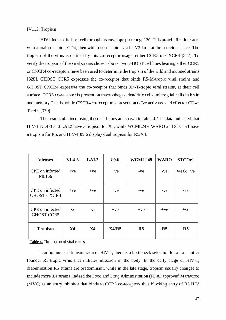
47
IV.1.2. Tropism
HIV binds to the host cell through its envelope protein gp120. This protein first interacts
with a main receptor, CD4, then with a co-receptor via its V3 loop at the protein surface. The
tropism of the virus is defined by this co-receptor usage, either CCR5 or CXCR4 [327]. To
verify the tropism of the viral strains chosen above, two GHOST cell lines bearing either CCR5
or CXCR4 co-receptors have been used to determine the tropism of the wild and mutated strains
[328]. GHOST CCR5 expresses the co-receptor that binds R5-M-tropic viral strains and
GHOST CXCR4 expresses the co-receptor that binds X4-T-tropic viral strains, at their cell
surface. CCR5 co-receptor is present on macrophages, dendritic cells, microglial cells in brain
and memory T cells, while CXCR4 co-receptor is present on naïve activated and effector CD4+
T cells [329].
The results obtained using these cell lines are shown in table 4. The data indicated that
HIV-1 NL4-3 and LAI.2 have a tropism for X4, while WCML249, WARO and STCOr1 have
a tropism for R5, and HIV-1 89.6 display dual tropism for R5/X4.
Viruses
NL4-3
LAI.2
89.6
WCML249
WARO
STCOr1
CPE on infected
M8166
+ve
+ve
+ve
-ve
-ve
weak +ve
CPE on infected
GHOST CXCR4
+ve
+ve
+ve
-ve
-ve
-ve
CPE on infected
GHOST CCR5
-ve
-ve
+ve
+ve
+ve
+ve
Tropism
X4
X4
X4/R5
R5
R5
R5
Table 4. The tropism of viral clones.
During mucosal transmission of HIV-1, there is a bottleneck selection for a transmitter
founder R5-tropic virus that initiates infection in the body. In the early stage of HIV-1,
dissemination R5 strains are predominant, while in the late stage, tropism usually changes to
include more X4 strains. Indeed the Food and Drug Administration (FDA) approved Maraviroc
(MVC) as an entry inhibitor that binds to CCR5 co-receptors thus blocking entry of R5 HIV

48
strains by antagonism for the co-receptor. However, it was found that MVC was not able to
block entry of HIV strains with X4-tropic or R5/X4 envelopes. So nowadays it is important to
use a combination of drugs to block entry of other tropic strains [330].
Based on these results, three infectious molecular clones were selected to continue the
study. Two molecular clones were mono-tropic for either CXCR4 (pNL4-3) or CCR5
(pWARO) and the third one was dual-tropic for both CXCR4 and CCR5 (p89.6), as shown in
Figure 17.
Figure 17. Infectivity assay of selected HIV-1 strains on GHOST cells using microscopy. Infectivity is
detected by CPE identified by formation of multinucleated syncytial giant cells. The NL4-3 strain shows CPE in
CXCR4 GHOST cells, but not in CCR5 GHOST cells; the 89.6 strain shows CPE in both CXCR4 and CCR5
GHOST cells; the WARO strain shows CPE only in CCR5 GHOST cells.
In order to maximize the treatment with ART it is best to assess the tropism of the
infecting HIV strain in the patient. This is possible using the commercial Trofile assay
(Monogram Biosciences), a phenotype and genotype test, but it is expensive, labor intensive
and time consuming [331]. Instead, we used the two GHOST/CD4 cell lines, expressing CCR5
or CXCR4 that were infected separately with the HIV-1 viral strains. Then, depending on the

49
phenotype changes observed in these cells after 5-8 days post-inoculation, HIV-1 strains were
classified into type R5, X4 or R5/X4 according to their ability to infect these indicator CD4+
cell lines. Another cell type could have been used such as described by Ceresola et al., where
U87/CD4 /CCR5 or U87/CD4 /CXCR4 cell lines were utilized as indicator cells [332].
IV.1.3. Infectivity
In order to evaluate the efficacy of the viruses to assemble into infectious particles,
HEK-293T cells were transfected with the three chosen clones and the supernatants were
collected each day for three days. The supernatants were titrated on TZM-bl cells. The results
shown in Figure 18 indicate that the two viruses pNL4-3 and p89.6 gave high titer, that
increased with time. These titers were comparable to values obtained for SHIV-ku2 (50000
BCFU/ml). In contrast, WARO gave low titer of about 1000 BCFU/ml.
Figure 18. TZM-bl titration of HIV-1 stocks produced at day 1, day 2 and day 3 post transfection in HEK-
293T cells.
Dilutions of virus stocks of NL-4-3, 89.6 and WARO produced in transfected HEK-
293T cells and harvested at days 1, 2 and 3 post transfection were used to inoculate duplicate
wells of TZM-bl monolayers in 24 wells plate. At 72 hours post-inoculation, cells were fixed
and X-gal stained as described in Materials and Methods. After 2-8 hours, X Gal solution was

50
removed and cells rinsed with PBS and blue cell forming units scored, calculated/ml and used
to plot the graph. Each virus stock was aliquoted, stored at -80°C [333-334].
Despite following the same protocol of transfection for all three strains, WARO always
showed the lowest level of viral production. In addition, the production was followed during
three days for all strains. As shown in Figure 17, HEK-293T production of NL4-3 and 89.6
increased during the three successive days, while WARO did not show any increase remaining
very low compared with the other two strains. To overcome this problem we tried to inoculate
the U937 cells that are known to express CCR5 co-receptors at high level on their surface. This
monocytic cell line can be differentiated into macrophage using suitable concentrations of
phorbol myristate acetate (PMA). Thus, the differentiated macrophage should replicate
productively HIV, but the titration of WARO was still very weak. Therefore, dealing with U937
cell line to produce high enough titration of WARO was not pursued. A potential alternative
would have been to use the MOLT T-lymphoblastoid cell line, because these cells express high
level of both the CD4 receptor and the CCR5 co-receptor on their surface. It has been reported
in the literature that when MOLT-4 cells are infected with R5 HIV-1 viral strains such as JR-
FL or Ba-L, CPE was observed in the culture supernatants, indicating that this cell line might
be better suited for R5 HIV-1 strain expression [335-336].
IV.1.4. Production of viral stocks
Because of the efficiency of M8166 cells, they were chosen to amplify and produce
large stocks of infectious virus particles. Cells were infected for four days then the supernatants
were removed and replaced with fresh medium that was also collected 24 h later. These
supernatants were titrated on TZM-bl and M8166 cells to determine the BCFU/ml and HIV-1
TCID50/ml respectively. The results obtained are shown in Table 5.
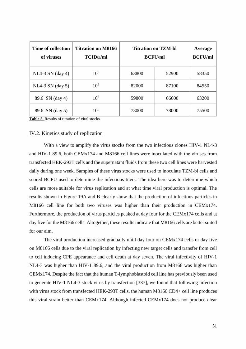
51
Time of collection
of viruses
Titration on M8166
TCID50/ml
Titration on TZM-bl
BCFU/ml
Average
BCFU/ml
NL4-3 SN (day 4)
105
63800
52900
58350
NL4-3 SN (day 5)
106
82000
87100
84550
89.6 SN (day 4)
105
59800
66600
63200
89.6 SN (day 5)
106
73000
78000
75500
Table 5. Results of titration of viral stocks.
IV.2. Kinetics study of replication
With a view to amplify the virus stocks from the two infectious clones HIV-1 NL4-3
and HIV-1 89.6, both CEMx174 and M8166 cell lines were inoculated with the viruses from
transfected HEK-293T cells and the supernatant fluids from these two cell lines were harvested
daily during one week. Samples of these virus stocks were used to inoculate TZM-bl cells and
scored BCFU used to determine the infectious titers. The idea here was to determine which
cells are more suitable for virus replication and at what time viral production is optimal. The
results shown in Figure 19A and B clearly show that the production of infectious particles in
M8166 cell line for both two viruses was higher than their production in CEMx174.
Furthermore, the production of virus particles peaked at day four for the CEMx174 cells and at
day five for the M8166 cells. Altogether, these results indicate that M8166 cells are better suited
for our aim.
The viral production increased gradually until day four on CEMx174 cells or day five
on M8166 cells due to the viral replication by infecting new target cells and transfer from cell
to cell inducing CPE appearance and cell death at day seven. The viral infectivity of HIV-1
NL4-3 was higher than HIV-1 89.6, and the viral production from M8166 was higher than
CEMx174. Despite the fact that the human T-lymphoblastoid cell line has previously been used
to generate HIV-1 NL4-3 stock virus by transfection [337], we found that following infection
with virus stock from transfected HEK-293T cells, the human M8166 CD4+ cell line produces
this viral strain better than CEMx174. Although infected CEMx174 does not produce clear
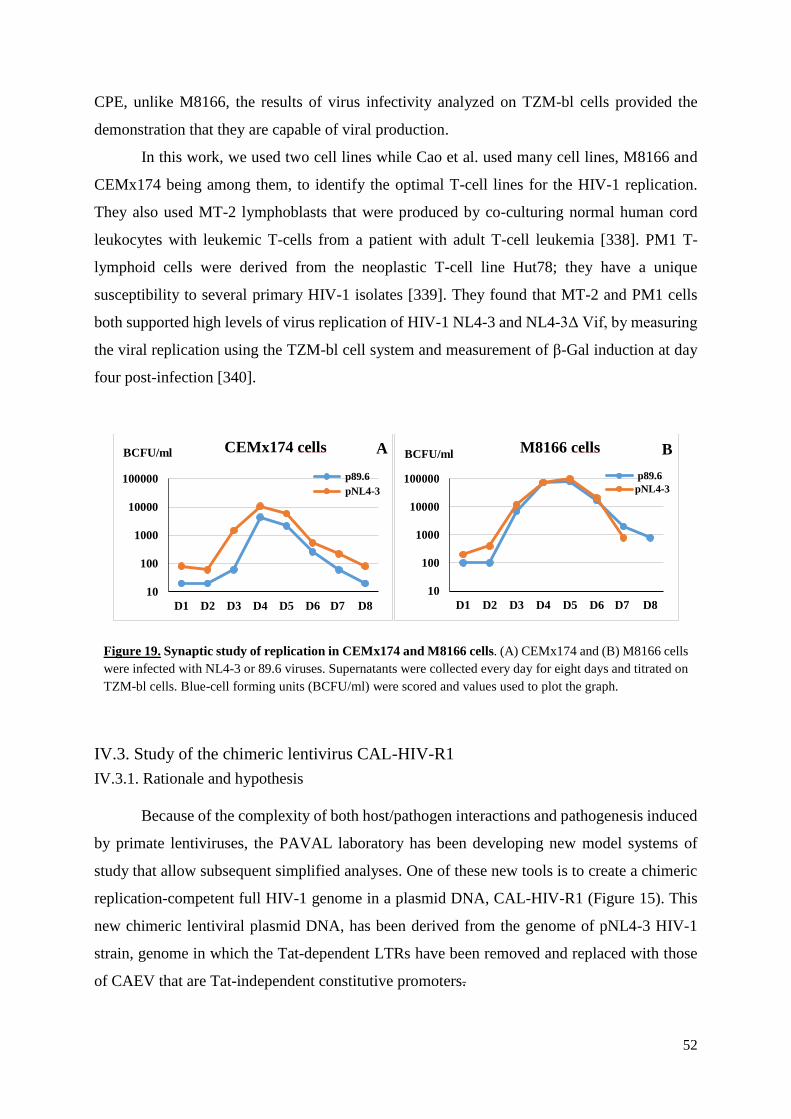
52
CPE, unlike M8166, the results of virus infectivity analyzed on TZM-bl cells provided the
demonstration that they are capable of viral production.
In this work, we used two cell lines while Cao et al. used many cell lines, M8166 and
CEMx174 being among them, to identify the optimal T-cell lines for the HIV-1 replication.
They also used MT-2 lymphoblasts that were produced by co-culturing normal human cord
leukocytes with leukemic T-cells from a patient with adult T-cell leukemia [338]. PM1 T-
lymphoid cells were derived from the neoplastic T-cell line Hut78; they have a unique
susceptibility to several primary HIV-1 isolates [339]. They found that MT-2 and PM1 cells
both supported high levels of virus replication of HIV-1 NL4-3 and NL4-3Δ Vif, by measuring
the viral replication using the TZM-bl cell system and measurement of β-Gal induction at day
four post-infection [340].
Figure 19. Synaptic study of replication in CEMx174 and M8166 cells. (A) CEMx174 and (B) M8166 cells
were infected with NL4-3 or 89.6 viruses. Supernatants were collected every day for eight days and titrated on
TZM-bl cells. Blue-cell forming units (BCFU/ml) were scored and values used to plot the graph.
IV.3. Study of the chimeric lentivirus CAL-HIV-R1
IV.3.1. Rationale and hypothesis
Because of the complexity of both host/pathogen interactions and pathogenesis induced
by primate lentiviruses, the PAVAL laboratory has been developing new model systems of
study that allow subsequent simplified analyses. One of these new tools is to create a chimeric
replication-competent full HIV-1 genome in a plasmid DNA, CAL-HIV-R1 (Figure 15). This
new chimeric lentiviral plasmid DNA, has been derived from the genome of pNL4-3 HIV-1
strain, genome in which the Tat-dependent LTRs have been removed and replaced with those
of CAEV that are Tat-independent constitutive promoters.
10
100
1000
10000
100000
D1 D2 D3 D4 D5 D6 D7 D8
CEMx174 cells
p89.6
pNL4-3
BCFU/ml A
10
100
1000
10000
100000
D1 D2 D3 D4 D5 D6 D7 D8
M8166 cells
p89.6
pNL4-3
BCFU/ml B
53
Furthermore, a few nucleotides (about 30) from the LTRs of HIV have been kept and
fused to CAEV LTRs in order to promote integration. These should allow the formation of the
HIV-1 specific attachment sequence recognized by the HIV integrase that catalyzes dsDNA
integration into cell genome. As a consequence, we should observe a constitutive expression of
the virus in all infected cells.
Indeed, HIV infects human CD4+ T cells and monocyte-macrophage cell lineages both
in cell culture and in infected individuals, eventually causing AIDS. Like all HIV strains, the
HIV-1 pNL4-3 strain has LTRs that are dependent on Tat transactivation. In contrast, the
caprine arthritis encephalitis virus, CAEV, a natural lentivirus pathogen of goats, has an in vivo
tropism restricted to the monocyte/macrophage cell lineage. CAEV does not induce
immunodeficiency in his host but leads to arthritis and mastitis in infected adults, and under
some circumstances encephalitis in infected kids. The LTRs of CAEV have been shown to be
independent from Tat transactivation since they have strong constitutive promoters.
Furthermore, since the new CAL-HIV-R1 construct is derived from parental pNL4-3 HIV-1
strain, thus we expect tropism to be towards cells coexpressing the CD4 and the co-receptor
CXCR4.
IV.3.2. Objective
The main goal of this work was to study the biological and cytopathological properties
of this CAEV/HIV chimeric lentivirus DNA generated in the laboratory. More specifically, we
wanted to look into the properties of replication and cytotoxicity associated with virus
replication. Will this virus infect CD4 + T-lymphocyte bearing the CXCR4 co-receptor and
replicate productively in these cells, as is the case with the parental viruses. The study should
determine whether the replacement of LTRs is associated with a change in cell tropism, some
modification of the kinetics of gene expression and/or an alteration or loss of the
latency/persistence properties. The final aim is to be able to investigate potential modifications
of the replication and biopathogenic properties of this viral construct in animal models.
IV.3.3. Strategy
The DNA of CAL-HIV-R1 was amplified from three clones (clone 9, 24 and 34). After
purification of the plasmid, HEK-293T cells and the T-immortalized goat embryo fibroblast
cell line (TIGEF) were transfected with this chimeric DNA in order to produce the virus, while
TZM-bl cells were also transfected to verify that Tat is functional. The titration of transfected

54
HEK-293T cell supernatants were performed by inoculating TZM-bl cell monolayers. M8166
and CEMx174-T4 cells were co-cultured on transfected HEK-293T cells, and then harvested
and used to perform serial passages by infecting M8166 to adapt the virus to the replication on
these cells.
Tat of this chimeric virus is expressed and functional because blue cells were observed
after staining the transfected TZM-bl cells. Surprisingly TIGEF was transfected with this
chimera and although that this cell line is of goat origin and our construct contain CAEV LTRs,
there was no viral production in the supernatant that was verified by infecting TZM-bl. Viral
production of the transfected HEK-293T cells was also very weak as shown at day one in Figure
19. Thus, we decided to try adapting the chimeric virus by serial passages in M8166 cells co-
cultured on transfected HEK-293T cells.
IV.3.4. Adaptation
As shown in Figure 20 titration of supernatants on TZM-bl cells indicates a small
increase in virus production over the first ten passages but then it started to decrease again
despite the daily addition of fresh M8166 cells.
Figure 20. Adaptation by serial passages of CAL-HIV-R1 clone 24 co-cultured on M8166
cells. Supernatants collected after each passage (three days) were titrated using TZM-bl
cells, blue-cell forming units (BCFU/ml) were scored and values used to plot the graph.
This was surprising, as increase of viral replication through adaptation performed by
serial passages has been used before. One such example can be found in Kwofie and Miura
0
20
40
60
80
100
120
140
160
180
1 2 3 4 5 6 7 8 9 10 11 12 13 14 15 16 17
Titration of adaptation by serial passagesBCFU/ml
Passages
55
2013 [341], where the authors adapted the chimeric virus SHIV-NM-3rN to a monkey-derived
cell line by 20 serial passages getting viral progeny able to replicate much better than HIV-1
NL4-3 from which the HIV-1 env gene originated and SIVmac239 parental viruses used as
backbone. First, they serially passaged SHIV-NM-3rN on the M8166 cell line for about 20
passages. Second, the progeny was passed serially on HSC-F T cell line derived from
Cynomolgus Monkey, originally immortalized by Herpesvirus saimiri, to increase the
replication and cytotoxicity rates of SHIV in HSC-F cells. This evolution perhaps is due to the
appearance of point mutations [341]. Another way for viral strain adaptation could also be
performed in vivo by serial passages in animal model, as for example in monkeys [342].
IV.4. Antibody responses induced by CAL-SHIV-IN-
Beside the pathogenesis of HIV-1, PAVAL laboratory has focused for several years on
the study of lentivirus vaccine vectors. A novel HIV-based lentivector DNA vaccine, CAL-
SHIV-IN-, has recently been developed and tested in animal models. CAL-SHIV-IN- encodes
the complete genome of SHIV-KU2 without the integrase gene and both LTRs have been
replaced with those of CAEV, a naturally attenuated goat lentivirus. In contrast to SIV or HIV,
the CAEV LTRs present the advantage of having a constitutive promoter allowing a Tat
independent gene expression.
When administered alone and in a single dose injection, this novel lentivector DNA can
stimulate strong T-cell mediated and humoral immune responses against all antigens expressed
by the vaccine. Indeed, in a recent study conducted in our laboratory, six cynomolgus macaques
were vaccinated once with this DNA construct and were monitored longitudinally during 80
weeks for vaccine specific immune responses. Following this period, these six vaccinated
macaques together with six control animals were challenged by the rectal mucosal route with
repetitive low doses of a heterologous SIVmac251. Careful analysis of the peripheral blood
cells and serum indicated the presence strong vaccine-specific cellular and humoral responses
in all six vaccinated animals, and these responses were augmented following the injection of
the challenge virus. In contrast, no response was observed in any of the control animals before
the challenge, which induced classical seroconversion and antigen-specific T cell responses.
Interestingly, all animals of the vaccinated group had lower viremia at one-week post infection
and showed a lower peak of viremia than the non-vaccinated control animals. In addition, all
vaccinated animals progressively controlled the challenge virus to barely detectable levels of
viral genomes using the quantitative real-time RT-PCR. Altogether, these data enabled the
56
question whether the vaccinated animals have raised neutralized antibodies that could have
participated in lowering the virus replication and the persistent control of viremia.
Because the presence of neutralizing antibodies blocks the virus at the entry portal and
has been correlated with protection against HIV-1 infection and progression to AIDS, the goal
of my study was to examine and evaluate whether among the antibodies elicited by this vaccine,
there are those with neutralizing activity against HIV-1 and/or SIV. The neutralization activity
was analyzed on serum samples from six immunized and six control animals. Samples were
taken at 1, 22, 26, 30, 42 and 46 weeks post rectal challenge with SIVmac251. We used the
TZM-bl/β-Galactosidase assay by scoring the blue cell forming units (BCFU). As shown in
Figure 21, no neutralizing antibody activity was detected during the entire time of experiment.
Further, we used the TZM-bl/luciferase assay and this analysis gave also negative results
confirming the β-Galactosidase results. The animals were euthanized shortly after week 46 in
order to perform further pathological and histological analyses.
There are several types of binding antibody responses to envelope glycoproteins; non-
neutralizing antibodies bind to Env but not with the trimer that is responsible of viral entry,
while strain-specific neutralizing antibodies (NAbs) and broadly neutralizing antibodies
(bNAbs) are able to bind to the native trimer gp Env [343].
In a previous study our laboratory proved the existence of antibodies both against Gag
and Env induced in mice or macaques following immunization of animals with the HIV
lentivector vaccine, CAL-SHIV-IN- plasmid [307-308]. The main objective here was to
examine whether injection of the replication competent challenge virus has activated
development of the neutralizing antibodies against either antigens expressed by the DNA
vaccine of those expressed by the challenge virus SIVmac251 strain. As shown in Figure 21,
none of the serum samples from the vaccinated or from the control animals was found to have
neutralizing activity against both virus stocks from the infectious molecular clones of SHIV-
KU2 (the parental virus of the vaccine) and of SIVmac239 (a clone derived from SIVmac251).
The lack of neutralizing activity was persistent during all 48 weeks post challenges in all
animals. This absence of Nabs is perhaps linked to the short period of time (less than one year)
of infection and the presence of these antibodies to develop needs more time post-infection.
The second possibility is that the virus in all infected animals did not replicate persistently at
very high titers enough to promote the emergence of such antibodies.
57
Figure 21. Evaluation of sero-neutralization activity in serum of macaques challenged with SIVmac
251. Serum samples from vaccinated animals (in red) and from control animals (in green) were taken at
indicated weeks post infection with the challenge virus. Control with the challenge virus is shown in black.
In the left side are the sero-neutralization against SHIV-KU2 and in the right side against SIVmac239. Values
are numbers of blue cell forming units in each well.
58
If one looks back at the history of neutralizing antibodies in HIV infected patients, it
appears that the initial neutralizing antibody response to this virus arises approximately 3
months, but can take years after transmission and this response is strain-specific [344]. The
initial antibody response is restricted to the T/F virus and it induces viral escape. The newly
generated virus mutants are resistant to neutralization by autologous plasma [345] and by the
time NAbs to these new isolates appear the virus has again escaped. This interplay between
antibodies and virus variants leads to accumulation of neutralizing antibodies that have poor
activity or these latter are restricted to limited isolates in the great majority of patients (~80%).
However in the remaining proportion (~20%) of patients the response induces antibodies with
a wide neutralization breadth: broadly neutralizing antibodies, bNAbs [346-347] However, this
takes about 2-4 years post-infection, therefore they don’t have a strong effect on virus control.
These bNAbs are often associated with high viral load, as detected by real-time RT-PCR, and
low counts of CD4+ T cells [348]. In these individuals, the autologous plasma viruses are
sensitive to NAbs neutralization, where the targets of these Abs are frequently either the CD4bs
or the V3 loop of HIV-1 Env [349]. Recently, there has been great interest in the use of bNAbs,
isolated few years ago, for their notable therapeutic and prophylactic effectiveness in the animal
models [343]. Indeed, passive infusion of broadly-neutralizing antibodies isolated from HIV-
1-infected individuals were shown to be able to protect against SHIV acquisition in NHP
models [350-351].
59
V. Conclusions
The aim of this project was to prepare appropriate reagents and tools for studying the
host HIV-1 interactions leading to the complex pathogenesis. These reagents are needed to
characterize various DNA constructs generated in our laboratory to study host/pathogen
interactions. First, it was important to characterize properties of various HIV-1 strains by
studying replication competence and tropism, and verifying infectivity of viral particles. Second
work was perform to help prepare a new chimeric lentivirus that should not undergo latency
target cells. Finally, humoral immune responses induced in vaccinated macaques challenged
with a heterologous pathogenic SIVmac were analyzed.
Study of the replication competency
The replication competency of the X4- and R5/X4-tropic HIV-1 strains can be analyzed
by infecting M8166 T4 cells serially for many rounds, where these cells can develop clearly
visible CPE. However, the R5-tropic HIV-1 strains cannot replicate in M8166 cell line because
the cells lack expression of the CCR5 co-receptor at their surface. R5-tropic HIV-1 strains can
be studied by infecting cell line that coexpress both the main CD4 receptor and the CCR5 co-
receptor at their surface, such as the adherent GHOST Hi5 or the non-adherent MOLT-4 cell
line. While infection of GHOST Hi5 enables the expression of the endogenous GFP, infection
of MOLT-4 induces typical CPE by forming large syncytia detectable by microscopy, which
correlates with the productive replication of virus.
The tropism
From the above reported data, we conclude that there are three types of HIV viral strains
(X4, R5, R5/X4) depending on their tropism. We were able to distinguish the properties of each
strain by their ability to infect or not specific cell lines. Thus, the strain able to infect GHOST
CCR5 is an HIV-1 R5-tropic strain, while the strain able to infect GHOST CXCR4 is an HIV-
1 X4-tropic strain, and the strain capable of infecting both GHOST cell lines is an R5/X4-tropic
strain. It is also possible to use the U87/CD4 /CCR5 or U87/CD4 /CXCR4 cell lines as indicator
cells to study the tropism of HIV-1 strains.
Infectivity of viral particles
Two methods were used for viral titration: the TCID50 was assessed using the M8166
cell line and the BCFU using the TZM-bl cell line. The presence of infectious virus can be

60
verified for all HIV-1 strains, either X4- or X4/R5- or even R5-tropic strain with TZM-bl cells,
while M8166 cells are only useful for titration of X4-tropic and X4/R5-tropic HIV-1 strains,
but not the R5-tropic HIV-1 strain. Thus, in order to determine the titers in TCID50 of the R5-
tropic strain, it is better to use the MOLT-4/CCR5 cell line, which is highly susceptible to R5-
tropic HIV-1.
Viral amplification
The M8166 cell line is highly susceptible and therefore does replicate efficiently X4-
and R5/X4-tropic HIV-1 strains, but much less efficiently the R5-tropic strains. In addition, in
our hands using the monocyte U937 cell line to amplify R5-tropic HIV-1 strain was complicated
and insufficient, because monocytes undergo latency when infected with HIV-1 and need to be
differentiated into macrophages to productively replicate the virus. However, using PMA to
perform this process, we could not measure the efficacy of differentiation of cells into
macrophages and we did not obtain high virus production. Therefore, we concluded that it is
better to use another cell line, such as MOLT-4/CCR5, which is highly permissive for R5-tropic
HIV-1 replication, because cells of this line coexpress both CD4 and CCR5 receptors at high
levels, and these non-adherent cells are highly susceptible to fusion and syncytium formation
upon infection. There also exists a MOCHA-4/CXCR4/CCR5 cell line that is highly permissive
for both X4- and R5-tropic HIV-1 strain replication. Although R5 HIV-1 is the major viral
population transmitted by sexual intercourse and replicates during the asymptomatic stage in
infected individuals, R5-tropic HIV-1 does not replicate efficiently in vitro in human T-cell
lines and shows only weak cytopathogenicity [352].
Kinetic study of HIV-1 replication
The viral infectivity of the X4-tropic HIV-1 NL4-3 is higher than the X4/R5-dual tropic
HIV-1 89.6 strains and the viral production in M8166 is higher than CEMx174. Day four had
higher viral production of infected CEMx174, while day five had the higher viral production of
infected M8166 cells for the two viral strains. The infection is more effective when many viral
particles pass from the non-infected cell to the recipient non-infected target cell via viral
synapses attacking the non-infected healthy cells, infecting them and leading to cell depletion.
This cell-to-cell transmission can protect the viral particles from NAbs [353-354].
61
Adaptation of chimeric virus in vitro
Adaptation of the chimeric virus CAL-HIV-R1 in vitro by serial passages on cell lines
resulted only in a small and transient increase of viral production during the first ten passages.
However, viral production did not continue and started to decrease. It might have been better
to passage the chimeric virus ex vivo in human PBMCs [355] or bone marrow that have CXCR4
co-receptor [356] or CD34+ cord cells using scid-hu–Thy/Liv mice that produces human
haematopoietic (CD34+) progenitor stem cells and mature human lymphocytes [357].
Neutralizing antibodies
Despite the fact that our experiment with infected macaques lasted 46 weeks, the
presence of the NAbs was not detected in the collected serum of the challenged and control
animals, even though production of antibodies against viral proteins was shown. Perhaps it
would have been better to keep the animals for a longer time to verify the presence of these
antibodies. Indeed, some HIV infected patients can produce bNAbs after 2-4 years. In our
neutralizing assay, we used SIVmac239 while the strain used for the challenge was SIVmac251.
The T/F SIVmac251 elicit NAbs after 5-8 months of infection, but in low titers. [358].
62
VI. References
1. Sobrino-Vegas, P.; Moreno, S.; Rubio, R.; Viciana, P.; Bernardino, J. I.; Blanco, J. R.;
Bernal, E.; Asensi, V.; Pulido, F.; del Amo, J.; Hernando, V., Impact of late presentation
of HIV infection on short-, mid- and long-term mortality and causes of death in a
multicenter national cohort: 2004-2013. J Infect 2016, 72 (5), 587-596.
2. Castilla, J.; Martinez de Aragon, M. V.; Gutierrez, A.; Llacer, A.; Belza, M. J.; Ruiz,
C.; Perez de la Paz, J.; Noguer, I., Impact of human immunodeficiency virus infection
on mortality among young men and women in Spain. Int J Epidemiol 1997, 26 (6), 1346-
1351.
3. Fevrier, M.; Dorgham, K.; Rebollo, A., CD4+ T cell depletion in human
immunodeficiency virus (HIV) infection: role of apoptosis. Viruses 2011, 3 (5), 586-
612.
4. Robbins, K. E.; Lemey, P.; Pybus, O. G.; Jaffe, H. W.; Youngpairoj, A. S.; Brown, T.
M.; Salemi, M.; Vandamme, A.-M.; Kalish, M. L., U.S. Human Immunodeficiency
Virus Type 1 Epidemic: Date of Origin, Population History, and Characterization of
Early Strains. Journal of Virology 2003, 77 (11), 6359-6366.
5. Knolle, G., [Three names--one virus: HTLV-III/LAV/HIV]. Die Quintessenz 1987, 38
(8), 1429-1430.
6. Rook, A. H.; Lane, H. C.; Folks, T.; McCoy, S.; Alter, H.; Fauci, A. S., Sera from
HTLV-III/LAV antibody-positive individuals mediate antibody-dependent cellular
cytotoxicity against HTLV-III/LAV-infected T cells. The Journal of Immunology 1987,
138 (4), 1064-1067.
7. Nájera, R.; Herrera, M. I.; Andrés, R. d., Human Immunodeficiency Virus and Related
Retroviruses. Western Journal of Medicine 1987, 147 (6), 702-708.
8. Miller, R. J.; Cairns, J. S.; Bridges, S.; Sarver, N., Human Immunodeficiency Virus and
AIDS: Insights from Animal Lentiviruses. Journal of Virology 2000, 74 (16), 7187-
7195.
9. Durand, S.; Cimarelli, A., The inside out of lentiviral vectors. Viruses 2011, 3 (2), 132-
159.
10. Hart, D.; Frerichs, G. N.; Rambaut, A.; Onions, D. E., Complete nucleotide sequence
and transcriptional analysis of snakehead fish retrovirus. Journal of Virology 1996, 70
(6), 3606-3616.
11. White, D. O., Medical Virology. Gulf Professional Publishing: 1994; p 626.
12. Chatterjea, M. N.; Shinde, R., Textbook of Medical Biochemistry. JP Medical Ltd: 2011;
p 893.
13. Maclachlan, N. J.; Dubovi, E. J., Fenner's Veterinary Virology. Academic Press: 2010;
p 530.
14. Clark, D. P.; Pazdernik, N. J., Biotechnology: Academic Cell Update Edition. Academic
Press: 2011; p 768.
15. Sandrin, V.; Muriaux, D.; Darlix, J.-L.; Cosset, F.-L., Intracellular Trafficking of Gag
and Env Proteins and Their Interactions Modulate Pseudotyping of Retroviruses.
Journal of Virology 2004, 78 (13), 7153-7164.
16. Dudley, D. M.; Bailey, A. L.; Mehta, S. H.; Hughes, A. L.; Kirk, G. D.; Westergaard,
R. P.; O’Connor, D. H., Cross-clade simultaneous HIV drug resistance genotyping for
reverse transcriptase, protease, and integrase inhibitor mutations by Illumina MiSeq.
Retrovirology 2014, 11 (1).
17. Nakagawa, S.; Bai, H.; Sakurai, T.; Nakaya, Y.; Konno, T.; Miyazawa, T.; Gojobori,
T.; Imakawa, K., Dynamic Evolution of Endogenous Retrovirus-Derived Genes
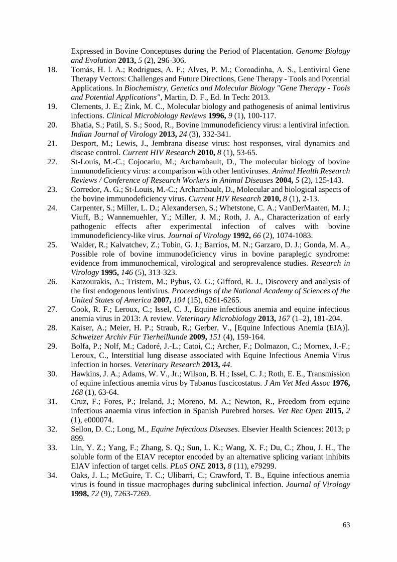
63
Expressed in Bovine Conceptuses during the Period of Placentation. Genome Biology
and Evolution 2013, 5 (2), 296-306.
18. Tomas, H. l. A.; Rodrigues, A. F.; Alves, P. M.; Coroadinha, A. S., Lentiviral Gene
Therapy Vectors: Challenges and Future Directions, Gene Therapy - Tools and Potential
Applications. In Biochemistry, Genetics and Molecular Biology "Gene Therapy - Tools
and Potential Applications", Martin, D. F., Ed. In Tech: 2013.
19. Clements, J. E.; Zink, M. C., Molecular biology and pathogenesis of animal lentivirus
infections. Clinical Microbiology Reviews 1996, 9 (1), 100-117.
20. Bhatia, S.; Patil, S. S.; Sood, R., Bovine immunodeficiency virus: a lentiviral infection.
Indian Journal of Virology 2013, 24 (3), 332-341.
21. Desport, M.; Lewis, J., Jembrana disease virus: host responses, viral dynamics and
disease control. Current HIV Research 2010, 8 (1), 53-65.
22. St-Louis, M.-C.; Cojocariu, M.; Archambault, D., The molecular biology of bovine
immunodeficiency virus: a comparison with other lentiviruses. Animal Health Research
Reviews / Conference of Research Workers in Animal Diseases 2004, 5 (2), 125-143.
23. Corredor, A. G.; St-Louis, M.-C.; Archambault, D., Molecular and biological aspects of
the bovine immunodeficiency virus. Current HIV Research 2010, 8 (1), 2-13.
24. Carpenter, S.; Miller, L. D.; Alexandersen, S.; Whetstone, C. A.; VanDerMaaten, M. J.;
Viuff, B.; Wannemuehler, Y.; Miller, J. M.; Roth, J. A., Characterization of early
pathogenic effects after experimental infection of calves with bovine
immunodeficiency-like virus. Journal of Virology 1992, 66 (2), 1074-1083.
25. Walder, R.; Kalvatchev, Z.; Tobin, G. J.; Barrios, M. N.; Garzaro, D. J.; Gonda, M. A.,
Possible role of bovine immunodeficiency virus in bovine paraplegic syndrome:
evidence from immunochemical, virological and seroprevalence studies. Research in
Virology 1995, 146 (5), 313-323.
26. Katzourakis, A.; Tristem, M.; Pybus, O. G.; Gifford, R. J., Discovery and analysis of
the first endogenous lentivirus. Proceedings of the National Academy of Sciences of the
United States of America 2007, 104 (15), 6261-6265.
27. Cook, R. F.; Leroux, C.; Issel, C. J., Equine infectious anemia and equine infectious
anemia virus in 2013: A review. Veterinary Microbiology 2013, 167 (1–2), 181-204.
28. Kaiser, A.; Meier, H. P.; Straub, R.; Gerber, V., [Equine Infectious Anemia (EIA)].
Schweizer Archiv Für Tierheilkunde 2009, 151 (4), 159-164.
29. Bolfa, P.; Nolf, M.; Cadoré, J.-L.; Catoi, C.; Archer, F.; Dolmazon, C.; Mornex, J.-F.;
Leroux, C., Interstitial lung disease associated with Equine Infectious Anemia Virus
infection in horses. Veterinary Research 2013, 44.
30. Hawkins, J. A.; Adams, W. V., Jr.; Wilson, B. H.; Issel, C. J.; Roth, E. E., Transmission
of equine infectious anemia virus by Tabanus fuscicostatus. J Am Vet Med Assoc 1976,
168 (1), 63-64.
31. Cruz, F.; Fores, P.; Ireland, J.; Moreno, M. A.; Newton, R., Freedom from equine
infectious anaemia virus infection in Spanish Purebred horses. Vet Rec Open 2015, 2
(1), e000074.
32. Sellon, D. C.; Long, M., Equine Infectious Diseases. Elsevier Health Sciences: 2013; p
899.
33. Lin, Y. Z.; Yang, F.; Zhang, S. Q.; Sun, L. K.; Wang, X. F.; Du, C.; Zhou, J. H., The
soluble form of the EIAV receptor encoded by an alternative splicing variant inhibits
EIAV infection of target cells. PLoS ONE 2013, 8 (11), e79299.
34. Oaks, J. L.; McGuire, T. C.; Ulibarri, C.; Crawford, T. B., Equine infectious anemia
virus is found in tissue macrophages during subclinical infection. Journal of Virology
1998, 72 (9), 7263-7269.
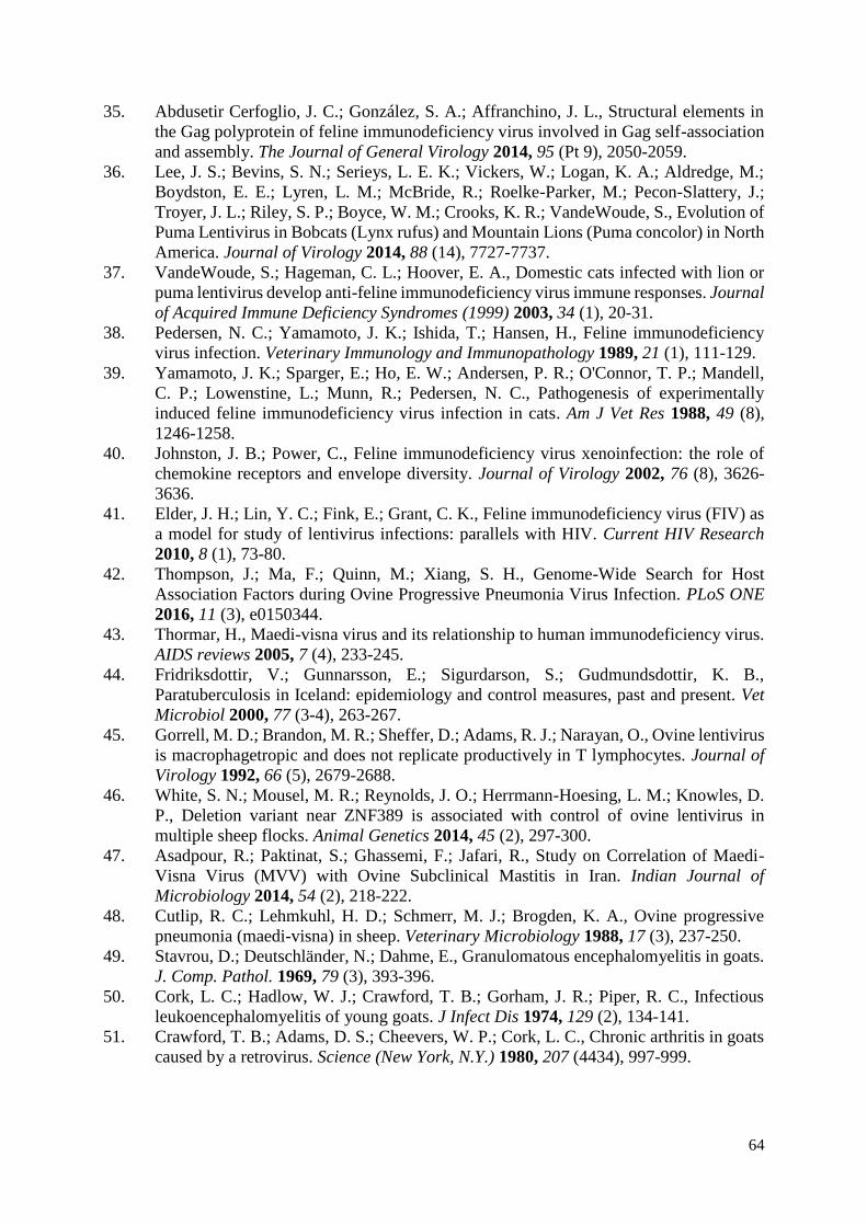
64
35. Abdusetir Cerfoglio, J. C.; González, S. A.; Affranchino, J. L., Structural elements in
the Gag polyprotein of feline immunodeficiency virus involved in Gag self-association
and assembly. The Journal of General Virology 2014, 95 (Pt 9), 2050-2059.
36. Lee, J. S.; Bevins, S. N.; Serieys, L. E. K.; Vickers, W.; Logan, K. A.; Aldredge, M.;
Boydston, E. E.; Lyren, L. M.; McBride, R.; Roelke-Parker, M.; Pecon-Slattery, J.;
Troyer, J. L.; Riley, S. P.; Boyce, W. M.; Crooks, K. R.; VandeWoude, S., Evolution of
Puma Lentivirus in Bobcats (Lynx rufus) and Mountain Lions (Puma concolor) in North
America. Journal of Virology 2014, 88 (14), 7727-7737.
37. VandeWoude, S.; Hageman, C. L.; Hoover, E. A., Domestic cats infected with lion or
puma lentivirus develop anti-feline immunodeficiency virus immune responses. Journal
of Acquired Immune Deficiency Syndromes (1999) 2003, 34 (1), 20-31.
38. Pedersen, N. C.; Yamamoto, J. K.; Ishida, T.; Hansen, H., Feline immunodeficiency
virus infection. Veterinary Immunology and Immunopathology 1989, 21 (1), 111-129.
39. Yamamoto, J. K.; Sparger, E.; Ho, E. W.; Andersen, P. R.; O'Connor, T. P.; Mandell,
C. P.; Lowenstine, L.; Munn, R.; Pedersen, N. C., Pathogenesis of experimentally
induced feline immunodeficiency virus infection in cats. Am J Vet Res 1988, 49 (8),
1246-1258.
40. Johnston, J. B.; Power, C., Feline immunodeficiency virus xenoinfection: the role of
chemokine receptors and envelope diversity. Journal of Virology 2002, 76 (8), 3626-
3636.
41. Elder, J. H.; Lin, Y. C.; Fink, E.; Grant, C. K., Feline immunodeficiency virus (FIV) as
a model for study of lentivirus infections: parallels with HIV. Current HIV Research
2010, 8 (1), 73-80.
42. Thompson, J.; Ma, F.; Quinn, M.; Xiang, S. H., Genome-Wide Search for Host
Association Factors during Ovine Progressive Pneumonia Virus Infection. PLoS ONE
2016, 11 (3), e0150344.
43. Thormar, H., Maedi-visna virus and its relationship to human immunodeficiency virus.
AIDS reviews 2005, 7 (4), 233-245.
44. Fridriksdottir, V.; Gunnarsson, E.; Sigurdarson, S.; Gudmundsdottir, K. B.,
Paratuberculosis in Iceland: epidemiology and control measures, past and present. Vet
Microbiol 2000, 77 (3-4), 263-267.
45. Gorrell, M. D.; Brandon, M. R.; Sheffer, D.; Adams, R. J.; Narayan, O., Ovine lentivirus
is macrophagetropic and does not replicate productively in T lymphocytes. Journal of
Virology 1992, 66 (5), 2679-2688.
46. White, S. N.; Mousel, M. R.; Reynolds, J. O.; Herrmann-Hoesing, L. M.; Knowles, D.
P., Deletion variant near ZNF389 is associated with control of ovine lentivirus in
multiple sheep flocks. Animal Genetics 2014, 45 (2), 297-300.
47. Asadpour, R.; Paktinat, S.; Ghassemi, F.; Jafari, R., Study on Correlation of Maedi-
Visna Virus (MVV) with Ovine Subclinical Mastitis in Iran. Indian Journal of
Microbiology 2014, 54 (2), 218-222.
48. Cutlip, R. C.; Lehmkuhl, H. D.; Schmerr, M. J.; Brogden, K. A., Ovine progressive
pneumonia (maedi-visna) in sheep. Veterinary Microbiology 1988, 17 (3), 237-250.
49. Stavrou, D.; Deutschländer, N.; Dahme, E., Granulomatous encephalomyelitis in goats.
J. Comp. Pathol. 1969, 79 (3), 393-396.
50. Cork, L. C.; Hadlow, W. J.; Crawford, T. B.; Gorham, J. R.; Piper, R. C., Infectious
leukoencephalomyelitis of young goats. J Infect Dis 1974, 129 (2), 134-141.
51. Crawford, T. B.; Adams, D. S.; Cheevers, W. P.; Cork, L. C., Chronic arthritis in goats
caused by a retrovirus. Science (New York, N.Y.) 1980, 207 (4434), 997-999.
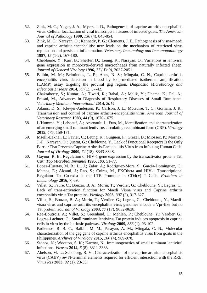
65
52. Zink, M. C.; Yager, J. A.; Myers, J. D., Pathogenesis of caprine arthritis encephalitis
virus. Cellular localization of viral transcripts in tissues of infected goats. The American
Journal of Pathology 1990, 136 (4), 843-854.
53. Zink, M. C.; Narayan, O.; Kennedy, P. G.; Clements, J. E., Pathogenesis of visna/maedi
and caprine arthritis-encephalitis: new leads on the mechanism of restricted virus
replication and persistent inflammation. Veterinary Immunology and Immunopathology
1987, 15 (1-2), 167-180.
54. Chebloune, Y.; Karr, B.; Sheffer, D.; Leung, K.; Narayan, O., Variations in lentiviral
gene expression in monocyte-derived macrophages from naturally infected sheep.
Journal of General Virology 1996, 77 ( Pt 9), 2037-2051.
55. Balbin, M. M.; Belotindos, L. P.; Abes, N. S.; Mingala, C. N., Caprine arthritis
encephalitis virus detection in blood by loop-mediated isothermal amplification
(LAMP) assay targeting the proviral gag region. Diagnostic Microbiology and
Infectious Disease 2014, 79 (1), 37-42.
56. Chakraborty, S.; Kumar, A.; Tiwari, R.; Rahal, A.; Malik, Y.; Dhama, K.; Pal, A.;
Prasad, M., Advances in Diagnosis of Respiratory Diseases of Small Ruminants.
Veterinary Medicine International 2014, 2014.
57. Adams, D. S.; Klevjer-Anderson, P.; Carlson, J. L.; McGuire, T. C.; Gorham, J. R.,
Transmission and control of caprine arthritis-encephalitis virus. American Journal of
Veterinary Research 1983, 44 (9), 1670-1675.
58. L’Homme, Y.; Leboeuf, A.; Arsenault, J.; Fras, M., Identification and characterization
of an emerging small ruminant lentivirus circulating recombinant form (CRF). Virology
2015, 475, 159-171.
59. Mselli-Lakhal, L.; Favier, C.; Leung, K.; Guiguen, F.; Grezel, D.; Miossec, P.; Mornex,
J.-F.; Narayan, O.; Querat, G.; Chebloune, Y., Lack of Functional Receptors Is the Only
Barrier That Prevents Caprine Arthritis-Encephalitis Virus from Infecting Human Cells.
Journal of Virology 2000, 74 (18), 8343-8348.
60. Gaynor, R. B., Regulation of HIV-1 gene expression by the transactivator protein Tat.
Curr Top Microbiol Immunol 1995, 193, 51-77.
61. Lopez-Huertas, M. R.; Li, J.; Zafar, A.; Rodriguez-Mora, S.; Garcia-Dominguez, C.;
Mateos, E.; Alcami, J.; Rao, S.; Coiras, M., PKCtheta and HIV-1 Transcriptional
Regulator Tat Co-exist at the LTR Promoter in CD4(+) T Cells. Frontiers in
Immunology 2016, 7, 69.
62. Villet, S.; Faure, C.; Bouzar, B. A.; Morin, T.; Verdier, G.; Chebloune, Y.; Legras, C.,
Lack of trans-activation function for Maedi Visna virus and Caprine arthritis
encephalitis virus Tat proteins. Virology 2003, 307 (2), 317-327.
63. Villet, S.; Bouzar, B. A.; Morin, T.; Verdier, G.; Legras, C.; Chebloune, Y., Maedi-
visna virus and caprine arthritis encephalitis virus genomes encode a Vpr-like but no
Tat protein. Journal of Virology 2003, 77 (17), 9632-9638.
64. Rea-Boutrois, A.; Villet, S.; Greenland, T.; Mehlen, P.; Chebloune, Y.; Verdier, G.;
Legras-Lachuer, C., Small ruminant lentivirus Tat protein induces apoptosis in caprine
cells in vitro by the intrinsic pathway. Virology 2009, 383 (1), 93-102.
65. Padiernos, R. B. C.; Balbin, M. M.; Parayao, A. M.; Mingala, C. N., Molecular
characterization of the gag gene of caprine arthritis encephalitis virus from goats in the
Philippines. Archives of Virology 2015, 160 (4), 969-978.
66. Stonos, N.; Wootton, S. K.; Karrow, N., Immunogenetics of small ruminant lentiviral
infections. Viruses 2014, 6 (8), 3311-3333.
67. Abelson, M. L.; Schoborg, R. V., Characterization of the caprine arthritis encephalitis
virus (CAEV) rev N-terminal elements required for efficient interaction with the RRE.
Virus Res 2003, 92 (1), 23-35.
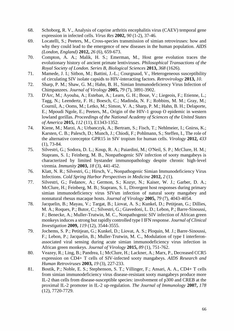
66
68. Schoborg, R. V., Analysis of caprine arthritis encephalitis virus (CAEV) temporal gene
expression in infected cells. Virus Res 2002, 90 (1-2), 37-46.
69. Locatelli, S.; Peeters, M., Cross-species transmission of simian retroviruses: how and
why they could lead to the emergence of new diseases in the human population. AIDS
(London, England) 2012, 26 (6), 659-673.
70. Compton, A. A.; Malik, H. S.; Emerman, M., Host gene evolution traces the
evolutionary history of ancient primate lentiviruses. Philosophical Transactions of the
Royal Society of London. Series B, Biological Sciences 2013, 368 (1626).
71. Mamede, J. I.; Sitbon, M.; Battini, J.-L.; Courgnaud, V., Heterogeneous susceptibility
of circulating SIV isolate capsids to HIV-interacting factors. Retrovirology 2013, 10.
72. Sharp, P. M.; Shaw, G. M.; Hahn, B. H., Simian Immunodeficiency Virus Infection of
Chimpanzees. Journal of Virology 2005, 79 (7), 3891-3902.
73. D'Arc, M.; Ayouba, A.; Esteban, A.; Learn, G. H.; Boue, V.; Liegeois, F.; Etienne, L.;
Tagg, N.; Leendertz, F. H.; Boesch, C.; Madinda, N. F.; Robbins, M. M.; Gray, M.;
Cournil, A.; Ooms, M.; Letko, M.; Simon, V. A.; Sharp, P. M.; Hahn, B. H.; Delaporte,
E.; Mpoudi Ngole, E.; Peeters, M., Origin of the HIV-1 group O epidemic in western
lowland gorillas. Proceedings of the National Academy of Sciences of the United States
of America 2015, 112 (11), E1343-1352.
74. Kiene, M.; Marzi, A.; Urbanczyk, A.; Bertram, S.; Fisch, T.; Nehlmeier, I.; Gnirss, K.;
Karsten, C. B.; Palesch, D.; Munch, J.; Chiodi, F.; Pohlmann, S.; Steffen, I., The role of
the alternative coreceptor GPR15 in SIV tropism for human cells. Virology 2012, 433
(1), 73-84.
75. Silvestri, G.; Sodora, D. L.; Koup, R. A.; Paiardini, M.; O'Neil, S. P.; McClure, H. M.;
Staprans, S. I.; Feinberg, M. B., Nonpathogenic SIV infection of sooty mangabeys is
characterized by limited bystander immunopathology despite chronic high-level
viremia. Immunity 2003, 18 (3), 441-452.
76. Klatt, N. R.; Silvestri, G.; Hirsch, V., Nonpathogenic Simian Immunodeficiency Virus
Infections. Cold Spring Harbor Perspectives in Medicine 2012, 2 (1).
77. Silvestri, G.; Fedanov, A.; Germon, S.; Kozyr, N.; Kaiser, W. J.; Garber, D. A.;
McClure, H.; Feinberg, M. B.; Staprans, S. I., Divergent host responses during primary
simian immunodeficiency virus SIVsm infection of natural sooty mangabey and
nonnatural rhesus macaque hosts. Journal of Virology 2005, 79 (7), 4043-4054.
78. Jacquelin, B.; Mayau, V.; Targat, B.; Liovat, A. S.; Kunkel, D.; Petitjean, G.; Dillies,
M. A.; Roques, P.; Butor, C.; Silvestri, G.; Giavedoni, L. D.; Lebon, P.; Barre-Sinoussi,
F.; Benecke, A.; Muller-Trutwin, M. C., Nonpathogenic SIV infection of African green
monkeys induces a strong but rapidly controlled type I IFN response. Journal of Clinical
Investigation 2009, 119 (12), 3544-3555.
79. Jochems, S. P.; Petitjean, G.; Kunkel, D.; Liovat, A. S.; Ploquin, M. J.; Barre-Sinoussi,
F.; Lebon, P.; Jacquelin, B.; Muller-Trutwin, M. C., Modulation of type I interferon-
associated viral sensing during acute simian immunodeficiency virus infection in
African green monkeys. Journal of Virology 2015, 89 (1), 751-762.
80. Veazey, R.; Ling, B.; Pandrea, I.; McClure, H.; Lackner, A.; Marx, P., Decreased CCR5
expression on CD4+ T cells of SIV-infected sooty mangabeys. AIDS Research and
Human Retroviruses 2003, 19 (3), 227-233.
81. Bostik, P.; Noble, E. S.; Stephenson, S. T.; Villinger, F.; Ansari, A. A., CD4+ T cells
from simian immunodeficiency virus disease-resistant sooty mangabeys produce more
IL-2 than cells from disease-susceptible species: involvement of p300 and CREB at the
proximal IL-2 promoter in IL-2 up-regulation. The Journal of Immunology 2007, 178
(12), 7720-7729.
67
82. Byrareddy, S. N.; Sidell, N.; Arthos, J.; Cicala, C.; Zhao, C.; Little, D. M.; Dunbar, P.;
Yang, G. X.; Pierzchalski, K.; Kane, M. A.; Mayne, A. E.; Song, B.; Soares, M. A.;
Villinger, F.; Fauci, A. S.; Ansari, A. A., Species-specific differences in the expression
and regulation of alpha4beta7 integrin in various nonhuman primates. The Journal of
Immunology 2015, 194 (12), 5968-5979.
83. Gordon, S. N.; Klatt, N. R.; Bosinger, S. E.; Brenchley, J. M.; Milush, J. M.; Engram,
J. C.; Dunham, R. M.; Paiardini, M.; Klucking, S.; Danesh, A.; Strobert, E. A.; Apetrei,
C.; Pandrea, I. V.; Kelvin, D.; Douek, D. C.; Staprans, S. I.; Sodora, D. L.; Silvestri, G.,
Severe depletion of mucosal CD4+ T cells in AIDS-free simian immunodeficiency
virus-infected sooty mangabeys. The Journal of Immunology 2007, 179 (5), 3026-3034.
84. Harris, L. D.; Tabb, B.; Sodora, D. L.; Paiardini, M.; Klatt, N. R.; Douek, D. C.;
Silvestri, G.; Müller-Trutwin, M.; Vasile-Pandrea, I.; Apetrei, C.; Hirsch, V.; Lifson, J.;
Brenchley, J. M.; Estes, J. D., Downregulation of Robust Acute Type I Interferon
Responses Distinguishes Nonpathogenic Simian Immunodeficiency Virus (SIV)
Infection of Natural Hosts from Pathogenic SIV Infection of Rhesus Macaques. Journal
of Virology 2010, 84 (15), 7886-7891.
85. O'Neil, S. P.; Suwyn, C.; Anderson, D. C.; Niedziela, G.; Bradley, J.; Novembre, F. J.;
Herndon, J. G.; McClure, H. M., Correlation of acute humoral response with brain virus
burden and survival time in pig-tailed macaques infected with the neurovirulent simian
immunodeficiency virus SIVsmmFGb. The American Journal of Pathology 2004, 164
(4), 1157-1172.
86. Lauck, M.; Switzer, W. M.; Sibley, S. D.; Hyeroba, D.; Tumukunde, A.; Weny, G.;
Shankar, A.; Greene, J. M.; Ericsen, A. J.; Zheng, H.; Ting, N.; Chapman, C. A.;
Friedrich, T. C.; Goldberg, T. L.; O’Connor, D. H., Discovery and full genome
characterization of a new SIV lineage infecting red-tailed guenons (Cercopithecus
ascanius schmidti) in Kibale National Park, Uganda. Retrovirology 2014, 11 (1).
87. McKay, R. A., “Patient Zero”. Bulletin of the History of Medicine 2014, 161-194.
88. Worobey, M.; Watts, T. D.; McKay, R. A.; Suchard, M. A.; Granade, T.; Teuwen, D.
E.; Koblin, B. A.; Heneine, W.; Lemey, P.; Jaffe, H. W., 1970s and 'Patient 0' HIV-1
genomes illuminate early HIV/AIDS history in North America. Nature 2016.
89. O'Leary, D., The syndemic of AIDS and STDS among MSM. Linacre Q 2014, 81 (1),
12-37.
90. Souquiere, S.; Makuwa, M.; Salle, B.; Kazanji, M., New strain of simian
immunodeficiency virus identified in wild-born chimpanzees from central Africa. PLoS
ONE 2012, 7 (9), e44298.
91. Keele, B. F.; Van Heuverswyn, F.; Li, Y.; Bailes, E.; Takehisa, J.; Santiago, M. L.;
Bibollet-Ruche, F.; Chen, Y.; Wain, L. V.; Liegeois, F.; Loul, S.; Ngole, E. M.;
Bienvenue, Y.; Delaporte, E.; Brookfield, J. F. Y.; Sharp, P. M.; Shaw, G. M.; Peeters,
M.; Hahn, B. H., Chimpanzee Reservoirs of Pandemic and Nonpandemic HIV-1.
Science (New York, N.Y.) 2006, 313 (5786), 523-526.
92. Heeney, J. L.; Rutjens, E.; Verschoor, E. J.; Niphuis, H.; ten Haaft, P.; Rouse, S.;
McClure, H.; Balla-Jhagjhoorsingh, S.; Bogers, W.; Salas, M.; Cobb, K.; Kestens, L.;
Davis, D.; van der Groen, G.; Courgnaud, V.; Peeters, M.; Murthy, K. K., Transmission
of Simian Immunodeficiency Virus SIVcpz and the Evolution of Infection in the
Presence and Absence of Concurrent Human Immunodeficiency Virus Type 1 Infection
in Chimpanzees. Journal of Virology 2006, 80 (14), 7208-7218.
93. Sharp, P. M.; Hahn, B. H., Origins of HIV and the AIDS Pandemic. Cold Spring Harbor
Perspectives in Medicine 2011, 1 (1).
94. Boue, V.; Locatelli, S.; Boucher, F.; Ayouba, A.; Butel, C.; Esteban, A.; Okouga, A. P.;
Ndoungouet, A.; Motsch, P.; Le Flohic, G.; Ngari, P.; Prugnolle, F.; Ollomo, B.; Rouet,
68
F.; Liegeois, F., High Rate of Simian Immunodeficiency Virus (SIV) Infections in Wild
Chimpanzees in Northeastern Gabon. Viruses 2015, 7 (9), 4997-5015.
95. D’arc, M.; Ayouba, A.; Esteban, A.; Learn, G. H.; Boué, V.; Liegeois, F.; Etienne, L.;
Tagg, N.; Leendertz, F. H.; Boesch, C.; Madinda, N. F.; Robbins, M. M.; Gray, M.;
Cournil, A.; Ooms, M.; Letko, M.; Simon, V. A.; Sharp, P. M.; Hahn, B. H.; Delaporte,
E.; Ngole, E. M.; Peeters, M., Origin of the HIV-1 group O epidemic in western lowland
gorillas. Proceedings of the National Academy of Sciences 2015, 112 (11), E1343-
E1352.
96. Crawford, D. H., Virus Hunt: The search for the origin of HIV/AIDs. Oxford University
Press: 2013; p 259.
97. Santiago, M. L.; Range, F.; Keele, B. F.; Li, Y.; Bailes, E.; Bibollet-Ruche, F.; Fruteau,
C.; Noë, R.; Peeters, M.; Brookfield, J. F. Y.; Shaw, G. M.; Sharp, P. M.; Hahn, B. H.,
Simian immunodeficiency virus infection in free-ranging sooty mangabeys (Cercocebus
atys atys) from the Taï Forest, Côte d'Ivoire: implications for the origin of epidemic
human immunodeficiency virus type 2. Journal of Virology 2005, 79 (19), 12515-
12527.
98. Ayouba, A.; Akoua-Koffi, C.; Calvignac-Spencer, S.; Esteban, A.; Locatelli, S.; Li, H.;
Li, Y.; Hahn, B. H.; Delaporte, E.; Leendertz, F. H.; Peeters, M., Evidence for
continuing cross-species transmission of SIVsmm to humans: characterization of a new
HIV-2 lineage in rural Côte d’Ivoire. AIDS 2013, 27 (15), 2488-2491.
99. Campbell-Yesufu, O. T.; Gandhi, R. T., Update on Human Immunodeficiency Virus
(HIV)-2 Infection. Clinical Infectious Diseases 2011, 52 (6), 780-787.
100. Descours, B.; Cribier, A.; Chable-Bessia, C.; Ayinde, D.; Rice, G.; Crow, Y.; Yatim,
A.; Schwartz, O.; Laguette, N.; Benkirane, M., SAMHD1 restricts HIV-1 reverse
transcription in quiescent CD4(+) T-cells. Retrovirology 2012, 9, 87.
101. Shingai, M.; Welbourn, S.; Brenchley, J. M.; Acharya, P.; Miyagi, E.; Plishka, R. J.;
Buckler-White, A.; Kwong, P. D.; Nishimura, Y.; Strebel, K.; Martin, M. A., The
Expression of Functional Vpx during Pathogenic SIVmac Infections of Rhesus
Macaques Suppresses SAMHD1 in CD4+ Memory T Cells. PLoS Pathog 2015, 11 (5),
e1004928.
102. Azevedo-Pereira, J. M.; Santos-Costa, Q.; Moniz-Pereira, J., HIV-2 infection and
chemokine receptors usage - clues to reduced virulence of HIV-2. Current HIV
Research 2005, 3 (1), 3-16.
103. Treviño, A.; Soriano, V.; Poveda, E.; Parra, P.; Cabezas, T.; Caballero, E.; Roc, L.;
Rodríguez, C.; Eiros, J. M.; Lopez, M.; De Mendoza, C.; Group, H. I. V. S. S., HIV-2
viral tropism influences CD4+ T cell count regardless of viral load. The Journal of
Antimicrobial Chemotherapy 2014, 69 (8), 2191-2194.
104. van der Graaf, M.; Diepersloot, R. J., Transmission of human immunodeficiency virus
(HIV/HTLV-III/LAV): a review. Infection 1986, 14 (5), 203-211.
105. Choices, N. H. S. Can HIV be transmitted through oral sex (fellatio and cunnilingus)?
- Health questions - NHS Choices.
http://www.nhs.uk/chq/pages/3098.aspx?categoryid=118
106. Royce, R. A.; Seña, A.; Cates, W.; Cohen, M. S., Sexual Transmission of HIV. New
England Journal of Medicine 1997, 336 (15), 1072-1078.
107. McCoy, C. B.; Metsch, L. R.; Chitwood, D. D.; Shapshak, P.; Comerford, S. T.,
Parenteral transmission of HIV among injection drug users: assessing the frequency of
multiperson use of needles, syringes, cookers, cotton, and water. Journal of Acquired
Immune Deficiency Syndromes and Human Retrovirology: Official Publication of the
International Retrovirology Association 1998, 18 Suppl 1, S25-29.
69
108. Gray, R. H.; Wawer, M. J.; Brookmeyer, R.; Sewankambo, N. K.; Serwadda, D.;
Wabwire-Mangen, F.; Lutalo, T.; Li, X.; vanCott, T.; Quinn, T. C., Probability of HIV-
1 transmission per coital act in monogamous, heterosexual, HIV-1-discordant couples
in Rakai, Uganda. Lancet 2001, 357 (9263), 1149-1153.
109. Moodley, J.; Wennberg, J. L., HIV in pregnancy. Current Opinion in Obstetrics &
Gynecology 2005, 17 (2), 117-121.
110. Nkwo, P., Prevention of mother to child transmission of human immunodeficiency
virus: the nigerian perspective. Annals of Medical and Health Sciences Research 2012,
2 (1), 56-65.
111. Dorosko, S. M.; Connor, R. I., Primary human mammary epithelial cells endocytose
HIV-1 and facilitate viral infection of CD4+ T lymphocytes. Journal of Virology 2010,
84 (20), 10533-10542.
112. Desposito, F.; McSherry, G. D.; Oleske, J. M., Blood product acquired HIV infection in
children. Pediatric Annals 1988, 17 (5), 341-345.
113. Simonds, R. J., HIV transmission by organ and tissue transplantation. AIDS (London,
England) 1993, 7 Suppl 2, S35-38.
114. Global-AIDS-update. Global AIDS update 2016.
http://www.who.int/hiv/pub/arv/global-AIDS-update-2016_en.pdf?ua=1.
115. Shafiee, H.; Wang, S.; Inci, F.; Toy, M.; Henrich, T. J.; Kuritzkes, D. R.; Demirci, U.,
Emerging Technologies for Point-of-Care Management of HIV Infection. Annual
Review of Medicine 2015, 66 (1), 387-405.
116. Gray, R.; Kigozi, G.; Kong, X.; Ssempiija, V.; Makumbi, F.; Wattya, S.; Serwadda, D.;
Nalugoda, F.; Sewenkambo, N. K.; Wawer, M. J., The effectiveness of male
circumcision for HIV prevention and effects on risk behaviors in a posttrial follow-up
study. AIDS (London, England) 2012, 26 (5), 609-615.
117. Ngemu, E. K.; Khayeka-Wandabwa, C.; Kweka, E. J.; Choge, J. K.; Anino, E.; Oyoo-
Okoth, E., Effectiveness of option B highly active antiretroviral therapy (HAART)
prevention of mother-to-child transmission (PMTCT) in pregnant HIV women. BMC
Research Notes 2014, 7 (1).
118. McCormack, S.; Dunn, D. T.; Desai, M.; Dolling, D. I.; Gafos, M.; Gilson, R.; Sullivan,
A. K.; Clarke, A.; Reeves, I.; Schembri, G.; Mackie, N.; Bowman, C.; Lacey, C. J.;
Apea, V.; Brady, M.; Fox, J.; Taylor, S.; Antonucci, S.; Khoo, S. H.; Rooney, J.;
Nardone, A.; Fisher, M.; McOwan, A.; Phillips, A. N.; Johnson, A. M.; Gazzard, B.;
Gill, O. N., Pre-exposure prophylaxis to prevent the acquisition of HIV-1 infection
(PROUD): effectiveness results from the pilot phase of a pragmatic open-label
randomised trial. Lancet 2016, 387 (10013), 53-60.
119. Aloia, R. C.; Tian, H.; Jensen, F. C., Lipid composition and fluidity of the human
immunodeficiency virus envelope and host cell plasma membranes. Proceedings of the
National Academy of Sciences of the United States of America 1993, 90 (11), 5181-
5185.
120. Schulz, T. F.; Jameson, B. A.; Lopalco, L.; Siccardi, A. G.; Weiss, R. A.; Moore, J. P.,
Conserved structural features in the interaction between retroviral surface and
transmembrane glycoproteins? AIDS Research and Human Retroviruses 1992, 8 (9),
1571-1580.
121. Barrie, K. A.; Perez, E. E.; Lamers, S. L.; Farmerie, W. G.; Dunn, B. M.; Sleasman, J.
W.; Goodenow, M. M., Natural variation in HIV-1 protease, Gag p7 and p6, and
protease cleavage sites within gag/pol polyproteins: amino acid substitutions in the
absence of protease inhibitors in mothers and children infected by human
immunodeficiency virus type 1. Virology 1996, 219 (2), 407-416.

70
122. Speck, R. R.; Flexner, C.; Tian, C. J.; Yu, X. F., Comparison of human
immunodeficiency virus type 1 Pr55(Gag) and Pr160(Gag-pol) processing
intermediates that accumulate in primary and transformed cells treated with peptidic and
nonpeptidic protease inhibitors. Antimicrobial Agents and Chemotherapy 2000, 44 (5),
1397-1403.
123. Sakuma, T.; Barry, M. A.; Ikeda, Y., Lentiviral vectors: basic to translational. Biochem
J 2012, 443 (3), 603-618.
124. Abd El-Wahab, E. W.; Smyth, R. P.; Mailler, E.; Bernacchi, S.; Vivet-Boudou, V.;
Hijnen, M.; Jossinet, F.; Mak, J.; Paillart, J. C.; Marquet, R., Specific recognition of the
HIV-1 genomic RNA by the Gag precursor. Nat Commun 2014, 5, 4304.
125. Turner, B. G.; Summers, M. F., Structural biology of HIV. Journal of Molecular Biology
1999, 285 (1), 1-32.
126. Pettit, S. C.; Everitt, L. E.; Choudhury, S.; Dunn, B. M.; Kaplan, A. H., Initial cleavage
of the human immunodeficiency virus type 1 GagPol precursor by its activated protease
occurs by an intramolecular mechanism. Journal of Virology 2004, 78 (16), 8477-8485.
127. Fitzgerald, M. E.; Drohat, A. C., Studies of RNA/DNA Polypurine Tracts. Chemistry &
Biology 2008, 15 (3), 203-204.
128. Dingwall, C.; Ernberg, I.; Gait, M. J.; Green, S. M.; Heaphy, S.; Karn, J.; Lowe, A. D.;
Singh, M.; Skinner, M. A.; Valerio, R., Human immunodeficiency virus 1 tat protein
binds trans-activation-responsive region (TAR) RNA in vitro. Proceedings of the
National Academy of Sciences of the United States of America 1989, 86 (18), 6925-
6929.
129. Sarafianos, S. G.; Das, K.; Tantillo, C.; Clark, A. D.; Ding, J.; Whitcomb, J. M.; Boyer,
P. L.; Hughes, S. H.; Arnold, E., Crystal structure of HIV-1 reverse transcriptase in
complex with a polypurine tract RNA:DNA. The EMBO Journal 2001, 20 (6), 1449-
1461.
130. Guntaka, R. V., Transcription termination and polyadenylation in retroviruses.
Microbiol Rev 1993, 57 (3), 511-521.
131. Gómez-Lucía, E.; Collado, V. M.; Miró, G.; Doménech, A., Effect of Type-I Interferon
on Retroviruses. Viruses 2009, 1 (3), 545-573.
132. Tachedjian, G.; Radzio, J.; Sluis-Cremer, N., Relationship between enzyme activity and
dimeric structure of recombinant HIV-1 reverse transcriptase. Proteins 2005, 60 (1), 5-
13.
133. Mulky, A.; Kappes, J. C., Analysis of human immunodeficiency virus type 1 reverse
transcriptase subunit structure/function in the context of infectious virions and human
target cells. Antimicrobial Agents and Chemotherapy 2005, 49 (9), 3762-3769.
134. Das, K.; Martinez, S. E.; Bandwar, R. P.; Arnold, E., Structures of HIV-1 RT-
RNA/DNA ternary complexes with dATP and nevirapine reveal conformational
flexibility of RNA/DNA: insights into requirements for RNase H cleavage. Nucleic
Acids Research 2014, 42 (12), 8125-8137.
135. Cimarelli, A.; Darlix, J. L., HIV-1 reverse transcription. Methods in Molecular Biology
(Clifton, N.J.) 2014, 1087, 55-70.
136. Zheng, R.; Jenkins, T. M.; Craigie, R., Zinc folds the N-terminal domain of HIV-1
integrase, promotes multimerization, and enhances catalytic activity. Proceedings of the
National Academy of Sciences of the United States of America 1996, 93 (24), 13659-
13664.
137. Chiu, T. K.; Davies, D. R., Structure and function of HIV-1 integrase. Current Topics
in Medicinal Chemistry 2004, 4 (9), 965-977.
138. Andrake, M. D.; Skalka, A. M., Retroviral Integrase: Then and Now. Annu Rev Virol
2015, 2 (1), 241-264.
71
139. Li, M.; Ivanov, V.; Mizuuchi, M.; Mizuuchi, K.; Craigie, R., DNA requirements for
assembly and stability of HIV-1 intasomes. Protein Science: A Publication of the
Protein Society 2012, 21 (2), 249-257.
140. Bojja, R. S.; Andrake, M. D.; Merkel, G.; Weigand, S.; Dunbrack, R. L.; Skalka, A. M.,
Architecture and assembly of HIV integrase multimers in the absence of DNA
substrates. The Journal of Biological Chemistry 2013, 288 (10), 7373-7386.
141. Delelis, O.; Carayon, K.; Saib, A.; Deprez, E.; Mouscadet, J. F., Integrase and
integration: biochemical activities of HIV-1 integrase. Retrovirology 2008, 5, 114.
142. Chen, J. C. H.; Krucinski, J.; Miercke, L. J. W.; Finer-Moore, J. S.; Tang, A. H.; Leavitt,
A. D.; Stroud, R. M., Crystal structure of the HIV-1 integrase catalytic core and C-
terminal domains: A model for viral DNA binding. Proceedings of the National
Academy of Sciences 2000, 97 (15), 8233-8238.
143. Graf, E. H.; O'Doherty, U., Quantitation of integrated proviral DNA in viral reservoirs.
Current Opinion in HIV and AIDS 2013, 8 (2), 100-105.
144. Hoshikawa, N.; Kojima, A.; Yasuda, A.; Takayashiki, E.; Masuko, S.; Chiba, J.; Sata,
T.; Kurata, T., Role of the gag and pol genes of human immunodeficiency virus in the
morphogenesis and maturation of retrovirus-like particles expressed by recombinant
vaccinia virus: an ultrastructural study. The Journal of General Virology 1991, 72 ( Pt
10), 2509-2517.
145. Wondrak, E. M.; Nashed, N. T.; Haber, M. T.; Jerina, D. M.; Louis, J. M., A Transient
Precursor of the HIV-1 Protease ISOLATION, CHARACTERIZATION, AND
KINETICS OF MATURATION. Journal of Biological Chemistry 1996, 271 (8), 4477-
4481.
146. Ashorn, P.; McQuade, T. J.; Thaisrivongs, S.; Tomasselli, A. G.; Tarpley, W. G.; Moss,
B., An inhibitor of the protease blocks maturation of human and simian
immunodeficiency viruses and spread of infection. Proceedings of the National
Academy of Sciences of the United States of America 1990, 87 (19), 7472-7476.
147. Kohl, N. E.; Emini, E. A.; Schleif, W. A.; Davis, L. J.; Heimbach, J. C.; Dixon, R. A.;
Scolnick, E. M.; Sigal, I. S., Active human immunodeficiency virus protease is required
for viral infectivity. Proceedings of the National Academy of Sciences of the United
States of America 1988, 85 (13), 4686-4690.
148. Titanji, B. K.; Aasa-Chapman, M.; Pillay, D.; Jolly, C., Protease inhibitors effectively
block cell-to-cell spread of HIV-1 between T cells. Retrovirology 2013, 10 (1).
149. Konvalinka, J.; Krausslich, H. G.; Muller, B., Retroviral proteases and their roles in
virion maturation. Virology 2015, 479-480, 403-417.
150. Sattentau, Q. J., The role of the envelope glycoproteins in HIV-1 transmission and
pathogenesis. Perspectives in Drug Discovery and Design 1996, 5 (1), 1-16.
151. Binley, J. M.; Sanders, R. W.; Master, A.; Cayanan, C. S.; Wiley, C. L.; Schiffner, L.;
Travis, B.; Kuhmann, S.; Burton, D. R.; Hu, S.-L.; Olson, W. C.; Moore, J. P.,
Enhancing the proteolytic maturation of human immunodeficiency virus type 1
envelope glycoproteins. Journal of Virology 2002, 76 (6), 2606-2616.
152. Olshevsky, U.; Helseth, E.; Furman, C.; Li, J.; Haseltine, W.; Sodroski, J., Identification
of individual human immunodeficiency virus type 1 gp120 amino acids important for
CD4 receptor binding. Journal of Virology 1990, 64 (12), 5701-5707.
153. Clapham, P. R.; McKnight, A., HIV-1 receptors and cell tropism. Br Med Bull 2001, 58,
43-59.
154. Balakrishna, L. S.; Kondapi, A. K., Role of Host Proteins in HIV-1 Early Replication
In Advances in Molecular Retrovirology, Saxena, S. K., Ed. 2016.
72
155. Nguyen, D. G.; Hildreth, J. E. K., Involvement of macrophage mannose receptor in the
binding and transmission of HIV by macrophages. European Journal of Immunology
2003, 33 (2), 483-493.
156. Mao, Y.; Wang, L.; Gu, C.; Herschhorn, A.; Desormeaux, A.; Finzi, A.; Xiang, S. H.;
Sodroski, J. G., Molecular architecture of the uncleaved HIV-1 envelope glycoprotein
trimer. Proceedings of the National Academy of Sciences of the United States of America
2013, 110 (30), 12438-12443.
157. Gallo, S. A.; Finnegan, C. M.; Viard, M.; Raviv, Y.; Dimitrov, A.; Rawat, S. S.; Puri,
A.; Durell, S.; Blumenthal, R., The HIV Env-mediated fusion reaction. Biochim Biophys
Acta 2003, 1614 (1), 36-50.
158. Dey, B.; Svehla, K.; Xu, L.; Wycuff, D.; Zhou, T.; Voss, G.; Phogat, A.; Chakrabarti,
B. K.; Li, Y.; Shaw, G.; Kwong, P. D.; Nabel, G. J.; Mascola, J. R.; Wyatt, R. T.,
Structure-based stabilization of HIV-1 gp120 enhances humoral immune responses to
the induced co-receptor binding site. PLoS Pathogens 2009, 5 (5).
159. Zolla-Pazner, S.; Cardozo, T., STRUCTURE–FUNCTION RELATIONSHIPS OF
HIV-1 ENVELOPE SEQUENCE-VARIABLE REGIONS PROVIDE A PARADIGM
FOR VACCINE DESIGN. Nature reviews. Immunology 2010, 10 (7), 527-535.
160. Gozhenko, A. I., Intracellular Transport of HIV-1 Matrix Protein Associated with Viral
RNA. World Journal of AIDS 2013, 03 (01), 33-35.
161. Hearps, A. C.; Jans, D. A., Regulating the functions of the HIV-1 matrix protein. AIDS
Research and Human Retroviruses 2007, 23 (3), 341-346.
162. Levin, R.; Mhashilkar, A. M.; Dorfman, T.; Bukovsky, A.; Zani, C.; Bagley, J.; Hinkula,
J.; Niedrig, M.; Albert, J.; Wahren, B.; Göttlinger, H. G.; Marasco, W. A., Inhibition
of early and late events of the HIV-1 replication cycle by cytoplasmic Fab intrabodies
against the matrix protein, p17. Molecular Medicine 1997, 3 (2), 96-110.
163. Kyere, S. K.; Mercredi, P. Y.; Dong, X.; Spearman, P.; Summers, M. F., The HIV-1
matrix protein does not interact directly with the protein interactive domain of AP-3δ.
Virus Research 2012, 169 (2), 411-414.
164. Pornillos, O.; Ganser-Pornillos, B. K.; Kelly, B. N.; Hua, Y.; Whitby, F. G.; Stout, C.
D.; Sundquist, W. I.; Hill, C. P.; Yeager, M., X-Ray Structures of the Hexameric
Building Block of the HIV Capsid. Cell 2009, 137 (7), 1282-1292.
165. Sundquist, W. I.; Kräusslich, H.-G., HIV-1 Assembly, Budding, and Maturation. Cold
Spring Harbor Perspectives in Medicine 2012, 2 (7).
166. Zhang, B.; Liu, D.; Bao, Z.; Chen, B.; Li, C.; Jiang, H.; Wang, X.; Mi, Z.; An, X.; Lu,
J.; Tong, Y., High level soluble expression, one-step purification and characterization
of HIV-1 p24 protein. Virology Journal 2011, 8 (1).
167. Dong, H.; Liu, J.; Zhu, H.; Ou, C.-Y.; Xing, W.; Qiu, M.; Zhang, G.; Xiao, Y.; Yao, J.;
Pan, P.; Jiang, Y., Two types of nanoparticle-based bio-barcode amplification assays to
detect HIV-1 p24 antigen. Virology Journal 2012, 9.
168. Wu, H.; Mitra, M.; Naufer, M. N.; McCauley, M. J.; Gorelick, R. J.; Rouzina, I.; Musier-
Forsyth, K.; Williams, M. C., Differential contribution of basic residues to HIV-1
nucleocapsid protein's nucleic acid chaperone function and retroviral replication.
Nucleic Acids Research 2014, 42 (4), 2525-2537.
169. Levin, J. G.; Mitra, M.; Mascarenhas, A.; Musier-Forsyth, K., Role of HIV-1
nucleocapsid protein in HIV-1 reverse transcription. RNA Biology 2010, 7 (6), 754-774.
170. Webb, J. A.; Jones, C. P.; Parent, L. J.; Rouzina, I.; Musier-Forsyth, K., Distinct binding
interactions of HIV-1 Gag to Psi and non-Psi RNAs: Implications for viral genomic
RNA packaging. RNA 2013, 19 (8), 1078-1088.
171. Chan, B.; Musier-Forsyth, K., The nucleocapsid protein specifically anneals tRNALys-
3 onto a noncomplementary primer binding site within the HIV-1 RNA genome in vitro.
73
Proceedings of the National Academy of Sciences of the United States of America 1997,
94 (25), 13530-13535.
172. Selig, L.; Pages, J. C.; Tanchou, V.; Prévéral, S.; Berlioz-Torrent, C.; Liu, L. X.;
Erdtmann, L.; Darlix, J. L.; Benarous, R.; Benichou, S., Interaction with the p6 Domain
of the Gag Precursor Mediates Incorporation into Virions of Vpr and Vpx Proteins from
Primate Lentiviruses. Journal of Virology 1999, 73 (1), 592-600.
173. Kudoh, A.; Takahama, S.; Sawasaki, T.; Ode, H.; Yokoyama, M.; Okayama, A.;
Ishikawa, A.; Miyakawa, K.; Matsunaga, S.; Kimura, H.; Sugiura, W.; Sato, H.; Hirano,
H.; Ohno, S.; Yamamoto, N.; Ryo, A., The phosphorylation of HIV-1 Gag by atypical
protein kinase C facilitates viral infectivity by promoting Vpr incorporation into virions.
Retrovirology 2014, 11.
174. Varthakavi, V.; Smith, R. M.; Bour, S. P.; Strebel, K.; Spearman, P., Viral protein U
counteracts a human host cell restriction that inhibits HIV-1 particle production.
Proceedings of the National Academy of Sciences 2003, 100 (25), 15154-15159.
175. Emeagwali, N.; Hildreth, J. E. K., Human immunodeficiency virus type 1 Vpu and
cellular TASK proteins suppress transcription of unintegrated HIV-1 DNA. Virology
Journal 2012, 9.
176. Gonzalez, M. E., Vpu Protein: The Viroporin Encoded by HIV-1. Viruses 2015, 7 (8),
4352-4368.
177. Schubert, U.; Antón, L. C.; Bacík, I.; Cox, J. H.; Bour, S.; Bennink, J. R.; Orlowski, M.;
Strebel, K.; Yewdell, J. W., CD4 glycoprotein degradation induced by human
immunodeficiency virus type 1 Vpu protein requires the function of proteasomes and
the ubiquitin-conjugating pathway. Journal of Virology 1998, 72 (3), 2280-2288.
178. Nomaguchi, M.; Fujita, M.; Adachi, A., Role of HIV-1 Vpu protein for virus spread and
pathogenesis. Microbes and Infection / Institut Pasteur 2008, 10 (9), 960-967.
179. Ruiz, A.; Guatelli, J. C.; Stephens, E. B., The Vpu Protein: New Concepts in Virus
Release and CD4 Down-Modulation. Current HIV Research 2010, 8 (3), 240-252.
180. Arias, J. F.; Iwabu, Y.; Tokunaga, K., Sites of action of HIV-1 Vpu in BST-2/tetherin
downregulation. Current HIV Research 2012, 10 (4), 283-291.
181. Cavallari, I.; Rende, F.; D'Agostino, D. M.; Ciminale, V., Converging Strategies in
Expression of Human Complex Retroviruses. Viruses 2011, 3 (8), 1395-1414.
182. Pandey, R. C.; Datta, D.; Mukerjee, R.; Srinivasan, A.; Mahalingam, S.; Sawaya, B. E.,
HIV-1 Vpr: a closer look at the multifunctional protein from the structural perspective.
Current HIV Research 2009, 7 (2), 114-128.
183. Pirrone, V.; Mell, J.; Janto, B.; Wigdahl, B., Biomarkers of HIV Susceptibility and
Disease Progression. EBioMedicine 2014, 1 (2-3), 99-100.
184. de Silva, S.; Planelles, V.; Wu, L., Differential Effects of Vpr on Single-cycle and
Spreading HIV-1 Infections in CD4+ T-cells and Dendritic Cells. PLoS ONE 2012, 7
(5).
185. Zhao, R. Y.; Bukrinsky, M. I., HIV-1 accessory proteins: VpR. Methods in Molecular
Biology (Clifton, N.J.) 2014, 1087, 125-134.
186. Zhou, T.; Dang, Y.; Baker, J. J.; Zhou, J.; Zheng, Y.-H., Evidence for Vpr-dependent
HIV-1 Replication in Human CD4+ CEM.NKR T-Cells. Retrovirology 2012, 9.
187. Zhao, R. Y.; Li, G.; Bukrinsky, M. I., Vpr-host interactions during HIV-1 viral life cycle.
Journal Of Neuroimmune Pharmacology: The Official Journal Of The Society On
Neuroimmune Pharmacology 2011, 6 (2), 216-229.
188. Levy, D. N.; Fernandes, L. S.; Williams, W. V.; Weiner, D. B., Induction of cell
differentiation by human immunodeficiency virus 1 vpr. Cell 1993, 72 (4), 541-550.
74
189. Jenkins, Y.; Pornillos, O.; Rich, R. L.; Myszka, D. G.; Sundquist, W. I.; Malim, M. H.,
Biochemical Analyses of the Interactions between Human Immunodeficiency Virus
Type 1 Vpr and p6Gag. Journal of Virology 2001, 75 (21), 10537-10542.
190. Gabuzda, D. H.; Li, H.; Lawrence, K.; Vasir, B. S.; Crawford, K.; Langhoff, E.,
Essential role of vif in establishing productive HIV-1 infection in peripheral blood T
lymphocytes and monocyte/macrophages. Journal of Acquired Immune Deficiency
Syndromes 1994, 7 (9), 908-915.
191. Schafer, A.; Bogerd, H. P.; Cullen, B. R., Specific packaging of APOBEC3G into HIV-
1 virions is mediated by the nucleocapsid domain of the gag polyprotein precursor.
Virology 2004, 328 (2), 163-168.
192. Kao, S.; Miyagi, E.; Khan, M. A.; Takeuchi, H.; Opi, S.; Goila-Gaur, R.; Strebel, K.,
Production of infectious human immunodeficiency virus type 1 does not require
depletion of APOBEC3G from virus-producing cells. Retrovirology 2004, 1 (1).
193. Wang, Y.; Kinlock, B. L.; Shao, Q.; Turner, T. M.; Liu, B., HIV-1 Vif inhibits G to A
hypermutations catalyzed by virus-encapsidated APOBEC3G to maintain HIV-1
infectivity. Retrovirology 2014, 11 (1).
194. Apolonia, L.; Schulz, R.; Curk, T.; Rocha, P.; Swanson, C. M.; Schaller, T.; Ule, J.;
Malim, M. H., Promiscuous RNA Binding Ensures Effective Encapsidation of
APOBEC3 Proteins by HIV-1. PLoS Pathogens 2015, 11 (1).
195. Landi, A.; Iannucci, V.; Nuffel, A. V.; Meuwissen, P.; Verhasselt, B., One Protein to
Rule them All: Modulation of Cell Surface Receptors and Molecules by HIV Nef.
Current HIV Research 2011, 9 (7), 496-504.
196. Greenway, A. L.; McPhee, D. A.; Allen, K.; Johnstone, R.; Holloway, G.; Mills, J.;
Azad, A.; Sankovich, S.; Lambert, P., Human Immunodeficiency Virus Type 1 Nef
Binds to Tumor Suppressor p53 and Protects Cells against p53-Mediated Apoptosis.
Journal of Virology 2002, 76 (6), 2692-2702.
197. Rücker, E.; Grivel, J.-C.; Münch, J.; Kirchhoff, F.; Margolis, L., Vpr and Vpu Are
Important for Efficient Human Immunodeficiency Virus Type 1 Replication and CD4+
T-Cell Depletion in Human Lymphoid Tissue Ex Vivo. Journal of Virology 2004, 78
(22), 12689-12693.
198. Mann, J. K.; Chopera, D.; Omarjee, S.; Kuang, X. T.; Le, A. Q.; Anmole, G.; Danroth,
R.; Mwimanzi, P.; Reddy, T.; Carlson, J.; Radebe, M.; Goulder, P. J. R.; Walker, B. D.;
Abdool Karim, S.; Novitsky, V.; Williamson, C.; Brockman, M. A.; Brumme, Z. L.;
Ndung'u, T., Nef-mediated down-regulation of CD4 and HLA class I in HIV-1 subtype
C infection: association with disease progression and influence of immune pressure.
Virology 2014, 468-470, 214-225.
199. Malbec, M.; Sourisseau, M.; Guivel-Benhassine, F.; Porrot, F.; Blanchet, F.; Schwartz,
O.; Casartelli, N., HIV-1 Nef promotes the localization of Gag to the cell membrane and
facilitates viral cell-to-cell transfer. Retrovirology 2013, 10.
200. Lundquist, C. A.; Zhou, J.; Aiken, C., Nef Stimulates Human Immunodeficiency Virus
Type 1 Replication in Primary T Cells by Enhancing Virion-Associated gp120 Levels:
Coreceptor-Dependent Requirement for Nef in Viral Replication. Journal of Virology
2004, 78 (12), 6287-6296.
201. Tobiume, M.; Takahoko, M.; Yamada, T.; Tatsumi, M.; Iwamoto, A.; Matsuda, M.,
Inefficient enhancement of viral infectivity and CD4 downregulation by human
immunodeficiency virus type 1 Nef from Japanese long-term nonprogressors. Journal
of Virology 2002, 76 (12), 5959-5965.
202. Learmont, J. C.; Geczy, A. F.; Mills, J.; Ashton, L. J.; Raynes-Greenow, C. H.; Garsia,
R. J.; Dyer, W. B.; McIntyre, L.; Oelrichs, R. B.; Rhodes, D. I.; Deacon, N. J.; Sullivan,
J. S., Immunologic and virologic status after 14 to 18 years of infection with an

75
attenuated strain of HIV-1. A report from the Sydney Blood Bank Cohort. N Engl J Med
1999, 340 (22), 1715-1722.
203. Richter, S. N.; Frasson, I.; Palù, G., Strategies for inhibiting function of HIV-1 accessory
proteins: a necessary route to AIDS therapy? Current Medicinal Chemistry 2009, 16
(3), 267-286.
204. Fernandes, J.; Jayaraman, B.; Frankel, A., The HIV-1 Rev response element. RNA
Biology 2012, 9 (1), 6-11.
205. Karn, J.; Stoltzfus, C. M., Transcriptional and posttranscriptional regulation of HIV-1
gene expression. Cold Spring Harbor Perspectives in Medicine 2012, 2 (2), a006916.
206. Lusvarghi, S.; Sztuba-Solinska, J.; Purzycka, K. J.; Pauly, G. T.; Rausch, J. W.; Grice,
S. F. J. L., The HIV-2 Rev-response element: determining secondary structure and
defining folding intermediates. Nucleic Acids Research 2013, 41 (13), 6637-6649.
207. Jayaraman, B.; Crosby, D. C.; Homer, C.; Ribeiro, I.; Mavor, D.; Frankel, A. D., RNA-
directed remodeling of the HIV-1 protein Rev orchestrates assembly of the Rev-Rev
response element complex. Elife 2014, 3, e04120.
208. Pugliese, A.; Vidotto, V.; Beltramo, T.; Petrini, S.; Torre, D., A review of HIV-1 Tat
protein biological effects. Cell Biochem Funct 2005, 23 (4), 223-227.
209. Mann, D. A.; Frankel, A. D., Endocytosis and targeting of exogenous HIV-1 Tat protein.
The EMBO Journal 1991, 10 (7), 1733-1739.
210. Paz, S.; Krainer, A. R.; Caputi, M., HIV-1 transcription is regulated by splicing factor
SRSF1. Nucleic Acids Research 2014, 42 (22), 13812-13823.
211. Das, A. T.; Harwig, A.; Berkhout, B., The HIV-1 Tat Protein Has a Versatile Role in
Activating Viral Transcription ▿.Journal of Virology 2011, 85 (18), 9506-9516.
212. Karn, J., Tackling Tat. Journal of Molecular Biology 1999, 293 (2), 235-254.
213. Romani, B.; Engelbrecht, S.; Glashoff, R. H., Functions of Tat: the versatile protein of
human immunodeficiency virus type 1. Journal of General Virology 2010, 91 (Pt 1), 1-
12.
214. Huang, L.; Bosch, I.; Hofmann, W.; Sodroski, J.; Pardee, A. B., Tat protein induces
human immunodeficiency virus type 1 (HIV-1) coreceptors and promotes infection with
both macrophage-tropic and T-lymphotropic HIV-1 strains. Journal of Virology 1998,
72 (11), 8952-8960.
215. López-Huertas, M. R.; Mateos, E.; Sánchez Del Cojo, M.; Gómez-Esquer, F.; Díaz-Gil,
G.; Rodríguez-Mora, S.; López, J. A.; Calvo, E.; López-Campos, G.; Alcamí, J.; Coiras,
M., The presence of HIV-1 Tat protein second exon delays fas protein-mediated
apoptosis in CD4+ T lymphocytes: a potential mechanism for persistent viral
production. The Journal of Biological Chemistry 2013, 288 (11), 7626-7644.
216. McCloskey, T. W.; Ott, M.; Tribble, E.; Khan, S. A.; Teichberg, S.; Paul, M. O.; Pahwa,
S.; Verdin, E.; Chirmule, N., Dual role of HIV Tat in regulation of apoptosis in T cells.
The Journal of Immunology 1997, 158 (2), 1014-1019.
217. Zhong, Y.; Zhang, B.; Eum, S. Y.; Toborek, M., HIV-1 Tat triggers nuclear localization
of ZO-1 via Rho signaling and cAMP response element-binding protein activation. J
Neurosci 2012, 32 (1), 143-150.
218. Campbell, G. R.; Loret, E. P., What does the structure-function relationship of the HIV-
1 Tat protein teach us about developing an AIDS vaccine? Retrovirology 2009, 6 (1).
219. Vignali, D. A., CD4 on the road to coreceptor status. The Journal of Immunology 2010,
184 (11), 5933-5934.
220. Freeman, M. M.; Seaman, M. S.; Rits-Volloch, S.; Hong, X.; Kao, C. Y.; Ho, D. D.;
Chen, B., Crystal structure of HIV-1 primary receptor CD4 in complex with a potent
antiviral antibody. Structure 2010, 18 (12), 1632-1641.
76
221. Anderson, H. A.; Roche, P. A., MHC class II association with lipid rafts on the antigen
presenting cell surface. Biochim Biophys Acta 2015, 1853 (4), 775-780.
222. Dumas, F.; Preira, P.; Salome, L., Membrane organization of virus and target cell plays
a role in HIV entry. Biochimie 2014, 107 Pt A, 22-27.
223. Sugden, S. M.; Bego, M. G.; Pham, T. N.; Cohen, E. A., Remodeling of the Host Cell
Plasma Membrane by HIV-1 Nef and Vpu: A Strategy to Ensure Viral Fitness and
Persistence. Viruses 2016, 8 (3), 67.
224. Lopalco, L., CCR5: From Natural Resistance to a New Anti-HIV Strategy. Viruses
2010, 2 (2), 574-600.
225. Berger, E. A.; Murphy, P. M.; Farber, J. M., Chemokine receptors as HIV-1 coreceptors:
roles in viral entry, tropism, and disease. Annu Rev Immunol 1999, 17, 657-700.
226. Kedzierska, K.; Crowe, S. M., The role of monocytes and macrophages in the
pathogenesis of HIV-1 infection. Curr Med Chem 2002, 9 (21), 1893-1903.
227. Barmania, F.; Potgieter, M.; Pepper, M. S., Mutations in C-C chemokine receptor type
5 (CCR5) in South African individuals. Int J Infect Dis 2013, 17 (12), e1148-1153.
228. Laurichesse, J. J.; Persoz, A.; Theodorou, I.; Rouzioux, C.; Delfraissy, J. F.; Meyer, L.,
Improved virological response to highly active antiretroviral therapy in HIV-1-infected
patients carrying the CCR5 Delta32 deletion. HIV Med 2007, 8 (4), 213-219.
229. Feng, Y.; Broder, C. C.; Kennedy, P. E.; Berger, E. A., HIV-1 entry cofactor: functional
cDNA cloning of a seven-transmembrane, G protein-coupled receptor. Science 1996,
272 (5263), 872-877.
230. Murdoch, C., CXCR4: chemokine receptor extraordinaire. Immunological reviews
2000, 177, 175-184.
231. Lin, N.; Gonzalez, O. A.; Registre, L.; Becerril, C.; Etemad, B.; Lu, H.; Wu, X.;
Lockman, S.; Essex, M.; Moyo, S.; Kuritzkes, D. R.; Sagar, M., Humoral Immune
Pressure Selects for HIV-1 CXC-chemokine Receptor 4-using Variants. EBioMedicine
2016, 8, 237-247.
232. Theze, J., [CD4 lymphocytes as targets and actors in the pathogenesis of HIV infection-
-therapeutic implications]. Bull Acad Natl Med 2008, 192 (7), 1453-1466; discussion
1466-1458.
233. Verollet, C.; Le Cabec, V.; Maridonneau-Parini, I., HIV-1 Infection of T Lymphocytes
and Macrophages Affects Their Migration via Nef. Frontiers in Immunology 2015, 6,
514.
234. Rottman, J. B.; Ganley, K. P.; Williams, K.; Wu, L.; Mackay, C. R.; Ringler, D. J.,
Cellular localization of the chemokine receptor CCR5. Correlation to cellular targets of
HIV-1 infection. The American Journal of Pathology 1997, 151 (5), 1341-1351.
235. Arasteh, K.; Stocker, H., Tropism switch in patients infected with HIV-1 and its clinical
implications for the treatment with CCR5-receptor inhibitors. Eur J Med Res 2007, 12
(9), 397-402.
236. Veazey, R. S.; Mansfield, K. G.; Tham, I. C.; Carville, A. C.; Shvetz, D. E.; Forand, A.
E.; Lackner, A. A., Dynamics of CCR5 expression by CD4(+) T cells in lymphoid
tissues during simian immunodeficiency virus infection. Journal of Virology 2000, 74
(23), 11001-11007.
237. Barreto-de-Souza, V.; Arakelyan, A.; Margolis, L.; Vanpouille, C., HIV-1 vaginal
transmission: cell-free or cell-associated virus? Am J Reprod Immunol 2014, 71 (6),
589-599.
238. Keele, B. F.; Giorgi, E. E.; Salazar-Gonzalez, J. F.; Decker, J. M.; Pham, K. T.; Salazar,
M. G.; Sun, C.; Grayson, T.; Wang, S.; Li, H.; Wei, X.; Jiang, C.; Kirchherr, J. L.; Gao,
F.; Anderson, J. A.; Ping, L. H.; Swanstrom, R.; Tomaras, G. D.; Blattner, W. A.;
Goepfert, P. A.; Kilby, J. M.; Saag, M. S.; Delwart, E. L.; Busch, M. P.; Cohen, M. S.;
77
Montefiori, D. C.; Haynes, B. F.; Gaschen, B.; Athreya, G. S.; Lee, H. Y.; Wood, N.;
Seoighe, C.; Perelson, A. S.; Bhattacharya, T.; Korber, B. T.; Hahn, B. H.; Shaw, G. M.,
Identification and characterization of transmitted and early founder virus envelopes in
primary HIV-1 infection. Proceedings of the National Academy of Sciences of the
United States of America 2008, 105 (21), 7552-7557.
239. Joseph, S. B.; Swanstrom, R.; Kashuba, A. D.; Cohen, M. S., Bottlenecks in HIV-1
transmission: insights from the study of founder viruses. Nature reviews. Microbiology
2015, 13 (7), 414-425.
240. Shen, H. S.; Yin, J.; Leng, F.; Teng, R. F.; Xu, C.; Xia, X. Y.; Pan, X. M., HIV
coreceptor tropism determination and mutational pattern identification. Sci Rep 2016, 6,
21280.
241. Mortier, V.; Dauwe, K.; Vancoillie, L.; Staelens, D.; Van Wanzeele, F.; Vogelaers, D.;
Vandekerckhove, L.; Chalmet, K.; Verhofstede, C., Frequency and predictors of HIV-1
co-receptor switch in treatment naive patients. PLoS ONE 2013, 8 (11), e80259.
242. White, E. J.; McColgan, B.; Kassaye, S.; Zijenah, L.; Katzenstein, D., Unusual five
amino acid insert within subtype C HIV-1 envelope contributes to dual-tropism (X4R5).
AIDS 2010, 24 (7), 1063-1064.
243. Waters, L.; Mandalia, S.; Randell, P.; Wildfire, A.; Gazzard, B.; Moyle, G., The impact
of HIV tropism on decreases in CD4 cell count, clinical progression, and subsequent
response to a first antiretroviral therapy regimen. Clinical Infectious Diseases: An
Official Publication of the Infectious Diseases Society of America 2008, 46 (10), 1617-
1623.
244. Mild, M.; Kvist, A.; Esbjornsson, J.; Karlsson, I.; Fenyo, E. M.; Medstrand, P.,
Differences in molecular evolution between switch (R5 to R5X4/X4-tropic) and non-
switch (R5-tropic only) HIV-1 populations during infection. Infect Genet Evol 2010, 10
(3), 356-364.
245. Yi, Y.; Shaheen, F.; Collman, R. G., Preferential use of CXCR4 by R5X4 human
immunodeficiency virus type 1 isolates for infection of primary lymphocytes. Journal
of Virology 2005, 79 (3), 1480-1486.
246. Freed, E. O., HIV-1 assembly, release and maturation. Nature reviews. Microbiology
2015, 13 (8), 484-496.
247. Volberding, P. A.; Deeks, S. G., Antiretroviral therapy and management of HIV
infection. Lancet 2010, 376 (9734), 49-62.
248. Ruelas, D. S.; Greene, W. C., An integrated overview of HIV-1 latency. Cell 2013, 155
(3), 519-529.
249. Bosque, A.; Famiglietti, M.; Weyrich, A. S.; Goulston, C.; Planelles, V., Homeostatic
proliferation fails to efficiently reactivate HIV-1 latently infected central memory CD4+
T cells. PLoS Pathog 2011, 7 (10), e1002288.
250. Pasternak, A. O.; Lukashov, V. V.; Berkhout, B., Cell-associated HIV RNA: a dynamic
biomarker of viral persistence. Retrovirology 2013, 10, 41.
251. Pierson, T.; Hoffman, T. L.; Blankson, J.; Finzi, D.; Chadwick, K.; Margolick, J. B.;
Buck, C.; Siliciano, J. D.; Doms, R. W.; Siliciano, R. F., Characterization of chemokine
receptor utilization of viruses in the latent reservoir for human immunodeficiency virus
type 1. Journal of Virology 2000, 74 (17), 7824-7833.
252. Tyagi, M.; Bukrinsky, M., Human Immunodeficiency Virus (HIV) Latency: The Major
Hurdle in HIV Eradication. Molecular Medicine 2012, 18 (1), 1096-1108.
253. Siliciano, R. F.; Greene, W. C., HIV Latency. Cold Spring Harbor Perspectives in
Medicine 2011, 1 (1).
254. Donahue, D. A.; Kuhl, B. D.; Sloan, R. D.; Wainberg, M. A., The viral protein Tat can
inhibit the establishment of HIV-1 latency. Journal of Virology 2012, 86 (6), 3253-3263.

78
255. Razooky, B. S.; Pai, A.; Aull, K.; Rouzine, I. M.; Weinberger, L. S., A Hardwired HIV
Latency Program. Cell 2015, 160 (5), 990-1001.
256. Lassen, K.; Han, Y.; Zhou, Y.; Siliciano, J.; Siliciano, R. F., The multifactorial nature
of HIV-1 latency. Trends Mol Med 2004, 10 (11), 525-531.
257. Cameron, P. U.; Saleh, S.; Sallmann, G.; Solomon, A.; Wightman, F.; Evans, V. A.;
Boucher, G.; Haddad, E. K.; Sekaly, R.-P.; Harman, A. N.; Anderson, J. L.; Jones, K.
L.; Mak, J.; Cunningham, A. L.; Jaworowski, A.; Lewin, S. R., Establishment of HIV-
1 latency in resting CD4+ T cells depends on chemokine-induced changes in the actin
cytoskeleton. Proceedings of the National Academy of Sciences 2010, 107 (39), 16934-
16939.
258. Lint, C. V.; Bouchat, S.; Marcello, A., HIV-1 transcription and latency: an update.
Retrovirology 2013, 10 (1).
259. Zhou, Y.; Zhang, H.; Siliciano, J. D.; Siliciano, R. F., Kinetics of Human
Immunodeficiency Virus Type 1 Decay following Entry into Resting CD4+ T Cells.
Journal of Virology 2005, 79 (4), 2199-2210.
260. Armitage, A. E.; Deforche, K.; Chang, C.-h.; Wee, E.; Kramer, B.; Welch, J. J.; Gerstoft,
J.; Fugger, L.; McMichael, A.; Rambaut, A.; Iversen, A. K. N., APOBEC3G-Induced
Hypermutation of Human Immunodeficiency Virus Type-1 Is Typically a Discrete “All
or Nothing” Phenomenon. PLoS Genet 2012, 8 (3).
261. Amie, S. M.; Daly, M. B.; Noble, E.; Schinazi, R. F.; Bambara, R. A.; Kim, B., Anti-
HIV host factor SAMHD1 regulates viral sensitivity to nucleoside reverse transcriptase
inhibitors via modulation of cellular deoxyribonucleoside triphosphate (dNTP) levels.
The Journal of Biological Chemistry 2013, 288 (28), 20683-20691.
262. Reinhard, C.; Bottinelli, D.; Kim, B.; Luban, J., Vpx rescue of HIV-1 from the antiviral
state in mature dendritic cells is independent of the intracellular deoxynucleotide
concentration. Retrovirology 2014, 11 (1), 1-18.
263. Busnadiego, I.; Kane, M.; Rihn, S. J.; Preugschas, H. F.; Hughes, J.; Blanco-Melo, D.;
Strouvelle, V. P.; Zang, T. M.; Willett, B. J.; Boutell, C.; Bieniasz, P. D.; Wilson, S. J.,
Host and viral determinants of Mx2 antiretroviral activity. Journal of Virology 2014, 88
(14), 7738-7752.
264. Matreyek, K. A.; Engelman, A., Viral and cellular requirements for the nuclear entry of
retroviral preintegration nucleoprotein complexes. Viruses 2013, 5 (10), 2483-2511.
265. Coiras, M.; Lopez-Huertas, M. R.; Perez-Olmeda, M.; Alcami, J., Understanding HIV-
1 latency provides clues for the eradication of long-term reservoirs. Nature Reviews
Microbiology 2009, 7 (11), 798-812.
266. Kumar, A.; Abbas, W.; Herbein, G., HIV-1 Latency in Monocytes/Macrophages.
Viruses 2014, 6 (4), 1837-1860.
267. Archin, N. M.; Sung, J. M.; Garrido, C.; Soriano-Sarabia, N.; Margolis, D. M.,
Eradicating HIV-1 infection: seeking to clear a persistent pathogen. Nature reviews.
Microbiology 2014, 12 (11), 750-764.
268. Friedman, J.; Cho, W. K.; Chu, C. K.; Keedy, K. S.; Archin, N. M.; Margolis, D. M.;
Karn, J., Epigenetic silencing of HIV-1 by the histone H3 lysine 27 methyltransferase
enhancer of Zeste 2. Journal of Virology 2011, 85 (17), 9078-9089.
269. Mbonye, U.; Karn, J., Transcriptional control of HIV latency: cellular signaling
pathways, epigenetics, happenstance and the hope for a cure. Virology 2014, 454-455,
328-339.
270. Ananworanich, J.; Dube, K.; Chomont, N., How does the timing of antiretroviral therapy
initiation in acute infection affect HIV reservoirs? Current Opinion in HIV and AIDS
2015, 10 (1), 18-28.
79
271. Jones, R. B.; Mueller, S.; O'Connor, R.; Rimpel, K.; Sloan, D. D.; Karel, D.; Wong, H.
C.; Jeng, E. K.; Thomas, A. S.; Whitney, J. B.; Lim, S. Y.; Kovacs, C.; Benko, E.;
Karandish, S.; Huang, S. H.; Buzon, M. J.; Lichterfeld, M.; Irrinki, A.; Murry, J. P.;
Tsai, A.; Yu, H.; Geleziunas, R.; Trocha, A.; Ostrowski, M. A.; Irvine, D. J.; Walker,
B. D., A Subset of Latency-Reversing Agents Expose HIV-Infected Resting CD4+ T-
Cells to Recognition by Cytotoxic T-Lymphocytes. PLoS Pathog 2016, 12 (4),
e1005545.
272. Jayakumar, P.; Berger, I.; Autschbach, F.; Weinstein, M.; Funke, B.; Verdin, E.;
Goldsmith, M. A.; Keppler, O. T., Tissue-resident macrophages are productively
infected ex vivo by primary X4 isolates of human immunodeficiency virus type 1.
Journal of Virology 2005, 79 (8), 5220-5226.
273. Kumar, A.; Herbein, G., The macrophage: a therapeutic target in HIV-1 infection.
Molecular and Cellular Therapies 2014, 2 (1).
274. Zaikos, T. D.; Collins, K. L., Long-lived reservoirs of HIV-1. Trends in Microbiology
2014, 22 (4), 173-175.
275. Coleman, C. M.; Wu, L., HIV interactions with monocytes and dendritic cells: viral
latency and reservoirs. Retrovirology 2009, 6, 51.
276. Heesters, B. A.; Myers, R. C.; Carroll, M. C., Follicular dendritic cells: dynamic antigen
libraries. Nature Reviews Immunology 2014, 14 (7), 495-504.
277. Durand, C. M.; Ghiaur, G.; Siliciano, J. D.; Rabi, S. A.; Eisele, E. E.; Salgado, M.; Shan,
L.; Lai, J. F.; Zhang, H.; Margolick, J.; Jones, R. J.; Gallant, J. E.; Ambinder, R. F.;
Siliciano, R. F., HIV-1 DNA is detected in bone marrow populations containing CD4+
T cells but is not found in purified CD34+ hematopoietic progenitor cells in most
patients on antiretroviral therapy. J Infect Dis 2012, 205 (6), 1014-1018.
278. Flynn, J. K.; Gorry, P. R., Stem memory T cells (TSCM)—their role in cancer and HIV
immunotherapies. Clinical & Translational Immunology 2014, 3 (7).
279. Saksena, N. K.; Potter, S. J., Reservoirs of HIV-1 in vivo: implications for antiretroviral
therapy. AIDS reviews 2003, 5 (1), 3-18.
280. Chun, T. W.; Nickle, D. C.; Justement, J. S.; Meyers, J. H.; Roby, G.; Hallahan, C. W.;
Kottilil, S.; Moir, S.; Mican, J. M.; Mullins, J. I.; Ward, D. J.; Kovacs, J. A.; Mannon,
P. J.; Fauci, A. S., Persistence of HIV in gut-associated lymphoid tissue despite long-
term antiretroviral therapy. J Infect Dis 2008, 197 (5), 714-720.
281. Jambo, K. C.; Banda, D. H.; Kankwatira, A. M.; Sukumar, N.; Allain, T. J.; Heyderman,
R. S.; Russell, D. G.; Mwandumba, H. C., Small alveolar macrophages are infected
preferentially by HIV and exhibit impaired phagocytic function. Mucosal Immunol
2014, 7 (5), 1116-1126.
282. Abbas, W.; Tariq, M.; Iqbal, M.; Kumar, A.; Herbein, G., Eradication of HIV-1 from
the macrophage reservoir: an uncertain goal? Viruses 2015, 7 (4), 1578-1598.
283. Calcagno, A.; Simiele, M.; Alberione, M. C.; Bracchi, M.; Marinaro, L.; Ecclesia, S.;
Di Perri, G.; D'Avolio, A.; Bonora, S., Cerebrospinal fluid inhibitory quotients of
antiretroviral drugs in HIV-infected patients are associated with compartmental viral
control. Clinical Infectious Diseases: An Official Publication of the Infectious Diseases
Society of America 2015, 60 (2), 311-317.
284. Zhang, H.; Dornadula, G.; Beumont, M.; Livornese, L., Jr.; Van Uitert, B.; Henning, K.;
Pomerantz, R. J., Human immunodeficiency virus type 1 in the semen of men receiving
highly active antiretroviral therapy. N Engl J Med 1998, 339 (25), 1803-1809.
285. Cory, T. J.; Schacker, T. W.; Stevenson, M.; Fletcher, C. V., Overcoming
pharmacologic sanctuaries. Current Opinion in HIV and AIDS 2013, 8 (3), 190-195.

80
286. Voronin, Y.; Manrique, A.; Bernstein, A., The future of HIV vaccine research and the
role of the Global HIV Vaccine Enterprise. Current Opinion in HIV and AIDS 2010, 5
(5), 414-420.
287. Goudsmit, J.; Back, N. K.; Nara, P. L., Genomic diversity and antigenic variation of
HIV-1: links between pathogenesis, epidemiology and vaccine development. The
FASEB Journal 1991, 5 (10), 2427-2436.
288. Garcia, F.; Leon, A.; Gatell, J. M.; Plana, M.; Gallart, T., Therapeutic vaccines against
HIV infection. Hum Vaccin Immunother 2012, 8 (5), 569-581.
289. Poon, B.; Hsu, J. F.; Gudeman, V.; Chen, I. S.; Grovit-Ferbas, K., Formaldehyde-
treated, heat-inactivated virions with increased human immunodeficiency virus type 1
env can be used to induce high-titer neutralizing antibody responses. Journal of Virology
2005, 79 (16), 10210-10217.
290. Gil, C.; Climent, N.; Garcia, F.; Hurtado, C.; Nieto-Marquez, S.; Leon, A.; Garcia, M.
T.; Rovira, C.; Miralles, L.; Dalmau, J.; Pumarola, T.; Almela, M.; Martinez-Picado, J.;
Lifson, J. D.; Zamora, L.; Miro, J. M.; Brander, C.; Clotet, B.; Gallart, T.; Gatell, J. M.,
Ex vivo production of autologous whole inactivated HIV-1 for clinical use in therapeutic
vaccines. Vaccine 2011, 29 (34), 5711-5724.
291. Grovit-Ferbas, K.; Hsu, J. F.; Ferbas, J.; Gudeman, V.; Chen, I. S., Enhanced binding
of antibodies to neutralization epitopes following thermal and chemical inactivation of
human immunodeficiency virus type 1. Journal of Virology 2000, 74 (13), 5802-5809.
292. Sheppard, H. W., Inactivated- or killed-virus HIV/AIDS vaccines. Curr Drug Targets
Infect Disord 2005, 5 (2), 131-141.
293. Carter, D.; Reed, S. G., Role of adjuvants in modeling the immune response. Current
Opinion in HIV and AIDS 2010, 5 (5), 409-413.
294. Daniel, M. D.; Kirchhoff, F.; Czajak, S. C.; Sehgal, P. K.; Desrosiers, R. C., Protective
effects of a live attenuated SIV vaccine with a deletion in the nef gene. Science 1992,
258 (5090), 1938-1941.
295. Paul, W. E., Can the immune response control HIV infection? Cell 1995, 82 (2), 177-
182.
296. Yu, H. T.; Wang, J. Y.; Tian, D.; Wang, M. X.; Li, Y.; Yuan, L.; Chen, W. J.; Li, D.;
Zhuang, M.; Ling, H., Comparison of the patterns of antibody recall responses to HIV-
1 gp120 and hepatitis B surface antigen in immunized mice. Vaccine 2016, 34 (50),
6276-6284.
297. Gong, Z.; Martin-Garcia, J. M.; Daskalova, S. M.; Craciunescu, F. M.; Song, L.; Dorner,
K.; Hansen, D. T.; Yang, J. H.; LaBaer, J.; Hogue, B. G.; Mor, T. S.; Fromme, P.,
Biophysical Characterization of a Vaccine Candidate against HIV-1: The
Transmembrane and Membrane Proximal Domains of HIV-1 gp41 as a Maltose Binding
Protein Fusion. PLoS ONE 2015, 10 (8), e0136507.
298. Excler, J. L.; Michael, N. L., Lessons from HIV-1 vaccine efficacy trials. Current
Opinion in HIV and AIDS 2016, 11 (6), 607-613.
299. Ondondo, B. O., The influence of delivery vectors on HIV vaccine efficacy. Frontiers
in Microbiology 2014, 5, 439.
300. Barouch, D. H.; Picker, L. J., Novel vaccine vectors for HIV-1. Nature reviews.
Microbiology 2014, 12 (11), 765-771.
301. Clarke, D. K.; Hendry, R. M.; Singh, V.; Rose, J. K.; Seligman, S. J.; Klug, B.; Kochhar,
S.; Mac, L. M.; Carbery, B.; Chen, R. T., Live virus vaccines based on a vesicular
stomatitis virus (VSV) backbone: Standardized template with key considerations for a
risk/benefit assessment. Vaccine 2016, 34 (51), 6597-6609.
302. Gray, G. E.; Mayer, K. H.; Elizaga, M. L.; Bekker, L. G.; Allen, M.; Morris, L.;
Montefiori, D.; De Rosa, S. C.; Sato, A.; Gu, N.; Tomaras, G. D.; Tucker, T.; Barnett,

81
S. W.; Mkhize, N. N.; Shen, X.; Downing, K.; Williamson, C.; Pensiero, M.; Corey, L.;
Williamson, A. L., Subtype C gp140 Vaccine Boosts Immune Responses Primed by the
South African AIDS Vaccine Initiative DNA-C2 and MVA-C HIV Vaccines after More
than a 2-Year Gap. Clin Vaccine Immunol 2016, 23 (6), 496-506.
303. Haynes, B. F., New approaches to HIV vaccine development. Curr Opin Immunol 2015,
35, 39-47.
304. Holvold, L. B.; Myhr, A. I.; Dalmo, R. A., Strategies and hurdles using DNA vaccines
to fish. Vet Res 2014, 45, 21.
305. Ingolotti, M.; Kawalekar, O.; Shedlock, D. J.; Muthumani, K.; Weiner, D. B., DNA
vaccines for targeting bacterial infections. Expert Rev Vaccines 2010, 9 (7), 747-763.
306. Saade, F.; Petrovsky, N., Technologies for enhanced efficacy of DNA vaccines. Expert
Rev Vaccines 2012, 11 (2), 189-209.
307. Arrode-Bruses, G.; Moussa, M.; Baccard-Longere, M.; Villinger, F.; Chebloune, Y.,
Long-term central and effector SHIV-specific memory T cell responses elicited after a
single immunization with a novel lentivector DNA vaccine. PLoS ONE 2014, 9 (10),
e110883.
308. Moussa, M.; Arrode-Bruses, G.; Manoylov, I.; Malogolovkin, A.; Mompelat, D.;
Ishimwe, H.; Smaoune, A.; Ouzrout, B.; Gagnon, J.; Chebloune, Y., A novel non-
integrative single-cycle chimeric HIV lentivector DNA vaccine. Vaccine 2015, 33 (19),
2273-2282.
309. Shin, S. Y., Recent update in HIV vaccine development. Clin Exp Vaccine Res 2016, 5
(1), 6-11.
310. Shibata, R.; Adachi, A., SIV/HIV recombinants and their use in studying biological
properties. AIDS Research and Human Retroviruses 1992, 8 (3), 403-409.
311. Reimann, K. A.; Li, J. T.; Veazey, R.; Halloran, M.; Park, I. W.; Karlsson, G. B.;
Sodroski, J.; Letvin, N. L., A chimeric simian/human immunodeficiency virus
expressing a primary patient human immunodeficiency virus type 1 isolate env causes
an AIDS-like disease after in vivo passage in rhesus monkeys. Journal of Virology 1996,
70 (10), 6922-6928.
312. Li, J.; Lord, C. I.; Haseltine, W.; Letvin, N. L.; Sodroski, J., Infection of cynomolgus
monkeys with a chimeric HIV-1/SIVmac virus that expresses the HIV-1 envelope
glycoproteins. J Acquir Immune Defic Syndr 1992, 5 (7), 639-646.
313. Joag, S. V.; Li, Z.; Foresman, L.; Stephens, E. B.; Zhao, L. J.; Adany, I.; Pinson, D. M.;
McClure, H. M.; Narayan, O., Chimeric simian/human immunodeficiency virus that
causes progressive loss of CD4+ T cells and AIDS in pig-tailed macaques. Journal of
Virology 1996, 70 (5), 3189-3197.
314. Lu, Y.; Salvato, M. S.; Pauza, C. D.; Li, J.; Sodroski, J.; Manson, K.; Wyand, M.; Letvin,
N.; Jenkins, S.; Touzjian, N.; Chutkowski, C.; Kushner, N.; LeFaile, M.; Payne, L. G.;
Roberts, B., Utility of SHIV for testing HIV-1 vaccine candidates in macaques. J Acquir
Immune Defic Syndr Hum Retrovirol 1996, 12 (2), 99-106.
315. Tasca, S.; Tsai, L.; Trunova, N.; Gettie, A.; Saifuddin, M.; Bohm, R.; Chakrabarti, L.;
Cheng-Mayer, C., Induction of potent local cellular immunity with low dose X4
SHIV(SF33A) vaginal exposure. Virology 2007, 367 (1), 196-211.
316. Nishimura, Y.; Brown, C. R.; Mattapallil, J. J.; Igarashi, T.; Buckler-White, A.; Lafont,
B. A.; Hirsch, V. M.; Roederer, M.; Martin, M. A., Resting naive CD4+ T cells are
massively infected and eliminated by X4-tropic simian-human immunodeficiency
viruses in macaques. Proceedings of the National Academy of Sciences of the United
States of America 2005, 102 (22), 8000-8005.
82
317. Mumbauer, A.; Gettie, A.; Blanchard, J.; Cheng-Mayer, C., Efficient mucosal
transmissibility but limited pathogenicity of R5 SHIV SF162P3N in Chinese-origin
rhesus macaques. J Acquir Immune Defic Syndr 2013, 62 (5), 496-504.
318. Ren, W.; Mumbauer, A.; Zhuang, K.; Harbison, C.; Knight, H.; Westmoreland, S.;
Gettie, A.; Blanchard, J.; Cheng-Mayer, C., Mucosal transmissibility, disease induction
and coreceptor switching of R5 SHIVSF162P3N molecular clones in rhesus macaques.
Retrovirology 2013, 10, 9.
319. Nishimura, Y.; Shingai, M.; Willey, R.; Sadjadpour, R.; Lee, W. R.; Brown, C. R.;
Brenchley, J. M.; Buckler-White, A.; Petros, R.; Eckhaus, M.; Hoffman, V.; Igarashi,
T.; Martin, M. A., Generation of the pathogenic R5-tropic simian/human
immunodeficiency virus SHIVAD8 by serial passaging in rhesus macaques. Journal of
Virology 2010, 84 (9), 4769-4781.
320. Harouse, J. M.; Gettie, A.; Tan, R. C.; Blanchard, J.; Cheng-Mayer, C., Distinct
pathogenic sequela in rhesus macaques infected with CCR5 or CXCR4 utilizing SHIVs.
Science 1999, 284 (5415), 816-819.
321. Asmal, M.; Luedemann, C.; Lavine, C. L.; Mach, L. V.; Balachandran, H.; Brinkley,
C.; Denny, T. N.; Lewis, M. G.; Anderson, H.; Pal, R.; Sok, D.; Le, K.; Pauthner, M.;
Hahn, B. H.; Shaw, G. M.; Seaman, M. S.; Letvin, N. L.; Burton, D. R.; Sodroski, J. G.;
Haynes, B. F.; Santra, S., Infection of monkeys by simian-human immunodeficiency
viruses with transmitted/founder clade C HIV-1 envelopes. Virology 2015, 475, 37-45.
322. Minardi da Cruz, J. C.; Singh, D. K.; Lamara, A.; Chebloune, Y., Small ruminant
lentiviruses (SRLVs) break the species barrier to acquire new host range. Viruses 2013,
5 (7), 1867-1884.
323. Reina, R.; de Andres, D.; Amorena, B., Immunization against small ruminant
lentiviruses. Viruses 2013, 5 (8), 1948-1963.
324. Morner, A.; Bjorndal, A.; Albert, J.; Kewalramani, V. N.; Littman, D. R.; Inoue, R.;
Thorstensson, R.; Fenyo, E. M.; Bjorling, E., Primary human immunodeficiency virus
type 2 (HIV-2) isolates, like HIV-1 isolates, frequently use CCR5 but show promiscuity
in coreceptor usage. Journal of Virology 1999, 73 (3), 2343-2349.
325. Escarpe, P.; Zayek, N.; Chin, P.; Borellini, F.; Zufferey, R.; Veres, G.; Kiermer, V.,
Development of a sensitive assay for detection of replication-competent recombinant
lentivirus in large-scale HIV-based vector preparations. Mol Ther 2003, 8 (2), 332-341.
326. Hematian, A.; Sadeghifard, N.; Mohebi, R.; Taherikalani, M.; Nasrolahi, A.; Amraei,
M.; Ghafourian, S., Traditional and Modern Cell Culture in Virus Diagnosis. Osong
Public Health Res Perspect 2016, 7 (2), 77-82.
327. Moore, J. P.; Kitchen, S. G.; Pugach, P.; Zack, J. A., The CCR5 and CXCR4
coreceptors--central to understanding the transmission and pathogenesis of human
immunodeficiency virus type 1 infection. AIDS Research and Human Retroviruses
2004, 20 (1), 111-126.
328. Polzer, S.; Dittmar, M. T.; Schmitz, H.; Schreiber, M., The N-linked glycan g15 within
the V3 loop of the HIV-1 external glycoprotein gp120 affects coreceptor usage, cellular
tropism, and neutralization. Virology 2002, 304 (1), 70-80.
329. Dejucq, N.; Simmons, G.; Clapham, P. R., Expanded tropism of primary human
immunodeficiency virus type 1 R5 strains to CD4(+) T-cell lines determined by the
capacity to exploit low concentrations of CCR5. Journal of Virology 1999, 73 (9), 7842-
7847.
330. Woollard, S. M.; Kanmogne, G. D., Maraviroc: a review of its use in HIV infection and
beyond. Drug Des Devel Ther 2015, 9, 5447-5468.
331. Arruda, L. B.; Araujo, M. L.; Martinez, M. L.; Gonsalez, C. R.; Duarte, A. J.; Coakley,
E.; Lie, Y.; Casseb, J., Determination of viral tropism by genotyping and phenotyping

83
assays in Brazilian HIV-1-infected patients. Rev Inst Med Trop Sao Paulo 2014, 56 (4),
287-290.
332. Ceresola, E. R.; Nozza, S.; Sampaolo, M.; Pignataro, A. R.; Saita, D.; Ferrarese, R.;
Ripa, M.; Deng, W.; Mullins, J. I.; Boeri, E.; Tambussi, G.; Toniolo, A.; Lazzarin, A.;
Clementi, M.; Canducci, F., Performance of commonly used genotypic assays and
comparison with phenotypic assays of HIV-1 coreceptor tropism in acutely HIV-1-
infected patients. J Antimicrob Chemother 2015, 70 (5), 1391-1395.
333. Wu, X.; Parast, A. B.; Richardson, B. A.; Nduati, R.; John-Stewart, G.; Mbori-Ngacha,
D.; Rainwater, S. M.; Overbaugh, J., Neutralization escape variants of human
immunodeficiency virus type 1 are transmitted from mother to infant. Journal of
Virology 2006, 80 (2), 835-844.
334. Jasinghe, V. J.; Peyrotte, E. A.; Meyers, A. F.; Gajanayaka, N.; Ball, T. B.; Sandstrom,
P.; Lavigne, C., Human rElafin Inhibits HIV-1 Replication in Its Natural Target Cells.
Biores Open Access 2013, 2 (2), 128-137.
335. Baba, M.; Miyake, H.; Okamoto, M.; Iizawa, Y.; Okonogi, K., Establishment of a
CCR5-expressing T-lymphoblastoid cell line highly susceptible to R5 HIV type 1. AIDS
Research and Human Retroviruses 2000, 16 (10), 935-941.
336. Dillon, S. M.; Lee, E. J.; Donovan, A. M.; Guo, K.; Harper, M. S.; Frank, D. N.;
McCarter, M. D.; Santiago, M. L.; Wilson, C. C., Enhancement of HIV-1 infection and
intestinal CD4+ T cell depletion ex vivo by gut microbes altered during chronic HIV-1
infection. Retrovirology 2016, 13, 5.
337. Rimsky, L. T.; Shugars, D. C.; Matthews, T. J., Determinants of human
immunodeficiency virus type 1 resistance to gp41-derived inhibitory peptides. Journal
of Virology 1998, 72 (2), 986-993.
338. Harada, S.; Koyanagi, Y.; Yamamoto, N., Infection of HTLV-III/LAV in HTLV-I-
carrying cells MT-2 and MT-4 and application in a plaque assay. Science 1985, 229
(4713), 563-566.
339. Lusso, P.; Cocchi, F.; Balotta, C.; Markham, P. D.; Louie, A.; Farci, P.; Pal, R.; Gallo,
R. C.; Reitz, M. S., Jr., Growth of macrophage-tropic and primary human
immunodeficiency virus type 1 (HIV-1) isolates in a unique CD4+ T-cell clone (PM1):
failure to downregulate CD4 and to interfere with cell-line-tropic HIV-1. Journal of
Virology 1995, 69 (6), 3712-3720.
340. Cao, J.; Isaacson, J.; Patick, A. K.; Blair, W. S., High-throughput human
immunodeficiency virus type 1 (HIV-1) full replication assay that includes HIV-1 Vif
as an antiviral target. Antimicrobial Agents and Chemotherapy 2005, 49 (9), 3833-3841.
341. Kwofie, T.; Miura, T., Increased Virus Replication and Cytotoxicity of Non-pathogenic
Simian Human Immuno Deficiency Viruses-NM-3rN After Serial Passage in a Monkey-
Derived Cell Line. Annals of Medical and Health Sciences Research 2013, 3 (1), 55-61.
342. Song, R. J.; Chenine, A. L.; Rasmussen, R. A.; Ruprecht, C. R.; Mirshahidi, S.; Grisson,
R. D.; Xu, W.; Whitney, J. B.; Goins, L. M.; Ong, H.; Li, P. L.; Shai-Kobiler, E.; Wang,
T.; McCann, C. M.; Zhang, H.; Wood, C.; Kankasa, C.; Secor, W. E.; McClure, H. M.;
Strobert, E.; Else, J. G.; Ruprecht, R. M., Molecularly cloned SHIV-1157ipd3N4: a
highly replication- competent, mucosally transmissible R5 simian-human
immunodeficiency virus encoding HIV clade C Env. Journal of Virology 2006, 80 (17),
8729-8738.
343. Burton, D. R.; Mascola, J. R., Antibody responses to envelope glycoproteins in HIV-1
infection. Nat Immunol 2015, 16 (6), 571-576.
344. Gray, E. S.; Moore, P. L.; Choge, I. A.; Decker, J. M.; Bibollet-Ruche, F.; Li, H.;
Leseka, N.; Treurnicht, F.; Mlisana, K.; Shaw, G. M.; Karim, S. S.; Williamson, C.;
84
Morris, L., Neutralizing antibody responses in acute human immunodeficiency virus
type 1 subtype C infection. Journal of Virology 2007, 81 (12), 6187-6196.
345. Rong, R.; Li, B.; Lynch, R. M.; Haaland, R. E.; Murphy, M. K.; Mulenga, J.; Allen, S.
A.; Pinter, A.; Shaw, G. M.; Hunter, E.; Robinson, J. E.; Gnanakaran, S.; Derdeyn, C.
A., Escape from autologous neutralizing antibodies in acute/early subtype C HIV-1
infection requires multiple pathways. PLoS Pathog 2009, 5 (9), e1000594.
346. Haynes, B. F.; McElrath, M. J., Progress in HIV-1 vaccine development. Current
Opinion in HIV and AIDS 2013, 8 (4), 326-332.
347. Ringe, R.; Bhattacharya, J., Preventive and therapeutic applications of neutralizing
antibodies to Human Immunodeficiency Virus Type 1 (HIV-1). Ther Adv Vaccines
2013, 1 (2), 67-80.
348. Landais, E.; Huang, X.; Havenar-Daughton, C.; Murrell, B.; Price, M. A.;
Wickramasinghe, L.; Ramos, A.; Bian, C. B.; Simek, M.; Allen, S.; Karita, E.; Kilembe,
W.; Lakhi, S.; Inambao, M.; Kamali, A.; Sanders, E. J.; Anzala, O.; Edward, V.; Bekker,
L. G.; Tang, J.; Gilmour, J.; Kosakovsky-Pond, S. L.; Phung, P.; Wrin, T.; Crotty, S.;
Godzik, A.; Poignard, P., Broadly Neutralizing Antibody Responses in a Large
Longitudinal Sub-Saharan HIV Primary Infection Cohort. PLoS Pathog 2016, 12 (1),
e1005369.
349. Moody, M. A.; Gao, F.; Gurley, T. C.; Amos, J. D.; Kumar, A.; Hora, B.; Marshall, D.
J.; Whitesides, J. F.; Xia, S. M.; Parks, R.; Lloyd, K. E.; Hwang, K. K.; Lu, X.;
Bonsignori, M.; Finzi, A.; Vandergrift, N. A.; Alam, S. M.; Ferrari, G.; Shen, X.;
Tomaras, G. D.; Kamanga, G.; Cohen, M. S.; Sam, N. E.; Kapiga, S.; Gray, E. S.;
Tumba, N. L.; Morris, L.; Zolla-Pazner, S.; Gorny, M. K.; Mascola, J. R.; Hahn, B. H.;
Shaw, G. M.; Sodroski, J. G.; Liao, H. X.; Montefiori, D. C.; Hraber, P. T.; Korber, B.
T.; Haynes, B. F., Strain-Specific V3 and CD4 Binding Site Autologous HIV-1
Neutralizing Antibodies Select Neutralization-Resistant Viruses. Cell Host & Microbe
2015, 18 (3), 354-362.
350. Baba, T. W.; Liska, V.; Hofmann-Lehmann, R.; Vlasak, J.; Xu, W.; Ayehunie, S.;
Cavacini, L. A.; Posner, M. R.; Katinger, H.; Stiegler, G.; Bernacky, B. J.; Rizvi, T. A.;
Schmidt, R.; Hill, L. R.; Keeling, M. E.; Lu, Y.; Wright, J. E.; Chou, T. C.; Ruprecht,
R. M., Human neutralizing monoclonal antibodies of the IgG1 subtype protect against
mucosal simian-human immunodeficiency virus infection. Nature Medicine 2000, 6 (2),
200-206.
351. Hessell, A. J.; Hangartner, L.; Hunter, M.; Havenith, C. E.; Beurskens, F. J.; Bakker, J.
M.; Lanigan, C. M.; Landucci, G.; Forthal, D. N.; Parren, P. W.; Marx, P. A.; Burton,
D. R., Fc receptor but not complement binding is important in antibody protection
against HIV. Nature 2007, 449 (7158), 101-104.
352. Miyake, H.; Iizawa, Y.; Baba, M., Novel reporter T-cell line highly susceptible to both
CCR5- and CXCR4-using human immunodeficiency virus type 1 and its application to
drug susceptibility tests. Journal of Clinical Microbiology 2003, 41 (6), 2515-2521.
353. Malbec, M.; Porrot, F.; Rua, R.; Horwitz, J.; Klein, F.; Halper-Stromberg, A.; Scheid,
J. F.; Eden, C.; Mouquet, H.; Nussenzweig, M. C.; Schwartz, O., Broadly neutralizing
antibodies that inhibit HIV-1 cell to cell transmission. The Journal of Experimental
Medicine 2013, 210 (13), 2813-2821.
354. Scharf, L.; Scheid, J. F.; Lee, J. H.; West, A. P., Jr.; Chen, C.; Gao, H.; Gnanapragasam,
P. N.; Mares, R.; Seaman, M. S.; Ward, A. B.; Nussenzweig, M. C.; Bjorkman, P. J.,
Antibody 8ANC195 reveals a site of broad vulnerability on the HIV-1 envelope spike.
Cell Rep 2014, 7 (3), 785-795.
85
355. Zhang, Y. J.; Fredriksson, R.; McKeating, J. A.; Fenyo, E. M., Passage of HIV-1
molecular clones into different cell lines confers differential sensitivity to neutralization.
Virology 1997, 238 (2), 254-264.
356. Kortesidis, A.; Zannettino, A.; Isenmann, S.; Shi, S.; Lapidot, T.; Gronthos, S., Stromal-
derived factor-1 promotes the growth, survival, and development of human bone
marrow stromal stem cells. Blood 2005, 105 (10), 3793-3801.
357. Hatziioannou, T.; Evans, D. T., Animal models for HIV/AIDS research. Nature reviews.
Microbiology 2012, 10 (12), 852-867.
358. Yeh, W. W.; Rahman, I.; Hraber, P.; Coffey, R. T.; Nevidomskyte, D.; Giri, A.; Asmal,
M.; Miljkovic, S.; Daniels, M.; Whitney, J. B.; Keele, B. F.; Hahn, B. H.; Korber, B. T.;
Shaw, G. M.; Seaman, M. S.; Letvin, N. L., Autologous neutralizing antibodies to the
transmitted/founder viruses emerge late after simian immunodeficiency virus
SIVmac251 infection of rhesus monkeys. Journal of Virology 2010, 84 (12), 6018-6032.
86
VII. Summary in French.
Le syndrome d'immunodéficience acquise (SIDA) est induit par le virus de
l'immunodéficience humaine de type 1 (VIH-1). Le SIDA est une des principales causes de
mortalité dans le monde. L’infection par le VIH-1 induit une activation immunitaire chronique
entrainant une déplétion progressive des lymphocytes T CD4+ circulant et dans les organes, ce
qui provoque le développement du SIDA. Ce dernier est caractérisé par l’apparition et la
prolifération de pathogènes infectieux opportunistes. Les premières indications de cette maladie
remontent au début des années 1980 avec l’identification de la prolifération de pneumocystose
chez les homosexuels aux Etats Unis. En 1983, un retrovirus du genre lentivirus fut isolé par
des chercheurs de l'Institut Pasteur à partir du ganglion lymphatique d'un patient. Ce virus
associé à une lymphadénopathie fut initialement appelé virus T-lymphotrope humain de type
III (ou HTLV-III en anglais). Cependant, en 1986, le Comité International de Taxonomie des
Virus a recommandé le terme de VIH-1 pour ce virus responsable du SIDA.
Le VIH-1 appartient au genre Lentivirus, de la sous-famille des Orthoretrovirinae et de la
famille des Retroviridae. En plus d’un second lentivirus VIH-2 chez l’homme, ce genre contient
des virus qui infectent les plusieurs espèces de vertébrés. On trouve un groupe de virus de
l'immunodéficience simienne (SIV) qui infectent plusieurs espèces de primates non-humains ;
le virus de l'immunodéficience bovine (BIV) ; le virus de l'anémie infectieuse des équidés
(EAIV) ; le virus de l'immunodéficience féline (FIV) : le lentivirus du puma (PLV), le virus
maedi-visna du mouton (MVM), le virus de l'arthrite et de l’encéphalite caprine (CAEV). Ces
deux derniers sont regroupés dans un sous-groupe appelé lentivirus des petits ruminants (SRLV
pour small ruminant lentiviruses) et il a été établi que les SIVcpz et SIVgor ont franchi la
barrière d’espèces, du chimpanzé et du gorille vers l'homme respectivement, générant le VIH-
1. Le VIH-1 est constitué de quatre groupes : les groupes M et N résultent du virus de chimpanzé
(SIVcpz) tandis que les groupes O et P dérivent du gorille (SIVgor). Le virus de
l'immunodéficience humaine de type 2 (VIH-2) a d’abord été trouvé en Afrique de l'Ouest au
milieu des années 1980. Le réservoir naturel de ce virus est chez le singe Mangabey couronné
(SIVsmm) qui après passage et adaptation chez l’homme est devenu VIH-2. Le VIH-2 se réparti
en huit groupes distincts typés de A à H. Le génome du VIH-2 est similaire à celui du VIH-1
dans son organisation et sa réplication, avec l’exception que le génome du VIH-2 porte un gène
accessoire vpx, absent dans celui du VIH-1 et à l’inverse le gène vpu, présent dans le génome
de VIH-1 est absent dans celui du VIH-2. La transmission interhumaine du VIH-1 s’effectue
essentiellement lors, de rapports sexuels non protégés entre personnes infectées et non-
87
infectées, de l'injection de drogue avec des seringues/aiguilles souillées, de la transmission de
la mère au nouveau-né lors de la naissance et l’allaitement et de la transfusion de sang
contaminé. Depuis son apparition, environ 80 millions d'individus dans le monde entier ont été
infectés par le VIH-1 : près de la moitié d'entre eux sont déjà décédés et l'autre moitié vit avec
le virus. Cependant, les thérapies antirétrovirales récentes (ART) ont permis de réduire
fortement la mortalité en permettant aux personnes infectées de survivre plus longtemps, mais
aussi de réduire la transmission interhumaine.
Sur le plan structural, le VIH est un virus comportant deux molécules identiques d’ARN
monocaténaire logées dans une capside sphérique, qui elle-même est entourée d’une bicouche
lipidique externe provenant de la membrane de la cellule hôte. Le génome des rétrovirus
réplicatifs porte au moins trois gènes dit de structure : gag, pol et env compris entre deux
longues séquences répétées terminales (LTR). Les protéines de la capside sont codées par le
gène gag, les glycoprotéines de l’enveloppe sont codées par le gène env et les enzymes virales
de réplication sont codées par le gène pol. En plus de ces trois gènes, le génome du VIH-1 porte
également des gènes régulateurs et accessoires (tat, rev, nef, vif, vpr et vpu) qui expriment des
protéines qui régulent la réplication virale et la pathogenèse.
Cycle de réplication du VIH : L’entrée du virus dans la cellule cible commence par
l’interaction de la glycoprotéine de surface SU du virus avec le récepteur membranaire CD4 de
la cellule hôte. Cette première étape est suivie d'une seconde interaction avec un co-récepteur
(CCR5 ou CXCR4) induisant des changements de conformation dans la glycoprotéine
transmembranaire TM qui permettent de démasquer le peptide fusion de celle-ci qui permet au
virus de fusionner sa membrane glycoprotéique avec la membrane de la cellule cible. La capside
virale est alors internalisée dans le cytoplasme de la cellule infectée et la RT convertit l'ARN
en un ADN double brins borné par des séquences répétées terminales : les LTRs. Cet ADN est
transporté dans le noyau par le complexe de préintégration puis intégré par l’IN sous forme de
provirus dans le génome cellulaire. Le provirus utilise la machinerie cellulaire pour la
transcription et la traduction des gènes, produisant les protéines virales qui s’assemblent avec
l'ARN viral et bourgeonnent hors de la cellule hôte et mâturent en particules infectieuses. Les
principales étapes du cycle de réplication du VIH-1, représentent les cibles essentielles des
médicaments développés pour combattre le virus. Un assemblage de ces médicaments est
aujourd’hui à la base d'un traitement antirétroviral hautement actif (HAART en anglais) qui
aide à la survie des patients infectés.
88
La molécule CD4 est une glycoprotéine présente à la surface de certains globules blancs, tels
que certains lymphocytes T, les monocytes, les macrophages et les cellules dendritiques. Ce
récepteur CD4 interagit directement avec la molécule du complexe majeur d'histocompatibilité
de classe II (MHC classe II) à la surface de la cellule présentatrice d'antigènes (CPA) pour la
reconnaissance de l'antigène étranger, conduisant à l'activation des lymphocytes T. Le VIH-1
utilise cette molécule comme récepteur principal pour l'entrée dans la cellule cible. Les
protéines virales Vpu et Nef exprimées par les cellules infectées diminuent la quantité de CD4
à la surface, empêchant ainsi la surinfection des cellules déjà infectées. En plus de ce récepteur
principal le VIH-1 utilise des corécepteurs membranaires pour accomplir son entrée dans la
cellule cible. Les co-récepteurs utilisés principalement par VIH-1 sont les récepteurs aux
chimiokines : CCR5 et CXCR4. Le tropisme viral indique la préférence du virus à se lier à un
des co-récepteurs de la cellule hôte. On distingue trois types de souches de VIH-1 : la R5-
tropique utilise le co-récepteur CCR5, la X4-tropique utilise le co-récepteur CXCR4 et enfin la
X4/R5-tropique utilise soit l’un soit l’autre co-récepteur. La souche de type R5, responsable de
la dissémination de l’infection dans l’organisme est principalement trouvée au début
d'infection, alors que les souches X4 ou R5/X4 apparaissent plus tardivement.
La latence définie l’ADN viral dormant dans les cellules infectées. Elle concerne une faible
proportion (1/106-107) de cellules infectées, et a lieu soit avant l’intégration de l’ADN virale :
latence pré-intégration, soit après l’intégration : latence post-intégration. Cette dernière est la
plus fréquente. La latence s’établit an début de l'infection aiguë pour établir le réservoir. Le
réservoir cellulaire le plus important est constitué de cellules T mémoires CD4+ qui
maintiennent le provirus sous forme latente. Les monocytes, les macrophages, les cellules
souches hématopoïétiques, et les cellules dendritiques contribuent également au réservoir
cellulaire du VIH-1. De plus, le foie, le système nerveux central, les reins, les poumons et les
organes génitaux représentent des tissus anatomiques sanctuaires pour le virus. Les organes
lymphoïdes, la rate, les ganglions lymphatiques et le tissu lymphoïde associé au tube digestif
(GALT en anglais) sont des sites majeurs de la réplication virale. Malheureusement, la barrière
hémato-encéphalique limite l’entrée des agents utilisé dans le traitement HAART dans le
système nerveux central. De même le tractus reproducteur féminin est peu permissif à ces
agents et de ce fait il est donc difficile de purger complètement le virus de l’organisme.
Vaccins contre le VIH-1 : Depuis la découverte du VIH-1, des travaux très importants ont été
réalisés pour développer un vaccin sûr, efficace et durable pour stopper le VIH. Mais jusqu'à
présent, les stratégies et les prototypes vaccinaux contre le VIH ont conduit à des échecs répétés
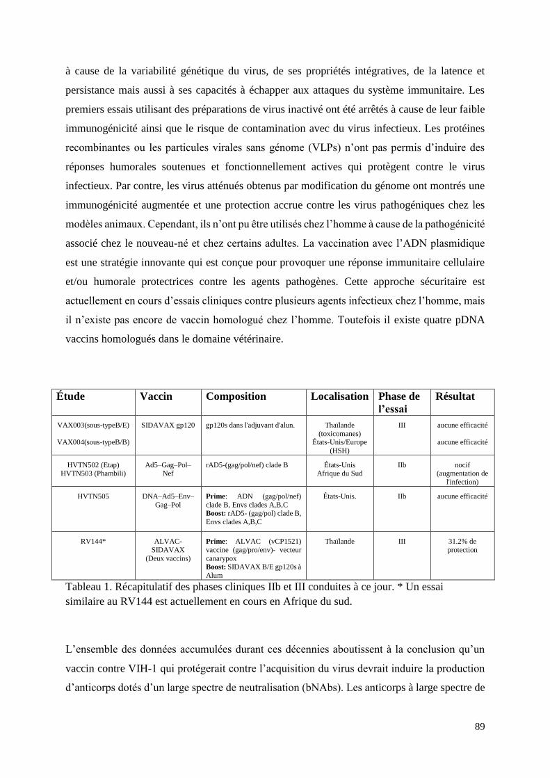
89
à cause de la variabilité génétique du virus, de ses propriétés intégratives, de la latence et
persistance mais aussi à ses capacités à échapper aux attaques du système immunitaire. Les
premiers essais utilisant des préparations de virus inactivé ont été arrêtés à cause de leur faible
immunogénicité ainsi que le risque de contamination avec du virus infectieux. Les protéines
recombinantes ou les particules virales sans génome (VLPs) n’ont pas permis d’induire des
réponses humorales soutenues et fonctionnellement actives qui protègent contre le virus
infectieux. Par contre, les virus atténués obtenus par modification du génome ont montrés une
immunogénicité augmentée et une protection accrue contre les virus pathogéniques chez les
modèles animaux. Cependant, ils n’ont pu être utilisés chez l’homme à cause de la pathogénicité
associé chez le nouveau-né et chez certains adultes. La vaccination avec l’ADN plasmidique
est une stratégie innovante qui est conçue pour provoquer une réponse immunitaire cellulaire
et/ou humorale protectrices contre les agents pathogènes. Cette approche sécuritaire est
actuellement en cours d’essais cliniques contre plusieurs agents infectieux chez l’homme, mais
il n’existe pas encore de vaccin homologué chez l’homme. Toutefois il existe quatre pDNA
vaccins homologués dans le domaine vétérinaire.
Étude Vaccin Composition Localisation Phase de
l’essai
Résultat
VAX003(sous-typeB/E)
VAX004(sous-typeB/B)
SIDAVAX gp120
gp120s dans l'adjuvant d'alun.
Thaïlande
(toxicomanes) États-Unis/Europe
(HSH)
III
aucune efficacité
aucune efficacité
HVTN502 (Etap) HVTN503 (Phambili)
Ad5–Gag–Pol–Nef
rAD5-(gag/pol/nef) clade B
États-Unis Afrique du Sud
IIb
nocif (augmentation de
l'infection)
HVTN505
DNA–Ad5–Env–
Gag–Pol
Prime: ADN (gag/pol/nef)
clade B, Envs clades A,B,C Boost: rAD5- (gag/pol) clade B,
Envs clades A,B,C
États-Unis.
IIb
aucune efficacité
RV144*
ALVAC-
SIDAVAX
(Deux vaccins)
Prime: ALVAC (vCP1521)
vaccine (gag/pro/env)- vecteur
canarypox Boost: SIDAVAX B/E gp120s à
Alum
Thaïlande
III
31.2% de
protection
Tableau 1. Récapitulatif des phases cliniques IIb et III conduites à ce jour. * Un essai
similaire au RV144 est actuellement en cours en Afrique du sud.
L’ensemble des données accumulées durant ces décennies aboutissent à la conclusion qu’un
vaccin contre VIH-1 qui protégerait contre l’acquisition du virus devrait induire la production
d’anticorps dotés d’un large spectre de neutralisation (bNAbs). Les anticorps à large spectre de

90
neutralisation peuvent se développer naturellement chez une minorité de patients infectés par
le VIH-1 après plusieurs années d'infection. Ces bNAbs ont le potentiel de protéger les
individus non infectés contre l’acquisition des virus pathogéniques ainsi que de bloquer la
propagation virale chez les individus infectés ; cependant leur développement pour une
utilisation à large échelle chez l’homme reste un énorme challenge.
Les objectifs généraux : En raison de la complexité des interactions hôte/pathogène qui sont
associées à l'infection par le VIH-1 chez l'hôte naturel et les modèles animaux actuels, un
modèle plus simple est jugé nécessaire pour faciliter les études afin de mieux comprendre les
mécanismes sous-jacents de la pathogenèse exacerbée chez l’homme. Etant donné que
l’infection par le VIH-1 est restreinte à l'homme et aux grands singes, il n'existe pas de modèle
animal simple et accessible pour conduire des études poussées en infection expérimentale. Un
virus chimérique entre les génomes des virus d’immunodéficience simienne et humaine (SHIV)
a été développé pour contourner les mécanismes de restriction liés à la réplication du VIH-1
dans les cellules de macaques. Ce virus chimérique exprime à la fois des gènes du SIV et du
VIH-1, créant ainsi un nouveau modèle pour étudier la pathogenèse du VIH-1 et tester des
vaccins chez des espèces de macaques qui sont reproduits en nombre dans les centres de
primatologie. Le SHIV-KU2 utilisé dans notre laboratoire est un des prototypes de ces virus
chimériques. Il produit les glycoprotéines d’enveloppe de type X4 du VIH-1 dans le génome
du SIVmac, et par conséquent, l'infection de macaques avec ce type de SHIV induit très
rapidement (2-3 semaines) la phase dite tardive de la pathogenèse du VIH-1 caractérisée par la
déplétion quasi-totale des cellules T CD4+. Notre laboratoire travaille à développer des virus
chimériques dotés du tropisme R5 qui devraient reproduire les premiers stades de l'infection
par le VIH-1. Ces virus constitueraient des outils essentiels pour évaluer l'efficacité des
prototypes de vaccins, mais aussi pour étudier les différentes propriétés réplicatives. En ce qui
concerne la latence du VIH-1, l'expression du génome viral est bloquée par des interactions
complexes de facteurs du virus et ceux de certaines cellules infectées. Cette complexité rend
difficile l’étude des mécanismes impliqués pour développer des thérapies appropriées. Par
conséquent il existe un réel besoin de créer des modèles dans lesquels le virus est incapable
d’effectuer la phase de latence. Notre laboratoire a développé de nouveaux génomes
chimériques de lentivirus utilisant une partie du génome de CAEV qui porte des LTRs dotés de
promoteurs constitutifs qui sont indépendants de la transactivation par Tat.
L'objectif principal de ce projet de thèse est d'utiliser les outils développés dans notre
laboratoire pour étudier les interactions hôte/VIH-1 qui, de l'infection conduisent à une
91
pathogénie complexe. Par ailleurs, nous voulions examiner la présence ou non de l’activité
neutralisante dans les réponses immunitaires humorales induites chez les macaques vaccinés
avec un lentivecteur à ADN et infectés au bout de 18 mois avec un virus d’épreuve SIVmac251
pathogène et hétérologue.
La stratégie du travail comporte la préparation d’outils appropriés pour étudier la capacité de
réplication, le tropisme et le pouvoir infectieux de trois souches VIH-1 : VIH-1 pNL4-3, VIH-
1 p89.6 et VIH-1 pWARO. Les ADNs plasmidiques portant les génomes complets de ces
souches ont été produits puis utilisés pour produire et amplifier des stocks viraux pour réaliser
une étude comparée avec notre génome chimérique CAL-VIH-R1. Ces trois souches ont été
choisies parmi plus d’une dizaine de clones moléculaires infectieux obtenus de la base de
données et de réactifs mis à disposition par le programme de réactifs SIDA du NIH. Les virus
produits par transfection de cellules HEK-239T ont été utilisés pour examiner leurs propriétés
réplicatives sur plusieurs lignées cellulaires.
Suite à l’infection des cellules T CD4+ de la lignée humaine M8166, les souches virales VIH-
1 pNL4-3 et p89.6 ont induit des effets cytopathiques typiques après trois à six jours d’infection.
Ces résultats indiquent que ces cellules ont été infectées par ces souches virales alors qu’au
contraire aucun signe d’infection n’a été observé sur les cellules inoculées avec la souche virale
VIH-1 WARO.
Pour caractériser d’avantage le tropisme de ces souches, nous avons utilisé des cellules
indicatrices de type GHOST qui co-expriment le récepteur CD4 avec soit le co-récepteur CCR5
soit le CXCR4. Si des cellules GHOST exprimant le co-récepteur CCR5 sont infectées par une
souche virale, ce virus est donc défini comme ayant un tropisme R5, et à l’inverse si des GHOST
exprimant le co-récepteur CXCR4 sont infectées par une souche virale, celle-ci est alors définie
comme ayant un tropisme X4. Quand une souche infecte les deux types cellulaires
indifféremment elle est définie comme ayant un double tropisme X4/R5. Les virus VIH-1 NL4-
3 et 89.6 ont causé des effets cytotoxiques sur les cellules GHOST CXCR4, et les souches VIH-
1 WARO et 89.6 ont causé ces effets sur des cellules GHOST CCR5. Par conséquent, la souche
VIH-1 NL4-3 est définie de tropisme X4, la souche WARO est définie de tropisme R5, tandis
que la souche VIH-1 89.6 est définie de double tropisme R5 et X4.
Les titres infectieux des stocks viraux produits ont été évalués par inoculation de cellules TZM-
bl et évaluation du nombre de cellules bleues induites par infection, ou par l’évaluation de la
TCID50/ml en utilisant la lignée cellulaire indicatrice M8166. Les résultats de ces analyses ont

92
montré que la production virale des souches VIH-1 NL4-3 et 89.6, contrairement à celle de la
souche VIH-1 WARO, a augmenté progressivement au cours des trois premiers jours. Afin de
préparer des stocks viraux, les cellules M8166 se sont avérées efficaces pour les souches VIH-
1 NL4-3 et 89.6, tandis que pour la souche VIH-1 WARO nous avons utilisé la lignée humaine
monocytaire U-937, que nous avons différenciée en macrophages mais l’infection de cette
dernière s’est montrée inefficace. Il serait donc préférable d'utiliser la lignée de cellules
TCD4+/CCR5+ MOLT T pour résoudre ce problème.
Les études de la cinétique de réplication des souches VIH-1 NL4-3 et 89.6 ont été effectuées
sur les cellules CEMx174 et M8166. Les surnageants des cellules infectées ont été recueillis
tous les 24h pendant huit jours puis les titres infectieux ont été examinés sur des cellules TZM-
bl. La production virale a augmenté progressivement jusqu'au quatrième jour sur les cellules
CEMx174 et au jusqu’au cinquième jour post infection sur les cellules M8166. La mortalité
cellulaire est devenue très importante au septième jour. Le titre infectieux du VIH-1 NL4-3 a
été trouvé supérieur à celui du VIH-1 89.6 et la production virale sur la lignée M8166 a été plus
élevée que celle sur la lignée CEMx174.
Le génome ADN double-brins du CAL-HIV-R1 comprend tous les gènes du génome du VIH-
1 pNL4-3, cependant les LTRs ont été délétées et remplacées par celles du CAEV. Ces LTR
ont des promoteurs constitutifs qui expriment fortement les gènes qu’ils contrôlent.
L’adaptation de la réplication virale par passages successifs du virus issu du clone moléculaire
de CAL-HIV-R1 a été tentée sur des cellules de la lignée M8166, puis les surnageant ont été
titrés en utilisant des cellules TZM-bl. Les résultats ont montré une légère augmentation de la
production de virus au cours des dix premiers passages, mais ensuite la production virale a
diminué.
Un dernier aspect du travail porte sur la détection et l’évaluation d'anticorps neutralisants dans
les échantillons d’animaux vaccinés avec le lentivecteur CAL-SHIV-IN- et non vaccinés, après
infection avec un virus d’épreuve. Pour ce faire, des échantillons de sérum provenant d'animaux
vaccinés et d'animaux témoins ont été prélevés à raison de plusieurs semaines après l'infection
avec le virus d’épreuve SIVmac251. Les échantillons de sérum prélevés aux semaines 1, 22,
26, 30 42 et 46 post-infection avec le virus d’épreuve ont été testés pour leur capacité à
neutraliser l’infection par séro-neutralisation réalisée contre les souches virales SHIV-KU2 et
SIVmac239. Dans le temps imparti pour cette expérimentation animale, la présence d’anticorps

93
neutralisant n’a pu être mise en évidence bien qu’une étude parallèle ait démontré que tous les
animaux avaient développé des réponses immunes cellulaires et humorales spécifiques.
L’ensemble de ce travail a permis de tester des méthodes et de valider des outils indispensables
pour l’étude comparée des propriétés du nouvel virus chimérique dérivé du génome de VIH-1 :
le CAL-HIV-IN- construit au laboratoire et dont la conception permettrait une expression
constitutive contrairement à l’expression Tat-dépendante du VIH-1.
Related Documents