APPLIED AND ENVIRONMENTAL MICROBIOLOGY, June 2007, p. 3595–3604 Vol. 73, No. 11 0099-2240/07/$08.000 doi:10.1128/AEM.02083-06 Copyright © 2007, American Society for Microbiology. All Rights Reserved. Functional Analysis of D-Alanylation of Lipoteichoic Acid in the Probiotic Strain Lactobacillus rhamnosus GG Mo ´nica Perea Ve ´lez, 1 Tine L. A. Verhoeven, 1 Christian Draing, 2 Sonja Von Aulock, 2 Markus Pfitzenmaier, 3 Armin Geyer, 3 Ivo Lambrichts, 4 Corinne Grangette, 5 Bruno Pot, 5 Jos Vanderleyden, 1 and Sigrid C. J. De Keersmaecker 1 * Centre of Microbial and Plant Genetics, Katholieke Universiteit Leuven, Kasteelpark Arenberg 20, 3001 Leuven, Belgium 1 ; Department of Biochemical Pharmacology, University of Konstanz, D-78457 Konstanz, Germany 2 ; Institute for Organic Chemistry, Philipps-Universita ¨t Marburg, D-35043 Marburg, Germany 3 ; Laboratory of Histology, BIOMED Research Institute, University Hasselt, Agoralaan Gebouw D, 3590 Diepenbeek, Belgium 4 ; and Bacte ´riologie des E ´ cosyste `mes, Institut Pasteur Lille, 59019 Lille Cedex, France 5 Received 4 September 2006/Accepted 24 March 2007 Lipoteichoic acid (LTA) is a macroamphiphile molecule which performs several functions in gram-positive bacteria, such as maintenance of cell wall homeostasis. D-Alanylation of LTA requires the proteins encoded by the dlt operon, and this process is directly related to the charge properties of this polymer strongly contributing to its function. The insertional inactivation of dltD of the probiotic strain Lactobacillus rhamnosus GG (ATCC 53103) resulted in the complete absence of D-alanyl esters in the LTA as confirmed by nuclear magnetic resonance analysis. This was reflected in modifications of the bacterial cell surface properties. The dltD strain showed 2.4-fold-increased cell length, a low survival capacity in response to gastric juice challenge, an increased sensitivity to human beta-defensin-2, an increased rate of autolysis, an increased capacity to initiate growth in the presence of an anionic detergent, and a decreased capacity to initiate growth in the presence of cationic peptides compared to wild-type results. However, in vitro experiments revealed no major differences for adhesion to human intestinal epithelial cells, biofilm formation, and immunomodulation. These properties are considered to be important for probiotics. The role of the dlt operon in lactobacilli is discussed in view of these results. The cell wall of gram-positive bacteria constitutes a protec- tive barrier essential for survival, shape, and integrity. Proteins and teichoic acids (TAs) composed of wall teichoic acid (WTA) and/or lipoteichoic acid (LTA) are associated with this peptidoglycan-containing wall (33, 40). WTA is covalently linked to the peptidoglycan, whereas LTA is a macroamphi- phile molecule with its glycolipid moiety anchored to the mem- brane and its polyglycerophosphate (Gro-P) chain extending into the wall (40). Together, TAs are the most abundant poly- anions of gram-positive bacteria and represent up to 50% of the cell wall dry weight (24). Glycosyl substitutions in WTA and D-alanyl ester (D-Ala ester) substitutions in LTA are directly related to the charge properties of these polymers and strongly contribute to their function, as evidenced by the phenotypes of strains with genetic changes altering the glycosyl substitution and D-alanylation process (17, 33, 45, 57). The D-Ala ester substitution of LTA requires four proteins encoded by the dlt operon. Two of these proteins are the D-alanyl carrier protein ligase (Dcl, encoded by dltA), which activates D-alanine by use of ATP, and the D-alanyl carrier protein (Dcp), which is encoded by dltC. DltB is a putative transmembrane protein predicted to be involved in the passage of the activated D-alanyl-Dcp complex across the glycerol phos- phate backbone of LTA. Finally, the DltD membrane protein facilitates the binding of Dcp for ligation with D-Ala and ad- ditionally has thioesterase activity for removing mischarged D-alanyl carrier proteins (13, 40). Inactivation of genes within this operon in various gram-positive bacteria results in the decrease or complete absence of D-Ala esters from LTA (1, 18, 35, 46, 47). D-Ala-deficient mutants are found to exhibit a variety of phenotypic changes that could be attributed to the resulting charge modification of their cell surface. For in- stance, alterations of cell morphology associated with defects in septum formation (9, 41, 45), variations in modulation of the activity of autolysins (6, 19, 55, 58), differences in cation bind- ing to the cell envelope required for enzyme function (33), alterations of the electromechanical properties of the cell wall (40), altered resistance to antimicrobial cationic peptides (8, 40), modified adhesion, epithelial cell invasion, and virulence (1, 35), effects on biofilm formation (7, 18, 23), and alterations in immune response (14, 22, 38, 39, 51) have been reported. While it is apparent that the D-Ala esters of LTA play an essential role in the physiology and properties of the cell sur- face of gram-positive bacteria, the genotype-phenotype rela- tion of the dlt operon is complex and appears to be species dependent. By construction of a dltD knockout mutant, this study aimed to determine first the role of the dltD gene in the D-alanylation of the LTA in the probiotic strain Lactobacillus rhamnosus GG. Probiotic bacteria are defined as “live microorganisms which, when administered in adequate amounts, confer a health benefit on the host” (20). L. rhamnosus GG is a well- * Corresponding author. Mailing address: Centre of Microbial and Plant Genetics, K. U. Leuven, Kasteelpark Arenberg 20, 3001 Leuven, Belgium. Phone: 32 16 321631. Fax: 32 16 321966. E-mail: sigrid [email protected]. Published ahead of print on 13 April 2007. 3595

Welcome message from author
This document is posted to help you gain knowledge. Please leave a comment to let me know what you think about it! Share it to your friends and learn new things together.
Transcript
APPLIED AND ENVIRONMENTAL MICROBIOLOGY, June 2007, p. 3595–3604 Vol. 73, No. 110099-2240/07/$08.00�0 doi:10.1128/AEM.02083-06Copyright © 2007, American Society for Microbiology. All Rights Reserved.
Functional Analysis of D-Alanylation of Lipoteichoic Acid in theProbiotic Strain Lactobacillus rhamnosus GG�
Monica Perea Velez,1 Tine L. A. Verhoeven,1 Christian Draing,2 Sonja Von Aulock,2Markus Pfitzenmaier,3 Armin Geyer,3 Ivo Lambrichts,4 Corinne Grangette,5
Bruno Pot,5 Jos Vanderleyden,1 and Sigrid C. J. De Keersmaecker1*Centre of Microbial and Plant Genetics, Katholieke Universiteit Leuven, Kasteelpark Arenberg 20, 3001 Leuven, Belgium1;
Department of Biochemical Pharmacology, University of Konstanz, D-78457 Konstanz, Germany2; Institute for Organic Chemistry,Philipps-Universitat Marburg, D-35043 Marburg, Germany3; Laboratory of Histology, BIOMED Research Institute,
University Hasselt, Agoralaan Gebouw D, 3590 Diepenbeek, Belgium4; and Bacteriologie des Ecosystemes,Institut Pasteur Lille, 59019 Lille Cedex, France5
Received 4 September 2006/Accepted 24 March 2007
Lipoteichoic acid (LTA) is a macroamphiphile molecule which performs several functions in gram-positivebacteria, such as maintenance of cell wall homeostasis. D-Alanylation of LTA requires the proteins encoded bythe dlt operon, and this process is directly related to the charge properties of this polymer strongly contributingto its function. The insertional inactivation of dltD of the probiotic strain Lactobacillus rhamnosus GG (ATCC53103) resulted in the complete absence of D-alanyl esters in the LTA as confirmed by nuclear magneticresonance analysis. This was reflected in modifications of the bacterial cell surface properties. The dltD strainshowed 2.4-fold-increased cell length, a low survival capacity in response to gastric juice challenge, anincreased sensitivity to human beta-defensin-2, an increased rate of autolysis, an increased capacity to initiategrowth in the presence of an anionic detergent, and a decreased capacity to initiate growth in the presence ofcationic peptides compared to wild-type results. However, in vitro experiments revealed no major differencesfor adhesion to human intestinal epithelial cells, biofilm formation, and immunomodulation. These propertiesare considered to be important for probiotics. The role of the dlt operon in lactobacilli is discussed in view ofthese results.
The cell wall of gram-positive bacteria constitutes a protec-tive barrier essential for survival, shape, and integrity. Proteinsand teichoic acids (TAs) composed of wall teichoic acid(WTA) and/or lipoteichoic acid (LTA) are associated with thispeptidoglycan-containing wall (33, 40). WTA is covalentlylinked to the peptidoglycan, whereas LTA is a macroamphi-phile molecule with its glycolipid moiety anchored to the mem-brane and its polyglycerophosphate (Gro-P) chain extendinginto the wall (40). Together, TAs are the most abundant poly-anions of gram-positive bacteria and represent up to 50% of thecell wall dry weight (24). Glycosyl substitutions in WTA andD-alanyl ester (D-Ala ester) substitutions in LTA are directlyrelated to the charge properties of these polymers and stronglycontribute to their function, as evidenced by the phenotypes ofstrains with genetic changes altering the glycosyl substitutionand D-alanylation process (17, 33, 45, 57).
The D-Ala ester substitution of LTA requires four proteinsencoded by the dlt operon. Two of these proteins are theD-alanyl carrier protein ligase (Dcl, encoded by dltA), whichactivates D-alanine by use of ATP, and the D-alanyl carrierprotein (Dcp), which is encoded by dltC. DltB is a putativetransmembrane protein predicted to be involved in the passageof the activated D-alanyl-Dcp complex across the glycerol phos-
phate backbone of LTA. Finally, the DltD membrane proteinfacilitates the binding of Dcp for ligation with D-Ala and ad-ditionally has thioesterase activity for removing mischargedD-alanyl carrier proteins (13, 40). Inactivation of genes withinthis operon in various gram-positive bacteria results in thedecrease or complete absence of D-Ala esters from LTA (1, 18,35, 46, 47). D-Ala-deficient mutants are found to exhibit avariety of phenotypic changes that could be attributed to theresulting charge modification of their cell surface. For in-stance, alterations of cell morphology associated with defectsin septum formation (9, 41, 45), variations in modulation of theactivity of autolysins (6, 19, 55, 58), differences in cation bind-ing to the cell envelope required for enzyme function (33),alterations of the electromechanical properties of the cell wall(40), altered resistance to antimicrobial cationic peptides (8,40), modified adhesion, epithelial cell invasion, and virulence(1, 35), effects on biofilm formation (7, 18, 23), and alterationsin immune response (14, 22, 38, 39, 51) have been reported.While it is apparent that the D-Ala esters of LTA play anessential role in the physiology and properties of the cell sur-face of gram-positive bacteria, the genotype-phenotype rela-tion of the dlt operon is complex and appears to be speciesdependent.
By construction of a dltD knockout mutant, this study aimedto determine first the role of the dltD gene in the D-alanylationof the LTA in the probiotic strain Lactobacillus rhamnosusGG. Probiotic bacteria are defined as “live microorganismswhich, when administered in adequate amounts, confer ahealth benefit on the host” (20). L. rhamnosus GG is a well-
* Corresponding author. Mailing address: Centre of Microbial andPlant Genetics, K. U. Leuven, Kasteelpark Arenberg 20, 3001 Leuven,Belgium. Phone: 32 16 321631. Fax: 32 16 321966. E-mail: [email protected].
� Published ahead of print on 13 April 2007.
3595
studied probiotic strain (4, 21). For this reason, the second aimof this study was to investigate the role of the D-alanylation ofthe L. rhamnosus GG LTA in some morphological, membranecharge property, and probiotic characteristics, such as adher-ence to human intestinal epithelial cells, biofilm formation,resistance to gastric juice challenge, and immunomodulation ofhuman intestinal epithelial cells and peripheral blood mono-nuclear cells.
MATERIALS AND METHODS
Bacterial strains, plasmids, media, and growth conditions. The bacterialstrains and plasmids used in this study are listed in Table 1. L. rhamnosus GG wasroutinely grown in nonshaken MRS medium (Difco) at 37°C. Escherichia colicells were grown in Luria-Bertani (LB) medium with aeration at 37°C. Whenrequired, antibiotics were used at the following concentrations: 10 �g/ml tetra-cycline, 100 �g/ml ampicillin, and 5 �g/ml (L. rhamnosus GG) or 100 �g/ml (E.coli) erythromycin.
DNA manipulations. Routine molecular biology techniques were employed asdescribed before (49). Primer sequences used in this study are listed in Table 2(Eurogentec). Enzymes for molecular biology were purchased from New En-gland Biolabs and used according to the instructions of the suppliers. PlasmidDNA from E. coli was prepared using QIAGEN Miniprep kits. ChromosomalDNA from L. rhamnosus GG was isolated as previously described (15).
Construction and analysis of the L. rhamnosus GG dltD mutant (CMPG5540).The complete genome sequence of the dlt operon of Lactobacillus rhamnosusATCC 7469 is published in the NCBI database under accession numberAF192553 (13). Based on this sequence, a fragment of 2,682 bp containing the L.rhamnosus GG dltD gene and its 779-bp upstream and 635-bp downstreamregions was amplified using primers Pro-230 and Pro-231 and cloned in pCRII-TOPO, yielding pCMPG5221. Subsequently, the dltD gene with its flankingregions was subcloned as a HindIII-EcoRV fragment in pFAJ5301, resultingin pCMPG5222. To inactivate the dltD gene, a BbsI-NcoI fragment frompCMPG5522 was replaced by the tetracycline resistance cassette tet(M) previously
amplified from plasmid pMD5057 of L. plantarum 5057 (11) by use of primersPro-221 and Pro-222. The resulting suicide vector, pCMPG5223, was electropo-rated into L. rhamnosus GG (15). Transformants were selected for resistance to10 �g/ml of tetracycline. Confirmation of DNA recombination was performed byPCR using primers Pro-261 and Pro-231 and Southern hybridization using a dltDprobe synthesized with primers Pro-261 and Pro-262. The dltD mutant wasdesignated CMPG5540 and was further analyzed.
Complementation of the knockout dltD mutant. For the construction of thecomplemented strain, the functional dltD gene was cloned under the control ofthe L. rhamnosus GG ldhL promoter in pCMPG5901, yielding pCMPG5226.Subsequently, an EclII136-NotI fragment containing the functional dltD geneand the ldhL promoter was subcloned into pLAB1301 (28). This construct was
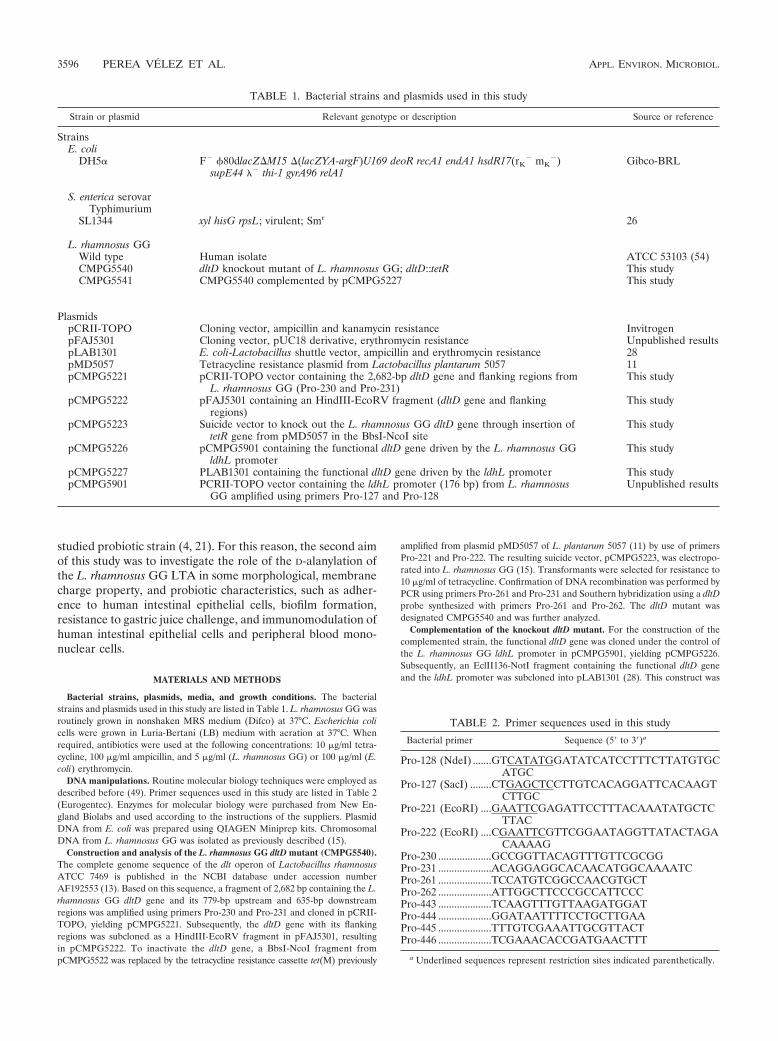
TABLE 1. Bacterial strains and plasmids used in this study
Strain or plasmid Relevant genotype or description Source or reference
StrainsE. coli
DH5� F� �80dlacZ�M15 �(lacZYA-argF)U169 deoR recA1 endA1 hsdR17(rK� mK
�)supE44 �� thi-1 gyrA96 relA1
Gibco-BRL
S. enterica serovarTyphimurium
SL1344 xyl hisG rpsL; virulent; Smr 26
L. rhamnosus GGWild type Human isolate ATCC 53103 (54)CMPG5540 dltD knockout mutant of L. rhamnosus GG; dltD::tetR This studyCMPG5541 CMPG5540 complemented by pCMPG5227 This study
PlasmidspCRII-TOPO Cloning vector, ampicillin and kanamycin resistance InvitrogenpFAJ5301 Cloning vector, pUC18 derivative, erythromycin resistance Unpublished resultspLAB1301 E. coli-Lactobacillus shuttle vector, ampicillin and erythromycin resistance 28pMD5057 Tetracycline resistance plasmid from Lactobacillus plantarum 5057 11pCMPG5221 pCRII-TOPO vector containing the 2,682-bp dltD gene and flanking regions from
L. rhamnosus GG (Pro-230 and Pro-231)This study
pCMPG5222 pFAJ5301 containing an HindIII-EcoRV fragment (dltD gene and flankingregions)
This study
pCMPG5223 Suicide vector to knock out the L. rhamnosus GG dltD gene through insertion oftetR gene from pMD5057 in the BbsI-NcoI site
This study
pCMPG5226 pCMPG5901 containing the functional dltD gene driven by the L. rhamnosus GGldhL promoter
This study
pCMPG5227 PLAB1301 containing the functional dltD gene driven by the ldhL promoter This studypCMPG5901 PCRII-TOPO vector containing the ldhL promoter (176 bp) from L. rhamnosus
GG amplified using primers Pro-127 and Pro-128Unpublished results
TABLE 2. Primer sequences used in this study
Bacterial primer Sequence (5� to 3�)a
Pro-128 (NdeI) .......GTCATATGGATATCATCCTTTCTTATGTGCATGC
Pro-127 (SacI) ........CTGAGCTCCTTGTCACAGGATTCACAAGTCTTGC
Pro-221 (EcoRI) ....GAATTCGAGATTCCTTTACAAATATGCTCTTAC
Pro-222 (EcoRI) ....CGAATTCGTTCGGAATAGGTTATACTAGACAAAAG
Pro-230 ....................GCCGGTTACAGTTTGTTCGCGGPro-231 ....................ACAGGAGGCACAACATGGCAAAATCPro-261 ....................TCCATGTCGGCCAACGTGCTPro-262 ....................ATTGGCTTCCCGCCATTCCCPro-443 ....................TCAAGTTTGTTAAGATGGATPro-444 ....................GGATAATTTTCCTGCTTGAAPro-445 ....................TTTGTCGAAATTGCGTTACTPro-446 ....................TCGAAACACCGATGAACTTT
a Underlined sequences represent restriction sites indicated parenthetically.
3596 PEREA VELEZ ET AL. APPL. ENVIRON. MICROBIOL.
designated pCMPG5227 and introduced in CMPG5540 by electroporation aspreviously described (15). Transformants were selected for resistance to 10 �g/mlof tetracycline and to 5 �g/ml of erythromycin. The complemented strain,CMPG5541, was confirmed by PCR using primers Pro-127, Pro-262, and Pro-261and included in all assays performed.
Cloning and analysis of the L. rhamnosus GG dlt operon. Concomitantly, inorder to sequence the additional genes within the dlt operon of L. rhamnosusGG, primers Pro-443, Pro-444, Pro-445, and Pro-446 were designed after selec-tion on the DNA sequence of the dlt operon of L. rhamnosus ATCC 7469. ThePCRs were carried out with Pfx (Invitrogen), a DNA polymerase with proof-reading activity. The PCR fragments were cloned in pCRII-TOPO and se-quenced by the chain termination dideoxynucleoside triphosphate method (50)with a BigDye Terminator V3.1 cycle sequencing kit, using an ABI PRISM3100-Avant genetic analyzer (Applied Biosystems, Lennik, Belgium). Databaseswere screened for similarities by using BLAST (2, 3), and alignment of overlap-ping fragments was performed with Vector NTI Advance 10 ContigExpresssoftware (Informax, Oxford, United Kingdom).
LTA purification. For the LTA isolation, 11.83 g of lyophilized L. rhamnosusGG cells and 22.12 g of the dltD mutant (CMPG5540) cells were extracted usingbutanol and hydrophobic interaction chromatography as previously described(38), with minor modifications. Briefly, bacterial cells were disrupted for 15 minby sonication and resuspended in 50 ml of 0.1 M citrate buffer (pH 4.7). Thebacterial lysate was mixed while being stirred with an equal volume of n-butanol(Merck, Darmstadt, Germany) for 20 min at room temperature. The suspensionwas then centrifuged at 8,300 g for 40 min, resulting in a two-phase system. Thelower aqueous phase was lyophilized and subsequently resuspended in 35 ml ofchromatography start buffer (15% n-propanol in 0.1 M ammonium acetate; pH4.7) followed by 60 min of centrifugation at 26,900 g and sterilization byfiltration (0.2 �m). The supernatant was subjected to hydrophobic interactionchromatography on an octyl-Sepharose column (2.5 by 11 cm) using a lineargradient of from 15% to 60% n-propanol in 0.1 M ammonium acetate (pH 4.7).LTA-containing fractions were identified by their phosphate content based onthe formation of phosphomolybdenum blue from phosphate (52). The endotoxincontamination of the LTA preparations was assessed by the kinetic Limulusamoebocyte lysate assay (Charles River, Charleston, SC).
LTA structure analysis by NMR spectroscopy. Nuclear magnetic resonance(NMR) spectra of the LTA samples were determined with a Bruker Avance 600MHz spectrometer with a 5-mm probe at 300 K. Spectra were obtained usingD2O solutions and 3-(trimethylsilyl)3,3,2,2-tetradeuteropropionic acid sodiumsalt (d4-TSPA) as an internal standard for 1H NMR (H, 0.00 ppm) and acetonefor chemical shifts of 13C (C, 30.02 ppm). Assignments were taken from two-dimensional homonuclear double-quantum-filtered correlation spectroscopy, to-tal correlated spectroscopy, and rotational nuclear Overhauser effect spectros-copy experiments using a water suppression technique and two-dimensionalheteronuclear single-quantum correlation (1H to 13C) spectra. In the total cor-related spectroscopy experiments the mixing times were 100 ms, and the rota-tional nuclear Overhauser effect spectroscopy experiments were performed with200 ms of spinlock time. Data acquisition and processing were done usingstandard Bruker software. The average number of repeating units in the poly-glycerophosphate backbone, the percentage of substitution, and the chain lengthof the fatty acids in the membrane anchor were calculated directly from theintegrals of the proton spectra.
Transmission electron microscopy. Bacteria were grown overnight (16 h at37°C). Uncoated grids were used as a probe to adsorb bacterial cells. The gridswere placed on a drop of bacterial suspension for 15 s, incubated in 0.25%phosphotungstenic acid (pH � 7) for 30 s, and washed three times, and excessliquid was drained. The bacteria were observed with a Philips EM 208S trans-mission electron microscope at 80 kV. Images were digitalized using an SISimage analysis system.
Analysis of autolysis. Triton X-100 was used to study induced autolysis undernongrowing conditions as previously described (48), with minor modifications.Briefly, cells were grown overnight, harvested by centrifugation (4,000 g for 20min at 4°C), washed three times with equal volumes of phosphate buffer (pH 6.5;10 mM), and resuspended (optical density at 600 nm [OD600] of �1.6) inphosphate buffer (pH 6.5; 200 mM) containing 0.05% (vol/vol) Triton X-100.The cell suspension was incubated at 37°C under agitation, and autolysis wasmonitored by examining the decrease of OD600 in time.
Growth in the presence of the cationic peptides nisin and polymyxin B. Nisinand polymyxin B were purchased from Sigma-Aldrich. Overnight cultures con-taining about 108 to 109 CFU/ml of L. rhamnosus GG wild-type, dltD mutant, andcomplemented strain cells were diluted 15,000-fold in MRS medium containingeither nisin at a concentration between 0.2 and 2.5 �g/ml or polymyxin B at aconcentration between 8 and 1,000 �g/ml. Bacteria were grown at 37°C with
continuous shaking in order to avoid cell aggregation, and the OD600 was mea-sured automatically each 30 min during at least 80 h using a Bioscreen Cinstrument (Labsystems Ltd. Oy). Concomitantly, growth of L. rhamnosus GG,the dltD mutant, and the complemented strain in the presence of an anionicdetergent (sodium dodecyl sulfate [SDS] at 0.01% and 0.015%) was assessed.Each run was performed at least in triplicate.
Human beta-defensin sensitivity assay. L. rhamnosus GG wild-type and dltDmutant cells were tested for their sensitivity to 3 �g/ml human beta-defensin 1(hBD1) and hBD2. After 3 h of incubation at 37°C, bacterial viability wasmeasured by plating serial dilutions on MRS agar as previously described (15).The experiment was performed in triplicate.
Survival in simulated gastric juice. Simulated gastric juice was prepared andsurvival tests were performed as previously described (10). The percentagesof survival were calculated by comparing the cell numbers before and afteraddition to simulated gastric juice at 0, 30, 60, and 90 min. The experiment wasperformed in triplicate.
In vitro adhesion assay to Caco-2 and HT-29 human intestinal epithelial celllines. Caco-2 and HT-29 cells were purchased from ATCC (Rockville, MD).Cells were routinely grown in 75-cm2 culture flasks under conditions of 37°C, 5%CO2, and 90% relative humidity. For both cell lines, Dulbecco modified Eaglemedium (DMEM)/F-12 (GibcoBRL) (1:1) supplemented with 10% fetal bovineserum (FBS; HyClone) but without antibiotics was used as the culture medium.Cells were passaged every 3 days (at 70 to 80% confluence) at a split ratio of 1to 7. For adhesion experiments, epithelial cells were plated at a density of 40,000cells/cm2 in 12-well plates (Cellstar). Confluence was reached within 3 to 4 daysafter seeding, and monolayers were used for the experiments 15 days afterseeding. Adherence of L. rhamnosus GG wild-type, dltD mutant, and comple-mented strain cells to the epithelial cells was examined by adding 1.5 ml ofDMEM containing 109 CFU/ml. After incubation at 37°C for 45 min, epithelialcells were washed two times with prewarmed phosphate-buffered saline (PBS).Subsequently, 100 �l of 1 trypsin-EDTA (Invitrogen) was added to each welland incubated for 10 min at 37°C. Finally, 900 �l of PBS was added and mixed,and serial dilutions were plated out. Plates were incubated at 37°C for 72 h.Adhesion of L. rhamnosus GG wild-type, dltD mutant, and complemented straincells was tested in triplicate in three independent experiments.
In vitro biofilm assay. In vitro biofilm formation was determined as previouslydescribed (16, 36), with minor modifications. Briefly, biofilm formation wasassayed on polystyrene pegs hanging into microtiter plate wells. The pegs wereplaced in wells containing a bacterial concentration of 3 107 CFU in 200 �lAOAC medium (Difco) (36) and incubated in anaerobic jars for 72 h at 37°C.Medium was changed every day. Wells containing sterile AOAC medium and L.rhamnosus GG wild-type cells were included as negative and positive controls,respectively. Each experiment was performed in triplicate.
Immunomodulation of HT-29 cell line. HT-29 cells were maintained as de-scribed above. L. rhamnosus GG wild-type and dltD mutant cells were grownovernight, centrifuged at 4,000 g and 4°C for 20 min, and washed with coldPBS. Immunomodulation of HT-29 cells was examined by adding 1.5 ml ofDMEM without FBS containing 5 106 CFU/ml of either L. rhamnosus GGwild-type or dltD mutant cells. Salmonella enterica serovar Typhimurium SL1344cells in a concentration of 5 106 CFU/ml and interleukin-1 beta (IL-1 )(Sigma-Aldrich) (10 ng/ml) were used as positive controls, and DMEM withoutFBS was used as a negative control. After incubation at 37°C for 3.5 h (5% CO2
and 90% humidity), epithelial cells were washed two times with prewarmed PBS.Subsequently, 200 �l of PBS was added to each well and RNA extraction wasperformed by using a High Pure RNA isolation kit (Roche Molecular Biochemi-cals) and following the manufacturer’s instructions. After isolation, RNA integ-rity was analyzed using an Agilent RNA 600 kit and 2100 bioanalyzer expertsoftware. Cytokine gene expression was determined by real-time reverse trans-criptase PCR using primers and probes as previously described (43).
Cytokine induction in peripheral blood mononuclear cells (PBMC). Cytokineinduction using a 24-h culture of bacterial cells, and using Streptococcus gordoniiLMG17843 and E. coli TG1 cells as internal controls, was performed as previ-ously described (22).
Nucleotide sequence accession number. The dlt sequence of L. rhamnosus GGhas been deposited in the NCBI database under GenBank accession numberDQ906101.
RESULTS
Analysis of the dlt operon of L. rhamnosus GG. Since thegenome sequence of L. rhamnosus GG is unavailable, a ho-mology strategy based on the dlt sequence of L. rhamnosus
VOL. 73, 2007 FUNCTIONAL ANALYSIS OF DltD IN L. RHAMNOSUS GG 3597
ATCC 7469 (AF192553) (13) was used to isolate the dlt geneswithin the dlt operon of L. rhamnosus GG cells. Based on thisstrategy, a continuous 5.8-kb sequence of L. rhamnosus GGgenomic DNA was determined. The analysis of the sequencerevealed four open reading frames (dltA, dltB, dltC, and dltD),as depicted in Fig. 1. Additionally, the in silico-translated pro-tein sequences encoded by the L. rhamnosus GG dlt operon,i.e., those corresponding to DltA protein (506 amino acids[aa]), DltB protein (405 aa), DltC protein (81 aa), and DltDprotein (423 aa), were used for screening databases usingBLAST (2, 3). Homologies of these L. rhamnosus GG Dltprotein sequences to the Dlt protein sequences of L. rhamno-sus ATCC 7469 and L. plantarum WCFS1 are summarized inTable 3.
Finally, the dltD gene of L. rhamnosus GG which encodes aputative protein of 423 aa with a theoretical mass and pI of47.987 kDa and 9.57, respectively, was successfully knockedout by the insertion of the tetracycline cassette from L. plan-tarum 5057. The correct insertion was confirmed by PCR andSouthern hybridization as described in Materials and Methods(data not shown).
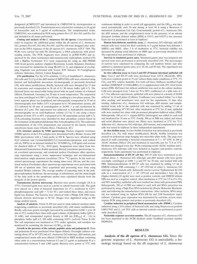
LTA purification and structure analysis. After purification,different LTA-containing fractions were identified. For L.rhamnosus GG wild-type cells (Fig. 2A), fractions 41 to 49 werepooled, resulting in 14.81 mg LTA. For the dltD mutant cells(Fig. 2B), the phosphate determination revealed fractions 41to 46 (represented by peak 1) and fractions 48 to 88 (repre-sented by peak 2) with 9.56 mg LTA in peak 1 and 9.26 mgLTA in peak 2, respectively. Endotoxin contamination of allLTA extractions showed lipopolysaccharide contamination be-low 0.6 endotoxin units/mg LTA, i.e., less than 60 pg lipopoly-saccharide per mg LTA.
The molecular structure determined by NMR spectroscopyrevealed that LTA from L. rhamnosus GG wild-type cells isconstituted of Gro-P with D-alanyl esters as unique detectablesubstituents (74% D-Ala:Gro-P). LTA from the L. rhamnosusGG wild type is formed by average chains of 50 glycerophos-phate residues (Fig. 3). For the dltD mutant, two fractions wereeluted from the chromatography column as previously men-tioned. The major peak (fractions 41 to 46) and the secondpeak (fractions 48 to 55) contain LTA molecules with averagelengths of only 29 and 7 glycerophosphate residues, respec-tively. D-Alanyl ester substitution in the dltD mutant was com-pletely abolished in both peaks. The analysis of the glycolipicmoiety in the wild type reveals an average fatty acid chain ofC14, with one double bond per fatty acid (two double bonds inevery membrane anchor). The fatty acid chain in the dltDmutant was on average two carbon atoms longer (C16) in com-parison to that seen in the parental strain. Likewise, as in the
FIG. 1. Genetic organization of the L. rhamnosus GG dlt operon. Aschematic representation of the dlt operon (�5.8 kb) of L. rhamnosusGG as revealed by sequencing and BLAST analysis is presented. Openreading frames corresponding to dltA, dltB, dltC, and dltD are indi-cated with arrows. The putative promoter region (5� end), representedin the figure by a flag, contains a �10 “TATTAA” region and a �35“TGGTTT” region separated by 19 bp. The potential ribosome bind-ing site “GGGGG” is located 8 bp upstream of the putative ATG startcodon of dltA (not shown). A second potential ribosomal binding site,“AAAGAGG,” was found 9 bp upstream of the putative start codon ofthe dltC gene (not shown). dltB overlaps the stop codon of dltA by 1 bp,and dltD overlaps the stop codon of dltC by 4 bp. dltC and dltB areseparated by a 69-bp intergenic region. The 3� end of the operonreveals a putative terminator 99 bp downstream of the dltD stop codon,represented in the figure by a stem-loop. It is a 12-bp inverted repeatfollowed by a series of T residues (TTTATTTT).
TABLE 3. Homology analysis of the translated L. rhamnosus GG Dlt proteins
Strain% Homology (GenBank accession no.)
ReferenceDltA DltB DltC DltD
L. rhamnosus ATCC 7469 99 (AAF09201) 95 (AAF09202) 100 (AAF09203) 99 (AAF09204) 12L. plantarum WCFS1 62 (NP_785546) 64 (NP_785546) 64 (NP_785544) 51 (NP_785543) 30
FIG. 2. LTA elution profiles and phosphate content of L. rhamno-sus GG wild-type and mutant cells. The phosphate determination ofthe LTA fractions from L. rhamnosus GG wild-type (A) and dltDmutant (B) cells is plotted together with the correspondent propanolgradient of the fast protein liquid chromatography.
3598 PEREA VELEZ ET AL. APPL. ENVIRON. MICROBIOL.

wild type, in the dltD mutant one double bond per fatty acidwas present (data not shown).
Increase in cell length and defects in septum formationassessed by transmission electron microscopy. Comparison oftransmission electron micrographs of the L. rhamnosus GGwild-type and dltD mutant cells showed an increase in thelength of the mutant of about 2.4-fold in comparison to the
parental strain results (Fig. 4A). Additionally, morphologicalalterations at the level of the septum and defective cell sepa-ration in the dltD mutant were observed (Fig. 4B).
Analysis of autolysis. As shown in Fig. 5, the dltD mutantlyses to a greater extent than the parental strain. An OD600
decrease of 0.2 units for the dltD mutant corresponding to�108 CFU/ml was observed during the first hour of incuba-
FIG. 3. Schematic representation of the structure of the LTA from the L. rhamnosus GG wild type determined from NMR spectroscopyanalysis. The average number of repeating units in the polyglycerophosphate backbone, the average percentage of substitution, and the averagechain length of the fatty acids in the membrane anchor were calculated directly from the integrals of the proton spectra. R1 and R2, fatty acidchains. D-Ala ester substitution of the wild type (R � D-Ala, OH) is replaced by OH groups in the dltD mutant.
FIG. 4. Effect of D-alanylation on cell length and septum formation. (A) Ultrastructural analysis of the cell morphology of L. rhamnosus GGwild-type and dltD mutant cells grown overnight in MRS medium and visualized with transmission electron microscopy at 80 kV. The cell lengthof the complemented strain was restored to wild-type values. (B) Defect in septum morphology. The arrow shows the altered cell wall around theseptum in the dltD mutant.
VOL. 73, 2007 FUNCTIONAL ANALYSIS OF DltD IN L. RHAMNOSUS GG 3599

tion in comparison to the OD600 value obtained for the wildtype.
Effects of the cationic peptides nisin and polymyxin B andthe anionic detergent SDS on the growth of L. rhamnosus GGwild-type and dltD mutant cells. The capacity of L. rhamnosusGG and the dltD mutant cells to grow in medium supple-mented with the cationic peptides nisin and polymyxin B wasevaluated. In general, a minimum of �10 h of lag-phase retar-dation was observed for the mutant grown in the presence ofnisin at different concentrations compared to the wild-typeresults. However, the final cell densities reached were similarfor the two strains (Fig. 6). For polymyxin B, the dltD mutantwas found to have �10 h of lag-phase retardation even with aconcentration of polymyxin B 100 times lower than that usedfor the wild type. Again, no differences were observed regard-ing final cell density (data not shown). Additionally, the ability
of L. rhamnosus GG and the dltD mutant to grow in mediumcontaining low concentrations of a strong anionic detergent(SDS) was evaluated. In this case, the dltD mutant was shownto be less affected by the action of SDS than the parental strain,reaching a higher optical density in the stationary phase (Fig.7). The sensitivity of the dltD mutant to the tested cationicpeptides and SDS was restored to wild-type levels when themutant was complemented with the dltD gene (data notshown).
Sensitivity of L. rhamnosus GG wild type and dltD mutantcells to human beta-defensins. We investigated the effect ofhBD1 and hBD2 on L. rhamnosus GG wild-type and dltDmutant cells (Fig. 8). While hBD1 showed no effect on L.rhamnosus GG wild-type or dltD mutant cells after 3 h ofincubation, the cells were sensitive to hBD2. The dltD mutantwas on average �50% more sensitive to cationic hBD2 thanthe parental strain (Fig. 8).
Survival in simulated gastric juice. The dltD mutant showeda strong reduction in the capacity to survive the in vitro gastricjuice challenge, resulting in complete loss of viability after 30min of incubation (Fig. 9). Additionally, the capacity to initiate
FIG. 5. Effects of the mutation of the dltD gene on autolysis. Au-tolysis of the L. rhamnosus GG wild type (squares) and the dltD mutant(CMPG5540) (circles) was followed in time by incubation of the cellswith a lysis inducer (Triton X-100). The values reported in the exper-iment did not differ by more than 5%. Therefore, single datum pointsare presented in the figure without standard deviation bars. OD600values comparable to wild-type values were obtained for the comple-mented strain (CMPG5541).
FIG. 6. Effect of the cationic peptide nisin on the growth of L.rhamnosus GG wild-type and dltD mutant cells. No differences ingrowth in MRS medium between the L. rhamnosus GG wild type andthe dltD mutant were observed. Therefore, average growth values forboth strains are represented by squares (control). Values obtained forthe growth of the L. rhamnosus GG wild type (triangles) versus that ofthe dltD mutant (CMPG5540) (circles) in medium containing 1.5�g/ml nisin represent the average results of three independent runs.
FIG. 7. Effect of SDS on the growth of the L. rhamnosus GG wildtype and the dltD mutant. Values obtained for the growth of the L.rhamnosus GG wild type (cross and lines) versus that of the dltDmutant (CMPG5540) (triangles and squares) in medium containingeither 0.01% or 0.015% SDS represent the average results of threeindependent runs.
FIG. 8. Sensitivity of the L. rhamnosus GG wild type (wt) and thedltD mutant to human beta-defensins. Bacterial viability of L. rham-nosus GG wild-type and dltD mutant cells after 3 h of contact with 3�g/ml hBD1 (stippled white bars) or hBD2 (stippled gray bars) wasdetermined by plate counting. The values represent percentages ofsurvival of the initial numbers of L. rhamnosus cells. No human beta-defensins were added to the controls (black bars). The error barsindicate standard deviations of the results of three independent mea-surements.
3600 PEREA VELEZ ET AL. APPL. ENVIRON. MICROBIOL.
growth under conditions of low pH (3.5, 4.0, and 4.5) wasevaluated. Results confirmed that the acid tolerance of thedltD mutant was strongly diminished compared to that of thewild type (data not shown).
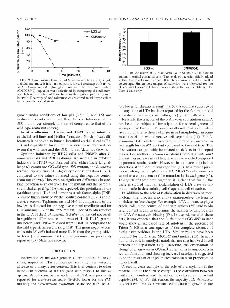
In vitro adhesion to Caco-2 and HT-29 human intestinalepithelial cell lines and biofilm formation. No significant dif-ferences in adhesion to human intestinal epithelial cells (Fig.10) and capacity to form biofilm in vitro were observed be-tween the wild type and the dltD mutant (data not shown).
Cytokine induction by HT-29 cells and PBMCs after L.rhamnosus GG and dltD challenge. An increase in cytokineinduction in HT-29 was observed after either bacterial chal-lenge (L. rhamnosus GG wild type, dltD mutant, and S. entericaserovar Typhimurium SL1344) or cytokine stimulation (IL-1 )compared to the values obtained using the negative control(data not shown). However, no significant differences in cyto-kine induction were observed for the mutant and the parentalstrain challenge (Fig. 11A). As expected, the proinflammatorycytokines tested (IL-8 and tumor necrosis factor alpha [TNF-�]) were highly induced by the positive controls (IL-1 and S.enterica serovar Typhimurium SL1344) in comparison to thelow levels detected for the negative control (medium) and forL. rhamnosus GG or the dltD mutant. Lack of D-Ala residuesin the LTA of the L. rhamnosus GG dltD mutant did not resultin significant differences in the levels of IL-10, IL-12, gammainterferon, and TNF-� released from PBMC in comparison tothe wild-type strain results (Fig. 11B). The gram-negative con-trol strain (E. coli) induced more IL-10 than the gram-positivebacteria (L. rhamnosus GG and S. gordonii), as previouslyreported (25) (data not shown).
DISCUSSION
Inactivation of the dltD gene in L. rhamnosus GG has astrong impact on LTA composition, resulting in a completeabsence of D-alanyl ester content. This is in contrast to otherlactic acid bacteria so far analyzed with respect to the dltoperon. A reduction in D-alanylation of LTA was previouslyreported for Lactococcus lactis (fivefold lower for the dltDmutant) and Lactobacillus plantarum NCIMB8826 (8- to 40-
fold lower for the dltB mutant) (45, 55). A complete absence ofD-alanylation of LTA has been reported for the dltA mutants ofa number of gram-positive pathogens (1, 18, 35, 46, 47).
Recently, the function of the D-Ala ester substitution in LTAhas been the subject of investigation for several genera ofgram-positive bacteria. Previous results with D-Ala ester-defi-cient mutants have shown changes in cell morphology, in somecases associated with defective cell separation (41). For L.rhamnosus GG, electron micrographs showed an increase incell length for the dltD mutant compared to the wild type. Thisobservation can probably be related to defects in the septalregion. For another L. rhamnosus strain (the ATCC 7469 dltDmutant), an increase in cell length was also reported comparedto parental strain results. However, in this case no obviousalteration at the septum was reported (13). In a recent publi-cation, elongated L. plantarum NCIMB8826 cells were ob-served as a consequence of the mutation in the dltB gene (45).Taking all of these data together, it is clear that for all thebacteria studied thus far, D-alanylation of LTA plays an im-portant role in determining cell shape and cell septation.
In addition to the role of D-alanylation in determining mor-phology, this process also allows gram-positive bacteria tomodulate surface charge. For example, LTA appears to play acrucial role in the control of autolysin activity (55), and D-Alaester content seems to determine the number of anionic siteson LTA for autolysin binding (58). In accordance with thesedata, it was expected that the L. rhamnosus GG dltD mutantwould show an increased rate of autolysis in the presence ofTriton X-100 as a consequence of the complete absence ofD-Ala ester residues in the LTA. Similar results have beenreported for the L. lactis MG1363 dltD mutant (55). In addi-tion to the role in autolysis, autolysins are also involved in celldivision and separation (32). Therefore, the observation ofelongated L. rhamnosus GG dltD mutant cells having defects inseptum formation and showing increased autolysis is suggestedto be the result of changes in electromechanical properties ofthe cell wall.
A second clear example of the role of D-alanylation in themodification of the surface charge is the correlation betweenD-Ala ester content and the action of cationic antimicrobialpeptides (34, 40). For this reason, the capacity of L. rhamnosusGG wild-type and dltD mutant cells to initiate growth in the
FIG. 10. Adhesion of L. rhamnosus GG and the dltD mutant tohuman intestinal epithelial cells. The levels of bacteria initially addedto the Caco-2 cells were set to 100%. Data shown are relative to thispercentage. Similar percentages of adhesion were observed for theHT-29 and Caco-2 cell lines. Graphs show the values obtained forCaco-2 cells only.
FIG. 9. Comparison of survival of L. rhamnosus GG wild-type (wt)and dltD mutant cells in simulated gastric juice. Percentages of survivalof L. rhamnosus GG (triangles) compared to the dltD mutant(CMPG5540) (squares) were calculated by comparing the cell num-bers before and after addition to simulated gastric juice at 30-minintervals. Recovery of acid tolerance was restored to wild-type valuesin the complemented strain.
VOL. 73, 2007 FUNCTIONAL ANALYSIS OF DltD IN L. RHAMNOSUS GG 3601

presence of two cationic peptides was evaluated. As expected,a significant difference between the dltD mutant and the pa-rental strain was observed. On the other hand, it can be spec-ulated that negatively charged compounds might be repelled toa greater extent in bacteria with a lower degree of D-alanyla-tion. Our results indeed show that inactivation of the dltD genein L. rhamnosus GG increases the capacity of the bacterium togrow in a medium containing SDS, a potent anionic detergent.
Human beta-defensins are cationic host defense peptidesexpressed by epithelial cells. It has been reported that consti-tutively expressed hBD1 can mediate epithelial interactionswith the commensal flora whereas hBD2 may participate in thehost defense response to enteric microbes that can breach theepithelial barrier (15). We have previously demonstrated thatL. rhamnosus GG is not sensitive to hBD1, whereas it is verysensitive to hBD2 (15). Consequently, considering these datatogether with the charge-mediated mode of action of this an-timicrobial peptide, it can be speculated that the increasedsensitivity of the L. rhamnosus GG dltD mutant to hBD2 islikely the result of an increased net negative charge of thebacterial cell envelope as a consequence of the modification inthe D-Ala content of the LTA (40).
In general, probiotic bacteria should survive gastric transit inorder to confer beneficial effects on the host. A mutation in thedltD gene of L. rhamnosus GG causes a dramatic decrease inacid tolerance. This particular phenotype is interesting, takinginto account that cell wall components, soluble factors, andgenomic DNA from L. rhamnosus GG have been found tohave a strong immunostimulatory capacity (27, 56). Conse-quently, after gastric transit, the dltD mutant could still exertimmunological benefits as a result of release of cell wall com-ponents and DNA.
After colonization of the gastrointestinal tract, in vivo biofilmformation on the intact intestinal mucosa represents for the hostan additional part of the mucosal barrier (31, 37, 44). In thepresent study we found no differences either in adhesion to thetested human intestinal epithelial cells or in biofilm formationbetween L. rhamnosus GG wild-type and dltD mutant cells.
The capacity of probiotic bacteria to stimulate or regulatethe mucosal immune system and therefore maintain the gutimmunological barrier has been widely studied (5, 22, 42, 53).Furthermore, the role of D-alanylation of the LTA in the anti-inflammatory properties of the probiotic strain L. plantarumNCIMB8826 was recently described (22). The immunomodu-
FIG. 11. Cytokine response of human HT-29 intestinal cells and PBMC to stimulation with L. rhamnosus GG and dltD mutant. (A) Barsrepresent the averages of triplicate results of three independent reverse transcriptase PCR experiments. Black bars, IL-8; stippled white bars, IL-15;stippled light gray bars, transforming growth factor ; stippled dark gray bars, TNF-�. (B) Results represent the cytokine responses of at least sixindividual donors as determined by enzyme-linked immunosorbent assays. Black bars, L. rhamnosus GG wild type; stippled white bars, dltD mutant.
3602 PEREA VELEZ ET AL. APPL. ENVIRON. MICROBIOL.

lation of an L. plantarum dltB mutant was significantly differentfrom that of the parental strain in the in vivo and in vitro modelsystems studied (IL-10/IL-12 ratio, 1.1 for wild type and 160.8for the dltB mutant) (22). In the case of L. rhamnosus GG,cytokine stimulation of human intestinal epithelial cells andperipheral blood mononuclear cells was not significantly al-tered by the lack of D-Ala ester substitution in the dltD mutantof L. rhamnosus GG (IL-10/IL-12 ratio, 51.09 for the wild typeand 4.33 for the dltD mutant), although the level of D-Alacontent of the L. rhamnosus GG wild type is 1.8 times higherthan that of the D-Ala content of the LTA from L. plantarum.These results are in agreement with the findings of B. Pot andC. Grangette that a dltD mutant of Lactococcus lactis MG1363did not yield a considerable increase in anti-inflammatory po-tential either (unpublished data).
In addition to the differences in LTA D-Ala substitutionresults observed, the cell wall of L. rhamnosus contains onlyone type of TA, LTA (29), in contrast to L. plantarum, whichcontains two types of TAs, LTA and WTA. Although it hasbeen reported that WTA from L. plantarum carries D-Ala andglucose residues in a strain-dependent ratio (40), no reportsabout the substitution of WTA in the particular strainNCIMB8826 have been published. However, it can be specu-lated that the degree of D-alanylation of WTA from L. plan-tarum NCIMB8826 is affected by the mutation in the dltB gene,since the D-Ala ester substituents of WTA are derived fromthose of LTA (40). Additionally, the LTA from the L. planta-rum NCIMB8826 dltB mutant was shown to contain a largeamount of glucose substitutions, whereas glucose substitutionswere nearly undetectable in the LTA from the NCIMB8826wild type (22). These differences in the LTA (and WTA) of L.plantarum and L. rhamnosus GG strains might well offer anexplanation for the different immunological responses in chal-lenges with the two different species.
Moreover, other structural features of the LTA need to beconsidered as key factors for immune stimulation. For in-stance, the LTA glycolipid anchor and the length of the Gro-Pbackbone, as they have been reported to have immunostimu-latory potential in other gram-positive bacteria (14, 38), shouldbe taken into account. Interestingly, the fatty acid chains of theglycolipid anchor of the L. rhamnosus GG dltD mutant showedan average increase in length of two carbon atoms compared tothe length of fatty acid chains of the lipid anchor in the wildtype. Additionally, the polyglycerophosphate chains, contain-ing an average of 50 Gro-P residues in the L. rhamnosus GGwild type, were reduced to averages of 29 Gro-P and 7 Gro-Presidues for the major and minor peaks of the dltD mutant,respectively. In contrast, the Gro-P backbone of the L. plan-tarum dltB mutant increased threefold in length compared tothe backbone in the wild type. It can therefore be concludedfrom both studies that altering the D-Ala substitution of LTA,by either a dltD (L. rhamnosus GG) or a dltB (L. plantarum)mutation, also affects the other building blocks (glycolipid an-chor and/or polyglycerophosphate chains) of LTA. This is ofinterest and needs to be taken into account for future studiesof the immunomodulatory properties of LTA.
Conclusively, our study showed the importance of DltD ofthe probiotic strain Lactobacillus rhamnosus GG in the biosyn-thesis of the LTA. Lack of D-alanylation of the LTA affects anumber of cell morphology and surface properties but does not
affect important probiotic characteristics, including immuno-modulation properties. On the other hand, it is very likely thatthe dltD mutant of L. rhamnosus GG will more easily lyse afteradministration than will the wild type. Therefore, as a fol-low-up study it will be interesting to compare immunomodu-lation effects in vivo.
ACKNOWLEDGMENTS
M. Perea Velez holds a Ph.D. grant from the Interfaculty Councilfor Development Cooperation of K. U. Leuven (IRO-16302). S. DeKeersmaecker is a postdoctoral research associate of the Belgian Fundfor Scientific Research (FWO-Vlaanderen). Additionally, this workwas financially supported by the K. U. Leuven Research Council (IDO/03/005) and the Flemish Institute for the Promotion of Innovation byScience and Technology (IWT-Vlaanderen, Brussels, Belgium)through projects STWW-000162 and SBO-040073.
We thank Chantal Mathieu and Lutgart Overbergh for providing uswith primers and probes and necessary equipment for the reversetranscriptase PCR analysis at the Laboratory for Experimental Medi-cine and Endocrinology (LEGENDO, Laboratory for ExperimentalTransplantation, University Hospital Gasthuisberg, K. U. Leuven), P.Augustijns for providing the Caco-2 cells used in this study, V. Denninfor help with the PBMC analysis, D. Valckx and W. Cockx for technicalassistance, and M. Fauvart for his valuable graphical assistance.
REFERENCES
1. Abachin, E., C. Poyart, E. Pellegrini, E. Milohanic, F. Fiedler, P. Berche, andP. Trieu-Cuot. 2002. Formation of D-alanyl-lipoteichoic acid is required foradhesion and virulence of Listeria monocytogenes. Mol. Microbiol. 43:1–14.
2. Altschul, S. F., W. Gish, W. Miller, E. W. Myers, and D. J. Lipman. 1990.Basic local alignment search tool. J. Mol. Biol. 215:403–410.
3. Altschul, S. F., T. L. Madden, A. A. Schaffer, J. H. Zhang, Z. Zhang, W.Miller, and D. J. Lipman. 1997. Gapped BLAST and PSI-BLAST: a newgeneration of protein database search programs. Nucleic Acids Res. 25:3389–3402.
4. Alvarez-Olmos, M. I., and R. A. Oberhelman. 2001. Probiotic agents andinfectious diseases: a modern perspective on a traditional therapy. Clin.Infect. Dis. 32:1567–1576.
5. Baken, K. A., J. Ezendam, E. R. Gremmer, A. de Klerk, J. L. Pennings, B.Matthee, A. A. Peijnenburg, and H. van Loveren. 2006. Evaluation of im-munomodulation by Lactobacillus casei Shirota: immune function, autoim-munity and gene expression. Int. J. Food Microbiol. 112:8–18.
6. Bierbaum, G., and H. G. Sahl. 1985. Induction of autolysis of staphylococciby the basic peptide antibiotics Pep 5 and nisin and their influence on theactivity of autolytic enzymes. Arch. Microbiol. 141:249–254.
7. Boyd, D. A., D. G. Cvitkovitch, A. S. Bleiweis, M. Y. Kiriukhin, D. V.Debabov, F. C. Neuhaus, and I. R. Hamilton. 2000. Defects in D-alanyl-lipoteichoic acid synthesis in Streptococcus mutans results in acid sensitivity.J. Bacteriol. 182:6055–6065.
8. Cao, M., and J. D. Helmann. 2004. The Bacillus subtilis extracytoplasmic-function �X factor regulates modification of the cell envelope and resistanceto cationic antimicrobial peptides. J. Bacteriol. 186:1136–1146.
9. Clemans, D. L., P. E. Kolenbrander, D. V. Debabov, Q. Zhang, R. D.Lunsford, H. Sakone, C. J. Whittaker, M. P. Heaton, and F. C. Neuhaus.1999. Insertional inactivation of genes responsible for the D-alanylation oflipoteichoic acid in Streptococcus gordonii DL1 (Challis) affects intragenericcoaggregations. Infect. Immun. 67:2464–2474.
10. Corcoran, B. M., C. Stanton, G. F. Fitzgerald, and R. P. Ross. 2005. Survivalof probiotic lactobacilli in acidic environments is enhanced in the presence ofmetabolizable sugars. Appl. Environ. Microbiol. 71:3060–3067.
11. Danielsen, M. 2002. Characterization of the tetracycline resistance plasmidpMD5057 from Lactobacillus plantarum 5057 reveals a composite structure.Plasmid 48:98–103.
12. Debabov, D. V., M. P. Heaton, Q. Zhang, K. D. Stewart, R. H. Lambalot, andF. C. Neuhaus. 1996. The D-alanyl carrier protein in Lactobacillus casei:cloning, sequencing, and expression of dltC. J. Bacteriol. 178:3869–3876.
13. Debabov, D. V., M. Y. Kiriukhin, and F. C. Neuhaus. 2000. Biosynthesis oflipoteichoic acid in Lactobacillus rhamnosus: role of DltD in D-alanylation. J.Bacteriol. 182:2855–2864.
14. Deininger, S., A. Stadelmaier, S. von Aulock, S. Morath, R. R. Schmidt, andT. Hartung. 2003. Definition of structural prerequisites for lipoteichoic acid-inducible cytokine induction by synthetic derivatives. J. Immunol. 170:4134–4138.
15. De Keersmaecker, S. C., K. Braeken, T. L. Verhoeven, M. Perea Velez, S.Lebeer, J. Vanderleyden, and P. Hols. 2006. Flow cytometric testing of greenfluorescent protein-tagged Lactobacillus rhamnosus GG for response to de-fensins. Appl. Environ. Microbiol. 72:4923–4930.
VOL. 73, 2007 FUNCTIONAL ANALYSIS OF DltD IN L. RHAMNOSUS GG 3603

16. De Keersmaecker, S. C., C. Varszegi, N. van Boxel, L. W. Habel, K. Metzger,R. Daniels, K. Marchal, D. De Vos, and J. Vanderleyden. 2005. Chemicalsynthesis of (S)-4,5-dihydroxy-2,3-pentanedione, a bacterial signal moleculeprecursor, and validation of its activity in Salmonella typhimurium. J. Biol.Chem. 280:19563–19568.
17. Delcour, J., T. Ferain, M. Deghorain, E. Palumbo, and P. Hols. 1999. Thebiosynthesis and functionality of the cell-wall of lactic acid bacteria. AntonieLeeuwenhoek 76:159–184.
18. Fabretti, F., C. Theilacker, L. Baldassarri, Z. Kaczynski, A. Kropec, O.Holst, and J. Huebner. 2006. Alanine esters of enterococcal lipoteichoic acidplay a role in biofilm formation and resistance to antimicrobial peptides.Infect. Immun. 74:4164–4171.
19. Fischer, W., P. Rosel, and H. U. Koch. 1981. Effect of alanine ester substi-tution and other structural features of lipoteichoic acids on their inhibitoryactivity against autolysins of Staphylococcus aureus. J. Bacteriol. 146:467–475.
20. Food and Agriculture Organization of the United Nations and World HealthOrganization. 2001. Evaluation of health and nutritional properties of pow-der milk and live lactic acid bacteria. Food and Agriculture Organization ofthe United Nations and World Health Organization expert consultationreport. Food and Agriculture Organization of the United Nations and WorldHealth Organization, Rome, Italy.
21. Gorbach, S. L. 2000. Probiotics and gastrointestinal health. Am. J. Gastro-enterol. 95:S2–S4.
22. Grangette, C., S. Nutten, E. Palumbo, S. Morath, C. Hermann, J. Dewulf, B.Pot, T. Hartung, P. Hols, and A. Mercenier. 2005. Enhanced antiinflamma-tory capacity of a Lactobacillus plantarum mutant synthesizing modifiedteichoic acids. Proc. Natl. Acad. Sci. USA 102:10321–10326.
23. Gross, M., S. E. Cramton, F. Gotz, and A. Peschel. 2001. Key role of teichoicacid net charge in Staphylococcus aureus colonization of artificial surfaces.Infect. Immun. 69:3423–3426.
24. Hancock, I. C. 2001. Teichoic acids of Gram-positive bacteria, p. 79–92. InM. Sussman (ed.), Molecular medical microbiology. Academic Press, SanDiego, CA.
25. Hessle, C., L. A. Hanson, and A. E. Wold. 1999. Lactobacilli from humangastrointestinal mucosa are strong stimulators of IL-12 production. Clin.Exp. Immunol. 116:276–282.
26. Hoiseth, S. K., and B. A. Stocker. 1981. Aromatic-dependent Salmonellatyphimurium are non-virulent and effective as live vaccines. Nature 291:238–239.
27. Iliev, I. D., H. Kitazawa, T. Shimosato, S. Katoh, H. Morita, F. He, M.Hosoda, and T. Saito. 2005. Strong immunostimulation in murine immunecells by Lactobacillus rhamnosus GG DNA containing novel oligode-oxynucleotide pattern. Cell Microbiol. 7:403–414.
28. Josson, K., T. Scheirlinck, F. Michiels, C. Platteeuw, P. Stanssens, H. Joos,P. Dhaese, M. Zabeau, and J. Mahillon. 1989. Characterization of a Gram-positive broad-host-range plasmid isolated from Lactobacillus hilgardii. Plas-mid 21:9–20.
29. Kelemen, M. V., and J. Baddiley. 1961. Structure of the intracellular glycerolteichoic acid from Lactobacillus casei ATCC 7469. Biochem. J. 80:246–254.
30. Kleerebezem, M., J. Boekhorst, R. Van Kranenburg, D. Molenaar, O. P.Kuipers, R. Leer, R. Tarchini, S. A. Peters, H. M. Sandbrink, M. W. Fiers,W. Stiekema, R. M. Klein Lankhorst, P. A. Bron, S. M. Hoffer, M. N. NieropGroot, R. Kerkhoven, M. De Vries, B. Ursing, W. M. de Vos, and R. J. Siezen.2003. Complete genome sequence of Lactobacillus plantarum WCFS1. Proc.Natl. Acad. Sci. USA 100:1990–1995.
31. Kleessen, B., B. Sykura, H. J. Zunft, and M. Blaut. 1997. Effects of inulin andlactose on fecal microflora, microbial activity, and bowel habit in elderlyconstipated persons. Am. J. Clin. Nutr. 65:1397–1402.
32. Koch, A. L. 2001. Autolysis control hypotheses for tolerance to wall antibi-otics. Antimicrob. Agents Chemother. 45:2671–2675.
33. Koprivnjak, T., V. Mlakar, L. Swanson, B. Fournier, A. Peschel, and J. P.Weiss. 2006. Cation-induced transcriptional regulation of the dlt operon ofStaphylococcus aureus. J. Bacteriol. 188:3622–3630.
34. Kovacs, M., A. Halfmann, I. Fedtke, M. Heintz, A. Peschel, W. Vollmer, R.Hakenbeck, and R. Bruckner. 2006. A functional dlt operon, encoding pro-teins required for incorporation of D-alanine in teichoic acids in gram-positive bacteria, confers resistance to cationic antimicrobial peptides inStreptococcus pneumoniae. J. Bacteriol. 188:5797–5805.
35. Kristian, S. A., V. Datta, C. Weidenmaier, R. Kansal, I. Fedtke, A. Peschel,R. L. Gallo, and V. Nizet. 2005. D-Alanylation of teichoic acids promotesgroup A Streptococcus antimicrobial peptide resistance, neutrophil survival,and epithelial cell invasion. J. Bacteriol. 187:6719–6725.
36. Lebeer, S., S. C. De Keersmaecker, T. L. Verhoeven, A. A. Fadda, K. Mar-
chal, and J. Vanderleyden. 2007. Functional analysis of luxS in the probioticstrain Lactobacillus rhamnosus GG reveals a central metabolic role impor-tant for growth and biofilm formation. J. Bacteriol. 189:860–871.
37. Macfarlane, G. T., E. Furrie, and S. Macfarlane. 2004. Bacterial milieu andmucosal bacteria in ulcerative colitis. Novartis Found. Symp. 263:57–64.
38. Morath, S., A. Geyer, and T. Hartung. 2001. Structure-function relationshipof cytokine induction by lipoteichoic acid from Staphylococcus aureus. J. Exp.Med. 193:393–397.
39. Morath, S., A. Stadelmaier, A. Geyer, R. R. Schmidt, and T. Hartung. 2002.Synthetic lipoteichoic acid from Staphylococcus aureus is a potent stimulus ofcytokine release. J. Exp. Med. 195:1635–1640.
40. Neuhaus, F. C., and J. Baddiley. 2003. A continuum of anionic charge:structures and functions of D-alanyl-teichoic acids in gram-positive bacteria.Microbiol. Mol. Biol. Rev. 67:686–723.
41. Ntamere, A. S., D. J. Taron, and F. C. Neuhaus. 1987. Assembly of D-alanyl-lipoteichoic acid in Lactobacillus casei: mutants deficient in the D-alanyl estercontent of this amphiphile. J. Bacteriol. 169:1702–1711.
42. O’Hara, A. M., P. O’Regan, A. Fanning, C. O’Mahony, J. Macsharry, A.Lyons, J. Bienenstock, L. O’Mahony, and F. Shanahan. 2006. Functionalmodulation of human intestinal epithelial cell responses by Bifidobacteriuminfantis and Lactobacillus salivarius. Immunology 118:202–215.
43. Overbergh, L., A. Giulietti, D. Valckx, R. Decallonne, R. Bouillon, and C.Mathieu. 2003. The use of real-time reverse transcriptase PCR for thequantification of cytokine gene expression. J. Biomol. Tech. 14:33–43.
44. Palestrant, D., Z. E. Holzknecht, B. H. Collins, W. Parker, S. E. Miller, andR. R. Bollinger. 2004. Microbial biofilms in the gut: visualization by electronmicroscopy and by acridine orange staining. Ultrastruct. Pathol. 28:23–27.
45. Palumbo, E., M. Deghorain, P. S. Cocconcelli, M. Kleerebezem, A. Geyer, T.Hartung, S. Morath, and P. Hols. 2006. D-Alanyl ester depletion of teichoicacids in Lactobacillus plantarum results in a major modification of lipo-teichoic acid composition and cell wall perforations at the septum mediatedby the Acm2 autolysin. J. Bacteriol. 188:3709–3715.
46. Peschel, A., M. Otto, R. W. Jack, H. Kalbacher, G. Jung, and F. Gotz. 1999.Inactivation of the dlt operon in Staphylococcus aureus confers sensitivity todefensins, protegrins, and other antimicrobial peptides. J. Biol. Chem. 274:8405–8410.
47. Poyart, C., E. Pellegrini, M. Marceau, M. Baptista, F. Jaubert, M. C. Lamy,and P. Trieu-Cuot. 2003. Attenuated virulence of Streptococcus agalactiaedeficient in D-alanyl-lipoteichoic acid is due to an increased susceptibility todefensins and phagocytic cells. Mol. Microbiol. 49:1615–1625.
48. Raychaudhuri, D., and A. N. Chatterjee. 1985. Use of resistant mutants tostudy the interaction of Triton X-100 with Staphylococcus aureus. J. Bacte-riol. 164:1337–1349.
49. Sambrook, J., E. F. Fritsch, and T. Maniatis. 1989. Molecular cloning: alaboratory manual, 2nd ed. Cold Spring Harbor Laboratory, Cold SpringHarbor, NY.
50. Sanger, F., S. Nicklen, and A. R. Coulson. 1977. DNA sequencing withchain-terminating inhibitors. Proc. Natl. Acad. Sci. USA 74:5463–5467.
51. Schar-Zammaretti, P., and J. Ubbink. 2003. The cell wall of lactic acidbacteria: surface constituents and macromolecular conformations. Biophys.J. 85:4076–4092.
52. Schnitger, H., K. Papenberg, E. Ganse, R. Czok, T. Bucher, and H. Adam.1959. Chromatographie phosphathaltiger Metabolite eines menschlichenLeberpunktats. Biochem. Z. 332:167–185.
53. Sheil, B., J. Macsharry, L. O’Callaghan, A. O’Riordan, A. Waters, J. Mor-gan, J. K. Collins, L. O’Mahony, and F. Shanahan. 2006. Role of interleukin(IL-10) in probiotic-mediated immune modulation: an assessment in wild-type and IL-10 knock-out mice. Clin. Exp. Immunol. 144:273–280.
54. Sherwood, L., and M. D. Gorbach. 1996. The discovery of Lactobacillus GG.Nutr. Today 31:2S–4S.
55. Steen, A., E. Palumbo, M. Deghorain, P. S. Cocconcelli, J. Delcour, O. P.Kuipers, J. Kok, G. Buist, and P. Hols. 2005. Autolysis of Lactococcus lactisis increased upon D-alanine depletion of peptidoglycan and lipoteichoicacids. J. Bacteriol. 187:114–124.
56. Tao, Y., K. A. Drabik, T. S. Waypa, M. W. Musch, J. C. Alverdy, O. Schnee-wind, E. B. Chang, and E. O. Petrof. 2006. Soluble factors from LactobacillusGG activate MAPKs and induce cytoprotective heat shock proteins in in-testinal epithelial cells. Am. J. Physiol. Cell Physiol. 290:C1018–C1030.
57. Volkman, B. F., Q. Zhang, D. V. Debabov, E. Rivera, G. C. Kresheck, andF. C. Neuhaus. 2001. Biosynthesis of D-alanyl-lipoteichoic acid: the tertiarystructure of apo-D-alanyl carrier protein. Biochemistry 40:7964–7972.
58. Wecke, J., M. Perego, and W. Fischer. 1996. D-Alanine deprivation of Ba-cillus subtilis teichoic acids is without effect on cell growth and morphologybut affects the autolytic activity. Microb. Drug Resist. 2:123–129.
3604 PEREA VELEZ ET AL. APPL. ENVIRON. MICROBIOL.
Related Documents


