Function of the spinal cord, Function of the spinal cord, cerebellum and brainstem cerebellum and brainstem Romana Šlamberová, MD PhD Romana Šlamberová, MD PhD Department of Normal, Pathological and Clinical Physiology

Function of the spinal cord, cerebellum and brainstem Romana Šlamberová, MD PhD Department of Normal, Pathological and Clinical Physiology.
Dec 18, 2015
Welcome message from author
This document is posted to help you gain knowledge. Please leave a comment to let me know what you think about it! Share it to your friends and learn new things together.
Transcript

Function of the spinal cord, Function of the spinal cord, cerebellum and brainstemcerebellum and brainstem
Romana Šlamberová, MD Romana Šlamberová, MD PhDPhD
Department of Normal, Pathological and Clinical
Physiology

IntroductionIntroduction
Slides from the lecture. Respecting the copyrights it was not
possible to publish pictures showed at the lecture at our website.
© 2007, Romana Slamberova, MD PhD

Spinal cord (1)Spinal cord (1)
The spinal cord is an extension of the brain and is enclosed in and protected by the bony vertebral column.
Main function = transmission of neural inputs between the periphery and the brain.
The peripheral regions of the spinal cord contains neuronal white matter tracts containing sensory and motor neurons.
The central region is gray matter that contains nerve cell bodies.
The central canal is an anatomic extension of the fourth ventricle.

Cerebrospinal fluid - in the subarachnoid space
Generally: Ventral roots – motor nerves Dorsal roots – sensory nerves
The ventral and dorsal roots later join to form paired spinal nerves, one on each side of the spinal cord.
Spinal cord (Spinal cord (22))
Three meninges cover the spinal cord - the outer dura mater, the arachnoid membrane, and the innermost pia mater (continuation of the brainstem)
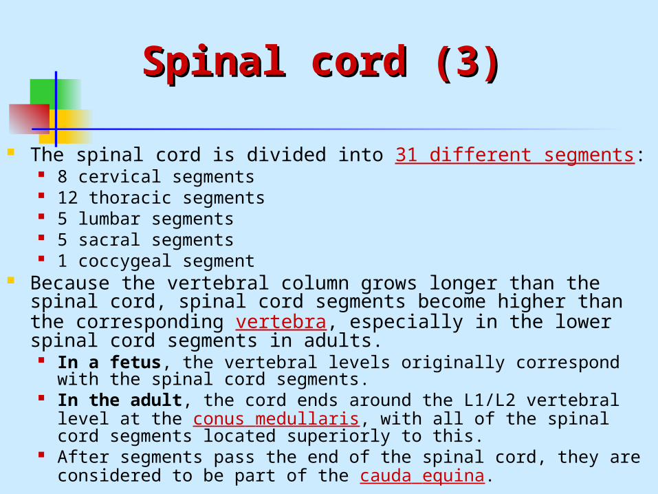
Spinal cord (Spinal cord (33))
The spinal cord is divided into 31 different segments: 8 cervical segments 12 thoracic segments 5 lumbar segments 5 sacral segments 1 coccygeal segment
Because the vertebral column grows longer than the spinal cord, spinal cord segments become higher than the corresponding vertebra, especially in the lower spinal cord segments in adults.
In a fetus, the vertebral levels originally correspond with the spinal cord segments.
In the adult, the cord ends around the L1/L2 vertebral level at the conus medullaris, with all of the spinal cord segments located superiorly to this.
After segments pass the end of the spinal cord, they are considered to be part of the cauda equina.
Cauda equinaCauda equina
The cauda equina is a structure within the lower end of the spinal column, that consists of nerve roots and rootlets from above.
Reminds a horse's tail. Clinical relevance:
possible to inject or colect the cerebral spinal fluid without a danger.

Connections between brain Connections between brain and spinal cordand spinal cord

Pyramidal tractPyramidal tract
The corticospinal or pyramidal tract is a massive collection of axons that travel between the cerebral cortex of the brain and the spinal cord.
Mostly contains motor axons. Consists of two separate tracts in the spinal cord: the
lateral corticospinal tract and the medial (anterior) corticospinal tract.
Originates from cells in layer V of the motor cortex. The lateral corticospinal tract: Most of the cortico-spinal
fibers (about 85%) cross over to the contralateral side in the medulla oblongata (pyramidal decussation).
The medial (anterior) corticospinal tract: The remainder of them (15%) cross over at the level that they exit the spinal cord.
End as a synapse with another neuron in the ventral horn.

Extrapyramidal motor Extrapyramidal motor pathwayspathways
The extrapyramidal system is a neural network located in the brain that is part of the motor system involved in the coordination of movement.
Parts of extrapyramidal system: Reticulospinal tract – proprioception, voluntary
movements, autonomic functions. Vestibulospinal tract – posture in space, righting
reflexes, voluntary and unvoluntary movements. Tectospinal tract – head movements based of the
eye and ear inputs. Olivospinal tract – from cerebellum, striatum and
cortex. Coordination of movements. Rubrospinal tract – from cerebellum and motor
cortex.

Spinal shock Spinal shock
is the phenomena surrounding transection of the spinal cord that leads to temporary loss or depression of all or most spinal reflex activity below the level of a spinal lesion.
When lesion above C3-C5 = no breeding the period of spinal shock can last from hours to 6 weeks. In the acute stage, there will be hypotone paralysis,
areflexia, loss of sensory function and dysautonomia. Patient shows retention of the bladder due to the imparied reflex of emptying the bladder.
In post acute stage, first autonomic reflexes come to normal. Patient shows autonomic dysreflexia if the lesion level is above T6.
In the chronic stage, there will be hypertone paralysis, hyper-reflexia, spastic-reflex bladde. Patient at this stage shows incontinence.

Brown-Sequard syndromeBrown-Sequard syndrome
It was first described in 1850 by the British neurologist Charles Édouard Brown-Sequard (1817-1896), who studied the anatomy and physiology of the spinal cord.
a loss of motor abilities (paralysis and ataxia) and sensation caused by the lateral hemisection of the spinal cord.
The hemisection of the cord results in a lesion of each of the three main neural systems:
The corticospinal lesion produces ipsilateral spastic paralysis. The lesion to fasciculus gracilis or fasciculus cuneus results in
ipsilateral loss of vibration and proprioception (position sense). The loss of the spinothalamic tract leads to pain and temperature
sensation being lost from the contralateral side beginning one or two segments below the lesion.
Tactile sensation not affected much (both crossed and uncrossed pathways).
Above the level of lesion – hyperesthesia (increased sensation) – irritation of dorsal horns.

Dermatomic area Dermatomic area (dermatome)(dermatome)
is an area of skin that is supplied by a single pair of dorsal roots.
The body can be divided into regions that are mainly supplied by a single spinal nerve.
There are 8 cervical (one for the head, and one for each cervical vertebra), 12 thoracic, 5 lumbar and 5 sacral spinal nerves.
This innervates the body in a patterned form. Along the thorax and abdomen it is simply like a stack of discs forming a human, each supplied by a different spinal nerve.
Along the arms and the legs, the pattern is different: the dermatomes run longitudinally along the limbs.
Clinical significance: Dermatomes are useful in neurology for finding the site of
damage to the spine. Herpes zoster infections (shingles) can reveal dermatomic
areas.
DermatomesDermatomes

Cerebellum (1)Cerebellum (1)
The cerebellum (Latin: "little brain") plays an important role in the integration of sensory perception and motor output.
Many neural pathways link the cerebellum with the motor cortex—which sends information to the muscles causing them to move—and the spinocerebellar tract—which provides feedback on the position of the body in space (proprioception).
The cerebellum integrates these pathways, using the constant feedback on body position to fine-tune motor movements.
Also, recent brain imaging studies using functional magnetic resonance imaging (fMRI) show that the cerebellum is important for language processing and selective attention.

Cerebellum (2)Cerebellum (2)
The cerebellum contains similar gray and white matter divisions as the cerebrum. The white matter is known as the arbor vitae (Tree of Life) due to its branched.
The cerebellum is divided into two large hemispheres, much like the cerebrum, and contains ten smaller lobules.
There are three phylogenetic divisions within the cerebellum: archicerebellum (the flocculonodular lobe), paleocerebellum (anterior lobe), and neocerebellum (posterior lobe).
The cerebellum can also be divided by function: The midline division is the cerebellar vermis (“worm”) and lateral hemispheres.

Cerebellum (3)Cerebellum (3)
A: Midbrain B: Pons C: Medulla D: Spinal cord E: Fourth ventricle F: Arbor vitae G:
archicerebellum H:
paleocerebellum I: neocerebellum

Archicerebellum Archicerebellum (Vestibulocerebellum)(Vestibulocerebellum)
associated with the flocculonodular lobe mainly involved in balance (
vestibular system) and eye movement functions
receives input from the inferior and medial vestibular nuclei
sends fibers back to the vestibular nuclei, creating a feedback loop
Function: constant maintenance of balance

Paleocerebellum Paleocerebellum (Spinocerebellum)(Spinocerebellum)
associated with the anterior lobe receives its inputs from the dorsal and
ventral spinocerebellar tracts, which carry information about the position and forces acting on the legs
sends axonal projections to the deep cerebellar nuclei
Function: controls proprioception related to muscle tone (maintenance of posture)

Neocerebellum Neocerebellum (Cerebrocerebellum)(Cerebrocerebellum)
associated with the posterior lobe receives input from the pontocerebellar
tract projects to the deep cerebellar nuclei Function: motor control
the coordination of fine finger movements (fine movements)
feedback correction of motor activity operates in conjunction with the cortical motor
centres to plan movement

The functional organization The functional organization (1)(1)

The functional organization The functional organization (2)(2)
Vermis receives its inputs mainly from the spinocerebellar tracts from the
trunk of the body information on the position and balance sends projections to the fastigial nucleus of the cerebellum, which
then sends output to the vestibular nuclei Intermediate zone (paravermis)
receives input from the corticopontocerebellar fibers that originate from the motor cortex
also receives sensory feedback from the muscles Control of muscles
Lateral zone receives input from the parietal cortex via pontocerebellar mossy
fibers Information regarding the location of the body in the world The large numbers of feedback circuits allow for the integration of
this body position information with indications of muscle position, strength, and speed.

Cerebellar nuclei (1)Cerebellar nuclei (1)
The 4 deep cerebellar nuclei are in the center of the cerebellum, embedded in the white matter.
receive inhibitory (GABAergic) inputs from Purkinje cells in the
cerebellar cortex excitatory (glutamatergic) inputs from mossy fiber
pathways Most output fibers of the cerebellum originate from
these nuclei. One exception is that fibers from the flocculonodular
lobe synapse directly on vestibular nuclei without first passing through the deep cerebellar nuclei.

Cerebellar nuclei (2)Cerebellar nuclei (2)

Cerebellar nuclei (3)Cerebellar nuclei (3)
From lateral to medial, the four deep cerebellar nuclei are: The Dentate, Emboliform, Globose, Fastigial easy mnemonic device: "Don't Eat Greasy Food“
The Dentate nuclei are deep within the lateral hemispheres, Emboliform and Globose nuclei are located in the paravermal (intermediate) zone, and the Fastigial nuclei are in the vermis.
In general, each pair of deep nuclei is associated with a corresponding region of cerebellar surface anatomy.

Dysfunction of the Dysfunction of the cerebellumcerebellum
Problems in walking, balance, and accurate hand and arm movement (ataxia).
Neuropsychiatric disorders such as dyslexia, schizophrenia and autism appear to be associated with a deficiency in the cerebellum.
Patients with cerebellar lesions (injuries) typically exhibit "intention tremors"—a tremor occurring during movement rather than at rest (as seen in Parkinson's disease).
Patients may also show dysmetria, i.e., an overestimation or underestimation of force, resulting in overshoot or undershoot when reaching for a target.
Inability to perform rapid alternating movements. In case of intoxication: uncoordinated movements, swaying, unstable
walking, and a wide gait. Chronic alcohol abuse is also a common cause of cerebellar lesions.
Alcohol abuse can lead to thiamine deficiency, which in the cerebellum will cause degeneration of the anterior lobe. This degeneration leads to a wide, staggering gait but does not affect arm movement or speech.
Patients with a cerebellar lesion may have nystagmus with the fast stroke pointing towards the side of the lesion.
Brain stem Brain stem
The brain stem is the lower part of the brain, adjoining and structurally continuous with the spinal cord.
Parts of brain stem: the pons, medulla oblongata, and sometimes midbrain.
Pons VaroliiPons Varolii
Located in rostral to the medulla oblongata, caudal to the midbrain, and ventral to the cerebellum.
Function: relays sensory information between the cerebellum
and cerebrum. Some theories posit that it has a role in dreaming. Control of respiration:
the apneustic center - lower pons the pneumotaxic center - upper pons
A number of cranial nerve nuclei are present in the pons (from top to bottom):
the trigeminal nerve, abducens nucleus, vestibulocochlear nuclei, facial nerve nucleus

Medulla oblongataMedulla oblongata
Located in rostral to the spinal cord, caudal to the pons and ventral to the cerebellum.
Function: controls autonomic functions: respiration,
blood pressure, heart rate, reflex arcs, vomiting relays nerve signals between the brain and
spinal cord Cranial nerve nuclei: the hypoglossal nerve,
glossopharyngeal and vagus nerves.

Reticular formationReticular formation
It is absolutely essential for the basic functions of life (stereotypical actions, such as walking, sleeping, and lying down) and is phylogenetically one of the oldest portions of the brain.
It is a poorly-differentiated area of the brain stem, centered roughly in the pons.
Parts: the ascending reticular activating system connects to
areas in the thalamus, hypothalamus, and cortex the descending reticular activating system connects
to the cerebellum and sensory nerves
Function of the Reticular Function of the Reticular formationformation
The RF appears to not only control physical behaviors such as sleep, but also has been shown to play a major role in alertness, fatigue, and motivation to perform various activities.
Some researchers have speculated that the reticular formation controls approximately 25 specific and mutually-exclusive behaviors, including sleeping, walking, eating, urination, defecation, and sexual activity.
The reticular formation has also been traced as one of the sources for the introversion (easily stimulated RF) and extroversion (less easily stimulated RF) character traits.

Spinal reflexes (1)Spinal reflexes (1)

Spinal reflexes (2)Spinal reflexes (2)

Spinal reflexes (3)Spinal reflexes (3)
Related Documents