HAL Id: tel-03208760 https://tel.archives-ouvertes.fr/tel-03208760 Submitted on 26 Apr 2021 HAL is a multi-disciplinary open access archive for the deposit and dissemination of sci- entific research documents, whether they are pub- lished or not. The documents may come from teaching and research institutions in France or abroad, or from public or private research centers. L’archive ouverte pluridisciplinaire HAL, est destinée au dépôt et à la diffusion de documents scientifiques de niveau recherche, publiés ou non, émanant des établissements d’enseignement et de recherche français ou étrangers, des laboratoires publics ou privés. Fuctional characterization of different candidate effectors from the root rot oomycete Aphanomyces euteiches Laurent Camborde To cite this version: Laurent Camborde. Fuctional characterization of different candidate effectors from the root rot oomycete Aphanomyces euteiches. Vegetal Biology. Université Paul Sabatier - Toulouse III, 2020. English. NNT : 2020TOU30227. tel-03208760

Welcome message from author
This document is posted to help you gain knowledge. Please leave a comment to let me know what you think about it! Share it to your friends and learn new things together.
Transcript
HAL Id: tel-03208760https://tel.archives-ouvertes.fr/tel-03208760
Submitted on 26 Apr 2021
HAL is a multi-disciplinary open accessarchive for the deposit and dissemination of sci-entific research documents, whether they are pub-lished or not. The documents may come fromteaching and research institutions in France orabroad, or from public or private research centers.
L’archive ouverte pluridisciplinaire HAL, estdestinée au dépôt et à la diffusion de documentsscientifiques de niveau recherche, publiés ou non,émanant des établissements d’enseignement et derecherche français ou étrangers, des laboratoirespublics ou privés.
Fuctional characterization of different candidate effectorsfrom the root rot oomycete Aphanomyces euteiches
Laurent Camborde
To cite this version:Laurent Camborde. Fuctional characterization of different candidate effectors from the root rotoomycete Aphanomyces euteiches. Vegetal Biology. Université Paul Sabatier - Toulouse III, 2020.English. �NNT : 2020TOU30227�. �tel-03208760�

Abstract
Oomycetes are eukaryote pathogens able to infect plants and animals. During host interaction,
oomycetes secrete various molecules, named effectors, to counteract plant defence and modulate
plant immunity. Two different classes of cytoplasmic effectors have been described to date, Crinklers
(CRNs) and RxLR proteins. The translocation process allowing the entrance into the host cells is still
unclear, and while extended research gave insight into some molecular targets and role during
infection, most of effectors have not been characterized.
In the root rot pathogen of legumes Aphanomyces euteiches, only the CRNs are present. Based on a
previous study reported by our research group, we published an opinion paper focused on the
emergence of DNA damaging effectors and their role during infection.
Previous experiments indicate that one of these Crinklers, AeCRN5, harbours a functional translocation
domain and once the protein reaches host nuclei, dramatically disturbs root development. Here we
reveal that AeCRN5 binds to RNA and interferes with biogenesis of various small RNAs, implicated in
defence mechanisms or plant development.
Furthermore, comparative genetic analyses revealed a new class of putative effectors specific to
Aphanomyces euteiches, composed by a large repertoire of small-secreted protein coding genes (SSP),
potentially involved during root infection. Preliminary results on these SSPs point out that AeSSP1256,
which contains a functional nuclear localisation signal, enhances host susceptibility.
Functional characterisation of AeSSP1256 evidenced that this effector binds to RNA, relocalizes a plant
RNA helicase and interferes with its activity, causing stress on plant ribosome biogenesis.
This work highlights that various effectors target nucleic acids and reveals that two effectors from
distinct family are able to interact with plant RNA in order to interfere with RNA related defence
mechanisms and plant development to promote pathogen infection.
Keywords: Oomycetes, nucleus, DNA damage, RNA-binding proteins, CRN, SSP.


Remerciements
Alors par qui commencer… les membres de mon jury bien sûr, à commencer par Claire Veneault-Fourrey et Bruno Favery qui ont accepté d’évaluer mon travail. En ces temps de Covid et à l’heure où j’écris ces lignes, je ne sais pas encore si on se verra masqués, ou par écrans interposés, mais je vous remercie sincèrement pour le temps que vous m’accordez. Merci aussi à Christophe Roux pour avoir gentiment accepté de faire parti de mon jury.
Un grand merci à Bernard Dumas, ancien chef de l’équipe lors de mon arrivée au laboratoire. Merci pour la confiance que tu m’as accordé et pour m’avoir un peu poussé à faire une thèse. Bon pour m’avoir beaucoup poussé à faire une thèse. Beaucoup.
Mention spéciale à la plateforme d’imagerie, pour leur compétence, leur disponibilité et leur gentillesse en commençant par Alain Jauneau, avec qui j’ai passé des heures autour d’un laser, d’un microscope et d’un tableau Velleda, pour apprendre le FRET-FLIM. Il existe des gens qui rendent tout intéressant. Je n’oublie pas bien sûr Cécile Pouzet, avec qui j’ai beaucoup de plaisir à travailler, et qui en plus veut bien me prêter ses jouets à 500 000 euros. Merci aussi à Yves Martinez et Aurélie Le Ru, d’une patience rare, même si Aurélie me coûte plus cher en bières. Heureusement que vous êtes là.
Merci à Jean Philippe Combier pour les discussions et les suggestions apportées. 5 min de discussion avec JP, c’est 5 mois de manips derrière. Faut pas y aller trop souvent non plus…
Merci aux personnes avec qui j’ai travaillé sur ce sujet de thèse, notamment Annelyse, Amandine et Marie Alexane que j’ai eu en stage, Chiel Pel et Sarah Courbier qui ont initié le projet sur les SSP et avec qui j’ai passé de très bons moments. Diana Ramirez qui a effectué sa thèse sur les CRNs, ce qui nous a permis de découvrir le monde merveilleux des amphibiens, l’odeur de l’animal, les inséminations de grenouilles femelles, les injections d’ARN dans des centaines d’embryons. En fait on oublie, mais vraiment, travailler sur les plantes, c’est bien.
Là c’est le paragraphe dédié aux gens qui m’ont rendu la vie plus facile. Par exemple David, qui réceptionne mes bons de commandes, donc mes erreurs hebdomadaires, dans une ambiance de zénitude et d’encens qui rappelle que rien n’est jamais grave avec David, c’est reposant. Catherine, notre gestionnaire, qui a su aussi être très patiente, mais sans la musique zen et l’encens. Son “CammmmmBBBOOORDEEEE” résonne encore dans ma tête.
Merci aux membres de mon équipe, ancienne et actuelle, à ceux du LabCom, à Thomas et Olivier pour les discussions et conseils, c’est toujours agréable de parler avec vous. Merci à Charlène qui m’a soutenu, surveillé mes réinscriptions et ma phobie administrative, à Malo, à Andreï. Merci EMILIE, oui en majuscule, parce que tu m’as beaucoup aidé dans la gestion des affaires courantes comme on dit, en se partageant les tâches à merveille. En gros tu t’occupais de tout ce qui ne me plaisait pas!
Enfin, un grand merci à ma directrice de thèse, Elodie Gaulin, qui a su me guider et me soutenir durant ce travail. Ta patience et la facilité avec laquelle tu évacues la pression m’ont beaucoup aidé. C’est un vrai plaisir de travailler avec toi!
A ma famille, dont mes parents, éternelle source d’inspiration, à mes enfants, qui vont retrouver leur pôpa, et à ma femme, pour tout ce qu’elle est.

List of abbreviations
Ae Aphanomyces euteiches
AEPs Apoplastic Effector Proteins
AM Arbuscular Mycorrhiza
Avr Avirulence
Bd Batrachochytrium dendrobatidis
BIC Biotrophic Interfacial Complex
CSEPs Candidate Secreted Effector Proteins
CBEL Cellulose Binding ELicitor
CRN Crinkling and Necrosis
CSEPs Candidate Secreted Effector Proteins
CWDEs Cell Wall Degrading Enzymes
ECM Ectomycorrhizal Fungi
ER Endoplasmic Reticulum
ESTs Expressed Sequence Tags
ETI Effector-Triggered Immunity
EVs Extracellular Vesicles
FLIM Fluorescence Lifetime Imaging Microscopy
FRET Fluorescence Resonance Energy Transfer
GFP Green Fluorescent Protein
GWAS Genome Wide Association Study
HGT Horizontal Gene Transfer
HIGS Host-Induced Gene Silencing
HR Hypersensitive Response
HRP HorseRadish Peroxidase
HSP Heat Shock Protein
JA Jasmonic Acid
MAMP Microbe-Associated Molecular Pattern
MAPKKK Mitogen Activated Protein (MAP) Kinase Kinase Kinase
MAX Magnaporthe Avrs and ToX B-like effectors
NES Nuclear Export Signal
NLPs Necrosis and Ethylene inducing peptide 1 (Nep1)-like proteins
NLRs Leucine-Rich Repeat proteins
NLS Nuclear Localization Signal
PAMP Pattern-Associated Molecular Pattern
PTGS Post-Transcriptional Gene Silencing
PTI PAMPs-Triggered Immunity
PBS Phosphate-Buffered Saline
PCW Plant Cell Wall
PR Pathogenesis Related
PTI PAMP-Triggered Immunity
QTL Quantitative Trait Loci
R Resistance
RALPHs RnAse‐Like Proteins Associated with Haustoria
REase Restriction Endonuclease (REase) superfamily
RIP Repeat-Induced Point mutations
RBPs RNA Binding Protein
rDNA Ribosomal DNA
ROS Reactive Oxygen Species
SA Salicylic Acid
SAR Systemic Acquired Resistance
SAR Stramenopiles, Alveolates, and Rhizaria
SDS Sodium Dodecyl Sulfate
siRNA Small Interfering RNAs
SSPs Small-Secreted Proteins
SNPs Single Nucleotide Polymorphism
TALE Transcription Activator-Like Effector
TBS Tris-Buffered Saline
TE Transposable Element
TFs Transcription Factors
Ubi Ubiquitin
WGA Wheat Germ Agglutinin
Y2H Yeast-2-Hybrid

Table des matières Abstract .....................................................................................................................................................
List of abbreviations .................................................................................................................................
I – CHAPTER I: General Introduction ...................................................................................................... 1
I-1. Oomycetes and fungi, The World Is Not Enough .......................................................................... 1
I-1.1. The Phantom menace ................................................................................................................ 1
I-1.2. Defence and Resistance against pathogens ............................................................................... 2
I-2. Oomycetes, so close and yet so far from Fungi ............................................................................ 6
I-2.1. The false brothers ...................................................................................................................... 6
I-2.2. Lifestyle: oomycete and fungi in front of the mirror ................................................................. 6
I-2.3. Oomycete phylogeny, still a growing tree ................................................................................. 8
I-2.4. Oomycetes, origin(s) and evolution ........................................................................................... 9
I-3. Effectors, the infectious Swiss knife ............................................................................................ 10
I-3.1. Effector genes evolution .......................................................................................................... 11
I-3.2. Apoplastic and intracellular effectors ...................................................................................... 13
I-3.2 a. Apoplastic effectors: in front of the Wall .............................................................................. 13
I-3.2 b. Intracellular effectors: Destroy from within .......................................................................... 15
I-3.3. Intracellular effectors targets: hit the defence key players ..................................................... 21
I-4. Aphanomyces: an oomycete genus to study effectors and host adaptation ............................. 24
I-4.1. Aphanomyces euteiches, the Legume threat ........................................................................... 25
I-4.2. Aphanomyces euteiches – Medicago truncatula pathosystem ............................................... 27
I-4.3. Insight into Aphanomyces euteiches intracellular effectors .................................................... 29
I-5. Scope of the thesis ...................................................................................................................... 31
II – CHAPTER II: DNA-Damaging Effectors: New Players in the Effector Arena
(Camborde et al. TIPS, 2019) ................................................................................................................. 33
III – CHAPTER III: AeCRN5 effector from A. euteiches targets plant RNA and perturbs RNA silencing
............................................................................................................................................................... 42
IV – CHAPTER IV: Genomics analysis of Aphanomyces spp. identifies a new class of oomycete
effector associated with host adaptation (Gaulin et al. BMC Biol, 2018) ........................................... 74
V – CHAPTER V: A DEAD-Box RNA helicase from Medicago truncatula is hijacked by an RNA-binding
effector from the root pathogen Aphanomyces euteiches to facilitate host infection ..................... 97
Complementary results: Aphanomyces euteiches effectors from two different families interact and
modulate their activity. ...................................................................................................................... 135
General discussion and perspectives ................................................................................................. 142
References .......................................................................................................................................... 153


Chapter I
General Introduction

1
I – CHAPTER I: General Introduction
I-1. Oomycetes and fungi, The World Is Not Enough
I-1.1. The Phantom menace
Plants and animals have to face constantly with abiotic stresses, like environmental
modifications due to climate change, including higher temperature, pH variation or long
drought for instance, but also various biotic stresses due to multiple interactions with other
organisms, from bacteria to nematodes, via fungi, oomycetes, viruses or insects. Unlike
animals, who can move to find a better environment, plants are rooted in place and must
adapt very quickly to changes or attacks. One of the major biotic threats are eukaryotic
filamentous microorganisms, represented by oomycetes and fungi, which comprise several of
the most devastating plant and animal pathogens, considered as a major threat for
agriculture, but also for natural terrestrial or oceanic ecosystems (Beakes et al., 2012).
Even if humans and most mammals are remarkably resistant to invasive fungal
diseases, in the same time entire ecosystems are currently devastated by fungal pathogens
(Fisher et al., 2012; Casadevall, 2017). Bats or reptiles are threatened with extinction due to
pathogenic fungi (Fisher et al., 2012; Casadevall, 2017). Another example of feared fungus is
Batrachochytrium dendrobatidis, considered as the major threat for amphibians causing a
catastrophic loss of biodiversity (Fisher et al., 2012; Scheele et al., 2019). Fungal diseases also
impact plant crops, destroying a third of all food crops annually and impacting the most
important crops (rice, wheat, maize, potatoes, and soybean) (Fisher et al., 2012; Almeida et
al., 2019). For instance, the wheat stem rust caused by the fungus P. gramini sf. tritici, which
has being threating wheat cultures since 1998, had disastrous impact in the Middle East and
West Asia, with reduction in yields up to 40% (Pennisi, 2010). Very recently, researchers
warned and reported the re-emergence of this fungus in Western Europe (Saunders et al.,
2019; Bhattacharya, 2017). Another example is the rice blast disease agent Magnaporthe
oryzae, one of the most economically devastating fungus that infect rice as well as other grass
2
species including wheat. Only on rice, annual yield losses can reach 20% in many production
zones but the entire harvest can be lost when significant outbreak occur (Prabhu et al., 2009).
Oomycetes also comprise devastating pathogens and represent the most problematic
group of disease-causing organisms in both agriculture and aquaculture (Derevnina et al.,
2016b). However, oomycetes stand as notorious plant pathogens with remarkable examples,
like Phytophthora infestans causing late blight triggering the Irish potato famine in 1840 (Haas
et al., 2009). Phytophthora species are responsible of serious diseases affecting crop yields.
The annual economic loss on tomato and potato due to P. infestans was estimated at $ 6.7
billion (Haas et al., 2009). On soybean, for North America, the average annual yield loss caused
by P. sojae was estimated at 1.1 million tons, from 2007 to 2014 (Allen et al., 2017). Others
notable species are P. palmivora and P. capsici, causing agents of cocoa black pod causing yield
losses of 20–30% annually (Adeniyi, 2019). On legumes, Aphanomyces euteiches, the causing
agent of root rot, represent one of the major limitations to pea production worldwide (Wu et
al., 2018). All those examples highlight the important impact of plant pathogen oomycetes,
but some species are also responsible for devastating diseases in natural ecosystems or in
aquaculture. For instance, members of the Saprolegnia genus, such as S. parasitica infecting
freshwater fish, are involved in the decline of wild salmon populations around the world
(Phillips et al., 2008; van West, 2006). Another example of killing agent is Aphanomyces astaci,
parasite of fresh-water decapods and causing crayfish plague. Originate from North America,
it is now present in Europe and has been nominated among the “100 of the World’s Worst
Invasive Alien Species” in the Global Invasive Species Database (GISD).
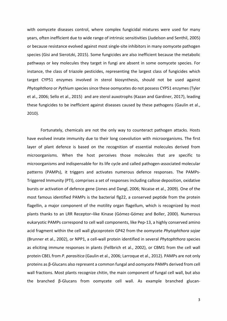
I-1.2. Defence and Resistance against pathogens
Despite the impact of these diseases and the increase of dedicated research, it is still
challenging to control fungal or oomycete attacks. To reach high-quality crops with optimal
yields, modern agriculture had resort to intensive use of fungicides that frequently became
ineffective due to high adaptation frequency, caused by gene mutations, leading to the
emergence of new fungal races (Zhou et al., 2007; Lucas et al., 2015). Same problem occurs
3
with oomycete diseases control, where complex fungicidal mixtures were used for many
years, often inefficient due to wide range of intrinsic sensitivities (Judelson and Senthil, 2005)
or because resistance evolved against most single-site inhibitors in many oomycete pathogen
species (Gisi and Sierotzki, 2015). Some fungicides are also inefficient because the metabolic
pathways or key molecules they target in fungi are absent in some oomycete species. For
instance, the class of triazole pesticides, representing the largest class of fungicides which
target CYP51 enzymes involved in sterol biosynthesis, should not be used against
Phytophthora or Pythium species since these oomycetes do not possess CYP51 enzymes (Tyler
et al., 2006; Sello et al., 2015) and are sterol auxotrophs (Kazan and Gardiner, 2017), leading
these fungicides to be inefficient against diseases caused by these pathogens (Gaulin et al.,
2010).
Fortunately, chemicals are not the only way to counteract pathogen attacks. Hosts
have evolved innate immunity due to their long coevolution with microorganisms. The first
layer of plant defence is based on the recognition of essential molecules derived from
microorganisms. When the host perceives those molecules that are specific to
microorganisms and indispensable for its life cycle and called pathogen-associated molecular
patterns (PAMPs), it triggers and activates numerous defence responses. The PAMPs-
Triggered Immunity (PTI), comprises a set of responses including callose deposition, oxidative
bursts or activation of defence gene (Jones and Dangl, 2006; Nicaise et al., 2009). One of the
most famous identified PAMPs is the bacterial flg22, a conserved peptide from the protein
flagellin, a major component of the motility organ flagellum, which is recognized by most
plants thanks to an LRR Receptor–like Kinase (Gómez-Gómez and Boller, 2000). Numerous
eukaryotic PAMPs correspond to cell wall components, like Pep-13, a highly conserved amino
acid fragment within the cell wall glycoprotein GP42 from the oomycete Phytophthora sojae
(Brunner et al., 2002), or NPP1, a cell-wall protein identified in several Phytophthora species
as eliciting immune responses in plants (Fellbrich et al., 2002), or CBM1 from the cell wall
protein CBEL from P. parasitica (Gaulin et al., 2006; Larroque et al., 2012). PAMPs are not only
proteins as β-Glucans also represent a common fungal and oomycete PAMPs derived from cell
wall fractions. Most plants recognize chitin, the main component of fungal cell wall, but also
the branched β-Glucans from oomycete cell wall. As example branched glucan-
4
chitosaccharides from the oomycete Aphanomyces euteiches induce defence and calcium
signals in Medicago truncatula root cells (Nars et al., 2013).
Faced to PTI, microorganisms evolved and secreted hundreds of pathogenesis-related
molecules, named effectors, to modulate immunity and facilitate host colonization. In turn,
some hosts evolve to detect specifically those molecules, leading to the Effector-Triggered-
Immunity (ETI). Perception is mediated by receptors know as resistance proteins (R) that
directly or indirectly recognize some secreted effectors, then called avirulence proteins (AVR).
This process was previously named gene-for-gene resistance (R/AVR) (Van Der Biezen and
Jones, 1998). This recognition is frequently associated to a hypersensitive response (HR), a
localized host cell death to confine the pathogen at the infection site (Jones and Dangl, 2006).
Then, major R genes have been used by breeding companies to protect crops against fungal
plant diseases (Stuthman et al., 2007). However, the strategy using a single resistance gene
often turns out to be inefficient due to adaptation of pathogen populations, which have a high
evolutionary potential and rapidly evolve by AVR genes mutations to become virulent. For
instance, the fungus Leptosphaeria maculans, the causing agent of the phoma stem canker
disease on oilseed rape (Brassica napus), produce new strains by mutations of genes rendering
the corresponding major host resistance genes ineffective in only three years (Sprague et al.,
2006). Similarly, appearance of new and more virulent pathotypes of the downy mildew
(Plasmopara halstedii) in sunflower leads researchers to identify new R genes in order to
combine them in varieties carrying a wide range of resistance genes (Pecrix et al., 2019, 2018).
Nowadays, major R genes are deployed in cultivars in combination with sustainable disease
management practices like precise chemical treatments in order to prolong the use of those
resistance genes (Mitrousia et al., 2018).
In addition, another aspect of genetic resistance is related to a quantitative resistance
with a partial reduction of symptoms and disease severity (Kamoun et al., 1999). This partial
resistance is due to quantitative resistance genes localized in genome area named
Quantitative Trait Loci (QTLs). Even if this resistance is frequently less efficient than gene-for-
gene resistance like R-AVR gene interaction (Hu et al., 2008; Pilet-Nayel et al., 2017), it
appeared to be more durable, with a lower selection pressure for pathogens, which limit
mutations, and resistance acquired by the expression of different QTLs is more difficult to
5
circumvent (Poland et al., 2009). For instance, no resistant pea, lentil cultivars are available
against the oomycete Aphanomyces euteiches that causes the devastating root rot diseases
of legumes. However, genome-wide association studies based on the model legume Medicago
truncatula identified one major and several minors QTLs contributing to the tolerance (Badis
et al., 2015; Bonhomme et al., 2014, 2019). Then some Aphanomyces resistance QTLs were
identified in pea but fine mapping to identify underlying genes is still challenging (Hamon et
al., 2013; Desgroux et al., 2016). In lentil, numerous QTLs were recently detected and some
genes are under validation (Ma et al., 2020; Marzougui et al., 2019). Similarly, the oilseed rape
(Brassica napus), threatened by stem rot caused by the fungus Sclerotinia sclerotiorum,
represents another crop with absence of resistant lines. Currently, breeding for Sclerotinia
resistance in B. napus is only based on germplasms with quantitative resistance genes (Wu et
al., 2013) and the identification of new QTLs is still an active research (Qasim et al., 2020). In
rice, where many R genes were characterized, QTLs were also identified. Then, the resistance
in cultivars to the blast fungus Magnaporthe oryzae is controlled by a combination of both
major genes and QTLs (Kang et al., 2016).
The use of chemicals to threat animal pathogens invasion triggered also the
development of chemical-resistance coupled with negative side-effects on the ecosystem.
Then, alternative strategies have to be developed. In aquatic culture for instance, biological
control strategies are under development to control zoosporic diseases due to chytrid fungus
and oomycetes (Frenken et al., 2019). This include for example a project of immunization
against the oomycete Saprolegnia parasitica using a serine protease, the identification of
stimuli able to increase the production of natural antifungal peptides produced by the skin of
amphibians, or the modification of the pathogen fitness using secondary parasites (Frenken
et al., 2019). While those projects are promising, much work still needs to be done to
implement biological-control applications in aquaculture (Frenken et al., 2019). Biocontrol
strategies are also currently develop to protect plant against pathogens (Köhl et al., 2019).

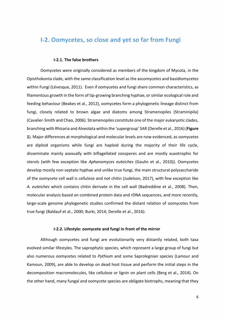
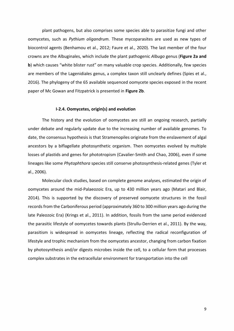
Figure 1: Simplified tree of eukaryotes showing the distant relationship between oomycetes
and fungi.
SAR is an acronym of its constituents: Stramenopiles, Alveolates, and Rhizaria. Oomycetes and Fungi are highlight
in red. Adapted from (McGowan and Fitzpatrick, 2020).
6
I-2. Oomycetes, so close and yet so far from Fungi
I-2.1. The false brothers
Oomycetes were originally considered as members of the kingdom of Mycota, in the
Opisthokonta clade, with the same classification level as the ascomycetes and basidiomycetes
within Fungi (Lévesque, 2011). Even if oomycetes and fungi share common characteristics, as
filamentous growth in the form of tip-growing branching hyphae, or similar ecological role and
feeding behaviour (Beakes et al., 2012), oomycetes form a phylogenetic lineage distinct from
fungi, closely related to brown algae and diatoms among Stramenopiles (Straminipila)
(Cavalier-Smith and Chao, 2006). Stramenopiles constitute one of the major eukaryotic clades,
branching with Rhizaria and Alveolata within the ‘supergroup’ SAR (Derelle et al., 2016) (Figure
1). Major differences at morphological and molecular levels are now evidenced, as oomycetes
are diploid organisms while fungi are haploid during the majority of their life cycle,
disseminate mainly asexually with biflagellated zoospores and are mostly auxotrophic for
sterols (with few exception like Aphanomyces euteiches (Gaulin et al., 2010)). Oomycetes
develop mostly non-septate hyphae and unlike true fungi, the main structural polysaccharide
of the oomycete cell wall is cellulose and not chitin (Judelson, 2017), with few exception like
A. euteiches which contains chitin derivate in the cell wall (Badreddine et al., 2008). Then,
molecular analysis based on combined protein data and rDNA sequences, and more recently,
large-scale genome phylogenetic studies confirmed the distant relation of oomycetes from
true fungi (Baldauf et al., 2000; Burki, 2014; Derelle et al., 2016).
I-2.2. Lifestyle: oomycete and fungi in front of the mirror
Although oomycetes and fungi are evolutionarily very distantly related, both taxa
evolved similar lifestyles. The saprophytic species, which represent a large group of fungi but
also numerous oomycetes related to Pythium and some Saprolegnian species (Lamour and
Kamoun, 2009), are able to develop on dead host tissue and perform the initial steps in the
decomposition macromolecules, like cellulose or lignin on plant cells (Berg et al., 2014). On
the other hand, many fungal and oomycete species are obligate biotrophs, meaning that they


7
are unculturable on artificial media, and grow only on living cells. Those species require
metabolic active tissues to achieve their life cycle and then are highly adapted to their host,
such as downy mildew Plasmopara viticola, which infects grapevine (Vitis vinifera), Albugo
candida, the causing agent of white rust on crucifers (Kamoun et al., 2015) or the pathogenic
fungus Blumeria graminis causing powdery mildew on barley (Thomas et al., 2001) and the
smut fungus Ustilago maydis on corn (Banuett and Herskowitz, 1996). By contrast, many plant
pathogenic oomycetes or fungi, especially species of the genus Phytophthora, or
fungi/Ascomycota like Colletotrichum or Magnaporthe, display an intermediate lifestyle called
hemibiotrophy, starting infections like biotrophs by establishing a transient biotrophic
relationship with the host, then switch to necrotrophic phase later in the disease cycle
(Latijnhouwers et al., 2003; Lamour and Kamoun, 2009; Thines, 2018). Finally, necrotrophic
pathogens kill host tissues to feed during the colonisation like the fungus Botrytis cinerea, the
causal agent of gray mold, an economically devastating disease, which serves as a model
species for plant-necrotroph interactions (Petrasch et al., 2019). Pythium represent the largest
genus of necrotrophic oomycetes, but some aquatic pathogenic oomycetes like Lagenisma
coscinodisci are also efficient necrotrophic organisms, able to kill marine diatoms in few days
by hijacking the host’s alkaloid metabolism (Vallet et al., 2019). However, the classification in
hemibiotrophy or necrotrophy is not always clear, as for the oomycete Aphanomyces
euteiches that causes root rot of legumes (Judelson and Ah-Fong, 2019).
Both oomycetes and fungi share similar traits for host interaction. Dispersal of
oomycetes is mediated by water or wind through asexual sporangia or directly by the release
of asexual motile zoopores from sporangia (Tyler, 2002). Once oomycete zoospores have
reached host surface, they encyst by shedding their flagella and secrete adhesion molecules
(Hardham and Shan, 2009; Carzaniga et al., 2001). Asexual spores of fungi as conidies are
transported by wind and water, before an adhesion step to the host due to the secretion of
adhesion molecules. The germinated cyst produce hyphae able to penetrate inside cell layers,
mainly by using a pathogenic structure called appressorium, then vegetative hyphae grow in
intercellular space and develop haustoria which penetrated inside host cells (Fawke et al.,
2015). Oomycetes and fungi hyphae can also penetrate by natural opening such as stomata
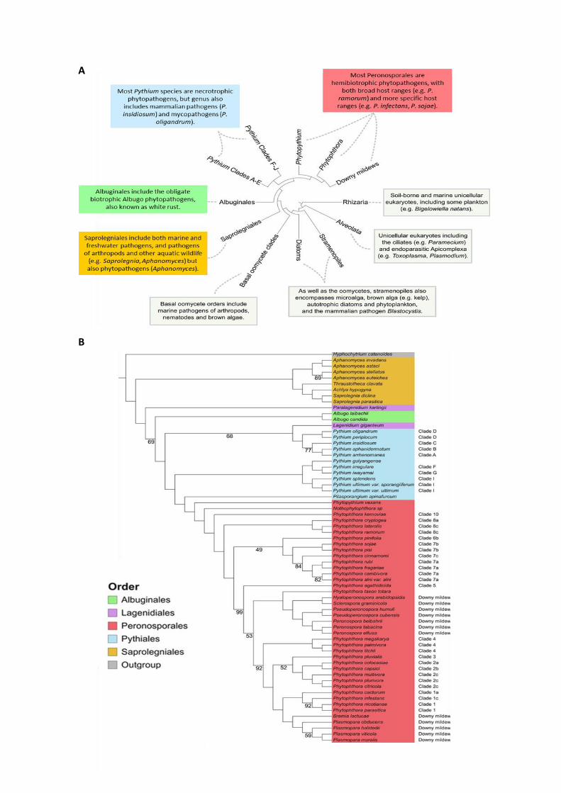
A
B

8
Figure 2: Phylogeny of the Oomycetes.
(A) Consensus phylogeny of the oomycete class within the greater SAR grouping, including information pertaining
to various taxa. Adapted from (McCarthy and Fitzpatrick, 2017). (B) Maximum-likelihood phylogeny of the 65
oomycete species based on the concatenation of 102 conserved BUSCO sequences. The stramenopile
Hyphochytrium catenoides is included as an outgroup. All nodes have 100% bootstrap support except where
indicated. Species are colored according to their order. Phytophthora clades are indicated as designated by Blair,
Coffey, Park, Geiser, and Kang (2008) and Pythium clades are as designated by de Cock et al. (2015). From
(McGowan and Fitzpatrick, 2020).
(Lucas, 2020). Numerous enzymes to break the host barriers (i.e. cell wall, cutin) are also
produced by oomycetes and fungi during the penetration and colonization steps. However,
some oomycetes do not form haustoria, like Pythium ultimum or neither appressorium, such
as Aphanomyces euteiches (Gaulin et al., 2008). Finally, they complete their life cycle by
producing new asexual spores and/or by making their sexual life cycle/stage.
I-2.3. Oomycete phylogeny, still a growing tree
Oomycete phylogeny is still subject to revision due to new genome availability. To
date, 65 oomycete species have publicly available genome sequences deposited in databases
(McGowan and Fitzpatrick, 2020) and although many species are yet unsampled, the current
consensus phylogeny of the oomycetes split them into a basal order and four major “crown”
orders: the Peronosporales, Pythiales, Albuginales, and Saprolegniales (Beakes et al., 2014;
McCarthy and Fitzpatrick, 2017; McGowan and Fitzpatrick, 2020) (Figure 2a). The basal order
of oomycetes includes exclusively marine organisms which are predominantly parasites of
seaweeds, nematodes or arthropods (Beakes et al., 2012). The Saprolegniales order is the
most basal of the four major crown orders and includes saprophytes and animal parasites,
such as the fish pathogen Saprolegnia (Hulvey et al., 2007), and also the plant and animal
pathogenic Aphanomyces genus (Gaulin et al., 2007) (Figure 2a and b). The Peronosporales
order includes the largest group of terrestrial organisms and represent the best studied order,
comprising the well-known oomycete Phytophthora genus. It is also composed by the
phytopathogenic Phytopythium genus as well as downy mildew such as Hyaloperonospora,
Plasmopara or Sclerospora genera (Fletcher et al., 2019; McCarthy and Fitzpatrick, 2017;
McGowan and Fitzpatrick, 2020) (Figure 2a and b). The Pythiales order contains animal and

9
plant pathogens, but also comprises some species able to parasitize fungi and other
oomycetes, such as Pythium oligandrum. These mycoparasites are used as new types of
biocontrol agents (Benhamou et al., 2012; Faure et al., 2020). The last member of the four
crowns are the Albuginales, which include the plant pathogenic Albugo genus (Figure 2a and
b) which causes “white blister rust” on many valuable crop species. Additionally, few species
are members of the Lagenidiales genus, a complex taxon still unclearly defines (Spies et al.,
2016). The phylogeny of the 65 available sequenced oomycete species exposed in the recent
paper of Mc Gowan and Fitzpatrick is presented in Figure 2b.
I-2.4. Oomycetes, origin(s) and evolution
The history and the evolution of oomycetes are still an ongoing research, partially
under debate and regularly update due to the increasing number of available genomes. To
date, the consensus hypothesis is that Stramenopiles originate from the enslavement of algal
ancestors by a biflagellate photosynthetic organism. Then oomycetes evolved by multiple
losses of plastids and genes for phototropism (Cavalier-Smith and Chao, 2006), even if some
lineages like some Phytophthora species still conserve photosynthesis-related genes (Tyler et
al., 2006).
Molecular clock studies, based on complete genome analyses, estimated the origin of
oomycetes around the mid-Palaeozoic Era, up to 430 million years ago (Matari and Blair,
2014). This is supported by the discovery of preserved oomycete structures in the fossil
records from the Carboniferous period (approximately 360 to 300 million years ago during the
late Paleozoic Era) (Krings et al., 2011). In addition, fossils from the same period evidenced
the parasitic lifestyle of oomycetes towards plants (Strullu-Derrien et al., 2011). By the way,
parasitism is widespread in oomycetes lineage, reflecting the radical reconfiguration of
lifestyle and trophic mechanism from the oomycetes ancestor, changing from carbon fixation
by photosynthesis and/or digests microbes inside the cell, to a cellular form that processes
complex substrates in the extracellular environment for transportation into the cell
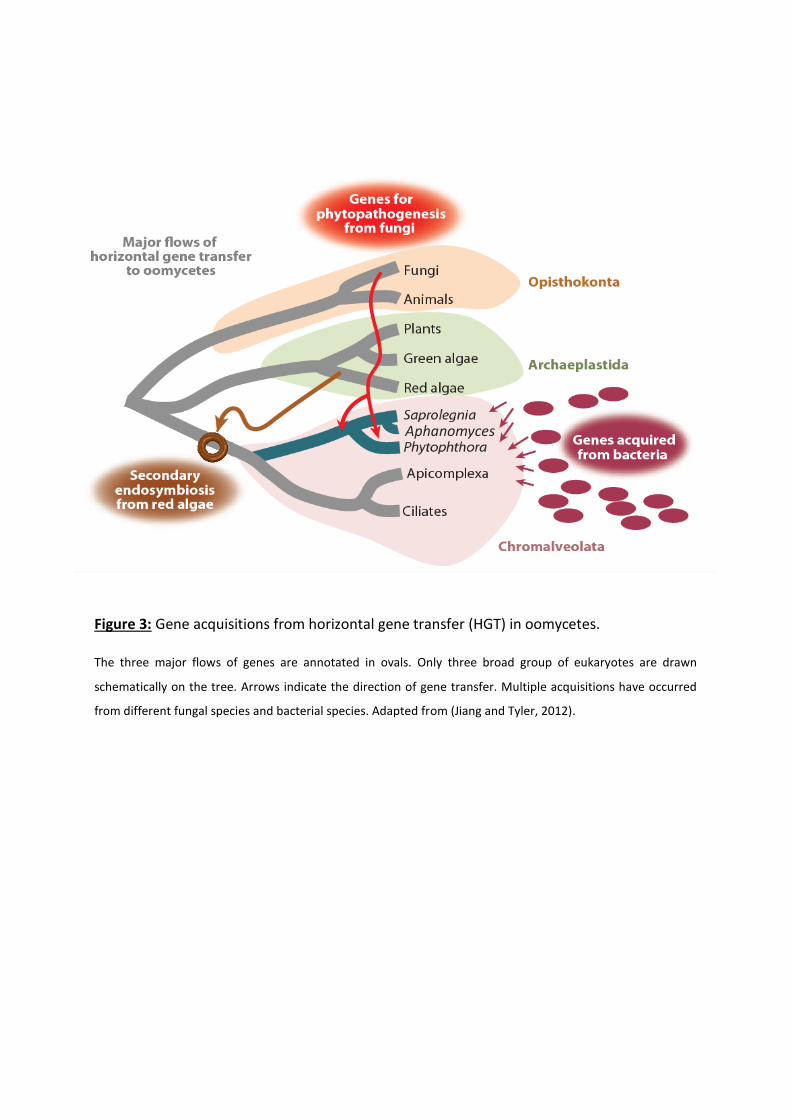
Figure 3: Gene acquisitions from horizontal gene transfer (HGT) in oomycetes.
The three major flows of genes are annotated in ovals. Only three broad group of eukaryotes are drawn
schematically on the tree. Arrows indicate the direction of gene transfer. Multiple acquisitions have occurred
from different fungal species and bacterial species. Adapted from (Jiang and Tyler, 2012).
Aphanomycess
10
(Savory et al., 2015; Beakes et al., 2012). It is thought that horizontal gene transfer (HGT),
especially from bacteria and fungi, supported this evolution for pathogenicity and virulence
genes (Jiang and Tyler, 2012; Savory et al., 2015; McCarthy and Fitzpatrick, 2016) (Figure 3).
Notably, HGT had a major impact upon the evolution of the secretomes of oomycetes, which
represent all the molecules released out of the cell into the external environment such as
hydrolytic enzymes, toxins and effectors (Jiang and Tyler, 2012; Savory et al., 2015).
I-3. Effectors, the infectious Swiss knife
This diversity of lifestyle, coupled with the wide host range and various environment
displayed by oomycetes and fungi raised questions about genetic and molecular mechanisms
involved in their evolution and rapid adaptation to their hosts and environmental changes
(Raffaele and Kamoun, 2012; Judelson, 2012). One answer is that for both oomycetes and
fungi, success of infection mainly relies on large repertoires of secreted proteins defining the
secretome. The secretome represents all the molecules secreted by the microbe to adapt to
new environmental resources or changes in his close environment (McCotter et al., 2016). The
estimated size of fungi / oomycete secretome range from 4–15% of the total gene number
(Girard et al., 2013; Pellegrin et al., 2015), with a highly variable composition closely related
to the niche the microbes reside in (Soanes et al., 2008). This comprises a wide range of
proteases, lipases, enzymes and small-secreted proteins (SSPs) to achieve functions such as
nutrient acquisition, detoxification or cell wall manufacture (Feldman et al., 2020; Pellegrin et
al., 2015). Among secreted proteins, some affect host physiology to neutralize plant defences
and promote microorganism colonisation, the so-called effectors. Effectors include mainly
proteins, secondary metabolites but also nucleic acids (e.g. small RNAs) (Wang et al., 2019).
Therefore, secreted effectors evolved quickly, have different function, localization and may
affect various host processes to enhance infection.
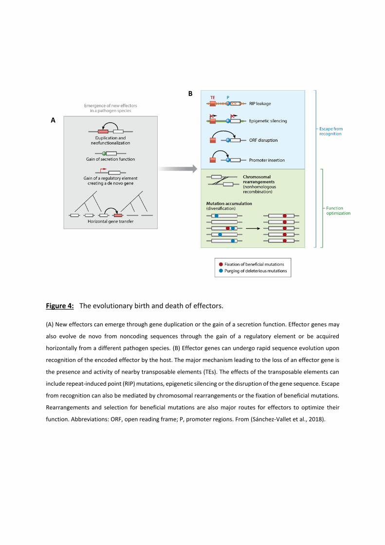
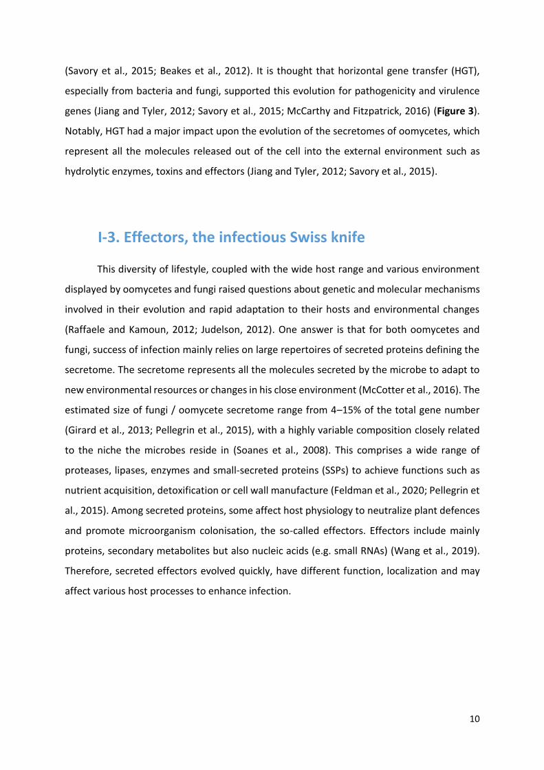
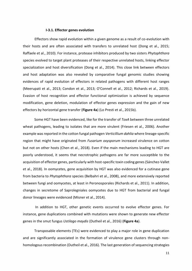
Figure 4: The evolutionary birth and death of effectors.
(A) New effectors can emerge through gene duplication or the gain of a secretion function. Effector genes may
also evolve de novo from noncoding sequences through the gain of a regulatory element or be acquired
horizontally from a different pathogen species. (B) Effector genes can undergo rapid sequence evolution upon
recognition of the encoded effector by the host. The major mechanism leading to the loss of an effector gene is
the presence and activity of nearby transposable elements (TEs). The effects of the transposable elements can
include repeat-induced point (RIP) mutations, epigenetic silencing or the disruption of the gene sequence. Escape
from recognition can also be mediated by chromosomal rearrangements or the fixation of beneficial mutations.
Rearrangements and selection for beneficial mutations are also major routes for effectors to optimize their
function. Abbreviations: ORF, open reading frame; P, promoter regions. From (Sánchez-Vallet et al., 2018).
A
B
11
I-3.1. Effector genes evolution
Effectors show rapid evolution within a given genome as a result of co-evolution with
their hosts and are often associated with transfers to unrelated host (Dong et al., 2015;
Raffaele et al., 2010). For instance, protease inhibitors produced by two sisters Phytophthora
species evolved to target plant proteases of their respective unrelated hosts, linking effector
specialization and host diversification (Dong et al., 2014). This close link between effectors
and host adaptation was also revealed by comparative fungal genomic studies showing
evidences of rapid evolution of effectors in related pathogens with different host ranges
(Meerupati et al., 2013; Condon et al., 2013; O’Connell et al., 2012; Richards et al., 2019).
Evasion of host recognition and effector functional optimization is achieved by sequence
modification, gene deletion, modulation of effector genes expression and the gain of new
effectors by horizontal gene transfer (Figure 4a) (Lo Presti et al., 2015b).
Some HGT have been evidenced, like for the transfer of ToxA between three unrelated
wheat pathogens, leading to isolates that are more virulent (Friesen et al., 2006). Another
example was reported in the cotton fungal pathogen Verticillium dahlia where lineage-specific
region that might have originated from Fusarium oxysporum increased virulence on cotton
but not on other hosts (Chen et al., 2018). Even if the main mechanisms leading to HGT are
poorly understood, it seems that necrotrophic pathogens are far more susceptible to the
acquisition of effector genes, particularly with host-specific toxin coding genes (Sánchez-Vallet
et al., 2018). In oomycetes, gene acquisition by HGT was also evidenced for a cutinase gene
from bacteria to Phytophthora species (Belbahri et al., 2008), and more extensively reported
between fungi and oomycetes, at least in Peronosporales (Richards et al., 2011). In addition,
changes in secretome of Saprolegniales oomycetes due to HGT from bacterial and fungal
donor lineages were evidenced (Misner et al., 2014).
In addition to HGT, other genetic events occurred to evolve effector genes. For
instance, gene duplications combined with mutations were shown to generate new effector
genes in the smut fungus Ustilago maydis (Dutheil et al., 2016) (Figure 4a).
Transposable elements (TEs) were evidenced to play a major role in gene duplication
and are significantly associated in the formation of virulence gene clusters through non-
homologous recombination (Dutheil et al., 2016). The last generation of sequencing strategies

12
greatly increased the quality of genome assemblies and gave new insight into effector
evolution and genome organization. Firstly, it revealed that TEs content was often
underestimated. For example, the last version of Colletotrichum higginsianum genome
contains 7% TEs whereas it was estimated to only 1.2% in the first assembly (Dallery et al.,
2017). Then, it is now clear that many effector genes are not randomly distributed across the
genomes and are associated with TEs and repetitive sequences in specific genome
compartments. These results have led to the “two-speed genome” model in which some
pathogen genomes have a bipartite architecture with essential genes in the core genome,
protected from deleterious mutations, and the accessory genome where effector genes take
place in a rapid evolutionary compartments (Raffaele and Kamoun, 2012; Croll and McDonald,
2012).
Rapid host adaptation can lead to effector recognition that triggers host defence.
Hence, adaptive pressure on effector gene sequence can force mutations in order to modify,
modulate or delete a given effector to escape host recognition. The most efficient mechanism
leading to the loss of an effector gene is related to the activity of TEs. TEs can drive multiple
effects on gene sequence, from gene disruption to repeat-induced point (RIP) mutations
(Figure 4b). Adaptive loss of function was reported in the fungal pathogen of wheat
Zymoseptoria tritici, where gene losses affected more than 10% of all genes in the genome,
including both effectors and genes with conserved functions such as secondary metabolite
gene clusters (Hartmann and Croll, 2017).
In addition to TEs activity, two types of mutations are known to modulate effector
genes evolution (Figure 4b) (reviewed in (Sánchez-Vallet et al., 2018)). The first type of
mutation consists in substitutions, insertions or deletions that change the protein properties
of a given effector. The second type of mutation concerns neutral mutations with weak but
cumulative effects. Fixation of beneficial mutations leads to optimization of the effector
function and can infer the past selective history at the effector locus (Sánchez-Vallet et al.,
2018).
Transcriptional silencing of an effector gene is another mechanism involved to escape
host recognition, which preserve the effector sequence (Gijzen et al., 2014; Whisson et al.,
2012). This was observed for the Phytophthora sojae effector gene Avr3a that is recognized in
soybean plants carrying the resistance gene Rps3a. Silenced Avr3a alleles were transmitted
13
and persisted over multiple generations suggesting that transgenerational gene silencing at
this locus mediated the gain of virulence phenotype (Qutob et al., 2013).
I-3.2. Apoplastic and intracellular effectors
During eukaryotic filamentous pathogens-plant interactions, two types of effectors can
be distinguished depending on their localization. Apoplastic effectors proteins (AEPs) stay in
the plant extracellular space (i.e. apoplast) while intracellular effectors proteins traffic into
the host cell in various compartments.
I-3.2 a. Apoplastic effectors: in front of the Wall
The apoplast is a hostile environment notably due to secreted basal defence
compounds like proteases, secondary metabolites or hydrolytic enzymes like chitinases
(Doehlemann and Hemetsberger, 2013; Jashni et al., 2015). The release of PAMPs in the
apoplast due to activity of plant chitinases or β-glucanases that disrupt microbial cell wall
integrity leads to their perception through cell surface-localized immune receptors, such as
Lysin motif (LysM)-containing proteins, which activates plant immune system (Cook et al.,
2015; Liu et al., 2012). Thus, to counteract this first recognition process, numerous fungal and
some oomycete AEPs have been characterized to evade glycan-triggered immunity or to
protect cell wall microorganism from degradation (reviewed in (Rocafort et al., 2020)).
Phytophthora spp. for instance secrete glucanase inhibitor proteins (GIPs) to inhibit the
degradation of pathogen β-1,3/1,6-glucans and the release of defence-eliciting
oligosaccharides by host endoglucanases (Rose, 2002; Damasceno et al., 2008). The tomato
fungal pathogen Cladosporium fulvum secretes two characterized AEPs, the chitin-binding
effector protein Avr4, which protects fungal hyphae against hydrolysis by plant chitinases (van
den Burg et al., 2006), and Ecp6, an effector which uses LysM domains that competitively
sequesters chitin oligomers from host immune receptors leading to the perturbation of chitin-
triggered host immunity (Sánchez-Vallet et al., 2013). Other LysM effectors have been shown
to contribute to virulence through chitin binding in other plant pathogenic fungi like
Magnaporthe oryzae, Colletotrichum higginsianum and Verticillium dahlia (Kombrink et al.,
2017; Mentlak et al., 2012; Takahara et al., 2016). Interestingly, AEPs with similar roles to both
Avr4 and Ecp6 have been described in the fungal wheat pathogen Zymoseptoria tritici
14
(Marshall et al., 2011; Sánchez-Vallet et al., 2020) but also in the mutualistic fungus
Trichoderma atroviride and in the arbuscular mycorrhizal fungus Rhizophagus irregularis
(Zeng et al., 2020; Romero-Contreras et al., 2019). This indicates that both pathogenic and
mutualistic microbes use AEPs to evade glycan-triggered immunity.
Thus, many characterized AEPs act to supress this glycan-triggered immunity (Rocafort
et al., 2020) but other families of AEPs have been described. One large group of apoplastic
effectors commonly found in fungi and oomycetes are cell wall degrading enzymes (CWDEs),
which play a major role in pathogenicity, contributing to plant cell wall degradation. Thus, this
family includes hundreds genes coding for enzymes such as cellulases, hemicellulases,
pectinases, β-1,3-glucanases, glyceraldehyde hydrolases, carbohydrate binding molecules and
other proteases able to degrade glycoproteins. The aim is to reduce the complexity of the cell
wall structure to facilitate entry and colonization of the host. In animal pathogen interaction,
those enzymes are absent and replaced by other specific enzymes. For instance, the plant
pathogen oomycete A. euteiches possesses a large repertoire of CWDEs coding genes that
target plant cell wall polysaccharides, absent in Aphanomyces astaci, the causing agent of
crayfish plague. In turn, A. astaci shows an expansion of protease genes predicted to target
chitin, the main component of the crayfish shell ((Gaulin et al., 2018) and see CHAPTER IV).
Recently, it has been shown that a CWDE effector was protected by another AEP, acting as a
decoy. Indeed, Phytophthora sojae displays an apoplastic effector, called PsXLP1, able to
promote infection by protecting PsXEG1, another effector with xyloglucanase activity
essential for full virulence but targeted for inhibition by GmGIP1, a soybean protein. Then,
PsXLP1 binds to GmGIP1 and functions as a decoy to protect PsXEG1 from the inhibitory action
of GmGIP1 (Ma et al., 2017).
Some AEPs are considered as toxins, called necrosis-inducing proteins (NLPs), able to
cause cell death. NLPs were first identified from culture filtrate of Fusarium oxysporum but
have been isolated in oomycetes, fungi and bacteria, and have the ability to induce cell death
and ethylene accumulation in plants (Gijzen and Nürnberger, 2006; Cobos et al., 2019). The
structure of NLPs is remarkably conserved among long phylogenetic distance, from bacteria
to oomycetes (Feng et al., 2014; Ottmann et al., 2009). However, the role of NLPs during
infection is unclear. When studies reported evidences that NLPs function as virulence factors
that increase pathogen growth in host plants or extend the host range (Veit et al., 2001;
15
Mattinen et al., 2004; Pemberton et al., 2005), others revealed that mutations in some NLP
genes from various fungi like Fusarium oxysporum or Botrytis cinerea do not reduce their
virulence (Cuesta Arenas et al., 2010; Bailey et al., 2002). In addition, most identified NLPs are
perceived by the host as PAMPs leading to the stimulation of PTI, such as NLPs from
Phytophthora species in Arabidopsis (Qutob et al., 2006, 2002), or from Pythium in various
dicotyledonous plants (Veit et al., 2001).
In oomycetes, particularly in Phytophthora and Pythium species, elicitins represent
another family of small AEPs and display similar characteristics with NLPs. Elicitins are
structurally conserved and induce a sustained oxidative burst that leads to hypersensitive
response (HR) cell death in most case (Derevnina et al., 2016). Plants from different botanical
families perceived elicitins as MAMPs, which induce activation of defence through MAMP-
triggered immunity (MTI) (Derevnina et al., 2016). Then, like NLPs, the role of elicitins is still
unclear. Since elicitins bind sterol and other lipids (Osman et al., 2001) and given the fact that
most oomycetes including Phytophthora are sterol auxotrophs, elicitins are proposed to act
as sterol-carrier proteins (Mikes et al., 1998). As sterols and fatty acids stimulate sexual
reproduction and oospore production in Phytophthora, elicitins could contribute to the
appearance of more virulent strains (Chepsergon et al., 2020).
Finally it is anticipated that some apoplastic effectors, especially cyclic peptides, could
play a role in self-defence against competitor antimicrobial compounds, or in manipulating
the apoplastic microbiome to promote host colonization (Snelders et al., 2018; Rocafort et al.,
2020).
I-3.2 b. Intracellular effectors: Destroy from within
The second class of effectors are secreted proteins translocated to the host cytoplasm
or intracellular compartments. In oomycetes, the first (and the largest) family of cytoplasmic
effectors, named RxLR effectors, were identified by comparative sequence analysis of
predicted secreted avirulence proteins from several oomycete species, leading to the
identification of a conserved amino acid motif, namely the RxLR-EER motif (Rehmany et al.,
2005). Thus more than 350 RxLRs effectors characterized by their R (arginine) – X (any amino
16
acid) – L (Lysine) – R (arginine) motif after signal peptide sequence, were predicted in
Phytophthora species (Tyler et al., 2006). Then presence of RxLR genes was evidenced in
numerous Phytophthora species, where several hundred putative RxLRs were predicted (Haas
et al., 2009; Jiang and Tyler, 2012), but only one in Saprolegniales species (Trusch et al., 2018).
Finally, RxLR and RxLR-like effectors may also be present in fungi (Kale and Tyler, 2011) as in
the endophytic fungus Piriformospora indica (named later Serendipita indica) in which 5
proteins with a degenerated RxLR motif were predicted to be secreted but none of them were
found to be up-regulated during colonization of barley roots (Zuccaro et al., 2011).
RxLR proteins contain a conserved N-terminal motif in addition to a predicted signal
peptide and a highly variable C-terminal part that allows biological function (Birch et al., 2006).
It was proposed that the RxLR motif acts as a signal for host delivery (Whisson et al., 2007;
Dou et al., 2008). In addition, RxLR effectors have been reported to translocate into host cells
in the absence of the pathogen, after binding of the RxLR motif to lipids via phospholipid-
mediated endocytosis (Kale and Tyler, 2011; Kale et al., 2010). However, studies made on
other RxLR effectors could not observed this entry mechanism and finally exclude the
phospholipid binding as a general host entry mechanism (Gan et al., 2010; Yaeno and Shirasu,
2013; Wawra et al., 2012). Then, pathogen-independent translocation of effectors into plant
cells is controversially discussed and the entry mechanism of effectors is still unclear (Wawra
et al., 2013). A recent study demonstrated that the RxLR motif of the Phytophthora infestans
effector AVR3a was cleaved before secretion (Wawra et al., 2017). Even more recently, in the
oomycete Saprolegnia parasitica, it was reported that the uptake process of the RxLR protein
SpHtp3 is guided by a gp96-like receptor via its C-terminal region, but not by the N-terminal
RxLR motif (Trusch et al., 2018). After translocation into host cell, a major part of RxLR
effectors target nucleus, but some have a nucleo-cytoplasmic localization when others
accumulate in membranes (Sperschneider et al., 2017), as described for the oomycete
Hyaloperonospora arabidopsidis (Caillaud et al., 2012).
The identification of RxLR effectors with conserved motif and the availability of
Phytophthora infestans genome lead to the discovery of another family of intracellular
effectors named CRNs, for CRinkling and Necrosis effectors. CRNs were first identified in the
plant pathogenic oomycete Phytophthora infestans. To identify pathogen-secreted proteins
potentially involved in the manipulation of host processes, a large screen of cDNA coding for
17
secreted proteins were expressed in N. benthamiana and tomato leaves. Two of which, named
CRN1 and CRN2, presenting similarities at the sequence level were found to cause a CRinkling
and Necrosis (CRN) phenotype when expressed in plant tissue (Torto et al., 2003). Like RxLR
proteins, CRNs present a modular architecture with a conserved N-ter signal characterized by
LxLFLAK-derived amino acid sequence (with possible variation) followed by a highly variable
C-ter domain (Schornack et al., 2010). With the increasing number of available genomes, many
studies performed on other oomycetes revealed that, in contrast to the RxLR protein family,
CRN coding genes are widespread in oomycete lineage, and were found in all plant pathogenic
oomycetes sequenced to date including Peronosporales (Haas et al., 2009; Tyler et al., 2006;
Baxter et al., 2010), Albuginales (Kemen et al., 2011), Pythiales (Adhikari et al., 2013; Lévesque
et al., 2010) and Saprolegniales (Gaulin et al., 2008). Some CRN-like coding genes were also
predicted in the animal pathogen Aphanomyces astaci, the causing agent of the Crayfish
plague ((Gaulin et al., 2018) and see CHAPTER IV). The identification of hundreds CRNs genes
in Aphanomyces species, which are early divergent species among the “crown” oomycetes,
suggests that CRNs are an ancient class of conserved oomycete effector proteins (Schornack
et al., 2010).
CRNs have a modular architecture with two distinct protein regions. The N-terminus
domains, composed around 130 amino acids (aa), contains a conserved LxLFLAK or LxLFLAK-
derivate motifs (within the first 60 aa) and more diversified DWL domains. Another highly
conserved HVLVxxP motif marks the end of the N-terminal region (Figure 5a). This N-terminal
part is presumed to specify the secretion and the translocation of the protein into the host.
The functionality of CRNs N-termini domain was initially tested via an infection-translocation
assay ((Schornack et al., 2010) and see P29-30 of this manuscript for more details). In this
study, three CRN N-termini of P. infestans (CRN2, CRN8 and CRN16) and one A. euteiches
(AeCRN5) were fused to C-terminal domain of the P. infestans Avr3a RxLR protein, and
introduced in Phytophthora capsici. Those strains were used to infect transgenic N.
benthamiana leaves expressing the potato resistance protein R3a.
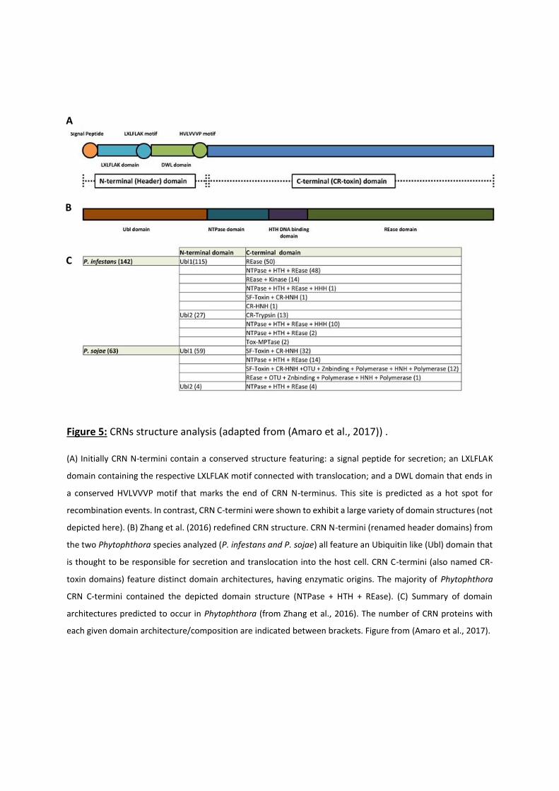
Figure 5: CRNs structure analysis (adapted from (Amaro et al., 2017)) .
(A) Initially CRN N-termini contain a conserved structure featuring: a signal peptide for secretion; an LXLFLAK
domain containing the respective LXLFLAK motif connected with translocation; and a DWL domain that ends in
a conserved HVLVVVP motif that marks the end of CRN N-terminus. This site is predicted as a hot spot for
recombination events. In contrast, CRN C-termini were shown to exhibit a large variety of domain structures (not
depicted here). (B) Zhang et al. (2016) redefined CRN structure. CRN N-termini (renamed header domains) from
the two Phytophthora species analyzed (P. infestans and P. sojae) all feature an Ubiquitin like (Ubl) domain that
is thought to be responsible for secretion and translocation into the host cell. CRN C-termini (also named CR-
toxin domains) feature distinct domain architectures, having enzymatic origins. The majority of Phytophthora
CRN C-termini contained the depicted domain structure (NTPase + HTH + REase). (C) Summary of domain
architectures predicted to occur in Phytophthora (from Zhang et al., 2016). The number of CRN proteins with
each given domain architecture/composition are indicated between brackets. Figure from (Amaro et al., 2017).
A
B
C
18
As chimera proteins induced avirulence by the recognition of the R3a protein, it was
concluded that those N-termini domains allow secretion and translocation of C-termini CRN
proteins into host cells. Even more, mutations in the LxLFLAK conserved motif indicate that
these motifs are necessary for translocation function. Importantly, N-termini domains of
AeCRN5 and CRN16 were demonstrated to be functional, even if no signal peptide were
predicted in the first 30 amino acids. These results demonstrate that despite the absence of
signal peptide, which was reported for numerous CRNs (Stam et al., 2013b; Voß et al., 2018;
Amaro et al., 2017; Adhikari et al., 2013; Gaulin et al., 2018), an unpredictable secretion signal
is present in this region and ensure secretion of CRNs in oomycetes (Schornack et al., 2010).
A recent study proposed to reconsider the requirement of LxLFLAK motifs in CRN
translocation and challenged the classification of CRNs proteins as members of a larger order
of Eukaryotic effectors (Zhang et al., 2016). In this paper, authors performed multiple in silico
analyses using a combination of sequence alignments and structure prediction programs,
coupled to comparative genomics to assess CRN occurrence across the Eukaryote taxon.
Results of those analyses ruled out the presence of signal peptides and indicate that the
proteins containing the LxLFLAK motif but also numerous proteins lacking this motif were
predicted to have an ubiquitin-like structure, similar to those found in the N-terminal region
of SSK1 (mitogen activated protein kinase) / Mcs4 (mitotic catastrophe suppressor 4)
signalling proteins in fungi. The LxLFLAK motif was located in strand 2 and 3 of this ubiquitin-
like domain (Ubl), suggesting that structural features rather than sequence conservation
underpin CRN translocation (Zhang et al., 2016). Authors then renamed the N-terminal region
as a Header Domain (Figure 5b). SSK1 orthologs play important roles in stress responses in
various true fungi, such as oxidative and osmotic shock, and in some cases in a
phosphorylation-dependent manner, employing an interaction between their N-terminal
domains and a MAPKKK heteromer (Morigasaki and Shiozaki, 2013; Yu et al., 2016). From this,
the authors suggest that CRN Ubl N-terminal domains could facilitate translocation inside the
host by analogous mechanisms (Zhang et al., 2016) but functional studies are require to
support this new concept. Similarly, the classification of CRN C-termini was challenged by the
study of Zhang and colleagues (Zhang et al., 2016). Initially 36 different conserved subdomains
that can assemble in different combinations defining C-terminal subfamilies were identified
in P. infestans (Haas et al., 2009). Then it has been proposed that the highly variable
19
organisation of C-termini was the result of recombination events between subdomains.
Following studies indicated that among oomycetes, most of these subfamilies are present and
that specific subfamilies can also be found. For example, in P. capsici, 30 of these subfamilies
are present while 7 new subfamilies appear specific to this species (Stam et al., 2013b).
Similarly, in A. euteiches, 160 CRN gene models have been described, among which 12 C-
termini domains are novel subdomains (Gaulin et al., 2008). In the study of Zhang and
colleagues, even if high levels of diversity are still present, the re-classification of CRN C-
terminal domains led to a limited set of domain configurations that were found to be
prevalent. Numerous CRN C-termini are related to enzymatic activities (Figure 5c). For
instance, one-fourth of all CRN C-termini analysed contains a P-loop NTPase domain, coupled
with a nuclease domain of the restriction endonuclease (REase) superfamily. In the same way,
approximately one-sixth of the C-termini domains harbour REase superfamily domain
combined with protein kinase domain. Then, those NTPase or Kinase domains, but also other
domains like HNH domain, could regulate REase activity or affinity toward nucleic acids (Zhang
et al., 2016), suggesting that targeting nucleic acids could be a shared feature among CRNs.
This hypothesis is supported by two studies that report the DNA binding activity of CRN
proteins in addition to the nuclear localization of numerous CRNs (Song et al., 2015; Ramirez-
Garcés et al., 2016; Amaro et al., 2017).
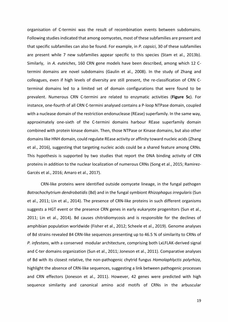
CRN-like proteins were identified outside oomycete lineage, in the fungal pathogen
Batrachochytrium dendrobatidis (Bd) and in the fungal symbiont Rhizophagus irregularis (Sun
et al., 2011; Lin et al., 2014). The presence of CRN-like proteins in such different organisms
suggests a HGT event or the presence CRN genes in early eukaryote progenitors (Sun et al.,
2011; Lin et al., 2014). Bd causes chitridiomycosis and is responsible for the declines of
amphibian population worldwide (Fisher et al., 2012; Scheele et al., 2019). Genome analyses
of Bd strains revealed 84 CRN-like sequences presenting up to 46.5 % of similarity to CRNs of
P. infestans, with a conserved modular architecture, comprising both LxLFLAK-derived signal
and C-ter domains organization (Sun et al., 2011; Joneson et al., 2011). Comparative analyses
of Bd with its closest relative, the non-pathogenic chytrid fungus Homolaphlyctis polyrhiza,
highlight the absence of CRN-like sequences, suggesting a link between pathogenic processes
and CRN effectors (Joneson et al., 2011). However, 42 genes were predicted with high
sequence similarity and canonical amino acid motifs of CRNs in the arbuscular
20
endomycorrhizal (AM) fungus Rhizophagus irregularis (Lin et al., 2014). The functional
characterization of the RiCRN1 protein evidenced the biological role of this Crinkler in the
establishment of the symbiosis, especially on the initiation of arbuscule development (Voß et
al., 2018). Furthermore, the study of Zhang et al. revealed that CRN effectors are also present
in free-living eukaryotes and land plants that are not known to have a pathogenic lifestyle,
indicating that CRNs are widespread in Eukaryotes (Zhang et al., 2016). Thus it was proposed
that CRN proteins could be firstly involved in inter-organismal conflicts, after which in some
host-pathogen interactions, these proteins were co-opted as effectors (Zhang et al., 2016;
Amaro et al., 2017).
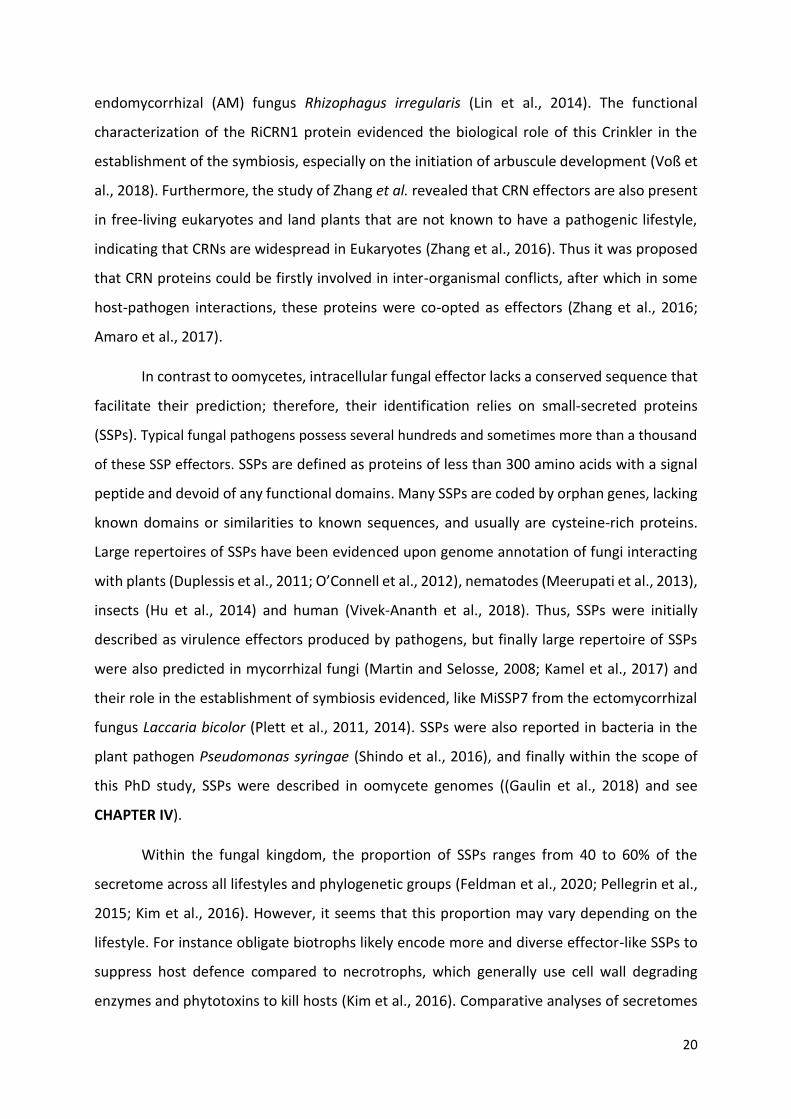
In contrast to oomycetes, intracellular fungal effector lacks a conserved sequence that
facilitate their prediction; therefore, their identification relies on small-secreted proteins
(SSPs). Typical fungal pathogens possess several hundreds and sometimes more than a thousand
of these SSP effectors. SSPs are defined as proteins of less than 300 amino acids with a signal
peptide and devoid of any functional domains. Many SSPs are coded by orphan genes, lacking
known domains or similarities to known sequences, and usually are cysteine-rich proteins.
Large repertoires of SSPs have been evidenced upon genome annotation of fungi interacting
with plants (Duplessis et al., 2011; O’Connell et al., 2012), nematodes (Meerupati et al., 2013),
insects (Hu et al., 2014) and human (Vivek-Ananth et al., 2018). Thus, SSPs were initially
described as virulence effectors produced by pathogens, but finally large repertoire of SSPs
were also predicted in mycorrhizal fungi (Martin and Selosse, 2008; Kamel et al., 2017) and
their role in the establishment of symbiosis evidenced, like MiSSP7 from the ectomycorrhizal
fungus Laccaria bicolor (Plett et al., 2011, 2014). SSPs were also reported in bacteria in the
plant pathogen Pseudomonas syringae (Shindo et al., 2016), and finally within the scope of
this PhD study, SSPs were described in oomycete genomes ((Gaulin et al., 2018) and see
CHAPTER IV).
Within the fungal kingdom, the proportion of SSPs ranges from 40 to 60% of the
secretome across all lifestyles and phylogenetic groups (Feldman et al., 2020; Pellegrin et al.,
2015; Kim et al., 2016). However, it seems that this proportion may vary depending on the
lifestyle. For instance obligate biotrophs likely encode more and diverse effector-like SSPs to
suppress host defence compared to necrotrophs, which generally use cell wall degrading
enzymes and phytotoxins to kill hosts (Kim et al., 2016). Comparative analyses of secretomes
21
also identified shared or lifestyle-specific SSPs between saprotrophic and Ectomycorrhizal
(ECM) fungi, indicating that presence of SSPs is not limited to fungi interacting with living
plants (Pellegrin et al., 2015).
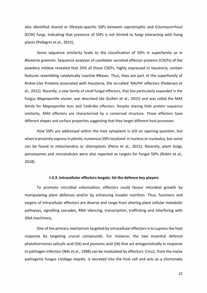
Some sequence similarity leads to the classification of SSPs in superfamily as in
Blumeria graminis. Sequence analyses of candidate secreted effector proteins (CSEPs) of the
powdery mildew revealed that 25% of those CSEPs, highly expressed in haustoria, contain
features resembling catalytically inactive RNases. Thus, they are part of the superfamily of
RnAse‐Like Proteins associated with Haustoria, the so‐called ‘RALPH’ effectors (Pedersen et
al., 2012). Recently, a new family of small fungal effectors, that has particularly expanded in the
fungus Magnaporthe oryzae, was described (de Guillen et al., 2015) and was called the MAX
family for Magnaporthe Avrs and ToxB-like effectors. Despite sharing little protein sequence
similarity, MAX effectors are characterized by a conserved structure. Those effectors have
different shapes and surface properties suggesting that they target different host processes.
How SSPs are addressed within the host cytoplasm is still an opening question, but
when transiently express in planta, numerous SSPs localized in nucleus or nucleolus, but some
can be found in mitochondria or chloroplasts (Petre et al., 2015). Recently, plant Golgi,
peroxisomes and microtubules were also reported as targets for fungal SSPs (Robin et al.,
2018).
I-3.3. Intracellular effectors targets: hit the defence key players
To promote microbial colonization, effectors could favour microbial growth by
manipulating plant defences and/or by enhancing invader nutrition. Thus, functions and
targets of intracellular effectors are diverse and range from altering plant cellular metabolic
pathways, signalling cascades, RNA silencing, transcription, trafficking and interfering with
DNA machinery.
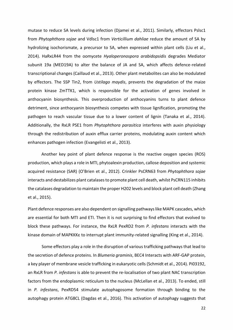
One of the primary mechanism targeted by intracellular effectors is to supress the host
response by targeting crucial compounds. For instance, the two essential defence
phytohormones salicylic acid (SA) and jasmonic acid (JA) that act antagonistically in response
to pathogen infection (Niki et al., 1998) can be modulated by effectors. Cmu1, from the maize
pathogenic fungus Ustilago maydis, is secreted into the host cell and acts as a chorismate
22
mutase to reduce SA levels during infection (Djamei et al., 2011). Similarly, effectors Pslsc1
from Phytophthora sojae and Vdlsc1 from Verticillium dahliae reduce the amount of SA by
hydrolizing isochorismate, a precursor to SA, when expressed within plant cells (Liu et al.,
2014). HaRxLR44 from the oomycete Hyaloperonospora arabidopsidis degrades Mediator
subunit 19a (MED19A) to alter the balance of JA and SA, which affects defence-related
transcriptional changes (Caillaud et al., 2013). Other plant metabolites can also be modulated
by effectors. The SSP Tin2, from Ustilago maydis, prevents the degradation of the maize
protein kinase ZmTTK1, which is responsible for the activation of genes involved in
anthocyanin biosynthesis. This overproduction of anthocyanins turns to plant defence
detriment, since anthocyanin biosynthesis competes with tissue lignification, promoting the
pathogen to reach vascular tissue due to a lower content of lignin (Tanaka et al., 2014).
Additionally, the RxLR PSE1 from Phytophthora parasitica interferes with auxin physiology
through the redistribution of auxin efflux carrier proteins, modulating auxin content which
enhances pathogen infection (Evangelisti et al., 2013).
Another key point of plant defence response is the reactive oxygen species (ROS)
production, which plays a role in MTI, phytoalexin production, callose deposition and systemic
acquired resistance (SAR) (O’Brien et al., 2012). Crinkler PsCRN63 from Phytophthora sojae
interacts and destabilizes plant catalases to promote plant cell death, whilst PsCRN115 inhibits
the catalases degradation to maintain the proper H202 levels and block plant cell death (Zhang
et al., 2015).
Plant defence responses are also dependent on signalling pathways like MAPK cascades, which
are essential for both MTI and ETI. Then it is not surprising to find effectors that evolved to
block these pathways. For instance, the RxLR PexRD2 from P. infestans interacts with the
kinase domain of MAPKKKε to interrupt plant immunity-related signalling (King et al., 2014).
Some effectors play a role in the disruption of various trafficking pathways that lead to
the secretion of defence proteins. In Blumeria graminis, BEC4 Interacts with ARF-GAP protein,
a key player of membrane vesicle trafficking in eukaryotic cells (Schmidt et al., 2014). Pi03192,
an RxLR from P. infestans is able to prevent the re-localisation of two plant NAC transcription
factors from the endoplasmic reticulum to the nucleus (McLellan et al., 2013). To ended, still
in P. infestans, PexRD54 stimulate autophagosome formation through binding to the
autophagy protein ATG8CL (Dagdas et al., 2016). This activation of autophagy suggests that
23
the pathogen produce this effector to selectively eliminate some of the molecules that the
plant use to defend itself (Dagdas et al., 2016).
Another major defence mechanism is RNA silencing. This process was firstly described
in plant-virus interactions, where viral RNA is recognized as a MAMP and induces small
interfering RNAs (siRNAs) which trigger the cleavage of viral RNAs. In response, viruses have
developed suppressors of RNA silencing to allow the virus proliferation in the host (Vance,
2001). This defence system is also targeted by other microbes. PSR1 and PSR2, two RxLR
effectors from P. sojae, suppress RNA silencing and enhance susceptibility to P. sojae (Qiao et
al., 2013). PSR1 is able to bind with a conserved nuclear protein called PSR1-interacting
protein 1 (PINP1), which is involved in small RNA biogenesis. Alteration of small RNA
production in plants leads to developmental defects and hyper-susceptibility to Phytophthora
infections, which is similar to transgenic plants expressing the PSR1 protein (Qiao et al., 2015).
A PSR2-like effector was found in the related species Phytophthora infestans with the same
RNA silencing suppression activity, meaning that PSR2 represents a prevalent effector family
conserved within the genus Phytophthora (Xiong et al., 2014). Then, in viruses and oomycetes,
RNA silencing suppression is a key strategy for infection (Qiao et al., 2013).
Inhibition or alteration of gene transcription in order to down regulate genes involved
in defence responses is also a common process shared by various microorganisms to facilitate
the association with the plant. In Rhizophagus irregularis, SP7 targets nucleus and interacts
with the transcription factor ERF19 to block the plant immune system (Kloppholz et al., 2011).
The RxLR PsAvh23 from P. sojae disrupts the formation of the ADA2-GCN5 subcomplex and
subsequently represses the expression of defence genes by decreasing GCN5-mediated
H3K9ac levels, suggesting that the pathogen manipulates host histone acetylation to gain
virulence (Kong et al., 2017).
Transcription can also be altered by effectors which bind directly to nucleic acids, like
PsCRN108 from P. sojae which targets HSP promoters to block association with heat shock
transcription factors (Song et al., 2015). Furthermore, nucleic acids and especially DNA itself
could be targeted, as AeCRN13 from Aphanomyces euteiches, where it binds directly to DNA
and triggers double strand breaks (Ramirez-Garcés et al., 2016).

24
Finally, the vast majority of intracellular effectors from different fungal and oomycete
families, including RxLR, CRNs and SSPs, localizes in nucleus when expressed in host tissue.
Since it was evidenced that plant DNA is altered during infection of various pathogens (Song
and Bent, 2014) and regarding the large number of intracellular effectors that target nucleus,
we propose that DNA-damaging effectors could be a family of proteins involved in plant-
filamentous pathogen interactions and represents the subject of the Chapter II of this thesis
(see Chapter II: DNA-Damaging Effectors: New Players in the Effector Arena).
I-4. Aphanomyces: an oomycete genus to study effectors
and host adaptation
The Aphanomyces genus belongs to the Saprolegniales order and has been shown to
contain three major lineages, including plant pathogens, aquatic animal pathogens, and
saprophytic species (Diéguez-Uribeondo et al., 2009), making this genus an interesting model
to understand evolutionary mechanisms involved in adaptation of oomycetes to distantly
related hosts and environmental niches (Figure 6). It contains around 40 species, but this
number is inconsistent due to the difficult culture and identification of some species.
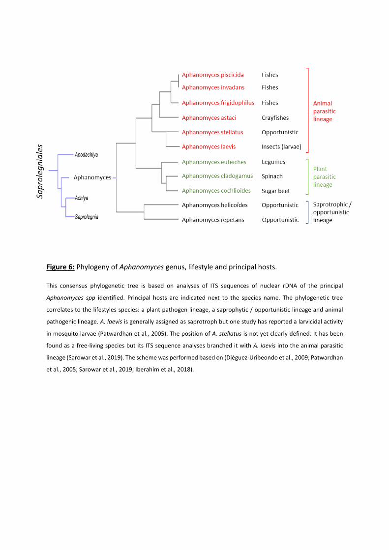
Figure 6: Phylogeny of Aphanomyces genus, lifestyle and principal hosts.
This consensus phylogenetic tree is based on analyses of ITS sequences of nuclear rDNA of the principal
Aphanomyces spp identified. Principal hosts are indicated next to the species name. The phylogenetic tree
correlates to the lifestyles species: a plant pathogen lineage, a saprophytic / opportunistic lineage and animal
pathogenic lineage. A. laevis is generally assigned as saprotroph but one study has reported a larvicidal activity
in mosquito larvae (Patwardhan et al., 2005). The position of A. stellatus is not yet clearly defined. It has been
found as a free-living species but its ITS sequence analyses branched it with A. laevis into the animal parasitic
lineage (Sarowar et al., 2019). The scheme was performed based on (Diéguez-Uribeondo et al., 2009; Patwardhan
et al., 2005; Sarowar et al., 2019; Iberahim et al., 2018).
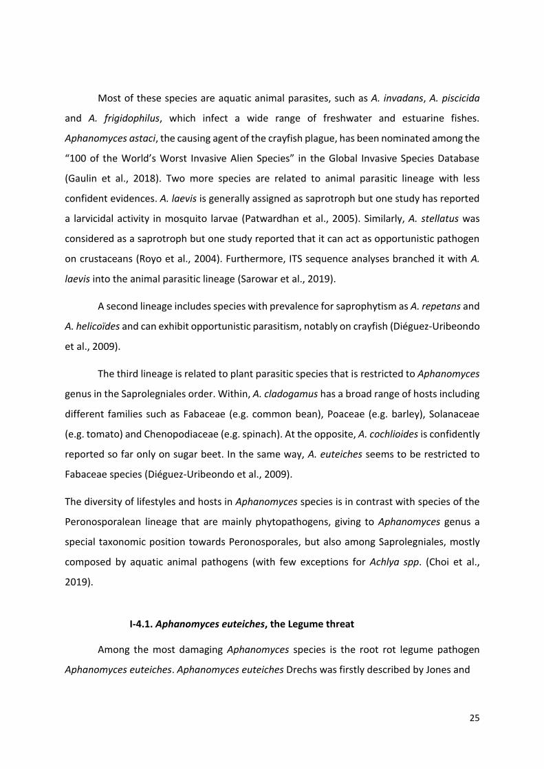
25
Most of these species are aquatic animal parasites, such as A. invadans, A. piscicida
and A. frigidophilus, which infect a wide range of freshwater and estuarine fishes.
Aphanomyces astaci, the causing agent of the crayfish plague, has been nominated among the
“100 of the World’s Worst Invasive Alien Species” in the Global Invasive Species Database
(Gaulin et al., 2018). Two more species are related to animal parasitic lineage with less
confident evidences. A. laevis is generally assigned as saprotroph but one study has reported
a larvicidal activity in mosquito larvae (Patwardhan et al., 2005). Similarly, A. stellatus was
considered as a saprotroph but one study reported that it can act as opportunistic pathogen
on crustaceans (Royo et al., 2004). Furthermore, ITS sequence analyses branched it with A.
laevis into the animal parasitic lineage (Sarowar et al., 2019).
A second lineage includes species with prevalence for saprophytism as A. repetans and
A. helicoïdes and can exhibit opportunistic parasitism, notably on crayfish (Diéguez-Uribeondo
et al., 2009).
The third lineage is related to plant parasitic species that is restricted to Aphanomyces
genus in the Saprolegniales order. Within, A. cladogamus has a broad range of hosts including
different families such as Fabaceae (e.g. common bean), Poaceae (e.g. barley), Solanaceae
(e.g. tomato) and Chenopodiaceae (e.g. spinach). At the opposite, A. cochlioides is confidently
reported so far only on sugar beet. In the same way, A. euteiches seems to be restricted to
Fabaceae species (Diéguez-Uribeondo et al., 2009).
The diversity of lifestyles and hosts in Aphanomyces species is in contrast with species of the
Peronosporalean lineage that are mainly phytopathogens, giving to Aphanomyces genus a
special taxonomic position towards Peronosporales, but also among Saprolegniales, mostly
composed by aquatic animal pathogens (with few exceptions for Achlya spp. (Choi et al.,
2019).
I-4.1. Aphanomyces euteiches, the Legume threat
Among the most damaging Aphanomyces species is the root rot legume pathogen
Aphanomyces euteiches. Aphanomyces euteiches Drechs was firstly described by Jones and
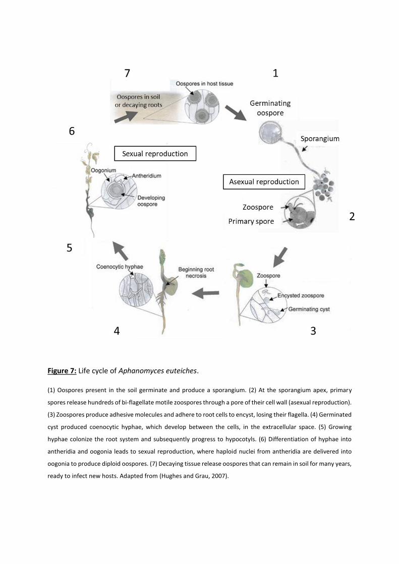
Figure 7: Life cycle of Aphanomyces euteiches.
(1) Oospores present in the soil germinate and produce a sporangium. (2) At the sporangium apex, primary
spores release hundreds of bi‐flagellate motile zoospores through a pore of their cell wall (asexual reproduction).
(3) Zoospores produce adhesive molecules and adhere to root cells to encyst, losing their flagella. (4) Germinated
cyst produced coenocytic hyphae, which develop between the cells, in the extracellular space. (5) Growing
hyphae colonize the root system and subsequently progress to hypocotyls. (6) Differentiation of hyphae into
antheridia and oogonia leads to sexual reproduction, where haploid nuclei from antheridia are delivered into
oogonia to produce diploid oospores. (7) Decaying tissue release oospores that can remain in soil for many years,
ready to infect new hosts. Adapted from (Hughes and Grau, 2007).
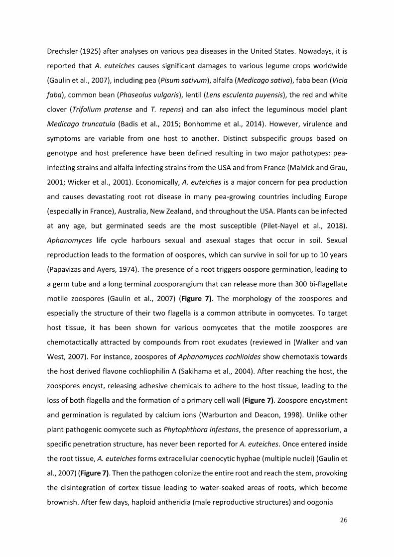
26
Drechsler (1925) after analyses on various pea diseases in the United States. Nowadays, it is
reported that A. euteiches causes significant damages to various legume crops worldwide
(Gaulin et al., 2007), including pea (Pisum sativum), alfalfa (Medicago sativa), faba bean (Vicia
faba), common bean (Phaseolus vulgaris), lentil (Lens esculenta puyensis), the red and white
clover (Trifolium pratense and T. repens) and can also infect the leguminous model plant
Medicago truncatula (Badis et al., 2015; Bonhomme et al., 2014). However, virulence and
symptoms are variable from one host to another. Distinct subspecific groups based on
genotype and host preference have been defined resulting in two major pathotypes: pea-
infecting strains and alfalfa infecting strains from the USA and from France (Malvick and Grau,
2001; Wicker et al., 2001). Economically, A. euteiches is a major concern for pea production
and causes devastating root rot disease in many pea-growing countries including Europe
(especially in France), Australia, New Zealand, and throughout the USA. Plants can be infected
at any age, but germinated seeds are the most susceptible (Pilet-Nayel et al., 2018).
Aphanomyces life cycle harbours sexual and asexual stages that occur in soil. Sexual
reproduction leads to the formation of oospores, which can survive in soil for up to 10 years
(Papavizas and Ayers, 1974). The presence of a root triggers oospore germination, leading to
a germ tube and a long terminal zoosporangium that can release more than 300 bi‐flagellate
motile zoospores (Gaulin et al., 2007) (Figure 7). The morphology of the zoospores and
especially the structure of their two flagella is a common attribute in oomycetes. To target
host tissue, it has been shown for various oomycetes that the motile zoospores are
chemotactically attracted by compounds from root exudates (reviewed in (Walker and van
West, 2007). For instance, zoospores of Aphanomyces cochlioides show chemotaxis towards
the host derived flavone cochliophilin A (Sakihama et al., 2004). After reaching the host, the
zoospores encyst, releasing adhesive chemicals to adhere to the host tissue, leading to the
loss of both flagella and the formation of a primary cell wall (Figure 7). Zoospore encystment
and germination is regulated by calcium ions (Warburton and Deacon, 1998). Unlike other
plant pathogenic oomycete such as Phytophthora infestans, the presence of appressorium, a
specific penetration structure, has never been reported for A. euteiches. Once entered inside
the root tissue, A. euteiches forms extracellular coenocytic hyphae (multiple nuclei) (Gaulin et
al., 2007) (Figure 7). Then the pathogen colonize the entire root and reach the stem, provoking
the disintegration of cortex tissue leading to water-soaked areas of roots, which become
brownish. After few days, haploid antheridia (male reproductive structures) and oogonia

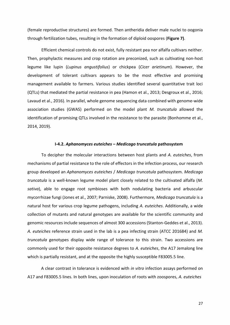
27
(female reproductive structures) are formed. Then antheridia deliver male nuclei to oogonia
through fertilization tubes, resulting in the formation of diploid oospores (Figure 7).
Efficient chemical controls do not exist, fully resistant pea nor alfalfa cultivars neither.
Then, prophylactic measures and crop rotation are preconized, such as cultivating non-host
legume like lupin (Lupinus angustifolius) or chickpea (Cicer arietinum). However, the
development of tolerant cultivars appears to be the most effective and promising
management available to farmers. Various studies identified several quantitative trait loci
(QTLs) that mediated the partial resistance in pea (Hamon et al., 2013; Desgroux et al., 2016;
Lavaud et al., 2016). In parallel, whole genome sequencing data combined with genome-wide
association studies (GWAS) performed on the model plant M. truncatula allowed the
identification of promising QTLs involved in the resistance to the parasite (Bonhomme et al.,
2014, 2019).
I-4.2. Aphanomyces euteiches – Medicago truncatula pathosystem
To decipher the molecular interactions between host plants and A. euteiches, from
mechanisms of partial resistance to the role of effectors in the infection process, our research
group developed an Aphanomyces euteiches / Medicago truncatula pathosystem. Medicago
truncatula is a well-known legume model plant closely related to the cultivated alfalfa (M.
sativa), able to engage root symbioses with both nodulating bacteria and arbuscular
mycorrhizae fungi (Jones et al., 2007; Parniske, 2008). Furthermore, Medicago truncatula is a
natural host for various crop legume pathogens, including A. euteiches. Additionally, a wide
collection of mutants and natural genotypes are available for the scientific community and
genomic resources include sequences of almost 300 accessions (Stanton-Geddes et al., 2013).
A. euteiches reference strain used in the lab is a pea infecting strain (ATCC 201684) and M.
truncatula genotypes display wide range of tolerance to this strain. Two accessions are
commonly used for their opposite resistance degrees to A. euteiches, the A17 Jemalong line
which is partially resistant, and at the opposite the highly susceptible F83005.5 line.
A clear contrast in tolerance is evidenced with in vitro infection assays performed on
A17 and F83005.5 lines. In both lines, upon inoculation of roots with zoospores, A. euteiches
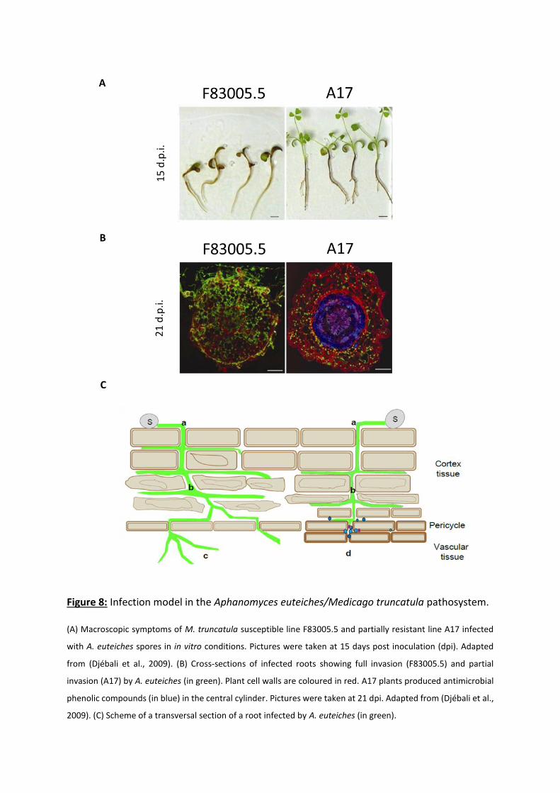
Figure 8: Infection model in the Aphanomyces euteiches/Medicago truncatula pathosystem.
(A) Macroscopic symptoms of M. truncatula susceptible line F83005.5 and partially resistant line A17 infected
with A. euteiches spores in in vitro conditions. Pictures were taken at 15 days post inoculation (dpi). Adapted
from (Djébali et al., 2009). (B) Cross-sections of infected roots showing full invasion (F83005.5) and partial
invasion (A17) by A. euteiches (in green). Plant cell walls are coloured in red. A17 plants produced antimicrobial
phenolic compounds (in blue) in the central cylinder. Pictures were taken at 21 dpi. Adapted from (Djébali et al.,
2009). (C) Scheme of a transversal section of a root infected by A. euteiches (in green).
A
B
C
15
d.p
.i.
21
d.p
.i.

28
On the left side, the scheme describes infection in F83005.5 were an asexual spore (S) has landed on the
rhizoplane and germinated to produce a germ tube giving rise to an infectious hyphae that directly penetrates
root cortex tissues (a). Hyphae develops between root cells of cortex which becomes completely colonized 6
days post inoculation. Cortical cells died as A. euteiches develops leading to root disassembly and water-soaked
symptoms typical of root rot disease (b). The pathogen reaches the vascular cylinder before completion of its
cycle (not shown) (c). On the right side, the infection is depicted in the tolerant host (A17) were the plant
produces supplementary pericycle cell layers with higher levels of lignin in their cell walls, reinforcing the root
stele. In addition to this mechanical barrier, cells produce antimicrobial compounds (in blue) (d). These
cytological responses restrain the advance of the pathogen to the vascular cylinder. Scheme from (Ramirez-
Garces, 2014).
hyphae penetrate inside the roots and grow between cells of the outer cortex tissue within 1
day. As mentioned above, no specialized infectious structures as appressoria or haustoria have
been reported. The pathogen presents an intercellular development and invades the whole
cortex area within 3 to 6 days. 15 days post inoculation (dpi) with A. euteiches zoospores,
susceptible F83005.5 plants harbour no or few leaves and very few secondary roots, while the
tolerant A17 plants still develop aerial part and present branched brownish roots (Figure 8a).
At 21 dpi, most of susceptible plants are dead (Djébali et al., 2009). While oomycete cell wall
is mostly composed by cellulose, A. euteiches has an original cell wall containing around 10%
of chitosaccharides exposed at the cell wall surface (Badreddine et al., 2008; Nars et al., 2013).
This structural characteristic allows the staining of hyphae using wheat-germ agglutinin lectin
(WGA) coupled with fluorophore. Confocal analyses performed on cross section of inoculated
A17 or F83005 roots revealed a whole colonization of all cell layers in the susceptible lines,
indicating that the pathogen reached the central cylinder (Figure 8b). Invasion of vascular
system in F83005 lines seems to start after 6 dpi and is confirmed at 15 and 21 dpi. In contrast,
in the tolerant A17 plants, A. euteiches hyphae were restricted to the root cortex, where
defence related phenolic compounds are produced (Figure 8b) (Djébali et al., 2009). In
addition to phenolic compounds production, A17 plants produce supplementary pericycle cell
layers coupled with a reinforcement of the cell walls that might act as a physical barrier for
the invading hyphae (Figure 8c). Furthermore, partial resistance of A17 is correlated to an
increase of lateral roots (Djébali et al., 2009).

29
Genetic approaches coupled to the characterization of infection phenotypes in M.
truncatula have led to the identification of a major QTL (Djébali et al., 2009). Forward GWAS
experiments refined this result and identified several candidate genes and pinpointed two
independent major loci (Bonhomme et al., 2014). Within the most significant locus, Single
Nucleotide Polymorphisms (SNPs) found in the promoter and coding region of an F-box gene
have been spotted out and linked to the variable tolerance of M. truncatula against A.
euteiches (Bonhomme et al., 2014). Additionally, it was shown that basal levels of flavonoids
play a significant role in resistance to A. euteiches, and could inhibit zoospore germination
(Badis et al., 2015). Finally, recently, a local score approach technic improved GWAS
resolution, refining the previously reported major locus, underlying a new tyrosine kinase
candidate gene involved in resistance, and detected minor QTLs (Bonhomme et al., 2019).
I-4.3. Insight into Aphanomyces euteiches intracellular effectors
Before this PhD, previous analyses on A. euteiches were performed to gain insights into
effectors biology and to unravel the biological functions of identified intracellular effectors. A
first transcriptomic analysis on A. euteiches revealed the absence of RxLR effectors while more
than 160 CRN genes have been detected (Gaulin et al., 2007). Among them, some are induced
during plant colonization, such as AeCRN5 and AeCRN13. We then characterized one of these
CRNs, AeCRN13, which harbours an N-ter domain containing a characteristic LYLALK motif
(derivate of the LxLFLAK motif in Aphanomyces) coupled with HVLVxxP motif, followed by a C-
ter domain composed by DFA-DDC subdomains reported in Phytophthora CRN13s (Ramirez-
Garcés et al., 2016) (Figure 9a). This work reports that AeCRN13 act as a genotoxin through
its binding to plant DNA and activates DNA-damage responses (DDR). It also provides
evidences that the effect is conserved among CRN13 family since its closest ortholog from the
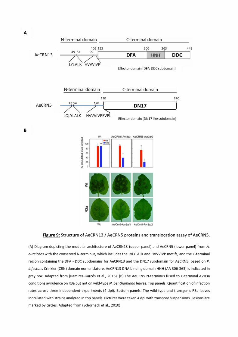
Figure 9: Structure of AeCRN13 / AeCRN5 proteins and translocation assay of AeCRN5.
(A) Diagram depicting the modular architecture of AeCRN13 (upper panel) and AeCRN5 (lower panel) from A.
euteiches with the conserved N-terminus, which includes the LxLYLALK and HVVVIVP motifs, and the C-terminal
region containing the DFA - DDC subdomains for AeCRN13 and the DN17 subdomain for AeCRN5, based on P.
infestans Crinkler (CRN) domain nomenclature. AeCRN13 DNA binding domain HNH (AA 306-363) is indicated in
grey box. Adapted from (Ramirez-Garcés et al., 2016). (B) The AeCRN5 N-terminus fused to C-terminal AVR3a
conditions avirulence on R3a but not on wild-type N. benthamiana leaves. Top panels: Quantification of infection
rates across three independent experiments (4 dpi). Bottom panels: The wild-type and transgenic R3a leaves
inoculated with strains analyzed in top panels. Pictures were taken 4 dpi with zoospore suspensions. Lesions are
marked by circles. Adapted from (Schornack et al., 2010).
30
chitrid fungus B. dendrobatidis acts similarly. Both Ae and Bd CRN13 proteins contain an HNH
motif widespread in metal finger endonucleases present in all life kingdoms. This motif is
responsible of the DNA binding ability of AeCRN13 since a mutated version in this domain
failed to bind DNA. Hence, AeCRN13 and BdCRN13 trigger DNA double strand breaks. We then
reported that the plant senses this insult and activates the DDR pathway to repair its DNA
(Ramirez-Garcés et al., 2016).
Sequences analyses of AeCRN5 confirmed the presence of the conserved motifs
LxLYLALK and HVVVIVP within the N-terminal domain. Then, DN17-like subdomain was
revealed by comparison of the C-terminal domain with P. infestans Crinkler domain
nomenclature (Figure 9a). To assess if the N-terminal domain of CRNs could be responsible of
the translocation of the C-terminal domain of the protein in host cells, our research group
collaborated with Schornack and colleagues to perform translocation assays on various CRNs
from Peronosporales or Saprolegniales members. The principle of these assays is based on the
recognition of the C-terminal domain of the avirulence protein Avr3a from P. infestans by the
resistance protein R3a which takes place in the cytosol of plant cells. This recognition leads to
ETI and full depletion of infection. Chimeric constructs containing N-terminal domains of
CRNs, notably AeCRN5, fused to C-terminal domain of Avr3a were introduced into P. capsici.
Inoculation of this P. capsici strains on wild-type N. benthamiana leaves (lacking R3a) leads to
normal infection symptoms, but failed to infect R3a N. benthamiana leaves, constitutively
expressing R3a resistance proteins, indicating recognition of the AVR3a effector domain by
intracellular R3a (Figure 9b). These results evidenced that the N-terminal domain of AeCRN5
can mediate the delivery of the effector protein inside host cells (Schornack et al., 2010).
31
I-5. Scope of the thesis
I joined the LRSV research team in 2011 as CNRS engineer (IE) and I started to work on
functional analysis of Aphanomyces euteiches effector thanks to my expertise on molecular
biology on plant viruses developed in Paris in the team of Isabelle Jupin (Institut Jacques
Monod).
When I started my thesis in 2018, the major part of intracellular effectors described in
plant pathogenic interactions was reported to target plant nucleus, such as all CRNs and
numerous RxLR from phytopathogenic oomycetes, or numerous fungal SSPs. At this time only
two microbial effectors were evidenced to target host DNA: AeCRN13 studied in our research
group (Ramirez-Garcés et al., 2016) and PsCRN108 from P. sojae, a CRN which binds to DNA
to deregulate HSP genes expression (Song et al., 2015). In addition, we also discovered by
confocal analysis and transient expression in Nicotiana cells that AeCRN5 also target plant
nucleus, suggesting that this CRN may also play a role at the nuclear level. While there was
accumulating evidences that effector from bacteria target host nucleus and act as DNA-
damaging compounds in mammalian cells, only AeCRN13 was reported as a eukaryotic DNA-
damaging effector.
The Chapter II of this manuscript consists to an opinion paper published in Trends in
Plant Science in 2019 (doi: 10.1016/j.tplants.2019.09.012.) where I pinpoint DNA-damaging
effectors in plant microbe interactions.
The Chapter III reports on functional analyses of AeCRN5. Knowing that its N-terminal
domain is an effective host-targeting signal (Schornack et al., 2010) and that AeCRN5 is nuclear
localized in planta, this CRN gene was selected as candidate to decipher the mode of action of
intracellular effector.
In the same time, spectacular advances in sequencing technologies allow us to gain
insight into Aphanomyces ssp. genomes. We took advantage of the broad host range of
Aphanomyces genus to make comparative genome analyses between animal and plant
Aphanomyces strains. The aim of this collaborative work was to confront the different
secretomes and to focus on the different classes of effectors. Those analyses lead to the
identification of a new class of oomycete effectors related to SSPs. We also undertook

32
functional characterization of a cluster of SSP genes, and identified AeSSP1256 as a small
nuclear localized protein that enhances oomycete colonisation. The results were published in
BMC Biology in 2018 (doi: 10.1186/s12915-018-0508-5) and represents the Chapter IV of this
manuscript.
We next focused on AeSSP1256 protein to decipher the role and the biological impact
of this SSP gene on plant cells and host development. The results are provided in the Chapter
V of this manuscript and available in BioRxiv (doi.org/10.1101/2020.06.17.157404) and
submitted for evaluation to a peer-journal. Complementary results were obtained during this
functional analysis and complete the chapter V of the manuscript.


Chapter II
DNA-Damaging Effectors:
New Players in the Effector Arena

33
II – CHAPTER II: DNA-Damaging Effectors: New Players in the
Effector Arena (Camborde et al. TIPS, 2019)
Functional analyses of A. euteiches AeCRN13 revealed that this protein targets host
DNA to trigger DNA damages. BdCRN13, the closest ortholog in the chytrid fungus
Batrachochytrium dendrobatidis also induces DNA damages and triggers DNA damage
responses (DDR) when expressed in a non-related host, such as plant cell (Ramirez-Garcés et
al., 2016).
In this opinion paper, we discuss about DNA damage as a strategy used by pathogens during
infection. DSBs and DDR have been evidenced in animal pathogens, especially in pathogenic
bacteria, which produce DNA-damaging compounds. These compounds, able to cause directly
or indirectly DNA breaks that result in mutations or cell death are named genotoxins. Several
examples are described in the paper. We then wonder whether plant pathogens could also
produce genotoxins and what could be the role of these compounds during infection. Finally
we present the host defence mechanism that consists in a DDR signalling cascade, better
characterized in animal than in plant.
34
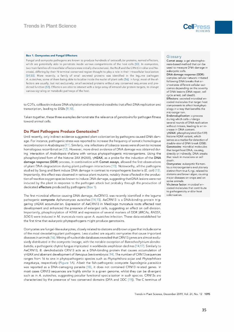
35

36
37
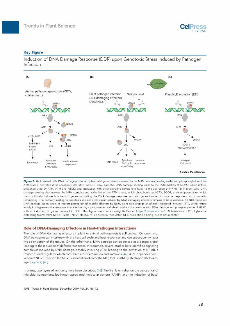
38

39
40

41

Chapter III
AeCRN5 effector from A. euteiches targets plant
RNA and perturbs RNA silencing

42
III – CHAPTER III: AeCRN5 effector from A. euteiches targets
plant RNA and perturbs RNA silencing
During my PhD I continued the functional analysis of AeCRN5 effector from A.
euteiches. AeCRN5 was identified in a cDNA library generated from A. euteiches grown in close
proximity to roots of M. truncatula (Gaulin et al., 2008). Its C-terminus was previously shown
to trigger necrosis and to localize in nuclei in N. benthamiana cells. Furthermore, it was
evidenced that its N-terminal domain was able to translocate the C-terminal part of the
protein in host cells (Schornack et al., 2010). AeCRN5 harbors a DN17 domain as its C-terminus
based on the Phytophthora CRNs domains nomenclature, without any predicted functional
activity.
Diana Ramirez-Garcés, a previous PhD student in the team, started the functional
characterization of AeCRN5 during her PhD in 2014 and evidenced that AeCRN5 triggers
necrosis when transiently express in N. benthamiana leaves or M. truncatula roots. During my
PhD I complete the functional analysis to provide new elements to decipher the mode of
action of AeCRN5. Briefly, we first identified that AeCRN5 could interfere with RNA silencing
but the mechanism is still unclear, and complementary results are required to support this
conclusion. Additionally we observed that AeCRN5 could interact with the plant SERRATE
protein (SE), known to participate to alternative splicing and microRNA biogenesis pathway in
plants like A. thaliana (Raczynska et al., 2014). Finally, we found that when expressed in N.
benthamiana leaves, AeCRN5 seems to interfere with the processing of pre-miRNA,
accumulating the longer primary transcripts (pri-miRNAs) which require the activity of
different proteins, including the SERRATE protein.
I decided to present the data of the next chapter formatted for submission in peer
review journal, keeping in mind that complementary results or repetitions are required to
support the main conclusion of this article.

43
AeCRN5 effector from A. euteiches targets plant RNA and
interferes with RNA silencing and miRNA processing
Laurent Camborde 1,2,+, Diana Ramirez-Garcés 1,2,+, Alain Jauneau 1,2, Yves Martinez 1,2 , Bernard
Dumas 1,2, Elodie Gaulin 1,2 *.
1 Université de Toulouse, UPS, Laboratoire de Recherche en Sciences Végétales, 24 chemin de
Borde Rouge, Auzeville, F-31326, Castanet-Tolosan, France
2 CNRS, Laboratoire de Recherche en Sciences Végétales, 24 chemin de Borde Rouge, Auzeville,
F-31326, Castanet-Tolosan, France
*: Corresponding author
+: participate equally to this work
Address for correspondence: Dr Elodie Gaulin
UMR 5546 CNRS-UPS Pôle de Biotechnologie Végétale
24, chemin de Borde Rouge BP 42617 Auzeville
31320 Castanet-Tolosan, France Tel.: +33 (0) 5 34 32 38 03 Fax: +33 (0) 5 34 32 38 e-mail: [email protected]
Keywords: Effector, Crinkler, Aphanomyces, Medicago, nuclear bodies, RNA silencing, miRNA
biogenesis
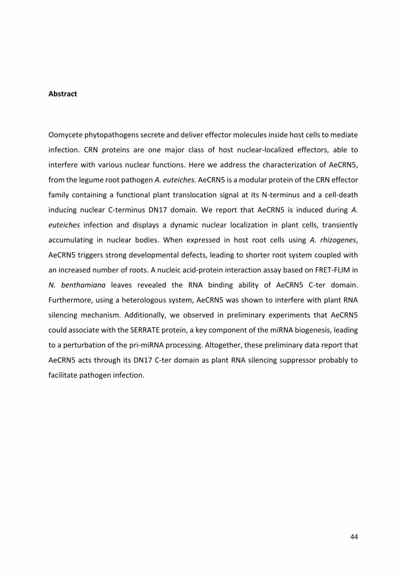
44
Abstract
Oomycete phytopathogens secrete and deliver effector molecules inside host cells to mediate
infection. CRN proteins are one major class of host nuclear-localized effectors, able to
interfere with various nuclear functions. Here we address the characterization of AeCRN5,
from the legume root pathogen A. euteiches. AeCRN5 is a modular protein of the CRN effector
family containing a functional plant translocation signal at its N-terminus and a cell-death
inducing nuclear C-terminus DN17 domain. We report that AeCRN5 is induced during A.
euteiches infection and displays a dynamic nuclear localization in plant cells, transiently
accumulating in nuclear bodies. When expressed in host root cells using A. rhizogenes,
AeCRN5 triggers strong developmental defects, leading to shorter root system coupled with
an increased number of roots. A nucleic acid-protein interaction assay based on FRET-FLIM in
N. benthamiana leaves revealed the RNA binding ability of AeCRN5 C-ter domain.
Furthermore, using a heterologous system, AeCRN5 was shown to interfere with plant RNA
silencing mechanism. Additionally, we observed in preliminary experiments that AeCRN5
could associate with the SERRATE protein, a key component of the miRNA biogenesis, leading
to a perturbation of the pri-miRNA processing. Altogether, these preliminary data report that
AeCRN5 acts through its DN17 C-ter domain as plant RNA silencing suppressor probably to
facilitate pathogen infection.
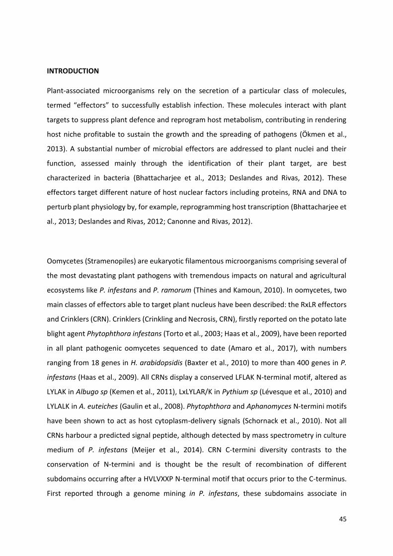
45
INTRODUCTION
Plant-associated microorganisms rely on the secretion of a particular class of molecules,
termed “effectors” to successfully establish infection. These molecules interact with plant
targets to suppress plant defence and reprogram host metabolism, contributing in rendering
host niche profitable to sustain the growth and the spreading of pathogens (Ökmen et al.,
2013). A substantial number of microbial effectors are addressed to plant nuclei and their
function, assessed mainly through the identification of their plant target, are best
characterized in bacteria (Bhattacharjee et al., 2013; Deslandes and Rivas, 2012). These
effectors target different nature of host nuclear factors including proteins, RNA and DNA to
perturb plant physiology by, for example, reprogramming host transcription (Bhattacharjee et
al., 2013; Deslandes and Rivas, 2012; Canonne and Rivas, 2012).
Oomycetes (Stramenopiles) are eukaryotic filamentous microorganisms comprising several of
the most devastating plant pathogens with tremendous impacts on natural and agricultural
ecosystems like P. infestans and P. ramorum (Thines and Kamoun, 2010). In oomycetes, two
main classes of effectors able to target plant nucleus have been described: the RxLR effectors
and Crinklers (CRN). Crinklers (Crinkling and Necrosis, CRN), firstly reported on the potato late
blight agent Phytophthora infestans (Torto et al., 2003; Haas et al., 2009), have been reported
in all plant pathogenic oomycetes sequenced to date (Amaro et al., 2017), with numbers
ranging from 18 genes in H. arabidopsidis (Baxter et al., 2010) to more than 400 genes in P.
infestans (Haas et al., 2009). All CRNs display a conserved LFLAK N-terminal motif, altered as
LYLAK in Albugo sp (Kemen et al., 2011), LxLYLAR/K in Pythium sp (Lévesque et al., 2010) and
LYLALK in A. euteiches (Gaulin et al., 2008). Phytophthora and Aphanomyces N-termini motifs
have been shown to act as host cytoplasm-delivery signals (Schornack et al., 2010). Not all
CRNs harbour a predicted signal peptide, although detected by mass spectrometry in culture
medium of P. infestans (Meijer et al., 2014). CRN C-termini diversity contrasts to the
conservation of N-termini and is thought be the result of recombination of different
subdomains occurring after a HVLVXXP N-terminal motif that occurs prior to the C-terminus.
First reported through a genome mining in P. infestans, these subdomains associate in
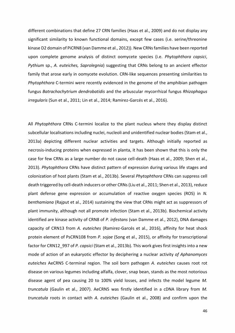
46
different combinations that define 27 CRN families (Haas et al., 2009) and do not display any
significant similarity to known functional domains, except few cases (i.e. serine/threonine
kinase D2 domain of PiCRN8 (van Damme et al., 2012)). New CRNs families have been reported
upon complete genome analysis of distinct oomycete species (i.e. Phytophthora capsici,
Pythium sp., A. euteiches, Saprolegnia) suggesting that CRNs belong to an ancient effector
family that arose early in oomycete evolution. CRN-like sequences presenting similarities to
Phytophthora C-termini were recently evidenced in the genome of the amphibian pathogen
fungus Batrachochytrium dendrobatidis and the arbuscular mycorrhizal fungus Rhizophagus
irregularis (Sun et al., 2011; Lin et al., 2014; Ramirez-Garcés et al., 2016).
All Phytophthora CRNs C-termini localize to the plant nucleus where they display distinct
subcellular localisations including nuclei, nucleoli and unidentified nuclear bodies (Stam et al.,
2013a) depicting different nuclear activities and targets. Although initially reported as
necrosis-inducing proteins when expressed in planta, it has been shown that this is only the
case for few CRNs as a large number do not cause cell-death (Haas et al., 2009; Shen et al.,
2013). Phytophthora CRNs have distinct pattern of expression during various life stages and
colonization of host plants (Stam et al., 2013b). Several Phytophthora CRNs can suppress cell
death triggered by cell-death inducers or other CRNs (Liu et al., 2011; Shen et al., 2013), reduce
plant defense gene expression or accumulation of reactive oxygen species (ROS) in N.
benthamiana (Rajput et al., 2014) sustaining the view that CRNs might act as suppressors of
plant immunity, although not all promote infection (Stam et al., 2013b). Biochemical activity
identified are kinase activity of CRN8 of P. infestans (van Damme et al., 2012), DNA damages
capacity of CRN13 from A. euteiches (Ramirez-Garcés et al., 2016), affinity for heat shock
protein element of PsCRN108 from P. sojae (Song et al., 2015), or affinity for transcriptional
factor for CRN12_997 of P. capsici (Stam et al., 2013b). This work gives first insights into a new
mode of action of an eukaryotic effector by deciphering a nuclear activity of Aphanomyces
euteiches AeCRN5 C-terminal region. The soil born pathogen A. euteiches causes root rot
disease on various legumes including alfalfa, clover, snap bean, stands as the most notorious
disease agent of pea causing 20 to 100% yield losses, and infects the model legume M.
truncatula (Gaulin et al., 2007). AeCRN5 was firstly identified in a cDNA library from M.
truncatula roots in contact with A. euteiches (Gaulin et al., 2008) and confirm upon the

47
sequencing of the complete genome (Gaulin et al., 2018). AeCRN5 presents a modular
architecture with an N-terminal functional LYLALK ensuring host delivery (Schornack et al.,
2010) and a DN17 family domain at its C-terminus.
Here we show that AeCRN5 is induced during infection of M. truncatula roots. AeCRN5 C-ter
is nuclear localized within the roots and triggers strong developmental defects. When
expressed in N. benthamiana, the nuclear localisation is required to induce cell death.
Furthermore, we observe a dynamic relocalization of AeCRN5 C-ter from nucleoplasm to
nuclear bodies that required plant RNA. Additionally, using a FRET-FLIM assay in N.
benthamiana leaves, we found that AeCRN5 C-ter associates to plant RNA. Finally, AeCRN5 C-
ter seems to interfere with plant gene silencing mechanism. Taken together, these results
indicate that CRN DN17 family function targets plant RNA and interferes with RNA silencing.
As preliminary results, we identified a putative interaction with the plant SERRATE protein,
which seems to modulate miRNA processing.

Figure 1: Amino acid sequence analyses of AeCRN5.
(A) Diagram depicting the modular architecture of AeCRN5 from A. euteiches with the conserved N-terminus,
which includes the LxLYLALK and HVVVIVP motifs, and the C-terminal region containing the DN17 subdomain,
based on P. infestans Crinkler (CRN) domain nomenclature. NLS sequence is indicated in coloured box. (B)
AeCRN5 N-terminus three-dimensional structure predicted by the Phyre2 server. The three most confident
(>95%) protein structures used as template by Phyre2 are also represented. Those templates belong to the ubl
domain of human ddi2 (c2n7dA), to the -Grasp (Ubiquitin-like) superfamily (d1v5oa) and to human ubiquitin-
like domain of ubiquilin 1 (c2klcA). (C) Multiple amino acid sequences alignment of the C-termini domains from
the closest orthologs of AeCRN5 C-ter domain. Organism names and sequence accession numbers are indicated
in front of the sequences, and correspond to Restriction Endonuclease 5 domain in Zhang et al (Zhang et al.,
2016) report. Background colours indicate residue conservation according to the legend. Alignment was
performed by CLC Workbench (Qiagen).
1
130

48
RESULTS
AeCRN5 is a DN17 family protein with a modular architecture
The AeCRN5 (Ae201684_4018.1, http://www.polebio.lrsv.ups-tlse.fr/cgi-bin/gb2/gbrowse/Ae201684_V3/) N-
terminus (1-130aa) is characterized by a LQLYLALK (47-54aa) motif and a HVVVIVPEVPL (120-
130aa) motif marking its end (Figure 1a). Although it lacks a predicted signal peptide, the
AeCRN5 N-terminus is a functional secretion domain mediating translocation of oomycete
effectors to plant cell (Schornack et al., 2010). A recent study proposed a new classification of
the N-termini domains of CRNs proteins, based not only on amino acid sequences but also on
secondary structure predictions (Zhang et al., 2016). Then we submitted AeCRN5 N-ter
domain to three-dimensional modelling using the structure prediction server Phyre2 (Kelley
et al., 2015). The most confident three-dimensional predictions of AeCRN5 N-terminal domain
were based on template models from Ubiquitin family, such as the -Grasp (ubiquitin-like)
domain superfamily or the ubI domain of human ddi2 protein (Figure 1b). This is in agreement
with the analyses conducted by Zhang and colleagues (Zhang et al., 2016) that classify the N-
termini of CRNs as header domains containing the ubiquitin-like fold.
The C-terminal region shows a sequence identity of 41% with the CRN DN17 family domain
of P. infestans (Haas et al., 2009) and harbors a nuclear localisation signal (NLS 141-156aa)
consistent with its plant nuclear localization when expressed in Nicotiana benthamiana leaves
(Schornack et al., 2010). CRN-like sequences including DN17 family have been reported not
only in oomycetes, but also in pathogenic or mutualistic fungi, such as in the chytrid fungus
Batrachochytrium dendrobatidis (Bd), a pathogen of amphibians, and in the arbuscular
mycorrhizal fungus Rhizophagus irregularis (Ri) (Sun et al., 2011; Lin et al., 2014). Sequence
comparison of C-terminal domain of AeCRN5 shows that it is closest to the chytrid B.
dendrobatidis (45% identity) than to oomycetes CRNs (maximum 41% identity) (Figure 1c).
Although AeCRN5 C-ter domain was not included in the analyses of Zhang et al. (2016), the
closest orthologs (included in the alignment from Figure 1C) were used in this study. Hence,
the C-termini domains of the CRN5-like from the chytrids Bd (Bd_87128 and Bd_26694; 45%
identity for both) and Rozella allomycis (09G_001773; 44% identity), from oomycetes such as
Aphanomyces invadans (H310_01635; 43% identity), P. infestans (CRN5_Q2M408; 41%
identity) or P. sojae (Physodraft-264761; 40% identity), were all predicted to contain a
Restriction Endonuclease 5 (REase 5) domain (Zhang et al., 2016) at their C-terminus.
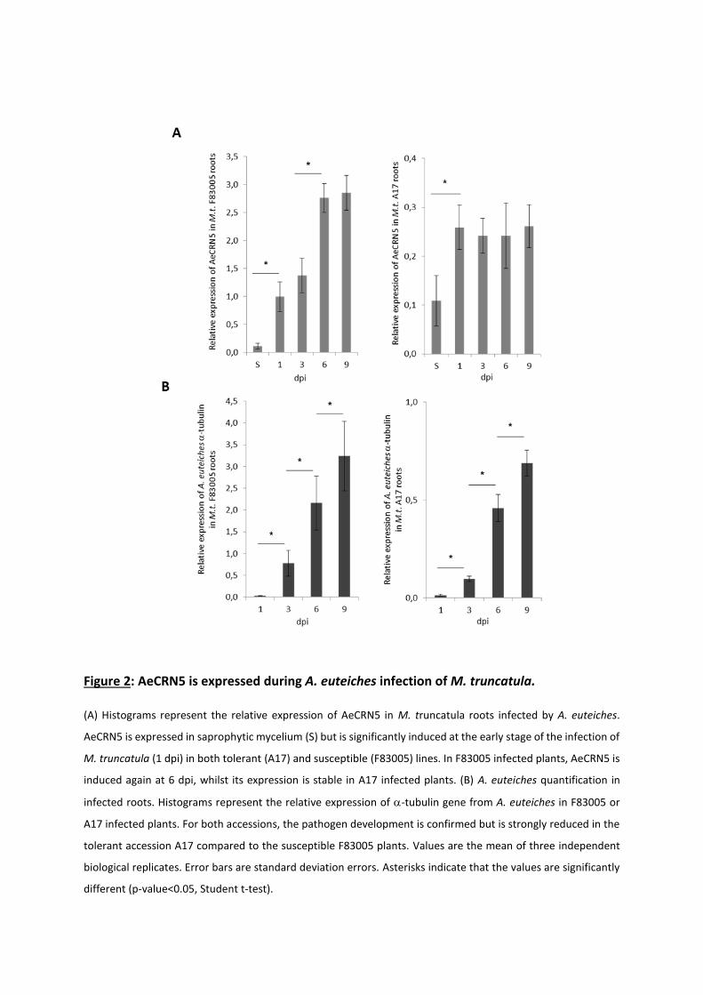
Figure 2: AeCRN5 is expressed during A. euteiches infection of M. truncatula.
(A) Histograms represent the relative expression of AeCRN5 in M. truncatula roots infected by A. euteiches.
AeCRN5 is expressed in saprophytic mycelium (S) but is significantly induced at the early stage of the infection of
M. truncatula (1 dpi) in both tolerant (A17) and susceptible (F83005) lines. In F83005 infected plants, AeCRN5 is
induced again at 6 dpi, whilst its expression is stable in A17 infected plants. (B) A. euteiches quantification in
infected roots. Histograms represent the relative expression of -tubulin gene from A. euteiches in F83005 or
A17 infected plants. For both accessions, the pathogen development is confirmed but is strongly reduced in the
tolerant accession A17 compared to the susceptible F83005 plants. Values are the mean of three independent
biological replicates. Error bars are standard deviation errors. Asterisks indicate that the values are significantly
different (p-value<0.05, Student t-test).
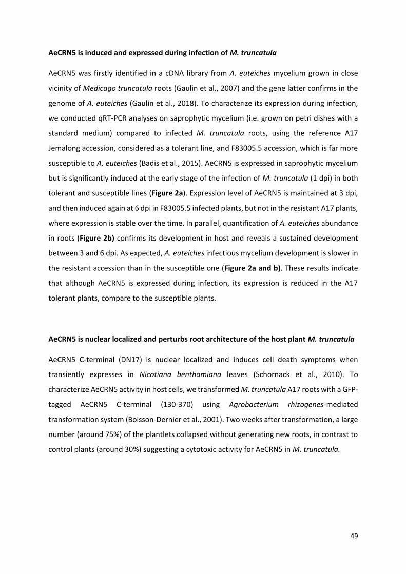
49
AeCRN5 is induced and expressed during infection of M. truncatula
AeCRN5 was firstly identified in a cDNA library from A. euteiches mycelium grown in close
vicinity of Medicago truncatula roots (Gaulin et al., 2007) and the gene latter confirms in the
genome of A. euteiches (Gaulin et al., 2018). To characterize its expression during infection,
we conducted qRT-PCR analyses on saprophytic mycelium (i.e. grown on petri dishes with a
standard medium) compared to infected M. truncatula roots, using the reference A17
Jemalong accession, considered as a tolerant line, and F83005.5 accession, which is far more
susceptible to A. euteiches (Badis et al., 2015). AeCRN5 is expressed in saprophytic mycelium
but is significantly induced at the early stage of the infection of M. truncatula (1 dpi) in both
tolerant and susceptible lines (Figure 2a). Expression level of AeCRN5 is maintained at 3 dpi,
and then induced again at 6 dpi in F83005.5 infected plants, but not in the resistant A17 plants,
where expression is stable over the time. In parallel, quantification of A. euteiches abundance
in roots (Figure 2b) confirms its development in host and reveals a sustained development
between 3 and 6 dpi. As expected, A. euteiches infectious mycelium development is slower in
the resistant accession than in the susceptible one (Figure 2a and b). These results indicate
that although AeCRN5 is expressed during infection, its expression is reduced in the A17
tolerant plants, compare to the susceptible plants.
AeCRN5 is nuclear localized and perturbs root architecture of the host plant M. truncatula
AeCRN5 C-terminal (DN17) is nuclear localized and induces cell death symptoms when
transiently expresses in Nicotiana benthamiana leaves (Schornack et al., 2010). To
characterize AeCRN5 activity in host cells, we transformed M. truncatula A17 roots with a GFP-
tagged AeCRN5 C-terminal (130-370) using Agrobacterium rhizogenes-mediated
transformation system (Boisson-Dernier et al., 2001). Two weeks after transformation, a large
number (around 75%) of the plantlets collapsed without generating new roots, in contrast to
control plants (around 30%) suggesting a cytotoxic activity for AeCRN5 in M. truncatula.
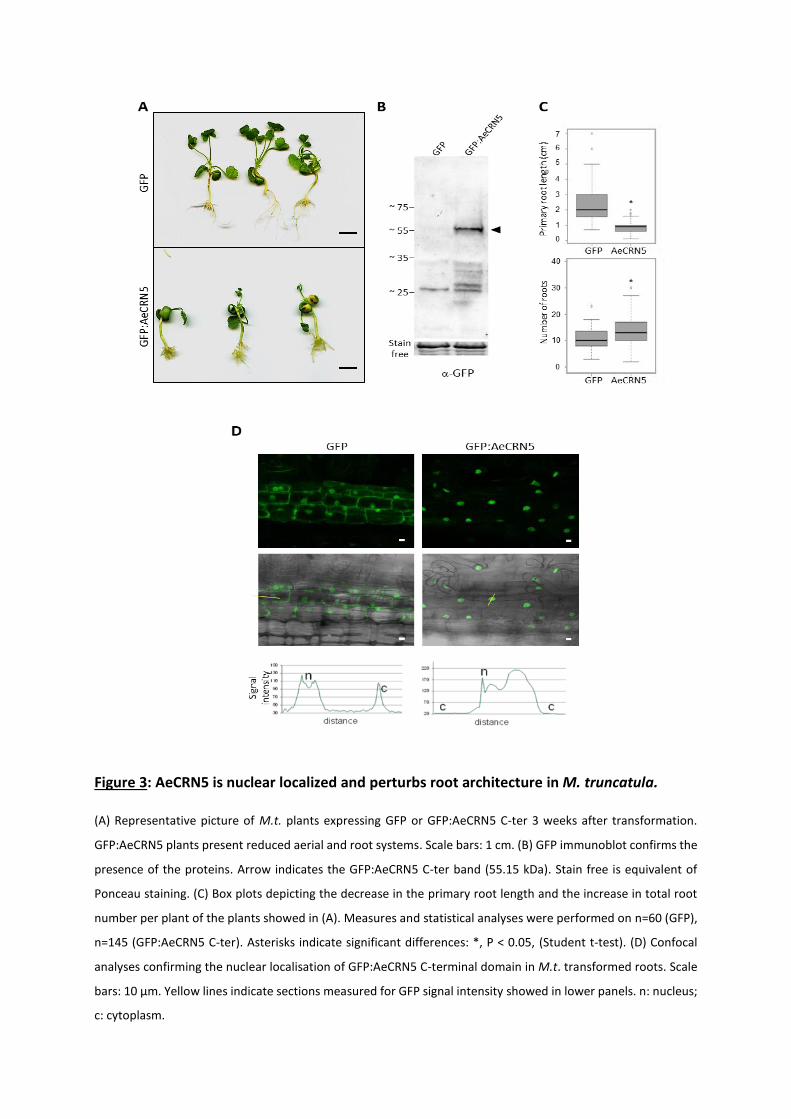
Figure 3: AeCRN5 is nuclear localized and perturbs root architecture in M. truncatula.
(A) Representative picture of M.t. plants expressing GFP or GFP:AeCRN5 C-ter 3 weeks after transformation.
GFP:AeCRN5 plants present reduced aerial and root systems. Scale bars: 1 cm. (B) GFP immunoblot confirms the
presence of the proteins. Arrow indicates the GFP:AeCRN5 C-ter band (55.15 kDa). Stain free is equivalent of
Ponceau staining. (C) Box plots depicting the decrease in the primary root length and the increase in total root
number per plant of the plants showed in (A). Measures and statistical analyses were performed on n=60 (GFP),
n=145 (GFP:AeCRN5 C-ter). Asterisks indicate significant differences: *, P < 0.05, (Student t-test). (D) Confocal
analyses confirming the nuclear localisation of GFP:AeCRN5 C-terminal domain in M.t. transformed roots. Scale
bars: 10 µm. Yellow lines indicate sections measured for GFP signal intensity showed in lower panels. n: nucleus;
c: cytoplasm.
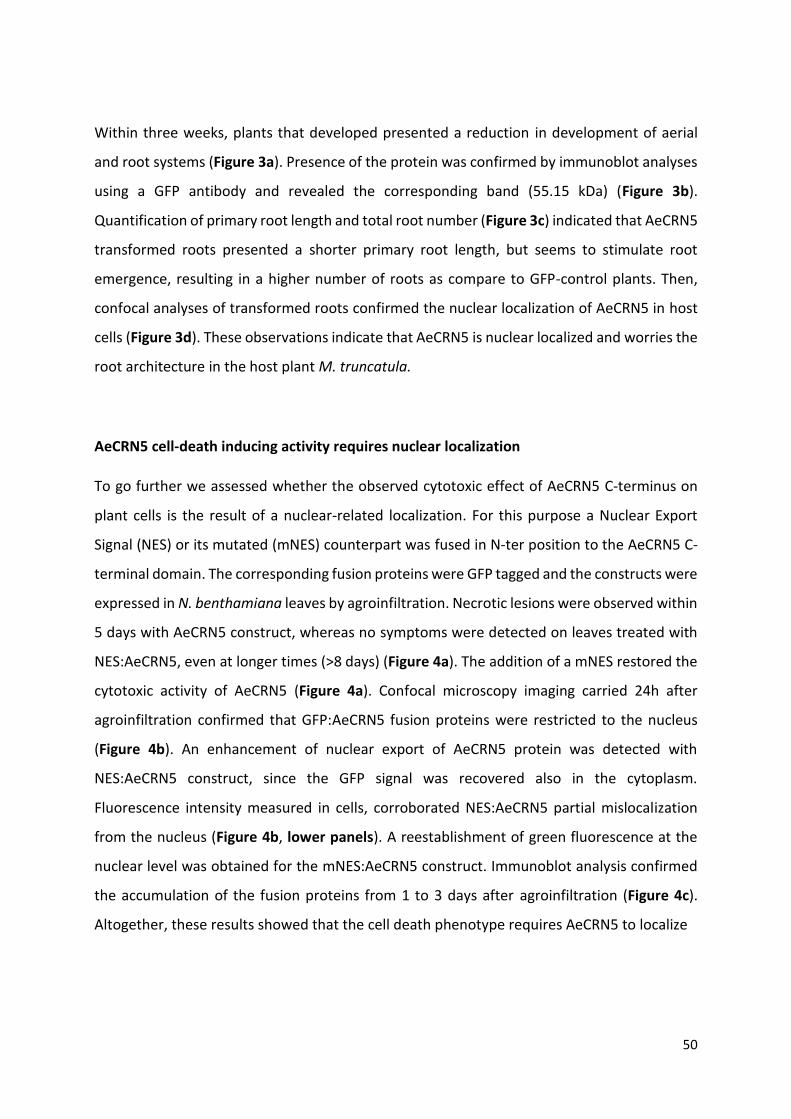
50
Within three weeks, plants that developed presented a reduction in development of aerial
and root systems (Figure 3a). Presence of the protein was confirmed by immunoblot analyses
using a GFP antibody and revealed the corresponding band (55.15 kDa) (Figure 3b).
Quantification of primary root length and total root number (Figure 3c) indicated that AeCRN5
transformed roots presented a shorter primary root length, but seems to stimulate root
emergence, resulting in a higher number of roots as compare to GFP-control plants. Then,
confocal analyses of transformed roots confirmed the nuclear localization of AeCRN5 in host
cells (Figure 3d). These observations indicate that AeCRN5 is nuclear localized and worries the
root architecture in the host plant M. truncatula.
AeCRN5 cell-death inducing activity requires nuclear localization
To go further we assessed whether the observed cytotoxic effect of AeCRN5 C-terminus on
plant cells is the result of a nuclear-related localization. For this purpose a Nuclear Export
Signal (NES) or its mutated (mNES) counterpart was fused in N-ter position to the AeCRN5 C-
terminal domain. The corresponding fusion proteins were GFP tagged and the constructs were
expressed in N. benthamiana leaves by agroinfiltration. Necrotic lesions were observed within
5 days with AeCRN5 construct, whereas no symptoms were detected on leaves treated with
NES:AeCRN5, even at longer times (>8 days) (Figure 4a). The addition of a mNES restored the
cytotoxic activity of AeCRN5 (Figure 4a). Confocal microscopy imaging carried 24h after
agroinfiltration confirmed that GFP:AeCRN5 fusion proteins were restricted to the nucleus
(Figure 4b). An enhancement of nuclear export of AeCRN5 protein was detected with
NES:AeCRN5 construct, since the GFP signal was recovered also in the cytoplasm.
Fluorescence intensity measured in cells, corroborated NES:AeCRN5 partial mislocalization
from the nucleus (Figure 4b, lower panels). A reestablishment of green fluorescence at the
nuclear level was obtained for the mNES:AeCRN5 construct. Immunoblot analysis confirmed
the accumulation of the fusion proteins from 1 to 3 days after agroinfiltration (Figure 4c).
Altogether, these results showed that the cell death phenotype requires AeCRN5 to localize

Figure 4: The biological function of AeCRN5 requires nuclear localization.
Figure 5: AeCRN5 transiently accumulates in nuclear bodies.
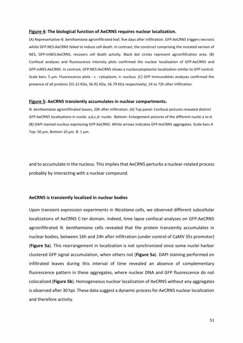
51
Figure 4: The biological function of AeCRN5 requires nuclear localization.
(A) Representative N. benthamiana agroinfiltrated leaf, five days after infiltration. GFP:AeCRN5 triggers necrosis
whilst GFP:NES:AeCRN5 failed to induce cell death. In contrast, the construct comprising the mutated version of
NES, GFP:mNES:AeCRN5, recovers cell death activity. Black dot circles represent agroinfiltration area. (B)
Confocal analyses and fluorescence intensity plots confirmed the nuclear localization of GFP:AeCRN5 and
GFP:mNES:AeCRN5. In contrast, GFP:NES:AeCRN5 shows a nucleocytoplasmic localization similar to GFP control.
Scale bars: 5 µm. Fluorescence plots : c : cytoplasm; n: nucleus. (C) GFP Immunoblots analyses confirmed the
presence of all proteins (55.15 KDa, 56.92 KDa, 56.79 KDa respectively), 24 to 72h after infiltration.
Figure 5: AeCRN5 transiently accumulates in nuclear compartments.
N. benthamiana agroinfiltrated leaves, 20h after infiltration. (A) Top panel: Confocal pictures revealed distinct
GFP:AeCRN5 localizations in nuclei. a,b,c,d: nuclei. Bottom: Enlargement pictures of the different nuclei a to d.
(B) DAPI stained nucleus expressing GFP:AeCRN5. White arrows indicates GFP:AeCRN5 aggregates. Scale bars A
Top: 50 µm, Bottom 10 µm. B: 1 µm.
and to accumulate in the nucleus. This implies that AeCRN5 perturbs a nuclear-related process
probably by interacting with a nuclear compound.
AeCRN5 is transiently localized in nuclear bodies
Upon transient expression experiments in Nicotiana cells, we observed different subcellular
localizations of AeCRN5 C-ter domain. Indeed, time lapse confocal analyses on GFP:AeCRN5
agroinfiltrated N. benthamiana cells revealed that the protein transiently accumulates in
nuclear bodies, between 16h and 24h after infiltration (under control of CaMV 35s promotor)
(Figure 5a). This rearrangement in localization is not synchronized since some nuclei harbor
clustered GFP signal accumulation, when others not (Figure 5a). DAPI staining performed on
infiltrated leaves during this interval of time revealed an absence of complementary
fluorescence pattern in these aggregates, where nuclear DNA and GFP fluorescence do not
colocalized (Figure 5b). Homogeneous nuclear localization of AeCRN5 without any aggregates
is observed after 30 hpi. These data suggest a dynamic process for AeCRN5 nuclear localization
and therefore activity.
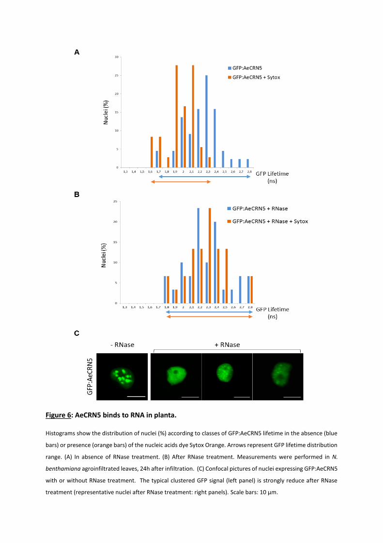
Figure 6: AeCRN5 binds to RNA in planta. Histograms show the distribution of nuclei (%) according to classes of GFP:AeCRN5 lifetime in the absence (blue
bars) or presence (orange bars) of the nucleic acids dye Sytox Orange. Arrows represent GFP lifetime distribution
range. (A) In absence of RNase treatment. (B) After RNase treatment. Measurements were performed in N.
benthamiana agroinfiltrated leaves, 24h after infiltration. (C) Confocal pictures of nuclei expressing GFP:AeCRN5
with or without RNase treatment. The typical clustered GFP signal (left panel) is strongly reduce after RNase
treatment (representative nuclei after RNase treatment: right panels). Scale bars: 10 µm.

52
AeCRN5 interacts with nuclear plant RNA
Plant nuclear bodies comprise different dynamic structures including for instance the
nucleolus, Cajal bodies, nuclear speckles or Dicing bodies (Petrovská et al., 2015). Since RNA
is a major component found in those compartments (Petrovská et al., 2015; Bazin et al., 2018),
we decided to analyse whether AeCRN5 C-ter may associate with plant RNA. We developed a
robust in planta system to test protein-nucleic acid interactions based of FRET-FLIM
(Fluorescence Resonance Energy Transfer coupled to Fluorescence Lifetime Imaging) to
determine whether C-terminal AeCRN5 could interact with nucleic acids, and more specifically
to RNAs (Ramirez-Garcés et al., 2016; Camborde et al., 2017). This experiment is based on N.
benthamiana cells expressing the GFP-fusion proteins in absence or in presence of the nucleic
acid dye Sytox Orange. This dye can absorb energy released by GFP-tagged proteins (donor)
during fluorescence only if GFP is in close proximity to the dye (acceptor). Such transfer of
energy conducts to a decrease of GFP lifetime, inferring its interaction to nucleic acids.
Additionally to GFP alone (used as a negative control since GFP proteins do not interact with
nucleic acids), we performed measurements on cells expressing the DNA-binding protein H2B
in fusion to GFP (GFP:H2B) as a positive control of protein-nucleic acid interactions.
Fluorescence lifetime of GFP for all constructs is given in table 1. As expected, no significate
decrease in GFP lifetime was observed for the GFP proteins in presence or absence of Sytox
Orange (Table 1). In contrast, the GFP lifetime of GFP:H2B proteins decreases from 2.38 ns +/-
0.02 to 1.83 ns +/-0.04 in presence of Sytox Orange, revealing as expected a close proximity
of GFP-tagged H2B proteins with nucleic acids (Table 1). Those results on control proteins are
in accordance with our previous study (Ramirez-Garcés et al., 2016). In the case of
GFP:AeCRN5, GFP lifetime was measured in nuclei harbouring a GFP clustered fluorescence,
corresponding to nuclear bodies. In that case, GFP lifetime significantly decreases from 2.20
ns +/-0.04 in absence of acceptor to 1.90 ns +/- 0.03 in presence of Sytox Orange, indicating
that the C-terminal domain of AeCRN5 is in close association with nucleic acids (Table 1 and
Figure 6a). Since Sytox Orange labels DNA and RNA, in order to discriminate the nature of
nucleic acids targeted by AeCRN5, foliar discs were treated with RNAse and GFP lifetime was
measured with or without Sytox Orange staining. Efficiency of this treatment was already
confirmed on an RNA-binding protein called NSR-b, which lost the interaction with RNA in
those conditions (Camborde et al., 2017). After RNAse treatment, in absence of Sytox, the

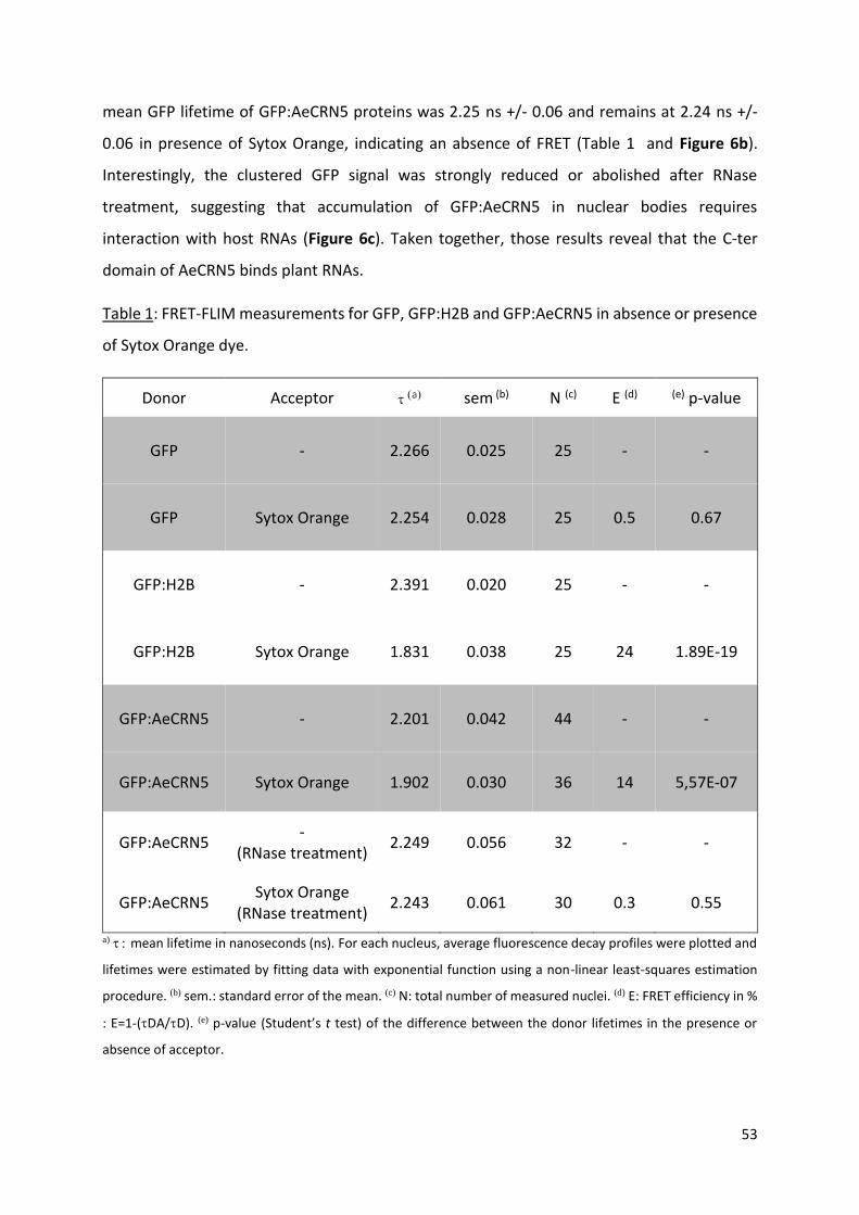
53
mean GFP lifetime of GFP:AeCRN5 proteins was 2.25 ns +/- 0.06 and remains at 2.24 ns +/-
0.06 in presence of Sytox Orange, indicating an absence of FRET (Table 1 and Figure 6b).
Interestingly, the clustered GFP signal was strongly reduced or abolished after RNase
treatment, suggesting that accumulation of GFP:AeCRN5 in nuclear bodies requires
interaction with host RNAs (Figure 6c). Taken together, those results reveal that the C-ter
domain of AeCRN5 binds plant RNAs.
Table 1: FRET-FLIM measurements for GFP, GFP:H2B and GFP:AeCRN5 in absence or presence
of Sytox Orange dye.
Donor Acceptor a sem (b) N (c) E (d) (e) p-value
GFP - 2.266 0.025 25 - -
GFP Sytox Orange 2.254 0.028 25 0.5 0.67
GFP:H2B - 2.391 0.020 25 - -
GFP:H2B Sytox Orange 1.831 0.038 25 24 1.89E-19
GFP:AeCRN5 - 2.201 0.042 44 - -
GFP:AeCRN5 Sytox Orange 1.902 0.030 36 14 5,57E-07
GFP:AeCRN5 -
(RNase treatment) 2.249 0.056 32 - -
GFP:AeCRN5 Sytox Orange
(RNase treatment) 2.243 0.061 30 0.3 0.55
a) mean lifetime in nanoseconds (ns). For each nucleus, average fluorescence decay profiles were plotted and
lifetimes were estimated by fitting data with exponential function using a non-linear least-squares estimation
procedure. (b) sem.: standard error of the mean. (c) N: total number of measured nuclei. (d) E: FRET efficiency in %
: E=1-(DA/D). (e) p-value (Student’s t test) of the difference between the donor lifetimes in the presence or
absence of acceptor.
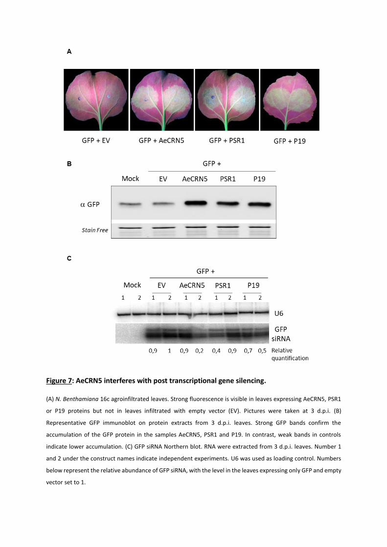
Figure 7: AeCRN5 interferes with post transcriptional gene silencing. (A) N. Benthamiana 16c agroinfiltrated leaves. Strong fluorescence is visible in leaves expressing AeCRN5, PSR1
or P19 proteins but not in leaves infiltrated with empty vector (EV). Pictures were taken at 3 d.p.i. (B)
Representative GFP immunoblot on protein extracts from 3 d.p.i. leaves. Strong GFP bands confirm the
accumulation of the GFP protein in the samples AeCRN5, PSR1 and P19. In contrast, weak bands in controls
indicate lower accumulation. (C) GFP siRNA Northern blot. RNA were extracted from 3 d.p.i. leaves. Number 1
and 2 under the construct names indicate independent experiments. U6 was used as loading control. Numbers
below represent the relative abundance of GFP siRNA, with the level in the leaves expressing only GFP and empty
vector set to 1.
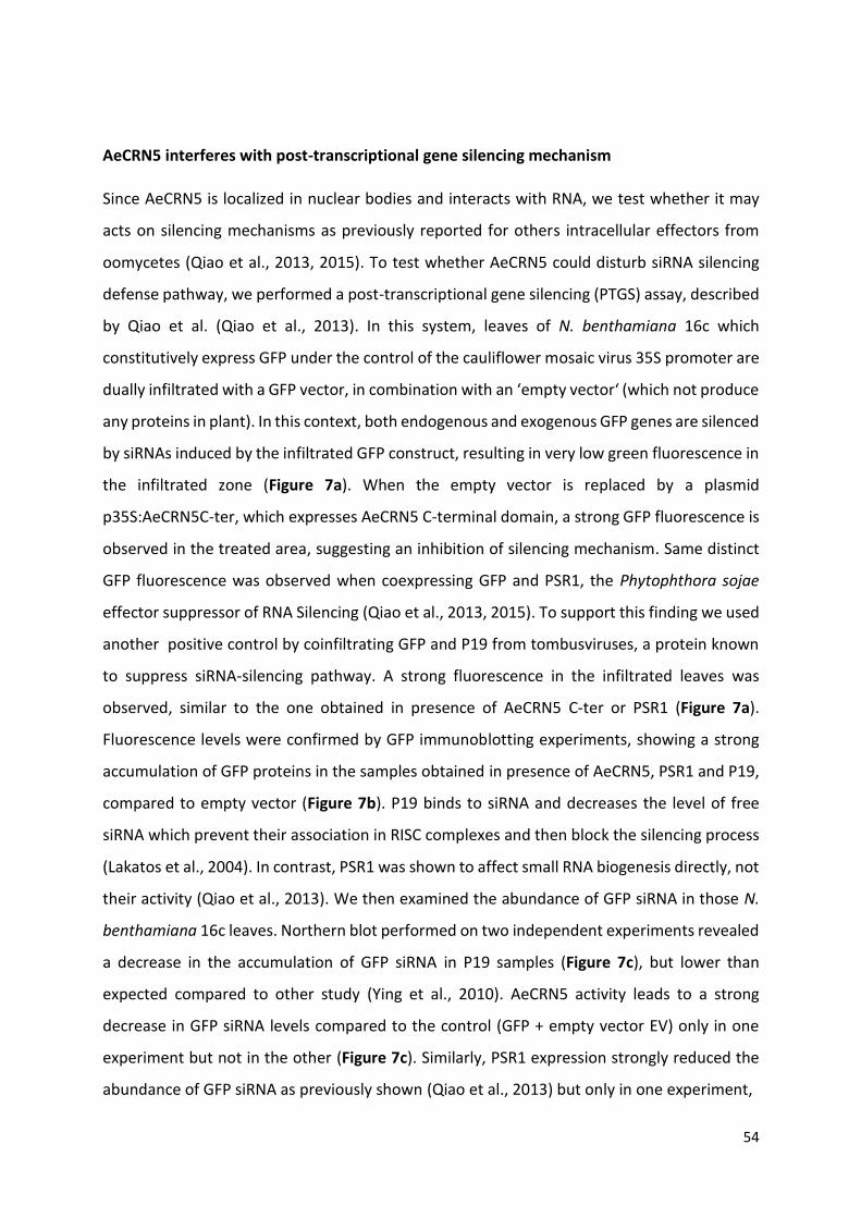
54
AeCRN5 interferes with post-transcriptional gene silencing mechanism
Since AeCRN5 is localized in nuclear bodies and interacts with RNA, we test whether it may
acts on silencing mechanisms as previously reported for others intracellular effectors from
oomycetes (Qiao et al., 2013, 2015). To test whether AeCRN5 could disturb siRNA silencing
defense pathway, we performed a post-transcriptional gene silencing (PTGS) assay, described
by Qiao et al. (Qiao et al., 2013). In this system, leaves of N. benthamiana 16c which
constitutively express GFP under the control of the cauliflower mosaic virus 35S promoter are
dually infiltrated with a GFP vector, in combination with an ‘empty vector‘ (which not produce
any proteins in plant). In this context, both endogenous and exogenous GFP genes are silenced
by siRNAs induced by the infiltrated GFP construct, resulting in very low green fluorescence in
the infiltrated zone (Figure 7a). When the empty vector is replaced by a plasmid
p35S:AeCRN5C-ter, which expresses AeCRN5 C-terminal domain, a strong GFP fluorescence is
observed in the treated area, suggesting an inhibition of silencing mechanism. Same distinct
GFP fluorescence was observed when coexpressing GFP and PSR1, the Phytophthora sojae
effector suppressor of RNA Silencing (Qiao et al., 2013, 2015). To support this finding we used
another positive control by coinfiltrating GFP and P19 from tombusviruses, a protein known
to suppress siRNA-silencing pathway. A strong fluorescence in the infiltrated leaves was
observed, similar to the one obtained in presence of AeCRN5 C-ter or PSR1 (Figure 7a).
Fluorescence levels were confirmed by GFP immunoblotting experiments, showing a strong
accumulation of GFP proteins in the samples obtained in presence of AeCRN5, PSR1 and P19,
compared to empty vector (Figure 7b). P19 binds to siRNA and decreases the level of free
siRNA which prevent their association in RISC complexes and then block the silencing process
(Lakatos et al., 2004). In contrast, PSR1 was shown to affect small RNA biogenesis directly, not
their activity (Qiao et al., 2013). We then examined the abundance of GFP siRNA in those N.
benthamiana 16c leaves. Northern blot performed on two independent experiments revealed
a decrease in the accumulation of GFP siRNA in P19 samples (Figure 7c), but lower than
expected compared to other study (Ying et al., 2010). AeCRN5 activity leads to a strong
decrease in GFP siRNA levels compared to the control (GFP + empty vector EV) only in one
experiment but not in the other (Figure 7c). Similarly, PSR1 expression strongly reduced the
abundance of GFP siRNA as previously shown (Qiao et al., 2013) but only in one experiment,
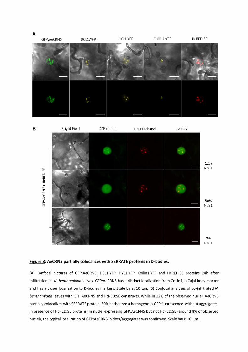
Figure 8: AeCRN5 partially colocalizes with SERRATE proteins in D-bodies.
(A) Confocal pictures of GFP:AeCRN5, DCL1:YFP, HYL1:YFP, Coilin1:YFP and HcRED:SE proteins 24h after
infiltration in N. benthamiana leaves. GFP:AeCRN5 has a distinct localization from Coilin1, a Cajal body marker
and has a closer localization to D-bodies markers. Scale bars: 10 µm. (B) Confocal analyses of co-infiltrated N.
benthamiana leaves with GFP:AeCRN5 and HcRED:SE constructs. While in 12% of the observed nuclei, AeCRN5
partially colocalizes with SERRATE protein, 80% harboured a homogenous GFP fluorescence, without aggregates,
in presence of HcRED:SE proteins. In nuclei expressing GFP:AeCRN5 but not HcRED:SE (around 8% of observed
nuclei), the typical localization of GFP:AeCRN5 in dots/aggregates was confirmed. Scale bars: 10 µm.
55
not in the other (Figure 7c). Altogether, these results suggest that AeCRN5 can interfere with
PTGS mechanism but supplementary experiments are needed to support this conclusion.
PRELIMINARY RESULTS
AeCRN5 is transiently localized in nuclear Dicing-bodies
To precise the nuclear localisation of AeCRN5 we infiltrated in N. benthamiana leaves with
several nuclear markers to visualize Cajal bodies (such as Coilin-1) and D-bodies (such as Dicer-
like 1 DCL1, HYPONASTIC LEAVES1 HYL1 and SERRATE SE). Coilin-1, DCL1 and HYL1 are cloned
with a YFP tag in C-ter, whereas SE was fused with HcRED in N-ter. Cloning with other
fluorescent tags is in progress. We compared the localization of GFP:AeCRN5 with each marker
in N. benthamiana cells 20-24h after agroinfiltration. Confocal analyses revealed a distinct
localization for Coilin-1:YFP, with fluorescent dots close or inside nucleolus. DCL1, HYL1 and
SE localize in D-bodies and this profile could be partially similar to AeCRN5 (Figure 8a). To go
further we next co-infiltrated GFP:AeCRN5 and HcRED:SE and observed their localization one
day after treatment. In 92% of the observed nuclei, both proteins were detected in same
nuclei with two types of labelling pattern. The preferential pattern observed in 80% of the
nuclei correspond to a homogenous GFP fluorescence, without any aggregates (Figure 8b). In
12% of the nuclei, both proteins seems to colocalize in nuclear bodies probably D-bodies
(Figure 8b). In nuclei expressing only GFP:AeCRN5 but not HcRED:SE (around 8% of observed
nuclei), GFP:AeCRN5 is detected as typical clustered accumulation (Figure 8b). Although
nuclear bodies markers and experimental repetitions are required, these suggest that AeCRN5
could localize in D-bodies, where it could interact with the SERRATE proteins.
AeCRN5 interferes with miRNA biogenesis
SERRATE (SE) is a major actor involved in the biogenesis of micro-RNA (miRNAs) (Lobbes et al.,
2006; Wang et al., 2019). Since AeCRN5 transiently colocalizes with SE and interacts with RNA,
we also hypothesize that AeCRN5 could perturb SE proteins during the maturation of miRNA.
SE is involved in maturation of the primary transcripts (pri and pre-miRNA) into mature miRNA.
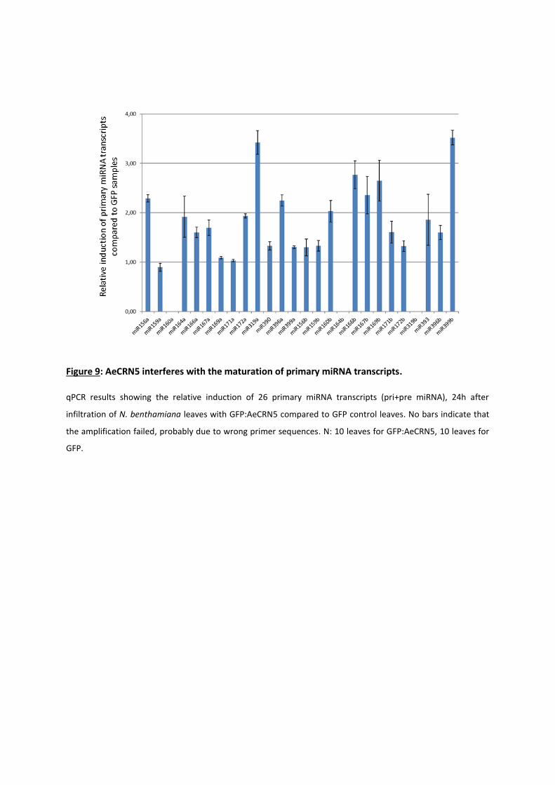
Figure 9: AeCRN5 interferes with the maturation of primary miRNA transcripts.
qPCR results showing the relative induction of 26 primary miRNA transcripts (pri+pre miRNA), 24h after
infiltration of N. benthamiana leaves with GFP:AeCRN5 compared to GFP control leaves. No bars indicate that
the amplification failed, probably due to wrong primer sequences. N: 10 leaves for GFP:AeCRN5, 10 leaves for
GFP.
56
Therefore, interference in the maturation process will trigger an accumulation of primary
miRNA transcripts. We decided to analyse several primary miRNA transcript levels, described
in the literature as key regulators of root architecture and biotic interactions, on N.
benthamiana leaves agroinfiltrated with GFP:AeCRN5 or GFP (as a negative control). The
presence of the GFP:AeCRN5 localization in D-bodies from 20h to 24h after agroinfiltration
was confirmed by confocal observations and before sampling the corresponding leaves. We
next selected 26 miR, already sequenced and analysed in various reports (for review see
(Couzigou and Combier, 2016). Primers for qPCR amplification were designed according to
miRBase (Kozomara et al., 2019) (http://www.mirbase.org/) based on Nicotiana tabacum
sequences (N. benthamiana is not available). QPCR analyses were performed on ten
GFP:AeCRN5 and ten GFP agroinfiltrated leaves. Results revealed that most of the primary
transcripts analysed accumulates in the AeCRN5 samples compared to the GFP controls
(Figure 9), suggesting that AeCRN5 perturbs the miRNA biogenesis.
DISCUSSION
To favor the establishment of disease, microorganisms have gained the ability to deliver
effector molecules inside host cells. The important number of effectors targeting host nuclei
places this organelle, and functions related to it, as important hubs whose perturbations might
be of crucial importance for the outcome of infection (Bhattacharjee et al., 2013; Khan et al.,
2018). A recent study reported that in average, 38% of phytopathogenic oomycete
intracellular effectors target nucleus, close to the number reported for plant bacterial
pathogens (35%) (Khan et al., 2018). CRN proteins are a family of nuclear-localized effectors
widespread in oomycete lineage, with related sequences found in fungal species B.
dendrobatidis and R. irregularis. In this work, we undertook the characterization of AeCRN5
of the root pathogen A. euteiches. We show that AeCRN5 is express during M. truncatula
infection and perturbs host root development. We reveal that AeCRN5 is mainly localized in
Nuclear bodies (D-bodies) and targets plant RNA at the nuclear level as well as SERRATE
protein, to interfere with RNA processes.
57
AeCRN5 is a modular CRN DN17 protein family with orthologous sequences in Phytophthora
sp. and true fungal species including the chytrid B. dendrobatidis and the endomycorrhiza R.
irregularis. The functional translocation signal of AeCRN5 is characterized by LYLALK and
HVVVIP motifs and the absence of an obvious signal peptide (Schornack et al., 2010; Gaulin et
al., 2007). Since a study from Zhang et al. proposed to reconsider the N-terminal CRNs
classification (Zhang et al., 2016), we submitted to a structure prediction server the N-terminal
sequence of AeCRN5. This in silico analysis confirms the classification of AeCRN5 N-ter as a
header domain containing ubiquitin-like fold.
The C-terminus corresponds to a CRN DN17 domain family with a NLS, according to the
classification established from Phytophthora CRNs (Haas et al., 2009; Schornack et al., 2010),
which have no significant similarity to functional domain. Although AeCRN5 was not included
in the analysis of Zhang and colleagues, the closest orthologs of AeCRN5 found in Bd fungus,
other Aphanomyces species or Phytophthora species, were included and were predicted to
contain a Restriction Endonuclease 5 domain (Zhang et al., 2016). Here we confirm this
prediction for AeCRN5. Phytophthora CRNs were originally identified as activators of plant cell
death upon their in planta expression (Torto et al., 2003), although not all CRNs promote
infection including the AeCRN5 ortholog from P. capsici (Stam et al., 2013a). CRN5 sequences
from A. euteiches were firstly reported in a cDNA library from mycelium grown in close vicinity
of M. truncatula roots (Gaulin et al., 2008). Here we showed by qRT-PCR analysis, that AeCRN5
is expressed during vegetative growth and expression goes up during root infection, but is
differentially induced depending on the susceptibility of the plants. In susceptible line, an
increase in expression is observed firstly at 1 dpi, then between 3 and 6 dpi, a stage where
browning of roots is observed in combination to an entire colonization of the root cortex of
M. truncatula, and the initiation of propagation to vascular tissues (Djébali et al., 2009). In
contrast, in tolerant line, AeCRN5 expression is stable after a rapid induction at 1 dpi. This
could be related to differential plant responses, involving for instance cross-kingdom RNAi,
where plant transports small RNAs into pathogens to suppress the expression of virulence
related genes. This defense response was recently reported in fungal plant association, where
it was evidenced that Arabidopsis sRNAs are delivered into Botrytis cinerea cells to induce
silencing of pathogenicity-related genes (Cai et al., 2018). In a same way, an alpha/beta
hydrolase gene from Fusarium graminearum, required for fungal infection, is targeted and
58
silenced by a miRNA produced by wheat (Jiao and Peng, 2018). Interestingly, this defense
mechanism has been recently reported in plant-oomycete interaction. Arabidopsis infection
by Phytophthora capsici leads to an increased production of plant small interfering RNAs
(siRNAs) which are delivered into Phytophthora to silence target genes during natural infection
(Hou et al., 2019). However, this cross-kingdom silencing has not yet been mentioned for
Medicago truncatula or other legume in plant-pathogen interactions. Furthermore,
transcriptomic analyses conducted on infected F83005 susceptible accession compared to A.
euteiches mycelium or zoospores samples indicate that only 2% of CRNs genes were induced
at 3 and 9 dpi (1 dpi was not possible to analyse) (Gaulin et al., 2018), consistent with AeCRN5
expression in F83005 roots. Given that 13% of CRN genes are upregulated in zoospores as
compared to in vitro grown mycelium, a subset of AeCRNs is potentially involved at the early
stage of Medicago infection (Gaulin et al., 2018). Finally, in P. capsici, CRNs genes were divided
in two groups according to their expression patterns. P. capsici DN17 ortholog felt in Class 2
where gene expression gradually increases to peak in the late infection stages (Stam et al.,
2013b), as observed for AeCRN5 gene expression in susceptible Medicago line, suggesting a
role in the later stage of colonization.
We further explored the function of AeCRN5 by using a GFP-tagged version of the C-terminal
domain. As observed in N. benthamiana leaves (Schornack et al., 2010), AeCRN5 is nuclear
localized also in host Medicago cells. Overexpression of AeCRN5 in M. truncatula roots
displayed a cytotoxic effect leading in few days to death of transformed plants. The surviving
dwarfed plants harbored reduced root systems with a higher number of roots. These results
corroborate observations made during M. truncatula roots infection, where susceptible
accessions present, within few days after A. euteiches infection, a decrease of secondary root
development and necrosis of roots (Djébali et al., 2009).
Confocal studies on transiently transformed N. benthamiana leaves showed that DN17
cytotoxic effect of AeCRN5 required a plant nuclear accumulation. It is in accordance with the
observed reduction of cell death on N. benthamiana leaves, upon nuclear exclusion of CRN8
(D2 domain) from P. infestans (Schornack et al., 2010). Similar results were reported for P.
sojae and P. capsici CRNs (PsCRN63 and PcCRN4) (Liu et al., 2011; Mafurah et al., 2015) and
AeCRN13 from A. euteiches (Ramirez-Garcés et al., 2016). Our results confirm that nuclear
59
localization is an important requirement for the cell-death inducing activity of necrotic CRN
effectors.
We observed that AeCRN5 DN17 shuttles between nucleoplasm and plant nuclear bodies
where DNA is excluded. Previous study on P. capsici DN17 CRN domain revealed a clustered
distribution pattern confined to the nucleoplasm upon overexpression in N. benthamiana
leaves (Stam et al., 2013a). Furthermore, FRET-FLIM measurements revealed the close vicinity
of AeCRN5 C-ter domain with plant nucleic acids. This FRET-FLIM assay has been successfully
used to demonstrate protein-RNA specific interaction in plant cells (Camborde et al., 2017).
Here this assay confirms the RNA binding ability of AeCRN5 C-terminal domain.
Recently, Khan and colleagues reviewed some properties of effector targets across diverse
phytopathogens, including bacteria, fungi and oomycetes. They reported that only 1 to 3% of
effector targets had a molecular function related to RNA processing (Khan et al., 2018).
Candidate effectors of the fungus Blumeria graminis display similarities to microbial RNAses
and, although not carrying hydrolytic site, are speculated to interact with host RNA; among
these, BEC1054 has been described as a ribonuclease-like effector (Pedersen et al., 2012;
Pliego et al., 2013). Host RNA perturbation has also been proposed for some effectors of the
nematode M. incognita as they harbor putative RNA binding domains (Bellafiore et al., 2008).
The dynamics and clustered localization of AeCRN5 DN17 domain in combination with its
proximity to plant RNA, strongly suggest a ‘nuclear bodies pattern’. Even if further
experiments are on going to precise the subnuclear localization of AeCRN5, we decided to test
the activity of the C-ter DN17 domain on silencing mechanisms as previously reported for
others intracellular effectors from oomycetes (Qiao et al., 2013, 2015). Using transient
expression assay in N. benthamiana 16c, we found that AeCRN5 DN17 domain interferes with
post-transcriptional gene silencing, even if the effect on siRNA biogenesis is still unclear.
Hence, the biological function of AeCRN5 could be similar to the one reported for RxLR PSR1
from P. sojae. However, PSR1 do not interact with RNA, but with a host DEAD-box RNA
helicase (named PINP1) required for the accumulation of endogenous small RNAs and
considered as a positive regulator of plant immunity. Other studies describe the role of
intracellular effectors on RNA-binding proteins (RBPs), such as Pi04089, an RxLR effector from
P. infestans that targets a host RBP to promote infection (Wang et al., 2015), but without
interacting with RNA. Further experiments are required to decipher the role of AeCRN5 on
60
RNAs, for instance using mutated version of AeCRN5 C-ter domain, unable to bind RNA, or by
testing the nuclease activity of the C-terminal domain, classified as a REase5 domain by Zhang
et al. (Zhang et al., 2016). Hence, it seems that manipulation of host RNA and related processes
may be a common infection strategy.
We also present some preliminary results where we detected a putative interaction of
AeCRN5 with the SERRATE (SE) protein, localized in nuclear Dicing-bodies. Co-
immunoprecipitation experiments coupled with FRET-FLIM analyses for instance are
necessary to confirm a physical interaction between the two partners. miRNA biogenesis is a
highly controlled and complex process, in which SE is a core component in interaction with
multiple protein partners. For instance, a very recent study reported the role of the
Arabidopsis RNA-binding protein MAC5 that interacts with SE to protect pri-miRNAs from
SERRATE-dependent exoribonuclease activities (Li et al., 2020). Due to the central role of SE
in the miRNA biogenesis, we further tested the impact of AeCRN5 expression on miRNA
primary transcripts accumulation in N. benthamiana leaves. Interestingly, in most of the
selected sequences, we found a significant induction of primary transcripts in presence of
AeCRN5 C-ter proteins. Despite we can not exclude that expression of AeCRN5 triggers an
increase in miRNA primary transcripts production, we hypothesise that AeCRN5 interferes
with the dicing complex where SE is a major component, perturbs its activity, resulting in an
accumulation of pri-pre miRNA. Interestingly, the P. sojae RxLR effector PSR1 that acts on
siRNA accumulation was also proposed to interfere with miRNA biogenesis. Indeed, even if
qPCR measurements didn’t reveal a significant effect on pri-miRNA, RNA blotting experiments
on 2 selected genes revealed a reduce accumulation of pre-miRNA. Then authors suggest that
PSR1 could inhibit DCL1-mediated processing of pri-miRNAs (Qiao et al., 2013).
To go further on the biological function of AeCRN5, we need to perform quantitative PCR on
mature miR sequences to confirm our hypothesis. A mutated version of AeCRN5 C-ter domain,
unable to bind RNA and to localize in D-bodies should not interfere with miRNA biogenesis.
Additionally, resistance to A. euteiches in M. truncatula plants overexpressing SE or in
opposite silenced SE gene could be measured to analyse the impact of SE activity on infection
process. Finally, pri-miRNA or mature miRNA analyses (using RT-qPCR) in M. truncatula plants
61
infected by A. euteiches, or in AeCRN5 overexpressing M.t plants could strengthen the role of
AeCRN5 on miRNA biogenesis.
In conclusion, AeCRN5 is an effector with a functional Ubi N-ter domain and a C-ter domain
that targets RNA to interfere with RNA processes such as post-transcriptional gene silencing
or miRNA biogenesis.
MATERIAL AND METHODS
Plant material, microbial strains, and growth conditions
M. truncatula F83005.5 or Jemalong A17 seeds were scarified, sterilized, and cultured in vitro
for root transformation and infection as previously described (Djébali et al., 2009; Boisson-
Dernier et al., 2001). Infection of roots with zoospores of A. euteiches (strain ATCC 201684)
was performed as Djébali et al., 2009. N. benthamiana plants were grown from seeds in
growth chambers at 70% of humidity with a 16h/8h dark at 24/20°C temperature regime. A.
euteiches (ATCC 201684) was grown on saprophytic conditions as previously reported
(Badreddine et al., 2008). All E.coli strains (DH5α, DB3.5, BL21AI), A. tumefaciens (GV3101::
pMP90RK) and A. rhizogenes (ArquaI) used were grown in LB medium with the appropriate
antibiotics.
Sequence analyses
AeCRN5 N-terminal domain was submitted to structure prediction Phyre2 server (Kelley et al.,
2015). Oomycetal and fungal orthologs of AeCRN5 (Ae201684_4018.1,
http://www.polebio.lrsv.ups-tlse.fr/cgi-bin/gb2/gbrowse/Ae201684_V3/) was retrieved by
BlastP searches on the National Center for Biotechnology Information (NCBI) website using
AeCRN5 C-terminal domain as query. From this result, sequences from the closest orthologs
(B. dendrobatidis Bd_26694 and Bd_87128; A. invadans H310_01635; R. allomycis
O9G_001773; P. infestans CRN5 Q2M408.1; P. insidiosum GAY06505.1 and P. sojae
Physodraft_264761) were extracted and C-termini domains were aligned using CLC
Workbench software (Qiagen).
62
RNA extraction and qRT-PCR
For AeCRN5 quantification: Samples were ground on liquid nitrogen and total RNA extracted
using the RNAeasy kit (Qiagen). Reverse transcription was performed on 1 µg of total RNA
using the AppliedBiosystems kit (Life Technologies-Invitrogen). cDNAs were diluted 50-fold for
qPCR reaction. Each qPCR reaction was performed on a final volume of 10 µl corresponding to
8 µl of PCR mix (0.5 µM of each primer and 5 µl SYBRGreen, Applied Biosystems) and 2 µl of
the diluted cDNA and was conducted on a QuantStudio 6 (Applied Biosystems, Foster City, CA,
USA) device using the following conditions: 5 min at 95°C, followed by 45 cycles of 15 s at 95°C
and 1 min at 60°C. Each reaction was conducted on triplicates for cDNAs of four biological
replicates. Primers F: 5’-GAAATTCTGCAAGAACTCCA-3’ and R: 5’-
CAATAAAGATGTTGAGAGTGGC-3’ were used for the detection of AeCRN5
(Ae201684_4018.1). Primers F: 5’-TGTCGACCCACTCCTTGTTG-3’ and R: 5’-
TCGTGAGGGACGAGATGACT-3’ were used to assess the expression of A. euteiches’s α-tubulin
gene (Ae_22AL7226) and normalized AeCRN5 expression. Histone 3-like of M. truncatula,
previously described (Rey et al., 2013) was used to normalize A. euteiches abundance during
infection. Relative expression of AeCRN5 and α-tubulin genes were calculated using the 2-
∆∆Cq method described by (Livak and Schmittgen, 2001).
For Pri-miRNA measurements: Ten N. benthamiana leaves were agroinfiltrated with GFP or
GFP:AeCRN5. 20h to 24h after agroinfiltration, confocal observations confirm the clustered
localisation for GFP:AeCRN5 and the corresponding leaves were sampled and frozen in liquid
nitrogen. Total RNA was extracted using the RNAeasy kit (Qiagen) and reverse transcription
was performed on 1 µg of total RNA using the AppliedBiosystems kit (Life Technologies-
Invitrogen) using random primers. Primers of the 26 pri-miRNA selected were designed
according to miRBase (Kozomara et al., 2019) (http://www.mirbase.org/) based on Nicotiana
tabacum sequences (N. benthamiana is not available) and are listed in Supplementary Table
1. For each gene, expression levels were standardized using N. benthamiana L23 gene
(TC19271-At2g39460 ortholog) and F-box gene (Niben.v0.3.Ctg24993647-At5g15710
ortholog) validated for qPCR (Liu et al., 2012). Relative abundance was calculated using the 2-
∆∆Cq method.
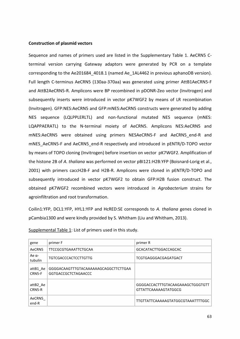
63
Construction of plasmid vectors
Sequence and names of primers used are listed in the Supplementary Table 1. AeCRN5 C-
terminal version carrying Gateway adaptors were generated by PCR on a template
corresponding to the Ae201684_4018.1 (named Ae_1AL4462 in previous aphanoDB version).
Full length C-terminus AeCRN5 (130aa-370aa) was generated using primer AttB1AeCRN5-F
and AttB2AeCRN5-R. Amplicons were BP recombined in pDONR-Zeo vector (Invitrogen) and
subsequently inserts were introduced in vector pK7WGF2 by means of LR recombination
(Invitrogen). GFP:NES:AeCRN5 and GFP:mNES:AeCRN5 constructs were generated by adding
NES sequence (LQLPPLERLTL) and non-functional mutated NES sequence (mNES:
LQAPPAERATL) to the N-terminal moiety of AeCRN5. Amplicons NES:AeCRN5 and
mNES:AeCRN5 were obtained using primers NESAeCRN5-F and AeCRN5_end-R and
mNES_AeCRN5-F and AeCRN5_end-R respectively and introduced in pENTR/D-TOPO vector
by means of TOPO cloning (Invitrogen) before insertion on vector pK7WGF2. Amplification of
the histone 2B of A. thaliana was performed on vector pBI121:H2B:YFP (Boisnard-Lorig et al.,
2001) with primers caccH2B-F and H2B-R. Amplicons were cloned in pENTR/D-TOPO and
subsequently introduced in vector pK7WGF2 to obtain GFP:H2B fusion construct. The
obtained pK7WGF2 recombined vectors were introduced in Agrobacterium strains for
agroinfiltration and root transformation.
Coilin1:YFP, DCL1:YFP, HYL1:YFP and HcRED:SE corresponds to A. thaliana genes cloned in
pCambia1300 and were kindly provided by S. Whitham (Liu and Whitham, 2013).
Supplemental Table 1: List of primers used in this study.
gene primer F primer R
AeCRN5 TTCCGCGTGAAATTCTGCAA GCACATACTTGGACCAGCAC
Ae α-tubulin
TGTCGACCCACTCCTTGTTG TCGTGAGGGACGAGATGACT
attB1_AeCRN5-F
GGGGACAAGTTTGTACAAAAAAGCAGGCTTCTTGAAGGTGACCGCTCTAGAACCC
attB2_AeCRN5-R
GGGGACCACTTTGTACAAGAAAGCTGGGTGTTGTTATTCAAAAAGTATGGCG
AeCRN5_end-R
TTGTTATTCAAAAAGTATGGCGTAAATTTTGGC

64
NESAeCRN5-F
CACCCTTCAACTTCCTCCTCTTGAAAGACTTACTCTTTTGAAGGTTGACCGCTCTAGAACCC
mNESAeCRN5-F
CACCCTTCAAGCTCCTCCTGCTGAAAGAGCTACTCTTTTGAAGGTGACCGCTCTAGAACCC
caccH2B-F
CACCATGGCGAAGGCAGATAAGAAACCAGC
H2B-R TTAAGAACTCGTAAACTTCGTAACCGCC
mGFP5-F GATCATATGAAGCGGCACGACTTCT
mGFP5-R_T7prom
ATCGAGTAATACGACTCACTATAGGGTTCCAACTTGTGGCCGAGGATG
Nb_L23 AAGGATGCCGTGAAGAAGATGT GCATCGTAGTCAGGAGTCAACC
Nb_F-BOX
GGCACTCACAAACGTCTATTTC ACCTGGGAGGCATCCTGTTAT
miR156a CGGAGGTGGAAATTTTTGAA AAAAGGGACAGTGCAACTCAA
miR156b TCCTGCACCCATATTGAACA GAGGAAGCGGATTGAAAGTG
miR159a TAAGCTGCCGACCTATGGAT GGCAATGAAGCTCCTGACAT
miR159b CCAGCTAGGCTACCTCGTGA TATTGAGCGGGAGCTGTCTT
miR160a TGGATGACTTTGAGCCCTTT GATCACGGATACGCTCCAAT
miR160b TATTTCGGGGATATGCTTGG TTGCAGAGCTCATCGGAATA
miR164a AACCATTGATCGGAGCTGAG GAAGAAGGGCACATGGAAAA
miR164b GCAGGGCATGTGCACTACTA TTTGACGGAAAATCACGACA
miR166a ATGTTGTCTGGCTCGAGGTC CCGACGACACTAAACCATGA
miR166b GCTGGCTCGACACAATTACTC TGAGAGGAATGAAGCCTGGT
miR167a CCAGCATGATCTGGTACGAA GGAAAAGCCAGACCTCAAGA
miR167b TTTTCCTGTTTTGGGTTGGA TATTGGTGGCGAGTGATTGA
miR169a GAAGGTTCAATGCCCTTTTG CTGCGGCAAATATGAGAGGT
miR169b GATGACTTGCCTGGTCCATT AAGATGACTTGCCTGCAACC
miR171a GAGAATTGTCCGGCCAGTAA CTAAGCTTGAGGCAGCTGGT
miR171b GGTGAGGTTCAATCCGAAGA CGGCTCAATCTGAGATCGTT
miR172a TGTCAACAGTTTTTGCAGATG GGATCCATAGGGAGCAAAAA
miR172b GGCCAAAAACAGATCTCCAC ATTTTCCTGCTCCCTCCTTC
miR319a GCCGACTCATTCATCCAAAT CTACGGAGGTGCGTTTGACT
miR319b CCCTAGTGGGTGCAGATGAT CGAGGAACAAGGGTAATCCA
miR390 GGAGGGATAGCACCATGAAA GCGCCAAAATGATTGAAAGT
miR393 GATCGCATTGATCCCATTTC AGTCCGAAGGGATAGCATGA
miR396a GCTTTATTGAACCGCAACAA TGGCTCTCTTTGTATTTTTCCA
miR396b TTCAGTGGGGAAGAAGTTCAA CAAGTCCTATCATGCTTTTCCA
miR399a ATTGATCCCTGCTGACGATG TACATCGGTCGTTGTTGGAA
miR399b AGAGAAATGCGAGCGAAGAT TTCTCCTTTGGCAAATCCAG
65
Immunoblot analyses
Samples corresponding to agroinfiltrated N. benthamiana leaves were frozen in liquid
nitrogen. Protein extraction was performed as Schornack et al., 2010. Proteins were separated
by SDS-PAGE and electroblotted on nitrocellulose membranes (BioRad). GFP-tagged proteins
were revealed using anti-GFP monoclonal antibodies 1:1000 (Merck #11814460001) followed
by Goat Anti-Mouse IgG-HRP Conjugate (BioRad #1706516). Clarity ECL (BioRad #1705060)
was used for the revelation step.
Agrobacterium-mediated transformation
Generation of M. truncatula composite plants was performed as described by Boisson-dernier
et al, 2001 using ARQUA-1 (A. rhizogenes) strain. Leaf infiltration were performed with A.
tumefaciens (GV3101::pMP90RK) as described by Schornack et al., 2010.
Confocal microscopy
Foliar discs (5-8 mm of diameter) of infiltrated leaves of N. benthamiana were sampled at
different time points after agroinfiltration and mounted on microscope slides for live cell
imaging. For DAPI staining, discs were fixed in a PBS, 4% (v/v) paraformaldehyde solution and
then stained with DAPI (3 µg/µL). Scans were performed on a Leica TCS SP8 device using
wavelengths 488nm (GFP) and 350 nm (DAPI) and with a 40x water immersion lens.
Acquisitions were performed in a sequential mode to avoid overlapping fluorescence signals.
Images were treated with Image J software and correspond to Z projections of scanned
tissues.
Preparation of N. benthamiana epidermal leaves for FRET / FLIM experiments
Samples were prepared as described in (Camborde et al., 2017; Ramirez-Garcés et al., 2016;
Le Roux et al., 2015). Briefly, discs of N. benthamiana agroinfiltrated leaves were fixed 20-24
hours after treatment by vacuum infiltrating a TBS (TRIS 25 mM, NaCl 140 mM, KCl 3 mM) 4
% (w/v) paraformaldehyde solution before incubation 20 min at 4°C. Samples were

66
permeabilized 10 min at 37°C using a digestion buffer supplemented with 20 µg/ml of
proteinase K (Invitrogen) as described in (Camborde et al., 2017; Escouboué et al., 2019).
Nucleic acid staining was performed by vacuum-infiltration of a 5 µM of Sytox Orange
(Invitrogen) solution, before incubation of the samples 30 min at room temperature. When
RNAse treatment was performed, foliar discs were incubated 15 min at room temperature
with 0.5 µg/ml of RNAse A (Roche) before acid nucleic staining. Foliar discs were washed with
and mounted on TBS before observations on an inverted microscope (Eclipse TE2000E, Nikon,
Japan).
FRET / FLIM measurements
Fluorescence lifetime measurements were performed in time domain using a streak camera
(Camborde et al., 2017). The light source is a mode-locked Ti:sapphire laser (Tsunami, model
3941, Spectra-Physics, USA) pumped by a 10W diode laser (Millennia Pro, Spectra-Physics)
and delivering ultrafast femtosecond pulses of light with a fundamental frequency of 80MHz.
A pulse picker (model 3980, Spectra-Physics) is used to reduce the repetition rate to 2MHz to
satisfy the requirements of the triggering unit (working at 2MHz). The experiments were
carried out at λ = 820 nm (multiphoton excitation mode). All images were acquired with a 60x
oil immersion lens (plan APO 1.4 N.A., IR) mounted on an inverted microscope (Eclipse
TE2000E, Nikon, Japan). The fluorescence emission is directed back into the detection unit
through a short pass filter λ<750 nm) and a band pass filter (515/30 nm). The detector is a
streak camera (Streakscope C4334, Hamamatsu Photonics, Japan) coupled to a fast and high-
sensitivity CCD camera (model C8800-53C, Hamamatsu). For each nucleus, average
fluorescence decay profiles were plotted and lifetimes were estimated by fitting data with
exponential function using a non-linear least-squares estimation procedure. Fluorescence
lifetime of the donor (GFP) was experimentally measured in the presence and absence of the
acceptor (Sytox Orange). FRET efficiency (E) was calculated by comparing the lifetime of the
donor in the presence (DA) or absence (D) of the acceptor: E=1-(DA) / (D). Statistical
comparisons between control (donor) and assay (donor + acceptor) lifetime values were
performed by Student t-test. For each experiment, a minimum of four leaf discs removed from
two agroinfiltrated leaves were used to collect data.

67
Post-Transcriptional Gene Silencing assay
PTGS assays were performed as described by (Qiao et al., 2013, 2015). Briefly, N. benthamiana
16c at the six-leaf stage (with a constitutive GFP expression) were agroinfiltrated with
p35S::GFP vector combined with pEG100 as empty vector, or pK2GW7::AeCRN5-Cter,
pEG101::PSR1 (Qiao et al., 2013) or pK2GW7::P19. pEG100, p35S::GFP and pEG101::PSR1
were kindly provided by Dr W. Ma, and pK2GW7::P19 by Dr N. Pauly from LIPM lab.
Green fluorescence was visualized using a handheld long-wavelength UV lamp (Black-Ray B-
100AP, Ultraviolet Products). Agrobacterium carrying the empty vector pEG100 was used as
negative control whilst PSR1 and P19 constructs were used as positive controls.
Leaves were examined 3 days after Agrobacterium infiltration in the infiltrated leaf areas and
sampled in liquid nitrogen. GFP Immunoblots were performed after total protein extraction
as described in this paper. To produce siRNA probe, we first amplified approx. 200bp of the
mGFP5 using cDNA from 16c leaves (RNA extraction and RT were performed as described in
this paper), with mGFP5-F and mGFP5-R_T7prom primers (Supplemental Table 1). The GFP
siRNA probe was generated using the MEGAScript high-yield T7 kit (Ambion) in the presence
of [α-32P] UTP. U6 served as a loading control.
References
Amaro, T.M.M.M., Thilliez, G.J.A., Motion, G.B., and Huitema, E. (2017). A Perspective on
CRN Proteins in the Genomics Age: Evolution, Classification, Delivery and Function
Revisited. Front. Plant Sci. 8: 99.
Badis, Y., Bonhomme, M., Lafitte, C., Huguet, S., Balzergue, S., Dumas, B., and Jacquet, C.
(2015). Transcriptome analysis highlights preformed defences and signalling pathways
controlled by the prAe1 quantitative trait locus (QTL), conferring partial resistance to
Aphanomyces euteiches in Medicago truncatula. Mol. Plant Pathol. 16: 973–986.
Badreddine, I., Lafitte, C., Heux, L., Skandalis, N., Spanou, Z., Martinez, Y., Esquerré-Tugayé,
M.T., Bulone, V., Dumas, B., and Bottin, A. (2008). Cell wall chitosaccharides are
68
essential components and exposed patterns of the phytopathogenic oomycete
Aphanomyces euteiches. Eukaryot. Cell 7: 1980–1993.
Baxter, L. et al. (2010). Signatures of adaptation to obligate biotrophy in the
Hyaloperonospora arabidopsidis genome. Science (80-. ). 330: 1549–1551.
Bazin, J., Romero, N., Rigo, R., Charon, C., Blein, T., Ariel, F., and Crespi, M. (2018). Nuclear
Speckle RNA Binding Proteins Remodel Alternative Splicing and the Non-coding
Arabidopsis Transcriptome to Regulate a Cross-Talk Between Auxin and Immune
Responses. Front. Plant Sci. 9: 1209.
Bellafiore, S., Shen, Z., Rosso, M.N., Abad, P., Shih, P., and Briggs, S.P. (2008). Direct
identification of the Meloidogyne incognita secretome reveals proteins with host cell
reprogramming potential. PLoS Pathog. 4.
Bhattacharjee, S., Garner, C.M., and Gassmann, W. (2013). New clues in the nucleus:
transcriptional reprogramming in effector-triggered immunity. Front. Plant Sci. 4: 364.
Boisnard-Lorig, C., Colon-Carmona, a, Bauch, M., Hodge, S., Doerner, P., Bancharel, E.,
Dumas, C., Haseloff, J., and Berger, F. (2001). Dynamic analyses of the expression of the
HISTONE::YFP fusion protein in arabidopsis show that syncytial endosperm is divided in
mitotic domains. Plant Cell 13: 495–509.
Boisson-Dernier, A., Chabaud, M., Garcia, F., Bécard, G., Rosenberg, C., and Barker, D.G.
(2001). Agrobacterium rhizogenes-transformed roots of Medicago truncatula for the
study of nitrogen-fixing and endomycorrhizal symbiotic associations. Mol. Plant-Microbe
Interact. 14: 695–700.
Cai, Q., Cai, Q., Qiao, L., Wang, M., He, B., Lin, F., Palmquist, J., Huang, H., and Jin, H. (2018).
Plants send small RNAs in extracellular vesicles to fungal pathogen to silence virulence
genes. 4142: 1–9.
Camborde, L., Jauneau, A., Brière, C., Deslandes, L., Dumas, B., and Gaulin, E. (2017).
Detection of nucleic acid–protein interactions in plant leaves using fluorescence lifetime
imaging microscopy. Nat. Protoc. 12: 1933–1950.
69
Canonne, J. and Rivas, S. (2012). Bacterial effectors target the plant cell nucleus to subvert
host transcription. Plant Signal. Behav. 7.
Couzigou, J.M. and Combier, J.P. (2016). Plant microRNAs: key regulators of root architecture
and biotic interactions. New Phytol. 212.
van Damme, M., Bozkurt, T.O., Cakir, C., Schornack, S., Sklenar, J., Jones, A.M.E., and
Kamoun, S. (2012). The Irish Potato Famine Pathogen Phytophthora infestans
Translocates the CRN8 Kinase into Host Plant Cells. PLoS Pathog. 8.
Deslandes, L. and Rivas, S. (2012). Catch me if you can: Bacterial effectors and plant targets.
Trends Plant Sci. 17.
Djébali, N. et al. (2009). Partial Resistance of Medicago truncatula to Aphanomyces euteiches
is associated with protection of the root stele and is controlled by a major QTL rich in
proteasome-related genes. Mol. Plant-Microbe Interact. 22: 1043–1055.
Escouboué, M., Camborde, L., Jauneau, A., Gaulin, E., and Deslandes, L. (2019). Preparation
of plant material for analysis of protein–Nucleic acid interactions by FRET-FLIM. In
Methods in Molecular Biology (Humana Press Inc.), pp. 69–77.
Gaulin, E. et al. (2018). Genomics analysis of Aphanomyces spp. identifies a new class of
oomycete effector associated with host adaptation. BMC Biol. 16.
Gaulin, E., Haget, N., Khatib, M., Herbert, C., Rickauer, M., and Bottin, A. (2007). Transgenic
sequences are frequently lost in Phytophthora parasitica transformants without
reversion of the transgene-induced silenced state. Can. J. Microbiol. 53: 152–157.
Gaulin, E., Madoui, M.-A., Bottin, A., Jacquet, C., Mathé, C., Couloux, A., Wincker, P., and
Dumas, B. (2008). Transcriptome of Aphanomyces euteiches: new oomycete putative
pathogenicity factors and metabolic pathways. PLoS One 3: e1723.
Haas, B.J. et al. (2009). Genome sequence and analysis of the Irish potato famine pathogen
Phytophthora infestans. Nature 461: 393–398.
Hou, Y. et al. (2019). A Phytophthora Effector Suppresses Trans-Kingdom RNAi to Promote
Disease Susceptibility. Cell Host Microbe 25: 153-165.e5.
Jiao, J. and Peng, D. (2018). Wheat microRNA1023 suppresses invasion of Fusarium
70
graminearum via targeting and silencing fgsg_03101. J. Plant Interact. 13.
Kelley, L.A., Mezulis, S., Yates, C.M., Wass, M.N., and Sternberg, M.J.E. (2015). The Phyre2
web portal for protein modeling, prediction and analysis. Nat. Protoc. 10.
Kemen, E., Gardiner, A., Schultz-Larsen, T., Kemen, A.C., Balmuth, A.L., Robert-Seilaniantz,
A., Bailey, K., Holub, E., Studholme, D.J., MacLean, D., and Jones, J.D.G. (2011). Gene
gain and loss during evolution of obligate parasitism in the white rust pathogen of
Arabidopsis thaliana. PLoS Biol. 9.
Khan, M., Seto, D., Subramaniam, R., and Desveaux, D. (2018). Oh, the places they’ll go! A
survey of phytopathogen effectors and their host targets. Plant J. 93: 651–663.
Kozomara, A., Birgaoanu, M., and Griffiths-Jones, S. (2019). MiRBase: From microRNA
sequences to function. Nucleic Acids Res. 47.
Lakatos, L., Szittya, G., Silhavy, D., and Burgyán, J. (2004). Molecular mechanism of RNA
silencing suppression mediated by p19 protein of tombusviruses. EMBO J. 23: 876–84.
Le Roux, C. et al. (2015). A Receptor Pair with an Integrated Decoy Converts Pathogen
Disabling of Transcription Factors to Immunity. Cell 161: 1074–1088.
Lévesque, C.A. et al. (2010). Genome sequence of the necrotrophic plant pathogen Pythium
ultimum reveals original pathogenicity mechanisms and effector repertoire. Genome Biol. 11.
Li, S., Li, M., Liu, K., Zhang, H., Zhang, S., Zhang, C., and Yu, B. (2020). MAC5, an RNA-binding
protein, protects pri-miRNAs from SERRATE-dependent exoribonuclease activities. Proc.
Natl. Acad. Sci.: 202008283.
Lin, K. et al. (2014). Single Nucleus Genome Sequencing Reveals High Similarity among Nuclei
of an Endomycorrhizal Fungus. PLoS Genet. 10.
Liu, D., Shi, L., Han, C., Yu, J., Li, D., and Zhang, Y. (2012). Validation of Reference Genes for
Gene Expression Studies in Virus-Infected Nicotiana benthamiana Using Quantitative
Real-Time PCR. PLoS One 7.
Liu, J.-Z. and Whitham, S. a (2013). Overexpression of a soybean nuclear localized type-III DnaJ
domain-containing HSP40 reveals its roles in cell death and disease resistance. Plant J.
74: 110–21.

71
Liu, T., Ye, W., Ru, Y., Yang, X., Gu, B., Tao, K., Lu, S., Dong, S., Zheng, X., Shan, W., Wang, Y.,
and Dou, D. (2011). Two host cytoplasmic effectors are required for pathogenesis of
Phytophthora sojae by suppression of host defenses. Plant Physiol. 155: 490–501.
Livak, K.J. and Schmittgen, T.D. (2001). Analysis of relative gene expression data using real-
time quantitative PCR and the 2-ΔΔCT method. Methods 25: 402–408.
Lobbes, D., Rallapalli, G., Schmidt, D.D., Martin, C., and Clarke, J. (2006). SERRATE: a new
player on the plant microRNA scene. EMBO Rep. 7: 1052–1058.
Mafurah, J.J., Ma, H., Zhang, M., Xu, J., He, F., Ye, T., Shen, D., Chen, Y., Rajput, N.A., and
Dou, D. (2015). A Virulence Essential CRN Effector of Phytophthora capsici Suppresses
Host Defense and Induces Cell Death in Plant Nucleus. PLoS One 10: e0127965.
Meijer, H.J.G., Mancuso, F.M., Espadas, G., Seidl, M.F., Chiva, C., Govers, F., and Sabidó, E.
(2014). Profiling the secretome and extracellular proteome of the potato late blight
pathogen Phytophthora infestans. Mol. Cell. Proteomics 13.
Ökmen, B., Etalo, D.W., Joosten, M.H.A.J., Bouwmeester, H.J., de Vos, R.C.H., Collemare, J.,
and de Wit, P.J.G.M. (2013). Detoxification of α-tomatine by Cladosporium fulvum is
required for full virulence on tomato. New Phytol. 198: 1203–1214.
Pedersen, C. et al. (2012). Structure and evolution of barley powdery mildew effector
candidates. BMC Genomics 13: 694.
Petrovská, B., Šebela, M., and Doležel, J. (2015). Inside a plant nucleus: Discovering the
proteins. J. Exp. Bot. 66: 1627–1640.
Pliego, C. et al. (2013). Host-induced gene silencing in barley powdery mildew reveals a class
of ribonuclease-like effectors. Mol. Plant-Microbe Interact. 26.
Qiao, Y. et al. (2013). Oomycete pathogens encode RNA silencing suppressors. Nat. Genet.:
1–6.
Qiao, Y., Shi, J., Zhai, Y., Hou, Y., and Ma, W. (2015). Phytophthora effector targets a novel
component of small RNA pathway in plants to promote infection. Proc. Natl. Acad. Sci. U.
S. A. 112: 5850–5855.
72
Raczynska, K.D., Stepien, A., Kierzkowski, D., Kalak, M., Bajczyk, M., McNicol, J., Simpson,
C.G., Szweykowska-Kulinska, Z., Brown, J.W.S., and Jarmolowski, A. (2014). The
SERRATE protein is involved in alternative splicing in Arabidopsis thaliana. Nucleic Acids
Res. 42: 1224–1244.
Rajput, N.A., Zhang, M., Ru, Y., Liu, T., Xu, J., Liu, L., Mafurah, J.J., and Dou, D. (2014).
Phytophthora sojae Effector PsCRN70 Suppresses Plant Defenses in Nicotiana
benthamiana. PLoS One 9: e98114.
Ramirez-Garcés, D., Camborde, L., Pel, M.J.C., Jauneau, A., Martinez, Y., Néant, I., Leclerc,
C., Moreau, M., Dumas, B., and Gaulin, E. (2016). CRN13 candidate effectors from plant
and animal eukaryotic pathogens are DNA-binding proteins which trigger host DNA
damage response. New Phytol. 210.
Rey, T., Nars, A., Bonhomme, M., Bottin, A., Huguet, S., Balzergue, S., Jardinaud, M.-F., Bono,
J.-J., Cullimore, J., Dumas, B., Gough, C., and Jacquet, C. (2013). NFP, a LysM protein
controlling Nod factor perception, also intervenes in Medicago truncatula resistance to
pathogens. New Phytol. 198: 875–886.
Schornack, S., van Damme, M., Bozkurt, T.O., Cano, L.M., Smoker, M., Thines, M., Gaulin, E.,
Kamoun, S., and Huitema, E. (2010). Ancient class of translocated oomycete effectors
targets the host nucleus. Proc. Natl. Acad. Sci. U. S. A. 107: 17421–6.
Shen, D., Liu, T., Ye, W., Liu, L., Liu, P., Wu, Y., Wang, Y., and Dou, D. (2013). Gene Duplication
and Fragment Recombination Drive Functional Diversification of a Superfamily of
Cytoplasmic Effectors in Phytophthora sojae. PLoS One 8.
Song, T., Ma, Z., Shen, D., Li, Q., Li, W., Su, L., Ye, T., Zhang, M., Wang, Y., and Dou, D. (2015).
An Oomycete CRN Effector Reprograms Expression of Plant HSP Genes by Targeting their
Promoters. PLoS Pathog. 11: 1–30.
Stam, R., Howden, A.J.M., Delgado-Cerezo, M., Amaro, T.M.M.M., Motion, G.B., Pham, J.,
and Huitema, E. (2013a). Characterization of cell death inducing Phytophthora capsici
CRN effectors suggests diverse activities in the host nucleus. Front. Plant Sci. 4.
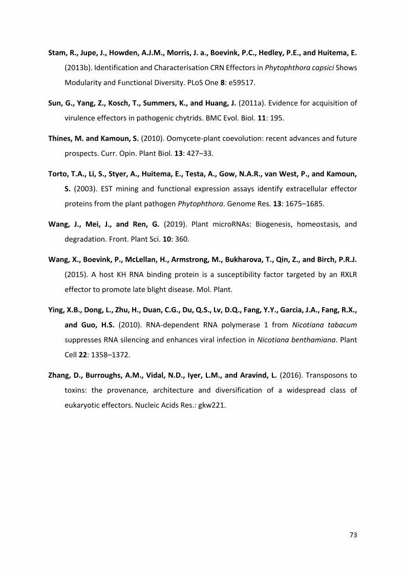
73
Stam, R., Jupe, J., Howden, A.J.M., Morris, J. a., Boevink, P.C., Hedley, P.E., and Huitema, E.
(2013b). Identification and Characterisation CRN Effectors in Phytophthora capsici Shows
Modularity and Functional Diversity. PLoS One 8: e59517.
Sun, G., Yang, Z., Kosch, T., Summers, K., and Huang, J. (2011a). Evidence for acquisition of
virulence effectors in pathogenic chytrids. BMC Evol. Biol. 11: 195.
Thines, M. and Kamoun, S. (2010). Oomycete-plant coevolution: recent advances and future
prospects. Curr. Opin. Plant Biol. 13: 427–33.
Torto, T.A., Li, S., Styer, A., Huitema, E., Testa, A., Gow, N.A.R., van West, P., and Kamoun,
S. (2003). EST mining and functional expression assays identify extracellular effector
proteins from the plant pathogen Phytophthora. Genome Res. 13: 1675–1685.
Wang, J., Mei, J., and Ren, G. (2019). Plant microRNAs: Biogenesis, homeostasis, and
degradation. Front. Plant Sci. 10: 360.
Wang, X., Boevink, P., McLellan, H., Armstrong, M., Bukharova, T., Qin, Z., and Birch, P.R.J.
(2015). A host KH RNA binding protein is a susceptibility factor targeted by an RXLR
effector to promote late blight disease. Mol. Plant.
Ying, X.B., Dong, L., Zhu, H., Duan, C.G., Du, Q.S., Lv, D.Q., Fang, Y.Y., Garcia, J.A., Fang, R.X.,
and Guo, H.S. (2010). RNA-dependent RNA polymerase 1 from Nicotiana tabacum
suppresses RNA silencing and enhances viral infection in Nicotiana benthamiana. Plant
Cell 22: 1358–1372.
Zhang, D., Burroughs, A.M., Vidal, N.D., Iyer, L.M., and Aravind, L. (2016). Transposons to
toxins: the provenance, architecture and diversification of a widespread class of
eukaryotic effectors. Nucleic Acids Res.: gkw221.

Chapter IV
Genomics analysis of Aphanomyces spp.
identifies a new class of oomycete effector
associated with host adaptation

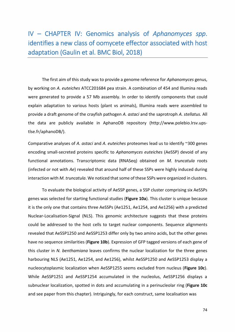
74
IV – CHAPTER IV: Genomics analysis of Aphanomyces spp.
identifies a new class of oomycete effector associated with host
adaptation (Gaulin et al. BMC Biol, 2018)
The first aim of this study was to provide a genome reference for Aphanomyces genus,
by working on A. euteiches ATCC201684 pea strain. A combination of 454 and Illumina reads
were generated to provide a 57 Mb assembly. In order to identify components that could
explain adaptation to various hosts (plant vs animals), Illumina reads were assembled to
provide a draft genome of the crayfish pathogen A. astaci and the saprotroph A. stellatus. All
the data are publicly available in AphanoDB repository (http://www.polebio.lrsv.ups-
tlse.fr/aphanoDB/).
Comparative analyses of A. astaci and A. euteiches proteomes lead us to identify ~300 genes
encoding small-secreted proteins specific to Aphanomyces euteiches (AeSSP) devoid of any
functional annotations. Transcriptomic data (RNASeq) obtained on M. truncatula roots
(infected or not with Ae) revealed that around half of these SSPs were highly induced during
interaction with M. truncatula. We noticed that some of these SSPs were organized in clusters.
To evaluate the biological activity of AeSSP genes, a SSP cluster comprising six AeSSPs
genes was selected for starting functional studies (Figure 10a). This cluster is unique because
it is the only one that contains three AeSSPs (Ae1251, Ae1254, and Ae1256) with a predicted
Nuclear-Localisation-Signal (NLS). This genomic architecture suggests that these proteins
could be addressed to the host cells to target nuclear components. Sequence alignments
revealed that AeSSP1250 and AeSSP1253 differ only by two amino acids, but the other genes
have no sequence similarities (Figure 10b). Expression of GFP tagged versions of each gene of
this cluster in N. benthamiana leaves confirms the nuclear localization for the three genes
harbouring NLS (Ae1251, Ae1254, and Ae1256), whilst AeSSP1250 and AeSSP1253 display a
nucleocytoplasmic localization when AeSSP1255 seems excluded from nucleus (Figure 10c).
While AeSSP1251 and AeSSP1254 accumulated in the nucleolus, AeSSP1256 displays a
subnuclear localization, spotted in dots and accumulating in a perinucleolar ring (Figure 10c
and see paper from this chapter). Intriguingly, for each construct, same localisation was
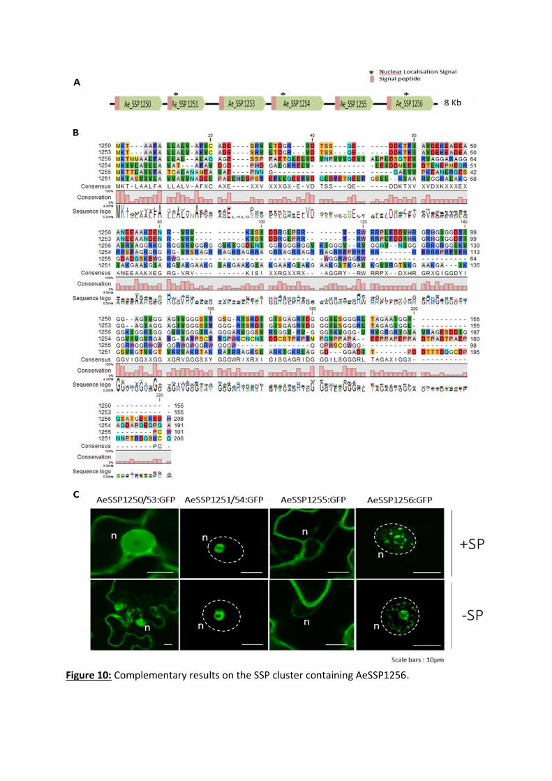
Figure 10: Complementary results on the SSP cluster containing AeSSP1256.

75
Figure 10: Complementary results on the SSP cluster containing AeSSP1256.
(A) Scheme of the SSP cluster organization. Six genes on the same orientation, among 8 kB, compose this cluster.
Each gene contains a signal peptide and three genes harbour a NLS (AeSSP1251, 1254 and 1256). (B) Amino acid
sequence alignment of the six genes, performed on CLC Workbench. AeSSP1250 and AeSSP1253 differ by only
2AA. Except these two genes, no sequence similarities are observed among the cluster. (C) Localization of
AeSSP1256 cluster proteins expressed in N. benthamiana leaves. For each gene, GFP was fused in C-ter and
transform in Agrobacterium tumefaciens. Confocal analyses were conducted 24h after agroinoculation in N.
benthamiana leaves. AeSSP1250 and AeSSP1253 have a similar nucleocytoplasmic localization (pictures
correspond to AeSSP1250, similar pictures were obtained from AeSSP1253), when AeSSP1255 is excluded from
nucleus. Similar nuclear localization was observed for AeSSP1251 and AeSSP1254 (pictures correspond to
AeSSP1254), with an accumulation in nucleolus, indicating that the NLS was functional. In the same way,
AeSSP1256 is nuclear localized but spotted in dots and accumulates in a perinucleolar ring. Same localisation was
observed in presence (upper panels) or absence (lower panels) of their own signal peptide (SP). White dotted
lines indicate nuclei. n: nucleus. Scale bars: 10µm.
observed in presence or absence of their own signal peptide (SP) (Figure 10c). We then
evidenced that the AeSSP1256 entered the plant secretory pathway thanks to its native signal
peptide, using endoplasmic reticulum (ER) retention motif and drug assay (see Fig. 8 - BMC
biology paper from this chapter).
Due to the fact that A. euteiches transformation is not yet available, we used Phytophthora
capsici infection assay to investigate whether those SSPs could act as effectors. After
expression of each member of the AeSSP cluster on tobacco leaves, followed by P. capsici
inoculation, it appeared that only AeSSP1256 enhances N. benthamiana susceptibility to P.
capsici. These data suggest that AeSSP1256 and therefore SSPs are a new class of oomycete
effectors.


76
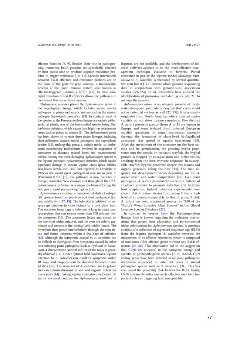
77
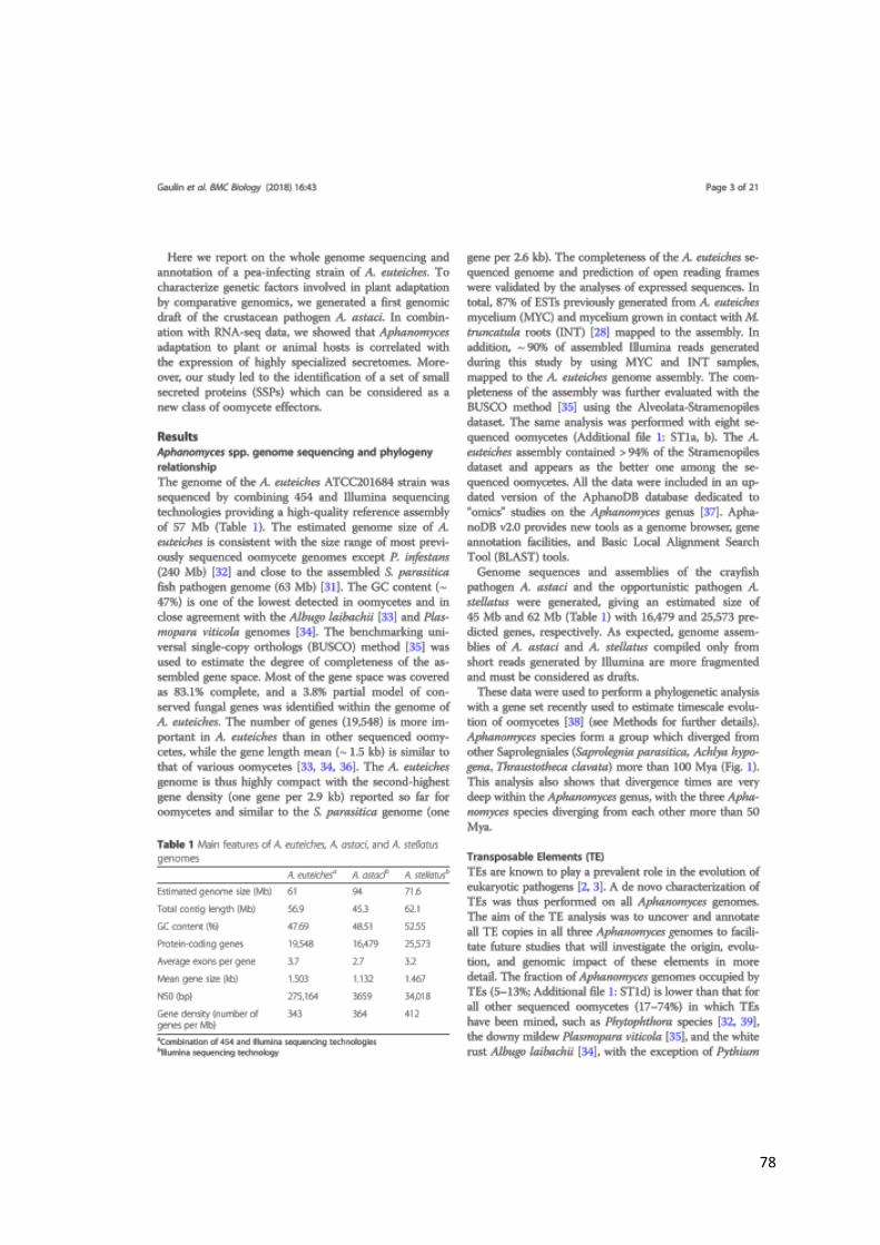
78
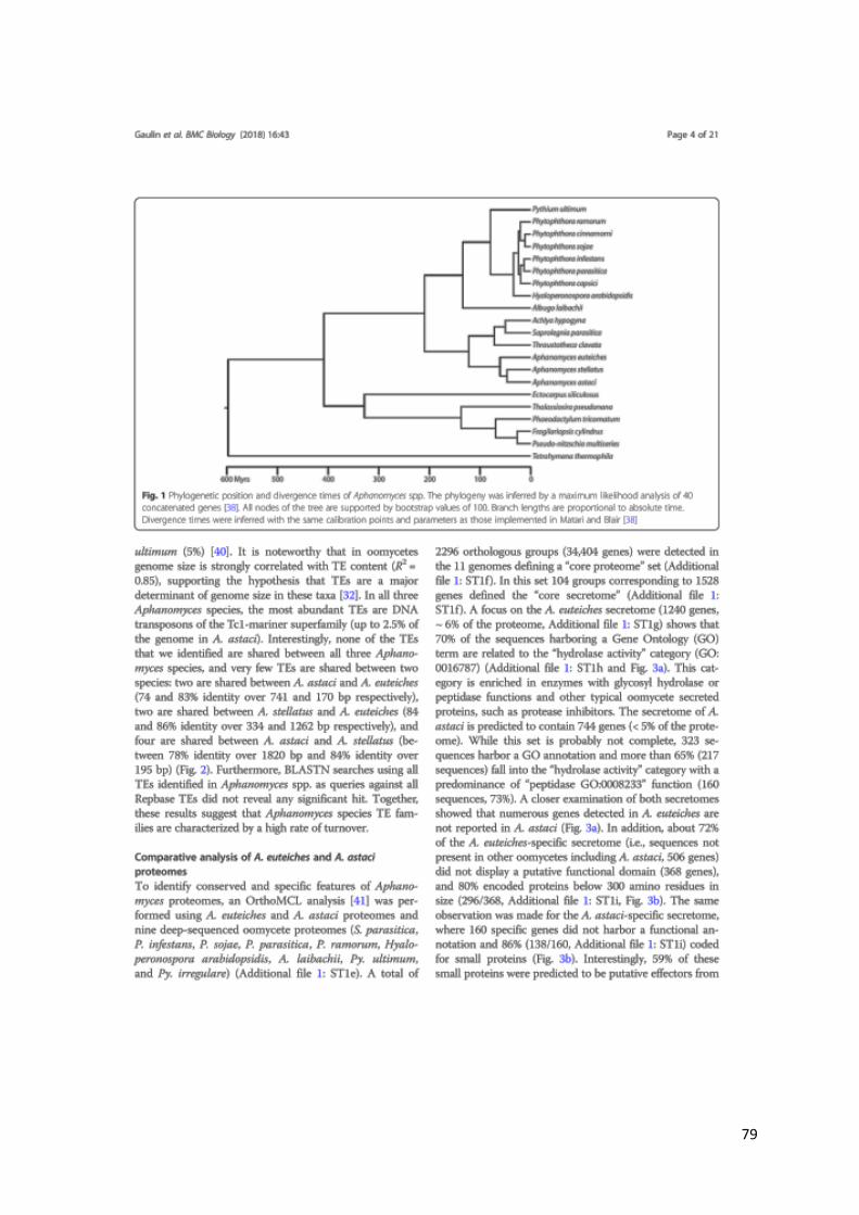
79
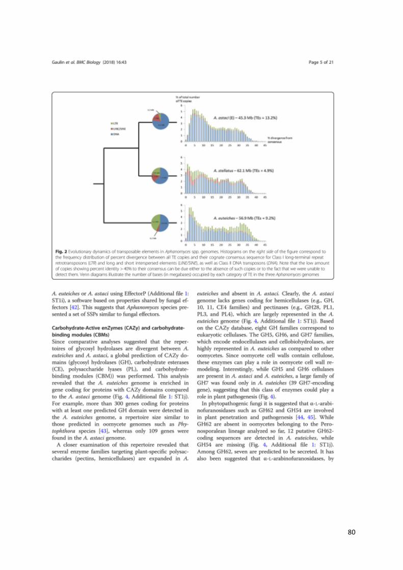
80
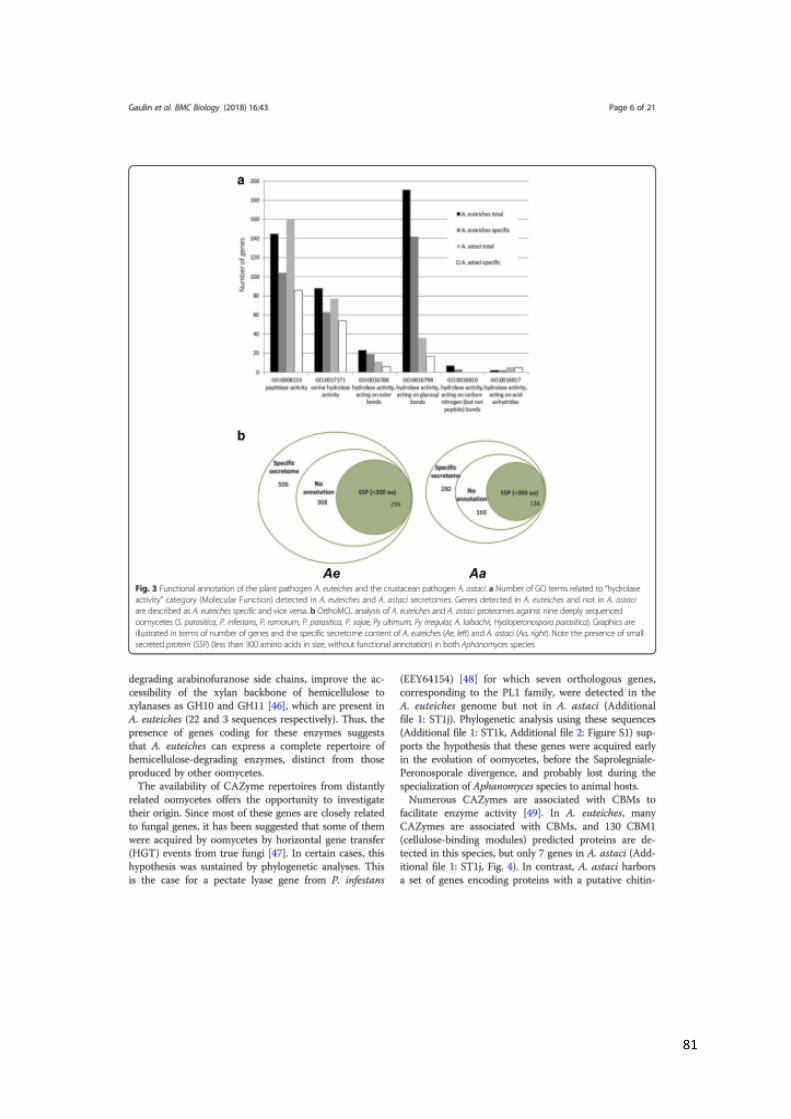
81

82
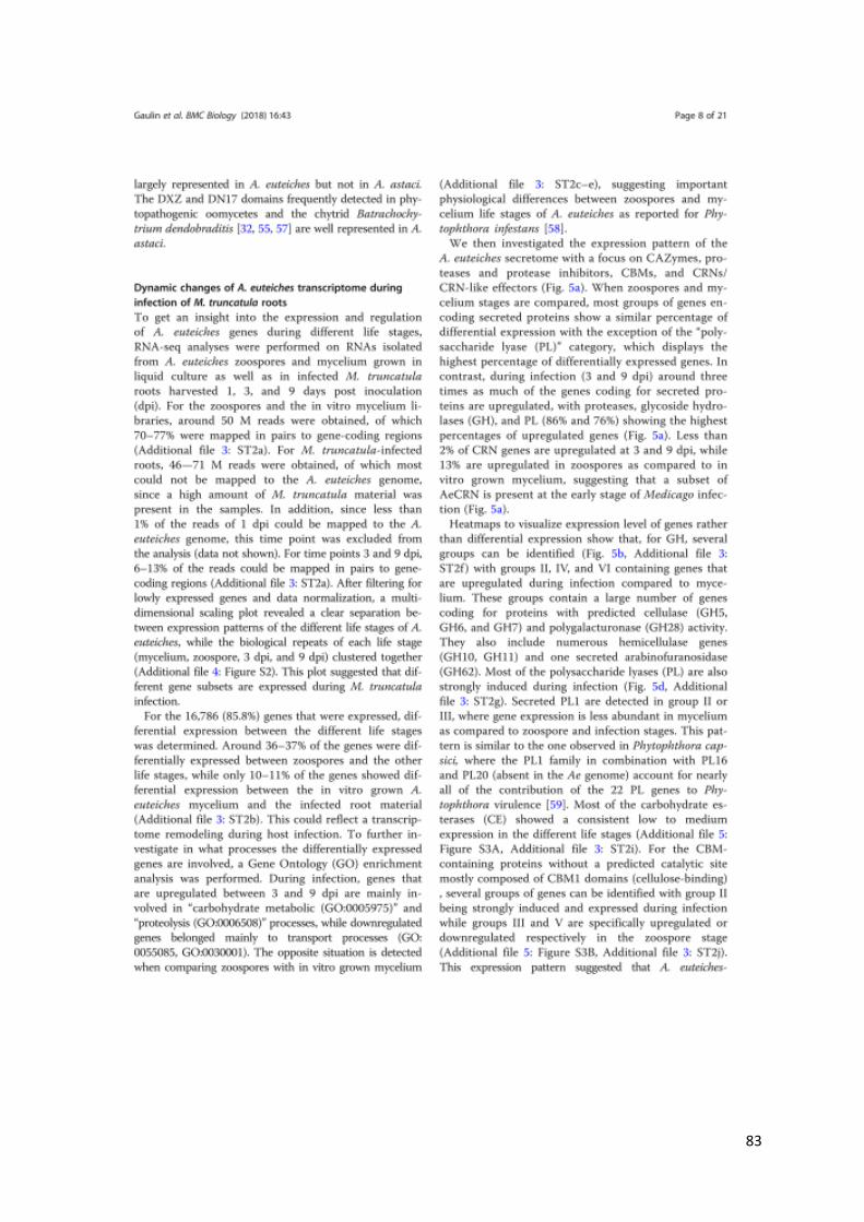
83
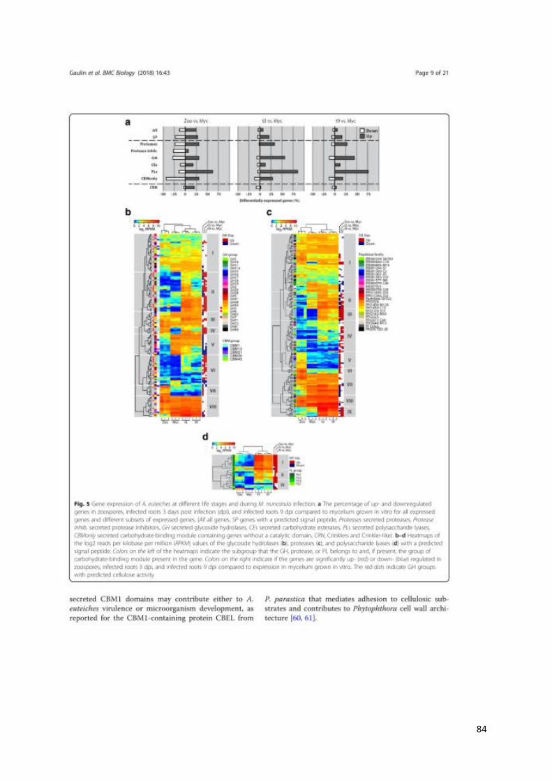
84
85
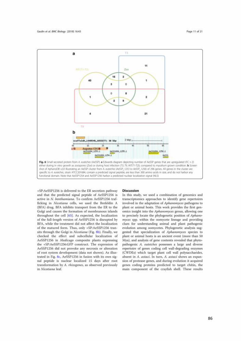
86
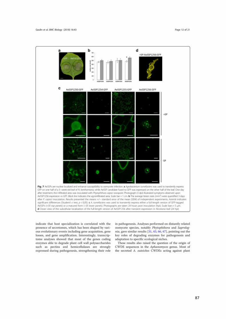
87
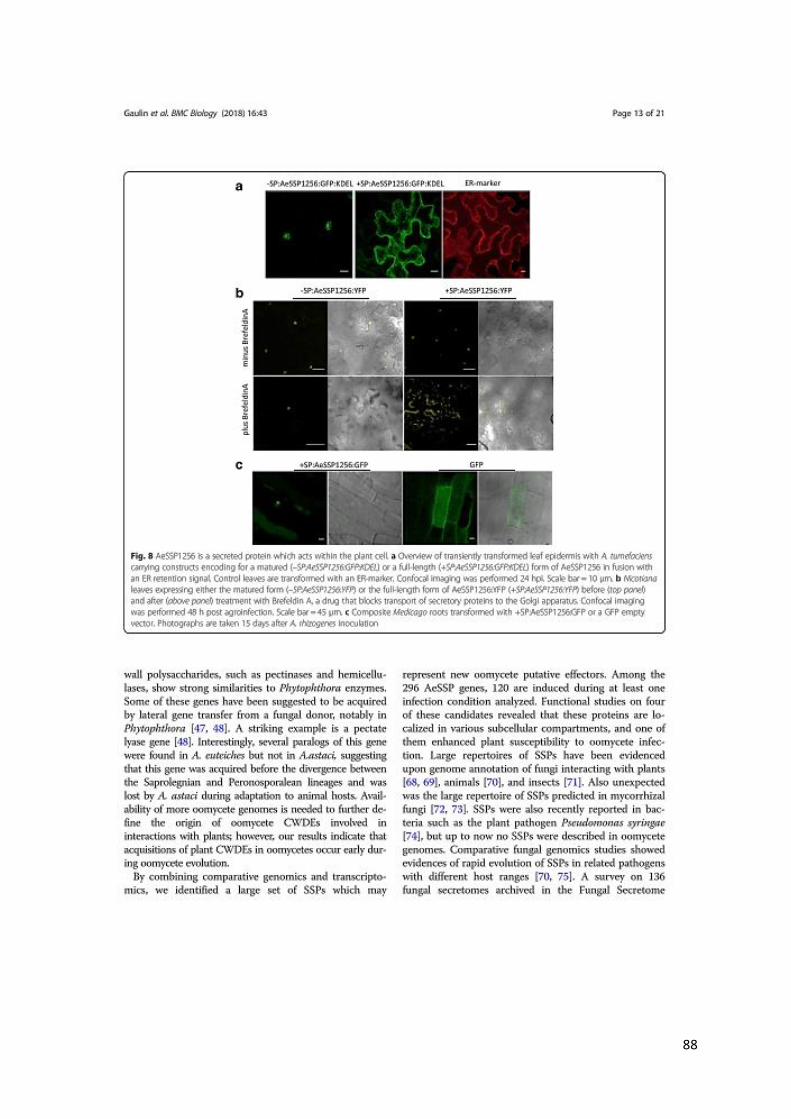
88
89
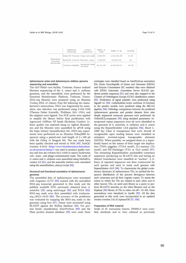
90
91
92
93
94

95
96


Chapter V
A DEAD-Box RNA helicase from Medicago
truncatula is hijacked by an RNA-binding effector
from the root pathogen Aphanomyces euteiches
to facilitate host infection

97
V – CHAPTER V: A DEAD-Box RNA helicase from Medicago
truncatula is hijacked by an RNA-binding effector from the root
pathogen Aphanomyces euteiches to facilitate host infection (Camborde et al., submitted and available at BioXiv: doi: https://doi.org/10.1101/2020.06.17.157404)
In a previous study we identified among a cluster composed of six SSP that AeSSP1256
enhances oomycete infection and harbour a nuclear-localisation when transiently express in
Nicotiana benthamiana cells (Gaulin et al., 2018). We then undertake a functional
characterization of AeSSP1256 to decipher its activity by using M. truncatula. Sequence
analyses predict that AeSSP1256 sequence contains RNA binding motifs (Figure 1 from this
Chapter). By using FRET-FLIM analyses based on a method that I developed in collaboration
with the Imagery Platform Tri-IBIsa Genotoul (Camborde et al., 2017; Escouboué et al., 2019),
we showed that AeSSP1256 binds plant RNA (Figure 1 from this Chapter).
When expressed in M. truncatula roots, AeSSP1256 is localized around the nucleolus
of the host cells and induces a strong delay in root development (Figure 2 from this Chapter).
Furthermore, the presence of AeSSP1256 enhances the susceptibility to A. euteiches infection
(Figure 2 from this Chapter).
Transcriptomic analyses revealed that expression of AeSSP1256 in M. truncatula roots
leads to a downregulation of genes implicated in ribosome biogenesis pathway (Figure 3 from
this Chapter), suggesting that the effector provokes ribosomal stress when present in the host.
A yeast-two hybrid approach using cDNA library obtained from A. euteiches-infected
Medicago roots allows the identification of host targets (Supplemental Table 2 from this
Chapter) and A. euteiches targets (complementary results of this Chapter).
Among Medicago targets, we confirmed that AeSSP1256 associates with a nucleolar
L7 ribosomal protein and a M. truncatula RNA helicase (MtRH10) orthologous to the
Arabidopsis RNA helicase RH10 (Figure 4 from this Chapter).
98
Whereas MtRH10 is able to interact with nucleic acids, this association is abolished in
the presence of AeSSP1256 (Figure 5 from this Chapter).
Promoter:GUS composite plants revealed that MtRH10 is expressed preferentially in
the meristematic root cells (Figure 6 from this Chapter).
Missense MtRH10 plants displayed similar phenotype than overexpressing AeSSP1256
plants, leading to shorter roots with developmental delay and are more susceptible to A.
euteiches infection (Figure 7 from this Chapter).
These results show that the effector AeSSP1256 facilitates pathogen infection by
causing stress on plant ribosome biogenesis and by hijacking a host RNA helicase involved in
root development and resistance to root pathogens.
99
A DEAD BOX RNA helicase from Medicago truncatula is hijacked by an RNA-binding
effector from the root pathogen Aphanomyces euteiches to facilitate host infection
L. Camborde1, A. Kiselev1, M.J.C. Pel1, *, A. Leru2, A. Jauneau2, C. Pouzet2, B. Dumas1, E.
Gaulin1
Affiliations
1Laboratoire de Recherche en Sciences Végétales (LRSV), Université de Toulouse, CNRS, UPS,
France
2Plateforme d’Imagerie FRAIB-TRI, Université de Toulouse, CNRS, France
*Present address: Bacteriology Group, National Reference Centre (NRC), Dutch National Plant
Protection Organization (NPPO-NL), P.O. Box. 9102, 6700 HC Wageningen, the Netherlands
Corresponding author: Elodie Gaulin
LRSV, UMR CNRS 5546 Université de Toulouse,
24, Chemin de Borde-Rouge
31320 Auzeville, France
Mail : [email protected]
Keywords: effectors, RNA-helicase, plant development, ribosome biogenesis pathway,
oomycete, Medicago, nucleolar stress

100
Abstract
Microbial effectors from plant pathogens are molecules that target host components to
facilitate colonization. While eukaryotic pathogens are virtually able to produce hundreds of
effectors, the underlying molecular mechanisms allowing effectors to promote infection are
still largely unexplored. In this study, we show that the effector AeSSP1256 from the soilborne
oomycete pathogen Aphanomyces euteiches is a RGG/RG protein able to interact with nuclear
RNA in vivo. Heterologous expression of AeSSP1256 delays Medicago truncatula root
development and facilitates pathogen colonization. We found by transcriptomic analyses of
AeSSP1256 expressing roots that AeSSP1256 downregulated genes implicated in ribosome
biogenesis pathway. Transcriptomic analyses of AeSSP1256-expressing roots show a
downregulation of genes implicated in ribosome biogenesis pathway. A yeast-two hybrid
approach reveals that AeSSP1256 associates with a nucleolar L7 ribosomal protein and a M.
truncatula RNA helicase (MtRH10) orthologous to the Arabidopsis RNA helicase RH10.
Association of AeSSP1256 with MtRH10 impaired the capacity of MtRH10 to bind nucleic acids.
Promoter:GUS composite plants revealed that MtRH10 is expressed preferentially in the
meristematic root cells. Missense MtRH10 plants displayed shorter roots with developmental
delay and are more susceptible to A. euteiches infection. These results show that the effector
AeSSP1256 facilitates pathogen infection by causing stress on plant ribosome biogenesis and
by hijacking a host RNA helicase involved in root development and resistance to root
pathogens.
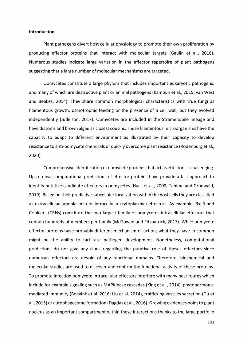
101
Introduction
Plant pathogens divert host cellular physiology to promote their own proliferation by
producing effector proteins that interact with molecular targets (Gaulin et al., 2018).
Numerous studies indicate large variation in the effector repertoire of plant pathogens
suggesting that a large number of molecular mechanisms are targeted.
Oomycetes constitute a large phylum that includes important eukaryotic pathogens,
and many of which are destructive plant or animal pathogens (Kamoun et al., 2015; van West
and Beakes, 2014). They share common morphological characteristics with true fungi as
filamentous growth, osmotrophic feeding or the presence of a cell wall, but they evolved
independently (Judelson, 2017). Oomycetes are included in the Stramenopile lineage and
have diatoms and brown algae as closest cousins. These filamentous microorganisms have the
capacity to adapt to different environment as illustrated by their capacity to develop
resistance to anti-oomycete chemicals or quickly overcome plant resistance (Rodenburg et al.,
2020).
Comprehensive identification of oomycete proteins that act as effectors is challenging.
Up to now, computational predictions of effector proteins have provide a fast approach to
identify putative candidate effectors in oomycetes (Haas et al., 2009; Tabima and Grünwald,
2019). Based on their predictive subcellular localization within the host cells they are classified
as extracellular (apoplasmic) or intracellular (cytoplasmic) effectors. As example, RxLR and
Crinklers (CRNs) constitute the two largest family of oomycetes intracellular effectors that
contain hundreds of members per family (McGowan and Fitzpatrick, 2017). While oomycete
effector proteins have probably different mechanism of action, what they have in common
might be the ability to facilitate pathogen development. Nonetheless, computational
predictions do not give any clues regarding the putative role of theses effectors since
numerous effectors are devoid of any functional domains. Therefore, biochemical and
molecular studies are used to discover and confirm the functional activity of these proteins.
To promote infection oomycete intracellular effectors interfere with many host routes which
include for example signaling such as MAPKinase cascades (King et al., 2014), phytohormone-
mediated immunity (Boevink et al. 2016; Liu et al. 2014), trafficking vesicles secretion (Du et
al., 2015) or autophagosome formation (Dagdas et al., 2016). Growing evidences point to plant
nucleus as an important compartment within these interactions thanks to the large portfolio
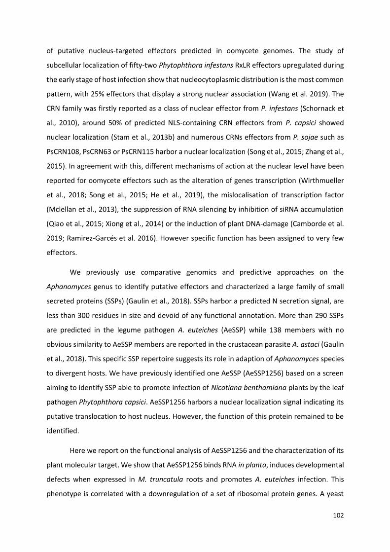
102
of putative nucleus-targeted effectors predicted in oomycete genomes. The study of
subcellular localization of fifty-two Phytophthora infestans RxLR effectors upregulated during
the early stage of host infection show that nucleocytoplasmic distribution is the most common
pattern, with 25% effectors that display a strong nuclear association (Wang et al. 2019). The
CRN family was firstly reported as a class of nuclear effector from P. infestans (Schornack et
al., 2010), around 50% of predicted NLS-containing CRN effectors from P. capsici showed
nuclear localization (Stam et al., 2013b) and numerous CRNs effectors from P. sojae such as
PsCRN108, PsCRN63 or PsCRN115 harbor a nuclear localization (Song et al., 2015; Zhang et al.,
2015). In agreement with this, different mechanisms of action at the nuclear level have been
reported for oomycete effectors such as the alteration of genes transcription (Wirthmueller
et al., 2018; Song et al., 2015; He et al., 2019), the mislocalisation of transcription factor
(Mclellan et al., 2013), the suppression of RNA silencing by inhibition of siRNA accumulation
(Qiao et al., 2015; Xiong et al., 2014) or the induction of plant DNA-damage (Camborde et al.
2019; Ramirez-Garcés et al. 2016). However specific function has been assigned to very few
effectors.
We previously use comparative genomics and predictive approaches on the
Aphanomyces genus to identify putative effectors and characterized a large family of small
secreted proteins (SSPs) (Gaulin et al., 2018). SSPs harbor a predicted N secretion signal, are
less than 300 residues in size and devoid of any functional annotation. More than 290 SSPs
are predicted in the legume pathogen A. euteiches (AeSSP) while 138 members with no
obvious similarity to AeSSP members are reported in the crustacean parasite A. astaci (Gaulin
et al., 2018). This specific SSP repertoire suggests its role in adaption of Aphanomyces species
to divergent hosts. We have previously identified one AeSSP (AeSSP1256) based on a screen
aiming to identify SSP able to promote infection of Nicotiana benthamiana plants by the leaf
pathogen Phytophthora capsici. AeSSP1256 harbors a nuclear localization signal indicating its
putative translocation to host nucleus. However, the function of this protein remained to be
identified.
Here we report on the functional analysis of AeSSP1256 and the characterization of its
plant molecular target. We show that AeSSP1256 binds RNA in planta, induces developmental
defects when expressed in M. truncatula roots and promotes A. euteiches infection. This
phenotype is correlated with a downregulation of a set of ribosomal protein genes. A yeast

103
two hybrid approach identified a host RNA helicase (MtRH10) and a L7 ribosomal protein as
interactors of AeSSP1256. By FRET-FLIM analyses we reveal that AeSSP1256 co-opts MtRH10
to abolish its nucleic acid binding capacity. We provide a mechanistic explanation of this
observation by demonstrating the implication of MtRH10 in roots development by generating
missense and overexpressing Medicago lines. Finally we observed that silenced-MtRH10 roots
are highly susceptible to A. euteiches infection like AeSSP1256-expressing roots, showing that
MtRH10 as AeSSP1256 activities modify the outcome of the infection. We now present results
supporting effector-mediated manipulation of a nuclear RNA helicase as a virulence
mechanism during plant-eukaryotic pathogens interactions.
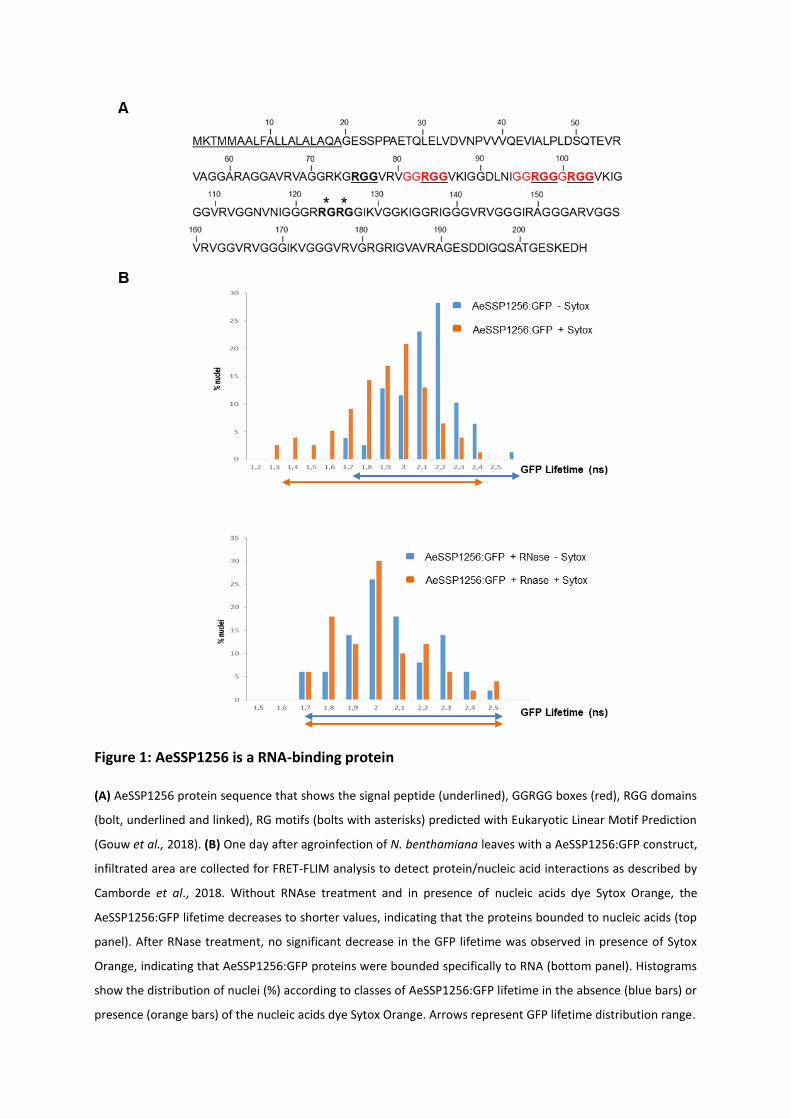
Figure 1: AeSSP1256 is a RNA-binding protein
(A) AeSSP1256 protein sequence that shows the signal peptide (underlined), GGRGG boxes (red), RGG domains
(bolt, underlined and linked), RG motifs (bolts with asterisks) predicted with Eukaryotic Linear Motif Prediction
(Gouw et al., 2018). (B) One day after agroinfection of N. benthamiana leaves with a AeSSP1256:GFP construct,
infiltrated area are collected for FRET-FLIM analysis to detect protein/nucleic acid interactions as described by
Camborde et al., 2018. Without RNAse treatment and in presence of nucleic acids dye Sytox Orange, the
AeSSP1256:GFP lifetime decreases to shorter values, indicating that the proteins bounded to nucleic acids (top
panel). After RNase treatment, no significant decrease in the GFP lifetime was observed in presence of Sytox
Orange, indicating that AeSSP1256:GFP proteins were bounded specifically to RNA (bottom panel). Histograms
show the distribution of nuclei (%) according to classes of AeSSP1256:GFP lifetime in the absence (blue bars) or
presence (orange bars) of the nucleic acids dye Sytox Orange. Arrows represent GFP lifetime distribution range.

104
Results
AeSSP1256 contains RGG/RG domains and binds RNA in planta
AeSSP1256 is a member of a large family of A. euteiches effectors devoid of any predicted
functional domain, except the presence a peptide signal at the Nterminus (Gaulin et al., 2018).
As showed in Figure 1A, AeSSP1256 protein is enriched in glycine (30% of the amino acid
sequence), residues. Analysis using the Eukaryotic Linear Motif database (Gouw et al., 2018)
revealed 3 GGRGG motifs (positions 81-85; 95-99 and 99-103). These motifs are variant
arginine methylation site from arginine-glycine(-glycine) (RGG/RG) domains, presents in many
ribonucleoproteins and involved in RNA binding (Thandapani et al., 2013; Bourgeois et al.,
2020).. We then noticed the presence of two di-RGG domains (RGG(X0-5)RGG) (position 75-85
and 97-103) and one di-RG domains (RG(X0-5)RG) (position 123-126) corresponding to RGG or
RG motifs that are spaced less than 5 residues (Chong et al., 2018). According to RGG/RG
definition, those repeats occur in low-complexity region of the protein (position 60-180)
(Chong et al., 2018) and are associated with di-glycine motifs and GR or GGR sequences (Figure
1A), which are also common in RGG/RG-containing proteins (Chong et al., 2018). Considering
that RGG/RG domains are conserved from yeast to humans (Rajyaguru and Parker, 2012) and
represent the second most common RNA binding domain in the human genome (Ozdilek et
al., 2017), we thereby investigated the RNA binding ability of AeSSP1256.
To test this, we performed a FRET-FLIM assay on N. benthamiana agroinfiltrated leaves with
AeSSP1256:GFP fusion protein in presence or absence of Sytox Orange to check its capacity to
bind nucleic acids (Camborde et al. 2017). Briefly AeSSP1256:GFP construct is transiently
express in N. benthamiana leaves where it accumulates in the nucleus (Gaulin et al., 2018).
Samples are collected 24h after treatment and nucleic acids labeled with the Sytox Orange
dye. In presence of Sytox, if the GFP fusion protein is in close proximity (<10nm) with nucleic
acids, the GFP lifetime of the GFP tagged protein will significantly decrease, due to energy
transfer between the donor (GFP) and the acceptor (Sytox). To distinguish RNA interactions
from DNA interactions, an RNase treatment can be performed. In the case of a specific RNA-
protein interaction, no FRET acceptor will be available due to RNA degradation and the
lifetime of the GFP tagged protein will then return at basal values. It appeared that GFP
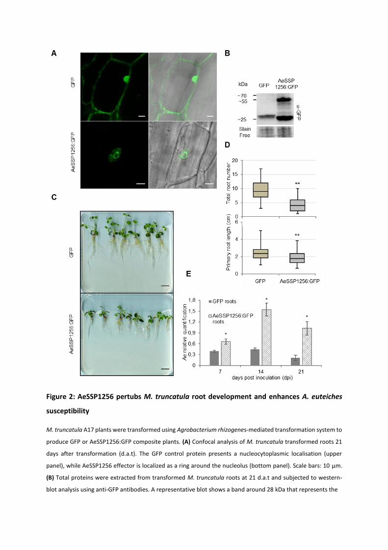
Figure 2: AeSSP1256 pertubs M. truncatula root development and enhances A. euteiches
susceptibility
M. truncatula A17 plants were transformed using Agrobacterium rhizogenes-mediated transformation system to
produce GFP or AeSSP1256:GFP composite plants. (A) Confocal analysis of M. truncatula transformed roots 21
days after transformation (d.a.t). The GFP control protein presents a nucleocytoplasmic localisation (upper
panel), while AeSSP1256 effector is localized as a ring around the nucleolus (bottom panel). Scale bars: 10 µm.
(B) Total proteins were extracted from transformed M. truncatula roots at 21 d.a.t and subjected to western-
blot analysis using anti-GFP antibodies. A representative blot shows a band around 28 kDa that represents the
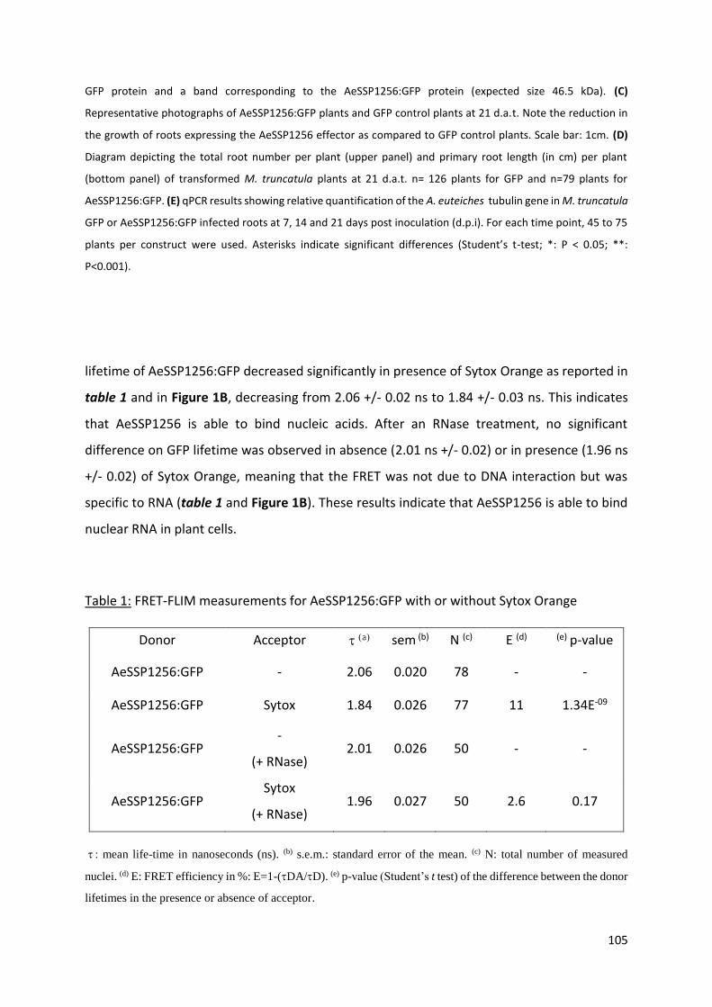
105
GFP protein and a band corresponding to the AeSSP1256:GFP protein (expected size 46.5 kDa). (C)
Representative photographs of AeSSP1256:GFP plants and GFP control plants at 21 d.a.t. Note the reduction in
the growth of roots expressing the AeSSP1256 effector as compared to GFP control plants. Scale bar: 1cm. (D)
Diagram depicting the total root number per plant (upper panel) and primary root length (in cm) per plant
(bottom panel) of transformed M. truncatula plants at 21 d.a.t. n= 126 plants for GFP and n=79 plants for
AeSSP1256:GFP. (E) qPCR results showing relative quantification of the A. euteiches tubulin gene in M. truncatula
GFP or AeSSP1256:GFP infected roots at 7, 14 and 21 days post inoculation (d.p.i). For each time point, 45 to 75
plants per construct were used. Asterisks indicate significant differences (Student’s t-test; *: P < 0.05; **:
P<0.001).
lifetime of AeSSP1256:GFP decreased significantly in presence of Sytox Orange as reported in
table 1 and in Figure 1B, decreasing from 2.06 +/- 0.02 ns to 1.84 +/- 0.03 ns. This indicates
that AeSSP1256 is able to bind nucleic acids. After an RNase treatment, no significant
difference on GFP lifetime was observed in absence (2.01 ns +/- 0.02) or in presence (1.96 ns
+/- 0.02) of Sytox Orange, meaning that the FRET was not due to DNA interaction but was
specific to RNA (table 1 and Figure 1B). These results indicate that AeSSP1256 is able to bind
nuclear RNA in plant cells.
Table 1: FRET-FLIM measurements for AeSSP1256:GFP with or without Sytox Orange
Donor Acceptor a sem (b) N (c) E (d) (e) p-value
AeSSP1256:GFP - 2.06 0.020 78 - -
AeSSP1256:GFP Sytox 1.84 0.026 77 11 1.34E-09
AeSSP1256:GFP -
(+ RNase) 2.01 0.026 50 - -
AeSSP1256:GFP Sytox
(+ RNase) 1.96 0.027 50 2.6 0.17
mean life-time in nanoseconds (ns). (b) s.e.m.: standard error of the mean. (c) N: total number of measured
nuclei. (d) E: FRET efficiency in %: E=1-(DA/D). (e) p-value (Student’s t test) of the difference between the donor
lifetimes in the presence or absence of acceptor.


106
AeSSP1256 impairs M. truncatula root development and susceptibility to A. euteiches
To check whether expression of AeSSP1256 may have an effect on the host plant, we
transformed M. truncatula (Mt) roots, with a native version of GFP tagged AeSSP1256. As
previously observed (Gaulin et al., 2018), confocal analyses confirmed the nuclear localization
of the protein in root cells, with accumulation around the nucleolus as a perinucleolar ring
(Figure 2A) despite the presence of signal peptide (Gaulin et al., 2018). Anti-GFP western blot
analysis on total proteins extracted from transformed roots confirmed the presence of GFP-
tagged AeSSP1256 (46.7 kDa expected size) (Figure 2B). We noticed the presence of a second
band around 28 kDa, which is probably free GFP due to the cleavage of the tagged protein.
AeSSP1256:GFP transformed plants showed delayed development (Figure 2C), with total
number of roots and primary root length per plant being significantly lower than values
obtained with GFP control plants (Figure 2D). As previously observed in N. benthamiana, when
a KDEL-endoplasmic reticulum (ER) retention signal is added to the native AeSSP1256
construct (Gaulin et al., 2018), AeSSP1256:KDEL:GFP proteins mainly accumulates in the ER
(Supplemental Figure 1A-C) and roots showed no significant differences in development as
compared to GFP control roots (Supplemental Figure 1D and E). In contrast a construct devoid
of native signal peptide (SP) shows that the proteins accumulated in root cell nuclei
(Supplemental Figure 1B), leading to abnormal root development, with symptoms similar to
those observed in presence of the AeSSP1256:GFP construct, including shorter primary root
and lower number of roots (Supplemental Figure 1D and E). Altogether these data show that
within the host, AeSSP1256 triggers roots developmental defects thanks to its nuclear
localization.
To investigate whether AeSSP1256 modifies the outcome of the infection, AeSSP1256-
transformed roots were inoculated with A. euteiches zoospores. RT-qPCR analyses at 7, 14 and
21 days post inoculation were performed to follow pathogen development. At each time of
the kinetic, A. euteiches is more abundant in M. truncatula roots expressing the effector than
in GFP control roots (respectively 1.5, 3 and 5 times more) (Figure 2E). This indicates that roots
are more susceptible to A. euteiches in presence of AeSSP1256. Transversal sections of A17-
transformed roots followed by Wheat-Germ-Agglutinin (WGA) staining to detect the presence
of A. euteiches, showed that the pathogen is still restricted to the root cortex either in the
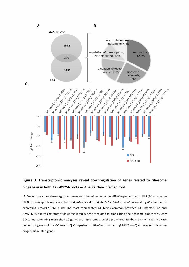
Figure 3: Transcriptomic analyses reveal downregulation of genes related to ribosome
biogenesis in both AeSSP1256 roots or A. euteiches-infected root
(A) Venn diagram on downregulated genes (number of genes) of two RNASeq experiments: F83 (M. truncatula
F83005.5 susceptible roots infected by A.euteiches at 9 dpi), AeSSP1256 (M. truncatula Jemalong A17 transiently
expressing AeSSP1256:GFP). (B) The most represented GO-terms common between F83-infected line and
AeSSP1256-expressing roots of downregulated genes are related to ‘translation and ribosome-biogenesis’. Only
GO terms containing more than 10 genes are represented on the pie chart. Numbers on the graph indicate
percent of genes with a GO term. (C) Comparison of RNASeq (n=4) and qRT-PCR (n=5) on selected ribosome
biogenesis-related genes.
107
presence or absence of AeSSP1256 (Supplemental Figure 2). This phenotype is similar to the
one observed in the natural A17 M. truncatula tolerant line infected by A. euteiches (Djébali
et al., 2009). This data suggests that defence mechanisms like protection of the central
cylinder (Djébali et al., 2009) are still active in AeSSP1256-expressing roots.
AeSSP1256 affects the expression of genes related to ribosome biogenesis
To understand how AeSSP1256 affects M. truncatula roots development and facilitates A.
euteiches infection, we performed expression analyses by RNASeq using AeSSP1256-
expressing roots and GFP controls roots. 4391 genes were differentially express (DE) between
the two conditions (p adjusted-value <10-5) (Supplemental Table 1a). Enrichment analysis of
‘Biological process’ GO-terms showed the presence of ‘ribosome biogenesis’ and
‘organonitrogen compound biosynthetic, cellular amide metabolic’ processes terms among
the most enriched in AeSSP1256 roots as compared to GFP-expressing roots (Supplemental
Table 1b). We noticed that over 90% of DE-genes from ‘ribosome biogenesis’ and ‘translation’
categories are downregulated in AeSSP1256-expressing roots, suggesting that expression of
the effector within the roots affects ribosome biogenesis pathway (Supplemental Table 1a).
To evaluate whether expression of AeSSP1256 mimics infection of M. truncatula by A.
euteiches infection through downregulation of genes related to ribosome biogenesis, we
analyzed RNASeq data previously generated on the susceptible F83005.5 M. truncatula line
nine days after root infection (Gaulin et al., 2018). As shown on the Venn diagram depicting
the M. truncatula downregulated genes in the different conditions (Figure 3A, Supplemental
Table1c), among the 270 common downregulated genes between AeSSP1256-expressing
roots and susceptible F83-infected lines, 58 genes (>20%) are categorized in the ‘ribosome
biogenesis’ and ‘translation’ GO term (Figure 3B). We next selected seventeen M. truncatula
genes to confirm the effect via qRT-PCR. First, we selected ten A. thaliana genes related to
plant developmental control (i,e mutants with shorter roots phenotype) (Supplemental Table
1d) by Blast searches (>80% identity) in A17 line r5.0 genome portal (Pecrix et al., 2018). In
addition, seven nucleolar genes coding for ribosomal and ribonucleotides proteins and related
to the ‘ribosome biogenesis’ in M. truncatula were selected for expression analysis based on
KEGG pathway map (https://www.genome.jp/kegg-bin/show_pathway?ko03008)
(Supplemental Table 1d). As shown on Figure 3C, all of the selected genes from M. truncatula

108
are downregulated in presence of AeSSP1256, supporting the RNAseq data. Altogether, these
expression data show that the effector by itself mimics some effects induced by pathogen
infection of the susceptible F83 line. At this stage of the study, results point to a perturbation
of the ribosome biogenesis pathway of the host plant by the AeSSP1256 effector.
AeSSP1256 targets a DEAD-box RNA helicase and a L7 ribosomal protein
To decipher how AeSSP1256 can affect ribosome biogenesis pathway of the host plant and
knowing that numerous RNA-binding proteins interact with protein partners, we searched for
AeSSP1256 host protein targets. For this, a Yeast two hybrid (Y2H) library composed of cDNA
from M. truncatula roots infected with A. euteiches was screened with the mature form of the
effector. Eight M. truncatula coding genes were identified as potential protein targets
(Supplemental Table 2a), all these genes but one (a lecithin retinol acyltransferase gene)
correspond to putative nuclear proteins in accordance with the observed subcellular
localization of AeSSP1256.
To confirm the Y2H results, we first expressed AeSSP1256 and candidates in N. benthamiana
cells to observe their subcellular localization and performed FRET-FLIM experiments to
validate protein-protein interactions. Only two candidates showed co-localization with
AeSSP1256, a L7 ribosomal protein (RPL7, MtrunA17_Chr4g0002321) and a predicted RNA
helicase (MtrunA17_Chr5g0429221). CFP-tagged version of RPL7 displays a nucleolar
localization, with partial co-localization areas in presence of AeSSP1256 (Supplemental Figure
3, Table 2b). FRET-FLIM measurements confirmed the interaction of RPL7:CFP protein with
AeSSP1256:YFP effector (Supplemental Figure 3, Table 2b), with a mean CFP lifetime of 2.83
ns +/- 0.03 in absence of the SSP protein, leading to 2.46 ns +/- 0.03 in presence of
AeSSP1256:YFP (Supplemental Table 2b).
The second candidate is a predicted DEAD-box ATP-dependent RNA helicase
(MtrunA17_Chr5g0429221), related to the human DDX47 RNA helicase and the RRP3 RH in
yeast. Blast analysis revealed that the closest plant orthologs were AtRH10 in Arabidopsis
thaliana and OsRH10 in Oryza sativa. Consequently the M. truncatula protein target of
AeSSP1256 was named MtRH10. The conserved domains of DEAD-box RNA helicase are
depicted in the alignment of MtRH10 with DDX47, RRP3, AtRH10, OsRH10 proteins
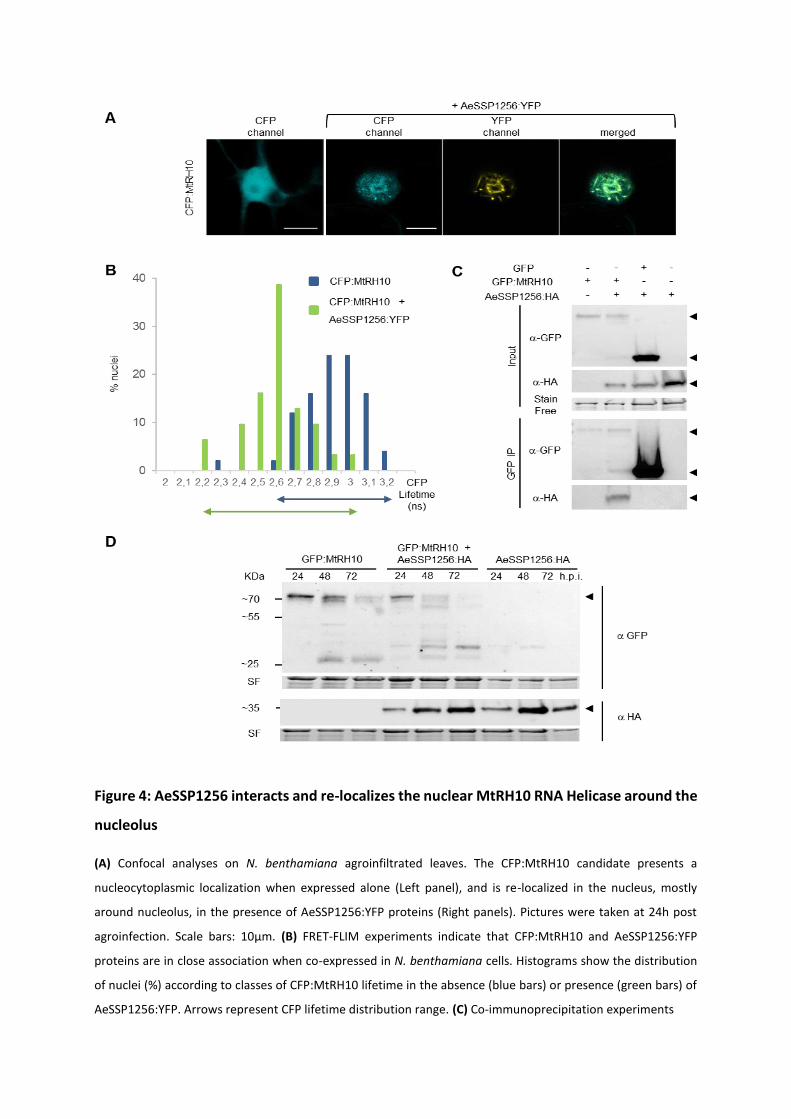
Figure 4: AeSSP1256 interacts and re-localizes the nuclear MtRH10 RNA Helicase around the
nucleolus
(A) Confocal analyses on N. benthamiana agroinfiltrated leaves. The CFP:MtRH10 candidate presents a
nucleocytoplasmic localization when expressed alone (Left panel), and is re-localized in the nucleus, mostly
around nucleolus, in the presence of AeSSP1256:YFP proteins (Right panels). Pictures were taken at 24h post
agroinfection. Scale bars: 10µm. (B) FRET-FLIM experiments indicate that CFP:MtRH10 and AeSSP1256:YFP
proteins are in close association when co-expressed in N. benthamiana cells. Histograms show the distribution
of nuclei (%) according to classes of CFP:MtRH10 lifetime in the absence (blue bars) or presence (green bars) of
AeSSP1256:YFP. Arrows represent CFP lifetime distribution range. (C) Co-immunoprecipitation experiments
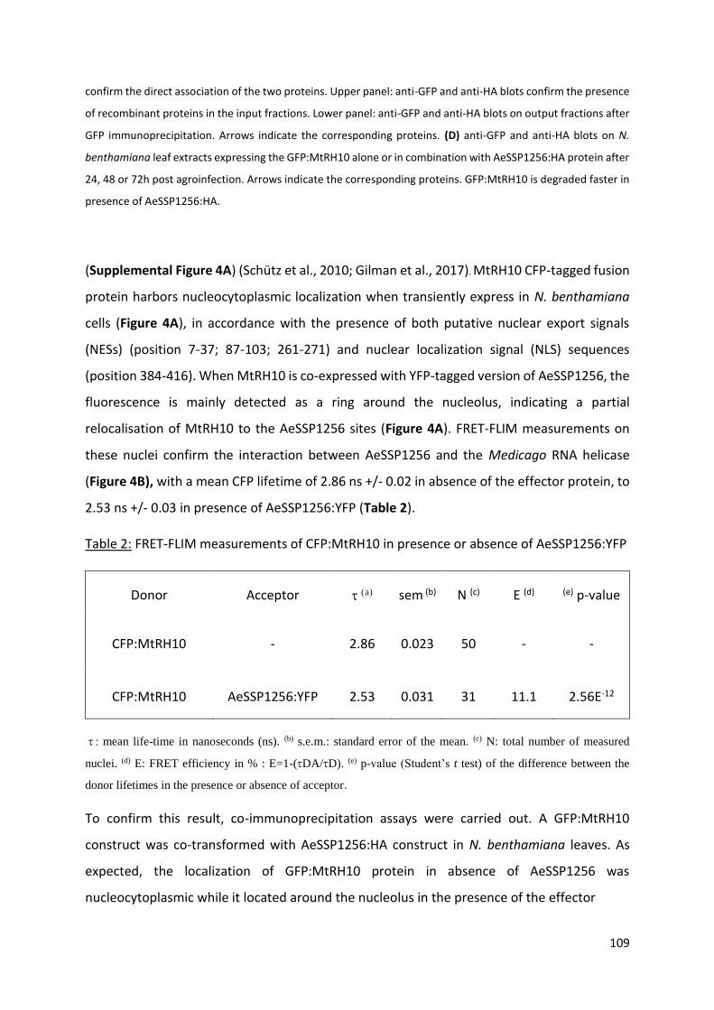
109
confirm the direct association of the two proteins. Upper panel: anti-GFP and anti-HA blots confirm the presence
of recombinant proteins in the input fractions. Lower panel: anti-GFP and anti-HA blots on output fractions after
GFP immunoprecipitation. Arrows indicate the corresponding proteins. (D) anti-GFP and anti-HA blots on N.
benthamiana leaf extracts expressing the GFP:MtRH10 alone or in combination with AeSSP1256:HA protein after
24, 48 or 72h post agroinfection. Arrows indicate the corresponding proteins. GFP:MtRH10 is degraded faster in
presence of AeSSP1256:HA.
(Supplemental Figure 4A) (Schütz et al., 2010; Gilman et al., 2017). MtRH10 CFP-tagged fusion
protein harbors nucleocytoplasmic localization when transiently express in N. benthamiana
cells (Figure 4A), in accordance with the presence of both putative nuclear export signals
(NESs) (position 7-37; 87-103; 261-271) and nuclear localization signal (NLS) sequences
(position 384-416). When MtRH10 is co-expressed with YFP-tagged version of AeSSP1256, the
fluorescence is mainly detected as a ring around the nucleolus, indicating a partial
relocalisation of MtRH10 to the AeSSP1256 sites (Figure 4A). FRET-FLIM measurements on
these nuclei confirm the interaction between AeSSP1256 and the Medicago RNA helicase
(Figure 4B), with a mean CFP lifetime of 2.86 ns +/- 0.02 in absence of the effector protein, to
2.53 ns +/- 0.03 in presence of AeSSP1256:YFP (Table 2).
Table 2: FRET-FLIM measurements of CFP:MtRH10 in presence or absence of AeSSP1256:YFP
Donor Acceptor a sem (b) N (c) E (d) (e) p-value
CFP:MtRH10 - 2.86 0.023 50 - -
CFP:MtRH10 AeSSP1256:YFP 2.53 0.031 31 11.1 2.56E-12
mean life-time in nanoseconds (ns). (b) s.e.m.: standard error of the mean. (c) N: total number of measured
nuclei. (d) E: FRET efficiency in % : E=1-(DA/D). (e) p-value (Student’s t test) of the difference between the
donor lifetimes in the presence or absence of acceptor.
To confirm this result, co-immunoprecipitation assays were carried out. A GFP:MtRH10
construct was co-transformed with AeSSP1256:HA construct in N. benthamiana leaves. As
expected, the localization of GFP:MtRH10 protein in absence of AeSSP1256 was
nucleocytoplasmic while it located around the nucleolus in the presence of the effector

Figure 5: AeSSP1256 inhibits RNA binding activity of MtRH10
(A) FRET-FLIM experiments on N. benthamiana cells expressing GFP:MtRH10 in presence or absence of nucleic
acids dye Sytox Orange. In presence of Sytox Orange, the GFP:MtRH10 lifetime decreases to shorter values,
indicating that the proteins bounded to nucleic acids. (B) In presence of AeSSP1256:HA, when GFP:MtRH10 is re-
localized around the nucleolus and interacts with AeSSP1256, no significant decrease in the GFP lifetime was
observed in presence of Sytox Orange, meaning that the re-localized GFP:MtRH10 proteins were not able to
interact with nucleic acids. Histograms show the distribution of nuclei (%) according to classes of GFP:MtRH10
lifetime in the absence (blue bars) or presence (orange bars) of the nucleic acids dye Sytox Orange. Arrows
represent GFP lifetime distribution range.
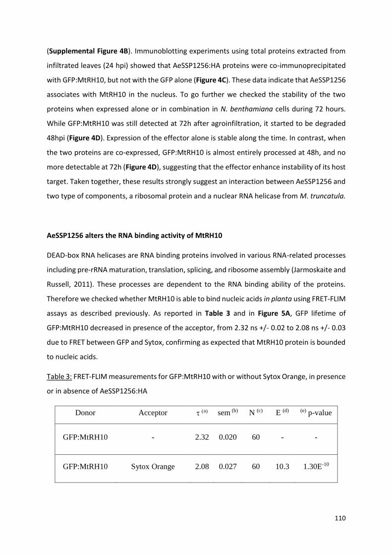
110
(Supplemental Figure 4B). Immunoblotting experiments using total proteins extracted from
infiltrated leaves (24 hpi) showed that AeSSP1256:HA proteins were co-immunoprecipitated
with GFP:MtRH10, but not with the GFP alone (Figure 4C). These data indicate that AeSSP1256
associates with MtRH10 in the nucleus. To go further we checked the stability of the two
proteins when expressed alone or in combination in N. benthamiana cells during 72 hours.
While GFP:MtRH10 was still detected at 72h after agroinfiltration, it started to be degraded
48hpi (Figure 4D). Expression of the effector alone is stable along the time. In contrast, when
the two proteins are co-expressed, GFP:MtRH10 is almost entirely processed at 48h, and no
more detectable at 72h (Figure 4D), suggesting that the effector enhance instability of its host
target. Taken together, these results strongly suggest an interaction between AeSSP1256 and
two type of components, a ribosomal protein and a nuclear RNA helicase from M. truncatula.
AeSSP1256 alters the RNA binding activity of MtRH10
DEAD-box RNA helicases are RNA binding proteins involved in various RNA-related processes
including pre-rRNA maturation, translation, splicing, and ribosome assembly (Jarmoskaite and
Russell, 2011). These processes are dependent to the RNA binding ability of the proteins.
Therefore we checked whether MtRH10 is able to bind nucleic acids in planta using FRET-FLIM
assays as described previously. As reported in Table 3 and in Figure 5A, GFP lifetime of
GFP:MtRH10 decreased in presence of the acceptor, from 2.32 ns +/- 0.02 to 2.08 ns +/- 0.03
due to FRET between GFP and Sytox, confirming as expected that MtRH10 protein is bounded
to nucleic acids.
Table 3: FRET-FLIM measurements for GFP:MtRH10 with or without Sytox Orange, in presence
or in absence of AeSSP1256:HA
Donor Acceptor a sem (b) N (c) E (d) (e) p-value
GFP:MtRH10 - 2.32 0.020 60 - -
GFP:MtRH10 Sytox Orange 2.08 0.027 60 10.3 1.30E-10

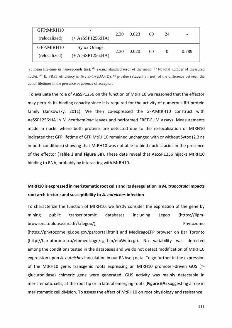
111
GFP:MtRH10
(relocalized)
-
(+ AeSSP1256:HA) 2.30 0.023 60 24 -
GFP:MtRH10
(relocalized)
Sytox Orange
(+ AeSSP1256:HA) 2.30 0.020 60 0 0.789
mean life-time in nanoseconds (ns). (b) s.e.m.: standard error of the mean. (c) N: total number of measured
nuclei. (d) E: FRET efficiency in % : E=1-(DA/D). (e) p-value (Student’s t test) of the difference between the
donor lifetimes in the presence or absence of acceptor.
To evaluate the role of AeSSP1256 on the function of MtRH10 we reasoned that the effector
may perturb its binding capacity since it is required for the activity of numerous RH protein
family (Jankowsky, 2011). We then co-expressed the GFP:MtRH10 construct with
AeSSP1256:HA in N. benthamiana leaves and performed FRET-FLIM assays. Measurements
made in nuclei where both proteins are detected due to the re-localization of MtRH10
indicated that GFP lifetime of GFP:MtRH10 remained unchanged with or without Sytox (2.3 ns
in both conditions) showing that MtRH10 was not able to bind nucleic acids in the presence
of the effector (Table 3 and Figure 5B). These data reveal that AeSSP1256 hijacks MtRH10
binding to RNA, probably by interacting with MtRH10.
MtRH10 is expressed in meristematic root cells and its deregulation in M. truncatula impacts
root architecture and susceptibility to A. euteiches infection
To characterize the function of MtRH10, we firstly consider the expression of the gene by
mining public transcriptomic databases including Legoo (https://lipm-
browsers.toulouse.inra.fr/k/legoo/), Phytozome
(https://phytozome.jgi.doe.gov/pz/portal.html) and MedicagoEFP browser on Bar Toronto
(http://bar.utoronto.ca/efpmedicago/cgi-bin/efpWeb.cgi). No variability was detected
among the conditions tested in the databases and we do not detect modification of MtRH10
expression upon A. euteiches inoculation in our RNAseq data. To go further in the expression
of the MtRH10 gene, transgenic roots expressing an MtRH10 promoter-driven GUS ( -
glucuronidase) chimeric gene were generated. GUS activity was mainly detectable in
meristematic cells, at the root tip or in lateral emerging roots (Figure 6A) suggesting a role in
meristematic cell division. To assess the effect of MtRH10 on root physiology and resistance

Figure 6: MtRH10 is expressed in meristematic cells of Medicago truncatula and its
deregulation impacts root architecture
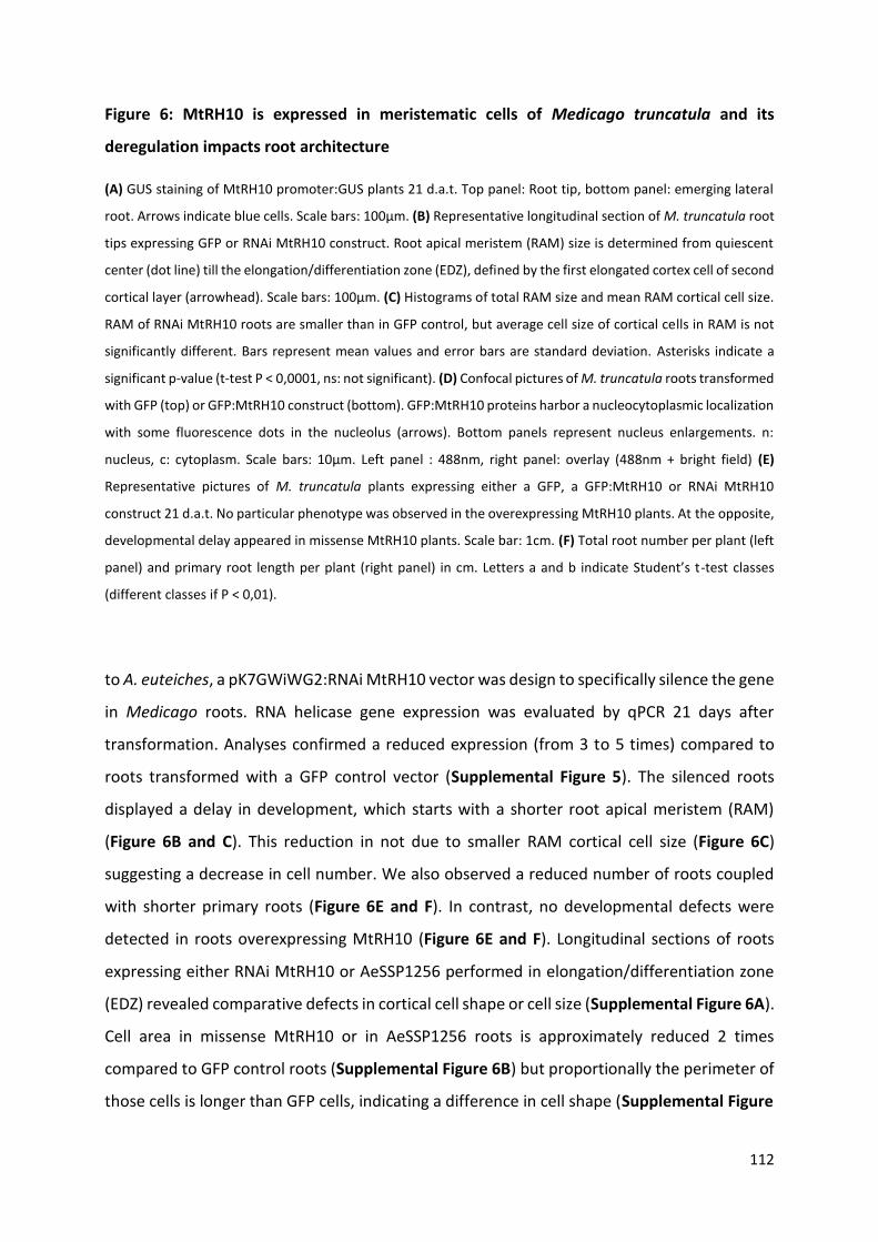
112
Figure 6: MtRH10 is expressed in meristematic cells of Medicago truncatula and its
deregulation impacts root architecture
(A) GUS staining of MtRH10 promoter:GUS plants 21 d.a.t. Top panel: Root tip, bottom panel: emerging lateral
root. Arrows indicate blue cells. Scale bars: 100µm. (B) Representative longitudinal section of M. truncatula root
tips expressing GFP or RNAi MtRH10 construct. Root apical meristem (RAM) size is determined from quiescent
center (dot line) till the elongation/differentiation zone (EDZ), defined by the first elongated cortex cell of second
cortical layer (arrowhead). Scale bars: 100µm. (C) Histograms of total RAM size and mean RAM cortical cell size.
RAM of RNAi MtRH10 roots are smaller than in GFP control, but average cell size of cortical cells in RAM is not
significantly different. Bars represent mean values and error bars are standard deviation. Asterisks indicate a
significant p-value (t-test P < 0,0001, ns: not significant). (D) Confocal pictures of M. truncatula roots transformed
with GFP (top) or GFP:MtRH10 construct (bottom). GFP:MtRH10 proteins harbor a nucleocytoplasmic localization
with some fluorescence dots in the nucleolus (arrows). Bottom panels represent nucleus enlargements. n:
nucleus, c: cytoplasm. Scale bars: 10µm. Left panel : 488nm, right panel: overlay (488nm + bright field) (E)
Representative pictures of M. truncatula plants expressing either a GFP, a GFP:MtRH10 or RNAi MtRH10
construct 21 d.a.t. No particular phenotype was observed in the overexpressing MtRH10 plants. At the opposite,
developmental delay appeared in missense MtRH10 plants. Scale bar: 1cm. (F) Total root number per plant (left
panel) and primary root length per plant (right panel) in cm. Letters a and b indicate Student’s t-test classes
(different classes if P < 0,01).
to A. euteiches, a pK7GWiWG2:RNAi MtRH10 vector was design to specifically silence the gene
in Medicago roots. RNA helicase gene expression was evaluated by qPCR 21 days after
transformation. Analyses confirmed a reduced expression (from 3 to 5 times) compared to
roots transformed with a GFP control vector (Supplemental Figure 5). The silenced roots
displayed a delay in development, which starts with a shorter root apical meristem (RAM)
(Figure 6B and C). This reduction in not due to smaller RAM cortical cell size (Figure 6C)
suggesting a decrease in cell number. We also observed a reduced number of roots coupled
with shorter primary roots (Figure 6E and F). In contrast, no developmental defects were
detected in roots overexpressing MtRH10 (Figure 6E and F). Longitudinal sections of roots
expressing either RNAi MtRH10 or AeSSP1256 performed in elongation/differentiation zone
(EDZ) revealed comparative defects in cortical cell shape or cell size (Supplemental Figure 6A).
Cell area in missense MtRH10 or in AeSSP1256 roots is approximately reduced 2 times
compared to GFP control roots (Supplemental Figure 6B) but proportionally the perimeter of
those cells is longer than GFP cells, indicating a difference in cell shape (Supplemental Figure
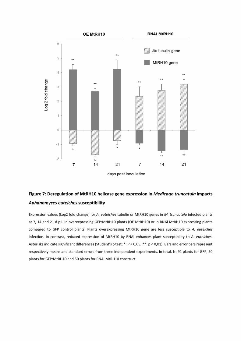
Figure 7: Deregulation of MtRH10 helicase gene expression in Medicago truncatula impacts
Aphanomyces euteiches susceptibility
Expression values (Log2 fold change) for A. euteiches tubulin or MtRH10 genes in M. truncatula infected plants
at 7, 14 and 21 d.p.i. in overexpressing GFP:MtRH10 plants (OE MtRH10) or in RNAi MtRH10 expressing plants
compared to GFP control plants. Plants overexpressing MtRH10 gene are less susceptible to A. euteiches
infection. In contrast, reduced expression of MtRH10 by RNAi enhances plant susceptibility to A. euteiches.
Asterisks indicate significant differences (Student’s t-test; *: P < 0,05, **: p < 0,01). Bars and error bars represent
respectively means and standard errors from three independent experiments. In total, N: 91 plants for GFP, 50
plants for GFP:MtRH10 and 50 plants for RNAi MtRH10 construct.
113
6C). We noticed that most of EDZ cells in GFP roots present a rectangular shape, which seem
impaired in missense MtRH10 and AeSSP1256 expressing roots. Thus we measured the
perimeter-bounding rectangle (PBR) which calculates the smallest rectangle possible to draw
with a given cell. A perimeter/PBR ratio of 1 indicates that the cell is rectangular. As presented
in Supplemental Figure 6D, the perimeter/PBR ratio in GFP roots is close to 1 and significantly
different than those observed in RNAi MtRH10 and AeSSP1256 roots. This analysis reveals that
the reduction of MtRH10 expression or the expression of the effector AeSSP1256 in Medicago
roots, impairs the cortical cell shape. The similar phenotypic changes observed on MtRH10-
silenced roots and AeSSP1256-expressing roots, suggest that the effector may affect MtRH10
activity in cell division regions.
Having shown that MtRH10 is implicated in M. truncatula roots development, we test whether
this biological function is related to pathogen colonisation. We therefore investigate by qPCR
the presence of A. euteiches in silenced and overexpressed MtRH10 roots infected by the
pathogen. As shown on Figure 7, overexpression of MtRH10 reduce the amount of mycelium
in roots after 7, 14 and 21 dpi (1.8, 3.3 and 1.6 times less, respectively). We note by western-
blot analyses a slight decrease in MtRH10 amount upon the time probably due to the
accumulation of the AeSSP1256 effector (Supplemental Figure 7). As expected in roots where
MtRH10 is silenced to 2 to 3 times as compared to GFP control roots, qPCR analyses revealed
approximately 5 to 10 times more of the pathogen at 7, 14 and 21 dpi (Figure 7). Taken
together these infection assays show that MtRH10 is involved in conferring basal resistance
to A. euteiches at the root level.
114
Discussion
Protein effectors from filamentous plant pathogens such as fungi and oomycetes facilitate
host colonization by targeting host components. However, the molecular mechanisms that
enhance plant susceptibility to the pathogen are still poorly understood. Here we report that
the A. euteiches AeSSP1256 RNA-binding effector facilitate host infection by downregulating
expression of plant ribosome-related genes and by hijacking from its nucleic target MtRH10,
a Medicago nuclear RNA-helicase (RH). Thus, the current study unravels a new strategy in
which pathogenic oomycete triggers plant nucleolar stress to promote infection.
AeSSP1256 is an effector from the oomycete root pathogen A. euteiches previously shown to
enhance oomycete infection (Gaulin et al., 2018). Despite the absence of any functional
domain, in silico RGG/RG RNA-binding motif prediction (see for review (Thandapani et al.,
2013)) prompt us to show by FRET/FLIM analysis that the secreted AeSSP1256 effector is an
RNA-binding protein (RBP). RNAs play essential role in cell physiology and it is not surprising
that filamentous plant pathogens may rely on RNA-dependent process to control host
infection (for review see (Göhre et al., 2013; Pedersen et al., 2012). Moreover RBPs are key
players in the regulation of the post-transcriptional processing and transport of RNA
molecules (Yang et al., 2018b). However, to our knowledge only three examples of RBPs acting
as virulence factor of plant pathogens are known. This includes the glycine-rich protein
MoGrp1 from the rice pathogen Magnaporthe oryzae (Gao et al., 2019), the UmRrm75 of
Ustilago maydis (Rodríguez-Kessler et al., 2012) and the secreted ribonuclease effector
CSEP0064/BEC1054 of the fungal pathogen Blumeria graminis which probably interferes with
degradation of host ribosomal RNA (Pennington et al., 2019). This situation is probably due to
the absence of conventional RNA-binding domain which render this type of RBP undetectable
by prediction algorithms. The future studies that will aim to unravel the atlas of RNA-binding
effectors in phytopathogens should not only rely on computational analysis but will have to
use functional approaches such as crystallization of the protein to validate function as
performed with CSEP0064/BEC1054 effector (Pennington et al., 2019) or screening method
like the RNA interactome capture (RIC) assay develops in mammals (Castello et al., 2012).
115
We observed that when expressed inside roots of the partially resistant Jemalong A17 M.
truncatula line, AeSSP1256 triggers developmental defects such as shorter primary roots and
delay in roots development. In addition, those composite plants promote infection of A.
euteiches. This modification in the output of the infection is highly relevant since we previously
observed that M. truncatula quantitative resistance to A. euteiches is correlated to the
development of secondary roots (Rey et al., 2016). Defects in roots development and retarded
growth are typical characteristics of auxin-related and ribosomal proteins mutants reported
in Arabidopsis (Ohbayashi et al., 2017; Wieckowski and Schiefelbein, 2012).
This activity is dependent on the nucleolar rim localization of AeSSP1256, closed to the
nucleolus. The nucleolus is a membrane-free subnuclear compartment essential for the highly
complex process of ribosome biogenesis (reviewed in (Shaw and Brown, 2012). Ribosome
biogenesis is linked to cell growth and required coordinated production of processed
ribosomal RNA (rRNA), ribosomal biogenesis factors and ribosomal proteins (RP). In the
nucleolus, ribosome biogenesis starts with the transcription of pre-rRNAs from rRNA genes,
followed by their processing and assembly with RPs into two ribosome subunits (ie small and
large subunit). In animals, perturbation of any steps of ribosome biogenesis in the nucleolus
can cause a nucleolar stress or ribosomal stress which stimulates specific signaling pathway
leading for example to arrest of cell growth (Pfister, 2019). The nucleolar rim localization of
AeSSP1256 within the host cells suggested that this effector could interfere with ribosome
biogenesis pathway to facilitate infection. This speculation was further strengthened by
RNAseq experiments, which showed that within A17-roots, AeSSP1256 downregulated
numerous genes implicated in ribosome biogenesis pathway, notably ribosomal protein
genes. This effect was also detected in susceptible F83 M. truncatula lines infected by A.
euteiches indicating that AeSSP1256, mimics some A.euteiches effects during roots invasion.
Y2H approach led to the identification of putative AeSSP1256 plant targets and all but one
correspond to predicted nuclear M. truncatula proteins. By a combination of multiple
experiments as FRET-FLIM to detect protein/protein interactions, a L7 ribosomal protein
(MtrunA17_Chr4g0002321) and a DExD/H box RNA helicase ATP-dependent
(MtrunA17_Chr5g0429221) were confirmed as AeSSP1256-interacting proteins. The DExD/H
(where x can be any amino acid) box protein family include the largest family of RNA-helicase
(RH). Rather than being processive RH, several DExD/H box proteins may act as ‘RNA
116
chaperone’ promoting the formation of optimal RNA structures by unwinding locally the RNA
(for review see (Fuller-Pace, 2006)). These proteins are of major interest due to their
participation to all the aspects of RNA processes such as RNA export and translation, splicing
but the most common function of these proteins is in ribosome biogenesis including assembly
(Jarmoskaite and Russell, 2011). Specific function of RH is probably due to the presence of a
variable C-terminal ‘DEAD’ domain in contrast to the well conserved N-terminal ‘helicase core’
domain (for review see (Fuller-Pace, 2006)). This structural organization was detected in the
MtRH10. This M. truncatula protein corresponds to the ortholog of the nucleolar human
DDX47 (Sekiguchi et al., 2006), the nuclear yeast RRP3 (O’Day, 1996) and the nucleolar
Arabidopsis AtRH10 RNA-helicases, all involved in ribosome biogenesis (Liu and Imai 2018;
Matsumura et al. 2016), and the nucleolar rice OsRH10 (TOGR1) involved in rRNA homeostasis
(Wang et al. 2016).
Like its human ortholog DDX47 (Sekiguchi et al., 2006), MtRH10 possesses a bipartite nuclear
transport domain which can function as a nuclear localization signal (NLS) and two nuclear
export signal (NES), and thereby it probably shuttles between the cytoplasm and the nucleus
as reported for many others RNA helicases involved in rRNA biogenesis and splicing function
(Sekiguchi et al. 2006; Wang et al. 2009). Fluorescence analysis showed a relocalization of the
nucleocytoplasmic MtRH10 in the nucleoli periphery, when it is transiently co-express with
AeSSP1256 in N. benthamiana cells. The change in MtRH10 distribution suggests that the
interaction between the two proteins caused a mislocation of MtRH10 that can probably
affect its activity. We thereby check the nucleic acid binding capacity of MtRH10 by FRET-FLIM
approach. The decrease in the lifetime of GFP revealed the ability of MtRH10 to bind nucleic
acids. Knowing that both proteins display the same properties, we further evidenced that the
presence of AeSSP1256 effector inhibits the nucleic binding capacity of MtRH10. This
mechanism was also reported for the RNA-binding HopU1 effector from the plant bacterial
pathogen Pseudomonas syringae which associate to the glycin-rich RNA binding 7 protein
(GRP7) of Arabidopsis to abolish GRP7 binding to immune gene transcripts (ie FLS2 receptor,
(Nicaise et al., 2013)). Here we cannot exclude that AeSSP1256 also blocks the putative
helicase activity of MtRH10, but we favored an inhibitory mechanism of AeSSP1256 on
MtRH10 activity as complex and at least in part due to both protein-protein interaction and
nucleic acid interaction with the two proteins. Interestingly, we also noticed that co-
117
expression of both proteins led to decrease in MtRH10 probably due to degradation of the
protein. While this observation warrants further analyses, this effect is reminiscent of other
effector activities which destabilize their targets (for review see (Langin et al., 2020)).
Plant genomes encode a large variety of DExD/H RH family in comparison to other organisms
and numerous studies have shown that several are associated through their activity with plant
development, hormone signaling or responses to abiotic stresses (for review see (Liu and Imai
2018)). Very few studies reported that DExD/H RH could also be involved in biotic stresses,
like responses to pathogens. One example is the DExD/H RH OsBIRH1 from rice that enhanced
disease resistance against Alternaria brassicicola and Pseudomonas syringae through
activation of defense-related genes (Li et al. 2008). A recent study on oomycete reports the
binding of the Phytophthora sojae RxLR PSR1 effector to a putative nuclear DExD/H RH.
Although the affinity for nucleic acids was not evaluated for the RH, association of both
partners promote pathogen infection by suppressing small RNA biogenesis of the plant (Qiao
et al., 2015). Here we showed that MtRH10 knockdown tolerant A17 lines supported higher-
level accumulation of A. euteiches in contrast to overexpressed MtRH10 lines, indicating the
importance of MtRH10 for M. truncatula roots defense against soil-borne pathogens.
This works reveals that MtRH10 expression is restricted at the root apical meristematic zone
(RAM) where cells divide (ie, primary and lateral roots). Missense MtRH10 roots harbor
defects in the primary root growth and reduced number of roots. Longitudinal sections in
elongation zone (EDZ) of these composite roots show a significant reduction in the size and
shape modification of cortical cells indicating that MtRH10 is required for normal cell division.
Defect in primary roots elongation is also detected in silenced AtRH10 and OsRH10 mutant
(Matsumura et al. 2016; Wang et al. 2016). Thus MtRH10 plays a role on Medicago root
development as its orthologs OsRH10 and AtRH10. At the cellular level we also observed in
AeSSP1256-expressing roots, reduction in cell size in elongation zone, with defects in cell
shape and in adhesion between cells of the cortex, maybe due to a modification of the middle
lamella (Zamil and Geitmann, 2017). Thus AeSSP1256 triggers similar or enhanced effect on
host roots development as the one detected in defective MtRH10 composite plants,
supporting the concept that the activity of the effector on MtRH10 consequently leads to
developmental roots defects. Several reports have indicated that Arabidopsis knockout of
genes involved in rRNA biogenesis or in ribosome assembly cause abnormal plant
118
development including restriction and retardation in roots growth (Ohtani et al., 2013; Huang
et al., 2016, 2010). These common features suggest the existence of a common mechanism
that regulate growth in response to insults of the ribosome biogenesis pathway, known as
nucleolar stress response (for review see (Ohbayashi and Sugiyama, 2018)). How plant cells
sense perturbed ribosome biogenesis and nucleolar problems is still an opening question
(Sáez-Vásquez and Delseny, 2019), but the ANAC082 transcription factor from Arabidopsis can
be a ribosomal stress response mediator (Ohbayashi et al., 2017). In addition the recent report
on the activity of the nucleolar OsRH10 (TOGR1, MtRH10 ortholog) implicated in plant primary
metabolism through is activity on rRNA biogenesis, suggests that metabolites may play a role
in this process. Finally our current study indicates that nuclear RNA-binding effector like
AeSSP1256, by interacting with MtRH10, can act as a stimulus of the ribosomal stress
response.
This work established a connection between the ribosome biogenesis pathway, a nuclear
DExD/H RH, root development and resistance against oomycetes. Our data document that the
RNA binding AeSSP1256 oomycete effector that the parasite expresses during infection
downregulated expression of ribosome-related genes and hijacked MtRH10, a nuclear DExD/H
RH involved in root development, to promote host infection. This work not only provides
insights into plant-root oomycete interactions but also reveals the requirement of fine-tuning
of plant ribosome biogenesis pathways for infection success.
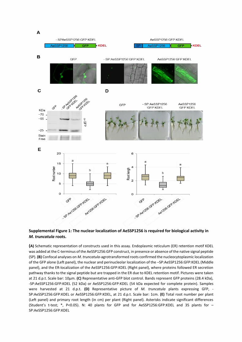
Supplemental Figure 1: The nuclear localization of AeSSP1256 is required for biological activity in
M. truncatula roots.
(A) Schematic representation of constructs used in this assay. Endoplasmic reticulum (ER) retention motif KDEL
was added at the C-terminus of the AeSSP1256:GFP construct, in presence or absence of the native signal peptide
(SP). (B) Confocal analyses on M. truncatula-agrotransformed roots confirmed the nucleocytoplasmic localization
of the GFP alone (Left panel), the nuclear and perinucleolar localization of the –SP:AeSSP1256:GFP:KDEL (Middle
panel), and the ER-localization of the AeSSP1256:GFP:KDEL (Right panel), where proteins followed ER secretion
pathway thanks to the signal peptide but are trapped in the ER due to KDEL retention motif. Pictures were taken
at 21 d.p.t. Scale bar: 10µm. (C) Representative anti-GFP blot control. Bands represent GFP proteins (28.4 kDa),
-SP:AeSSP1256:GFP:KDEL (52 kDa) or AeSSP1256:GFP:KDEL (54 kDa expected for complete protein). Samples
were harvested at 21 d.p.t. (D) Representative picture of M. truncatula plants expressing GFP, -
SP:AeSSP1256:GFP:KDEL or AeSSP1256:GFP:KDEL, at 21 d.p.t. Scale bar: 1cm. (E) Total root number per plant
(Left panel) and primary root length (in cm) per plant (Right panel). Asterisks indicate significant differences
(Student’s t-test; *, P<0.05). N: 40 plants for GFP and for AeSSP1256:GFP:KDEL and 35 plants for –
SP:AeSSP1256:GFP:KDEL

Supplemental Figure 2: Invasion of M. truncatula roots by the pathogen is unchanged in
AeSSP1256 effector-expressing roots.
Cross-section of tolerant A17 M. truncatula lines expressing either the GFP control vector (left) or the
effector AeSSP1256:GFP (right) construct and infected by A. euteiches, 21 days after infection.
Mycelium was stained by Wheat Germ Agglutinin (Red) assay. Green fluorescence indicate GFP alone
or the GFP-tagged effector. Accumulation of phenolic compounds due to the presence of the pathogen
is visualized in blue (Djebali et al., 2009). No notable modification in the infection process is detected
and the pathogen is still restricted to the root cortex in the effector-transformed roots as in wild type
tolerant A17 line. CC: cortical cells. Scale bars: 100 µm
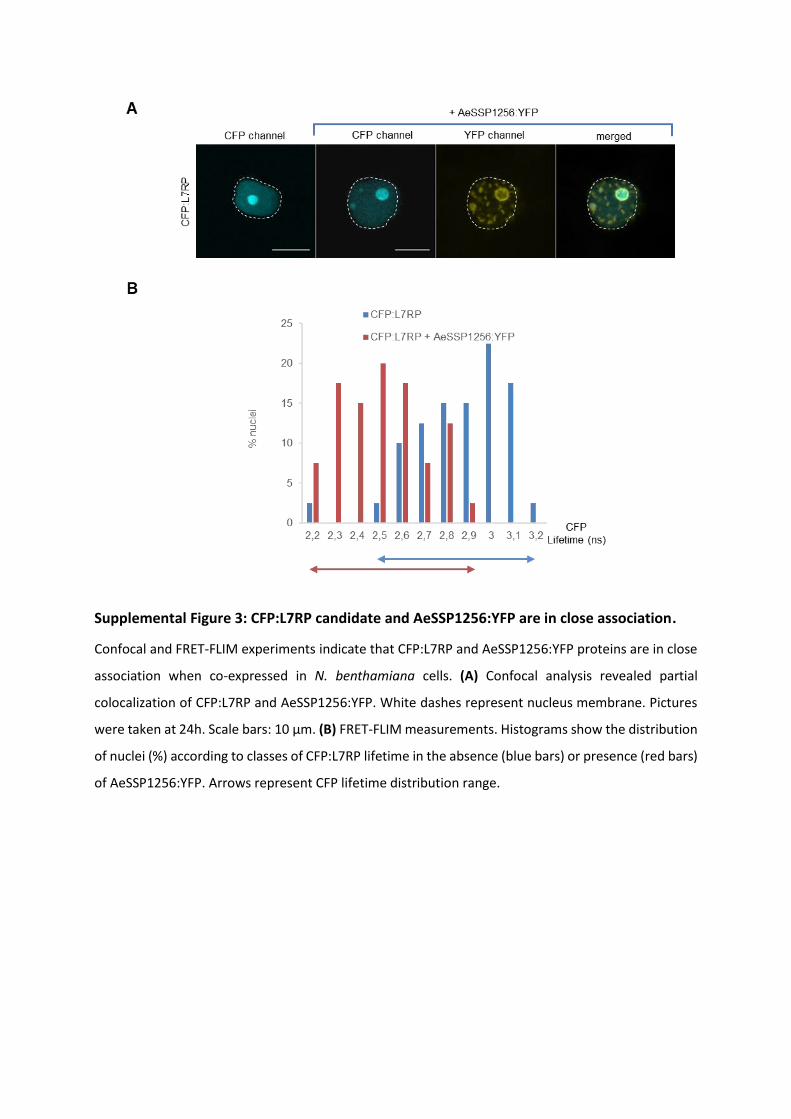
Supplemental Figure 3: CFP:L7RP candidate and AeSSP1256:YFP are in close association.
Confocal and FRET-FLIM experiments indicate that CFP:L7RP and AeSSP1256:YFP proteins are in close
association when co-expressed in N. benthamiana cells. (A) Confocal analysis revealed partial
colocalization of CFP:L7RP and AeSSP1256:YFP. White dashes represent nucleus membrane. Pictures
were taken at 24h. Scale bars: 10 µm. (B) FRET-FLIM measurements. Histograms show the distribution
of nuclei (%) according to classes of CFP:L7RP lifetime in the absence (blue bars) or presence (red bars)
of AeSSP1256:YFP. Arrows represent CFP lifetime distribution range.
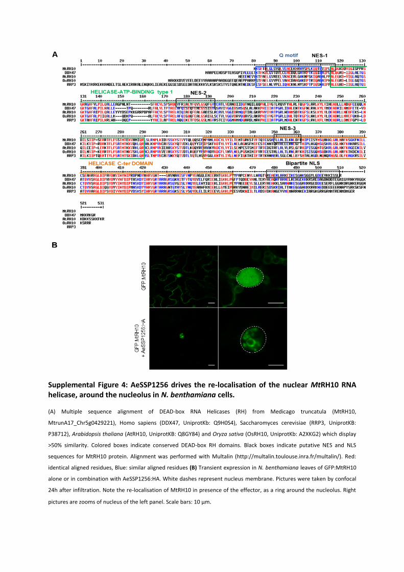
Supplemental Figure 4: AeSSP1256 drives the re-localisation of the nuclear MtRH10 RNA helicase, around the nucleolus in N. benthamiana cells.
(A) Multiple sequence alignment of DEAD-box RNA Helicases (RH) from Medicago truncatula (MtRH10,
MtrunA17_Chr5g0429221), Homo sapiens (DDX47, UniprotKb: Q9H0S4), Saccharomyces cerevisiae (RRP3, UniprotKB:
P38712), Arabidopsis thaliana (AtRH10, UniprotKB: Q8GY84) and Oryza sativa (OsRH10, UniprotKb: A2XKG2) which display
>50% similarity. Colored boxes indicate conserved DEAD-box RH domains. Black boxes indicate putative NES and NLS
sequences for MtRH10 protein. Alignment was performed with Multalin (http://multalin.toulouse.inra.fr/multalin/). Red:
identical aligned residues, Blue: similar aligned residues (B) Transient expression in N. benthamiana leaves of GFP:MtRH10
alone or in combination with AeSSP1256:HA. White dashes represent nucleus membrane. Pictures were taken by confocal
24h after infiltration. Note the re-localisation of MtRH10 in presence of the effector, as a ring around the nucleolus. Right
pictures are zooms of nucleus of the left panel. Scale bars: 10 µm.
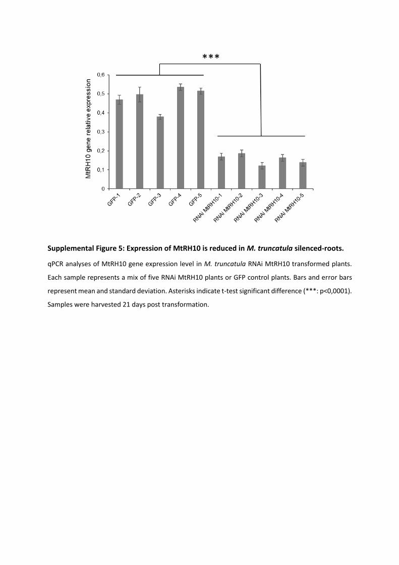
Supplemental Figure 5: Expression of MtRH10 is reduced in M. truncatula silenced-roots.
qPCR analyses of MtRH10 gene expression level in M. truncatula RNAi MtRH10 transformed plants.
Each sample represents a mix of five RNAi MtRH10 plants or GFP control plants. Bars and error bars
represent mean and standard deviation. Asterisks indicate t-test significant difference (***: p<0,0001).
Samples were harvested 21 days post transformation.
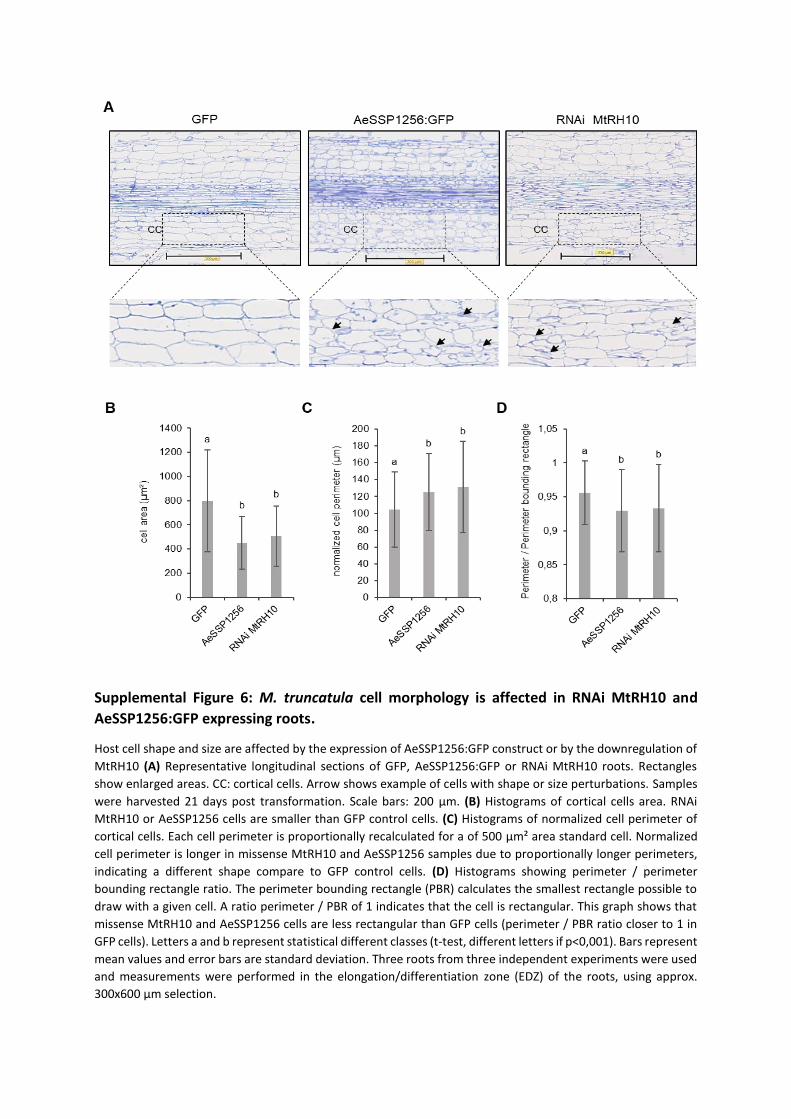
Supplemental Figure 6: M. truncatula cell morphology is affected in RNAi MtRH10 and
AeSSP1256:GFP expressing roots.
Host cell shape and size are affected by the expression of AeSSP1256:GFP construct or by the downregulation of
MtRH10 (A) Representative longitudinal sections of GFP, AeSSP1256:GFP or RNAi MtRH10 roots. Rectangles
show enlarged areas. CC: cortical cells. Arrow shows example of cells with shape or size perturbations. Samples
were harvested 21 days post transformation. Scale bars: 200 µm. (B) Histograms of cortical cells area. RNAi
MtRH10 or AeSSP1256 cells are smaller than GFP control cells. (C) Histograms of normalized cell perimeter of
cortical cells. Each cell perimeter is proportionally recalculated for a of 500 µm² area standard cell. Normalized
cell perimeter is longer in missense MtRH10 and AeSSP1256 samples due to proportionally longer perimeters,
indicating a different shape compare to GFP control cells. (D) Histograms showing perimeter / perimeter
bounding rectangle ratio. The perimeter bounding rectangle (PBR) calculates the smallest rectangle possible to
draw with a given cell. A ratio perimeter / PBR of 1 indicates that the cell is rectangular. This graph shows that
missense MtRH10 and AeSSP1256 cells are less rectangular than GFP cells (perimeter / PBR ratio closer to 1 in
GFP cells). Letters a and b represent statistical different classes (t-test, different letters if p<0,001). Bars represent
mean values and error bars are standard deviation. Three roots from three independent experiments were used
and measurements were performed in the elongation/differentiation zone (EDZ) of the roots, using approx.
300x600 µm selection.
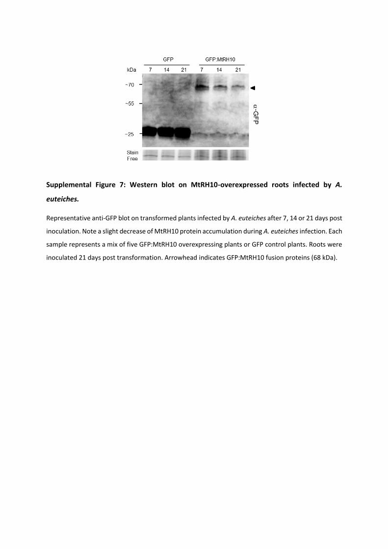
Supplemental Figure 7: Western blot on MtRH10-overexpressed roots infected by A.
euteiches.
Representative anti-GFP blot on transformed plants infected by A. euteiches after 7, 14 or 21 days post
inoculation. Note a slight decrease of MtRH10 protein accumulation during A. euteiches infection. Each
sample represents a mix of five GFP:MtRH10 overexpressing plants or GFP control plants. Roots were
inoculated 21 days post transformation. Arrowhead indicates GFP:MtRH10 fusion proteins (68 kDa).
119
Material and Methods
Plant material, microbial strains, and growth conditions
M. truncatula A17 seeds were in vitro-cultured and transformed as previously described
(Boisson-Dernier et al., 2001; Djébali et al., 2009). A. euteiches (ATCC 201684) zoospore
inoculum were prepared as in (Badreddine et al., 2008). For root infections, each plant was
inoculated with a total of 10 µl of zoospores suspension at 105 cells.ml-1. Plates were placed
in growth chambers with a 16h/8h light/dark and 22/20°C temperature regime. N.
benthamiana plants were grown from seeds in growth chambers at 70% of humidity with a
16h/8h light/dark and 24/20°C temperature regime. E.coli strains (DH5α, DB3.5), A.
tumefaciens (GV3101::pMP90) and A. rhizogenes (ARQUA-1) strains were grown on LB
medium with the appropriate antibiotics.
Construction of plasmid vectors and Agrobacterium-mediated transformation
GFP control plasmid (pK7WGF2), +SPAeSSP1256:GFP and +SPAeSSP1256:YFP (named
AeSSP1256:GFP and AeSSP1256:YFP in this study for convenience) and minus or plus signal
peptide AeSSP1256:GFP:KDEL constructs were described in (Gaulin et al., 2018). Primers used
in this study are listed in Supplemental Table 3. M. truncatula candidates sorted by Y2H assay
(MtrunA17_Chr7g0275931, MtrunA17_Chr2g0330141, MtrunA17_Chr5g0407561,
MtrunA17_Chr5g0429221, MtrunA17_Chr1g0154251, MtrunA17_Chr3g0107021,
MtrunA17_Chr7g0221561, MtrunA17_Chr4g0002321) were amplified by Pfx Accuprime
polymerase (Thermo Fisher; 12344024) and introduced in pENTR/ D-TOPO vector by means
of TOPO cloning (Thermo Fisher; K240020) and then transferred to pK7WGF2, pK7FWG2
(http://gateway.psb.ugent.be/), pAM-PAT-35s::GTW:CFP or pAM-PAT-35s::CFP:GTW binary
vectors.
Using pENTR/ D-TOPO:AeSSP1256, described in (Gaulin et al., 2018), AeSSP1256 was
transferred by LR recombination in pAM-PAT-35s::GTW:3HA for co-immunoprecipitation and
western blot experiments to create a AeSSP1256:HA construct and in pUBC-RFP-DEST (Grefen
et al., 2010) to obtain a AeSSP1256:RFP construct for FRET FLIM analysis. For RNAi of MtRH10
(MtrunA17_Chr5g0429221), a 328 nucleotides sequence in the 3’UTR was amplified by PCR
(see Supplemental Table 3), introduced in pENTR/D-TOPO vector and LR cloned in
120
pK7GWiWG2(II)-RedRoot binary vector (http://gateway.psb.ugent.be/) to obtain RNAi
MtRH10 construct. This vector allows hairpin RNA expression and contains the red fluorescent
marker DsRED under the constitutive Arabidopsis Ubiquitin10 promoter
(http://gateway.psb.ugent.be/), to facilitate screening of transformed roots. For MtRH10
promoter expression analyses, a 1441nt region downstream of the start codon of MtRH10
gene was amplified by PCR (see Supplemental Table 3), fused to β-glucuronidase gene (using
pICH75111 vector (Engler et al., 2014)) and inserted into pCambia2200:DsRED derivative
plasmid (Fliegmann et al., 2013) by Golden Gate cloning to generate PromoterMtRH10:GUS
vector.
Generation of M. truncatula composite plants was performed as described by (Boisson-
Dernier et al., 2001) using ARQUA-1 A. rhizogenes strain. For leaf infiltration, GV3101 A.
tumefaciens transformed strains were syringe-infiltrated as described by (Gaulin et al., 2002).
Cross-section sample preparation for confocal microscopy
M. truncatula A17 plants expressing GFP or AeSSP1256:GFP constructs were inoculated with
A. euteiches zoospores 21 days after transformation as indicated previously. Roots were
harvested 21 days post inoculation, embedded in 5% low-melting point agarose and cutted
using a vibratome (VT1000S; Leica, Rueil-Malmaison, France) as described in (Djébali et al.,
2009). Cross-sections were stained using Wheat Germ Agglutin (WGA)-Alexa Fluor 555
conjugate (Thermo Fischer; W32464), diluted at 50 μg/ml in PBS for 30min to label A.
euteiches.
RNA-Seq experiments
Roots of composite M. truncatula A17 plants expressing GFP or AeSSP1256:GFP constructs
were harvested one week later after first root emergence. Before harvest, roots were checked
for GFP-fluorescence by live macroimaging (Axiozoom, Carl Zeiss Microscopy, Marly le Roi,
France) and GFP-positive roots were excised from plants by scalpel and immediately frozen in
liquid nitrogen. Four biological replicates per condition were performed (GFP vs AeSSP1256-
expressing roots), for each biological replicate 20-40 transformed plants were used. Total RNA
was extracted using E.Z.N.A.® total RNA kit (Omega bio-tek) and then purified using Monarch®
RNA Cleanup Kit (NEB). cDNA library was produced using MultiScribe™ Reverse Transcriptase
kit using mix of random and poly-T primers under standard conditions for RT-PCR program.
121
Libraries preparation was processed in GeT-PlaGe genomic platform
(https://get.genotoul.fr/en/; Toulouse, France) and sequenced using Illumina HiSeq3000
sequencer. The raw data was trimmed with trmigalore (version 0.6.5)
(https://github.com/FelixKrueger/TrimGalore) with cutadapt and FastQC options, and
mapped to M. truncatula cv. Jemalong A17 reference genome V. 5.0 (Pecrix et al., 2018) using
Hisat2 (version 2.1.0) (Kim et al., 2019). Samtools (version 1.9) algorithms fixmate and
markdup (Li et al., 2009) were used to clean alignments from duplicated sequences. Reads
were counted by HTseq (version 0.9.1) (Anders et al., 2015) using reference GFF file. The count
files were normalized and different expression were quantified using DESeq2 algorithm (Love
et al., 2014), false-positive hits were filtered using HTS filter (Rau et al., 2013). GO enrichment
were done using ErmineJ (Lee et al., 2005) and topGO (Alexa and Rahnenfuhrer 2020)
software. RNASeq experiments on F83005.5 (F83) susceptible plants infected by A. euteiches
and collected nine days after infection are described in (Gaulin et al., 2018).
RNA extraction and qRT-PCR
RNA was extracted using the E.Z.N.A® Plant RNA kit (Omega Bio-tek). For reverse transcription,
1µg of total RNA were used and reactions were performed with the High-Capacity cDNA
Reverse Transcription Kit from Applied Biosystems and cDNAs obtained were diluted 10 fold.
qPCR reactions were performed as described in (Ramirez-Garcés et al., 2016) and conducted
on a QuantStudio 6 (Applied Biosystems) device using the following conditions: 10min at 95°C,
followed by 40 cycles of 15s at 95°C and 1min at 60°C. All reactions were conducted in
triplicates.
To evaluate A. euteiches’s infection level, expression of Ae α-tubulin coding gene
(Ae_22AL7226, (Gaulin et al., 2008)) was analyzed and histone 3-like gene and EF1 gene of
M. truncatula (Rey et al., 2013) were used to normalize plant abundance during infection. For
Aphanomyces infection in plant over-expressing GFP, AeSSP1256:GFP or GFP:MtRH10, cDNAs
from five biological samples were analyzed, given that a sample was a pool of 3 to 5 plants,
for each time point, on three independent experiments, representing 45 to 75 transformed
plants per construct. M. truncatula roots were harvested 7, 14 and 21 dpi. For missense
MtRH10 experiments, downregulation of MtRH10 gene was first verified using cDNAs from
five biological samples, given that a sample was a pool of 5 plants, harvested 21 days post
transformation. For A. euteiches inoculation, three biological samples were analyzed, given
122
that a sample was a pool of 3 plants, for each time point, on two independent experiments,
representing around 50 transformed missense MtRH10 plants. Relative expression of Ae α-
tubulin or MtRH10 helicase genes were calculated using the 2-∆∆Ct method (Livak and
Schmittgen, 2001). For qPCR validation of RNAseq experiment, cDNAs from five biological
replicates (pool of three plants) of AeSSP1256-expressing roots were extracted 21 days post
transformation. Primers used for qPCR are listed in Supplemental Table 3.
Yeast Two Hybrid assays
An ULTImate Y2H™ was carried out by Hybrigenics‐services (https://www.hybrigenics-
services.com) using the native form of AeSSP1256 (20-208 aa) as bait against a library
prepared from M. truncatula roots infected by A. euteiches. The library was prepared by
Hybrigenics‐services using a mixture of RNA isolated from uninfected M. truncatula F83005.5
(+/- 12%), M. truncatula infected with A. euteiches ATCC201684 harvested one day post
infection (+/- 46%) and M. truncatula infected with A. euteiches harvested six days post
infection (+/- 42%). This library is now available to others customers on Hybrigenics‐services.
For each interaction identified during the screen performed by Hybrigenics (65 millions
interaction tested), a ‘Predicted Biological Score (PBS)’ was given which indicates the reliability
of the identified interaction. The PBS ranges from A (very high confidence of the interaction)
to F (experimentally proven technical artifacts). In this study we kept eight candidates with a
PBS value from ‘A and C’ for validation.
Analysis of amino acid sequence of MtRH10
Conserved motifs and domains of DEAD-box RNA helicase were found using ScanProsite tool
on ExPASy web site (https://prosite.expasy.org/scanprosite/). MtRH10 putative NLS motif
was predicted by cNLS Mapper with a cut-off score of 4.0 (Kosugi et al., 2009), and the putative
NES motifs were predicted by NES Finder 0.2 (http://research.nki.nl/fornerodlab/NES-
Finder.htm) and the NetNES 1.1 Server (la Cour et al., 2004).
Immunoblot analysis
N. benthamiana leaves, infected M. truncatula roots or roots of M. truncatula composite
plants were ground in GTEN buffer (10% glycerol, 25 mM Tris pH 7.5, 1 mM EDTA, 150 mM
NaCl) with 0.2% NP-40, 10 mM DTT and protease inhibitor cocktail 1X (Merck; 11697498001).
123
Supernatants were separated by SDS-PAGE and blotted to nitrocellulose membranes. For GFP
and GFP variant fusion proteins detection, anti-GFP from mouse IgG1κ (clones 7.1 and 13.1)
(Merck; 11814460001) were used when monoclonal Anti-HA antibodies produced in mouse
(Merck; H9658) were chosen to detect HA recombinant proteins. After incubation with anti-
mouse secondary antibodies coupled to horseradish peroxidase (BioRad; 170-6516), blots
were revealed using ECL Clarity kit (BioRad; 170-5060).
Co-immunoprecipitation assay
Co-immunoprecipitation was performed on N. benthamiana infiltrated leaves expressing GFP,
GFP:MtRH10 or AeSSP1256:HA tagged proteins. Total proteins were extracted with GTEN
buffer and quantified by Bradford assay. 50 µg of total proteins were incubated 3H at 4°C with
30 µl of GFP-Trap Agarose beads (Chromotek; gta-20) under gentle agitation for GFP-tagged
protein purification. After four washing steps with GTEN buffer containing 0,05 % Tween-20,
beads were boiled in SDS loading buffer.
Confocal microscopy
Scanning was performed on a Leica TCS SP8 confocal microscope. For GFP and GFP variant
recombinant proteins, excitation wavelengths were 488 nm (GFP) whereas 543 nm were used
for RFP variant proteins. Images were acquired with a 40x water immersion lens or a 20x water
immersion lens and correspond to Z projections of scanned tissues. All confocal images were
analyzed and processed using the Image J software.
Cytological observations of transformed roots
Roots of composite plants expressing GFP, AeSSP1256:GFP, GFP:MtRH10 or RNAi MtRH10
were fixed, polymerized and cutted as described in (Ramirez-Garcés et al., 2016). NDPview2
software was used to observe longitudinal root sections of GFP or missense MtRH10 plants
and to measure RAM size. Image J software was used for all others measurements. Average
RAM cells size were estimated by measuring all the cells from a same layer from the quiescent
center to the RAM boundary. Mean values were then calculated from more than 200 cells. In
the elongation zone (EDZ) of GFP, AeSSP1256:GFP or missense MtRH10 roots, cell area and
cell perimeter were measured in rectangular selection of approximately 300x600 µm (two
selections per root). To obtain a normalized cell perimeter, each cell perimeter is

124
proportionally recalculated for a of 500 µm² area standard cell. To estimate cell shape
differences, considering that cortical cells in EDZ of GFP control roots are mostly rectangular,
we measured the perimeter bounding rectangle (PBR), which represent the smallest rectangle
enclosing the cell. Then we calculated the ratio perimeter / PBR. Rectangular cells have a
perimeter / PBR ratio close to 1. Three roots per construct from three independent
experiments were used.
FRET / FLIM measurements
For protein-protein interactions, N. benthamiana agroinfiltrated leaves were analysed as
described in (Tasset et al., 2010). For protein-nucleic acid interactions, samples were treated
as described in (Camborde et al., 2017; Escouboué et al., 2019). Briefly, 24 h agroinfiltrated
leaf discs were fixed with a 4% (w/v) paraformaldehyde solution. After a permeabilization step
of 10 min at 37°C using 200 µg/ml of proteinase K (Thermo Fisher; 25530049), nucleic acid
staining was performed by vaccum-infiltrating a 5 µM of Sytox Orange (Thermo Fisher;
S11368) solution. For RNase treatment, foliar discs were incubated 15 min at room
temperature with 0.5 mg/ml of RNAse A (Merck; R6513) before nucleic acid staining. Then
fluorescence lifetime measurements were performed in time domain using a streak camera
as described in (Camborde et al., 2017). For each nucleus, fluorescence lifetime of the donor
(GFP recombinant protein) was experimentally measured in the presence and absence of the
acceptor (Sytox Orange). FRET efficiency (E) was calculated by comparing the lifetime of the
donor in the presence (DA) or absence (D) of the acceptor: E=1-(DA) / (D). Statistical
comparisons between control (donor) and assay (donor + acceptor) lifetime values were
performed by Student t-test. For each experiment, nine leaf discs collected from three
agroinfiltrated leaves were used.
Accession Numbers
Transcriptomic data are available at the National Center for Biotechnology Information (NCBI),
on Gene Expression Omnibus (GEO) under accession number [GEO:GSE109500] for RNAseq
corresponding to M. truncatula roots (F83005.5 line) infected by A. euteiches (9 dpi) and
Sequence Read Archive (SRA) under accession number PRJNA631662 for RNASeq samples
corresponding to M. truncatula roots (A17) expressing either a GFP construct or a native
AeSSP1256:GFP construct. SRA data will be release upon acceptation of the manuscript.
125
Supplemental Data
The following supplemental data are available:
Supplemental Figure 1: the nuclear localization of AeSSP1256 is required for biological activity
in M. truncatula roots
Supplemental Figure 2: invasion of M. truncatula roots by the pathogen is unchanged in
AeSSP1256 effector-expressing roots
Supplemental Figure 3: CFP:L7RP candidate and AeSSP1256:YFP are in close association
Supplemental Figure 4: AeSSP1256 drives the re-localisation of the nuclear MtRH10 RNA
helicase, around the nucleolus in N. benthamiana cells
Supplemental Figure 5: Expression of MtRH10 is reduced in M. truncatula silenced-roots
Supplemental Figure 6: M. truncatula cell morphology is affected in RNAi MtRH10 and
AeSSP1256:GFP expressing roots
Supplemental Figure 7: Western blot and confocal analyses on MtRH10-overexpressed roots
infected by A. euteiches
Supplemental Table 1: RNASeq data of M. truncatula roots (A17) expressing either GFP
construct or AeSSP1256:GFP construct. ST1a. Differentially expressed genes (DE),
padj<0,0001. ST1b. Top10 GO of DE. ST1c. Venn diagram. ST1d. qRT-PCR.
Supplemental Table 2: Yeast two-hybrid screening. STE2a. List of putative AeSSP1256
interactors after Y2H screening of M. truncatula roots infected by the pathogen. ST2b. FRET-
FLIM validation of CFP:L7RP candidate
Supplemental Table 3: List of primers used in this study
126
Contributions
LC designed, performed molecular approaches on AeSSP1256 and wrote the manuscript, AK
prepared and analyzed the RNAseq-experiments performed in this study and wrote the
manuscript, AJ and LC performed FRET/FLIM analyses, CP and LC developed confocal studies,
ALR performed cross and longitudinal sections studies and analyzed roots architecture of the
different samples, MJCP prepared and analyzed yeast two hybrid assay, performed candidates
cloning. BD analyzed the data and wrote the manuscript. EG conceived, designed, and
analyzed the experiments, managed the collaborative work, and wrote the manuscript. All
authors read and approved the final manuscript.
Conflict of Interest
The authors declare that they have no conflict of interest.
Acknowledgements
The authors would like to thanks the GeT-PlaGe genomic platform
(https://get.genotoul.fr/en/; Toulouse, France) for RNASeq studies; H. San-Clemente and M.
Aguilar for statistical analysis help (LRSV, France); S. Courbier and A. Camon for their
assistance in cloning steps. This work was supported by the French Laboratory of Excellence
project "TULIP" (ANR-10-LABX-41; ANR-11-IDEX-0002-02) and by the European Union’s
Horizon 2020 Research and Innovation program under grant agreement No 766048.
127
References
Alexa A and Rahnenfuhrer J (2020). topGO: Enrichment Analysis for Gene Ontology. R package version
2.40.0. .
Anders, S., Pyl, P.T., and Huber, W. (2015). HTSeq--a Python framework to work with high-throughput
sequencing data. Bioinformatics 31: 166–169.
Badreddine, I., Lafitte, C., Heux, L., Skandalis, N., Spanou, Z., Martinez, Y., Esquerré-Tugayé, M.T.,
Bulone, V., Dumas, B., and Bottin, A. (2008). Cell wall chitosaccharides are essential components
and exposed patterns of the phytopathogenic oomycete Aphanomyces euteiches. Eukaryot. Cell
7: 1980–1993.
Boevink, P.C., Wang, X., McLellan, H., He, Q., Naqvi, S., Armstrong, M.R., Zhang, W., Hein, I., Gilroy,
E.M., Tian, Z., and Birch, P.R.J. (2016). A Phytophthora infestans RXLR effector targets plant PP1c
isoforms that promote late blight disease. Nat. Commun. 7: 10311.
Boisnard-Lorig, C., Colon-Carmona, a, Bauch, M., Hodge, S., Doerner, P., Bancharel, E., Dumas, C.,
Haseloff, J., and Berger, F. (2001). Dynamic analyses of the expression of the HISTONE::YFP fusion
protein in arabidopsis show that syncytial endosperm is divided in mitotic domains. Plant Cell 13:
495–509.
Boisson-Dernier, A., Chabaud, M., Garcia, F., Bécard, G., Rosenberg, C., and Barker, D.G. (2001).
Agrobacterium rhizogenes-transformed roots of Medicago truncatula for the study of nitrogen-
fixing and endomycorrhizal symbiotic associations. Mol. Plant-Microbe Interact. 14: 695–700.
Bourgeois, B., Hutten, S., Gottschalk, B., Hofweber, M., Richter, G., Sternat, J., Abou-Ajram, C., Göbl,
C., Leitinger, G., Graier, W.F., Dormann, D., and Madl, T. (2020). Nonclassical nuclear localization
signals mediate nuclear import of CIRBP. Proc. Natl. Acad. Sci. U. S. A. 117: 8503–8514.
Camborde, L., Jauneau, A., Brière, C., Deslandes, L., Dumas, B., and Gaulin, E. (2017). Detection of
nucleic acid–protein interactions in plant leaves using fluorescence lifetime imaging microscopy.
Nat. Protoc. 12: 1933–1950.
Camborde, L., Raynaud, C., Dumas, B., and Gaulin, E. (2019). DNA-Damaging Effectors: New Players
in the Effector Arena. Trends Plant Sci. 24: 1094–1101.
Castello, A., Fischer, B., Eichelbaum, K., Horos, R., Beckmann, B.M., Strein, C., Davey, N.E.,
Humphreys, D.T., Preiss, T., Steinmetz, L.M., Krijgsveld, J., and Hentze, M.W. (2012). Insights
into RNA Biology from an Atlas of Mammalian mRNA-Binding Proteins. Cell 149: 1393–1406.
128
Chong, P.A., Vernon, R.M., and Forman-Kay, J.D. (2018). RGG/RG Motif Regions in RNA Binding and Phase
Separation. J. Mol. Biol. 430: 4650–4665.
la Cour, T., Kiemer, L., Mølgaard, A., Gupta, R., Skriver, K., and Brunak, S. (2004). Analysis and prediction of
leucine-rich nuclear export signals. Protein Eng. Des. Sel. 17: 527–536.
Dagdas, Y.F. et al. (2016). An effector of the irish potato famine pathogen antagonizes a host autophagy cargo
receptor. Elife 5.
Djébali, N. et al. (2009). Partial Resistance of Medicago truncatula to Aphanomyces euteiches is associated with
protection of the root stele and is controlled by a major QTL rich in proteasome-related genes. Mol. Plant-
Microbe Interact. 22: 1043–1055.
Du, Y., Mpina, M.H., Birch, P.R.J., Bouwmeester, K., and Govers, F. (2015). Phytophthora infestans RXLR effector
AVR1 interacts with exocyst component Sec5 to manipulate plant immunity. Plant Physiol. 169: 1975–1990.
Engler, C., Youles, M., Gruetzner, R., Ehnert, T.M., Werner, S., Jones, J.D.G., Patron, N.J., and Marillonnet, S.
(2014). A Golden Gate modular cloning toolbox for plants. ACS Synth. Biol. 3: 839–843.
Escouboué, M., Camborde, L., Jauneau, A., Gaulin, E., and Deslandes, L. (2019). Preparation of plant material
for analysis of protein–Nucleic acid interactions by FRET-FLIM. In Methods in Molecular Biology (Humana
Press Inc.), pp. 69–77.
Fliegmann, J. et al. (2013). Lipo-chitooligosaccharidic symbiotic signals are recognized by LysM receptor-like
kinase LYR3 in the legume Medicago truncatula. ACS Chem. Biol. 8: 1900–1906.
Fuller-Pace, F. V (2006). DExD/H box RNA helicases: multifunctional proteins with important roles in
transcriptional regulation. Nucleic Acids Res. 34: 4206–4215.
Gao, X., Yin, C., Liu, X., Peng, J., Chen, D., He, D., Shi, W., Zhao, W., Yang, J., and Peng, Y.-L. (2019). A glycine-
rich protein MoGrp1 functions as a novel splicing factor to regulate fungal virulence and growth in
Magnaporthe oryzae. Phytopathol. Res. 1: 2.
Gaulin, E. et al. (2018). Genomics analysis of Aphanomyces spp. identifies a new class of oomycete effector
associated with host adaptation. BMC Biol. 16.
Gaulin, E., Jauneau, A., Villalba, F., Rickauer, M., Esquerré-Tugayé, M.-T., and Bottin, A. (2002). The CBEL
glycoprotein of Phytophthora parasitica var-nicotianae is involved in cell wall deposition and adhesion to
cellulosic substrates. J. Cell Sci. 115: 4565–75.
Gaulin, E., Madoui, M.-A., Bottin, A., Jacquet, C., Mathé, C., Couloux, A., Wincker, P., and Dumas, B. (2008).
Transcriptome of Aphanomyces euteiches: new oomycete putative pathogenicity factors and metabolic
pathways. PLoS One 3: e1723.
Gilman, B., Tijerina, P., and Russell, R. (2017). Distinct RNA-unwinding mechanisms of DEAD-box and DEAH-box
129
RNA helicase proteins in remodeling structured RNAs and RNPs. Biochem. Soc. Trans. 45: 1313–1321.
Göhre, V., Haag, C., and Feldbrügge, M. (2013). RNA Biology in Fungal Phytopathogens. PLoS Pathog.
9: e1003617.
Gouw, M. et al. (2018). The eukaryotic linear motif resource - 2018 update. Nucleic Acids Res. 46:
D428–D434.
Grefen, C., Donald, N., Hashimoto, K., Kudla, J., Schumacher, K., and Blatt, M.R. (2010). A ubiquitin-
10 promoter-based vector set for fluorescent protein tagging facilitates temporal stability and
native protein distribution in transient and stable expression studies. Plant J. 64: 355–365.
Haas, B.J. et al. (2009). Genome sequence and analysis of the Irish potato famine pathogen
Phytophthora infestans. Nature 461: 393–398.
He, Q., McLellan, H., Hughes, R.K., Boevink, P.C., Armstrong, M., Lu, Y., Banfield, M.J., Tian, Z., and
Birch, P.R.J. (2019). Phytophthora infestans effector SFI3 targets potato UBK to suppress early
immune transcriptional responses. New Phytol. 222: 438–454.
Huang, C.-K., Huang, L.-F., Huang, J.-J., Wu, S.-J., Yeh, C.-H., and Lu, C.-A. (2010). A DEAD-Box Protein,
AtRH36, is Essential for Female Gametophyte Development and is Involved in rRNA Biogenesis in
Arabidopsis. Plant Cell Physiol. 51: 694–706.
Huang, C.-K., Shen, Y.-L., Huang, L.-F., Wu, S.-J., Yeh, C.-H., and Lu, C.-A. (2016). The DEAD-Box RNA
Helicase AtRH7/PRH75 Participates in Pre-rRNA Processing, Plant Development and Cold
Tolerance in Arabidopsis. Plant Cell Physiol. 57: 174–191.
Jankowsky, E. (2011). RNA helicases at work: Binding and rearranging. Trends Biochem. Sci. 36: 19–
29.
Jarmoskaite, I. and Russell, R. (2011). DEAD-box proteins as RNA helicases and chaperones. Wiley
Interdiscip. Rev. RNA 2: 135–52.
Judelson, H.S. (2017). Metabolic Diversity and Novelties in the Oomycetes. Annu. Rev. Microbiol. 71:
annurev-micro-090816-093609.
Kamoun, S. et al. (2015). The Top 10 oomycete pathogens in molecular plant pathology. Mol. Plant
Pathol. 16: 413–434.
Kim, D., Paggi, J.M., Park, C., Bennett, C., and Salzberg, S.L. (2019). Graph-based genome alignment
and genotyping with HISAT2 and HISAT-genotype. Nat. Biotechnol. 37: 907–915.

130
King, S.R.F., McLellan, H., Boevink, P.C., Armstrong, M.R., Bukharova, T., Sukarta, O., Win, J.,
Kamoun, S., Birch, P.R.J., and Banfield, M.J. (2014). Phytophthora infestans RXLR effector
PexRD2 interacts with host MAPKKK ε to suppress plant immune signaling. Plant Cell 26: 1345–
59.
Kosugi, S., Hasebe, M., Tomita, M., and Yanagawa, H. (2009). Systematic identification of cell cycle-
dependent yeast nucleocytoplasmic shuttling proteins by prediction of composite motifs. Proc.
Natl. Acad. Sci. U. S. A. 106: 10171–10176.
Langin, G., Gouguet, P., and Üstün, S. (2020). Microbial Effector Proteins – A Journey through the
Proteolytic Landscape. Trends Microbiol.
Lee, H.K., Braynen, W., Keshav, K., and Pavlidis, P. (2005). ErmineJ: Tool for functional analysis of gene
expression data sets. BMC Bioinformatics 6: 269.
Li, D., Liu, H., Zhang, H., Wang, X., and Song, F. (2008). OsBIRH1, a DEAD-box RNA helicase with
functions in modulating defence responses against pathogen infection and oxidative stress. J.
Exp. Bot. 59: 2133–2146.
Li, H., Handsaker, B., Wysoker, A., Fennell, T., Ruan, J., Homer, N., Marth, G., Abecasis, G., and
Durbin, R. (2009). The Sequence Alignment/Map format and SAMtools. Bioinformatics 25: 2078–
2079.
Liu, T. et al. (2014). Unconventionally secreted effectors of two filamentous pathogens target plant
salicylate biosynthesis. Nat. Commun. 5: 4686.
Liu, Y. and Imai, R. (2018). Function of plant DExD/H-Box RNA helicases associated with ribosomal RNA
biogenesis. Front. Plant Sci. 9.
Livak, K.J. and Schmittgen, T.D. (2001). Analysis of relative gene expression data using real-time
quantitative PCR and the 2-ΔΔCT method. Methods 25: 402–408.
Love, M.I., Huber, W., and Anders, S. (2014). Moderated estimation of fold change and dispersion for
RNA-seq data with DESeq2. Genome Biol. 15: 550.
Matsumura, Y. et al. (2016). A genetic link between epigenetic repressor AS1-AS2 and a putative small
subunit processome in leaf polarity establishment of Arabidopsis. Biol. Open 5: 942–954.
McGowan, J. and Fitzpatrick, D.A. (2017). Genomic, Network, and Phylogenetic Analysis of the
Oomycete Effector Arsenal. mSphere 2.
Mclellan, H., Boevink, P.C., Armstrong, M.R., Pritchard, L., Gomez, S., Morales, J., Whisson, S.C.,
131
Beynon, J.L., and Birch, P.R.J. (2013). An RxLR Effector from Phytophthora infestans Prevents Re-
localisation of Two Plant NAC Transcription Factors from the Endoplasmic Reticulum to the
Nucleus. 9.
Nicaise, V., Joe, A., Jeong, B.R., Korneli, C., Boutrot, F., Westedt, I., Staiger, D., Alfano, J.R., and Zipfel,
C. (2013). Pseudomonas HopU1 modulates plant immune receptor levels by blocking the
interaction of their mRNAs with GRP7. EMBO J. 32: 701–712.
O’Day, C. (1996). 18S rRNA processing requires the RNA helicase-like protein Rrp3. Nucleic Acids Res.
24: 3201–3207.
Ohbayashi, I., Lin, C.Y., Shinohara, N., Matsumura, Y., Machida, Y., Horiguchi, G., Tsukaya, H., and
Sugiyama, M. (2017). Evidence for a role of ANAC082 as a ribosomal stress response mediator
leading to growth defects and developmental alterations in Arabidopsis. Plant Cell 29: 2644–
2660.
Ohbayashi, I. and Sugiyama, M. (2018). Plant Nucleolar Stress Response, a New Face in the NAC-
Dependent Cellular Stress Responses . Front. Plant Sci. 8: 2247.
Ohtani, M., Demura, T., and Sugiyama, M. (2013). Arabidopsis ROOT INITIATION DEFECTIVE1, a DEAH-
Box RNA Helicase Involved in Pre-mRNA Splicing, Is Essential for Plant Development. Plant Cell
25: 2056–2069.
Ozdilek, B.A., Thompson, V.F., Ahmed, N.S., White, C.I., Batey, R.T., and Schwartz, J.C. (2017).
Intrinsically disordered RGG/RG domains mediate degenerate specificity in RNA binding. Nucleic
Acids Res. 45: 7984–7996.
Pecrix, Y. et al. (2018). Whole-genome landscape of Medicago truncatula symbiotic genes. Nat. Plants
4: 1017–1025.
Pedersen, C. et al. (2012). Structure and evolution of barley powdery mildew effector candidates. BMC
Genomics 13: 694.
Pennington, H.G. et al. (2019). The fungal ribonuclease-like effector protein CSEP0064/BEC1054
represses plant immunity and interferes with degradation of host ribosomal RNA. PLOS Pathog.
15: e1007620.
Pfister, A.S. (2019). Emerging role of the nucleolar stress response in autophagy. Front. Cell. Neurosci.
13: 156.
Qiao, Y., Shi, J., Zhai, Y., Hou, Y., and Ma, W. (2015). Phytophthora effector targets a novel component
132
of small RNA pathway in plants to promote infection. Proc. Natl. Acad. Sci. U. S. A. 112: 5850–5855.
Rajyaguru, P. and Parker, R. (2012). RGG motif proteins: Modulators of mRNA functional states. Cell
Cycle 11: 2594–2599.
Ramirez-Garcés, D., Camborde, L., Pel, M.J.C., Jauneau, A., Martinez, Y., Néant, I., Leclerc, C.,
Moreau, M., Dumas, B., and Gaulin, E. (2016). CRN13 candidate effectors from plant and animal
eukaryotic pathogens are DNA-binding proteins which trigger host DNA damage response. New
Phytol. 210.
Rau, A., Gallopin, M., Celeux, G., and Jaffrézic, F. (2013). Data-based filtering for replicated high-
throughput transcriptome sequencing experiments. Bioinformatics 29: 2146–2152.
Rey, T., Laporte, P., Bonhomme, M., Jardinaud, M.-F., Huguet, S., Balzergue, S., Dumas, B., Niebel,
A., and Jacquet, C. (2016). MtNF-YA1, A Central Transcriptional Regulator of Symbiotic Nodule
Development, Is Also a Determinant of Medicago truncatula Susceptibility toward a Root
Pathogen. Front. Plant Sci. 7: 1837.
Rey, T., Nars, A., Bonhomme, M., Bottin, A., Huguet, S., Balzergue, S., Jardinaud, M.-F., Bono, J.-J.,
Cullimore, J., Dumas, B., Gough, C., and Jacquet, C. (2013). NFP, a LysM protein controlling Nod
factor perception, also intervenes in Medicago truncatula resistance to pathogens. New Phytol.
198: 875–886.
Rodenburg, S.Y.A., de Ridder, D., Govers, F., and Seidl, M.F. (2020). Oomycete metabolism is highly
dynamic and reflects lifestyle adaptations (Cold Spring Harbor Laboratory).
Rodríguez-Kessler, M., Baeza-Montañez, L., García-Pedrajas, M.D., Tapia-Moreno, A., Gold, S.,
Jiménez-Bremont, J.F., and Ruiz-Herrera, J. (2012). Isolation of UmRrm75, a gene involved in
dimorphism and virulence of Ustilago maydis. Microbiol. Res. 167: 270–282.
Sáez-Vásquez, J. and Delseny, M. (2019). Ribosome biogenesis in plants: From functional 45S
ribosomal DNA organization to ribosome assembly factors. Plant Cell 31: 1945–1967.
Schornack, S., van Damme, M., Bozkurt, T.O., Cano, L.M., Smoker, M., Thines, M., Gaulin, E., Kamoun,
S., and Huitema, E. (2010). Ancient class of translocated oomycete effectors targets the host
nucleus. Proc. Natl. Acad. Sci. U. S. A. 107: 17421–6.
Schütz, P. et al. (2010). Comparative Structural Analysis of Human DEAD-Box RNA Helicases. PLoS One
5: e12791.
Sekiguchi, T., Hayano, T., Yanagida, M., Takahashi, N., and Nishimoto, T. (2006). NOP132 is required
133
for proper nucleolus localization of DEAD-box RNA helicase DDX47. Nucleic Acids Res. 34: 4593–
4608.
Shaw, P. and Brown, J. (2012). Nucleoli: Composition, function, and dynamics. Plant Physiol. 158: 44–
51.
Song, T., Ma, Z., Shen, D., Li, Q., Li, W., Su, L., Ye, T., Zhang, M., Wang, Y., and Dou, D. (2015). An
Oomycete CRN Effector Reprograms Expression of Plant HSP Genes by Targeting their Promoters.
PLoS Pathog. 11: 1–30.
Stam, R., Jupe, J., Howden, A.J.M., Morris, J. a., Boevink, P.C., Hedley, P.E., and Huitema, E. (2013b).
Identification and Characterisation CRN Effectors in Phytophthora capsici Shows Modularity and
Functional Diversity. PLoS One 8: e59517.
Tabima, J.F. and Grünwald, N.J. (2019). effectR : An Expandable R Package to Predict Candidate RxLR
and CRN Effectors in Oomycetes Using Motif Searches. Mol. Plant-Microbe Interact. 32: 1067–
1076.
Tasset, C., Bernoux, M., Jauneau, A., Pouzet, C., Briére, C., Kieffer-Jacquinod, S., Rivas, S., Marco, Y.,
and Deslandes, L. (2010). Autoacetylation of the Ralstonia solanacearum effector PopP2 targets
a lysine residue essential for RRS1-R-mediated immunity in arabidopsis. PLoS Pathog. 6.
Thandapani, P., O’Connor, T.R., Bailey, T.L., and Richard, S. (2013). Defining the RGG/RG Motif. Mol.
Cell 50: 613–623.
Wang, D., Qin, B., Li, X., Tang, D., Zhang, Y., Cheng, Z., and Xue, Y. (2016). Nucleolar DEAD-Box RNA
Helicase TOGR1 Regulates Thermotolerant Growth as a Pre-rRNA Chaperone in Rice. PLoS Genet.
12.
Wang, H., Gao, X., Huang, Y., Yang, J., and Liu, Z.R. (2009). P68 RNA helicase is a nucleocytoplasmic
shuttling protein. Cell Res. 19: 1388–1400.
Wang, S. et al. (2019). Phytophthora infestans RXLR effectors act in concert at diverse subcellular
locations to enhance host colonization. J. Exp. Bot. 70: 343–356.
van West, P. and Beakes, G.W. (2014). Animal pathogenic Oomycetes. Fungal Biol. 118: 525–526.
Wieckowski, Y. and Schiefelbein, J. (2012). Nuclear ribosome biogenesis mediated by the DIM1A rRNA
dimethylase is required for organized root growth and epidermal patterning in Arabidopsis. Plant
Cell 24: 2839–2856.
134
Wirthmueller, L. et al. (2018). Arabidopsis downy mildew effector HaRxL106 suppresses plant
immunity by binding to RADICAL-INDUCED CELL DEATH1. New Phytol. 220: 232–248.
Xiong, Q., Ye, W., Choi, D., Wong, J., Qiao, Y., Tao, K., Wang, Y., and Ma, W. (2014). Phytophthora
Suppressor of RNA Silencing 2 Is a Conserved RxLR Effector that Promotes Infection in Soybean
and Arabidopsis thaliana. Mol. Plant-Microbe Interact. 27: 1379–1389.
Yang, X., Yang, M., Deng, H., and Ding, Y. (2018). New Era of Studying RNA Secondary Structure and
Its Influence on Gene Regulation in Plants. Front. Plant Sci. 9: 671.
Zamil, M.S. and Geitmann, A. (2017). The middle lamella—more than a glue. Phys. Biol. 14: 015004.
Zhang, M., Li, Q., Liu, T., Liu, L., Shen, D., Zhu, Y., Liu, P., Zhou, J.-M., and Dou, D. (2015). Two
Cytoplasmic Effectors of Phytophthora sojae Regulate Plant Cell Death via Interactions with Plant
Catalases. Plant Physiol. 167: 164–175.


Chapter V Complementary results
Aphanomyces euteiches effectors from two different
families interact and modulate their activity

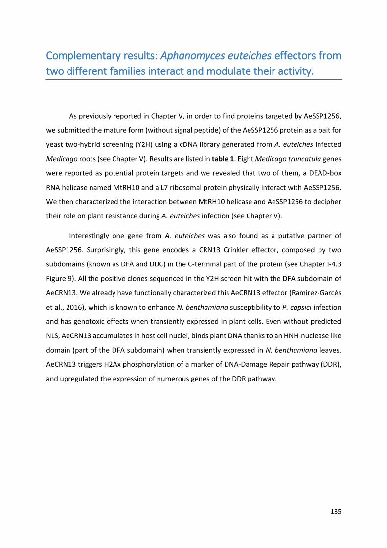
135
Complementary results: Aphanomyces euteiches effectors from
two different families interact and modulate their activity.
As previously reported in Chapter V, in order to find proteins targeted by AeSSP1256,
we submitted the mature form (without signal peptide) of the AeSSP1256 protein as a bait for
yeast two-hybrid screening (Y2H) using a cDNA library generated from A. euteiches infected
Medicago roots (see Chapter V). Results are listed in table 1. Eight Medicago truncatula genes
were reported as potential protein targets and we revealed that two of them, a DEAD-box
RNA helicase named MtRH10 and a L7 ribosomal protein physically interact with AeSSP1256.
We then characterized the interaction between MtRH10 helicase and AeSSP1256 to decipher
their role on plant resistance during A. euteiches infection (see Chapter V).
Interestingly one gene from A. euteiches was also found as a putative partner of
AeSSP1256. Surprisingly, this gene encodes a CRN13 Crinkler effector, composed by two
subdomains (known as DFA and DDC) in the C-terminal part of the protein (see Chapter I-4.3
Figure 9). All the positive clones sequenced in the Y2H screen hit with the DFA subdomain of
AeCRN13. We already have functionally characterized this AeCRN13 effector (Ramirez-Garcés
et al., 2016), which is known to enhance N. benthamiana susceptibility to P. capsici infection
and has genotoxic effects when transiently expressed in plant cells. Even without predicted
NLS, AeCRN13 accumulates in host cell nuclei, binds plant DNA thanks to an HNH-nuclease like
domain (part of the DFA subdomain) when transiently expressed in N. benthamiana leaves.
AeCRN13 triggers H2Ax phosphorylation of a marker of DNA-Damage Repair pathway (DDR),
and upregulated the expression of numerous genes of the DDR pathway.

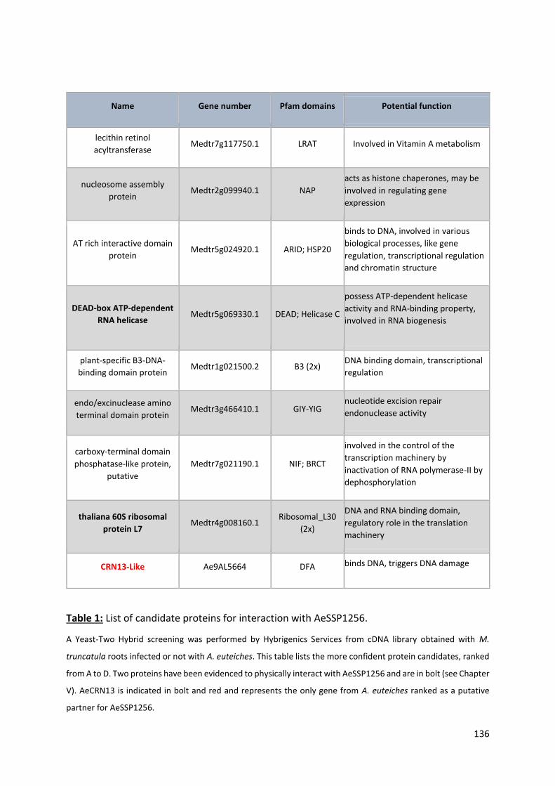
136
Table 1: List of candidate proteins for interaction with AeSSP1256.
A Yeast-Two Hybrid screening was performed by Hybrigenics Services from cDNA library obtained with M.
truncatula roots infected or not with A. euteiches. This table lists the more confident protein candidates, ranked
from A to D. Two proteins have been evidenced to physically interact with AeSSP1256 and are in bolt (see Chapter
V). AeCRN13 is indicated in bolt and red and represents the only gene from A. euteiches ranked as a putative
partner for AeSSP1256.
Name Gene number Pfam domains Potential function
lecithin retinol
acyltransferase Medtr7g117750.1 LRAT Involved in Vitamin A metabolism
nucleosome assembly
protein Medtr2g099940.1 NAP
acts as histone chaperones, may be
involved in regulating gene
expression
AT rich interactive domain
protein Medtr5g024920.1 ARID; HSP20
binds to DNA, involved in various
biological processes, like gene
regulation, transcriptional regulation
and chromatin structure
DEAD-box ATP-dependent
RNA helicase Medtr5g069330.1 DEAD; Helicase C
possess ATP-dependent helicase
activity and RNA-binding property,
involved in RNA biogenesis
plant-specific B3-DNA-
binding domain protein Medtr1g021500.2 B3 (2x)
DNA binding domain, transcriptional
regulation
endo/excinuclease amino
terminal domain protein Medtr3g466410.1 GIY-YIG
nucleotide excision repair
endonuclease activity
carboxy-terminal domain
phosphatase-like protein,
putative
Medtr7g021190.1 NIF; BRCT
involved in the control of the
transcription machinery by
inactivation of RNA polymerase-II by
dephosphorylation
thaliana 60S ribosomal
protein L7 Medtr4g008160.1
Ribosomal_L30
(2x)
DNA and RNA binding domain,
regulatory role in the translation
machinery
CRN13-Like Ae9AL5664 DFA binds DNA, triggers DNA damage

Figure 11: AeCRN13 interacts with AeSSP1256 and is relocalized by the SSP.
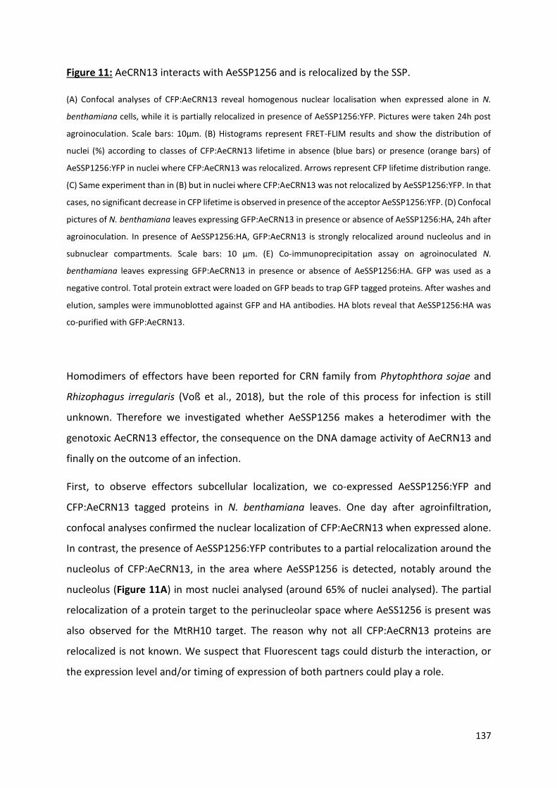
137
Figure 11: AeCRN13 interacts with AeSSP1256 and is relocalized by the SSP.
(A) Confocal analyses of CFP:AeCRN13 reveal homogenous nuclear localisation when expressed alone in N.
benthamiana cells, while it is partially relocalized in presence of AeSSP1256:YFP. Pictures were taken 24h post
agroinoculation. Scale bars: 10µm. (B) Histograms represent FRET-FLIM results and show the distribution of
nuclei (%) according to classes of CFP:AeCRN13 lifetime in absence (blue bars) or presence (orange bars) of
AeSSP1256:YFP in nuclei where CFP:AeCRN13 was relocalized. Arrows represent CFP lifetime distribution range.
(C) Same experiment than in (B) but in nuclei where CFP:AeCRN13 was not relocalized by AeSSP1256:YFP. In that
cases, no significant decrease in CFP lifetime is observed in presence of the acceptor AeSSP1256:YFP. (D) Confocal
pictures of N. benthamiana leaves expressing GFP:AeCRN13 in presence or absence of AeSSP1256:HA, 24h after
agroinoculation. In presence of AeSSP1256:HA, GFP:AeCRN13 is strongly relocalized around nucleolus and in
subnuclear compartments. Scale bars: 10 µm. (E) Co-immunoprecipitation assay on agroinoculated N.
benthamiana leaves expressing GFP:AeCRN13 in presence or absence of AeSSP1256:HA. GFP was used as a
negative control. Total protein extract were loaded on GFP beads to trap GFP tagged proteins. After washes and
elution, samples were immunoblotted against GFP and HA antibodies. HA blots reveal that AeSSP1256:HA was
co-purified with GFP:AeCRN13.
Homodimers of effectors have been reported for CRN family from Phytophthora sojae and
Rhizophagus irregularis (Voß et al., 2018), but the role of this process for infection is still
unknown. Therefore we investigated whether AeSSP1256 makes a heterodimer with the
genotoxic AeCRN13 effector, the consequence on the DNA damage activity of AeCRN13 and
finally on the outcome of an infection.
First, to observe effectors subcellular localization, we co-expressed AeSSP1256:YFP and
CFP:AeCRN13 tagged proteins in N. benthamiana leaves. One day after agroinfiltration,
confocal analyses confirmed the nuclear localization of CFP:AeCRN13 when expressed alone.
In contrast, the presence of AeSSP1256:YFP contributes to a partial relocalization around the
nucleolus of CFP:AeCRN13, in the area where AeSSP1256 is detected, notably around the
nucleolus (Figure 11A) in most nuclei analysed (around 65% of nuclei analysed). The partial
relocalization of a protein target to the perinucleolar space where AeSS1256 is present was
also observed for the MtRH10 target. The reason why not all CFP:AeCRN13 proteins are
relocalized is not known. We suspect that Fluorescent tags could disturb the interaction, or
the expression level and/or timing of expression of both partners could play a role.

138
To confirm the protein-protein interaction we performed a FRET-FLIM assay in order to
measure the fluorescence lifetime of the CFP:AeCRN13 proteins in the nucleus, in absence or
in presence of AeSSP1256:YFP. In presence of AeSSP1256:YFP, analysis was conducted on
nuclei where CFP:AeCRN13 was relocalized. In these nuclei, the CFP lifetime significantly
decreases due to FRET effect with AeSSP1256:YFP acceptor, from 2.82 +/- 0.016 ns to 2.14 +/-
0.031 ns, indicating a close association between the two proteins (Table 2). The distribution
of all the measurements is plotted on Figure 11B and clearly shows the shift into shorter CFP
lifetime. Moreover, no significant difference in CFP lifetime was observed in nuclei where
AeCRN13 is not relocalized in presence of AeSSP1256. Value from 2.82 +/- 0.016 ns in mean
when CFP:AeCRN13 is expressed alone and 2.75 +/- 0.02 ns in presence of AeSSP1256:YFP are
reported (see Table 2). This result is illustrate by the Figure 11C. Altogether, these data
indicate that the relocalization of AeCRN13 is probably due to its interaction with AeSSP1256.
Table 2: FRET-FLIM measurements of CFP:AeCRN13 with or without AeSSP1256:YFP.
mean life-time in nanoseconds (ns). (b) s.e.m.: standard error of the mean. (c) N: total number of measured
nuclei. (d) E: FRET efficiency in % : E=1-(DA/D). (e) p-value (Student’s t test) of the difference between the donor
lifetimes in the presence or absence of acceptor.
To confirm the results and to decipher whether the fluorescent tag could perturb the
interaction, AeSSP1256 was fused to a triple HA tag, much smaller than the YFP tag (around 5
kDa for the triple HA against 27 kDa for YFP) (cloning is described in the paper from Chapter
V). Similar results were obtained when CFP:AeCRN13 was coexpressed with HA-tagged version
Donor Acceptor a sem (b) N (c) E (d) (e) p-value
CFP:AeCRN13 - 2.819 0.016 40 - -
CFP:AeCRN13 AeSSP1256:YFP 2.138 0.031 40 24 4.59E-32
CFP:AeCRN13 AeSSP1256:YFP
NO relocalization 2.750 0.020 40 2.3 0.07
139
of AeSSP1256 where approximately half of observed nuclei harbor a relocalized CFP:AeCRN13
around the nucleolus. Intriguingly, when a GFP:AeCRN13 construct is coexpressed with HA-
tagged version of AeSSP1256, more than 90% of observed nuclei harbor a relocalized
GFP:AeCRN13 around the nucleolus, and this relocalization appeared stronger than observed
with AeSSP1256:YFP (Figure 11D). This could be due to different spatial organization and
three-dimensional structure of GFP, CFP, YFP and HA tags.
To test whether the DNA binding ability of AeCRN13 could play a role in the interaction with
AeSSP1256, we coexpressed in N. benthamiana a mutated version of AeCRN13, named
AeCRN13AAA (see Ramirez-Garcès et al. 2016), with AeSSP1256:HA. AeCRN13AAA contains three
alanine in place of the corresponding Histidine, Asparagine and Histidine of the HNH domain
leading to a mutated protein unable to bind nucleic acids and to trigger DNA damage (see
(Ramirez-Garcés et al., 2016)). Confocal analyses confirm the strong relocalization of the
mutated GFP:AeCRN13AAA in presence of AeSSP1256:HA, suggesting that the HNH domain of
AeCRN13 is not involved in the interaction with AeSSP1256 (Figure 11D).
To confirm the interaction between both effectors, we performed co-immunoprecipitation
(CoIP) assays using total proteins extracted from N. benthamiana agroinfiltrated leaves with
AeSSP1256:HA and GFP:AeCRN13 constructs. As a control experiment, GFP construct was
coexpressed with an AeSSP1256:HA construct. After total protein extraction 24 hours after
treatment, samples were purified on GFP beads (protocol is described in the paper from
Chapter V), washed, and finally loaded on polyacrylamide gels for immunoblotting. As
expected, no AeSSP1256 was detected when only coexpressed with GFP, while GFP antibodies
confirmed the presence of the GFP:AeCRN13 proteins (around 65 kDa) and HA antibodies
revealed the presence of AeSSP1256:HA proteins (around 25 kDa) when both partners are
coexpressed. This data indicates that AeSSP1256:HA was pull down with GFP:AeCRN13 and
confirms their interaction (Figure 11E).
We then check whether AesSP1256 and AeCRN13 effectors may also interact in host cells. The
co-transformation of M. truncatula roots with AeSSP1256:HA and GFP:AeCRN13 constructs is
poorly efficient. Nevertheless three weeks after transformation of A17-Jemalong Medicago
roots, few roots where most nuclei displayed a GFP fluorescence in subnuclear compartments
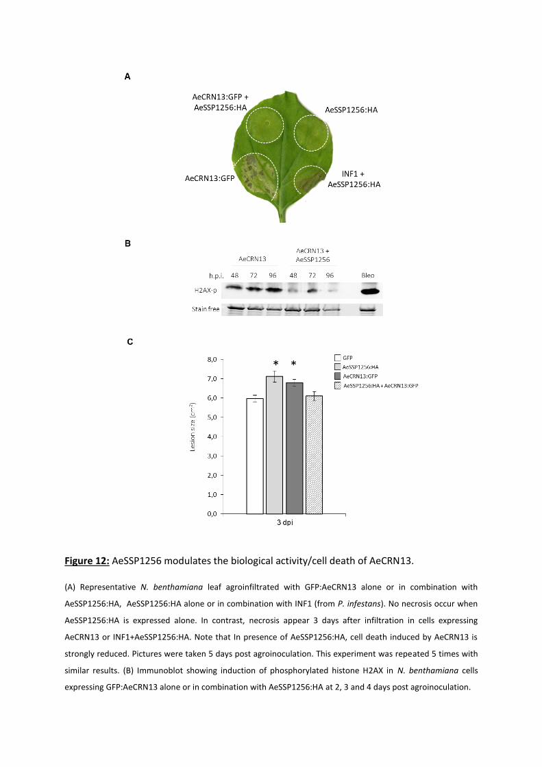
Figure 12: AeSSP1256 modulates the biological activity/cell death of AeCRN13.
(A) Representative N. benthamiana leaf agroinfiltrated with GFP:AeCRN13 alone or in combination with
AeSSP1256:HA, AeSSP1256:HA alone or in combination with INF1 (from P. infestans). No necrosis occur when
AeSSP1256:HA is expressed alone. In contrast, necrosis appear 3 days after infiltration in cells expressing
AeCRN13 or INF1+AeSSP1256:HA. Note that In presence of AeSSP1256:HA, cell death induced by AeCRN13 is
strongly reduced. Pictures were taken 5 days post agroinoculation. This experiment was repeated 5 times with
similar results. (B) Immunoblot showing induction of phosphorylated histone H2AX in N. benthamiana cells
expressing GFP:AeCRN13 alone or in combination with AeSSP1256:HA at 2, 3 and 4 days post agroinoculation.
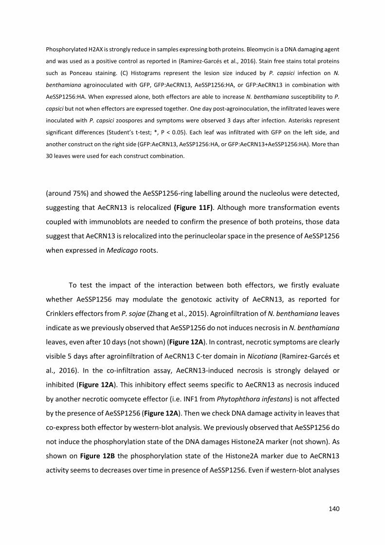
140
Phosphorylated H2AX is strongly reduce in samples expressing both proteins. Bleomycin is a DNA damaging agent
and was used as a positive control as reported in (Ramirez-Garcés et al., 2016). Stain free stains total proteins
such as Ponceau staining. (C) Histograms represent the lesion size induced by P. capsici infection on N.
benthamiana agroinoculated with GFP, GFP:AeCRN13, AeSSP1256:HA, or GFP:AeCRN13 in combination with
AeSSP1256:HA. When expressed alone, both effectors are able to increase N. benthamiana susceptibility to P.
capsici but not when effectors are expressed together. One day post-agroinoculation, the infiltrated leaves were
inoculated with P. capsici zoospores and symptoms were observed 3 days after infection. Asterisks represent
significant differences (Student’s t-test; *, P < 0.05). Each leaf was infiltrated with GFP on the left side, and
another construct on the right side (GFP:AeCRN13, AeSSP1256:HA, or GFP:AeCRN13+AeSSP1256:HA). More than
30 leaves were used for each construct combination.
(around 75%) and showed the AeSSP1256-ring labelling around the nucleolus were detected,
suggesting that AeCRN13 is relocalized (Figure 11F). Although more transformation events
coupled with immunoblots are needed to confirm the presence of both proteins, those data
suggest that AeCRN13 is relocalized into the perinucleolar space in the presence of AeSSP1256
when expressed in Medicago roots.
To test the impact of the interaction between both effectors, we firstly evaluate
whether AeSSP1256 may modulate the genotoxic activity of AeCRN13, as reported for
Crinklers effectors from P. sojae (Zhang et al., 2015). Agroinfiltration of N. benthamiana leaves
indicate as we previously observed that AeSSP1256 do not induces necrosis in N. benthamiana
leaves, even after 10 days (not shown) (Figure 12A). In contrast, necrotic symptoms are clearly
visible 5 days after agroinfiltration of AeCRN13 C-ter domain in Nicotiana (Ramirez-Garcés et
al., 2016). In the co-infiltration assay, AeCRN13-induced necrosis is strongly delayed or
inhibited (Figure 12A). This inhibitory effect seems specific to AeCRN13 as necrosis induced
by another necrotic oomycete effector (i.e. INF1 from Phytophthora infestans) is not affected
by the presence of AeSSP1256 (Figure 12A). Then we check DNA damage activity in leaves that
co-express both effector by western-blot analysis. We previously observed that AeSSP1256 do
not induce the phosphorylation state of the DNA damages Histone2A marker (not shown). As
shown on Figure 12B the phosphorylation state of the Histone2A marker due to AeCRN13
activity seems to decreases over time in presence of AeSSP1256. Even if western-blot analyses
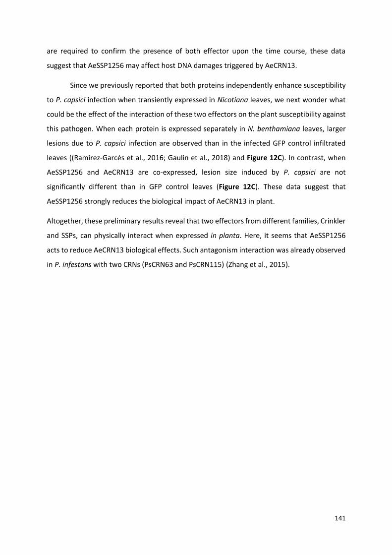
141
are required to confirm the presence of both effector upon the time course, these data
suggest that AeSSP1256 may affect host DNA damages triggered by AeCRN13.
Since we previously reported that both proteins independently enhance susceptibility
to P. capsici infection when transiently expressed in Nicotiana leaves, we next wonder what
could be the effect of the interaction of these two effectors on the plant susceptibility against
this pathogen. When each protein is expressed separately in N. benthamiana leaves, larger
lesions due to P. capsici infection are observed than in the infected GFP control infiltrated
leaves ((Ramirez-Garcés et al., 2016; Gaulin et al., 2018) and Figure 12C). In contrast, when
AeSSP1256 and AeCRN13 are co-expressed, lesion size induced by P. capsici are not
significantly different than in GFP control leaves (Figure 12C). These data suggest that
AeSSP1256 strongly reduces the biological impact of AeCRN13 in plant.
Altogether, these preliminary results reveal that two effectors from different families, Crinkler
and SSPs, can physically interact when expressed in planta. Here, it seems that AeSSP1256
acts to reduce AeCRN13 biological effects. Such antagonism interaction was already observed
in P. infestans with two CRNs (PsCRN63 and PsCRN115) (Zhang et al., 2015).

General discussion
and
perspectives
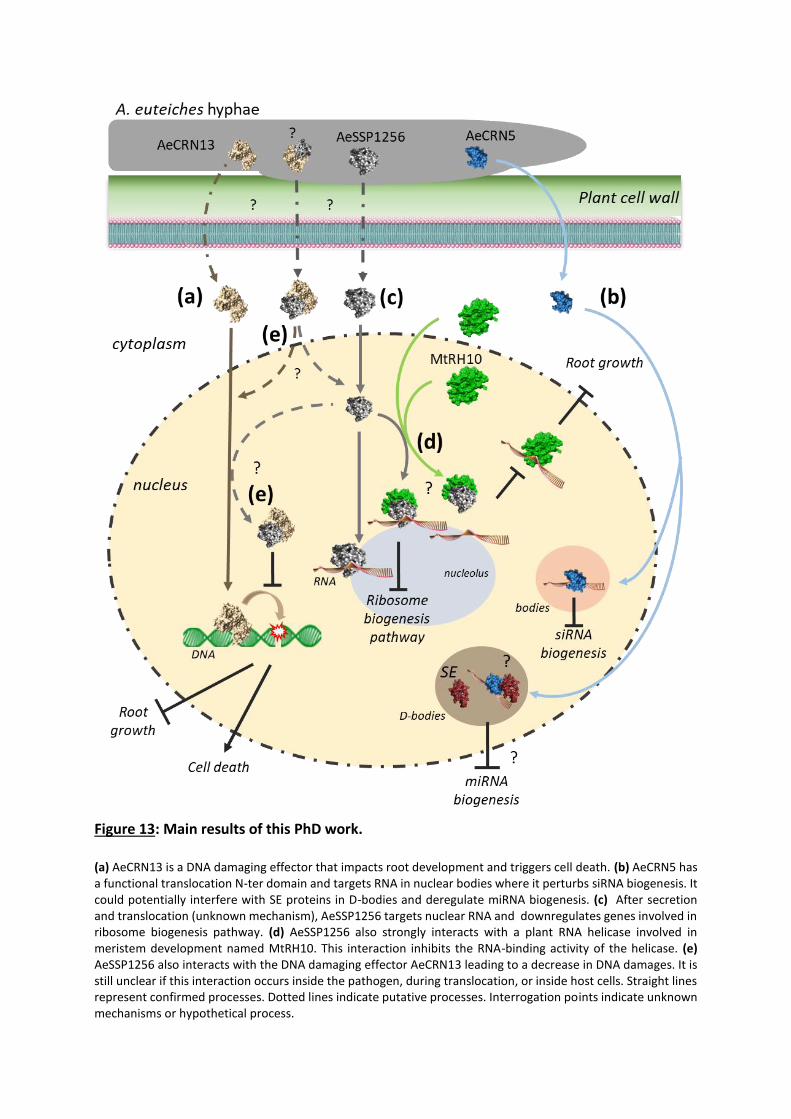
Figure 13: Main results of this PhD work.
(a) AeCRN13 is a DNA damaging effector that impacts root development and triggers cell death. (b) AeCRN5 has a functional translocation N-ter domain and targets RNA in nuclear bodies where it perturbs siRNA biogenesis. It could potentially interfere with SE proteins in D-bodies and deregulate miRNA biogenesis. (c) After secretion and translocation (unknown mechanism), AeSSP1256 targets nuclear RNA and downregulates genes involved in ribosome biogenesis pathway. (d) AeSSP1256 also strongly interacts with a plant RNA helicase involved in meristem development named MtRH10. This interaction inhibits the RNA-binding activity of the helicase. (e) AeSSP1256 also interacts with the DNA damaging effector AeCRN13 leading to a decrease in DNA damages. It is still unclear if this interaction occurs inside the pathogen, during translocation, or inside host cells. Straight lines represent confirmed processes. Dotted lines indicate putative processes. Interrogation points indicate unknown mechanisms or hypothetical process.
142
General discussion and perspectives
The aim of this PhD project is to develop a better understanding of the molecular mechanisms
underlying virulence and pathogenicity of the oomycete pathogen of legumes A. euteiches
that cause root rot diseases. The study was focus on microbial secreted proteins called
effectors that target host components to promote pathogen invasion. Knowing that numerous
fungal and oomycetes effectors target the nuclear compartment of the host plant we firstly
published in TIPS (Camborde et al., 2019) a review that reports on the activity of these
eukaryotic effectors (Chapter II). Then we focused our work on AeCRN5, a crinkler effector
from A. euteiches previously reported as a cell-death inducing protein able to target plant
nucleus (Chapter III). Comparative analyses of Aphanomyces spp. reveal a large family of
small-secreted proteins (SSPs) never reported in oomycetes in contrast to fungal pathogens
(Chapter IV). Using various technology an array of SSPs was tested for their effector activity
and AeSSP1256 has been selected for functional characterization. We decipher the activity of
AeSSP1256 against plant targets and identify that AeSSP1256 can interact with another
effector from A. euteiches, AeCRN13 previously reported as a DNA-damaging effector
(Chapter V). This PhD work showed the biological functions of two pivotal virulence factors of
A. euteiches. In this chapter, we reflect on the major findings of this study and discuss future
strategies to pursue our work on effectors functions.
CRNs and SSPs in oomycetes
The first aim of this work was to deepen knowledge in the repertoire and the mechanisms of
action of intracellular effectors from the root pathogenic oomycete Aphanomyces euteiches.
A. euteiches expression data from previous work suggested a large number of CRN coding
genes and in opposite the absence of RxLR protein effectors. This result was confirmed by
comparative analyses of A. euteiches, A. astaci and A. cladogamus genomes performed during
this PhD (Chapter IV). In the same time, a study using other bioinformatic criteria detected
between 16 to 25 RxLR-like genes in A. invadans and A. astaci respectively (McGowan and
Fitzpatrick, 2017). As expected, authors found that 87% of the predicted RxLR proteins are
located in Peronosporales species (McGowan and Fitzpatrick, 2017). They also confirm the
143
large expansion of putative cytoplasmic genes predicted in Phytophthora species, with
approximately 600 RxLR genes and almost 200 CRNs genes for P. infestans, giving a huge
number of putative cytoplasmic effectors to achieve infection. In comparison, A. euteiches has
“only” around 160 CRNs genes. This observation raises question about the arsenal of
intracellular effectors secreted by Aphanomyces compared to other oomycetes, especially to
Phytophthora species. Only 2% of the CRN genes were upregulated at 3 and 9 days post
inoculation, and 13% upregulated in zoospores as compared to in vitro grown mycelium,
suggesting that a subset of AeCRN is present at the early stage of Medicago infection and that
another set of CRN genes seems to be produced at later stages. These results are in
accordance with the dynamic expression of CRN genes reported in Phytophthora (Stam et al.,
2013a), and underlines the relative low number of intracellular effector coding genes induced
during host infection to sustain A. euteiches development. The genomic and transcriptomic
analyses of A. euteiches also revealed a large repertoire of small-secreted protein (SSP)-
encoding genes that are highly induced during plant infection and not detected in other
oomycetes. SSPs are widely present in fungi and are involved in the interaction between host
and mutualistic or pathogenic microorganisms (Veneault-Fourrey and Martin, 2011; Lo Presti
et al., 2015). This finding paves the way to new research on this type of molecules potentially
secreted by others oomycetes like Phytophthora.
Host nucleic acids as a target: Let’s play with DNA
Despite the central role of nucleic acids in a living cell, few example of intracellular effectors
able to interact with nucleic acids have been described to date in filamentous eukaryotic
microorganisms. In a very recent review on intracellular effectors from filamentous
phytopathogens, He and colleagues collected data from the literature describing verified
targets of 41 intracellular oomycete effectors and 30 from fungi (He et al., 2020). Only three
of these effectors target DNA and among them, two are Crinkler/CRNs proteins. The
Phytophthora sojae effector CRN108 binds to heat-shock element (HSE) promoters to prevent
their expression (Song et al., 2015). This CRN contains an HhH DNA binding domain, widely
distributed in DNA repair or synthesis proteins and reported to have sequence-non-specific
DNA-binding activity (Pavlov et al., 2002). The other CRN protein, AeCRN13, binds plant DNA
thanks to its HNH motif (Ramirez-Garcés et al., 2016) found in more than 500 nucleases or in
144
bacterial toxins such as colicins, produced by some E. coli strains. AeCRN13 trigger DNA-
damage of the host cell (Figure 13a). The third reported effector is CgEP1 from the fungus
Colletotrichum graminicola, presented as a double-stranded DNA-binding protein that
modulates transcriptional activity (Vargas et al., 2016).
DNA binding effectors from animal and plant pathogenic bacteria are also reported (see
Chapter II). One of the most known example are the transcription activator-like effector (TALE)
proteins. TALE proteins derived from bacteria and are built from tandem repeat units that can
be linked to form a string-like structure, able to bind DNA. TALES are unstable proteins, able
to follow the shape of the double helix through a conformational heterogeneity that facilitates
macromolecular assembly (Schuller et al., 2019).
Another example of DNA binding effector has been described in root pathogenic cyst
nematodes. Cyst nematodes are root endoparasites that infect a wide range of crops. Then, it
was reported that GLAND4, an effector secreted by Heterodera glycines and H. schachtii
(parasites of soybean and sugar beet respectively), is a small DNA binding protein that
represses gene expression of defense related genes. The C‐terminal domain of GLAND4
possesses acidic and hydrophobic amino acids structure similar to those found in TALE
proteins (Barnes et al., 2018).
Finally, in the fish oomycete pathogen Saprolegnia parasitica, SpHtp3 effector contains a
bifunctional nuclease domain and therefore degrades RNA and DNA in host cell nuclei (Trusch
et al., 2018).
Then it seems that targeting host DNA could be a common strategy shared by various animal
and plant bacterial pathogens, but also nematodes and filamentous eukaryotic pathogens.
The role on the pathogenesis depends on the type of DNA-effector interactions. Some DNA
binding proteins, such as bacterial TALEs, CRN108 from Phytophthora or fungal CgEP1 protein,
interfere with transcriptional activity and defense gene expression to manipulate host
immunity. For DNA-damaging effectors, the consequences are less clear. Triggering DNA
damage perturbs the host cell cycle and subsequently favors the colonization of the tissues.
On the other hand, DNA damage can be sensed as a danger signal leading to the induction of
defense responses (see Chapter II). Characterized DNA-damaging effectors are expressed at
the later stage of infection (such as AeCRN13 and SpHtp3) and could correspond to the switch
145
to a necrophytic phase of the infection. Future studies on this type of effectors are needed to
precise the role of DNA-damaging effectors in the outcome of the infection.
Host nucleic acids as a target: Let’s play with RNA
Among the 71 described intracellular effectors from filamentous phytopathogens reported in
He et al., 2020, six of them (8%) target RNA trafficking or RNA processing (He et al., 2020).
Among them, two effectors from Phytophthora sp. stabilize host RNA-binding proteins to
regulate mRNA biogenesis and plant immunity (Huang et al., 2017; Wang et al., 2015). Two
fungal effectors have been reported to potentially interfere with mRNA processing. One is an
RxLR protein from Magnaporthe oryzae that interacts with a nucleoporin required for
accumulation of PR gene transcripts (Tang et al., 2017). The other is a candidate effector from
the wheat rust fungus Puccinia striiformis that accumulates in processing bodies where it
interacts with a protein involved in mRNA decapping (Petre et al., 2016). Finally, two RxLR
effectors from P. sojae (PSR1 and PSR2) suppress RNA silencing by interfering with small RNA
biogenesis (Qiao et al., 2015; Xiong et al., 2014; Hou et al., 2019). Based on obtained results
we can include AeCRN5 and AeSSP1256 in this list.
However, none of these effectors was described to bind directly to RNA. This PhD work
showed that AeCRN5 is an RNA-binding protein with modular architecture, comprising a
functional translocation N-terminal domain folded as Ubiquitin family proteins, then classified
as Ubi1-Header domain according to Zhang et al. (2016). The C-ter domain comprises the
DN17 subdomain related to the usual classification based on P. infestans sequences (Haas et
al., 2009). Interestingly, even if AeCRN5 was not included in the study of Zhang et al. (2016),
the C-termini of the closest orthologs were described as REase5 domains, and we assumed
after sequence alignment that AeCRN5 is a member of this REase5 family. AeCRN5 C-ter is
nuclear localized and this localization is required to trigger necrosis in N. benthamiana leaves.
When expressed in host cells, AeCRN5 strongly affects root development (Figure 13b). In
addition to RNA binding ability, we found that it could interfere with PTGS mechanism, but
the effect on siRNA accumulation is still unclear and requires additional experiments. This role
was described in the oomycete P. sojae, with RxLR proteins PSR1 and PSR2. However, the
mechanism and the final impact seem different since PSR1 and PSR2 were not reported to
146
bind RNA, but to interact with RNA binding proteins. PSR1 promotes infection by interacting
with PINP1, a DEAD-box RNA helicase, to repress siRNA biogenesis in the plant hosts. PSR2
interacts with dsRNA-binding protein 4 (DRB4), which associates with Dicer-like 4 (DCL4), to
inhibit secondary siRNA biogenesis to interfere with trans-kingdom RNAi (Hou et al., 2019).
In addition, we reported preliminary results about the putative localization of AeCRN5 C-ter
in D-bodies, a direct or indirect interaction with the SERRATE protein, and then a perturbation
in miRNA biogenesis (Figure 13b). As discussed in Chapter III, we still need to precise the
subnuclear localization of AeCRN5 and to perform quantitative PCR on mature miR sequences
to confirm the role of AeCRN5 on miRNA maturation. Additionally, we will construct mutated
version of AeCRN5 C-ter domain, with substitution of the five catalytic residues conserved in
REase5 domain in alanine amino acid (Zhang et al., 2016). We will then tested its RNA-binding
capability, its localization in D-bodies, and expected to obtain a mutant no longer able to
interfere with miRNA biogenesis. Additionally, pri-miRNA or mature miRNA analyses (using
RT-qPCR) in M. truncatula plants infected by A. euteiches, or in AeCRN5 overexpressing M.t
plants could strengthen the role of AeCRN5 on miRNA biogenesis.
Effectors with RNAse-like activity and associated with Haustoria (RALPH) are largely detected
in barley powdery mildew Blumeria graminis, and constitute the so-called RALPH effectors
(Pedersen et al., 2012). This effector family contains around 120 candidate genes but few of
them have been characterized, and two (BEC1011 and BEC1054) were predicted to adopt a
ribonuclease structure but lack the key active amino acid sites necessary for ribonuclease
activity, suggesting that these proteins are non-functional ribonucleases (Pliego et al., 2013;
Spanu, 2015). Finally, a recent study evidenced the RNase-like fold of BEC1054 and reported
its RNA-binding activity. Authors suggest that the role of this effector could be to protect rRNA
by inhibiting the action of plant ribosome-inactivating proteins, repressing host cell death, an
unviable interaction for this biotrophic fungus (Pennington et al., 2019).
Functional analysis of AeSSP1256 indicates that this SSP is also an RNA-binding protein (RBP)
(Figure 13c). Like AeCRN5, AeSSP1256 has a subnuclear localization, but with clustered
accumulation around the nucleolus, and strongly perturbs the root development of host plant.
Additionally, AeSSP1256 interacts with a host ribosomal protein and a DEAD-box RNA helicase.
Transcriptomic analyses also indicate a downregulation of ribosomal protein genes implicated
in ribosome biogenesis pathway. Thus, ribosome biogenesis and activity seems to be a
147
common target for various pathogens. For example, in addition to the role of the fungal RALPH
effector BEC1054 on plant ribosome-inactivating proteins, the Hs32E03 effector from the
nematode H. schachtii manipulates host ribosomal biogenesis to promote parasitism.
Hs32E03 alters acetylation of histones involved in the transcription of rRNA, a major
component of ribosomes, leading to an increase in rRNA levels (Vijayapalani et al., 2018). The
additional ribosome synthesis is necessary for nematode-host interaction.
In our study, we reveal that the RNA-binding protein AeSSP1256 interferes with a DEAD-box
RNA helicase (named MtRH10) by inhibiting the RNA binding ability of the helicase (Figure
13d). DEAD-box RNA helicases are also targeted by another oomycete effector (PSR1 from P.
sojae) and represent a common target in mammal and plant-virus interactions, where DEAD-
box helicases contribute to innate immune signalling, or can block multiple steps in the viral
replication process (Taschuk and Cherry, 2020; Wu and Nagy, 2019). Although we do not know
the exact function of MtRH10 except its implication in Medicago roots development (Chapter
V), DEAD-box RNA helicases are known to be key players in ribosome assembly and/or in
ribosomal protein synthesis in eukaryotes, like in human or in plant, as well as in bacteria (Iost
and Jain, 2019; Martin et al., 2013; Liu and Imai, 2018). Future studies will aim to decipher the
putative link between ribosomal biogenesis pathway and MtRH10 activity in Medicago.
Expression level of the ribosomal genes downregulated in M. truncatula expressing
AeSSP1256 will be evaluated in the MtRH10 RNAi plants.
AeSSP1256 is a member of a cluster, which contains 5 other SSP encoding genes. Among them,
AeSSP1251 and AeSSP1254 also harbor a NLS sequence and present the same expression
profile as AeSSP1256. Hence, it could be interesting to test whether those proteins can
interact together and observe their putative synergetic association. Progress in molecular
cloning, especially with Golden gate technology, allows to clone longer and multiple
sequences.
Target relocalization: “Come together right now over me…”
We reported in Chapter V that AeSSP1256 can strongly relocalize MtRH10 plant helicase.
Additionally, we presented complementary results about AeSSP1256, showing an interaction
and relocalization with AeCRN13 (Figure 13e).
148
Target relocalization was already observed for several effectors. The fungal effector PstGSRE1
from P. striiformis inhibits the nuclear localization of the ROS-associated transcription factor
TaLOL2 in wheat (Qi et al., 2019). In oomycetes, P. sojae PsAvh52 recruits a host cytoplasmic
transacetylase into nuclear speckles to promote early colonization. In P. infestans and Bremia
lactucae, multiple effectors have been shown to interact with and prevent the nuclear
translocation of ER-associated tail-anchored transcription factors (McLellan et al., 2013;
Meisrimler et al., 2019).
In N. benthamiana leaves, the AeSSP1256-AeCRN13 association reduces AeCRN13 biological
effects. Such antagonism interaction was already observed in P. infestans with two CRNs
(PsCRN63 and PsCRN115) (Zhang et al., 2015). In the A. euteiches natural infection, AeSSP1256
and AeCRN13 genes show similar expression profiles, with higher level at later stages of the
infection, supporting the idea that both protein could be present at the same time in
Medicago roots. One role of the AeSSP1256 could be to moderate the effects of AeCRN13, for
instance to avoid early cell death. However, later stages of infection should correspond to the
switch in a necrotrophic phase, where cell death can occurs.
We can not rule out the possibility that AeSSP1256 / AeCRN13 interaction does not occur
inside the host cells, but only during the secretion process, for example to avoid CRN13 toxicity
against A. euteiches DNA. Such association is well described as Effector-Immunity pairs in
bacteria (Yang et al., 2018).
AeSSP1256 contains a signal peptide that should lead the secretion outside the microorganism
through the conventional pathway. In contrast, as many other CRNs, such signal peptide is
absent in AeCRN13, and it is suggested that RxLR or CRNs could be secreted via unconventional
secretory pathway (Wang et al., 2017; Amaro et al., 2017). One can suppose that both protein
could interact within secreted microbial vesicles that are released from the pathogen and then
address to the host cells. Here, both protein transit to reach the nucleus, where they can
interact with other components, such as RNA for AeSSP1256, releasing free AeCRN13 that
targets host DNA (Figure 13e). Such extracellular vesicles (EVs) have been reported in plant
microbe interactions, especially for fungi (for review see (Rizzo et al., 2020)). However, it is
still an open question whether mutualistic or parasitic fungi use EVs to deliver effector
molecules to plants during interaction. Since preliminary experiments using Transmission
Electron Microscopy on infected roots suggest the presence of EVs during A. euteiches / M.

149
truncatula infection, one perspective of this study also resides in the identification of the
process that allow delivery of the effectors within the plant cells.
Looking for a needle in a haystack
One of the most challenging question about effector research is how to deal with hundreds
predicted genes. Although transcriptomic data help to distinguish genes induced during
infection and then potentially involved in host interaction, sequence analyses often failed to
detect conserved motif related to a biological function, especially for SSP genes.
In our study, AeSSP1256 putative RNA-binding motif was in silico identified and allowed us to
confirm its affinity for nucleic acids by FRET-FLIM assays, but numerous effectors are devoid
of predicted functional domain. Structure prediction of the effector can be an efficient tool to
overcome this limitation. A recent study challenged the classification of CRN proteins by
combining sequence analyses and structure prediction. Authors determined that most of the
CRN C-ter domains displayed two architectural types: an NTPase domain coupled with a
nuclease domain of the restriction endonuclease (REase) superfamily and a REase superfamily
domain combined with an eukaryote-type protein kinase domain (see Chapter I Figure 4 and
(Zhang et al., 2016)). Accordingly, we also predicted REase domain in the Cter of AeCRN5 and
then confirm its RNA-binding capacity (Chapter III). Zhang and collaborators proposed that C-
ter containing REase domains that primarily act on target cell DNA, could explain the cell-
death-causing capacity reported for numerous CRNs (Zhang et al., 2016). They also suggest
that some CRN with REases domain have evolved to target RNA (Zhang et al., 2016). Although
experimental data are needed to support their hypothesis, future studies on CRNs should
include experiments to detect nucleic acid-protein interactions.
The structural prediction of proteins, performed by dedicated server such as i-Tasser or
Phyre2, are frequently included in recent studies of effectors. Such prediction analyses were
successfully used on CRNs (Voß et al., 2018), RxLR proteins (Deb et al., 2018), bacterial
effectors (Dhroso et al., 2018; Borah and Jha, 2019) and fungal SSPs (Zhang et al., 2017; Gong
et al., 2020). In this study AeCRN5 structural prediction confirms the putative fold reported by
Zhang et al., 2016 for CRNs. Furthermore, some studies using crystallography reported the
conserved function of sequence-unrelated proteins. It is well illustrated with Magnaporthe
150
oryzae avirulence and ToxB-like (MAX) effectors. This effector family was identified using NMR
spectroscopy to determine the three-dimensional structures of two sequence-unrelated M.
oryzae effectors (de Guillen et al., 2015). These analyses revealed that both proteins shared
highly similar six β-sandwich structures stabilized by a disulfide bridge. Finally, using structural
similarity searches, authors found that another effector from M. oryzae and an effector of the
wheat tan spot pathogen Pyrenophora tritici-repentis, named ToxB, harbored the same
structures, leading to the identification of the MAX effectors (de Guillen et al., 2015). Recently,
using crystallography experiments, structural analyses on MAX effector proteins alone or in
complex with their NLRs targets (leucine-rich repeat proteins) provided detailed insights into
their recognition mechanisms (Guo et al., 2018). Similarly, crystal structure of the effector
AvrLm4–7 of Leptosphaeria maculans, the causal agent of stem canker in Brassica napus
(oilseed rape), was resolved and validated to understand the specificity of recognition by two
plant R proteins (Blondeau et al., 2015). In oomycetes, crystal structure of an RxLR effector
from P. capsici was recently revealed (Zhao et al., 2018).
This PhD work also reveal that effector from distinct family (SSP/CRN) may interact together
probably to enhance/repress their activity. Structural modeling of microbial effector will help
to predict this protein-protein association that could be not detected by in silico data mining.
Several bioinformatics programs dedicated to effector prediction exist, such as EffectorP 2.0
(Sperschneider et al., 2018) or even more recently EffHunter, a tool for fungal effector
prediction (Carreón-Anguiano et al., 2020). However, the subcellular localization of the
predicted effector within the host cell is still uncertain using this software and functional
studies are required. It is experimentally challenging to monitor effector trafficking but
recently some studies reported the translocation of effectors from fungi or oomycetes into
host cells. In M. oryzae, using a long-term time-lapse imaging method, the translocation of a
GFP-tagged SSP from a particular infectious area, called Biotrophic Interfacial Complex (BIC),
into host cells was evidenced (Nishimura et al., 2016). In oomycetes, it was evidenced by live-
cell imaging that the RxLR effector Pi04314 from P. infestans was translocated from the
haustorium into plant cells (Wang et al., 2017). In Aphanomyces euteiches, this kind of
experiments is even more challenging since this pathogen doesn’t make haustorium or BIC
and is not yet transformable.
151
Thus, another option will be to take advantage of progress in proteomic approaches in order
to detect microbial effectors inside the host cells. This approach was used successfully on
wheat infected by Fusarium graminearum (Fabre et al., 2019). One main limitation in this
approach is to distinguish plant compartments from the microorganism, nevertheless plant
cytoplasm or nuclei to identify intracellular effectors can be discriminate either by labeling the
compartment or by collecting samples using laser-microdissection experiments. Mass
spectrometry analysis of the proteins will give a short list of putative intracellular effectors
and host targets.
Concluding Remarks and Outlooks
Oomycete and fungal effectors acting as virulence factors are key players in plant-microbe
interactions. While in silico approaches allow prediction of effector repertoire in numerous
fungi and oomycetes, further investigations are required to characterize their activity during
host infection. Indeed the exact function of numerous effectors and how that is related to
host immunity are still unknown. This PhD study shows that the nuclear compartment of the
host plant is a major target for numerous oomycete effectors. While it was recently shown
that microbial effector can target different host proteins, this work also shows that effectors
from different family can associate and target plant nucleus. These results reveal a new layer
of complexity in the mode of action of eukaryotic effectors. More analyses to study the
structural relationship between effectors and between effectors and their targets are needed
to precise the consequences of these interactions.
However, in the coming years, the relevance of the choice of candidate effectors for functional
characterization will be crucial. Indeed, regarding the results provided by the extensive
research on effectors, it seems that every biological process is targeted by one or numerous
effectors. This includes sensing, signalling, defence reaction, transcription, RNA processes,
DNA integrity, cell development, etc. Hence, in an objective to increase crop plant resistance,
it seems difficult to block or regulate tens of molecules that target so many processes. Then,
understanding the mechanisms involved in the effector delivery could lead to the
development of molecules able to break the bridge between plant cells and pathogen hyphae,
preventing the release of those molecules in host cells.

152
Another way to improve plant fitness is to understand the role and the interaction between
pathogens, plants and the other microorganisms present in root close proximity, named plant
microbiome (Song et al., 2020; Turner et al., 2013). This represents an emerging topic that
needs to go deeper in the molecular interactions between partners.
Since relative few numbers of effector genes are expressed during plant colonisation as AeCRN
or AeSSP effectors, numerous microbial effectors could play a role in other situation than host
infection, especially in microbe-microbe interactions that occur in the microbiome.
To conclude, one threat resides in the emergence of new diseases due to the acquisition of a
new host by an existing plant pathogen. Determining the mechanisms that govern host-
specificity is crucial to understand host-switching events and variation in virulence strains.
Effectors are part of the molecules involved in this host adaptation. In Aphanomyces, our
comparative genome analyses between different strains underline variation in their SSP
repertoire, suggesting that those molecules could play a role in host adaptation.
Future studies will aim to elucidate the crucial roles in pathogenicity and in microbiome
interactions of A. euteiches effectors to improve host tolerance against the pathogen.


References

153
References
Adeniyi, D. (2019). Diversity of Cacao pathogens and impact on yield and global production. In Theobroma Cacao - Deploying Science for Sustainability of Global Cocoa Economy (IntechOpen).
Adhikari, B.N., Hamilton, J.P., Zerillo, M.M., Tisserat, N., Lévesque, C.A., and Buell, C.R. (2013). Comparative Genomics Reveals Insight into Virulence Strategies of Plant Pathogenic Oomycetes. PLoS One 8.
Allen, T.W. et al. (2017). Soybean yield loss estimates due to diseases in the United States and Ontario, Canada, from 2010 to 2014. Plant Heal. Prog. 18: 19–27.
Almeida, F., Rodrigues, M.L., and Coelho, C. (2019). The Still Underestimated Problem of Fungal Diseases Worldwide. Front. Microbiol. 10: 214.
Amaro, T.M.M.M., Thilliez, G.J.A., Motion, G.B., and Huitema, E. (2017). A Perspective on CRN Proteins in the Genomics Age: Evolution, Classification, Delivery and Function Revisited. Front. Plant Sci. 8: 99.
André Lévesque, C. (2011). Fifty years of oomycetes-from consolidation to evolutionary and genomic exploration. Fungal Divers. 50: 35–46.
Badis, Y., Bonhomme, M., Lafitte, C., Huguet, S., Balzergue, S., Dumas, B., and Jacquet, C. (2015). Transcriptome analysis highlights preformed defences and signalling pathways controlled by the prAe1 quantitative trait locus (QTL), conferring partial resistance to Aphanomyces euteiches in Medicago truncatula. Mol. Plant Pathol. 16: 973–986.
Badreddine, I., Lafitte, C., Heux, L., Skandalis, N., Spanou, Z., Martinez, Y., Esquerré-Tugayé, M.T., Bulone, V., Dumas, B., and Bottin, A. (2008). Cell wall chitosaccharides are essential components and exposed patterns of the phytopathogenic oomycete Aphanomyces euteiches. Eukaryot. Cell 7: 1980–1993.
Bailey, B.A., Apel-Birkhold, P.C., and Luster, D.G. (2002). Expression of NEP1 by Fusarium oxysporum f. sp. erythroxyli after gene replacement and overexpression using polyethylene glycol-mediated transformation. Phytopathology 92.
Baldauf, S.L., Roger, A.J., Wenk-Siefert, I., and Doolittle, W.F. (2000). A kingdom-level phylogeny of eukaryotes based on combined protein data. Science (80-. ). 290: 972–977.
Banuett, F. and Herskowitz, I. (1996). Discrete developmental stages during teliospore formation in the corn smut fungus, Ustilago maydis. Development 122.
Barnes, S.N., Wram, C.L., Mitchum, M.G., and Baum, T.J. (2018). The plant-parasitic cyst nematode effector GLAND4 is a DNA-binding protein. Mol. Plant Pathol. 19: 2263–2276.
Baxter, L. et al. (2010). Signatures of adaptation to obligate biotrophy in the Hyaloperonospora arabidopsidis genome. Science (80-. ). 330: 1549–1551.
Beakes, G.W., Glockling, S.L., and Sekimoto, S. (2012). The evolutionary phylogeny of the oomycete “fungi.” Protoplasma 249: 3–19.

154
Beakes, G.W., Honda, D., and Thines, M. (2014). 3 Systematics of the Straminipila: Labyrinthulomycota, Hyphochytriomycota, and Oomycota. In Systematics and Evolution (Springer Berlin Heidelberg), pp. 39–97.
Belbahri, L., Calmin, G., Mauch, F., and Andersson, J.O. (2008). Evolution of the cutinase gene family: Evidence for lateral gene transfer of a candidate Phytophthora virulence factor. Gene 408: 1–8.
Benhamou, N., le Floch, G., Vallance, J., Gerbore, J., Grizard, D., and Rey, P. (2012). Pythium oligandrum: An example of opportunistic success. Microbiol. (United Kingdom) 158: 2679–2694.
Berg, B., McClaugherty, C., Berg, B., and McClaugherty, C. (2014). Decomposer Organisms. In Plant Litter (Springer Berlin Heidelberg), pp. 35–52.
Bhattacharya, S. (2017). Deadly new wheat disease threatens Europe’s crops. Nature 542: 145–146.
Van Der Biezen, E.A. and Jones, J.D.G. (1998). Plant disease-resistance proteins and the gene-for-gene concept. Trends Biochem. Sci. 23: 454–456.
Birch, P.R.J., Rehmany, A.P., Pritchard, L., Kamoun, S., and Beynon, J.L. (2006). Trafficking arms: oomycete effectors enter host plant cells. Trends Microbiol. 14: 8–11.
Blondeau, K. et al. (2015). Crystal structure of the effector AvrLm4-7 of Leptosphaeria maculans reveals insights into its translocation into plant cells and recognition by resistance proteins. Plant J. 83.
Bonhomme, M. et al. (2014). High-density genome-wide association mapping implicates an F-box encoding gene in Medicago truncatula resistance to Aphanomyces euteiches. New Phytol. 201: 1328–1342.
Bonhomme, M., Fariello, M.I., Navier, H., Hajri, A., Badis, Y., Miteul, H., Samac, D.A., Dumas, B., Baranger, A., Jacquet, C., and Pilet-Nayel, M.L. (2019). A local score approach improves GWAS resolution and detects minor QTL: application to Medicago truncatula quantitative disease resistance to multiple Aphanomyces euteiches isolates. Heredity (Edinb). 123.
Borah, S.M. and Jha, A.N. (2019). Identification and analysis of structurally critical fragments in HopS2. BMC Bioinformatics 19.
Brunner, F., Rosahl, S., Lee, J., Rudd, J.J., Geiler, C., Kauppinen, S., Rasmussen, G., Scheel, D., and Nürnberger, T. (2002). Pep-13, a plant defense-inducing pathogen-associated pattern from Phytophthora transglutaminases. EMBO J. 21.
van den Burg, H.A., Harrison, S.J., Joosten, M.H.A.J., Vervoort, J., and de Wit, P.J.G.M. (2006). Cladosporium fulvum Avr4 Protects Fungal Cell Walls Against Hydrolysis by Plant Chitinases Accumulating During Infection. Mol. Plant-Microbe Interact. 19: 1420–1430.
Burki, F. (2014). The eukaryotic tree of life from a global phylogenomic perspective. Cold Spring Harb. Perspect. Biol. 6.
155
Caillaud, M.C., Asai, S., Rallapalli, G., Piquerez, S., Fabro, G., and Jones, J.D.G. (2013). A Downy Mildew Effector Attenuates Salicylic Acid-Triggered Immunity in Arabidopsis by Interacting with the Host Mediator Complex. PLoS Biol. 11.
Caillaud, M.C., Piquerez, S.J.M., Fabro, G., Steinbrenner, J., Ishaque, N., Beynon, J., and Jones, J.D.G. (2012). Subcellular localization of the Hpa RxLR effector repertoire identifies a tonoplast-associated protein HaRxL17 that confers enhanced plant susceptibility. Plant J. 69: 252–265.
Camborde, L., Jauneau, A., Brière, C., Deslandes, L., Dumas, B., and Gaulin, E. (2017). Detection of nucleic acid–protein interactions in plant leaves using fluorescence lifetime imaging microscopy. Nat. Protoc. 12: 1933–1950.
Camborde, L., Raynaud, C., Dumas, B., and Gaulin, E. (2019). DNA-Damaging Effectors: New Players in the Effector Arena. Trends Plant Sci. 24: 1094–1101.
Carreón-Anguiano, K.G., Islas-Flores, I., Vega-Arreguín, J., Sáenz-Carbonell, L., and Canto-Canché, B. (2020). Effhunter: A tool for prediction of effector protein candidates in fungal proteomic databases. Biomolecules 10.
Carzaniga, R., Bowyer, P., and O’Connell, R.J. (2001). Production of extracellular matrices during development of infection structures by the downy mildew Peronospora parasitica. New Phytol. 149: 83–93.
Casadevall, A. (2017). Don’t Forget the Fungi When Considering Global Catastrophic Biorisks. Heal. Secur. 15: 341–342.
Cavalier-Smith, T. and Chao, E.E.-Y. (2006). Phylogeny and megasystematics of phagotrophic heterokonts (kingdom Chromista). J. Mol. Evol. 62: 388–420.
Chen, J.Y. et al. (2018). Comparative genomics reveals cotton-specific virulence factors in flexible genomic regions in Verticillium dahliae and evidence of horizontal gene transfer from Fusarium. New Phytol. 217.
Chepsergon, J., Motaung, T.E., Bellieny-Rabelo, D., and Moleleki, L.N. (2020). Organize, don’t agonize: Strategic success of Phytophthora species. Microorganisms 8: 1–21.
Choi, Y.J., Lee, S.H., Nguyen, T.T.T., Nam, B., and Lee, H.B. (2019). Characterization of Achlya americana and A. bisexualis (Saprolegniales, Oomycota) Isolated from Freshwater Environments in Korea. Mycobiology 47.
Cobos, R., Calvo-Peña, C., Álvarez-Pérez, J.M., Ibáñez, A., Diez-Galán, A., González-García, S., García-Angulo, P., Acebes, J.L., and Coque, J.J.R. (2019). Necrotic and cytolytic activity on grapevine leaves produced by nep1-like proteins of diplodia seriata. Front. Plant Sci. 10.
Condon, B.J. et al. (2013). Comparative genome structure, secondary metabolite, and effector coding capacity across Cochliobolus pathogens. PLoS Genet. 9: e1003233.
Cook, D.E., Mesarich, C.H., and Thomma, B.P.H.J. (2015). Understanding Plant Immunity as a Surveillance System to Detect Invasion. Annu. Rev. Phytopathol. 53.
Croll, D. and McDonald, B.A. (2012). The accessory genome as a cradle for adaptive evolution in pathogens. PLoS Pathog. 8.

156
Cuesta Arenas, Y., Kalkman, E.R.I.C., Schouten, A., Dieho, M., Vredenbregt, P., Uwumukiza, B., Osés Ruiz, M., and van Kan, J.A.L. (2010). Functional analysis and mode of action of phytotoxic Nep1-like proteins of Botrytis cinerea. Physiol. Mol. Plant Pathol. 74.
Dagdas, Y.F. et al. (2016). An effector of the irish potato famine pathogen antagonizes a host autophagy cargo receptor. Elife 5.
Dallery, J.F. et al. (2017). Gapless genome assembly of Colletotrichum higginsianum reveals chromosome structure and association of transposable elements with secondary metabolite gene clusters. BMC Genomics 18.
Damasceno, C.M.B., Bishop, J.G., Ripoll, D.R., Win, J., Kamoun, S., and Rose, J.K.C. (2008). Structure of the glucanase inhibitor protein (GIP) family from Phytophthora species suggests coevolution with plant endo-β-1,3-glucanases. Mol. Plant-Microbe Interact. 21.
Deb, D., Anderson, R.G., How-Yew-Kin, T., Tyler, B.M., and McDowell, J.M. (2018). Conserved RxLR effectors from oomycetes Hyaloperonospora arabidopsidis and Phytophthora sojae suppress PAMP- and effector-triggered immunity in diverse plants. Mol. Plant-Microbe Interact. 31: 374–385.
Derelle, R., López-García, P., Timpano, H., and Moreira, D. (2016). A Phylogenomic Framework to Study the Diversity and Evolution of Stramenopiles (=Heterokonts). Mol. Biol. Evol. 33: 2890–2898.
Derevnina, L. et al. (2016a). Nine things to know about elicitins. New Phytol. 212.
Derevnina, L., Petre, B., Kellner, R., Dagdas, Y.F., Sarowar, M.N., Giannakopoulou, A., de la Concepcion, J.C., Chaparro-Garcia, A., Pennington, H.G., van West, P., and Kamoun, S. (2016b). Emerging oomycete threats to plants and animals. Philos. Trans. R. Soc. B Biol. Sci. 371.
Desgroux, A. et al. (2016). Genome-wide association mapping of partial resistance to Aphanomyces euteiches in pea. BMC Genomics 17: 124.
Dhroso, A., Eidson, S., and Korkin, D. (2018). Genome-wide prediction of bacterial effector candidates across six secretion system types using a feature-based statistical framework. Sci. Rep. 8.
Diéguez-Uribeondo, J., García, M.A., Cerenius, L., Kozubíková, E., Ballesteros, I., Windels, C., Weiland, J., Kator, H., Söderhäll, K., and Martín, M.P. (2009). Phylogenetic relationships among plant and animal parasites, and saprotrophs in Aphanomyces (Oomycetes). Fungal Genet. Biol. 46: 365–76.
Djamei, A. et al. (2011). Metabolic priming by a secreted fungal effector. Nature 478: 395–398.
Djébali, N. et al. (2009). Partial Resistance of Medicago truncatula to Aphanomyces euteiches is associated with protection of the root stele and is controlled by a major QTL rich in proteasome-related genes. Mol. Plant-Microbe Interact. 22: 1043–1055.
Doehlemann, G. and Hemetsberger, C. (2013). Apoplastic immunity and its suppression by filamentous plant pathogens. New Phytol. 198: 1001–1016.
Dong, S. et al. (2014). Effector specialization in a lineage of the Irish potato famine pathogen.
157
Science 343: 552–5.
Dong, S., Raffaele, S., and Kamoun, S. (2015). The two-speed genomes of filamentous pathogens: waltz with plants. Curr. Opin. Genet. Dev. 35: 57–65.
Dou, D. et al. (2008). Conserved C-Terminal Motifs Required for Avirulence and Suppression of Cell Death by Phytophthora sojae effector Avr1b. Plant Cell Online 20: 1118–1133.
Duplessis, S. et al. (2011). Obligate biotrophy features unraveled by the genomic analysis of rust fungi. Proc. Natl. Acad. Sci. U. S. A. 108: 9166–9171.
Dutheil, J.Y., Mannhaupt, G., Schweizer, G., Sieber, C.M.K., Münsterkötter, M., Güldener, U., Schirawski, J., and Kahmann, R. (2016). A tale of genome compartmentalization: The evolution of virulence clusters in smut fungi. Genome Biol. Evol. 8.
Evangelisti, E., Govetto, B., Minet-Kebdani, N., Kuhn, M.-L., Attard, A., Ponchet, M., Panabières, F., and Gourgues, M. (2013). The Phytophthora parasitica RXLR effector Penetration-Specific Effector 1 favours Arabidopsis thaliana infection by interfering with auxin physiology. New Phytol. 199: 476–489.
Fabre, F., Bormann, J., Urbach, S., Roche, S., Langin, T., and Bonhomme, L. (2019). Unbalanced Roles of Fungal Aggressiveness and Host Cultivars in the Establishment of the Fusarium Head Blight in Bread Wheat. Front. Microbiol. 10.
Faure, C. et al. (2020). Long-read genome sequence of the sugar beet rhizosphere mycoparasite Pythium oligandrum. G3 Genes, Genomes, Genet. 10: 431–436.
Fawke, S., Doumane, M., and Schornack, S. (2015). Oomycete Interactions with Plants: Infection Strategies and Resistance Principles. Microbiol. Mol. Biol. Rev. 79: 263–280.
Feldman, D., Yarden, O., and Hadar, Y. (2020). Seeking the Roles for Fungal Small-Secreted Proteins in Affecting Saprophytic Lifestyles. Front. Microbiol. 11: 455.
Fellbrich, G., Romanski, A., Varet, A., Blume, B., Brunner, F., Engelhardt, S., Felix, G., Kemmerling, B., Krzymowska, M., and Nürnberger, T. (2002). NPP1, a Phytophthora-associated trigger of plant defense in parsley and Arabidopsis. Plant J. 32.
Feng, B.-Z., Zhu, X.-P., Fu, L., Lv, R.-F., Storey, D., Tooley, P., and Zhang, X.-G. (2014). Characterization of necrosis-inducing NLP proteins in Phytophthora capsici. BMC Plant Biol. 14: 126.
Fisher, M.C., Henk, D. a, Briggs, C.J., Brownstein, J.S., Madoff, L.C., McCraw, S.L., and Gurr, S.J. (2012). Emerging fungal threats to animal, plant and ecosystem health. Nature 484: 186–94.
Fletcher, K., Gil, J., Bertier, L.D., Kenefick, A., Wood, K.J., Zhang, L., Reyes-Chin-Wo, S., Cavanaugh, K., Tsuchida, C., Wong, J., and Michelmore, R. (2019). Genomic signatures of heterokaryosis in the oomycete pathogen Bremia lactucae. Nat. Commun. 10.
Frenken, T., Agha, R., Schmeller, D.S., van West, P., and Wolinska, J. (2019). Biological Concepts for the Control of Aquatic Zoosporic Diseases. Trends Parasitol. 35: 571–582.
Friesen, T.L., Stukenbrock, E.H., Liu, Z., Meinhardt, S., Ling, H., Faris, J.D., Rasmussen, J.B., Solomon, P.S., McDonald, B.A., and Oliver, R.P. (2006). Emergence of a new disease as

158
a result of interspecific virulence gene transfer. Nat. Genet. 38.
Gan, P.H.P., Rafiqi, M., Ellis, J.G., Jones, D.A., Hardham, A.R., and Dodds, P.N. (2010). Lipid binding activities of flax rust AvrM and AvrL567 effectors. Plant Signal. Behav. 5: 1272–1275.
Gaulin, E. et al. (2006). Cellulose binding domains of a Phytophthora cell wall protein are novel pathogen-associated molecular patterns. Plant Cell 18.
Gaulin, E. et al. (2018). Genomics analysis of Aphanomyces spp. identifies a new class of oomycete effector associated with host adaptation. BMC Biol. 16.
Gaulin, E., Bottin, A., and Dumas, B. (2010). Sterol biosynthesis in oomycete pathogens. Plant Signal. Behav. 5: 258–260.
Gaulin, E., Haget, N., Khatib, M., Herbert, C., Rickauer, M., and Bottin, A. (2007). Transgenic sequences are frequently lost in Phytophthora parasitica transformants without reversion of the transgene-induced silenced state. Can. J. Microbiol. 53: 152–157.
Gaulin, E., Jacquet, C., Bottin, A., and Dumas, B. (2007). Root rot disease of legumes caused by Aphanomyces euteiches. Mol. Plant Pathol. 8: 539–548.
Gaulin, E., Madoui, M.-A., Bottin, A., Jacquet, C., Mathé, C., Couloux, A., Wincker, P., and Dumas, B. (2008). Transcriptome of Aphanomyces euteiches: new oomycete putative pathogenicity factors and metabolic pathways. PLoS One 3: e1723.
Gijzen, M., Ishmael, C., and Shrestha, S.D. (2014). Epigenetic control of effectors in plant pathogens. Front. Plant Sci. 5.
Gijzen, M. and Nürnberger, T. (2006). Nep1-like proteins from plant pathogens: Recruitment and diversification of the NPP1 domain across taxa. Phytochemistry 67.
Girard, V., Dieryckx, C., Job, C., and Job, D. (2013). Secretomes: The fungal strike force. Proteomics 13: 597–608.
Gisi, U. and Sierotzki, H. (2015). Oomycete Fungicides: Phenylamides, Quinone Outside Inhibitors, and Carboxylic Acid Amides. In Fungicide Resistance in Plant Pathogens (Springer Japan), pp. 145–174.
Gómez-Gómez, L. and Boller, T. (2000). FLS2: An LRR receptor-like kinase involved in the perception of the bacterial elicitor flagellin in Arabidopsis. Mol. Cell 5.
Gong, A. dong, Jing, Z. ying, Zhang, K., Tan, Q. qun, Wang, G. liang, and Liu, W. de (2020). Bioinformatic analysis and functional characterization of the CFEM proteins in maize anthracnose fungus Colletotrichum graminicola. J. Integr. Agric. 19.
de Guillen, K., Ortiz-Vallejo, D., Gracy, J., Fournier, E., Kroj, T., and Padilla, A. (2015). Structure Analysis Uncovers a Highly Diverse but Structurally Conserved Effector Family in Phytopathogenic Fungi. PLoS Pathog. 11.
Guo, L., Cesari, S., De Guillen, K., Chalvon, V., Mammri, L., Ma, M., Meusnier, I., Bonnot, F., Padilla, A., Peng, Y.L., Liu, J., and Kroj, T. (2018). Specific recognition of two MAX effectors by integrated HMA domains in plant immune receptors involves distinct binding surfaces. Proc. Natl. Acad. Sci. U. S. A. 115: 11637–11642.
159
Haas, B.J. et al. (2009). Genome sequence and analysis of the Irish potato famine pathogen Phytophthora infestans. Nature 461: 393–398.
Hamon, C. et al. (2013). QTL meta-analysis provides a comprehensive view of loci controlling partial resistance to Aphanomyces euteiches in four sources of resistance in pea. BMC Plant Biol. 13: 45.
Hardham, A.R. and Shan, W. (2009). Cellular and Molecular Biology of Phytophthora–Plant Interactions. In The Mycota (Springer Berlin Heidelberg), pp. 4–27.
Hartmann, F.E. and Croll, D. (2017). Distinct trajectories of massive recent gene gains and losses in populations of a microbial eukaryotic pathogen. Mol. Biol. Evol. 34.
He, Q., McLellan, H., Boevink, P.C., and Birch, P.R.J. (2020). All Roads Lead to Susceptibility: The Many Modes of Action of Fungal and Oomycete Intracellular Effectors. Plant Commun. 1.
Hou, Y. et al. (2019). A Phytophthora Effector Suppresses Trans-Kingdom RNAi to Promote Disease Susceptibility. Cell Host Microbe 25: 153-165.e5.
Hu, X., Xiao, G., Zheng, P., Shang, Y., Su, Y., Zhang, X., Liu, X., Zhan, S., St. Leger, R.J., and Wang, C. (2014). Trajectory and genomic determinants of fungal-pathogen speciation and host adaptation. Proc. Natl. Acad. Sci. U. S. A. 111: 16796–16801.
Huang, J. et al. (2017). An oomycete plant pathogen reprograms host pre-mRNA splicing to subvert immunity. Nat. Commun. 8: 2051.
Hughes, T.J. and Grau, C.R. (2007). Aphanomyces root rot or common root rot of legumes. Plant Heal. Instr.
Hulvey, J.P., Padgett, D.E., and Bailey, J.C. (2007). Species boundaries within Saprolegnia (Saprolegniales, Oomycota) based on morphological and DNA sequence data . Mycologia 99: 421–429.
Iberahim, N.A., Trusch, F., and van West, P. (2018). Aphanomyces invadans, the causal agent of Epizootic Ulcerative Syndrome, is a global threat to wild and farmed fish. Fungal Biol. Rev. 32.
Iost, I. and Jain, C. (2019). A DEAD-box protein regulates ribosome assembly through control of ribosomal protein synthesis. Nucleic Acids Res. 47.
Jashni, M.K., Mehrabi, R., Collemare, J., Mesarich, C.H., and de Wit, P.J.G.M. (2015). The battle in the apoplast: Further insights into the roles of proteases and their inhibitors in plant–pathogen interactions. Front. Plant Sci. 6.
Jiang, R.H.Y. and Tyler, B.M. (2012). Mechanisms and Evolution of Virulence in Oomycetes. Annu. Rev. Phytopathol. 50: 295–318.
Jones, J.D.G. and Dangl, J.L. (2006). The plant immune system. Nature 444: 323–329.
Jones, K.M., Kobayashi, H., Davies, B.W., Taga, M.E., and Walker, G.C. (2007). How rhizobial symbionts invade plants: The Sinorhizobium - Medicago model. Nat. Rev. Microbiol. 5.
Joneson, S., Stajich, J.E., Shiu, S.H., and Rosenblum, E.B. (2011). Genomic transition to pathogenicity in chytrid fungi. PLoS Pathog. 7.
160
Judelson, H.S. (2012). Dynamics and innovations within oomycete genomes: insights into biology, pathology, and evolution. Eukaryot. Cell 11: 1304–12.
Judelson, H.S. (2017). Metabolic Diversity and Novelties in the Oomycetes. Annu. Rev. Microbiol. 71: annurev-micro-090816-093609.
Judelson, H.S. and Ah-Fong, A.M.V. (2019). Exchanges at the plant-oomycete interface that influence disease. Plant Physiol. 179: 1198–1211.
Judelson, H.S. and Senthil, G. (2005). Investigating the role of ABC transporters in multifungicide insensitivity in Phytophthora infestans. Mol. Plant Pathol. 7: 17–29.
Kale, S.D. et al. (2010). External Lipid PI3P Mediates Entry of Eukaryotic Pathogen Effectors into Plant and Animal Host Cells. Cell 142: 284–295.
Kale, S.D. and Tyler, B.M. (2011). Entry of oomycete and fungal effectors into plant and animal host cells. Cell. Microbiol. 13: 1839–1848.
Kamel, L., Tang, N., Malbreil, M., San Clemente, H., Le Marquer, M., Roux, C., and dit Frey, N.F. (2017). The comparison of expressed candidate secreted proteins from two arbuscular mycorrhizal fungi unravels common and specific molecular tools to invade different host plants. Front. Plant Sci. 8: 124.
Kamoun, S. et al. (2015). The Top 10 oomycete pathogens in molecular plant pathology. Mol. Plant Pathol. 16: 413–434.
Kamoun, S., Huitema, E., and Vleeshouwers, V.G.A.A. (1999). Resistance to oomycetes: A general role for the hypersensitive response? Trends Plant Sci. 4.
Kang, H. et al. (2016). Dissection of the genetic architecture of rice resistance to the blast fungus Magnaporthe oryzae. Mol. Plant Pathol. 17: 959–972.
Kazan, K. and Gardiner, D.M. (2017). Targeting pathogen sterols: Defence and counterdefence? PLoS Pathog. 13.
Kemen, E., Gardiner, A., Schultz-Larsen, T., Kemen, A.C., Balmuth, A.L., Robert-Seilaniantz, A., Bailey, K., Holub, E., Studholme, D.J., MacLean, D., and Jones, J.D.G. (2011). Gene gain and loss during evolution of obligate parasitism in the white rust pathogen of Arabidopsis thaliana. PLoS Biol. 9.
Khan, M., Seto, D., Subramaniam, R., and Desveaux, D. (2018). Oh, the places they’ll go! A survey of phytopathogen effectors and their host targets. Plant J. 93: 651–663.
Kim, K.-T., Jeon, J., Choi, J., Cheong, K., Song, H., Choi, G., Kang, S., and Lee, Y.-H. (2016). Kingdom-Wide Analysis of Fungal Small Secreted Proteins (SSPs) Reveals their Potential Role in Host Association. Front. Plant Sci. 7: 186.
King, S.R.F., McLellan, H., Boevink, P.C., Armstrong, M.R., Bukharova, T., Sukarta, O., Win, J., Kamoun, S., Birch, P.R.J., and Banfield, M.J. (2014). Phytophthora infestans RXLR effector PexRD2 interacts with host MAPKKK ε to suppress plant immune signaling. Plant Cell 26: 1345–59.
Kloppholz, S., Kuhn, H., and Requena, N. (2011). A secreted fungal effector of Glomus intraradices promotes symbiotic biotrophy. Curr. Biol. 21: 1204–9.
161
Köhl, J., Kolnaar, R., and Ravensberg, W.J. (2019). Mode of Action of Microbial Biological Control Agents Against Plant Diseases: Relevance Beyond Efficacy. Front. Plant Sci. 10: 845.
Kombrink, A., Rovenich, H., Shi-Kunne, X., Rojas-Padilla, E., van den Berg, G.C.M., Domazakis, E., de Jonge, R., Valkenburg, D.J., Sánchez-Vallet, A., Seidl, M.F., and Thomma, B.P.H.J. (2017). Verticillium dahliae LysM effectors differentially contribute to virulence on plant hosts. Mol. Plant Pathol. 18.
Kong, L. et al. (2017). A Phytophthora Effector Manipulates Host Histone Acetylation and Reprograms Defense Gene Expression to Promote Infection. Curr. Biol. 27: 981–991.
Krings, M., Taylor, T.N., and Dotzler, N. (2011). The fossil record of the peronosporomycetes (Oomycota). Mycologia 103: 445–457.
Lamour, K. and Kamoun, S. (2009). Oomycete Genetics and Genomics K. Lamour and S. Kamoun, eds (John Wiley & Sons, Inc.: Hoboken, NJ, USA).
Larroque, M., Barriot, R., Bottin, A., Barre, A., Rougé, P., Dumas, B., and Gaulin, E. (2012). The unique architecture and function of cellulose-interacting proteins in oomycetes revealed by genomic and structural analyses. BMC Genomics 13.
Latijnhouwers, M., De Wit, P.J.G.M., and Govers, F. (2003). Oomycetes and fungi: Similar weaponry to attack plants. Trends Microbiol. 11: 462–469.
Lavaud, C., Baviere, M., Le Roy, G., Hervé, M.R., Moussart, A., Delourme, R., and Pilet-Nayel, M.-L. (2016). Single and multiple resistance QTL delay symptom appearance and slow down root colonization by Aphanomyces euteiches in pea near isogenic lines. BMC Plant Biol. 16: 166.
Lévesque, C.A. et al. (2010). Genome sequence of the necrotrophic plant pathogen Pythium ultimum reveals original pathogenicity mechanisms and effector repertoire. Genome Biol. 11.
Lin, K. et al. (2014). Single Nucleus Genome Sequencing Reveals High Similarity among Nuclei of an Endomycorrhizal Fungus. PLoS Genet. 10.
Liu, D., Shi, L., Han, C., Yu, J., Li, D., and Zhang, Y. (2012). Validation of Reference Genes for Gene Expression Studies in Virus-Infected Nicotiana benthamiana Using Quantitative Real-Time PCR. PLoS One 7.
Liu, J.-Z. and Whitham, S. a (2013). Overexpression of a soybean nuclear localized type-III DnaJ domain-containing HSP40 reveals its roles in cell death and disease resistance. Plant J. 74: 110–21.
Liu, T. et al. (2014). Unconventionally secreted effectors of two filamentous pathogens target plant salicylate biosynthesis. Nat. Commun. 5: 4686.
Liu, T., Liu, Z., Song, C., Hu, Y., Han, Z., She, J., Fan, F., Wang, J., Jin, C., Chang, J., Zhou, J.-M., and Chai, J. (2012). Chitin-induced dimerization activates a plant immune receptor. Science 336: 1160–4.
Liu, Y. and Imai, R. (2018). Function of plant DExD/H-Box RNA helicases associated with ribosomal RNA biogenesis. Front. Plant Sci. 9.

162
Lucas, J.A. (2020). Plant Pathology and Plant Pathogens John Wiley & Sons, ed.
Lucas, J.A., Hawkins, N.J., and Fraaije, B.A. (2015). The Evolution of Fungicide Resistance. Adv. Appl. Microbiol. 90: 29–92.
Ma, Y. et al. (2020). Dissecting the Genetic Architecture of Aphanomyces Root Rot Resistance in Lentil by QTL Mapping and Genome-Wide Association Study. Int. J. Mol. Sci. 21: 2129.
Ma, Z. et al. (2017). A paralogous decoy protects Phytophthora sojae apoplastic effector PsXEG1 from a host inhibitor. Science (80-. ). 355.
Malvick, D.K. and Grau, C.R. (2001). Characteristics and frequency of Aphanomyces euteiches races 1 and 2 associated with alfalfa in the midwestern United States. Plant Dis. 85.
Marshall, R., Kombrink, A., Motteram, J., Loza-Reyes, E., Lucas, J., Hammond-Kosack, K.E., Thomma, B.P.H.J., and Rudd, J.J. (2011). Analysis of Two in Planta Expressed LysM Effector Homologs from the Fungus Mycosphaerella graminicola Reveals Novel Functional Properties and Varying Contributions to Virulence on Wheat. PLANT Physiol. 156: 756–769.
Martin, F. and Selosse, M.A. (2008). The Laccaria genome: A symbiont blueprint decoded. New Phytol. 180: 296–310.
Martin, R., Straub, A.U., Doebele, C., and Bohnsack, M.T. (2013). DExD/H-box RNA helicases in ribosome biogenesis. RNA Biol. 10.
Marzougui, A., Ma, Y., Zhang, C., McGee, R.J., Coyne, C.J., Main, D., and Sankaran, S. (2019). Advanced Imaging for Quantitative Evaluation of Aphanomyces Root Rot Resistance in Lentil. Front. Plant Sci. 10: 383.
Matari, N.H. and Blair, J.E. (2014). A multilocus timescale for oomycete evolution estimated under three distinct molecular clock models. BMC Evol. Biol. 14: 101.
Mattinen, L., Tshuikina, M., Mäe, A., and Pirhonen, M. (2004). Identification and characterization of Nip, necrosis-inducing virulence protein of Erwinia carotovora subsp. carotovora. Mol. Plant-Microbe Interact. 17.
Mccarthy, C.G.P. and Fitzpatrick, D.A. (2016). Systematic Search for Evidence of Interdomain Horizontal Gene Transfer from Prokaryotes to Oomycete Lineages.
McCarthy, C.G.P. and Fitzpatrick, D.A. (2017). Phylogenomic Reconstruction of the Oomycete Phylogeny Derived from 37 Genomes. mSphere 2.
McCotter, S.W., Horianopoulos, L.C., and Kronstad, J.W. (2016). Regulation of the fungal secretome. Curr. Genet. 62: 533–545.
McGowan, J. and Fitzpatrick, D.A. (2017). Genomic, Network, and Phylogenetic Analysis of the Oomycete Effector Arsenal. mSphere 2.
McGowan, J. and Fitzpatrick, D.A. (2020). Recent advances in oomycete genomics. In Advances in Genetics (Academic Press Inc.), pp. 175–228.
Mclellan, H., Boevink, P.C., Armstrong, M.R., Pritchard, L., Gomez, S., Morales, J., Whisson, S.C., Beynon, J.L., and Birch, P.R.J. (2013). An RxLR Effector from Phytophthora infestans Prevents Re-localisation of Two Plant NAC Transcription Factors from the Endoplasmic
163
Reticulum to the Nucleus. 9.
Meerupati, T., Andersson, K.-M., Friman, E., Kumar, D., Tunlid, A., and Ahrén, D. (2013). Genomic mechanisms accounting for the adaptation to parasitism in nematode-trapping fungi. PLoS Genet. 9: e1003909.
Meisrimler, C.N., Pelgrom, A.J.E., Oud, B., Out, S., and Van den Ackerveken, G. (2019). Multiple downy mildew effectors target the stress-related NAC transcription factor LsNAC069 in lettuce. Plant J. 99.
Mentlak, T.A., Kombrink, A., Shinya, T., Ryder, L.S., Otomo, I., Saitoh, H., Terauchi, R., Nishizawa, Y., Shibuya, N., Thomma, B.P.H.J., and Talbot, N.J. (2012). Effector-Mediated Suppression of Chitin-Triggered Immunity by Magnaporthe oryzae Is Necessary for Rice Blast Disease. Plant Cell 24: 322–335.
Mikes, V., Milat, M.L., Ponchet, M., Panabières, F., Ricci, P., and Blein, J.P. (1998). Elicitins, proteinaceous elicitors of plant defense, are a new class of sterol carrier proteins. Biochem. Biophys. Res. Commun. 245.
Misner, I., Blouin, N., Leonard, G., Richards, T.A., and Lane, C.E. (2014). The secreted proteins of Achlya hypogyna and Thraustotheca clavata identify the ancestral oomycete secretome and reveal gene acquisitions by horizontal gene transfer. Genome Biol. Evol. 7.
Mitrousia, G.K., Huang, Y.J., Qi, A., Sidique, S.N.M., and Fitt, B.D.L. (2018). Effectiveness of Rlm7 resistance against Leptosphaeria maculans (phoma stem canker) in UK winter oilseed rape cultivars. Plant Pathol. 67: 1339–1353.
Morigasaki, S. and Shiozaki, K. (2013). Phosphorelay-dependent and-independent regulation of MAPKKK by the Mcs4 response regulator in fission yeast. Commun. Integr. Biol. 6.
Nars, A. et al. (2013). Aphanomyces euteiches Cell Wall Fractions Containing Novel Glucan-Chitosaccharides Induce Defense Genes and Nuclear Calcium Oscillations in the Plant Host Medicago truncatula. PLoS One 8.
Nicaise, V., Joe, A., Jeong, B.R., Korneli, C., Boutrot, F., Westedt, I., Staiger, D., Alfano, J.R., and Zipfel, C. (2013). Pseudomonas HopU1 modulates plant immune receptor levels by blocking the interaction of their mRNAs with GRP7. EMBO J. 32: 701–712.
Nicaise, V., Roux, M., and Zipfel, C. (2009). Recent Advances in PAMP-Triggered Immunity against Bacteria: Pattern Recognition Receptors Watch over and Raise the Alarm. Plant Physiol. 150: 1638–1647.
Niki, T., Mitsuhara, I., Seo, S., Ohtsubo, N., and Ohashi, Y. (1998). Antagonistic Effect of Salicylic Acid and Jasmonic Acid on the Expression of Pathogenesis-Related (PR) Protein Genes in Wounded Mature Tobacco Leaves. Plant Cell Physiol. 39: 500–507.
Nishimura, T., Mochizuki, S., Ishii-Minami, N., Fujisawa, Y., Kawahara, Y., Yoshida, Y., Okada, K., Ando, S., Matsumura, H., Terauchi, R., Minami, E., and Nishizawa, Y. (2016). Magnaporthe oryzae Glycine-Rich Secretion Protein, Rbf1 Critically Participates in Pathogenicity through the Focal Formation of the Biotrophic Interfacial Complex. PLoS Pathog. 12.
164
O’Brien, J.A., Daudi, A., Butt, V.S., and Paul Bolwell, G. (2012). Reactive oxygen species and their role in plant defence and cell wall metabolism. Planta 236: 765–779.
O’Connell, R.J. et al. (2012). Lifestyle transitions in plant pathogenic Colletotrichum fungi deciphered by genome and transcriptome analyses. Nat. Genet. 44: 1060–1065.
Osman, H., Mikes, V., Milat, M.L., Ponchet, M., Marion, D., Prangé, T., Maume, B.F., Vauthrin, S., and Blein, J.P. (2001). Fatty acids bind to the fungal elicitor cryptogein and compete with sterols. FEBS Lett. 489.
Ottmann, C., Luberacki, B., Küfner, I., Koch, W., Brunner, F., Weyand, M., Mattinen, L., Pirhonen, M., Anderluh, G., Seitz, H.U., Nürnberger, T., and Oecking, C. (2009). A common toxin fold mediates microbial attack and plant defense. Proc. Natl. Acad. Sci. U. S. A. 106.
Papavizas, G.C. and Ayers, W.A. (1974). Aphanomyces species and their root diseases in pea and sugarbeet. Tech. Bull. Agric. Res. Serv. United States Dep. Agric.
Parniske, M. (2008). Arbuscular mycorrhiza: The mother of plant root endosymbioses. Nat. Rev. Microbiol. 6.
Patwardhan, A., Gandhe, R., Ghole, V., and Mourya, D. (2005). Larvicidal activity of the fungus Aphanomyces (oomycetes: Saprolegniales) against Culex quinquefasciatus. J. Commun. Dis. 37.
Pavlov, A.R., Belova, G.I., Kozyavkin, S.A., and Slesarev, A.I. (2002). Helix-hairpin-helix motifs confer salt resistance and processivity on chimeric DNA polymerases. Proc. Natl. Acad. Sci. U. S. A. 99.
Pecrix, Y., Buendia, L., Penouilh-Suzette, C., Maréchaux, M., Legrand, L., Bouchez, O., Rengel, D., Gouzy, J., Cottret, L., Vear, F., and Godiard, L. (2019). Sunflower resistance to multiple downy mildew pathotypes revealed by recognition of conserved effectors of the oomycete Plasmopara halstedii. Plant J. 97: 730–748.
Pecrix, Y., Penouilh-Suzette, C., Muños, S., Vear, F., and Godiard, L. (2018). Ten Broad Spectrum Resistances to Downy Mildew Physically Mapped on the Sunflower Genome. Front. Plant Sci. 9: 1780.
Pedersen, C. et al. (2012). Structure and evolution of barley powdery mildew effector candidates. BMC Genomics 13: 694.
Pellegrin, C., Morin, E., Martin, F.M., and Veneault-Fourrey, C. (2015). Comparative analysis of secretomes from ectomycorrhizal fungi with an emphasis on small-secreted proteins. Front. Microbiol. 6.
Pemberton, C.L., Whitehead, N.A., Sebaihia, M., Bell, K.S., Hyman, L.J., Harris, S.J., Matlin, A.J., Robson, N.D., Birch, P.R.J., Carr, J.P., Toth, I.K., and Salmond, G.P.C. (2005). Novel quorum-sensing-controlled genes in Erwinia carotovora subsp. carotovora: Identification of a fungal elicitor homologue in a soft-rotting bacterium. Mol. Plant-Microbe Interact. 18.
Pennington, H.G. et al. (2019). The fungal ribonuclease-like effector protein CSEP0064/BEC1054 represses plant immunity and interferes with degradation of host

165
ribosomal RNA. PLOS Pathog. 15: e1007620.
Pennisi, E. (2010). Armed and dangerous. Science (80-. ). 327: 804–805.
Petrasch, S., Silva, C.J., Mesquida-Pesci, S.D., Gallegos, K., van den Abeele, C., Papin, V., Fernandez-Acero, F.J., Knapp, S.J., and Blanco-Ulate, B. (2019). Infection strategies deployed by Botrytis cinerea, Fusarium acuminatum, and Rhizopus stolonifer as a function of tomato fruit ripening stage. Front. Plant Sci. 10: 223.
Petre, B., Saunders, D.G.O., Sklenar, J., Lorrain, C., Krasileva, K. V., Win, J., Duplessis, S., and Kamoun, S. (2016). Heterologous expression screens in Nicotiana benthamiana identify a candidate effector of the wheat yellow rust pathogen that associates with processing bodies. PLoS One 11.
Petre, B., Saunders, D.G.O., Sklenar, J., Lorrain, C., Win, J., Duplessis, S., and Kamoun, S. (2015). Candidate Effector Proteins of the Rust Pathogen Melampsora larici-populina Target Diverse Plant Cell Compartments. Mol. Plant-Microbe Interact. 28: 689–700.
Phillips, A.J., Anderson, V.L., Robertson, E.J., Secombes, C.J., and van West, P. (2008). New insights into animal pathogenic oomycetes. Trends Microbiol. 16: 13–9.
Pilet-Nayel, M.L., Moury, B., Caffier, V., Montarry, J., Kerlan, M.C., Fournet, S., Durel, C.E., and Delourme, R. (2017). Quantitative resistance to plant pathogens in pyramiding strategies for durable crop protection. Front. Plant Sci. 8: 27.
Plett, J.M., Daguerre, Y., Wittulsky, S., Vayssieres, A., Deveau, A., Melton, S.J., Kohler, A., Morrell-Falvey, J.L., Brun, A., Veneault-Fourrey, C., and Martin, F. (2014). Effector MiSSP7 of the mutualistic fungus Laccaria bicolor stabilizes the Populus JAZ6 protein and represses jasmonic acid (JA) responsive genes. Proc. Natl. Acad. Sci. 111: 8299–8304.
Plett, J.M., Kemppainen, M., Kale, S.D., Kohler, A., Legué, V., Brun, A., Tyler, B.M., Pardo, A.G., and Martin, F. (2011). A secreted effector protein of Laccaria bicolor is required for symbiosis development. Curr. Biol. 21: 1197–203.
Pliego, C. et al. (2013). Host-induced gene silencing in barley powdery mildew reveals a class of ribonuclease-like effectors. Mol. Plant-Microbe Interact. 26.
Poland, J.A., Balint-Kurti, P.J., Wisser, R.J., Pratt, R.C., and Nelson, R.J. (2009). Shades of gray: the world of quantitative disease resistance. Trends Plant Sci. 14.
Prabhu, A.S., Filippi, M.C., Silva, G.B., Silva Lobo, V.L., and Morais, O.P. (2009). An Unprecedented Outbreak of Rice Blast on a Newly Released Cultivar BRS Colosso in Brazil. In Advances in Genetics, Genomics and Control of Rice Blast Disease (Springer Netherlands), pp. 257–266.
Lo Presti, L., Lanver, D., Schweizer, G., Tanaka, S., Liang, L., Tollot, M., Zuccaro, A., Reissmann, S., and Kahmann, R. (2015). Fungal Effectors and Plant Susceptibility. Annu. Rev. Plant Biol. 66: 513–545.
Qasim, M.U., Zhao, Q., Shahid, M., Samad, R.A., Ahmar, S., Wu, J., Fan, C., and Zhou, Y. (2020). Identification of QTLs Containing Resistance Genes for Sclerotinia Stem Rot in Brassica napus Using Comparative Transcriptomic Studies. Front. Plant Sci. 11: 776.
Qi, T., Guo, J., Liu, P., He, F., Wan, C., Islam, M.A., Tyler, B.M., Kang, Z., and Guo, J. (2019).
166
Stripe Rust Effector PstGSRE1 Disrupts Nuclear Localization of ROS-Promoting Transcription Factor TaLOL2 to Defeat ROS-Induced Defense in Wheat. Mol. Plant 12.
Qiao, Y. et al. (2013). Oomycete pathogens encode RNA silencing suppressors. Nat. Genet.: 1–6.
Qiao, Y., Shi, J., Zhai, Y., Hou, Y., and Ma, W. (2015). Phytophthora effector targets a novel component of small RNA pathway in plants to promote infection. Proc. Natl. Acad. Sci. U. S. A. 112: 5850–5855.
Qutob, D. et al. (2006). Phytotoxicity and innate immune responses induced by Nep1-like proteins. Plant Cell 18.
Qutob, D., Kamoun, S., and Gijzen, M. (2002). Expression of a Phytophthora sojae necrosis-inducing protein occurs during transition from biotrophy to necrotrophy. Plant J. 32.
Qutob, D., Patrick Chapman, B., and Gijzen, M. (2013). Transgenerational gene silencing causes gain of virulence in a plant pathogen. Nat. Commun. 4.
Raffaele, S. et al. (2010). Genome evolution following host jumps in the Irish potato famine pathogen lineage. Science 330: 1540–3.
Raffaele, S. and Kamoun, S. (2012). Genome evolution in filamentous plant pathogens: why bigger can be better. Nat. Rev. Microbiol. 10: 417–30.
Ramirez-Garces, D. (2014). Analyses of CRN effectors (crinkler and necrosis) of the oomycete Aphanomyces euteiches. http://www.theses.fr.
Ramirez-Garcés, D., Camborde, L., Pel, M.J.C., Jauneau, A., Martinez, Y., Néant, I., Leclerc, C., Moreau, M., Dumas, B., and Gaulin, E. (2016). CRN13 candidate effectors from plant and animal eukaryotic pathogens are DNA-binding proteins which trigger host DNA damage response. New Phytol. 210.
Rehmany, A.P., Gordon, A., Rose, L.E., Allen, R.L., Armstrong, M.R., Whisson, S.C., Kamoun, S., Tyler, B.M., Birch, P.R.J., and Beynon, J.L. (2005). Differential recognition of highly divergent downy mildew avirulence gene alleles by RPP1 resistance genes from two Arabidopsis lines. Plant Cell 17: 1839–1850.
Richards, J.K., Stukenbrock, E.H., Carpenter, J., Liu, Z., Cowger, C., Faris, J.D., and Friesen, T.L. (2019). Local adaptation drives the diversification of effectors in the fungal wheat pathogen Parastagonospora nodorum in the United States. PLOS Genet. 15: e1008223.
Richards, T.A., Soanes, D.M., Jones, M.D.M., Vasieva, O., Leonard, G., Paszkiewicz, K., Foster, P.G., Hall, N., and Talbot, N.J. (2011). Horizontal gene transfer facilitated the evolution of plant parasitic mechanisms in the oomycetes. Proc. Natl. Acad. Sci. U. S. A. 108.
Rizzo, J., Rodrigues, M.L., and Janbon, G. (2020). Extracellular Vesicles in Fungi: Past, Present, and Future Perspectives. Front. Cell. Infect. Microbiol. 10.
Robin, G.P., Kleemann, J., Neumann, U., Cabre, L., Dallery, J.-F., Lapalu, N., and O’Connell, R.J. (2018). Subcellular Localization Screening of Colletotrichum higginsianum Effector Candidates Identifies Fungal Proteins Targeted to Plant Peroxisomes, Golgi Bodies, and Microtubules. Front. Plant Sci. 9: 562.
167
Rocafort, M., Fudal, I., and Mesarich, C.H. (2020). Apoplastic effector proteins of plant-associated fungi and oomycetes. Curr. Opin. Plant Biol. 56: 9–19.
Romero-Contreras, Y.J., Ramírez-Valdespino, C.A., Guzmán-Guzmán, P., Macías-Segoviano, J.I., Villagómez-Castro, J.C., and Olmedo-Monfil, V. (2019). Tal6 From Trichoderma atroviride Is a LysM Effector Involved in Mycoparasitism and Plant Association. Front. Microbiol. 10.
Rose, J.K.C. (2002). Molecular Cloning and Characterization of Glucanase Inhibitor Proteins: Coevolution of a Counterdefense Mechanism by Plant Pathogens. Plant Cell Online 14: 1329–1345.
Royo, F., Andersson, G., Bangyeekhun, E., Múzquiz, J.L., Söderhäll, K., and Cerenius, L. (2004). Physiological and genetic characterisation of some new Aphanomyces strains isolated from freshwater crayfish. Vet. Microbiol. 104.
Sakihama, Y., Shimai, T., Sakasai, M., Ito, T., Fukushi, Y., Hashidoko, Y., and Tahara, S. (2004). A photoaffinity probe designed for host-specific signal flavonoid receptors in phytopathogenic Peronosporomycete zoospores of Aphanomyces cochlioides. Arch. Biochem. Biophys. 432.
Sánchez-Vallet, A. et al. (2020). A secreted LysM effector protects fungal hyphae through chitin-dependent homodimer polymerization. PLOS Pathog. 16: e1008652.
Sánchez-Vallet, A., Fouché, S., Fudal, I., Hartmann, F.E., Soyer, J.L., Tellier, A., and Croll, D. (2018). The Genome Biology of Effector Gene Evolution in Filamentous Plant Pathogens. Annu. Rev. Phytopathol. 56: annurev-phyto-080516-035303.
Sánchez-Vallet, A., Saleem-Batcha, R., Kombrink, A., Hansen, G., Valkenburg, D.J., Thomma, B.P.H.J., and Mesters, J.R. (2013). Fungal effector Ecp6 outcompetes host immune receptor for chitin binding through intrachain LysM dimerization. Elife 2013.
Sarowar, M.N., Hossain, M.J., Nasrin, T., Naznin, T., Hossain, Z., and Rahman, M.M. (2019). Molecular identification of oomycete species affecting aquaculture in Bangladesh. Aquac. Fish. 4.
Saunders, D.G.O., Pretorius, Z.A., and Hovmøller, M.S. (2019). Tackling the re-emergence of wheat stem rust in Western Europe. Commun. Biol. 2: 1–3.
Savory, F., Leonard, G., and Richards, T.A. (2015). The Role of Horizontal Gene Transfer in the Evolution of the Oomycetes. PLOS Pathog. 11: e1004805.
Scheele, B.C. et al. (2019). Amphibian fungal panzootic causes catastrophic and ongoing loss of biodiversity. Science (80-. ). 363: 1459–1463.
Schmidt, S.M., Kuhn, H., Micali, C., Liller, C., Kwaaitaal, M., and Panstruga, R. (2014). Interaction of a Blumeria graminis f. sp. hordei effector candidate with a barley ARF-GAP suggests that host vesicle trafficking is a fungal pathogenicity target. Mol. Plant Pathol. 15: 535–549.
Schornack, S., van Damme, M., Bozkurt, T.O., Cano, L.M., Smoker, M., Thines, M., Gaulin, E., Kamoun, S., and Huitema, E. (2010). Ancient class of translocated oomycete effectors targets the host nucleus. Proc. Natl. Acad. Sci. U. S. A. 107: 17421–6.
168
Schuller, K.G., Mitra, J., Ha, T., and Barrick, D. (2019). Functional instability allows access to DNA in longer transcription activator-like effector (Tale) arrays. Elife 8.
Sello, M.M. et al. (2015). Diversity and evolution of cytochrome P450 monooxygenases in Oomycetes. Sci. Rep. 5: 1–13.
Shindo, T. et al. (2016). Screen of Non-annotated Small Secreted Proteins of Pseudomonas syringae Reveals a Virulence Factor That Inhibits Tomato Immune Proteases. PLOS Pathog. 12: e1005874.
Snelders, N.C., Kettles, G.J., Rudd, J.J., and Thomma, B.P.H.J. (2018). Plant pathogen effector proteins as manipulators of host microbiomes? Mol. Plant Pathol. 19.
Soanes, D.M., Alam, I., Cornell, M., Wong, H.M., Hedeler, C., Paton, N.W., Rattray, M., Hubbard, S.J., Oliver, S.G., and Talbot, N.J. (2008). Comparative genome analysis of filamentous fungi reveals gene family expansions associated with fungal pathogenesis. PLoS One 3.
Song, C., Zhu, F., Carrión, V.J., and Cordovez, V. (2020). Beyond Plant Microbiome Composition: Exploiting Microbial Functions and Plant Traits via Integrated Approaches. Front. Bioeng. Biotechnol. 8: 896.
Song, J. and Bent, A.F. (2014). Microbial Pathogens Trigger Host DNA Double-Strand Breaks Whose Abundance Is Reduced by Plant Defense Responses. PLoS Pathog. 10: e1004030.
Song, T., Ma, Z., Shen, D., Li, Q., Li, W., Su, L., Ye, T., Zhang, M., Wang, Y., and Dou, D. (2015). An Oomycete CRN Effector Reprograms Expression of Plant HSP Genes by Targeting their Promoters. PLoS Pathog. 11: 1–30.
Spanu, P.D. (2015). RNA-protein interactions in plant disease: hackers at the dinner table. New Phytol. 207: 991–995.
Sperschneider, J., Catanzariti, A.M., Deboer, K., Petre, B., Gardiner, D.M., Singh, K.B., Dodds, P.N., and Taylor, J.M. (2017). LOCALIZER: Subcellular localization prediction of both plant and effector proteins in the plant cell. Sci. Rep. 7.
Sperschneider, J., Dodds, P.N., Gardiner, D.M., Singh, K.B., and Taylor, J.M. (2018). Improved prediction of fungal effector proteins from secretomes with EffectorP 2.0. Mol. Plant Pathol. 19.
Spies, C.F.J., Grooters, A.M., Lévesque, C.A., Rintoul, T.L., Redhead, S.A., Glockling, S.L., Chen, C. yu, and de Cock, A.W.A.M. (2016). Molecular phylogeny and taxonomy of Lagenidium-like oomycetes pathogenic to mammals. Fungal Biol. 120.
Sprague, S.J., Balesdent, M.H., Brun, H., Hayden, H.L., Marcroft, S.J., Pinochet, X., Rouxel, T., and Howlett, B.J. (2006). Major gene resistance in Brassica napus (oilseed rape) is overcome by changes in virulence of populations of Leptosphaeria maculans in France and Australia. In European Journal of Plant Pathology (Springer), pp. 33–40.
Stam, R., Howden, A.J.M., Delgado-Cerezo, M., Amaro, T.M.M.M., Motion, G.B., Pham, J., and Huitema, E. (2013a). Characterization of cell death inducing Phytophthora capsici CRN effectors suggests diverse activities in the host nucleus. Front. Plant Sci. 4.
Stam, R., Jupe, J., Howden, A.J.M., Morris, J. a., Boevink, P.C., Hedley, P.E., and Huitema, E.
169
(2013b). Identification and Characterisation CRN Effectors in Phytophthora capsici Shows Modularity and Functional Diversity. PLoS One 8: e59517.
Stanton-Geddes, J. et al. (2013). Candidate Genes and Genetic Architecture of Symbiotic and Agronomic Traits Revealed by Whole-Genome, Sequence-Based Association Genetics in Medicago truncatula. PLoS One 8: e65688.
Strullu-Derrien, C., Kenrick, P., Rioult, J.P., and Strullu, D.G. (2011). Evidence of parasitic Oomycetes (Peronosporomycetes) infecting the stem cortex of the Carboniferous seed fern Lyginopteris oldhamia. Proc. R. Soc. B Biol. Sci. 278: 675–680.
Stuthman, D.D., Leonard, K.J., and Miller-Garvin, J. (2007). Breeding Crops for Durable Resistance to Disease. Adv. Agron. 95: 319–367.
Sun, G., Yang, Z., Kosch, T., Summers, K., and Huang, J. (2011). Evidence for acquisition of virulence effectors in pathogenic chytrids. BMC Evol. Biol. 11: 195.
Takahara, H. et al. (2016). Colletotrichum higginsianum extracellular LysM proteins play dual roles in appressorial function and suppression of chitin-triggered plant immunity. New Phytol. 211.
Tanaka, S., Brefort, T., Neidig, N., Djamei, A., Kahnt, J., Vermerris, W., Koenig, S., Feussner, K., Feussner, I., and Kahmann, R. (2014). A secreted Ustilago maydis effector promotes virulence by targeting anthocyanin biosynthesis in maize. Elife 3: e01355.
Tang, M., Ning, Y., Shu, X., Dong, B., Zhang, H., Wu, D., Wang, H., Wang, G.-L., and Zhou, B. (2017). The Nup98 Homolog APIP12 Targeted by the Effector AvrPiz-t is Involved in Rice Basal Resistance Against Magnaporthe oryzae. Rice 10: 5.
Taschuk, F. and Cherry, S. (2020). DEAD-box helicases: Sensors, regulators, and effectors for antiviral defense. Viruses 12.
Thines, M. (2018). Oomycetes. Curr. Biol. 28: R812–R813.
Thomas, S.W., Rasmussen, S.W., Glaring, M.A., Rouster, J.A., Christiansen, S.K., and Oliver, R.P. (2001). Gene identification in the obligate fungal pathogen Blumeria graminis by expressed sequence tag analysis. Fungal Genet. Biol. 33.
Torto, T.A., Li, S., Styer, A., Huitema, E., Testa, A., Gow, N.A.R., van West, P., and Kamoun, S. (2003). EST mining and functional expression assays identify extracellular effector proteins from the plant pathogen Phytophthora. Genome Res. 13: 1675–1685.
Trusch, F. et al. (2018). Cell entry of a host-targeting protein of oomycetes requires gp96. Nat. Commun. 9: 1–12.
Turner, T.R., James, E.K., and Poole, P.S. (2013). The plant microbiome. Genome Biol. 14: 209.
Tyler, B.M. (2002). Molecular Basis of Recognition Between Phytophthora Pathogens and their Hosts. Annu. Rev. Phytopathol 40: 137–67.
Tyler, B.M. et al. (2006). Phytophthora Genome Sequences Uncover Evolutionary Origins and Mechanisms of Pathogenesis. Science (80-. ). 313: 1261–1266.
Vallet, M., Baumeister, T.U.H., Kaftan, F., Grabe, V., Buaya, A., Thines, M., Svatoš, A., and Pohnert, G. (2019). The oomycete Lagenisma coscinodisci hijacks host alkaloid synthesis
170
during infection of a marine diatom. Nat. Commun. 10: 1–8.
Vance, V. (2001). RNA Silencing in Plants--Defense and Counterdefense. Science (80-. ). 292: 2277–2280.
Vargas, W.A., Sanz-Martín, J.M., Rech, G.E., Armijos-Jaramillo, V.D., Rivera, L.P., Echeverria, M.M., Díaz-Mínguez, J.M., Thon, M.R., and Sukno, S.A. (2016). A Fungal Effector With Host Nuclear Localization and DNA-Binding Properties Is Required for Maize Anthracnose Development. Mol. Plant-Microbe Interact. 29: 83–95.
Veit, S., Wörle, J.M., Nürnberger, T., Koch, W., and Seitz, H.U. (2001). A novel protein elicitor (PaNie) from Pythium aphanidermatum induces multiple defense responses in carrot, Arabidopsis, and tobacco. Plant Physiol. 127.
Veneault-Fourrey, C. and Martin, F. (2011). Mutualistic interactions on a knife-edge between saprotrophy and pathogenesis. Curr. Opin. Plant Biol. 14.
Vijayapalani, P., Hewezi, T., Pontvianne, F., and Baum, T.J. (2018). An Effector from the Cyst Nematode Heterodera schachtii Derepresses Host rRNA Genes by Altering Histone Acetylation. Plant Cell 30: 2795–2812.
Vivek-Ananth, R.P., Mohanraj, K., Vandanashree, M., Jhingran, A., Craig, J.P., and Samal, A. (2018). Comparative systems analysis of the secretome of the opportunistic pathogen Aspergillus fumigatus and other Aspergillus species. Sci. Rep. 8: 6617.
Voß, S., Betz, R., Heidt, S., Corradi, N., and Requena, N. (2018). RiCRN1, a Crinkler Effector From the Arbuscular Mycorrhizal Fungus Rhizophagus irregularis, Functions in Arbuscule Development. Front. Microbiol. 9: 1–18.
Walker, C.A. and van West, P. (2007). Zoospore development in the oomycetes. Fungal Biol. Rev. 21.
Wang, S., Boevink, P.C., Welsh, L., Zhang, R., Whisson, S.C., and Birch, P.R.J. (2017). Delivery of cytoplasmic and apoplastic effectors from Phytophthora infestans haustoria by distinct secretion pathways. New Phytol.: 205–215.
Wang, X., Boevink, P., McLellan, H., Armstrong, M., Bukharova, T., Qin, Z., and Birch, P.R.J. (2015). A host KH RNA binding protein is a susceptibility factor targeted by an RXLR effector to promote late blight disease. Mol. Plant.
Wang, Y., Tyler, B.M., and Wang, Y. (2019). Defense and Counterdefense During Plant-Pathogenic Oomycete Infection. Annu. Rev. Microbiol. 73.
Warburton, A.J. and Deacon, J.W. (1998). Transmembrane Ca2+ fluxes associated with zoospore encystment and cyst germination by the phytopathogen Phytophthora parasitica. Fungal Genet. Biol. 25.
Wawra, S., Agacan, M., Boddey, J.A., Davidson, I., Gachon, C.M.M., Zanda, M., Grouffaud, S., Whisson, S.C., Birch, P.R.J., Porter, A.J., and Van West, P. (2012). Avirulence protein 3a (AVR3a) from the potato pathogen Phytophthora infestans forms homodimers through its predicted translocation region and does not specifically bind phospholipids. J. Biol. Chem. 287: 38101–38109.
Wawra, S., Djamei, A., Albert, I., Nürnberger, T., Kahmann, R., and Van West, P. (2013). In
171
vitro translocation experiments with RxLR-reporter fusion proteins of avr1b from Phytophthora sojae and AVR3a from Phytophthora infestans fail to demonstrate specific autonomous uptake in plant and animal cells. Mol. Plant-Microbe Interact. 26.
Wawra, S., Trusch, F., Matena, A., Apostolakis, K., Linne, U., Zhukov, I., Stanek, J., Koźmiński, W., Davidson, I., Secombes, C.J., Bayer, P., and van West, P. (2017). The RxLR Motif of the Host Targeting Effector AVR3a of Phytophthora infestans Is Cleaved before Secretion. Plant Cell 29: 1184–1195.
van West, P. (2006). Saprolegnia parasitica, an oomycete pathogen with a fishy appetite: new challenges for an old problem. Mycologist 20: 99–104.
Whisson, S.C. et al. (2007). A translocation signal for delivery of oomycete effector proteins into host plant cells. Nature 450: 115–118.
Whisson, S.C., Vetukuri, R.R., Avrova, A.O., and Dixelius, C. (2012). Can silencing of transposons contribute to variation in effector gene expression in Phytophthora infestans ? . Mob. Genet. Elements 2.
Wicker, E., Hullé, M., and Rouxel, F. (2001). Pathogenic characteristics of isolates of Aphanomyces euteiches from pea in France. Plant Pathol. 50.
Wu, C.Y. and Nagy, P.D. (2019). Blocking tombusvirus replication through the antiviral functions of DDX17-like RH30 DEAD-box helicase. PLoS Pathog. 15: e1007771.
Wu, J., Cai, G., Tu, J., Li, L., Liu, S., Luo, X., Zhou, L., Fan, C., and Zhou, Y. (2013). Identification of QTLs for Resistance to Sclerotinia Stem Rot and BnaC.IGMT5.a as a Candidate Gene of the Major Resistant QTL SRC6 in Brassica napus. PLoS One 8.
Wu, L., Chang, K.F., Conner, R.L., Strelkov, S., Fredua-Agyeman, R., Hwang, S.F., and Feindel, D. (2018). Aphanomyces euteiches: A Threat to Canadian Field Pea Production. Engineering 4: 542–551.
Xiong, Q., Ye, W., Choi, D., Wong, J., Qiao, Y., Tao, K., Wang, Y., and Ma, W. (2014). Phytophthora Suppressor of RNA Silencing 2 Is a Conserved RxLR Effector that Promotes Infection in Soybean and Arabidopsis thaliana. Mol. Plant-Microbe Interact. 27: 1379–1389.
Yaeno, T. and Shirasu, K. (2013). The RXLR motif of oomycete effectors is not a sufficient element for binding to phosphatidylinositol monophosphates. Plant Signal. Behav. 8.
Yang, X., Long, M., and Shen, X. (2018). Effector–immunity pairs provide the T6SS nanomachine its offensive and defensive capabilities. Molecules 23.
Yu, P.L., Chen, L.H., and Chung, K.R. (2016). How the pathogenic fungus Alternaria alternata copes with stress via the response regulators SSK1 and SHO1. PLoS One 11.
Zeng, T., Rodriguez-Moreno, L., Mansurkhodzaev, A., Wang, P., van den Berg, W., Gasciolli, V., Cottaz, S., Fort, S., Thomma, B.P.H.J., Bono, J.J., Bisseling, T., and Limpens, E. (2020). A lysin motif effector subverts chitin-triggered immunity to facilitate arbuscular mycorrhizal symbiosis. New Phytol. 225.
Zhang, D., Burroughs, A.M., Vidal, N.D., Iyer, L.M., and Aravind, L. (2016). Transposons to toxins: the provenance, architecture and diversification of a widespread class of
172
eukaryotic effectors. Nucleic Acids Res.: gkw221.
Zhang, M., Li, Q., Liu, T., Liu, L., Shen, D., Zhu, Y., Liu, P., Zhou, J.-M., and Dou, D. (2015a). Two Cytoplasmic Effectors of Phytophthora sojae Regulate Plant Cell Death via Interactions with Plant Catalases. Plant Physiol. 167: 164–175.
Zhang, X., Nguyen, N., Breen, S., Outram, M.A., Dodds, P.N., Kobe, B., Solomon, P.S., and Williams, S.J. (2017). Production of small cysteine-rich effector proteins in Escherichia coli for structural and functional studies. Mol. Plant Pathol. 18.
Zhao, L., Zhang, X., Zhang, X., Song, W., Li, X., Feng, R., Yang, C., Huang, Z., and Zhu, C. (2018). Crystal structure of the RxLR effector PcRxLR12 from Phytophthora capsici. Biochem. Biophys. Res. Commun. 503.
Zhou, E., Jia, Y., Singh, P., Correll, J.C., and Lee, F.N. (2007). Instability of the Magnaporthe oryzae avirulence gene AVR-Pita alters virulence. Fungal Genet. Biol. 44: 1024–1034.
Zuccaro, A. et al. (2011). Endophytic life strategies decoded by genome and transcriptome analyses of the mutualistic root symbiont Piriformospora indica. PLoS Pathog. 7.

Résumé
Les oomycètes sont des microorganismes eucaryotes capables d'infecter des plantes ou des animaux.
Lors de l'interaction avec leur hôte, les oomycètes produisent des molécules, appelées effecteurs,
capables d’interagir avec des composants moléculaires des cellules de l’hôte afin de perturber les
réponses de défense et ainsi favoriser le développement du microorganisme. Les Crinklers (CRNs) et
les protéines à domaine RxLR représentent les deux grandes familles d'effecteurs cytoplasmiques
décrites chez les oomycetes. La grande majorité de ces effecteurs ont cependant un mode d'action
encore inconnu. Chez l'oomycète parasite racinaire des légumineuses Aphanomyces euteiches, il
apparait que seuls les CRNs sont présents. En se basant sur des travaux précédemment publiés par
notre équipe, nous proposons une revue sur le rôle de certains effecteurs engendrant des dommages
sur l’ADN des cellules hôtes. De précédent travaux portant sur le Crinkler AeCRN5 ont démontré que
cet effecteur possédait un domaine fonctionnel de translocation dans la cellule végétale et impactait
fortement la croissance racinaire. Mes travaux révèlent que cet effecteur se lie à l'ARN de la cellule
hôte et perturbe la biogenèse de petits ARN impliqués dans la défense ou dans la croissance de la
plante. De plus, nous avons pu mettre en évidence une nouvelle classe d’effecteurs potentiels
composée de petites protéines sécrétées appelées SSP, spécifiques d’Aphanomyces euteiches. Les
premières analyses sur ces SSP ont montré que AeSSP1256 augmente la sensibilité de la plante hôte.
L’analyse fonctionnelle de cet effecteur a révélé que AeSSP1256 est capable de se lier à l'ARN ainsi
qu'à une RNA helicase de la plante, perturbant son activité et engendrant un stress nucleolaire,
perturbant la biogénèse des ribosomes.
Ces travaux mettent en évidence que les acides nucléiques peuvent être la cible de différents types
d’effecteurs et démontrent que deux effecteurs de familles différentes sont capables de se lier aux
ARN afin de perturber des mécanismes de défense et de croissance de la plante, favorisant le
développement du microorganisme.
Abstract
Oomycetes are eukaryote pathogens able to infect plants and animals. During host interaction,
oomycetes secrete various molecules, named effectors, to counteract plant defence and modulate
plant immunity. Crinklers (CRNs) and RxLR proteins represent the two main classes of cytoplasmic
effectors described in oomycetes to date. Most of these effectors have not been yet characterized.
In the root rot pathogen of legumes Aphanomyces euteiches, only the CRNs are present. Based on a
previous study reported by our research group, we published an opinion paper focused on the
emergence of DNA damaging effectors and their role during infection.
Previous experiments indicated that one of these Crinklers, AeCRN5, harboured a functional
translocation domain and dramatically disturbed root development. Here we reveal that AeCRN5 binds
to RNA and interferes with biogenesis of various small RNAs, implicated in defence mechanisms or
plant development. Additionally, comparative genetic analyses revealed a new class of putative
effectors specific to Aphanomyces euteiches, composed by a large repertoire of small-secreted protein
coding genes (SSP). Preliminary results on these SSPs point out that AeSSP1256 enhances host
susceptibility. Functional characterisation of AeSSP1256 evidenced that this effector binds to RNA,
relocalizes a plant RNA helicase and interferes with its activity, causing stress on plant ribosome
biogenesis.
This work highlights that various effector target nucleic acids and reveals that two effectors from
distinct family are able to interact with plant RNA in order to interfere with RNA related defence
mechanisms and plant development to promote pathogen infection.
Related Documents


![Candidate gene based association mapping in Fusarium ...Candidate gene based association mapping ... effectors, which disassemble the host defense response [6, 7]. From the point of](https://static.cupdf.com/doc/110x72/614a622d12c9616cbc696146/candidate-gene-based-association-mapping-in-fusarium-candidate-gene-based-association.jpg)