RESEARCH ARTICLE Open Access Fucoidan present in brown algae induces apoptosis of human colon cancer cells Eun Ji Kim 2 , So Young Park 1 , Jae-Yong Lee 2,3 , Jung Han Yoon Park 1,2,4* Abstract Background: Fucoidan is a sulfated polysaccharide found in brown algae; it has been shown to exhibit a number of biological effects, including anti-tumor effects. In this study, we evaluated the effects of fucoidan on apoptosis in HT-29 and HCT116 human colon cancer cells. Methods: HT-29 and HCT116 cells were cultured with various concentrations of fucoidan (0 - 20 μg/mL). Apoptosis was assayed via Hoechst staining and Annexin V staining followed by flow cytometric analysis. Western blot analyses and JC-1 staining were conducted to determine the levels of apoptosis-regulating proteins and mitochondrial membrane permeability, respectively. Results: Fucoidan induced substantial reductions in viable cell numbers and apoptosis of HT-29 and HCT116 cells in a dose-dependent manner. In HT-29 cells, fucoidan also increased the levels of cleaved caspases-8, -9, -7, and -3, and cleaved poly (ADP-ribose) polymerase (PARP) levels. The levels of the X-linked inhibitor of apoptosis protein and survivin were attenuated in the fucoidan-treated cells. Fucoidan was also shown to enhance mitochondrial membrane permeability, as well as the cytochrome c and Smac/Diablo release from the mitochondria. Fucoidan increased the levels of the Bak and truncated Bid proteins, but reduced the levels of Mcl-1. Additionally, fucoidan increased the levels of the tumor necrosis factor-related apoptosis-inducing ligand, Fas and death receptor 5 proteins. The caspase-8 and -9 inhibitors Z-IETD-FMK and Z-LEHD-FMK induced a reduction in fucoidan-mediated apoptosis. Caspase-8 inhibitor inhibited the fucoidan-induced cleavage of Bid, caspases-9 and -3, and PARP. Conclusion: The findings of this study indicate that fucoidan induces apoptosis in HT-29 and HCT116 human colon cancer cells, and that this phenomenon is mediated via both the death receptor-mediated and mitochondria-mediated apoptotic pathways. These results suggest that fucoidan may prove useful in the development of a colon cancer-preventive protocol. Background Colorectal cancer is one of the most prevalent cancers in the United States and is the second-most-frequent cause of cancer-related mortality [1]. Additionally, the worldwide incidence rates of this cancer have been increasing steadily in recent years. Although early-stage colorectal cancer can be successfully treated surgically, advanced-stage colorectal cancer frequently recurs and becomes fatal, even in patients receiving combination chemotherapy [2]. Chemotherapeutic agents such as cis- platin are routinely used in the treatment of advanced- stage colorectal cancer, but provide only minimal survival benefits, due to several factors–including drug resistance, side effects, and toxicity [3,4]. Recently, the development of cancer chemoprevention protocols employing natural or synthetic agents for the prevention or suppression of progression to invasive cancer has been recognized as a field with enormous potential to reduce cancer burden [5]. Therefore, there is an urgent need for novel chemopreventive agents with minimal or no side effects and toxicities. In recent years, bioactive compounds derived from natural sources have become the focus of a substantial amount of attention from researchers seeking to develop chemopreventive agents, due primarily to the potential cancer-preventive and/or therapeutic activities of many of these compounds at non-toxic levels. However, continued research into the action mechanisms of such compounds will be necessary * Correspondence: [email protected] 1 Department of Food Science and Nutrition, Hallym University, Chuncheon, 200-702, Korea Full list of author information is available at the end of the article Kim et al. BMC Gastroenterology 2010, 10:96 http://www.biomedcentral.com/1471-230X/10/96 © 2010 Kim et al; licensee BioMed Central Ltd. This is an Open Access article distributed under the terms of the Creative Commons Attribution License (http://creativecommons.org/licenses/by/2.0), which permits unrestricted use, distribution, and reproduction in any medium, provided the original work is properly cited.

Welcome message from author
This document is posted to help you gain knowledge. Please leave a comment to let me know what you think about it! Share it to your friends and learn new things together.
Transcript
RESEARCH ARTICLE Open Access
Fucoidan present in brown algae inducesapoptosis of human colon cancer cellsEun Ji Kim2, So Young Park1, Jae-Yong Lee2,3, Jung Han Yoon Park1,2,4*
Abstract
Background: Fucoidan is a sulfated polysaccharide found in brown algae; it has been shown to exhibit a numberof biological effects, including anti-tumor effects. In this study, we evaluated the effects of fucoidan on apoptosis inHT-29 and HCT116 human colon cancer cells.
Methods: HT-29 and HCT116 cells were cultured with various concentrations of fucoidan (0 - 20 μg/mL). Apoptosiswas assayed via Hoechst staining and Annexin V staining followed by flow cytometric analysis. Western blotanalyses and JC-1 staining were conducted to determine the levels of apoptosis-regulating proteins andmitochondrial membrane permeability, respectively.
Results: Fucoidan induced substantial reductions in viable cell numbers and apoptosis of HT-29 and HCT116 cellsin a dose-dependent manner. In HT-29 cells, fucoidan also increased the levels of cleaved caspases-8, -9, -7, and -3,and cleaved poly (ADP-ribose) polymerase (PARP) levels. The levels of the X-linked inhibitor of apoptosis proteinand survivin were attenuated in the fucoidan-treated cells. Fucoidan was also shown to enhance mitochondrialmembrane permeability, as well as the cytochrome c and Smac/Diablo release from the mitochondria. Fucoidanincreased the levels of the Bak and truncated Bid proteins, but reduced the levels of Mcl-1. Additionally, fucoidanincreased the levels of the tumor necrosis factor-related apoptosis-inducing ligand, Fas and death receptor 5proteins. The caspase-8 and -9 inhibitors Z-IETD-FMK and Z-LEHD-FMK induced a reduction in fucoidan-mediatedapoptosis. Caspase-8 inhibitor inhibited the fucoidan-induced cleavage of Bid, caspases-9 and -3, and PARP.
Conclusion: The findings of this study indicate that fucoidan induces apoptosis in HT-29 and HCT116 humancolon cancer cells, and that this phenomenon is mediated via both the death receptor-mediated andmitochondria-mediated apoptotic pathways. These results suggest that fucoidan may prove useful in thedevelopment of a colon cancer-preventive protocol.
BackgroundColorectal cancer is one of the most prevalent cancersin the United States and is the second-most-frequentcause of cancer-related mortality [1]. Additionally, theworldwide incidence rates of this cancer have beenincreasing steadily in recent years. Although early-stagecolorectal cancer can be successfully treated surgically,advanced-stage colorectal cancer frequently recurs andbecomes fatal, even in patients receiving combinationchemotherapy [2]. Chemotherapeutic agents such as cis-platin are routinely used in the treatment of advanced-stage colorectal cancer, but provide only minimal
survival benefits, due to several factors–including drugresistance, side effects, and toxicity [3,4]. Recently, thedevelopment of cancer chemoprevention protocolsemploying natural or synthetic agents for the preventionor suppression of progression to invasive cancer hasbeen recognized as a field with enormous potential toreduce cancer burden [5]. Therefore, there is an urgentneed for novel chemopreventive agents with minimal orno side effects and toxicities. In recent years, bioactivecompounds derived from natural sources have becomethe focus of a substantial amount of attention fromresearchers seeking to develop chemopreventive agents,due primarily to the potential cancer-preventive and/ortherapeutic activities of many of these compounds atnon-toxic levels. However, continued research into theaction mechanisms of such compounds will be necessary
* Correspondence: [email protected] of Food Science and Nutrition, Hallym University, Chuncheon,200-702, KoreaFull list of author information is available at the end of the article
Kim et al. BMC Gastroenterology 2010, 10:96http://www.biomedcentral.com/1471-230X/10/96
© 2010 Kim et al; licensee BioMed Central Ltd. This is an Open Access article distributed under the terms of the Creative CommonsAttribution License (http://creativecommons.org/licenses/by/2.0), which permits unrestricted use, distribution, and reproduction inany medium, provided the original work is properly cited.
for credible assessments of the cancer chemopreventivequalities of these bioactive food components.Fucoidan is a complex sulfated polysaccharide that is
found in the cell walls of several edible brown algae,including Fucus vesiculosus. The structures and compo-sitions of fucoidan vary among different brown seaweedspecies, but generally the compound consists primarilyof L-fucose and sulfate, along with small quantities ofD-galactose, D-mannose, D-xylose, and uronic acid[6-8]. Many previous reports have shown that fucoidanexerts anti-bacterial [9], anti-viral [10], anti-coagulant[11], antioxidant [12], anti-inflammatory [11,13], andimmunomodulatory effects [9,14]. There have also beena variety of studies addressing the anticarcinogeniceffects of fucoidan. In previous in vivo studies conductedusing xenograft models, fucoidan has been reported tosuppress the growth of Ehrlich ascites carcinoma [15,16]and Lewis lung adenocarcinoma [17], and has also beenshown to inhibit the metastasis of Lewis lung adenocar-cinoma [17] and 13762 MAT rat mammary adenocarci-noma [18]. The findings of previous in vitro studieshave demonstrated that fucoidan inhibits the growth ofnon-small-cell bronchopulmonary carcinoma NSCLC-N6 cells [19] and human lymphoma HS-Sultan cells[20], and also inhibits the invasion of HT1080 humanfibrosarcoma cells and the angiogenic activity of HeLahuman uterine carcinoma cells [21]. However, to thebest of our knowledge, the effects of fucoidan on thegrowth of colon cancer cells and its underlying mechan-isms have yet to be determined in detail.The inhibition of apoptosis, a universal and efficient
cellular suicide pathway, is known as one of the hall-mark characteristics of cancer [22]. The transformationof colorectal epithelium to carcinoma, in particular, isassociated with a progressive inhibition of apoptosis.The inhibition of apoptosis in colorectal cancer contri-butes to tumor growth, promotes neoplastic progres-sion, and confers resistance to cytotoxic anticanceragents [23]. Therefore, bioactive compounds with theability to induce apoptosis in cancer cells can beemployed as cancer chemopreventive and/or che-motherapeutic agents. Apoptosis occurs via two princi-pal pathways: namely, the mitochondria-mediated anddeath receptor-mediated pathways. The receptor-mediated pathway is triggered by the binding of death-inducing ligands to cell surface receptors. The mito-chondria-mediated pathway is triggered by a variety ofapoptotic stimuli, which converge at the mitochondria,leading to the release of cytochrome c from the mito-chondria to the cytoplasm. The two apoptosis pathwaysconverge on caspase-3 and subsequently on otherproteases and nucleases that drive the terminal eventsof apoptosis. These apoptosis pathways are tightly
controlled by a variety of regulators, including thecaspases, Bcl-2 family proteins, and the inhibitor ofapoptosis protein (IAP) family [24,25].The principal objective of this study was to determine
whether fucoidan inhibits the growth of colon cancercells, and to determine the mechanisms relevant to thiseffect. We determined that fucoidan induces apoptosis inHT-29 human colon cancer cells via both death recep-tor-mediated and mitochondria-mediated pathways.
MethodsMaterialsThe reagents employed in this study were purchasedfrom the indicated suppliers: 3-[4,5-dimethylthiazol-2-yl]-2,5-diphenyltetrazolium bromide (MTT), biobenzi-mide H 33258 (Hoechst 33258), Z-IETD-FMK,Z-LEHD-FMK, 5,5’,6,6’-tetrachloro-1,1’,3,3’-tetraethyl-imidacarbocyanine iodide (JC-1), anti-b-actin antibody,and anti-a-tubulin antibody (Sigma-Aldrich Co.); Dul-becco’s Modified Eagle’s Medium/Ham’s F-12 nutrientmixture (DMEM/F-12) (Gibco BRL, Gaithersburg, MD,USA); fetal bovine serum (FBS) (Cambrex Bio Technol-ogy, Walkersville, MD, USA); a horseradish peroxidase(HRP)-conjugated anti-rabbit, anti-goat, and anti-mouseIgG (Amersham Biosciences, Arlington Heights, IL,USA); antibodies against cleaved caspase-3, cleaved cas-pase-7, cleaved caspase-9, cleaved poly (ADP-ribose)polymerase (PARP), caspase-8, Bid, survivin, andX-linked inhibitor of apoptosis protein (XIAP) (Cell Sig-naling Technology, Beverly, MA, USA); phycoerythrin-conjugated Annexin V (PE-Annexin V), 7-amino-actino-mycin D (7-AAD), and antibodies against cytochrome cand tumor necrosis factor-related apoptosis-inducingligand (TRAIL) (BD Pharmingen, Franklin Lake, NJ,USA); antibodies against Bcl-2, Bax, Fas, Fas ligand(FasL), Smac/Diablo, and heat shock protein (HSP) 60(Santa Cruz Biotechnology, Santa Cruz, CA, USA); anti-bodies against death receptor (DR) 4 and 5 (Imgenex,San Diego, CA, USA). Where not noted otherwise, allother materials were acquired from Sigma-Aldrich Co.
Cell culture and cell viability assayHT-29 and HCT116 human colon cancer cells andFHC human normal colon epithelial cells wereobtained from the American Type Culture Collection(Manassas, VA, USA). HT-29 and HCT116 cells weremaintained in DMEM/F12 containing 100 mL/L ofFBS with 100,000 U/L of penicillin and 100,000 mg/Lof streptomycin. FHC cells were maintained inDMEM/F12 supplemented with 100 mL/L of FBS,10 μg/L of cholera toxin, 5 mg/L of insulin, 5 mg/L oftransferrin, 100 μg/L of hydrocortisone, 100,000 U/L ofpenicillin, and 100,000 mg/L of streptomycin. In an
Kim et al. BMC Gastroenterology 2010, 10:96http://www.biomedcentral.com/1471-230X/10/96
Page 2 of 11
effort to characterize the effects of fucoidan on cellgrowth, we plated cells in 24-well plates with DMEM/F-12 containing 100 mL/L of FBS. Prior to fucoidantreatment, the cell monolayers were rinsed and serum-deprived for 24 h with DMEM/F-12 containing 10 mL/L of charcoal-stripped FBS (serum-deprivation med-ium). Following serum-deprivation, the monolayerswere treated with various concentrations (0, 5, 10, 20μg/mL) of fucoidan in serum-deprivation medium for24, 48 or 72 h. Viable cell numbers were estimated viaan MTT assay, as described previously [26]. The fucoi-dan (Sigma) was prepared from Fucus vesiculosus via amodified version of the method described by Black etal. [27] and a crude polysaccharide composed predo-minantly (> 95%) of sulfated fucose. We employed theserum deprivation medium containing 10 mL/L ofcharcoal-stripped FBS in order to minimize the possi-ble effects of various growth factors and phytochem-icals in the FBS.
Detection of the morphological changes due to apoptosisIn order to determine whether or not fucoidan induceschromatin condensation and fragmentation, both ofwhich are recognized morphological features of apopto-sis, HT-29 cells were plated on cell culture coverslipswith DMEM/F-12 containing 100 mL/L of FBS. Oneday later, the cells were serum-deprived with serum-deprivation medium for 24 h. After serum deprivation,the cells were incubated for 72 h in serum-deprivationmedium containing 0 or 20 μg/mL of fucoidan. Thecells were stained with 10 mg/L of Hoechst 33258 dyeand then examined via fluorescent microscopy, as pre-viously described [28].
Quantification of apoptotic cellsHT-29 and HCT116 cells were plated in 24-well plateswith DMEM/F-12 containing 100 mL/L of FBS. Oneday later, the cells were serum-deprived with serum-deprivation medium for 24 h. After serum deprivation,the cells were incubated for 72 h in serum-deprivationmedium containing 0, 5, 10, or 20 μg/mL of fucoidan.The numbers of early apoptotic cells were estimated viaPE-Annexin V and 7-AAD staining as previouslydescribed [26]. After staining, we performed flow cyto-metry using a FACScan™system (Becton Dickinson,Franklin Lake, NJ, USA), and then the data were ana-lyzed using ModFit V.1.2. Software (Becton Dickinson).
Flow cytometric measurement of mitochondrialmembrane potentialHT-29 cells were plated in 24-well plates with DMEM/F-12 containing 100 mL/L of FBS. One day later, thecells were serum-deprived with serum-deprivation med-ium for 24 h. After serum deprivation, the cells were
incubated for 48 h in serum-deprivation medium con-taining 0, 5, 10, or 20 μg/mL of fucoidan. We deter-mined the mitochondrial membrane potential using thedual emission dye, JC-1, in accordance with the methoddescribed previously by Jung et al. [29]. After stainingthe cells with JC-1, the numbers of cells exhibitinggreen and red fluorescence were quantified via flowcytometry using FACScan™, and then the data were ana-lyzed with ModFit V.1.2. software.
Western blot analysisHT-29, HCT116, and FHC cells were plated in 100 mmdishes with DMEM/F-12 containing 100 mL/L of FBS.The next day, the cells were serum-deprived for 24 hwith serum-deprivation medium. After serum depriva-tion, the cells were incubated in serum-deprivation med-ium containing 0, 5, 10, or 20 μg/mL of fucoidan for 36,48, or 60 h. The total cell lysates were then prepared aspreviously described [30]. Cytosolic proteins were sepa-rated in accordance with the method described by Egu-chi et al. [31]. We determined the protein contents inthe total cell lysates and cytoplasmic fractions using aBCA protein assay kit (Pierce, Rockford, IL, USA). Theproteins of the total cell lysates and cytoplasmic frac-tions were subsequently resolved on a sodium dodecylsulfate (SDS) - 4% to 20% or 10% to 20% polyacrylamidegel, and then transferred onto polyvinylidene fluoridemembranes (Millipore, Bedford, MA, USA). Westernblot analyses were conducted as previously described[30]. We detected the signals on the basis of enhancedchemiluminescence using SuperSignal® West DuraExtended Duration Substrate (Pierce). The relativeabundance of each band was quantified via the Bio-pro-file Bio-1 D application (Vilber-Lourmat, Marine la Val-lee, France), and the expression levels were normalizedto b-actin.
Statistical analysisThe results were expressed as the means ± SEM, andanalyzed via ANOVA. Differences among the treatmentgroups were analyzed via Duncan’s multiple-range testsusing the SAS system for Windows V 9.1 (SAS Institute,Cary, NC, USA). Differences were considered significantat P < 0.05.
ResultsFucoidan inhibits the growth of HT-29 and HCT116 cellsWe initially assessed the effects of different concentra-tions (5, 10, 20 μg/mL) of fucoidan on the growth ofHT-29 and HCT116 cells by measuring the viable cellnumbers via MTT assays. In HT-29 cells, fucoidanreduced the numbers of viable cells in a dose-dependentfashion, with a 64.9 ± 1.5% reduction in cell numbersnoted 72 h after the addition of 20 μg/mL (Figure 1A).
Kim et al. BMC Gastroenterology 2010, 10:96http://www.biomedcentral.com/1471-230X/10/96
Page 3 of 11
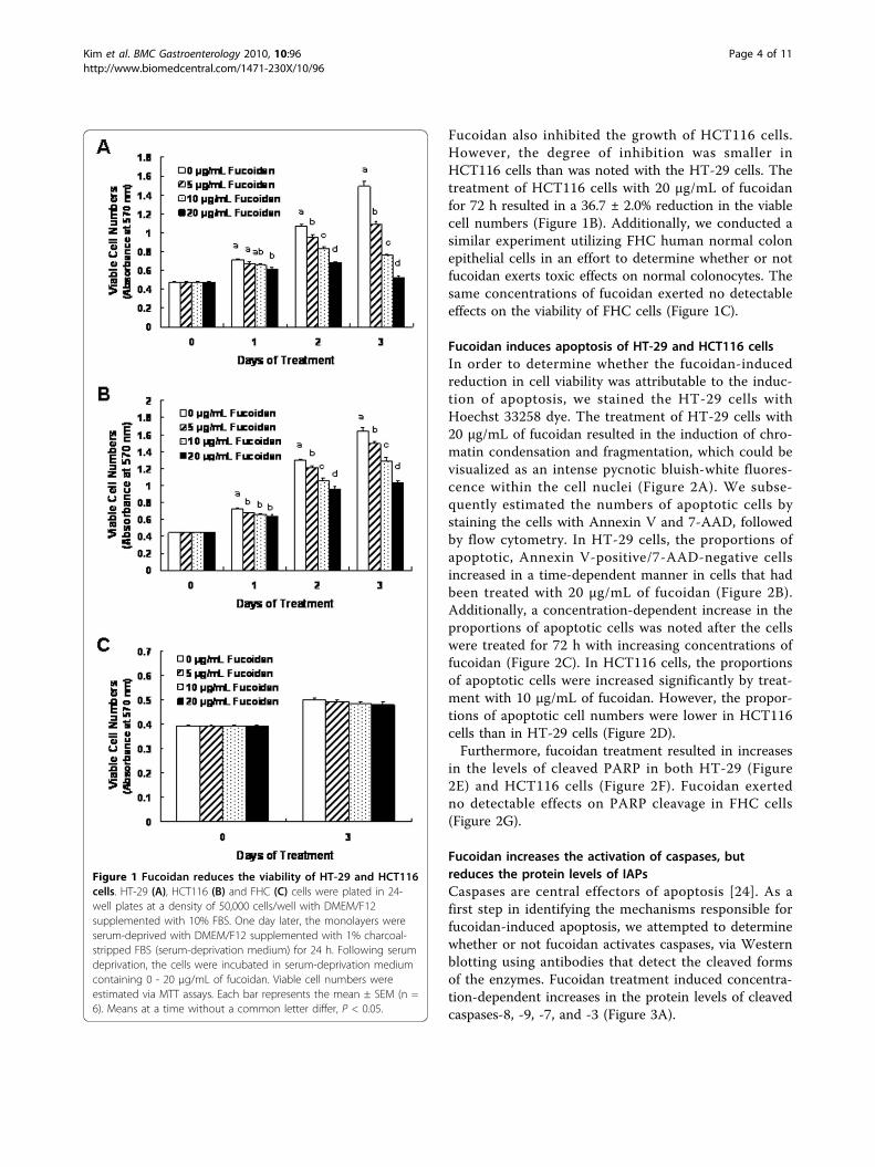
Fucoidan also inhibited the growth of HCT116 cells.However, the degree of inhibition was smaller inHCT116 cells than was noted with the HT-29 cells. Thetreatment of HCT116 cells with 20 μg/mL of fucoidanfor 72 h resulted in a 36.7 ± 2.0% reduction in the viablecell numbers (Figure 1B). Additionally, we conducted asimilar experiment utilizing FHC human normal colonepithelial cells in an effort to determine whether or notfucoidan exerts toxic effects on normal colonocytes. Thesame concentrations of fucoidan exerted no detectableeffects on the viability of FHC cells (Figure 1C).
Fucoidan induces apoptosis of HT-29 and HCT116 cellsIn order to determine whether the fucoidan-inducedreduction in cell viability was attributable to the induc-tion of apoptosis, we stained the HT-29 cells withHoechst 33258 dye. The treatment of HT-29 cells with20 μg/mL of fucoidan resulted in the induction of chro-matin condensation and fragmentation, which could bevisualized as an intense pycnotic bluish-white fluores-cence within the cell nuclei (Figure 2A). We subse-quently estimated the numbers of apoptotic cells bystaining the cells with Annexin V and 7-AAD, followedby flow cytometry. In HT-29 cells, the proportions ofapoptotic, Annexin V-positive/7-AAD-negative cellsincreased in a time-dependent manner in cells that hadbeen treated with 20 μg/mL of fucoidan (Figure 2B).Additionally, a concentration-dependent increase in theproportions of apoptotic cells was noted after the cellswere treated for 72 h with increasing concentrations offucoidan (Figure 2C). In HCT116 cells, the proportionsof apoptotic cells were increased significantly by treat-ment with 10 μg/mL of fucoidan. However, the propor-tions of apoptotic cell numbers were lower in HCT116cells than in HT-29 cells (Figure 2D).Furthermore, fucoidan treatment resulted in increases
in the levels of cleaved PARP in both HT-29 (Figure2E) and HCT116 cells (Figure 2F). Fucoidan exertedno detectable effects on PARP cleavage in FHC cells(Figure 2G).
Fucoidan increases the activation of caspases, butreduces the protein levels of IAPsCaspases are central effectors of apoptosis [24]. As afirst step in identifying the mechanisms responsible forfucoidan-induced apoptosis, we attempted to determinewhether or not fucoidan activates caspases, via Westernblotting using antibodies that detect the cleaved formsof the enzymes. Fucoidan treatment induced concentra-tion-dependent increases in the protein levels of cleavedcaspases-8, -9, -7, and -3 (Figure 3A).
Figure 1 Fucoidan reduces the viability of HT-29 and HCT116cells. HT-29 (A), HCT116 (B) and FHC (C) cells were plated in 24-well plates at a density of 50,000 cells/well with DMEM/F12supplemented with 10% FBS. One day later, the monolayers wereserum-deprived with DMEM/F12 supplemented with 1% charcoal-stripped FBS (serum-deprivation medium) for 24 h. Following serumdeprivation, the cells were incubated in serum-deprivation mediumcontaining 0 - 20 μg/mL of fucoidan. Viable cell numbers wereestimated via MTT assays. Each bar represents the mean ± SEM (n =6). Means at a time without a common letter differ, P < 0.05.
Kim et al. BMC Gastroenterology 2010, 10:96http://www.biomedcentral.com/1471-230X/10/96
Page 4 of 11
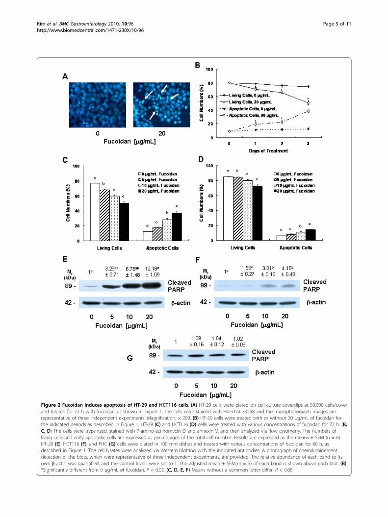
Figure 2 Fucoidan induces apoptosis of HT-29 and HCT116 cells. (A) HT-29 cells were plated on cell culture coverslips at 50,000 cells/coverand treated for 72 h with fucoidan, as shown in Figure 1. The cells were stained with Hoechst 33258 and the microphotograph images arerepresentative of three independent experiments. Magnification, × 200. (B) HT-29 cells were treated with or without 20 μg/mL of fucoidan forthe indicated periods as described in Figure 1. HT-29 (C) and HCT116 (D) cells were treated with various concentrations of fucoidan for 72 h. (B,C, D) The cells were trypsinzed, stained with 7-amino-actinomycin D and annexin V, and then analyzed via flow cytometry. The numbers ofliving cells and early apoptotic cells are expressed as percentages of the total cell number. Results are expressed as the means ± SEM (n = 6).HT-29 (E), HCT116 (F), and FHC (G) cells were plated in 100 mm dishes and treated with various concentrations of fucoidan for 60 h, asdescribed in Figure 1. The cell lysates were analyzed via Western blotting with the indicated antibodies. A photograph of chemiluminescentdetection of the blots, which were representative of three independent experiments, are provided. The relative abundance of each band to itsown b-actin was quantified, and the control levels were set to 1. The adjusted mean ± SEM (n = 3) of each band is shown above each blot. (B)*Significantly different from 0 μg/mL of fucoidan, P < 0.05. (C, D, E, F) Means without a common letter differ, P < 0.05.
Kim et al. BMC Gastroenterology 2010, 10:96http://www.biomedcentral.com/1471-230X/10/96
Page 5 of 11
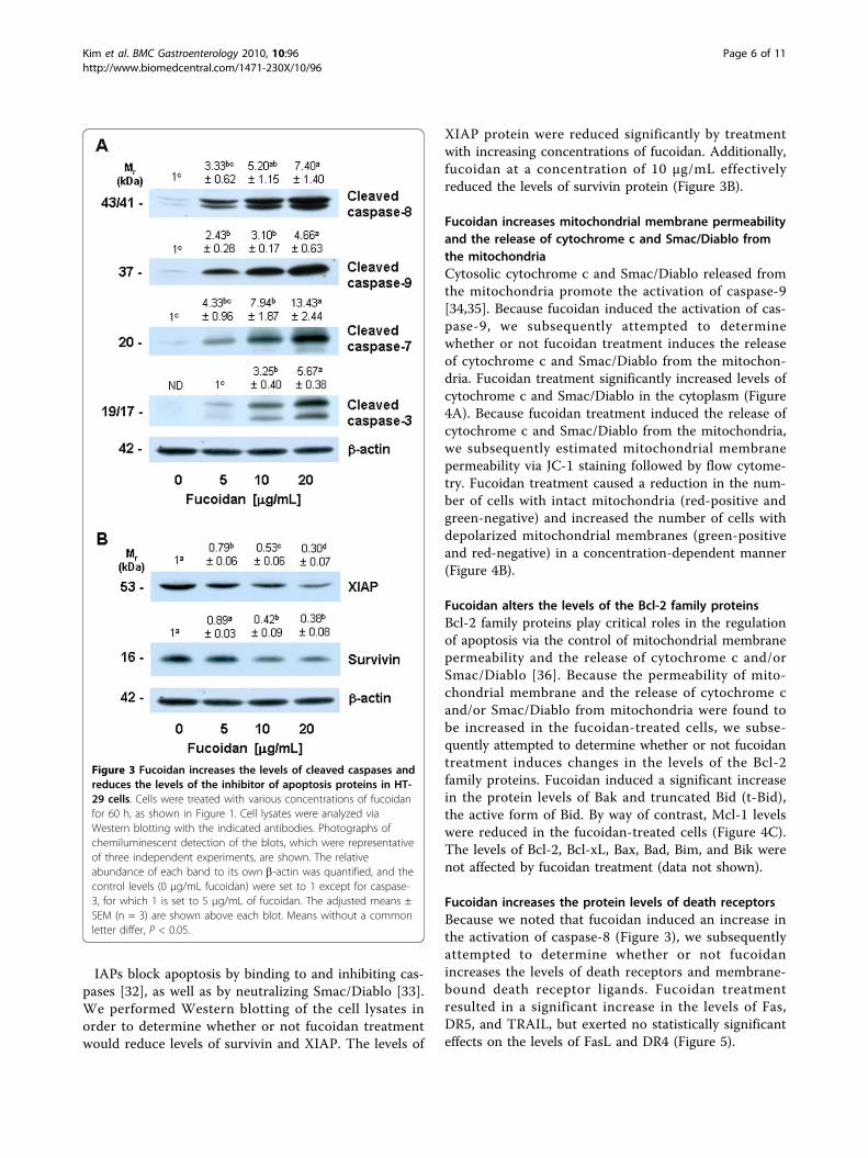
IAPs block apoptosis by binding to and inhibiting cas-pases [32], as well as by neutralizing Smac/Diablo [33].We performed Western blotting of the cell lysates inorder to determine whether or not fucoidan treatmentwould reduce levels of survivin and XIAP. The levels of
XIAP protein were reduced significantly by treatmentwith increasing concentrations of fucoidan. Additionally,fucoidan at a concentration of 10 μg/mL effectivelyreduced the levels of survivin protein (Figure 3B).
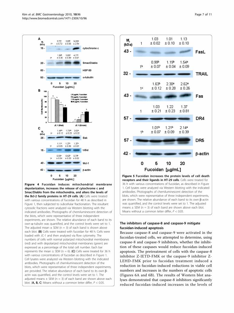
Fucoidan increases mitochondrial membrane permeabilityand the release of cytochrome c and Smac/Diablo fromthe mitochondriaCytosolic cytochrome c and Smac/Diablo released fromthe mitochondria promote the activation of caspase-9[34,35]. Because fucoidan induced the activation of cas-pase-9, we subsequently attempted to determinewhether or not fucoidan treatment induces the releaseof cytochrome c and Smac/Diablo from the mitochon-dria. Fucoidan treatment significantly increased levels ofcytochrome c and Smac/Diablo in the cytoplasm (Figure4A). Because fucoidan treatment induced the release ofcytochrome c and Smac/Diablo from the mitochondria,we subsequently estimated mitochondrial membranepermeability via JC-1 staining followed by flow cytome-try. Fucoidan treatment caused a reduction in the num-ber of cells with intact mitochondria (red-positive andgreen-negative) and increased the number of cells withdepolarized mitochondrial membranes (green-positiveand red-negative) in a concentration-dependent manner(Figure 4B).
Fucoidan alters the levels of the Bcl-2 family proteinsBcl-2 family proteins play critical roles in the regulationof apoptosis via the control of mitochondrial membranepermeability and the release of cytochrome c and/orSmac/Diablo [36]. Because the permeability of mito-chondrial membrane and the release of cytochrome cand/or Smac/Diablo from mitochondria were found tobe increased in the fucoidan-treated cells, we subse-quently attempted to determine whether or not fucoidantreatment induces changes in the levels of the Bcl-2family proteins. Fucoidan induced a significant increasein the protein levels of Bak and truncated Bid (t-Bid),the active form of Bid. By way of contrast, Mcl-1 levelswere reduced in the fucoidan-treated cells (Figure 4C).The levels of Bcl-2, Bcl-xL, Bax, Bad, Bim, and Bik werenot affected by fucoidan treatment (data not shown).
Fucoidan increases the protein levels of death receptorsBecause we noted that fucoidan induced an increase inthe activation of caspase-8 (Figure 3), we subsequentlyattempted to determine whether or not fucoidanincreases the levels of death receptors and membrane-bound death receptor ligands. Fucoidan treatmentresulted in a significant increase in the levels of Fas,DR5, and TRAIL, but exerted no statistically significanteffects on the levels of FasL and DR4 (Figure 5).
Figure 3 Fucoidan increases the levels of cleaved caspases andreduces the levels of the inhibitor of apoptosis proteins in HT-29 cells. Cells were treated with various concentrations of fucoidanfor 60 h, as shown in Figure 1. Cell lysates were analyzed viaWestern blotting with the indicated antibodies. Photographs ofchemiluminescent detection of the blots, which were representativeof three independent experiments, are shown. The relativeabundance of each band to its own b-actin was quantified, and thecontrol levels (0 μg/mL fucoidan) were set to 1 except for caspase-3, for which 1 is set to 5 μg/mL of fucoidan. The adjusted means ±SEM (n = 3) are shown above each blot. Means without a commonletter differ, P < 0.05.
Kim et al. BMC Gastroenterology 2010, 10:96http://www.biomedcentral.com/1471-230X/10/96
Page 6 of 11
The inhibitors of caspase-8 and caspase-9 mitigatefucoidan-induced apoptosisBecause caspase-8 and caspase-9 were activated in thefucoidan-treated cells, we attempted to determine, usingcaspase-8 and caspase-9 inhibitors, whether the inhibi-tion of these caspases would reduce fucoidan-inducedapoptosis. The pretreatment of cells with the caspase-8inhibitor Z-IETD-FMK or the caspase-9 inhibitor Z-LEHD-FMK prior to fucoidan treatment induced areduction in fucoidan-induced reductions in viable cellnumbers and increases in the numbers of apoptotic cells(Figures 6A and 6B). The results of Western blot ana-lyses demonstrated that caspase-8 inhibitors significantlyreduced fucoidan-induced increases in the levels of
Figure 4 Fucoidan induces mitochondrial membranedepolarization, increases the release of cytochrome c andSmac/Diablo from the mitochondria, and alters the levels ofthe Bcl-2 family proteins in HT-29 cells. (A) Cells were treatedwith various concentrations of fucoidan for 48 h as described inFigure 1, then subjected to subcellular fractionation. The resultantcytosolic fractions were analyzed via Western blotting with theindicated antibodies. Photographs of chemiluminescent detection ofthe blots, which were representative of three independentexperiments, are shown. The relative abundance of each band to itsown a-tubulin was quantified, and the control levels were set to 1.The adjusted mean ± SEM (n = 3) of each band is shown aboveeach blot. (B) Cells were treated with fucoidan for 48 h. Cells wereloaded with JC-1 and then analyzed via flow cytometry. Thenumbers of cells with normal polarized mitochondrial membranes(red) and with depolarized mitochondrial membranes (green) areexpressed as a percentage of the total cell number. Each barrepresents the mean ± SEM (n = 6). (C) Cells were treated for 36 hwith various concentrations of fucoidan as described in Figure 1.Cell lysates were analyzed via Western blotting with the indicatedantibodies. Photographs of chemiluminescent detection of theblots, which were representative of three independent experiments,are provided. The relative abundance of each band to its own b-actin was quantified, and the control levels were set to 1. Theadjusted means ± SEM (n = 3) of each band are shown above eachblot. (A, B, C) Means without a common letter differ, P < 0.05.
Figure 5 Fucoidan increases the protein levels of cell deathreceptors and their ligands in HT-29 cells. Cells were treated for36 h with various concentrations of fucoidan, as described in Figure1. Cell lysates were analyzed via Western blotting with the indicatedantibodies. Photographs of chemiluminescent detection of theblots, which were representative of three independent experiments,are shown. The relative abundance of each band to its own b-actinwas quantified, and the control levels were set to 1. The adjustedmeans ± SEM (n = 3) of each band are shown above each blot.Means without a common letter differ, P < 0.05.
Kim et al. BMC Gastroenterology 2010, 10:96http://www.biomedcentral.com/1471-230X/10/96
Page 7 of 11
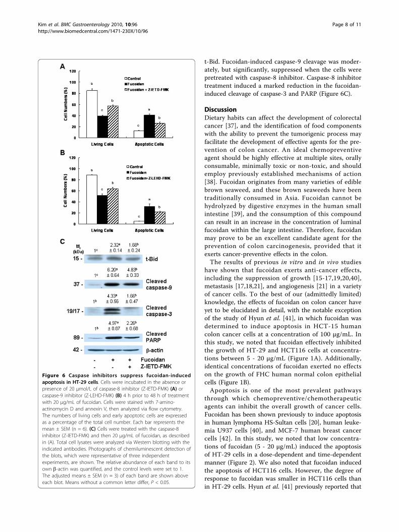
t-Bid. Fucoidan-induced caspase-9 cleavage was moder-ately, but significantly, suppressed when the cells werepretreated with caspase-8 inhibitor. Caspase-8 inhibitortreatment induced a marked reduction in the fucoidan-induced cleavage of caspase-3 and PARP (Figure 6C).
DiscussionDietary habits can affect the development of colorectalcancer [37], and the identification of food componentswith the ability to prevent the tumorigenic process mayfacilitate the development of effective agents for the pre-vention of colon cancer. An ideal chemopreventiveagent should be highly effective at multiple sites, orallyconsumable, minimally toxic or non-toxic, and shouldemploy previously established mechanisms of action[38]. Fucoidan originates from many varieties of ediblebrown seaweed, and these brown seaweeds have beentraditionally consumed in Asia. Fucoidan cannot behydrolyzed by digestive enzymes in the human smallintestine [39], and the consumption of this compoundcan result in an increase in the concentration of luminalfucoidan within the large intestine. Therefore, fucoidanmay prove to be an excellent candidate agent for theprevention of colon carcinogenesis, provided that itexerts cancer-preventive effects in the colon.The results of previous in vitro and in vivo studies
have shown that fucoidan exerts anti-cancer effects,including the suppression of growth [15-17,19,20,40],metastasis [17,18,21], and angiogenesis [21] in a varietyof cancer cells. To the best of our (admittedly limited)knowledge, the effects of fucoidan on colon cancer haveyet to be elucidated in detail, with the notable exceptionof the study of Hyun et al. [41], in which fucoidan wasdetermined to induce apoptosis in HCT-15 humancolon cancer cells at a concentration of 100 μg/mL. Inthis study, we noted that fucoidan effectively inhibitedthe growth of HT-29 and HCT116 cells at concentra-tions between 5 - 20 μg/mL (Figure 1A). Additionally,identical concentrations of fucoidan exerted no effectson the growth of FHC human normal colon epithelialcells (Figure 1B).Apoptosis is one of the most prevalent pathways
through which chemopreventive/chemotherapeuticagents can inhibit the overall growth of cancer cells.Fucoidan has been shown previously to induce apoptosisin human lymphoma HS-Sultan cells [20], human leuke-mia U937 cells [40], and MCF-7 human breast cancercells [42]. In this study, we noted that low concentra-tions of fucoidan (5 - 20 μg/mL) induced the apoptosisof HT-29 cells in a dose-dependent and time-dependentmanner (Figure 2). We also noted that fucoidan inducedthe apoptosis of HCT116 cells. However, the degree ofresponse to fucoidan was smaller in HCT116 cells thanin HT-29 cells. Hyun et al. [41] previously reported that
Figure 6 Caspase inhibitors suppress fucoidan-inducedapoptosis in HT-29 cells. Cells were incubated in the absence orpresence of 20 μmol/L of caspase-8 inhibitor (Z-IETD-FMK) (A) orcaspase-9 inhibitor (Z-LEHD-FMK) (B) 4 h prior to 48 h of treatmentwith 20 μg/mL of fucoidan. Cells were stained with 7-amino-actinomycin D and annexin V, then analyzed via flow cytometry.The numbers of living cells and early apoptotic cells are expressedas a percentage of the total cell number. Each bar represents themean ± SEM (n = 6). (C) Cells were treated with the caspase-8inhibitor (Z-IETD-FMK) and then 20 μg/mL of fucoidan, as describedin (A). Total cell lysates were analyzed via Western blotting with theindicated antibodies. Photographs of chemiluminescent detection ofthe blots, which were representative of three independentexperiments, are shown. The relative abundance of each band to itsown b-actin was quantified, and the control levels were set to 1.The adjusted means ± SEM (n = 3) of each band are shown aboveeach blot. Means without a common letter differ, P < 0.05.
Kim et al. BMC Gastroenterology 2010, 10:96http://www.biomedcentral.com/1471-230X/10/96
Page 8 of 11

high concentrations of fucoidan (100 μg/mL) inducedapoptosis in HCT-15 cells. These observations suggestthat fucoidan induces apoptosis in human colon cancercells, but that the efficacy of fucoidan in inducing apop-tosis varies among different types of colon cancer cells.The potent in vitro efficacy of fucoidan in colon cancercells indicates that fucoidan may potentially prove usefulin the prevention of colon carcinoma. However, itremains to be determined whether or not fucoidan sup-presses the development of colon cancer in both animalcancer models and humans. Additionally, it will also benecessary to determine why the degree of response tofucoidan varies among different types of colon cancercells.Chemopreventive/chemotherapeutic agents induce
apoptosis in a variety of cancer cells via a variety ofmechanisms. Aisa et al. [20] reported previously thatfucoidan induces apoptosis via the activation of caspase-3 and downregulation of the ERK pathway in humanHS-Sultan cells. Fucoidan has been shown to induceapoptosis in MCF-7 cells via a caspase-8-dependentpathway [42]. Additionally, Hyun et al. [41] reportedthat 100 μg/mL of fucoidan induced apoptosis in HCT-15 cells via the activation of caspase-9 and -3 accompa-nied by changes in Bcl-2 and Bax, as well as changes inthe phosphorylation of ERK, p38 kinase, and Akt. Inthis study, we noted that fucoidan at a concentration of5 - 20 μg/mL 1) increased the activation of caspases, 2)reduced the protein levels of IAPs, 3) increased mito-chondrial membrane permeability and cytochrome cand Smac/Diablo release, 4) increased the levels of Bakand t-Bid but reduced the levels of Mcl-1, and 5)increased the levels of Fas, DR5, and TRAIL in HT-29human colon cancer cells. We also noted that the inhi-bitors of caspase-8 and caspase-9 reduced fucoidan-induced apoptosis. The results of this study show thatfucoidan induces apoptosis through the activation ofcaspases via both death receptor-mediated and mito-chondria-mediated apoptotic pathways.Caspases perform critically important roles in the
induction of apoptosis. Caspases are classified based ontheir mode of activation as either initiator or effectorcaspases. Initiators such as caspase-8 and -9 are referredto as apical caspases, which are activated by a variety ofapoptotic signals. Activated initiator caspases can cleaveand activate effector caspases such as caspase-3 and cas-pase-7, which in turn cleave a variety of cellular sub-strates, most notably PARP. One of the most importantfunctions of PARP is to help repair single-strand DNAnicks; thus, cleaved PARP is a useful marker for apopto-sis [24,43]. In this study, we determined that fucoidaninduces the activation of caspases-8, -9, -3, and -7 (Fig-ure 3), as well as PARP cleavage (Figure 2D). Addition-ally, we noted that individual caspase-8 or -9 specific
inhibitors induced a reduction in fucoidan-inducedapoptosis. These results show that the activation ofthese caspases is one of the principal mechanisms bywhich fucoidan induces apoptosis.Caspase activation is triggered primarily via two dis-
tinct but interconnected pathways–namely, the deathreceptor- and mitochondria-mediated pathways. In thedeath receptor-mediated pathway, the binding of deathreceptor ligands (e.g., FasL and TRAIL) to their specificdeath receptors (e.g., Fas, DR4 and DR5) located on theplasma membrane induces the activation of caspase-8.Activated caspase-8 directly triggers the activation ofdownstream caspase-3 and/or cleaves Bid, a BH3-onlypro-apoptotic Bcl-2 family protein. Upon cleavage, t-Bidtranslocates to the mitochondria, where it enhances thepermeability of the mitochondrial membrane, and subse-quently induces cytochrome c release and caspase-9activation [24]. We determined that fucoidan treatmentinduced an increase in the levels of Fas, TRAIL, andDR5 proteins (Figure 5). Caspase-8 and t-Bid levels werealso shown to have increased in the fucoidan-treatedcells (Figure 3 and 4). Additionally, we noted that thecaspase-8 inhibitor, Z-IETD-FMK, effectively mitigatesfucoidan-induced apoptosis and PARP cleavage. Further-more, this inhibitor was shown to reduce the fucoidan-induced cleavage of Bid, caspase-9, and caspase-3 (Fig-ure 6). These findings demonstrate that the activation ofcaspase-8 contributes to the activation of caspase-3 viaboth the direct activation of this enzyme and via theactivation of caspase-9 by Bid cleavage, in fucoidan-trea-ted cells. Collectively, our findings demonstrate that theactivation of the death receptor-mediated pathway isinvolved in fucoidan-induced apoptosis in HT-29 cells,via both the direct and indirect activation of caspase-3.In the mitochondria-mediated pathway, apoptotic sti-
muli enhance the permeability of the outer mitochon-drial membranes and the subsequent release into thecytoplasm of pro-apoptotic factors, including cyto-chrome c and Smac/Diablo. Cytosolic cytochrome csubsequently binds to apoptosis protease-activating fac-tor 1 (Apaf-1) and inactive procaspase-9 to form anapoptosome, thereby resulting in caspase-9 activation.Activated caspase-9, in turn, triggers the subsequentcleavage of caspases-3 and -7 [34,44]. In addition todemonstrating that fucoidan induces caspases-3 and -7,we determined that fucoidan increased mitochondrialmembrane permeability, the release of cytochrome cfrom mitochondria, and the activation of caspase-9 (Fig-ures 3 and 4). The activation of the mitochondria-mediated pathway results in the release of Smac/Diablo,which confiscates the IAP obstruction of caspase activa-tion [45]. IAPs, including survivin and XIAP, functionby binding to and inhibiting several caspases, such ascaspase-9 [33]. In this study, we determined that
Kim et al. BMC Gastroenterology 2010, 10:96http://www.biomedcentral.com/1471-230X/10/96
Page 9 of 11
fucoidan treatment increased the release of Smac/Diablofrom the mitochondria (Figure 4), but reduced survivinand XIAP levels in HT-29 cells (Figure 3). Our resultsshowed that both the increases in the release of cyto-chrome c and Smac/Diablo and the downregulation ofIAPs were involved in the fucoidan-induced regulationof caspase-9 activity and apoptosis in HT-29 cells.The permeability of the mitochondrial membrane is
regulated precisely by the Bcl-2 family proteins. Anti- orpro-apoptotic Bcl-2 family proteins reside within thecytoplasm or on the outer membranes of the mitochon-dria. In response to apoptotic stimuli, these proteinsform either homo- or hetero-dimers, and then appear toperform distinct functions in the regulation of mito-chondrial membrane permeability [46,47]. In this study,fucoidan was shown to affect the levels of Bcl-2 familyproteins. Fucoidan treatment increased the levels of pro-apoptotic Bak and t-Bid, but reduced the levels of anti-apoptotic Mcl-1 (Figure 4). The findings of this studydemonstrate that the alteration in Bcl-2 family proteinscontributed to an increase in mitochondrial membranepermeability and cytochrome c and Smac/Diablo release,and subsequent caspase-9 activation in fucoidan-treatedHT-29 cells.
ConclusionThe results of this study showed that fucoidan inhibitsgrowth and induces apoptosis in HT-29 human coloncancer cells, and this effect is mediated by the activationof caspases. The findings of the present study show thatfucoidan activates caspases via both the death receptor-mediated and mitochondria-mediated apoptotic path-ways. This study provides a molecular basis for usingfucoidan as a potential apoptosis-inducing agent. Thus,studies should be conducted in the future to evaluatethe potential of fucoidan as a colon cancer-preventiveagent in experimental animal models and in humans.
Abbreviations7-AAD: 7-amino-actinomycin D; DMEM/F-12: Dulbecco’s Modified Eagle’sMedium/Ham’s F-12 nutrient mixture; DR: death receptor; FasL: Fas ligand;FBS: fetal bovine serum; HRP: horse-radish peroxidase; HSP: heat shockprotein; IAP: inhibitor of apoptosis protein: JC-1,5,5’,6,6’-tetrachloro-1,1’,3,3’-tetraethyl-imidacarbocyanine iodide; MTT: 3-[4,5-dimethylthiazol-2-yl]-2,5-diphenyltetrazolium bromide; PARP: poly (ADP-ribose) polymerase; t-Bid:truncated Bid; TRAIL: tumor necrosis factor-related apoptosis-inducing ligand;XIAP: X-linked inhibitor of apoptosis protein
AcknowledgementsThis work was supported by research grants from the National ResearchFoundation of Korea (NRF) for the Biofoods Research Program, Ministry ofEducation, Science and Technology and supported by the Ministry ofKnowledge Economy through the Center for Efficacy Assessment andDevelopment of Functional Foods and Drugs at Hallym University, Korea.
Author details1Department of Food Science and Nutrition, Hallym University, Chuncheon,200-702, Korea. 2Center for Efficacy Assessment and Development of
Functional Foods and Drugs, Hallym University, Chuncheon, 200-702, Korea.3Department of Biochemistry, College of Medicine, Hallym University,Chuncheon, 200-702, Korea. 4Medical & Bio-Materials Research Center,Hallym University, Chuncheon, 200-702, Korea.
Authors’ contributionsEJK, JYL, and JHYP designed this project. EJK performed the overallbiochemical analysis and wrote the first draft of the manuscript. SYPperformed Hoechst staining. JHYP directed the overall study and revised themanuscript. All authors contributed to the discussion of the data, and readand approved the final manuscript.
Competing interestsThe authors declare that they have no competing interests.
Received: 26 April 2010 Accepted: 22 August 2010Published: 22 August 2010
References1. Jemal A, Siegel R, Ward E, Hao Y, Xu J, Murray T, Thun MJ: Cancer statistics,
2008. CA Cancer J Clin 2008, 58(2):71-96.2. Chung KY, Saltz LB: Adjuvant therapy of colon cancer: current status and
future directions. Cancer J 2007, 13(3):192-197.3. Rabik CA, Dolan ME: Molecular mechanisms of resistance and toxicity
associated with platinating agents. Cancer Treat Rev 2007, 33(1):9-23.4. Macdonald JS, Astrow AB: Adjuvant therapy of colon cancer. Semin Oncol
2001, 28(1):30-40.5. Mann JR, Backlund MG, DuBois RN: Mechanisms of disease: Inflammatory
mediators and cancer prevention. Nature clinical practice 2005,2(4):202-210.
6. Li B, Lu F, Wei X, Zhao R: Fucoidan: structure and bioactivity. Molecules2008, 13(8):1671-1695.
7. Bilan MI, Grachev AA, Ustuzhanina NE, Shashkov AS, Nifantiev NE, Usov AI:Structure of a fucoidan from the brown seaweed Fucus evanescens C.Ag. Carbohydrate research 2002, 337(8):719-730.
8. Bilan MI, Grachev AA, Shashkov AS, Nifantiev NE, Usov AI: Structure of afucoidan from the brown seaweed Fucus serratus L. Carbohydrateresearch 2006, 341(2):238-245.
9. Zapopozhets TS, Besednova NN, Loenko Iu N: [Antibacterial andimmunomodulating activity of fucoidan]. Antibiot Khimioter 1995,40(2):9-13.
10. Hayashi K, Nakano T, Hashimoto M, Kanekiyo K, Hayashi T: Defensiveeffects of a fucoidan from brown alga Undaria pinnatifida againstherpes simplex virus infection. Int Immunopharmacol 2008, 8(1):109-116.
11. Cumashi A, Ushakova NA, Preobrazhenskaya ME, D’Incecco A, Piccoli A,Totani L, Tinari N, Morozevich GE, Berman AE, Bilan MI, et al: A comparativestudy of the anti-inflammatory, anticoagulant, antiangiogenic, andantiadhesive activities of nine different fucoidans from brown seaweeds.Glycobiology 2007, 17(5):541-552.
12. Wang J, Zhang Q, Zhang Z, Li Z: Antioxidant activity of sulfatedpolysaccharide fractions extracted from Laminaria japonica. Int J BiolMacromol 2008, 42(2):127-132.
13. Matsumoto S, Nagaoka M, Hara T, Kimura-Takagi I, Mistuyama K, Ueyama S:Fucoidan derived from Cladosiphon okamuranus Tokida amelioratesmurine chronic colitis through the down-regulation of interleukin-6production on colonic epithelial cells. Clin Exp Immunol 2004,136(3):432-439.
14. Choi EM, Kim AJ, Kim YO, Hwang JK: Immunomodulating activity ofarabinogalactan and fucoidan in vitro. J Med Food 2005, 8(4):446-453.
15. Itoh H, Noda H, Amano H, Zhuaug C, Mizuno T, Ito H: Antitumor activityand immunological properties of marine algal polysaccharides,especially fucoidan, prepared from Sargassum thunbergii ofPhaeophyceae. Anticancer Res 1993, 13(6A):2045-2052.
16. Zhuang C, Itoh H, Mizuno T, Ito H: Antitumor active fucoidan from thebrown seaweed, umitoranoo (Sargassum thunbergii). Biosci BiotechnolBiochem 1995, 59(4):563-567.
17. Alekseyenko TV, Zhanayeva SY, Venediktova AA, Zvyagintseva TN,Kuznetsova TA, Besednova NN, Korolenko TA: Antitumor andantimetastatic activity of fucoidan, a sulfated polysaccharide isolatedfrom the Okhotsk Sea Fucus evanescens brown alga. Bull Exp Biol Med2007, 143(6):730-732.
Kim et al. BMC Gastroenterology 2010, 10:96http://www.biomedcentral.com/1471-230X/10/96
Page 10 of 11
18. Coombe DR, Parish CR, Ramshaw IA, Snowden JM: Analysis of theinhibition of tumour metastasis by sulphated polysaccharides. Int JCancer 1987, 39(1):82-88.
19. Riou D, Colliec-Jouault S, Pinczon du Sel D, Bosch S, Siavoshian S, Le Bert V,Tomasoni C, Sinquin C, Durand P, Roussakis C: Antitumor andantiproliferative effects of a fucan extracted from ascophyllum nodosumagainst a non-small-cell bronchopulmonary carcinoma line. AnticancerRes 1996, 16(3A):1213-1218.
20. Aisa Y, Miyakawa Y, Nakazato T, Shibata H, Saito K, Ikeda Y, Kizaki M:Fucoidan induces apoptosis of human HS-sultan cells accompanied byactivation of caspase-3 and down-regulation of ERK pathways. Am JHematol 2005, 78(1):7-14.
21. Ye J, Li Y, Teruya K, Katakura Y, Ichikawa A, Eto H, Hosoi M, Hosoi M,Nishimoto S, Shirahata S: Enzyme-digested Fucoidan Extracts Derivedfrom Seaweed Mozuku of Cladosiphon novae-caledoniae kylin InhibitInvasion and Angiogenesis of Tumor Cells. Cytotechnology 2005, 47(1-3):117-126.
22. Hanahan D, Weinberg RA: The hallmarks of cancer. Cell 2000, 100(1):57-70.23. Bedi A, Pasricha PJ, Akhtar AJ, Barber JP, Bedi GC, Giardiello FM,
Zehnbauer BA, Hamilton SR, Jones RJ: Inhibition of apoptosis duringdevelopment of colorectal cancer. Cancer research 1995, 55(9):1811-1816.
24. Jin Z, El-Deiry WS: Overview of cell death signaling pathways. Cancer BiolTher 2005, 4(2):139-163.
25. Ghobrial IM, Witzig TE, Adjei AA: Targeting apoptosis pathways in cancertherapy. CA Cancer J Clin 2005, 55(3):178-194.
26. Kim EJ, Holthuizen PE, Park HS, Ha YL, Jung KC, Park JH: Trans-10,cis-12-conjugated linoleic acid inhibits Caco-2 colon cancer cell growth. Am JPhysiol Gastrointest Liver Physiol 2002, 283(2):G357-367.
27. Black WAP, Dewar ET, Woodward FN: Manufacture of algal chemicals. IV -Laboratory-scale isolation of fucoidin from brown marine algae. J SciFood Agric 1952, 3:122-129.
28. Kim EJ, Park SY, Shin HK, Kwon DY, Surh YJ, Park JH: Activation of caspase-8 contributes to 3,3’-Diindolylmethane-induced apoptosis in coloncancer cells. J Nutr 2007, 137(1):31-36.
29. Jung JI, Lim SS, Choi HJ, Cho HJ, Shin HK, Kim EJ, Chung WY, Park KK,Park JH: Isoliquiritigenin induces apoptosis by depolarizing mitochondrialmembranes in prostate cancer cells. J Nutr Biochem 2006, 17(10):689-696.
30. Cho HJ, Kim WK, Kim EJ, Jung KC, Park S, Lee HS, Tyner AL, Park JH:Conjugated linoleic acid inhibits cell proliferation and ErbB3 signaling inHT-29 human colon cell line. Am J Physiol Gastrointest Liver Physiol 2003,284(6):G996-1005.
31. Eguchi Y, Srinivasan A, Tomaselli KJ, Shimizu S, Tsujimoto Y: ATP-dependent steps in apoptotic signal transduction. Cancer research 1999,59(9):2174-2181.
32. Schimmer AD: Inhibitor of apoptosis proteins: translating basicknowledge into clinical practice. Cancer research 2004, 64(20):7183-7190.
33. Wilkinson JC, Wilkinson AS, Scott FL, Csomos RA, Salvesen GS, Duckett CS:Neutralization of Smac/Diablo by inhibitors of apoptosis (IAPs). Acaspase-independent mechanism for apoptotic inhibition. J Biol Chem2004, 279(49):51082-51090.
34. Li P, Nijhawan D, Budihardjo I, Srinivasula SM, Ahmad M, Alnemri ES,Wang X: Cytochrome c and dATP-dependent formation of Apaf-1/caspase-9 complex initiates an apoptotic protease cascade. Cell 1997,91(4):479-489.
35. Du C, Fang M, Li Y, Li L, Wang X: Smac, a mitochondrial protein thatpromotes cytochrome c-dependent caspase activation by eliminatingIAP inhibition. Cell 2000, 102(1):33-42.
36. Cory S, Huang DC, Adams JM: The Bcl-2 family: roles in cell survival andoncogenesis. Oncogene 2003, 22(53):8590-8607.
37. WCRF/AICR: Food, Nutrition, Physical Activity and the Prevention ofCancer: a Global Perspective. Washington, DC American Institute forCancer Research 2009.
38. Rajamanickam S, Agarwal R: Natural products and colon cancer: currentstatus and future prospects. Drug Dev Res 2008, 69(7):460-471.
39. Bilan MI, Grachev AA, Ustuzhanina NE, Shashkov AS, Nifantiev NE, Usov AI:A highly regular fraction of a fucoidan from the brown seaweed Fucusdistichus L. Carbohydrate research 2004, 339(3):511-517.
40. Teruya T, Konishi T, Uechi S, Tamaki H, Tako M: Anti-proliferative activity ofoversulfated fucoidan from commercially cultured Cladosiphonokamuranus TOKIDA in U937 cells. Int J Biol Macromol 2007,41(3):221-226.
41. Hyun JH, Kim SC, Kang JI, Kim MK, Boo HJ, Kwon JM, Koh YS, Hyun JW,Park DB, Yoo ES, et al: Apoptosis inducing activity of fucoidan in HCT-15colon carcinoma cells. Biol Pharm Bull 2009, 32(10):1760-1764.
42. Yamasaki-Miyamoto Y, Yamasaki M, Tachibana H, Yamada K: Fucoidaninduces apoptosis through activation of caspase-8 on human breastcancer MCF-7 cells. J Agric Food Chem 2009, 57(18):8677-8682.
43. Danial NN: BCL-2 family proteins: critical checkpoints of apoptotic celldeath. Clin Cancer Res 2007, 13(24):7254-7263.
44. Antonsson B, Martinou JC: The Bcl-2 protein family. Exp Cell Res 2000,256(1):50-57.
45. Deng Y, Lin Y, Wu X: TRAIL-induced apoptosis requires Bax-dependentmitochondrial release of Smac/DIABLO. Genes Dev 2002, 16(1):33-45.
46. Cory S, Adams JM: The Bcl2 family: regulators of the cellular life-or-deathswitch. Nat Rev Cancer 2002, 2(9):647-656.
47. Breckenridge DG, Xue D: Regulation of mitochondrial membranepermeabilization by BCL-2 family proteins and caspases. Curr Opin CellBiol 2004, 16(6):647-652.
Pre-publication historyThe pre-publication history for this paper can be accessed here:http://www.biomedcentral.com/1471-230X/10/96/prepub
doi:10.1186/1471-230X-10-96Cite this article as: Kim et al.: Fucoidan present in brown algae inducesapoptosis of human colon cancer cells. BMC Gastroenterology 2010 10:96.
Submit your next manuscript to BioMed Centraland take full advantage of:
• Convenient online submission
• Thorough peer review
• No space constraints or color figure charges
• Immediate publication on acceptance
• Inclusion in PubMed, CAS, Scopus and Google Scholar
• Research which is freely available for redistribution
Submit your manuscript at www.biomedcentral.com/submit
Kim et al. BMC Gastroenterology 2010, 10:96http://www.biomedcentral.com/1471-230X/10/96
Page 11 of 11
Related Documents

![Fucoidan Extract Induces Apoptosis in MCF-7 Cells via a ... · and sulfate, along with low amounts of xylose, uronic acid, and galactose [1–2]. Fucoidan has been reported to possess](https://static.cupdf.com/doc/110x72/5ec00e89405c9837c25fb420/fucoidan-extract-induces-apoptosis-in-mcf-7-cells-via-a-and-sulfate-along-with.jpg)