Eur Arch Psychiatry Clin Neurosci (2001) 251 : 225 – 231 © Steinkopff Verlag 2001 ■ Abstract Establishing the diagnosis in patients with clinical signs and symptoms suggesting primary degen- erative disease with marked frontal lobe involvement is difficult. Neuroimaging methods, in particular positron emission tomography (PET) with the tracer 18 fluoro-2- deoxyglucose (FDG) and cerebrospinal fluid (CSF) ex- amination of β-amyloid and tau-protein levels may give additional information. We report five patients with clinical and radiological features of degenerative de- mentia with predominantly frontal involvement and one patient with primary progressive aphasia Diagnostic work-up included computed tomography (CT), mag- netic resonance imaging (MRI), PET and tau-protein and β-amyloid level determination in CSF. While neu- ropsychological performance varied among patients,CT and MRI demonstrated persistently frontal lobe involve- ment. PET revealed corresponding changes with frontal hypometabolism, but in addition, four patients (among them two with no corresponding temporal changes in CT or MRI) showed a decreased glucose uptake in the temporal cortices. CSF samples from five patients re- vealed elevated β-amyloid 1–42 and tau levels in three and two patients, respectively. Reduced β-amyloid 1–40 was found in two patients.We conclude that occurrence of clinical symptoms of frontotemporal dementia is ac- companied by frontal hypometabolism regardless of ad- ditional clinical findings. The value of determination of β-amyloid and tau protein levels remains to be deter- mined. ■ Key words Frontotemporal dementia · Magnetic resonance imaging · PET · Tau protein · β-amyloid Introduction Clinical signs of frontotemporal dementia (FTD) com- prise disinhibition, loss of initiative, hyper-oral tenden- cies, utilization behavior, echolalia, perseveration and reduced speech output [2, 19, 20].Assessment includes a thorough physical and neurological examination, neu- ropsychological testing and neuroimaging procedures such as computed tomography and magnetic resonance imaging especially to exclude other types of dementia such as vascular dementia or brain tumors. Accurate clinical diagnosis is often difficult to estab- lish. While definite diagnosis can be obtained only by analysis of neuropathological features,correct diagnosis is of increased importance with the advent of new drugs for the treatment of Alzheimer’s disease. Recent studies indicate that functional neuroimaging methods such as positron emission tomography (PET) with 18 fluoro-2-deoxyglucose (FDG) may facilitate the diagnosis of Alzheimer’s disease [7], but only sporadic reports exist about the diagnostic value of PET for diag- nosis of FTD [8, 34]. Cerebrospinal fluid (CSF) examinations are neces- sary to exclude other medical conditions in particular inflammatory diseases of the central nervous system. CSF examinations can be extended to β-amyloid 1–42, β-amyloid 1–40 [10, 29] and tau-protein [9], which are ORIGINAL PAPER M. Jauss · K. Herholz · L. Kracht · J. Pantel · T. Hartmann · M. Jensen · M. Essig · J. Schröder Frontotemporal dementia: clinical, neuroimaging, and molecular biological findings in 6 patients Received: 26 March 2001 / Accepted: 28 August 2001 EAPCN 337 M. Jauss, M. D. () Dept. of Neurology University of Giessen Am Steg 14 35385 Giessen, Germany Tel.: + 49-6 41/9 94 53 38 Fax: + 49-6 41/9 91 99 36 E-Mail: [email protected] M. Jauss · J. Pantel · J. Schröder Section of Geriatric Psychiatry University of Heidelberg, Germany T. Hartmann ZMBH Heidelberg University of Heidelberg, Germany M. Jensen Karolinska Institut Huddinge, Sweden M. Essig DKFZ Heidelberg, Germany K. Herholz · L. Kracht Max-Planck-Institut für neurologische Forschung Köln, Germany

Welcome message from author
This document is posted to help you gain knowledge. Please leave a comment to let me know what you think about it! Share it to your friends and learn new things together.
Transcript

Eur Arch Psychiatry Clin Neurosci (2001) 251 : 225–231 © Steinkopff Verlag 2001
■ Abstract Establishing the diagnosis in patients withclinical signs and symptoms suggesting primary degen-erative disease with marked frontal lobe involvement isdifficult. Neuroimaging methods, in particular positronemission tomography (PET) with the tracer 18fluoro-2-deoxyglucose (FDG) and cerebrospinal fluid (CSF) ex-amination of β-amyloid and tau-protein levels may giveadditional information. We report five patients withclinical and radiological features of degenerative de-mentia with predominantly frontal involvement and onepatient with primary progressive aphasia Diagnosticwork-up included computed tomography (CT), mag-netic resonance imaging (MRI), PET and tau-proteinand β-amyloid level determination in CSF. While neu-ropsychological performance varied among patients,CTand MRI demonstrated persistently frontal lobe involve-ment. PET revealed corresponding changes with frontalhypometabolism, but in addition, four patients (amongthem two with no corresponding temporal changes inCT or MRI) showed a decreased glucose uptake in the
temporal cortices. CSF samples from five patients re-vealed elevated β-amyloid 1–42 and tau levels in threeand two patients, respectively. Reduced β-amyloid 1–40was found in two patients. We conclude that occurrenceof clinical symptoms of frontotemporal dementia is ac-companied by frontal hypometabolism regardless of ad-ditional clinical findings. The value of determination ofβ-amyloid and tau protein levels remains to be deter-mined.
■ Key words Frontotemporal dementia · Magneticresonance imaging · PET · Tau protein · β-amyloid
Introduction
Clinical signs of frontotemporal dementia (FTD) com-prise disinhibition, loss of initiative, hyper-oral tenden-cies, utilization behavior, echolalia, perseveration andreduced speech output [2, 19, 20].Assessment includes athorough physical and neurological examination, neu-ropsychological testing and neuroimaging proceduressuch as computed tomography and magnetic resonanceimaging especially to exclude other types of dementiasuch as vascular dementia or brain tumors.
Accurate clinical diagnosis is often difficult to estab-lish. While definite diagnosis can be obtained only byanalysis of neuropathological features,correct diagnosisis of increased importance with the advent of new drugsfor the treatment of Alzheimer’s disease.
Recent studies indicate that functional neuroimagingmethods such as positron emission tomography (PET)with 18fluoro-2-deoxyglucose (FDG) may facilitate thediagnosis of Alzheimer’s disease [7], but only sporadicreports exist about the diagnostic value of PET for diag-nosis of FTD [8, 34].
Cerebrospinal fluid (CSF) examinations are neces-sary to exclude other medical conditions in particularinflammatory diseases of the central nervous system.CSF examinations can be extended to β-amyloid 1–42,β-amyloid 1–40 [10, 29] and tau-protein [9], which are
ORIGINAL PAPER
M. Jauss · K. Herholz · L. Kracht · J. Pantel · T. Hartmann · M. Jensen · M. Essig · J. Schröder
Frontotemporal dementia: clinical, neuroimaging, and molecular biological findings in 6 patients
Received: 26 March 2001 / Accepted: 28 August 2001
EAPC
N 337
M. Jauss, M. D. (�)Dept. of NeurologyUniversity of GiessenAm Steg 1435385 Giessen, GermanyTel.: +49-6 41/9 94 53 38Fax: +49-6 41/9 91 99 36E-Mail: [email protected]
M. Jauss · J. Pantel · J. SchröderSection of Geriatric PsychiatryUniversity of Heidelberg, Germany
T. HartmannZMBH HeidelbergUniversity of Heidelberg, Germany
M. JensenKarolinska Institut Huddinge, Sweden
M. EssigDKFZ Heidelberg, Germany
K. Herholz · L. KrachtMax-Planck-Institut für neurologische ForschungKöln, Germany

226
discussed to be biochemical markers for Alzheimer’sdisease.
We report a series of 6 patients with clinical symp-toms of FTD with special emphasis on clinical work upincluding PET and CSF levels of β-amyloid 1–42,β-amy-loid 1–40, and tau-protein.
Patients and methods
Between 1992 and 1996, six patients admitted to the Section of Geri-atric Psychiatry, University of Heidelberg were clinically diagnosed asFTD or primary progressive aphasia according to history, medicaland psychopathological state and neuropsychological assessment af-ter exclusion of a history of head trauma, birth injury, electroconvul-sive therapy, or substance abuse. Symptoms of frontotemporal de-mentia were quantitatively assessed using the 9-point FTD scale [6].According to latest consensus criteria [21] that have considered fron-totemporal dementia and primary progressive aphasia as prototypicsyndromes for frontotemporal lobar degeneration we have includedboth patients with clinical diagnosis of frontotemporal dementia andwith primary progressive aphasia [16].
■ Clinical evaluation and assessment
All patients underwent thorough general and neurological examina-tions including computed tomography, MRI, and laboratory studiesin order to exclude metabolic, toxic, and inflammatory causes of theirdementia syndrome. None of the patients had evidence of cere-brovascular disease on CT and MRI scans.
Severity of cognitive impairment was assessed using the MiniMental State Examination (MMSE) [3], the Global Deterioration Scale(GDS) [25], and the Brief Cognitive Rating Scale (BCRS) [24].
For further neuropsychological characterization, the followingtests were applied: cognitive flexibility – Farbworttest (FWT) [22];verbal fluency – controlled word association (FAS) [31]; attentionalperformance – Alterskonzentrationstest (AKT) [22]; declarative mem-ory performance – Buschke selective reminding task (BSR) in the Ger-man version [12] and praxia – Apraxia test [12].
For interindividual comparison,test results were converted into z-values on the basis of the norm values established for healthy controls[13, 22, 31]. Z-values of less than –1 represent a test performance be-low those of 66 % of an age-matched healthy control sample, while z-values of less then –2 refer to a performance worse than 98 % of anage-matched healthy control sample.
■ Apparative tests
CT was obtained using standard 8mm slices parallel to orbito-meatalline, MRI was obtained with routine T1-weighted sagital and T2-weighted axial sequences.Both were rated qualitatively by the authors(J. P., M. E.) with respect to general and frontal atrophy on a 4-pointscale (absent, mild, moderate, marked). Electroencephalography(EEG) was obtained using standard 16-channel recording using the10–20 method for electrode placement, monopolar and bipolar leadswere used. Methods for CSF analysis (β-amyloid and tau-protein) aredescribed elsewhere [9, 10, 29].
PET scans were obtained after intravenous injection of160–380 mBq 18fluoro-2-deoxyglucose at the Max Planck Institute inCologne. Patients were examined in a resting state on an ECAT EX-ACT scanner, and local cerebral metabolic rates of glucose (CMRGlc,in µmol/100 g/min) in the whole brain were determined in 47 slicescomprising the whole brain using the Sokoloff model with adjust-ment of K1 to measured activity and a lumped constant of 0.42 [33].Slices were oriented along the AC-PC line.
The study was approved by the local ethical committee.
Patient characteristics and apparative findings
The clinical, neuropsychological, neuroimaging, andPET findings of the patients are listed below and sum-marized in Table 1 (clinical rating scales), Table 2 (neu-ropsychological testing), and Table 3 (CSF findings).
■ Patient 1
A 51 year old male, (who ran a transportation business)was admitted with a 10 month history of progressivecognitive decline, memory deficits, and paranoid delu-sions.
Neurological findings were normal except for grasp-ing and a positive palmo-mental reflex. During the clin-ical course he developed signs of severe behavioral dis-
Table 1 Clinical scales with the characteristics of the patients under investigation.BCRS axes are separated and mean values are given, where a maximum of 7 (severeimpairment) and a minimum of 1 (unimpaired) can be obtained. The Hachinskiischemia score was evaluated according to [15]. BCRS 1–5 includes concentration,short-term memory, long-term memory, orientation and ability of patient to carefor himself. BCRS 6–10 includes speech, psychomotor function, mood and behav-ior, constructive praxia and calculation
Characteristic Pat. 1 Pat. 2 Pat. 3 Pat. 4 Pat. 5 Pat. 6
MMSE 2 18 25 27 10 18FTD scale 7 9 7 6 9 8Hachinski ischemia score 2 1 3 2 1 2Global deterioration scale 6 4 2 2 5 4BCRS axes 1–5 3.4 3.6 2.2 1.8 5.3 4.2BCRS axes 6–10 4.5 3 3 1.4 3.8 4.5
Table 2 Neuropsychological testing. Test scores are given in z values: z values ofless than –1 represent a performance worse than 66% of an age-matched groupwhile a z value of less then –2 represent a performance worse than 98% of an age-matched group (marked with bold letters in the table). *) not able to co-operate
Characteristic Pat. 1 Pat. 2 Pat. 3 Pat. 4 Pat. 5 Pat. 6
Cognitive flexibility (FWT) *) –2.8 –2.5 –1.1 –2.3 –0.8Verbal fluency (FAS) –1.9 –0.9 –0.8 –2.9 –2.1 –0.6Attention performance (AKT) –0.7 –0.8 –0.9 –0.6 –1.6 –2.9Declarative memory –0.5 –0.4 –1.7 0.2 –1.9 –1.7
performance (BSR)Praxia (Apraxia test) –0.9 0.8 –1.3 0.8 –1.7 –0.6
Table 3 Laboratory characteristics. In healthy elderly controls (n=17) tau proteinwas 246.6 ± 111.4 pg/ml (mean and standard deviation) according to the labora-tory reference. In healthy elderly controls β-amyloid 1–40 was 2311 ± 546 and β-amyloid 1–42 was 74 ± 30 pM [10]. *) marks a value beyond 2-fold standard devi-ation
Characteristic Pat. 1 Pat. 2 Pat. 3 Pat. 4 Pat. 5
CSF protein [g/l] 0.40 0.11 0.31 0.55 0.36CSF cells [per µl] 6 1 4 1 1tau-protein [pg/ml] 164.0 174.4 138.0 323.30 315.40β-amyloid 40 [pM] 920* 2056 767* 2363 1373β-amyloid 42 [pM] 101 180* 201* 130 135*
227
inhibition such as hyperorality (eating candles and flow-ers). Neuropsychiatric testing revealed a lack of compre-hension with a severely impaired attentional perform-ance and verbal fluency. The patient was not able toco-operate in a test for cognitive flexibility (FWT).
Clinical diagnosis was frontotemporal dementia sinceclinical presentation revealed impairment of both frontaland temporal lobe functions.
EEG showed a mild general slowing with no focal ab-normalities and no signs for Jacob-Creutzfeld disease(e. g. triphasic waves). CSF findings, except reduced β-amyloid 40, were within normal limits.
CT and MRI showed a marked internal and moderategeneral and a marked frontal atrophy.
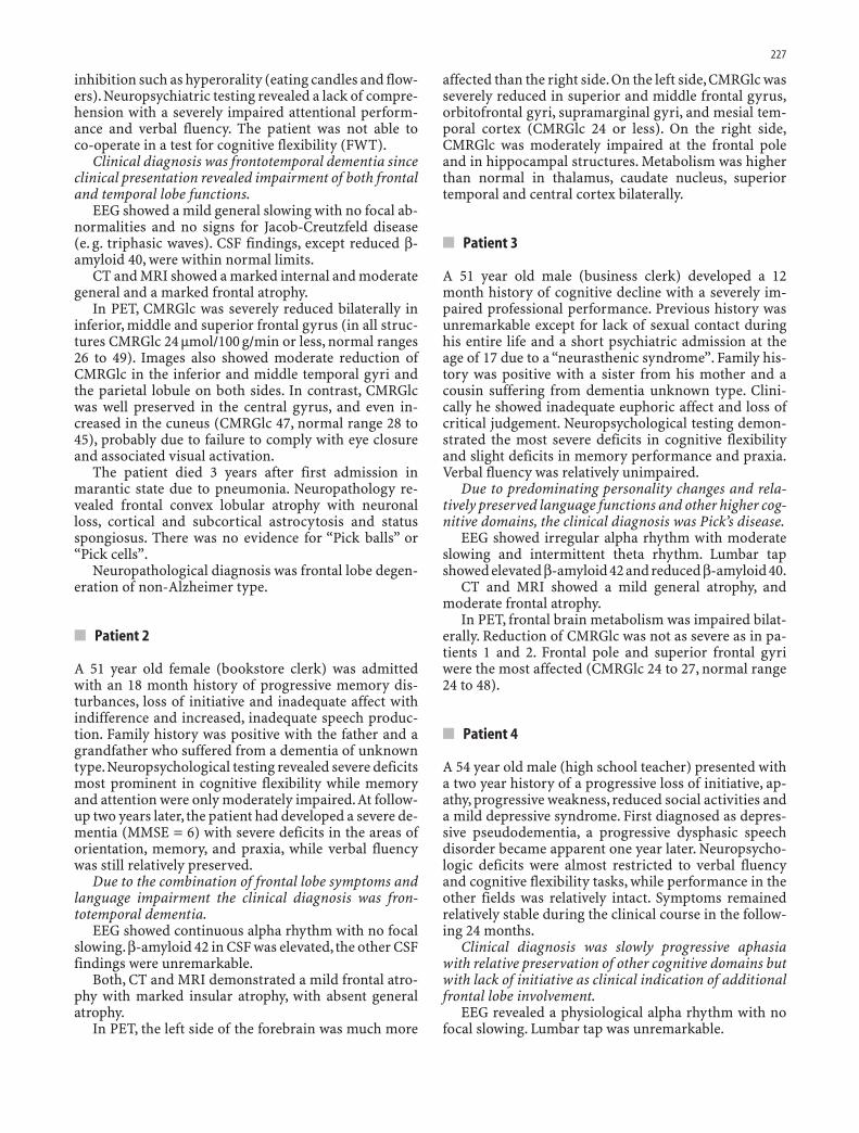
In PET, CMRGlc was severely reduced bilaterally ininferior, middle and superior frontal gyrus (in all struc-tures CMRGlc 24 µmol/100 g/min or less, normal ranges26 to 49). Images also showed moderate reduction ofCMRGlc in the inferior and middle temporal gyri andthe parietal lobule on both sides. In contrast, CMRGlcwas well preserved in the central gyrus, and even in-creased in the cuneus (CMRGlc 47, normal range 28 to45), probably due to failure to comply with eye closureand associated visual activation.
The patient died 3 years after first admission inmarantic state due to pneumonia. Neuropathology re-vealed frontal convex lobular atrophy with neuronalloss, cortical and subcortical astrocytosis and statusspongiosus. There was no evidence for “Pick balls” or“Pick cells”.
Neuropathological diagnosis was frontal lobe degen-eration of non-Alzheimer type.
■ Patient 2
A 51 year old female (bookstore clerk) was admittedwith an 18 month history of progressive memory dis-turbances, loss of initiative and inadequate affect withindifference and increased, inadequate speech produc-tion. Family history was positive with the father and agrandfather who suffered from a dementia of unknowntype.Neuropsychological testing revealed severe deficitsmost prominent in cognitive flexibility while memoryand attention were only moderately impaired.At follow-up two years later, the patient had developed a severe de-mentia (MMSE = 6) with severe deficits in the areas oforientation, memory, and praxia, while verbal fluencywas still relatively preserved.
Due to the combination of frontal lobe symptoms andlanguage impairment the clinical diagnosis was fron-totemporal dementia.
EEG showed continuous alpha rhythm with no focalslowing.β-amyloid 42 in CSF was elevated, the other CSFfindings were unremarkable.
Both, CT and MRI demonstrated a mild frontal atro-phy with marked insular atrophy, with absent generalatrophy.
In PET, the left side of the forebrain was much more
affected than the right side.On the left side,CMRGlc wasseverely reduced in superior and middle frontal gyrus,orbitofrontal gyri, supramarginal gyri, and mesial tem-poral cortex (CMRGlc 24 or less). On the right side,CMRGlc was moderately impaired at the frontal poleand in hippocampal structures. Metabolism was higherthan normal in thalamus, caudate nucleus, superiortemporal and central cortex bilaterally.
■ Patient 3
A 51 year old male (business clerk) developed a 12month history of cognitive decline with a severely im-paired professional performance. Previous history wasunremarkable except for lack of sexual contact duringhis entire life and a short psychiatric admission at theage of 17 due to a “neurasthenic syndrome”. Family his-tory was positive with a sister from his mother and acousin suffering from dementia unknown type. Clini-cally he showed inadequate euphoric affect and loss ofcritical judgement. Neuropsychological testing demon-strated the most severe deficits in cognitive flexibilityand slight deficits in memory performance and praxia.Verbal fluency was relatively unimpaired.
Due to predominating personality changes and rela-tively preserved language functions and other higher cog-nitive domains, the clinical diagnosis was Pick’s disease.
EEG showed irregular alpha rhythm with moderateslowing and intermittent theta rhythm. Lumbar tapshowed elevated β-amyloid 42 and reduced β-amyloid 40.
CT and MRI showed a mild general atrophy, andmoderate frontal atrophy.
In PET, frontal brain metabolism was impaired bilat-erally. Reduction of CMRGlc was not as severe as in pa-tients 1 and 2. Frontal pole and superior frontal gyriwere the most affected (CMRGlc 24 to 27, normal range24 to 48).
■ Patient 4
A 54 year old male (high school teacher) presented witha two year history of a progressive loss of initiative, ap-athy, progressive weakness, reduced social activities anda mild depressive syndrome. First diagnosed as depres-sive pseudodementia, a progressive dysphasic speechdisorder became apparent one year later. Neuropsycho-logic deficits were almost restricted to verbal fluencyand cognitive flexibility tasks, while performance in theother fields was relatively intact. Symptoms remainedrelatively stable during the clinical course in the follow-ing 24 months.
Clinical diagnosis was slowly progressive aphasiawith relative preservation of other cognitive domains butwith lack of initiative as clinical indication of additionalfrontal lobe involvement.
EEG revealed a physiological alpha rhythm with nofocal slowing. Lumbar tap was unremarkable.

228
Fig. 1 MRI at admission of patient 3: four coronalslices of T1 weighted MRI with 8 mm thickness fromoccipital to frontal are displayed. Note that only everysecond acquired slice is displayed.
Fig. 2 FDG-PET image of patient 1: Representativetransaxial (left column), coronal (middle column), andsagittal slices (right column). Right brain side is on theleft and vice versa.

229
CT and MRI showed a mild frontal atrophy andmarked left temporal lobe atrophy while general atrophywas absent. Similar findings were obtained at follow-uptwo years later.
PET showed metabolic impairment of the left tem-poral lobe (CMRGlc 23 to 28). Impairment was most se-vere in inferior temporal gyrus, temporal pole andmesial temporal structures (CMRGlc 24 or less). FrontalCMRGlc was slightly lower on the left side. On the rightside metabolism was reduced at the temporal pole only.A moderate asymmetry with a lower left than right glu-cose uptake was found in the frontal lobe.
■ Patient 5
A 69 year old male (retired nurse) was admitted with a3 year history of memory loss with aggressive behaviorand delusion of jealousy, loss of impulse control, sexualdisinhibition and violent outburst, increased appetiteand first occurrence of increased alcohol abuse.Six months before admission, he developed disorienta-tion and loss of interpersonal distance. Family historywas unremarkable.
Neuropsychological testing revealed a generalizedimpairment with most severe deficits in verbal fluencyand cognitive flexibility tasks.
FTD was diagnosed due to the relatively preservedmemory and due to predominance of frontal symptoms.
The EEG showed mild,generalized slowing with focalslowing in the left frontal and right frontotemporalleads. CSF findings were in the normal range except re-duced β-amyloid 42. CT displayed moderate general andfrontal atrophy.
PET and MRI were not performed due to a metal for-eign body in the left maxillary bone.
■ Patient 6
A 55 year old female (9 years of school, no further edu-cation, housekeeping duties) presented with a history ofchronic paranoid symptoms for the last 4 years. Due todecreasing performance in daily living activities, shehad to be admitted in a nursing home.
Clinical diagnosis was organic delusional disorderwith the differential diagnosis of frontotemporal demen-tia with delusional symptoms.
Neuropsychological testing revealed severe deficitsin attention and declarative memory performance.
EEG showed an alpha-rhythm with inconstant leftoccipital slowing.
CT scan revealed a mild general atrophy and mode-rate frontal atrophy with only mild progression com-pared to a previous CT 4 years ago. MRI was not tole-rated due to claustrophobia. PET demonstratedfrontotemporal hypometabolism.
Discussion
On clinical examination, the patients reported here pre-sented a variety of psychopathological and neuropsy-chological symptoms including depressive and para-noid changes, mnestic deficits, behavioral changes andspeech abnormalities.
Assessed by neuropsychological testing a moderate tohigh score (greater 6) on the FTD scale with severely im-paired cognitive flexibility was characteristic for FTDand present in all but one patient. While verbal fluencywas impaired in patients 1, 2, 4, and 5 only in patient 4deficits were limited to verbal fluency corresponding toa circumscribed atrophy in the left temporal lobe. Ex-tremely low values in MMSE in one patient (pat. 1) weredue to limited capabilities in verbal comprehension af-fecting MMSE performance.
Structural imaging (CT, MRI) revealed frontal (pat. 1and 3), frontotemporal (pat. 2, 4, and 6), and basal gan-glia atrophy (pat. 5). In patient 4, structural imagingdemonstrated pronounced atrophic changes of the lefttemporal lobe while the frontal lobes were only mildlyimpaired. Since re-examination two years later revealedsimilar findings with pronounced speech abnormalitiesbut only discrete frontal signs on clinical examination,a slowly progressive aphasia [17] was diagnosed in this patient. Frontal atrophy in FTD was described byprevious studies in 15 FTD patients [18] and 18 FTD pa-tients, respectively [13]. In accordance with the presentfindings, these studies demonstrate that atrophicchanges are not always restricted to the frontal lobe butmay also extend to the temporal lobe in a number of pa-tients.
Recent studies identified hippocampal atrophy to betypical for AD [23].However Laakso who compared hip-pocampal atrophy between patients with AD and FTDfound diffuse hippocampal atrophy in AD, while at-rophic changes were predominantly observed in the an-terior hippocampus in the FTD patients [14].
Electroencephalographic examinations were normalor showed mild generalized slowing. This confirms pre-vious studies [5, 11] which found abnormalities such asgeneralized severe slowing in AD but not FTD patients.However, two patients (patient 5 and patient 6) showedfocal involvement indicating circumscribed changes.
PET with FDG as a tracer revealed a markedly re-duced glucose uptake in the frontal lobes in all but onepatient. While functional changes were restricted to thefrontal lobes in a single patient only, three patients (1, 2,and 6) also showed an impaired temporal glucose up-take reflecting an extension of degenerative processesbeyond the frontal lobes. On neuropsychological testingthese patients showed a severely impaired cognitiveflexibility. The lack of temporal involvement in patient 3was further substantiated by an only moderately im-paired verbal fluency.
A similar pattern of abnormalities was communi-cated by Miller and Gearhart and Garraux who investi-

230
gated 15 FTD patients in a SPECT study [18] and 6 FTDpatients in a PET study, respectively [4]. Moreover,Miller and Gearhart described a close association be-tween morphological and functional changes usingSPECT: while frontal atrophy on MRI was present in allbut one patient a reduced frontotemporal regional cere-bral blood flow in SPECT was demonstrated in the en-tire FTD group.
Patient 4 showed a severely impaired verbal fluencywith PET changes being almost restricted to the lefttemporal lobe. These findings reassemble results from aprevious PET study [32] which found no involvementbeyond the temporal regions in six patients with pri-mary progressive aphasia.
A reduced glucose uptake in the temporo-parietal as-sociation cortex and in the medial temporal lobe is com-monly found in AD [27, 28]. However, Ishii et al. who in-vestigated regional glucose metabolism in 21 FTD and21 AD patients using PET found significantly lower hip-pocampal values in the FTD patients [8]. When com-pared with both normal controls and AD patients, theFTD patients were characterized by a reduced glucoseuptake in five of six frontal regions including also pa-tient 5 who clinically presented only with temporal lobesymptoms.
Although PET may be helpful to understand patternsof metabolic impairment in neurodegenerative diseaseit is expensive and therefore only rarely used for clinicaldiagnosis. The fact that diagnosis often relies on clinicalcriteria and morphological imaging procedures is con-sidered in the consensus criteria on frontotemporal lo-bar degeneration [21].
Recent studies of protein markers in CSF describedincreased β-amyloid 1–42 and tau levels in AD [10, 26].While β-amyloid 1–42 levels decrease with further clin-ical progression of AD [10, 29], increased tau levels arefound in the majority of AD patients, irrespective oftheir clinical stage. Among the FTD patients reportedhere, 3 patients showed increased β-amyloid 1–42. Theinconstant elevated β-amyloid 1–42 values in our groupof FTD may reflect the limitation of neurodegenerativeprocess in contrast to AD patients where in later stagesthe degenerative process is extended almost to the entirecortex. Only a small amount of information on CSF tau-levels in FTD patients exists. None of our patientsdemonstrated markedly elevated tau levels (more than atwofold standard deviation above our laboratory refer-ence values) in accordance with previous studies de-scribing less marked tau level elevation in FTD patientsthan in AD patients [1] or even normal tau levels [30].
The group of patients combined under the clinical di-agnosis of FTD is heterogeneous. However, we concludethat occurrence of clinical symptoms of frontotemporaldementia is accompanied by frontal hypometabolismregardless of additional clinical findings. Larger studiesare required in order to better establish the use of CSFmarkers in the diagnostic process.Despite different clin-ical presentation, variations in neuropsychological pro-file and diverse results in ancillary testing, PET demon-
strated frontal or temporal hypometabolism in all casesand contributed therefore significantly to the diagnosticprocess.
References
1. Arai H, Morikawa Y, Higuchi M, Matsui T, Clark CM, Miura M,Machida N, Lee VM, Trojanowski JQ, Sasaki H (1997) Cere-brospinal fluid tau levels in neurodegenerative diseases with dis-tinct tau-related pathology. Biochem Biophys Res Commun236(2):262–264
2. Cummings JL, Benson F, Hill MA, Read S (1985) Aphasia in de-mentia of the Alzheimer type. Neurology 35(3):394–397
3. Folstein MF, Folstein SE, McHugh PR (1975) “Mini-mental state”.A practical method for grading the cognitive state of patients forthe clinician. J Psychiatr Res 12(3):189–198
4. Garraux G, Salmon E, Degueldre C, Lemaire C, Laureys S, FranckG (1999) Comparison of impaired subcortico-frontal metabolicnetworks in normal aging,subcortico-frontal dementia,and cor-tical frontal dementia. Neuroimage 10(2):149–162
5. Gustafson L, Brun A, Passant U (1992) Frontal lobe degenerationof non-Alzheimer type. Baillieres Clin Neurol 1(3):559–582
6. Gustafson L, Nilsson L (1982) Differential diagnosis of preseniledementia on clinical grounds. Acta Psychiatr Scand65(3):194–209
7. Herholz K (1995) FDG PET and differential diagnosis of demen-tia. Alzheimer Dis Assoc Disord 9(1):6–16
8. Ishii K, Sakamoto S, Sasaki M, Kitagaki H, Yamaji S, HashimotoM, Imamura T, Shimomura T, Hirono N, Mori E (1998) Cerebralglucose metabolism in patients with frontotemporal dementia. JNucl Med 39(11):1875–1878
9. Jensen M, Basun H, Lannfelt L (1995) Increased cerebrospinalfluid tau in patients with Alzheimer’s disease. Neurosci Lett186(2–3):189–191
10. Jensen M, Schroder J, Blomberg M, Engvall B, Pantel J, Ida N, Ba-sun H, Wahlund LO, Werle E, Jauss M, Beyreuther K, Lannfelt L,Hartmann T (1999) Cerebrospinal fluid A beta42 is increasedearly in sporadic Alzheimer’s disease and declines with diseaseprogression. Ann Neurol 45(4):504–511
11. Johannesson G, Brun A, Gustafson I, Ingvar DH (1977) EEG inpresenile dementia related to cerebral blood flow and autopsyfindings. Acta Neurol Scand 56(2):89–103
12. Kessler J, Denzler P, Markowitsch HJ (1988) Demenz Test. Wein-heim: Beltz
13. Kitagaki H, Mori E, Yamaji S, Ishii K, Hirono N, Kobashi S, HataY (1998) Frontotemporal dementia and Alzheimer disease: eval-uation of cortical atrophy with automated hemispheric surfacedisplay generated with MR images. Radiology 208(2):431–439
14. Laakso MP, Frisoni GB, Kononen M, Mikkonen M, Beltramello A,Geroldi C, Bianchetti A, Trabucchi M, Soininen H, Aronen HJ(2000) Hippocampus and entorhinal cortex in frontotemporaldementia and Alzheimer’s disease: a morphometric MRI study.Biol Psychiatry 47(12):1056–1063
15. Loeb C,Gandolfo C (1983) Diagnostic evaluation of degenerativeand vascular dementia. Stroke 14(3):399–401
16. Mesulam MM (1982) Slowly progressive aphasia without gene-ralized dementia. Ann Neurol 11(6):592–598
17. Mesulam MM, Weintraub S (1992) Spectrum of primary pro-gressive aphasia. Baillieres Clin Neurol 1(3):583–609
18. Miller BL, Gearhart R (1999) Neuroimaging in the diagnosis offrontotemporal dementia.Dement Geriatr Cogn Disord 10 Suppl1:71–74
19. Neary D (1990) Dementia of frontal lobe type. J Am Geriatr Soc38(1):71–72
20. Neary D (1995) Neuropsychological aspects of frontotemporaldegeneration. Ann N Y Acad Sci 769:15–22
21. Neary D, Snowden JS, Gustafson L, Passant U, Stuss D, Black S,Freedman M, Kertesz A, Robert PH, Albert M, Boone K, MillerBL, Cummings J, Benson DF (1998) Frontotemporal lobar de-generation: a consensus on clinical diagnostic criteria. Neurol-ogy 51(6):1546–1554

231
22. Oswald WD, Fleischmann UM (1986) Das Nürnberger Altersin-ventar. Nürnberg: Universität Erlangen-Nürnberg
23. Pantel J, Schröder J, Essig M, Popp D, Dech H, Knopp MV, SchadLR, Eysenbach K, Backenstrass M, Friedlinger M (1997) Quanti-tative magnetic resonance imaging in geriatric depression andprimary degenerative dementia. J Affect Disord 42(1):69–83
24. Reisberg B, Ferris SH (1988) Brief Cognitive Rating Scale(BCRS). Psychopharmacol Bull 24(4):629–636
25. Reisberg B,Ferris SH,de Leon MJ,Crook T (1982) The Global De-terioration Scale for assessment of primary degenerative de-mentia. Am J Psychiatry 139(9):1136–1139
26. Schönknecht P, Pantel J, Werle E, Hartmann T, Essig M, Bau-dendistel K,Beyreuther K,Schroder J (2000) Tau-Protein-Spiegelin der Diagnostik der Alzheimer Demenz. Fortschr NeurolPsychiatr 68(10):439–446
27. Schröder J, Buchsbaum MJ, Shihabuddin L, Tang C, Wei T-S,Spiegel-Cohnen J, Hazlett E, Abel L, Luu-Hsia C, Ciaravolo TM,Marin D, Davis KL (2001) Patterns of cortical activity and mem-ory performance in Alzheimer’s disease. Biol Psychiatry49(5):426–436
28. Schröder J, Pantel J (1999) Morphologische und funktionelleBildgebung. In: Förstl H, Bickel H, Kurz A (eds) Alzheimer De-menz: Grundlagen, Klinik und Therapie. Heidelberg, Berlin:Springer, pp 129–152
29. Schröder J, Pantel J, Ida N, Essig M, Hartmann T, Knopp MV,Schad LR, Sandbrink R, Sauer H, Masters CL, Beyreuther K(1997) Cerebral changes and cerebrospinal fluid beta-amyloid inAlzheimer’s disease: a study with quantitative magnetic reso-nance imaging. Mol Psychiatry 2(6):505–507
30. Sjogren M, Minthon L, Davidsson P, Granerus A-K, Clarberg A,Vanderstichele H, Vanmechelen E, Wallin A, Blennow K (2000)CSF levels of tau, beta-amyloid(1–42) and GAP-43 in frontotem-poral dementia, other types of dementia and normal aging. JNeural Transm 107(5):563–579
31. Spreen O, Strauss E (1991) A compendium of Neuropsychologi-cal Tests: Administration, Norms and Commentary. New York:Oxford University Press
32. Tyrrell PJ, Warrington EK, Frackowiak RS, Rossor MN (1990)Heterogeneity in progressive aphasia due to focal cortical atro-phy. A clinical and PET study. Brain 113(Pt 5):1321–1336
33. Wienhard K, Pawlik G, Herholz K, Wagner R, Heiss WD (1985)Estimation of local cerebral glucose utilization by positron emis-sion tomography of [18F]2-fluoro-2-deoxy-D-glucose: a criticalappraisal of optimization procedures. J Cereb Blood Flow Metab5(1):115–125
34. Wong AH, van Tol HH, Schoots O, Shugar G (1996) Familial de-mentia of frontal lobe type. Can J Psychiatry 41(10):645–647
Related Documents