Review Article From the Role of Microbiota in Gut-Lung Axis to SARS-CoV- 2 Pathogenesis Sara Ahmadi Badi , 1,2 Samira Tarashi , 1,2 Abolfazl Fateh , 1,2 Pejman Rohani , 3 Andrea Masotti , 4 and Seyed Davar Siadat 1,2 1 Microbiology Research Center, Pasteur Institute of Iran, Tehran, Iran 2 Mycobacteriology and Pulmonary Research Department, Pasteur Institute of Iran, Tehran, Iran 3 Pediatric Gastroenterology, Hepatology and Nutrition Research Center Research Institute for Children Health, Tehran, Iran 4 Children’s Hospital Bambino Gesù-IRCCS, Research Laboratories, V.le di San Paolo 15, 00146 Rome, Italy Correspondence should be addressed to Sara Ahmadi Badi; [email protected] and Seyed Davar Siadat; [email protected] Received 3 November 2020; Revised 4 March 2021; Accepted 7 April 2021; Published 20 April 2021 Academic Editor: Fabíola B Filippin Monteiro Copyright © 2021 Sara Ahmadi Badi et al. This is an open access article distributed under the Creative Commons Attribution License, which permits unrestricted use, distribution, and reproduction in any medium, provided the original work is properly cited. Severe acute respiratory syndrome-coronavirus 2 (SARS-CoV-2) is responsible for the outbreak of a new viral respiratory infection. It has been demonstrated that the microbiota has a crucial role in establishing immune responses against respiratory infections, which are controlled by a bidirectional cross-talk, known as the “gut-lung axis.” The effects of microbiota on antiviral immune responses, including dendritic cell (DC) function and lymphocyte homing in the gut-lung axis, have been reported in the recent literature. Additionally, the gut microbiota composition affects (and is affected by) the expression of angiotensin-converting enzyme-2 (ACE2), which is the main receptor for SARS-CoV-2 and contributes to regulate inflammation. Several studies demonstrated an altered microbiota composition in patients infected with SARS-CoV-2, compared to healthy individuals. Furthermore, it has been shown that vaccine efficacy against viral respiratory infection is influenced by probiotics pretreatment. Therefore, the importance of the gut microbiota composition in the lung immune system and ACE2 expression could be valuable to provide optimal therapeutic approaches for SARS-CoV-2 and to preserve the symbiotic relationship of the microbiota with the host. 1. Introduction Respiratory tract infections which are responsible for 4 million deaths annually around the world are regarded as health-threatening diseases [1]. The risk of coronavirus respiratory infection has increased following two recent pan- demics, which occurred in 2002 by severe acute respiratory syndrome coronavirus-1 (SARS-CoV-1) and in 2012 by the Middle East respiratory syndrome coronavirus (MERS- CoV), with 10% and 37% mortality rates, respectively. In 2019, a new coronavirus infection originating from China (SARS-CoV-2) caused the third coronavirus pandemic [2–4]. It is well established that human health and disease depend on interactions between the immunity system and a coevolved microbial population, called the microbiota. There are symbiotic relationships between the host and the microbi- ota that colonize the skin and mucosal surfaces, such as gastrointestinal and respiratory tracts [5]. The main commen- sal microbial community, known as the gut microbiota, is located in the gut, an apparatus with the greatest mucosal surface and interactions with external stimuli. The gut micro- biota continuously interacts with the host to preserve homeo- stasis through the regulation of major signaling pathways (e.g., immune, metabolic, neurologic, and endocrine path- ways), as well as by modulating the epigenetic status [6, 7]. The regulating potential of the gut microbiota is not restricted to the gut, but can reach also distant organs, such as brain, liver, kidneys, and lungs. There are various bidirectional pathways, including the gut-brain, gut-liver, gut-kidney, and gut-lung pathways, which give rise to inter- organ communication, with microbiota playing a key role [7–10]. In this study, we focused on various aspects of microbiota in the gut-lung axis that can be considered as potential strategies exploitable for preventing, controlling, and treating respiratory infections, especially coronavirus disease 2019 (COVID-19). Hindawi Mediators of Inflammation Volume 2021, Article ID 6611222, 12 pages https://doi.org/10.1155/2021/6611222

Welcome message from author
This document is posted to help you gain knowledge. Please leave a comment to let me know what you think about it! Share it to your friends and learn new things together.
Transcript
Review ArticleFrom the Role of Microbiota in Gut-Lung Axis to SARS-CoV-2 Pathogenesis
Sara Ahmadi Badi ,1,2 Samira Tarashi ,1,2 Abolfazl Fateh ,1,2 Pejman Rohani ,3
Andrea Masotti ,4 and Seyed Davar Siadat 1,2
1Microbiology Research Center, Pasteur Institute of Iran, Tehran, Iran2Mycobacteriology and Pulmonary Research Department, Pasteur Institute of Iran, Tehran, Iran3Pediatric Gastroenterology, Hepatology and Nutrition Research Center Research Institute for Children Health, Tehran, Iran4Children’s Hospital Bambino Gesù-IRCCS, Research Laboratories, V.le di San Paolo 15, 00146 Rome, Italy
Correspondence should be addressed to Sara Ahmadi Badi; [email protected] and Seyed Davar Siadat; [email protected]
Received 3 November 2020; Revised 4 March 2021; Accepted 7 April 2021; Published 20 April 2021
Academic Editor: Fabíola B Filippin Monteiro
Copyright © 2021 Sara Ahmadi Badi et al. This is an open access article distributed under the Creative Commons AttributionLicense, which permits unrestricted use, distribution, and reproduction in anymedium, provided the original work is properly cited.
Severe acute respiratory syndrome-coronavirus 2 (SARS-CoV-2) is responsible for the outbreak of a new viral respiratory infection.It has been demonstrated that the microbiota has a crucial role in establishing immune responses against respiratory infections,which are controlled by a bidirectional cross-talk, known as the “gut-lung axis.” The effects of microbiota on antiviral immuneresponses, including dendritic cell (DC) function and lymphocyte homing in the gut-lung axis, have been reported in the recentliterature. Additionally, the gut microbiota composition affects (and is affected by) the expression of angiotensin-convertingenzyme-2 (ACE2), which is the main receptor for SARS-CoV-2 and contributes to regulate inflammation. Several studiesdemonstrated an altered microbiota composition in patients infected with SARS-CoV-2, compared to healthy individuals.Furthermore, it has been shown that vaccine efficacy against viral respiratory infection is influenced by probiotics pretreatment.Therefore, the importance of the gut microbiota composition in the lung immune system and ACE2 expression could be valuableto provide optimal therapeutic approaches for SARS-CoV-2 and to preserve the symbiotic relationship of themicrobiota with the host.
1. Introduction
Respiratory tract infections which are responsible for 4million deaths annually around the world are regarded ashealth-threatening diseases [1]. The risk of coronavirusrespiratory infection has increased following two recent pan-demics, which occurred in 2002 by severe acute respiratorysyndrome coronavirus-1 (SARS-CoV-1) and in 2012 by theMiddle East respiratory syndrome coronavirus (MERS-CoV), with 10% and 37% mortality rates, respectively. In2019, a new coronavirus infection originating from China(SARS-CoV-2) caused the third coronavirus pandemic [2–4].
It is well established that human health and diseasedepend on interactions between the immunity system and acoevolved microbial population, called the microbiota. Thereare symbiotic relationships between the host and themicrobi-ota that colonize the skin and mucosal surfaces, such asgastrointestinal and respiratory tracts [5]. Themain commen-
sal microbial community, known as the gut microbiota, islocated in the gut, an apparatus with the greatest mucosalsurface and interactions with external stimuli. The gut micro-biota continuously interacts with the host to preserve homeo-stasis through the regulation of major signaling pathways(e.g., immune, metabolic, neurologic, and endocrine path-ways), as well as by modulating the epigenetic status [6, 7].
The regulating potential of the gut microbiota is notrestricted to the gut, but can reach also distant organs, suchas brain, liver, kidneys, and lungs. There are variousbidirectional pathways, including the gut-brain, gut-liver,gut-kidney, and gut-lung pathways, which give rise to inter-organ communication, with microbiota playing a key role[7–10]. In this study, we focused on various aspects ofmicrobiota in the gut-lung axis that can be considered aspotential strategies exploitable for preventing, controlling,and treating respiratory infections, especially coronavirusdisease 2019 (COVID-19).
HindawiMediators of InflammationVolume 2021, Article ID 6611222, 12 pageshttps://doi.org/10.1155/2021/6611222
2. Microbiota Gut-Lung Axis
The gut microbiota can modulate the host physiology, as afunction of their composition and derivatives (e.g., metabo-lites and other components). In a symbiotic state, the gutmicrobiota reinforces health status through beneficial localand systemic interactions with the host, especially by regulat-ing innate and adaptive immunity and providing criticaldefense against pathogenic colonization in the gut and otherorgans (e.g., lungs), thus creating a gut-lung axis [11, 12].Therefore, disruption of the gut microbiota composition(dysbiosis), which can result from changes in diet or antibi-otic consumption, adversely affects the lung microbiota com-position, immunity, and pathophysiology and can predisposeto respiratory diseases [13]. It has been reported that a lowerdiversity of gut microbiota composition with significantreduction of short chain fatty acid (SCFA) producer bacteriasuch as Faecalibacterium prausnitzii results to an increasedrisk of asthma and cystic fibrosis (CF) in children [14, 15].In this regard, studies showed that lung function and immu-nity could be influenced by dietary fermentable fiber whichaffects gut microbiota composition and its metabolomeprofile especially SCFAs. A high fiber diet clinically reducesmortality by respiratory diseases via modulating inflamma-tory mediators such as IL-8, [16] IL-6, and C-reactive protein(CRP) [17–19]. Furthermore, it has been reported thatfermented milk containing lactic acid bacteria are able torecover natural killer (NK) cell which are reduced for exam-ple by smoking [20].
There is an association between early antibiotic adminis-tration and development of asthma and allergic diseasescaused by an altered microbiota composition [21, 22]. More-over, the disruption of gut microbiota composition caused byantibiotic consumption reduced pulmonary defense againstrespiratory pathogens such as Pseudomonas aeruginosa,Staphylococcus aureus, Mycobacterium tuberculosis (Mtb),and Influenza A virus through impairment of colonizationresistance and lung immunity [23–26].
The respiratory tract is colonized by low-density micro-biota, harboring phyla including Firmicute, Bacteroidetes,Proteobacteria, and Actinobacteria [27]. Also, Prevotella,Veillonella, Streptococcus, and Pseudomonas are dominantgenera in healthy lung microbiota [28]. The balance of bacte-rial immigration and elimination across lungs and theirproliferation rate shapes the composition of the lung micro-biota [29]. Furthermore, it has been reported that changesin the lung microbiota can influence the gut microbiota viasystemic circulation [30].
2.1. Role of Microbiota Gut-Lung Axis in Immune SystemRegulation. The gut microbiota has local and systemic regu-latory effects on innate and adaptive immune systems dueto the presence of almost 70% of immune cells in the gastro-intestinal tract, especially in the lamina propria [12]. Thisinteraction is governed by the gut barrier function, whichinvolves the epithelium layer, mucous, and immunomodula-tory mediators. The epithelium layer consists of specializedintestinal epithelial cells with different functions such asabsorptive enterocytes (for nutrient absorption and mainte-
nance of epithelial integrity), Paneth cells (for secretion ofantimicrobial peptide such as α-defensin), goblet cells (forsecretion of mucin and trefoil peptides), microfold cells (forsecretion of Ig-A and presenting antigens to dendritic cell(DC)), and enteroendocrine cells (for production ofhormones such as glucagon-like peptide 1 (GLP-1) andGLP-2). These cells are connected together by tight junctions(Tj) proteins (e.g., occludin, zonula occludens (ZOs), andjunctional adhesion molecules (JAM)) to create a dynamicand complex intestinal interface (gut barrier) for the regula-tion of microbiota-host interaction. Dysregulation of Tjprotein expression and localization, mucin thickness, prolif-eration, and renewal of epithelial lining can lead to anincrease in the gut barrier permeability [31]. Gut microbiotacomposition, metabolites, and immunogenic components,known as microbe-associated molecular patterns (MAMPs),control both the gut barrier function and inflammatorystatus (Figure 1) [32]. Furthermore, gut barrier functionand immune system are considered two important factorsto shape microbiota composition [33].
Therefore, dysbiosis disrupts the gut barrier function andinduces hyperpermeability of the epithelium lining (which isalso considered as a dysbiosis inducer), leading to theincreased activation of innate immune system receptors,called pattern recognition receptors (PRRs), which areexpressed by immune and nonimmune cells. It also favorsTh and Th17 cell differentiation over regulatory T cell (Treg)differentiation by DC sampling from the lumen into thelamina propria [34]. The elevated proinflammatoryresponses, resulting from this event, are considered as thestarting point of various inter- and extraintestinal disordersinduction and development [7, 32] (Figure 1). Moreover,the regulatory effect of the gut microbiota on the extraintes-tinal T cell population, which contributes to systemic immu-nity control, has been reported. Several studies on animalmodels have shown that a specific strain of the gut microbi-ota affects differentiation of T cell subsets. For example,expansion of CD4+ T cells, Treg cells, and Th17 cells has beenattributed to the colonization of Bacteroides fragilis,Clostridia, and segmented filamentous bacteria (SFB) ingerm-free mice, respectively [35–37].
Tissue-specific homing of lymphocytes, mediated by che-mokines and cognate receptors, can be determined by DCfunction, which is dependent on the gut microbiota compo-sition. DCs are continuously sampled from the microbiotaand pathogen-derived MAMPs. They migrate to draininglymph nodes, where they induce T cell activation and differ-entiation (Figure 1) [38]. Chemokine (C-C motif) ligand 20(CCL20) is expressed by various tissues (e.g., epithelial cellsof the gut and lungs at the basal level) and increased bytoll-like receptor (TLR) activation and proinflammatorysignals. The activation of CCR6 by CCL20 induces homing ofCD4+T cells andDCs in the gut-lung axis [39]. Evidence showsthat lung DCs are involved in imprinting of CCR4 T cells byincreasing the level of CCL17, a cognate ligand produced bythe lungs and increased after infection (Figure 1) [40].
The gut microbiota plays a determinative role in theregulation of IgA-producing plasma cells from activatedand differentiated B cells, which specifically release antibodies
2 Mediators of Inflammation
DC
GutMigration of DCs
to the GALT
Migration of activatedDCs to the MLN
Activation of T cellsubsets and B cells
andacquisition of
homing receptors
Activated lymphocytes( T cells and plasma cells) SCFAs
MDPs
MLN
GALT
Activated DCs
Gut microbiotaand metabolites
M cell
TLR
Lymphatic andcirculating
system
Antigen
Lung Alveolus
CCR4/6�17
CCR4/6CD4+
CCL17 and CCL20
CDPs Monocytessubtypes
Inflammatorymonocytes
(Ly6c+)Patrollingmonocytes
(Ly6c–)
Bone marrow
Bone
Inflammatorymacrophages
and DCs
Repairing AAMs
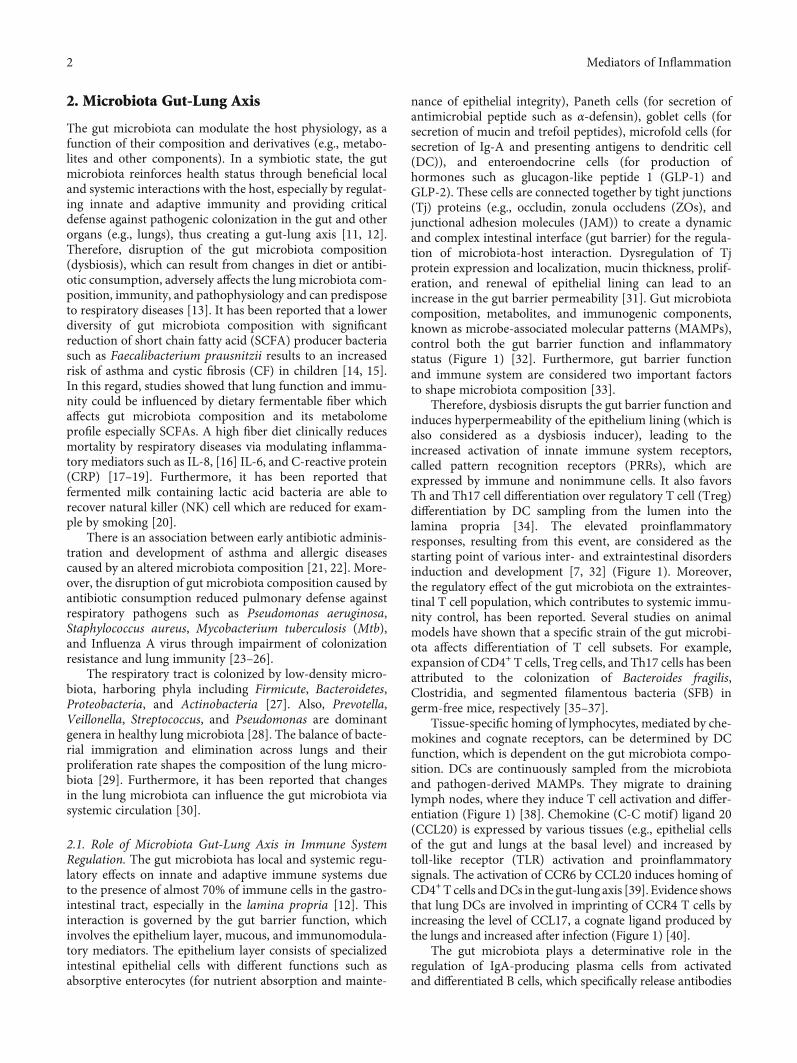
Figure 1: Relationship between the gut microbiota and lung immunity: the interaction between the intestinal commensal bacteria andestablishing of lung immunity is mediated by various factors, including PAMPs, PRRs, SCFAs, intestinal integrity, and immune cells ofthe lamina propria. In a normal state, DCs are continuously sampled from the lumen through M-cell activity, extension of dendrites, andthe gut barrier function, which determine bacterial/PAMP translocation. After DC sampling, these cells migrate to GALT and then MLNto regulate differentiation and homing of lymphocytes (T and B cells) depending on the released certain cytokines in respect to gutmicrobiota composition. The activated T and B cells are distributed in the lungs via circulation. Also, the levels of CCL20 and CCL17,which are produced by the lungs after microbial exposure, contribute to imprinting of T cell subsets, based on the cognate CCRs.Furthermore, SCFAs can penetrate into the bone marrow and influence lung immunity by affecting MDP differentiation to inflammatoryor anti-inflammatory immune cells. Inflammatory macrophages and DCs in the lungs are derived from CDPs and Ly6C+ inflammatorymonocytes. Alternatively, activated macrophages (AAMs) are anti-inflammatory immune lung cells, derived from Ly6C- patrollingmonocytes subtypes.
3Mediators of Inflammation

and shape the mucosal immunity. Sensitized B cells which arelocated in lamina propria (Peyer’s patches) by inhaled and gutmicrobiota antigens can reach the respiratory tract to producespecific IgAs and translocate onto the mucosal surface. Asmentioned earlier, the gut microbiota composition is a keyfactor in the gut barrier function, regulating interactionsbetween antigens and the immune system [41, 42]. Therefore,the conserved symbiotic relationship of the gut microbiotawith immunity reinforces lung immunity immune systemagainst respiratory bacterial and viral pathogens.
The intact bacteria and immunogenic components candiffuse through the intestinal lymphatic system towardsystemic circulation and the lungs. Therefore, bacterial clear-ance of alveolar macrophages, neutrophil recruitment, andantibacterial factors, derived from the bronchus epithelium,depend on the gut microbiota composition and its deriva-tives [43]. Moreover, the interplay between diet and thegut microbiota determines immunity, as reflected in the lungphysiology [44]. Generally, diet is a key factor in the gutmicrobiota composition and SCFA profile. For example,saturation of fatty acids in the diet can affect luminal immu-nity, since saturated and unsaturated fatty acids are consid-ered as TLR agonists and antagonists, respectively, andhave an impact on the gut microbiota composition [45].
Moreover, SCFAs which are mainly produced fromdietary fibers by bacterial fermentation, are multitaskingmolecules, associated with the maintenance of immunehomeostasis through various mechanisms: (i) reinforcementof the intestinal epithelium integrity; (ii) increasing the levelof mucin-producing goblet cells; (iii) elevating the intestinalIgA production; (iv) improvement of intestinal cell survivaland repair via NLRP3 inflammasome activation; (v) activa-tion of macrophage and DC signaling by G-protein coupledreceptors (GPR109A) for interleukin-10 (IL-10) production;and (vi) induction of intestinal Foxp3 Treg cell differentia-tion by GPR43 sensing [46–49].
The epigenetic role of SCAFs (butyrate) in regulation ofintestinal inflammation has been reported to induce the sup-pression of histone deacetylase (HDAC) activity, followed bythe induction of colonic Foxp3+ Treg cell expression [50].Moreover, in a symbiotic state, SCFAs preserve the desiredintestinal bacterial community through intestinal hypoxia,resulting from dominancy of colonocyte metabolism by fattyacid beta-oxidation and oxidative phosphorylation in themitochondria [51, 52]. Furthermore, the gut microbiotaand its SCFAs have hematopoiesis-regulating effects in thebone marrow. Circulating SCFAs can penetrate into the bonemarrow and affect lung immunity in allergic airway diseasesand respiratory infections (e.g., influenza virus infection)through differentiation of common DC precursors (CDPs),macrophages, and DC progenitors (MDPs) [53]. In the bonemarrow, CDPs and two monocyte subtypes, including Ly6C+
(Gr1+) inflammatory monocytes and Ly6C- (Gr1-) patrollingmonocytes, are derived from MDPs. In inflammatoryconditions, such as viral infections, severe tissue damageis induced by uncontrolled immune responses, such asincreased differentiation of inflammatory Ly6C+ monocytesto inflammatory DCs and macrophages, which can triggerthe immunopathology of the lungs [54].
Trompette and collaborators studied the effect of gutmicrobiota on bone marrow hematopoiesis and effective lungimmunity and found that high-fiber diets and SCFA metab-olites affect the bone marrow hematopoiesis by increasingthe level of Ly6C- (Gr1-) patrolling monocyte subtypes. Theelevated level of patrolling monocytes dampens tissue dam-age by increasing the airway count of alternatively activatedmacrophages (AAMs), which participate in tissue protectionand repair (Figure 1). Moreover, SCFAs enhance the functionof CD8+ effector T cells against influenza infection by alteringT cell metabolism [55].
Desaminotyrosine (DAT) is another microbial metabo-lite, produced by flavonoid and amino acid metabolism. Thismetabolite is correlated with type-I IFN activity, which playsa key role in viral immunity. Clostridium orbiscindens, amember of the gut microbiota, can produce DAT from flavo-noids and has a protective effect against influenza infectionand decreased mortality in influenza-infected mice [56].Besides the gut microbiota metabolites, studies on extracellu-lar vesicles (EVs), which can be derived from the intestinalgut microbiota as new systemic mediators, are notably grow-ing. Generally, EVs are nanosized particles, containingenclosed MAMPs, hydrolytic enzymes, and nucleic acids,which can diffuse across the body to regulate the host func-tion, especially immune responses [57].
Many reports indicate that EVs derived from importantgut microbiota members, including Bacteroides fragilis,Akkermansia muciniphila, and Faecalibacterium prausnitzii,may have important immunomodulatory effects [58–60].Therefore, the assessment of the gut microbiota EV patternscan be potentially used for screening disease progression [61,62]. Overall, the composition of the gut microbiota, associ-ated with diet, can determine lung immunity by changinginnate and adaptive immune responses. Therefore, there areprominent aspects of microbiota gut-lung axis that can beconsidered as promising targets in the prevention, control,and treatment of SARS-CoV-2 infection.
3. Respiratory Diseases Controlled byMicrobiota Gut-Lung Axis
There are various reports discussing the changes of gut andlung microbiota during respiratory diseases (caused by bacte-rial and viral pathogens). In this regard, Dumas et al. studiedthe importance of microbiota in acute lung infections, suchas pneumonia, induced by Pseudomonas aeruginosa, Strepto-coccus pneumoniae, and Klebsiella pneumoniae in antibiotic-treated germ-free mice [43]. The protective activity of the gutand lung microbiota against pneumonia is mainly mediatedby nucleotide-binding oligomerization domain-like receptorand IL-17A-driven granulocyte macrophage-colony-stimulating factor (GM-CSF) signaling pathways, whichpromote innate immune responses, especially pathogenclearance by alveolar macrophages [63].
In addition to respiratory bacterial pathogens, viral infec-tions, caused by influenza virus, respiratory syncytial virus(RSV), SARS-CoV-1, MERS, and SARS-CoV-2, which maybe followed by secondary bacterial pneumonia, are importantcauses of morbidity and mortality worldwide. Overall, the
4 Mediators of Inflammation
interactions between respiratory pathogens, microbiota, andimmune system can determine the severity of respiratory dis-eases, a relationship that is also closely linked to antibacterialand antiviral immune responses (e.g., type I IFN, type II IFN,and IL-17), antibody responses, and colonization resistanceby the gut and lung microbiota.
Microbiota is among the major determinants of lungimmunity, and respiratory viral infections can affect the gutand lung microbiota composition. Influenza-infected miceexhibited an altered intestinal microbiota composition as afunction of the increased abundance of Enterobacteriaceaeand decreased amount of SFB. These induce intestinalimmune injury due to the involvement of the CCL25-CCR9axis in recruiting lymphocytes (i.e., lung-derived CD4+ effec-tor T cells secreting IFN-γ) into the intestine and Th17 cellspromotion [64].
Ichinohe and collaborators emphasized the role of gutmicrobiota in the regulation of antiviral responses of CD4,CD8, and B cells against respiratory influenza virus infection,especially through inflammasome activation by providingproper MAMPs for prime immunity. They found that theantibiotic-induced microbiota changes resulted in failure inthe production of inflammasome-dependent cytokines.These changes also impaired homeostasis and migration oflung DCs into lymph nodes to prime T cell responses againstinfluenza virus in mice [26]. In this regard, Wang andcoworkers studied the possible protective role of the lungmicrobiota in subsequent lung injury and lethal inflamma-tion, resulting from influenza infection. They reported a sig-nificant decrease in lung injury caused by Staphylococcusaureus, a common colonizer of the upper respiratory tract,by promoting M2 polarization of alveolar macrophages,followed by anti-inflammatory cytokines [65].
The impact of viral pulmonary infections on the gutmicrobiota composition has been attributed to changes insystemic immune signals and bacterial translocation to thegut [66]. Therefore, modulation of the gut microbiotacomposition, based on the pre/probiotic interventions, hastherapeutic effects on respiratory viral infections such aspneumonia. Furthermore, the potential of postbiotics inter-vention in the modulation of immunity in various diseasesincluding asthma, COPD, and respiratory infections has beenreported. Postbiotics are defined as microbial components,soluble factors, and metabolites which are, respectively,secreted or released by live microbial cell or its lysate andinactivated [67, 68]. There are several reports demonstratingthe beneficial effects of probiotics in influenza-infected mice.A previous study showed that intranasal or oral administra-tion of Lactobacillus plantarum DK119 conferred protectivedefense against a lethal dose of influenza A virus by modulat-ing DC and macrophage activities and also increasing thelevels of IL-12 and IFN-γ in the bronchoalveolar fluid [69].
Moreover, oral administration of L. paracasei CNCM I-1518 strain preactivates the immune system to clear morerapidly the influenza virus by early stimulation of proinflam-matory cytokines and recruitment of immune cells. In a pre-vious study, after viral infection L. paracasei provided bettertissue homeostasis through IL-13 and IL-15 production byT cells, which promoted hyperplasia of lung epithelial cells
during inflammation, compared to control-fed mice [70].Smith and collaborators showed higher mortality rate anddecreased antiviral responses against influenza infection inhigh-fat-diet-induced obese (DIO) mice, compared to thelean group [71]. Additionally, Yoda et al. targeted the gutmicrobiota by oral administration of heat-inactivated L. gas-seri TMC0356 (postbiotic) to alleviate obesity-induced lungimmune disruption in DIOmice [72]. These findings empha-size the increased susceptibility of obese mice to respiratoryviral infections due to immune dysregulation controlled bythe gut microbiota.
4. Effects of Microbiota on Vaccination for ViralRespiratory Infections
Vaccine efficacy is determined by various factors, includinggenetic background, lifestyle, mode of delivery, nutrition,age, gender, geographical region, and economic status,which play critical roles in the composition of gut microbi-ota. As mentioned earlier, innate and adaptive immuneresponses are controlled by gut microbiota, which hasimmunomodulatory effects. Evidence shows that differencesin vaccine efficacy between certain populations with distinctcharacteristics affect the gut microbiota and immune status.In this regard, previous studies have reported differences inthe rotavirus vaccine efficacy between countries [73, 74].
The beneficial effects of pre- and probiotic interventionshave been reported to increase immune responsiveness torespiratory viral vaccination, as shown by the improvementof innate and adaptive immune responses in animal modelsand clinical trials [75, 76]. Moreover, previous studies havereported the orchestrating role of the gut microbiota inTLR5 activation, plasma cell differentiation, and antibodyresponses to influenza virus, which are defective inantibiotic-treated Trl5(-/-) mice and are improved by oraladministration of flagellated E. coli [77]. Moreover, recombi-nant probiotic strains have been introduced as adjuvants foredible vaccines to provide safer and better immunization [78,79]. Lei et al. designed a recombinant Lactococcus lactisstrain, expressing H5N1 hemagglutinin antigen, as a stableoral vector of influenza vaccine. They identified higher levelsof hemagglutinin-specific IgA antibodies in the serum andfecal samples of mice [80].
There are several clinical trials confirming the immuno-modulatory role of probiotic pretreatments in increasingimmune responses to influenza vaccination. For example,pretreatment with Lactobacillus rhamnosus GG, Bifidobac-terium animalis, and Lactobacillus paracasei before influenzavaccination improved the vaccine immunogenicity againstthe H3N2 influenza strain [81, 82]. Nasal microbiota partic-ipates to determine immunogenicity of vaccines. It has beenshown that administration of the Live, Attenuated InfluenzaVaccine (LAIV) induces changes in the upper respiratorymicrobiota, producing specific influenza antibodies. Salket al. demonstrated a significant association between theincreased alpha diversity and the presence of Lactobacillushelveticus, Prevotella melaninogenica, Streptococcus infantis,Veillonella dispar, and Bacteroides ovatus in the nasal micro-biota and specific IgGs after LAIV administration [83].
5Mediators of Inflammation

5. SARS-CoV Immunopathologyand Microbiota
Coronaviruses (CoV) contain a positive-sense single-strandedRNA genome, which is enclosed within an envelope, contain-ing spike glycoprotein (S), membrane protein (M), envelopeprotein (E), and in some cases, hemagglutinin-esterase (HE).These viruses are divided into four subgroups of α, β, ɣ, andδ, based on the genotypes and serological properties, with αand β subtypes causing human infections [84]. In the pastdecade, the world has experienced three life-threatening CoVinfections, caused by SARS-CoV-1, MERS-CoV, and a novelbetacoronavirus, called SARS-CoV-2, which has caused signif-icant mortality during the current pandemic.
The genome sequencing of SARS-CoV-2 revealed 79.5%similarity to the SARS-CoV genome [85]. The entry ofSARS-CoV into the host cells is mediated by binding ofvariable receptor-binding domain (RBD) of S protein toAngiotensin-converting enzyme 2 (ACE2) receptor, whichis expressed in the heart, lungs, kidneys, and gastrointestinaltract [86]. In SARS-CoV-2 infection, inflammatory responsesbegin in type II lung pneumocytes after the virus binds toACE2. The proteolytic activity of type 2 transmembrane pro-tease (TMPRSS2) requires viral entry through ACE2 cleavageand S protein [87].
ACE2 has a protective and regulatory role in Renin-angiotensin-aldosterone system (RAAS) mainly throughtwo pathways: (i) Angiotensin-converting enzyme (ACE)cleaves angiotensin I (Ang I) into Ang II that interacts withAng II type 1 receptor (AT1R). The activation of this way(ACE/Ang II/AT1R) leads to higher blood pressure andinflammation caused by increased vasoconstriction, renalreabsorption of sodium/water and induction of proinflam-matory chemokines [88]. (ii) ACE2-Ang1-7-MasR pathwaywhere ACE2 is a key enzyme converting Ang II into Ang 1-7 peptide, whereas Ang I is converted into inactive Ang 1-9.After this step, Ang 1-9 are metabolized to Ang1-7 by ACE.The peptide is recognized by Mas receptor to negatively reg-ulate RAAS system in many lung and heart functions andblood pressure homeostasis. ACE2-Ang1-7-Mas pathwayexerts the beneficial effect against hypertension and acutelung injury by inactivation Ang II which is upregulated inthese conditions (Figure 2) [89, 90]. It has been shown thatSARS-CoV infection significantly downregulates ACE2 inthe lungs [91]. In this state, also, the production of Ang1-7which is regulated by ACE2 activity is diminished. Therefore,the lack of ACE2-Ang1-7-Mas pathway activity leads to lossof its protective effects, and ACE/Ang II/AT1R pathway isoveractivated and accumulates Ang II. These cascade eventsare observable during pulmonary and acute lung injury andfibrosis [92, 93].
Evidence shows that hypertension and diabetic patients,who therapeutically receive ACE inhibitors (ACEIs) andAT1R blockers (ARBs), have elevated ACE2 levels and couldbe at high-risk for COVID-19 infection. In these patients,Ang II which causes hypertension and inflammation isincreased, and ACE/Ang II/AT1R is activated. ACE2 con-trols Ang II level and activity and balance RASS by cleavageAng II to Ang 1-7 peptides to exert protective effect by inter-
action with MasR [94]. It has been shown that ACE2 isinsensitive to inhibition by ACEIs [95]. Moreover, ACEImedication and ARB medication increase ACE2 geneexpression and activity which led to an overactivation ofACE/Ang II/AT1R by inactivation of Ang II. Upregulationof ACE2 in these patients can facilitate the SARS-CoV-2entry [96, 97]. As mentioned before, SARS-CoV-2 signifi-cantly decreases ACE2 after entry into the lung and attenuatesits protective effect against lung injury and failure. ACE2 couldact as a double-edged sword for these patients due to its dualfunction as a gate of SARS-CoV-2 entry and also protectingof lung injury and cardiovascular and renal complication indiabetic patients [98]. Nevertheless, European MedicinesAgency (EMA) suggested to maintain these medications indiabetic and hypertensive patients due to the increased mor-tality resulting from the withdraw of these medications [99](Figure 2). A big question arises: can the increase of ACE2levels after ACEIs and ARBs medication have adverse (byfacilitation of SARS-CoV-2 entry) or beneficial (by protectiverole in RAAS system) effect in diabetic and hypertensivepatients during COVID-19 infection?
Also, ACE2 plays a key role in gastrointestinal inflamma-tion and the gut microbiota composition [100]. A recentstudy highlighted the critical role of the gut microbiota inthe colonic ACE2 gene expression in gnotobiotic rats andreported its implication on the COVID-19 pathologythrough the gut-lung axis [101]. The abundance of Bacter-oides showed a negative correlation with the COVID-19severity and the fecal load of SARS-CoV-2 [102]. Bacteroidesspecies including B. dorei, B. thetaiotaomicron, B. massilien-sis, and B. ovatus are able to downregulate ACE2 expressionin the colonocytes of mice [103]. These findings suggest thepossible protective role of the Bacteroides spp. as importantgut microbiota member against COVID-19 infection bydownregulation of ACE2 and reduction of SARS-CoV-2entry [92]. In fact, since SARS-CoV-2 entry is linked toACE2 expression level, an increased level may promote theviral entry, whereas its downregulation reduces the ACE2-Ang1-7-Mas pathway and further protect from lung injuriesduring SARS-CoV-2 infection.
The pathogen-associated molecular patterns (PAMPs) ofviral infection are recognized by two innate immune recep-tors (PRRs), including TLR3 and retinoic acid-induciblegene-like-I- (RIG-I-) like receptors (RLRs), which sense viralRNA to induce type I IFN (IFN-α and IFN-β), as a majorantiviral and immunomodulatory mediator, promoting mac-rophage, NK cell, B cell, and T cell activities [104]. It has beenshown that TLR3 activates IRF3 and NF-κB to express type IIFN and trigger proinflammatory responses through theTRIF-dependent pathway as adaptor protein [105]. TLR4activation in the MyD88-dependent TRIF signaling pathwayoccurs during respiratory viral infections.
TLR4 expression increases after an immune response to aviral infection in bronchial epithelial and alveolar cells [106].TLR signaling pathways promote the production of IFN-α,IFN-β, IL-6, TNF, IFN-γ, CCL5, and IFN-stimulated genes,which are produced during acute respiratory distress syn-drome (ARDS) and viral infections [107]. In this regard,Totura et al. reported that the absence of TRIF- or MyD88-
6 Mediators of Inflammation

dependent TLR pathways resulted in the death of miceinfected with SARS-CoV. They suggested that balancebetween the two arms of TLR signaling provides effectiveantiviral responses to severe SARS-CoV. Their resultsdemonstrated the possible protective effect of TLR3 andTLR4 agonists as protective therapeutic strategy againstSARS-CoV infection [108].
RLRs, such as RIG-I and melanoma differentiation-associated protein 5 (MDA5), are cytosolic PRRs recognizingviral dsRNA. They contain C-terminal domains (CTD) andN-terminal caspase recruiting domains (CARD) to senseRNA and activate mitochondrial antiviral signaling proteins(MAVS) as downstream adaptor proteins, promoting antivi-ral responses (e.g., type I IFN) [109]. In this regard, Luhighlighted the potential of SARS-CoV in inhibiting IFNresponses by N protein, which contributes to SARS-CoVpathogenesis [107].
DCs play key roles in combining innate and adaptiveimmune responses by affecting T cell and B cell activation.Generally, the polarization of DCs affects the outcomes of
viral infection. The conventional DCs (cDCs) and plasmacy-toid DCs (pDCs) induce the production of type I IFN by PRRrecognition of viral PAMPs, including TLR3, RIGI, MDA5,and TLR7-9 [110]. In a normal state, there are three DC sub-types in the lungs, including CD103+ cDCs, CD11b+ cDCs,and pDCs, while in inflammatory conditions, monocyte-derived DCs (moDCs) are recruited into the lungs. Thesubtypes of DCs and polarization of T cells are determinedby the type of respiratory virus and DC expression of PRRs.Differentiation of T cells into CD4+ T, CD8+ T, Treg, andTh17 cells is controlled by DC function through activationof DC PRRs and cytokine and chemokine receptors [111].Therefore, skewing of T cell polarization can result in hostdamage and increase the severity of disease during viralrespiratory infections.
CD8+ T cells, which are essential in clearing virus-infected lung cells and promote immune injury, are near to80% of infiltrated immune cells into the lungs in SARS-CoV patients [112]. The production of SARS-CoV is activatedin B cells through CD4+ T cell function. In this regard, Chen
COVID-19
Entry to host cellsby ACE2
ACE2downregulation
Diabetic andhypertention
ACEts andARBs
ACE2upregulation
Vasoconstriction
RAAS
ACE/Ang II/AT1R ACE2-Ang1-7-MasR
infection
Sodium/water reabsorptionInflammationFibrosis
VasodilationSodium/water excretionAnti-inflammatoryAnti-fibrotic
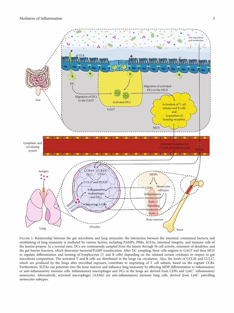
Figure 2: The function of two arms of renin-angiotensin-aldosterone system (RAAS) axis: the RAAS consists of two pathways including (i)ACE/Ang II/AT1R: in the pathway, Ang II which cleaved from Ang I by ACE activity, interacts with AT1R to induce vasoconstriction,inflammation, and fibrosis. (ii) ACE2-Ang1-7-MasR: in the pathway Ang 1-7 negatively regulate RAAS through promotion ofvasodilation, anti-inflammatory and antifibrotic effects by interaction with MasR. Ang 1-7 are produced from cleavage of Ang II by ACE2or metabolized of inactivated Ang 1-9 (cleaved from Ang I by ACE2) by ACE. The balance between two arms determines healthy state. InCOVID-19 infection, ACE2, main receptor to SARS-CoV-2 entrance, is significantly decreased which results to inhibition of protectivefunction of ACE2-Ang1-7-MasR arm. In opposite, the increased level of ACE2 resulted from ACEI and ARB medication in diabetic andhypertensive patients is considered as a double-edged sword which has been raised a big question: which aspects of increased ACE2 couldbe dominated during COVID-19 infection? increased susceptibility to viral infection or protective potential in RAAS system.
7Mediators of Inflammation

et al. reported the significance of CD4+ T cells in mice infectedwithSARS-CoV, as depletionof these cells resulted in increasedpneumonia and delayed respiratory viral clearance, associatedwith decreased neutralizing antibodies and recruitment ofimmune cells to the lungs [113]. Also, SARS-CoV-specificCD4 and CD8memory T cells may play a vital role in protect-ing against reinfectionwith SARS-CoV. Channappanavar et al.found that unlike CD4+ T cells, CD8+ memory T cells remainup to six years after SARS-CoV infection, mediating protectiveeffects against lethal SARS-CoV infection [114].
Considering the importance of microbiota in the gut-lung axis in COVID-19 patients, reports about this topic haveprogressively increased. The metatranscriptome sequencingof bronchoalveolar lavage fluid showed similar microbiotabetween COVID-19 and community-acquired pneumonia(CAP) patients. The dominance of pathogens or higher countof oral and upper respiratory tract symbiotic bacteria wasreported in these patients, compared to the healthy controls[115]. Although the main target of SARS-CoV-2 is the lung,some meta-analyses have reported gastrointestinal manifes-tations and the presence of SARS-CoV-2 RNAs in anal swabsand stool samples of COVID-19 patients [116, 117].
Several studies demonstrated the presence of a gut micro-biota alteration in stool samples of COVID-19 patients,compared to healthy individuals. In this regard, Zou et al.reported that the gut microbiota of COVID-19 patientscontained less beneficial commensal bacteria, such as F.prausnitzii, and was enriched with bacteremia-associatedpathogens, which could increase the severity of diseasecourse due to secondary bacterial infections [102]. Also, ina pilot study, it has been documented a higher abundanceof Parabacteroides merdae, Bacteroides stercoris, Alistipesonderdonkii, and Lachnospiraceae, SCFAs producer bacteria,in fecal samples of SARS-CoV-2 patients with low to noneinfectivity [118]. According to a previous study, a significantdiversity reduction was observed in the gut microbiota ofCOVID-19 patients (similar to patients with H1N1 infec-tion), compared to healthy subjects. Interestingly, gut micro-biota signature was significantly different between patientswith SARS-CoV-2, patients with H1N1 infection, andcontrol individuals, as a function of the abundance of oppor-tunistic pathogens [119]. It is also important to avoid unnec-essary antibiotic administrations that may cause a potentialreduction of symbionts and determine a gut microbiota dys-biosis during COVID-19 treatment [102, 120]. Furthermore,a healthy diet rich in fibers (whole grains and vegetables)should be considered as beneficial for COVID-19 patients’treatment due to their significant anti-inflammatory poten-tial and ability to target the microbiota-lung axis [121]. Sincegut microbiota finely tune local and systemic immuneresponses and alter its composition, it may have an impor-tant role in the host sensitivity toward COVID-19, secondarybacterial infections, and organ failure and damage.
6. Conclusions
The crucial role of gut microbiota in establishment and pro-viding innate and adaptive immunity in the respiratory tracthas been demonstrated. According to the composition of the
gut microbiota and its products (e.g., metabolites and com-ponents), pulmonary immune responses can be explainedthrough various pathways: (i) ACE2 expression; (ii) activa-tion of PRRs, such as TLRs, NLRs, and RLRs for producingantiviral responses, such as type I IFN and proinflammatorycytokines; (iii) translocation of CDP andMDP subtypes fromthe bone marrow to the lungs; and (iv) activation and hom-ing of T and B cells from the gut-associated lymphoid tissueto the lungs. Moreover, the presence of commensal bacteriain the respiratory tract may affect alveolar macrophage polar-ization (M2) to dampen lung injury, induced by elevatedinflammatory responses. On the other hand, the beneficialrole of probiotic pretreatment in vaccination efficacy againstviral respiratory tract infections has been discussed. There-fore, differences between populations (reflecting the microbi-ota composition) could be attributed to the sensitivity andseverity of SARS-CoV-2 infection. Finally, further researchon the study of microbiota gut-lung axis is essential to designa therapeutic strategy and develop a vaccine against SARS-CoV-2 infection.
Data Availability
The data used to support the findings of this study are avail-able from the corresponding authors upon request.
Conflicts of Interest
The authors declare that they have no conflict of interest.
References
[1] T. Ferkol and D. Schraufnagel, “The global burden of respira-tory disease,” Annals of the American Thoracic Society,vol. 11, no. 3, pp. 404–406, 2014.
[2] A. Zumla, D. S. Hui, and S. Perlman, “Middle East respiratorysyndrome,” The Lancet, vol. 386, no. 9997, pp. 995–1007,2015.
[3] D. Koh and J. Sng, “Lessons from the past: perspectives onsevere acute respiratory syndrome,” Asia Pacific Journal ofPublic Health, vol. 22, 3_suppl, pp. 132S–136S, 2010.
[4] N. Zhu, D. Zhang, W.Wang et al., “A novel coronavirus frompatients with pneumonia in China, 2019,” New England Jour-nal of Medicine, vol. 382, no. 8, pp. 727–733, 2020.
[5] M. T. Limborg and P. Heeb, Coevolution of Hosts and TheirMicrobiome, Multidisciplinary Digital Publishing Institute,2018.
[6] H. J. Flint, S. H. Duncan, K. P. Scott, and P. Louis, “Linksbetween diet, gut microbiota composition and gut metabo-lism,” Proceedings of the Nutrition Society, vol. 74, no. 1,pp. 13–22, 2015.
[7] A. I. Petra, S. Panagiotidou, E. Hatziagelaki, J. M. Stewart,P. Conti, and T. C. Theoharides, “Gut-microbiota-brain axisand its effect on neuropsychiatric disorders with suspectedimmune dysregulation,” Clinical therapeutics, vol. 37, no. 5,pp. 984–995, 2015.
[8] B. Chassaing, L. Etienne-Mesmin, and A. T. Gewirtz, “Micro-biota-liver axis in hepatic disease,” Hepatology, vol. 59, no. 1,pp. 328–339, 2014.
8 Mediators of Inflammation

[9] A. Ramezani and D. S. Raj, “The gut microbiome, kidney dis-ease, and targeted interventions,” Journal of the AmericanSociety of Nephrology, vol. 25, no. 4, pp. 657–670, 2014.
[10] B. J. Marsland, A. Trompette, and E. S. Gollwitzer, “The gut–lung axis in respiratory disease,” Annals of the American Tho-racic Society, vol. 12, Supplement 2, pp. S150–S1S6, 2015.
[11] R. Enaud, R. Prevel, E. Ciarlo et al., “The gut-lung axis inhealth and respiratory diseases: a place for inter-organ andinter-kingdom crosstalks,” Frontiers in Cellular and InfectionMicrobiology, vol. 10, 2020.
[12] S. Bengmark, “Gut microbiota, immune development andfunction,” Pharmacological Research, vol. 69, no. 1, pp. 87–113, 2013.
[13] K. F. Budden, S. L. Gellatly, D. L. Wood et al., “Emergingpathogenic links between microbiota and the gut-lung axis,”Nature Reviews Microbiology, vol. 15, no. 1, pp. 55–63, 2017.
[14] E. Bruzzese, M. L. Callegari, V. Raia et al., “Disrupted intesti-nal microbiota and intestinal inflammation in children withcystic fibrosis and its restoration with Lactobacillus GG: arandomised clinical trial,” PLoS One, vol. 9, no. 2, articlee87796, 2014.
[15] G. Ranucci, V. Buccigrossi, M. B. d. Freitas, A. Guarino, andA. Giannattasio, “Early-life intestine microbiota and lunghealth in children,” Journal of Immunology Research,vol. 2017, Article ID 8450496, 5 pages, 2017.
[16] D. E. King, B. M. Egan, R. F. Woolson, Mainous AG 3rd,Y. al-Solaiman, and A. Jesri, “Effect of a high-fiber diet vsa fiber-supplemented diet on C-reactive protein level,”Archives of Internal Medicine, vol. 167, no. 5, pp. 502–506,2007.
[17] A. Trompette, E. S. Gollwitzer, K. Yadava et al., “Gut micro-biota metabolism of dietary fiber influences allergic airwaydisease and hematopoiesis,” Nature Medicine, vol. 20, no. 2,pp. 159–166, 2014.
[18] R. Varraso, S. E. Chiuve, T. T. Fung et al., “Alternate HealthyEating Index 2010 and risk of chronic obstructive pulmonarydisease among US women and men: prospective study,” BMJ,vol. 350, no. feb03 7, article h286, 2015.
[19] I. Halnes, K. J. Baines, B. S. Berthon, L. K. MacDonald-Wicks,P. G. Gibson, and L. G. Wood, “Soluble fibre meal challengereduces airway inflammation and expression of GPR43 andGPR41 in asthma,” Nutrients, vol. 9, no. 1, p. 57, 2017.
[20] K. Morimoto, T. Takeshita, M. Nanno, S. Tokudome, andK. Nakayama, “Modulation of natural killer cell activity bysupplementation of fermented milk containing Lactobacilluscasei in habitual smokers,” Preventive Medicine, vol. 40, no. 5,pp. 589–594, 2005.
[21] E. Mitre, A. Susi, L. E. Kropp, D. J. Schwartz, G. H. Gorman,and C. M. Nylund, “Association between use of acid-suppressive medications and antibiotics during infancy andallergic diseases in early childhood,” JAMA Pediatrics,vol. 172, no. 6, article e180315, 2018.
[22] J. Ni, H. Friedman, B. C. Boyd et al., “Early antibiotic expo-sure and development of asthma and allergic rhinitis in child-hood,” BMC Pediatrics, vol. 19, no. 1, pp. 225–228, 2019.
[23] A. Dumas, D. Corral, A. Colom et al., “The host microbiotacontributes to early protection against lung colonization byMycobacterium tuberculosis,” Frontiers in Immunology,vol. 9, p. 2656, 2018.
[24] R. Dessein, M. Bauduin, T. Grandjean et al., “Antibiotic-related gut dysbiosis induces lung immunodepression and
worsens lung infection in mice,” Critical Care, vol. 24, no. 1,p. 611, 2020.
[25] H. Wang, P. Lian, X. Niu et al., “TLR4 deficiency reduces pul-monary resistance to Streptococcus pneumoniae in gutmicrobiota-disrupted mice,” PLoS One, vol. 13, no. 12, articlee0209183, 2018.
[26] T. Ichinohe, I. K. Pang, Y. Kumamoto et al., “Microbiota reg-ulates immune defense against respiratory tract influenza Avirus infection,” Proceedings of the National Academy of Sci-ences, vol. 108, no. 13, pp. 5354–5359, 2011.
[27] G. Yu, M. H. Gail, D. Consonni et al., “Characterizing humanlung tissue microbiota and its relationship to epidemiologicaland clinical features,” Genome Biology, vol. 17, no. 1, p. 163,2016.
[28] R. P. Dickson and G. B. Huffnagle, “The lung microbiome:new principles for respiratory bacteriology in health and dis-ease,” PLoS Pathogens, vol. 11, no. 7, article e1004923, 2015.
[29] R. P. Dickson, F. J. Martinez, and G. B. Huffnagle, “The roleof the microbiome in exacerbations of chronic lung diseases,”The Lancet, vol. 384, no. 9944, pp. 691–702, 2014.
[30] M. A. Sze, M. Tsuruta, S. W. J. Yang et al., “Changes in thebacterial microbiota in gut, blood, and lungs following acuteLPS instillation into mice lungs,” PloS one, vol. 9, no. 10, arti-cle e111228, 2014.
[31] C. Chelakkot, J. Ghim, and S. H. Ryu, “Mechanisms regulat-ing intestinal barrier integrity and its pathological implica-tions,” Experimental & Molecular Medicine, vol. 50, no. 8,pp. 1–9, 2018.
[32] N. Kamada, S.-U. Seo, G. Y. Chen, and G. Núñez, “Role of thegut microbiota in immunity and inflammatory disease,”Nature Reviews Immunology, vol. 13, no. 5, pp. 321–335,2013.
[33] D. Zheng, T. Liwinski, and E. Elinav, “Interaction betweenmicrobiota and immunity in health and disease,” CellResearch, vol. 30, no. 6, pp. 492–506, 2020.
[34] J. Stephani, K. Radulovic, and J. H. Niess, “Gut microbiota,probiotics and inflammatory bowel disease,” ArchivumImmunologiae et Therapiae Experimentalis, vol. 59, no. 3,pp. 161–177, 2011.
[35] E. C. Martens, E. C. Lowe, H. Chiang et al., “Recognition anddegradation of plant cell wall polysaccharides by two humangut symbionts,” PLoS Biology, vol. 9, no. 12, article e1001221,2011.
[36] S. K. Mazmanian, C. H. Liu, A. O. Tzianabos, and D. L.Kasper, “An immunomodulatory molecule of symbioticbacteria directs maturation of the host immune system,”Cell, vol. 122, no. 1, pp. 107–118, 2005.
[37] H.-J. Wu, I. I. Ivanov, J. Darce et al., “Gut-residing segmentedfilamentous bacteria drive autoimmune arthritis via T helper17 cells,” Immunity, vol. 32, no. 6, pp. 815–827, 2010.
[38] D. R. Samuelson, D. A. Welsh, and J. E. Shellito, “Regulationof lung immunity and host defense by the intestinal microbi-ota,” Frontiers in Microbiology, vol. 6, p. 1085, 2015.
[39] T. Ito, W. F. Carson IV, K. A. Cavassani, J. M. Connett, andS. L. Kunkel, “CCR6 as a mediator of immunity in the lungand gut,” Experimental Cell Research, vol. 317, no. 5,pp. 613–619, 2011.
[40] Z. Mikhak, J. P. Strassner, and A. D. Luster, “Lung dendriticcells imprint T cell lung homing and promote lung immunitythrough the chemokine receptor CCR4,” Journal of Experi-mental Medicine, vol. 210, no. 9, pp. 1855–1869, 2013.
9Mediators of Inflammation
[41] Y. He, Q. Wen, F. Yao, D. Xu, Y. Huang, and J. Wang, “Gut–lung axis: the microbial contributions and clinical implica-tions,” Critical Reviews in Microbiology, vol. 43, no. 1,pp. 81–95, 2017.
[42] K. Matsuno, H. Ueta, Z. Shu et al., “The microstructure ofsecondary lymphoid organs that support immune cell traf-ficking,” Archives of Histology and Cytology, vol. 73, no. 1,pp. 1–21, 2010.
[43] A. Dumas, L. Bernard, Y. Poquet, G. Lugo-Villarino, andO. Neyrolles, “The role of the lung microbiota and the gut–lung axis in respiratory infectious diseases,” Cellular Microbi-ology, vol. 20, no. 12, article e12966, 2018.
[44] S. Anand and S. S. Mande, “Diet, microbiota and gut-lungconnection,” Frontiers in Microbiology, vol. 9, p. 2147, 2018.
[45] D. H. Hwang, J.-A. Kim, and J. Y. Lee, “Mechanisms for theactivation of Toll-like receptor 2/4 by saturated fatty acidsand inhibition by docosahexaenoic acid,” European Journalof Pharmacology, vol. 785, pp. 24–35, 2016.
[46] M. B. Geuking, J. Cahenzli, M. A. Lawson et al., “Intestinalbacterial colonization induces mutualistic regulatory T cellresponses,” Immunity, vol. 34, no. 5, pp. 794–806, 2011.
[47] N. Singh, A. Gurav, S. Sivaprakasam et al., “Activation ofGpr109a, receptor for niacin and the commensal metabolitebutyrate, suppresses colonic inflammation and carcinogene-sis,” Immunity, vol. 40, no. 1, pp. 128–139, 2014.
[48] S. Fukuda, H. Toh, K. Hase et al., “Bifidobacteria can protectfrom enteropathogenic infection through production of ace-tate,” Nature, vol. 469, no. 7331, pp. 543–547, 2011.
[49] L. V. Hooper and A. J. Macpherson, “Immune adaptations thatmaintain homeostasis with the intestinal microbiota,” NatureReviews Immunology, vol. 10, no. 3, pp. 159–169, 2010.
[50] P. M. Smith, M. R. Howitt, N. Panikov et al., “The micro-bial metabolites, short-chain fatty acids, regulate colonicTreg cell homeostasis,” Science, vol. 341, no. 6145,pp. 569–573, 2013.
[51] T. Varga, Z. Czimmerer, and L. Nagy, “PPARs are a uniqueset of fatty acid regulated transcription factors controllingboth lipid metabolism and inflammation,” Biochimica et Bio-physica Acta (BBA)-Molecular Basis of Disease, vol. 1812,no. 8, pp. 1007–1022, 2011.
[52] Y. Litvak, M. X. Byndloss, and A. J. Bäumler, “Colonocytemetabolism shapes the gut microbiota,” Science, vol. 362,no. 6418, article eaat9076, 2018.
[53] A. T. Dang and B. J. Marsland, “Microbes, metabolites, andthe gut–lung axis,” Mucosal Immunology, vol. 12, no. 4,pp. 843–850, 2019.
[54] F. Geissmann, M. G. Manz, S. Jung, M. H. Sieweke,M. Merad, and K. Ley, “Development of monocytes, mac-rophages, and dendritic cells,” Science, vol. 327, no. 5966,pp. 656–661, 2010.
[55] A. Trompette, E. S. Gollwitzer, C. Pattaroni et al., “Dietaryfiber confers protection against flu by shaping Ly6c− patrol-ling monocyte hematopoiesis and CD8+ T cell metabolism,”Immunity, vol. 48, no. 5, pp. 992–1005.e8, 2018.
[56] A. L. Steed, G. P. Christophi, G. E. Kaiko et al., “Themicrobialmetabolite desaminotyrosine protects from influenzathrough type I interferon,” Science, vol. 357, no. 6350,pp. 498–502, 2017.
[57] S. Ahmadi Badi, A. Moshiri, A. Fateh et al., “Microbiota-derived extracellular vesicles as new systemic regulators,”Frontiers in Microbiology, vol. 8, p. 1610, 2017.
[58] S. A. Badi, S. Khatami, S. Irani, and S. D. Siadat, “Inductioneffects of bacteroides fragilis derived outer membrane vesicleson toll like receptor 2, toll like receptor 4 genes expressionand cytokines concentration in human intestinal epithelialcells,” Cell Journal, vol. 21, no. 1, p. 57, 2019.
[59] N. Rabiei, S. Ahmadi Badi, F. Ettehad Marvasti, T. NejadSattari, F. Vaziri, and S. D. Siadat, “Induction effects of_Faecalibacterium prausnitzii_ and its extracellular vesicleson toll-like receptor signaling pathway gene expression andcytokine level in human intestinal epithelial cells,” Cyto-kine, vol. 121, p. 154718, 2019.
[60] F. Ashrafian, A. Shahriary, A. Behrouzi et al., “Akkermansiamuciniphila-derived extracellular vesicles as a mucosal deliv-ery vector for amelioration of obesity in mice,” Frontiers inMicrobiology, vol. 10, p. 2155, 2019.
[61] Y.-J. Huh, J.-Y. Seo, J. Nam et al., “Bariatric/metabolic sur-gery induces noticeable changes of microbiota and theirsecreting extracellular vesicle composition in the gut,” Obe-sity Surgery, vol. 29, no. 8, pp. 2470–2484, 2019.
[62] N. S. Barteneva, Y. Baiken, E. Fasler-Kan et al., “Extracellularvesicles in gastrointestinal cancer in conjunction with micro-biota: on the border of kingdoms,” Biochimica et BiophysicaActa (BBA)-reviews on Cancer, vol. 1868, no. 2, pp. 372–393, 2017.
[63] R. L. Brown, R. P. Sequeira, and T. B. Clarke, “The microbiotaprotects against respiratory infection via GM-CSF signaling,”Nature Communications, vol. 8, no. 1, p. 1512, 2017.
[64] J. Wang, F. Li, H.Wei, Z.-X. Lian, R. Sun, and Z. Tian, “Respi-ratory influenza virus infection induces intestinal immuneinjury via microbiota-mediated Th17 cell–dependent inflam-mation,” Journal of Experimental Medicine, vol. 211, no. 12,pp. 2397–2410, 2014.
[65] J. Wang, F. Li, R. Sun et al., “Bacterial colonization dampensinfluenza-mediated acute lung injury via induction of M2alveolar macrophages,” Nature Communications, vol. 4,no. 1, pp. 1–10, 2013.
[66] S. Hanada, M. Pirzadeh, K. Y. Carver, and J. C. Deng, “Respi-ratory viral infection-induced microbiome alterations andsecondary bacterial pneumonia,” Frontiers in Immunology,vol. 9, p. 2640, 2018.
[67] J. Żółkiewicz, A. Marzec, M. Ruszczyński, and W. Feleszko,“Postbiotics—a step beyond pre- and probiotics,” Nutrients,vol. 12, no. 8, p. 2189, 2020.
[68] A. M. Koatz, N. A. Coe, A. Cicerán, and A. J. Alter, “Clinicaland immunological benefits of OM-85 bacterial lysate inpatients with allergic rhinitis, asthma, and COPD and recur-rent respiratory infections,” Lung, vol. 194, no. 4, pp. 687–697, 2016.
[69] M.-K. Park, V. NGO, Y.-M. Kwon et al., “Lactobacillus plan-tarum DK119 as a probiotic confers protection against influ-enza virus by modulating innate immunity,” PloS One, vol. 8,no. 10, article e75368, 2013.
[70] N. Belkacem, N. Serafini, R. Wheeler et al., “Lactobacillusparacasei feeding improves immune control of influenzainfection in mice,” PloS One, vol. 12, no. 9, article e0184976,2017.
[71] A. G. Smith, P. A. Sheridan, J. B. Harp, and M. A. Beck,“Diet-induced obese mice have increased mortality andaltered immune responses when infected with influenzavirus,” The Journal of Nutrition, vol. 137, no. 5, pp. 1236–1243, 2007.
10 Mediators of Inflammation
[72] K. Yoda, F. He, K. Miyazawa, M. Kawase, A. Kubota, andM. Hiramatsu, “Orally administered heat-killed Lactobacillusgasseri TMC0356 alters respiratory immune responses andintestinal microbiota of diet-induced obese mice,” Journal ofApplied Microbiology, vol. 113, no. 1, pp. 155–162, 2012.
[73] K. Zaman, D. D. Anh, J. C. Victor et al., “Efficacy of pentava-lent rotavirus vaccine against severe rotavirus gastroenteritisin infants in developing countries in Asia: a randomised, dou-ble-blind, placebo-controlled trial,” The Lancet, vol. 376,no. 9741, pp. 615–623, 2010.
[74] G. E. Armah, S. O. Sow, R. F. Breiman et al., “Efficacy of pen-tavalent rotavirus vaccine against severe rotavirus gastroen-teritis in infants in developing countries in sub-SaharanAfrica: a randomised, double-blind, placebo-controlled trial,”The Lancet, vol. 376, no. 9741, pp. 606–614, 2010.
[75] W.-T. Lei, P.-C. Shih, S.-J. Liu, C.-Y. Lin, and T.-L. Yeh,“Effect of probiotics and prebiotics on immune response toinfluenza vaccination in adults: a systematic review andmeta-analysis of randomized controlled trials,” Nutrients,vol. 9, no. 11, p. 1175, 2017.
[76] T.-L. Yeh, P.-C. Shih, S.-J. Liu et al., “The influence of prebi-otic or probiotic supplementation on antibody titers afterinfluenza vaccination: a systematic review and meta-analysis of randomized controlled trials,”Drug Design, Devel-opment and Therapy, vol. Volume 12, pp. 217–230, 2018.
[77] J. Z. Oh, R. Ravindran, B. Chassaing et al., “TLR5-mediatedsensing of gut microbiota is necessary for antibody responsesto seasonal influenza vaccination,” Immunity, vol. 41, no. 3,pp. 478–492, 2014.
[78] W. Beyer, A. Palache, J. C. de Jong, and A. Osterhaus, “Cold-adapted live influenza vaccine versus inactivated vaccine: sys-temic vaccine reactions, local and systemic antibodyresponse, and vaccine efficacy: a meta-analysis,” Vaccine,vol. 20, no. 9-10, pp. 1340–1353, 2002.
[79] M. Y. Chowdhury, R. Li, J.-H. Kim et al., “Mucosal vaccina-tion with recombinant Lactobacillus casei-displayed CTA1-conjugated consensus matrix protein-2 (sM2) induces broadprotection against divergent influenza subtypes in BALB/cmice,” PloS One, vol. 9, no. 4, article e94051, 2014.
[80] H. Lei, Y. Xu, J. Chen, X. Wei, and D. M.-K. Lam, “Immuno-protection against influenza H5N1 virus by oral administra-tion of enteric-coated recombinant Lactococcus lactis mini-capsules,” Virology, vol. 407, no. 2, pp. 319–324, 2010.
[81] L. E. Davidson, A.-M. Fiorino, D. R. Snydman, and P. L. Hib-berd, “Lactobacillus GG as an immune adjuvant for live-attenuated influenza vaccine in healthy adults: a randomizeddouble-blind placebo-controlled trial,” European Journal ofClinical Nutrition, vol. 65, no. 4, pp. 501–507, 2011.
[82] G. Rizzardini, D. Eskesen, P. C. Calder, A. Capetti,L. Jespersen, and M. Clerici, “Evaluation of the immune ben-efits of two probiotic strains Bifidobacterium animalis ssp.lactis, BB-12® and Lactobacillus paracasei ssp. paracasei, L.casei 431® in an influenza vaccination model: a randomised,double-blind, placebo-controlled study,” British Journal ofNutrition, vol. 107, no. 6, pp. 876–884, 2012.
[83] H. M. Salk, W. L. Simon, N. D. Lambert et al., “Taxa of thenasal microbiome are associated with influenza-specific IgAresponse to live attenuated influenza vaccine,” PLoS One,vol. 11, no. 9, article e0162803, 2016.
[84] J. Cui, F. Li, and Z.-L. Shi, “Origin and evolution of patho-genic coronaviruses,” Nature Reviews Microbiology, vol. 17,no. 3, pp. 181–192, 2019.
[85] P. Zhou, X.-L. Yang, X.-G. Wang et al., “Discovery of a novelcoronavirus associated with the recent pneumonia outbreakin humans and its potential bat origin,” 2020, BioRxiv.
[86] T. G. Ksiazek, D. Erdman, C. S. Goldsmith et al., “A novelcoronavirus associated with severe acute respiratory syn-drome,” New England Journal of Medicine, vol. 348, no. 20,pp. 1953–1966, 2003.
[87] F. A. Rabi, M. S. Al Zoubi, G. A. Kasasbeh, D. M. Salameh,and A. D. Al-Nasser, “SARS-CoV-2 and coronavirus disease2019: what we know so far,” Pathogens, vol. 9, no. 3, p. 231,2020.
[88] V. Bhalla, C. A. Blish, and A. M. South, “A historical perspec-tive on ACE2 in the COVID-19 era,” Journal of HumanHypertension, 2020.
[89] M. Liu, T. Wang, Y. Zhou, Y. Zhao, Y. Zhang, and J. Li,“Potential role of ACE2 in coronavirus disease 2019(COVID-19) prevention and management,” Journal ofTranslational Internal Medicine, vol. 8, no. 1, pp. 9–19, 2020.
[90] Y. Imai, K. Kuba, S. Rao et al., “Angiotensin-convertingenzyme 2 protects from severe acute lung failure,” Nature,vol. 436, no. 7047, pp. 112–116, 2005.
[91] K. Kuba, Y. Imai, S. Rao et al., “A crucial role of angiotensinconverting enzyme 2 (ACE2) in SARS coronavirus–inducedlung injury,” Nature Medicine, vol. 11, no. 8, pp. 875–879,2005.
[92] L. Samavati and B. D. Uhal, “ACE2, much more than just areceptor for SARS-COV-2,” Frontiers in Cellular and Infec-tion Microbiology, vol. 10, p. 317, 2020.
[93] C. T. Cole-Jeffrey, M. Liu, M. J. Katovich, M. K. Raizada, andV. Shenoy, “ACE2 and microbiota: emerging targets for car-diopulmonary disease therapy,” Journal of CardiovascularPharmacology, vol. 66, no. 6, pp. 540–550, 2015.
[94] M. Iwai and M. Horiuchi, “Devil and angel in the renin–angiotensin system: ACE–angiotensin II–AT 1 receptor axisvs. ACE2–angiotensin-(1–7)–Mas receptor axis,” Hyperten-sion Research, vol. 32, no. 7, pp. 533–536, 2009.
[95] C. M. Ferrario, J. Jessup, M. C. Chappell et al., “Effect ofangiotensin-converting enzyme inhibition and angiotensinII receptor blockers on cardiac angiotensin-convertingenzyme 2,” Circulation, vol. 111, no. 20, pp. 2605–2610, 2005.
[96] L. Fang, G. Karakiulakis, and M. Roth, “Are patients withhypertension and diabetes mellitus at increased risk forCOVID-19 infection?,” The Lancet Respiratory Medicine,vol. 8, no. 4, article e21, 2020.
[97] M. A. Hill, C. Mantzoros, and J. R. Sowers, “Commentary:COVID-19 in patients with diabetes,” Metabolism, vol. 107,p. 154217, 2020.
[98] C. Cristelo, C. Azevedo, J. M. Marques, R. Nunes, andB. Sarmento, “SARS-CoV-2 and diabetes: new challengesfor the disease,” Diabetes Research and Clinical Practice,vol. 164, article 108228, 2020.
[99] E. M. Agency, EMA Advises Continued Use of Medicines forHypertension, Heart or Kidney Disease during COVID-19Pandemic, EMA, 2020.
[100] T. Hashimoto, T. Perlot, A. Rehman et al., “ACE2 linksamino acid malnutrition to microbial ecology and intestinalinflammation,”Nature, vol. 487, no. 7408, pp. 477–481, 2012.
[101] T. Yang, S. Chakraborty, P. Saha et al., “Gnotobiotic ratsreveal that gut microbiota regulates colonic mRNA of Ace2,the receptor for SARS-CoV-2 infectivity,” Hypertension,vol. 76, no. 1, pp. e1–e3, 2020.
11Mediators of Inflammation

[102] T. Zuo, F. Zhang, G. C. Lui et al., “Alterations in gut microbi-ota of patients with COVID-19 during time of hospitaliza-tion,” Gastroenterology, vol. 159, no. 3, pp. 944–955.e8, 2020.
[103] N. Geva-Zatorsky, E. Sefik, L. Kua et al., “Mining the humangut microbiota for immunomodulatory organisms,” Cell,vol. 168, no. 5, pp. 928–43.e11, 2017.
[104] G. Li, Y. Fan, Y. Lai et al., “Coronavirus infections andimmune responses,” Journal of Medical Virology, vol. 92,no. 4, pp. 424–432, 2020.
[105] S. Akira, S. Uematsu, and O. Takeuchi, “Pathogen recogni-tion and innate immunity,” Cell, vol. 124, no. 4, pp. 783–801, 2006.
[106] M. M. Monick, T. O. Yarovinsky, L. S. Powers et al., “Respi-ratory Syncytial Virus Up-regulates TLR4 and Sensitizes Air-way Epithelial Cells to Endotoxin,” Journal of BiologicalChemistry, vol. 278, no. 52, pp. 53035–53044, 2003.
[107] X. Lu, J.’. Pan, J. Tao, and D. Guo, “SARS-CoV nucleocapsidprotein antagonizes IFN-β response by targeting initial stepof IFN-β induction pathway, and its C-terminal region is crit-ical for the antagonism,” Virus Genes, vol. 42, no. 1, pp. 37–45, 2011.
[108] A. L. Totura, A. Whitmore, S. Agnihothram et al., “Toll-likereceptor 3 signaling via TRIF contributes to a protectiveinnate immune response to severe acute respiratory syn-drome coronavirus infection,” MBio, vol. 6, no. 3, articlee00638, 2015.
[109] F. Civril, M. Bennett, M. Moldt et al., “The RIG-I ATPasedomain structure reveals insights into ATP-dependent anti-viral signalling,” EMBO Reports, vol. 12, no. 11, pp. 1127–1134, 2011.
[110] G. Freer and D. Matteucci, “Influence of dendritic cells onviral pathogenicity,” PLoS Pathogens, vol. 5, no. 7,p. e1000384, 2009.
[111] T. H. Kim and H. K. Lee, “Differential roles of lung dendriticcell subsets against respiratory virus infection,” Immune Net-work, vol. 14, no. 3, pp. 128–137, 2014.
[112] Q. Maloir, K. Ghysen, C. von Frenckell, R. Louis, and J. Guiot,“Acute respiratory distress revealing antisynthetase syn-drome,” Revue Medicale de Liege, vol. 73, no. 7-8, pp. 370–375, 2018.
[113] J. Chen, Y. F. Lau, E. W. Lamirande et al., “Cellular immuneresponses to severe acute respiratory syndrome coronavirus(SARS-CoV) infection in senescent BALB/c mice: CD4+ Tcells are important in control of SARS-CoV infection,” Jour-nal of Virology, vol. 84, no. 3, pp. 1289–1301, 2010.
[114] R. Channappanavar, C. Fett, J. Zhao, D. K. Meyerholz, andS. Perlman, “Virus-specific memory CD8 T cells provide sub-stantial protection from lethal severe acute respiratory syn-drome coronavirus infection,” Journal of Virology, vol. 88,no. 19, pp. 11034–11044, 2014.
[115] Z. Shen, Y. Xiao, L. Kang et al., “Genomic diversity of SARS-CoV-2 in coronavirus disease 2019,” Clinical Infectious Dis-eases, vol. 71, no. 15, pp. 713–720, 2020.
[116] K. S. Cheung, I. F. Hung, P. P. Chan et al., “GastrointestinalManifestations of SARS-CoV-2 Infection and Virus Load inFecal Samples From a Hong Kong Cohort: Systematic Reviewand Meta-analysis,” Gastroenterology, vol. 159, no. 1, pp. 81–95, 2020.
[117] N. Chen, M. Zhou, X. Dong et al., “Epidemiological and clin-ical characteristics of 99 cases of 2019 novel coronavirus
pneumonia in Wuhan, China: a descriptive study,” The Lan-cet, vol. 395, no. 10223, pp. 507–513, 2020.
[118] T. Zuo, Q. Liu, F. Zhang et al., “Depicting SARS-CoV-2 faecalviral activity in association with gut microbiota compositionin patients with COVID-19,” Gut, vol. 70, no. 2, articlegutjnl-2020, 2020.
[119] S. Gu, Y. Chen, Z. Wu et al., “Alterations of the gutmicrobiota in patients with Coronavirus Disease 2019 orH1N1 influenza,” Clinical Infectious Diseases, vol. 71,no. 10, pp. 2669–2678, 2020.
[120] T. M. Rawson, L. S. Moore, N. Zhu et al., “Bacterial and fun-gal coinfection in individuals with coronavirus: a rapid reviewto support COVID-19 antimicrobial prescribing,” ClinicalInfectious Diseases, vol. 71, no. 9, pp. 2459–2468, 2020.
[121] L. Conte and D. M. Toraldo, “Targeting the gut–lung micro-biota axis by means of a high-fibre diet and probiotics mayhave anti-inflammatory effects in COVID-19 infection,”Therapeutic Advances in Respiratory Disease, vol. 14, article1753466620937170, 2020.
12 Mediators of Inflammation
Related Documents