Minireview From p63 to p53 across p73 Sabrina Strano a , Mario Rossi b , Giulia Fontemaggi a , Eliana Munarriz a , Silvia Soddu a , Ada Sacchi a , Giovanni Blandino a; * a Molecular Oncogenesis Laboratory, Regina Elena Cancer Institute, Via delle Messi d’Oro 156, 00158 Rome, Italy b Dipartimento di Biologia, University of Rome ‘Tor Vergata’, 00133 Rome, Italy Received 2 December 2000; accepted 9 January 2001 First published online 19 January 2001 Edited by Gianni Cesareni Abstract Most genes are members of a family. It is generally believed that a gene family derives from an ancestral gene by duplication and divergence. The tumor suppressor p53 was a striking exception to this established rule. However, two new p53 homologs, p63 and p73, have recently been described [1^6]. At the sequence level, p63 and p73 are more similar to each other than each is to p53, suggesting the possibility that the ancestral gene is a gene resembling p63/p73, while p53 is phylogenetically younger [1,2]. The complexity of the family has also been enriched by the alternatively spliced forms of p63 and p73, which give rise to a complex network of proteins involved in the control of cell proliferation, apoptosis and development [1,2,4,7^9]. In this review we will mainly focus on similarities and differences as well as relationships among p63, p73 and p53. ß 2001 Fed- eration of European Biochemical Societies. Published by Else- vier Science B.V. All rights reserved. Key words: p63; p53; p73; DNA damage; Transcription; Protein^protein interaction 1. Genomic organization of p53 family members The genomic organization of the p53 gene is highly con- served among di¡erent species. The human p53 gene is ap- proximately 20 kb and contains 11 exons [10]. The p73 and p63 genes are approximately 65 kb and contain 14 and 15 exons, respectively [1,2]. The p53 family members share a large ¢rst intron that is 10.7 kb in p53 and close to 32 kb in p73 and p63 [11]. In p53, as well as in p73, exon 1 is non- coding and the mRNA derived from this exon might in£uence translation [12]. Thus, p53, p73 and probably p63 show a high similarity in the exon/intron organization (Fig. 1A). The human p73 promoter has recently been characterized [13]. It has a TATA-like box and displays a low homology to the p53 promoter [14]. Partial characterization of a large re- gion upstream of the start site of exon 1 has revealed the presence of three E2F sites which may account for the recent ¢nding that p73 expression is triggered, at the transcriptional level, by E2F-1 overexpression [13,15,16] (Fig. 1B). The puta- tive ATG for the p73 variants is located in exon 2. Interest- ingly, a 1 kb fragment in the large intron upstream of the ATG functions as a silencer suggesting that regulatory ele- ments located in this region may contribute to a tight regu- lation of p73 expression in di¡erent tissues or in response to di¡erent stimuli (Fontemaggi and Blandino, unpublished ob- servations) (Fig. 1B). At present, too little is known about the transcriptional regulation of p53 family members to allow ¢rm conclusions. Further characterization of the promoter regions of p73 and p63 will tell us whether the three genes of the family also share common regulatory elements. 2. Structure of p53 family members Human p53 is translated from a single mRNA with a single open reading frame. It comprises 393 amino acid residues and includes three main functional domains : an N-terminal trans- activation domain (TAD), a central DNA binding core do- main (DBD) and a C-terminal oligomerization domain (OLD) [17^20]. The integrity of the above mentioned domains is strictly required for the e/cient binding of p53 to recognition sites of target genes as well as for transcriptional activation [17]. Unlike p53, the p63 and p73 genes encode several poly- peptides (Fig. 2). Three p63 isoforms, K, L and Q, are trans- lated from RNA molecules that share a common 5P end and di¡er at their 3P end because of alternative splicings [2,4]. Three N-terminal deleted p63 isoforms are generated by a second internal promoter located upstream of exon 3 [2]. In- terestingly, these deleted isoforms lose the ability to transacti- vate target genes and may function as dominant negative of either p63 isoforms or p53 [2]. Two p73 polypeptides were originally identi¢ed. The longer one, named p73K, comprises 636 amino acids. The shorter one, named p73L, lacks the C- terminal tail and derives from an alternative splicing of exon 13 [1]. The amino acid sequence of p73L coincides with the 494 amino-terminal residues of p73K with the addition of a short carboxy-terminal tail of ¢ve residues. Four additional p73 spliced variants have recently been identi¢ed [7^9]. Fur- thermore, an isoform of mouse p73 truncated in the N-termi- nus has recently been found and shown to prevent apoptosis in sympathetic neurons after nerve growth factor withdrawal or p53 overexpression [21]. The p63 and p73 proteins display a high homology to p53. The most prominent degree of homology with p53 is found in the DBD (63%) [1]. Furthermore, the critical residues for the proper folding of the entire domain, as well as for the binding to the target DNA sequences, are strictly conserved [1,19]. A lower, but still signi¢cant degree of homology to p53 occurs at 0014-5793 / 01 / $20.00 ß 2001 Federation of European Biochemical Societies. Published by Elsevier Science B.V. All rights reserved. PII:S0014-5793(01)02119-6 *Corresponding author. Fax: (39)-6-49852505. E-mail: [email protected] FEBS 24569 FEBS Letters 490 (2001) 163^170

Welcome message from author
This document is posted to help you gain knowledge. Please leave a comment to let me know what you think about it! Share it to your friends and learn new things together.
Transcript

Minireview
From p63 to p53 across p73
Sabrina Stranoa, Mario Rossib, Giulia Fontemaggia, Eliana Munarriza, Silvia Soddua,Ada Sacchia, Giovanni Blandinoa;*
aMolecular Oncogenesis Laboratory, Regina Elena Cancer Institute, Via delle Messi d'Oro 156, 00158 Rome, ItalybDipartimento di Biologia, University of Rome `Tor Vergata', 00133 Rome, Italy
Received 2 December 2000; accepted 9 January 2001
First published online 19 January 2001
Edited by Gianni Cesareni
Abstract Most genes are members of a family. It is generallybelieved that a gene family derives from an ancestral gene byduplication and divergence. The tumor suppressor p53 was astriking exception to this established rule. However, two new p53homologs, p63 and p73, have recently been described [1^6]. Atthe sequence level, p63 and p73 are more similar to each otherthan each is to p53, suggesting the possibility that the ancestralgene is a gene resembling p63/p73, while p53 is phylogeneticallyyounger [1,2].The complexity of the family has also been enriched by thealternatively spliced forms of p63 and p73, which give rise to acomplex network of proteins involved in the control of cellproliferation, apoptosis and development [1,2,4,7^9].In this review we will mainly focus on similarities and differencesas well as relationships among p63, p73 and p53. ß 2001 Fed-eration of European Biochemical Societies. Published by Else-vier Science B.V. All rights reserved.
Key words: p63; p53; p73; DNA damage; Transcription;Protein^protein interaction
1. Genomic organization of p53 family members
The genomic organization of the p53 gene is highly con-served among di¡erent species. The human p53 gene is ap-proximately 20 kb and contains 11 exons [10]. The p73 andp63 genes are approximately 65 kb and contain 14 and 15exons, respectively [1,2]. The p53 family members share alarge ¢rst intron that is 10.7 kb in p53 and close to 32 kbin p73 and p63 [11]. In p53, as well as in p73, exon 1 is non-coding and the mRNA derived from this exon might in£uencetranslation [12]. Thus, p53, p73 and probably p63 show a highsimilarity in the exon/intron organization (Fig. 1A).
The human p73 promoter has recently been characterized[13]. It has a TATA-like box and displays a low homology tothe p53 promoter [14]. Partial characterization of a large re-gion upstream of the start site of exon 1 has revealed thepresence of three E2F sites which may account for the recent¢nding that p73 expression is triggered, at the transcriptionallevel, by E2F-1 overexpression [13,15,16] (Fig. 1B). The puta-tive ATG for the p73 variants is located in exon 2. Interest-ingly, a 1 kb fragment in the large intron upstream of the
ATG functions as a silencer suggesting that regulatory ele-ments located in this region may contribute to a tight regu-lation of p73 expression in di¡erent tissues or in response todi¡erent stimuli (Fontemaggi and Blandino, unpublished ob-servations) (Fig. 1B). At present, too little is known about thetranscriptional regulation of p53 family members to allow¢rm conclusions. Further characterization of the promoterregions of p73 and p63 will tell us whether the three genesof the family also share common regulatory elements.
2. Structure of p53 family members
Human p53 is translated from a single mRNA with a singleopen reading frame. It comprises 393 amino acid residues andincludes three main functional domains: an N-terminal trans-activation domain (TAD), a central DNA binding core do-main (DBD) and a C-terminal oligomerization domain (OLD)[17^20]. The integrity of the above mentioned domains isstrictly required for the e¤cient binding of p53 to recognitionsites of target genes as well as for transcriptional activation[17]. Unlike p53, the p63 and p73 genes encode several poly-peptides (Fig. 2). Three p63 isoforms, K, L and Q, are trans-lated from RNA molecules that share a common 5P end anddi¡er at their 3P end because of alternative splicings [2,4].Three N-terminal deleted p63 isoforms are generated by asecond internal promoter located upstream of exon 3 [2]. In-terestingly, these deleted isoforms lose the ability to transacti-vate target genes and may function as dominant negative ofeither p63 isoforms or p53 [2]. Two p73 polypeptides wereoriginally identi¢ed. The longer one, named p73K, comprises636 amino acids. The shorter one, named p73L, lacks the C-terminal tail and derives from an alternative splicing of exon13 [1]. The amino acid sequence of p73L coincides with the494 amino-terminal residues of p73K with the addition of ashort carboxy-terminal tail of ¢ve residues. Four additionalp73 spliced variants have recently been identi¢ed [7^9]. Fur-thermore, an isoform of mouse p73 truncated in the N-termi-nus has recently been found and shown to prevent apoptosisin sympathetic neurons after nerve growth factor withdrawalor p53 overexpression [21].
The p63 and p73 proteins display a high homology to p53.The most prominent degree of homology with p53 is found inthe DBD (63%) [1]. Furthermore, the critical residues for theproper folding of the entire domain, as well as for the bindingto the target DNA sequences, are strictly conserved [1,19]. Alower, but still signi¢cant degree of homology to p53 occurs at
0014-5793 / 01 / $20.00 ß 2001 Federation of European Biochemical Societies. Published by Elsevier Science B.V. All rights reserved.PII: S 0 0 1 4 - 5 7 9 3 ( 0 1 ) 0 2 1 1 9 - 6
*Corresponding author. Fax: (39)-6-49852505.E-mail: [email protected]
FEBS 24569 14-2-01 Cyaan Magenta Geel Zwart
FEBS 24569 FEBS Letters 490 (2001) 163^170

Fig. 1. Genomic organization of p63, p73 and p53. Exon/intron organization of p63, p73 and p53 with their relative isoforms. The sizes of in-trons and exons are not drawn to scale (A). The promoter region of p63 has not been cloned yet. All genes present a large intron 1 and theATG is in exon 2. The region upstream of exon 1 is di¡erently organized between p53 and p73 (B). In p73 the positions are relative to the ¢rstnucleotide of the published p73 cDNA [1]. In p53 the positions are relative to the major start site for transcription.
FEBS 24569 14-2-01 Cyaan Magenta Geel Zwart
S. Strano et al./FEBS Letters 490 (2001) 163^170164

TAD (22^29%) and OLD (42%) of p63 and p73 [22]. Thethree-dimensional structure of the C-terminal tail of p73 hasrecently been solved by nuclear magnetic resonance spectros-copy. It consists of a ¢ve-helix bundle (487^554 residues)characterized by a marked similarity to the structure of sterileK motif (SAM) domains [23]. These domains are shown to beprotein^protein interaction modules present in several cyto-plasmic signaling proteins and in transcription factors[24,25]. For instance, SAM-mediated dimerization has beenproposed to contribute to Eph receptor activation and self-association of ETS transcription factors. However, by usingdi¡erent approaches, Arrowsmith's group has recently re-ported that the p73 SAM domain does not homo-oligomerize[23]. Thus, further evidence needs to be collected in order toestablish whether the SAM domain modulates p73 and per-haps p63 functions, by either interacting directly with targetproteins or modifying the C-terminal tail.
3. Regulation of p53 family members
It has been found that several stress signals strongly andrapidly activate p53. Due to its biological outcomes, includingapoptotic death, p53 activity needs to be tightly controlled.Several studies have clearly reported that p53 protein levelsincrease swiftly in response to DNA damage and to othertypes of stress, mainly through a signi¢cant increase in theprotein half-life [26^28]. It has recently been reported thatMDM2 is a key player in the regulation of p53 stability.MDM2, the product of a p53-inducible gene, binds to andsuppresses p53 activity by promoting its proteolytic degrada-tion [29^32]. p53 induces the expression of MDM2 that, inturn, controls p53 activity and stability, giving rise to an au-toregulatory feedback loop. Mdm2 de¢cient mice are early
lethal but the simultaneous deletion of p53 rescues this phe-notype, indicating that the MDM2-mediated control of p53activities is crucial for proper development [33,34]. Interfer-ence with the binding of MDM2 to p53 by monoclonal anti-bodies or competitor peptides causes stabilization and accu-mulation of p53 even in unstressed cells [35]. On the otherhand, tumor cells carrying mutant forms of p53 are believedto have elevated levels of p53 protein because of their inabilityto increase Mdm2 gene expression, with consequent impair-ment of MDM2-mediated degradation of p53 [36,37]. Recentstudies have shed light on the mechanism by which MDM2promotes p53 degradation. MDM2 itself shows a speci¢c E3ubiquitin ligase activity, which is su¤cient to covalently at-tach ubiquitin groups to p53 as well as to itself [38^40]. Nu-cleo-cytoplasmic shuttling of MDM2 is important in promot-ing p53 degradation e¤ciently [41^44]. It has been proposedthat MDM2 may be responsible for translocating p53 into thecytoplasm where degradation takes place. However, a nuclearexport signal for p53 has recently been identi¢ed [45]. These¢ndings indicate that MDM2 and p53 may exit from thenucleus independently. In contrast, no nuclear export signalshave been identi¢ed in p63 and p73 proteins.
p73 was shown to up-regulate at the transcriptional levelthe expression of MDM2 that, in turn, reduces p73-dependenttranscription in di¡erent reporter assays [46]. These ¢ndingssuggest the existence of a p73/MDM2 regulatory loop similarto the p53/MDM2 loop. However MDM2, by binding top73K and L, reduces their transcriptional activity but doesnot induce their degradation [47,48]. Furthermore, in contrastto p53, MDM2 binding promotes stability of p73K and L [49].Thus, at least with respect to MDM2-mediated degradation,p53 and p73 are clearly divergent.
An additional level of regulation of p53 and p73 is medi-
Fig. 2. The protein structure of the di¡erent isoforms of the three family members is shown. The major functional implications, according tothe knockout mice, are schematically represented by the neuronal cell for p73 and the epithelial cell for p63. The ubiquitous presence and thestress-induced activation of p53 has been exempli¢ed by a generic cell. TAD: transactivation domain; DBD: DNA binding domain; OLD:oligomerization domain; SAM: sterile K motif.
FEBS 24569 14-2-01 Cyaan Magenta Geel Zwart
S. Strano et al./FEBS Letters 490 (2001) 163^170 165

ated by the covalent addition of SUMO-1 [50^52]. Unlikep73, sumolation of p53 increases its transcriptional activity.If this apparent discrepancy re£ects an additional divergencein the regulation of p53 versus p73 needs to be explored byfurther experiments.
4. Signals from DNA damage activate di¡erently p53, p73 andp63
Over the past few years, many e¡orts have been focused onunderstanding the mechanisms underlying p53 stabilization.Many types of DNA damage that cause p53 stabilizationhave been reported to induce phosphorylation of p53 at spe-ci¢c sites [20,53]. Of particular interest are Ser15, Ser20, Ser37and Thr18 of human p53, whose phosphorylation reduces theassociation with MDM2 and consequently protects p53 fromdegradation [54^58] (Fig. 3). A clear example of the chain ofevents connecting DNA damage to p53 stabilization is pro-vided by the ionizing radiation-activated ATM kinase that, byphosphorylating p53 at Ser15 reduces its degradation byMDM2 [59,60]. p53 stabilization can also be achieved byphosphorylation of MDM2 that results in reduced associationwith p53 [61].
Unlike p53, p73 was originally shown not to be induced inresponse to UV irradiation [1]. Later on, it was reported thatcisplatin and ionizing radiation could regulate p73 throughprotein accumulation or tyrosine phosphorylation, respec-tively [62^64] (Fig. 3). These post-translational modi¢cationsof p73 occur through its physical interaction with the activec-Abl kinase and promote the apoptotic activity of p73 [62^64].Furthermore, p73 can also be acetylated by p300 upon treat-ment with cisplatin (Costanzo and Levrero, personal commu-nication). Taken together, these ¢ndings indicate that regula-tion of p73 in response to di¡erent types of DNA damage is acomplex phenomenon that may be mediated by the recruit-ment of di¡erent upstream proteins that modify p73.
By looking at the overall picture of covalent modi¢cationsof p53 family members in response to DNA damage, a strik-ing divergence emerges between p53 and p73. Unlike p53, p73stabilization seems only to be triggered by a subset of DNAdamaging agents. Moreover, the p73 response to stress wasfound to be mediated by tyrosine phosphorylation while this
type of modi¢cation was never observed in p53 [63,64]. Thisraises the question on what is the function of p73 tyrosinephosphorylation in response to DNA damage. A simple ex-planation would suggest that p73 recruits proteins that con-tain SH2 domains [65]. This would imply that cells exposed toDNA damage recruit a p73-dependent pathway distinct fromthat activated by p53. For instance, the p73-dependent path-way in response to DNA damage could preferentially be acti-vated in cells that have an inactive p53 protein. Thus, signalsgenerated by DNA damage are integrated by either p53 orp73 to induce speci¢c cellular responses that may also dependon the speci¢c cellular context. Whether the third member ofthe family, p63, is also involved in mechanisms underlying cellresponses to DNA damage needs to be thoroughly investi-gated.
5. E2F-1-induced apoptosis by activation of p53 or p73
p53 can also be activated in response to oncogenes such asRas, Myc, E1A and L-catenin [66^69]. The molecular mecha-nism underlying this stabilization has recently been elucidatedby the ¢nding that the deregulated overexpression of onco-proteins causes accumulation of p14ARF, a small proteinencoded by the INK4a-ARF locus [70^72]. p14ARF interactswith MDM2 in a region distinct from the binding domain ofp53 and promotes p53 stability through protection fromMDM2-mediated degradation [73^76]. Induction of p14ARFis mainly at the transcriptional level and it can be ascribed toE2F transcription factors [77]. Loss of physiological regula-tion of E2F family is frequently found in human cancers,indicating that deregulated activity of these transcription fac-tors contributes to tumor development [78]. The importanceof the integrity of E2F activity in response to oncogenic stressis made apparent by E2F-1-induced apoptosis [79]. Increasingevidence indicates that E2F-1 can induce apoptosis in p53-dependent and -independent ways [80]. Recent work by theVousden group has shown that exogenous expression of E2F-1 sensitizes p53-null cells to the apoptosis induced by tumornecrosis factor K through the inhibition of anti-apoptotic re-sponses, as reported for activation of NF-kB [81]. An addi-tional mechanism for E2F-1 induction of p53-independent ap-optosis has recently been provided by reports that p73 isinduced at the transcriptional level by exogenous E2F-1 over-expression in p53-null cells [15]. Induction of p73 by E2F-1 isalso triggered by T-cell receptor (TCR)-mediated apoptosis asshown by the reduction of the apoptotic rate upon introduc-tion of a p73 dominant negative [16]. Further support to thefunctional link between E2F-1 and p73 emerges from the re-sistance of primary T-cells derived from E2F-1 or p73 de¢-cient mice to undergo TCR-mediated apoptosis [16]. Thus,TCR-activated apoptosis is triggered by a speci¢c pathwayin which p73 is not recruited because of p53 absence or in-activation but is the main downstream regulator of apoptosis.
6. p53 family members bind di¡erently to viral oncoproteins
p53 was originally discovered in 1979 as a protein that co-precipitates with the large T antigen of SV40 [82,83] (Table 1).Since this ¢rst observation, other viral oncoproteins such asE6 of human papilloma virus and E1B 55 kDa of adenoviruswere reported to bind to and inactivate p53 [84,85]. Thus,elimination of p53 activity is considered to be an essential
Fig. 3. Schematic representation of the feedback loop between p53/MDM2 and p73/MDM2 in response to ionizing radiation (IR) andcisplatin (CDDP). The biggest circles indicate protein stabilization.
FEBS 24569 14-2-01 Cyaan Magenta Geel Zwart
S. Strano et al./FEBS Letters 490 (2001) 163^170166
step for DNA tumor virus transformation [86]. In contrast,none of the above mentioned viral oncoproteins binds or in-activates p73 or p63 [87^89]. However, the possibility existsthat they can be bound and inactivated by other viral pro-teins. Indeed, controversial data have been reported on theability of E4orf6 of adenovirus to inhibit p73 transcriptionalactivity [89,90].
Adenovirus E1A promotes p53 stability through p14ARF-mediated sequestration of MDM2 [68,91]. However, E1A canalso promote p53 degradation by binding to the transcription-al coactivator protein p300/CBP. The latter associates withthe transactivation domain of p53, thereby causing Mdm2gene transcription and consequently proteolytic degradationof its product [92^94]. In addition, p300 interacts with bothp53 and MDM2 through domains distinct from those in-volved in transcriptional coactivation [95]. Via these interac-tions, p300 might allow the assembly of protein complexesrequired for e¤cient p53 degradation. Similar to p53, thecomplex p300/CBP also binds to the N-terminus of p73 pro-moting its transcriptional activity [89]. Whether acetylation ofp73 by p300/CBP, as shown for p53, results in a more e¤cientrecognition of DNA target sequences and in a higher tran-scriptional activity remains to be established.
7. Phenotypes of p533=3, p633=3 and p733=3 de¢cient mice
Clues to the physiological roles of p53, p63 and p73 camefrom the respective knockout mice. The main phenotype ofthe p53 de¢cient mouse is the high incidence of spontaneoustumors, mainly sarcomas and lymphomas [96]. Together withthe fact that these mice are frequently viable, these ¢ndingsstrongly indicate that p53 plays a pivotal role as a tumorsuppressor gene [96]. In contrast, p63 de¢cient mice areborn alive but show striking defects in development. Theirskin does not progress from the early stages of development,lacking strati¢cation as well as expression of di¡erentiationmarkers. The mammary glands, hair follicles and teeth areabsent in p633=3 mice [97,98]. In agreement with this pheno-type, p63 was recently found mutated in patients with EECsyndrome whose defects, ectrodactyly, ectodermal dysplasiaand facial clefts, closely resemble the phenotype of thep633=3 mice [99].
p73 de¢cient mice exhibit severe defects, including hydro-cephalus, hippocampal dysgenesis, chronic infections and in-£ammation, and abnormalities in the pheromone sensorypathway. However, they do not develop any spontaneous tu-mors [100].
From the phenotypes described, a functional divergenceamong p53, p63 and p73 clearly emerges. While p53 behavesas a canonical tumor suppressor gene, both p63 and p73 playa major role in ectodermal di¡erentiation and neurogenesis,respectively. However, these ¢ndings do not exclude that eachmember of the p53 family can exert some of the functionsascribed speci¢cally to other members. This would explainwhy inactivation of p53 interferes with muscle or hemato-poietic di¡erentiation in vitro or Xenopus laevis developmentin vivo and, alternatively, why p63 and p73 can recapitulatep53-induced apoptosis as well as growth arrest [1,2,101^108].Thus, the functions of p53 family members might overlap, atleast in speci¢c tissues, as a result of the requirement forconcerted and simultaneous activity of p53, p63 and p73 atspeci¢c stages of development.
Fig. 4. Homo- and hetero-complexes among the di¡erent members of the p53 family and the mutant p53 in tumor and normal cells. The cellswere drawn without nuclei since it is not known whether these complexes are exclusively nuclear or they are also present in the cytoplasm.Most of the data available so far on hetero-complexes are from in vitro studies or co-immunoprecipitation in tumor cell lines. Thus, it is possi-ble that the picture is not so divergent between normal and tumor cells. The complexes with a mutant p53 are obvious exceptions to thishypothesis.
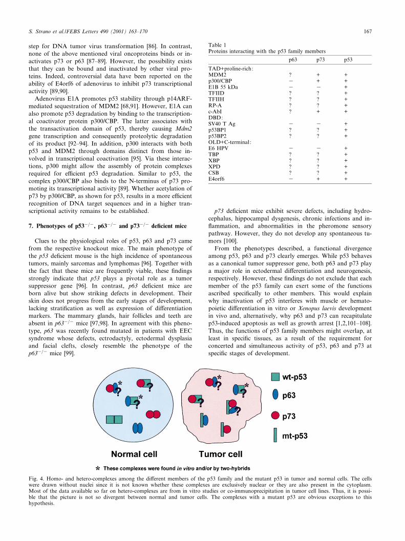
Table 1Proteins interacting with the p53 family members
p63 p73 p53
TAD+proline-rich:MDM2 ? + +p300/CBP 3 + +E1B 55 kDa 3 3 +TFIID ? ? +TFIIH ? ? +RP-A ? ? +c-AbI ? + +DBD:SV40 T Ag 3 3 +p53BP1 ? ? +p53BP2 ? ? +OLD+C-terminal:E6 HPV 3 3 +TBP ? ? +XBP ? ? +XPD ? ? +CSB ? ? +E4orf6 3 + +
FEBS 24569 14-2-01 Cyaan Magenta Geel Zwart
S. Strano et al./FEBS Letters 490 (2001) 163^170 167
8. p53, p73 and p63 in human cancers
The p53 tumor suppressor gene is the most frequent targetfor genetic alterations in human cancers [109]. The most prev-alent type of p53 mutations consists of missense mutations,often within the highly conserved DBD of the protein [18,20],leading to loss of the wild-type activity. However, at variancewith other tumor suppressor genes, cells bearing p53 muta-tions typically maintain the expression of full-length protein.This may suggest that, at least certain mutant forms of p53can actively contribute to cancer progression through `gain offunction' oncogenic activity. Such activity might depend onthe speci¢c p53 mutation and on the cell context in which thebiological outcome of the gain of function is evaluated [110].We and others have previously reported that conformationalmutants such as p53His175, but not DNA contact mutants,can increase cellular resistance to etoposide or contribute togenomic instability by abrogating the mitotic spindle check-point and consequently facilitating the generation of aneu-ploid cells [111^114]. The molecular mechanisms underlyingthe gain of function activities of mutant p53 remain to beelucidated. We can delineate two mechanisms through whichmutant p53 exerts gain of function activities. The ¢rst onerelies on the assumption that mutant p53 can bind to DNAthrough the association with DNA binding proteins and acti-vate speci¢c target genes using its functional TAD [115]. Insupport of this mechanism, it has been reported that humantumor-derived p53, whose TAD was inactivated by site-di-rected mutagenesis, lost the ability to increase tumorigenicityin vitro and in vivo [116]. In a second scenario, mutant p53binds to and sequesters proteins whose function is requiredfor anti-tumor functions such as apoptosis or growth inhibi-tion. Interestingly, it has been reported that human tumor-derived p53 mutants can associate with p73K and interferewith its transcriptional activity and ability to induce apoptosiswhen co-expressed in transient transfection assays [117]. Fur-ther studies have demonstrated that the association betweenmutant p53 and p73 occurs under physiological conditions asindicated by co-immunoprecipitation from various tumor cells[118,119]. Of note, di¡erent p73 variants exist in the cells,giving rise to a family of proteins that adds a new level ofcomplexity to the understanding of p73 signaling in cancercells [1,7,8]. Recent ¢ndings indicate that mutant p53 canalso be engaged in physical interactions with di¡erent iso-forms of p73 [119]. The Kaelin group has recently shownthat the association between human tumor-derived p53 mu-tants and p73 is governed by a common polymorphism atcodon 72 of p53 that encodes Arg or Pro. Thus, both thetype of p53 mutation and the polymorphism at codon 72in£uence whether mutant p53 interferes with p73 activity[118]. Heterodimers between mutant p53 and p63 have re-cently been shown to form in vitro and exist in tumor cells([118] and Strano and Blandino, unpublished observations),while further evidence needs to be collected to verify whethera triple complex (mt-p53/p63/p73) can assemble in cancercells. In that case, cancer cells carrying mutant p53 will pro-vide the ¢rst and clear example of a context in which p53family members interact with one another. It will be of inter-est to verify whether interactions occurring among the p53family members impact on the chemoresistance of tumor cells(Fig. 4).
While the DBD is the major site of mutations in p53, very
rare mutations in p73 and p63 have been found so far despiteextensive e¡orts [120^123]. Interestingly, the DBD of mutantp53 is su¤cient for the association with p73 isoforms [119].The DBDs of mutant p53 proteins have been regarded as`dead' domains since they cannot bind and activate p53 targetgenes. However, these DBDs acquire a protein^protein inter-action capacity that might contribute to the gain of functionactivities of mutant p53 by sequestering and inactivating pro-teins required for anti-tumor functions.
Acknowledgements: We are grateful to Moshe Oren, Oreste Segattoand Stefano Alema' for helpful suggestions and criticisms and MarcoCrescenzi for manuscript revision. We wish to thank Olimpia Montiand Alessia Baccarini, who are involved in addressing some of theunsolved questions reported in this review, via their thesis work. Thiswork is supported in part by Grant 369/bi from Telethon-Italy andQLG1-1999-00273 from the European Community.
References
[1] Kaghad, M., Bonnet, H., Yang, A., Creancier, L., Biscan, J.,Valent, A., Minty, A., Chalon, P., Lelias, J., Dumont, X., Fer-rara, P., McKeon, F. and Caput, D. (1997) Cell 90, 809^819.
[2] Yang, A., Kaghad, M., Wang, Y., Gillett, E., Fleming, M.D.,Dostch, V., Andrews, N.C., Caput, D. and McKeon, F. (1998)Mol. Cell 2, 305^316.
[3] Schmale, H. and Bamberger, C. (1997) Oncogene 15, 1363^1367.[4] Osada, M., Ohba, M., Kawahara, C., Ishioka, C., Kanamaru,
R., Katoh, I., Ikawa, Y., Nimura, Y., Nakagawara, A., Obinata,M. and Ikawa, S. (1998) Nat. Med. 4, 839^843.
[5] Trink, B., Okami, K., Wu, L., Sriuranpong, V., Jen, J. and Si-dransky, D. (1998) Nat. Med. 4, 747.
[6] Senoo, M., Seki, N., Ohira, M., Sugano, S., Watanabe, M., Ta-chibana, M., Tanaka, T., Shinkai, Y. and Kato, H. (1998) Bio-chem. Biophys. Res. Commun. 248, 603^607.
[7] De Laurenzi, V., Costanzo, A., Barcaroli, D., Terrinoni, A., Fal-co, M., Annichiarico-Petruzzelli, M., Levrero, M. and Melino, G.(1998) J. Exp. Med. 188, 1763^1768.
[8] De Laurenzi, V., Catani, M.V., Costanzo, A., Terrinoni, A.,Corazzari, M., Levrero, M., Knight, R.A. and Melino, G.(1999) Cell Death Di¡. 6, 389^390.
[9] Zaika, A., Kovalev, F., Marchenco, N. and Molle, U. (1999)Cancer Res. 59, 3257^3263.
[10] Soussi, T. and May, P. (1996) J. Biol. Mol. 260, 630^637.[11] Mai, M., Huang, H., Reed, C., Qian, C., Smith, J., Alderete, B.,
Jenkins, R., Smith, D. and Liu, W. (1998) Genomics 51, 359^363.[12] Mosner, J., Mummbenbraurer, T., Bauer, C., Sczakiel, G.,
Grosse, F. and Deppert, W. (1995) EMBO J. 14, 4442^4449.[13] Ding, Y., Inoue, T., Kamiyama, J., Tamura, Y., Ohtani-Fujita,
N., Igata, E. and Sakai, T. (1999) DNA Res. 6, 347^351.[14] Levrero, M., De Laurenzi, V., Costanzo, A., Sabatini, S., Gong,
J., Wang, J.W.J. and Melino, G. (2000) J. Cell. Sci. 113, 1661^1670.
[15] Irwin, M., Marin, M.C., Philip, A.C., Seelan, R.S., Smith, D.I.,Liu, W., Flores, E.R., Tsai, K.Y., Jacks, T., Vousden, K.H. andKaelin Jr., W.G. (2000) Nature 407, 645^648.
[16] Lissy, N.A., Davis, P.K., Irwin, M., Kaelin, W.G. and Dowdy,F. (2000) Nature 407, 642^644.
[17] Ko, L.J. and Prives, C. (1996) Genes Dev. 10, 1054^1072.[18] Levine, A.J. (1997) Cell 88, 323^331.[19] Oren, M. (1997) Cell 90, 829^832.[20] Prives, C. and Hall, P. (1999) J. Pathol. 187, 112^126.[21] Pozniak, C.D., Radinovic, S., Yang, A., McKeon, F., Kaplan,
D.R. and Miller, F.D. (2000) Science 289, 304^306.[22] Marin, M.C. and Kaelin Jr., W.G. (2000) Biochim. Biophys.
Acta 1470, M93^M100.[23] Chi, S.W., Ayed, A. and Arrowsmith, C.H. (1999) EMBO J. 18,
4438^4445.[24] Thanos, C. and Bowie, J. (1999) Protein Sci. 8, 1708^1710.[25] Bork, P. and Koonin, E.V. (1999) Nat. Genet. 18, 313^318.[26] Maltzman, W. and Czyzyk, L. (1984) Mol. Cell. Biol. 4, 1689^
1694.
FEBS 24569 14-2-01 Cyaan Magenta Geel Zwart
S. Strano et al./FEBS Letters 490 (2001) 163^170168

[27] Kastan, M.B., Onyekwere, O., Sidransky, D., Vogelstein, B. andCraig, R.W. (1991) Cancer Res. 51, 6304^6311.
[28] Giaccia, A.J. and Kastan, M.B. (1998) Genes Dev. 12, 2973^2983.
[29] Barak, Y., Juven, T., Ha¡ner, R. and Oren, M. (1993) EMBO J.12, 461^468.
[30] Wu, X.W., Bayle, J.H., Olson, D. and Levine, A.J. (1993) GenesDev. 7, 1126^1132.
[31] Haupt, Y., Maya, R., Kazaz, A. and Oren, M. (1997) Nature387, 296^299.
[32] Kubbutat, M.H.G., Jones, S.N. and Vousden, K.H. (1997) Na-ture 387, 299^303.
[33] Montes de Oca Luna, R., Wagner, D.S. and Lozano, G. (1995)Nature 378, 203^206.
[34] Jones, S.N., Roe, A.E., Donehower, L.A. and Bradley, A. (1995)Nature 378, 206^208.
[35] Bottger, A., Bottger, V., Sparks, A., Liu, W.L., Howard, S.F.and Lane, D.P. (1997) Curr. Biol. 7, 860^869.
[36] Chowdary, D.R., Dermody, J.J., Jha, K.K. and Ozer, H.L.(1994) Mol. Cell. Biol. 14, 1997^2003.
[37] Midgely, C.A. and Lane, D.P. (1997) Oncogene 15, 1179^1189.[38] Honda, R., Tanaka, H. and Yasuda, H. (1997) FEBS Lett. 420,
25^27.[39] Honda, R. and Yasuda, H. (1999) EMBO J. 18, 22^27.[40] Lohrum, M.A.E. and Vousden, K.H. (1999) Cell Death Di¡er. 6,
1162^1168.[41] Roth, J., Dobbelstein, M., Freedman, D.A., Shenk, T. and Lev-
ine, A.J. (1998) EMBO J. 17, 554^564.[42] Freedman, D.A. and Levine, A.J. (1998) Mol. Cell. Biol. 18,
7288^7293.[43] Lain, S., Midgley, C., Sparks, A., Lane, E.B. and Lane, D.P.
(1999) Exp. Cell Res. 248, 457^462.[44] Tao, W. and Levine, A.J. (1999) Proc. Natl. Acad. Sci. USA 96,
3077^3080.[45] Stommel, J.M., Marchenko, N.D., Jimenez, G.S., Moll, U.M.,
hope, T.J. and Wahl, G.M. (1999) EMBO J. 18, 1660^1672.[46] Zeng, X., Chen, L., Jost, C.A., Maya, R., Keller, D., Wang, X.,
Kaelin, W.G.J., Oren, M., Chen, J. and Lu, H. (1999) Mol. Cell.Biol. 19, 3257^3266.
[47] Balint, E. and Vousden, K.H. (1999) Oncogene 18, 3923^3929.[48] Dobbelstein, M., Wienzek, S., Koening, S. and Roth, J. (1999)
Oncogene 18, 2101^2106.[49] Ongkeko, W.M., Wang, X.Q., Siu, W.Y., Lau, A.W.S., Yama-
shita, K., Harris, A.L., Cox, L.X. and Poon, R.Y.C. (1999) Curr.Biol. 9, 829^832.
[50] Gostissa, M., Hengstermann, A., Fogal, V., Sandy, P., Schwarz,S.E., Sche¡ner, M. and Del Sal, G. (1999) EMBO J. 18, 6462^6471.
[51] Rodriguez, M.S., Desterro, J.M.P., Lain, S., Midgley, C.A.,Lane, D.P. and Hay, R.T. (1999) EMBO J. 18, 6455^6461.
[52] Minty, A., Dumont, X., Kaghad, M. and Caput, D. (2000)J. Biol. Chem. 275, 36316^36323.
[53] Oren, M. (1999) J. Biol. Chem. 274, 36031^36034.[54] Shieh, S.Y., Ikeda, M., Taya, Y. and Prives, C. (1997) Cell 91,
325^334.[55] Unger, T., Juven-Gershon, T., Moallem, E., Berger, M., Vogt
Sionov, R., Lozano, G., Oren, M. and Haupt, Y. (1999)EMBO J. 18, 1805^1814.
[56] Fuchs, S.Y., Adler, V., Pincus, M.R. and Ronai, Z. (1998) Proc.Natl. Acad. Sci. USA 95, 10541^10546.
[57] Bottger, V., Bottger, A., Garcia-Echeverria, C., Ramos, Y.F.,van der Eb, A.J., Jochemsen, A.G. and Lane, D.P. (1999) Onco-gene 18, 189^199.
[58] Ashcroft, M., Kubbutat, M.H. and Vousden, K.H. (1999) Mol.Cell. Biol. 19, 1751^1758.
[59] Banin, S., Moyal, L., Shieh, S., Taya, Y., Anderson, C.W., Ches-sa, L., Smorodinsky, N., Prives, C., Reiss, Y., Shiloh, Y. and Ziv,Y. (1998) Science 281, 1674^1677.
[60] Canman, C.E., Lim, D.S., Cimprich, K.A., Taya, Y., Tamai, K.,Sagaguchi, K., Appella, E., Kastan, M.B. and Siliciano, J.D.(1998) Science 281, 1677^1679.
[61] Mayo, L.D., Turchi, J.J. and Berberich, S.J. (1997) Cancer Res.57, 5013^5016.
[62] Gong, J.G., Costanzo, A., Yang, H.Q., Melino, G., Kaelin,
W.G., Levrero, M. and Wang, J.Y.J. (1999) Nature 399, 806^808.
[63] Agami, R., Blandino, G., Oren, M. and Shaul, Y. (1999) Nature399, 809^813.
[64] Yuan, Z.M., Shioya, H., Ishiko, T., Sun, X., Gu, J., Huang,Y.Y., Lu, H., Kharbanda, S., Weichselbaum, R. and Kufe, D.(1999) Nature 399, 814^817.
[65] Pawson, T. and Nash, P. (2000) Genes Dev. 14, 1027^1047.[66] Hermeking, H. and Eick, D. (1994) Science 265, 2091^2093.[67] Serrano, M., Lin, A.W., McCurrach, M.E., Beach, D. and Lowe,
S. (1997) Cell 88, 593^602.[68] Debbas, M. and White, E. (1993) Genes Dev. 7, 546^554.[69] Damalas, A., Ben-Ze'ev, A., Simcha, I., Shtutman, M., Leal,
J.F., Zhurinisky, J., Geiger, B. and Oren, M. (1999) EMBO J.18, 3054^3063.
[70] Zindy, F., Eischen, C.M., Randle, D.H., Kamijo, T., Cleveland,J.L., Sherr, C.J. and Roussel, M.F. (1998) Genes Dev. 12, 2424^2433.
[71] de Stanchina, E., McCurrach, M.E., Zindy, F., Shieh, S.Y., Fer-beyre, G., Samuelson, A.V., Prives, C., Roussel, M.F., Sherr,C.J. and Lowe, S.W. (1998) Genes Dev. 12, 2434^2442.
[72] Palmero, I., Pantoja, C. and Serrano, M. (1998) Nature 395, 125^126.
[73] Stott, F., Bates, S.A., James, M., McConnell, B.B., Starborg, M.,Brookes, S., Palmero, I., Hara, E., Ryan, K.M., Vousden, K.H.and Peters, G. (1998) EMBO J. 17, 5001^5014.
[74] Kamijo, T., Weber, J.D., Zambetti, G., Zindy, F., Roussel, M.F.and Sherr, C.J. (1998) Proc. Natl. Acad. Sci. USA 95, 8292^8297.
[75] Pomerantz, J., Schreiber-Agus, N., Liegeois, N.J., Silverman, A.,Alland, L., Chin, L., Potes, J., Chen, K., Orlow, I., Lee, H.W.,Cordon-Cardo, C. and DePinho, R.A. (1998) Cell 92, 713^723.
[76] Zhang, Y., Xiong, Y. and Yarbrough, W.G. (1998) Cell 92, 725^734.
[77] Bates, S., Phillips, A.C., Clark, P.A., Stott, F., Peters, G., Lud-wig, R.L. and vousden, K.H. (1998) Nature 395, 124^125.
[78] Hall, M. and Peters, G. (1996) Adv. Cancer Res. 68, 67^108.[79] Nevins, J.R. (1998) Cell Growth Di¡er. 9, 585^593.[80] Wu, X. and Levine, A.J. (1994) Proc. Natl. Acad. Sci. USA 91,
3602^3606.[81] Phillips, A.C., Ernst, M.K., Bates, S., Rice, N.R. and Vousden,
K.H. (1999) Mol. Cell. 4, 771^781.[82] Lane, D.P. and Crawford, L.V. (1979) Nature 278, 261^263.[83] Linzer, D.I.H. and Levine, A.J. (1979) Cell 17, 43^52.[84] Sarnow, P., Hearing, P., Anderson, C.W., Halbert, D.N., Shenk,
T. and Levine, A.J. (1984) J. Virol. 49, 692^700.[85] Lechner, M.S., Mack, D.H., Finicle, A.B., Crook, T., Vousden,
K.H. and Laiminis, L.A. (1992) EMBO J. 11, 3045^3052.[86] Yew, P.R. and Berk, A.J. (1992) Nature 357, 82^85.[87] Marin, M.C., Jost, C., DeCaprio, J.A., Caput, D. and Kaelin,
W.G. (1998) Mol. Cell. Biol. 18, 6316^6324.[88] Dobbelstein, M. and Roth, J. (1998) J. Gen. Virol. 79, 3079^
3083.[89] Steegenga, T., Shvarts, A., Riteco, N.B., Bos, J.L. and Jochem-
sen, A.G. (1999) Mol. Cell. Biol. 9, 3885^3894.[90] Higashino, F., Pipas, J.M. and Shenk, T. (1998) Proc. Natl.
Acad. Sci. USA 95, 15683^15687.[91] Sherr, C. (1998) Genes Dev. 12, 2984^2991.[92] Gu, W., Shi, X.L. and Roeder, R.G. (1997) Nature 387, 819^823.[93] Avvantaggiati, M.L., Ogryzko, V., Gardner, K., Giordano, A.,
Levine, A.S. and Kelly, K. (1997) Cell 89, 1175^1184.[94] Lill, N.L., Grossman, S.R., Ginsberg, D., DeCaprio, J. and Liv-
ingston, D.M. (1997) Nature 387, 823^827.[95] Grossman, S.R., Perez, M., Kung, A.L., Joseph, M., Mansur, C.,
Xiao, Z.X., Kumar, S., Howley, P.M. and Livingston, D.M.(1998) Mol. Cell. 2, 405^415.
[96] Donehower, L.A., Harvey, B.L., Slagle, B.L., McArthur, M.J.,Montgomery, C.A., Butel, J.S. and Bradley, A. (1992) Nature356, 215^221.
[97] Yang, A., Schweitzer, R., Sun, D., Kaghad, M., Walker, N.,Bronson, R.T., Tabin, C., Sharpe, A., Caput, D., Crum, C.and McKeon, F. (1999) Nature 398, 714^718.
[98] Mills, A., Zhenh, B., Wang, X., Vogel, H., Roop, D. and Brad-ley, A. (1999) Nature 398, 708^713.
FEBS 24569 14-2-01 Cyaan Magenta Geel Zwart
S. Strano et al./FEBS Letters 490 (2001) 163^170 169
[99] Celli, J., Duijf, P., Hamel, B.C.J., Bamshad, M., Kramer, B.,Smits, A.P.T., Newbury-Ecob, R., Hennekam, R.C.M., VanBuggenhout, G., Van Haering, A., Woods, C.G., Van Essen,A.J., De Waal, R., Vriend, G., Haber, D.A., Yang, A.,McKeon, F., Brunner, H.G. and Van Bokhoven, H. (1999)Cell 99, 143^153.
[100] Yang, A., Walker, N., Bronson, R., Kaghad, M., Oosterwegel,M., Bonnin, J., Vagner, C., Bonnet, H., Dikkes, P., Sharpe, A.,McKeon, F. and Caput, D. (2000) Nature 404, 99^103.
[101] Almog, N. and Rotter, V. (1997) Biochim. Biophys. Acta 1333,F1^F27.
[102] Soddu, S., Blandino, G., Scardigli, R., Coen, S., Marchetti, A.,Rizzo, M.G., Bossi, G., Cimino, L., Crescenzi, M. and Sacchi,A. (1996) J. Cell Biol. 134, 193^204.
[103] Mazzaro, G., Bossi, G., Coen, S., Sacchi, A. and Soddu, S.(1999) Oncogene 18, 5831^5835.
[104] Cerone, M.A., Marchetti, A., Bossi, G., Blandino, G., Sacchi,A. and Soddu, S. (2000) Cell Death Di¡er. 7, 506^508.
[105] Porrello, A., Cerone, M.A., Coen, S., Gurtner, A., Fontemaggi,G., Cimino, L., Piaggio, G., Sacchi, A. and Soddu, S. (2000)J. Cell. Biol. 151, 1295^1303.
[106] Martinelli, R., Blandino, G., Scardigli, R., Crescenzi, M., Lom-bardi, D., Sacchi, A. and Soddu, S. (1997) Oncogene 15, 607^611.
[107] Wallingford, J.B., Seufert, D.W., Virta, V.C. and Vize, P.D.(1997) Curr. Biol. 7, 747^757.
[108] Jost, C., Marin, M.C. and Kaelin Jr., W.G. (1997) Nature 389,191^194.
[109] Hollstein, M., Soussi, T., Thomas, G., von Breven, M. andBartsch, H. (1997) Rec. Res. Cancer. Res. 143, 369^389.
[110] Dittmer, D., Pati, S., Zambetti, G., Chu, S., Tereseky, K.,Moore, M., Finlay, C. and Levine, A.J. (1993) Nat. Genet. 4,42^46.
[111] Gualberto, A.S., Aldape, K., Kozakiewicz, K. and Tlsty, T.(1998) Proc. Natl. Acad. Sci. USA 95, 5166^5171.
[112] Li, R., Sutphin, D.P., Schwartz, D., Matas, D., Almog, N.,Wolkowicz, R., Gold¢nger, N., Pei, H., Prokocimer, M. andRotter, V. (1998) Oncogene 16, 3269^3278.
[113] Blandino, G., Levine, A.J. and Oren, M. (1999) Oncogene 18,477^485.
[114] Murphy, K.L., Dennis, A.P. and Rosen, J.M. (2000) FASEB J.14, 2291^2302.
[115] Fraizer, M.W., He, X., Wang, J., Gu, Z., Cleveland, J.L. andZambetti, G.P. (1998) Mol. Cell. Biol. 18, 3735^3743.
[116] Lin, J., Tereseky, A.K. and Levine, A.J. (1995) Oncogene 10,2387^2390.
[117] Di Como, C.J., Gaiddon, C. and Prives, C. (1999) Mol. Cell.Biol. 19, 1438^1449.
[118] Marin, M.C., Jost, C.A., Brooks, L.A., Irwin, M.S., O'Nions,J., Tidy, J.A., James, N., McGregor, J.M., Harwood, C.A.,Yulug, I.G., Voudsen, K.H., Allday, M.J., Gusterson, B., Ika-wa, S., Hinds, P.W., Crook, T. and Kaelin, W.G. (2000) Nat.Genet. 25, 47^55.
[119] Strano, S., Munarriz, E., Rossi, M., Cristofanelli, B., Shaul, Y.,Castagnoli, L., Levine, A.J., Sacchi, A., Cesareni, G., Oren, M.and Blandino, G. (2000) J. Biol. Chem. 275, 29503^29512.
[120] Mai, M., Yokomizo, A., Qiang, C., Yang, C., Tindall, D.J.,Smith, D.J. and Liu, W. (1998) Cancer Res. 58, 2347^2349.
[121] Nomoto, S., Haruki, N., Kondo, M., Konishi, H. and Takaha-shi, T. (1998) Cancer Res. 58, 1380^1385.
[122] Yokomizo, A., Mai, M., Tindall, D., Cheng, L., Bostwick,D.G., Naito, S., Smith, D.I. and Liu, W. (1999) Oncogene 18,1629^1633.
[123] Kaelin Jr., W.G. (1999) J. Natl. Cancer. Inst. 91, 594^598.
FEBS 24569 14-2-01 Cyaan Magenta Geel Zwart
S. Strano et al./FEBS Letters 490 (2001) 163^170170
Related Documents