Grand Valley State University ScholarWorks@GVSU Masters eses Graduate Research and Creative Practice 4-17-2009 From In-Vitro to In-Vivo: Corporate Development and Efficacy of a Topical Hair Growth Agent Derived from Natural Extracts Kelly Michael Glynn Grand Valley State University Follow this and additional works at: hp://scholarworks.gvsu.edu/theses Part of the Biology Commons is esis is brought to you for free and open access by the Graduate Research and Creative Practice at ScholarWorks@GVSU. It has been accepted for inclusion in Masters eses by an authorized administrator of ScholarWorks@GVSU. For more information, please contact [email protected]. Recommended Citation Glynn, Kelly Michael, "From In-Vitro to In-Vivo: Corporate Development and Efficacy of a Topical Hair Growth Agent Derived from Natural Extracts" (2009). Masters eses. 667. hp://scholarworks.gvsu.edu/theses/667

Welcome message from author
This document is posted to help you gain knowledge. Please leave a comment to let me know what you think about it! Share it to your friends and learn new things together.
Transcript

Grand Valley State UniversityScholarWorks@GVSU
Masters Theses Graduate Research and Creative Practice
4-17-2009
From In-Vitro to In-Vivo: Corporate Developmentand Efficacy of a Topical Hair Growth AgentDerived from Natural ExtractsKelly Michael GlynnGrand Valley State University
Follow this and additional works at: http://scholarworks.gvsu.edu/theses
Part of the Biology Commons
This Thesis is brought to you for free and open access by the Graduate Research and Creative Practice at ScholarWorks@GVSU. It has been acceptedfor inclusion in Masters Theses by an authorized administrator of ScholarWorks@GVSU. For more information, please [email protected].
Recommended CitationGlynn, Kelly Michael, "From In-Vitro to In-Vivo: Corporate Development and Efficacy of a Topical Hair Growth Agent Derived fromNatural Extracts" (2009). Masters Theses. 667.http://scholarworks.gvsu.edu/theses/667

FROM IN-VITRO TO IN-VIVO: CORPORATE DEVELOPMENT AND EFEICACY OF A TOPICAL HAIR GROWTH AGENT DERIVED EROM NATURAL EXTRACTS
A thesis submitted in partial fulfillment of the requirements for the degree ofMaster of Science
By
Kelly Michael Glynn
To
Biology Department Grand Valley State University
Allendale, Michigan April 17, 2009

Stultum est in luctu capillum sibi evellere, quasi calvito maeror levaretur
“It is foolish to pluck out one’s hair for sorrow, as if grief could he assuaged hybaldness”
Marqus Tullius Cicero Tusculanarum Disputationum (III, 26)
(http://www.worldofquotes.com/topic/Hair/index.html)
111

This work is dedicated to Vicki, for her love, encouragement, unwavering support and patience; to my entire family, especially my mom, without whom I never could have journeyed this far; and to Sam, Abbie and Hunter who were my motivation to finish.
Love always !
IV

ACKNOWLEDGEMENTS
My sincere gratitude is extended to my graduate committee- Dr. Roderick Morgan
of Grand Valley, Dr. David East and Mr. Lane Duvel, both from Amway Corporation,
whose guidance made this entire research objective an adventure and pleasure in
scientific discovery. Special thanks also go to Mrs. Robin Fleser and Ms. Betsy Lehner
for their time and patience in teaching me the ways of cell culture and providing working
cultures to propel the project forward. Mr. David Elower of Amway Corporation has
been a mentor my entire career, and his quest to solve complex problems, especially
within this project, has been a refreshing inspiration. To the Grand Valley Staff,
especially Dr. Mark Luttenton, Ms. Connie Ingham, Ms. Barb Ellis and Ms. Beverly
Tramper, for their guidance through the entire Masters administrative process, I am
deeply appreciative. I would also like to acknowledge Mr. Don Williams from Amway’s
Consumer Research Department for his assistance during the clinical study, Ms. Lachelle
Poling for her efforts in guiding me through Amway’s tuition program, and Ms Connie
Berg and Patty Linscott for their legal expertise. Lastly, I say thank you to Amway R&D
management- Ms. Martha Porter, Mr. Peter Roessler, Mr. John Coyle, Mr. Kem Charron,
Mr. Mark Gammage and Mr. Greg Evans- for allowing me to pursue my degree and
conduct my research on a corporate initiative and their continual support and interest in
the entire project. Thank you all!
ABSTRACT
FROM IN-VITRO TO IN-VIVO: CORPORATE DEVELOPMENT AND EFFICACY OF A TOPICAL HAIR GROWTH AGENT DERIVED FROM NATURAE EXTRACTS
by Kelly Michael Glynn
Androgenetic alopecia (male pattern baldness) affects up to 50% of the world’s
population, propelling the development for a possible treatment. The hair follicle is
influenced by several genetic and physiologic factors, which, when gone awry, lead to
androgenetic alopecia. Vascular endothelial and keratinocyte growth factors are
believed to be promoters of hair growth, as is inhibition of the proteasome complex. The
cytokine IE -la is also known to regulate follicle dynamics. The research objective
described herein was an attempt to develop a botanical blend, which could mediate the
above biomarkers, be successfully incorporated into a safe topical product and be
evaluated for in-vivo efficacy. By using an arbitrary scoring system to evaluate in-vitro
performance, botanical extracts were screened in cell culture and enzyme assays. A
Design of Experiments analysis, utilizing analyses of variance and multiple linear
regressions, was performed to derive an optimized blend of Eichochalcone, Saw
Palmetto, Shiso and Green Rooibos for incorporation into the prototype formulation.
After passing human irritancy and sensitization testing, these extracts were coupled with
liposomes to create a final prototype that was also screened for long-term stability. The
end product was used in a clinical-type trial, assessing its effectiveness to increase scalp
vi

hair density, promote anagen follicle activity and increase the growth rate of the hair
fiher. The product was henchmarked hy Rogaine® Extra Strength (5% minoxidil) and
Rovisomes Biotin (commercially available). The twelve-week study involved sixty-nine
males experiencing varying degrees of androgenetic alopecia who underwent 14” length
haircuts and a series of digital imaging focusing on a transition zone area of interest.
The three test products significantly increased hair density and the number of anagen
follicles compared to baseline values. Growth rate was up regulated for users of the
prototype and Rogaine®. Subjective self-assessment of the products revealed the
prototype to he the least effective in improving hair quality characteristics, but with no
significant difference to the other two products. These results indicate the herbal blend of
Lichochalcone, Saw Palmetto, Shiso and Green Rooibos, in a liposomal base, has the
potential to be an effective topical treatment for androgenetic alopecia.
Vll

TABLE OF CONTENTS
PAGE
EIST OE TAREES........................................................................................................... ix
EIST OF FIGURES................................................. xi
CHAPTER I- HAIR AS A BIOEOGICAE FUNCTION.........................................................1A.) INTRODUCTION.................. 1B.) EMBRYOLOGY OF H A IR ......................... 3C.) HAIR ANATOMY AND PHYSIOLOGY ....................................................... 6D.) STEM CELLS AND THE HAIR FOLLICLE....................................................... 9
CHAPTER II- HAIR CYCLES............................................................. 13A.) ANAGEN, CATAGEN, TELOGEN & EXOGEN................................. 13B.) TYPES OF HAIR ABNORMALITIES................................................................ 18
CHAPTER III- FACTORS INVOLVED E9 HAIR GROWTH/LOSS............................... 20A.) HORMONES............... 20B.) GENETICS AND GENES........................................................................... 24C.) MOLECULAR M ARKERS...................................................................................28D.) CLINICAL METHODS.......................................................................................... 30E.) RESEARCH OBJECTIVE.....................................................................................31
CHAPTER IV- MATERIALS AND METHODS..................... 33A.) VEGE AND KGE ASSAYS...................................................................................33B.) IE -la A SSAY ............................................................. 34C.) EXTRACT EFFICACY TESTING.......................................................................35D.) ELISA TESTING.......................................... 35E.) PROTEASOME ASSAY........................................................................................ 36E.) EVALUATION OE BOTANICAL EXTRACT PERFORMANCE................37G.) INGREDIENT FINALIZATION..........................................................................38H.) LIPOSOMAL PREPARATION AND PRODUCT STABILITY.................... 38I.) MICROBIAL TESTING........................................................................................ 39J.) CLERICAL PROTOCOL........................................................................................ 40
CHAPTER V- RESULTS...........................................................................................................44A.) SELECTION OF BOTANICAL EXTRACTS....................................................44B.) DOE ANALYSIS..................................................................................................... 47
viii

c.) INGREDIENT EINALIZATION..........................................................................50D.) LIPOSOME PREPARATION & PRODUCT STABILITY.............................. 51E.) CLINICAL STUDY ........................................ 55
CHAPTER VI- DISCUSSION .......................................................................................68
LITERATURE CITED .......................... 82
IX

LIST OF TABLES
TABLE PAGE
1. Format of Self-Perceived Questionnaire for Clinical Study................................... 43
2. Extract Scoring for in-vitro Assays................. 44
3. Extract Scores in Bioassays.......................... 46
4. DOE 1 Layout...................................................................... 47
5. DOE 2 Layout................................................................................................................ 48
6. Results of DOE Analyses 1 & 2 .....................................................................................49
7. Formulation of Prototype 2 ........................................................................................... 52
8. Stability Profile of Prototype 2 Formula..................................................................... 53
9. Product 586 User Demographics.......................... 55
10. Product 883 User Demographics.................................................... 56
11. Product 194 User Demographics.................................................................................. 57
12. Change in Density for Rovisomes .................................................................... 59
13. Change in Density for Prototype 2 ............................................................................... 59
14. Change in Density for Rogaine®........................ 60
15. ANOVA of Hair Density.............................................................................................. 60
16. Change in Anagen Hairs for Rovisomes.................... 61
17. Change in Anagen Hairs for Prototype 2 .....................................................................61
X
18. Change in Anagen Hairs for Rogaine®......................... 62
19. Change in Growth Rate for Rovisomes........................... 63
20. Change in Growth Rate for Prototype 2 .....................................................................63
21. Change in Growth Rate for Rogaine® .................................................................... 64
22. ANOVA of Growth Rates.................... 64
23. Correlations within Rovisomes C ell............................................................................66
24. Correlations within Prototype 2 C ell................................... 66
25. Correlations within Rogaine® Cell......................................................................... 66
26. Average Self-Perceived Assessments..........................................................................67
XI

LIST OF FIGURES
FIGURE PAGE
1. Development of the Hair Eollicle..................................... 4
2. Anatomical Structure of the Mature Hair Eollicle ..........................................7
3. Progression and Cyclic Nature of Hair Eollicles...................................................... 17
4. Liposome Particles in the Ideal Size Range for Prototype Stability Sam ples 54
5. Cut Global Images at Initial V isit............................................................................... 57
6. Clipped AGI Image at Initial Visit.............................................................................. 58
7. Shaved AGI Image at Initial V is it.............................................................................. 58
8. 72 Hour Eollow-Up AOI Im ages....................................................................... 58
9. Comparison of Average Objective Measurements...................................................65
Xll

CHAPTER 1 - HAIR AS A BIOLOGICAL ORGAN
A.) INTRODUCTION
From a non-biological view point, hair appears to be nothing more than a
“symbolic and psychosocial” (Hadshiew, et al., 2004) body part which can be modified to
bolster self-image and self-esteem from both individual and societal perceptions and
interactions (Stenn and Pans, 2001; Fans and Foitzik, 2004). In fact, it has been stated,
“the psychological importance of hair to man is in inverse ratio to its physical function”
(Ebling, 1976). Unbeknownst to many, human hairs are the only bodily appendage
which can be manipulated to influence societal relationships and improve self-image
(Stenn and Pans, 2001). These two attributes have lead to the creation of a multi-billion
dollar industry (Paus and Cotsarelis, 1999) which attempts to aid men and women with
their coiffed appearance.
Embedded within the skin, however, is a biological marvel that imparts the
physical characteristics of length, color, shape and diameter to the hair fiber, which is so
important to the mirror and society. This marvel is the hair follicle, which retains
embryonic cues to help create hair fibers throughout an individual’s life (Legue and
Nicolas, 2005). It does so in a cyclical process in an asynchronous fashion at the level of
the individual follicle (Stenn and Paus, 2001). Therefore, each human hair follicle can be
viewed as an independent biological entity that determines whether or not a visible hair
fiber is present. Through a series of intricate mechanisms, many of which are still
unknown, hair follicles can become quiescent beyond normal cyclical patterns leading to
diseased states generic all y termed alopecias. Aside from purely genetic manifestations of
1

baldness or aggressive infeetion, states of baldness sueh as alopecia areata (Cotsarelis and
Millar, 2001; Mulinari-Brenner and Bergfeld, 2001), telogen effluvium,
chemical/psychological induced baldness (Cotsarelis and Millar, 2001), and androgenetic
alopecia are all considered temporary states (Mulinari-Brenner and Bergfeld, 2001).
Contrary to perception, however, hair serves several critical biological functions.
These include defense against insects, camouflage, thermal regulation, sensory detection,
skin cleansing, and signal transporters (Stenn and Paus, 2001). Additional roles for hair
include ultraviolet protection and screens to prevent intrusion of foreign particles into
critical membranes such as the eye. Sexual communication is also influenced hy hair, or
the lack thereof, in events such as sexual selection in mating preference, identification of
puberty in adolescents and markers of masculinity in the appearance of chest, pubic and
heard hair (Camacho, et ah, 2000).
Since hair on all different parts of the hody can serve multiple biological
functions, the creation and development of the hair fiher must then have its own
biological apparatus operating under unique controls. Again, this apparatus is the hair
follicle (Camacho, et ah, 2000), and despite its uniqueness, its multi-mechanistic and
wondrous operation is concealed from the naked eye. Visibly, the only confirmation of
its existence is the emergence of the keratinized hair fiher protruding from the epidermal
surface. When this evidence is no longer present, or when the rate of its regeneration
capacity begins to diminish, the impact on humans can he monumental, despite being
painless and non-life threatening. Secondary side effects of hair loss include
psychological and emotional stress, shame, embarrassment, depression, loss of
confidence and self-worth, perception of age, and lack of societal acceptance (Hadshiew,
2

et al., 2004). These effects have been known to be especially significant in younger aged
men who suffer from some form of hair loss (Girman, et ah, 1998), hut both genders can
experience hair loss in some form (Camacho, et al., 2000). So, hair serves a
physiological, psychological and cosmetic role.
The premise for conducting hair research is to understand the biological
mechanisms that drive fiher growth and fulfill the accessory roles of this fibrous
appendage, and how such mechanisms may relate to other anatomical and physiological
processes. From a purely cosmetic standpoint, however, understanding how hair grows,
and uncovering the hope of how it may he restored in the case of baldness in humans, has
led to a race to be the first to claim success, even if marginal, at reversing a complex set
of interactions which ultimately create this void on the human scalp. A prominent
researcher in hair growth, Dominique Van Neste, has extensively researched the
biological phenomenon and cultural impacts of hair. He states:
As grooming may he controlled hy genetic factors it seems no surprise that hair has probably been a material of interest since the very early days of mankind. Engravings on the wall of caves and pre-historical sculptures provide the earliest representations of hair and clearly tell us about its symbolic dimensions. Hair- on the scalp and on the hody - is communication. It conveys messages about ourselves, it tells how we interconnect with social codes and status. Hair is everywhere! (Van Neste, 2003).
B.) EMBRYOLOGY OF HAIR
Hair follicle development begins in-utero, at two to three months, on the
eyebrows, lips, chin, and nose, with later development occurring on the back, abdomen,
and limbs. Follicle formation appears to he mediated in waves, with distance to the
preceding follicle being a determinant for new follicle placement. As a result, each
follicle maintains its own cycle (Serri and Cerimele, 1990). A precursor to follicle
3

formation, however, is the establishment of a connective network between epidermal and
mesenchymal tissues. Signal transmission from mesodermic tissue to embryonic
ectoderm causes thickening of the ectoderm and formation of the hair placode, which
emits return signals to the underlying mesenchyme, causing it to condense. Following
mesenchymal condensation, epithelial placode cells proliferate downward into the
mesenchymal tissue eventually forming the dermal papilla (Kulessa, et al., 2000). The
bulbous dermal papilla is the control center from which all-future hair growth regulation
and cycling will originate (Paus and Foitzik, 2004).
M acede Half Garm Hair Peg Mature Follicle
DermalCcmdensate g;
rWeduMa (Md) Corlex{Cx)
DennaH j — Inner Root Sheath (1RS)
—- Me
Figure 1- Development of the Hair Follicle From In-Utero to Maturity (used with permission from Dr. Elaine Fuchs 01/08/2009; Rendl et al., 2005, Fig. lA , p. 1911 )
With the basic hair follicle in place, surrounding kératinocytes will begin
differentiating (Kulessa, et al., 2000) to form eight different concentric layers of the
follicle. These include: the outer root sheath (ORS) and its companion layer; Henle’s and
Huxley’s layers; cuticle; and the medulla, cortex and cuticle of the actual hair fiber (Paus
and Foitzik, 2004).
The intimate signaling during follicle embryogenesis between the epithelial and
mesenchymal layers drives subsequent hair growth stages in post-natal life. Botchkarev
and Kishimoto (2003) state:
4

Extensive interactions between these two embryologically different hair follicle compartments lead to the formation of the hair shaft producing mini-organ that shows a cyclic activity during postnatal life with periods of active growth and hair shaft formation (anagen), apoptosis-driven involution (catagen), relative resting and hair shedding (telogen-exogen).
The continual cross talk between the ectoderm derived epidermis and mesoderm derived
mesenchyme (Serri and Cerimele, 1990) ultimately produces a distinct hair fiber. This
cross-talk is achieved through a combination of: genetic factors (Birch and Messenger,
2001; Midorikawa, et ah, 2004; Ishimatsu-Tsuji, et ah, 2005); a plethora of molecular
signaling (Hoffman, et ah, 1996; Kulessa, et al., 2000; Botchkarev, et al., 2001;
Botchkarev and Kishimoto, 2003); stem cell activity (Alonso and Fuchs, 2003; Blanpain,
et al., 2004; Legue and Nicolas, 2005; Kim, et al., 2006; Zhang, et al., 2006);
neuroimmunoendocrine circuitry (Paus, et ak, 2006); innervation (Hordinsky and
Ericson, 1996); hormones (Thornton, et ak, 1993; Hamada, et ak, 1996; Ellis, et ak, 1998;
Choi, et ak, 2001); and vascularization (Lachgar, et ak, 1996; Lachgar, et ak, 1998;
Sordello, et ak, 1998; Yano, et ak, 2001). The sum of these mechanisms results in
approximately five million hair follicles body-wide, with an estimated 80,000-150,000
follicles dispersed throughout the human scalp (Krause and Foitzik, 2006). After birth,
this number does not increase, whereas the size and shape of each follicle can (Paus and
Cotsarelis, 1999).
Initial fetal hair is termed lanugo hair and is typically shed at eight months in-
utero, replaced with additional lanugo hairs that last into the fourth month post-partum.
Vellus unpigmented, fine, short hairs, replace the secondary lanugo hairs, and typically
cover a majority of the skin surface. Through a prolonged continuation of the above
mechanisms certain regions of vellus hairs are transformed into terminal hairs, which are
5

thicker, longer and pigmented (Jankovic and Jankovic, 2004). The transformation of
terminal hairs back to vellus hairs on the scalp, and the underlying physiological changes
occurring within the follicle, is the trademark of androgenetic alopecia.
C.) HAIR ANATOMY AND PHYSIOLOGY
The hair follicle can be divided into three regions: biological synthesis,
keratinization and the hair fiber portion. Biological synthesis occurs at the hair bulb,
which encompasses the dermal papilla (Robbins, 1994) and the matrix (Krause and
Foitzik, 2006). Mesenchyme-derived dermal papilla cells cue the surrounding epithelial-
derived matrix cells to undergo mitosis during active growth (Philpott, et ak, 1990),
proliferating at one of the highest rates in the human body, even outpacing some forms of
cancers (Camacho, et ak, 2000; Krause and Foitzik, 2006). Deposition of melanin from
melanocytes embedded within the matrix (Paus and Cotsarelis, 1999) also occurs
resulting in coloration of the cortical cells of the emerging hair fiber. As epithelial cells
continue to proliferate, they are pushed upward toward the skin surface, where they enter
the matrix-derived inner root sheath (1RS). The 1RS consists of three distinct layers:
Henle’s layer, Huxley’s layer, and 1RS cuticle. Henle’s layer contains keratinized sheath
cells that help form a scaffold to support interior structures. Huxley’s layer, interior to
Henle’s layer, along with the 1RS cuticle, forms a rigid tube (Camacho, et ak, 2000) that
confers shape to the upward migrating cells and ultimately shapes the hair fiber (Paus and
Cotsarelis, 1999). The cells continue to elongate as they enter the zone of keratinization.
Sulfur transport, mainly in the form of cysteine, into the cells results in the formation of
disulfide bonds, and eventual keratin synthesis, imparting strength to the hair fiber.
Keratin synthesis continues until the cell is nearly filled with the fibrous material. As a
6

result, transcription and translation cease, nuclear degradation occurs, and the cell is
dehydrated. As this process occurs throughout the hair shaft cells, the fiber takes on its
final shape and diameter (Camacho, et ah, 2000; Robbins, 1994). As the hair fiber finally
emerges from the scalp, it is classified as a “keratin appendage from a follicle which is
embedded in the dermis,” composed of dehydrated cuticle, cortical and medullary cells
held together by biological cements (Robbins, 1994).
A
Figure 2- Anatomical Structure of the Mature Hair Follicle (http://www.pg.com/sdence/haircare/hair_twh_13.htm)
The aforementioned cuticle is the outermost covering of the hair fiber, formed
from an interlocking interaction with 1RS cuticles (Serri and Cerimele, 1990). Cuticle
cells are similar in length to cortex cells, but lack the cortex cells’ elasticity, and impart
chemical and mechanical resistance as well as moisture regulation to the hair fiber. The
cortex cells, derived from proliferating matrix, contain interwoven, keratin filaments,
which resemble a coiled structure microscopically. This longitudinal arrangement offers
great elastic properties to the hair fiber, compared to protective cuticle cells (Camacho, et
ak, 2000). The medulla is the innermost section of the hair fiber, and is derived from
7
apical dermal papilla cells. In scalp hairs, the medulla is often labeled as not present, but
in reality, these cells are often porous (Robbins, 1994) and highly vacuolated, making
their appearance unnoticeable (Camacho, et al., 2000). An additional structure, which is
involved in constructing the hair fiber and regulating follicle cycling events, is the outer
root sheath (ORS). The ORS surrounds the hair follicle in its entirety, from the
uppermost portion in the epidermis remaining with the permanent hair follicle, all the
way down to the dermally imbedded hair hulh (Serri and Cerimele, 1990). The ORS is
epithelial-derived, and throughout its length appears to have a multitude of functions
advantageous for skin repair as well as hair physiological activities. From potential
harboring of sebaceous glands and epidermal stems cells, to secretion of multiple
cellular-forming constituents, the ORS offers the skin repair mechanisms following
injury and/or damage. In addition, the ORS structure contains melanocytes (for
coloration), Langerhans’ cells (for immune response) and Merkel cells (for neurological
response), all of which are involved in skin restructuring or aiding the hair follicle to
respond to infection or sensory stimuli (Paus and Cotsarelis, 1999). External to the ORS,
and enclosing and separating the entire follicle from the skin epithelial layer, is a
membrane consisting of extracellular proteins (Rendl, et ah, 2005).
Biological components associated with and/or surrounding the hair follicle
include: the apocrine gland (perspiration); the sebaceous gland (lipid synthesis and
secretion); the isthmus (site of sensory fibers); the arrector pili muscle (sympathetic nerve
fibers synapsed with smooth muscle cells to aid in thermal barrier responses); the
infundibulum (the region, along with the hair canal, spanning the skin surface to just
above the sebaceous gland, representing the first body stmctures to be keratinized); and

the bulge (a stem cell repository) (Kanitakis, 2002; Serri and Cerimele, 1990).
D.) STEM CELLS AND THE HAIR FOLLICLE
As concisely summarized by Kolf et al, the stem cell niche:
encompasses all of the elements immediately surrounding the stem cells when they are in their native state, including the non-stem cells that might be in direct contact with them as well as ECM (extracellular matrix) and soluble molecules found in that locale. All of these act together to maintain the stem cells in their undifferentiated state. It is assumed that certain cues must find their way into the niche to signal the stem cells that their differentiation potential is needed for the regeneration or repopulation of a tissue (Kolf, et ah, 2007).
For the hair follicle, the bulge is the stem cell niche, supporting both hair regeneration
and the skin epithelium (Alonso and Fuchs, 2003) and marks the end of the permanent
hair follicle (Serri and Cerimele, 1990). The portion of the hair follicle inferior to the
bulge undergoes remodeling processes throughout the hair cycles. Downward growth
into the dermis, during active growth, is accomplished through epithelial-mesenchymal
communications between the permanent and regenerating portions of the follicle.
Derived from and residing in the ORS, the bulge was found to retain its relative position
of origin in individuals ranging in age from two weeks to twenty-one years (De Viragh
and Meuli, 1995).
Since the dermal papilla is referred to as the command center of the hair follicle
and is surrounded by highly proliferating matrix cells, initial hypotheses stated hair
follicle stem cells should logically reside somewhere in the same vicinity (De Viragh and
Meuli, 1995). In work done as early as 1994, however, evidence pointed to stem cell-like
activity occurring from a region in approximation to the arrector pili muscle (Rochat, et
ah, 1994). Further research verified the bulge was the location harboring stem cells to be
used to regenerate the active growth stage for the hair follicle, and possibly for aiding in
9

epidermis repair following tissue damage, since these epithelial stem cells can reproduce
sebaceous glands and skin layers when these components are destroyed (Paus and
Foitzik, 2004). Furthermore, when the colony-forming ability of kératinocytes isolated
throughout the hair follicle was examined, cells isolated from the bulge were able to
generate 95% of the colony-forming cells in culture, while only 5% of colony-forming
cells were attributed to the matrix region. Also, the colony forming kératinocytes showed
no increase in growth potential when co-cultured with papilla fibroblasts, an indication
that there may be a certain level of independence between the two follicle cell types and
how they regulate hair fiber growth (Kobayashi, et ak, 1993).
Within the basal layer of the epidermis, stem cells also reside, which through
division, upward movement, and terminal differentiation result in mature skin cells. It
has been suggested that the bulge stem cells may be the multi-potent progenitors of the
epidermal stem cells since: epidermal stem cells have no identifiable niche within the
epidermis; bulge stem cells exhibit slower cycling times than epidermal stem cells; and
retention of radioactive thymidine is longer within bulge cells, indicative of the cells not
undergoing rapid cell cycle/mitosis events. Daughter cells derived from bulge stem cells
eventually differentiate to assist in the formation/maintenance of the hair follicle matrix,
the sebaceous gland, and the basal layer of the epidermis (Alonso and Fuchs, 2003).
A prominent theory as to how the bulge drives hair follicle regeneration is known
as the bulge activation hypothesis. The premise is the mesenchymal dermal papilla emits
a signal to the bulge stem cells, which in turn, begin sending stem cells to the hair bulb
region. These mobile signal carriers create rapidly dividing kératinocytes, which will
form/reform the hair bulb, and through a series of additional stages, new hair fibers are
10

produced (Alonso and Fuchs, 2003; Camacho, et a l, 2000; Stenn and Pause, 2001). The
length of that fiber will correspond to the number of cell divisions taking place, and when
the proliferative capability of these is reached, catagen induction begins (Camacho, et a l,
2000).
Recent research has also indicated the matrix stem cells, derived from the bulge,
are highly organized and compartmentalized with each sector responsible for forming a
certain portion of the hair fiber. At the inner core of the matrix, multipotent stem cells
reside which produce daughter cells that give rise to transient progenitors of the various
structures of the hair fiber. These progenitors are in the layer external to the multipotent
stem cells. The third and final concentric layer consists of post-mitotic ancestors of the
transient cells, whose function is to construct the columns of the hair bulb which will be
used to construct the different components of the hair fiber. This organizational pattern
creates a radial distribution within the bulb. How cells align along the central vertical
axis of the bulb determines each cell’s fate, giving rise to the 1RS, hair fiber cuticle or the
medulla (Legue and Nicolas, 2005).
Stem cells are necessary for maintaining cellular and physiological balance and
also for initiating repair mechanisms and tissue regeneration following wounding. For
hair growth regulation, stem cells and their niche are critical for re-initiating the active
growth phase in the hair cycle. For this reason, characteristics of the bulge and its stem
cells include: 1.) The bulge is anatomically formed postnatally following the initial
growth stage; 2.) Basal layers within the bulge are attached to a basement membrane,
while several genes, responsible for producing cytoskeletal, extracellular matrix, cell
adhesion molecules and proteins are active within; 3.) The niche housing the stem cells
11

within the bulge is partitioned asymmetrically; 4.) Isolated stem cells are able to
reproduce several generations of clones in culture, as well as reproduce hair follicles and
sebaceous glands; 5.) Stem cells respond to external cues to initiate regeneration events;
6.) Once in the hair bulb region, stem cells undergo specific spatial organization to
properly reconstruct the hair follicle and eventual hair fiber. As a whole, these attributes
indicate the bulge is a pertinent player in hair growth and skin function (Blanpain, et al.,
2004; Legue and Nicolas, 2005).
12

CHAPTER II: HAIR CYCLES
A.) ANAGEN, CATAGEN, TELOGEN & EXOGEN
One of the most interesting aspects of the hair follicle is its cyclic nature, divided
among stages of active fiher growth (anagen), growth cessation and apoptosis of the
temporary follicle (catagen), and a period of rest and/or remodeling (telogen) (Muller-
Rover, et ah, 2001; Pans and Foitzik, 2004; Rendl, et al., 2005; Rohhins, 1994). As
technology has advanced to observe follicle morphogenesis, and the complexity of these
cyclic events has come to he understood, the hair follicle has become “an attractive
system for studying major biological phenomena” (Stenn and Pans, 2001).
Anagen begins with the cues to start the reconstruction of the follicle hulh and
ends when active growth ceases and additional cues are received to begin the
deconstruction of the same bulb. Anagen occurs in six distinct steps (Müller-Rôver, et
al., 2001), hallmarked by the rapid proliferation of matrix cells, active melanin deposition
via melanocytes and keratinization of epidermally progressing cells, all contributing to
the emergence of the characteristically distinct hair fiber (Rohhins, 1994; Stenn and Pans,
2001). The actual molecular cues, which initiate this growing process, remain obscure;
however, the anatomical events occurring to lead to active growth resemble those events
happening during embryonic follicle development. Epithelial cells divide in a downward
fashion to reach the dermis, where dermal papilla cells anchor to a basement membrane.
Upon reaching their end point, growth begins in an upward and outward fashion with the
development of the 1RS and hair shaft (Stenn and Paus, 2001).
The processes initiating anagen include trauma and/or wounding of the hair
follicle, hair plucking, and chemical influence. Merely cutting the hair fiber does not
13
induce anagen events (Stenn and Paus, 2001). In addition, the same cellular and
molecular pathways involved during embryonic follicle development are believed to take
part in the governing of anagen initiation and progression throughout life (Cotsarelis and
Millar, 2001).
Approximately 80-90% of a human’s scalp hairs are in the anagen stage (Robbins,
1994), which lasts between two to five years. The factor that determines the length of a
single hair fiber then is the duration of time the follicle spends in the anagen phase
(Mulinari-Brenner and Bergfeld, 2001), while the diameter of a hair fiber is determined
by the size of the dermal papilla. A larger dermal papilla generally contains more
proliferating matrix cells. The dermal papilla volume is created during the first stages of
anagen (Cotsarelis and Millar, 2001). The shape of the hair fiber is ultimately determined
by the shapes of the ORS and 1RS, through which the upward migrating cells are
funneled (Camacho, et al., 2000). In many cases, the anagen portion of the hair cycle
will function normally in the scalp through approximately ten progressions, which
corresponds to roughly forty years of age (Krause and Foitzik, 2006).
All of the work done during anagen is destroyed during the eight stages of catagen
(Müller-Rôver, et al., 2001) defined as “highly controlled involution of the hair follicle
resulting in apoptosis and terminal differentiation’’ (Krause and Foitzik, 2006). The
characteristic signs of apoptosis (cell shrinkage, blebbing, nuclear condensation, and
eventual cell fragmentation) are all observed in cells of the follicle (Botchkareva, et ah,
2006). The switch from anagen to catagen is again somewhat of a mystery in terms of
causation, molecular signaling and genetics, but certain events, such as chemical
application, trauma, and environmental factors have been found to invoke this regressive
14

phase (Stenn and Paus, 2001).
During catagen, the temporary portion is disassembled by means of regulated cell
death occurring in certain follicle structures. In addition, the dermal papilla separates
from the follicle bulb (Stenn and Paus, 2001). The size of the entire follicle is also
diminished, as is the position of the follicle. Follicles may reach into the subcutaneous
fat layer during anagen, while the same post-catagen follicle transcends into the dermal
tissue (Cotsarelis, 1997).
One of the most interesting aspects of apoptosis during catagen is it occurs in
waves, beginning with the area of melanin deposition in the bulb, spreading to the hair
matrix, then the ORS and 1RS, and finally converging on the hair shaft (Botchkareva, et
al., 2006). Even more intriguing, however, is dermal papilla fibroblasts do not undergo
apoptosis, nor do most bulge cells, at anytime during the hair cycles. This demonstrates
that these cell types are critical for future regeneration events in subsequent growth
cycles (Botchkareva, et al., 2006; Cotsarelis, 1997).
Considering the highly controlled state of catagen, a relatively small percentage of
scalp hairs are in this stage at any given time, roughly 1-2% (Robbins, 1994).
Furthermore, it is expedient, lasting anywhere from three to six weeks (Mulinari-Brenner
and Bergfeld, 2001). The catagen events of the hair follicle are “to delete the old hair
shaft factory and to bring the inductive machinery of the cell to a point where a new
follicle can form, utilizing once again, the stem cells of the bulge and the inductive
powers of the papilla” (Stenn and Paus, 2001).
The cycle in which most hairs spend their time, second to anagen, is telogen. This
is the resting phase of the follicle, or as alternately proposed, a “pre-regeneration” state of
15

anagen (Camacho, et al., 2000), or an anagen brake (Stenn and Paus, 2001). By the time
a follicle has entered into telogen, epithelial kératinocytes have surrounded compacted
dermal papilla fibroblasts, which have minimal proliferative activity (Paus and Foitzik,
2004). Even though follicle activity has diminished in telogen, relative to anagen and
catagen, the follicle still contains the necessary cell populations to generate a new follicle
and fiber in the next anagen cycle. These include epithelial stem cells, ORS
kératinocytes, and melanocytes (Camacho, et al., 2000). The dermal papilla is terminal
to and adjacent to the hair germ (the base of the quiet follicle) and the entire structure is
located in approximation to the arrector pili muscle, well into the dermal layer (Camacho,
et al., 2000; Stenn and PauS; 2001). Residing in the hair shaft is the club hair, or dead
hair (Paus and Cotsarelis, 1999) to which the root sheaths have attached. The bulb,
matrix and dermal papilla are now separated from the shaft, effectively preventing any
further growth of the hair fiber. The club hair will eventually be shed, in the cycle known
as exogen, which is believed to be distinct from the anagen cycle, even though the two
events can occur simultaneously. A club hair will still reside within the follicle, while a
new anagen phase has started rebuilding the lower, temporary portion of the regenerative
follicle. In fact, it has been proposed, the cycle of exogen encompasses the factors that
anchor the club hair into the follicle, and what molecular events take place to release the
club hair from the shaft (Stenn and Paus, 2001). On average, an individual loses
anywhere from 50-100 hairs per day as a result of the exogen event (Robbins, 1994).
The percentage of scalp follicles in telogen ranges from 10-20% (Robbins, 1994),
and lasts, on average, three to nine months (Camacho, et ah, 2000). From a historical
research perspective, little investment of time has been put into telogen research, and as
16

such, the magnitude of molecular biomarkers involved in this stage is still unknown
(Stenn and Paus, 2001).
a .
Figure 3- Progression and cyclic nature of hair follicles (used with permission from the New England Journal of Medicine 01/12/2009; Pans and Cotsarelis, 1999, Fig. 2, p. 493.)
17

B.) TYPES OF HAIR ABNORMALITIES
It is apparent the alterations in the hasic operations of the hair cycles will lead to
some type of hair disorder. In addition, since the number of follicles an individual
possesses is an emhryogenic determined entity, the mechanisms of operation in each
follicle will determine hair abnormalities (Mulinari-Brenner and Bergfeld, 2001). For
example, if anagen is excessively prolonged, hypertrichosis or hirsutism can result, both
of which are excessive hair growth disorders. When the anagen cycle is continually
shortened during successive cycles, or if telogen is extended, alopecia will result.
Different associated factors can drive a multitude of other hair disorders, including
alopecia areata (patchy hair loss either on the scalp or throughout the body caused by an
autoimmune malfunction); anagen effluvium (sudden shedding of actively growing hair
as is observed in chemotherapy patients); telogen effluvium (abnormal number of
follicles induced into telogen often due to certain drugs/medications or fever); permanent
alopecia (entire follicle is destroyed due to infection, autoimmune disorders or skin
cancers); and androgenetic alopecia (influenced by the androgenic steroids testosterone
and dihydrotestosterone [DHT] resulting in characteristic balding patterns on the scalp).
With the exception of genetic deletion and follicle organ deletion, malfunctions in one or
more of the cycling events, or aberrant factors and/or influences involved within a cycle,
will result in an abnormal manifestation of hair growth or hair loss (Hamada and Randall,
2006; Hibberts, et al., 1998; Mulinari-Brenner and Bergfeld, 2001, Paus and Cotsarelis,
1999).
The remainder of this review will focus mainly on androgenetic alopecia (AA),
which is the most prevalent form of hair loss on the scalp (Hoffman, 2003) accounting for
18

approximately 95% of those individuals suffering from some type of hair growth defect
(Choi, et ah, 2001). It is estimated between 40-50% of the world’s population is afflicted
with this disease (Krause and Foitzik, 2006; Robbins, 1994) with up to eighty million
Americans experiencing hair loss (Leavitt, 2003). It can affect both males and females
(Mulinari-Brenner and Bergfeld, 2001) and children as young as six years of age have
also been known to be susceptible (Tosti, et al., 2005). In regards to intervention, AA is a
non-permanent form of baldness (Paus and Cotsarelis, 1999) since the follicle does
remain embedded within the dermis and continues to cycle even in the absence of a
visible hair fiher (Mulinari-Brenner and Bergfeld, 2001). The biological complexity of
the hair follicle in terms of operational control is still a mystery waiting to be solved,
while the physiological function in humans has taken on a largely cosmetic role. The
hope for restoring hair growth remains limited with surgical repair via hair
transplantation being the only permanent fix to date (Leavitt, 2003). Hope for better
success, by easier means, will come if only the mystery of biological complexity is
unraveled. Understanding these mechanisms and the influences associated with them
may eventually lead to that cosmetic milestone.
19

CHAPTER III- FACTORS INVOLVED IN HAIR GROWTH/LOSS
A.) HORMONES
Two major contributing factors to A A are androgens and genetic predisposition
(Ellis, et al., 1998). When the synthesis of androgenic steroids begins with cholesterol,
several weaker intermediate hormones are produced which can and are converted to more
potent forms via enzymes. The major androgenic steroid circulating throughout the body
is testosterone, while the still more potent steroid is dihydrotestosterone (DHT) converted
from testosterone by the enzyme 5a-reductase (5a-R). Androgen potency is determined
by its binding affinity to the androgen receptor (AR) within a cell’s cytoplasm. A further
dimension to consider is free-circulating androgens. Sex-hormone-binding-globulin
(SHBG), binds almost 70% of available testosterone, while albumin takes hold of another
19%. That leaves approximately 10% of available testosterone as free circulating
hormone. It still remains unclear as to whether or not bound testosterone, onto either
protein, can be active. Once an androgen binds the AR, the complex shuttles to the
nucleus where it exerts its effects via gene transcription or suppression. The primary
androgens related to hair follicle physiology are testosterone and DHT, though additional
hormones and factors can certainly play a role in the regulation of hair growth/loss.
These include levels of available hormones, levels of conversion enzymes, the number of
androgen receptors present within a cell and/or tissue and the influence of the androgen
complex on genes directly involved in hair growth modulation (Hoffmann, 2003).
Some of the most interesting research conducted on hair growth regulation has
been the realization that androgens, particularly DHT, have different modulatory
properties throughout the body and even on the scalp (Hibberts, et ah, 1998; Hoffmann,
20

2003; Stenn and Paus, 2001; Thornton, et al., 1993). For instance, the occipital scalp is
androgen insensitive, while the frontal, parietal and coronal scalps are all androgen
sensitive. The scalp vertex is androgen sensitive but androgen independent. The axillary
portions of the body are androgen dependent whereas the eyebrows and eyelashes are
androgen insensitive (Stenn and Pans, 2001). So even though a majority of the body is
covered by hair, either terminal or vellus, not all hairs are affected in the same way by
androgens.
Extensive research has been done in an attempt to explain how different body
locales respond to androgens. Work done by Hibberts, Howell and Randall (1998) found
the number of androgen receptors in dermal papilla fibroblasts was significantly higher in
follicles extracted from balding scalp tissue compared to non-balding scalp tissue, while
the androgen binding affinity from both regions was identical, as was the protein content
of both receptors. Such a discovery implies the number of androgen receptors in a given
area may have a significant impact on hair growth. However, expression of androgen
receptor mRNA has also been found throughout the hair follicle in both balding and non-
balding individuals (Asada, et al., 2001), indicating more is needed than just a large
quantity of androgen receptors to precipitate a balding condition.
Additional factors influencing the expression and/or control of the AR include
phosphorylation of specific serine residues in the AR protein. When androgen is bound
to the AR, phosphorylation of these residues increases. Attention to the phosphorylation
of serine 213 in the AR protein is especially intriguing since it may be involved in certain
developmental processes, and has been shown to promote the degradation of the AR
(Taneja, et al., 2005). The discovery of isoformic co-activators for AR transcription has
21
also hinted at explaining the different responses to androgens throughout body tissue.
The short isoform, ARA70(3, was observed only in the dermal papilla portion of the
follicle, and its expression was reduced in balding tissue compared to non-balding tissue
(Lee, et ah, 2005). It is interesting to surmise that the co-activator for the AR is upstream
from AR transcription. Simultaneous expression of both the co-activator and receptor
protein could limit hair growth in the different scalp tissues by negatively regulating
dermal papilla proliferation signals.
When beard dermal papilla cells (androgen dependent and sensitive) were
compared to non-balding scalp dermal papilla cells (androgen independent and/or
insensitive) for the conversion of testosterone to DHT, via uptake of radiolabled
testosterone, it was discovered the beard cells only converted testosterone to DHT, not
the scalp cells. So not only is the AR important for hair growth modulation, but the
presence of the converting enzyme, 5a-R, is also a critical factor (Thornton, ct ah, 1993).
The 5a-R enzyme has two isoforms, 5a-Rl and 5a-R2, which have been found to
have specificity within the cell as well as in tissue activity (Stenn and Paus, 2001).
Epithelial cells of the hair follicle have an abundance of 5a-R l, compared to a limited
amount of 5a-R2, while the dermal papilla contains mRNA for 5a-R2 almost exclusively.
The quantities for both of these isoforms were not different in balding and non-balding
cases (Asada, et al., 2001). Since the dermal papilla appears to solely express 5a-R2
mRNA, and is the command center of the actively growing hair follicle, it seems
reasonable to believe the 5a-R2 isoform is an essential enzyme for androgen
metabolization where androgen influence plays a significant role in hair growth (Asada,
et ah, 2001 ; Hoffmann, 2003), and that the main site for androgen activity is in the
22

dermal papilla of the hair follicle (Hamada, et ah, 1996).
Additional research examined the levels of androgens in hair from the different
zones (balding versus non-balding) of the scalp. Serum levels of androgen were also
tested. Levels were compared within individuals and to controls (non-balding subjects).
Vertex DHT was higher in balding subjects versus non-balding individuals, but no real
difference in DHT levels was found to exist between balding and non-balding zones from
the same subject. Serum levels of both DHT and testosterone were also higher in balding
participants compared to non-balding participants (Bang, et ah, 2004).
Besides testosterone and DHT, estrogen and estrogen intermediates are also
involved in hair regulation in both males and females. Ohnemus et ah, (2006), in their
review of estrogen function, state; “estrogens and estrogen metabolism are at least as
important as androgens in male and female hair biology.” Reasons listed for this
premise include inhibition of hair re-growth in mice when estrogen is applied topically,
which directly opposes the common practice of topical application of estrogens for hair
growth stimulatory effects in women suffering from androgenetic alopecia. Such an
anomaly points to another complex mode of influence, further compounded by species
specificity. In addition, research is also referenced which points to estrogens having the
capability of squelching androgen metabolism, even in the dermal papilla, to the point
that the amount of DHT produced, following testosterone stimulation, is reduced. The
enzyme aromatase, which can convert testosterone to the less potent 17p-estradiol, has
also been isolated from cells which have active AR expression occurring, suggestive of a
complex regulatory role between the two hormone classes, which when gone awry, may
be manifested in some hair disorder. This complexity is further complicated by research
23

showing hormone receptors of the different classes can communicate with each other,
leading to alterations of the individual hormonal cascades and regulation of gene
expression (Ohnemus, et ah, 2006).
The hormone prolactin and its receptor were also found to exist in human hair
follicles, and that treatment with exogenous prolactin inhibited cultured follicle growth,
while endogenous prolactin, and its receptor, expression increased as follicles advanced
into the catagen cycle. The region of prolactin activity appears to be limited to epithelial
cells in the follicle since mesenchyme derived dermal papilla cells exhibited no presence
of the hormone, or its receptor (Foitzik, et ah, 2006).
In terms of hormones, the hair follicle exhibits a high degree of complexity, just
compounding the difficulty of understanding how this miniaturized organ operates. Even
though common male patterned baldness bears the moniker of the androgen steroid,
much more is in play in the regulation of hair growth, from conversion enzymes to
intermediates to expression locations.
B.) GENETICS AND GENES
Due to the high degree of integration between the different physiological systems
regulating hair growth, and how the phenotype or clinical appearance can manifest itself
over time, balding and non-balding conditions are polygenetic traits, which are reached
upon on a threshold crossing of genetic events gone awry. Birch and Messenger (2001)
examined first and second generation inheritance patterns for balding and non-balding
males. After five hundred seventy-two men were studied, the researchers concluded:
balding is common in Caucasian males; with increasing age, baldness also increases
regardless of what stage the balding pattern is; if a balding condition manifests itself
24

before the age of thirty, the probability is high for the father of these individuals to also
be bald; if men live long enough, they will go bald; males who are resistant to balding
typically come from non-balding families; and the female balding condition possesses a
higher threshold state since androgens contribute to baldness and are typically at lower
levels in females.
Several gene expression profile studies have been conducted comparing the
balding and non-balding traits. Macroarray research that examined expression levels of
1185 genes, ranging in function from cell cycle regulation to apoptosis, discovered nearly
ten percent [107] of the genes were alternately expressed in balding subjects. Genes
involved in signal transduction and cell cycle regulation both had decreased expression
levels in dermal papilla cells from balding sites. Furthermore, fourteen growth factor
genes also exhibited decreased expression. Taken together, these findings indicate the
balding follicles were functionally inactive (Midorikawa, et ah, 2004).
A microarray analysis comparing male and female gene expressions revealed
1436 genes were common to both genders, while ninety-seven genes showed differential
expression between the two sexes, with a majority [89] at higher levels in males, and only
eight were positively up regulated in females. What state of balding the test subjects
were in was not mentioned, but since hair fiber extractions included the upper portion of
the ORS sheath, follicles were most likely in the anagen cycle (Kim, et al. 2006).
A murine microarray study was done, whose hair follicle cycles can be
synchronized by epidermal dépilation via waxing. As the hair cycle stages progressed,
expression patterns were compared to non-depilated skin tissue. Some key findings of
this research show twenty-three days post dépilation, expression patterns returned to
25

baseline levels relative to the non-depilated tissue expressions. Following the hair
removal event, genes involved in inflammation response and anagen initiation were the
main factors, while in the mid-late anagen cycle, keratin related genes were active, all of
which correspond to the regenerative events of hair fiber formation (Ishimatsu-Tsuji, et
al., 2005).
In regards to specific gene events, Ellis, Stebbing and Harrap (2001), analyzed
androgen receptor gene polymorphisms in balding and non-balding males. They
determined a restriction fragment length polymorphism, StuI, residing in exon one of the
AR gene, “is a necessary, but not sufficient component of the polygenic predisposition to
male pattern baldness.” In young men with baldness, 98% percent of the subjects
possessed this marker, while older, balding males also exhibited polymorphism to a high
degree. Those without this particular polymorphism were not likely to go bald. In cases
where the marker was present, but baldness was not, the balding threshold may not have
been reached yet, and/or the other required polygenetic dispositions were not present in
those individuals. This was later corroborated on a different ethnic group, which showed
the dual band polymorphism in balding males resulted from a single nucleotide base
change (adenine to guanine) in the first exon of the X chromosome (Levy-Nissenbaum, et
ah, 2005).
The 5a-Reductase gene would also be a logical target for researchers hoping to
understand the genetics involved in baldness and/or hair growth regulation. Since the 5a-
reductase enzyme has two isoforms, there are two separate genes as well. SRD5A1 codes
for the 5a-Rl enzyme, and is located on chromosome five, whereas SRD5A2 codes for
the 5a-R2 form and is on chromosome two. What research has uncovered though, is both
26

genes have elevated expression levels in the frontal scalp for both men and women, but
no significant difference in distribution patterns could be discerned for both genes
between balding and non-balding states. The implication here is the 5a-R gene or
enzyme variability is not a contributing factor to a balding disposition. In addition, since
sons of balding fathers also tended to display baldness in this study, the likelihood of a
simple X-linked mode of inheritance appears unlikely. With all factors considered, a
polygenetic mechanism produces the balding phenotype (Ellis, et ah, 1998; Levy-
Nissenbaum, et ah, 2005).
The hairless gene, when defective in mice, will produce normal looking hair
follicles at birth. When the first catagen cycle occurs, however, the entire mouse
becomes bald with dermal cysts forming in the follicle. In regards to humans, alterations
in hairless leads to an entire body devoid of hair (Camacho, et ah, 2000). The dermal
papilla is separated from the hair shaft and improper catagen deconstruction of the
follicle results in loss of recovery for future hair growth events. Another human hair
deficiency, identified as MIM: 601705, is synonymous with expression of the recessive
nude phenotype in mice. Despite the presence of a hair follicle, the hair shafts cannot
break the epidermal barrier, resulting in complete baldness (O’Shaughnessy and
Christiano, 2004).
When the gene expression profiles are tallied, there is supporting evidence for
over one hundred seventy-nine genes, involved in hair growth functions (Ishimatsu-Tsuji,
et ah, 2005; Kim, et ah, 2006; Midorikawa, et ah, 2004) and at least one hundred
different proteins expressed in the hair follicle (O’Shaughnessy and Christiano, 2004).
Such a large number just re-emphasizes the complexity of the network controlling this
27
“miniaturized” organ and the uncertainty of which factor(s) is absolutely essential for
initiating the process of hair loss. Through a thorough examination of genetic analysis,
molecular factors, environmental influences, and inferential implications from both in-
vitro and animal studies, the intricacy of the hair follicle may be solved. Even then it is
still unknown if it can be manipulated in order to restore the proper balance ensuring a
cosmetically, socially and mirror image pleasing perception and appearance.
C.) MOLECULAR MARKERS
Since the hair follicle has come to be understood as a complex mini-organ, it has
garnered much interest “for studying major biological phenomena” (Stenn and Paus,
2001) in addition to hair biology research. Cell cultures of hair follicle kératinocytes and
dermal papilla fibroblasts are now commercially available, and isolation and culturing
techniques of surgically extracted follicle cell types are well established (Havlickova, et
ah, 2004; Stenn and Paus, 2001; Randall, 1996; Warren and Wong, 1994; Philpott, et ah,
1990; Buhl, et ah, 1989; Lattanand and Johnson, 1975). The use of such cell types and
cultures, along with animal models, have aided in identifying at least eighty-five growth
factors, transcription factors, cytokines and various protein and receptor constituents
involved in hair growth regulation (Stenn and Paus, 2001).
Two of the growth factors involved with the anagen stage of hair follicle (HP)
cycling are vascular endothelial growth factor (VBGF) and keratinocyte growth factor
(KGF). VEGF expression from hair follicle dermal papilla cells (HFDPC) is believed to
promote vascularization to the re-developing hair bulb during the telogen-anagen switch
(Lachgar and Moukadiri, et ah, 1996). This vascularization is believed to be a
reconnection of blood vessel networks (angiogenesis) (Yano, et ah, 2001), which has
28
been found to be active during anagen stages (Mecklenburg, et at., 2000). VEGF is a
critical growth factor for initiating angiogenic events (Lachgar, et ah, 1999; Lachgar, et
ah, 1998; Shweiki, et ah, 1993) and minoxidil treatment on human dermal papilla cells
showed up-regLilation and elevated expression of VEGF mRNA and protein (Lachgar, et
al., 1998).
KGF, also known as fibroblast growth factor-7, is produced by HFDPCs and has
been shown to mediate keratinocyte proliferation, with maximal expression occurring
during the anagen V sub-stage in murine studies, which corresponds to a period of rapid
hair fiber synthesis (Kawano, et ah, 2005). Elevated expression levels of KGF receptor
mRNA has been observed in rat embryo follicugenesis, while injections of recombinant
KGF resulted in marked hair growth, as did keratinocyte proliferation and follicle
hypertrophy (Danilenko, et ah, 1995). KGF is also an attractive research growth factor
target since it potentially involves mesenchymal-epithelial cross talk-HFDPC secretions
acting on kératinocytes and affects hair morphology in KGF null mice (Guo, et ah, 1996;
Finch, et ah, 1989), which is prevalent in HF dynamics.
Inhibition of the proteasome has also been found to be a stimulatory mechanism
for hair growth, with targeting of the same molecular markers initiating bone-remodeling
events. Specifically, compounds that can suppress proteasome function suggestively
promote hair follicles to enter the anagen hair cycle phase, thereby promoting hair fiber
growth. The proposed mechanism by which this occurs is through mediation of the
Hedgehog, Bone Morphogenic Protein (BMP) and Wnt signaling cascades. Even topical
application of aldehydic proteasome inhibitors induced anagen and resulted in significant
hair growth versus a negative control (Mundy, et ah, 2007). Sonic hedgehog is active
29

during embryonic development of the follicle (Dlugosz, 1999) and has been found to be
critical for subsequent hair fiber synthesize (Paladini, et ah, 2005; St. Jacques, et ah,
1998). The presence of Wnt signaling leads to activated P-catenin and target gene
transcription (Huelsken and Behrens, 2002), which has been sbown to promote tbe switch
from telogen follicles to anagen follicles and prolonged anagen function (Huntzicker and
Oro, 2008). Furthermore, it has been sbown P-catenin activates VEGF, associated in
angiogenesis, in certain colon cancers (Levy, et ah, 2002), an element already revealed as
pertinent to hair growth.
Expression of the cytokine IE -la is limited to HF kératinocytes, including those
in the HF matrix (Hoffmann, et ah, 1997; Xiong and Harmon, 1997; Philpott, et ah,
1996). The exact role of IE -la in HF dynamics is still elusive since some studies have
demonstrated inhibitory events (Hamada, et ah, 2003; Mabe, et ah, 1996) wbile more
recent research portrays a positive influence for up-regulating known hair growth
initiating factors (Boivin, et ah, 2006).
D.) CLINICAL METHODS
The use of liposomes in hair loss treatment applications has grown, since it has
been sbown tbat topical delivery of liposome-based formulations can penetrate into tbe
skin witb selective targeting to the HF (Jung, et ah, 2006). Incorporated into the vesicles
can be a wide range of active ingredients, varying in size and hydrophilicity (Li and
Hoffmann, 1997; Li and Hoffmann, 1995) that could potentially influence specific
molecular biomarkers and/or cellular constituents within HF structures. These markers
could include VEGF, KGF, IE -la and the proteasome. Incorporating botanical extracts
into liposomes, to treat androgenetic alopecia, is an attractive methodology to compete
30

with the only two approved Food and Drug Administrations treatments, Fropecia and
Rogaine® (Sawaya, 1998). In addition, by using botanicals, the negative aspects of using
animal-derived ingredients, availability issues, range of effectiveness (Aburjai and
Natsheh, 2003) and reduced costs, relative to total health care costs (Saikia, et ah, 2006)
are minimized. Furthermore, the drive to develop hair loss treatments will be propelled
by diminished male self-image resulting from hair loss, increasing hair loss in females
and an increasing aging population (Euromonitor, 2007).
From a current clinical perspective, the methods available to study hair
loss/growth patterns involve modifications of the classic trichogram (“forceful hair
pluck”, Sperling, 1991) enhanced by epiluminescence photography and digital imaging,
aptly named the phototrichogram. The primary parameters typically followed in hair loss
interventions are hair density and hair fiber diameter. Secondary, are the hair growth rate
and the anagen-telogen ratio (Hoffmann, 2001). Successful topical hair growth
treatments should be able to either individually or in combination: 1.) Arrest
miniaturization of the follicles; 2.) Promote terminal hair formation while reducing vellus
hairs; 3.) Promote actively growing hairs (anagen); 4.) Enhance the growth rate of
actively produced hair fibers and/or increase hair fiber diameters (Hoffmann and Van
Neste, 2005).
E.) RESEARCH OBJECTIVE
As discussed, the complexity of hair growth regulation and malfunction is
intricate. Testing all aspects would be time consuming and cost prohibitive from a
corporate perspective, whose main goal is to develop, market and sell an efficacious
31
product. As a result, the hair research team originated a muiti-faceted bioassay screening
approach, targeting established and novel factors associated with hair growth and loss.
The research objective described in detail in the following pages was three-fold.
First, several botanical extracts were screened for in-vitro efficacy of affecting specific
biomarkers and the proteasome, pertinent to hair growth regulation. These markers are
the growth factors VEGF and KGF and the cytokine IE-la. To our knowledge, this four
fold examination of cellular constituents, in response to a vast array of herbal extracts, is
a unique approach to researching hair loss/growth potential remedies. The optimal
performing extracts were then analyzed through a Design of Experiments (DOE) to
understand effective concentration ranges and synergistic effects. The second objective
involved incorporating the select blend of herbal components into a liposomal-based hair
growth promoting topical solution that meets human safety criteria and product stability
performance. Upon successful completion, the final objective was to conduct a semi-
clinical trial testing this developed herbal-liposomal based product on actual androgenic
alopecia subjects following modified phototrichogram protocols, using minoxidil (Extra
Strength Rogaine®) as the benchmark, with comparison to a proprietary liposomal blend
already clinically tested.
32
CHAPTER IV- MATERIALS AND METHODS
All cell culture preparation and cell treatment was carried out using standard
aseptic techniques in laminar flow-through hoods. All extracts were tested in duplicate
on the same plate, and at least one additional trial was conducted using a different
culturing and/or passage of cells, botanical extract concentration and/or reagents/kit lots.
The screening of extracts and DOE analyses occurred from Fehmary 2005-June 2006.
A.) VEGF AND KGF ASSAYS
Mesenchymal-derived human Hair Follicle Dermal Papilla Cells (HFDPC) and
HFDPC Growth Medium were purchased from Cell Applications (San Diego, CA, USA).
HFDPCs were stored in liquid nitrogen until flask seeding, at which time, 15mL of
HFDPC medium was dispensed into a 75 -cm^ (growing surface area) collagen coated
flask (BD BioSciences, Bedford, MA, USA). A single, frozen cryovial (>500,000 cells)
was thawed in a 37°C water hath for one minute, and the contents dispensed into the
prepared flask, followed hy a 1-mL medium rinse of the cryovial. The newly seeded
flask was placed in a 37°C / 5% CO2 incuhator overnight. Following cell attachment, the
HFDPC medium was replaced with fresh medium and cells were allowed to reach 80-
90% confluency (approximately one week). HFDPCs were then plated onto either 24-
well or 96-well, hovine collagen type 1 (BD BioSciences, Bedford, MA, USA) coated
plates after a Hanks Balanced Salt Solution without Ca^VMg^" rinse (Fisher Scientific,
Hanover Park, IF, USA) and detachment with trypsin/FDTA (Fisher Scientific, Hanover
Park, IF, USA). Cells were plated at concentrations of 35,000 cells/well, 500-pF of
medium (24-well plates) or 7,500 - 10,000 cells/well and 200-pF of medium (96-well
plates). Cell attachment was allowed to occur overnight in a 37°C / 5% CO 2 incuhator.
33
B.) IL - la ASSAY
Proliferating human epithelial-derived kératinocytes (HEKOOl) were purchased
from ATCC (Manassas, VA, USA) and stored in liquid nitrogen until seeding. Filter-
sterilized culture medium was prepared with Invitrogen’s Keratinocyte Serum Free
Medium-Ix (Carlsbad, CA, USA), supplemented with 1% F-glutamine (Invitrogen), 1%
penicillin and 1% amphotericin b (Mediatech, Manassas, VA, USA) and 0.1-0.2 qg/mU
Gibco Bovine Pituitary Extract (Carlsbad, CA, USA) and 17 ng/mU of Invitrogen’s
epidermal growth factor. A frozen cell vial (600,000 cells) was thawed in a 37°C water
bath for one minute and its contents were transferred to a centrifuge tube containing 9.0-
mU of HEKOOl medium. Following centrifugation at 1200 rpm for 10 minutes, the
resulting pellet was re-suspended in new culture medium and transferred to a Corning 75
cm^ flask (Corning, NY, USA), where 80-90% confluency was reached in a 37°C / 5%
CO2 incubator (3-4 days). Upon reaching the desired confluence, HEKOOls were rinsed
with Hanks Balanced Salt Solution with Ca ' /Mg ' and then detached with
trypsin/FDTA. A HEKOOl rinse medium (Keratinocyte serum free medium, 1%
amphotericin b, 1% penicillin and 10% Fetal bovine serum [Hyclone, Fogan, UT, USA])
was used to transfer cells to a centrifuge tube where pellet formation occurred at 1200
rpm for 10 minutes. Supernatant aspiration was followed by addition of 30mF culture
medium and plating HEKOOl occurred in 24 well cell plates (200,000 - 600,000 cells /
500 [xF) or 96 well cell plates (32,000 cells / 200-220 pF). Cell attachment was allowed
to occur overnight in a 37°C / 5% CO2 incubator.
34

c.) EXTRACT EFFICACY TESTING
Botanical extracts were tested on plated HFDPC and HEKOOl cells for VEGF,
KGE and IL -la (DOE only). Briefly, extracts were prepared to a 50-mg/mL concentrated
stock solution using dimethyl sulfoxide (Eisher Scientific, Eairlawn, NJ, USA), 99.5%
ACS ethyl alcohol (Acros, NJ, USA) and purified water at 50%, 30% and 20% levels.
Solvent concentrations were adjusted as appropriately for each extract’s solubility
properties. Extract solutions were vortexed thoroughly and sonicated in a 23°C water
bath for 10 minutes. Test solutions were prepared for each extract by diluting with the
proper medium type under aseptic conditions. Each extract was tested in-vitro at end
concentrations of 10 pg/mL, 1.0 pg/mL and 0.1 pg/mL. Cell culture plates were removed
from incubation and examined under an inverted microscope (Cambridge Instruments,
Buffalo, NY, USA) to ensure appropriate cell morphology and attachment to the culture
plate. Under aseptic conditions, media was carefully aspirated and replaced with the
proper amount of prepared extract. For controls, cells were treated with the same
volume of respective media only. After addition of the extract, cell culture plates were
returned to 37°C / 5% CO2 incubation for 24 hours. Following incubation, cell
supernatant was removed from the cell culture wells and transferred to vials for freezing
at -20°C until ELISA analysis.
D.) ELISA TESTING
HFDPC and HEKOOl cell supernatants were evaluated for their concentration of
VEGF/KGF and IE I a, respectively, by ELISA. Protocols for KGF (R&D Systems,
Minneapolis, MN, USA), VEGF (BioSource, Camarillo, CA, USA) and IL -la
(BioSource) were followed according to the manufacturers’ instructions, with the
35

exception of centrifugation of supernatants to eliminate particulates (due to minimal
volumes and clear nature of the supernatant). Colorimetric readings from ELISA plates
were measured on either the Wallac Victor^ 1420 Multilabel Counter (Turku, Finland) or
the Perkin-Elmer multilabel plate reader (Waltham, MA, USA). Amount of expressed
growth factor or cytokine was expressed as pg/mL and percent expression relative to the
control within each plate.
E.) PROTEASOME ASSAY
Boston BioChem’s (Cambridge, MA, USA) SDS Activation Format assay was
utilized to evaluate the ability of botanical extracts to suppress the 20S subunit activity,
the catalytic core of all proteasome isoforms, in cell-free systems. Activity is measured
by fluorescence readings of solutions, to which a peptidyl substrate is added, following
core activation by sodium dodecyl sulfate. Botanical extracts were prepared the same as
they were in the cell culture assays, but diluted to test concentrations with reaction buffer
provided in the assay kit. One hundred pL of diluted extracts were added to 96-well
Nunc (Rochester, NY, USA) black-wall and side multi-dish plates, followed by active
enzyme solution (0.0998% end concentration). Diluted substrate was then added (0.1%
end concentration) to the wells and the plates were incubated at ambient, but dark,
conditions for thirty minutes. Fluorescence was measured with the Perkin-Elmer plate
reader, set to an excitation wavelength of 360 nm and emission wavelength of 465 nm.
All samples were run in triplicate, including the positive controls (enzyme, substrate and
reaction buffer), negative controls (substrate and reaction buffer only) and reaction buffer
only. Influence of proteasome activity was determined by percentage of fluorescence
relative to the positive control.
36

F.) EVALUATION OF BOTANICAL EXTRACT PERFORMANCE
An arbitrary scoring system was developed to summarize the performance of each
extract for each assay. The scores were summed and extracts exhibiting elevation of
VEGF and KGF (126% - >300% of the control) and suppression of proteasome activity
(89% - <30%) were considered possible candidate botanical ingredients for a hair
growth-promoting product. Positive scores for each assay were deemed desirable in
terms of biomarker performance, and the score ranges were based upon the history of
maximal extract performance (up regulation of VFGF and KGF, relative to media
controls; suppression of the proteasome complex relative to positive control performance
of the proteasome and substrate) observed in early bioassay work. These selected
extracts were then analyzed through Fusion Pro Design of Experiments (DOE) software
(S-Matrix Corporation, Eureka, CA, USA) layouts based on testing of fixed ranges of
each extract, either in combination or as a single ingredient. Each DOE analysis
incorporated multiple, individually prepared replicates as a means to determine standard
error and statistical analysis included multiple linear regression and ANOVA. Two
DOEs were performed, with each measuring growth factor expression (VEGF and KGE),
cytokine expression (IL-la) and proteasome activity through the appropriate cell
treatment/assay methods described above. Each DOE analysis compared performance as
percent relative to the control in each assay, as well actual expression levels (pg/mL) for
each biomarker. DOE 1 consisted of five botanical extracts whereas DOE 2 evaluated
the performance of only four extracts. A follow-up ingredient optimization study was
also conducted, via DOE set-up and analysis, to arrive at a finalized ingredient testing
concentration.
37

G.) INGREDIENT FINALIZATION
The selected extracts were incorporated into a series of prototype bases to mimic
potential topical delivery. A maximal extract containing prototype formulation was
submitted for human Repeat Insult Patch Testing through TKL Research Incorporated
(Rochelle Park, NJ, USA). Briefly, select sites on volunteers are cleaned thoroughly and
allowed to air dry. Product soaked patches are adhered to the skin sites and are to be
removed 48 hours later by the participant. Within 48 - 72 hours after initial patch
application, trained clinicians who were evaluating any adverse reactions, including
inflammation and sensitization, graded participants. Approximately two - three weeks
following the first challenge, treated patches are again applied to the same sites, along
with additional new sites, with 48 hour contact time and follow-up grading within the 72
hour application time frame. Subsequent Patch tests were also conducted on individual
extracts to derive an approved blend of extracts of specific concentration to be utilized in
a prepared topical formulation.
H.) LIPOSOME PREPARATION AND PRODUCT STABILITY
Incorporation of the select extracts into a lecithin-based liposomal (following and
utilizing already established in-house protocols), leave-on topically applied product was
inhibited by safety requirements and suggested DOE usage levels. This balance was
achieved by extensive range testing of liposome-forming components, as well as external
stabilizing compounds, skin penetrating agents and aesthetically pleasing ingredients.
Trial formulations were performed following standard product development procedures
using common lab equipment. Liposome development was enhanced through the use of
a 110-Y Microfluidizer (Microfluidics Corp., Newton, MA, USA) to reach a desired
38

particle size, which would also be stable under standard stability test conditions.
Successful incorporation of the liposome into an applicable external phase was
determined by macroscopic stability observations (separation, haziness, discoloration
and/or malodor). Particle size was determined using Microtrac’s Nanotrac 150 particle
size analyzer (Montgomeryville, PA, USA) and followed after exposure to both high and
low temperature extremes (5°C, Ambient, 40°C and 50°C). pH stability was also
followed after storage at low and high temperatures by allowing samples to equilibrate to
room temperature then measured by the use of a calibrated “Basic pH Meter” (Denver
Instrument, Arvada, CO, USA). Acceptable results for all of tbese parameters would
determine successful incorporation of the extract blend into a liposomal-based usable hair
growth product.
I.) MICROBIAL TESTING
Standard American Type Culture Collection (ATCC, Rockville, MD, USA)
strains employed in preservative efficacy screening include Acinetobacter baumannii,
Aspergillus niger, Burkholderia cepacia, Candida albicans, Enterobacter gergoviae,
Escherichia coli, Klebsiella pneumoniae. Pseudomonas aeruginosa. Staphylococcus
aureus and Staphylococcus epidermidis. Briefly, the hair growth prototype was
aseptically checked for inherent microbial contamination by being plated witb microbial
content agar and tbe number of colony forming units, f or bacteria, mold and yeast tallied.
The presence for objectionable organisms (Gram negative rods/cocci. Staphylococcus
aureus and beta-hemolytic Streptococci) was also screened. If minimal to no growth of
contaminants was measured, the prototype was then inoculated with mold (0.5 mU/lOO g
prototype) and bacteria/yeast inoculum (0.5 mU/100 g prototype) and tested at two, four,
39
seven, ten, fourteen and twenty-eight days post inoculation by spreading (10" dilution)
inoculated product onto potato dextrose agar (mold and yeast) and microbial content test
agar (bacteria). Self-sterilization in seven days or less is considered an adequate interval
for efficacious microbial resistance (Access Business Group, LLC, Ada, MI, Internal Test
Procedure, 2008).
J.) CLINICAL PROTOCOL
A general recruitment letter was sent to approximately 800 male employees at
Amway Incorporated, seeking volunteer enrollment from those afflicted witb
androgenetic alopecia. Exclusions included those suffering from hypertension, heart
disease, diabetes, thyroid diseases, metabolic disorders and current, or recently stopped
users of hair growth products (topical and ingestible products). Exclusions were not
made for age, degree of baldness (Hamilton-Norwood, stages 1-8), nor years of balding
but these were noted. Incentives for participation included $150.00 worth of gift cards
to local merchants. Amway Research & Development staff screened all individuals who
expressed interest, with initial respondents numbering approximately 120. Initial
screenings included visual assessment and grading of the individual’s balding state, a
topical head image (Canfield Visia CR, Canfield Scientific Inc. and Mirror Software,
version 7.2, Canfield Imaging Systems, New Jersey, USA) and a brief questioning of
medical/medicine history. Through attrition and medical issues, the total number of
individuals selected for the study was 74, and this included Hamilton-Norwood scores of
1-8 and ages 18-65. The participating individuals were divided into three cells, one for
each test product and the cell demographics were stratified based on age range, the
Hamilton-Norwood balding score and years of experiencing a balding condition. All
40

participants were given, explained and signed Informed Consents, Patient Bill of Rights,
and Photo Release Forms prior to study commencement.
Upon commencement of the study, beginning November, 2007, enrolled
participants came to the Amway Research & Development Salon to have their entire
scalp hair cut to 14”, followed by a washing with Satinique™ Gentle Daily Cleanser.
After towel drying, a global, top of the scalp “cut” image was taken with the Visia CR.
An Area of Interest (AOI) was identified on each individual somewhere in a balding/non
balding transition zone, with equal portions shared between the two conditions. A non
permanent dye (Clairol® Natural Instincts 36 Midnight Black) was then applied to the
AOI and left on for 1.5 minutes, with minimal application to the scalp. Following the
soak time, the dye was removed with wetted gauze, and from the AOI and area of 2cm X
2cm was clipped to a minimal length (approximately 1 mm length of hair remained
within this zone). With a red extra fine-point permanent marker (Sanford Sharpie®,
USA), 2 temporary dots were placed within the AOI using a flexible, plastic template.
The center between these two reference points was measured from the left-to-right top
pinna-scalp joint and from the bridge of the nose back. A “clipped” marked 3OX image
was captured with the ScanHair Tablet 4701 (Biomedical Electronics, Bordeaux, France)
camera, captured through the Hauppauge WinTV2000 (Hauppauge, NY, USA) video
capture card. The AOI was then shaved smooth with an electronic shaver and additional
ScanHair “shaved” images were obtained. The red marks were re-darkened and
participants were instructed not to shampoo their hair for seventy-two hours, until the
follow-up visit was completed. At the follow-up appointment, the temporary marks were
41
relocated and re-marked as necessary. The AOI was dyed as before and washed with
gauze, followed by ScanHair “72 hour” images.
At the seventy-two hour visit, participants were given their product in a six-week
supply quantity, consisting of eighty-four bottles plus six extra. Each bottle contained
1.5-mL of product, equating to one application with instructions to use twice per day with
a four-hour minimum leave-on period. The product was to be dispensed over the entire
scalp, w%th special emphasis on balding/thinning areas, using a massaging technique and
allowed to air dry. Ideal application times were suggested to be once per morning and
once per evening. Product application occurred over a 12-week period (December 2007
-April 2008). In all, three cells were tested; Rovisomes Biotin (ROVI Cosmetics
International, supplied by RITA Corporation, USA), a clinically tested hair growth
prototype formula (code 586); the Access Business Group (Amway Corporation)
developed botanical formula (code 883, Prototype 2); and Rogaine® Extra Strength 5%
Minoxidil topical solution as a positive control (code 194).
Image analysis was performed with Image Pro Plus, version 5.1 (Media
Cybernetics Inc., Silver Springs, MD, USA) and Access Business Group-developed
softwares. Erom the “cut” global images, macro changes in hair coverage and/or pattern
could be observed. Anagen-telogen ratio counts were calculated from Image Pro Plus
enhanced images from Scan Hair “shaved” and “72 hour” photos, with “clipped” images
serving as guides for active and non-active follicles. Growth rates (mm/72 hours) were
also measured from Scan Hair “shaved” and “72 hour” photos with a calibrated 7 mm
Image Pro Plus scale. AOI density measurements were calculated from “72 hour”
photos, using the Access Business Group proprietary software. Density values correspond
42

to darkened pixel detection from the captured image and are automatically calculated as
the percent area covered within the specified frame. Pre and post-treatment values (initial
visit and twelve week visit, respectively) were statistically compared using the t-test. All
statistical analyses were conducted using Microsoft® Office Excel, 2003 SP3 and/or
StatGraphics Plus for Windows, version 2.0.
Lastly, at the conclusion of the study, participants completed self-assessment
questionnaires numerically rating the efficacy of the products, attributes of the products
and application procedures with results statistically compared by analysis of variance
(ANOVA). Rating values were based on a nine-point scale pertaining to likes/dislikes of
the hair growth product and agree/disagree statements relative to the efficacy of the
products on hair growth and hair appearance and texture. Table 1 shows the rating
categories and corresponding point values.
Table 1-Format of Self-Perceived Questionnaire Evaluating Product Quality & PerformanceDislike
Extremely(1)
Dislike Very
M uch (2)
DislikeM oderately
(3)
DislikeSom ewhat
(4)
N either Like nor Dislike
(5)
LikeSom ewhat
(6)
LikeModerately
(7)
LikeVeryM nch
(8)
LikeExtremely
(9)
OverallLikingAroma
D isagree Very
Strongly (1)
DisagreeStrongly
(2)
DisagreeModerately
(3)
Disagree(4)
NeitherAgree
norDisagree
Agree (6)Agree
M oderately(7)
AgreeStrongly
(8)
Agree Very Strongly (9)
Easy Dispension
Quick Absorption Enhanced
Natural Hair Improved
Hair Appearance
Improved Texture
Improved D en sity
Concept was Intriguing
(5)
43

CHAPTER V- RESULTS
A.) SELECTION OF BOTANICAL EXTRACTS
Composite botanical extract efficacy was calculated as the sum of trial
performances for each bioassay, with multiple trials being run for each extract in several
assays. Values for scoring (Table 2) were based on the effect of the extract to up or down
regulate each biomarker, relative to a media-only control for VEGF and KGF, and an
enzyme/substrate positive control for proteasome function ran in conjunction with each
trial.
Table 2-Extract Scoring for in-vitro AssaysBioassay
VEGF/KGF
Proteasome
Control Range Extract Score>300 30
300-276 25275-199 20200-176 15175-151 10150-126 5125-76 075-51 -550-26 -10<25 -15
>169 -20169-150 -15149-130 -10129-110 -5109-90 089^^ 5
1049-30 15<30 20
Several of the extracts were tested multiple times (n values in Table 3) to
understand response variability in the cell-culture systems (VFGF and KGF) and to set
the ran g e o f ex p ress io n fo r sco ring pui-poscs. I F - l a w as on ly ru n fo r the D O E p o rtio n o f
the research due to timing issues and controversy of the biomarker in HF regulation.
Extracts and composite scores are listed in Table 3. Of the forty-nine listed extracts,
seven were selected for potential incorporation into a usable product (Boswin 30,44

Lichochalcone, Phycojuvenine, Saw Palmetto, Shiso, Teavigo Phospholipids and Green
Rooibos) based on having the overall highest composite scores. From the selected list of
seven extracts, two were eliminated, one based on patent protection for using the extract,
or a derivative of it, in another hair growth claiming product (Teavigo Phospholipids:
epigallocatechin gallate in US Patent 106263A1) and one based on comparable
performance (i.e. Phycojuvenine compared to Saw Palmetto) to the five remaining
selected extracts. The final ingredients selected for optimization, and possible synergistic
effects, were Lichochalcone, Saw Palmetto, Shiso, Green Rooibos and Boswin 30.
45

Table 3- Extract Scores in Bioassays with extracts
# Extract VEGF KGF Proteasome Composite Score n1 Amentoflavone 10 -5 20 25 142 Apple Extract (Appol) -20 -5 15 -10 143 Applephenon SH 5 0 5 10 164 Astaxanthin 10 30 -10 30 235 Astilibin -5 - 10 0 146 Avocutine 5 5 -10 0 177 Bamboo (R1563) 30 0 -10 20 178 Bamboo AOB (R1558) -10 30 15 35 149 Bamboo E 0B -C 02 (R1555) 10 15 0 25 2010 Bamboo EOB-POl (R1560) -5 20 10 25 11n Bamboo EOB-SOl (R1557) 0 15 -5 10 1412 Bamboo E 0B -S 02 (R1556) 0 30 -5 25 1113 Bamboo Essence 1 (ACCL) 20 20 -5 35 1414 Bamboo Essence 2 (ACCL) -20 35 -15 0 1415 Bamboo EZR-2002 (R1559) -10 35 10 35 1116 Bamboo Stachyse (R1562) 0 15 -5 10 1717 Boswellia serrata -10 -10 25 5 1418 Boswellin forte 5 0 30 35 2019 Boswin 30 30 15 25 70 1720 Boswin 30 (5-loxin) -5 -10 20 5 2221 Centella asiatica extracts 0 -15 -5 -20 1622 Cocoa Extract -5 -10 -10 -25 1623 CoQlO TPM 15 0 -10 5 1724 Cosmoperine -20 -30 -10 -60 1425 EllagiC Acid -20 -15 10 -25 1426 GFS 75% 15 20 0 35 2027 Grape Seed Oil -10 -15 -10 -35 1628 Gravinol-S -25 20 15 10 1629 Green Coffee Antioxidant -5 30 -5 20 1430 Hops Oleoresin -10 -10 0 -20 1431 Isoginketin 74% 0 -10 20 10 1532 Kudzu Extract -5 -5 -30 -40 1433 Lichochalcone 40 40 40 120 2534 Luteolin -25 -10 0 -35 1435 Phycojuvenine 30 30 -5 55 1736 Phytavail Zn -5 -5 -5 -15 1637 Red Clover Special -5 -20 -25 -50 838 Rogaine® 0 -10 -10 -20 1439 Salvia -5 0 -15 -20 2640 Saw Palmetto (Euromed) 30 35 -5 60 1941 Saw Palmetto 5 45 -5 45 2042 Shiso 5 50 -10 45 3143 Soy Extract 15 15 -10 20 1544 Teavigo 5 -10 20 15 1645 Teavigo Phospholipids 25 15 10 50 1446 Green Rooibos 0 50 -5 45 2247 Vitagen -5 -5 -10 -20 1448 Vital ET -5 0 5 0 849 Ximenynic Acid -15 15 -10 -40 20
46

B.) DOE ANALYSIS
DOE 1 (Table 4) and DOE 2 (Table 5) analyses showed at least one of the
extracts had a significant influence (ANOVA) in all four hioassays (Table 6), and
response data did fit the regression models in all cases.
Table 4- DOE 1 LayoutRun Lichochalcone Boswin 30 Saw Palmetto Shiso Green Rooibos
1 0 0.5 1 0 02 0 0 1 0 13 0^5 0.75 0 2 5 0.25 0.754 0.5 1 . 1 1 05 1 1 0 0 06 0 0 1 0 17 0 0.5 0 1 08 1 0 0.5 0 19 1 0 1 0.5 010 0 0 0.5 1 111 0.5 0.5 0.5 0.5 0.512 0.75 0.25 0 2 5 0.75 0.7513 1 1 1 0 114 1 0 0 1 015 0.5 0 0 0 016 0 J 5 0.75 0.75 0.25 0 2 517 0.5 0 1 1 118 0 2 5 0.25 0.75 0 2 5 0.7519 1 0 0 1 020 0 0 0 0 021 0.75 0 2 5 0 2 5 0 2 5 0.7522 0 2 5 0 2 5 0.25 0 2 5 0.7523 0 1 0.5 0 124 0 1 1 0 0.525 1 0 0 0.5 126 0.5 1 0 1 127 0 2 5 0.75 0.25 0.75 0 2 528 0 0 1 1 029 0.5 0.5 0.5 0.5 0.530 0 2 5 0 2 5 0 2 5 0.25 0 2 531 0 0.5 1 0 032 0 0 1 1 033 0.5 1 0 1 034 0 2 5 0.75 0.75 0.75 0.7535 0 2 5 0 2 5 0.75 0 2 5 0 2 536 0 0 0 1 137 1 0 1 0 0.538 1 0.5 0 0 139 0 1 0 0 040 1 1 0 1 0.541 0 1 1 1 142 1 1 1 1 143 0 0.5 0 0 144 0 1 0 0 045 0 0 0 I 046 0 0 1 0 047 0 0 0 0 148 1 0 0 0 049 0 0 0 0 050 0 0 0 0 0
47

Table 5- DOE 2 LayoutRun Lichochalcone Saw Palmetto Shiso Green Rooibos
1 0.1 0.5 0 02 0.1 0.5 25 03 0.1 12.75 0 04 0.1 25 25 05 0.1 25 0 12.56 0.1 0.5 0 257 0.1 0.5 0 258 0.1 0.5 25 259 0.1 12.75 25 2510 0:1 25 12.5 2511 0.2 6.625 18.75 6.2512 0.2 18.875 6.25 6.2513 0.2 6.625 18.75 18.7514 0.2 18.875 18.75 18.7515 0.3 25 0 016 0.3 12.75 12.5 12.517 0.3 12.75 12.5 12.518 0.3 25 25 2519 0.4 18.875 18.75 6.2520 0.4 18.875 6.25 6.2521 0.4 6.625 6.25 18.7522 0.4 18.875 18.75 18.7523 0.5 0.5 25 024 0.5 0.5 0 025 0.5 0.5 25 026 0.5 25 12.5 027 0.5 25 25 028 0.5 25 25 029 0.5 25 0 12.530 0.5 25 25 12.531 0.5 0.5 0 2532 0.5 0.5 25 2533 0.5 25 0 2534 0.5 25 12.5 2535 2.875 6.625 18.75 6.2536 2.875 18.875 6.25 6.2537 2.875 18.875 6.25 18.7538 2.875 18.875 18.75 18.7539 5.25 25 0 040 5.25 0.5 12.5 12.541 5.25 12.75 12.5 12.542 5.25 12.75 12.5 12.543 5.25 25 25 2544 7.625 18.875 18.75 6.2545 7.625 18.875 6.25 6.2546 7.625 6.625 6.25 18.7547 7.625 18.875 18.75 18.7548 10 0.5 0 049 10 0.5 25 050 10 25 12.5 051 10 25 25 12.552 10 0.5 25 2553 10 25 0 2554 10 25 0 25
M u ltip le lin ear reg ressio n analysis in d ica ted th a t several o f the ex trac ts h ad sign ifican t
impacts on either up or down regulation (% control) of the specific hiomarkers.
48

Table 6- Results of DOE Analyses 1 & 2 (L= Lichochalcone, GR= Green Rooibos, P=Saw Palmetto,___________________________________________ S= Shiso)___________________________________________
DOE 1
Bioassay df Value p value n Regression Models (All contributing extracts listed, p < 0.05)
VEGF 3 0.59 < 0.0001 50 5.493 - 3.823 (Ex L) + 2.942(Ex GR)" - 1.251 (Ex. P * Ex S)KGF 2 0.61 < 0.0001 50 17.433 + 1,667(Ex L) - 1,886(Ex Lf
25361 - 3236(Ex L) - 649(Boswln 30) - 771 (Ex L * BoswinProteasome 6 0.80 < 0,0001 50 30) + 652(Ex L * Ex GR) + 771 (Boswin 30 * Ex S) - 660(Ex S
* Ex GR)IL -la ' 2 0.16 0.02 50 3.877 + 0.79(Boswin 30) - 0.076(Ex P * Ex GR)
DOE 2VEGF* 3 0.64 < 0.0001 54 4.756 - 1.321 (Ex L) - 1.513(Ex P) - 1.416(Ex L * Ex P)
KGF 3 0.91 < 0.0001 54 36.78404 -28.36144(Ex L) - 5.70125(Ex P) - 4.57404(Ex GRf
0.162 + 0.044(Ex L) - 0.030(Ex L)" + 0.002(Ex L * Ex GR) +Proteasome* 6 0.98 < 0.0001 54 0.002(Ex L * Ex P) + 0.002(Ex L * Ex S) + 0. 002(Ex GR * Ex
P)5.271 + 0.202(Ex L) + 0.213(Ex P) - 0.242(Ex L)^- 0.091 (Ex
L ’ Ex P)IL-1a' 4 0.70 < 0.0001 54
For VEGF, Lichochalcone was a down-regulator in both DOE studies. Saw Palmetto,
either in conjunction with Shiso (DOE 1) or Lichochalcone (DOE 2), also had a similar
effect. Higher concentrations of Green Rooibos negatively affected YEGE production.
KGE expression (pg/mL) had relatively high baselines in both DOEs, but Lichochalcone
had contradictory effects. In DOE 1, a dose-dependent effect was observed for
Lichochalcone, with higher concentrations leading to suppression of YEGE production.
Lichochalcone, on the other hand, was predominantly suppressive of KGE in DOE 2, as
was Saw Palmetto and higher concentrations of Green Rooibos.
Proteasome function was definitely suppressed by Lichochalcone and Boswin 30,
both separately and in combination, in DOE 1. Interestingly, when Lichochalcone or
Boswin 30 were combined with Green Rooibos and Shiso, respectively, proteasome
function was enhanced, while Green Rooibos and Shiso in combination down-regulated
* Data required transformation prior to an a lysis. For IL-1a, DOE 1, e raised to ttie regression m odel listed; for DOE 2, th e transformation invoived the sum of - 3 .1 9 4 + e raised to the m odel sum . For VEGF in DOE 2, the VEGF su m dep icted w a s squared.
49

proteasome activity, possibly indicative of antagonistic effects of these compounds. In
DOE 2, a higher concentration of Lichochalcone led to a reduction of proteasome
function, while all other significant combinations tended to slightly up-regulate the
proteasome. Lichochalcone, Boswin 30 and Saw Palmetto enhanced IL -la expression.
Saw Palmetto in combination with Green Rooibos suppressed IL -la expression (DOE 1),
as did Saw Palmetto with Lichochalcone (DOE 2). Since Boswin 30 demonstrated
proteasome inhibition, hut was less effective than Lichochalcone, as well as being a slight
up regulator of IL -la, it was dropped from consideration as a potential hair growth
promoting extract.
When the DOE optimization study was conducted, a total of seventeen
combinations of extracts were screened, with ranges of each extract derived from DOE 1
and DOE 2. From these studies, and corresponding analysis, three combinations of
extracts were determined to he suitable for product incorporation, whereas only one
combination had a positive influence in more than one of the hioassays. The final
optimal blend of extracts consisted of Lichochalcone at 2.5 pg/mL, Green Rooibos at
12.5 pg/mL, Saw Palmetto at 4.5 pg/mL and Shiso at 12.5 pg/mL. From the elicited
dose cellular responses, it was decided to treat these as tenfold concentrations in an actual
use product, thereby resulting in levels of 0.25% for Lichochalcone, 1.25% for Extracts
Green Rooibos and Shiso and 0.45% for Saw Palmetto.
C.) INGREDIENT FINALIZATION
Due to time constraints, a maximally concentrated botanical prototype
formulation (Prototype 1) was submitted for Repeat Insult Patch Testing to aüe out
severe irritancy issues and possible allergic sensitization occurrences in humans.
50

Prototype 1 consisted of 1% each of extracts Green Rooibos, Saw Palmetto and Shiso and
0.20% of Lichochalcone and 5% each of three skin penetration enhancers. Of the 109
subjects challenged and re-challenged with Prototype 1, one individual definitively
showed a sensitization response, along with five other potential participants. This
formulation also caused significant irritation. As a result, Prototype 1 was not approved
for further testing, in accordance with inherent corporate standards. Mild irritation
responses are acceptable for research continuation, but when severe irritation arises,
coupled with potentially widespread sensitization, re-formulation is mandated.
Subsequent submissions to TKL Research included: the vehicle of Prototype 1 only;
individual extracts along with the vehicle; reduced levels of both individual extracts and
penetration enhancers; and ultimately, diluted extracts only, tested individually. Through
the process of elimination, it was determined that the excessively high concentrations of
the skin penetration enhancers, in combination with the maximal concentration of the
botanical extracts, led to the sensitization and irritancy issues. The final approved usage
levels of each extract were: Lichochalcone at 0.25%, Green Rooibos at 1.0%, Saw
Palmetto at 0.45%, and Shiso at 1.25%. The skin penetration enhancers were capped at
0.4% usage levels.
D.) LIPOSOME PREPARATION & PRODUCT STABILITY
The last hurdles of extract incorporation into a testable hair growth product were
assembling extract-containing liposomes into a final formulation and achieving product
stability in terms of liposome size, pH and aesthetic character. A final formula was
developed with tolerable levels of hydrophobic extracts (Lichochalcone and Saw
Palmetto) incorporated into the lipid membrane of the liposome, while the hydrophilic
51

extracts (Green Rooibos and Shiso) were placed within the aqueous core of the liposome.
The remaining portions of each extract, up to their allowable levels, were placed within
either the water external phase or oil external phase, so as to achieve maximal
concentration. The final product. Prototype 2, is shown in Table 7 with select ingredient
ranges listed due to the proprietary and patent pending technology tested. Prototype 2
was successfully prepared in the lab, with the liposomal phase undergoing three passes in
the microfluidizer to achieve the desired particle size of 100 nm - 300 nm.
Table 7- Formulation of Prototype 2Phase A (W ater External Phase)
Chemical Name
Purified water
Diethylene glycol monoethyl ether
Dimethyl Isosorbide
Shiso
Green Rooibos
1.2.3-Propanetroil
End Phase A
Phase B (Oil External Phase)
Polyoxyethylene Oleyl Ether
1.3-Butanediol
1.3-Dioxolan-2-one, 4 -methyl
Lichochalcone
Saw Palmetto
Denatured Ethanol
End Phase B
Phase C (Liposome Phase)
Concentration
qs
0 .1 - 0 5 %
0.1 -0 .5%
0.6250%
0.9000%
i a - 4 # %
0 . 1 - 2 0 %
0.1 - 1.0%
0 . 1 - 1 0 %
0.2125%
0.2250%
10-15%
1,3-Butanediol 5 .0 -8 .0 %
Phosphatidlycholine 4 Ü -L 0 %
N-Oleoyl Phytosphingosine 0 .1 -0 .2 %
Beta-Sitosterol 0.01 -0 .5%
Mixed Tocopherols 0 .0 1 -0 .1 %
Saw Palmetto 0.2250%
Lichochalcone 0.0375%
Shiso 0.6250%
Green Rooibos 0.1000%
Purified water
End Phase C
Phenoxyethanol, Methyl-, Propyl- & Ethylparaben 0 .5 -1 0 %
Arginine 0.01 -0 .02%
52

A multi-environment stability was then conducted with the prototype stored in
glass jars. At subsequent evaluation points, the prototype’s pH and aesthetic properties
(Table 8) were evaluated, as was liposome particle size (Figure 4) and these were
compared to specification range and initial production values. The initial production pH
was below the specification range, most likely due to inadequate pH adjustment during
manufacturing. For the 5°C condition. Prototype 2 retained all acceptable attributes
throughout the test. Ambient stored samples exhibited a slight alteration in odor but were
satisfactory for color and appearance attributes. At both elevated temperature
conditions, noticeable discoloration occurred, as did a very slight thickening of the
product. The fragrance lost most of its tea-like character, which gave way to an oil-based
solvent scent.
Table 8- Stability Profile of Prototype 2 FormulaAesthetic Attributes: color, appearance & odor*AcceptableAmber, translucent liquid w/ ethanolic, tea-like fragrance
Acceptable Acceptable Acceptable
Amber w/ more medicinal odor No change No change
Amber, more viscous & naore solvent notes No changeLight, dirty brown color; more translucent w/ more solvent character
Light, dirty brown color; more viscous w/ solvent character
SpecificationpH
S.7-7.5Initial 5^2
1 Month @ 5°C 5.62 Months @ 5°C 5.53 Months @ 5°C 5.5
1 Month @ Ambient 5.62 Months @ Ambient 5.43 Months @ Ambient 5.4
1 Month @ 40°C 5.52 Months @ 40°C 5.3
3 Months @ 40°C 5.2
1 Month @ 50°C 5.5
T he formulator ev a lu a tes a esth etic attributes with com p arison s m ade to the 5°C sa m p les. Im m ediately after production the m easu red pH w a s out of the specification range of 5 .7 - 7 .5 .
This w a s d ue to an in adequ ate am ount of ad ded arginine to e lev a te the pH. All su b seq u en t v a lu es w ere then out of specification . T he b iggest drop in pH (3 m onths at 40°C ) to 5 .2 2 is not abnorm al for this product type. An ad eq u ate am ount of arginine would be n eed ed to co m p en sa te for this drop to k eep th e product within th e specification range after eleva ted tem perature storage .
53

For the ideal particle size range (Figure 4), there was no significant difference
between the initial sample and all stability samples (ANOVA, p = 0.34). In fact, for all
particle sizes (data not shown) measured (0.95 nm - 6540 nm, n =52), there was no
significant difference between any of the eleven samples (ANOVA, p = 1.0).
Ideal Liposome Particle Size Range Totals Following Stability Testing
100
I 80
I° 60 0)g>(0EC 40
■ - 20
F
Initial 1 Montti @ 3 Months @ 1 Month @ 3 Months @ 3 Months @ 1 Month @ 5C 5C Amb Amb 40C 50C
Stability Conditions
Figure 4- Liposome Particles in the Ideal Size Range for Prototype Stability Samples (+/- SE)
Prototype 2 was intrinsically clean with less than one hundred colony- forming
units of aerobic bacteria, yeast and mold isolated during plate counts. For the self
sterilization portion of the microbial testing, performance was robust for eliminating
bacteria, yeast and mold. With bacteria and yeast inoculation, product self-sterilization
o ccu rred in less th an tw o days, w h ile w ith m old , se lf-s te riliza tio n to o k less th an fou r
days. All results indicate Prototype 2 is a well-preserved formulation and meets industry
standards for microbial safety.
54

F.) CLINICAL STUDY
Cell demographics are detailed in Tables 9 - 11 .
Table 9 - Product 586 User Demographics with users in bold eventually dropping from the study. Adjusted values include only those individuals who remained in the study for the full 12 weeks.
Cell #1- RovisomesNumber Age Range N-H Balding Score Years Balding
101 39-49 8 20102 29-39 3 5103 4& j9 7 20104 18-29 4 2105 3&49 5 5106 29-39 6 10107 39-49 4 10108 39-49 7 18109 39-49 7 15110 49-59 6 5111 39-49 5 23112 29-39 7 10113 39-49 8 10114 49-59 7 35115 29-39 3 20116 29-39 8 10117 18-29 7 9118 29-39 8 10119 3&49 3 1120 49-59 6 20121 29-39 5 2122 3&49 8 15123 29 39 2 8124 29 39 1 1125 3&49 8 10126 29-39 4 3
Mean 5.72 11.8Adjusted 5.63 11.46Median 39-49 6 10
55

Table 10- Product 883 User Demographics with users in boldeventually dropping from the study. Adjusted values include only
Cell #2- Prototype 2Number Age Range N-H Balding Score Years Balding
201 59-69 4 10202 39-49 5 20203 49-59 7 10204 3 9 4 9 2 5205 39-49 4 2206 49-59 3 15207 18-29 8 10208 3 9 4 9 6 20209 59-69 7 20210 39-49 7 10211 3 9 4 9 7 15212 3 9 4 9 8 18213 49-59 8 20214 3 9 4 9 8 25215 49-59 6 6216 3 9 4 9 4 10217 49-59 6 7218 3 9 4 9 5 7219 49-59 7 5220 3 9 4 9 7 15221 29 39 5 3222 39-49 8 20223 29-39 6 4224 29-39 3 5
MeanAdjustedMedian 3 9 4 9
5.8755T8
6
11.7511.39
10
56

Table 11- Product 194 User Demographics with users in boldeventually dropping from the study. Adjusted values include only
Cell #3- Rogaine® 5 % MinoxidilNumber Age Range N-H Balding Score Years Balding
301 3^49 6 18302 3949 8 25303 49-59 8 20304 49-59 7 22305 49-59 7 20306 49-59 4 3307 49 59 4 5308 49-59 6 19309 49-59 7 10310 49-59 5 10311 49 59 8 20312 29-39 6 3313 3949 2 5314 3949 4 17315 49-59 5 12316 49-59 8 17317 3949 3 10318 29-39 7 8319 3949 7 15320 49-59 6 5321 49-59 7 10322 29-39 7 12323 3949 3 10324 29-39 7 16
Mean 5j^ 13.13Adjusted 5.82 13.27Median 49-59 6.5 12
Representative global “Cut”, AOI “Clipped” and “Shaved” and “72 Hour” follow
up images from all three cells at the initial visit are depicted in Figures 5 - 8 .
I I I I I r j l» b l I Im.iljî '- , |1 l-ilfi.ll I U
a. Cell 586 b. Cell 883 c. Cell 194
57

iFf'ÎL.ir,' M- t 11 [1 .1-1 \-fll [rTi.s -.- |i l=iiti.ii '-ii ■ -J L III 2 , Ji
a. Cell 586 b. Cell 883 c. Cell 194
tu u r s - -h I'L-J I [ I l = , = L_t Iiilti.il I ii - Z L li i 'i 2 - fi= '
a. Cell 586 b. Cell 883 c. Cell 194
I i-iiib \ 2 Mi.iir I-II- I |i 'hN 't Ini i-L - 1 L m ’- = , m
4
7
àa. Cell 586 b. Cell 883 c. Cell 194
Tables 12-14 represent the change in global hair density per individual in each
cell, with dropped participants omitted.
58

Table 12- Change in Density over 12 weeks for RovisomesNumber Pre-Treatment Density Post-Treatment Density % Change
102 2.46 2.75 11.8%103 1.14 2.27 99.1%105 3T0 2.95 -2.0%106 2.23 -13.6%107 3.60 6.14 70.6%108 122 3.03 -5.9%109 227 4.62 103.5%110 3.77 1027%111 2.56 2.83 10.5%112 2.91 2.09 -28.2%113 1.48 2.00 35.1%114 1.95 1.57 -19.5%115 2.84 1.88 -33.8%116 2.96 1.62 -45.3%117 2 in 2.90 40.1%118 3jW 406 44%119 2T7 3.31 19.5%120 1.81 2.Ci4 12.7%121 3.00 180 26.7%122 I j# 1.90 2.2%123 1.54 129 113.6%124 3.71 6.58 77.4%125 1.37 2.06 50.4%126 2JU 5.69 146.3%
Table 13- Change in Density over 12 weeks for Prototype 2Number Pre-Treatment Density Post-Treatment Density % Change
201 1.97 5J2 165.0%202 3.01 2^7 -4.7%203 4jG 5.74 18.4%204 6.51 7.16 10.0%205 2J0 4^8 51.1%206 3J8 3 j# 14.8%207 5.02 7.27 44.8%208 4.40 4^2 5.0%209 2.11 3^0 61.1%210 2J9 3^5 145%211 1.67 1.63 -2.4%212 123 3.57 10.5%213 0.71 0.72 1.4%214 1.21 1.62 33.9%215 1.44 1.62 12.5%216 4.12 4.44 7.8%217 2.53 2.60 2.8%218 2J9 2J8 16.3%219 2.96 3.74 26.4%220 1.78 2^0 23.6%221 2.04 2.15 5.4%223 2J5 3^9 30.5%224 5.01 5.12 2.2%
59
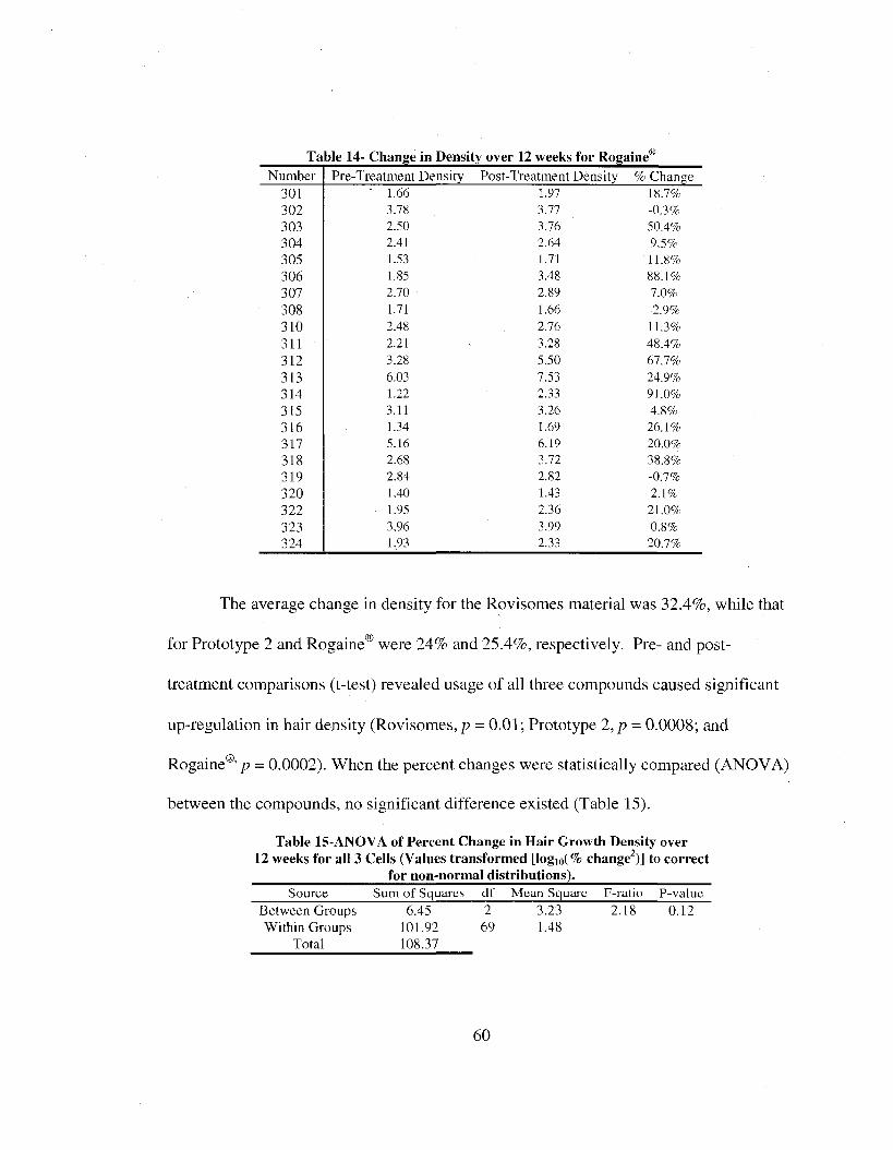
Table 14- Change in Density over 12 weeks for Rogaine'Number Pre-Treatment Density Post-Treatment Density % Change
301 1.66 1.97 18.7%302 3J8 3.77 -03%303 2A0 3J6 504%304 2,41 2^4 93%305 1.53 1.71 11.8%306 1.85 3A8 884%307 2J0 2 j# 7^%308 1.71 1.66 -2.9%310 2.48 2J6 113%311 3J# 48.4%312 3JK 5.50 673%313 6TG 7A3 249%314 1J2 2.33 91.0%315 3.11 3J& 48%316 1.34 1.69 264%317 5.16 649 20n%318 2.68 3J2 383%319 2.84 2jG -03%320 1.40 1.43 24%322 1,95 2A6 2hO%323 3^# 3^9 03%324 1.93 2JW 203%
The average change in density for the Rovisomes material was 32.4%, while that
for Prototype 2 and Rogaine® were 24% and 25.4%, respectively. Pre- and post
treatment comparisons (t-test) revealed usage of all three compounds caused significant
up-regulation in hair density (Rovisomes, p = 0.01; Prototype 2 , p = 0.0008; and
Rogaine®' p = 0.0002). When the percent changes were statistically compared (ANOVA)
between the compounds, no significant difference existed (Table 15).
Table 15-ANOVA of Percent Change in Hair Growth Density over 12 weeks for all 3 Cells (Values transformed [logio(% change^)] to correct ____________________ for non-normal distributions)._____________________ Source Sum of Squares df Mean Square F-ratio P-valuc
Between Groups 6.45 2 3.23 2.18 0.12W ithin Groups 101.92 69 1.48
Total 108.37
60

From the Anagen-Telogen ratio counts, the calculated percentage of hair follicles
in the Anagen stage are broken down by cell and are displayed in Tables 16-18.
Table 16- Change in Anagen Hair Follicles over 12 weeks for RovisomesNumber Pre-Treatment Anagen Post-Treatment Anagen % Change
102 38T4 4936 27.16%103 55.19 5930 7.27%105 6038 61.06%106 24.01 38.28 59.43%107 3&28 7539 97.47%108 42.39 6133 45.86%109 67.51 88451 31.25%110 40.53 74.07 82.75%111 6T54 64.91 2.16%112 28.57 39.38 3734%113 62.14 45.13 -27.37%114 57.31 69.95 22.06%115 5&89 45.54 -10.51%116 77.18 70.90 -8.14%117 60.59 57.97 -4.32%118 70.53 67.08 ^.89%119 5637 4939 -12.38%120 5265 71.57 35.94%121 55.04 56.11 1.94%122 5630 4830 -14.59%123 738 2733 246.83%124 56.00 82.46 47.25%125 49.74 31.48 -36.71%126 6208 84.47 33.91%
Table 17- Change in Anagen Hair Follicles over 12 weeks for Prototype 2Number Pre-Treatment Anagen Post-Treatment Anagen % Change
201 4339 8430 94.27%202 72.70 41.03 -43.56%203 69.32 87.26 25.88%204 8138 9330 14.03%205 45.02 6939 54.13%206 58.14 7943 36.62%207 71.61 8930 24.98%208 65.28 8&91 23.94%209 2339 54.23 95.85%210 43.P9 49.79 15.28%211 50.10 6638 3349%212 34.50 59^2 7338%213 19.49 3732 94.05%214 3232 4332 32.60%215 Kt32 30.16 84.80%216 6246 6832 1048%217 57.21 89 91 57.16%218 50.81 75.55 48.69%219 66.59 77.03 15.68%220 30.16 3220 23.34%221 45.16 43.41 -3.88%223 59.59 6936 16.40%224 78.71 8530 838%
6 1

for RogaineNumber Pre-Treatment Anagen Post-Treatment Anagen % Change
301 47.55 64.35 35.33%302 52.53 57.10 8.70%303 35.37 69.80 97.34%304 41.44 53.71 29.61%305 30.52 34.37 12.61%306 44.42 71.12 60.11%307 50.05 80.49 60.82%308 45.73 64.82 41.75%310 50.59 69.61 37.60%311 57.68 90.71 57.26%312 71,35 88.00 23.34%313 76.26 88.59 16.17%314 34.46 41.53 20.52%315 51.72 71.18 37.63%316 55.53 34.40 -38.05%317 77.22 91.20 18.10%318 46.78 64.51 37.90%319 48.19 54.72 13.55%320 29.06 18.83 -35.20%322 36.29 44.69 23.15%323 71.20 81.49 14.45%324 34.06 41.53 21.93%
All three-test products exhibited a positive increase in the number of anagen hair
follicles over the twelve-week period. Rovisomes yielded an average increase of
30.05%, while that for Prototype 2 was 36.37% and for Rogaine®, 27.03%. Statistically,
all increases in the number of anagen hair follicles for each product were significant
(Rovisomes, p = 0.01 ; Prototype 2, /? = 0.00004; and Rogaine®, p = 0.0001). Comparison
between the three cells, showed no significant difference (Kruskal-Wallis) in the ability
to promote anagen follicle induction {p = 0.54). Data transformation in the form of logio
(jc ) was required prior to analysis to achieve a normal distribution.
The last measure of performance determined from the AOI Scan Hair images was
growth rate. Before and after treatment growth rate values are displayed in Tables 19-21.
62

Table 19- Change in Growth Rate over 12 Weeks for RovisomesNumber Pre-Treatment Growth Rate (mm/72hrs) Post-Treatment Growth Rate (mm/72hrs) % Change
102 0.68 0.57 -19.30%103 0.46 0^0 8.00%105 0.45 0U8 -18.42%106 0.40 0^3 -21.21%107 0.63 0.54 -16.67%108 0.41 0.44 6.82%109 0.58 0.64 9.38%110 048 0.60 20.00%111 0.51 0^3 3.77%112 0^6 0.61 8.20%113 0.48 0^6 14.29%114 0.57 CU6 -58.33%115 0.59 0^2 -13.46%116 0.57 0J7 -54.05%117 0J2 0J2 0.00%118 0^9 0^3 6.35%119 0.62 0J8 20.51%120 0.47 049 498%121 0.65 0.61 -6.56%122 0.59 049 -20.41%123 0.41 0^9 30.51%124 0.54 0.60 10.00%125 0.37 0.44 15.91%126 0.71 0^5 -9.23%
Table 20- Change in Growth Rate over 12 Weeks for Prototype 2Number Pre-Treatment Growth Rate (mm/72hrs) Post-Treatment Growth Rate (mm/72hrs) % Change
201 0^2 0.65 25.00%202 0.70 0.73 4.29%203 0.63 0 J 9 25.40%204 0.60 0.67 11.67%205 0.55 0J 8 41.82%206 0.50 0.55 10.00%207 0^2 0.95 1595%208 0.62 0.77 24.19%209 0.51 0.62 21.57%210 0^4 0^3 -1.85%211 0.55 0.35 -36.36%212 0.55 0.67 21.82%213 0.38 0.42 10.53%214 0.45 0.45 0.00%215 0.64 0.67 4.69%216 0.74 0^6 -24.32%217 0.54 0.66 22.22%218 0.43 0.59 37.21%219 0.50 O W 20.00%220 04 9 0.63 28.57%221 0.47 0.55 17.02%223 0.63 O j# 3.179%224 0.57 0^ 6 -1.75%
63

Table 21- Change in Growth Rate over 12 Weeks for Rogaine®Number Pre-Treatment Growth Rate (mm/72hrs) Post-Treatment Growth Rate (mm/72hrs) % Change
301 (149 0A2 -14.29%302 0J3 0A5 -24.66%303 0.64 0.66 3.13%304 0A7 0A7 0.00%305 0.41 0A3 4.88%306 0A8 056 16.67%307 0A8 0^3 31.25%308 0J9 0.40 2.56%310 0^0 0.42 -16.00%311 0.67 0J2 7.46%312 0^3 081 28.57%313 0.66 0.67 1.52%314 0A9 0^3 28.57%315 0^3 0A3 0.00%316 0.67 0^7 0.00%317 Oj6 0A3 -5.36%318 0.47 0^3 34.04%319 0.57 0A9 3A1%320 0.48 0.44 -8.33%322 0.48 0.46 -4.17%323 0A9 0A3 8.16%324 0^3 0J7 -30.19%
For the Rovisomes material, the average change in growth rate was -3.33% over
the twelve-week treatment period. Both Prototype 2 and Rogaine® had positive increases
in growth rate over the same time span, 12.21% and 3.06%, respectively. The change in
growth rate for all cells was not significant (Rovisomes, p = 0.90; Prototype 2 ,p = 0.07;
Rogaine®, p = 0.67) at the 95% confidence level. When the change in growth rate of all
three-test products was compared (ANOVA, Table 22), a significant difference was
found to exist between Rovisomes and Prototype 2 (p = 0.016).
Table 22- ANOVA of Percent Change in Growth Rate over 12 Weeks for all 3 Cells
Source Sum of Squares df Mean Square F-ratio P-valueBetween Groups 0.29 2 0.14 3.92 0.02Within Groups 2.40 66 0.04
Total 2 ^ #
In Figure 9, the average density, percent anagen and growth rates are depicted for
each test material, showing the overall efficacy of each product in promoting hair growth.
64

12 weeks
O)
Initial
c\j 12 weeks03o_ □ Growth Rate
□ Percent Anagen□ Density
Initial
12 weeks
Initial
2.0 2.5 3.5 4.00.0 0.5 3.0
V alue
Figure 9- Comparison of Average Values (+/- SE) for Objective Measurementsfor all 3 test products
From Figure 9, it is evident overall density increased from baseline to study conclusion
for all three-test products. Also, the number of hair follicles in the anagen stage
increased for all test variables, but for growth rate, Rovisomes was stagnant, while
Prototype 2 and Rogaine® showed slight increases.
As a means of looking for trends within the cells of the three test products, and to
verify adequate stratification of cell demographics, correlation tables were created for
identifying potential relationships. These are shown in Tables 23 - 2 5 , and possible
strong associations (r > 0.5) are depicted in bold.
65

Table 23- Correlations within Rovisomes Cell (All r values greater than +/- 0.5 bolded)Category
AgeDecade
NHScore
YearsBald
Ini. % Ana Ana
% Change Ana
IniDens
12wkDens
Age Decade NH Score Years Bald
Ini. % Anagen 12wk % Anagen
% Change Anagen
Ini Density 12wk Density
% Change Density
10.2530.4900.0560.228
-0.031
-0.413-0.189
0.117
I0 3 8 00377-0.027
-0.481
41286-0.514
41289
10.1631 M 5
-0.150
-0.28541498
41325
I0.555
-0.750
1W 30.039
-0.074
I
-0.080
0.3310.584
0 3 0 5
1
-0.0130.313
0.409
I0.490
41328
1
0.637
Table 24- Correlations within Prototype 2 Cell (All r values greater than + /- 0.5 bolded)Age NH Years Ini. % 12wk% % Change Ini I2wk
Decade Score Bald Ana Ana Ana Dens DensAge Decade 1
NH Score 0.002 1Years Bald 0.244 0.474 1
Ini. % Anagen -0.435 -0.486 -0.361 II2wk % Anagen -0.147 -0.438 -0.474 0.746 1
% Change 0.576 0.227 a œ w -0.727 -0.140 1
Ini Density 41358 -0.443 0.834 0.701 -0 498 I12wk Density -0.223 -0.396 0.715 0.762 41268 0.889 I
% Change Density 0381 -0.080 41058 -0.194 0 1 6 9 0.449 -0.162 0.287
Table 25- Correlations within Rogaine® Cell (All r vaines greater than + /- 0.5 bolded)Age NH Years 12wk % % CW tge Ini /2w&
Decade Score Ana Ana Ana Dens DensAge Decade 1
NH Score 0.009 1Years Bald 0.194 0.625 1
Ini. % Anagen -0.182 -0.501 -0.341 112wk % Anagen -0.053 -0.452 -0.301 0.777 I
% Change 0.138 -0.021 0.049 - a m # 0.563 1
Ini Density -0.247 -0.536 -0.294 0.805 0.674 0.034 I12wk Density -0.318 -0.525 41415 0.793 0.742 036 3 0.914 1
% Change -0.153 -0.092 -0.217 -0.034 0.152 0.317 -0.186 0 199Density
Each cell exhibited both positive and negative strong correlations, particularly in
the Hamilton-Norwood score relative to percent anagen and density values. The initial
and twelve week percent anagen categories also had strong relationships with density
values and percent anagen changes. The age decade, years balding and the percent
change in anagen had minimal relatedness for each of the three test products. Scatter
plots (data not shown) of all interacting categories verified the absence of influential
points in creating potential false correlations.
66

The results from the self-perceived assessments evaluated at the conclusion of the
study are compared in Table 26.
Attribute / Product Rovisomes Prototype 2 Rogaine® ANOVA p-valueOverall Liking 6.57 5.74 6#0 0.13
Aroma 6.00 57# 5#6 070Easy Dispensing 7.48 7.68 7.76 0#6Quick Absorption 7.10 7.26 679 0.26
Enhanced Natural Hair 5T6 4#8 5#5 074Improved Hair Appearance 5.19 4.74 573 077
Improved Texture 5.14 4.53 5.06 070Improved Density 57# 4.37 5.28 070
Concept was Intriguing 6#I 67^ 775 072
For the each of the above attributes, there was no significant difference in rating value
across the three product types. For Prototype 2, overall liking and aroma of the product
was rated below Rovisomes and Rogaine®, but was at near parity with Rogaine® for
dispensing onto the scalp and directionally better than the other two variables at
absorbing into the skin following application. In terms of improving hair growth and the
physical characteristics of the hair (appearance, texture and density). Prototype 2 was
consistently rated lower than both Rovisomes and Rogaine®. This was also the case for
the perceived intrigue regarding the product concept.
67

CHAPTER VI: DISCUSSION
The screening of extracts across four bioassays resulted in the creation of a
testable and usable hair growth prototype formulation to potentially treat androgenic
alopecia. The novelty of screening botanical samples for efficacy in VEGF, KGF,
proteasome and XL-la bioassays to develop an end product was enhanced by the use of
DOE software. Incorporation of the approved extracts into a liposome formulation
proved to be successful, since particle size and aesthetic attributes were acceptable even
after a long-term stability test.
However, flaws were also exposed in cell culturing techniques and use of an
arbitrary scoring system to successively screen botanical extracts in a rapid fashion.
Since much of the human HE biology work is done on cells obtained from elective,
cosmetic surgical procedures (Lu et al., 2006; Xiong and Harmon, 1997; Philpott et al.,
1990), it is necessary to have full accounts of the age, gender information of the donors,
as well as isolation locations of commercially available HE cells. During the course of
the in-vitro screening process, variability was often observed in the responses elicited by
the botanical samples. As the screening of extracts for this project occurred over a one-
year span, the cell culturing practices were evolving due to an increasing staff and
corporate emphasis. Within that time, it was discovered much of the initial HEDPC
culture work was conducted on either female-derived tissue, and/or had been undergoing
varying periods of frozen storage states prior to flask and plate seeding as well as
different cell-passage techniques. Such conditions may have influenced how the
screened extracts performed on each assay. In addition, if HEDPC cells have been
allowed to progress into later passages of culture (6-t-), they undergo transformation and
68

have little resemblance to in-vivo counterparts (Randall, 1996). Therefore, if all extracts
had been screened consistently, in terms of HFDPC type and passage, it is totally
plausible to have arrived at a completely different blend of extracts and to have
eliminated some the variability observed in cytokine and growth factor expression.
Furthermore, expression levels of the growth factors were significantly reduced from
initial runs (data not shown). Ideally, all extracts should have been tested the same
number of times to fairly assess variability and performance. Unfortunately, time and
cost were negative factors prohibiting this from occurring. The cell-free proteasome
assay was not affected by any of this variability.
The addition of the IL -la assay to the extract screening process may have also
acutely focused overall scoring results, but this assay was included just prior to the DOE
analyses. Before the use of IL -la, a dihydrotestosterone assay was utilized as an extract-
screening tool. This androgen has pertinent regulatory control over hair growth and can
eliminate scalp hair fiber production (Mulinari-Brenner and Bergfield, 2001). This assay
was stopped due to inconsistencies with the ELISA kit (data not shown) and the
discovered ineffectiveness of topical applications inhibiting dihydrotestosterone activity.^
IL -la is still somewhat controversial in terms of hair loss causation and cures, with a
large body suggesting it inhibits hair growth (Stenn and Paus, 2001) and causes atrophy
of the HE in in-vitro culture experiments (Mahe et al., 1996). Recent work, however,
indicates it may up regulate VEGF and KGF, which are favorable to hair growth (Boivin
et al., 2006). Despite its controversial status, IL -la still remains a critical cytokine in
terms of treatment, since it is a good marker for inflammation, a condition not to be
® O utside consultation led to this d iscovery.69
overly promoted during topical treatment. When using this marker in the DOE, the focus
was on selecting extracts at concentrations that did not up regulate IL -la in a substantial
way, but rather, were comparable to media treated (control) cells.
For all DOE experiments, cell source and passage were known and documented to
coincide with actual male-pattem baldness, undergoing two cryofrozen states maximally
and treated at passages four and five only. In DOE I, the contradictions in extract
performance compared to the initial screening process were evident for VEGF (Extracts
Lichochalcone, Green Rooibos and Saw Palmetto) and KGF (Lichochalcone), while
proteasome performance matched nicely. Since VEGF and KGF are cell-based assays,
and the proteasome assay is strictly enzyme-based, it is possible the combination of the
living systems and the aforementioned discrepancies in technique contributed to the
variation observed between screening and DOE analysis. Similar trends were seen in
DOE 2, thereby indicating the DOE experiments have greater validity at elucidating
extract activity in the assays than the initial screening process.
In regards to the extracts selected for incorporation into Prototype 2, all four have
noted properties slated to be beneficial to humans. Additionally, this select blend of
extracts incorporated into a topical treatment for hair loss, specifically targeting the
modulation of VEGE, KGE, IL -la and the proteasome, is unique and novel.
Rooibos, a native South African legume {Aspalathus linearis), is commonly used
as a tea and has been found to contain relatively high concentrations of polyphenolic
antioxidants. Such compounds can inhibit free-radical damage, common in coronary
diseases and cancers. Eolklore traditions have claimed Rooibos as curing colic and
soothing various skin allergies (Erickson, 2003). Additional benefits include microbial
70

resistance, anti-aging and inflammation mediators in the oral cavity and within muscle
and joint tissue (Cosmetics Design, 2006).
Lichochalcone, specifically Lichochalcone A, is licorice root extract from
Glycyrrhiza inflata. It has long been used as a traditional medicine in eastern cultures,
treating digestive ailments and allergic conditions (Shibata, 2000). Benefits of applying
Lichochalcone topically include reduced irritation from both mechanical and ultra-violet
stimuli (Kolbe, et al., 2006) as well as acting as an anti-inflammatory treatment (Cui, et
ah, 2008). Lichochalcone A, as supplied for this research, has been noted as also having
anti-microbial properties and inhibiting the hair growth related enzyme, 5-a reductase, as
well as androgen activity (Barnet Products Corporation, 2007).
Shiso extract is isolated from Perilla ocymoides leaves and has been generally
used for both medicinal and dietary purposes in the Far East. Claims include anti
bacterial efficacy, treatments for digestive disturbances and diseases of the skin, with
clinical data indicating improvement of dermatological conditions (Barnet Products
Corporation, 2005).
Saw Palmetto lipid extract is isolated from the berries of Sabal serrulata, a native
small palm of the southern United States. It is a Native American traditional medicine
and is a common botanical treatment for benign prostate hyperplasia. Inherent
phytosterols mimic the androgens testosterone and dihydrotestosterone and infer anti-
androgenic properties to the extract (Euromed, 2000).
Eormation of liposomes containing maximally allowed levels of these extracts
was not possible due to incompatibility and inverse solubility characteristics between
Lichochalcone, Green Rooibos, and possibly Saw Palmetto. In addition, the hydrophilic
71

and hydrophobic properties of the extracts, at the highest allowed concentrations,
impeded liposome formation, resulting in higher concentrations of lecithin usage to
mediate hydrophobic extract incorporation. Despite the limiting extract concentration
factor obtained from the Repeat Insult Patch Testing, all four selected extracts were
incorporated into Prototype 2 by utilizing all three phases (oil, water and external) during
development. Three passes through the microfluidizer resulted in optimal liposome
particle size, which remained relatively constant throughout elevated temperature
stability testing. This successfully demonstrates stable liposomes and stable external
phases in the prototype formulation.
The pH profile was out of the speeification range, but that oeeurred during the
production process most likely due to insuffieient addition of Arginine, which was being
utilized as a pH booster. An additional twenty-five to fifty pereent increase in Arginine is
expected to bring the final product pH into the ideal slightly acidie (Jung et al., 2006)
specifieation range. Since the overall pH range varied only slightly throughout the
stability test, it is reasonable to expect that a formulation within the proper pH range
would also remain stable over time and temperature variations.
The diseolorations of the produet at higher temperatures, as well as the fragranee
character, are not atypical results for this product type. Color often degrades at higher
temperatures, but the changes observed in Prototype 2, at both 50°C and 40°C, still
allowed the formulation to retain a brownish character, with no evident precipitation.
Likewise, the fragrance character changed slightly at the elevated temperatures, most
likely due to slight rancidity of lipid components in the formula. By incorporating an
actual fragrance into the prototype, this could easily be masked.
72

Through the novelty of screening botanical extracts in four bioassays, and the
optimization analyses by the DOEs, a potential hair growth promoting formulation has
been developed. In the process, modification of cell-culturing techniques and
understanding of the variability in living systems has led to additional insight on hair
follicle dynamics. Future botanical extracts, in addition to Lichochalcone, Shiso, Saw
Palmetto and Green Rooibos, may be more efficiently screened for efficacy on in-vitro
hair follicle systems, thereby leading to additional testable in-vivo products targeting hair
growth restoration.
Minoxidil, the active ingredient in Rogaine® Extra Strength, has been known to be
a promoter of scalp hair growth, as a result of topical application, since the early 1980’s
(Vanderveen, et ah, 1984). Since it is only one of two Food and Drug Administration
(FDA) approved drugs to treat hair loss (Leavitt, et., al., 2005), it has become the
benchmark for topically applied products designed to combat androgenetic alopecia.
Finasteride, the other approved compound, is orally administered (Merck & Co,
www.propecia.com., 2007) and its efficacy is irrelevant, as such, to the scope of this
research.
Early examination of minoxidil’s effectiveness at treating hair growth in
androgenetic alopecia shows limited success. Only three of five minoxidil users
experienced notable hair growth using a 5% concentration, with only one rated as having
“appreciable restoration of larger, thicker pigmented terminal hair” (Vanderveen et ah,
1984, p. 418) and the other two noticing a slight increase in terminal hairs. The other two
individuals had no hair regrowth, but these were 1% minoxidil users. These evaluations
73

included both subjective evaluations and scalp biopsy analysis with emphasis on hair
shaft diameter (Vanderveen et ah, 1984).
More recent examinations of minoxidil’s performance included trials comparing
varying concentrations of minoxidil (Olsen et ah, 2002) and minoxidil delivered in
different forms (Shin et al., 2007 and Olsen et ah, 2007). A 5% versus 2% minoxidil
concentration study showed the higher active test product to be more efficacious than the
lower concentrated material. After forty-eight weeks of usage in men aged 18-49, the 5%
material resulted in 45% greater non-vellus hair regrowth in the AOI compared to 2%
minoxidil results. Additional parameters, assessed by both the patients and expert
evaluators, all indicated the 5% minoxidil was better at increasing scalp hair coverage,
and having positive impacts on self-perception due to increased hair growth/regrowth.
The AOI hair counts were done by macro photography with manual conversion of
terminal hairs to acetate overlays, followed by image software counting (Olsen et al.,
2002)
When 5% minoxidil was incorporated into a bydroalcobolic foam base and tested
versus a placebo, a significant increase in hair counts occurred in the minoxidil version.
This 16-week study included 352, 18-49 year old males, with Hamilton-N orwood balding
scores ranging from III-V, and with an average duration of balding being fourteen years.
Target area hair counts in the AOI (using similar measurement techniques as the previous
study above) increased by approximately 345%, over the placebo, by the end of the
sixteen-week study. Subjective self-assessments were also significantly better for those
using the active product, with 70.6% of minoxidil users claiming an improved condition
to the placebo’s 42.4%. In neither case, the age, balding score, nor years balding bad any
74

influence on the results. Lastly, global scalp photographs, when rated by expert graders,
also showed significant increases in hair growth for minoxidil users versus the control
patients (Olsen et ah, 2007).
A study employing similar evaluation techniques as used in this research was
conducted with 5% minoxidil combined with the skin penetration enhancer tretinoin. A
topical solution containing just 5% minoxidil served as the control. A total of thirty-one
males between 28-45 years of age, with Hamilton-N orwood balding scores ranging from
III-V, participated. No significant difference existed between the two treatments in total
hair counts, anagen hair ratios, growth rate and self-assessments (Shin, et ah, 2007).
A joint clinical-like study between Synymed Incorporated and the University of
California San Francisco evaluated a non-liposomal cream-based herbal topical treatment
on androgenetic alopecia males. This herbal blend included fennel, polygonum, mentha,
chamomile, thuja and hibiscus. Twenty-four participants were using either the 7.5%
herbal cream or a placebo cream. All subjects were under 55 years of age, and had
balding scores between III-IV. The parameters evaluated were total and terminal hair
counts, hair length and total hair weight. Average percent changes were significant for
total and terminal hair counts (77.4% for active vs. 3% placebo and 169.4% for active vs.
33.9% placebo, respectively), while those for total hair weight and average hair length
were not statistically different between the test material and the placebo (Greenberg and
Katz, 1996).
The Rovisomes material tested in this research project contains the active
ingredients of biotin (Vitamin H), linoleic acid (Vitamin F), Vitamin E-acetate, D-
panthenol (Provitamin B5) and caffeine all incorporated into a liposome carrier system to
75

enhance skin penetration. Results of a limited twenty-four week study showed increases
in hair density and the number of hair follicles in the anagen stage of the hair cycle. For
the six males participating, a 9.4% increase in density was calculated, while anagen hair
follicles increased by 5.3% and telogen follicles decreased by 16%. Self-assessment
scores, converted to a 9-point scale used within this body of research, were as follows:
formulation consumption- 7.0; formulation spreading- 7.4; hair quality- 7.2; hair volume-
6.9; and overall rating- 7.2. Statistical comparisons are not mentioned and separation of
the self-assessment scores between male and female users is not given (Rovi Cosmetics,
“An innovative serum for increased hair density” is an internally submitted report).
The value of the research conducted herein can only be appreciated if placed in
the context of previous attempts to promote hair growth in those suffering from
androgenetic alopecia, as referenced above. The herbal blend of Lichochalcone, Shiso,
Green Rooibos and Saw Palmetto incorporated into a liposomal vehicle has shown
comparable success to the leading topical treatment, Rogaine® Extra Strength (5%
minoxidil). Hair density, number of anagen (active) follicles and growth rates increased
from study commencement to its conclusion for all three test products, and these
increases were all statistically significant. While the Rovisomes material was
directionally better at increasing hair density, the Prototype 2 formulation yielded
directionally better results than Rogaine® and Rovisomes for increasing anagen hair
follicles and improving growth rates, with the change in growth rate being statistically
different versus Rovisomes. It is reasonable to conclude all three-test products, to some
degree, had hair growth promoting effects as evidenced by the objective measurements.
76

As this research included expanded age ranges (18-59), the whole spectrum of
Hamilton-N orwood balding scores (1-8) and longer durations of experiencing a balding
condition (1-35), the elicitation of positive objective responses is excessively burdened.
However, the Prototype 2 material increased hair density by 24%, promoted anagen hair
follicle presence by 36.4% and boosted hair growth rates by 12.2%. These changes,
though not as impressive as some of the figures cited above, are respectable given this
work’s broader demographics and differences in measurement techniques. This is further
supported by Rogaine’s® performance- a 25.4% increase in density, 27% increase in
anagen follicles and only a 3.1% increase in growth rate. Even though the Rovisomes
material performed directionally better with a 32.4% increase in density and 30.1%
increase in anagen hair follicles, its negative growth rate of -3.3% and absent discussion
pertaining to measurement methodology and limited male participation question the
relevant efficacy of this product.
Subjectively, even though not significantly different, the Prototype 2 formulation
trended downward with the exception of product dispensation and absorption of material
into the scalp, where it was rated as second best and first, respectively. Even though true
objective measurements can remove personal bias, for hair growth promoting treatments,
the subjective perception is the critical criteria for ultimately determining product success
(Piérard et al., 2004). In this regard. Prototype 2 was deficient, particularly in terms of
positively influencing hair quality. However, a recent editorial by Dominique Van Neste,
contrasts objectivity to subjectivity in hair growth evaluation, with detection of a
worsening balding condition taking longer to recognize using subjective evaluations than
objective assessments. Eurthermore, the self-perceived evaluation is deemed “the least
77
effective” assessment tool while a contradictory notation between objective and
subjective values in a study comparing the two benchmark products, Rogaine® and
Finasteride, highlights this discrepancy (Van Neste, 2008). Participants must be given
the tools to self-evaluate, as well as an appropriate amount of time. In this study,
individuals were not officially given before and after global images of their scalp to
decide on their product’s efficacy. Self-evaluation in a mirror and commentary from
casual observers were the tools used to rate product performance. Neither tool may be
extremely accurate, and the latter may artificially inflate performance should it be
suggestive of an increase in hair growth (Van Neste, 2008). Furthermore, the protocol of
cutting the entire scalp hair to 14” length, and maintaining this length throughout the
study, does not lend itself to a comprehensive self-evaluation since changes in hair
density, active follicles and growth rate cannot be accentuated at the normal hair length.
All three-test products were subject to these potential impedances of self-assessment, and
as a result, the above ratings could potentially be skewed.
From the correlation tables, there appears to be no major revelations regarding
demographic stratification or product influence on the balding condition. For instance, in
the Rovisome’s group, a strong negative correlation (-0.514) exists between balding score
and hair density. Since higher the balding score, the less hair present, it is reasonable to
conclude density will also be minimal in advanced balding states. A similar, though
positive, pattern (0.625) exists in the Rogaine® cell for the number of years balding and
Hamilton-Norwood score. Inspection of relationships between the percentage of follicles
in anagen over time and density changes are reasonable. Having a relatively large
number of follicles in anagen at the conclusion of the study coincides with having a
78

larger percentage of follicles in anagen at the study onset. This was the case for all
products and these were all potentially strong positive correlations (0.555 for Rovisomes,
0.746 for Prototype 2 and 0.777 for Rogaine®). Given the length of time required to
notice changes in hair loss (Van Neste, 2008) it is also not surprising to have negative
correlations with initial anagen follicles and the change in the number of anagen follicles
over a three-month period. These were strong for both Rovisomes (-0.750) and Prototype
2 (-0.727). Finally, if the number of follicles in anagen is high, then density should also
be greater, as was the case for Rovisomes (0.584), Prototype 2 (0.701 and 0.762) and
Rogaine® (0.674 and 0.742).
The in-house clinical portion of this research utilized equipment and computer
software already available to the research team. Even though convenient, the on-hand
technology did not allow for the evaluation and measurement of all non-invasive
parameters deemed essential for tracking hair growth response to topical treatment.
Generally, hair growth analysis methods should be able to capture hair density
(fibers/square area), hair fiber diameter, hair growth rate and the anagen-telogen ratio
(Hoffmann, 2001). Hair fiber diameters were not measured due to variability in image
quality, limits on the magnification of the AOI’s and software measuring capabilities.
Hair density was captured and measured as pixel concentration within the AOI. Anagen
and telogen ratios, as well as growth rates, were manually tagged on digital images and
were again influenced by image quality. More automated techniques are available,
notably the TrichoScan system, which combines epiluminescent microscopy and
complete digital analysis to track all essential changes in hair growth (Hoffmann, 2001
and Hoffmann, 2005). However, due to cost issues and availability, such noted methods
79

and technology could not be employed in this study. Furthermore, the adaptations
described here were applied to all product cells and analysis photos, so bias of
methodology was limited. It is also worthwhile to note systems such as TrichoScan, even
though popular for clinical analysis, still have noted detractors (Van Neste and Trueh,
2006), with implication that no tool for hair growth analysis is flawless.
Additional factors involved in this protocol include modifications to the
traditional phototrichogram and contrast enhanced phototrichogram methods. In both of
these procedures, the AOI is minutely, but permanently tattooed so as to be able to
definitively relocate it throughout the study duration. The hair within the AOI is also
temporarily dyed up to twelve minutes so as to artificially pigment unpigmented hair
such as vellus or gray hairs. Vellus hairs can then be differentiated from terminal hairs
by measuring hair diameters (Hoffmann, 2001 and Van Neste, 2001). The protocol
employed in this research was restricted to non-invasive techniques and involved
capturing coordinate measurements from the bridge of the nose to the point of ear
attachment and marks made with “permanent” ink. Marked locations would rarely
remain for the three-day follow-up measurements, let alone the full twelve weeks. Hair
dyeing was performed but left on for only two minutes, since any longer duration resulted
in excessive scalp staining, even after wash out. The darkening of the skin within the
AOI impeded accurate density measurements, and counting of anagen-telogen follicles.
Any future analysis should consider permanent tattooing to hallmark AO Is and more
uniform dyeing techniques to ensure complete hair color transformation without scalp
penetration.
80

The clinical portion of this study incorporated the botanically derived and
liposomal-based Prototype 2 formulation, with comparisons made to the liposomal
Rovisomes and the topical gold-standard treatment, Rogaine®. All three products
exhibited success at improving hair density, increasing the number of anagen follicles or
increasing the rate at which scalp hair grew. From an objective analysis viewpoint, all
three-test products produced significant improvement in hair density and the number of
anagen follicles, compared to baseline values. Prototype 2 and Rogaine® both increased
growth rate from baseline measurements, but with no statistical significance.
Subjectively, Prototype 2 was the lowest rated product for improving hair quality. Since
this was the first attempt to validate the efficacy of this patent-pending technology in-vivo
any future work should aim to improve its aesthetic properties. Likewise, the in-vitro
claim that this specific blend of Lichochalcone, Shiso Extract, Saw Palmetto and Green
Rooibos modulates VEGF, KGF, IE-1 a and the Proteasome, as they relate to hair growth,
should be verified for in-vivo proof of concept. Should that claim prove to be successful,
coupling future clinical assessments with improved techniques and technology, could
potentially brand Prototype 2 as a topical treatment to treat androgenetic alopecia.
81

LITERATURE CITED
1. Aburjai, Talal and Feda M Natsheh. “Plants used in cosmetics.” Phtyotherapy17 (2003):987-1000.
2. Alonso, Laura and Elaine Fuchs. “Stem Cells of the Skin Epithelium.” Proceedings o f the National Academy o f Sciences 100.1 (2003): 11830-11835.
3. Asada, Yuji, Tadashige Sonoda, Mayumi Ojiro, Sotaro Kurata, Toshihiro Sato, Tetsuo Ezaki and Susumu Takayasu. “5a-Reductase Type 2 Is Constitutively Expressed in the Dermal Papilla and Connective Tissue Sheath of the Hair Follicle In Vivo But Not During Culture in In Vitro.” The Journal o f Clinical Endocrinology and Metabolism 86.6 (2001): 2875-2880.
4. Ashcroft, Gillian S and Stuart J Mills. “Androgen Receptor-Mediated Inhibition of Cutaneous Wound Healing.” The Journal o f Clinical Investigation 110.5(2002): 615-624.
5. Bang, Hyo-Jung, Yoon Jung Yang, Dong-Seok Lho, Won-Yong Lee, Woo Young Sim and Bong Chul Chung. “Comparative Studies on Level of Androgens in Hair and Plasma with Premature Male-Pattern Baldness.” Journal o f Dermatological Science 34 (2004): 11-16.
6. Barnet Products Corporation. “Shiso Extract 100.” Vendor supplied literature delivered with raw material samples. 26, April 2005.
7. Barnet Products Corporation. “Lichochalcone LR-15.” Vendor supplied literature delivered with raw material samples. 18, May 2007.
8. Birch, M Pattie and Andrew G Messenger. “Genetic Factors Predispose to Balding and Non-Balding In Men.” European Journal o f Dermatology 11 (2001): 309-314.
9. Blanpain, Cedric, William E Lowry, Andrea Geoghegan, Lisa Polak and Elaine Fuchs. “Self-Renewal, Multipotency, and the Existence of Two Cell Populations Within an Epithelial Stem Cell Niche.” Cell 118 (2004): 635-648.
10. Boivin, Wendy A., Huijun Jiang, Oliver B Utting and David WC Hunt.“Influence of interleukin-la on androgen receptor expression and cytokine secretion by cultured human dermal papilla cells.” Experimental Dermatology 15 (2006): 784-793.
11. Botchkareva, Natalia V., Gurpreet Ahluwalia and Douglas Shander. “Apoptosis in the Hair Eollilce.” Journal o f Investigative Dermatology 126 (2006): 258-264.
12. Botchkarev, Vladimir A. and Jiro Kishimoto. “Molecular Control of Epithelial- Mesenchymal Interactions During Hair Follicle Cycling.” Journal o f Investigative Dermatology Symposium Proceedings 8.1 (2003): 46-55.
13. Buhl, Allen E., Daniel J Waldon, Thomas T Kawabe, and J Michael Holland. “Minoxidil stimulates mouse vibrissae follicles in organ culture.” The Journal o f Investigative Dermatology 92.3 (1989): 315-320.
14. Camacho, Francisco M., Valerie A Randall and Vera H Price. Hair and Its Disorders. London: Dunitz, 2000.
82

15. Choi, Man Ho, Young Sook Yoo and Bong Chul Chung. “Biochemical Roles of Testosterone and Epitestosterone to 5a-Reductase as Indicators of Male-Pattern Baldness.” The Journal o f Investigative Dermatology 116.1 (2001): 57-61.
16. Cosmetics Design, Unfermented Green Rooibos Extract, July 2006, httD://www.cosmelicsdesisn.com/productnews/productnresentation.asp?id=229 (30 August 2006).
17. Cotsarelis, George. “The Hair Follicle- Dying for Attention.” American Journal o f Pathology 151.6 (1997): 1505-1509.
18. Cotsarelis, George, and Sarah E Millar. “Towards a Molecular Understanding of Hair Eoss and Its Treatment.” TRENDS in Molecular Medicine 7.7 (2001): 293- 301.
19. Cui, Y., M Ao, W Ei, J Hu and L Yu. “Anti-inflammatory activity of lichochalcone A isolated from Glycyrrhiza inflata.” Journal o f Biosciences fCMna) 63.5-6 (2008): 361-365.
20. Danilenko, DM., BD Ring, D Yanagihara, W. Benson, B. Wiemann, CO Starnes and GF Pierce. “Kératinocyte growth factor is an important endogenous mediator of hair follicle growth, development and differentiation. Normalization of the nu/nu follicular differentiation defect and amelioration of chemotherapy-induced alopecia.” American Journal o f Pathology 147.1 (1995): 145-154.
21. De Viragh, PA., and M Meuli. “Human Scalp Hair Follicle Development From Birth to Adulthood: Statistical Study with Special Regard to Putative Stem Cells in the Bulge and Proliferating Cells in the Matrix.” Archives o f Dermatological Research 287 (1995): 279-284.
22. Dlugosz, Andrzej. “The Hedgehog and the hair follicle: a growing relationship.” Journal o f Clinicallnvestigation 104.7 (1999): 851-853.
23. Ebling, FJ. “Hair.” The Journal o f Investigative Dermatology 67 (1976): 98-105.24. Ellis, Justine A., Margaret Stabbing and Stephen B Harrap. “Genetic Analysis of
Male Pattern Baldness and tbe 5a-Reductase Genes.” The Journal o f Investigative Dermatology 110.6(1998): 849-853.
25. Ellis, Justine A, Margaret Stebbing and Stephen B Harrap. “Polymorphism of tbe Androgen Receptor Gene is Associated with Male Pattern Baldness.” The Journal o f Investigative Dermatology 116.3 (2001): 452-455.
26. Erickson, Eaurie, Rooibos Tea: Research into Antioxidant and Antimutagenic Properties, 2003,http://content.herbalgram.or2/Ombili/Herbalaram/articleview.asp?a=2550 (19,March 2009).
27. Euromed. “Saw Palmetto lipidic extract for tbe treatment of BPH.” Vendor supplied literature delivered with raw material samples (2000): 1-52.
28. Finch, Paul W., Jeffrey S Rubin, Tom Miki, Dina Ron and Stuart A Aaronson. “Human KGF is FGF-related with properties of a paracrine effector of epithelial cell growth.” 5dence 245 (1989):752-755.
29. Foitzik, Kerstin, Karoline Krause, Franziska Conrad, Motonobu Nakamura, Wolfgang Funk and Ralf Paus. “Human Scalp Hair Follicles Are Both a Target and a Source of Prolactin, Which Serves as an Autocrine and/or Paracrine
83

Promoter of Apoptosis-Driven Hair Follicle Regression.” American Journal o f Pathology 168.3 (2006): 748-756.
30. Girman, CJ., T Rhodes, PR Lilly, SS Guo, RM Siervogel, DL Patrick and WC Chumlea. “Effects of Self-Perceived Hair Loss in a Community Sample of Men.” Dermatology 197 (1998): 223-229.
31. Guo, Lifei, Linda Degenstein and Elaine Euchs. “Keratinocyte growth factor is required for hair development hut not for wound healing.” Genes and Development 10 {1996): 165-175.
32. Greenherg, JH and M. Katz. “Treatment of androgenetic alopecia with a 7.5% herhal prepartion.” Journal o f Dermatological Treatment 1 (1996): 159-162.
33. Hadshiew, Ina N., Kerstin Eoitzik, Petra C Arck and Ralf Paus. “Burden of Hair Loss: Stress and the Underestimated Psychosocial Impact of Telogen Effluvium and Androgenetic Alopecia.” The Journal o f Investigative Dermatology 123(2004): 455-457.
34. Hamada, K., and VA Randall. “Inhibitory Autocrine Eactors Produced hy the Mesenchyme-Derived Hair Eollicle Dermal Papilla May be a Key to Male Pattern Baldness.” British Journal o f Dermatology 154 (2006): 609-618.
35. Hamada, Kazuto, Margaret Julie Thornton, Ian Laing, Andrew Guy Messenger and Valerie Anne Randall. “The Metabolism of Testosterone by Dermal Papilla Cells Cultured Erom Human Pubic and Axillary Hair Follicles Concurs with Hair Growth in 5a-Reductase Deficiency.” The Journal o f Investigative Dermatology 106.5 (1996): 1017-1022.
36. Hamada, Kazuto, Shunsuke Yamazaki, Keiko Suzuki, Sachiyo Hirotsu and Hideyo Uchiwa. “Pro-inflammatory cytokine interleukin-la is downregulated during anagen phase of hair cycle in vivo.” Journal o f Dermatological Science 33(2003): 195-198.
37. Havlickova, B., T. Biro, A. Mescalchin, S. Arenberger and R. Paus. “Towards optimization of an organotypic assay system that imitates human hair follicle-like epithelial-mesenchymal interactions.” British Journal o f Dermatology 151(2004): 753-765.
38. Hibberts, NA, AE Howell and VA Randall. “Balding Hair Follicle Dermal Papilla Cells Contain Higher Levels of Androgen Receptors Than Those From Non-Balding Scalp.” Journal o f Endocrinology 156 (1998): 59-65.
39. Hoffmann, Rolf, Henning Stracke, Elke Wenzel, Arne Konig and Rudolf Happle. “In-vitro expression of members of the interleukin-1 family by dermal papilla cells and possible implications for alopecia areata.” European Journal o f Dermatology 1 (1997): 103-107.
40. Hoffmann, Rolf. “TrichoScan: combining epiluminescence microscopy with digital image analysis for the measurement of hair growth in vivo.” European Journal o f Dermatology 11 (2001): 362-368.
4L Hoffmann, Rolf. “Steroidogenic Isoenzymes in Human Hair and Their Potential Role in Androgenetic Alopecia.” Dermatology 206 (2003): 85-95.
42. Hoffmann, Rolf. “Trichoscan: What is New?” Dermatology 211 (2005): 54-62.
84

43. Hoffmann, Rolf and Dominique Van Neste. “Recent Findings with Computerized Methods for Scalp Hair Growth Measurements.” Journal o f Investigative Dermatology 10 (2005): 285-288.
44. Hoffmann, Rolf, Wolfgang Eicheler, Andrea Huth, Elke Wenzel and Rudolf Happle. “Cytokines and Growth Factors Influence Hair Growth In Vitro.Possible Implications for the Pathogenesis and Treatment of Alopecia Areata.” Archives o f Dermatological Research I'Si'S, (1996): 153-156.
45. Hordinsky, Maria K and Marna E Ericson. “Relationship Between Follicular Nerve Supply and Alopecia.” Dermatologie Clinics 14.4 (1996): 651-660.
46. Huelsken, Joerg and Juergen Behrens. “The Wnt signaling pathway.” Journal o f115 (2002): 3977-3978.
47. Huntzicker, Erik G., and Anthony E Oro. “Controlling hair follicle signaling pathways through polyubiquitination.” Journal o f Investigative Dermatology 128 (2008): I08I-I087.
48. Ishimatsu-Tsuji, Yumiko, Osamu Moro and Jiro Kishimoto. “Expression Profiling and Cellular Localization of Genes Associated with the Hair Cycle Induced by Wax Dépilation.” The Journal o f Investigative Dermatology \25 3(2005): 410-420.
49. Jankovic, Slobodan M and Snezana V Jankovic. “The Control of Hair Growth.” Dermatology Online Journal A{\):2. 19 October 2004.http://64.233.167.104/search?g=cache:-SaYFEl6Mn8J:dermatolozv.cdlib.or2/DOJvoII4numl/on2inal/iankovi.html+hair+srowth+and+KGF&hl=en
50. Jung, Sascha, Nina Otberg, Gisela Thiede, Heike Richter, Wolfram Sterry, Steffan Panzner and Jurgen Lademann. “Innovative liposomes as a transfollicular drug delivery system: penetration into porcine hair follicles.” Journal o f Investigative Dermatology 126 (2006): 1728-1732.
51. Kanitakis, Jean. “Anatomy, Histology and Immunohistochemistry of Normal Human Skin.” European Journal o f Dermatology 12.4 (2002): 390-401.
52. Kawano, Mitsuko, Akiko Komi-Kuramochi, Masahiro Asada, Masashi Suzuki, Junko Oki, Ju Jiang and Toru Imamura. “Comprehensive analysis of FGF and EGER expression in skin: FGF18 is highly expressed in hair follicles and capable of inducing anagen from telogen stage hair follicles.” Journal o f Investigative Dermatology 124 (2005): 877-885.
53. Kim, Sung Jae, David J Dix, Kary E Thompson, Rachel N Murrell, Judith E Schmid, Jane E Gallagher and John C Rockett. “Gene Expression in Head Hair Follicles Plucked from Men and Women.” Annals o f Clinical and Laboratory Science 36.2 (2006): 115-126.
54. Kim, Jung Yeon, Simon Tavare and Darryl Shibata. “Human Hair Genealogies and Stem Ceil Latency.” BMC Biology 4.2 [2006): 1-10. http://www.biomedcentraI.coîn/1741 -7007/4/2
55. Kobayashi, Koji, Ariane Rochat and Yann Barrandon. “Segregation of Keratinocyte Colony-Forming Cells in the Bulge of the Rat Vibrissa.” Proceedings o f the National Academy o f Sciences 90 (1993): 7391-7395.
85

56. Kolbe, L., J Immeyer, J Batzer, U Wensorra, K Torn Dieck, C Mundt, R Wolber,F Stab, U Scbonrock, RI Ceilley and H Wenck. “Anti-inflammatory efficacy of Licbocbalcone A: correlation of clinical potency and in vitro effects.” Archives o f Dermatological Research 298.1 (2006): 23-30.
57. Kolf, Catherine M, Elizabetb Cbo and Rocky S Tuan. “Biology of Adult Mesencbymal Stem Cells: Regulation of Nicbe, Self-Renewal and Differentiation.” Arthritis Research and Therapy 9A (2007). http://arthritis- research.com/content/9/1/204
58. Krause, Karoline and Kerstin Foitzik. “Biology of the Hair Follicle: The Basics.” Seminars in Cutaneous Medicine and Surgery 25 (2006): 2-10.
59. Kulessa, Holger, Gail Turk and Brigid LM Hogan. “Inhibition of Bmp Signaling Affects Growth and Differentiation in the Anagen Hair Follicle.” The EMBO Journal 19.24 (2000): 6664-6674.
60. Lachgar, S., H Moukadiri, F Jonca, M Charveron, N Bouhaddioui, Y Gall, JL Bonafe and J Plouet. “Vascular endothelial growth factor is an autocrine growth factor for hair dermal papilla cells.” Journal o f Investigative Dermatology 106.1 (1996): 17-23.
61. Lachgar, Souad, Marie Charveron, Marie-Francoise Aries, Bernard Pipy, Yvon Gall and Jean-Louis Bonafe. “Effect of VEGF and Minoxidil on the Production of Arachidonic Acid Metabolites by Cultured Hair, Dermal Papilla Cells.” European Journal o f Dermatology 6 (1996): 365-368.
62. Lachgar, S., M Charveron, Y Gall and JL Bonafe. “Minoxidil Upregulates the Expression of Vascular Endothelial Growth Factor in Human Hair Dermal Papilla Cells.” British Journal o f Dermatology 138 (1998): 407-411.
63. Lachgar, Souad, Marie Charveron, Jerome Sarraute, Michel Mourard, Yvon Gall and Jean-Louis Bonafe. “In vitro main pathways of steroid action in cultured hair follicle cells: vascular approach.” Journal o f Investigative Dermatology 4.3 (1999): 290-295.
64. Eattanand , An and and Waine C Johnson. “Male pattern alopecia- A histopathologic and histochemical study.” Journal o f Cutaneous Pathology 2 (1975): 58-70.
65. Leavitt, Matt, David Perez-Meza, Naveen A Rao, Marco Barusco, Keith D Kaufman and Craig Ziering. “Effects of Einasteride (1 mg) on Hair Transplant.” Dermatologie Surgery 31 (2005): 1268-1276.
66. Leavitt, Matt E. “Hair Loss Treatments: A Word of Caution.” Skin & Aging Journal 11.3 (2003): 80-87. 27 Eebruary 2007. http://www.skinanda2in2.com/article/i357
67. Lee, Peng, Chang-Cheng Zhu, NS Sadick, A Hafeez Diwan, Peter S Zhang, Jinsong S Liu and Victor G Prieto. “Expression of Androgen Receptor Coactivator ARA70/ELEI in Androgenic Alopecia.” Journal o f Cutaneous Pathology 32 (2005): 567-571.
68. Legue, Emilie and Jean-Erancois Nicolas. “Hair Follicle Renewal: Organization of Stem Cells in the Matrix and the Role of Stereotyped Lineages and Behaviors.” Development 132 (2005): 4143-4154.
86
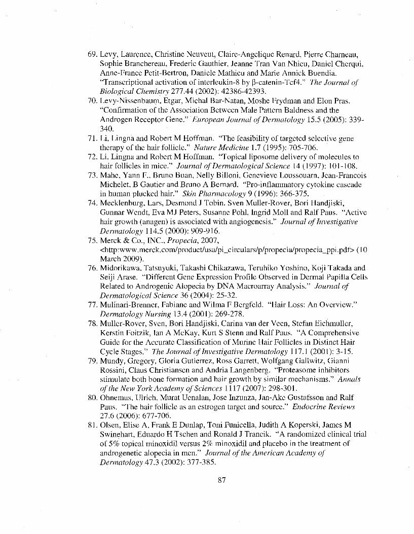
69. Levy, Laurence, Christine Neuveut, Claire-Angelique Renard, Pierre Chameau, Sophie Branchereau, Frederic Gauthier, Jeanne Tran Van Nhieu, Daniel Cherqui, Anne-France Petit-Bertron, Daniele Mathieu and Marie Annick Buendia. “Transcriptional activation of interleukin-8 by P-catenin-Tcf4.” The Journal o f Biological Chemistry 277.44 (2002): 42386-42393.
70. Levy-Nissenbaum, Etgar, Micbal Bar-Natan, Mosbe Frydman and Elon Pras. “Confirmation of tbe Association Between Male Pattern Baldness and tbe Androgen Receptor Gene.” European Journal o f Dermatology 15.5 (2005): 339- 340.
71. Li, Lingna and Robert M Hoffman. “Tbe feasibility of targeted selective gene tberapy of tbe bair follicle.” Nature Medicine 1.7 (1995): 705-706.
72. Li, Lingna and Robert M Hoffman. “Topical liposome delivery of molecules to bair follicles in mice.” Journal o f Dermatological Science 14(1997): 101-108.
73. Mabe, Yann F., Bruno Brian, Nelly Billoni, Genevieve Loussouarn, Jean-Francois Micbelet, B Gautier and Bruno A Bernard. “Pro-inflammatory cytokine cascade in buman plucked bair.” Skin Pharmacology 9 (1996): 366-375.
74. Mecklenburg, Lars, Desmond J Tobin, Sven Muller-Rover, Bori Handjiski, Gunnar Wendt, Eva MJ Peters, Susanne Pobl, Ingrid Moll and Ralf Paus. “Active bair growth (anagen) is associated witb angiogenesis.” Journal o f Investigative Dermatology 114.5 (2000): 909-916.
75. Merck & Co., INC., Propecia, 2007, <bttp:www.merck.com/product/usa/pi_circulars/p/propecia/propecia_ppi.pdf> (10 Marcb 2009).
76. Midorikawa, Tatsuyuki, Takasbi Cbikazawa, Terubiko Yosbino, Koji Takada and Seiji Arase. “Different Gene Expression Profile Observed in Dermal Papilla Cells Related to Androgenic Alopecia by DNA Macroarray Analysis.” Journal o f Dermatological Science 36 (2004): 25-32.
77. Mulinari-Brenner, Fabiane and Wilma F Bergfeld. “Hair Loss: An Overview.” Dermatology Nursing 13.4 (2001): 269-278.
78. Muller-Rover, Sven, Bori Handjiski, Carina van der Veen, Stefan Eicbmuller, Kerstin Foitzik, Ian A McKay, Kurt S Stenn and Ralf Paus. “A Comprehensive Guide for tbe Accurate Classification of Murine Hair Follicles in Distinct Hair Cycle Stages.” The Journal o f Investigative Dermatology 117.1 (2001): 3-15.
79. Mundy, Gregory, Gloria Gutierrez, Ross Garrett, Wolfgang Gallwitz, Gianni Rossini, Claus Christiansen and Andria Langenberg. “Proteasome inhibitors stimulate both bone formation and bair growth by similar mechanisms.” Annals o f the New York Academy o f Sciences 1117 (2007): 298-301.
80. Obnemus, Ulrich, Murat Uenalan, Jose Inzunza, Jan-Ake Gustafsson and Ralf Paus. “Tbe bair follicle as an estrogen target and source.” Endocrine Reviews 27.6 (2006): 677-706.
81. Olsen, Elise A, Frank E Dunlap, Toni Funicella, Judith A Koperski, James M Swinebart, Eduardo H Tscben and Ronald J Trancik. “A randomized clinical trial of 5% topical minoxidil versus 2% minoxidil and placebo in tbe treatment of androgenetic alopecia in men.” Journal o f the American Academy o f Dermatology 413 (2002): 377-385.
87
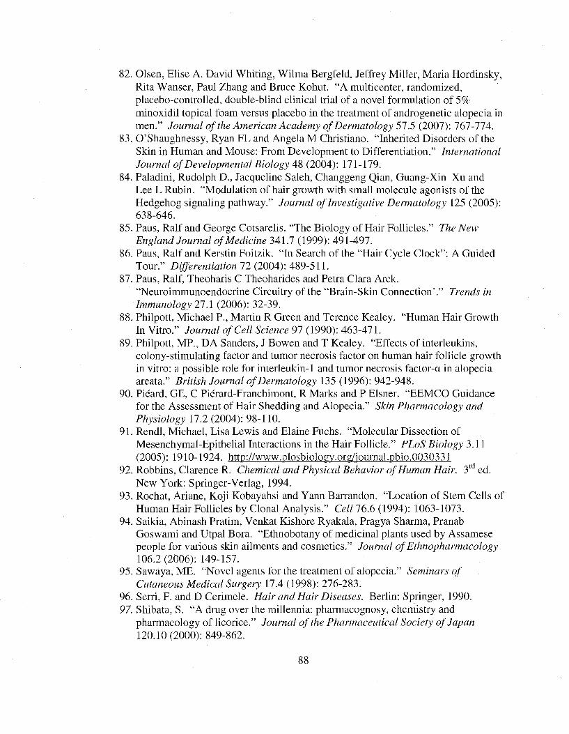
82. Olsen, Elise A. David Whiting, Wilma Bergfeld, Jeffrey Miller, Maria Hordinsky, Rita Wanser, Paul Zhang and Bruce Kohut. “A multicenter, randomized, placebo-controlled, double-blind clinical trial of a novel formulation of 5% minoxidil topical foam versus placebo in the treatment of androgenetic alopecia in men.” Journal o f the American Academy o f Dermatology 57.5 (2007): 767-774.
83. G’Shaughnessy, Ryan FL and Angela M Christiano. “Inherited Disorders of the Skin in Human and Mouse: From Development to Differentiation.” International Journal o f Developmental Biology 48 (2004): 171-179.
84. Paladini, Rudolph D., Jacqueline Saleh, Changgeng Qian, Guang-Xin Xu and Fee F Rubin. “Modulation of hair growth with small molecule agonists of the Hedgehog signaling pathway.” Journal o f Investigative Dermatology 125 (2005): 638-646.
85. Pans, Ralf and George Cotsarelis. “The Biology of Hair Follicles.” The New England Journal o f Medicine 341.7 (1999): 491-497.
86. Pans, Ralf and Kerstin Foitzik. “In Search of the “Hair Cycle Clock”: A Guided Tour.” Differentiation 72 (2004): 489-511.
87. Paus, Ralf, Theoharis C Theoharides and Petra Clara Arck. “Neuroimmunoendocrine Circuitry of the “Brain-Skin Connection’.” Trends in Immunology 21.1 (2006): 32-39.
88. Philpott, Michael P., Martin R Green and Terence Kealey. “Human Hair Growth In Vitro.” Journal o f Cell Science 97 (1990): 463-471.
89. Philpott, MP., DA Sanders, J Bowen and T Kealey. “Effects of interleukins, colony-stimulating factor and tumor necrosis factor on human hair follicle growth in vitro: a possible role for interleukin-1 and tumor necrosis factor-a in alopecia areata.” British Journal o f Dermatology 135 (1996): 942-948.
90. Picard, GF, C Piérard-Franchimont, R Marks and P Flsner. “FFMCO Guidance for the Assessment of Hair Shedding and Alopecia.” Skin Pharmacology and PAyfmZogy 17.2 (2004): 98-110.
91. Rendl, Michael, Fisa Fewis and Elaine Fuchs. “Molecular Dissection of Mesenchymal-Epithelial Interactions in the Hair Follicle.” PLoS Biology 3.11(2005): 1910-1924. http://www.plosbiolo2v.of2/iournal.pbio.0030331
92. Robbins, Clarence R. Chemical and Physical Behavior o f Human Hair. 3" ed. New York: Springer-Yerlag, 1994.
93. Rochat, Ariane, Koji Kobayahsi and Yann Barrandon. “Focation of Stem Cells of Human Hair Follicles by Clonal Analysis.” Cell 76.6 (1994): 1063-1073.
94. Saikia, Abinash Pratim, Yenkat Kishore Ryakala, Pragya Sharma, Pranab Goswami and Utpal Bora. “Fthnobotany of medicinal plants used by Assamese people for various skin ailments and cosmetics.” Joumcd o f Ethnopharmacology 106.2 (2006): 149-157.
95. Sawaya, ME. “Novel agents for the treatment of alopecia.” Seminars o f Cutaneous Medical Surgery 17.4 (1998): 276-283.
96. Serri, F. and D Cerimele. Hair and Hair Diseases. Berlin: Springer, 1990.97. Shibata, S. “A drug over the millennia: pharmacognosy, chemistry and
pharmacology of licorice.” Journal o f the Pharmaceutical Society o f Japan 120.10 (2000): 849-862.
88

98. Shin, Hyo Seung, Chong Hyim Won, Seung Ho Lee, Oh Sang Kwon, Kyn Han Kim and Hee Chul Eun. “Efficacy of 5% minoxidil versus combined 5% minoxidil and 0.01% tretinoin for male pattern hair loss.” American Journal o f Dermatology 8.5 (2007): 285-290.
99. Shweiki, Dorit, Ahuva Itin, Gera Neufeld, Hela Gitay-Goren and Eli Keshet. “Patterns of expression of vascular endothelial growth factor (VEGE) and VEGE receptors in mice suggest a role in hormonally regulated angiogenesis.” Journal o f Clinical Investigation 9 \ (1993): 2235-2243.
100. Sordello, Sylvie, Nicolas Bertrand and Jean Plouet. “Vascular Endothelial Growth Eactor Is Up-Regulated In Vitro and In Vivo by Androgens.”Biochemical and Biophysical Research Communications 251 (1998): 287-290.
101. Sperling, Leonard C. “Continuing Medical Education- Hair Anatomy for the Clinician.” Journal o f the American Academy o f Dermatology 25 A (1991): 1-17.
102. St-Jacques, B., HR Dassule, I Karavanova, VA Botchkarev, J Li, PS Danielian,JA McMahon, PM Lewis, R Paus and AP McMahon. “Sonic hedgehog signaling is essential for hair development.” Current Biology 8.19 (1998): 1058-1068.
103. Stenn, KS and R Paus. “Control of Hair Eollicle Cycling.” Physiological Reviews %\A {2Q0i)\ 449-494.
104. Taneja, Samir S., Susan Ha, Nicole K. Swenson, Hong Ying Huang, Peng Lee, Jonathon Melamed, Ellen Shapiro, Michael J. Garabedian, Susan K. Eogan. “Cell Specific Regulation of Androgen Receptor Phosphorylation In Vivo.” JBC Papers Manuscript M508442200. 6 October 2005. http://www.ibc.org/cgi/doi/] 0 .1074/ibc.M508442200
105. Thornton, MJ, I Laing, K Hamada, AG Messenger and VA Randall. “Differences in Testosterone Metabolism by Beard and Scalp Hair Eollicle Dermal Papilla Cells.” Clinical Endocrinology 39 (1993): 633-639.
106. Tosti, A., M lorizzo and BM Piraccini. “Androgenetic Alopecia in Children: Report of 20 Cases.” British Journal o f Dermatology 152 (2005): 556-559.
107. Ulrich, Ohnemus, Murat Uenalan, Jose Inzunza, Jan-Ake Gustafsson and Ralf Paus. “The Hair Eollicle as an Estrogen Target and Source.” Endocrine Reviews 27.6(2006): 677-706.
108. Vanderveen, Evelyn E., Charles N Ellis, Sewon Kang, Patrice Case, John T Headington, John J Voorhees and Neil A Swanson. “Topical Minoxidil for hair regrowth.” Journal o f the American Academy o f Dermatology 11.3 (1984): 416- 421.
109. Van Neste, Dominique JJ. “Contrast enhanced phototrichogram (CE-PTG): an improved non-invasive technique for measurement of scalp hair dynamics in androgenetic alopecia- validation study with histology after transverse sectioning of scalp biopsies.” European Journal o f Dermatology 11 (2001): 326-331.
110. Van Neste, Dominique. Hair Science and Technology. Belguim: Skinterface, 2003.
111. Vart Neste, Dominique. “Placebo pills, lotions or potions and the natural progression of patterned hair loss in males: another step away from “trichoquackery”?” European Journal o f Dermatology 18.4 (2008): 373-375.
89
112. Van Neste, D. and RM Trueb. “Critical study of hair growth analysis with computer-assisted methods.” Journal of the European Academy of Dermatology and Venereology 20 (2006): 578-583.
113. Warren, R., and TK Wong. “Stimulation of human scalp papilla cells by epithelial cells.” Archives o f Dermatological Research 2B>6 (1994): 1-5.
114. Xiong, Yimin and Charles S Harmon. “Interleukin-1(3 is differentially expressed by human dermal papilla cells in response to PKC activation and is a potent inhibitor of human hair follicle growth in organ culture.” Journal o f Interferon and Cytokine Research 17 (1997): 151-157.
115. Yano, Kiichiro, Lawrence F Brown and Michael Detmar. “Control of Hair Growth and Follilce Size by VFGF-Mediated Angiogenesis.” The Journal o f Clinicallnvestigation 107.4 (2001): 409-417.
116. Zhang, Yi, Mingming Xiang, Yun Wang, Jun Yan, Yijun Zeng, Jin Yu and Tian Yang. “Bulge Cells of Human Hair Follicles: Segregation, Cultivation and Properties.” Colloids and Surfaces 47 (2006): 50-56.
90
Related Documents