AMERICAN JOURNAL OF PHYSICAL ANTHROPOLOGY 75:549-565 (1988) A Case of Chondrodystrophic Dwarfism in the Italian Late Upper Paleolithic DAVID W. FRAYER, ROBERTO MACCHIARELLI, AND MARGHERITA MUSS1 Department of Anthropology, University of Kansas, Lawrence, Kansas 66045 (D. WE); Sezione di Antropologiq Soprintendema Speciale a1 Muse0 Nazionale Preistoric*Etnografico “L. Pigorini, ” 00144 Romq Italia (R. M.); Dipartamento di Scienze dell‘Antichitit, Universita di Roma ‘?La Sapienza, ” 00185 Romq Italia (M. M.) KEY WORDS Acromesomelic dysplasia, Romito ABSTRACT During excavations by P. Graziosi at Romito Cave near Papas- idero (Cosenza) in Calabria, Italy, two skeletons (Romito 1 and 2) were discov- ered in a common grave. Although the specimens were briefly described by Messeri (Atti X Riuniuone Scientifica Zst. Ztal. Preist. Protost., pp. 301-307, 1966), it is generally unknown in the anthropological literature that one of these individuals is a chondrodystrophic dwarf. As such, the specimen provides the earliest known case of dwarfism in the human skeletal record, extending the time span of this genetically determined growth abnormality to approxi- mately 10,000 years ago. The specimen (Romito 2) exhibits features typical of chondrodysplasia, including a high domed skull, compressed cranial base, and in the postcranial skeleton extremely shortened diaphyseal lengths. The unique combination of these features suggests this pathological condition is acrome- somelic dysplasia. Besides providing evidence for a greater antiquity of dwarf- ism than previously known, the fact that this individual reached late adolescence attests to tolerance of Upper Paleolithic groups for severely abnor- mal individuals and their ability to support members who were of limited economic value to the social group. Riparo del Romito is a large rock shelter located in Cosenza (Northern Calabria), close to the small town of Papasidero (39”52’ N, 15”54‘ E). The site is situated in the valley of the Lao River, which serves as a natural connection between the Thyrrenian and Ion- ian Seas. From 1961 to 1968 excavations were carried out at Romito by Professor P. Gra- ziosi and his team from the Istituto di Antro- pologia of the Universita degli Studi di Firenze. The site was divided into two main areas, a front region under a rock shelter and a rear area accessible by a narrow inner pas- sage. Stratigraphic differences between these regions occur, so that in the rear area the deposits had a depth of 8 m, with levels rang- ing from the Neolithic (6,420 * 70 years before the present (BPI to the early Epi-Grav- ettian (18,750 + 350 BP; Alessio et al., 1966, 1967). In the rock shelter the archaeological deposits were 1.45 thick and included only industries of the late Upper Paleolithic age, although Neolithic deposits were removed in historic times. During the excavations, six rather complete burials were discovered, as well as sporadic finds of isolated human bones. Two skeletons (Romito 1 and 2) were found in 1963, situated in the rock shelter and four others (Romito 3-6) were excavated between 1964 and 1965 in the rock shelter and in the rear passage. These latter speci- mens are unpublished and unavailable for study. According to our analysis of the original material, Romito 1 and 2 are a middle-aged female and an adolescent male, respectively, the latter showing clear signs of having been a chondrodystrophic dwarf. These two indi- viduals were discovered in a shallow, oval pit below undisturbed Epi-Gravettian layers (Graziosi, 1963). It is not completely clear if the two skeletons were buried at the same time. Graziosi (1963) writes they were found Received March 1, 1987; revision accepted September 1, 1987. @ 1988 ALAN R. LISS, INC.

Welcome message from author
This document is posted to help you gain knowledge. Please leave a comment to let me know what you think about it! Share it to your friends and learn new things together.
Transcript
AMERICAN JOURNAL OF PHYSICAL ANTHROPOLOGY 75:549-565 (1988)
A Case of Chondrodystrophic Dwarfism in the Italian Late Upper Paleolithic
DAVID W. FRAYER, ROBERTO MACCHIARELLI, AND MARGHERITA MUSS1 Department of Anthropology, University of Kansas, Lawrence, Kansas 66045 (D. W E ) ; Sezione di Antropologiq Soprintendema Speciale a1 Muse0 Nazionale Preistoric*Etnografico “L. Pigorini, ” 00144 Romq Italia (R. M.); Dipartamento di Scienze dell‘Antichitit, Universita di Roma ‘?La Sapienza, ” 00185 Romq Italia (M. M.)
KEY WORDS Acromesomelic dysplasia, Romito
ABSTRACT During excavations by P. Graziosi at Romito Cave near Papas- idero (Cosenza) in Calabria, Italy, two skeletons (Romito 1 and 2) were discov- ered in a common grave. Although the specimens were briefly described by Messeri (Atti X Riuniuone Scientifica Zst. Ztal. Preist. Protost., pp. 301-307, 1966), it is generally unknown in the anthropological literature that one of these individuals is a chondrodystrophic dwarf. As such, the specimen provides the earliest known case of dwarfism in the human skeletal record, extending the time span of this genetically determined growth abnormality to approxi- mately 10,000 years ago. The specimen (Romito 2) exhibits features typical of chondrodysplasia, including a high domed skull, compressed cranial base, and in the postcranial skeleton extremely shortened diaphyseal lengths. The unique combination of these features suggests this pathological condition is acrome- somelic dysplasia. Besides providing evidence for a greater antiquity of dwarf- ism than previously known, the fact that this individual reached late adolescence attests to tolerance of Upper Paleolithic groups for severely abnor- mal individuals and their ability to support members who were of limited economic value to the social group.
Riparo del Romito is a large rock shelter located in Cosenza (Northern Calabria), close to the small town of Papasidero (39”52’ N, 15”54‘ E). The site is situated in the valley of the Lao River, which serves as a natural connection between the Thyrrenian and Ion- ian Seas. From 1961 to 1968 excavations were carried out at Romito by Professor P. Gra- ziosi and his team from the Istituto di Antro- pologia of the Universita degli Studi di Firenze. The site was divided into two main areas, a front region under a rock shelter and a rear area accessible by a narrow inner pas- sage. Stratigraphic differences between these regions occur, so that in the rear area the deposits had a depth of 8 m, with levels rang- ing from the Neolithic (6,420 * 70 years before the present (BPI to the early Epi-Grav- ettian (18,750 + 350 BP; Alessio et al., 1966, 1967). In the rock shelter the archaeological deposits were 1.45 thick and included only industries of the late Upper Paleolithic age, although Neolithic deposits were removed in
historic times. During the excavations, six rather complete burials were discovered, as well as sporadic finds of isolated human bones. Two skeletons (Romito 1 and 2) were found in 1963, situated in the rock shelter and four others (Romito 3-6) were excavated between 1964 and 1965 in the rock shelter and in the rear passage. These latter speci- mens are unpublished and unavailable for study.
According to our analysis of the original material, Romito 1 and 2 are a middle-aged female and an adolescent male, respectively, the latter showing clear signs of having been a chondrodystrophic dwarf. These two indi- viduals were discovered in a shallow, oval pit below undisturbed Epi-Gravettian layers (Graziosi, 1963). It is not completely clear if the two skeletons were buried at the same time. Graziosi (1963) writes they were found
Received March 1, 1987; revision accepted September 1, 1987.
@ 1988 ALAN R. LISS, INC.
550 D.W. FRAYER, R. MACCHIARELLI, AND M. MUSS1
“one above the other,” but that the female (Romito 1) seemed to clasp the dwarf @omit0 2) with her arm. Moreover, the back of the dwarfs neck rested on the cheek of the fe- male. As figures or photographs of the grave are not published, it is not known if the two individuals were buried simultaneously or at different times, such as the Gravettian bur- ial from Grotte des Enfants (Sauter, 1983; Verneau, 1906). Given that almost 25 years have passed since the excavation of this grave, the exact positioning of the two indi- viduals in the grave may never be clear. It is important to note, however, that one other double burial from Romito has been reported (Graziosi, 1967; Messeri, 1966) and that this form of burial is found elsewhere in Italy during the late Pleistocene (Mussi, 1986, 1987a,b).
Even though a complete description of the lithic industry is unpublished, the strati- graphic position of the remains seems rela- tively certain. The archaeological levels in which the two individuals were found are dated to 11,150 f 150 BP (Alessio et al., 1966) and are attributed to an Epi-Paleolithic in- dustry of a Romanellian type (Graziosi, 1971, 1973). Corroborating this evidence, two large horn fragments were found in the grave (Graziosi, 19631, which are typical grave goods located in other burials (such as San Teodoro and Tagliente) of late Pleistocene age in Italy (Mussi, 1987a). Besides the radiocar- bon dates and grave goods, decorated bone points from the cave deposits are of style similar to other items of late Upper Paleo- lithic sites in Italy (Graziosi, 1962, 1967, 1973). Finally, engravings of bulls on the cave walls just adjacent to the Romito 112 grave (Graziosi, 1963, 1971, 1973) and liner inci- sions on boulders (Graziosi, 1964, 1973) are consistent with a late Upper Paleolithic or Final Epi-Gravettian date for this site. Thus, various lines of evidence point to a late Pleis- tocene age for these human remains.
Discovery of a dwarf in a later Upper Pa- leolithic context is significant in that it ex- tends the known antiquity of this condition at least 5,000 years. Other “little people” have been noted in prehistoric samples (Johnston, 19631, the earliest deriving from predynastic Egypt (Bleyer, 19401, approxi- mately 5,000 BP. In Europe the earliest cases are dated to the 9th-11th centuries (Farkas and Lengyel, 197112; GJadykowska-Rzec- zycka, 1980; Larje, 1985) from sites in Yugo- slavia, Poland, and Sweden. Until the
discovery at Romito, all these cases derived from complex social groups in which popula- tion size was large and cemeteries were ex- tensive. Presence of this rare condition in late Upper Paleolithic populations of much smaller population density and more selec- tive burial customs (Mussi, 1987a,b) is unex- pected, given the frequency of the condition in living groups.
SKELETAL ASPECTS OF CHONDRODYSTROPHIES
As reviewed by Rimoin (19751, abnormali- ties in cartilage growth (chondrodystrophies) leading to severely reduced long bone dimen- sions constitute a “heterogeneous group of disorders.” Although standard paleopathol- ogy sources (Brothwell, 1967; Ortner and Putschar, 1981) group different types of dwarfism together under the term “achon- droplasia,” medical diagnosis is considerably more specific about recognizing distinct syn- dromes. Thus, clinicians differentiate short- limb from short-trunk dwarfism and in the former classify different types according to whether the limbs are all short or if the prox- imal, middle, or distal limb segments are disproportionately af€ected (Rimoin, 1975). Overall, there are nearly 100 different disor- ders leading to disproportionate short stat- ure in humans (Horton, 1984) which result from a variety of genetic causes.
Based on the growth defects typical of dis- rupted cartilage proliferation, several dis- tinct skeletal criteria can be used to identify the general condition of chondrodystrophy. Because enchondral bone growth is severely inhibited, the most profound effects are seen in the postcranial skeleton (M$rch, 1941; Ort- ner and Putschar, 1981). Thus, all chondro- dystrophic individuals share the common characteristic of subnormal stature, but this can be achieved by different kinds of reduc- tions in limb segments. From skeletal stud- ies (Ortner and Putschar, 1981) it is also known that epiphyseal and metaphyseal re- gions of the long bones are large relative to the diaphysis and that muscle attachments are generally very robust. The skull, espe- cially the cranial base, is also affected in a number of the specific syndromes (Cohen et al., 1985; Rimoin, 19751, resulting in a de- pressed nasal bridge, bulging forehead, and malocclusion between the upper and lower jaws.
In our diagnosis of the Romito 2 dwarf, we have followed the diagnostic criteria pre- sented in the clinical literature, especially
UPPER PALEOLITHIC DWARF 551
those reviewed by Rimoin (1975). As de- scribed below, the morphological character- istics of the Romito 2 skeleton most closely conform to a dysplastic syndrome known as acromesomelic dwarfism (Langer et al., 1977; Langer and Garrett, 1980; Maroteaux et al., 1971). This disease is inherited as an autoso- ma1 recessive and is differentiated from achondroplasia, primarily by the extreme re- duction in the radius and ulna and shorten- ing of the hands and feet.
DESCRIPTION OF ROMITO 2 Preservation
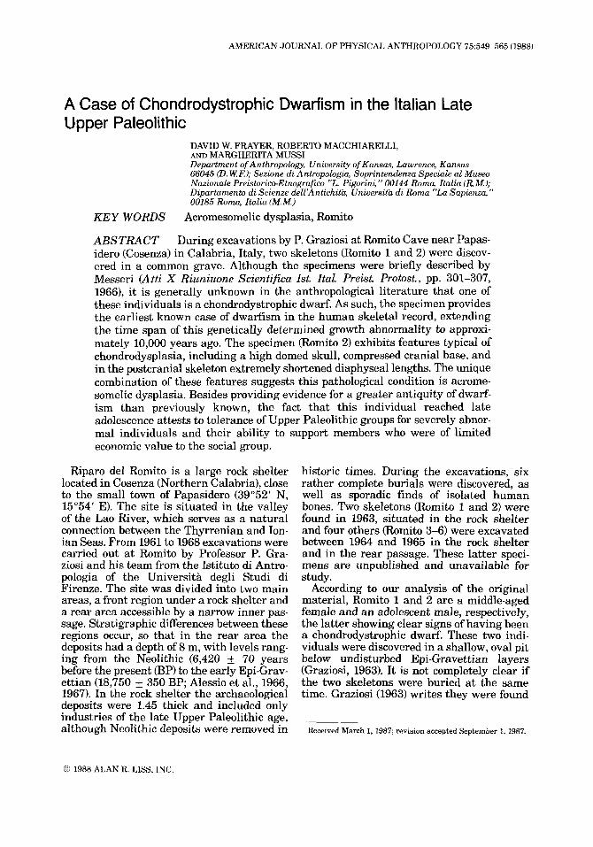
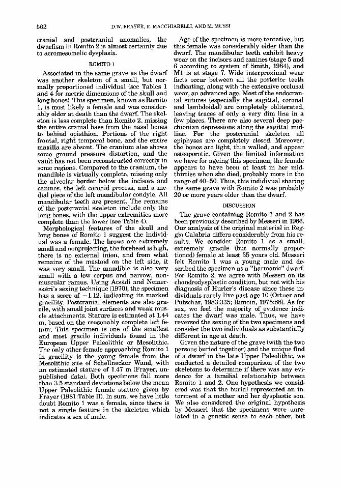
The specimen consists of the cranium, mandible, and portions of the postcranial skeleton (Fig. l), none of which are fossilized or mineralized to any appreciable extent. The cranium is virtually intact, missing only a portion of the anterior maxilla (including prosthion, the central incisors and left 121, the inferior, medial, and posterior walls of the right orbit, the right zygomatic arch, the base of the left zygomatic process, the inferi- ormost portion of the right mastoid, small portions of the occipital lateral and posterior to the foramen magnum, and the external surface of the right occipital condyle. Some damage to the skull occurred during or since excavation. There is a round hole (12.5 mm in diameter) on the left frontal directly above and behind the frontal boss. This is clearly of postmortem origin, since the exposed bone along the face of the break is white and un- stained by soil sediments. The mandible lacks the condyle and condylar process on both sides, along with portions of the alveolar bor- der and some teeth (left I1-P3; right 11-12, P3- M2). Despite these missing areas and post- mortem damage, the cranium and mandible are nearly complete and neither show signs of artifical deformation or in situ distortion.
Postcranial remains are more incomplete, limited exclusively to the long bones. Both femurs are present, as well as the left and right tibia and fibula. Left and right hu- merus, radius, and ulna are also present, with the left side better preserved than the right. No other postcranial remains could be located in the Museo Archeologico Nazionale in Reggio Calabria, although in his original report, Messeri (1966) mentions the existence of an innominate as well as both clavicles, vertebrae, and hand and foot bones for Rom- ito 2. It is unfortunate these bones have been lost, since their presence would aid in the
Fig. 1. Cranial and postcranial remains of Romito 2.
diagnosis of the specific forms of dysplasia as well as in the reconstruction of stature.
Sex Establishment of sex in skeletal remains is
always difficult (especially when pelvic re- mains are absent) and, given our lack of ex- perience in dealing with dyschondroplastic skeletons and uncertainties about the devel- opment of secondary sexual characteristics in dwarfs, we can only give an “educated guess” about the sex of Romito 2. However, several features indicate the specimen was very likely a male. These include a relatively large, broad, and projecting mastoid; a mod- erately large supramastoid crest; a muscular nuchal region with an external inion; a very bifid chin; and a broad mandibular ramus. In contrast to these male features, the supraor-
552 D.W. FRAYER, R. MACCHIARELLI, AND M. MUSS1
bital region shows no projection and the fore- head is extremely vertical over the orbits. Both of these features generally characterize females, but we consider the growth defect to have been more influential in producing these traits than any sexual effect. The long bones provide little help in determining sex, even though they possess robust muscle markings and large joint surfaces, since these were certainly influenced by growth anoma- lies and altered biomechanical stress on the appendicular skeleton. Although our attri- bution of sex is clearly not certain, we feel the majority of evidence indicates the speci- men is very likely male.
Age The age of Romito 2 is also difficult to as-
sess since ageing criteria must have also been influenced by the abnormal growth condition the individual suffered. For example, Bailey (1973:62) notes that dental eruption in achon- droplastics is “somewhat retarded.” Long bone growth, however, seems to proceed in the same sequence as normally statured in- dividuals, but the rate of increase in body height is considerably lower Worton et al., 1978). Thus, relative to the postcranial skel- eton, dental eruption is delayed. Using mul- tiple dental and skeletal standards developed for normal individuals, we estimate the age as between 14 and 19, but based on dis- crepancies between dental and skeletal mat- uration feel the individual was probably about 17.
For the dentition, all teeth (which are still located in the jaw) are in their full occlusal position except the third molars. Both max- illary M3’s are fully formed, but deep in their crypts, well above the alveolar border. In the mandible, the left M3 is well below the alveo- lar border, while the right M3 is nearly in the occlusal plane. Besides eruption, occlusal wear is moderate with upper and lower Ml’s showing cusp removal and small dentin patches. The second molars show no dentin exposure, but the cusps are blunted and mod- erately large interproximal facets occur on their mesial surfaces. Premolars exhibit blunting and small dentin patches and the canines are slightly worn with pinpoint den- tin exposure. The upper right lateral incisor is the most worn tooth in either jaw, showing a large area of exposed dentin. Thus, for the dentition, eruption and tooth wear indicate a subadult individual, with a lightly worn pos- terior dentition and incompletely erupted third molars.
Other skeletal features suggest a young age for the specimen. The spheno-occipital synchondrosis is incompletely fused in its lat- eral aspects, but this region was certainly affected by the growth anomaly. Most long bone epiphyses show remnants of a fusion line, especially on the proximal and distal humerus and distal portions of the radius, ulna, femur, and tibia. We were unable to make radiographs of the bones in Reggio Cal- abria, so it is not possible to determine the degree of internal epiphyseal fusion. How- ever, the external morphology indicates the regions were completely fused, but that re- modeling and obliteration of the fusion lines had yet to be completed. Like the basilar region, long bone epiphyseal closure was likely affected by the disruption in enchon- dral growth. Finally, according to Messeri (1966:302), an innominate with an incom- pletely fused iliac crest was originally pres- ent which would indicate an age less than 21, but we could not verify this since the piece now seems to be lost.
Judging from epiphyseal union in normal individuals (Krogman, 1962), the state of postcranial epiphyseal closure in Romito 2 correlates well with the dental age of the individual. However, some contradictory evi- dence occurs in the cranial vault, where the coronal, sagittal, and lambdoidal internal su- tures are partially obliterated, represented only by a thin line. Although there is no ectocranial suture closure, the degree of en- docranial obliteration would indicate an age in excess of 25. Despite this discrepancy, we feel the preponderance of evidence points to an age of about 17, with a range of 2 years. Finally with respect to cause of death, we could find no evidence of trauma or violence. Other than the growth anomaly, the speci- men showed no other chronic or acute patho- logical conditions.
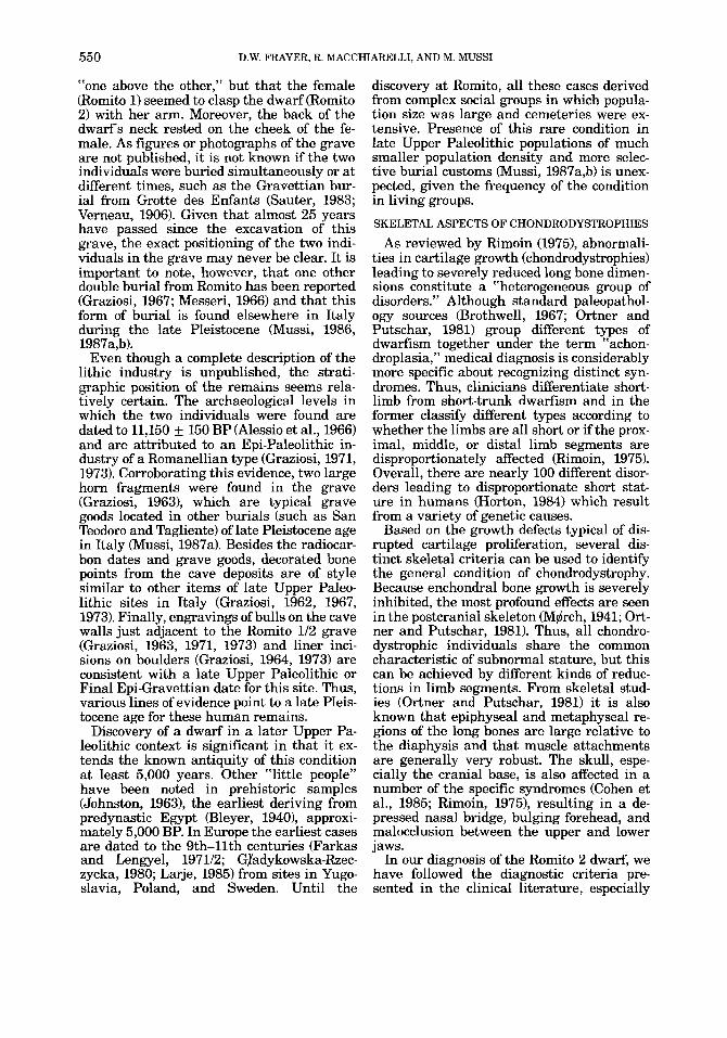
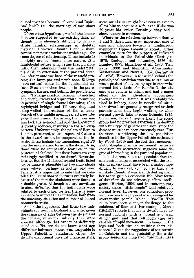
Description The skull is brachycephalic (cranial index
= 82.5) with very pronounced frontal and parietal bossing. The frontal bosses protrude markedly forward and occupy a more ante- rior position than the superior orbital bor- ders (Fig. 2). The bosses are large and take up most of the forehead region, and the ex- tension of the frontal bosses to the midline is so pronounced that there is no separation or depression between them. This condition re- sults in a forehead which is very high and extremely flat across the front and squared off from side-to-side above the orbits. The
UPPER PALEOLITHIC DWARF 553
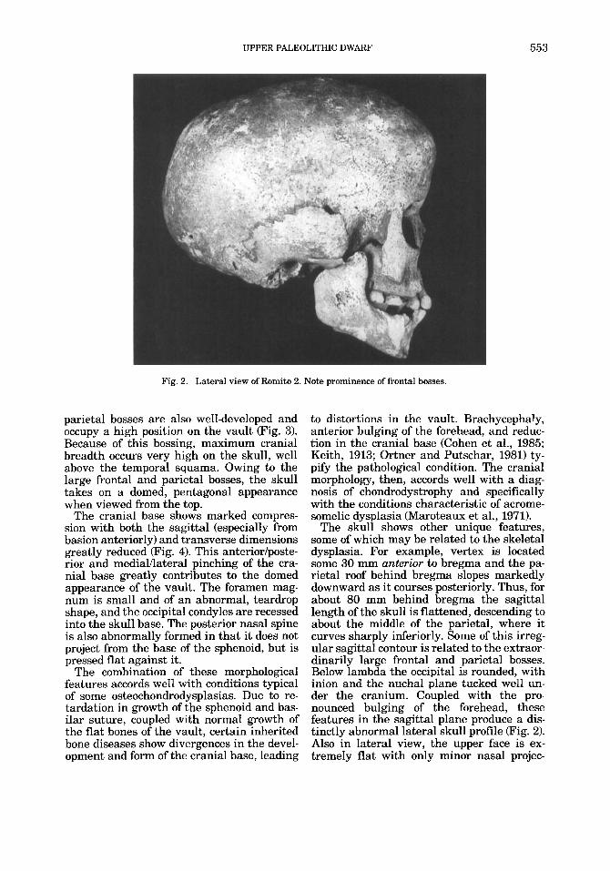
Fig. 2. Lateral view of Romito 2. Note prominence of frontal bosses.
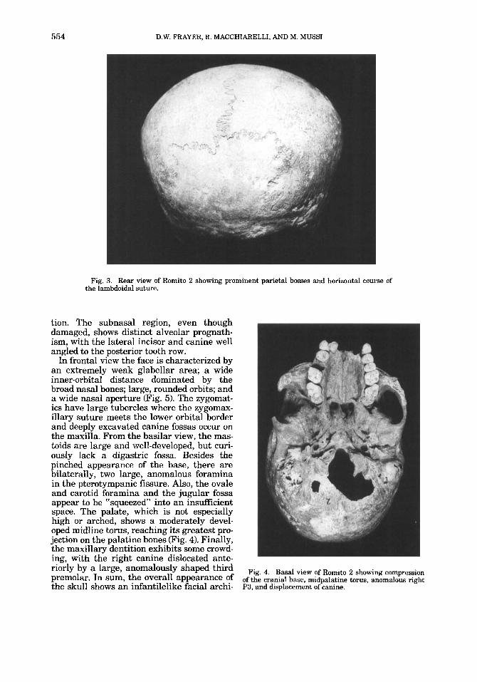
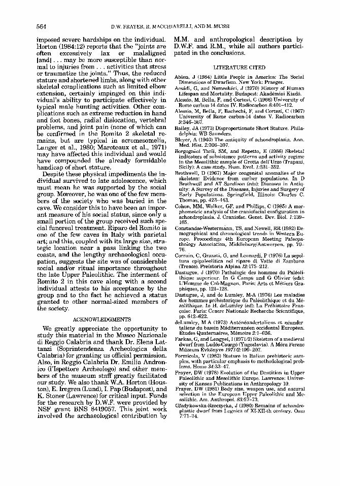
parietal bosses are also well-developed and occupy a high position on the vault (Fig. 3). Because of this bossing, maximum cranial breadth occurs very high on the skull, well above the temporal squama. Owing to the large frontal and parietal bosses, the skull takes on a domed, pentagonal appearance when viewed from the top.
The cranial base shows marked compres- sion with both the sagittal (especially from basion anteriorly) and transverse dimensions greatly reduced (Fig. 4). This anteriorlposte- rior and mediauateral pinching of the cra- nial base greatly contributes to the domed appearance of the vault. The foramen mag- num is small and of an abnormal, teardrop shape, and the occipital condyles are recessed into the skull base. The posterior nasal spine is also abnormally formed in that it does not project from the base of the sphenoid, but is pressed flat against it.
The combination of these morphological features accords we11 with conditions typical of some osteochondrodysplasias. Due to re- tardation in growth of the sphenoid and bas- ilar suture, coupled with normal growth of the flat bones of the vault, certain inherited bone diseases show divergences in the devel- opment and form of the cranial base, leading
to distortions in the vault. Brachycephaly, anterior bulging of the forehead, and reduc- tion in the cranial base (Cohen et al., 1985; Keith, 1913; Ortner and Putschar, 1981) ty- pify the pathological condition. The cranial morphology, then, accords well with a diag- nosis of chondrodystrophy and specifically with the conditions characteristic of acrome- somelic dysplasia (Maroteaux et al., 1971).
The skull shows other unique features, some of which may be related to the skeletal dysplasia. For example, vertex is located some 30 mm anterior to bregma and the pa- rietal roof behind bregma slopes markedly downward as it courses posteriorly. Thus, for about 80 mm behind bregma the sagittal length of the skull is flattened, descending to about the middle of the parietal, where it curves sharply inferiorly. Some of this imeg- ular sagittal contour is related to the extraor- dinarily large frontal and parietal bosses. Below lambda the occipital is rounded, with inion and the nuchal plane tucked well un- der the cranium. Coupled with the pro- nounced bulging of the forehead, these features in the sagittal plane produce a dis- tinctly abnormal lateral skull profile (Fig. 2). Also in lateral view, the upper face is ex- tremely flat with only minor nasal projec-
554 D.W. FRAYER, R. MACCHIARELLI, AND M. MUSS1
Fig. 3. Rear view of Romito 2 showing prominent parietal bosses and horizontal course of the lambdoidal suture.
tion. The subnasal region, even though damaged, shows distinct alveolar prognath- ism, with the lateral incisor and canine well angled to the posterior tooth row.
In frontal view the face is characterized by an extremely weak glabellar area; a wide inner-orbital distance dominated by the broad nasal bones; large, rounded orbits; and a wide nasal aperture (Fig. 5). The zygomat- ics have large tubercles where the zygomax- illary suture meets the lower orbital border and deeply excavated canine fossas occur on the maxilla. From the basilar view, the mas- toids are large and well-developed, but curi- ously lack a digastric fossa. Besides the pinched appearance of the base, there are bilaterally, two large, anomalous foramina in the pterotympanic fissure. Also, the ovale and carotid foramina and the jugular fossa appear to be "squeezed" into an insufficient space. The palate, which is not especially high or arched, shows a moderately devel- oped midline torus, reaching its greatest pro- jection on the palatine bones (Fig. 4). Finally, the maxillary dentition exhibits some crowd- ing, with the right canine dislocated ante- riorly by a large, anomalously shaped third premolar. In sum, the overall appearance of the skull shows an infantilelike facial archi-
Fig. 4. Basal view of Romito 2 showing compression of the cranial base, midpalatine torus, anomalous right P3, and displacement of canine.
UPPER PALEOLITHIC DWARF
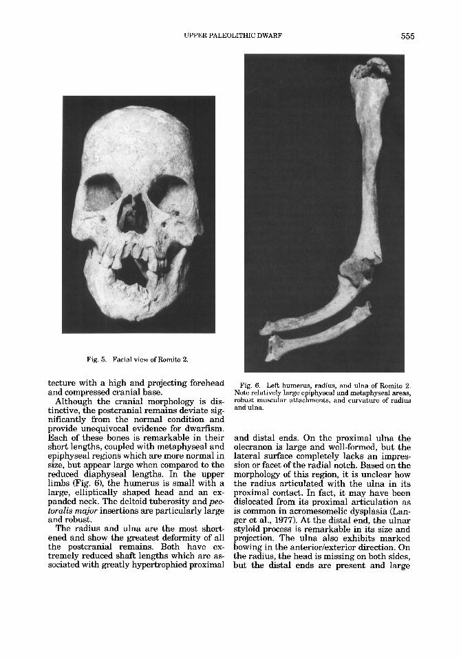
Fig. 5. Facial view of Romito 2.
555
tecture with a high and projecting forehead and compressed cranial base.
Although the cranial morphology is dis- tinctive, the postcranial remains deviate sig- nificantly from the normal condition and provide unequivocal evidence for dwarfism. Each of these bones is remarkable in their short lengths, coupled with metaphyseal and epiphyseal regions which are more normal in size, but appear large when compared to the reduced diaphyseal lengths. In the upper limbs (Fig. 6), the humerus is small with a large, elliptically shaped head and an ex- panded neck. The deltoid tuberosity and p e e toralis major insertions are particularly large and robust.
The radius and ulna are the most short- ened and show the greatest deformity of all the postcranial remains. Both have ex- tremely reduced shaft lengths which are as- sociated with greatly hypertrophied proximal
Fig. 6. Left humerus, radius, and ulna of Romito 2. Note relatively large epiphyseal and metaphyseal areas, robust muscular attachments, and curvature of radius and ulna.
and distal ends. On the proximal ulna the olecranon is large and well-formed, but the lateral surface completely lacks an impres- sion or facet of the radial notch. Based on the morphology of this region, it is unclear how the radius articulated with the ulna in its proximal contact. In fact, it may have been dislocated from its proximal articulation as is common in acromesomelic dysplasia &an- ger et al., 1977). At the distal end, the ulnar styloid process is remarkable in its size and projection. The ulna also exhibits marked bowing in the anteriorlexterior direction. On the radius, the head is missing on both sides, but the distal ends are present and large
556 D.W. FRAYER, R. MACCHIARELLI, AND M. MUSS1
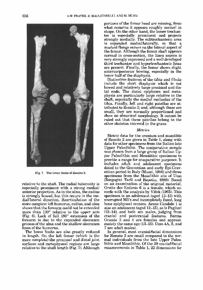
Fig. 7. The lower limbs of Romito 2.
relative to the shaft. The radial tuberosity is especially prominent with a strong mediaii anterior projection. As in the ulna, the radius is strongly bowed, but this occurs in the me- dialAatera1 direction. Rearticulation of the more complete left humerus, radius, and ulna shows that the forearm could not be extended more than 130" relative to the upper arm (Fig. 6). Lack of full 180" extension of the forearm is due to the expanded olecranon process of the ulna and the shallow olecranon fossa of the humerus.
The lower limbs are also greatly reduced in length. On the left femur (which is the more complete) the proximal and distal joint surfaces and metaphyseal regions are large relative to the shaft length (Fig. 7). Although
portions of the femur head are missing, from what remains it appears roughly normal in shape. On the other hand, the lesser trochan- ter is especially prominent and projects strongly medially. The subtrochanteric area is expanded medialAaterally, so that a marked flange occurs on the lateral aspect of the femur. Although the femur shaft appears normal in cross-section, the linea aspera is very strongly expressed and a well-developed third trochanter and hypertrochanteric fossa are present. Finally, the femur shows slight anteriorlposterior bowing, especially in the lower half of the diaphysis.
Distinctive features of the tibia and fibula include the short diaphysis which is not bowed and relatively large proximal and dis- tal ends. The distal epiphyses and meta- physis are particularly large relative to the shaft, especially the medial malleolus of the tibia. Finally, left and right patellas are at- tributed to Romito 2; and, although these are small, they are normally proportioned and show no abnormal morphology. It cannot be ruled out that these patellas belong to the other skeleton interred in the grave.
Metrics Metric data for the cranium and mandible
of Romito 2 are given in Table 1, along with data for other specimens from the Italian late Upper Paleolithic. The comparative sample was chosen from a large group of Italian Up- per Paleolithic and Mesolithic specimens to provide a range €or comparative purposes. It includes adult and adolescent specimens dated to the Gravettian and early Epi-Grav- ettian period in Italy (Mussi, 1986) and three specimens from the Mesolithic site of Uzzo (Borgogini Tarli and Repetto, 1986). Based on an examination of the original material, Grotte des Enfants 6 is a female, which ac- cords with the analysis by VlEek (1965). This specimen is an adolescent (aged 13-15) with unerupted M3's and incompletely fused, long bone epiphyseal centers. Arene Candide 1 is also an adolescent (aged 12-15), as is Paglicci (12-14); and both are males, judging from cranial and postcranial features. Barma Grande 3 and 4 are femaIes and approxi- mately the same age (13-15). Uzzo 4A, 5, and 7 are adult males).
In general, most craniaiifacial dimensions for Romito 2 are small compared to the nor- mal individuals from the late Upper Paleo- lithic and Mesolithic. Of the 39 craniaiifacial measurements in Table 1,22 dimensions for

TAB
LE 1
. Sel
ecte
d cr
ania
l mea
sure
men
ts fo
r R
omito
2 w
ith
com
pari
sons
to o
ther
Ita
lian
Upp
er P
aleo
lithi
c an
d M
esol
ithic
spe
cim
ens'
Gro
tte
des
Rom
ito 2
Rom
ito 1
Enf
ants
6 A
rene
Can
dide
1 P
anlic
ci 1
Bar
ma
Gra
nde
3 B
arm
a G
rand
e 4
Uzz
o 4A
Uzz
n 6
Tra
m 7
(mal
e)
(fem
ale)
(f
emal
e)
(mal
e)
Cra
nium
G
labe
lla-o
pist
ocra
nion
E
uryo
n-eu
ryon
B
asio
n-na
sion
O
pist
hion
-nas
ion
Bas
ion-
naso
spin
ale
Bas
ion-
breg
ma
Ast
erio
n-as
teri
on
Aur
icul
are-
auri
cula
re
Sten
ion-
sten
ion
Mas
toid
ale-
mas
toid
ale
Bas
ion-
sphe
noba
sion
P
ars
basi
lari
s bre
adth
Fo
ram
en m
agnu
m le
ngth
Fo
ram
en m
agnu
m b
read
th
Nas
ion-
breg
ma
arc
Nas
ion-
breg
ma c
hord
B
regm
a-la
mbd
a ar
c B
regm
a-la
mbd
a ch
ord
Lam
bda-
opis
thio
n ar
c L
ambd
a-op
isth
ion
chor
d N
asio
n-pr
osth
ion
Nas
ion-
naso
spin
ale
Nas
al b
read
th
Fron
tom
alte
mp-
fron
tom
alte
mp
Juga
le-j
ugal
e Zygomaxillare-zygomaxillare
Orb
ital
hei
ght
Orb
ital
bre
adth
O
rale
-sta
phyl
ion
Pala
te b
read
th a
t M2
Kor
onia
-kor
onia
G
onio
n-go
nion
Sy
mph
ysis
bre
adth
Sy
mph
ysis
hei
ght
Cor
pus
brea
dth
at M
1M2
Cor
pus
heig
ht a
t MliM
2 M
inim
um ra
mus
bre
adth
G
onio
n-gn
athi
on
Gon
ion-
infr
aden
tale
Man
dibl
e
177.
0 14
6.0
87.0
12
2.0
78.0
12
6.0
97.7
11
3.6
60.3
91
.0
13.5
16
.3
(33.
0)
(22.
5)
137
111.
5 13
8 12
7.0
109 85.5
(5
8.0)
41
.8
24.3
96
.3
109.
1 92
.9
30.6
38
.7
38.8
55
.7
91.7
98
.0
13.7
23
.0
14.9
25
.0
31.8
76
.2
77.4
180.
0 13
8.0
-
- - I
106.
0 (1
18.0
) 65
.0
-
- - - -
127
107.
7 13
8 12
2.7
- - - - - -
- - - - - - -
89.9
13
.4
23.0
11
.8
20.7
29
.2
75.0
nn n
193.
0 13
4.0
141.
0 - -
-
109.
0 -
-
109.
7 - - - -
133
116.
0 13
7 12
5.0
120 95.8
61
.4
48.3
23
.1
-
- - 25.0
38
.6
- - -
-
12.9
25
.7
16.5
22
.8
37.6
83
.3
89.7
198.
0 -
-
-
-
-
-
-
-
109.
0 24
.2
21.8
38
.9
30.6
14
1 11
9.0
134
120.
0 11
3 98.5
69
.7
41.4
24
.8
-
-
-
33.4
45
.5
47.9
-
-
-
-
-
15.5
37.5
-
-
-
174.
0 -
-
-
- - - - -
107.
8 I
- - -
119
101.
0 12
4 11
0.8
- - (62.
9)
(50.
0)
(21.
0)
(100
.0)
-
-
-
-
-
-
-
-
15.3
(2
6.5)
15
.9
26.8
33
.0
88.2
86
.8
-
174.
0 -
-
-
-
-
132
-
117.
5 -
127
-
109.
0 -
-
-
-
69.2
(6
3.0)
54
.8
(45.
5)
27.5
21
..2
-
-
-
-
-
13.6
-
28.1
-
14.5
-
26.9
-
34.7
-
89.4
-
91.2
189.
0 14
8.0
95.0
13
8.0
85.0
13
1.0
125.
7 12
9.3
73.7
10
9.6
-
-
43.2
35
.5
128
107.
8 12
3 11
1.3
130
100.
9 60
.0
50.5
19
.6
103.
0 12
0.5
95.0
27
.6
42.9
-
-
100.
3 10
3.0
15.5
26
.1
12.2
25
.2
30.7
87
.7
84.7
~~
~ .
~~
.
. _
~~
.
(fem
ale)
(m
ale)
(m
ale)
(m
ale)
G
ale)
(f
emal
e)
-
90.9
185.
0 19
0.0
143.
0 14
6.0
101.
0 -
143.
0 -
93.0
-
147.
0 -
119.
3 11
8.0
124.
6 13
3.1
77.0
-
115.
0 10
8.0
24.2
-
43.8
-
35.5
-
-
-
133
151
110.
4 12
7.1
142
126
125.
7 10
6.2
124
-
107.
9 -
60.6
-
47.7
-
23.3
-
102.
8 10
5.4
114.
0 -
88.2
-
27.3
-
40.4
-
63.6
-
-
-
97.7
-
106.
7 -
15.3
16
.0
30.7
29
.5
11.7
11
.7
27.2
30
.4
30.8
34
.4
85.6
95
.9
'All
mea
sure
men
ts in
mm
and
take
n on
ori
gina
ls b
y D
.W.F
(
) =
est
imat
ed.
558 D.W. FRAYER, R. MACCHIARELLI, AND M. MUSS1
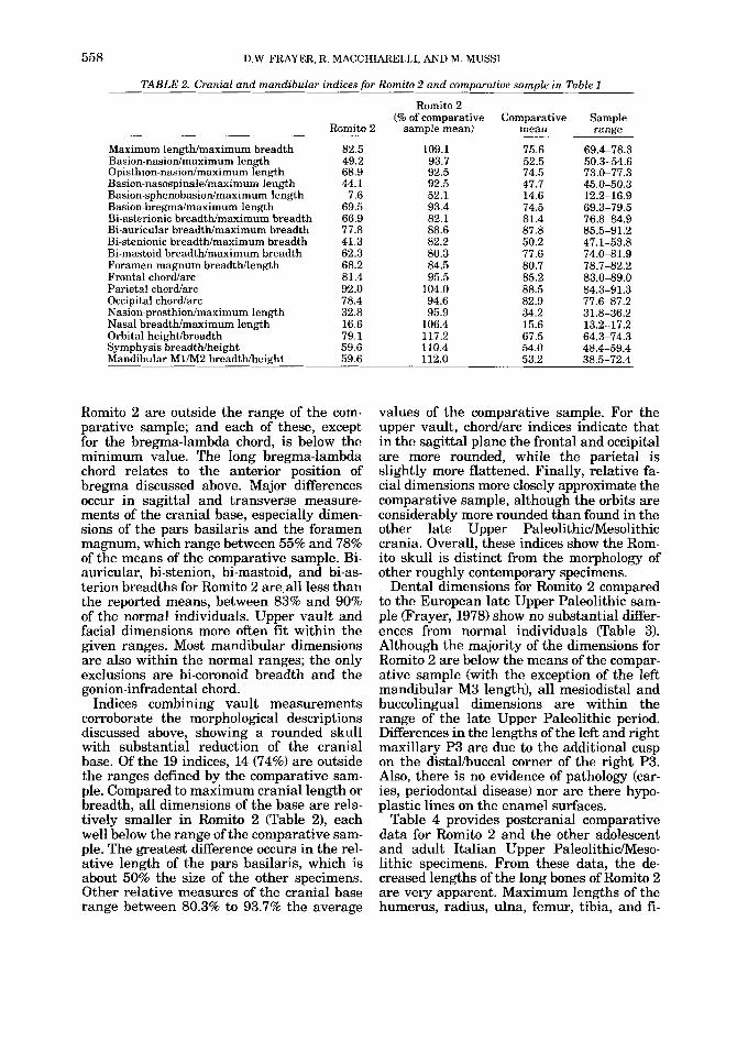
TABLE 2. Cranial and mandibular indices for Romito 2 and comparative sample in Table 1
Romito 2 (% of comparative Comparative Sample
Romito 2 sample mean) mean range
Maximum lengthlmaximum breadth 82.5 109.1 75.6 69.4-78.3 Basion-nasiodmaximum length 49.2 93.7 52.5 50.3-54.6 Opisthion-nasiodmaximum length 68.9 92.5 74.5 73.0-77.3 Basion-nasospinalehaximum length 44.1 92.5 47.7 45.0-50.3 Basion-sphenobasionhaximum length 7.6 52.1 14.6 12.2-16.9 Basion-bregmdmaximum length 69.5 93.4 74.5 69.3-79.5 Bi-asterionic breadthhaximum breadth 66.9 82.1 81.4 76.8-84.9
85.5-91.2 Bi-auricular breadthlmaximum breadth 77.8 88.6 87.8 Bi-stenionic breadtWmaximum breadth 41.3 82.2 50.2 47.1-53.8 Bi-mastoid breadtWmaximum breadth 62.3 80.3 77.6 74.0-81.9 Foramen magnum breadtMength 68.2 84.5 80.7 78.7-82.2 Frontal chordarc 81.4 95.5 85.2 83.0-89.0 Parietal chordarc 92.0 104.0 88.5 84.3-91.3 Occipital chordarc 78.4 94.6 82.9 77.6-87.2 Nasion-prosthionhaximum length 32.8 95.9 34.2 31.8-36.2 Nasal breadthhaximum length 16.6 106.4 15.6 13.2-17.2 Orbital heightbreadth 79.1 117.2 67.5 64.3-74.3 Symphysis breadthheight 59.6 110.4 54.0 48.4-59.4 Mandibular MUM2 breadthheight 59.6 112.0 53.2 38.5-72.4
Romito 2 are outside the range of the com- parative sample; and each of these, except for the bregma-lambda chord, is below the minimum value. The long bregma-lambda chord relates to the anterior position of bregma discussed above. Major differences occur in sagittal and transverse measure- ments of the cranial base, especially dimen- sions of the pars basilaris and the foramen magnum, which range between 55% and 78% of the means of the comparative sample. Bi- auricular, bi-stenion, bi-mastoid, and bi-as- terion breadths for Romito 2 are. all less than the reported means, between 83% and 90% of the normal individuals. Upper vault and facial dimensions more often fit within the given ranges. Most mandibular dimensions are also within the normal ranges; the only exclusions are bi-coronoid breadth and the gonion-infradental chord.
Indices combining vault measurements corroborate the morphological descriptions discussed above, showing a rounded skull with substantial reduction of the cranial base. Of the 19 indices, 14 (74%) are outside the ranges defined by the comparative sam- ple. Compared to maximum cranial length or breadth, all dimensions of the base are rela- tively smaller in Romito 2 (Table 2), each well below the range of the comparative sam- ple. The greatest difference occurs in the rel- ative length of the pars basilaris, which is about 50% the size of the other specimens. Other relative measures of the cranial base range between 80.3% to 93.7% the average
values of the comparative sample. For the upper vault, chord/arc indices indicate that in the sagittal plane the frontal and occipital are more rounded, while the parietal is slightly more flattened. Finally, relative fa- cial dimensions more closely approximate the comparative sample, although the orbits are considerably more rounded than found in the other late Upper PaleolithicMesolithic crania. Overall, these indices show the Rom- ito skull is distinct from the morphology of other roughly contemporary specimens.
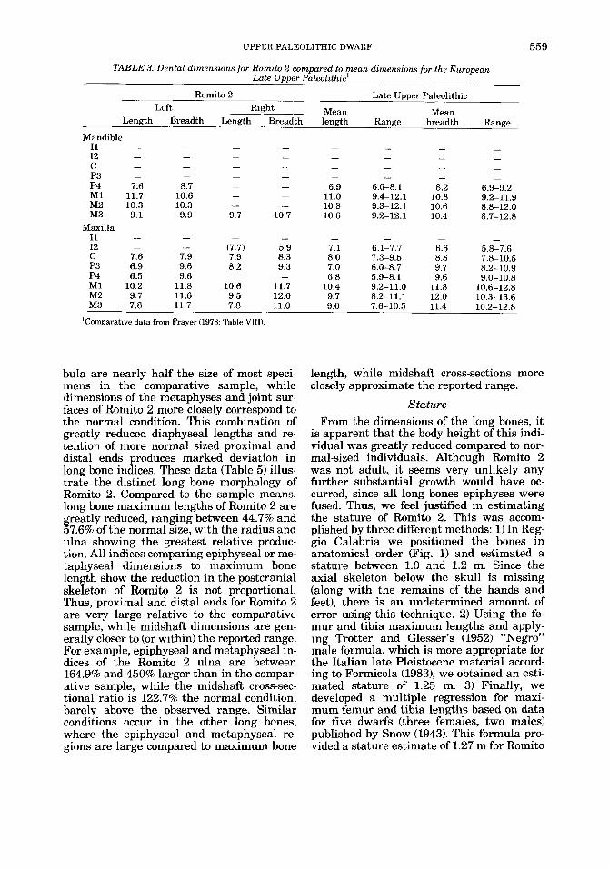
Dental dimensions for Romito 2 compared to the European late Upper Paleolithic sam- ple prayer, 1978) show no substantial differ- ences from normal individuals (Table 3). Although the majority of the dimensions for Romito 2 are below the means of the compar- ative sample (with the exception of the left mandibular M3 length), all mesiodistal and buccolingual dimensions are within the range of the late Upper Paleolithic period. Differences in the lengths of the left and right maxillary P3 are due to the additional cusp on the distalibuccal corner of the right P3. Also, there is no evidence of pathology (car- ies, periodontal disease) nor are there hypo- plastic lines on the enamel surfaces.
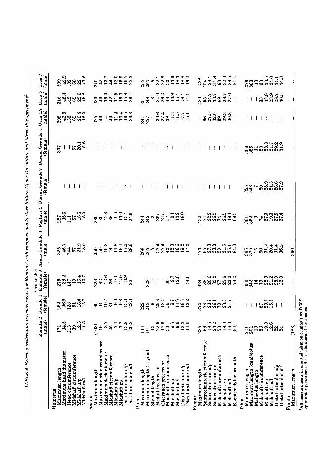
Table 4 provides postcranial comparative data for Romito 2 and the other adolescent and adult Italian Upper PaleolithicAIeso- lithic specimens. From these data, the de- creased lengths of the long bones of Romito 2 are very apparent. Maximum lengths of the humerus, radius, ulna, femur, tibia, and fi-
UPPER PALEOLITHIC DWARF
TABLE 3. Dental dimensions for Romito 2 compared to mean dimensions for the European Late Upper Paleolithic’
Romito 2 Late Upper Paleolithic
Mean Mean Left Right Length Breadth Length Breadth length Range breadth Range
Mandible - - - - - I1 - - -
I2 C P3 P4 7.6 8.7 - -
- 11.0 9.4-12.1 10.8 9.2-11.9 MI 11.7 10.6 -
- 10.8 9.3-12.1 10.6 8.8-12.0 M2 10.3 10.3 - M3 9.1 9.9 9.7 10.7 10.6 9.2-12.1 10.4 8.7-12.8
I1 I2 - - C 7.6 7.9 7.9 8.3 8.0 7.3-9.5 8.8 7.8-10.5
8.2-10.9 P3 6.9 9.6 8.2 9.3 7.0 6.0-8.7 9.7 - 6.8 5.9-8.1 9.6 9.0-10.8 P4 6.5 9.6 -
M1 10.2 11.8 10.6 11.7 10.4 9.2-11.0 11.8 10.6-12.8 M2 9.7 11.6 9.5 12.0 9.7 8.2-11.1 12.0 10.3-13.6 M3 7.8 11.7 7.8 11.0 9.0 7.6-10.5 11.4 10.2-12.8
- - - - - - - -
- - - - - - - -
- - - - - - - -
6.9 6.0-8.1 8.2 6.9-9.2
Maxilla - - - - - - - -
(7.7) 5.9 7.1 6.1-7.7 6.6 5.8-7.6
‘Comparative data from Frayer (1978: Table VIII).
559
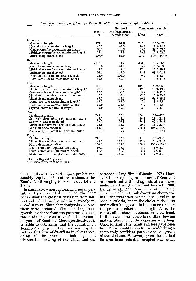
bula are nearly half the size of most speci- mens in the comparative sample, while dimensions of the metaphyses and joint sur- faces of Romito 2 more closely correspond to the normal condition. This combination of greatly reduced diaphyseal lengths and re- tention of more normal sized proximal and distal ends produces marked deviation in long bone indices. These data (Table 5) illus- trate the distinct long bone morphology of Romito 2. Compared to the sample means, long bone maximum lengths of Romito 2 are greatly reduced, ranging between 44.7% and 57.6% of the normal size, with the radius and ulna showing the greatest relative produc- tion. All indices comparing epiphyseal or me- taphyseal dimensions to maximum bone length show the reduction in the postcranial skeleton of Romito 2 is not proportional. Thus, proximal and distal ends for Romito 2 are very large relative to the comparative sample, while midshaft dimensions are gen- erally closer to (or within) the reported range. For example, epiphyseal and metaphyseal in- dices of the Romito 2 ulna are between 164.9% and 450% larger than in the compar- ative sample, while the midshaft cross-sec- tional ratio is 122.7% the normal condition, barely above the observed range. Similar conditions occur in the other long bones, where the epiphyseal and metaphyseal re- gions are large compared to maximum bone
length, while midshaft cross-sections more closely approximate the reported range.
Stature From the dimensions of the long bones, it
is apparent that the body height of this indi- vidual was greatly reduced compared to nor- mal-sized individuals. Although Romito 2 was not adult, it seems very unlikely any further substantial growth would have oc- curred, since all long bones epiphyses were fused. Thus, we feel justified in estimating the stature of Romito 2. This was accom- plished by three different methods: 1) In Reg- gio Calabria we positioned the bones in anatomical order (,Fig. 1) and estimated a stature between 1.0 and 1.2 m. Since the axial skeleton below the skull is missing (along with the remains of the hands and feet), there is an undetermined amount of error using this technique. 2) Using the fe- mur and tibia maximum lengths and apply- ing Trotter and Glesser’s (1952) “Negro” male formula, which is more appropriate for the Italian late Pleistocene material accord- ing to Formicola (1983), we obtained an esti- mated stature of 1.25 m. 3) Finally, we developed a multiple regression for maxi- mum femur and tibia lengths based on data for five dwarfs (three females, two males) published by Snow (1943). This formula pro- vided a stature estimate of 1.27 m for Romito
TAB
LE 4
. Sel
ecte
d po
stcr
ania
l m
easu
rem
ents
for
Rom
ito 2
wit
h co
mpa
riso
ns to
oth
er I
talia
n U
pper
Pal
eolit
hic
and
Mes
olith
ic s
peci
men
s'
C.m
t.t,e
den
- -. . . -. -
Enf
ants
6
(fem
ale)
A
rene
Can
dide
1 P
aglic
ci 1
Bar
ma
Gra
nde
3 B
arm
a G
rand
e 4
Uzz
o 4A
Uzz
o 5
Uzz
o 7
(mal
e)
(mal
e)
(fem
ale)
(f
emal
e)
(mal
e)
(mal
e) (
mal
e)
Rom
ito 2
Rom
ito 1
(m
ale)
(f
emal
e)
Hum
erus
M
axim
um le
ngth
M
axim
um h
ead
diam
eter
H
ead
circ
umfe
renc
e M
idsh
aft c
ircu
mfe
renc
e M
idsh
aft
dp
M
idsh
aft m
A
Max
imum
leng
th
Max
imum
nec
k ci
rcum
fere
nce
Max
imum
dec
k di
amet
er
Mid
shaf
t cir
cum
fere
nce
Mid
shaf
t dp
M
idsh
aft m
A D
ista
l art
icul
ar d
p
Dis
tal a
rtic
ular
mA
Max
imum
leng
th
Max
imum
leng
th (-
styl
oid)
St
yloi
d le
ngth
M
edia
l tro
chle
a ht
O
lecr
anon
pro
cess
br
Mid
shaf
t cir
cum
fere
nce
Mid
shaf
t dp
M
idsh
aft m
/l D
ista
l art
icul
ar d
p
Dis
tal a
rtic
ular
mA
Rad
ius
Uln
a
171
262
34.5
36
.9
113
123
39
51
12.3
16
.4
11.5
13
.7
275 38.2
14
7 49
16.4
12
.7
325
287
-
45.7
35
.6
-
144
111
-
67
57
21.9
18
.5
-
18.0
15
.0
-
-
307
298
316
309
-
133
142
132
57
65
65
68
20.1
20
.4
22.9
22
15
.6
16.0
15
.8
17.6
43.9
48
.4
42.9
-
(102
) 19
5 19
34
25
31
8.7
10.7
7.1
9.0
7.7
9.8
13.9
13
.0
20.2
22
.0
223 42
12.6
36
9.4
12.0
13
.9
22.1
250 50
15.8
44
11
.8
15.1
17
.3
28.6
230 39
12
.8
36 9.9
11.9
13
.4
24.6
225
233
240
43
43
42
-
15.3
13
.7
43
42
44
11.2
11
.5
12.0
16
.4
15.0
15
.9
16.5
15
.8
16.5
26
.3
26.1
25
.3
111
222
101
213
10
9 22
.9
25.8
17
.9
18.4
33
36
9.
5 9.
7 9.
6 11
.8
12,3
16
.6
11.0
12
.2
268
263 5 33.9
25
.9
48
12.3
16
.6
19.2
17
.2
244
242 2 26.5
21
.5
37 9.1
13.2
16
.0
-
-
-
241
251
255
-
-
237
248
250
-
-
4 3
5
-
-
30.6
34
.0
32.1
-
-
27.4
26
.2
22.8
-
-
11.3
13
.9
13.6
-
-
11.5
15
.4
18.3
-
-
17.7
18
.4
18.8
-
-
15.1
14
.1
16.2
-
39
48
52
-
-
229 -
-
36 9.7
12.6
14.6
-
Fem
ur
Max
imum
leng
th
225
370
Subt
roch
ante
ric
circ
umfe
renc
e 69
74
Su
btro
chan
teri
c al
p 18
.5
20.7
Su
btro
chan
teri
c m
A
23.2
26
.1
Mid
shaf
t cir
cum
fere
nce
58
70
Mid
shaf
t dp
19
.0
23.0
M
idsh
aft R
IA 16
.3
21.2
-
Bie
pico
ndyl
ar b
read
th
(54)
424 88
20
.0
33.2
77
473
432
-
95
74
27.3
22
.2
-
33.8
26
.5
-
90
74
31.5
26
.5
-
25.1
19
.1
-
84.8
69
.5
-
-
-
430
428
-
-
-
96
95
104
-
27.5
24
.7
28.4
-
32.6
33
.7
37.:4
-
89
89
93
25.6
22
.0
74.0
-
29.3
28
.7
32.3
-
26.8
27
.0
26.5
-
-
81.4
-
211
-
201
-
10
-
53
67
19.0
20
.7
12.6
15
.5
22
24
-
-
Tib
ia
Max
imum
leng
th
Max
imum
leng
th (-
mal
leol
us)
Mal
leol
us le
ngth
M
idsh
aft c
ircu
mfe
renc
e M
idsh
aft a
lp
Mid
shaf
t m/l
Dis
tal a
rtic
ular
dp
D
ista
l art
icul
ar m
A
Max
imum
leng
th
Fibu
la
356
342 14
79
28
.0
21.2
28
.9
32.0
385
361
355
366
-
-
376
370
352
348
355
-
-
363
-
13
15
9 7
11
-
90
74
80
83
-
93
93
31.2
27
.3
28.9
28
.3
-
33.8
33
.5
20.4
19
.3
21.3
21
.7
-
23.9
24
.0
31.4
27
.5
26.6
29
.2
-
28.7
31
.1
36.2
27
.4
27.9
31
.9
-
30.5
34
.3
-
-
-
-
360
-
-
'All
mea
sure
men
ts in
mm
and
take
n on
ori
gina
ls b
y D.W.F.
alp
= a
nter
opos
teri
or; m
A =
med
iola
tera
l; (
)est
imat
ed
UPPER PALEOLITHIC DWARF 561
TABLE 5. Indices of long bones for Romito 2 and the comparative sample in Table 4
Romito 2 Comparative sample Romito (% of comparative
2 samule mean) Mean Range
Humerus Maximum length Head diameterhaximum length Head circumference/maximum length Midshaft circumferencehaximum length Midshaft ap/midshaft ml
Maximum length Neck diameterhaximum length Midshaft circumferencehaximum length Midshaft ap/midshaft ml Distal articular aphaximum length Distal articular mllmaximum length
Maximum length Medial trochlear height/maximum length' Olecranon breadthhaximum length' Midshaft circumferencehnaximum length' Midshaft ap/midshaft ml Distal articular aphaximum length' Distal articular ml/maximum length' Styloid lengthhaximum length
Maximum length Subtroch. circumferencehaximum length Subtroch. ap/subtroch. ml Midshaft circumferencehaximum length Midshaft aphidshaft ml Bi-epicondylar breadthhaximum length
Maximum length Midshaft circumference/maximum length Midshaft aphidshaft ml Distal articular aphaximum length Distal articular ml/maximum length Medial malleolus lenpthlmaximum leneth
Radius
Ulna
Femur
Tibia
171 20.2 66.1 22.8
107.0
(102) 8.5
24.5 92.2 13.6 19.8
111 22.7 17.7 32.7 99.0 12.2 10.9 9.0
225 30.7 79.7 25.8
116.1 (24.0)
211 25.1
150.8 10.4 11.4
57.6 142.3 146.6 112.3 83.9
44.7 144.1 142.2 117.0 203.0 180.0
44.9 180.2 182.5 186.9 122.7 164.9 175.8 450.0
52.8 148.3 103.8 133.7 98.3
136.4
57.5 110.6 109.0 130.0 131.0
'Not including styloid process. Abbreviations are the same as Table 4.
2. Thus, these three techniques predict rea- sonably equivalent stature estimates for Romito 2, all ranging between about 1.0 and 1.3 m.
In summary, when comparing cranial, den- tal, and postcranial dimensions, the long bones show the greatest deviation from nor- mal individuals and result in a greatly re- duced stature. Since chondrodysplasias have their most profound effects on long bone growth, evidence from the postcranial skele- ton is the most conclusive for this general diagnosis of Romito 2. More specifically, it is possible to determine that the condition in Romito 2 is not achondroplasia, since, by def- inition, this form of dwarfism involves short- ening of the proximal limb segments (rhizomelia), bowing of the tibia, and the
4.7 151.6
297 14.2 45.1 20.3
127.5
228 5.9
17.2 78.8 6.7
11.0
247 12.6 9.7
17.5 80.7
7.4 6.2 2.0
426 20.7 76.8 19.3
118.1 17.6
367 22.7
138.4 8.0 8.7 3.1
262-325 12.4-14.9 38.7-53.5 17.8-22.0
119.7-144.9
195-250 4.7-6.6
15.7-19.1 68.3-91.8
5.8-7.3 9.9-1 1.7
222-268 10.9-13.7 8.3-11.6
15.3-20.8 68.9-98.3
6.6-7.8 5.5-6.5
.8-4.1
370-473 17.1-24.3 60.2-84.4 17.1-21.7
106.3-138.7 16.1-19.0
355-385 20.5-24.7
130.4-152.9 7.6-8.3 7.6-9.4 2.0-3.9
presence a long fibula (Rimoin, 1975). How- ever, the morphological features of Romito 2 are consistent with a diagnosis of acromeso- melic dwarfism (Langer and Garrett, 1980; Langer et al., 1977; Maroteaux et al., 1971). This form of short-limb dwarfism shows cra- nial abnormalities which are similar to achondroplasia, but in the skeleton the ulna and radius (as opposed to the humerus) show the greatest reduction in length. Also, the radius often shows subluxation of its head. In the lower limbs there is no tibia1 bowing and the fibula is not disproportionately long. Unfortunately, the hands and feet have been lost. These would be useful in establishing a completely confident pathological diagnosis of the skeleton. However, given the marked forearm bone reduction coupled with other
562 D.W. FRAYER, R. MACCHIARELLI. AND M. MUSS1
cranial and postcranial anomalies, the dwarfism in Romito 2 is almost certainly due to acromesomelic dysplasia.
ROMITO 1
Associated in the same grave as the dwarf was another skeleton of a small, but nor- mally proportioned individual (see Tables 1 and 4 for metric dimensions of the skull and long bones). This specimen, known as Romito 1, is most likely a female and was consider- ably older at death than the dwarf. The skel- eton is less complete than Romito 2, missing the entire cranial base from the nasal bones to behind opisthion. Portions of the right frontal, right temporal bone, and the entire maxilla are absent. The cranium also shows some ground pressure distortion, and the vault has not been reconstructed correctly in some regions. Compared to the cranium, the mandible is virtually complete, missing only the alveolar border below the incisors and canines, the left coronid process, and a me- dial piece of the left mandibular condyle. All mandibular teeth are present. The remains of the postcranial skeleton include only the long bones, with the upper extremities more complete than the lower (see Table 4).
Morphological features of the skull and long bones of Romito 1 suggest the individ- ual was a female. The brows are extremely small and nonprojeding, the forehead is high, there is no external inion, and from what remains of the mastoid on the left side, it was very small. The mandible is also very small with a low corpus and narrow, non- muscular ramus. Using Acsadi and Nemer- skeri’s sexing technique (1970), the specimen has a score of -1.12, indicating its marked gracility. Postcranial elements are also gra- cile, with small joint surfaces and weak mus- cle attachments. Stature is estimated at 1.44 m, based on the reasonably complete left fe- mur. This specimen is one of the smallest and most gracile individuals found in the European Upper Paleolithic or Mesolithic. The only other female approaching Romito 1 in gracility is the young female from the Mesolithic site of Schellnecker Wand, with an estimated stature of 1.47 m (Frayer, un- published data). Both specimens fall more than 3.5 standard deviations below the mean Upper Paleolithic female stature given by Frayer (1981:Table ID. In sum, we have little doubt Romito 1 was a female, since there is not a single feature in the skeleton which indicates a sex of male.
Age of the specimen is more tentative, but this female was considerably older than the dwarf. The mandibular teeth exhibit heavy wear on the incisors and canines (stage 5 and 6 according to system of Smith, 19841, and M1 is at stage 7. Wide interproximal wear facts occur between all the posterior teeth indicating, along with the extensive occlusal wear, an advanced age. Most of the endocran- ial sutures (especially the sagittal, coronal and lambdoidal) are completely obliterated, leaving traces of only a very dim line in a few places. There are also several deep pac- chionian depressions along the sagittal mid- line. For the postcranial skeleton all epiphyses are completely closed. Moreover, the bones are light, thin walled, and appear osteoporotic. Given the limited information we have for ageing this specimen, the female appears to have been at least in her mid- thirties when she died, probably more in the range of 40-50. Thus, this indidivual sharing the same grave with Romito 2 was probably 20 or more years older than the dwarf.
DISCUSSION
The grave containing Romito 1 and 2 has been previously described by Messeri in 1966. Our analysis of the original material in Reg- gio Calabria differs considerably from his re- sults. We consider Romito 1 as a small, extremely gracile (but normally propor- tioned) female at least 35 years old. Messeri felt Romito 1 was a young male and de- scribed the specimen as a “harmonic” dwarf. For Romito 2, we agree with Messeri on its chondrodysplastic condition, but not with his diagnosis of Hurler’s disease since these in- dividuals rarely live past age 10 (Ortner and Putschar, 1983:335; Rimoin, 1975:88). As for sex, we feel the majority of evidence indi- cates the dwarf was male. Thus, we have reversed the sexing of the two specimens and consider the two individuals as substantially different in age at death.
Given the nature of the grave (with the two persons buried together) and the unique find of a dwarf in the late Upper Paleolithic, we conducted a detailed comparison of the two skeletons to determine if there was any evi- dence for a familial relationship between Romito 1 and 2. One hypothesis we consid- ered was that the burial represented an in- terment of a mother and her dysplastic son. We also considered the original hypothesis by Messeri that the specimens were unre- lated in a genetic sense to each other, but
UPPER PALEOLITHIC DWARF 563
buried together because of some kind “spiri- tual link”: i.e., the marriage of two short people.
Of these two hypotheses, we feel the former is better supported by the existing data, al- though it is obviously difficult to demon- strate familial relationships in skeletal material. However, Romito 1 and 2 share several nonmetric variations which argue for some degree of relationship. These include 1) a highly arched frontomalare suture; 2) a lambdoidal suture which runs first horizon- tally, then inferiorly from the midsagittal plane; 3) an extension of the parietal notch far inferior onto the base of the mastoid pro- cess; 4) a large parietal notch bone; 5 ) large extra-sutural bones in the lambdoidal su- ture; 6) an anomalous foramen in the ptero- tympanic fissure, just behind the postglenoid wall; 7) a large mastoid foramen; 8) notched and deeply excavated supraorbital foramina; 9) presence of single frontal foramina; 10) a mylohyoid bridge; and 11) very deep and steep-walled impressions of the anterior branch of the middle meningeal arteries. Be- sides these cranial characters, the lower mo- lars lack the hypoconulid and the hypoconid is greatly reduced, resulting in the 4+ cusp pattern. Unfortunately, the palate of Romito 1 is not preserved, so two important features in the dwarf cannot be compared to the fe- male. These include the anomalous right P3 and the midpalatine torus in the dwarf. Also, there were no comparable features on the postcranial skeleton, but these bones are very strikingly modified in the dwarf. Neverthe- less, we feel the 11 shared cranial traits listed above make it plausible the two individuals were related, perhaps as mother and son. Finally, it is important to note that we com- piled the list of shared features precisely be- cause of the fact the skeletons were found in a double grave. Although we are unwilling to state definitely that the individuals were related to each other, we feel there is some evidence to support this contention, both from the mortuary situation and number of shared nonmetric traits.
As for the hypothesis that these two indi- viduals represent a husband and wife, given the disparity of ages between the dwarf and the female, it seems unlikely they were spouses, although this cannot be definitely ruled out. Yet, we do not know what age difference between spouses was acceptable to Upper Paleolithic standards. Given the dwarf’s exceptional physical characteristics,
some social rules might have been relaxed to allow him to acquire a wife, even if she was 20 years his senior. Certainly, they had a short stature in common.
Whatever the relationship between Romito 1 and 2, this burial is an important case of care and affection towards a handicapped member in Upper Paleolithic society. Other examples exist for the support of infirmed individuals in the Paleolithic (Dastugue, 1970; Dastugue and delumley, 1976; de- Lumley, 1973; Minellono et al., 1980; Trin- kaus, 1983) and Mesolithic (Constandse- Westermann and Newell, 1982; Corrain et al., 1976). However, in these individuals the pathological condition was due to trauma or was a product of advancing age in otherwise normal individuals. For Romito 2, the dis- ease was genetic in origin and had a major effect on the individual‘s appearance throughout his lifetime. It was probably no- ticed in infancy, since in nonclinical situa- tions dwarfs are generally recognized by their parents when they begin walking or when normal growth fails to occur (Rimoin, 1975; Stevenson, 1957). It seems likely the social group had no previous experience in dealing with this type of genetic condition, since the disease must have been extremely rare. Fur- thermore, considering the low population densities in the Upper Paleolithic estimated by Wobst (1976) and the fact that acromeso- melic dysplasia is an autosomal recessive condition, its Occurrence suggests some de- gree of inbreeding in the parental population.
It is also reasonable to speculate that the anatomical features associated with the skel- etal dysplasia must have been a major impe- diment to survival, so much so that it is unlikely Romito 2 was a contributing mem- ber to the group’s economic life. Most forms of dwarfism do not adversely affect intelli- gence Worton, 1984) and in contemporary society these “little people” lead relatively normal lives. However, one consistent prob- lem is access to a physical world designed for average-size people (Ablon, 1984:73). This must have been a major challenge to the existence of Romito 2. For example, M6rch (1941:37) reports that many dwarfs have ab- normal mobility with a “broad and wad- dling” gait, and that, although they are capable of rapid movement, “in many . . , the legs and back tire on walking short dis- tances.” Given the ruggedness of the terrain in Calabria and the probability the social group seasonally migrated, this must have
564 D.W. FRAYER, R. MACCHIARELLI. AND M. MUSS1
imposed severe hardships on the individual. Horton (1984:12) reports that the ‘Ijoints are often excessively lax or malaligned [and] . . . may be more susceptible than nor- mal to injuries from. . . activities that stress or traumatize the joints.” Thus, the reduced stature and shortened limbs, along with other skeletal complications such as limited elbow extension, certainly impinged on this indi- vidual’s ability to participate effectively in typical male hunting activities. Other com- plications such as extreme reduction in hand and foot bones, radial dislocation, vertebral problems, and joint pain (none of which can be confirmed in the Romito 2 skeletal re- mains, but are typical in acromesomelia, Langer et al., 1980; Maroteaux et al., 1971) may have affected this individual and would have compounded the already formidable handicap of short stature.
Despite these physical impediments the in- dividual survived to late adolescence, which must mean he was supported by the social group. Moreover, he was one of the few mem- bers of the society who was buried in the cave. We consider this to have been an impor- ant measure of his social status, since only a small portion of the group received such spe- cial funereal treatment. Riparo del Romito is one of the few caves in Italy with parietal art; and this, coupled with its large size, stra- tegic location near a pass linking the two coasts, and the lengthy archaeological occu- pation, suggests the site was of considerable social and/or ritual importance throughout the late Upper Paleolithic. The interment of Romito 2 in this cave along with a second individual attests to his acceptance by the group and to the fact he achieved a status granted to other normal-sized members of the society.
ACKNOWLEDGMENTS
We greatly appreciate the opportunity to study this material in the Museo Nazionale di Reggio Calabria and thank Dr. Elena Lat- tanzi (Soprintendenza Archeologica della Calabria) for granting us official permission. Also, in Reggio Calabria Dr. Emilia Andron- ico (I’Ispettore Archeologo) and other mem- bers of the museum staff greatly facilitated our study. We also thank W.A. Horton (Hous- ton), E. Iregren (Lund), I. Pap (Budapest), and K. Stoner (Lawrence) for critical input. Funds for the research by D.W.F. were provided by NSF grant BNS 8419057. This joint work involved the archaeological contribution by
M.M. and anthropological description by D.W.F. and R.M., while all authors partici- pated in the conclusions.
LITERATURE CITED
Ablon, J (1984) Little People in America: The Social Dimensions of Dwarfkm. New York Praeger.
Acsadi, G, and Nemeskeri, J (1970) History of Human Lifespan and Mortality. Budapest: Akademiai Kiado.
Alessio, M, Bella, F, and Cortesi, C (1966) University of Rome carbon-14 dates IV. Radiocarbon 8:401-412.
Alessio, M, Bella, F, Bachechi, F, and Cortesi, C (1967) University of Rome carbon-14 dates V. Radiocarbon
Bailey, JA (1973) Disproportionate Short Stature. Phila- delphia: WB Saunders.
Bleyer, A (1940) The antiquity of achondroplasia. Ann. Med. Hist. 2:306-307.
Borgognini Tarli, SM, and Repetto, E (1986) Skeletal indicators of subsistence patterns and activity regime in the Mesolithic sample of Grotta dell’Uzzo mapani , Sicily): A case study. Hum. Evol. 1:331-352.
Brothwell, D (1967) Major congenital anomalies of the skeleton: Evidence from earlier populations. In D Brothwell and AT Sandison (eds): Diseases in Antiq- uity: A Survey of the Diseases, Injuries and Surgery of Early Populations. Springfield, Illinois: Charles C. Thomas, pp. 423-443.
Cohen, MM, Walker, GF, and Phillips, C (1985) A mor- phometric analysis of the craniofacial configuration in achondroplasia. J . Craniofac. Genet. Dev. Biol. 1~139- 165.
Constandse-Westermann, TS, and Newell, RR (1982) De- mographical and chronological trends in Western Eu- rope. Proceedings 4th European Meeting Paleopa- thology Association, MiddleburglAntwerpen, pp. 70- 76.
Corrain, C, Grazati, G, and Leonardi, P (1976) La sepol- tura epipaleolitica nel riparo di Vatte di Zambana mento). Reistoria Alpina 12175-212.
Dastugue, J (1970) Pathologie des hommes du PalBoli- thique superieur. In G Camps and G Olivier (eds): L’Homme de CrB-Magnon. Paris: Arts et Metiers Gra- phiques, pp. 121-128.
Dastugue, J , and de Lumley, M-A (1976) Les maladies des hommes prehistorique du Paleolithique et du Me- solithique. In H. deLumley (ed): La Prehistoire Fran- qaise. Paris: Centre Nationale Recherche Scientifique, pp. 612-622.
delumley, M-A (1973) Anteneandertaliens et neander- taliens du bassin Mediterraneen occidental Europeen. Etudes Quaternaires, Memoire 2:l-626.
Farkas, G, and Lengyel, I (1971/2) Skeleton of a medieval dwarf from Lud6s-Csurg6 (Yugoslavia). A Mora Ferenc Mkeum Evkonyve 1971/2:199-207.
Formicola, V (1983) Stature in Italian prehistoric sam- ples, with particular emphasis to methodological prob- lems. Homo 34t33-47.
Frayer, DW (1978) Evolution of the Dentition in Upper Paleolithic and Mesolithic Europe. Lawrence: Univer- sity of Kansas Publications in Anthropology 10.
Frayer, DW (1981) Body size, weapon use, and natural selection in the European Upper Paleolithic and Me- solithic. Am. Anthropol. 8357-73.
GIadykowska-Rzeczycka, J (1980) Remains of achondro- plastic dwarf from Legnica of XI-XII-th century. Ossa 7:71-74.
9:346-367.

UPPER PALEOLITHIC DWARF 565
Graziosi, P (1962) Decouverte de gravures rupestres de type paleolithique dans 1’Abri del Romito (Italie). L’Anth. 66:262-268.
Graziosi, P (1963) Papasidero (Prov Cosenza). Riv. Sci, Preist. 18:315.
Graziosi, P (1964) Signes lineaires paleolithiques graves dans 1’Abri du Romito (Calabre). In ER Perello (ed): Miscelanea en Homenaje a1 Abate Henri Breuil. Bar- celona: Diputacidn Provincial de Barcelona, pp. 457- 466.
Graziosi, P (1967) Papasidero (Prov. Cosenza), Riv. Sci. Preist. 22:437-438.
Graziosi, P (1971) Dernietes decouvertes de gravures paleolithiques dans la grotte du Romito en Calabre. In Melanges de Prehistoire, d’Archeocivilis,ation et d’Eth- nologie OffeTts a A. Varagnac. Paris: Ecole Pratique des Hautes Etudes, pp. 355-359.
Graziosi, P (1973) L’Arte Preistorica in Italia. Firenze: Sansoni.
Horton, WA (1984) Disproportionate short stature. In VC Kelley (ed): Practice of Pediatrics. Philadelphia: Har- per and Row, Chapter 63, pp. 1-15.
Horton, WA, Ratter, JI, Rimoin, DL, Scott, CI, and Hall, JG (1978) Standard growth curves for achondroplasia. J. Pediatr. 93:435438.
Johnston, FE (1963) Achondroplastic dwarfs through his- tory. Clin. Pediatr. (Phila.) 2:703-708.
Keith, A (1913) Abnormal crania-achondroplastic and acrocephalic. J . Anat. 47:189-206.
Krogman, WM (1962) The Human Skeleton in Forensic Medicine. Springfield, Illinois: Charles C. Thomas.
Langer, LO, Beals, RK, Solomon, IL, Bard, PA, Bard, LA, Rissman, EM, Rogers, JG, Dorst, JP, Hall, JG, Sparkes, RS, and Franken, EA (1977) Acromesomelic dwarfism: Manifestations in childhood. Am. J. Med. Genet. 1:87-100.
Langer, LO, and Garrett, RT (1980) Acromesomelic dys- plasia. Radiology 137:349-355.
Larje, R (1985) The short Viking from Gotland. A case study. Archaeol. Environ. 4:259-271.
Maroteaux, P, Martinelli, B, and Campailla, E (1971) Le nanisme acromesomelique. Presse Med. 79:1839-1842.
Messeri, P (1966) Note paleopatologiche sul materiale scheletrico umano rinvenuto nella Gr. del Romito a Papasidero in Calabria (Cosenza). Atti X Riunione
Scientifica 1st. Ital. Preist. Protost., pp. 301-307. Minellono, F, Pardini, E, and Fornaciari, G (1980) Le
sepolture epigravettiane di Vado all’Arancio (Gro- setto). Riv. Sci. Preist. 35:3-44.
Mdrch, ET (1941) Chondrodystrophic Dwarfs in Den- mark. Copenhagen: Ejnar Munksgaard.
Mussi, M (1986) On the chronology of the burials found in the Grimaldi Caves. Antrop. Contemp. 9:95-104.
Mussi, M (1987a) Italian Palaeolithic and Mesolithic burials. Hum. Evol. (in press).
Mussi, M (198713) Societa dei vivi e societa dei morti: Le sepolture del Paleolitico in Italia e la lor0 interpreta- zione. In prep.
Ortner, DJ, and Putschar, WGJ (1981) Identification of Pathological Conditions in Human Skeletal Remains. Washington D.C.: Smithsonian Contrib. Anthropol. No. 28.
Rimoin, DL (1975) The chondrodystrophies. Adv. Hum. Genet. 5:l-118.
Sauter, M (1983) A propos de la presentation de la sepul- ture double de la grotte des Enfants a m Baoussi-Roussi (Grimaldi, Italie). Bull. Soc. Preh. Fr. 80:139-140.
Smith, BH (1984) Patterns of molar wear in hunter- gatherers and agriculturalists. Am. J. Phys. Anthro- pol. 63:39-56.
Snow, CE (1943) Two prehistoric Indian dwarf skeletons from Moundville. Ala. Mus. Nat. Hist. 21:l-90.
Stevenson, AC (1957) Achondroplasia: An account of the condition in Northern Ireland. Am. J. Hum. Genet. 9:81-91.
Trinkaus, E (1983) The Shanidar Neandertals. New York Academic Press.
Trotter, M, and Gleser, GC (1952) Estimation of stature from long bones of American whites and Negroes. Am. J. Phys. Anthropol. 10:463-514.
Verneau, R (1906) Les Grottes de Grimaldi: Anthropolo- gie. Monaco: Imprimerie de Monaco.
VIEek, E (1965) Rassendiagnose der aurignacienzeit- lichen Bestattungen in der Grotte des Enfants bei Gri- maldi. Anthropol. Am. 29:290-300.
Wobst, HM (1976) Locational relationships in Paleolithic society. In RH Ward and KM Weiss (eds): The Demo- graphic Evolution of Human Population. New York Academic, pp. 49-58.
Related Documents