University of Southern Denmark Frailty and osteoporosis in patients with hip fractures under the age of 60—a prospective cohort of 218 individuals Strøm Rönnquist, Sebastian; Viberg, Bjarke; Kristensen, Morten Tange; Palm, Henrik; Jensen, Jens Erik Beck; Madsen, Carsten Fladmose; Åkesson, Kristina E.; Overgaard, Søren; Rogmark, Cecilia Published in: Osteoporosis International DOI: 10.1007/s00198-021-06281-y Publication date: 2022 Document version: Final published version Document license: CC BY-NC Citation for pulished version (APA): Strøm Rönnquist, S., Viberg, B., Kristensen, M. T., Palm, H., Jensen, J. E. B., Madsen, C. F., Åkesson, K. E., Overgaard, S., & Rogmark, C. (2022). Frailty and osteoporosis in patients with hip fractures under the age of 60—a prospective cohort of 218 individuals. Osteoporosis International, 33(5), 1037-1055. https://doi.org/10.1007/s00198-021-06281-y Go to publication entry in University of Southern Denmark's Research Portal Terms of use This work is brought to you by the University of Southern Denmark. Unless otherwise specified it has been shared according to the terms for self-archiving. If no other license is stated, these terms apply: • You may download this work for personal use only. • You may not further distribute the material or use it for any profit-making activity or commercial gain • You may freely distribute the URL identifying this open access version If you believe that this document breaches copyright please contact us providing details and we will investigate your claim. Please direct all enquiries to [email protected] Download date: 23. Jun. 2022

Welcome message from author
This document is posted to help you gain knowledge. Please leave a comment to let me know what you think about it! Share it to your friends and learn new things together.
Transcript
University of Southern Denmark
Frailty and osteoporosis in patients with hip fractures under the age of 60—a prospectivecohort of 218 individuals
Strøm Rönnquist, Sebastian; Viberg, Bjarke; Kristensen, Morten Tange; Palm, Henrik;Jensen, Jens Erik Beck; Madsen, Carsten Fladmose; Åkesson, Kristina E.; Overgaard,Søren; Rogmark, Cecilia
Published in:Osteoporosis International
DOI:10.1007/s00198-021-06281-y
Publication date:2022
Document version:Final published version
Document license:CC BY-NC
Citation for pulished version (APA):Strøm Rönnquist, S., Viberg, B., Kristensen, M. T., Palm, H., Jensen, J. E. B., Madsen, C. F., Åkesson, K. E.,Overgaard, S., & Rogmark, C. (2022). Frailty and osteoporosis in patients with hip fractures under the age of60—a prospective cohort of 218 individuals. Osteoporosis International, 33(5), 1037-1055.https://doi.org/10.1007/s00198-021-06281-y
Go to publication entry in University of Southern Denmark's Research Portal
Terms of useThis work is brought to you by the University of Southern Denmark.Unless otherwise specified it has been shared according to the terms for self-archiving.If no other license is stated, these terms apply:
• You may download this work for personal use only. • You may not further distribute the material or use it for any profit-making activity or commercial gain • You may freely distribute the URL identifying this open access versionIf you believe that this document breaches copyright please contact us providing details and we will investigate your claim.Please direct all enquiries to [email protected]
Download date: 23. Jun. 2022
Vol.:(0123456789)1 3
Osteoporosis International https://doi.org/10.1007/s00198-021-06281-y
ORIGINAL ARTICLE
Frailty and osteoporosis in patients with hip fractures under the age of 60—a prospective cohort of 218 individuals
Sebastian Strøm Rönnquist1 · Bjarke Viberg2 · Morten Tange Kristensen3,4,5 · Henrik Palm6 · Jens‑Erik Beck Jensen5,7 · Carsten Fladmose Madsen8 · Kristina E. Åkesson9,10 · Søren Overgaard11,12 · Cecilia Rogmark1
Received: 27 October 2021 / Accepted: 20 December 2021 © The Author(s) 2022
AbstractSummary Research on younger patients with hip fractures is limited. This study adds knowledge on patient and injury char-acteristics, and DXA was investigated at the time of the fracture. Risk factors for osteoporosis and fractures were numerous among young patients, and osteoporosis was markedly more prevalent than in the general population.Introduction Knowledge on younger patients with hip fractures is limited. Common preconceptions are that they suffer fractures due to high-energy trauma, alcohol or substance use disorder but not associated to osteoporosis. We aimed to descriptively analyze the characteristics of young and middle-aged patients with hip fractures and examine bone mineral density (BMD) by dual-energy x-ray absorptiometry (DXA) at the time of the fracture.Methods A prospective multicenter cohort study on adult patients with hip fractures below age 60 collected detailed informa-tion on patient characteristics regarding demographics, trauma mechanism, previous fractures, comorbidity and medication, and lifestyle factors. DXA results were compared to population-based reference data.Results The cohort contains 91 women and 127 men, median age 53 (IQR 47–57). Most fractures, 83%, occurred in patients aged 45–59. Two-thirds of all fractures resulted from low-energy trauma. Half of the patients had prior fractures after age 20. Thirty-four percent were healthy, 31% had one previous disease, and 35% had multiple comorbidities. Use of medication associated with increased fracture risk was 32%. Smoking was prevalent in 42%, harmful alcohol use reported by 29%, and signs of drug-related problems by 8%. Osteoporosis according to WHO criteria was found in 31%, osteopenia in 57%, and normal BMD in 12%.Conclusion In patients with hip fractures below age 60, risk factors for osteoporosis and fractures were numerous. Moreover, the prevalence of osteoporosis was markedly higher than in the general population. We suggest that young and middle-aged patients with hip fractures undergo a thorough health investigation including DXA, regardless of trauma mechanism.
Keywords DXA · Epidemiology · Hip fracture · Osteoporosis · Young and middle-aged adults
Introduction
Young and middle-aged patients constitute one-tenth of the total hip fracture population [1–4], but the literature is scarce concerning this patient group in comparison to the elderly. Nevertheless, many orthopaedic surgeons have preconcep-tions regarding who these patients are. Common percep-tions are that younger patients suffer hip fractures due to
high-energy trauma, alcohol or substance use disorder but not due to osteoporosis [5, 6].
These preconceptions could emanate from older studies or studies from low- and middle-income countries, depicting high-energy trauma to be the main cause of hip fractures in younger patients, thereby dismissing any risk of osteopenia and osteoporosis [3, 7, 8]. Studies on samples representing the general Western world population of today suggest that young and middle-aged patients may have both osteopenia and osteoporosis regardless of trauma mechanism [1, 2, 9–12]. However, there are considerable limitations to these studies; conclusions on bone health were not based on dual-energy x-ray absorptiometry (DXA) investigation at the time
* Sebastian Strøm Rönnquist [email protected]
Extended author information available on the last page of the article
Osteoporosis International
1 3
of the fracture but solely on risk factors for osteoporosis, or on DXA measurement years after the hip fracture [1, 2, 9–11]. Only one smaller previous study performed DXA at the time of the hip fracture and found a high rate of low bone mineral density (BMD) in patients aged under 70 years [12].
We designed a prospective multicenter cohort study that assessed BMD by DXA at the time of the hip fracture in adults under 60 years of age and performed comparisons to other DXA reference materials. In addition, detailed information on patient and injury characteristics was obtained as this is not previously well investigated. This is the primary report on baseline results from the “Hip Fracture in adults Under 60 years of age” project (HFU-60), describing the demography and epidemiology of hip fractures in young and middle-aged patients, as well as lifestyle factors, comorbidity, and general health in the cohort together with analysis of DXA results.
Aims
In patients with hip fractures under the age of 60 years, we aimed to descriptively analyze their characteristics, with a focus on risk factors for fractures and osteoporosis, and describe BMD at the time of the hip fracture related to known normal values in the population.
Material and methods
Settings
Patients were included at any of the participating 4 depart-ments of orthopaedics and traumatology in Southern Scandi-navia – Lillebaelt Hospital, Odense University Hospital, and Copenhagen University Hospital Hvidovre (Denmark) and Skåne University Hospital Malmö (Sweden). Public health care is provided in both Denmark and Sweden. There is no cost for the patients in Denmark and a small patients’ fee in Swe-den. The departments participating in the study provide basic and advanced orthopaedic care within their local hospitals and also function as trauma centers for patients in their catchment areas. All hip fracture treatment within the catchment areas is performed at the participating orthopaedic departments. As the aim of the study was to describe the cohort, we did not per-form comparisons between the departments or the countries; all included patients were regarded as one common cohort.
Participants
Patients aged 18 to 59 years, who sustained an acute hip frac-ture (defined by ICD codes S72.00, S72.10, and S72.20) and treated within 4 weeks at any of the participating departments, were eligible for inclusion in the study regardless of medical,
cognitive, and functional pre-fracture status. Pathological frac-tures, i.e. due to tumour or metastases, were excluded. Other concomitant injuries were not a reason for exclusion from the study. The patients’ informed consent was obtained before inclusion in the study. Malmö started the inclusion in HFU-60 in July 2015, followed by the other centers in the first half of 2016. Inclusion was closed at all departments 31 Dec 2018.
Data collection
As we have collected multiple variables, all collected data is specified and defined in Appendix Table 5. Data on study participants was retrieved by the following means:
– Review of medical records and patient interviews Medical charts were reviewed in each hospital. Patients
were structurally interviewed post-operatively according to a questionnaire regarding lifestyle and health-related topics in addition to medical history.
From medical charts and patient interview, previous diseases and pharmacological treatment during 5 years prior to the hip fracture as well as information on the present injury were recorded. Review of the literature and expert discussions in the research group led to a selection of specific diseases and pharmacological treatments that may affect the risk of hip fracture (Appendix Tables 6 and 7), the conditions and drugs were chosen due to their known or presumed effects on bone mass, risk of falling, or the ability to hinder or modulate a fall [1, 5, 6, 13]. The specific diseases and medical treatments were recorded, as well all other diagnoses and treatments present in the records. American Society of Anesthesiologists’ (ASA) classification for the patients was assessed by the attending anesthesiologist pre-operatively and collected from medical charts [14]. The trauma mechanism was assessed and classified as either low-energy trauma, i.e. a fall from standing or a seated position, or not low-energy trauma if a higher degree of trauma energy led to the hip fracture.
– Physical activity assessment and functional test The patients’ pre-fracture physical activity level was
measured by a validated questionnaire, the Swedish Board of Health and Welfare physical activity questions (BHW-PA), which is a categorical outcome instrument for assessment of physical activity [15]. The total physi-cal activity score is a compound score of the time spent at physical exercise multiplied by two added to everyday physical activity time and is rated from minimal activity (3 points) to maximal activity (19 points) and a score of ≥ 11 indicates fulfilment of recommended WHO activ-ity of ≥ 150 min/week, previously confirmed by accel-erometery [16, 17]. In addition, the patients’ hand grip strength was measured by a physiotherapist using a Jamar
Osteoporosis International
1 3
dynamometer; the best of three measurements with the dominant hand was used for analysis [18].
– Alcohol and drug use—patient-reported data Alcohol and drug use were evaluated by the national
versions of Alcohol Use Disorders Identification Test (AUDIT) and the Drug Use Disorders Identification Test (DUDIT) in Swedish and Danish. The written validated questionnaires on alcohol and drug use were filled out by the patients during admission regarding their situation preceding the hip fracture. AUDIT was developed by the WHO to identify hazardous or harmful alcohol use. DUDIT is originally a Swedish instrument to identify individuals with drug-related problems. Both have later been translated to multiple languages and are used internationally. AUDIT contains 10 items with a maximum score of 40, and a score ≥ 6 for women and ≥ 8 for men indicates hazardous alcohol use. DUDIT holds 11 items on drug use, maximum score 44, drug-related problems are indicated by ≥ 2 for women and ≥ 6 for men. [19–21]
– Laboratory assessment Blood samples were part of the clinical work-up, taken
both pre-operatively and post-operatively (Appendix Table 8). The local hospitals’ accredited laboratory reference values were used as cut-off values for normal or pathological test results.
– Fracture classification The hip fracture was classified as either intra- or ext-
racapsular when a local researcher (orthopaedic surgeon) reviewed the pre-operative radiographs (AP and lateral view hip, AP pelvis).
– Bone mineral density investigation
DXA scans were performed at each hospital. Measure-ments were made at the lumbar spine and unfractured hip by local clinical standard regimes within 3 months post-fracture. The DXA scanners used within the study came from two different manufacturers, General Electric and Hologic, and showed variability regarding the results on DXA phantom and human control scans. Therefore, we decided to use the DXA results without calibration, as this also represents the clinical setting at the local departments where patients were investigated and eventually diagnosed and treated according to DXA result. We defined osteope-nia and osteoporosis by the WHO definitions according to T-score (normal ≥ − 1, osteopenia − 2.5 to − 1, osteoporo-sis ≤ − 2.5), as proposed by the International Osteoporosis Foundation when investigating younger individuals [22]. The diagnosis was based on the lowest result on lumbar spine, femoral neck, or total hip T-scores, as these sites have been shown to decrease similarly with age [23]. Patients recently investigated by DXA prior to the hip fracture were not re-scanned; the pre-operative results were included in
the analysis. Results for patients where DXA was performed later than 3 months post-fracture were excluded from analy-sis, as BMD has been shown to decrease with time after hip fracture [24]. Mean T-scores for the HFU-60 cohort were compared to the Third National Health and Nutrition Exami-nation Survey (NHANES III) data for hip DXA scans, a sample of the general American population which serves as reference database for hip DXA scans performed on both General Electric and Hologic DXA scanners [25, 26].
Bias
The legislation on personal privacy prohibits us to perform a drop-out analysis of patients confirmed eligible but not included in the study; we cannot estimate the influence of selection bias.
Study size
The current study consists of all 218 eligible patients accept-ing participation during the inclusion period. The study is mainly descriptive; wherefore, power calculations were not considered necessary.
Ethical considerations
HFU-60 was approved by ethical review boards in Sweden (Regionala etikprövningsnämnden Lund (Diarienummer: 2015/28)) and Denmark (Videnskabsetisk Komité for Region Syddanmark (Projekt ID: s-20150137)), registered at ClinicalTrials.gov (NCT03848195), and conducted in accordance with the Helsinki declaration. All participants gave written informed consent.
Statistics
Data was collected locally and then stored online, available to the participating researchers via password log-in, using Research Electronic Data Capture (REDCap) (project-redcap.org). Analysis of data was performed centrally for all patients, using IBM SPSS version 26. Data was assessed for normality and continuous variables are presented as mean (SD) and median (IQR), depending on normal distribution or not. Analysis of associations in categorical variables was calculated using chi2 test and T-test was used to compare means. Results are presented separately for women and men in order to describe the cohort most accurately. Patients were also grouped according to DXA result in order to describe risk factors for low BMD.
Osteoporosis International
1 3
Results
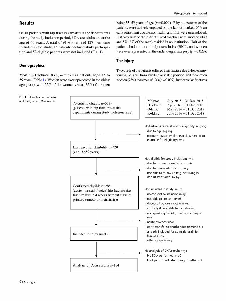
Of all patients with hip fractures treated at the departments during the study inclusion period, 6% were adults under the age of 60 years. A total of 91 women and 127 men were included in the study, 15 patients declined study participa-tion and 52 eligible patients were not included (Fig. 1).
Demographics
Most hip fractures, 83%, occurred in patients aged 45 to 59 years (Table 1). Women were overrepresented in the oldest age group, with 52% of the women versus 35% of the men
being 55–59 years of age (p = 0.009). Fifty-six percent of the patients were actively engaged on the labour market, 26% on early retirement due to poor health, and 11% were unemployed. Just over half of the patients lived together with another adult and 5% (8% of the men) resided in an institution. Half of the patients had a normal body mass index (BMI), and women were overrepresented in the underweight category (p = 0.023).
The injury
Two-thirds of the patients suffered their fracture due to low-energy trauma, i.e. a fall from standing or seated position, and more often women (78%) than men (61%) (p = 0.007). Intracapsular fractures
Fig. 1 Flowchart of inclusion and analysis of DXA results
Potentially eligible n=5525
(patients with hip fractures at the
departments during study inclusion time)
Examined for eligibility n=320
(age 18≤59 years)
Confirmed eligible n=285
(acute non-pathological hip fracture (i.e.
fracture within 4 weeks without signs of
primary tumour or metastasis))
Included in study n=218
No further examination for eligibility: n=5205 due to age n=5163 no investigator available at department to examine for eligibility n=42
Not eligible for study inclusion: n=35 due to tumour or metastasis n=6 due to non-acute fracture n=5 not able to follow up (e.g. not living in department area) n=24
Not included in study: n=67 no consent to inclusion n=15 not able to consent n=16 deceased before inclusion n=4 critically ill, not able to include n=4 not speaking Danish, Swedish or English n=3 acute psychosis n=4 early transfer to another department n=7 already included for contralateral hip fracture n=1 other reason n=13
Malmö: July 2015 – 31 Dec 2018
Hvidovre: Apr 2016 – 31 Dec 2018
Odense: May 2016 – 31 Dec 2018
Kolding: June 2016 – 31 Dec 2018
Analysis of DXA results n=184
No analysis of DXA result: n=34 No DXA performed n=26 DXA performed later than 3 months n=8

Osteoporosis International
1 3
were slightly more common than extracapsular ones (Table 1). No patients were diagnosed with an atypical femur fracture.
Lifestyle factors
Ninety-two percent of the patients reported no specific die-tary preferences (Table 2). Regarding physical activity, less than half of the patients reached a BHW-PA score of 11, i.e. a recommended physical activity level of 150 min/week, and hand grip strength showed a wide range, 4.5–80.0 kg. Smokers accounted for 42% of the patients and 16% were previous smokers. AUDIT results ranged from minimum to maximum score and 19 women (25%) and 37 men (31%)
reported a hazardous or harmful alcohol use. DUDIT results ranged from 0 to 36 points and 4 women (5%) and 11 men (10%) report signs of drug-related problems.
Medical history
A history of previous disease was common; in all, 313 dis-eases, both potentially hip fracture associated and other dis-eases, were found in 144 patients (66%) (Table 3). A total of 105 patients (48%) presented 188 diseases potentially associated with hip fracture and any other disease(s) were present in 98 patients (45%). Women presented a larger pro-portion than men of both potentially hip fracture associated
Table 1 Patients’ characteristicsAge Women n = 91 Men n = 127 Total n = 218Min–max 23–59 years 28–59 years 23–59 yearsMedian (IQR) 55 (51–57) 51 (45–56) 53 (47–57)Age groups: 18–24 1 (1%) 0 1 (.5%)25–29 0 1 (1%) 1 (.5%)30–34 3 (3%) 3 (2%) 6 (3%)35–39 1 (1%) 11 (9%) 12 (6%)40–44 2 (2%) 15 (11%) 17 (8%)45–49 11 (12%) 22 (17%) 33 (15%)50–54 26 (29%) 30 (24%) 56 (26%)55–59 47 (52%) 45 (35%) 92 (42%)Occupation Women n = 88 Men n = 124 Total n = 212Employed (full/part-time, self-employed) 47 (53%) 72 (58%) 119 (56%)On sick leave 9 (10%) 3 (2%) 12 (6%)Early retirement/disability pension 22 (25%) 34 (27%) 56 (26%)Unemployed (less than 3 years) 3 (3%) 5 (4%) 8 (4%)Unemployed (more than 3 years) 7 (8%) 7 (6%) 14 (7%)Does not work (other reason) 0 3 (2%) 3 (1%)Household circumstances Women n = 90 Men n = 125 Total n = 215Living alone 34 (38%) 45 (36%) 79 (37%)Living with someone else 54 (60%) 70 (56%) 124 (58%)Lives at institution 2 (2%) 10 (8%) 12 (5%)Body mass index (kg/m2) Women n = 91 Men n = 121 Total n = 212Mean (SD) 22.96 (± 4.62) 24.08 (± 3.85) 23.6 (± 4.2)Min–max 13.6–36.1 13.9–35.8 13.6–36.1BMI distribution:Underweight (< 18.5) 15 (17%) 6 (5%) 21 (10%)Normal (18.5–24.99) 42 (46%) 73 (60%) 115 (54%)Overweight (25.0–29.99) 28 (31%) 37 (31%) 65 (31%)Obese (> 30.00) 6 (7%) 5 (4%) 11 (5%)Trauma mechanism Women n = 91 Men n = 127 Total n = 218Low-energy trauma 71 (78%) 77 (61%) 148 (68%)Not low-energy trauma 20 (22%) 50 (39%) 70 (32%)Fracture type Women n = 91 Men n = 127 Total n = 218Intracapsular fracture 55 (60%) 71 (56%) 126 (58%)Extracapsular fracture 36 (40%) 56 (44%) 92 (42%)

Osteoporosis International
1 3
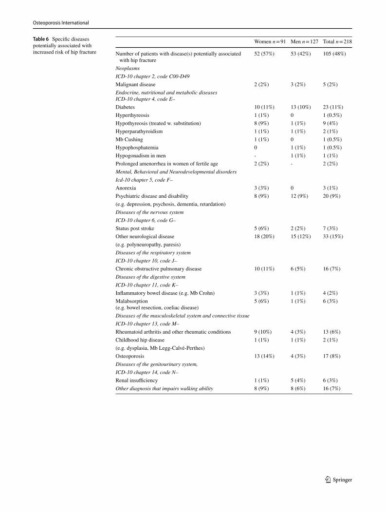
and other diseases (p = 0.025). The number of patients with multiple comorbidities is presented in Table 3. Specific dis-eases potentially associated with increased risk of hip frac-ture are presented in Appendix Table 6; neurological disease, diabetes, psychiatric disease and disability, osteoporosis, and chronic obstructive pulmonary disease were the most ubiqui-tous, accounting for 59% of the diseases.
Two-thirds of the patients were classified as ASA I or II, i.e. none or mild systemic disease, and the remaining patients were classified as ASA III–IV (Table 3). Half of the patients had a history of any previous fracture and 5% reported a previous hip fracture. In total, 17% of the cohort reported a family history of fragility fractures.
Previous medication
During the 5 years preceding the hip fracture, 135 patients (62%) had used any regular medication and 70 patients
(32%) had a total of 130 pharmacological treatments from medication groups potentially associated with increased fracture risk (Appendix Table 7). The most common treat-ments were proton pump inhibitors, selective serotonin reup-take inhibitors and opioids, together they accounted for 40% of the potentially hip fracture associated treatments.
Blood sample results
Blood samples were drawn on mean 1.4 (SD 1.1) days pre-operative and mean 2.04 (2.4) days post-operative. Results below reference in more than a quarter of the post-operative samples were found for calcium (29%), albumin (45%), vita-min D (52%), testosterone (60%) in men, and estradiol (85%) in women (Appendix Table 8). Of the pre-operative blood samples, leucocytes and CRP were above reference in 75% and 30%, and hemoglobin was below reference in 37% of the samples.
Table 2 Lifestyle factors
a Muslim, milk and cheese free, gluten free, phosphate reduced kidney dietb The Swedish Board of Health and Welfare physical activity questions (BHW-PA), a score of 11 and above fulfils WHO recommendationsc AUDIT Alcohol Use Disorders Identification Testd DUDIT Drug Use Disorders Identification Test
Diet Women n = 82 Men n = 121 Total n = 203Regular diet 78 (95%) 108 (89%) 186 (92%)Vegetarian/vegan 3 (4%) 3 (2%) 6 (3%)Diabetes diet 0 7 (6%) 7 (3%)Other a 1 (1%) 3 (3%) 4 (2%)Physical activity level b Women n = 85 Men n = 122 Total n = 207Median score (IQR) 9 (6–13) 9 (5–17) 9 (6–15)Physical activity level score ≥ 11 30 (35%) 54 (44%) 84 (41%)Hand grip strength (kg) Women n = 76 Men n = 116 Total n = 192Min–max 7.1–42.0 4.5–80.0 4.5–80.0Mean (SD) 28.1 (7.2) 46.1 (13.8) 39.0 (14.6)Smoking Women n = 88 Men n = 125 Total n = 213Never regular smoking 33 (38%) 57 (46%) 90 (42%)Previous smoker (quit > 2 years ago) 16 (18%) 18 (14%) 34 (16%)Current smoker (or quit < 2 years ago) 39 (44%) 50 (40%) 89 (42%)Pack years: Women n = 52 Men n = 58 Total n = 110Pack years min–max 0.3–70 0.9–107.5 0.3–107.5Pack years median (IQR) 25.4 (14.3–38.8) 30 (15–42.4) 29.3 (15–40)AUDIT c Women n = 76 Men n = 118 Total n = 194Hazardous or harmful alcohol use: 19 (25%) 37 (31%) 56 (29%)AUDIT min–max 0–40 0–36 0–40AUDIT median (IQR) 4 (1–5.75) 5 (3–9) 4 (2–8)DUDIT d Women n = 79 Men n = 111 Total n = 190Signs of drug-related problems: 4 (5%) 11 (10%) 15 (8%)DUDIT min–max 0–8 0–36 0–36DUDIT median (IQR) 0 (0–0) 0 (0–0) 0 (0–0)

Osteoporosis International
1 3
DXA results
A total of 184 patients’ DXA scan results were included in the analysis. They were aged 28–59 years, the median was 53 (IQR 47–57) years, compared to a median age of 55 (48–57) for the 26 patients not attending DXA and the 8 patients examined by DXA > 3 months post-fracture who were excluded from the analysis. Median time to DXA inves-tigation from hip fracture surgery was 5 (–324) days, 85% of the analyzed patients had DXA scans within 1 month. T-scores at the lumbar spine, total hip, and femoral neck were normal in 12%, osteopenic in 57%, and osteoporotic in 31% of the patients. The distribution was similar, with no statistically significant differences between women and men or between low-energy and not low-energy trauma mechanisms, but with a tendency of marginally better DXA results among patients with higher than low-energy trauma
mechanism (Table 4). In the youngest age groups, none of the patients had normal DXA results. Normal DXA was firstly seen in age group 40–44 and the highest proportion of normal results was found in age group 45–49 (27% normal).
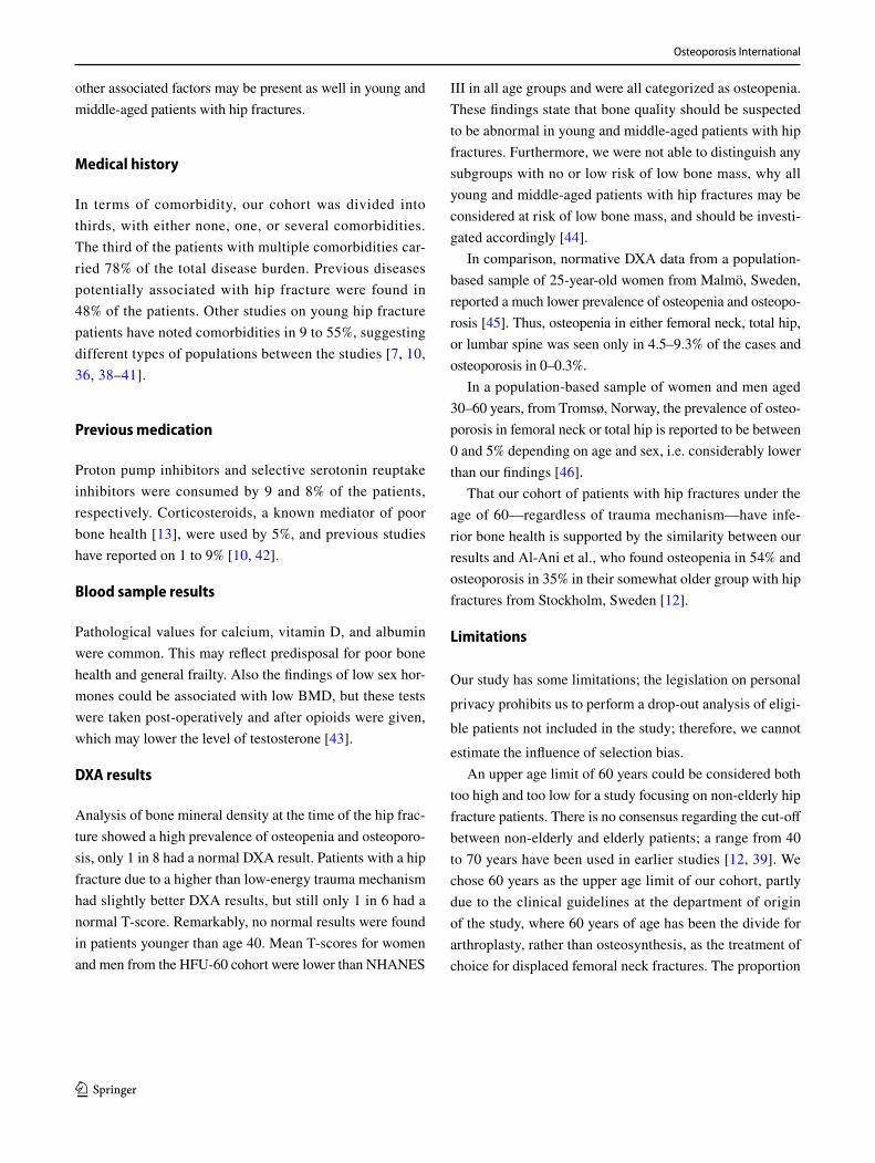
Mean T-scores at the femoral neck and total hip in our cohort were lower in all age groups for both women and men compared to NHANES III data (Fig. 2). NHANES III mean T-scores were all normal (≥ -1), but our cohort’s mean T-scores were categorized as osteopenia (< − 1 to > − 2.5) in all age groups. Mean T-scores for men from our cohort were lower with increasing age, as NHANES III mean T-scores for both men and women were. In contrast, mean T-scores in women from our cohort were higher with increasing age, both at the femoral neck and total hip.
Patient characteristics according to DXA result for 184 patients assessed by DXA are presented by sex in Appen-dix Table 9. Smoking, lower BMI, a family history of a fragility fracture, and low vitamin D were more frequent
Table 3 Medical history
a Specific diseases potentially associated with hip fracture are specified in Appendix Table 5
Women n = 91 Men n = 127 Total n = 218
Any previous disease 65 (71%) 79 (62%) 144 (66%)Patients with specific disease(s) potentially
associated with hip fracture a52 (57%) 53 (42%) 105 (48%)
Patients with any other disease(s) 49 (54%) 49 (39%) 98 (45%)Comorbidity (potentially hip fracture asso-
ciated and other diseases)Women n = 91 Men n = 127 Total n = 218
No previous disease 26 (29%) 48 (38%) 74 (34%)1 comorbidity 25 (28%) 43 (34%) 68 (31%)2 comorbidities 12 (13%) 20 (16%) 32 (15%)3 comorbidities 10 (11%) 10 (8%) 20 (9%)4 comorbidities 8 (9%) 3 (2%) 11 (5%)5 comorbidities 6 (7%) 1 (1%) 7 (3%)6 comorbidities 1 (1%) 1 (1%) 2 (1%)7 comorbidities 1 (1%) 1 (1%) 2 (1%)8 comorbidities 2 (2%) 0 2 (1%)ASA classification Women n = 91 Men n = 127 Total n = 218ASA I 16 (18%) 44 (35%) 60 (28%)ASA II 42 (46%) 44 (35%) 86 (39%)ASA III 31 (34%) 35 (28%) 66 (30%)ASA IV 2 (2%) 4 (3%) 6 (3%)Hospital admission Women n = 90 Men n = 124 Total n = 214Hospital admission within last year 24 (27%) 27 (22%) 51 (24%)Previous hip fracture Women n = 90 Men n = 124 Total n = 214Yes, contralateral 3 (3%) 6 (5%) 9 (4%)Yes, ipsilateral 0 1 (1%) 1 (.5%)Previous fracture (after 20 years of age) Women n = 87 Men n = 121 Total n = 208Previous other fracture 44 (51%) 53 (44%) 97 (47%)Fragility fracture in the family Women n = 86 Men n = 123 Total n = 209Yes 21 (24%) 14 (11%) 35 (17%)No 49 (57%) 82 (67%) 131 (63%)Do not know 16 (19%) 27 (22%) 43 (21%)

Osteoporosis International
1 3
Table 4 DXA diagnosis by sex, age group, and trauma mechanism
Diagnosis based on the lowest T-score on the lumbar spine, femoral neck, or total hip DXA investigation performed at the time of the fracture. Low-energy trauma was defined as a fall from standing or seated position, and any higher degree of trauma energy was classified as not low-energy trauma
NormalT-score ≥ − 1
OsteopeniaT-score − 2.5 to − 1
OsteoporosisT-score ≤ − 2.5
SexWomen n = 76 9 (12%) 41 (54%) 26 (34%)Men n = 108 13 (12%) 64 (60%) 31 (29%)Age groups25–29 n = 1 0 1 (100%) 030–34 n = 6 0 4 (67%) 2 (33%)35–39 n = 11 0 10 (91%) 1 (9%)40–44 n = 15 2 (13%) 8 (53%) 5 (33%)45–49 n = 26 7 (27%) 13 (50%) 6 (23%)50–54 n = 51 3 (6%) 31 (61%) 17 (33%)55–59 n = 74 10 (14%) 38 (51%) 26 (35%)Trauma mechanismLow-energy trauma n = 120 12 (10%) 65 (54%) 43 (36%)Not low-energy trauma n = 64 10 (16%) 40 (63%) 14 (22%)Totaln = 184 22 (12%) 105 (57%) 57 (31%)
-2.5
-2
-1.5
-1
-0.5
0
0.5
20-29 30-39 40-49 50-59
NHANES III TH men NHANES III FN men NHANES III TH women NHANES III FN women
HFU-60 TH men HFU-60 FN men HFU-60 TH women HFU-60 FN women
Age groups
Mea
n t-s
core
Fig. 2 Comparison of mean T-score by age group, HFU-60 vs. NHANES III. Multiple line chart of HFU-60 mean T-scores for women and men compared to NHANES III mean T-scores calculated from BMD data [25]. TH total hip, FN femoral neck. NHANES III mean BMD data for age groups were converted to T-scores using
the formula: T-score = (measured BMD – young adult mean BMD) / young adult population SD [23]. Mean T-scores for HFU-60 were significantly lower than NHANES III regarding both TH and FN for men (p = < .001), TH for women (p = .020), and FN for women (p = .027)
Osteoporosis International
1 3
in both women and men with low DXA results. Previous disease, previous fracture, and a higher number of comor-bidities were more common in women with low DXA. For men with low DXA, a higher AUDIT score and hip fracture due to low-energy trauma were more frequent.
Discussion
In the present study on patients with hip fractures under 60 years of age, we found several risk factors for osteoporosis and fractures in our study cohort, and also a high prevalence of osteopenia and osteoporosis upon DXA investigation at the time of the hip fracture.
Demographics
In contrast to hip fractures in the elderly, it has been shown before that the younger the patients, the larger the propor-tion of men is [3, 7, 9, 11]. Accordingly, 58% of our cohort were men. The explanation is probably a combination of multiple reasons resulting in young men being at higher risk of fractures than young women; in our cohort, the men presented heavier smoking, more alcohol and drug use, and more fractures due to a higher than low-energy trauma mechanism suggesting a more risk-taking behaviour in men.
The fact that only half of the participants were working, compared to four-fifths among the general population 20 to 64 years of age in Denmark and Sweden[27], reflects a socioeconomic distress known to be associated with an increased fracture risk [28].
The injury
Two-thirds of the hip fractures in our cohort were related to low-energy trauma, i.e. a fall from standing or a seated position, in contrast to the preconception that hip fractures in younger patients are caused mainly by high-energy trauma. Previous studies present large variations in trauma mecha-nisms, depending on inclusion criteria and settings. Studies on general hip fracture populations like ours support our finding that low-energy trauma was the cause for a majority of the fractures [1, 9, 10].
No patients were diagnosed with an atypical femur frac-ture; considering the previously reported low incidence of 1.74 fractures per 10 000 patient-years [29] and that only 7 patients used bisphosphonates, no atypical femur fractures were expected in the cohort.
Lifestyle factors
The physical activity level in our cohort was lower than a Swedish random population sample measured by the same questionnaire [15]. More than half of our study participants had a lower physical activity level than the recommended minimum according to the WHO of 150 min per week [17]. In contrast, 72% of the general population in Denmark and 66% in Sweden reach the recommended minimum physical activity level [30, 31].
On hand grip strength measurement, 57% of the women and 58% of the men had results lower than the mean from a random population sample of Danish women and men aged 19 to 72 years [18]. Considering that the hand grip strength was lower with increasing age in the general population, our younger cohort performed markedly lower results than the general population of comparable ages.
Smoking was 2.5 to 3.8 times as common in the study cohort than in the general population, where 17% smoke in Denmark and 11% in Sweden [32, 33]. The rate of smok-ers was also higher than in most other young hip fracture cohorts [1, 2, 34] but on par with Al-Ani et al. [12].
Among our study participants, harmful use of alcohol and drug-related problems were much more common than what is reported from the general population in the countries. One quarter of the women had a harmful alcohol consumption, and the Swedish average is suggested to be 11% [19]. The corresponding number for men was 31% in the study, and population data suggest 18 to 23% in Sweden and Denmark [19, 35]. In other observational studies, the presence of alco-hol use disorder varies from 15 to 38% [2, 9, 10, 36, 37]. A cohort from Scotland, described by Stearns et al., exhibit extreme use of alcohol and tobacco, 47 and 67%, but is char-acterized as a “largely very deprived population” [38].
On the DUDIT questionnaire, 8% of the study cohort presented a result indicating a drug-related problem, which is close to 3 times higher than previously reported from the Swedish population [20]. To the best of our knowledge, no earlier studies on patients with hip fractures have used DUDIT to estimate substance use disorder; wherefore, comparison is not possible. Still, the majority of the fracture patients did not have alcohol or substance use disorder, which is other-wise a perfunctory explanation often heard—that younger patients with hip fractures are “addicts”. This suggests that
Osteoporosis International
1 3
other associated factors may be present as well in young and middle-aged patients with hip fractures.
Medical history
In terms of comorbidity, our cohort was divided into thirds, with either none, one, or several comorbidities. The third of the patients with multiple comorbidities car-ried 78% of the total disease burden. Previous diseases potentially associated with hip fracture were found in 48% of the patients. Other studies on young hip fracture patients have noted comorbidities in 9 to 55%, suggesting different types of populations between the studies [7, 10, 36, 38–41].
Previous medication
Proton pump inhibitors and selective serotonin reuptake inhibitors were consumed by 9 and 8% of the patients, respectively. Corticosteroids, a known mediator of poor bone health [13], were used by 5%, and previous studies have reported on 1 to 9% [10, 42].
Blood sample results
Pathological values for calcium, vitamin D, and albumin were common. This may reflect predisposal for poor bone health and general frailty. Also the findings of low sex hor-mones could be associated with low BMD, but these tests were taken post-operatively and after opioids were given, which may lower the level of testosterone [43].
DXA results
Analysis of bone mineral density at the time of the hip frac-ture showed a high prevalence of osteopenia and osteoporo-sis, only 1 in 8 had a normal DXA result. Patients with a hip fracture due to a higher than low-energy trauma mechanism had slightly better DXA results, but still only 1 in 6 had a normal T-score. Remarkably, no normal results were found in patients younger than age 40. Mean T-scores for women and men from the HFU-60 cohort were lower than NHANES
III in all age groups and were all categorized as osteopenia. These findings state that bone quality should be suspected to be abnormal in young and middle-aged patients with hip fractures. Furthermore, we were not able to distinguish any subgroups with no or low risk of low bone mass, why all young and middle-aged patients with hip fractures may be considered at risk of low bone mass, and should be investi-gated accordingly [44].
In comparison, normative DXA data from a population-based sample of 25-year-old women from Malmö, Sweden, reported a much lower prevalence of osteopenia and osteopo-rosis [45]. Thus, osteopenia in either femoral neck, total hip, or lumbar spine was seen only in 4.5–9.3% of the cases and osteoporosis in 0–0.3%.
In a population-based sample of women and men aged 30–60 years, from Tromsø, Norway, the prevalence of osteo-porosis in femoral neck or total hip is reported to be between 0 and 5% depending on age and sex, i.e. considerably lower than our findings [46].
That our cohort of patients with hip fractures under the age of 60—regardless of trauma mechanism—have infe-rior bone health is supported by the similarity between our results and Al-Ani et al., who found osteopenia in 54% and osteoporosis in 35% in their somewhat older group with hip fractures from Stockholm, Sweden [12].
Limitations
Our study has some limitations; the legislation on personal privacy prohibits us to perform a drop-out analysis of eligi-ble patients not included in the study; therefore, we cannot estimate the influence of selection bias.
An upper age limit of 60 years could be considered both too high and too low for a study focusing on non-elderly hip fracture patients. There is no consensus regarding the cut-off between non-elderly and elderly patients; a range from 40 to 70 years have been used in earlier studies [12, 39]. We chose 60 years as the upper age limit of our cohort, partly due to the clinical guidelines at the department of origin of the study, where 60 years of age has been the divide for arthroplasty, rather than osteosynthesis, as the treatment of choice for displaced femoral neck fractures. The proportion
Osteoporosis International
1 3
of non-elderly individuals of all hip fracture patients is 2–13% according to previous reports [4, 6], the proportion in our material is 6% which corresponds well considering the variation in age limits in previous studies.
One could argue that there are few very young patients in our cohort, 91% of the patients are aged 40 to 59 years, but this is coherent with a Danish register study where 90% of hip fractures in patients under 65 years of age are found in patients aged 40 to 65 [4]. In a Swedish register study on patients with hip fractures younger than 50 years, the median age was 42 [47]. When excluding patients aged 50–59, our cohort show a corresponding median age of 44; hence, we consider our sample sufficiently representative.
Strengths
We consider our study to have important strengths; three quarters of the eligible cases were included in this multi-center prospective study. The patients were thoroughly investigated regarding demographics, trauma mechanism, lifestyle factors, comorbidity, and medication as well as by blood samples and DXA, providing an extensive descrip-tion of the patient group. The participating hospitals served both urban and rural catchment areas and provided care for all fracture cases regardless of trauma or patient type. Care given at low or no cost for the patients ensures that no one abstains from seeking hospital care. Thereby, we regard our study population to reflect the entire, heterogenous group of individuals suffering hip fractures in young and middle age. This is in contrast to studies performed at Level I trauma centers or health care systems were socially deprived indi-viduals have little access to hospital care, leading to selec-tion bias [7]. Our results are generalizable to many high-income countries, whilst other parts of the world may face more traffic or occupational injuries and a different case mix [8, 41].
We believe that DXA performed at the time of hip frac-ture in contrast to years after, more accurately describes the pre-fracture condition. Al-Ani et al. [12] have presented similar DXA results adjacent to the fracture, but in a smaller and older patient group. To put our DXA results in rela-tion to normative data, we have compared our findings to
reference populations regarding different ages and sex, both internationally used reference data (NHANES III) [25] and locally collected normative DXA result data [45, 46]. The comparisons support the argument that the bone health of young and middle-aged patients with hip fractures is inferior to what could be expected in the general population of the same ages.
Conclusion
Our cohort of patients with hip fractures under the age of 60 is heterogenous; the patients present a wide range of demo-graphics and lifestyle factors as well as previous fractures and comorbidities. Based on our findings, young and mid-dle-aged patients with hip fractures show signs of vitality and health, yet primarily—and more concerning—there is a high degree of frailty and risk factors for osteoporosis and fractures are numerous. We also found a high prevalence of osteopenia and osteoporosis compared to the general popula-tion, only one in eight had a normal DXA result.
Clinical perspective
The majority of the patients had previous medical conditions and abnormal blood sample results as well as inferior bone quality on BMD assessment by DXA. We suggest that all young and middle-aged patients with hip fractures should undergo a thorough health investigation including DXA, a non-invasive and relatively easily accessible procedure that previously have been reported to be performed in only less than half of young patients with hip fractures [1].
The variation in patient characteristics and physical abilities at the time of the hip fracture suggests that these patients have different needs regarding rehabilitation to reach their pre-fracture functional level and demands. Other rehabilitation pathways tailored to the needs of these patients, not only standard geriatric hip fracture rehabilita-tion, are assumingly needed. This will be analyzed further in future studies within the HFU-60 project.

Osteoporosis International
1 3
Appendix
Table 5 Definition of recorded variables
a The personal number is a national identification number including information on birth date and sex, unique to every individual, used nationally in both Denmark and Sweden
Variable Data source Method of assessment (measurement)
DemographicsAge Personal number a Age at fracture according to birth dateSex Personal number a Sex as defined by personal numberOccupation Patient interview Pre-defined category selected by patientHousehold circumstances Patient interview Pre-defined category selected by patientBMI Patient interview and medical charts Weight divided by squared length (kg/m2)The injuryTrauma mechanism Patient interview and medical charts Defined as low-energy or not low-energyFracture type Radiographs Classified by orthopaedic surgeonLifestyle factorsSmoking Patient interview Pre-defined category selected by patientPack years Patient interview Average number of cigarettes/day x years smokingDiet Patient interview Pre-defined category selected by patientAUDIT [19] Patient questionnaire Score according to questionnaire instructionsDUDIT [20] Patient questionnaire Score according to questionnaire instructionsPhysical activity level score (BHW-PA [15]) Patient interview Score according to questionnaire instructionsHand grip strength Functional test by physiotherapist Measured in kg by dynamometerMedical historyAny previous disease Patient interview and medical charts Presence of previous diseasePotentially hip fracture associated previous disease Patient interview and medical charts Presence of pre-specified previous diseaseHospital admission within 1 year prior to hip fracture Patient interview and medical charts Defined as yes or noASA classification [14] Medical charts Assessed by anesthesiologistPrevious hip fracture Patient interview and medical charts Defined as yes or noPrevious other fracture (after 20 years of age) Patient interview and medical charts Defined as yes or noFragility fracture in first-hand relative Patient interview and medical charts Defined as yes or noPrevious medication Patient interview and medical charts Presence of pre-specified medical treatmentsBlood sample result Medical charts Defined as normal or below/above referenceDXA result DXA investigation Result defined according to WHO definitions
Osteoporosis International
1 3
Table 6 Specific diseases potentially associated with increased risk of hip fracture
Women n = 91 Men n = 127 Total n = 218
Number of patients with disease(s) potentially associated with hip fracture
52 (57%) 53 (42%) 105 (48%)
NeoplasmsICD-10 chapter 2, code C00-D49Malignant disease 2 (2%) 3 (2%) 5 (2%)Endocrine, nutritional and metabolic diseasesICD-10 chapter 4, code E–Diabetes 10 (11%) 13 (10%) 23 (11%)Hyperthyreosis 1 (1%) 0 1 (0.5%)Hypothyreosis (treated w. substitution) 8 (9%) 1 (1%) 9 (4%)Hyperparathyroidism 1 (1%) 1 (1%) 2 (1%)Mb Cushing 1 (1%) 0 1 (0.5%)Hypophosphatemia 0 1 (1%) 1 (0.5%)Hypogonadism in men - 1 (1%) 1 (1%)Prolonged amenorrhea in women of fertile age 2 (2%) - 2 (2%)Mental, Behavioral and Neurodevelopmental disordersIcd-10 chapter 5, code F–Anorexia 3 (3%) 0 3 (1%)Psychiatric disease and disability 8 (9%) 12 (9%) 20 (9%)(e.g. depression, psychosis, dementia, retardation)Diseases of the nervous systemICD-10 chapter 6, code G–Status post stroke 5 (6%) 2 (2%) 7 (3%)Other neurological disease 18 (20%) 15 (12%) 33 (15%)(e.g. polyneuropathy, paresis)Diseases of the respiratory systemICD-10 chapter 10, code J–Chronic obstructive pulmonary disease 10 (11%) 6 (5%) 16 (7%)Diseases of the digestive systemICD-10 chapter 11, code K–Inflammatory bowel disease (e.g. Mb Crohn) 3 (3%) 1 (1%) 4 (2%)Malabsorption(e.g. bowel resection, coeliac disease)
5 (6%) 1 (1%) 6 (3%)
Diseases of the musculoskeletal system and connective tissueICD-10 chapter 13, code M–Rheumatoid arthritis and other rheumatic conditions 9 (10%) 4 (3%) 13 (6%)Childhood hip disease 1 (1%) 1 (1%) 2 (1%)(e.g. dysplasia, Mb Legg-Calvé-Perthes)Osteoporosis 13 (14%) 4 (3%) 17 (8%)Diseases of the genitourinary system,ICD-10 chapter 14, code N–Renal insufficiency 1 (1%) 5 (4%) 6 (3%)Other diagnosis that impairs walking ability 8 (9%) 8 (6%) 16 (7%)

Osteoporosis International
1 3
Table 7 Previous potentially hip fracture associated medication
Women n = 91 Men n = 127 Total n = 218
Previous use of potentially hip fracture associated medication 34 (37%) 36 (29%) 70 (32%)
ATC code Example of use/diagnosis Example of medication name
A02BCProton pump inhibitors
Peptic ulcer, gastro-oesopha-geal reflux disease
Omeprazole, pantoprazole, lansoprazole
12 (13%) 7 (6%) 19 (9%)
N06ABSelective serotonin reuptake
inhibitors
Antidepressants Citalopram, sertraline, fluox-etine
10 (11%) 8 (6%) 18 (8%)
N02AOpioids
Pain Morphine, methadone, fentanyl 8 (9%) 7 (6%) 15 (7%)
H02ABGlucocorticoids
Systemic corticosteroids Prednisolone, betamethasone, hydrocortisone
6 (7%) 6 (5%) 12 (6%)
B01AAntithrombotic agents
Antithrombotic therapy Heparin, dalteparin, warfarin 4 (4%) 8 (6%) 12 (6%)
N03AFCarboxamide derivates
Antiepileptics Carbamazepine, oxcarbazepine, rufinamide
5 (5%) 5 (4%) 10 (5%)
C03CALoop-diuretics
Hypertension, heart failure Furosemide 5 (5%) 4 (3%) 9 (4%)
M01ANSAID
RA, osteoarthrosis, pain Diclofenac, ibuprofen 4 (4%) 5 (4%) 9 (4%)
H03AAThyroid hormones
Hypothyreosis Levothyroxine, liothyronine 7 (8%) 1 (1%) 8 (4%)
M05BBisphosphonates
Osteoporosis Aledronic acid, zoledronic acid 3 (3%) 4 (3%) 7 (3%)
G03Sex hormones
Hormonal therapy Androgens, estrogens 3 (3%) 2 (2%) 5 (2%)
L01, L04Antineoplastic and immu-
nomodulating agents
Cancer, rheumatoid arthritis Cyklophosphamide, busulfan, methotrexate
2 (2%) 2 (2%) 4 (2%)
L02BGAromatase inhibitors
Cancer (breast, endometrial, prostatic)
Anastrozole, letrozole, exemes-tane
1 (1%) 0 1 (0.5%)
G03AC06Progestogens
Contraceptive Medroxyprogesterone 1 (1%) 0 1 (0.5%)

Osteoporosis International
1 3
Table 8 Blood sample results
No. of samples Below reference Normal Above reference
Women Men Women Men Women Men Women MenPre-operativeHemoglobin 90 127 40 (44%) 41 (32%) 50 (56%) 84 (66%) 0 2 (2%)Leucocytes 89 124 1 (1%) 0 23 (26%) 29 (23%) 65 (73%) 95 (77%)Platelet count 72 93 9 (13%) 7 (8%) 57 (79%) 80 (86%) 6 (8%) 6 (7%)CRP 90 124 N/A N/A 72 (80%) 77 (62%) 18 (20%) 47 (38%)Sodium (Na +) 90 127 24 (27%) 28 (22%) 63 (70%) 98 (77%) 3 (3%) 1 (1%)Potassium (K +) 90 127 14 (16%) 11 (9%) 71 (79%) 100 (79%) 5 (6%) 16 (13%)Creatinine 89 127 33 (37%) 18 (14%) 52 (58%) 91 (72%) 4 (5%) 18 (14%)INR 83 112 N/A N/A 73 (88%) 100 (89%) 10 (12%) 12 (11%)Post-operativeCalcium-P 77 112 24 (31%) 31 (28%) 53 (69%) 80 (71%) 0 1 (1%)Albumin 69 101 40 (58%) 37 (37%) 29 (42%) 64 (63%) 0 025-hydroxyvitamin D 74 104 34 (46%) 59 (57%) 40 (54%) 45 (43%) N/A N/AAlkaline phosphatase 84 107 1 (1%) 3 (3%) 68 (81%) 83 (78%) 15 (18%) 21 (20%)PTH 79 114 0 3 (3%) 71 (90%) 96 (84%) 8 (10%) 15 (13%)TSH 77 115 5 (7%) 0 69 (87%) 107 (93%) 3 (4%) 8 (7%)T3 24 26 6 (25%) 1 (4%) 17 (71%) 24 (92%) 1 (4%) 1 (4%)T4 26 33 0 0 26 (100%) 30 (91%) 0 3 (9%)P-Testosterone – 108 – 65 (60%) – 43 (40%) – 0Estradiol 72 – 61 (85%) – 11 (15%) – 0 –Tissue transglutaminase antibody 75 109 N/A N/A 74 (99%) 109 (100%) 1 (1%) 0

Osteoporosis International
1 3
Table 9 Patient characteristics according to DXA result, focusing on risk factors for osteoporosis
*Variables with missing data, the number of observations for each variable is indicated for each cell with missing data
Women Men
Low DXAT-score < − 1 n = 67
Normal DXAT-score ≥ − 1 n = 9
Low DXAT-score < − 1 n = 95
Normal DXAT-score ≥ − 1 n = 13
Age median (IQR) 54 (51–57) 56 (50–58.5) 51 (44–56) 49 (45–56.5)Smoking* n = 66 n = 94Non-smoker 24 (36%) 5 (56%) 41 (44%) 9 (69%)Previous smoker 13 (20%) 1 (11%) 13 (14%) 2 (15%)Current smoker 29 (44%) 3 (33%) 40 (43%) 2 (15%)Pack years* n = 40 n = 4 n = 47 n = 4Min–max 0.3–54 6.8–47 0.9–107.5 10–52.5Median (IQR) 25 (13–37) 34 (11–47) 30 (18–44) 26 (11–49)AUDIT* n = 58 n = 8 n = 90High AUDIT 15 (26%) 2 (25%) 28 (31%) 3 (23%)Min–max 0–40 0–13 0–36 0–24Median (IQR) 4 (1–6) 4.5 (1.5–5.8) 5 (3–9) 3 (0.5–7.5)DUDIT* n = 61 n = 7 n = 83 n = 12High DUDIT 3 (5%) 1 (14%) 10 (12%) 0Min–max 0–5 0–8 0–36 0–0BMI* n = 91Min–max 16.7–33.9 23.1–36 15.8–35.8 21.7–34.6Mean (SD) 22.8 (4) 28.4 (4.4) 24.1 (3.7) 26.4 (3.3)Trauma mechanismLow-energy trauma 50 (75%) 7 (78%) 58 (61%) 5 (39%)Not low-energy trauma 17 (25%) 2 (22%) 37 (39%) 8 (62%)Any disease 47 (70%) 5 (56%) 56 (59%) 11 (85%)Potentially hip fracture associated disease 40 (60%) 2 (22%) 37 (39%) 6 (46%)Other disease(s) 34 (51%) 4 (44%) 34 (36%) 8 (62%)Hospital admission within last year* 16 (24%) 2 (22%) 19 (20%) n = 94 4 (31%)ASA classificationMean (SD) 2.09 (0.71) 2.11 (0.93) 1.95 (0.86) 1.85 (0.69)ASA I 14 (21%) 2 (22%) 35 (37%) 4 (31%)ASA II 33 (49%) 5 (56%) 32 (34%) 7 (54%)ASA III 20 (30%) 1 (11%) 26 (27%) 2 (15%)ASA IV 0 1 (11%) 2 (2%) 0Comorbidity calculationMin–max 0–7 0–7 0–7 0–5Mean (SD) 1.82 (1.8) 1.33 (2.3) 1.09 (1.3) 1.69 (1.4)Any previous fracture* 34 (51%) 2 (22%) 37 (40%) n = 93 11 (85%)Fragility fracture in the family* 19 (30%) n = 64 1 (11%) 13 (14%) n = 92 0Previous potentially hip fracture associated
medication26 (39%) 3 (33%) 28 (30%) 4 (31%)
Blood samples below reference*Calcium 18 (31%) n = 59 2 (29%) n = 7 23 (26%) n = 86 4 (36%) n = 11Albumin 29 (55%) n = 53 4 (67%) n = 6 26 (34%) n = 76 4 (44%) n = 9Vitamin D 26 (46%) n = 56 1 (17%) n = 6 46 (59%) n = 78 4 (36%) n = 11Testosterone – – 47 (57%) n = 82 9 (75%) n = 12Estradiol 46 (85%) n = 54 5 (71%) n = 7 – –
Osteoporosis International
1 3
Acknowledgements During this work, we received help and support from people outside the author group, thank you all. We would like to mention the local DXA units for their help, with the investigation of patients and also when the first author visited with the phantom. Thank you to our local assistants for your help and administrative sup-port, especially project coordinator Marika Hell in Malmö, Anne Jess Hansen in Kolding, and project nurse Annie Gam-Pedersen in Odense. We would also like to acknowledge the work of the staff at the local wards and out-patient clinics where our patients were treated, and specifically the physiotherapists who performed the functional assess-ments. Thanks as well for the use of REDCap as part of OPEN, Open Patient data Explorative Network, Odense University Hospital, Region of Southern Denmark.
Funding Open access funding provided by Lund University. This work was supported by grants from Greta and Johan Kock Foundation, A. Påhlsson Foundation, H Järnhardt foundation, Skåne University Hos-pital Research Fund, the Research and Development Council of Region Skåne, the Swedish Research Council funding for clinical research in medicine, and “Region Syddanmarks forskningsfond” from the Region of Southern Denmark. None of the funders had influence on the scien-tific work of this study.
Data availability The data that support the findings of this study are available on request from the corresponding author. The data are not publicly available due to privacy or ethical restrictions.
Declarations None of the authors has potential conflicts of interest related to the study. Unrelated to the study, the authors have the fol-lowing potential conflicts of interest to disclose:
Bjarke Viberg: paid lectures from Osmedic, Swemac and Zimmer Bi-omet.Jens-Erik Beck Jensen: advisory boards Amgen, Eli Lilly, UCB, Ge-dion Richter. Speakers bureau Amgen, UCB, Utsuka. Grants Eli Lilly, Amgen.Kristina E Akesson: advisory boards Amgen, Astellas Pharma, FAN Network, UCB. Speakers bureau Amgen, Astellas Pharma, Chugai, UCB.Sebastian Strøm Rönnquist, Morten Tange Kristensen, Henrik Palm, Carsten Fladmose Madsen, Søren Overgaard, Cecilia Rogmark: noth-ing to declare.
Open Access This article is licensed under a Creative Commons Attri-bution-NonCommercial 4.0 International License, which permits any non-commercial use, sharing, adaptation, distribution and reproduction in any medium or format, as long as you give appropriate credit to the original author(s) and the source, provide a link to the Creative Com-mons licence, and indicate if changes were made. The images or other third party material in this article are included in the article's Creative Commons licence, unless indicated otherwise in a credit line to the material. If material is not included in the article's Creative Commons licence and your intended use is not permitted by statutory regula-tion or exceeds the permitted use, you will need to obtain permission directly from the copyright holder. To view a copy of this licence, visit http:// creat iveco mmons. org/ licen ses/ by- nc/4. 0/.
References
1. Wang MT, Yao SH, Wong P et al (2017) Hip fractures in young adults: a retrospective cross-sectional study of characteristics, injury mechanism, risk factors, complications and follow-up. Arch Osteoporos 12:46. https:// doi. org/ 10. 1007/ s11657- 017- 0339-y
2. Karantana A, Boulton C, Bouliotis G, et al. (May) Epidemiology and outcome of fracture of the hip in women aged 65 years and under: a cohort study. J Bone Jt Surg Br 93:658–64. 93-B/5/658 [pii] https:// doi. org/ 10. 1302/ 0301- 620X. 93B5. 24536
3. Robinson CM, Court-Brown CM, McQueen MM, Christie J (1995) Hip fractures in adults younger than 50 years of age. Epi-demiology and results. Clin Orthop Relat Res 238–46
4. Omari A, Madsen CM, Lauritzen JB et al (2019) Comorbidity and mortality after hip fracture in nineteen thousand six hundred and eighty two patients aged eighteen to sixty five years in Den-mark from 1996 to 2012. Int Orthop. https:// doi. org/ 10. 1007/ s00264- 019- 04323-z
5. Cheng K, Montgomery S, Housley S, Wheelwright E (2009) Clinical risk factors for hip fracture in young adults under 50 years old. Eur J Trauma Emerg Surg 35:40–42. https:// doi. org/ 10. 1007/ s00068- 008- 7177-y
6. Rogmark C, Kristensen MT, Viberg B et al (2018) Hip fractures in the non-elderly—who, why and whither? Injury 49:1445–1450. https:// doi. org/ 10. 1016/j. injury. 2018. 06. 028
7. Swiontkowski MF, Winquist RA, Hansen ST (1984) Fractures of the femoral neck in patients between the ages of twelve and forty-nine years. J Bone Jt Surg Am 66:837–846
8. Gupta M, Arya R-K, Kumar S et al (2016) Comparative study of multiple cancellous screws versus sliding hip screws in femo-ral neck fractures of young adults. Chin J Traumatol Zhonghua Chuang Shang Za Zhi 19:209–212. https:// doi. org/ 10. 1016/j. cjtee. 2015. 11. 021
9. Lofthus CM, Osnes EK, Meyer HE et al (2006) Young patients with hip fracture: a population-based study of bone mass and risk factors for osteoporosis. Osteoporos Int 17:1666–1672. https:// doi. org/ 10. 1007/ s00198- 006- 0176-0
10. Al-Ani AN, Neander G, Samuelsson B et al (2013) Risk factors for osteoporosis are common in young and middle-aged patients with femoral neck fractures regardless of trauma mechanism. Acta Orthop 84:54–59. https:// doi. org/ 10. 3109/ 17453 674. 2013. 765639
11. Boden SD, Labropoulos P, Saunders R (1990) Hip fractures in young patients: is this early osteoporosis? Calcif Tissue Int 46:65–72. https:// doi. org/ 10. 1007/ BF025 56089
12. Al-Ani AN, Cederholm T, Sääf M et al (2015) Low bone mineral density and fat-free mass in younger patients with a femoral neck fracture. Eur J Clin Invest 45:800–806. https:// doi. org/ 10. 1111/ eci. 12472
13. Kanis JA (2002) Diagnosis of osteoporosis and assessment of fracture risk. THE LANCET 359:8
14. ASA Physical Status Classification System. https:// www. asahq. org/ stand ards- and- guide lines/ asa- physi cal- status- class ifica tion- system. Accessed 3 Feb 2021
15. Olsson SJG, Ekblom Ö, Andersson E et al (2016) Categorical answer modes provide superior validity to open answers when asking for level of physical activity: a cross-sectional study. Scand J Public Health 44:70–76. https:// doi. org/ 10. 1177/ 14034 94815 602830
16. Kallings L Validering av Socialstyrelsens Screeningfrågor om Fysisk Aktivitet. GIH - The Swedish School of Sport and Health Sciences. https:// www. socia lstyr elsen. se/ globa lasse ts/ share point- dokum ent/ dokum ent- webb/ natio nella- riktl injer/ levna dsvan or- valid ering- av- indik atorf ragor- till- patie nter- om- fysisk- aktiv itet. pdf. Accessed 5 Dec 2020
17. World Health Organization (2020) WHO guidelines on physical activity and sedentary behaviour. https:// apps. who. int/ iris/ handle/ 10665/ 336656. Accessed 31 Jan 2021
18. Aadahl M, Beyer N, Linneberg A et al (2011) Grip strength and lower limb extension power in 19–72-year-old Danish men and women: the Health 2006 study. BMJ Open 1:e000192. https:// doi. org/ 10. 1136/ bmjop en- 2011- 000192

Osteoporosis International
1 3
19. Bergman H (2002) Alcohol use among Swedes and a psychomet-ric evaluation of the Alcohol Use Disorders Identification Test. Alcohol Alcohol 37:245–251. https:// doi. org/ 10. 1093/ alcalc/ 37.3. 245
20. Berman AH, Bergman H, Palmstierna T, Schlyter F (2005) Evalu-ation of the Drug Use Disorders Identification Test (DUDIT) in criminal justice and detoxification settings and in a Swedish popu-lation sample. Eur Addict Res 11:22–31. https:// doi. org/ 10. 1159/ 00008 1413
21. Saunders JB, Aasland OG, Babor TF et al (1993) Development of the alcohol use disorders identification test (AUDIT): WHO collaborative project on early detection of persons with harmful alcohol consumption-II. Addiction 88:791–804. https:// doi. org/ 10. 1111/j. 1360- 0443. 1993. tb020 93.x
22. For the IOF Committee of Scientific Advisors Working Group on Osteoporosis Pathophysiology, Ferrari S, Bianchi ML et al (2012) Osteoporosis in young adults: pathophysiology, diagnosis, and management. Osteoporos Int 23:2735–2748. https:// doi. org/ 10. 1007/ s00198- 012- 2030-x
23. Blake GM, Fogelman I (2007) The role of DXA bone density scans in the diagnosis and treatment of osteoporosis. Postgrad Med J 83:509–517. https:// doi. org/ 10. 1136/ pgmj. 2007. 057505
24. Karlsson M, Nilsson JÅ, Sernbo I et al (1996) Changes of bone mineral mass and soft tissue composition after hip fracture. Bone 18:19–22. https:// doi. org/ 10. 1016/ 8756- 3282(95) 00422-X
25. Looker AC, Wahner HW, Dunn WL et al (1998) Updated data on proximal femur bone mineral levels of US adults. Osteoporos Int 8:468–490. https:// doi. org/ 10. 1007/ s0019 80050 093
26. Hanson J (1997) Standardization of femur BMD. J Bone Miner Res 12:1316–1317. https:// doi. org/ 10. 1359/ jbmr. 1997. 12.8. 1316
27. Statistics | Eurostat. https:// ec. europa. eu/ euros tat/ datab rowser/ view/ t2020_ 10/ defau lt/ table? lang= en. Accessed 14 Jul 2021
28. Stenholm S, Vahtera J, Kjeldgård L et al (2015) Length of sick leave as a risk marker of hip fracture: a nationwide cohort study from Sweden. Osteoporos Int 26:943–949. https:// doi. org/ 10. 1007/ s00198- 014- 2985-x
29. Black DM, Geiger EJ, Eastell R et al (2020) Atypical femur fracture risk versus fragility fracture prevention with bisphos-phonates. N Engl J Med 383:743–753. https:// doi. org/ 10. 1056/ NEJMo a1916 525
30. Denmark - Physical activity factsheet – WHO/Europe. https:// www. euro. who. int/ en/ health- topics/ disea se- preve ntion/ physi cal- activ ity/ data- and- stati stics/ physi cal- activ ity- fact- sheets/ physi cal- activ ity- count ry- facts heets/ denma rk. Accessed 2 Feb 2021
31. SWEDEN Physical Activity Factsheet-WHO Europe. https:// www. euro. who. int/__ data/ assets/ pdf_ file/ 0009/ 288126/ SWE-DEN- Physi cal- Activ ity- Facts heet. pdf. Accessed 31 Jan 2021
32. Sundhedsstyrelsen (2014) Danskernes sundhed - Den Nationale Sundhedsprofil 2013. https:// www. sst. dk/-/ media/ Udgiv elser/ 2014/ Den- natio nale- sundh edspr ofil- 2013/ Dansk ernes- sundh ed,-d- ,- Den- natio nale- sundh edspr ofil- 2013. ashx. Accessed 19 Jan 2021
33. Färre röker, fler snusar. In: Stat. Cent. http:// www. scb. se/ hitta- stati stik/ artik lar/ 2018/ farre- roker- fler- snusar/. Accessed 19 Jan 2021
34. Razik F, Alexopoulos AS, El-Osta B et al (2012) Time to inter-nal fixation of femoral neck fractures in patients under sixty years–does this matter in the development of osteonecrosis of
femoral head? Int Orthop 36:2127–2132. https:// doi. org/ 10. 1007/ s00264- 012- 1619-1
35. Beich A, Gannik D, Saelan H, Thorsen T (2007) Screening and brief intervention targeting risky drinkers in Danish general practice–a pragmatic controlled trial. Alcohol Alcohol Oxf Oxfs 42:593–603. https:// doi. org/ 10. 1093/ alcalc/ agm063
36. Samuel AM, Russo GS, Lukasiewicz AM et al (2016) Surgical treatment of femoral neck fractures after 24 hours in patients between the ages of 18 and 49 is associated with poor inpatient outcomes: an analysis of 1361 patients in the National Trauma Data Bank. J Orthop Trauma 30:89–94. https:// doi. org/ 10. 1097/ BOT. 00000 00000 000456
37. Zetterberg CH, Irstam L, Andersson GB (1982) Femoral neck fractures in young adults. Acta Orthop Scand 53:427–435
38. Stearns AT, Jaberoo MC, Ashraf R et al (2009) Displaced intra-capuslar hip fractures in the working age alcohol-abusing patient. Scott Med J 54:16–20. https:// doi. org/ 10. 1258/ rsmsmj. 54.1. 16
39. Lin JC, Wu CC, Lo C et al (2014) Mortality and complications of hip fracture in young adults: a nationwide population-based cohort study. BMC Musculoskelet Disord 15:362. https:// doi. org/ 10. 1186/ 1471- 2474- 15- 362
40. Verettas DA, Galanis B, Kazakos K et al (2002) Fractures of the proximal part of the femur in patients under 50 years of age. Injury 33:41–45
41. Upadhyay A, Jain P, Mishra P et al (2004) Delayed internal fixa-tion of fractures of the neck of the femur in young adults. A pro-spective, randomised study comparing closed and open reduction. J Bone Jt Surg Br 86:1035–1040
42. Jain R, Koo M, Kreder HJ, et al. (2002) Comparison of early and delayed fixation of subcapital hip fractures in patients sixty years of age or less. J Bone Jt Surg Am 84-A:1605–12
43. O’Rourke TK, Wosnitzer MS (2016) Opioid-induced andro-gen deficiency (OPIAD): diagnosis, management, and lit-erature review. Curr Urol Rep 17:76. https:// doi. org/ 10. 1007/ s11934- 016- 0634-y
44. Cummings SR, Eastell R (2020) Stop (mis)classifying fractures as high- or low-trauma or as fragility fractures. Osteoporos Int 31:1023–1024. https:// doi. org/ 10. 1007/ s00198- 020- 05325-z
45. Callréus M, McGuigan F, Akesson K (2014) Country-specific young adult dual-energy X-ray absorptiometry reference data are warranted for T-score calculations in women: data from the peak-25 cohort. J Clin Densitom Off J Int Soc Clin Densitom 17:129–135. https:// doi. org/ 10. 1016/j. jocd. 2013. 03. 008
46. Emaus N, Omsland TK, Ahmed LA et al (2009) Bone mineral density at the hip in Norwegian women and men—prevalence of osteoporosis depends on chosen references: the Tromsø Study. Eur J Epidemiol 24:321–328. https:// doi. org/ 10. 1007/ s10654- 009- 9333-z
47. Thoors O, Mellner C, Hedström M (2021) Good clinical outcome for the majority of younger patients with hip fractures: a Swedish nationwide study on 905 patients younger than 50 years of age. Acta Orthop 1–5. https:// doi. org/ 10. 1080/ 17453 674. 2021. 18769 96
Publisher's note Springer Nature remains neutral with regard to jurisdictional claims in published maps and institutional affiliations.
Osteoporosis International
1 3
Authors and Affiliations
Sebastian Strøm Rönnquist1 · Bjarke Viberg2 · Morten Tange Kristensen3,4,5 · Henrik Palm6 · Jens‑Erik Beck Jensen5,7 · Carsten Fladmose Madsen8 · Kristina E. Åkesson9,10 · Søren Overgaard11,12 · Cecilia Rogmark1
1 Department of Orthopaedics, Lund University, Skåne University Hospital, Malmö and Lund, Sweden
2 Department of Orthopaedic Surgery and Traumatology, Lillebaelt Hospital, University Hospital of Southern Denmark, Kolding, Denmark
3 Departments of Physiotherapy and Orthopaedic Surgery, Copenhagen University Hospital – Amager and Hvidovre, Copenhagen, Denmark
4 Department of Physio- and Occupational Therapy, Copenhagen University Hospital – Bispebjerg and Frederiksberg, Copenhagen, Denmark
5 Department of Clinical Medicine, University of Copenhagen, Copenhagen, Denmark
6 Department of Orthopaedic Surgery, Copenhagen University Hospital – Bispebjerg and Frederiksberg, University of Copenhagen, Copenhagen, Denmark
7 Endocrine Department, Hvidovre University Hospital, Copenhagen, Denmark
8 Department of Orthopaedic Surgery and Traumatology, Odense University Hospital, Odense, Denmark
9 Department of Clinical Sciences, Lund University, Malmö, Sweden
10 Department of Orthopaedics, Skåne University Hospital, Malmö, Sweden
11 Department of Orthopaedic Surgery and Traumatology, Copenhagen University Hospital – Bispebjerg and Frederiksberg, University of Copenhagen, Copenhagen, Denmark
12 Department of Clinical Medicine, Faculty of Health and Medical Sciences, University of Copenhagen, Copenhagen, Denmark
Related Documents